Staff Working Paper No. 987 - Collateral requirements in ...
Upload
independentCategory
view
1download
0
RITRMGC
Cbfmmmt
mabdcc
pA
d
2
Journal of the American College of Cardiology Vol. 48, No. 1, 2006© 2006 by the American College of Cardiology Foundation ISSN 0735-1097/06/$32.00P
apid Decline of Collateral Circulationncreases Susceptibility to Myocardial Ischemiahe Trade-Off of Successful Percutaneousecanalization of Chronic Total Occlusionsarco Zimarino, MD, PHD, Arturo Ausiello, MD, Gaetano Contegiacomo, MD, Irene Riccardi, MD,iulia Renda, MD, PHD, Cesare Di Iorio, MD, Raffaele De Caterina, MD, PHDhieti, Italy
OBJECTIVES We evaluated the time-behavior of changes in collateral circulation after successful percuta-neous coronary intervention (PCI) with drug-eluting stents (DES) in chronic (�1 month)total occlusions (CTO), and assessed their relationship with myocardial ischemia.
BACKGROUND It has been hypothesized that the immediate reduction of collateral flow after PCI of CTOcould expose the patients to a higher risk of future ischemic events in the case of vesselreocclusion.
METHODS In 42 patients with CTO, two consecutive balloon inflations and final DES deployment wereperformed after positioning of a pressure guidewire. Minimal lumen diameter (MLD),diameter stenosis (DS), angiographic collateral grading (Rentrop score), myocardial(FFRmyo), coronary (FFRcor), and collateral fractional flow reserve (FFRcoll) were evalu-ated. Chest pain and the sum of ST-segment elevation (�ST) were analyzed to document theoccurrence and extent of myocardial ischemia.
RESULTS Percutaneous coronary intervention induced a progressive improvement of indexes of stenosisseverity (MLD, DS, Thrombolysis in Myocardial Infarction flow, FFRmyo, and FFRcor) and arapid reduction in collateral circulation (FFRcoll and Rentrop score). A progressive worsening ofischemia at each balloon inflation occurred, concomitant with the reduction of collateralcirculation. At linear regression analysis, an inverse relationship of FFRcoll with �ST (R2 �0.352, p � 0.001) and angina pain score (R2 � 0.247, p � 0.001) was observed.
CONCLUSIONS In CTO, collateral circulation, which provides most coronary flow at baseline, rapidly declinesafter successful stent implantation and the restoration of an antegrade flow. This rapidde-recruitment of collaterals is likely to put such patients at risk of future ischemicevents. (J Am Coll Cardiol 2006;48:59–65) © 2006 by the American College of
ublished by Elsevier Inc. doi:10.1016/j.jacc.2005.12.079
Cardiology Foundation
ftfWdddws
M
ScdmrCci
ogs
oronary collateral circulation offers an alternative source oflood supply to the myocardium when the original vesselails to provide sufficient blood flow (1–3). Collaterals limityocardial ischemia during coronary occlusion (4), mini-ize the infarct area, and predict the presence of viableyocardium in patients with a history of myocardial infarc-
ion (5).
See page 66
Contrast angiography has been the most widely usedethod to assess coronary collaterals in the past (6). The
vailability of miniaturized sensors to monitor coronarylood flow and pressure in humans now allows a moreetailed evaluation of the collateral circulation during per-utaneous coronary interventions (PCI) after guidewirerossing of the lesion (7).
Today, PCI of chronic total occlusions (CTO) is mostlyerformed with the use of drug-eluting stents (DES) (8,9).fter successful recanalization, an immediate loss of a large
From the Institute of Cardiology and Center of Excellence on Aging, “G.’Annunzio” University, Chieti, Italy.
(Manuscript received October 4, 2005; revised manuscript received Novemer 29,
005, accepted December 5, 2005.
raction of collateral flow occurs. It has been hypothesizedhat these changes expose the patients to a higher risk ofuture ischemic events in the case of vessel reocclusion (10).
e aimed to investigate the time-course of collaterale-recruitment after restoration of antegrade flow with theeployment of DES in a chronically occluded artery, and toetermine whether the reduction of collateral circulationould immediately reduce tolerance to ischemia during
ubsequent sudden occlusions of the main vessel.
ETHODS
tudy population. We studied 42 patients with stableoronary artery disease, who underwent successful DESeployment after recanalization of a CTO, with evidence ofyocardial ischemia (at exercise electrocardiography or
adionuclide stress perfusion imaging) attributed to theTO and the presence of angiographically documented
ollaterals to an epicardial vessel estimated to be �2.5 mmn diameter.
A CTO was defined as follows: 1) duration of thecclusion �1 month, as determined from a previous angio-ram, the date of a previous infarction, or the onset ofymptoms; and 2) a Thrombolysis In Myocardial Infarction
TIMI) coronary flow grade �1 (11).
apStoptsu1psce1(o(wtd
trpt4
t0aabpafatwctaC1
loiPa((m(te
A
gco
FPc
60 Zimarino et al. JACC Vol. 48, No. 1, 2006Decline of Collaterals and Myocardial Ischemia July 4, 2006:59–65
The study was approved by the local ethics committee,nd written informed consent was obtained from allatients.tudy protocol. PCI AND PRESSURE RECORDINGS. Percu-
aneous coronary intervention was performed via the fem-ral approach using 7-F guiding catheters in 68 consecutiveatients. All patients were on aspirin (100 mg/day) andiclopidine (250 mg/twice a day) or clopidogrel (75 mg/day)tarted at least three days before the procedure and contin-ed for six months thereafter. They all received a bolus of0,000 IU of heparin at the beginning of PCI. In 19atients (28%), we were unable to cross the lesion; PCI wasuccessfully performed in 45 cases (71%). After CTOrossing with a 0.014-inch guidewire, a 2.5-F over-the-wirexchange catheter (Transit, Cordis, Warren, New Jersey, in8 patients), or an over-the-wire 1.5-mm balloon catheterNinja, Cordis, in 27 patients) was advanced distal to thecclusion. The 0.014-inch pressure monitoring guidewirePressureWire, Radi Medical Systems, Uppsala, Sweden)as calibrated, and advanced distal to the target lesion,
aking care of positioning the pressure transducer in aisease-free, straight coronary segment. Three more pa-
Abbreviations and AcronymsCPI � collateral pressure indexCTO � chronic total occlusionDES � drug-eluting stentDS � percent diameter stenosisFFRcoll � collateral fractional flow reserveFFRcor � coronary fractional flow reserveFFRmyo � myocardial fractional flow reserveMLD � minimal lumen diameterPa � mean aortic pressurePCI � percutaneous coronary interventionPd � coronary distal pressurePv � central venous pressurePw � coronary wedge pressureRefD � reference diameterTIMI � Thrombolysis In Myocardial Infarction
igure 1. Flow chart of study protocol. Chest pain � chest pain score; DE
d � coronary distal pressure; Pw � coronary wedge pressure; QCA � quantitirculation; TIMI � Thrombolysis In Myocardial Infarction flow grade; �ST �ients were excluded from the study: one patient whoeceived a bare-metal stent and two patients in whom theressure wire was correctly positioned only after a predilation ofhe CTO. Therefore, the study population was comprised of2 patients.Percutaneous coronary intervention was performed with
wo subsequent balloon inflations (balloon/artery ratio.8 � 1:1) at 8 and 12 atms and the deployment of a DESt 14 atms, with a balloon artery ratio 1.2:1 (Fig. 1). Meanortic pressure (Pa), coronary wedge pressure (Pw) (both ataseline and during balloon inflations), and coronary distalressure (Pd) (after balloon inflations) were obtainedfter a bolus intracoronary injection of adenosine 50 �gor the left coronary artery or 30 �g for the right coronaryrtery. In order to minimize coronary microvascular resis-ances, for baseline and occlusion measurements, adenosineas injected in the coronary artery that supported the
ollateral circulation to the occluded artery (12). We,herefore, injected adenosine in the contralateral coronaryrtery in 15 cases (vide infra: angiographic evaluation).entral venous pressure (Pv) was assumed in all cases to be0 mm Hg (7).Myocardial fractional flow reserve (FFRmyo) was calcu-
ated as the maximum myocardial blood flow in the presencef a narrowing, divided by the theoretical maximum blood flown the absence of the narrowing: FFRmyo � (Pd � Pv)/(Pa �v). Coronary fractional flow reserve (FFRcor) was calculateds the maximum recruitable antegrade perfusion: FFRcor �Pd � Pw)/(Pa � Pw). Collateral fractional flow reserveFFRcoll) was calculated as the collateral contribution toyocardial perfusion: FFRcoll � FFR myo � FFRcor
13,14). In order to evaluate the instantaneous recruitability ofhe collateral circulation during coronary occlusion, the collat-ral pressure index (CPI) was calculated as: CPI � Pw/Pa (7).
NGIOGRAPHIC EVALUATION. Quantitative coronary an-iography was performed using the guiding catheter foralibration. The diameter of the vessel proximal to thecclusion was taken as the reference diameter (RefD), and
rug-eluting stent; ECG � electrocardiogram; Pa � mean aortic pressure;
S � d ative coronary angiography; Rentrop � angiographic grading of collateralsum of ST-segment elevation in all 12 leads as compared with baseline.
tssbd
floadgato
bivvgoccrt
cvdm
et
C
I
ta1e
rTpscEcFetapcSa
rmocfoagAWC
rccs0s
R
PcTfdt2t3Ttl3l
TS
AGDHDSPPPELMOT
D
Le
61JACC Vol. 48, No. 1, 2006 Zimarino et al.July 4, 2006:59–65 Decline of Collaterals and Myocardial Ischemia
he minimal lumen diameter (MLD) was measured at theite of the lesion. Because a total occlusion was the admis-ion criterion, the MLD was always made equal to 0 on theaseline angiogram. Percent diameter stenosis (DS) wasetermined as: DS (%) � [1 � (MLD/RefD)] · 100.The culprit vessel flow was assessed by means of the TIMI
ow grade, as: grade 0 � no antegrade flow beyond the pointf occlusion; grade 1 � contrast medium passes beyond therea of obstruction but fails to opacify the entire coronary bedistal to the obstruction for the duration of the cineangio-raphic filming sequence; grade 2 � contrast medium passescross the obstruction and opacifies the coronary artery distal tohe obstruction in �3 beats; grade 3 � the bed distal to thebstruction is opacified in �3 beats (11).
Collaterals to the occluded coronary artery were assessedy contrast injection of the donor artery and graded accord-ng to the Rentrop’s classification, as follows: grade 0 � noisible collateral vessels; grade 1 � faintly visible collateralessels without filling of the occluded epicardial vessel;rade 2 � partial filling; grade 3 � complete filling of theccluded vessel (6). The diagnostic study had identified aontralateral collateral circulation in 15 cases (36%). In suchases a bilateral femoral approach was obtained, and sepa-ate injections of contrast medium were performed duringhe study at each session of measurements.
The anatomic pathway of the collateral supply wasategorized as epicardial, when most collaterals were filledia connections on the epicardial surface, or intramyocar-ial, when collateral circulation was supplied through theyocardium.The assessment was performed independently by two
xperienced investigators, and resolved by consensus with ahird investigator in case of discordance.
LINICAL AND ELECTROCARDIOGRAPHIC ASSESSMENT OF
SCHEMIA SEVERITY. At the end of each balloon inflation,he intensity of anginal pain was assessed using a visualnalog scale, by asking patients to place a mark on a scale of0 ranging from no pain (0) to the most severe pain everxperienced (10) (15).
A standard 12-lead surface electrocardiogram (ECG) wasecorded at baseline and during 2 min of inflation (Fig. 1).he level of ST-segment displacement 80 ms after the Joint was determined in each lead, and the sum of ST-egment elevation versus baseline in all 12 leads (�ST) wasalculated and expressed in mm (1 mm � 0.1 mV). AllCG measurements were performed by an experienced
ardiologist blind to the study protocol.ollow-up. Follow-up was performed by direct visit andxercise stress testing at one month and every six monthshereafter. All patients were solicited to undergo repeatngiography at 12 months. Coronary angiography wasrompted by the occurrence of cardiac ischemic events or inase of symptoms or signs of myocardial ischemia.tatistical analysis. Categorical variables are expressed as
bsolute numbers and percent values or as median andiI
ange, as appropriate. Continuous variables are expressed asean � SD. Standard (baseline, post 1, post 2, and final) and
cclusive (balloon 1, balloon 2, and stent) measurements wereompared by the paired t test with the Bonferroni’s correctionor multiple comparisons. An interpatient comparison of theccurrence of myocardial ischemia (ECG modifications and/orngina) was performed by the McNemar’s test. Linear re-ression analysis was performed by standard methods.nalyses were performed with the aid of the SPSS forindows statistical software (release 11.5, SPSS Inc.,
hicago, Illinois).A value of p � 0.05 was considered significant for linear
egression and McNemar’s test. For post-hoc multipleomparisons, we elected a preassigned value of p/number ofomparisons (Bonferroni’s correction) as the threshold forignificance, and, therefore, a p � 0.008 (0.05/6) and a p �.016 (0.05/3) were used as a definition of statisticalignificance for n � 6 and n � 3 comparisons, respectively.
ESULTS
rocedural outcome. Clinical and baseline angiographicharacteristics of the 42 study patients are summarized inable 1. Percutaneous coronary intervention was success-
ully performed with two balloon inflations and final DESeployment in all the study patients. The proximal RefD ofhe CTO was 2.86 � 0.44 mm, balloon diameter was.48 � 0.37 mm, and the balloon/artery ratio 0.87 � 0.12;he diameter of the balloon used for the DES delivery was.13 � 0.50 mm, and the stent/artery ratio 1.10 � 0.08.wo overlapping DES were used in 16 patients (38%), and
hree DES were used in 9 cases (21%) to entirely cover theesion. Overall, 76 DES were used, of the following types:9 dexamethasone-eluting stents (Dexamet, Abbott Vascu-ar Devices, Ns Ulestraten, Holland), 27 sirolimus-eluting
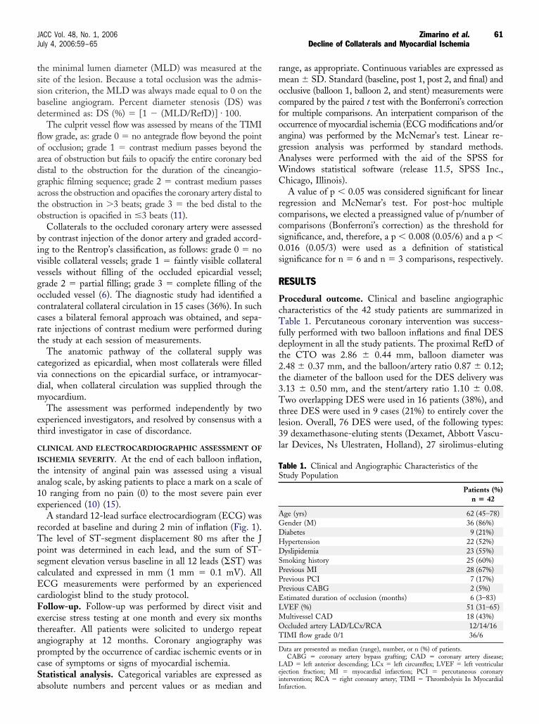
able 1. Clinical and Angiographic Characteristics of thetudy Population
Patients (%)n � 42
ge (yrs) 62 (45–78)ender (M) 36 (86%)iabetes 9 (21%)ypertension 22 (52%)yslipidemia 23 (55%)
moking history 25 (60%)revious MI 28 (67%)revious PCI 7 (17%)revious CABG 2 (5%)stimated duration of occlusion (months) 6 (3–83)VEF (%) 51 (31–65)ultivessel CAD 18 (43%)ccluded artery LAD/LCx/RCA 12/14/16IMI flow grade 0/1 36/6
ata are presented as median (range), number, or n (%) of patients.CABG � coronary artery bypass grafting; CAD � coronary artery disease;
AD � left anterior descending; LCx � left circumflex; LVEF � left ventricularjection fraction; MI � myocardial infarction; PCI � percutaneous coronary
ntervention; RCA � right coronary artery; TIMI � Thrombolysis In Myocardialnfarction.
s(
cmTctbBfgmpIca
paa0
tto(Fp
a
c00d(0Ol3�
mN
ws0eroaFppT3wiArwm
Ffrs(sp
TC
MDTRCC�
*osis; M
�
62 Zimarino et al. JACC Vol. 48, No. 1, 2006Decline of Collaterals and Myocardial Ischemia July 4, 2006:59–65
tents (Cypher, Cordis), and 10 paclitaxel-eluting stentsTaxus, Boston Scientific, Natick, Massachusetts).
Modification of angiographic, pressure-derived, electro-ardiographic, and clinical variables during PCI are sum-arized in Table 2. The improvement of MLD, DS, andIMI flow grade (p � 0.008 for all) was accompanied by a
oncomitant reduction in collateral circulation, evaluated byhe Rentrop’s grading and CPI (p � 0.016 for both). Theehavior of FFR-based measurements is shown in Figure 2.aseline myocardial perfusion was entirely due to the
unction of collaterals. With the re-establishment of ante-rade flow, the coronary component becomes more andore responsible for the observed increase of myocardial
erfusion, and fully accounts for the final result.nducibility of ischemia. During PCI, ST-segmenthanges were documented in 36 patients (86%), whilengina was reported by 27 patients (64%, p � 0.001).
During the subsequent balloon inflations and stent de-loyment, a significant worsening of ischemia was observed,s documented by the increase in �ST-segment elevationnd angina pain score versus the first balloon inflation (p �.008 for both) (Table 2).
igure 2. Modifications of fractional flow reserve (FFR) parameters:ractional flow reserve myocardial (FFRmyo), fractional flow coronaryeserve (FFRcor), and fractional flow reserve collateral (FFRcoll). Mea-urements were performed before (baseline), after the first balloon inflationPost 1), after the second balloon inflation (Post 2), and after drug-eluting
able 2. Angiographic, Pressure-Derived, Electrocardiographic, aoronary Occlusions
Baseline Balloon 1 Post 1
LD (mm) 0.1 � 0.2 1.0 � 0.5*S (%) 98 � 5 65 � 16*IMI flow grade 0.1 � 0.4 1.8 � 0.8*entrop 1.9 � 0.8PI 0.40 � 0.13hest pain 0 � 0.3ST (mm) 0.7 � 1.1
p � 0.008 vs. previous measurement; †p � 0.016 vs. previous measurement.CPI � collateral pressure index; DES � drug-eluting stent; DS � diameter sten
ST � sum of ST-segment elevation in all 12 leads.
atent deployment (Final). Data are mean � SD; p � 0.008 for eacharameter versus the previous time point.
Electrocardiogram modifications were documented in allhe 27 subjects complaining about angina during PCI. Inhis subgroup of patients, a �1 mm �ST-segment elevationccurred earlier and for a concomitant higher FFRcoll0.18 � 0.10) and CPI (0.38 � 0.13) compared with bothFRcoll (0.15 � 0.06, p � 0.0217) and CPI (0.34 � 0.13,� 0.011) calculated at the first occurrence of angina.A representative example of angiograms, coronary pressures,
nd ECG findings in one patient is shown in Figure 3.At linear regression analysis, an inverse relationship of
ollateral FFRcoll with both �ST-segment elevation (R2 �.352, p � 0.001) and angina pain score (R2 � 0.247, p �.001) was evident. Collateral pressure index had a lesseregree of correlation with both �ST-segment elevationR2 � 0.084, p � 0.012) and angina pain score (R2 �.093, p � 0.006).cclusion age and anatomic pathway of collaterals. Base-
ine FFRcoll was similar among 14 patients (33%) with 1 �months CTO (0.41 � 0.13) and in 28 subjects (67%) with3 months CTO (0.40 � 0.12, p � NS).At linear regression analysis, no relationship was docu-ented between age of CTO and baseline FFRcoll (p �S), nor between age of CTO and CPI (p � NS).A prevalent epicardial pathway of collateral circulation
as identified in 16 patients (38%). Epicardial collateralshowed a trend toward a higher baseline FFRcoll (0.44 �.12) and CPI (0.42 � 0.11) versus intramyocardial collat-rals (0.37 � 0.13, p � 0.089, and 0.36 � 0.12, p � 0.112,espectively). No differences were documented in FFRcollr CPI at the occurrence of ECG modifications or anginaccording to the anatomic pathway of collaterals.ollow-up. Late clinical follow-up was obtained in allatients at 17 � 5 months. No patient died during this timeeriod; three patients refused follow-up angiography.hirty-nine subjects underwent repeat angiography at 11 �months. No deaths occurred. Major adverse cardiac eventsere documented in 7 patients (17%): 1 (2.4%) myocardial
nfarction and 6 (14.3%) target vessel revascularizations.mong the 39 subjects who underwent repeat angiography,
eocclusion was identified in two cases (5.1%): 1 patient,ho experienced a myocardial infarction, was treated withedical therapy, and 1 patient, who had recurrence of
linical Variables During DES Implantation in Chronic Total
Balloon 2 Post 2 DES Final
1.7 � 0.4* 2.9 � 0.4*40 � 10* �2 � 7*2.7 � 0.6* 3 � 0.2
1.3 � 0.9† 0.7 � 0.7†0.33 � 0.09† 0.27 � 0.07†1.3 � 1.1† 2.8 � 2.1†3.5 � 2.6† 5.9 � 3.1†
LD � minimal lumen diameter; TIMI � Thrombolysis In Myocardial Infarction;
nd C
ngina, underwent successful repeat PCI. Subocclusive re-
sr(cd
D
Wfflci
tdisamtcdv
Ccrp(chmfirDbiEpFdcFttem
Fac((od a mae (Fina
63JACC Vol. 48, No. 1, 2006 Zimarino et al.July 4, 2006:59–65 Decline of Collaterals and Myocardial Ischemia
tenosis was identified in 5 subjects (13%) who underwentepeat PCI in 4 cases (10%) and bypass surgery in 1 case2.6%). In no case was disease progression anywhere in theoronary epicardial vessels requiring revascularizationocumented.
ISCUSSION
e observed a rapid and progressive reduction of collateralunction, evident soon after the restoration of antegradeow, during PCI of CTO. The early “de-recruitment” ofollateral circulation reduced tolerance to myocardialschemia induced by subsequent balloon inflation.
Coronary collaterals in humans preserve myocardial func-ion (1,3). Myocardial ischemia and the pressure gradienteriving from a high-grade stenosis are stimuli sufficient to
nduce the development of coronary collaterals througheveral biochemical signals, which include the release ofngiogenic growth factors (1–4). Collateral circulation isore developed in patients with preserved myocardial func-
ion (10,16). Although exhibiting a residual vasodilatoryapacity during moderate increases in myocardial oxygenemand, collateral circulation may, however, not fully pro-
igure 3. Angiographic findings, electrocardiographic and pressure measursterisk), with collateral circulation (white arrows) supported from the leoronary wedge pressure (Pw) documented a collateral pressure index (CPBalloon 1). After the first balloon inflation (Post 1), the percentage diamBalloon 2), CPI decreased to 0.46, and a slight ST-segment modification (f 42% (double white arrowheads) was documented by angiography (Postropped to 0.33, the patient experienced chest pain (grade 4/10), andlectrocardiogram. Angiography showed an 8% residual diameter stenosis
ide a normal coronary flow (17). w
ollateral modifications after PCI. Collateral functionan be accurately measured by FFRcoll; it is directlyelated to—and a likely determinant of—myocardialerfusion of the territory supplied by the occluded artery18). After the restoration of antegrade flow with suc-essful PCI of CTO, a rapid decline of collateral functionas been already documented (10); the regression is evenore evident when the vessel is persistently patent at a
ve-month follow-up (19). Our findings confirm theapid regression of collateral function after deployment ofES, but also add clinical relevance to these observations
y documenting a contextual reduction in the tolerance toschemia, here documented by us in �80% of cases byCG modifications and in �60% by angina. Tworessure-derived parameters describe collateral function:FRcoll expresses the collateral contribution to myocar-ial perfusion, while CPI evaluates the recruitability ofollateral circulation during coronary occlusion. Values ofFRcoll �0.25 and of CPI �0.30 were previously found
o be strongly associated with the presence of ischemia onhe ECG during coronary artery occlusion (20,21). In ourxperience, ECG modifications were documented at aean of FFRcoll 0.18 and a CPI of 0.38. Both indexes
ts during recanalization of a chronic right coronary artery occlusion (blacknary system at baseline. Measurements of mean aortic pressure (Pa) and
llateral pressure index � Pw/Pa) of 0.61 during the first balloon inflationtenosis was 65% (white arrowhead). During the second balloon inflationarrow) was evident on the electrocardiogram. A residual diameter stenosisuring the drug-eluting stent (DES) deployment, collateral pressure index
rked ST-segment elevation (double black arrows) was evident on thel).
emenft coroI) (coeter sblack2). D
ere inversely related to the inducibility of ischemia

esaCusTelramwba
teDotrmbsaeasrpdsoSsiPpdHi
afmp
aeabdaa
wtwip
cttmrdbmvDmaesht
ptpdppWFtmsfntt
sarCmd“tflpnoa
64 Zimarino et al. JACC Vol. 48, No. 1, 2006Decline of Collaterals and Myocardial Ischemia July 4, 2006:59–65
licited by balloon inflation, but FFRcoll showed atronger relationship with both �ST-segment elevationnd angina pain score than CPI.linical implications. Myocardial infarction may occur inp to about 12% of cases during the first six months after auccessful percutaneous recanalization of a CTO (22–25).his observation suggests that previously developed collat-
rals do not systematically exert a protective effect in case ofate reocclusion of the target vessel. The rapid “de-ecruitment” of the collateral circulation may put the patientt risk of future ischemic events. An impaired acute recruit-ent of collaterals has been documented in diabetic patientsith duration of CTO �3 months, and this finding haseen advocated as a hypothetical cause of the increaseddverse events after PCI among patients with diabetes (26).
Increased susceptibility to myocardial ischemia afterhe decline of collateral circulation is likely to becomeven more relevant with the current increasing use ofES. Stent thrombosis is a catastrophic event when it
ccurs without the protective role of a collateral circula-ion. Drug-eluting stents dramatically reduce the occur-ence of restenosis (27,28), but there is concern that theyight also be susceptible to very late (�1 year) throm-
osis related to delayed endothelialization of the stenttruts concomitant with the discontinuation of aspirinnd/or clopidogrel (29). In our study population, adversevents were documented in 17% of patients; this rateppears to be considerably higher than reported in othertudies, where �10% restenosis and �3% reocclusionates were documented after sirolimus or paclitaxel im-lantation (8,9). One explanation might be that aexamethasone-eluting stent, likely not equivalent toirolimus- or paclitaxel-eluting stents, was used in mostf our patients (51%).tudy limitations. In the calculation of FFR, Pv wasubstituted by a fixed value (10 mm Hg). This is a source ofnaccuracy, but does not affect the observed changes duringCI of CTOs (10). Only pressure measurements wereerformed. A Doppler wire was not used, and, therefore,ata on flow and resistances could not be calculated.owever, FFRcoll has been shown to be extremely accurate
n the quantitative assessment of collateral blood flow (30).Data on regional myocardial function were not system-
tically available. However, the attentuation of collateralunction, more pronounced in patients with akinesia in theyocardium supplied by the CTO, is evident also in
atients with normal ventricular function (31).For baseline and occlusion measurements, we injected
denosine in the coronary artery that supported the collat-ral circulation to the occluded artery. We acknowledge thatsystemic adenosine infusion would have been preferable,
ecause it would have minimized microvascular resistancesistal to the lesion treated by PCI even in absence ofntegrade flow. Selective adenosine infusion in one coronary
rtery may have changed peripheral resistances in a some- ohat less predictable fashion. Despite this technical limita-ion, the observed trend of changes in FFR measurementsas consistent throughout our study, fitted with pathophys-
ological predictions and, therefore, likely reflects our patho-hysiological interpretation.The functional significance of collateral vessels during
oronary occlusion was judged by clinical and ECG moni-oring only during brief periods of coronary occlusion, andhe method was not expanded to include hemodynamiconitoring, metabolic alterations, or assessment of global or
egional ejection fractions. The development of electrocar-iographic signs of ischemia during coronary occlusion maye influenced by the relationship between the size of theyocardium “at risk” and the extent of collateral-dependent
ascular bed, which were not assessed in the present study.uring balloon occlusions, regional systolic and diastolicyocardial function modifications are directly related to the
mount of collateral flow in the territory (32), and anchocardiographic evaluation would have allowed a moreensitive evaluation of the ischemic burden. These limitations,owever, do not appear to hamper the directional trend andhe temporal pattern of the phenomena observed.
We used three different types of DES in carrying out theresent study. The choice was left at the operator’s discre-ion and varied upon market’s availability during the studyeriod. There is no report on the effectiveness ofexamethasone-eluting stent in CTOs, and these devicesrobably cannot be rated as “equivalent” to sirolimus- andaclitaxel-eluting devices in terms of long-term outcome.
hen analyzed in subgroups according to the DES used,FR modifications were, however, similar for all three DES
ypes used. Exercise-induced vasoconstriction of the proxi-al and distal vessel segments adjacent to sirolimus-eluting
tents has been recently documented, and endothelial dys-unction has been hypothesized as the underlying mecha-ism (33). In our study, it appears, however, very unlikelyhat any drug eluted by the stent would have any effect onhe acute modifications of collateral circulation.
A long-term functional evaluation of collaterals was notystematically performed, and, therefore, no data are avail-ble on the recruitability of collateral circulation and itselationship to long-term tolerance to ischemia.
onclusions. In CTO, collateral circulation, providingost coronary flow at baseline, rapidly declines, almost
isappearing after successful stent implantation. The rapidde-recruitment” of collaterals reduces myocardial toleranceo ischemia, which can be easily elicited in a predictableashion during PCI. The quick changes of collateral circu-ation after the PCI resolution of a CTO may put theatient at risk of future ischemic events in case of sponta-eous sudden reocclusion of the vessel. This suggests the usef a more aggressive antiplatelet therapy, with prolongedspirin and clopidogrel administration, after recanalization
f CTO and successful stent implantation.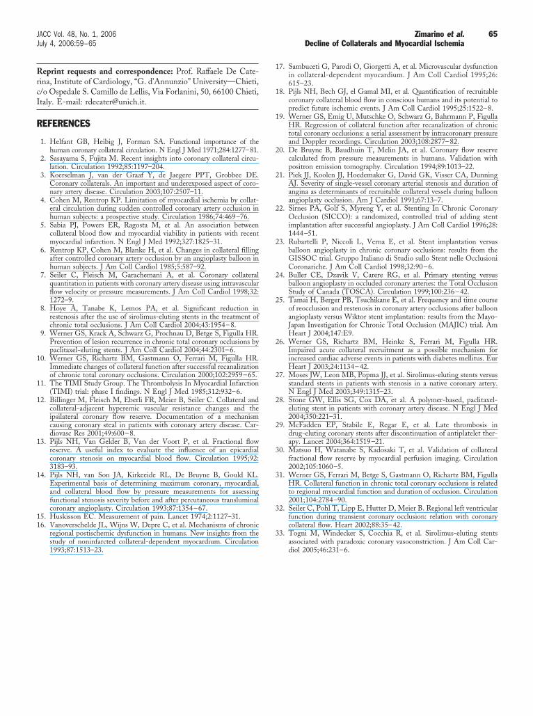
RrcI
R
1
1
1
1
1
11
1
1
1
2
2
2
2
2
2
2
2
2
2
3
3
3
3
65JACC Vol. 48, No. 1, 2006 Zimarino et al.July 4, 2006:59–65 Decline of Collaterals and Myocardial Ischemia
eprint requests and correspondence: Prof. Raffaele De Cate-ina, Institute of Cardiology, “G. d’Annunzio” University—Chieti,/o Ospedale S. Camillo de Lellis, Via Forlanini, 50, 66100 Chieti,taly. E-mail: [email protected].
EFERENCES
1. Helfant GB, Heibig J, Forman SA. Functional importance of thehuman coronary collateral circulation. N Engl J Med 1971;284:1277–81.
2. Sasayama S, Fujita M. Recent insights into coronary collateral circu-lation. Circulation 1992;85:1197–204.
3. Koerselman J, van der Graaf Y, de Jaegere PPT, Grobbee DE.Coronary collaterals. An important and underexposed aspect of coro-nary artery disease. Circulation 2003;107:2507–11.
4. Cohen M, Rentrop KP. Limitation of myocardial ischemia by collat-eral circulation during sudden controlled coronary artery occlusion inhuman subjects: a prospective study. Circulation 1986;74:469–76.
5. Sabia PJ, Powers ER, Ragosta M, et al. An association betweencollateral blood flow and myocardial viability in patients with recentmyocardial infarction. N Engl J Med 1992;327:1825–31.
6. Rentrop KP, Cohen M, Blanke H, et al. Changes in collateral fillingafter controlled coronary artery occlusion by an angioplasty balloon inhuman subjects. J Am Coll Cardiol 1985;5:587–92.
7. Seiler C, Fleisch M, Garachemani A, et al. Coronary collateralquantitation in patients with coronary artery disease using intravascularflow velocity or pressure measurements. J Am Coll Cardiol 1998;32:1272–9.
8. Hoye A, Tanabe K, Lemos PA, et al. Significant reduction inrestenosis after the use of sirolimus-eluting stents in the treatment ofchronic total occlusions. J Am Coll Cardiol 2004;43:1954–8.
9. Werner GS, Krack A, Schwarz G, Prochnau D, Betge S, Figulla HR.Prevention of lesion recurrence in chronic total coronary occlusions bypaclitaxel-eluting stents. J Am Coll Cardiol 2004;44:2301–6.
0. Werner GS, Richartz BM, Gastmann O, Ferrari M, Figulla HR.Immediate changes of collateral function after successful recanalizationof chronic total coronary occlusions. Circulation 2000;102:2959–65.
1. The TIMI Study Group. The Thrombolysis In Myocardial Infarction(TIMI) trial: phase I findings. N Engl J Med 1985;312:932–6.
2. Billinger M, Fleisch M, Eberli FR, Meier B, Seiler C. Collateral andcollateral-adjacent hyperemic vascular resistance changes and theipsilateral coronary flow reserve. Documentation of a mechanismcausing coronary steal in patients with coronary artery disease. Car-diovasc Res 2001;49:600–8.
3. Pijls NH, Van Gelder B, Van der Voort P, et al. Fractional flowreserve. A useful index to evaluate the influence of an epicardialcoronary stenosis on myocardial blood flow. Circulation 1995;92:3183–93.
4. Pijls NH, van Son JA, Kirkeeide RL, De Bruyne B, Gould KL.Experimental basis of determining maximum coronary, myocardial,and collateral blood flow by pressure measurements for assessingfunctional stenosis severity before and after percutaneous transluminalcoronary angioplasty. Circulation 1993;87:1354–67.
5. Huskisson EC. Measurement of pain. Lancet 1974;2:1127–31.6. Vanoverschelde JL, Wijns W, Depre C, et al. Mechanisms of chronic
regional postischemic dysfunction in humans. New insights from thestudy of noninfarcted collateral-dependent myocardium. Circulation
1993;87:1513–23.7. Sambuceti G, Parodi O, Giorgetti A, et al. Microvascular dysfunctionin collateral-dependent myocardium. J Am Coll Cardiol 1995;26:615–23.
8. Pijls NH, Bech GJ, el Gamal MI, et al. Quantification of recruitablecoronary collateral blood flow in conscious humans and its potential topredict future ischemic events. J Am Coll Cardiol 1995;25:1522–8.
9. Werner GS, Emig U, Mutschke O, Schwarz G, Bahrmann P, FigullaHR. Regression of collateral function after recanalization of chronictotal coronary occlusions: a serial assessment by intracoronary pressureand Doppler recordings. Circulation 2003;108:2877–82.
0. De Bruyne B, Baudhuin T, Melin JA, et al. Coronary flow reservecalculated from pressure measurements in humans. Validation withpositron emission tomography. Circulation 1994;89:1013–22.
1. Piek JJ, Koolen JJ, Hoedemaker G, David GK, Visser CA, DunningAJ. Severity of single-vessel coronary arterial stenosis and duration ofangina as determinants of recruitable collateral vessels during balloonangioplasty occlusion. Am J Cardiol 1991;67:13–7.
2. Sirnes PA, Golf S, Myreng Y, et al. Stenting In Chronic CoronaryOcclusion (SICCO): a randomized, controlled trial of adding stentimplantation after successful angioplasty. J Am Coll Cardiol 1996;28:1444–51.
3. Rubartelli P, Niccoli L, Verna E, et al. Stent implantation versusballoon angioplasty in chronic coronary occlusions: results from theGISSOC trial. Gruppo Italiano di Studio sullo Stent nelle OcclusioniCoronariche. J Am Coll Cardiol 1998;32:90–6.
4. Buller CE, Dzavik V, Carere RG, et al. Primary stenting versusballoon angioplasty in occluded coronary arteries: the Total OcclusionStudy of Canada (TOSCA). Circulation 1999;100:236–42.
5. Tamai H, Berger PB, Tsuchikane E, et al. Frequency and time courseof reocclusion and restenosis in coronary artery occlusions after balloonangioplasty versus Wiktor stent implantation: results from the Mayo-Japan Investigation for Chronic Total Occlusion (MAJIC) trial. AmHeart J 2004;147:E9.
6. Werner GS, Richartz BM, Heinke S, Ferrari M, Figulla HR.Impaired acute collateral recruitment as a possible mechanism forincreased cardiac adverse events in patients with diabetes mellitus. EurHeart J 2003;24:1134–42.
7. Moses JW, Leon MB, Popma JJ, et al. Sirolimus-eluting stents versusstandard stents in patients with stenosis in a native coronary artery.N Engl J Med 2003;349:1315–23.
8. Stone GW, Ellis SG, Cox DA, et al. A polymer-based, paclitaxel-eluting stent in patients with coronary artery disease. N Engl J Med2004;350:221–31.
9. McFadden EP, Stabile E, Regar E, et al. Late thrombosis indrug-eluting coronary stents after discontinuation of antiplatelet ther-apy. Lancet 2004;364:1519–21.
0. Matsuo H, Watanabe S, Kadosaki T, et al. Validation of collateralfractional flow reserve by myocardial perfusion imaging. Circulation2002;105:1060–5.
1. Werner GS, Ferrari M, Betge S, Gastmann O, Richartz BM, FigullaHR. Collateral function in chronic total coronary occlusions is relatedto regional myocardial function and duration of occlusion. Circulation2001;104:2784–90.
2. Seiler C, Pohl T, Lipp E, Hutter D, Meier B. Regional left ventricularfunction during transient coronary occlusion: relation with coronarycollateral flow. Heart 2002;88:35–42.
3. Togni M, Windecker S, Cocchia R, et al. Sirolimus-eluting stentsassociated with paradoxic coronary vasoconstriction. J Am Coll Car-
diol 2005;46:231–6.Copyright © 2022 FDOKUMEN