Raman investigation of artificial patinas on recent bronze protected by different azole type...
9
Raman investigation of artificial patinas on recent bronze – Part I: climatic chamber exposure † Polonca Ropret a,c and Tadeja Kosec b * In humid air, copper and its high copper alloys (bronze) tend to form an oxide layer (patina). Natural patinas protect copper and its alloys from further corrosion processes. On the other hand, artists have frequently deliberately patinated bronze for visual effects. Thus, it is of great importance to study the patina changing mechanism to follow its chemical changes and to predict in advance the likely corrosion processes. Green chloride and green nitrate patinas, applied over the brown artist’s patina, were tested, and also brown patina and the patina that develops on bare bronze. The Raman spectra were studied after chemical patination, and after exposing the patina samples in a climatic chamber, which can produce an environment that resembles an industrial atmosphere, for 12 weeks. The structures of the patinas and of the corrosion products were characterized by scanning electron microscopy, Raman spectroscopy and X-ray diffraction. Cuprite and cuprous sulfite were found on the brown patina, atacamite on the green chloride patina, and a mixture of gerhardite and rouaite on the blue to green nitrate type patina. After 12 weeks of exposure to humidity, a controlled concentration of SO 2 , and salt spray mist, the corrosion products changed. In general, clinoatacamite and paratacamite are the end corrosion products, after an interme- diate brochantite stage on the green chloride and green nitrate type patinas. The end products of each patina type are given. Copyright © 2012 John Wiley & Sons, Ltd. Keywords: bronze; artist’s patina; chemical patina; corrosion; Raman spectroscopy; climatic exposure Introduction Copper and bronze undergo spontaneous oxidation in humid air. First, red-coloured cuprite (Cu 2 O) is formed, which slowly turns black, forming tenorite (CuO). [1] Nowadays, air is frequently polluted with different sulfur, nitrate and carbon oxides, and in the presence of moisture, so-called acid rain occurs, which represents an aggressive environment for exposed bronze surfaces outdoors. As a result, the protective layer of copper oxides changes. [2–7] The morphology and mechanisms of formation of natural patinas on different bronzes, and their electrochemical properties, have been extensively studied. [2–11] Some artists choose to use artificial patina after their bronze sculp- tures have been cast. The light and shadows of artificial patina combine to give the curves and textures good definition and special artistic effects. There are numerous application techniques and formu- lae, among which the green colour, applied over brown patina, has been in use for the last few decades. [12] The brown colour is usually achieved by using potassium sul fide, which is also called ‘ liver of sulfur’ , whereas a green colour can be achieved by using chlorides, nitrates, sulfates or even phosphates. [12] There have been several studies that have described the testing of different chemical-artificial patinations by means of different electrochemical techniques. [13–18] Corrosion studies of different chemical and electrochemical patinations in urban acid rain have shown that patinas have to be protected to attain the desired colour, and at the same time to avoid or at least postpone corro- sion reactions. [13–15] The study by Marušić et al. was performed on two chemically-formed patinas (by the immersion technique and hot application, [16] ) and another involved a nitrate patina, applied with immersion and studied in a chloride solution. [17] The brown patinated bronze was tested after exposure to formic and acetic vapours, and studied by cathodic reduction using the electro- chemical technique. [18] Among data given in the recent literature, scientists have used different spectroscopic techniques, including Raman spectroscopy, as a tool for identifying corrosion products. [11,15,16,19–22] Some have used the latter technique to dif- ferentiate between the different minerals formed on bronze in dif- ferent environments. [22] Only a few studies have used Raman spectroscopy as a basic tool for the characterization of different corrosion products on bronze. [23–28] De la Roja et al. studied different green corrosion products, formed on copper and its alloys, used as green coloured pigments called verdigris. [23] There are some Raman data available on different copper chloride minerals, [24,25] and Raman spectra and * Correspondence to: Tadeja Kosec, National Building and Civil Engineering Insti- tute, Dimičeva 12, 1000 Ljubljana, Slovenia. E-mail: [email protected] † This article is part of the Journal of Raman Spectroscopy special issue entitled ”Raman spectroscopy in art and archaeology“ edited by Juan Manuel Madariaga and Danilo Bersani. a Conservation Centre, Institute for the Protection of the Cultural Heritage of Slovenia, Poljanska 40, 1000 Ljubljana, Slovenia b National Building and Civil Engineering Institute, Dimičeva 12, 1000 Ljubljana, Slovenia c Museum Conservation Institute, Smithsonian Institution, 4210 Silver Hill Rd., Suitland, MD 20746, USA J. Raman Spectrosc. 2012, 43, 1578–1586 Copyright © 2012 John Wiley & Sons, Ltd. Research article Received: 12 December 2011 Revised: 2 March 2012 Accepted: 2 March 2012 Published online in Wiley Online Library: 18 September 2012 (wileyonlinelibrary.com) DOI 10.1002/jrs.4068 1578
Transcript of Raman investigation of artificial patinas on recent bronze protected by different azole type...
Research article
Received: 12 December 2011 Revised: 2 March 2012 Accepted: 2 March 2012 Published online in Wiley Online Library: 18 September 2012
(wileyonlinelibrary.com) DOI 10.1002/jrs.4068
1578
Raman investigation of artificial patinason recent bronze – Part I: climaticchamber exposure†
Polonca Ropreta,c and Tadeja Kosecb*
In humid air, copper and its high copper alloys (bronze) tend to form an oxide layer (patina). Natural patinas protect copperand its alloys from further corrosion processes. On the other hand, artists have frequently deliberately patinated bronze forvisual effects. Thus, it is of great importance to study the patina changing mechanism to follow its chemical changes and topredict in advance the likely corrosion processes. Green chloride and green nitrate patinas, applied over the brown artist’spatina, were tested, and also brown patina and the patina that develops on bare bronze. The Raman spectra were studiedafter chemical patination, and after exposing the patina samples in a climatic chamber, which can produce an environmentthat resembles an industrial atmosphere, for 12weeks. The structures of the patinas and of the corrosion products werecharacterized by scanning electron microscopy, Raman spectroscopy and X-ray diffraction. Cuprite and cuprous sulfite werefound on the brown patina, atacamite on the green chloride patina, and a mixture of gerhardite and rouaite on the blue togreen nitrate type patina. After 12weeks of exposure to humidity, a controlled concentration of SO2, and salt spray mist,the corrosion products changed. In general, clinoatacamite and paratacamite are the end corrosion products, after an interme-diate brochantite stage on the green chloride and green nitrate type patinas. The end products of each patina type are given.Copyright © 2012 John Wiley & Sons, Ltd.
Keywords: bronze; artist’s patina; chemical patina; corrosion; Raman spectroscopy; climatic exposure
* Correspondence to: Tadeja Kosec, National Building and Civil Engineering Insti-tute, Dimičeva 12, 1000 Ljubljana, Slovenia. E-mail: [email protected]
† This article is part of the Journal of Raman Spectroscopy special issue entitled”Raman spectroscopy in art and archaeology“ edited by Juan ManuelMadariaga and Danilo Bersani.
a Conservation Centre, Institute for the Protection of the Cultural Heritage ofSlovenia, Poljanska 40, 1000 Ljubljana, Slovenia
b National Building and Civil Engineering Institute, Dimičeva 12, 1000 Ljubljana,Slovenia
c Museum Conservation Institute, Smithsonian Institution, 4210 Silver Hill Rd.,Suitland, MD 20746, USA
Introduction
Copper and bronze undergo spontaneous oxidation in humid air.First, red-coloured cuprite (Cu2O) is formed, which slowly turns black,forming tenorite (CuO).[1] Nowadays, air is frequently polluted withdifferent sulfur, nitrate and carbon oxides, and in the presence ofmoisture, so-called acid rain occurs, which represents an aggressiveenvironment for exposed bronze surfaces outdoors. As a result, theprotective layer of copper oxides changes.[2–7] The morphology andmechanisms of formation of natural patinas on different bronzes,and their electrochemical properties, have been extensivelystudied.[2–11]
Some artists choose to use artificial patina after their bronze sculp-tures have been cast. The light and shadows of artificial patinacombine to give the curves and textures good definition and specialartistic effects. There are numerous application techniques and formu-lae, among which the green colour, applied over brown patina, hasbeen in use for the last few decades.[12] The brown colour is usuallyachieved by using potassium sulfide, which is also called ‘liver ofsulfur’, whereas a green colour can be achieved by using chlorides,nitrates, sulfates or even phosphates.[12]
There have been several studies that have described the testingof different chemical-artificial patinations by means of differentelectrochemical techniques.[13–18] Corrosion studies of differentchemical and electrochemical patinations in urban acid rain haveshown that patinas have to be protected to attain the desiredcolour, and at the same time to avoid or at least postpone corro-sion reactions.[13–15] The study by Marušić et al. was performed ontwo chemically-formed patinas (by the immersion technique andhot application,[16]) and another involved a nitrate patina, applied
J. Raman Spectrosc. 2012, 43, 1578–1586
with immersion and studied in a chloride solution.[17] The brownpatinated bronze was tested after exposure to formic and aceticvapours, and studied by cathodic reduction using the electro-chemical technique.[18] Among data given in the recent literature,scientists have used different spectroscopic techniques, includingRaman spectroscopy, as a tool for identifying corrosionproducts.[11,15,16,19–22] Some have used the latter technique to dif-ferentiate between the different minerals formed on bronze in dif-ferent environments.[22]
Only a few studies have used Raman spectroscopy as a basictool for the characterization of different corrosion products onbronze.[23–28] De la Roja et al. studied different green corrosionproducts, formed on copper and its alloys, used as green colouredpigments called verdigris.[23] There are some Raman data availableon different copper chloride minerals,[24,25] and Raman spectra and
Copyright © 2012 John Wiley & Sons, Ltd.
Raman investigation of artificial patinas on recent bronze
band assignments of some common phases of corroded copper.[26]
There are, however, relatively few papers on spectroscopic Ramandata referring to different patinas on copper and alloys.[27,28]
Different recipes for artificial patination on copper with variouschlorides, carbonates and nitrate salts have been explored usingmicro-Raman spectroscopy.[27] It was found that chloride typerecipes induce the formation of atacamite products, and that differ-ent sulfate salts favour the formation of antlerite and brochantite.[27] Furthermore, patinas on copper were morphologically studied,and a Raman investigation was performed before and after expo-sure to an outdoor environment, for restoration and conservationpurposes.[28] The findings of the latter paper made it possible todifferentiate between natural and artificial patinas on copper.[28]
To the authors’ knowledge, there have been no reports about stud-ies of the corrosion processes of brown artist’s patinas and brownpatinas with an antique appearance (various green patinations).
Thus, it is important to emphasize that the current patinationtechnique (green patina over brown patina) has not yet beenspectroscopically studied. Moreover, the investigated patinasare formed on recent type bronze, not on copper, as in moststudies. There is a need for spectroscopic studies of chemicallyformed patinas and for their testing in an environment as closeas possible to that to which the patina would be exposed. It isexpected that vulnerable data would be obtained. In this studythe three different patinas (brown patina, green chloride patinaapplied over brown patina, green alternative nitrate patinaover brown patina, and bronze for comparison) have beenmorphologically and spectroscopically investigated before andafter exposure to environmental testing.
The aim of this work is to provide a systematic spectroscopicstudy of various patinations on recent type bronze CuSnZn tounderstand the processes of bronze aging in aggressive environ-ments. Raman spectroscopy, energy dispersive spectroscopy/scanning electron microscopy (EDS/SEM) analysis, XRD and opticalobservations were chosen as surface examination techniques.
Experimental
Preparation of the patinas
The bronze samples were cast in a sand mould of 100� 100� 5mm at a privately-owned foundry. The bronze consisted of 92.2%
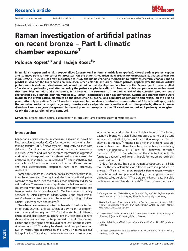
Figure 1. Photographs of (a) bare bronze, (b) brown patina, (c) green chloexposure: (e) bare bronze, (f) brown patina, (g) green chloride artificial patinwileyonlinelibrary.com/journal/jrs
J. Raman Spectrosc. 2012, 43, 1578–1586 Copyright © 2012 Joh
Cu, 5.90% Sn, 1.90% Zn. The 100� 100� 5mm samples weresand-blasted after casting, and were patinated soon afterwards.
Three artificial patinas were studied and the correspondingbare bronze. A brown patina on the bronze was achieved bybrushing the preheated surface with a 3% solution of K2S. Thesurface was then rinsed in a stream of tap water. Both chemicallyprepared green patinas were applied over the brown patina. Achloride type green patina was applied over the brown patinaon a warm surface at a temperature of about 50 �C by brushingwith a solution of (NH4)2CO3 and NH4Cl, 3 g each in 100mL ofwater. The green chloride patina was developed in the presenceof high humidity. The nitrate patina was achieved by brushing ahot brown bronze surface with a 3% Cu (NO3)2 solution. To geta well defined blue to green colour, the brown patina was leftto stand for 24 h. All the different patinations are presented inFig. 1(a–d). The presented samples were exposed to differentenvironmental conditions in climatic chambers.
Exposure to a climatic chamber
The exposures to a climatic chamber included:
• Exposure to an industrial climate chamber according tothe standard SIST EN ISO 6988:1999.[29] a Kohler-model HK430, Germany, climate chamber was used. The test surfaceswere placed 250 cm above water and leaned at an angle of15� to the vertical. The test conditions during 1 cycle (1 day)were: 8 h at 40� 3 �C and 100% relative humidity with theaddition of 0.2 l SO2, 16 h at 23� 5 �C and relative humidity75%. Total exposure time: 3weeks.
• Exposure of samples in a humidity chamber according tothe standard SIST EN ISO 6270-2 ATH: 2005.[30] The testconditions during 1 cycle (1 day): 8 h at 40 �3 �C and 100%relative humidity, 16 h at 23� 5 �C and relative humidity75%. Total exposure time: ~9weeks (8weeks plus 10 days).
• Exposure of samples to a salt spray chamber accordingto the standard SIST EN ISO 9227 NNS: 2006.[31] A modelVSC/KTW 1000 Votsch chamber was used. Total time 48 h at35 �1 �C and a 5% NaCl salt spray solution.
The exposure pattern was as follows: 3weeks in the industrialchamber, 8weeks in the humidity chamber, 2 days in the saltspray chamber, and 10 days in the humidity chamber.
ride artificial patina and (d) green nitrate patina before exposure. Aftera and (h) green nitrate patina. This figure is available in colour online at
n Wiley & Sons, Ltd. wileyonlinelibrary.com/journal/jrs
1579
P. Ropret and T. Kosec
1580
Scanning electron microscopy analysis
A low vacuum JEOL 5500 LV, JEOL, JAPAN (Japan) scanningelectron microscope SEM, equipped with an Oxford Inca energydispersive spectrometer (Oxford Instrument Analytical, UK), wasused to observe the surface products (EDS analysis) using anaccelerating voltage of 20 kV.When interpreting the results of quantitative EDS analysis in
low vacuum, it is necessary to take into account the specificcharacteristics of the mode of detection used. Because ofscattering of the incident beam, the detected X- ray may,partly, originate from the surroundings of the ‘examined spot’.In low vacuum the quantitative analysis of light elements suchas N, C and O is inaccurate so that merely a comparison ofelements at the analysed spots is reccomended. Thus, EDSresults given in the paper are semi quantitative and servemainly for comparison resons.
Raman spectroscopy
Raman spectra were obtained by using a Horiba Jobin YvonLabRAM HR800 Raman spectrometer coupled to an OlympusBXFM optical microscope. The measurements were performedusing a 514 nm laser excitation line, a 100� objective lens, anda 600 grooves/mm grating, which gave a spectral resolution of1.99 cm–1/pixel. The power at the samples was set between0.014 and 1.4mW. A multichannel air-cooled charge-coupled de-vice detector was used, with integration times of between 20 and35 s. The spectra are presented after baseline correction.
X-ray diffraction analysis
The phase composition and the crystallinity of the samples weredetermined by X-ray diffraction (XRD) analyses using an X-raypowder diffractometer with Cu Ka radiation (D4 Endeavor, BrukerAXS). XRD patterns were recorded at room temperature in the 2θrange from 10� to 70�. The diffractograms were analyzed usingEVA software (Bruker AXS) to define the phase composition.X-ray diffraction analyses were utilised only for the initial
chemically applied patinas, but not for the exposed samples.
Results and discussion
First, all the different chemical patinas were morphologicallyinvestigated, EDS analyses were performed and Raman spectrawere collected. When necessary, the XRD studies gave addi-tional information to complement the EDS and Raman results.The samples were then exposed in a climatic chamberfor 12weeks. After exposure to different environments, thesamples were checked by SEM/EDS and Raman spectroscopy.
Patina observation
The different patinas were first applied over the prepared bronzesurface (Fig. 1).Bare bronze from copper–zinc–tin type bronze gave a golden
blink to this type of alloy (Fig. 1(a)). EDS results of the bare bronzeare given in the Experimental Section.Because of environmental aspects there have been many
attempts to reduce the lead content in bronzes,[32] which arethus referred to as recent type bronze. In comparison to
wileyonlinelibrary.com/journal/jrs Copyright © 2012 John
CuSnZnPb alloys, the former has a more reddish compoundin its shade.
Brown patination gave a velvety-like dark brown to maroonshade (Fig. 1(b)). Chloride and nitrate patinations were appliedover the brown patina, which corresponds to today’s practiceamong sculptors. Chloride patination gave a green shade, notentirely covering the whole surface (Fig. 1(c)), whereas nitratepatination gave a blue to green colour to the brown surface(Fig. 1(d)).
In the second step, the prepatinated surfaces were analysedusing the low vacuum SEM technique, on the same day, toeliminate possible air-formed secondary corrosion products. Thepatina layers were qualitatively analysed using EDS.
A metallographic image of the bare bronze is presented,together with SEM images of the brown patina and the twogreen patinas, in Fig. 2. The surface of bare bronze was not even;several cast imperfections could be visually observed (Fig. 2(a)).The chemical composition of the recent bronze was determinedfrom the EDS analysis.
Morphological observation of the brown patina showedthree different areas: big tile-like structures, fine round particlesand dark spots (Fig. 2(b)). The elemental composition of thebrown patina layer was as follows: 7% of N, 8% O, 26%S, 1% of K, 57% of Cu, 0.5% of Sn (Fig. 2(b)). Zinc was notfound in the analysed layer. EDS analysis was performed atdifferent spots and a similar composition was found.
Two different areas were found after applying a green chloridepatina over the brown patinated bronze sample (Fig. 2(c)), i.e.crystals and smoother lower areas. The chemical composition ofthe crystals was as follows: 29% of O, 22% of Cl, 24% of Cu,17% of N, and 7% of C. The smooth dark areas consisted of39% of Cu, 0.6% of Sn, 17% of S, 24% of O, 1% of Cl, 14% of N,and 5% of C. The larger amounts of copper and sulfur showedthat there are some uncovered patches of the underlying brownpatina, so that the chloride patina did not entirely cover the basebrown patina.
All the different areas visible in the SEM image of the greennitrate patina, shown in Fig. 2(d), have a similar composition:16.3% of Cu 15% of N, 63% of O, 5% of C, 0.3% of Cl, and 0.2%of K. The nitrate areas smoothly covered the brown patina.Because no sulfur was found, it can be assumed that the brownpatina and its products were not uncovered. The whole areawas covered with similar grains, approx. 10 mm in size. Grains ofsimilar shape and size were observed on the copper surfaces thatwere patinated with copper nitrate, pH 3.5, using a soft cloth over5 days.[28] Subsequent molecular analysis and identification wasconducted by Raman spectroscopy and XRD analysis.
Raman results of the different patinas before exposure
The Raman spectra of the patinated bronze surface beforeexposure to different conditions in climatic chambers arepresented in Fig. 3. The Raman measurements were repeated atseveral locations on the surfaces of the samples.
First the brown patina was analysed, followed by the greenchloride and green nitrate patinas.
Although the high content of sulphur and copper in the EDSanalysis suggested the presence of copper sulphides in thebrown patina, it was difficult to obtain direct evidence of thisby Raman spectroscopy,[33] although the Raman shifts at 282,329 and 613 cm–1 (Fig. 3(a)) did suggest the presence of Cu2S. Itis important to emphasise that in this case the laser power was
Wiley & Sons, Ltd. J. Raman Spectrosc. 2012, 43, 1578–1586

Figure 2. SEM images of (a) bare bronze, (b) brown patina, (c) green chloride artificial patina and (d) green nitrate patina. This figure is available incolour online at wileyonlinelibrary.com/journal/jrs
Figure 3. Raman spectra of (a) brown patina, (b) chloride patina and, (c)nitrate patina. The laser power was set to 1.4, 0.014 and 1.4mW,respectively.
Figure 4. XRD difractograms of (a) brown, (b) green chloride and(c) green nitrate patina.
Raman investigation of artificial patinas on recent bronze
158
set to 0.14mW. In other attempts, when the laser power wasset between 4 and 14mW a band appeared at 980 cm–1, whichwas significant for the SO4
2– symmetric stretch, thus stronglysuggesting degradation of the sample because of the high laserpower as the formation of oxidation products occurred. On theother hand this result also provided indirect evidence on thepresence of sulphides. The well-defined Raman band at 220 cm–1
in the spectrum of the brown patina (Fig. 3(a)) is characteristicfor cuprous oxide Cu2O (cuprite),[7,32] which shows that someoxidation of the bronze occurred also during the treatment ofthe sample with the 3% solution of K2S. The XRD pattern shownin Fig. 4(a) confirms the presence of copper sulphide, with thestrongest line at 46.5�.
The chloride type patina showed very intense Raman bandsin the OH stretching region at 3442 and 3353 cm–1, whichpoints out the presence of a corrosion product containing OH
J. Raman Spectrosc. 2012, 43, 1578–1586 Copyright © 2012 Joh
groups (Fig. 3(b)). The bands at 984, 970, 910, 859, 831, 815,514, 450, 423, 360, 293, 268, 235, 192, 145 and 123 cm–1 provethe presence of atacamite (Cu2 (OH)3Cl).
[26–28] No bandssignificant for Cu2S and Cu2O, which were found in the brownpatina before the application of (NH4)2CO3 and NH4Cl, wereidentified, although the SEM/EDS results showed that the
n Wiley & Sons, Ltd. wileyonlinelibrary.com/journal/jrs
1
P. Ropret and T. Kosec
1582
chloride type patina did not entirely cover the base brown patina.In the revealed brown patches no useful Raman data couldbe obtained, which shows that in the remaining brown areas, too,the treatment changed the crystal structure of the brown patina.The XRD pattern did not disprove the presence of atacamite (Fig. 4(b)), because the lines for copper chloride compounds are ofvery low intensity. These are lines, in Fig. 4(b) markedwith an arrow,at 15.5�, 32.5�, 36.5� and 39.5�. The two strongest lines at 43�
and 50� belong to the underlying copper substrate.The nitrate type patina showed the strongest Raman band at
1049 cm–1 (Fig. 4(c)), which is significant for NO3 symmetricstretch. According to the data that are available in the literatureand the band assignments,[25] the Raman modes stronglysuggest the presence of gerhardite (Cu2(OH)3NO3), with bandsat 3547, 3479, 1435, 1419, 1338, 1323, 1049, 1025, 888, 809,720, 711, 667, 503, 473, 458, 437, 422, 411, 336, 278, 259, 214,186 and 165 cm–1 (Fig. 4(c)). The Raman results that wereobtained on the nitrate patina, and the data from the literature,are presented in Table 1. The considered Ref. [25] includes some
Table 1. Comparison of the nitrate type patina modes with theliterature data on mode positions and band assignments forgerhardite.[25] The Raman shifts in cm–1 and the relative intensitiesmeasured on the nitrate patina are given as follows: (vs) very strong,(s) strong, (m) medium, (w) weak, (vw) very weak, and (sh) shoulder
Nitrate patina Gerhardite Band assignments [25]
(Fig. 4(c)) Cu2(OH)3NO3;[25]
3556
3547m 3546
3479m-w 3477 OH stretching
3417
3391
1435 vw 1438
1419 vw 1417 NO3 asymmetric
1338 vwsh 1339 stretching
1323 w 1324
1052 NO3 symmetric
1049 vs 1048 stretching
1031
1025 vwsh 1024
888 vw 887 CuOH deformation mode
809 vw 805 NO3 symmetric bending
720 vw 720 NO3 out of plane bending
711 w 711
667 vw 668
503 w 503 Possible assignment to OH
deformation modes of
Cu(OH)3
473 wsh 474
458 w 458
437 vwsh 437
422 vwsh 423
411 w 410
336 vw 336 Possible assignment to
hydrogen bond mode
278 vw 279 Lattice vibrations
259 vw 258
214 vw 213
186 vw 189
165m-w 165
150 wsh 149
131 vw 132
wileyonlinelibrary.com/journal/jrs Copyright © 2012 John
fittings of the Raman spectrum, which resulted in more bandsthan were observed by the authors, but the general agreementis good. Gerhardite is an orthorhombic, naturally-occurringcopper nitrate mineral, whereas its polymorph, rouaite, ismonoclinic. The formation of the monoclinic form is favouredat high temperatures,[34] which corresponds to the conditionsof the nitrate patina formation described in the ExperimentalSection. For this reason, and because of the fact that thepresence of rouaite was not predicted by the Raman analysis,XRD measurements were used to investigate the nitrate patinaadditionally. The XRD spectrum of the nitrate patina is pre-sented in Fig. 4(c). The intense lines at 13�, 22� and 26�, arecommon to both minerals, gerhardite and rouaite, whereasthe distinguishable lines at 29.5�, 34�, 36.5�, 40,2� , 53� and58� are more intense for the synthetic copper mineral rouaite.Moreover, lines at 39� and 58�, which are the distinguishinglines for gerhardite,[28] were also detected. On the basis ofthese data, it can be concluded that probably both mineralswere formed by brushing of the hot brown bronze surface witha 3% of Cu (NO3)2 solution.
After exposure in a climatic chamber for 12weeks
Photos of bare bronze, brown patina, green chloride patina andgreen nitrate patina after exposure in a climatic chamber for12weeks are presented in Figs 1(e)–(h). It can be seen that allof the samples underwent a change of appearance during the12weeks of exposure in a climatic test chamber. The most visiblechange in surface colour was observed in the case of the barebronze and brown patinated bronze, because of formation of dif-ferent corrosion products. The bare bronze changed its colourfrom a golden peach colour to a brown colour, with visiblepatches of falling raindrops of green and purple colour (Fig. 1(e)). The brown patinated bronze became green over most partsof the exposed area, and remained brown in approximately 15%of the surface area (Fig. 1(f)). The green chloride (Fig. 1(g)) andgreen nitrate patinated (Fig. 1(h)) surfaces did not changesignificantly, although their colour deepened and became moreintensive with exposure.
After the differently patinated samples had been exposed for12weeks in the climatic chamber, Raman spectra were collectedfor them, and presented in Figs 5–8. Measurements wereperformed at different spots on the exposed patina layers onthe bare bronze, the brown patinated bronze, the green chlorideand the green nitrate patinated bronze.
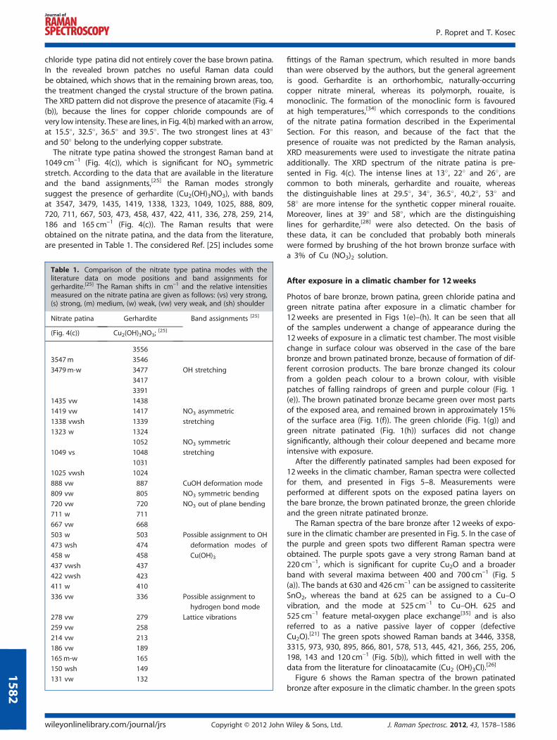
The Raman spectra of the bare bronze after 12weeks of expo-sure in the climatic chamber are presented in Fig. 5. In the case ofthe purple and green spots two different Raman spectra wereobtained. The purple spots gave a very strong Raman band at220 cm–1, which is significant for cuprite Cu2O and a broaderband with several maxima between 400 and 700 cm–1 (Fig. 5(a)). The bands at 630 and 426 cm–1 can be assigned to cassiteriteSnO2, whereas the band at 625 can be assigned to a Cu–Ovibration, and the mode at 525 cm–1 to Cu–OH. 625 and525 cm–1 feature metal-oxygen place exchange[35] and is alsoreferred to as a native passive layer of copper (defectiveCu2O).
[21] The green spots showed Raman bands at 3446, 3358,3315, 973, 930, 895, 866, 801, 578, 513, 445, 421, 366, 255, 206,198, 143 and 120 cm–1 (Fig. 5(b)), which fitted in well with thedata from the literature for clinoatacamite (Cu2 (OH)3Cl).
[26]
Figure 6 shows the Raman spectra of the brown patinatedbronze after exposure in the climatic chamber. In the green spots
Wiley & Sons, Ltd. J. Raman Spectrosc. 2012, 43, 1578–1586
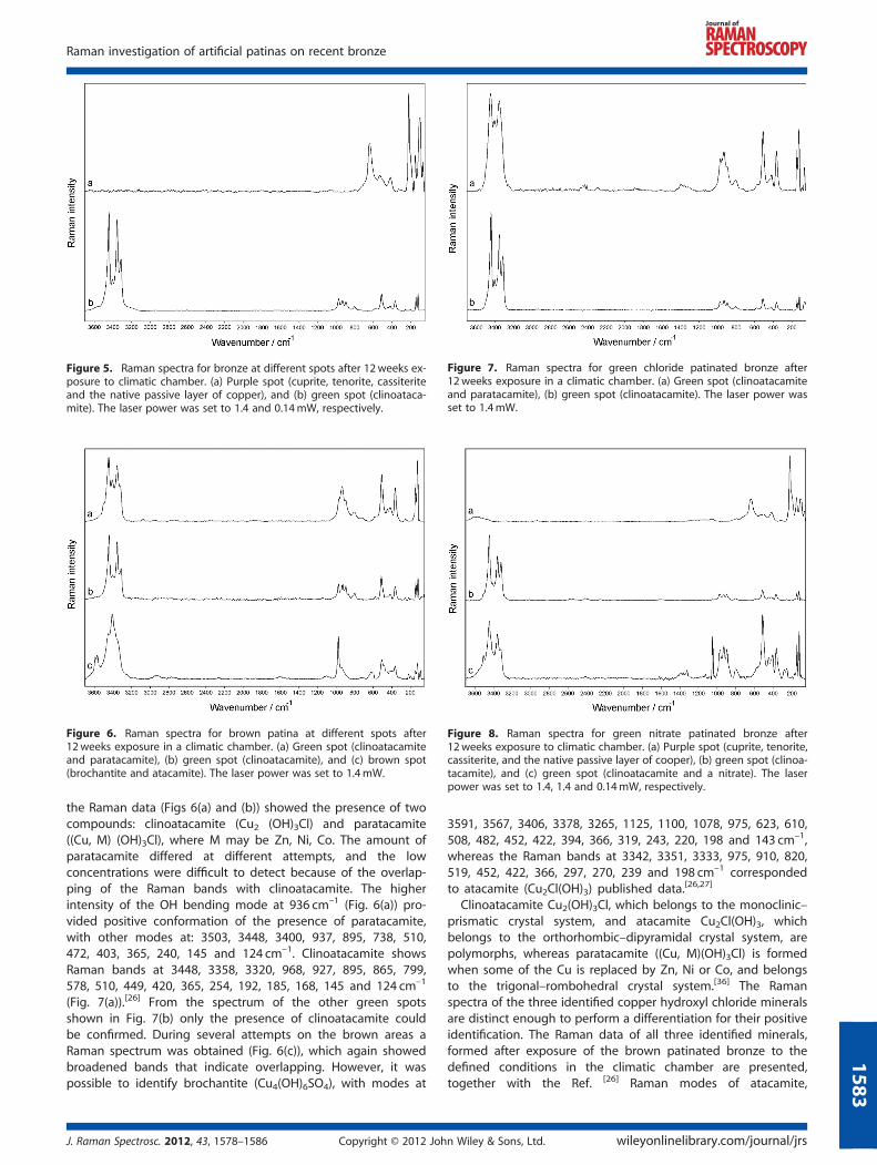
Figure 6. Raman spectra for brown patina at different spots after12weeks exposure in a climatic chamber. (a) Green spot (clinoatacamiteand paratacamite), (b) green spot (clinoatacamite), and (c) brown spot(brochantite and atacamite). The laser power was set to 1.4mW.
Figure 7. Raman spectra for green chloride patinated bronze after12weeks exposure in a climatic chamber. (a) Green spot (clinoatacamiteand paratacamite), (b) green spot (clinoatacamite). The laser power wasset to 1.4mW.
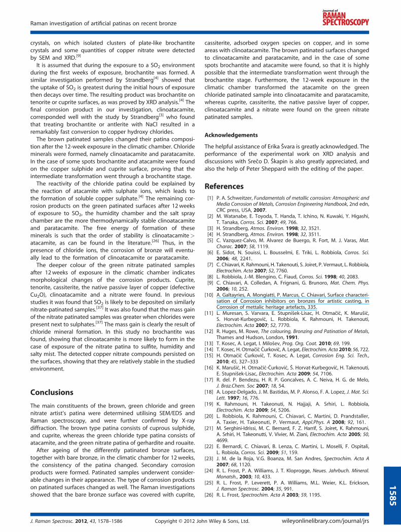
Figure 8. Raman spectra for green nitrate patinated bronze after12weeks exposure to climatic chamber. (a) Purple spot (cuprite, tenorite,cassiterite, and the native passive layer of cooper), (b) green spot (clinoa-tacamite), and (c) green spot (clinoatacamite and a nitrate). The laserpower was set to 1.4, 1.4 and 0.14mW, respectively.
Figure 5. Raman spectra for bronze at different spots after 12weeks ex-posure to climatic chamber. (a) Purple spot (cuprite, tenorite, cassiteriteand the native passive layer of copper), and (b) green spot (clinoataca-mite). The laser power was set to 1.4 and 0.14mW, respectively.
Raman investigation of artificial patinas on recent bronze
158
the Raman data (Figs 6(a) and (b)) showed the presence of twocompounds: clinoatacamite (Cu2 (OH)3Cl) and paratacamite((Cu, M) (OH)3Cl), where M may be Zn, Ni, Co. The amount ofparatacamite differed at different attempts, and the lowconcentrations were difficult to detect because of the overlap-ping of the Raman bands with clinoatacamite. The higherintensity of the OH bending mode at 936 cm–1 (Fig. 6(a)) pro-vided positive conformation of the presence of paratacamite,with other modes at: 3503, 3448, 3400, 937, 895, 738, 510,472, 403, 365, 240, 145 and 124 cm–1. Clinoatacamite showsRaman bands at 3448, 3358, 3320, 968, 927, 895, 865, 799,578, 510, 449, 420, 365, 254, 192, 185, 168, 145 and 124 cm–1
(Fig. 7(a)).[26] From the spectrum of the other green spotsshown in Fig. 7(b) only the presence of clinoatacamite couldbe confirmed. During several attempts on the brown areas aRaman spectrum was obtained (Fig. 6(c)), which again showedbroadened bands that indicate overlapping. However, it waspossible to identify brochantite (Cu4(OH)6SO4), with modes at
J. Raman Spectrosc. 2012, 43, 1578–1586 Copyright © 2012 Joh
3591, 3567, 3406, 3378, 3265, 1125, 1100, 1078, 975, 623, 610,508, 482, 452, 422, 394, 366, 319, 243, 220, 198 and 143 cm–1,whereas the Raman bands at 3342, 3351, 3333, 975, 910, 820,519, 452, 422, 366, 297, 270, 239 and 198 cm–1 correspondedto atacamite (Cu2Cl(OH)3) published data.[26,27]
Clinoatacamite Cu2(OH)3Cl, which belongs to the monoclinic–prismatic crystal system, and atacamite Cu2Cl(OH)3, whichbelongs to the orthorhombic–dipyramidal crystal system, arepolymorphs, whereas paratacamite ((Cu, M)(OH)3Cl) is formedwhen some of the Cu is replaced by Zn, Ni or Co, and belongsto the trigonal–rombohedral crystal system.[36] The Ramanspectra of the three identified copper hydroxyl chloride mineralsare distinct enough to perform a differentiation for their positiveidentification. The Raman data of all three identified minerals,formed after exposure of the brown patinated bronze to thedefined conditions in the climatic chamber are presented,together with the Ref. [26] Raman modes of atacamite,
n Wiley & Sons, Ltd. wileyonlinelibrary.com/journal/jrs
3
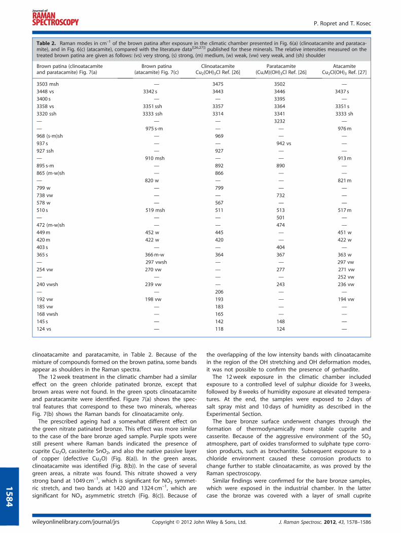
Table 2. Raman modes in cm–1 of the brown patina after exposure in the climatic chamber presented in Fig. 6(a) (clinoatacamite and parataca-mite), and in Fig. 6(c) (atacamite), compared with the literature data[[26,27]] published for these minerals. The relative intensities measured on thetreated brown patina are given as follows: (vs) very strong, (s) strong, (m) medium, (w) weak, (vw) very weak, and (sh) shoulder
Brown patina (clinoatacamiteand paratacamite) Fig. 7(a)
Brown patina(atacamite) Fig. 7(c)
ClinoatacamiteCu2(OH)3Cl Ref. [26]
Paratacamite(Cu,M)(OH)3Cl Ref. [26]
AtacamiteCu2Cl(OH)3 Ref. [27]
3503 msh — 3475 3502 —
3448 vs 3342 s 3443 3446 3437 s
3400 s — — 3395 —
3358 vs 3351 ssh 3357 3364 3351 s
3320 ssh 3333 ssh 3314 3341 3333 sh
— — — 3232 —
— 975 s-m — — 976m
968 (s-m)sh — 969 — —
937 s — — 942 vs —
927 ssh — 927 — —
— 910 msh — — 913m
895 s-m — 892 890 —
865 (m-w)sh — 866 — —
— 820 w — — 821m
799 w — 799 — —
738 vw — — 732 —
578 w — 567 — —
510 s 519 msh 511 513 517m
— — — 501 —
472 (m-w)sh — — 474 —
449m 452 w 445 — 451 w
420m 422 w 420 — 422 w
403 s — — 404 —
365 s 366m-w 364 367 363 w
— 297 vwsh — — 297 vw
254 vw 270 vw — 277 271 vw
— — — — 252 vw
240 vwsh 239 vw — 243 236 vw
— — 206 — —
192 vw 198 vw 193 — 194 vw
185 vw — 183 — —
168 vwsh — 165 — —
145 s — 142 148 —
124 vs — 118 124 —
P. Ropret and T. Kosec
1584
clinoatacamite and paratacamite, in Table 2. Because of themixture of compounds formed on the brown patina, some bandsappear as shoulders in the Raman spectra.The 12week treatment in the climatic chamber had a similar
effect on the green chloride patinated bronze, except thatbrown areas were not found. In the green spots clinoatacamiteand paratacamite were identified. Figure 7(a) shows the spec-tral features that correspond to these two minerals, whereasFig. 7(b) shows the Raman bands for clinoatacamite only.The prescribed ageing had a somewhat different effect on
the green nitrate patinated bronze. This effect was more similarto the case of the bare bronze aged sample. Purple spots werestill present where Raman bands indicated the presence ofcuprite Cu2O, cassiterite SnO2, and also the native passive layerof copper (defective Cu2O) (Fig. 8(a)). In the green areas,clinoatacamite was identified (Fig. 8(b)). In the case of severalgreen areas, a nitrate was found. This nitrate showed a verystrong band at 1049 cm–1, which is significant for NO3 symmet-ric stretch, and two bands at 1420 and 1324 cm–1, which aresignificant for NO3 asymmetric stretch (Fig. 8(c)). Because of
wileyonlinelibrary.com/journal/jrs Copyright © 2012 John
the overlapping of the low intensity bands with clinoatacamitein the region of the OH stretching and OH deformation modes,it was not possible to confirm the presence of gerhardite.
The 12week exposure in the climatic chamber includedexposure to a controlled level of sulphur dioxide for 3weeks,followed by 8weeks of humidity exposure at elevated tempera-tures. At the end, the samples were exposed to 2 days ofsalt spray mist and 10 days of humidity as described in theExperimental Section.
The bare bronze surface underwent changes through theformation of thermodynamically more stable cuprite andcasserite. Because of the aggressive environment of the SO2
atmosphere, part of oxides transformed to sulphate type corro-sion products, such as brochantite. Subsequent exposure to achloride environment caused these corrosion products tochange further to stable clinoatacamite, as was proved by theRaman spectroscopy.
Similar findings were confirmed for the bare bronze samples,which were exposed in the industrial chamber. In the lattercase the bronze was covered with a layer of small cuprite
Wiley & Sons, Ltd. J. Raman Spectrosc. 2012, 43, 1578–1586
Raman investigation of artificial patinas on recent bronze
crystals, on which isolated clusters of plate-like brochantitecrystals and some quantities of copper nitrate were detectedby SEM and XRD.[9]
It is assumed that during the exposure to a SO2 environmentduring the first weeks of exposure, brochantite was formed. Asimilar investigation performed by Strandberg[4] showed thatthe uptake of SO2 is greatest during the initial hours of exposurethen decays over time. The resulting product was brochantite ontenorite or cuprite surfaces, as was proved by XRD analysis.[4] Thefinal corrosion product in our investigation, clinoatacamite,corresponded well with the study by Strandberg[3] who foundthat treating brochantite or antlerite with NaCl resulted in aremarkably fast conversion to copper hydroxy chlorides.
The brown patinated samples changed their patina composi-tion after the 12-week exposure in the climatic chamber. Chlorideminerals were formed, namely clinoatacamite and paratacamite.In the case of some spots brochantite and atacamite were foundon the copper sulphide and cuprite surface, proving that theintermediate transformation went through a brochantite stage.
The reactivity of the chloride patina could be explained bythe reaction of atacamite with sulphate ions, which leads tothe formation of soluble copper sulphate.[4] The remaining cor-rosion products on the green patinated surfaces after 12weeksof exposure to SO2, the humidity chamber and the salt spraychamber are the more thermodynamically stable clinoatacamiteand paratacamite. The free energy of formation of theseminerals is such that the order of stability is clinoatacamite>atacamite, as can be found in the literature.[26] Thus, in thepresence of chloride ions, the corrosion of bronze will eventu-ally lead to the formation of clinoatacamite or paratacamite.
The deeper colour of the green nitrate patinated samplesafter 12weeks of exposure in the climatic chamber indicatesmorphological changes of the corrosion products. Cuprite,tenorite, cassiterite, the native passive layer of copper (defectiveCu2O), clinoatacamite and a nitrate were found. In previousstudies it was found that SO2 is likely to be deposited on similarlynitrate-patinated samples.[37] It was also found that the mass gainof the nitrate patinated samples was greater when chlorides werepresent next to sulphates.[37] The mass gain is clearly the result ofchloride mineral formation. In this study no brochantite wasfound, showing that clinoatacamite is more likely to form in thecase of exposure of the nitrate patina to sulfite, humidity andsalty mist. The detected copper nitrate compounds persisted onthe surfaces, showing that they are relatively stable in the studiedenvironment.
158
Conclusions
The main constituents of the brown, green chloride and greennitrate artist’s patina were determined utilising SEM/EDS andRaman spectroscopy, and were further confirmed by X-raydiffraction. The brown type patina consists of cuprous sulphide,and cuprite, whereas the green chloride type patina consists ofatacamite, and the green nitrate patina of gerhardite and rouaite.
After ageing of the differently patinated bronze surfaces,together with bare bronze, in the climatic chamber for 12weeks,the consistency of the patina changed. Secondary corrosionproducts were formed. Patinated samples underwent consider-able changes in their appearance. The type of corrosion productson patinated surfaces changed as well. The Raman investigationsshowed that the bare bronze surface was covered with cuprite,
J. Raman Spectrosc. 2012, 43, 1578–1586 Copyright © 2012 Joh
cassiterite, adsorbed oxygen species on copper, and in someareas with clinoatacamite. The brown patinated surfaces changedto clinoatacamite and paratacamite, and in the case of somespots brochantite and atacamite were found, so that it is highlypossible that the intermediate transformation went through thebrochantite stage. Furthermore, the 12-week exposure in theclimatic chamber transformed the atacamite on the greenchloride patinated sample into clinoatacamite and paratacamite,whereas cuprite, cassiterite, the native passive layer of copper,clinoatacamite and a nitrate were found on the green nitratepatinated samples.
Acknowledgements
The helpful assistance of Erika Švara is greatly acknowledged. Theperformance of the experimental work on XRD analysis anddiscussions with Srečo D. Škapin is also greatly appreciated, andalso the help of Peter Sheppard with the editing of the paper.
References[1] P. A. Schweitzer, Fundamentals of metallic corrosion: Atmospheric and
Media Corrosion of Metals, Corrosion Engineering Handbook, 2nd edn,CRC press, USA, 2007.
[2] M. Watanabe, E. Toyoda, T. Handa, T. Ichino, N. Kuwaki, Y. Higashi,T. Tanaka, Corros. Sci. 2007; 49, 766.
[3] H. Strandberg, Atmos. Environ. 1998; 32, 3521.[4] H. Strandberg, Atmos. Environ. 1998; 32, 3511.[5] C. Vazquez-Calvo, M. Alvarez de Buergo, R. Fort, M. J. Varas, Mat.
Charac. 2007; 58, 1119.[6] E. Sidot, N. Souissi, L. Bousselmi, E. Triki, L. Robbiola, Corros. Sci.
2006; 48, 2241.[7] C. Chiavari, K. Rahmouni, H. Takenouti, S. Joiret, P. Vermaut, L. Robbiola,
Electrochim. Acta 2007; 52, 7760.[8] L. Robbiola, J.-M. Blengino, C. Fiaud, Corros. Sci. 1998; 40, 2083.[9] C. Chiavari, A. Colledan, A. Frignani, G. Brunoro, Mat. Chem. Phys.
2006; 10, 252.[10] A. Galtayries, A. Mongiatti, P. Marcus, C. Chiavari, Surface characteri-
sation of Corrosion inhibitors on bronzes for artistic casting, inCorrosion of mettalic heritage artefacts, 335.
[11] L. Muresan, S. Varvara, E. Stupnišek-Lisac, H. Otmačić, K. Marušić,S. Horvat-Kurbegović, L. Robbiola, K. Rahmouni, H. Takenouti,Electrochim. Acta 2007; 52, 7770.
[12] R. Huges, M. Rowe, The colouring, Bronzing and Patination of Metals,Thames and Hudson, London, 1991.
[13] T. Kosec, A. Legat, I. Milošev, Prog. Org. Coat. 2010; 69, 199.[14] T. Kosec, H. Otmačić Ćurković, A. Legat, Electrochim. Acta 2010; 56, 722.[15] H. Otmaćić Ćurković, T. Kosec, A. Legat, Corrosion Eng. Sci. Tech.,
2010; 45, 327–333[16] K. Marušić, H. Otmačić-Ćurković, Š. Horvat-Kurbegović, H. Takenouti,
E. Stupnišek-Lisac, Electrochim. Acta 2009; 54, 7106.[17] R. del. P. Bendezu, H. R. P. Goncalves, A. C. Neiva, H. G. de Melo,
J. Braz.Chem. Soc 2007; 18, 54.[18] A. Lopez-Delgado, J. M. Bastidas, M. P. Alonso, F. A. Lopez, J. Mat. Sci.
Lett. 1997; 16, 776.[19] K. Rahmouni, H. Takeonuti, N. Hajjaji, A. Srhiri, L. Robbiola,
Electrochim. Acta 2009; 54, 5206.[20] L. Robbiola, K. Rahmouni, C. Chiavari, C. Martini, D. Prandstaller,
A. Taxier, H. Takeonuti, P. Vermaut, Appl.Phys. A 2008; 92, 161.[21] M. Serghini-Idrissi, M. C. Bernard, F. Z. Harrif, S. Joiret, K. Rahmouni,
A. Srhiri, H. Takeonutti, V. Vivier, M. Ziani, Electrochim. Acta 2005; 50,4699.
[22] E. Bernardi, C. Chiavari, B. Lenza, C. Martini, L. Moselli, F. Ospitali,L. Robiola, Corros. Sci. 2009; 51, 159.
[23] J. M. de la Roja, V.G. Boanza, M. San Andres, Spectrochim. Acta A2007; 68, 1120.
[24] R. L. Frost, P. A. Williams, J. T. Kloprogge, Neues. Jahrbuch. Mineral.Monatsh., 2003; 10, 433.
[25] R. L. Frost, P. Leverett, P. A. Williams, M.L. Weier, K.L. Erickson,J. Raman Spectrosc. 2004; 35, 991.
[26] R. L. Frost, Spectrochim. Acta A 2003; 59, 1195.
n Wiley & Sons, Ltd. wileyonlinelibrary.com/journal/jrs
5
P. Ropret and T. Kosec
1586
[27] V. Hayez, V. Costa, J. Guillaume, H. Terryn, A. Hubin, Analyst 2005;130, 550.
[28] V. Hayez, T. Segato, A. Hubin, H. Terryn, J. Raman. Spectrosc. 2006; 37, 1211.[29] SIST EN ISO 6988:1999, Metallic and other non-organic coatings -
Sulfur dioxide test with general condensation of moisture (ISO6988:1985).
[30] SIST EN ISO 6270–2: Paints and varnishes - Determination ofresistance to humidity.
[31] SIST EN ISO 9227: 2006 Corrosion tests in artificial atmospheres - Saltspray tests (ISO 9227:2006).
wileyonlinelibrary.com/journal/jrs Copyright © 2012 John
[32] F. Galesse, G. Laguzzi, L. Luvidi, V. Ferrari, S. Takacs, G. Venturi PaganiCesa, Corros. Sci. 2008; 50, 954.
[33] L. I. McCann, K. Trentleman, T. Possley, B. Golding, J. Raman.Spectrosc. 1999; 30, 121.
[34] N. Guillou, M. Louër, D. Louër, J. Solid State Chem 1994; 109, 307.[35] H. Y. H. Chan, C. G. Takoudis, M. J. Weaver, J. Phys. Chem. B 1999;
103, 357.[36] webmineral.com[37] P. Eriksson, L.-G. Johansson, J. Gullman, Corros. Sci. 1993;
34, 1083.
Wiley & Sons, Ltd. J. Raman Spectrosc. 2012, 43, 1578–1586