Radiological Findings in COVID-19 and Adaptive Approach in ...
19
Copyright © 2020 The Authors. https://doi.org/https://doi.org/10.12809/hkjr2017218 HONG KONG JOURNAL OF RADIOLOGY 17218 SPECIAL REPORT AND INVITED REVIEW Radiological Findings in COVID-19 and Adaptive Approach in Radiology Departments: Literature Review and Experience sharing LFH Ng * 1 , HHC Tsang 2 , FHY Wong 3 , MWC Law 4 , WH Chong 5 , CHN Ho 6 , JKJ Fung 7 , CCY Chan 8 , LSK Li 1 , KT Wong 4 , JCX Chan 5 , SHY Lam 3 , KH Wong 9 , PL Kwok 7 , L Xu 2 , TKK Lai 6 , KK Cheng 8 , TYW Hon 10 , JYH Hui 10 , SKY Kwok 5 , JKF Ma 1 1 Department of Radiology, Princess Margaret Hospital, Laichikok, Hong Kong 2 Department of Radiology and Organ Imaging, Queen Elizabeth Hospital, Jordan, Hong Kong 3 Department of Radiology, Queen Mary Hospital, Pokfulam, Hong Kong 4 Department of Imaging and Interventional Radiology, Prince of Wales Hospital, Shatin, Hong Kong 5 Department of Radiology, Tuen Mun Hospital, Tuen Mun, Hong Kong 6 Department of Radiology, Tseung Kwan O Hospital, Tseung Kwan O, Hong Kong 7 Department of Radiology, Pamela Youde Nethersole Eastern Hospital, Chai Wan, Hong Kong 8 Department of Diagnostic and Interventional Radiology, Kwong Wah Hospital, Yaumatei, Hong Kong 9 Department of Radiology, North District Hospital, Sheung Shui, Hong Kong 10 Department of Radiology, United Christian Hospital, Kwun Tong, Hong Kong Correspondence: Dr LFH Ng, Department of Radiology, Princess Margaret Hospital, Laichikok, Hong Kong Email: [email protected] Submitted: 24 Apr 2020; Accepted: 13 May 2020 Conflicts of Interest: All authors have disclosed no conflicts of interest. Funding/Support: This research received no specific grant from any funding agency in the public, commercial, or not-for-profit sectors. Ethics Approval: This study was approved by the Hong Kong Hospital Authority Central institutional Review Board (Ref CCO-2020-0010). This article has undergone peer review and has been accepted for publication in Hong Kong Journal of Radiology. This Author Accepted Manuscript has not been copyedited, typeset, or proofread, and this version may differ from the final published Version of Record. Please cite this article as an ‘Accepted Article’. https://doi.org/10.12809/hkjr2017218
-
Upload
khangminh22 -
Category
Documents
-
view
0 -
download
0
Transcript of Radiological Findings in COVID-19 and Adaptive Approach in ...

Copyright © 2020 The Authors. https://doi.org/https://doi.org/10.12809/hkjr2017218
HONG KONG JOURNAL OF RADIOLOGY
17218
SPECIAL REPORT AND INVITED REVIEW
Radiological Findings in COVID-19 and Adaptive Approach in Radiology
Departments: Literature Review and Experience sharing
LFH Ng *1, HHC Tsang 2, FHY Wong 3, MWC Law 4, WH Chong 5, CHN Ho 6, JKJ Fung 7, CCY
Chan 8, LSK Li 1, KT Wong 4, JCX Chan 5, SHY Lam 3, KH Wong 9, PL Kwok 7, L Xu 2, TKK Lai 6, KK Cheng 8, TYW Hon 10, JYH Hui 10, SKY Kwok 5, JKF Ma 1
1 Department of Radiology, Princess Margaret Hospital, Laichikok, Hong Kong
2 Department of Radiology and Organ Imaging, Queen Elizabeth Hospital, Jordan, Hong Kong
3 Department of Radiology, Queen Mary Hospital, Pokfulam, Hong Kong
4 Department of Imaging and Interventional Radiology, Prince of Wales Hospital, Shatin, Hong Kong
5 Department of Radiology, Tuen Mun Hospital, Tuen Mun, Hong Kong
6 Department of Radiology, Tseung Kwan O Hospital, Tseung Kwan O, Hong Kong
7 Department of Radiology, Pamela Youde Nethersole Eastern Hospital, Chai Wan, Hong Kong
8 Department of Diagnostic and Interventional Radiology, Kwong Wah Hospital, Yaumatei, Hong Kong
9 Department of Radiology, North District Hospital, Sheung Shui, Hong Kong
10 Department of Radiology, United Christian Hospital, Kwun Tong, Hong Kong
Correspondence: Dr LFH Ng, Department of Radiology, Princess Margaret Hospital, Laichikok, Hong
Kong
Email: [email protected]
Submitted: 24 Apr 2020; Accepted: 13 May 2020
Conflicts of Interest: All authors have disclosed no conflicts of interest.
Funding/Support: This research received no specific grant from any funding agency in the public,
commercial, or not-for-profit sectors.
Ethics Approval: This study was approved by the Hong Kong Hospital Authority Central institutional
Review Board (Ref CCO-2020-0010).
This article has undergone peer review and has been accepted for publication in Hong Kong Journal of
Radiology. This Author Accepted Manuscript has not been copyedited, typeset, or proofread, and this
version may differ from the final published Version of Record.
Please cite this article as an ‘Accepted Article’. https://doi.org/10.12809/hkjr2017218

Copyright © 2020 The Authors. https://doi.org/https://doi.org/10.12809/hkjr2017218
ABSTRACT
The outbreak of Coronavirus Disease 2019 (COVID-19) is the latest threat to global health. Radiological investigations play an important role in the management of patients with suspected or confirmed COVID-19 and radiologists should be familiar with the imaging characteristics. Being an integral component of the healthcare system, radiology departments have made adaptions to enhance infection control and strengthen the service. This article reviews the radiological features of COVID-19 on chest radiography and computed tomography, and shares experiences on the adaptive approach of radiology departments amidst the COVID-19 pandemic.
INTRODUCTION
In late December 2019, the World Health Organization (WHO) was informed of a cluster of cases of pneumonia of unknown cause detected in Wuhan City, Hubei Province, China. Subsequent deep sequencing analysis from lower respiratory tract samples indicated a novel coronavirus as the causative organism. International Committee on Taxonomy of Virus (ICTV) named the virus Severe Acute Respiratory Syndrome Coronavirus 2 (SARS-CoV-2)[1]. In February 2020, WHO officially named the disease Coronavirus Disease 2019 (COVID-19). The disease spread rapidly, resulting in an epidemic throughout China, followed by other countries around the world. On 11 March 2020, WHO officially declared COVID-19 a pandemic[2]. According to data published by WHO on 19 April 2020 (10:00 CEST), there has already been more than two million confirmed cases worldwide including more than 150,000 deaths from the disease[3].
The diagnosis of COVID-19 largely depends on clinical and epidemiological history with subsequent laboratory confirmation by real-time reverse-transcription polymerase chain reaction (RT-PCR) tests. On the other hand, radiological investigations also play an important role in the management of COVID-19 patients. The use of radiological investigations and the imaging features of COVID-19 will be elaborated in this article.
RADIOGLOGICAL INVESTIGATIONS FOR COVID-19
Role of Chest Radiography
Chest Radiography (CXR) is readily available and is commonly performed in patients presenting with respiratory symptoms of various causes. In patients with suspected COVID-19 infection, an initial CXR not only helps to detect features of pneumonia, but also helps to look for alternative diagnosis, such as pneumothorax, heart failure etc. Among countries facing resource constraints with limited availability and long turnaround time of RT-PCR tests, CXR is recommended for medical triage of patients who present with moderate to severe clinical features and a high pre-test probability of COVID-19[4].

Copyright © 2020 The Authors. https://doi.org/https://doi.org/10.12809/hkjr2017218
Depending on the clinical setting and time of presentation, CXR abnormalities are reported in 33.3% - 95% of COVID-19 patients[5-10]. In a study on 64 RT-PCR-confirmed COVID-19 patients from 4 hospitals in Hong Kong, 69% of patients demonstrated abnormalities on baseline CXR, with 80% of patients exhibiting CXR abnormalities at some point during their disease course[7]. As the sensitivity of CXR could be related to the time of imaging and severity of pulmonary involvement, a normal CXR cannot exclude the diagnosis of COVID-19 (Fig. 1A).
For patients with confirmed COVID-19, CXR has the advantage of being portable for imaging within the isolation rooms, thereby reducing the risk of disease transmission during patient transportation. Portable CXR is invaluable to assess disease progression and rule out complications. However, in patients with confirmed COVID-19 disease, CXR should only be performed when there is appropriate clinical need, such as when there is clinical deterioration, instead of a daily routine. Avoidance of non-value-added imaging is particularly important in the COVID-19 patient population, not only to minimize radiation risk, but also to reduce the risk of disease transmission to radiographers and to conserve personal protective equipment.
Imaging features on CXR
The most common findings on CXR are consolidation and ground glass opacities, more often found in a bilateral, peripheral, and lower zone distribution[5, 7](Fig. 1B). Pleural effusion is uncommon, found only in 3% of patients[7]. CXR findings reached peak severity at 10-12 days from symptom onset in a study in Hong Kong [7] (Fig.2). This is concordance with the earlier peak severity reported in CT at 6-11 days[11], which is more sensitive. This is also concordance with the clinical course, where sepsis and acute respiratory distress syndrome were found to occur at 9-12 days[12]. Nevertheless, CXR findings in COVID-19 are not organism specific and can overlap with other viral infections including influenza, severe acute respiratory syndrome (SARS) and middle east respiratory syndrome (MERS). Furthermore, co-infection with other respiratory pathogens can occur in patients with confirmed COVID-19 disease[13].
Fig. 1. (A) Normal CXR in a 39-year-old patient, taken on day 5 after RT-PCR confirming positive for COVID-19. (B) Right lower zone consolidation (arrowhead) on CXR in a 52-year-old patient, taken on day 6 after RT-PCR confirming positive for COVID-19.

Copyright © 2020 The Authors. https://doi.org/https://doi.org/10.12809/hkjr2017218
Fig. 2. Serial CXR in a 51-year-old patient, taken on day 1 (A), day 12 (B), and day 18 (C) after symptom onset. Extent of consolidation is largest on day 12.
Role of Computed Tomography
Despite chest computed tomography (CT) is less readily available and causes more radiation than CXR, it provides superior delineation of pulmonary involvement caused by COVID-19. Chest CT is reported to have higher sensitivity for the diagnosis of COVID-19 when compared with initial RT-PCR samples. Varying between different studies, the sensitivity of chest CT is found to be 98%, 97%, 93% and 61%[14-17]. Nevertheless, chest CT should not be used as the sole method in achieving the diagnosis of COVID-19 due to its limited specificity, which can be as low as 25%[15]. Discordance between results of RT-PCR and chest CT is commonly encountered and may lead to diagnostic confusion[18, 19]. A multidisciplinary approach, involving a combination of clinical history, clinical manifestations, imaging features and laboratory results, should therefore be required to achieve a timely and accurate diagnosis. According to a multinational consensus statement from the Fleischner Society, chest CT is not routinely indicated as a screening test for COVID-19 in asymptomatic individuals. Imaging is also not indicated for patients with mild features of COVID-19 unless they are at risk for disease progression. Chest CT, however, is indicated for patients showing worsening respiratory status and/or moderate to severe features of COVID-19[4].
CT can be used to evaluate complications related to COVID-19 infection. Pleural effusion, multiple tiny pulmonary nodules and mediastinal lymphadenopathy are atypical in COVID-19 pneumonia and their appearance should raise concern for bacterial superinfection or alternative diagnosis[20]. In addition, CT can readily detect other complications especially in COVID-19 patient who have been admitted under intensive care unit, to confirm or rule out pulmonary embolism during the acute setting.
Images Features on Chest CT

Copyright © 2020 The Authors. https://doi.org/https://doi.org/10.12809/hkjr2017218
The cardinal hallmark of COVID-19 pneumonia on chest CT is bilateral ground glass opacities (GGO) with or without consolidation in peripheral and posterior lungs (Fig. 3,4)[11, 21]. GGO often have round morphology (Fig.5) or are present with interlobular septal thickening and intralobular lines creating a “crazy-paving” pattern (Fig.6)[20]. GGO together with small areas of consolidation may suggest an organizing pneumonia pattern of lung injury[21].
Aligned by Hong Kong College of Radiologists (HKCR) and with the conjoint effort of all radiology centres in Hospital Authority of Hong Kong, chest CT images of COVID-19 confirmed cases scanned from 22 January 2020 to 16 April 2020 in six public hospitals in Hong Kong were reviewed by qualified radiologists under HKCR (Table 1). Total 110 patients with RT-PCR confirmed COVID-19 infection were included in this evaluation and 97 (88.2%) of them had abnormal findings on their first chest CT after admission. GGO (Fig. 3-7) was the most common finding, occurring in 95 cases (97.9%). Consolidation (Fig. 4) was the second most common finding, occurring in 57 cases (58.8%). Septal thickening and reticular/linear densities were present in 28 cases (28.9%) and 18 cases (18.6%), respectively. Bronchial wall thickening and dilatation (Fig. 4) were less commonly encountered, present in 13 cases (13.3%). Reticulonodular opacities and nodules were rare findings, both of which were found in the same patient (1 case, 1.0%). None of the cases had cavitatory nodules. In terms of distribution of the lesions, 94 cases (96.9%) had predominantly peripheral distribution. Bilateral and multilobar involvement were seen in 77 cases (79.4%) and 80 cases (82.5%), respectively. Other imaging findings were considered atypical, such as pleural effusion in 1 patient (1.0%) and mediastinal lymphadenopathy in 1 patient (1.0%). The observed imaging findings and patterns are in keeping with the results from other published studies.[11,15,17,18,19]
Recently in March 2020, the Radiological Society of North America (RSNA)proposed, with endorsement by the Society of Thoracic Radiology and American College of Radiology, structured CT reporting for suspected cases of COVID-19 pneumonia in order to promulgate the common findings of the disease and decrease reporting variability[20]. Findings are classified into four categories and details can be found in reference 20. The first category is the typical appearance with i) peripheral, bilateral GGOs +/- consolidation or intralobular lines (“crazy-paving” pattern), or ii) multifocal GGOs of rounded morphology +/- consolidation or intralobular lines, or iii) reverse halo sign or other findings of organizing pneumonia. The second category is indeterminate appearance with multifocal, diffuse, perihilar, or unilateral GGOs +/- consolidation lacking a specific distribution or few small GGOs that are non-rounded or non-peripheral. The third category is atypical appearance and the fourth category is negative for pneumonia.
Based on this RSNA proposed reporting language for COVID-19 pneumonia, the vast majority of our cases had typical (69%) or at least indeterminate appearances (18%) on chest CT, while only a small number belonged to atypical appearance (0.9%) or negative for pneumonia categories (11.8%) (Table 2).

Copyright © 2020 The Authors. https://doi.org/https://doi.org/10.12809/hkjr2017218
Table 1: Summary of 110 patients with chest CT scanned from 22 January 2020 to 16 April 2020 in six public hospitals
Total Number of Patients with Chest CT scanned 110
Age 47 (12-96)*
Sex
Male
Female
58 52
Abnormal Chest CT 97(88.2%)
CT findings
Ground glass opacity (GGO) 95 (97.9%)
Consolidation 57 (58.8%)
Solid nodule 1 (1.0%)
Reticulonodular opacities 1 (1.0%)
Cavitatory nodule 0 (0%)
Reticular/linear densities 18 (18.6%)
Septal thickening 28 (28.9%)
Bronchial wall thickening/dilatation 13 (13.4%)
Distribution
Peripheral 94 (96.9%)
Single lobe 17 (17.5%)
Multilobar 80 (82.5%)
Unilateral 20 (20.6%)
Bilateral 77 (79.4%)
Other CT findings
Pleural effusion 1 (1.0%)
Mediastinal lymphadenopathy 1 (1.0%)
*Age expressed in mean with range in brackets

Copyright © 2020 The Authors. https://doi.org/https://doi.org/10.12809/hkjr2017218
Table 2: RSNA Proposed Reporting Language for CT Findings Related to COVID-19[20]
Typical Appearance (Fig. 3-6) Number of cases = 76, 69%
i) Peripheral, bilateral GGO +/- consolidation or visible intralobular lines (“crazy-paving” pattern), or
ii) Multifocal GGO of rounded morphology +/- consolidation or visible intralobular lines, or
iii) Reverse halo sign or other findings of organizing pneumonia
Indeterminate appearance (Fig. 7) Number of cases = 20, 18%
Absence of typical features and presence of
i) Multifocal, diffuse, perihilar, or unilateral GGO +/- consolidation lacking a specific distribution and are non-rounded or non-peripheral, or
ii) Few very small GGO with a non-rounded and non-peripheral distribution
Atypical Appearance Number of cases = 1, 0.9%
Absence of typical or indeterminate features and presence of
i) Isolated lobar or segmental consolidation without GGO, or
ii) Discrete small nodules (centrilobular, “tree-in-bud”), or
iii) Lung cavitation, or
iv) Smooth interlobular septal thickening with pleural effusion
Negative for pneumonia Number of cases =13, 11.8%
No CT features to suggest pneumonia

Copyright © 2020 The Authors. https://doi.org/https://doi.org/10.12809/hkjr2017218
Fig. 3. Typical CT imaging features of COVID-19 pneumonia. Unenhanced, thin-section axial (A-C) and coronal reformatted images (D) of the lungs in a 39-year-old man performed 4 days after the onset of symptoms show bilateral, multifocal peripheral GGO with predominance in bilateral lower lobes. Line opacities are also seen which are likely due to subsegmental atelectasis at this early stage of disease.

Copyright © 2020 The Authors. https://doi.org/https://doi.org/10.12809/hkjr2017218
Fig. 4. Typical CT features of COVID-19 pneumonia. Unenhanced, thin-section axial images (A-D) of the lungs in a 65-year-old woman performed 14 days after the onset of symptoms show bilateral, multifocal peripheral GGO with consolidations (asterisks). Also note the presence of bronchial dilatation and wall thickening (arrows).

Copyright © 2020 The Authors. https://doi.org/https://doi.org/10.12809/hkjr2017218
Fig. 5. Typical CT imaging features of COVID-19 pneumonia. Unenhanced, thin-section axial (A-C) and coronal reformatted images (D) of the lungs in another 39-year-old man performed 5 days after the onset of symptoms show bilateral, multifocal rounded and peripheral GGO.

Copyright © 2020 The Authors. https://doi.org/https://doi.org/10.12809/hkjr2017218
Fig. 6. Typical CT imaging features of COVID-19 pneumonia. Unenhanced, thin-section axial images (A-D) of the lungs in a 75-year-old man performed 9 days after the onset of symptoms show bilateral, multifocal peripheral GGO with superimposed interlobular septal thickening and intralobular lines — giving rise to “crazy-paving” appearance (arrows).

Copyright © 2020 The Authors. https://doi.org/https://doi.org/10.12809/hkjr2017218
Follow up Chest CT
When necessary, serial chest CT can reflect disease evolution and monitor treatment effect[22]. In COVID-19 patients who deteriorate, initial findings of small ground glass opacities on chest CT would become more extensive, and might be associated with grow larger with crazy paving pattern and consolidation[22-24]. In severe and critical patients, the occurrence rates of consolidation, linear opacities, crazy-paving and bronchial thickening would increase, as well as the extent of lung involvement[23]. It would eventually proceed to ‘white lung’ appearance, seriously affecting patient’s lung function[24].
For patients who eventually recover from COVID 19 pneumonia, they show worst CT features at approximately 10 days after the initial onset of symptoms. At approximately 14 days, radiological signs of improvement would be seen[22]. Ai et al observed that chest CT improvement would precede RT-PCR results turning negative in some patients[15].
Recent researches revealed that COVID-19 shares similar CT features with organizing pneumonia, most notably atoll sign and peripheral multilobar ground glass opacities. Such similarities may hint presence of secondary organizing pneumonia in COVID-19 patients. Secondary organizing pneumonia had also been documented in viral pneumonia caused by SARS-CoV, MERS-CoV and influenza. Future
Fig. 7. Indeterminate CT imaging features of COVID-19. Unenhanced, thin-section axial images (A-D) of the lungs in a 51-year-old man performed 12 days after the onset of symptoms show unilateral, single lobar GGO lacking a specific distribution and are non-rounded.
Copyright © 2020 The Authors. https://doi.org/https://doi.org/10.12809/hkjr2017218
histological correlation may be required to confirm the above association. Since a small proportion of patients with organizing pneumonia would progress to pulmonary fibrosis (fibrosing organizing pneumonia), follow up CT imaging may be advisable for patients showing features of organizing pneumonia. Such practice is further supported by the fact that organizing pneumonia could be managed by corticosteroid effectively to prevent progression to fibrosis[25].
According to a multinational consensus statement from the Fleischner Society, follow up CT is indicated in a patient who has functional impairment and hypoxemia after recovery from COVID-19. The purpose of the follow up imaging is to differentiate between causes for such pulmonary function impairment after recovery, whether it is due to sequelae of infection and mechanical ventilation; or from a potentially treatable cause (e.g. organizing pneumonia as mentioned above)[4].
As stated above, not all COVID-19 patients will undergo a follow up chest CT. A single-center review on the features of follow up Chest CT was performed. There were 70 patients who had chest CT performed after confirmation of COVID-19 infection and 15 out of the 70 patients had second chest CT performed. The mean time interval of the second chest CT from the first positive RT-PCR test was 19.8 days (range 8-29). The mean time interval between the first and second CT was 16.5 days (range 4-23). The mean age of patients was 56.1 years old (range 27-80). All cases showed GGO and consolidative changes with peripheral distribution on the initial scan. One case showed complete resolution (6.7%) on the second CT, while other cases showed various degree of interval reduction in the GGO and consolidative changes. All cases with serial CT performed survived and were subsequently discharged. There were two cases with a third chest CT performed. One of them demonstrated features of secondary organizing pneumonia (Fig. 8). The other case showed complete resolution on the third scan (Fig. 9).
Recent publication in Radiology by Wang et al also mentioned that majority of patients discharged had residual disease on final scans[25]. The median illness days from symptom onset to discharge was 24 days and the CT changes peaked during illness days 6-11. The upsurge of GGO on serial scans[25] was not observed in our cases review, which could be related to a longer interval of the second CT (mean 19.8 days).

Copyright © 2020 The Authors. https://doi.org/https://doi.org/10.12809/hkjr2017218
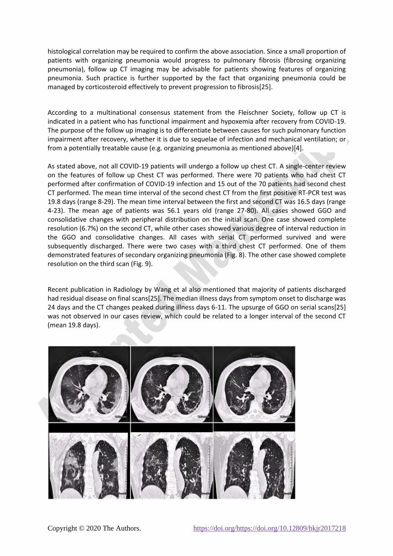
Fig. 8. Serial chest CT scans in a 56-year-old gentleman with COVID-19 pneumonia. Scan obtained on days 2 (left) after positive RT-PCR test showed peripheral ground glass opacities and consolidations without zonal predominance. Scan obtained on day 14 (middle) showed interval reduction of the mixed GGO and consolidative changes. Scan obtained on days 22 (right) showed the consolidations become arched bands with shaded margins. It was likely the result of perilobular inflammation, which could represent features of perilobular fibrosis found in secondary organizing pneumonia. Patient was discharge on day 32 after two consecutive negative RT-PCR tests.
Fig. 9. Serial chest CT scans in a 39-year-old gentleman with COVID-19 pneumonia. Scan obtained on days 2 (left) after positive RT-PCR test showed small peripheral consolidation and GGO scattered in both middle and lower lobes. Scan obtained on day 14 (middle) showed significant reduction of the consolidation and GGO. Scan obtained on days 28 (right) showed complete resolution. Patient was discharge on day 33 after two consecutive negative RT-PCR tests.
Comparing imaging features among COVID-19, SARS and MERS
The COVID-19 pandemic in 2020, SARS outbreak in 2003 and MERS outbreak in 2012 are all caused by viruses belonging to the same family of coronaviridae. It is understandable that they can mimic each other on clinical grounds. Radiologically they share some common features but also have noticeable differences.
In the largest series of COVID-19 in China, abnormalities on radiographs were detected in up to 59.1% of patients at presentation[26], which was less than reported radiographic detection rate of SARS and MERS (about 83%)[27, 28]. Among those who have radiological abnormalities, the distribution of changes most commonly involves the peripheral part of the lungs for all three infectious conditions. Unilateral abnormalities are more common in SARS and MERS than in COVID-19, where changes more frequently involve both lungs. Progression from focal unilateral peripheral lung involvement to

Copyright © 2020 The Authors. https://doi.org/https://doi.org/10.12809/hkjr2017218
bilateral multifocal lesions with upper lobes and perihilar involvement are associated with poor prognosis of severe cases in SARS (Fig. 10) and MERS, while the prognostic value of radiographic changes is unclear for COVID-19.
As with all viral pneumonia, the possible radiological patterns of parenchymal lung changes of these coronavirus infections are very similar. These include GGO, consolidation or a mixture of both. On CT, interlobular septal thickenings and intralobular lines within the parenchymal changes are also well described features, with possible progression into a crazy-paving pattern[27, 29].
It is also important to bear in mind the important negative radiological findings of these infections. Cavitation, centrilobular nodules, mediastinal or hilar lymphadenopathy and pleural effusion are not typical features in COVID-19 and SARS[29]. In the cases of MERS, pleural effusion is not uncommon (33%) and is reported to be associated with poorer prognosis[27].
In long term follow up imaging for SARS patient with pneumonia, areas of air trapping due to damaged ciliated respiratory epithelium and lung fibrosis have been reported[30]. Likewise, fibrosis is a relatively common (33%) feature in survivors of MERS[27]. As it has only been a few months since COVID-19 was first reported, the long term lung changes remain to be investigated with longer follow up.
Fig. 10A. CXR taken on presentation in a 42-year-old SARS patient, showing unilateral focal peripheral air space opacity at right lower zone.
Fig. 10B. Follow up CXR after 5 days shows progression into bilateral multifocal air space opacities.
Potential roles of Artificial Intelligence for COVID-19
Recently, Li et el reported a deep-learning algorithm which detects COVID-19 and differentiates it from community-acquired pneumonia[31]. Covering up to 4,000 multi-centered chest CT examinations, it

Copyright © 2020 The Authors. https://doi.org/https://doi.org/10.12809/hkjr2017218
generated promising per-examination sensitivity and specificity of 90% and 96% respectively, with an average test time of 4.5 seconds.
Reportedly, artificial intelligence (AI) algorithms have assisted in early warning, diagnosing, triaging cases and monitoring treatment response. RADLogics, for example, allows quantification of opacities while providing a scoring system which correlates with disease severity[32]. The Dutch University of Delft triages suspected COVID-19 cases based on CXR findings[33]. Nevertheless, for AI to be effective in combating the COVID-19 outbreak, high quality large population based data input and worldwide concerted efforts are required for further development.
ADAPTATION OF RADIOLOGY DEPARTMENTS DURING COVID-19 PANCEDMIC
The first confirmed case of COVID-19 in Hong Kong occurred on 23 January 2020 and the Hospital Authority(HA) elevated the response level in public hospitals from “Serious” to “Emergency” on 25 January 2020[34]. The first 20 cases of confirmed COVID-19 infection in Hong Kong were treated in an Infectious Disease Centre (IDC). Subsequent cases were distributed among other acute public hospitals in the city.
Modification and adaptation of the radiology department in essential in response to the COVID-19 pandemics[35,36].High level infection control measures were implemented in the radiology departments throughout HA. All suspected and confirmed cases of COVID-19 infection were treated in isolation wards in IDC. There were imaging facilities in IDC including general radiography, computed tomography, ultrasound and C-arm. The examination rooms were equipped with negative pressure to prevent the spread of infection. Most of the radiological examinations and procedures could be performed in IDC without the need to transport the patient to main radiology department, thus reducing the risk of cross infection. Portable radiographs were taken in the isolation wards with designated gown up and gown down areas for radiographers. The chest CT scans of suspected or confirmed COVID-19 cases were grouped together in specific sessions with allowance of air exchange time (15 minutes for IDC CT with air change rate of 12 ACH; 30 minutes for main CT with air change rate of 6 ACH) and room decontamination time between each case.
Infection control measures were also implemented in the main radiology department, including temperature screening of outpatients at the hospital or department entrances and universal masking. The radiological appointments for cases of suspected COVID-19 infection awaiting RT-PCR results would be deferred until the results are available. For confirmed cases which require radiological examinations for various clinical indications, CT was the more preferred modality than ultrasound due to shorter examination time and less physical contact between staff and patient.
Radiology service prioritization, re-organization and manpower re-deployment were initiated to enhance the support of COVID-19 care in terms of imaging diagnosis, assessment of disease severity and monitoring of progress and complication, as well as to maintain other essential diagnostic and interventional radiology service. This was achieved by rescheduling of

Copyright © 2020 The Authors. https://doi.org/https://doi.org/10.12809/hkjr2017218
outpatient elective appointments and deploying more manpower to perform inpatient and urgent reporting and interventional procedures to speed up patient discharge. Examinations with higher risks such as barium enema and modified barium swallow were suspended or reduced. Face-to-face clinical radiological meetings and educational meetings were suspended and replaced by video conferencing, email and phone discussion. Some radiology departments also segregated radiologists and radiographers into clean and dirty teams to prevent cross infection.
Staff training and engagement in infection control was another key measure. Personal infection prevention measures such as social distancing and vigilant handwashing were advocated. There was close liaison between the radiology department and the infection control team to ensure the adequate supply and distribution of personal protective equipment, enhancing infection control and staff morale. Timely communication with staff and other stakeholders including hospital management and infection control team was mediated through emails and social media apps to provide knowledge sharing and updates on information and policies[37, 38].
CONCLUSION
Radiological investigations play an important role in the diagnosis and management of COVID-19. The main imaging features on CXR include consolidation and ground glass opacities, more often found in a bilateral, peripheral, and lower zone distribution. Comparing with plain radiography, chest CT is more sensitive and provides superior delineation of the pulmonary involvement. The typical imaging findings of COVID-19 on chest CT are bilateral peripheral distribution of ground glass opacities with or without consolidation. Interlobular septal thickening and intralobular lines creating a “crazy-paving” pattern may also be seen. On the other hand, pleural effusion, lymphadenopathy and lung nodules are considered atypical. As it has only been a few months since the first case of COVID-19 was reported, further follow up would be necessary to delineate the long term radiological outcome in recovered COVID-19 patients.
Being an essential component of the medical system, radiology departments of the public health sector have made adaptations and enhanced infection control for the COVID-19 outbreak. As more experience is accumulated, we are confident that radiology departments will continue to function well and assist the whole medical profession to overcome the COVID-19 pandemic.
REFERENCES
1. Wang, L.-s., et al., A review of the 2019 Novel Coronavirus (COVID-19) based on current evidence. International Journal of Antimicrobial Agents, 2020: p. 105948.
2. World Health Organization. Coronavrius disease 2019 (COVID-19) situation report - 51. 2020, March 11; Available from: https://www.who.int/docs/default-source/coronaviruse/situation-reports/20200311-sitrep-51-covid-19.pdf?sfvrsn=1ba62e57_10.

Copyright © 2020 The Authors. https://doi.org/https://doi.org/10.12809/hkjr2017218
3. World Health Organization. Coronavrius disease 2019 (COVID-19) situation report - 90. 2020, April 19; Available from: https://www.who.int/docs/default-source/coronaviruse/situation-reports/20200419-sitrep-90-covid-19.pdf?sfvrsn=551d47fd_2.
4. Rubin, G.D., et al., The role of chest imaging in patient management during the covid-19 pandemic: A multinational consensus statement from the fleischner society. Chest, 2020.
5. Yoon, S.H., et al., Chest Radiographic and CT Findings of the 2019 Novel Coronavirus Disease (COVID-19): Analysis of Nine Patients Treated in Korea. Korean Journal of Radiology, 2020. 21.
6. Ng, M.-Y., et al., Imaging profile of the COVID-19 infection: radiologic findings and literature review. Radiology: Cardiothoracic Imaging, 2020. 2(1): p. e200034.
7. Wong, H.Y.F., et al., Frequency and distribution of chest radiographic findings in COVID-19 positive patients. Radiology, 2020: p. 201160.
8. Arentz, M., et al., Characteristics and outcomes of 21 critically ill patients with COVID-19 in Washington State. Jama, 2020.
9. Bandirali, M., et al., Chest X-ray findings in asymptomatic and minimally symptomatic quarantined patients in Codogno, Italy. Radiology, 2020: p. 201102.
10. Kim, E.S., et al., Clinical Course and Outcomes of Patients with Severe Acute Respiratory Syndrome Coronavirus 2 Infection: a Preliminary Report of the First 28 Patients from the Korean Cohort Study on COVID-19. Journal of Korean medical science, 2020. 35(13).
11. Salehi, S., et al., Coronavirus disease 2019 (COVID-19): a systematic review of imaging findings in 919 patients. American Journal of Roentgenology, 2020: p. 1-7.
12. Zhou, F., et al., Clinical course and risk factors for mortality of adult inpatients with COVID-19 in Wuhan, China: a retrospective cohort study. The Lancet, 2020.
13. Kim, D., et al., Rates of Co-infection Between SARS-CoV-2 and Other Respiratory Pathogens. JAMA, 2020.
14. Fang, Y., et al., Sensitivity of chest CT for COVID-19: comparison to RT-PCR. Radiology, 2020: p. 200432.
15. Ai, T., et al., Correlation of chest CT and RT-PCR testing in coronavirus disease 2019 (COVID-19) in China: a report of 1014 cases. Radiology, 2020: p. 200642.
16. Inui, S., et al., Chest CT findings in cases from the cruise ship “Diamond Princess” with coronavirus disease 2019 (COVID-19). Radiology: Cardiothoracic Imaging, 2020. 2(2): p. e200110.
17. Wen, Z., et al., Coronavirus Disease 2019: Initial Detection on Chest CT in a Retrospective Multicenter Study of 103 Chinese Subjects. Radiology: Cardiothoracic Imaging, 2020. 2(2): p. e200092.
18. Xu, X., et al., Imaging and clinical features of patients with 2019 novel coronavirus SARS-CoV-2. European Journal of Nuclear Medicine and Molecular Imaging, 2020: p. 1-6.
19. Bernheim, A., et al., Chest CT findings in coronavirus disease-19 (COVID-19): relationship to duration of infection. Radiology, 2020: p. 200463.
20. Simpson, S., et al., Radiological Society of North America Expert Consensus Statement on Reporting Chest CT Findings Related to COVID-19. Endorsed by the Society of Thoracic Radiology, the American College of Radiology, and RSNA. Radiology: Cardiothoracic Imaging, 2020. 2(2): p. e200152.
21. Ye, Z., et al., Chest CT manifestations of new coronavirus disease 2019 (COVID-19): a pictorial review. European Radiology, 2020: p. 1-9.
22. Pan, F., et al., Time course of lung changes on chest CT during recovery from 2019 novel coronavirus (COVID-19) pneumonia. Radiology, 2020: p. 200370.

Copyright © 2020 The Authors. https://doi.org/https://doi.org/10.12809/hkjr2017218
23. Li, K., et al., The Clinical and Chest CT Features Associated with Severe and Critical COVID-19 Pneumonia. Investigative radiology, 2020.
24. Pan, Y., et al., Initial CT findings and temporal changes in patients with the novel coronavirus pneumonia (2019-nCoV): a study of 63 patients in Wuhan, China. European radiology, 2020: p. 1-4.
25. Wang, Y., et al., Temporal changes of CT findings in 90 patients with COVID-19 pneumonia: a longitudinal study. Radiology, 2020: p. 200843.
26. Guan, W.-j., et al., Clinical Characteristics of Coronavirus Disease 2019 in China. New England Journal of Medicine, 2020.
27. Das, K.M., et al., Middle east respiratory syndrome coronavirus: what does a radiologist need to know? American Journal of Roentgenology, 2016. 206(6): p. 1193-1201.
28. Hosseiny, M., et al., Radiology perspective of coronavirus disease 2019 (COVID-19): lessons from severe acute respiratory syndrome and Middle East respiratory syndrome. American Journal of Roentgenology, 2020: p. 1-5.
29. Antonio, G.E., et al., Imaging of severe acute respiratory syndrome in Hong Kong. American Journal of Roentgenology, 2003. 181(1): p. 11-17.
30. Chang, Y.-C., et al., Pulmonary sequelae in convalescent patients after severe acute respiratory syndrome: evaluation with thin-section CT. Radiology, 2005. 236(3): p. 1067-1075.
31. Li, L., et al., Artificial intelligence distinguishes covid-19 from community acquired pneumonia on chest ct. Radiology, 2020: p. 200905.
32. Gozes, O., et al., Rapid ai development cycle for the coronavirus (covid-19) pandemic: Initial results for automated detection & patient monitoring using deep learning ct image analysis. arXiv preprint arXiv:2003.05037, 2020.
33. Systems, D.I. Triage for COVID-19 Using Artificial Intelligence on Chest X-Rays. 2020, April; Available from: https://www.delft.care/cad4covid/.
34. Hospital Authority. Press Release: Hospital Authority activates Emergency Response Level. 2020, January 25; Available from: https://www8.ha.org.hk/qmh/index_doc/eng.pdf.
35. Mossa-Basha M, Meltzer CC, Kim DC, Tuite MJ, Kolli KP, Tan BS. Radiology Department Preparedness for COVID19: Radiology Scientific Expert Panel. Radiology. 2020 Mar 16:200988. https://doi: 10.1148/radiol.2020200988. Epub ahead of print. PMID: 32175814.
36. Goh Y, Chua W, Lee JKT, Leng Ang BW, Liang CR, Tan CA, Wen Choong DA, Hoon HX, Leong Ong MK, Quek ST, Operational Strategies to Prevent COVID-19 spread in Radiology: Experience from a Singapore Radiology Department after SARS, Journal of the American College of Radiology (2020), doi: https://doi.org/10.1016/j.jacr.2020.03.027.
37. Huang, Z., et al., The Battle Against Coronavirus Disease 2019 (COVID-19): Emergency Management and Infection Control in a Radiology Department. Journal of the American College of Radiology, 2020.
38. Thomas, S.T.L. and C.Y. Wai, The lessons of SARS in Hong Kong. Clinical medicine, 2010. 10(1): p. 50.