Radioguided Surgery for Breast Cancer - Springer LINK
39
Radioguided Surgery for Breast Cancer Francesco Giammarile, Federica Orsini, Renato A. Valdés Olmos, Sergi Vidal-Sicart, Armando E. Giuliano, and Giuliano Mariani Contents Introduction .......................................... 3 General Background .................................. 3 Breast Cancer .......................................... 4 Sentinel Lymph Node in Breast Cancer ............ 5 Pathophysiological Aspects ........................... 5 Indications ............................................. 5 Procedures ............................................. 5 Image Interpretation ................................... 12 Procedures in the Surgical Suite ...................... 14 Clinical Controversial Aspects ........................ 18 Primary Localizing Techniques ....................... 21 Radioprotection ....................................... 24 Future Perspectives .................................... 25 Concluding Remarks ................................ 26 References ............................................ 26 F. Giammarile (*) Médecine Nucléaire – Groupement Hospitalier Est, Université Claude Bernard Lyon 1, Bron Cedex, France Biophysique – Faculté Charles Mérieux Lyon, Oullins Cedex, France e-mail: [email protected] F. Orsini Section of Nuclear Medicine, «Maggiore della Carità» University Hospital, Novara, Italy R.A.V. Olmos Nuclear Medicine Section and Interventional Molecular Imaging Laboratory, Department of Radiology, Leiden University Medical Centre, Leiden, The Netherlands Nuclear Medicine Department, Antoni van Leeuwenhoek Hospital, The Netherlands Cancer Institute, Amsterdam, The Netherlands S. Vidal-Sicart Nuclear Medicine Department, Hospital Clinic Barcelona, Barcelona, Spain A.E. Giuliano Surgical Oncology, Samuel Oschin Comprehensive Cancer Institute, Cedars-Sinai, Los Angeles, CA, USA G. Mariani Regional Center of Nuclear Medicine, University of Pisa, Pisa, Italy # Springer International Publishing Switzerland 2016 H.W. Strauss et al. (eds.), Nuclear Oncology , DOI 10.1007/978-3-319-26067-9_28-1 1
-
Upload
khangminh22 -
Category
Documents
-
view
0 -
download
0
Transcript of Radioguided Surgery for Breast Cancer - Springer LINK
Radioguided Surgery for BreastCancer
Francesco Giammarile, Federica Orsini, Renato A. ValdésOlmos, Sergi Vidal-Sicart, Armando E. Giuliano, and GiulianoMariani
Contents
Introduction . . . . . . . . . . . . . . . . . . . . . . . . . . . . . . . . . . . . . . . . . . 3General Background . . . . . . . . . . . . . . . . . . . . . . . . . . . . . . . . . . 3Breast Cancer . . . . . . . . . . . . . . . . . . . . . . . . . . . . . . . . . . . . . . . . . . 4
Sentinel Lymph Node in Breast Cancer . . . . . . . . . . . . 5Pathophysiological Aspects . . . . . . . . . . . . . . . . . . . . . . . . . . . 5Indications . . . . . . . . . . . . . . . . . . . . . . . . . . . . . . . . . . . . . . . . . . . . . 5Procedures . . . . . . . . . . . . . . . . . . . . . . . . . . . . . . . . . . . . . . . . . . . . . 5
Image Interpretation . . . . . . . . . . . . . . . . . . . . . . . . . . . . . . . . . . . 12Procedures in the Surgical Suite . . . . . . . . . . . . . . . . . . . . . . 14Clinical Controversial Aspects . . . . . . . . . . . . . . . . . . . . . . . . 18Primary Localizing Techniques . . . . . . . . . . . . . . . . . . . . . . . 21Radioprotection . . . . . . . . . . . . . . . . . . . . . . . . . . . . . . . . . . . . . . . 24Future Perspectives . . . . . . . . . . . . . . . . . . . . . . . . . . . . . . . . . . . . 25
Concluding Remarks . . . . . . . . . . . . . . . . . . . . . . . . . . . . . . . . 26
References . . . . . . . . . . . . . . . . . . . . . . . . . . . . . . . . . . . . . . . . . . . . 26
F. Giammarile (*)Médecine Nucléaire – Groupement Hospitalier Est,Université Claude Bernard Lyon 1, Bron Cedex, France
Biophysique – Faculté Charles Mérieux Lyon, OullinsCedex, Francee-mail: [email protected]
F. OrsiniSection of Nuclear Medicine, «Maggiore della Carità»University Hospital, Novara, Italy
R.A.V. OlmosNuclear Medicine Section and Interventional MolecularImaging Laboratory, Department of Radiology, LeidenUniversity Medical Centre, Leiden, The Netherlands
Nuclear Medicine Department, Antoni van LeeuwenhoekHospital, The Netherlands Cancer Institute, Amsterdam,The Netherlands
S. Vidal-SicartNuclear Medicine Department, Hospital Clinic Barcelona,Barcelona, Spain
A.E. GiulianoSurgical Oncology, Samuel Oschin ComprehensiveCancer Institute, Cedars-Sinai, Los Angeles, CA, USA
G. MarianiRegional Center of Nuclear Medicine, University of Pisa,Pisa, Italy
# Springer International Publishing Switzerland 2016H.W. Strauss et al. (eds.), Nuclear Oncology,DOI 10.1007/978-3-319-26067-9_28-1
1

AbstractSentinel lymph node biopsy is the standard sur-gical procedure for staging clinically tumor-freeregional nodes in patients with early-stagebreast cancer. This technique has spared theadditional morbidity of axillary lymph nodedissection without compromising diagnosticaccuracy and prognostic information. However,it is still important to discuss current techniquesand some controversies. Current data indicatethat the combined radiocolloid injection app-roach (both superficial and deep injections)results in a higher identification rate of sentinellymph nodes. Routine preoperative scinti-graphic imaging helps the intraoperative searchfor sentinel lymph nodes and is vital fordetecting extra-axillary or aberrant nodes, aswell as for patients who have had prior corebreast biopsy or surgery. SPECT/CT imaging,in addition to conventional lymphoscintigraphy,leads to improved preoperative visualizationand localization of sentinel lymph nodes, espe-cially if performed for specific indications. Thecombined use of radioactive tracers and bluedyes is more effective in detecting sentinellymph nodes than either modality used aloneand is therefore recommended for routine use.
Intraoperative imagingwith portable gammacameras is being increasingly employed, en-hancing the reliability of the gamma probe byadding clear imaging of the surgical fields,especially when the injection site is close tothe lymphatic basin. Portable gamma camerascan also be useful during radioguided occultlesion localization procedures in patients withnon-palpable breast lesions.
Advances in radiopharmaceuticals andcomputer technology make it possible to inte-grate optical, hybrid tracers and 3D renderingsystems that facilitate intraoperative sentinellymph node identification.
KeywordsBreast cancer • Sentinel lymph node biopsy •Nuclear medicine • Axillary lymph node dis-section • Radiocolloid injection • Radioguidedoccult lesion localization
Glossary[18F]FDG 2-Deoxy-2-[18F]fluoro-D-
glucose99mTc-MAA 99mTc-macroaggregated
albuminALND Axillary lymph node dissectionART Axillary radiotherapyASCO Americal Society of Clinical
OncologyCT X-ray computed tomographyDCIS Ductal carcinoma in situFOV Field of viewGOSTT Guided intraoperative scinti-
graphic tumor targetingH&E Hematoxylin and eosin stainingICG Indocyanine greenIHC Immunohistochemistry stainingIMC Lymph nodes of the internal
mammary chainIMN Internal mammary lymph nodesLEHR Low-energy high-resolutionLEUHR Low-energy ultra-high
resolutionLN Lymph nodeMRI Magnetic resonance imagingNACT Neoadjuvant systemic
chemotherapyPET Positron emission tomographyPET/CT Positron emission tomography/
computed tomographyROLL Radioguided occul lesion
localizationSLN Sentinel lymph nodeSLNB Sentinel lymph node biopsySNOLL Sentinel node occult lesion
localization (a combined pro-cedure of simultaneous SLNBand ROLL in the same surgicalsession)
SPECT Single photon emission com-puted tomography
SPECT/CT Single photon emission com-puted tomography/computedtomography
SPIO Superparamagnetic iron oxideUS Ultrasonography
2 F. Giammarile et al.
Introduction
General Background
Radioguided surgery constitutes a wide range ofprocedures which involve close collaborationbetween different specialties (nuclear medicineand surgery, often pathology) [1].
“Radioguided surgery” includes a set of pre-,intra-, and postoperative techniques and proce-dures that are designed to optimize oncologicsurgery. All these technologies and applicationscan be encompassed in the recently coined con-cept of guided intraoperative scintigraphic tumortargeting (GOSTT) [2]. The basic feature thatmost obviously characterizes GOSTT is the pre-operative administration of a radiopharmaceutical(either interstitially or systemically), associatedwith the intraoperative use of a handheld radioac-tivity counting probe (most often the so-calledgamma probe) that facilitates the task of the sur-geon – that is, the identification and removal of thetarget tissue, either a lymph node or the tumoritself – by virtue of preferential radioactivity accu-mulation in the target tissue. Intraoperative explo-ration of the surgical field with the gamma probe(which has recently evolved to allow intra-operative imaging as well) is made possible by aset of preoperative techniques employed by thenuclear medicine physician to achieve accumula-tion of the radiopharmaceutical in the specifictarget lesion. In this scenario, the most recentadvances are based on growing interactionamong different components of the complexarmamentarium now available to the imagingcommunity (including hybrid imaging, hybridimaging agents, and/or virtual navigation sys-tems) and to the surgical community (includingrobot-assisted surgery) [2–6].
The concentration of a radiopharmaceutical ina target lesion can be achieved by three mainmechanisms: (1) interstitial administration of anadequate radiopharmaceutical, typically a radio-colloid, that scintigraphically depicts the patternof lymphatic drainage from the site of a solidepithelial tumor, i.e., performing lymphoscin-tigraphy to identify the sentinel lymph node
(s) (SLN) of the tumor; (2) systemic administra-tion of a radiopharmaceutical that preferentiallyaccumulates in the target lesion, e.g., 99mT-sestamibi for localization of parathyroid adeno-mas or [18F]FDG for [18F]FDG-avid tumorlesions; and (3) direct intralesional administrationof a radiopharmaceutical such as 99mTc-macroag-gregated human albumin (99mTc-MAA, com-prised of particles that, by virtue of theirrelatively large size, are virtually indefinitelyretained at the injection site) for the so-calledradioguided occult lesion localization (ROLL)for non-palpable breast tumors.
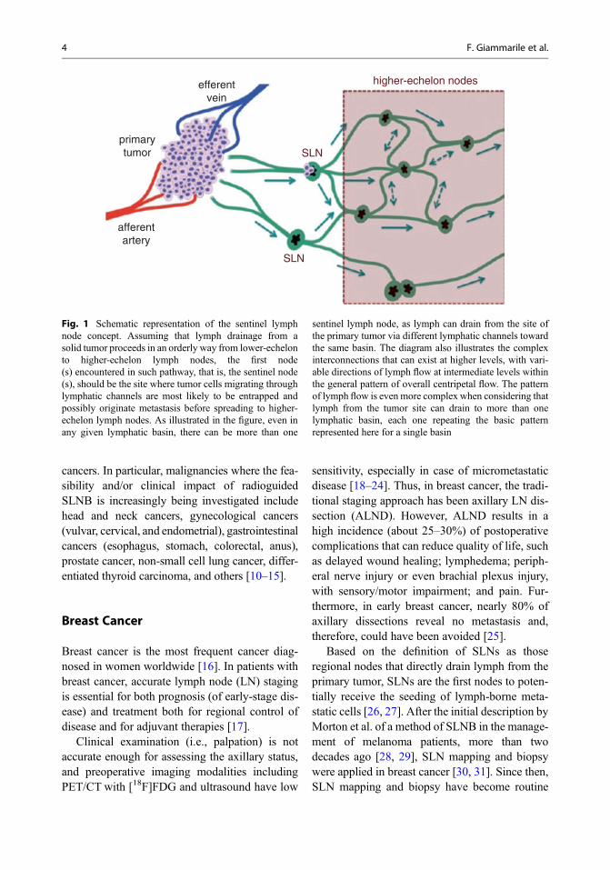
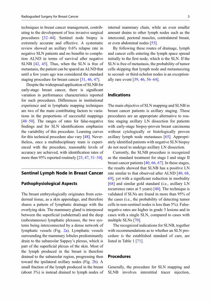
In the last few years, GOSTTapplications haverapidly expanded especially to perform sentinellymph node biopsy (SLNB). The sentinel lymphnode (SLN) procedure is a diagnostic stagingprocedure that is applied in a variety of tumortypes. The procedure aims to determine thetumor status of the SLN(s). Although historicallythe term “sentinel lymph node” was used todescribe a group of nodes seen in patients withpenile carcinoma [7], an SLN is currently definedas a lymph node on a direct drainage pathwayfrom the primary tumor [8, 9]. The concept isbased on the premise that lymph flow from theprimary tumor travels sequentially to the SLN andthen onto the other regional lymph nodes (Fig. 1).The SLN is the node most likely to harbormetastases.
The histopathologic status of this node shouldreflect the histopathologic status of the entirenodal basin, and additional treatment of thenodal basin (e.g., surgery) is routinely performedin case of metastatic involvement of the SLN –although the presence per se of metastatic tumorcells in the SLN is not the only factor determininglymph node dissection of the basin of interest (seefurther below in this chapter). A negative SLN,however, would justify a wait-and-see policyavoiding unnecessary elective lymph node dissec-tions and the associated morbidity, hospital stay,and costs.
In addition to being continually applied inpatients with breast cancer and cutaneous mela-noma, radioguided SLNB is being explored inpatients with a wide variety of other solid epithelial
Radioguided Surgery for Breast Cancer 3
cancers. In particular, malignancies where the fea-sibility and/or clinical impact of radioguidedSLNB is increasingly being investigated includehead and neck cancers, gynecological cancers(vulvar, cervical, and endometrial), gastrointestinalcancers (esophagus, stomach, colorectal, anus),prostate cancer, non-small cell lung cancer, differ-entiated thyroid carcinoma, and others [10–15].
Breast Cancer
Breast cancer is the most frequent cancer diag-nosed in women worldwide [16]. In patients withbreast cancer, accurate lymph node (LN) stagingis essential for both prognosis (of early-stage dis-ease) and treatment both for regional control ofdisease and for adjuvant therapies [17].
Clinical examination (i.e., palpation) is notaccurate enough for assessing the axillary status,and preoperative imaging modalities includingPET/CT with [18F]FDG and ultrasound have low
sensitivity, especially in case of micrometastaticdisease [18–24]. Thus, in breast cancer, the tradi-tional staging approach has been axillary LN dis-section (ALND). However, ALND results in ahigh incidence (about 25–30%) of postoperativecomplications that can reduce quality of life, suchas delayed wound healing; lymphedema; periph-eral nerve injury or even brachial plexus injury,with sensory/motor impairment; and pain. Fur-thermore, in early breast cancer, nearly 80% ofaxillary dissections reveal no metastasis and,therefore, could have been avoided [25].
Based on the definition of SLNs as thoseregional nodes that directly drain lymph from theprimary tumor, SLNs are the first nodes to poten-tially receive the seeding of lymph-borne meta-static cells [26, 27]. After the initial description byMorton et al. of a method of SLNB in the manage-ment of melanoma patients, more than twodecades ago [28, 29], SLN mapping and biopsywere applied in breast cancer [30, 31]. Since then,SLN mapping and biopsy have become routine
SLN
SLN
higher-echelon nodes
afferentartery
primarytumor
efferentvein
Fig. 1 Schematic representation of the sentinel lymphnode concept. Assuming that lymph drainage from asolid tumor proceeds in an orderly way from lower-echelonto higher-echelon lymph nodes, the first node(s) encountered in such pathway, that is, the sentinel node(s), should be the site where tumor cells migrating throughlymphatic channels are most likely to be entrapped andpossibly originate metastasis before spreading to higher-echelon lymph nodes. As illustrated in the figure, even inany given lymphatic basin, there can be more than one
sentinel lymph node, as lymph can drain from the site ofthe primary tumor via different lymphatic channels towardthe same basin. The diagram also illustrates the complexinterconnections that can exist at higher levels, with vari-able directions of lymph flow at intermediate levels withinthe general pattern of overall centripetal flow. The patternof lymph flow is even more complex when considering thatlymph from the tumor site can drain to more than onelymphatic basin, each one repeating the basic patternrepresented here for a single basin
4 F. Giammarile et al.
techniques in breast cancer management, contrib-uting to the development of less invasive surgicalprocedures [32–44]. Sentinel node biopsy isextremely accurate and effective. A systematicreview showed an axillary 0.6% relapse rate innegative SLN patients and no benefits to comple-tion ALND in terms of survival after negativeSLNB [42, 45]. Thus, when the SLN is free ofmetastasis, the patient can be spared an ALND thatuntil a few years ago was considered the standardstaging procedure for breast cancer [31, 46, 47].
Despite the widespread application of SLNB forearly-stage breast cancer, there is significantvariation in performance characteristics reportedfor such procedures. Differences in institutionalexperience and in lymphatic mapping techniquesare two of the main contributing factors to varia-tions in the proportions of successful mappings[48–50]. The ranges of rates for false-negativefindings and for SLN identifications emphasizethe variability of this procedure. Learning curvesfor this technical procedure also vary [48]. Never-theless, once a multidisciplinary team is experi-enced with the procedure, reasonable levels ofaccuracy are achieved, with identification rates ofmore than 95% reported routinely [25, 47, 51–54].
Sentinel Lymph Node in Breast Cancer
Pathophysiological Aspects
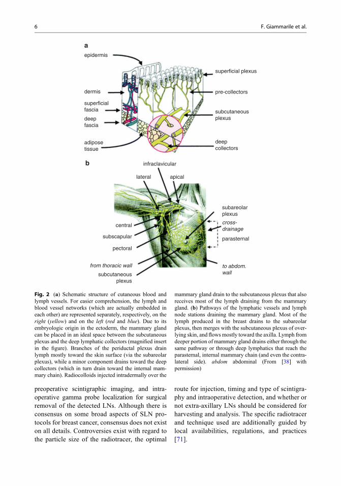
The breast embryologically originates from ecto-dermal tissue, as a skin appendage, and thereforeshares a pattern of lymphatic drainage with theoverlying skin. The mammary gland is interposedbetween the superficial (subdermal) and the deep(subcutaneous) lymphatic plexuses, the two sys-tems being interconnected by a dense network oflymphatic vessels (Fig. 2a). Lymphatic vesselssurrounding the mammary lobules predominantlydrain to the subareolar Sappey’s plexus, which ispart of the superficial plexus of the skin. Most ofthe lymph produced in the breast is thereforedrained to the subareolar region, progressing thentoward the ipsilateral axillary nodes (Fig. 2b). Asmall fraction of the lymph produced in the breast(about 3%) is instead drained to lymph nodes of
internal mammary chain, while an even smalleramount drains to other lymph nodes such as theintercostal, pectoral muscles, contralateral breast,or even abdominal nodes [55].
By following these routes of drainage, lymphand cancer cells entering the lymph space spreadinitially to the first node, which is the SLN. If theSLN is free of metastasis, the probability of tumorcells skipping that lymph node and metastasizingto second- or third-echelon nodes is an exception-ally rare event [39, 46, 56–64].
Indications
The main objective of SLNmapping and SLNB inbreast cancer patients is axillary staging. Theseprocedures are an appropriate alternative to rou-tine staging axillary LN dissection for patientswith early-stage biopsy-proven breast carcinomawithout cytologically or histologically provenaxillary lymph node metastases [65]. Appropri-ately identified patients with negative SLN biopsydo not need to undergo axillary LN dissection.
Currently, the SLNB procedure is recognizedas the standard treatment for stage I and stage IIbreast cancer patients [40, 66, 67]. In these stages,the results showed that SLNB has a positive LNrate similar to that observed after ALND [40, 68,69], yet with a significant reduction in morbidity[68] and similar gold standard (i.e., axillary LNrecurrence rates at 5 years) [40]. The technique isvalidated if SLNs are found in more than 95% ofthe cases (i.e., the probability of detecting tumorcells in non-sentinel nodes is less than 5%). False-negative rates are higher in grade 3 lesions and incases with a single SLN, compared to cases withmultiple SLNs [70].
The recognized indications for SLNB, togetherwith recommendations as to whether an SLN pro-cedure is the established standard of care, arelisted in Table 1 [71].
Procedures
Generally, the procedure for SLN mapping andSLNB involves interstitial tracer injection,
Radioguided Surgery for Breast Cancer 5
preoperative scintigraphic imaging, and intra-operative gamma probe localization for surgicalremoval of the detected LNs. Although there isconsensus on some broad aspects of SLN pro-tocols for breast cancer, consensus does not existon all details. Controversies exist with regard tothe particle size of the radiotracer, the optimal
route for injection, timing and type of scintigra-phy and intraoperative detection, and whether ornot extra-axillary LNs should be considered forharvesting and analysis. The specific radiotracerand technique used are additionally guided bylocal availabilities, regulations, and practices[71].
superficial plexus
pre-collectors
subcutaneousplexus
deepcollectors
subareolarplexus
parasternal
cross-drainage
central
lateral apical
infraclavicular
subscapular
pectoral
subcutaneousplexus
from thoracic wall to abdom.wall
dermis
epidermis
a
b
superficialfascia
deepfascia
adiposetissue
Fig. 2 (a) Schematic structure of cutaneous blood andlymph vessels. For easier comprehension, the lymph andblood vessel networks (which are actually embedded ineach other) are represented separately, respectively, on theright (yellow) and on the left (red and blue). Due to itsembryologic origin in the ectoderm, the mammary glandcan be placed in an ideal space between the subcutaneousplexus and the deep lymphatic collectors (magnified insertin the figure). Branches of the periductal plexus drainlymph mostly toward the skin surface (via the subareolarplexus), while a minor component drains toward the deepcollectors (which in turn drain toward the internal mam-mary chain). Radiocolloids injected intradermally over the
mammary gland drain to the subcutaneous plexus that alsoreceives most of the lymph draining from the mammarygland. (b) Pathways of the lymphatic vessels and lymphnode stations draining the mammary gland. Most of thelymph produced in the breast drains to the subareolarplexus, then merges with the subcutaneous plexus of over-lying skin, and flows mostly toward the axilla. Lymph fromdeeper portion of mammary gland drains either through thesame pathway or through deep lymphatics that reach theparasternal, internal mammary chain (and even the contra-lateral side). abdom abdominal (From [38] withpermission)
6 F. Giammarile et al.
Procedures in Nuclear MedicineThree main parameters define an optimal traceradministration technique for radioguided SLNB:injection site, injected volume, and injected activ-ity. A fourth parameter to be taken into account isthe time elapsed between injection and surgery, asit specifically influences the amount of radioactiv-ity to be injected [72, 73].
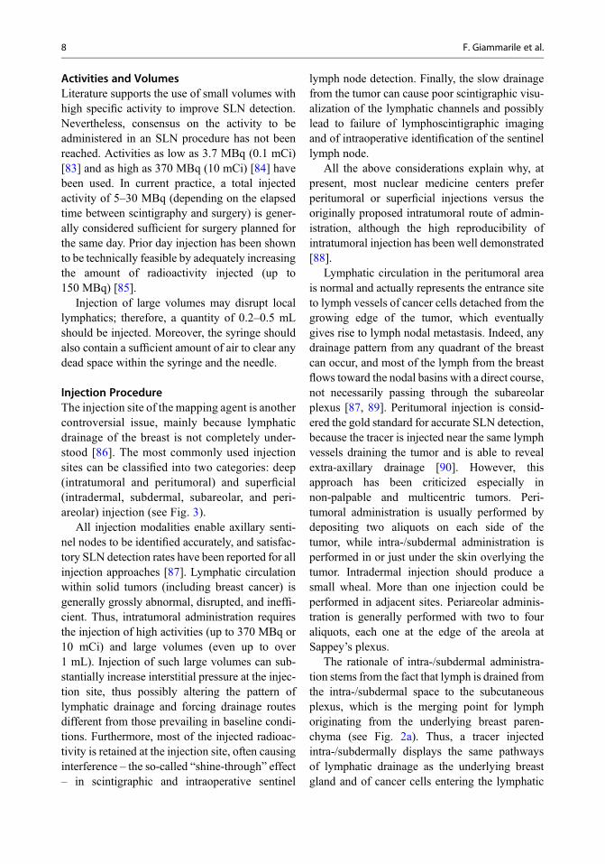
RadiopharmaceuticalsSeveral 99mTc-based agents have been used forradioguided SLNB for breast cancer (see Table 2)[74]. The radiopharmaceuticals most widely usedare 99mTc-sulfur colloid (particle size: 15–5,000 nm,but usually filtered to select a more restricted rangeof particles’ size), 99mTc-nanocolloid (5–100 nm),and 99mTc-antimony trisulfide (3–30 nm).
The ideal radiotracer should show rapid transitto SLNs with prolonged retention in the nodes. In
general, the drainage, distribution, and clearanceof radioactive colloids by the lymphatic systemmay vary and are dependent on the size of theparticles. Small particles are drained and clearedfirst; large particles are drained and cleared lastand may be retained longer at the injection site.Studies have shown the success rate of identifica-tion of axillary SLNs is not significantly affectedby the particle size of the radiotracer [38, 75–77].Thus, the selection of radiotracer is based moreon local availability than on differences in SLNdetection. However, there is general agreementthat a 100–200nm sized radiocolloid should beconsidered the best compromise between fastlymphatic drainage and optimal retention in SLNs[78].
New tracers have been developed in recentyears. The tracer most recently made commer-cially available is Lymphoseek®, which is com-posed of a dextran backbone with multipleglucose and mannose residues attached to DTPAfor 99mTc-labeling. The potential advantages of itssmall molecular size (7.1 nm) and the receptor-targeted nature of the mannose moieties in 99mTc-Tilmanocept include rapid transit from the pri-mary site to the SLN as well as selective accumu-lation in that node, with limited pass-through tosecond-echelon nodes [79–82].
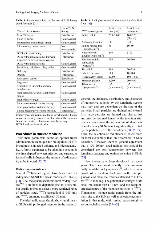
Table 1 Recommendations on the use of SLN biopsy(Modified from [71])
Clinical circumstanceUse of SLNbiopsy
T1 or T2 tumor Established
T3 or T4 tumor Controversial
Multicentric or multifocal tumor Controversial
Inflammatory breast cancer Notrecommended
DCIS with mastectomy Established
DCIS without mastectomy, but withsuspected or proven microinvasion
Established
DCIS without mastectomy Controversial
Suspicious, palpable axillary nodes Controversial
Older age Established
Obesity Established
Male breast cancer Established
Pregnancy Controversial
Evaluation of internal mammarylymph nodes
Controversial
Prior diagnostic or excisional breastbiopsy
Controversial
Prior axillary surgery Controversial
Prior non-oncologic breast surgery Controversial
After preoperative systemic therapy Controversial
Before preoperative systemic therapy Established
Controversial indications are those for which SLN biopsyis not universally accepted or for which the evidencebehind the practice is limited or entirely missingDCIS ductal carcinoma in situ
Table 2 Radiopharmaceutical characteristics (Modifiedfrom [74])
99mTc-based agentsParticle sizemax (nm)
Particle sizemean (nm)
Sulfur colloid 350–5,000 100–220(filtered)
Antimony trisulfide 80 3–30
Sulfide nanocolloid(Lymphoscint®)
80 10–50
Nanocolloidalalbumin (Nanocoll®)
100 5–80
Rhenium sulfidenanocolloid(Nanocis®)
500 50–200
Tin colloid 800 30–250
Labeled dextran 800 10–400
Hydroxyethyl starch 1,000 100–1,000
Stannous phytate 1,200 200–400
Tilmanocept(Lymphoseek®)
�7(equivalence)
�7(equivalence)
Radioguided Surgery for Breast Cancer 7
Activities and VolumesLiterature supports the use of small volumes withhigh specific activity to improve SLN detection.Nevertheless, consensus on the activity to beadministered in an SLN procedure has not beenreached. Activities as low as 3.7 MBq (0.1 mCi)[83] and as high as 370 MBq (10 mCi) [84] havebeen used. In current practice, a total injectedactivity of 5–30 MBq (depending on the elapsedtime between scintigraphy and surgery) is gener-ally considered sufficient for surgery planned forthe same day. Prior day injection has been shownto be technically feasible by adequately increasingthe amount of radioactivity injected (up to150 MBq) [85].
Injection of large volumes may disrupt locallymphatics; therefore, a quantity of 0.2–0.5 mLshould be injected. Moreover, the syringe shouldalso contain a sufficient amount of air to clear anydead space within the syringe and the needle.
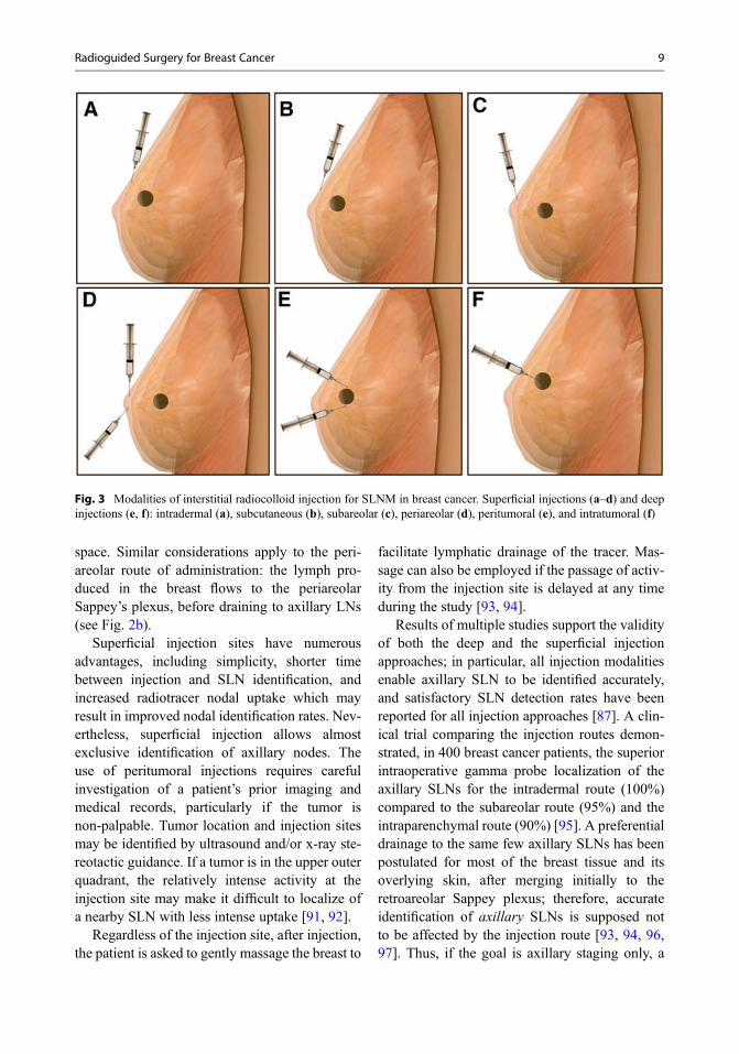
Injection ProcedureThe injection site of the mapping agent is anothercontroversial issue, mainly because lymphaticdrainage of the breast is not completely under-stood [86]. The most commonly used injectionsites can be classified into two categories: deep(intratumoral and peritumoral) and superficial(intradermal, subdermal, subareolar, and peri-areolar) injection (see Fig. 3).
All injection modalities enable axillary senti-nel nodes to be identified accurately, and satisfac-tory SLN detection rates have been reported for allinjection approaches [87]. Lymphatic circulationwithin solid tumors (including breast cancer) isgenerally grossly abnormal, disrupted, and ineffi-cient. Thus, intratumoral administration requiresthe injection of high activities (up to 370 MBq or10 mCi) and large volumes (even up to over1 mL). Injection of such large volumes can sub-stantially increase interstitial pressure at the injec-tion site, thus possibly altering the pattern oflymphatic drainage and forcing drainage routesdifferent from those prevailing in baseline condi-tions. Furthermore, most of the injected radioac-tivity is retained at the injection site, often causinginterference – the so-called “shine-through” effect– in scintigraphic and intraoperative sentinel
lymph node detection. Finally, the slow drainagefrom the tumor can cause poor scintigraphic visu-alization of the lymphatic channels and possiblylead to failure of lymphoscintigraphic imagingand of intraoperative identification of the sentinellymph node.
All the above considerations explain why, atpresent, most nuclear medicine centers preferperitumoral or superficial injections versus theoriginally proposed intratumoral route of admin-istration, although the high reproducibility ofintratumoral injection has been well demonstrated[88].
Lymphatic circulation in the peritumoral areais normal and actually represents the entrance siteto lymph vessels of cancer cells detached from thegrowing edge of the tumor, which eventuallygives rise to lymph nodal metastasis. Indeed, anydrainage pattern from any quadrant of the breastcan occur, and most of the lymph from the breastflows toward the nodal basins with a direct course,not necessarily passing through the subareolarplexus [87, 89]. Peritumoral injection is consid-ered the gold standard for accurate SLN detection,because the tracer is injected near the same lymphvessels draining the tumor and is able to revealextra-axillary drainage [90]. However, thisapproach has been criticized especially innon-palpable and multicentric tumors. Peri-tumoral administration is usually performed bydepositing two aliquots on each side of thetumor, while intra-/subdermal administration isperformed in or just under the skin overlying thetumor. Intradermal injection should produce asmall wheal. More than one injection could beperformed in adjacent sites. Periareolar adminis-tration is generally performed with two to fouraliquots, each one at the edge of the areola atSappey’s plexus.
The rationale of intra-/subdermal administra-tion stems from the fact that lymph is drained fromthe intra-/subdermal space to the subcutaneousplexus, which is the merging point for lymphoriginating from the underlying breast paren-chyma (see Fig. 2a). Thus, a tracer injectedintra-/subdermally displays the same pathwaysof lymphatic drainage as the underlying breastgland and of cancer cells entering the lymphatic
8 F. Giammarile et al.
space. Similar considerations apply to the peri-areolar route of administration: the lymph pro-duced in the breast flows to the periareolarSappey’s plexus, before draining to axillary LNs(see Fig. 2b).
Superficial injection sites have numerousadvantages, including simplicity, shorter timebetween injection and SLN identification, andincreased radiotracer nodal uptake which mayresult in improved nodal identification rates. Nev-ertheless, superficial injection allows almostexclusive identification of axillary nodes. Theuse of peritumoral injections requires carefulinvestigation of a patient’s prior imaging andmedical records, particularly if the tumor isnon-palpable. Tumor location and injection sitesmay be identified by ultrasound and/or x-ray ste-reotactic guidance. If a tumor is in the upper outerquadrant, the relatively intense activity at theinjection site may make it difficult to localize ofa nearby SLN with less intense uptake [91, 92].
Regardless of the injection site, after injection,the patient is asked to gently massage the breast to
facilitate lymphatic drainage of the tracer. Mas-sage can also be employed if the passage of activ-ity from the injection site is delayed at any timeduring the study [93, 94].
Results of multiple studies support the validityof both the deep and the superficial injectionapproaches; in particular, all injection modalitiesenable axillary SLN to be identified accurately,and satisfactory SLN detection rates have beenreported for all injection approaches [87]. A clin-ical trial comparing the injection routes demon-strated, in 400 breast cancer patients, the superiorintraoperative gamma probe localization of theaxillary SLNs for the intradermal route (100%)compared to the subareolar route (95%) and theintraparenchymal route (90%) [95]. A preferentialdrainage to the same few axillary SLNs has beenpostulated for most of the breast tissue and itsoverlying skin, after merging initially to theretroareolar Sappey plexus; therefore, accurateidentification of axillary SLNs is supposed notto be affected by the injection route [93, 94, 96,97]. Thus, if the goal is axillary staging only, a
Fig. 3 Modalities of interstitial radiocolloid injection for SLNM in breast cancer. Superficial injections (a–d) and deepinjections (e, f): intradermal (a), subcutaneous (b), subareolar (c), periareolar (d), peritumoral (e), and intratumoral (f)
Radioguided Surgery for Breast Cancer 9
superficial tracer injection (periareolar, sub-areolar, subdermal, intradermal) may be prefera-ble to a deep injection (peritumoral, intratumoral)due to better and quicker visualization of axillarySLNs [98]. On the other hand, an importantadvantage of deep injection is the improved detec-tion of extra-axillary SLNs: after peritumoraladministration, lymphoscintigraphy shows drain-age to the internal mammary chain in 20–30% ofthe cases, while this fraction is much lower (<3%)after intra-/subdermal or periareolar administra-tion [66, 99, 100]. Thus, if one’s aim is to stageextra-axillary nodal basins as well as the axilla,deep injection is recommended.
The superficial routes of administration aregenerally preferred in the case of superficial, eas-ily palpable tumors and the peritumoral route fordeeply seated tumors. The periareolar route can beused mainly in upper quadrant tumors to avoidpossible cross talk owing to the short distancebetween the peritumoral depot and the axillarySLNs and is particularly recommended in casesof non-palpable or multifocal tumors [101, 102].The combination of both injection techniques(deep and superficial) in the same patient mayimprove SLN detection [90].
Imaging ProceduresLymphatic mapping allows to determine the num-ber of LNs that are on a direct drainage pathwayand to locate the SLNs [71] [103]. Preoperativeimaging is strongly recommended due to variabil-ity in breast lymphatic drainage into the axilla andextra-axillary nodes [104]. Thus, preoperativelymphatic mapping has the potential to bothimprove accuracy (especially in extra-axillaryLN) and reduce morbidity relative to the use ofhandheld gamma probes alone [34, 71]. Preoper-ative imaging also serves as quality control on theuse of the appropriate tracer, failure of the injec-tion, failure of the radiopharmaceutical, and man-agement of the appropriate breast and axilla –injection of the proper side (L/R). Reasons not touse preoperative lymphoscintigraphy are logisti-cal or because there is no definite evidence of ahigher intraoperative success rate in theharvesting of axillary SLNs [105, 106].
Timing: In order to identify all SLNs and toavoid confusion with radiocolloid stasis in a lym-phatic vessel, images are acquired with an ade-quate delay after injection. This delay may varyaccording to the radiopharmaceutical used, theinjection site, and the patient’s characteristics(lymphatic drainage can be slower in elderly oroverweight patients). While smaller particlesallow quick visualization of SLNs, larger particleshave slow transit in the lymphatic system thattends to minimize visualization of non-sentinelsecond-tier nodes (lymph nodes downstream ofSLNs) [107]. After superficial tracer administra-tion, lymphatic drainage and subsequent lymphnode visualization is usually quicker than afterperitumoral injection (20–30 min compared to2–3 h on average). After 15–18 h, during surgery,the amount of radiocolloid migrated to LNs rep-resents about 1% of the injected activity aftersuperficial administration, while it is about 0.1%after peritumoral administration.
SLNs are generally visualized within 1–2 h,and the patient should be in the operating theaterwithin 2–30 h of radiocolloid injection, dependingon the facility’s schedule [71, 107]. In the event asurgery is scheduled for early morning, injectionand imaging may be safely performed the after-noon prior to the surgery [108].
Gamma camera parameters: A single- or dual-head gamma camera system with large field-of-view (FOV) detectors is generally used to acquireplanar emission and, if desired, single-photon com-puted tomographic (SPECT) or SPECT/computedtomographic (SPECT/CT) images. Low-energy,high-resolution, or low-energy high-resolution col-limators should be used. The energywindow shouldbe 15% (�5%) centered on the 140 keV photopeakof 99mTc.
Image acquisition: Dynamic (flow) imaging isnot often used in SLN procedures for breast can-cer, but can provide information useful to SLNlocalization.
Planar (static) imaging should be performed15–30 min, and 2–4 h post injection, and asneeded thereafter up to 18–30 h. At least two,preferably all three, of the following imagesshould be acquired: anterior, 45� anterior oblique,
10 F. Giammarile et al.
and lateral. Each image is typically 3–5 min induration. For a system with large FOV detectors,the pixel size is recommended to be approxi-mately 2 mm and the matrix size 256 � 256with zoom 1 or, rarely, 128 � 128 with zoom2. If 2 mm pixel size is not feasible on the system,the smallest pixel size available should be used.
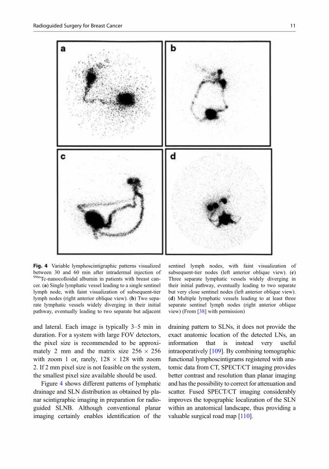
Figure 4 shows different patterns of lymphaticdrainage and SLN distribution as obtained by pla-nar scintigraphic imaging in preparation for radio-guided SLNB. Although conventional planarimaging certainly enables identification of the
draining pattern to SLNs, it does not provide theexact anatomic location of the detected LNs, aninformation that is instead very usefulintraoperatively [109]. By combining tomographicfunctional lymphoscintigrams registered with ana-tomic data from CT, SPECT/CT imaging providesbetter contrast and resolution than planar imagingand has the possibility to correct for attenuation andscatter. Fused SPECT/CT imaging considerablyimproves the topographic localization of the SLNwithin an anatomical landscape, thus providing avaluable surgical road map [110].
Fig. 4 Variable lymphoscintigraphic patterns visualizedbetween 30 and 60 min after intradermal injection of99mTc-nanocolloidal albumin in patients with breast can-cer. (a) Single lymphatic vessel leading to a single sentinellymph node, with faint visualization of subsequent-tierlymph nodes (right anterior oblique view). (b) Two sepa-rate lymphatic vessels widely diverging in their initialpathway, eventually leading to two separate but adjacent
sentinel lymph nodes, with faint visualization ofsubsequent-tier nodes (left anterior oblique view). (c)Three separate lymphatic vessels widely diverging intheir initial pathway, eventually leading to two separatebut very close sentinel nodes (left anterior oblique view).(d) Multiple lymphatic vessels leading to at least threeseparate sentinel lymph nodes (right anterior obliqueview) (From [38] with permission)
Radioguided Surgery for Breast Cancer 11
SPECTacquisition for SLN detection should beperformed with a dual-detector SPECT systemequipped with LEHR or LEUHR collimators.Acquisition parameters should include matrix sizeof 128 � 128 (4–5mmpixels) and 120 or 128 pro-jections over 360� with 20�25 s/projection.
Both low-dose CT (140 kVp, 2.5 mA) andconventional CT (140 kVp, 30–150 mA) can pro-vide useful anatomical detail that can be used foranatomical localization and, if desired, attenua-tion correction.
Mapping of all direct tumor-draining LNsrequires knowledge of the number and location ofthese SLNs, which will be provided by SPECT/CTin addition to planar images. SPECT/CT imagingprovides significant information in the large major-ity of patients, with useful preoperative compli-mentary information to the surgeons: betterlocation, reduced surgical time, and greater confi-dence of the surgeons with the technique [111].
It has been shown that SPECT/CT images candetect additional SLNs not visualized on planarimages in a substantial number of patients inwhom the conventional images are difficult tointerpret [110–112]. In the majority of cases, thesurgical team appreciates the anatomic informa-tion provided by the fused SPECT/CT images andthe surgical time is reduced [113]. However,because the current conventional approach basedon combined radiocolloid and blue dye injection,preoperative planar scintigraphic imaging, andintraoperative gamma probe counting has provenvery successful (with SLN detection rates over95%), the added value of SPECT/CT imagingseems to be limited to a small fraction of breastcancer patients undergoing SLNB. Current recog-nized indications for SPECT/CT imaging in breastcancer patients are non-visualization of SLNs atconventional imaging, obesity, and presence ofextra-axillary SLNs or otherwise unusual drain-age (e.g., in cases of previous breast surgery) [71].SPECT/CT imaging might also be performed ifthe conventional images are difficult to interpret(e.g., if contamination is suspected or an SLN islocated near to the injection area) [112, 113].
When acquiring planar imaging, a 57Co floodsource can be positioned between the patient’s
body and the collimator in order to obtain somereference anatomic landmarks in the scintigraphicimage (see Fig. 5). Alternatively, the body contourcan be delineated by moving a 57Co point sourceduring scintigraphic acquisition (see Fig. 6).SPECT/CT acquisitions obviate the problem ofidentifying anatomic landmarks as a referencefor topographic location of the SLN(s) (seeFigs. 7 and 8) [109, 114–118].
Surface marks that provide a method to trian-gulate SLNs and to estimate their depths aredesired by some surgeons. Surface locationsshould be marked on the skin with a small spotof indelible ink, and the depth of the node shouldbe described. When marking the skin in the imag-ing process, an attempt should be made to positionthe patient’s arm in the same position as it will beplaced during surgery.
Image Interpretation
Early and delayed lymphoscintigraphic planarimages identify SLNs in a majority of cases [71].Major criteria to identify LNs as SLNs are thetime of appearance and, occasionally, visualiza-tion of lymphatic channels (if dynamic imagingwas performed). Usually, SLNs cannot be readilydistinguished from second-tier LNs. The SLN isnot necessarily the hottest node, although that isoften the case. Separate lymphatic channels thatdrain to different LNs identify each of those asdistinct SLNs, even though they may be located inthe same anatomic region. When drainage to morethan one anatomic region is seen, each of thoseregions has at least one SLN.
In current protocols SPECT/CT is performedfollowing delayed planar images. This sequentialacquisition is helpful to clarify the role of bothmodalities. For imaging interpretation the majorcriteria to identify LNs visualized on lymphoscin-tigraphy as SLNs are the visualization of lym-phatic ducts, the time of appearance, the lymphnode basin, and the intensity of lymph nodeuptake [105, 119]. Following these criteria visu-alized radioactive lymph nodes may be classifiedas:
12 F. Giammarile et al.
(A) Definitively SLNs: this category concerns allLNs draining from the site of the primary tumorthrough their own lymphatic vessel or a singleradioactive LN in a certain lymphatic basin.
(B) Highly probable SLNs: this category includesLNs appearing between the injection site anda first draining node or LNs with increasinguptake appearing in other lymph nodestations.
(C) Less probable SLNs: all higher-echelon LNsmay be included in this category.
Axillary LNs represent the main basin for breastlymphatic drainage, but different patterns can alsooccur in some cases. Drainage to the internal mam-mary basin is present in up to 35–40% of patientsafter intratumoral/peritumoral radiocolloid injec-tion. Other unusually located SLNs are also
observed in a non-negligible fraction of patients:intramammary (prepectoral) in 6%, interpectoral in2%, and infraclavicular in 3% [120, 121].
The report to the referring physician shoulddescribe the orientations of the images acquired,the radiopharmaceutical, the method of administra-tion, the amount and volume of activity injected,the location of the SLNs on each image, and anysource of error or inaccuracy of the procedure.
The images and report should be available bythe time the patient arrives in the surgical suite – inelectronic form or as hard copy. If this is notpossible, the critical information should berelayed directly to the surgeon. A close workingrelationship between the imaging department andthe surgeon is critical for accurate disseminationof information regarding numbers and locationsof sentinel lymph nodes.
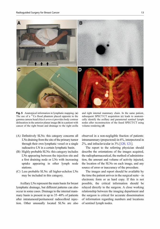
Fig. 5 Anatomical information in lymphatic mapping. (a)The use of a 57Co flood phantom placed opposite to thegamma camera head (black arrows) provides body contourdelineation in the anterior planar image (b) in a patient withcancer of the right breast and drainage to the right axilla
and right internal mammary chain. In the same patient,subsequent SPECT/CT acquisition (c) leads to anatomi-cally identify the axillary and parasternal sentinel lymphnodes after reconstruction of the fused SPECT/CT usingvolume rendering (d)
Radioguided Surgery for Breast Cancer 13
Procedures in the Surgical Suite
Blue Dye Lymph Node LocalizationRegarding the use of blue dye for optical guidanceduring surgery, there is general agreement thatcombined administration of radiocolloid andblue dye using both superficial injection anddeep injection enhances SLN detection [38, 94,97]. A possible advantage of the combined tech-nique is where macrometastasis in the SLN mayinhibit tracer accumulation [122, 123].
Blue dye can be injected around the primarytumor 10–20 min prior to surgery in a volume of2–5 mL. The site of injection can be gently mas-saged after the administration or if the drainage ofactivity from the injection site is delayed at anytime during the study [94]. Within 5–15 min, theSLNs are colored. Washout is evident afterapproximately 45 min.
Multiple studies have established the validityof blue dyes as markers for SLNs with reasonablyhigh detection rates (ranging from 75% to 80%)[124]; nevertheless such rates are slightly lowerthan those achieved with radiocolloids. In mostcases, the same SLNs are detected by the twomethods. Disadvantages of using blue dyes are
as follows: (i) impossibility to evaluate extra-axillary nodes, (ii) temporary blue tattooing ofthe skin or areola (for patients with breast conser-vation surgery), and (iii) induction of anaphylacticreactions (which require resuscitation in 0.5–1.0%of patients and that contraindicate its use in preg-nant women) [124–131].
Radioguided SurgeryIntraoperative detection of SLNs is usually radio-guided by a gamma-detection probe. Such probesshould be designed and constructed to be suitablefor intraoperative use, in order to be able to detectthe SLN from the skin surface as well as within theexposed surgical cavity [26]. The probe is placedin a sterile bag to be used in the sterile surgicalfield. A display capable of providing clear instan-taneous and cumulative counts is a major require-ment. It is helpful if the instantaneous count rate isfed to an audio signal that conveys count rateinformation.
The count rates obtained with the gammaprobe during surgery are recorded per unit timewith the probe in the surgical field, over the nodebefore excision (in vivo) and after excision(ex vivo). A background tissue count is also
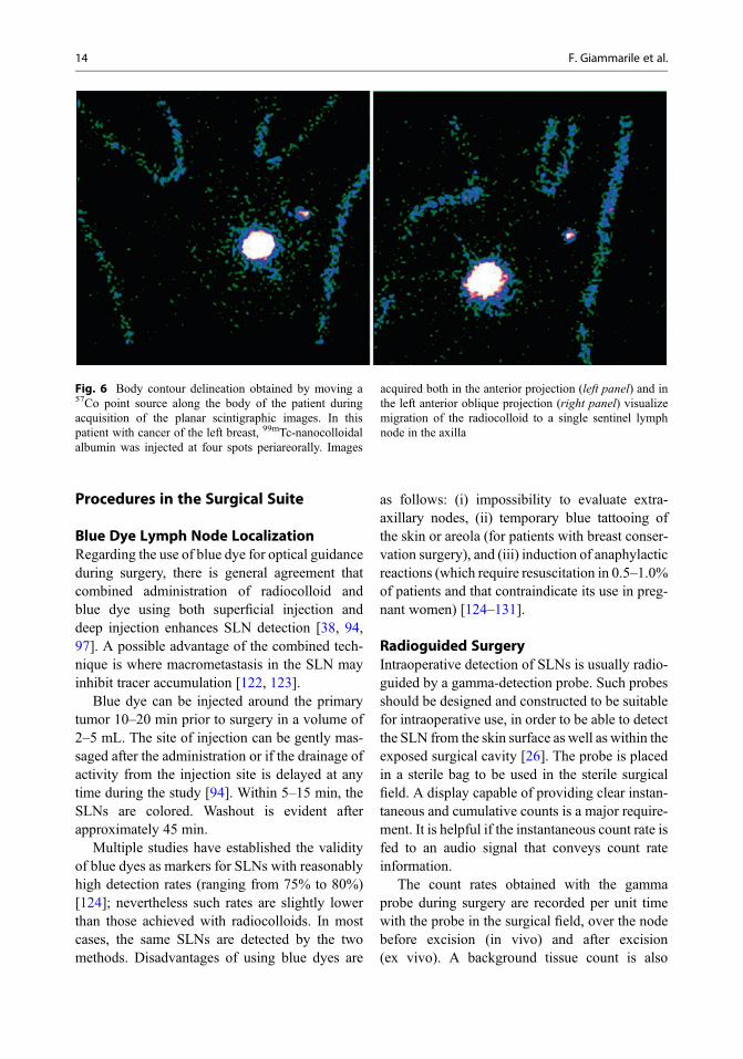
Fig. 6 Body contour delineation obtained by moving a57Co point source along the body of the patient duringacquisition of the planar scintigraphic images. In thispatient with cancer of the left breast, 99mTc-nanocolloidalalbumin was injected at four spots periareorally. Images
acquired both in the anterior projection (left panel) and inthe left anterior oblique projection (right panel) visualizemigration of the radiocolloid to a single sentinel lymphnode in the axilla
14 F. Giammarile et al.
recorded with the probe pointing away from theinjection site, nodal activity, or other physiologi-cal accumulation sites (i.e., liver) [71].
Just before starting surgery and with the patientpositioned on the operating table, using theimages and skin markings as guides, the gammaprobe scans the axilla or any other region wheretracer accumulation has been visualized in orderto confirm correct identification and localizationof the SLN(s) and to select the optimum locationfor incision. This task requires the sensitivity ofthe detector to be sufficient to identify a weaklyactive SLN when attenuated by, typically, up to5 cm of soft tissue. The surgeon then introducesthe probe through the skin incision to guide dis-section to the hot node(s). Discriminating activitycounts within the SLN from those originatingfrom nearby sites requires the probe to be well
collimated with a small angle of view. The detec-tor should offer a high level of shielding againstradiation hitting the side of the probe assembly.However, when working with the probe, it isimportant to direct the probe away from activityat the injection sites.
When a hot SLN has been removed, the surgi-cal bed should be checked to confirm removal ofthe hot node(s) and to evaluate remaining activity.Owing to the limited spatial resolution of thegamma camera, LNs closer than approximately15–20 mm may appear on lymphoscintigraphyas one single hot spot; so, in some cases anotherhot node may still be present at a close locationafter removal of the hottest SLN. In this regard,the use of SPECT/CT imaging is very helpfulbecause it may provide information about theactual presence of a cluster of LNs rather than a
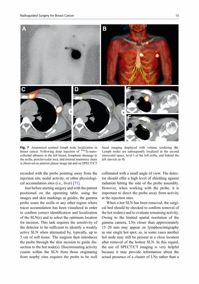
Fig. 7 Anatomical sentinel lymph node localization inbreast cancer. Following deep injection of 99mTc-nano-colloidal albumin in the left breast, lymphatic drainage tothe axilla, periclavicular area, and internal mammary chainis observed on anterior planar image (a) and on SPECT/CT
fused imaging displayed with volume rendering (b).Lymph nodes are subsequently localized in the secondintercostal space, level I of the left axilla, and behind theleft clavicle (c–f)
Radioguided Surgery for Breast Cancer 15
single SLN. When other sources of activity arefound in the lymphatic basin, the decision ofwhether to remove them will depend upon thereport from lymphoscintigraphy and the workingdefinition of “nodes to remove” [132, 133]. Inprinciple, SLNB requires the removal of allSLNs receiving direct lymphatic drainage fromthe site of the primary tumor. In practice, this isnot always achieved. In cases with multiple radio-labeled LNs, it is often difficult to distinguishbetween SLNs and second-tier LNs. The issue ofhow many SLNs should be biopsied when multi-ple radioactive LNs are found is still debated. Inthis regard, while removing too few nodes maymiss potential metastases in regional LNs, indis-criminate removal of all radioactive axillary nodesmay cause morbidity similar to that experiencedafter conventional ALND (in addition to theunnecessarily increased burden for histopatholog-ical analysis).
Several operational definitions of the SLNhave evolved over time in order to decide exactlywhich nodes should be removed to maximizethe likelihood of locating the “true” biologicSLN and to minimize the superfluous removal ofnon-SLNs. Some authors base SLN identificationon the absolute number of counts per secondrecorded for the presumed nodes, while othersconsider the ratio of the “in vivo” or “ex vivo”radioactive counts in the SLNs relative to back-ground or to neighboring non-SLNs. Empiricthresholds corresponding to (i) 10% or 20% ofthe counting rate in the first LN removed (whichis usually the most radioactive) or (ii) at least tentimes the background count, taken at a locationremote from the injection site, are widely reportedin the literature [98, 134–137]. It is generallyaccepted that removing more than five LNs fromthe axilla does not result in marked improvementin the sensitivity of axillary SLNB [138–143]. If
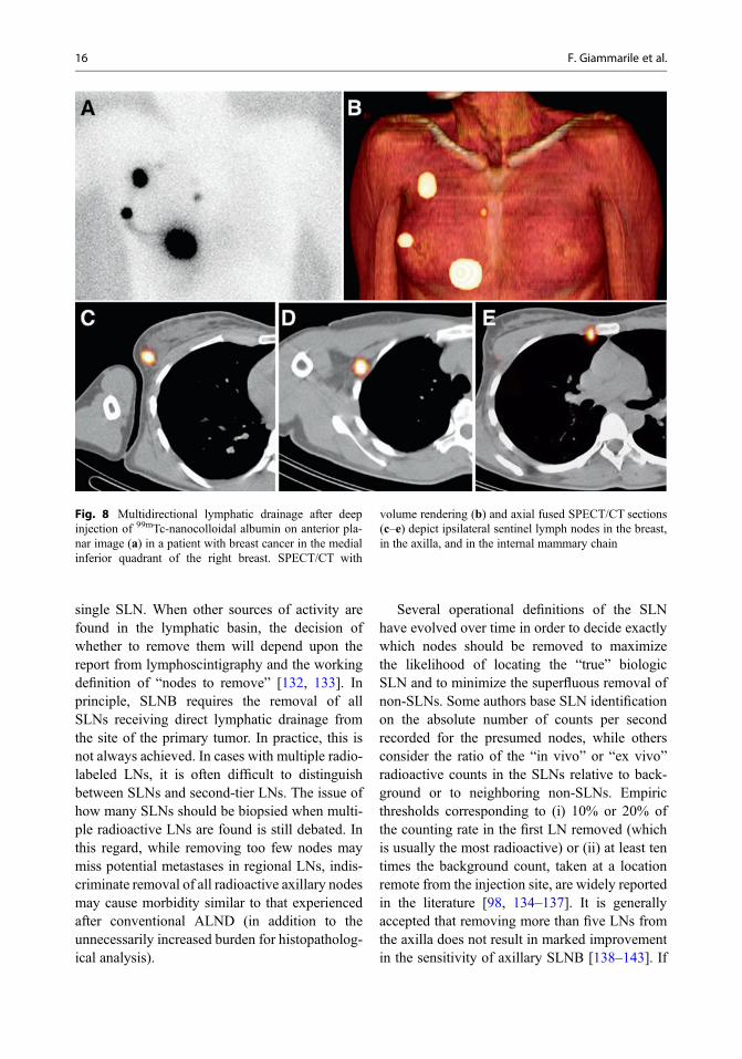
Fig. 8 Multidirectional lymphatic drainage after deepinjection of 99mTc-nanocolloidal albumin on anterior pla-nar image (a) in a patient with breast cancer in the medialinferior quadrant of the right breast. SPECT/CT with
volume rendering (b) and axial fused SPECT/CT sections(c–e) depict ipsilateral sentinel lymph nodes in the breast,in the axilla, and in the internal mammary chain
16 F. Giammarile et al.
blue dye is used, it can be a useful adjunct foraiding SLN localization and harvesting. Blue dyegenerally results in a lower SLN detection ratethan radiotracers, but it can be used in additionto radiocolloids. Following injection, the blue dyedrains to the SLNs, staining the channels, whichcan be followed to the first-echelon nodes. Directvisualization and dissection of these channelsfacilitate SLN localization.
Deeply located SLNs are difficult to detectintraoperatively because of tissue attenuation;furthermore, the large amount of radioactivityretained at the injection site may cause nearbylocated SLNs to be hidden because of the shine-through effect. Patients who have undergoneprevious breast surgery or radiation may demon-strate nodes in locations not typically seen inpatients without a history of prior surgery. Thelymphatic duct to the original SLN may beobstructed by tumor growth or the original SLNmay be entirely replaced by disease. Conse-quently, lymphatic drainage may be eitherdiverted to a non-sentinel node or no lymphnodes may be visualized, increasing false-negative results.
The use of SPECT/CT images can help local-ization of focal activity [144] as can the use ofintraoperative imaging with portable gamma cam-eras; the latter imaging equipment is generallyused simply to verify that all radioactive lymphnodes of interest are removed [4]. Finally, to min-imize false-negative results, the open axillashould be palpated and suspicious lymph nodesharvested, even if these are neither hot nor blue.
SLN Non-visualization or FailedIntraoperative DetectionIt is important to consider that when an SLN is notdetected intraoperatively, this corresponds to afailure of the method and not to a false-negativecase (better defined as when an axillary relapse isobserved despite a prior negative SLNB). Themajority of patients with preoperative lympho-scintigraphic SLN non-visualization will have atleast one SLN detected intraoperatively, either bya gamma probe alone or by a gamma probe com-bined with blue dye. While logistically difficult inmost centers, a second radiocolloid injection,
following perhaps a different injection route, maybe useful to visualize previously non-visualized SLNs.
In approximately 1–2% of the patients, SLNswill not be detected preoperatively or intra-operatively, and the status of axillary LNs cannotbe determined. Old age, obesity, tumor locationother than in the upper outer quadrant and non-visualization of SLNs on preoperative lympho-scintigraphy may be associated with failed SLNlocalization [142]. The significance of preopera-tive scintigraphic SLN non-visualization is not yetknown. Some studies have suggested that patientswith unsuccessful axillary mapping may have anincreased risk of metastatic axillary involvement[145]. There is no definitive consensus on what todo if an SLN cannot be visualized. However,current standards of care recommend axillary LNdissection when intraoperative SLN identificationis not achieved [146].
New approaches and strategies have been pro-posed in case of failure to visualize SLN(s) withconventional lymphoscintigraphy. Recently, Pouwet al. demonstrated in a large cohort of patients thatSPECT/CT provided SLN visualization in 23.2%of cases with non-visualization of the SLN onplanar imaging (66/284). In those patients receiv-ing reinjection after persistent SPECT/CT non-visualization, the SLN visualization rate reached62.1% (36/58). Thus, an adjustment of the clinicalprotocols (logistically not easy) may be proposedwhen no SLN is visualized during planar imaging[147].
Histopathology of SLNsDetailed histopathological analysis of the SLN isthe standard procedure on which to base selectionof the postoperative management strategy ofbreast cancer patients. By focusing on only afew lymph nodes rather than on 15–20 nodes asgenerally harvested during an axillary dissection,the pathologist can completely dissect and exam-ine at 50–100 μm intervals each SLN. However,protocols for SLN analysis have not yet beenstandardized; therefore, high variability in proce-dures still exists among different centers.
Immunohistochemistry (IHC) considerably im-proves sensitivity by identifying micrometastases
Radioguided Surgery for Breast Cancer 17
and even isolated tumor cells, which are generallymissed with conventional hematoxylin and eosin(H&E) staining alone [148, 149]. Methods formolecular biology analysis, such as those based onthe reverse transcription polymerase chain reaction,are also being used for SLN analysis, although theyare generally characterized by relatively poor repro-ducibility and longer time for analysis. Neverthe-less, equipment for fast, even intraoperative,analysis has recently been made commerciallyavailable; a potential disadvantage of such newtechniques is that the whole SLN is usually homog-enized and processed for molecular analysis, with-out parallel conventional histopathologic analysisbeing conducted [150, 151].
Different procedures for intraoperative SLNanalysis have been developed, including thetouch imprint of one or more slices (relativelylow sensitivity, but very high specificity), stainingof one or several intraoperative frozen sections,and even IHC for cytokeratins as the most exhaus-tive method. In this case, if the SLN has metasta-sis, it is possible to perform ALND immediately.On the other hand, if complete intraoperativehistopathologic evaluation of the SLN is notperformed, it is necessary to wait for definitivehistology usually obtained within a week. Ifmetastases are detected, ALNDmay be performedwith a second procedure.
No significant difference exists in terms of5-year survival rate between patients withSLN-positive and those with SLN-negativemetastases by IHC. Consequently, it would seemthat SLN micrometastases identified only by IHCare clinically insignificant and that IHC stainingof SLNs appears to be unnecessary. IHC should belimited to particular cases, such as infiltratinglobular carcinoma, for which it is difficult todetect SLN metastases with H&E staining alone[78, 152, 153].
Qualifications and Responsibilitiesof PersonnelSLN studies should only be performed by surgeonsand nuclear medicine specialists who havereceived specific training in such procedures [154].
An initial supervised learning phase isrecommended to harmonize and optimize
interaction between these specialists. The mostimportant parameters to test such a multi-disciplinary team are (a) percentage of SLNBssuccessfully identified and (b) percentage of falsenegatives.
It is often considered that 20–40 proceduresunder guidance are sufficient in order to imple-ment radioguided SLNB into the routine clinicalpractice of a given hospital. These numbers, how-ever, are highly variable, and SLNB should onlybe introduced to clinical practice where the teamdemonstrates high identification rate and accuracy[40, 71, 78, 98, 155, 156].
Clinical Controversial Aspects
T3–T4 TumorsThe evidence regarding the safety of sentinel nodebiopsy is mainly based on studies including T1and small T2 tumors only [71, 78, 157–160].However, a few reports suggest that false-negativerate and axillary recurrence reported in largertumors are similar [63, 161].
Multiple (Multifocal/Multicentric)TumorsMultifocal breast cancer is defined as separate fociof ductal carcinoma more than 2 cm apart withinthe same quadrant, while multicentric breast can-cer indicates the presence of separate independentfoci of carcinoma in different quadrants [101].Until recently, SLNB was contraindicated inpatients with multicentric and multifocal breastcancer because it was believed that it was difficultto localize the true SLN, and a negative SLNBwould not exclude the possibility of positive LNmetastasis in basins draining from other regions ofthe breast. However, most of the mammary glandcan actually be considered as a single unit withlymph drainage to only a few designated lymphnodes in the axilla [93, 162]. In this regard, theefficacy of SLNB in patients with multifocal/multicentric cancer has been shown to be equalto that in patients with unicentric breast cancer.This means that the presence of multiple tumorsshould not affect lymphatic drainage and the pos-sibility to perform SLNB with superficial
18 F. Giammarile et al.
injection [163, 164]. Nevertheless, it should benoted that the prevalence of axillary metastasesseems higher in multifocal or multicentric tumors.Furthermore, high false-negative rates have beenreported [165]. However, even if there are limitedand heterogenic data on the efficacy and safety ofSLNB in multiple breast cancer [102, 166], thereported axillary recurrence rates are acceptable,and the SLNb may be performed in patients withmultifocal or multicentric tumors [63, 101, 163].
Ductal Carcinoma In Situ (DCIS)and Breast Conservation SurgeryBy definition, DCIS does not metastasize toregional lymph nodes. However, controversyexists over the use of SLNB in patients withpreoperative diagnosis of DCIS [167]. In fact,core needle/vacuum-assisted minimally invasivebiopsy may be affected by sampling error; inva-sive disease is found at surgery in about 15–30%of patients with DCIS [168, 169]. Because of thelow prevalence of metastatic involvement and thefeasibility of SLNB after breast-conserving sur-gery, SLNB should not be considered a standardprocedure in the treatment of all patients withDCIS, but only recommended in those patientsundergoing mastectomy [170–172]. However,wide local excision before SLNB can alter lym-phatic drainage, especially to the internal mam-mary nodes (IMNs) [173, 174]. Thus, SLNBcould also be an option in women treated withbreast-conserving surgery when there is a highrisk of invasive cancer at final diagnosis (i.e.,palpability of the lesion or presence of a mammo-graphic mass) [175].
Suspicious Palpable Axillary NodesPalpable axillary LN may be tumor negative in upto 40% of the patients [176, 177]. The proportionis lower when considering suspected LN identi-fied by noninvasive techniques during preopera-tive staging (US, CT, MRI, or [18F]FDG-PET). Inany case, axillary ultrasound with fine needleaspiration cytology or core needle biopsy fromthe suspicious nodes is a widely accepted policy.In that case, SLNB can be performed in patientswith palpable LNs, if negative in the preoperativediagnosis. However, the suspicious, palpable LNs
should be harvested for histopathological evalua-tion, even when neither hot nor blue.
Evaluation of Internal Mammaryand Other Extra-Axillary NodesAlthough the IMNs, in the same way as the axilla,are a first-echelon nodal drainage site in breastcancer, the importance of their treatment haslong been debated [71, 178]. Randomized trialshave failed to demonstrate a survival benefit fromsurgical internal mammary chain (IMC) dissec-tion, and several retrospective studies haveshown that IMNs are rarely the first site of recur-rence [179–184]. However, the recent widespreadadoption of SLNB has stimulated a criticalreappraisal of such early results. Furthermore,the virtually systematic application of adjuvantsystemic and/or locoregional radiotherapyencourages reexamination of the significance ofIMN metastases [185]. There is strong evidencethat postmastectomy radiotherapy to chest walland nodal basins (including IMC) reduces bothrecurrence and breast cancer mortality in axilla-positive patients, even when systemic therapy isgiven [186]. However, internal mammary radia-tion remains controversial, mainly because of thedifficulties in selecting patients at risk of occultinternal mammary involvement [187, 188].
It is generally recognized that mapping ofIMNs requires deep injection of the lymphaticmapping agent, either peritumorally or intra-tumorally [99, 100, 189]. Moreover, the fusedSPECT/CT images represent a further technicalsolution to increase the identification rate ofIMNs. Nevertheless, the rates of detection andintraoperative harvesting of IMNs are muchlower than those for axillary LNs. Visualizationof the IMNs has been detected in approximatelyone third of patients with breast cancer receivingdeep radiocolloid injection, of which about63–92% could be harvested during surgery, and11–27% of them had metastases [178, 190–192].
In conclusion, there is no doubt that IMNmetastasis has prognostic significance similar toprognostic importance to axillary nodal involve-ment [193–195]. However, the significance ofIMN biopsy is not clear. There is evidence thatIMN mapping leads to upstage migration and to
Radioguided Surgery for Breast Cancer 19
modifications of treatment planning with respectto radiotherapy and systemic therapy, but moreevidence is necessary to support the idea thatIMN mapping will improve the outcome of treat-ment and survival, perhaps because IMN drainageat lymphoscintigraphy is more difficult to demon-strate than axillary drainage [178, 196]. Thus, an“integrated and multidisciplinary technique” isrequired to evaluate IMN drainage [192, 197].
Previous SurgeryAlthough the lymph drainage is probably changedin patients who have undergone previous breastsurgery, current data indicate that lymphatic map-ping is feasible with accuracies comparable to theresults obtained in the general population[198–200].
Prior excisional biopsy: The lymph drainagepattern may be altered in patients who have under-gone prior procedures, as non-axillary drainagehas been identified more often in reoperativeSLNB than in primary SLNB. In 73% of suchpatients, migration to the regional nodal drainagebasins has been noted in ipsilateral axillary, supra-clavicular, internal mammary, interpectoral, andcontralateral axillary nodes [173, 201–203]. How-ever, there is evidence that sentinel node biopsyperformed in the area of previous breast biopsy isnot affected significantly by the prior procedure asregards success of the second procedure [204,205].
Prior other breast surgeries: SLNB can beperformed in patients undergoing breast surgerydue to a local recurrence after breast conservationsurgery in patients with DCIS. Although plasticsurgery for breast augmentation or reductionrequires major tissue movements, it does not con-traindicate the SLN procedure [206, 207].
Prior axillary surgery: A second SLNB can beperformed in patients with a local recurrence afterbreast conservation surgery and negative axillarySLN biopsy, although the success rate may belower when compared with a primary SLNbiopsy. Furthermore, extra-axillary SLNs arevisualized more frequently in this group ofpatients. Encouraging results have been reported
regarding axillary recurrences but, due to the rar-ity of the cases, the evidence is not solid. On theother hand, there is no evidence that these patientsbenefit from diagnostic axillary lymph nodedissection [208].
Axillary Lymph Node DissectionReview of surveillance, epidemiology, andend-result data has shown that the use of ALNDfor SLN metastasis has decreased in recent years[53, 209]. Actually, the management of breastcancer continues to advance toward more mini-mally invasive approaches, and the role of ALNDfor patients whose SLNs contain metastases islikely to become less important in the future.Cancer biology is much better understood nowthan it was when ALND was introduced. Conse-quently, the decision to administer systemic ther-apy is influenced by a variety of patient- andtumor-related factors, with lymph node tumor sta-tus influencing [210–212], but not necessarilydictating the use of chemotherapy [213–215].
Indeed, a high rate of locoregional control isachieved with modern multimodality therapy,including axillary radiotherapy (ART), even with-out ALND. Likewise, no significant difference isobserved in disease-free survival or in overallsurvival between SLN plus ALND andSLN-only groups for selected patients with earlynodal metastases, suggesting that ALND mightnot be required for all SLN-positive breast cancerwomen [216–219].
Thus, the ASCO Update Committee recom-mended that clinicians should avoid ALND incases of women affected by early-stage breast can-cer with one or two SLN metastases, who willreceive breast-conserving surgerywith convention-ally fractionated whole-breast radiotherapy. In-stead, clinicians might offer ALND to womensuffering from early-stage breast cancer withnodal metastases found on SLNB who will receivemastectomy [43, 44, 50, 220–222].
Neoadjuvant ChemotherapyNeoadjuvant systemic chemotherapy (NACT) isestablished for locally advanced breast cancer and
20 F. Giammarile et al.
is increasingly used for early-stage disease as well[223, 224]. Debate is ongoing on whether SLNBis accurate enough after NACT or whether itshould be performed before starting NACT.Performing SLNB before or after primary sys-temic treatment has advantages and disadvantagesin both cases. Before NACT, SLNB yields a moreprecise axillary staging, with useful informationabout possible nodal spread. Nevertheless, theprocedure can postpone the beginning of treat-ment, and two surgeries may be necessary. AfterNACT, SLNB may lead to an underestimation ofthe initial stage of the disease because the tumorregression pattern in the axilla is unknown[225–227]. On the other hand, axillary nodal sta-tus after NACT is a highly significant prognosticfactor. Pathologic complete response in the axillacan be achieved in up to 40% of patients. Thesepatients can be spared ALND and the associatedmorbidity. Available data show that SLNB fol-lowing NACT in cN0 patients is acceptable[226, 228–238].
A second issue concerns the possibility to per-form SLNB in patients with initial node-positivedisease who are downstaged by NACT to cN0. Atpresent, SLNB may not be routinelyrecommended after NACT in patients with priormetastatic nodes. Changes in approach andpatient selection would be necessary to supportthe use of SLN surgery as an alternative to ALNDin this patient population [160, 239–243].
PregnancyMany studies have demonstrated that prenataldoses from sentinel node imaging, when properlyperformed, are low enough that they do not signif-icantly increase the risk of prenatal death, malfor-mation, or mental impairment (see further below inthe “Radioprotection” section) [244–247]. Thus,pregnancy is not an absolute contraindication forSLNB, in patients with early lesions and clinically/US negative axilla, but it is recommended to reducethe time interval between lymphoscintigraphy andsurgery in order to reduce the injected activity (i.e.,using a single day protocol). Furthermore, sincesmall quantities of the radioactive colloid may be
excreted with breast milk, lactation should besuspended for 24 h after radiopharmaceuticaladministration. It is also important to considerthat vital dyes may have some contraindicationsin pregnancy [248–250]. Pregnant women withbreast cancer should be followed by a multi-disciplinary team and be clearly informed aboutthe potential risks of radioactive tracers balancedagainst the risk of delaying therapy or omittingnodal staging [248, 251].
Primary Localizing Techniques
ROLLScreening programs for breast cancer have led toan increase in detection of non-palpable breasttumors. Current approaches to breast cancer sur-gery aim at removing the lesion with an adequateclearance margin while, at the same time, accu-rately assessing the risk of distant metastases.Effective localization procedures are required toensure complete excision of small non-palpablelesions detected on either symptomatic mammog-raphy or screening mammography. Several local-ization techniques have been developed for thispurpose.
Hook-wire localization of non-palpable lesionshas been the most widely used preoperative tech-nique for many years. Although this is a reason-ably effective technique, it involves a number ofdisadvantages. First, the entry site of the wire isoften not at the ideal location for surgical incisionat the time of operation. This may lead to addi-tional unnecessary dissection and suboptimal cos-metic results. In addition, the wire must be placedon the day of operation, necessitating the coordi-nation of radiology and operative schedules. Themost important disadvantage, however, is theinaccuracy of localizing the target lesion percuta-neously and during dissection. This results in highrates of reoperation for tissue margins involved incarcinoma [252, 253].
Intraoperative US imaging without preopera-tive wire localization has been used to map exci-sion of non-palpable breast lesions; however, this
Radioguided Surgery for Breast Cancer 21
technique has limitations, as it is feasible only inpatients whose breast lesion is visible at US imag-ing [254–259].
The “radioguided occult lesion localization”(ROLL) approach [260–262] has gained popular-ity for non-palpable tumor lesions, includingbreast cancer. ROLL involves injection, into thecenter of the lesion, of a small amount of radioac-tive tracer that does not migrate from the site ofinterstitial injection, typically 99mTc-MAA. Injec-tion is performed on the same day or on the daybefore surgery, under mammographic or US guid-ance (activity injected ranges from 2 to 150MBq).Surgeons identify the lesion intraoperatively as ahot spot by using a handheld gamma probe, whichallows accurate lesion localization and removalwith minimal excision of healthy tissue (the skinincision is made at the site with highest counts or ata site suitable for oncoplastic breast surgery). Afterspecimen resection, residual activity in the surgicalfield must be checked to avoid the possibility ofmissing some residual involved tissue [263]. Thistechnique enables a good cosmetic outcome.
ROLL is a well-tolerated and feasible tech-nique for localizing early-stage breast cancer inthe course of breast-conserving surgery and is asuitable replacement for wire-guided localization[264–268]. Reported advantages of the ROLLtechnique include (i) easy and precise intra-operative localization of the breast lesion;(ii) complete lesion resection, with free marginsand reduced needs for second operations; (iii) anincreased capacity to center the lesion within thespecimen; and (iv) a surgical approach (skin inci-sion) that is independent from the intralesionalradiotracer injection procedure [269–273]. Some
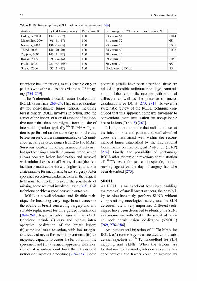
potential pitfalls have been described; these arerelated to possible radiotracer spillage, contami-nation of the skin, or the injection path or ductaldiffusion, as well as the presence of micro-calcifications or DCIS [270, 271]. However, asystematic review of the ROLL technique con-cluded that this approach compares favorably toconventional wire localization for non-palpablebreast lesions (Table 3) [267].
It is important to notice that radiation doses atthe injection site and patient and staff absorbeddoses are maintained well within the recom-mended limits established by the InternationalCommission on Radiological Protection (ICRP)[274]. Finally, the possibility of performingROLL after systemic intravenous administrationof 99mTc-sestamibi (as a nonspecific, tumor-seeking agent) on the day of surgery has alsobeen described [275].
SNOLLAs ROLL is an excellent technique enablingthe removal of small breast cancers, the possibil-ity to simultaneously perform SLNB withoutcompromising oncological safety and the SLNdetection rate is very important. Different tech-niques have been described to identify the SLNsin combination with ROLL, the so-called senti-nel node occult lesion localization (SNOLL)[269, 276–284].
An intratumoral injection of 99mTc-MAA forROLL of a tumor may be associated with a sub-dermal injection of 99mTc-nanocolloid for SLNmapping and SLNB. When the lesions arelocated near to the areola, intraoperative interfer-ence between the tracers could be avoided by
Table 3 Studies comparing ROLL and hook-wire techniques [266]
Authors n (ROLL–hook wire) Detection (%) Free margins (ROLL versus hook wire) (%) p
Gallegos, 2004 132 (65–67) 100 83 versus 64 0.014
Macmillan, 2004 95 (48–47) 100 61 versus 72 NS
Nadeem, 2004 130 (65–65) 100 83 versus 57 0.001
Thind, 2005 140 (70–70) 100 84 versus 60 0.002
Zgajnar, 2004 143 (51–92) 100 70 versus 44
Rönkä, 2005 78 (64–14) 100 89 versus 79 0.05
Fraile, 2005 233 (65–168) 100 80 versus 70 NS
Strnad, 2006 33 (21–12) 100 Hook wire < ROLL NS
22 F. Giammarile et al.
elevation of the dermis and the subdermal areaafter skin incision [276]. Another possibility is touse a single intratumoral injection for both ROLLand SNOLL in the same session [280]. As asingle procedure for localization of breast lesionsand sentinel nodes, SNOLL may improve theentire surgical procedure. The majority of thestudies published so far show a high percentageof successful tumor resection and intraoperativeSLN localization with reduced failure [267, 269,276, 279–284].
Radioactive SeedsAlternatives to hook-wire localization of occultbreast lesions include carbon trace as well as theuse of sealed radioactive seeds. The seeds areessentially the same as the ones used in brachy-therapy for cancer of the prostate, namely, a4.5–0.8 mm titanium capsule containing aceramic cylinder enriched with 125I-iodine.Iodine-125 has a long decay time (half-life of59.4 days) and emits low-energy photons(27 keV). The use of one or two seeds with thislow photon energy has a negligible effect on thesurrounding tissue. The radioactive seed is placedin the center of the breast lesion using an 18 Gneedle fixed in a needle holder under mammo-graphic or ultrasonographic guidance; after suc-cessful positioning, the exact location isconfirmed by mammography. During surgery,excision of the lesion is guided by using a hand-held gamma probe [285].
If a SNOLL technique is scheduled, the99mTc-colloid is subsequently injected, aroundthe tumor or through a superficial route. Thus,the handheld gamma probe can be switchedbetween the 27 keV energy window of the 125Isource and the 140 keV of 99mTc, allowing dis-crimination between the emissions of the tworadioisotopes. Effective seed removal is verifiedby the absence of 125I activity in the breast and itspresence in the specimen. X-ray of the surgicalspecimen may confirm the presence of the seedand the relation of the lesion to the resectionmargins.
It has been shown that radioguided seed local-ization in non-palpable breast lesions is at leastequivalent to the hook-wire technique in terms of
ease of procedure, removing the target lesion,volume of breast tissue excised, obtaining nega-tive margins, avoiding a second operative inter-vention, and allowing for simultaneous axillarystaging [285–289].
Added Value of Intraoperative PortableGamma CamerasRecently, several types of portable or handheldmini gamma cameras have become available forclinical practice; while some of these portablegamma cameras are not specifically designedfor radioguided surgery, other models arefocused on different applications of SLNB [4,5, 290, 291].
Appropriate use of a portable gamma cameraenhances the reliability of the gamma probe byadding a clear image of the surgical field. The useof an intraoperative imaging device implies thepossibility to better plan the surgical approach, tolocalize surgical targets in complex anatomicalareas, to monitor the lymphatic basin before andafter removal of the hot nodes, and, above all, toverify the correct SLN excision. Moreover, theuse of point sources (e.g., 133Ba or 125I) facilitatesSLN localization, as these sources can be depictedseparately on the screen of the portable gammacamera, thus functioning as a pointer in the searchfor the SLNs. Nevertheless, their role in breastcancer surgery is still to be clarified. Intra-operative imaging using a portable gamma cam-era might be useful only when no conventionalgamma camera is available for preoperative imag-ing, in particular in cases with extra-axillary drain-age. Portable gamma cameras have also been usedwith promising results in other GOSTT environ-ments regarding breast cancer, such as in ROLL orSNOLL procedures [290–296].
An interesting recent development of intra-operative imaging consists in combining conven-tional gamma probes with position and orientationtracking systems such as the so-called freehandSPECT, which permits a virtual reconstruction ina 3D environment (see Fig. 9) [4, 5, 293]. Allthese technologies will play an increasing role inthe future extension of the GOSTT concept, inorder to provide a better roadmap for radioguidedsurgery [2, 5].
Radioguided Surgery for Breast Cancer 23
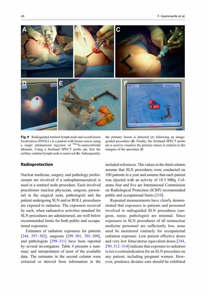
Radioprotection
Nuclear medicine, surgery, and pathology profes-sionals are involved if a radiopharmaceutical isused in a sentinel node procedure. Each involvedpractitioner (nuclear physician, surgeon, person-nel in the surgical suite, pathologist) and thepatient undergoing SLN and/or ROLL proceduresare exposed to radiation. The exposures receivedby each, when radioactive activities standard forSLN procedures are administered, are well belowrecommended limits for both public and occupa-tional exposures.
Estimates of radiation exposures for patients[244, 297–302], surgeons [299–301, 303–309],and pathologists [299–311] have been reportedby several investigators. Table 4 presents a sum-mary and interpretation of most of the availabledata. The estimates in the second column wereextracted or derived from information in the
included references. The values in the third columnassume that SLN procedures were conducted on100 patients in a year and assume that each patientwas injected with an activity of 18.5 MBq. Col-umns four and five are International Commissionon Radiological Protection (ICRP) recommendedpublic and occupational limits [310].
Repeated measurements have clearly demon-strated that exposures to patients and personnelinvolved in radioguided SLN procedures (sur-geon, nurse, pathologist) are minimal. Sinceexposures in SLN procedures of all nonnuclearmedicine personnel are sufficiently low, noneneed be monitored routinely for occupationalradiation exposure. Low patient effective dosesand very low fetus/uterus equivalent doses [244,299, 312–314] indicate that exposure to radiationis not a contraindication for an SLN procedure onany patient, including pregnant women. How-ever, prudence dictates care should be exhibited
Fig. 9 Radioguided sentinel lymph node and occult lesionlocalization (SNOLL) in a patient with breast cancer usinga single intratumoral injection of 99mTc-nanocolloidalalbumin. Using a freehand SPECT probe (a), first theaxillary sentinel lymph node is removed (b). Subsequently,
the primary lesion is detected (c) following an image-guided procedure (d). Finally, the freehand SPECT probe(e) is used to visualize the primary tumor in relation to themargins of the specimen (f)
24 F. Giammarile et al.
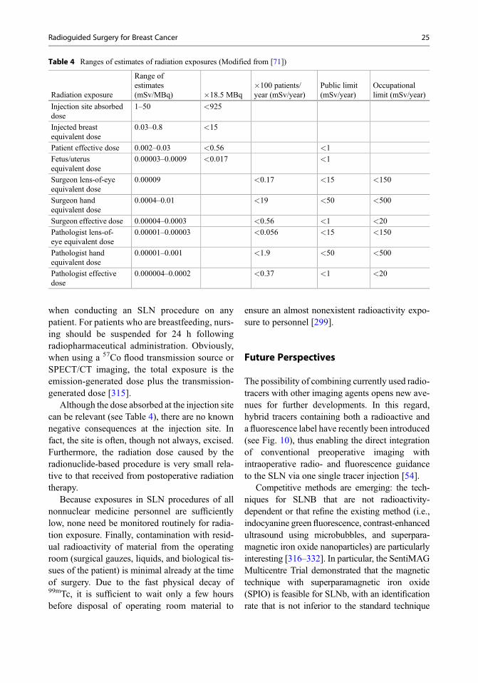
when conducting an SLN procedure on anypatient. For patients who are breastfeeding, nurs-ing should be suspended for 24 h followingradiopharmaceutical administration. Obviously,when using a 57Co flood transmission source orSPECT/CT imaging, the total exposure is theemission-generated dose plus the transmission-generated dose [315].
Although the dose absorbed at the injection sitecan be relevant (see Table 4), there are no knownnegative consequences at the injection site. Infact, the site is often, though not always, excised.Furthermore, the radiation dose caused by theradionuclide-based procedure is very small rela-tive to that received from postoperative radiationtherapy.
Because exposures in SLN procedures of allnonnuclear medicine personnel are sufficientlylow, none need be monitored routinely for radia-tion exposure. Finally, contamination with resid-ual radioactivity of material from the operatingroom (surgical gauzes, liquids, and biological tis-sues of the patient) is minimal already at the timeof surgery. Due to the fast physical decay of99mTc, it is sufficient to wait only a few hoursbefore disposal of operating room material to
ensure an almost nonexistent radioactivity expo-sure to personnel [299].
Future Perspectives
The possibility of combining currently used radio-tracers with other imaging agents opens new ave-nues for further developments. In this regard,hybrid tracers containing both a radioactive anda fluorescence label have recently been introduced(see Fig. 10), thus enabling the direct integrationof conventional preoperative imaging withintraoperative radio- and fluorescence guidanceto the SLN via one single tracer injection [54].
Competitive methods are emerging: the tech-niques for SLNB that are not radioactivity-dependent or that refine the existing method (i.e.,indocyanine green fluorescence, contrast-enhancedultrasound using microbubbles, and superpara-magnetic iron oxide nanoparticles) are particularlyinteresting [316–332]. In particular, the SentiMAGMulticentre Trial demonstrated that the magnetictechnique with superparamagnetic iron oxide(SPIO) is feasible for SLNb, with an identificationrate that is not inferior to the standard technique
Table 4 Ranges of estimates of radiation exposures (Modified from [71])
Radiation exposure
Range ofestimates(mSv/MBq) �18.5 MBq
�100 patients/year (mSv/year)
Public limit(mSv/year)
Occupationallimit (mSv/year)
Injection site absorbeddose
1–50 <925
Injected breastequivalent dose
0.03–0.8 <15
Patient effective dose 0.002–0.03 <0.56 <1
Fetus/uterusequivalent dose
0.00003–0.0009 <0.017 <1
Surgeon lens-of-eyeequivalent dose
0.00009 <0.17 <15 <150
Surgeon handequivalent dose
0.0004–0.01 <19 <50 <500
Surgeon effective dose 0.00004–0.0003 <0.56 <1 <20
Pathologist lens-of-eye equivalent dose
0.00001–0.00003 <0.056 <15 <150
Pathologist handequivalent dose
0.00001–0.001 <1.9 <50 <500
Pathologist effectivedose
0.000004–0.0002 <0.37 <1 <20
Radioguided Surgery for Breast Cancer 25
[333–337]. Recently, it has been shown that theSLN status can be evaluated with high accuracypreoperatively using contrast-enhanced colorDoppler ultrasonography [338, 339].
However, a systematic review suggested thatthese new methods have clinical potential butyield high levels of false-negative results andpresently cannot challenge the existing standardprocedure. Further assessment of these techniquesagainst the standard dual technique in randomizedtrials is thus needed [340].
Concluding Remarks
The development and wide acceptance of SLNBhas deeply affected the management of breastcancer. Several technical and clinical controver-sies have been raised during the development ofthis technique. The resolution of these controver-sies should result in the standardization of the
procedure and in the expansion of the number ofpatients evaluated with SLNB in the future. Themodern approach in breast cancer care, whichincludes more detailed screening diagnostics, path-ological evaluation, improved planning of surgicaland radiation therapy, and more effective systemictreatment, emphasizes the need for ongoing re-evaluation of the “standard” locoregional therapy[341, 342].
References
1. Mariani G, Giuliano AE, Strauss HW, editors. Radio-guided surgery – a comprehensive team approach.New York: Springer; 2008.
2. Zaknun JJ, Giammarile F, Valdés Olmos R, Vidal-Sicart S, Mariani G. Changing paradigms in radio-guided surgery and intraoperative imaging: theGOSTT concept. Eur J Nucl Med Mol Imaging. 2012;39:1–3.
3. Chin PT, Welling MM, Meskers SC, Valdes OlmosRA, Tanke H, van Leeuwen FW. Optical imaging as
Fig. 10 Resection of an infraclavicular sentinel lymphnode in a patient with high-risk breast cancer using thehybrid tracer ICG-99mTc-nanocolloid. The sentinel lymphnode is first located on the skin projection using a portablegamma camera (a) and a handheld gamma probe (b).
Subsequently, a portable near-infrared camera (c) is usedto depict the fluorescence signal on the screen (d). Thisenables to remove the node (e) and to perform ex vivocontrol of the fluorescence signal (f, g)
26 F. Giammarile et al.

an expansion of nuclear medicine: cerenkov-basedluminescence vs fluorescence-based luminescence.Eur J Nucl Med Mol Imaging. 2013;40:1283–91.
4. Vidal-Sicart S, Rioja ME, Paredes P, Keshtgar MR,Valdés Olmos RA. Contribution of perioperativeimaging to radioguided surgery. Q J Nucl Med MolImaging. 2014;58:140–60.
5. Valdés Olmos RA, Vidal-Sicart S, Giammarile F,Zaknun JJ, Van Leeuwen FW, Mariani G. TheGOSTT concept and hybrid mixed/virtual/augmentedreality environment radioguided surgery. Q J NuclMed Mol Imaging. 2014;58:207–15.
6. KleinJan GH, van den Berg NS, de Jong J, Wit EM,Thygessen H, Vegt E, van der Poel HG, van LeeuwenFW. Multimodal hybrid imaging agents for sentinelnode mapping as a means to (re)connect nuclear med-icine to advances made in robot-assisted surgery. EurJ Nucl Med Mol Imaging. 2016;43:1278–87.
7. Cabañas RM. An approach for the treatment of penilecarcinoma. Cancer. 1977;39:456–66.
8. Ollila DW, Brennan MB, Giuliano AE. The role ofintraoperative lymphatic mapping and sentinellymphadenectomy in the management of patientswith breast cancer. Adv Surg. 1999;32:349–64.
9. Nieweg OE, Tanis PJ, Kroon BBR. The definition of asentinel node. Ann Surg Oncol. 2001;9:538–41.
10. Mariani G, Manca G, Valdés AR, Orsini F, Vidal-Sicart S, editors. Atlas of lymphoscintigraphy andsentinel node mapping – a pictorial case-basedapproach. Milan: Springer; 2013.
11. de Bree R, Nieweg OE. The history of sentinel nodebiopsy in head and neck cancer: from visualization oflymphatic vessels to sentinel nodes. Oral Oncol.2015;51:819–23.
12. Smith B, Backes F. The role of sentinel lymph nodesin endometrial and cervical cancer. J Surg Oncol.2015;112:753–60.
13. Yashiro M, Matsuoka T. Sentinel node navigationsurgery for gastric cancer: overview and perspective.World J Gastrointest Surg. 2015;27(7):1–9.
14. Jakobsen JK. Sentinel node biopsy in uro-oncology: ahistory of the development of a promising concept.Urol Oncol. 2015;33:486–93.
15. Shersher DD, Liptay MJ. Status of sentinel lymphnode mapping in non-small cell lung cancer. CancerJ. 2015;21:17–20.
16. Siegel RL, Miller KD, Jemal A. Cancer statistics. CACancer J Clin. 2015;65:5–29.
17. Zurrida S, Veronesi U. Milestones in breast cancertreatment. Breast J. 2015;21:3–12.
18. Veronesi U, De Cicco C, Galimberti VE, et al. Acomparative study on the value of FDG-PET andsentinel node biopsy to identify occult axillary metas-tases. Ann Oncol. 2007;18:473–8.
19. Liu Y. Role of FDG PET-CT in evaluation oflocoregional nodal disease for initial staging of breastcancer. World J Clin Oncol. 2014;5:982–9.
20. Futamura M, Asano T, Kobayashi K, et al. Predictionof macrometastasis in axillary lymph nodes of
patients with invasive breast cancer and the utility ofthe SUV lymph node/tumor ratio using FDG-PET/CT. World J Surg Oncol. 2015;13:49.
21. Krammer J, Schnitzer A, Kaiser CG, et al. 18F-FDGPET/CT for initial staging in breast cancer patients –is there a relevant impact on treatment planning com-pared to conventional staging modalities? Eur Radiol.2015;25:2460–9.
22. Boone BA, Huynh C, Spangler ML, et al. Axillarylymph node burden in invasive breast cancer: a com-parison of the predictive value of ultrasound-guidedneedle biopsy and sentinel lymph node biopsy. ClinBreast Cancer. 2015;15:e243–8.
23. Hyun SJ, Kim EK, Yoon JH, Moon HJ, KimMJ. Adding MRI to ultrasound and ultrasound-guided fine-needle aspiration reduces the false-negative rate of axillary lymph node metastasis diag-nosis in breast cancer patients. Clin Radiol.2015;70:716–22.
24. You S, Kang DK, Jung YS, An YS, Jeon GS, KimTH. Evaluation of lymph node status after neo-adjuvant chemotherapy in breast cancer patients:comparison of diagnostic performance of ultrasound,MRI and 18F-FDG PET/CT. Br J Radiol. 2015;88(1052):20150143.
25. Beek MA, Verheuvel NC, Luiten EJ, et al. Twodecades of axillary management in breast cancer. BrJ Surg. 2015;102:1658–64.
26. Keshtgar MRS, Ell PJ. Sentinel lymph node detectionand imaging. Eur J Nucl Med. 1999;26:57–67.
27. Chatterjee A, Serniak N, Czerniecki BJ. Sentinellymph node biopsy in breast cancer: a work in pro-gress. Cancer J. 2015;21:7–10.
28. Morton DL, Wen DR, Wong JH, et al. Technicaldetails of intraoperative lymphatic mapping for earlystage melanoma. Arch Surg. 1992;127:392–9.
29. Nieweg OE, Uren RF, Thompson JF. The history ofsentinel lymph node biopsy. Cancer J. 2015;21:3–6.
30. Krag DN, Weaver D, Alex JC, Fairbank JT. Surgicalresection and radiolocalization of sentinel lymphnode in breast cancer using a gamma probe. SurgOncol. 1993;2:335–9.
31. Giuliano AE, Kirgan D, Guenther JM. Lymphaticmapping and sentinel lymphadenectomy for breastcancer. Ann Surg. 1994;220:391–401.
32. Noguchi M, Katev N, Miyazaki I. Diagnosis of axil-lary lymph node metastases in patients with breastcancer. Breast Cancer Res Treat. 1996;40:283–93.
33. Taylor A, Murray D, Herda S, Vansant J, AlazrakiN. Dynamic lymphoscintigraphy to identify the senti-nel and satellite nodes. ClinNuclMed. 1996;21:755–8.
34. Pijpers R, Meijer S, Hoekstra OS, et al. Impact oflymphoscintigraphy on sentinel node identificationwith technetium-99m-colloid albumin in breast can-cer. J Nucl Med. 1997;38:366–8.
35. Giuliano AE. Lymphatic mapping and sentinel nodebiopsy in breast cancer. JAMA. 1997;277:791–2.
36. Veronesi U, Paganelli G, Galimberti V, et al. Sentinelnode biopsy to avoid axillary dissection in breast
Radioguided Surgery for Breast Cancer 27
cancer patients with clinically negative lymph-nodes.Lancet. 1997;349:1864–7.
37. International Breast Cancer Consensus Conference.Image-detected breast cancer: state of the art diagno-sis and treatment. International Breast Cancer Con-sensus Conference. J Am Coll Surg. 2001;193:297–302.
38. Mariani G, Moresco L, Viale G, et al. Radioguidedsentinel lymph node biopsy in breast cancer surgery. JNucl Med. 2001;42:1198–215.
39. Veronesi U, Paganelli G, Viale G, et al. A randomizedcomparison of sentinel-node biopsy with routine axil-lary dissection in breast cancer. N Engl J Med. 2003;349:546–53.
40. Veronesi U, Paganelli G, Viale G, et al. Sentinel-lymph-node biopsy as a staging procedure in breastcancer: update of a randomized controlled study. Lan-cet Oncol. 2006;7:983–90.
41. Benson JR, Della Rovere GQ, Axilla ManagementConsensus Group. Management of the axilla inwomen with breast cancer. Lancet Oncol. 2007;8:331–48.
42. Giuliano AE, Gangi A. Sentinel node biopsy andimproved patient care. Breast J. 2015;21:27–31.
43. Lopez Penha TR, van Roozendaal LM, Smidt ML,et al. The changing role of axillary treatment inbreast cancer: who will remain at risk for develop-ing arm morbidity in the future? Breast. 2015;24:543–7.
44. Layeequr Rahman R, Crawford SL, Siwawa P. Man-agement of axilla in breast cancer – the saga con-tinues. Breast. 2015;24:343–53.
45. Pepels MJ, Vestjens JH, de Boer M, et al. Safety ofavoiding routine use of axillary dissection in earlystage breast cancer: a systematic review. Breast Can-cer Res Treat. 2011;125:301–13.
46. Atalay C. New concepts in axillary management ofbreast cancer. World J Clin Oncol. 2014;10:895–900.
47. Sledge GW, Mamounas EP, Hortobagyi GN, BursteinHJ, Goodwin PJ, Wolff AC. Past, present, and futurechallenges in breast cancer treatment. J Clin Oncol.2014;32:1979–86.
48. Buscombe J, Paganelli G, Burak ZE, European Asso-ciation of Nuclear Medicine Oncology Committeeand Dosimetry Committee, et al. Sentinel node inbreast cancer procedural guidelines. Eur J Nucl MedMol Imaging. 2007;34:2154–9.
49. Lizarraga IM, Weigel RJ. Axillary lymph node dis-section for breast cancer: primum non nocere. Eur JSurg Oncol. 2015;41:955–7.
50. van den Hoven I, Voogd AC, Roumen RM. A para-digm shift in axillary breast cancer treatment; from“treat all-except” toward “treat none-unless”. ClinBreast Cancer. 2015;15:399–402.
51. Galimberti V, Manika A, Maisonneuve P, et al. Long-term follow-up of 5262 breast cancer patients withnegative sentinel node and no axillary dissection con-firms low rate of axillary disease. Eur J Surg Oncol.2014;40:1203–8.
52. Han HJ, Kim JR, Nam HR, Keum KC, Suh CO, KimYB. Clinical outcomes after sentinel lymph nodebiopsy in clinically node-negative breast cancerpatients. Radiat Oncol J. 2014;32:132–7.
53. Joyce DP, Manning A, Carter M, et al. Meta-analysis to determine the clinical impact of axillarylymph node dissection in the treatment of invasivebreast cancer. Breast Cancer Res Treat. 2015;153:235–40.
54. Tsujimoto M. Recent advances in sentinel nodebiopsy in breast surgery. Breast Cancer. 2015;22:211.
55. Suami H, Pan WR, Taylor GI. Historical review ofbreast lymphatic studies. Clin Anat. 2009;22:531–6.
56. Torrenga H, Fabry H, van der Sijp JR, van Diest PJ,Pijpers R, Meijer S. Omitting axillary lymph nodedissection in sentinel node negative breast cancerpatients is safe: a long term follow-up analysis. JSurg Oncol. 2004;88:4–7.
57. Carlo JT, Grant MD, Knox SM, Jones RC, HamiltonCS, Livingston SA, Kuhn JA. Survival analysis fol-lowing sentinel lymph node biopsy: a validation trialdemonstrating its accuracy in staging early breastcancer. BUMC Proc. 2005;18:103–7.
58. Fuhrman GM, Gambino J, Bolton JS, Farr G, JiangX. Five-year follow-up after sentinel node mappingfor breast cancer demonstrates better than expectedtreatment outcomes. Am Surg. 2005;71:564–9.
59. Sanjuàn A, Vidal-Sicart S, Zanón G, et al. Clinicalaxillary recurrence after sentinel node biopsy in breastcancer: a follow-up study of 220 patients. Eur J NuclMed Mol Imaging. 2005;32:932–6.
60. Smidt ML, Janssen CM, Kuster DM, Bruggink ED,Strobbe LJ. Axillary recurrence after a negative sen-tinel node biopsy for breast cancer: incidence andclinical significance. Ann Surg Oncol. 2005;12:29–33.
61. Veronesi U, Galimberti V, Mariani L, et al. Sentinelnode biopsy in breast cancer: early results in953 patients with negative sentinel node biopsy andno axillary dissection. Eur J Cancer. 2005;41:197–8.
62. de Kanter AY, Menke-Pluymers MM, Wouters MW,Burgmans I, van Geel AN, Eggermont AM. Five-yearfollow-up of sentinel node negative breast cancerpatients. Eur J Surg Oncol. 2006;32:282–6.
63. Veronesi U, Galimberti V, Paganelli G, et al. Axillarymetastases in breast cancer patients with negativesentinel nodes: follow-up of 3,548 cases. Eur J Can-cer. 2009;45:1381–8.
64. Veronesi U, Viale G, Paganelli G, et al. Sentinellymph node biopsy in breast cancer: ten-year resultsof a randomized control study. Ann Surg. 2010;251:595–600.
65. Kaufmann M, Morrow M, von Minckwitz G, HarrisJR, Biedenkopf Expert Panel Members. Locoregionaltreatment of primary breast cancer: consensus recom-mendations from an International Expert Panel. Can-cer. 2010;116:1184–91.
66. van der Ploeg IM, Nieweg OE, van Rijk MC, ValdésOlmos RA, Kroon BB. Axillary recurrence after a
28 F. Giammarile et al.
tumour-negative sentinel node biopsy in breast cancerpatients: a systematic review and meta-analysis of theliterature. Eur J Surg Oncol. 2008;34:1277–84.
67. Krag DN, Anderson SJ, Julian TB, et al. Sentinel-lymph-node resection compared with conventionalaxillary-lymph-node dissection in clinically node-negative patients with breast cancer: overall survivalfindings from the NSABP B-32 randomised phase3 trial. Lancet Oncol. 2010;11:927–33.
68. Purushotham AD, Upponi S, Klevesath MB, et al.Morbidity after sentinel lymph node biopsy in pri-mary breast cancer: results from a randomized con-trolled trial. J Clin Oncol. 2005;23:4312–21.
69. Mansel RE, Fallowfield L, Kissin M, et al. Random-ized multicenter trial of sentinel node biopsy versusstandard axillary treatment in operable breast cancer:the ALMANAC Trial. J Natl Cancer Inst.2006;98:599–609.
70. Goyal A, Newcombe RG, Chhabra A, Mansel RE,ALMANAC Trialists Group. Factors affecting failedlocalisation and false-negative rates of sentinel nodebiopsy in breast cancer – results of the ALMANACvalidation phase. Breast Cancer Res Treat. 2006;99:203–8.
71. Giammarile F, Alazraki N, Aarsvold JN, et al. TheEANM and SNMMI practice guideline forlymphoscintigraphy and sentinel node localization inbreast cancer. Eur J Nucl Med Mol Imaging.2013;40:1932–47.
72. Mount MG,White NR, Nguyen CL, Orr RK, Hird RB.Evaluating one day versus two days preoperativelymphoscintigraphy protocols for sentinel lymph nodebiopsy in breast cancer. Am Surg. 2015;81:454–7.
73. Wang H, Heck K, Pruitt SK, et al. Impact of delayedlymphoscintigraphy for sentinel lymph node biopsyfor breast cancer. J Surg Oncol. 2015;111:931–4.
74. Wilhelm AJ, Mijnhout GS, Franssen EJF. Radiophar-maceuticals in sentinel lymph-node detection – anoverview. Eur J Nucl Med. 1999;26:S36–42.
75. Clarke D, Khoni N, Mansel ER. Sentinel node biopsyin breast cancer. ALMANAC trial. World J Surg.2001;25:819–22.
76. Burak WE, Agnese DM, Povoski SP. Advances in thesurgical management of early stage invasive breastcancer. Curr Probl Surg. 2004;41:877–936.
77. Bourgeois P. Scintigraphic investigations of the lym-phatic system: the influence of injected volume andquantity of labeled colloidal tracer. J Nucl Med.2007;48:693–5.
78. Lyman GH, Giuliano AE, Somerfield MR, et al.American Society of Clinical Oncology guidelinerecommendations for sentinel lymph node biopsy inearly-stage breast cancer. J Clin Oncol. 2005;23:7703–20.
79. Vera DR, Wallace AM, Hoh CK. A synthetic macro-molecule for sentinel node detection: 99mTc-DTPA-mannosyl-dextran. J Nucl Med. 2001;42:951–9.
80. Wallace AM, Han LK, Povoski SP, et al. Comparativeevaluation of [99mTc]tilmanocept for sentinel lymph
node mapping in breast cancer patients: results of twophase 3 trials. Ann Surg Oncol. 2013;20:2590–9.
81. Dambros Gabbi MC, Masiero PR, Uchoa D, MoraesIV, Biazus JV, Edelweiss MI. Comparison betweenpreoperative and intraoperative injection of 99mTcDextran-500 for sentinel lymph node localization inbreast cancer. Am J Nucl Med Mol Imaging. 2014;4:602–10.
82. Baker JL, Pu M, Tokin CA, et al. Comparison of[99mTc]tilmanocept and filtered [99mTc]sulfur colloidfor identification of SLNs in breast cancer patients.Ann Surg Oncol. 2015;22:40–5.
83. McCarter MD, Yeung H, Yeh S, et al. Localization ofthe sentinel node in breast cancer: identical resultswith same-day and day-before isotope injection.Ann Surg Oncol. 2001;8:682–6.
84. van der Ent FW, Kengen RA, van der Pol HA,Hoofwijk AG. Sentinel node biopsy in 70 unselectedpatients with breast cancer: increased feasibility byusing 10 mCi radiocolloid in combination with a bluedye tracer. Eur J Surg Oncol. 1999;25:24–9.
85. Gray RJ, Pockaj BA, Roarke MC. Injection of 99mTc-labeled sulfur colloid the day before operation forbreast cancer sentinel lymph node mapping is assuccessful as injection the day of operation. Am JSurg. 2004;188:685–9.
86. Suami H, Pan WR, Mann GB, Taylor GI. The lym-phatic anatomy of the breast and its implications forsentinel lymph node biopsy: a human cadaver study.Ann Surg Oncol. 2008;15:863–71.
87. Ahmed M, Purushotham AD, Horgan K, Klaase JM,Douek M. Meta-analysis of superficial versus deepinjection of radioactive tracer and blue dye for lym-phatic mapping and detection of sentinel lymph nodesin breast cancer. Br J Surg. 2015;102:169–81.
88. Tanis PJ, Valdés Olmos RA, Muller SH, NiewegOE. Lymphatic mapping in patients with breast carci-noma: reproducibility of lymphoscintigraphic results.Radiology. 2003;228:546–51.
89. Tanis PJ, Nieweg OE, Valdes Olmos RA, KroonBB. Anatomy and physiology of lymphatic drainageof the breast from the perspective of sentinel nodebiopsy. J Am Coll Surg. 2001;192:399–409.
90. Hindie E, Groheux D, Espie M, et al. Sentinel nodebiopsy in breast cancer. Bull Cancer. 2009;96:713–25.
91. Pelosi E, BelloM,GriorsM, et al. Sentinel lymph nodedetection in patients with early-stage breast cancer:comparison of periareolar and subdermal/peritumoralinjection techniques. J Nucl Med. 2004;45:220–5.
92. Chakera AH, Friis E, Hesse U, et al. Factors of impor-tance for scintigraphic non-visualization of sentinelnodes in breast cancer. Eur J Nucl Med Mol Imaging.2005;32:286–93.
93. Nieweg OE, Estourgie SH, van Rijk MC, KroonBB. Rationale for superficial injection techniques inlymphatic mapping in breast cancer patients. J SurgOncol. 2004;87:153–6.
94. Noguchi M, Inokuchi M, Zen Y. Complement ofperitumoral and subareolar injection in breast cancer
Radioguided Surgery for Breast Cancer 29
sentinel lymph node biopsy. J Surg Oncol. 2009;100:100–5.
95. Povoski SP, Olsen JO, Young DC, et al. Prospectiverandomized clinical trial comparing intradermal,intraparenchymal, and subareolar injection routes forsentinel lymph node mapping and biopsy in breastcancer. Ann Surg Oncol. 2006;13:1412–21.
96. Linehan DC, Hill ADK, Akhurst T, et al. Intradermalradiocolloid and intraparenchymal blue dye injectionoptimize sentinel node identification in breast cancerpatients. Ann Surg Oncol. 1999;6:286–93.
97. Pesek S, Ashikaga T, Krag LE, Krag D. The false-negative rate of sentinel node biopsy in patients withbreast cancer: a meta-analysis. World J Surg. 2012;36:2239–51.
98. Martin 2nd RC, EdwardsMJ,Wong SL, et al. Practicalguidelines for optimal gamma probe detection of sen-tinel lymph nodes in breast cancer: results of a multi-institutional study. Surgery. 2000;128:139–44.
99. Paganelli G, Galimberti V, Trifirò G, et al. Internalmammary node lymphoscintigraphy and biopsy inbreast cancer. Q J Nucl Med. 2002;46:138–44.
100. Krynyckyi BR, Chun H, Kim HH, Eskandar Y, KimCK, Machac J. Factors affecting visualization rates ofinternal mammary sentinel nodes during lymphoscin-tigraphy. J Nucl Med. 2003;44:1387–93.
101. Kumar R, Jana S, Heiba SI, et al. Retrospective anal-ysis of sentinel node localization in multifocal multi-centric, palpable, or non palpable breast cancer. JNucl Med. 2003;44:7–10.
102. Knauer M, Konstantiniuk P, Haid A, et al. Multi-centric breast cancer: a new indication for sentinelnode biopsy – a multi-institutional validation study.J Clin Oncol. 2006;24:3374–80.
103. Uren RF, Howman-Giles R, Chung D, Thompson JF.Imaging sentinel lymph nodes. Cancer J. 2015;21:25–32.
104. Moncayo VM, Aarsvold JN, Alazraki NP. Lympho-scintigraphy and sentinel nodes. J Nucl Med. 2015;56:901–7.
105. McMasters KM, Wong SL, Tuttle TM, et al. Preoper-ative lymphoscintigraphy for breast cancer does notimprove the ability to identify axillary sentinel lymphnodes. Ann Surg. 2000;231:724–31.
106. Marchal F, Rauch P, Morel O, et al. Results of preop-erative lymphoscintigraphy for breast cancer are pre-dictive of identification of axillary sentinel lymphnodes. World J Surg. 2006;30:55–62.
107. De Cicco C, Cremonesi M, Luini A, et al. Lympho-scintigraphy and radioguided biopsy of the sentinelaxillary node in breast cancer. J Nucl Med. 1998;39:2080–4.
108. Babiera GV, Delpassand ES, Breslin TM, et al. Lym-phatic drainage patterns on early versus delayedbreast lymphoscintigraphy performed after injectionof filtered Tc-99m sulfur colloid in breast cancerpatients undergoing sentinel lymph node biopsy.Clin Nucl Med. 2005;30:11–5.
109. Lerman H, Metser U, Lievshitz G, et al. Lymphoscin-tigraphic sentinel node identification in patients withbreast cancer: the role of SPECT-CT. Eur J Nucl MedMol Imaging. 2006;33:329–37.
110. Keidar Z, Israel O, Krausz Y. SPECT/CT in tumorimaging: technical aspects and clinical applications.Semin Nucl Med. 2003;33:205–18.
111. Vermeeren L, van der Ploeg IM, Valdes Olmos RA,et al. SPECT/CT for preoperative sentinel node local-ization. J Surg Oncol. 2010;101:184–90.
112. van der Ploeg IM, Nieweg OE, Kroon BB, et al. Theyield of SPECT/CT for anatomical lymphatic map-ping in patients with breast cancer. Eur J Nucl MedMol Imaging. 2009;36:903–9.
113. Jimenez-Heffernan A, Ellman A, et al. Results of aprospective International Atomic Energy Ahency(IAEA) sentinel node trial on the value of SPECT/CT over planar imaging in various malignancies. JNucl Med. 2015;56:1338–44.
114. Even-Sapir E, Lerman H, Lievshitz G, et al.Lymphoscintigraphy for sentinel node mappingusing a hybrid SPECT/CT system. J Nucl Med. 2003;44:1413–20.
115. Lerman H, Lievshitz G, Zak O, Metser U,Schneebaum S, Even-Sapir E. Improved sentinel nodeidentification by SPECT/CT in overweight patientswith breast cancer. J Nucl Med. 2007;48:201–6.
116. Valdés Olmos RA, Rietbergen DD, Vidal-Sicart S,Manca G, Giammarile F, Mariani G. Contribution ofSPECT/CT imaging to radioguided sentinel lymphnode biopsy in breast cancer, melanoma, and othersolid cancers: from “open and see” to “see and open”.Q J Nucl Med Mol Imaging. 2014;58:127–39.
117. Serrano-Vicente J, Rayo-Madrid JI, Domínguez-Grande ML, et al. Role of SPECT-CT in breast cancersentinel node biopsy when internal mammary chaindrainage is observed. Clin Transl Oncol. 2016;18:418–25.
118. Tomiguchi M, Yamamoto-Ibusuki M, Yamamoto Y,et al. Prediction of sentinel lymph node status usingsingle-photon emission computed tomography(SPECT)/computed tomography (CT) imaging ofbreast cancer. Surg Today. 2016;46:214–23.
119. Vidal-Sicart S, Brouwer OR, Valdés-Olmos RA.Evaluation of the sentinel lymph node combiningSPECT/CT with the planar image and its importancefor the surgical act. Rev Esp Med Nucl. 2011;30:331–7.
120. Uren RF, Howman-Giles R, Chung D, ThompsonJF. Nuclear medicine aspects of melanoma and breastlymphatic mapping. Semin Oncol. 2004;31:338–48.
121. van Rijk MC, Tanis PJ, Nieweg OE, et al. Clinicalimplications of sentinel nodes outside the axilla andinternal mammary chain in patients with breast can-cer. J Surg Oncol. 2006;94:281–6.
122. Estourgie SH, Nieweg OE, Valdés Olmos RA,Rutgers EJ, Peterse JL, Kroon BB. Eight false-negative sentinel lymph node procedures in breast
30 F. Giammarile et al.
cancer: what went wrong? Eur J Surg Oncol. 2003;29:336–40.
123. OʼReilly EA, Prichard RS, Al Azawi D, et al. Thevalue of isosulfan blue dye in addition to isotopescanning in the identification of the sentinel lymphnode in breast cancer patients with a positivelymphoscintigraphy: a randomized controlled trial(ISRCTN98849733). Ann Surg. 2015;262:243–8.
124. Anan K, Mitsuyama S, Kuga H, et al. Double map-ping with subareolar blue dye and peritumoral greendye injections decreases the false negative rate of dyeonly sentinel node biopsy for early breast cancer:2 site injection is more accurate than 1-site injection.Surgery. 2006;139:624–9.
125. Albo D, Wayne JD, Hunt KK, et al. Anaphylacticreactions to isosulfan blue dye during sentinellymph node biopsy for breast cancer. Am J Surg.2001;182:393–8.
126. Borgen PI. Isosulfan blue dye reactions during senti-nel lymph node mapping for breast cancer. AnesthAnalg. 2002;95:385–8.
127. Montgomery LL, Thorne AC, Van Zee KJ, et al.Incidence of anaphylactoid reactions to isosulfanblue dye during breast carcinoma lymphatic mappingin patients treated with preoperative prophylaxis:results of a surgical prospective clinical practice pro-tocol. Cancer. 2005;104:692–9. Dal.
128. Scherer K, Studer W, Figueiredo V, Bircher AJ. Ana-phylaxis to isosulfan blue and cross-reactivity to pat-ent blue V: case report and review of the nomenclatureof vital blue dyes. Ann Allergy Asthma Immunol.2006;96:497–500.
129. Rodier JF, Velten M, Wilt M, et al. Prospective multi-centric randomized study comparing periareolar andperitumoral injection of radiotracer and blue dye forthe detection of sentinel lymph node in breast sparingprocedures: FRANSENODE trial. J Clin Oncol.2007;25:3664–9.
130. Varghese P, Abdel-Rahman AT, Akberali S, et al.Methylene blue dye – a safe and effective alternativefor sentinel lymph node localization. Breast J. 2008;14:61–7.
131. Iqbal FM, Basit A, Salem F, Vidya R. Feeling blue,going green and finding other attractive alternatives: acase of biphasic anaphylaxis to patent blue and aliterature review of alternative sentinel nodelocalisation methods. BMJ Case Rep. 2015;pii:bcr2015213107. doi: 10.1136/bcr-2015-213107.
132. Chung A, Yu J, Stempel M, Patil S, Cody H, Mont-gomery L. Is the “10% rule” equally valid for allsubsets of sentinel-node-positive breast cancerpatients? Ann Surg Oncol. 2008;15:2728.
133. Liu LC, Lang JE, Jenkins T, et al. Is it necessary toharvest additional lymph nodes after resection of themost radioactive sentinel lymph node in breast can-cer? J Am Coll Surg. 2008;207:853–8.
134. Morton DL, Bostick PJ. Will the true sentinel nodeplease stand? Ann Surg Oncol. 1999;6:12–4.
135. Nathanson SD. Will the true sentinel node pleasestand? Ann Surg Oncol. 1999;6:514–6.
136. McMasters KM, Reintgen DS, Ross MI, et al. Senti-nel lymph node biopsy for melanoma: how manyradioactive nodes should be removed? Ann SurgOncol. 2001;8:192–7.
137. Manca G, Romanini A, Pellegrino D, et al. Optimaldetection of sentinel lymph node metastases byintraoperative radioactive threshold and molecularanalysis in patients with melanoma. J Nucl Med.2008;49:1769–75.
138. Aarsvold JN, Alazraki NP. Update on detection ofsentinel lymph nodes in patients with breast cancer.Semin Nucl Med. 2005;35:116–28.
139. Goyal A, Newcombe RG, Mansel RE, Axillary Lym-phatic Mapping Against Nodal Axillary Clearance(ALMANAC) Trialists Group. Clinical relevance ofmultiple sentinel nodes in patients with breast cancer.Br J Surg. 2005;92:438–42.
140. Serrano Vicente J, Infante de la Torre JR, DomínguezGrande ML, et al. Optimization of sentinel lymphnode biopsy in breast cancer by intraoperative axillarypalpation. Rev Esp Med Nucl. 2010;29:8–11.
141. Ban EJ, Lee JS, Koo JS, Park S, Kim SI, Park BW.Howmany sentinel lymph nodes are enough for accu-rate axillary staging in t1-2 breast cancer? J BreastCancer. 2011;14:296–300.
142. Cheng G, Kurita G, Kurita S, Torigian DA, AlaviA. Current status of sentinel lymph-node biopsy inpatients with breast cancer. Eur J Nucl Med MolImaging. 2011;38:562–75.
143. Schuman S, Walker G, Avisar E. Processing sentinelnodes in breast cancer: when and how many? ArchSurg. 2011;146:389–93.
144. Uren RF, Howman-Giles R, ChungDK, et al. SPECT/CTscans allow precise anatomical location of sentinellymph nodes in breast cancer and redefine lymphaticdrainage from the breast to the axilla. Breast.2012;21:480–6.
145. Brenot-Rossi I, Houvenaeghel G, Jacquemier J, et al.Nonvisualization of axillary sentinel node duringlymphoscintigraphy: is there a pathologic signifi-cance in breast cancer? J Nucl Med. 2003;44:1232–7.
146. Leidenius MH, Krogerus LA, Toivonen TS,Leppänen EA, von Smitten KA. The sensitivity ofaxillary staging when using sentinel node biopsy inbreast cancer. Eur J Surg Oncol. 2003;29:849–53.
147. Pouw B, Hellingman D, Kieft M, Vogel WV, van OsKJ, Rutgers EJ, Valdés Olmos RA, Stokkel MP. Thehidden sentinel node in breast cancer: reevaluating therole of SPECT/CT and tracer reinjection. Eur J SurgOncol. 2016;42:497–503.
148. Krishnamurthy S,Meric-Bernstam F, Lucci A, et al. Aprospective study comparing touch imprint cytology,frozen section analysis, and rapid cytokeratinimmunostain for intraoperative evaluation of axillarysentinel lymph nodes in breast cancer. Cancer. 2009;115:1555–62.
Radioguided Surgery for Breast Cancer 31
149. Dixon JM, Rutgers E, Hunt KK. Intraoperativeassessment of axillary lymph nodes in patients withbreast cancer. BMJ. 2014;349:g6803.
150. Bernet L, Cano R, Martinez M, et al. Diagnosis of thesentinel lymph node in breast cancer: a reproduciblemolecular method: a multicentric Spanish study. His-topathology. 2011;58:863–9.
151. Castellano I, Macrì L, Deambrogio C, et al.Reliability of whole sentinel lymph node analysis byone-step nucleic acid amplification for intraoperativediagnosis of breast cancer metastases. Ann Surg.2012;255:334–42.
152. Goldhirsch A, Wood WC, Coates AS, Gelber RD,Thurlimann B, Senn HJ. Strategies for subtypes –dealing with the diversity of breast cancer: highlightsof the St. Gallen International Expert Consensus onthe Primary Therapy of Early Breast Cancer. AnnOncol. 2011;22:1736–47.
153. Shimazu K, Noguchi S. Clinical significance of breastcancer micrometastasis in the sentinel lymph node.Surg Today. 2016;46:155–60.
154. Morita ET, Chang J, Leong SP. Principles andcontroversies in lymphoscintigraphy with emphasison breast cancer. Surg Clin North Am. 2000;80:1721–39.
155. Giuliano AE, Jones RC, Brennan M, Statman R.Sentinel lymphadenectomy in breast cancer. J ClinOncol. 1997;15:2345–50.
156. Krag D, Weaver D, Ashikaga T, et al. The sentinelnode in breast cancer – a multi-center validationstudy. N Engl J Med. 1998;339:941–6.
157. Tausch C, Konstantiniuk P, Kugler F, Austrian Senti-nel Node Study Group, et al. Sentinel lymph nodebiopsy after preoperative chemotherapy for breastcancer: findings from the Austrian Sentinel NodeStudy Group. Ann Surg Oncol. 2008;15:3378–83.
158. Zervoudis S, Iatrakis G, Tomara E, Bothou A,Papadopoulos G, Tsakiris G. Main controversies inbreast cancer. World J Clin Oncol. 2014;5:359–73.
159. Assi H, Sbaity E, Abdelsalam M, Shamseddine A.Controversial indications for sentinel lymph nodebiopsy in breast cancer patients. Biomed Res Int.2015;2015:405949.
160. Manca G, Rubello D, Tardelli E, et al. Sentinel lymphnode biopsy in breast cancer: indications, contraindi-cations, and controversies. Clin Nucl Med. 2016;41:126–33.
161. Meretoja TJ, Leidenius MH, Heikkilä PS, JoensuuH. Sentinel node biopsy in breast cancer patientswith large or multifocal tumors. Ann Surg Oncol.2009;16:1148–55.
162. Zavagno G, Rubello D, Franchini Z, et al. Axillarysentinel lymph nodes in breast cancer: a single lym-phatic pathway drains the entire mammary gland. EurJ Surg Oncol. 2005;31:479–84.
163. Goyal A, Newcombe RG, Mansel RE, et al. Sentinellymph node biopsy in patients with multifocal breastcancer. Eur J Surg Oncol. 2004;30:475–9.
164. Gentilini O, Trifirò G, Soteldo J, et al. Sentinel lymphnode biopsy in multicentric breast cancer. The expe-rience of the European Institute of Oncology. Eur JSurg Oncol. 2006;32:507–10.
165. Spillane AJ, Brennan ME. Accuracy of sentinellymph node biopsy in large and multifocal/multi-centric breast carcinoma – a systematic review. Eur JSurg Oncol. 2011;37:371–85.
166. Giard S, Chauvet MP, Penel N, et al. Feasibility ofsentinel lymph node biopsy in multiple unilateralsynchronous breast cancer: results of a French pro-spective multi-institutional study (IGASSU 0502).Ann Oncol. 2010;21:1630–5.
167. Francis AM, Haugen CE, Grimes LM, et al. Is senti-nel lymph node dissection warranted for patients witha diagnosis of ductal carcinoma in situ? Ann SurgOncol. 2015;22:4270–9.
168. Bruening W, Fontanarosa J, Tipton K, Treadwell JR,Launders J, Schoelles K. Systematic review: compar-ative effectiveness of core-needle and open surgicalbiopsy to diagnose breast lesions. Ann Intern Med.2010;152:238–46.
169. Brennan ME, Turner RM, Ciatto S, et al. Ductal car-cinoma in situ at core-needle biopsy: meta-analysis ofunderestimation and predictors of invasive breast can-cer. Radiology. 2011;260:119–28.
170. Intra M, Rotmensz N, Veronesi P, et al. Sentinel nodebiopsy is not a standard procedure in ductal carcinomain situ of the breast: the experience of the EuropeanInstitute of Oncology on 854 patients in 10 years. AnnSurg. 2008;247:315–9.
171. Virnig BA, Tuttle TM, Shamliyan T, Kane RL. Ductalcarcinoma in situ of the breast: a systematic review ofincidence, treatment, and outcomes. J Natl CancerInst. 2010;102:170–8.
172. Kotani H, Yoshimura A, Adachi Y, et al. Sentinellymph node biopsy is not necessary in patients diag-nosed with ductal carcinoma in situ of the breast bystereotactic vacuum-assisted biopsy. Breast Cancer.2016;23:190–4.
173. Taback B, Nguyen P, Hansen N, Edwards GK,Conway K, Giuliano AE. Sentinel lymph node biopsyfor local recurrence of breast cancer after breast-conserving therapy. Ann Surg Oncol. 2006;13:1099–104.
174. Tunon-de-Lara C, Chauvet MP, Baranzelli MC,et al. The role of sentinel lymph node biopsy andfactors associated with invasion in extensive DCISof the breast treated by mastectomy: The Cinnamomeprospective multicenter study. Ann Surg Oncol. 2015;22:3853–60.
175. Schneider C, Trocha S, McKinley B, et al. The use ofsentinel lymph node biopsy in ductal carcinoma insitu. Am Surg. 2010;76:943–6.
176. Specht MC, Fey JV, Borgen PI, Cody 3rd HS. Is theclinically positive axilla in breast cancer really a con-traindication to sentinel lymph node biopsy? J AmColl Surg. 2005;200:10–4.
32 F. Giammarile et al.
177. Usmani S, Ahmed N, Saleh NA, Huda FA,Amanguno HG, Amir T, Kandari FA. The clinicalutility of combining pre-operative axillary ultraso-nography and fine needle aspiration cytology withradionuclide guided sentinel lymph node biopsy inbreast cancer patients with palpable axillary lymphnodes. Eur J Radiol. 2015;84:2515–20.
178. Kim H, Shin MJ, Kim SJ, Kim IJ, Park I. The relationof visualization of internal mammary lymph nodes onlymphoscintigraphy to axillary lymph node metasta-ses in breast cancer. Lymphat Res Biol. 2014;12:295–300.
179. Veronesi U, Valagussa P. Inefficacy of internal mam-mary nodes dissection in breast cancer surgery. Can-cer. 1981;47:170–5.
180. Meier P, Ferguson DJ, Karrison T. A controlled trial ofextended radical mastectomy. Cancer. 1985;55:880–91.
181. Lacour J, Le MG, Hill C, Kramar A, Contesso G,Sarrazin D. Is it useful to remove internal mammarynodes in operable breast cancer? Eur J Surg Oncol.1987;13:309–14.
182. Meier P, Ferguson DJ, Karrison T. A controlled trialof extended radical versus radical mastectomy.Ten-year results. Cancer. 1989;63:188–95.
183. Morimoto T, Monden Y, Takashima S, et al. Five-yearresults of a randomized clinical trial comparing mod-ified radical mastectomy and extended radical mas-tectomy for stage II breast cancer. Surg Today.1994;24:210–4.
184. Veronesi U, Marubini E, Mariani L, Valagussa P,Zucali R. The dissection of internal mammary nodesdoes not improve the survival of breast cancerpatients. 30-year results of a randomised trial. Eur JCancer. 1999;35:1320–5.
185. Madsen EV, Aalders KC, van der Heiden-van derLoo M, et al. Prognostic significance of tumor-positive internal mammary sentinel lymph nodes inbreast cancer: a multicenter cohort study. Ann SurgOncol. 2015;22:4254–62.
186. McGale P, Taylor C, Correa C, et al. Effect of radio-therapy after mastectomy and axillary surgery on10-year recurrence and 20-year breast cancer mortal-ity: meta-analysis of individual patient data for 8135women in 22 randomised trials. Lancet. 2014;383:2127–35.
187. Hennequin C, Fourquet A. Controversy about internalmammary chain irradiation in breast cancer. CancerRadiother. 2014;18:351–5.
188. Poortmans PM, Collette S, Kirkove C, et al. Internalmammary and medial supraclavicular irradiation inbreast cancer. N Engl J Med. 2015;373:317–27.
189. Estourgie SH, Tanis PJ, Nieweg OE, Valdés OlmosRA, Rutgers EJ, Kroon BB. Should the hunt forinternal mammary chain sentinel nodes begin? Anevaluation of 150 breast cancer patients. Ann SurgOncol. 2003;10:935–41.
190. Paredes P, Vidal-Sicart S, Zanón G, et al. Clinicalrelevance of sentinel lymph nodes in the internal
mammary chain in breast cancer patients. Eur J NuclMed Mol Imaging. 2005;32:1283–7.
191. Bourre JC, Payan R, Collomb D, et al. Can the senti-nel lymph node technique affect decisions to offerinternal mammary chain irradiation? Eur J NuclMed Mol Imaging. 2009;36:758–64.
192. Manca G, Volterrani D, Mazzarri S, et al. Sentinellymph node mapping in breast cancer: a criticalreappraisal of the internal mammary chain issue. Q JNucl Med Mol Imaging. 2014;58:114–26.
193. Singletary SE, Allred C, Ashley P, et al. Revision ofthe American Joint Committee on Cancer stagingsystem for breast cancer. J Clin Oncol. 2002;20:3628–36.
194. Edge SB, Compton CC. The American Joint Com-mittee on Cancer: the 7th edition of the AJCC cancerstaging manual and the future of TNM. Ann SurgOncol. 2010;17:1471–4.
195. Noushi F, Spillane AJ, Uren RF, Gebski V. Internalmammary node metastasis in breast cancer: predictivemodels to determine status and management algo-rithms. Eur J Surg Oncol. 2010;36:16–22.
196. Leidenius MH, Krogerus LA, Toivonen TS,Leppänen EA, von Smitten KA. The clinical valueof parasternal sentinel node biopsy in breast cancer.Ann Surg Oncol. 2006;13:321–6.
197. Ozmen V, Ozcinar B, Bozdogan A, Eralp Y, Yavuz E,Dincer M. The effect of internal mammary lymphnode biopsy on the therapeutic decision and survivalof patients with breast cancer. Eur J Surg Oncol.2015;41:1368–72.
198. Bergkvist L, Frisell J, Liljegren G, Celebioglu F,Damm S, Thorn M. Multicentre study of detectionand false-negative rates in sentinel node biopsy forbreast cancer. Br J Surg. 2001;88:1644–8.
199. Intra M, Trifirò G, Viale G, et al. Second biopsy ofaxillary sentinel lymph node for reappearing breastcancer after previous sentinel lymph node biopsy.Ann Surg Oncol. 2005;12:895–9.
200. Newman EA, Cimmino VM, Sabel MS, et al. Lym-phatic mapping and sentinel lymph node biopsy forpatients with local recurrence after breast-conservation therapy. Ann Surg Oncol. 2006;13:52–7.
201. Port ER, Garcia-Etienne CA, Park J, Fey J, Borgen PI,Cody 3rd HS. Reoperative sentinel lymph nodebiopsy: a new frontier in the management of ipsilat-eral breast tumor recurrence. Ann Surg Oncol.2007;14:2209–14.
202. Tasevski R, Gogos AJ, Mann GB. Reoperative senti-nel lymph node biopsy in ipsilateral breast cancerrelapse. Breast. 2009;18:322–6.
203. Kaur P, Kiluk JV, Meade T, et al. Sentinel lymph nodebiopsy in patients with previous ipsilateral completeaxillary lymph node dissection. Ann Surg Oncol.2011;18:727–32.
204. Luini A, Galimberti V, Gatti G, et al. The sentinelnode biopsy after previous breast surgery; preliminary
Radioguided Surgery for Breast Cancer 33
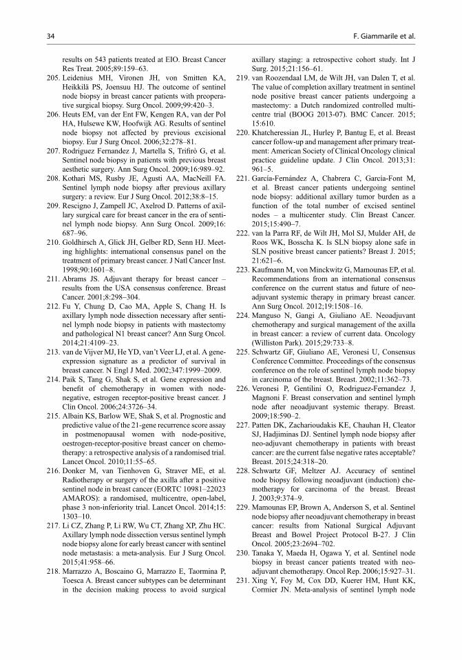
results on 543 patients treated at EIO. Breast CancerRes Treat. 2005;89:159–63.
205. Leidenius MH, Vironen JH, von Smitten KA,Heikkilä PS, Joensuu HJ. The outcome of sentinelnode biopsy in breast cancer patients with preopera-tive surgical biopsy. Surg Oncol. 2009;99:420–3.
206. Heuts EM, van der Ent FW, Kengen RA, van der PolHA, Hulsewe KW, Hoofwijk AG. Results of sentinelnode biopsy not affected by previous excisionalbiopsy. Eur J Surg Oncol. 2006;32:278–81.
207. Rodriguez Fernandez J, Martella S, Trifirò G, et al.Sentinel node biopsy in patients with previous breastaesthetic surgery. Ann Surg Oncol. 2009;16:989–92.
208. Kothari MS, Rusby JE, Agusti AA, MacNeill FA.Sentinel lymph node biopsy after previous axillarysurgery: a review. Eur J Surg Oncol. 2012;38:8–15.
209. Rescigno J, Zampell JC, Axelrod D. Patterns of axil-lary surgical care for breast cancer in the era of senti-nel lymph node biopsy. Ann Surg Oncol. 2009;16:687–96.
210. Goldhirsch A, Glick JH, Gelber RD, Senn HJ. Meet-ing highlights: international consensus panel on thetreatment of primary breast cancer. J Natl Cancer Inst.1998;90:1601–8.
211. Abrams JS. Adjuvant therapy for breast cancer –results from the USA consensus conference. BreastCancer. 2001;8:298–304.
212. Fu Y, Chung D, Cao MA, Apple S, Chang H. Isaxillary lymph node dissection necessary after senti-nel lymph node biopsy in patients with mastectomyand pathological N1 breast cancer? Ann Surg Oncol.2014;21:4109–23.
213. van de VijverMJ, He YD, van’t Veer LJ, et al. A gene-expression signature as a predictor of survival inbreast cancer. N Engl J Med. 2002;347:1999–2009.
214. Paik S, Tang G, Shak S, et al. Gene expression andbenefit of chemotherapy in women with node-negative, estrogen receptor-positive breast cancer. JClin Oncol. 2006;24:3726–34.
215. Albain KS, Barlow WE, Shak S, et al. Prognostic andpredictive value of the 21-gene recurrence score assayin postmenopausal women with node-positive,oestrogen-receptor-positive breast cancer on chemo-therapy: a retrospective analysis of a randomised trial.Lancet Oncol. 2010;11:55–65.
216. Donker M, van Tienhoven G, Straver ME, et al.Radiotherapy or surgery of the axilla after a positivesentinel node in breast cancer (EORTC 10981–22023AMAROS): a randomised, multicentre, open-label,phase 3 non-inferiority trial. Lancet Oncol. 2014;15:1303–10.
217. Li CZ, Zhang P, Li RW, Wu CT, Zhang XP, Zhu HC.Axillary lymph node dissection versus sentinel lymphnode biopsy alone for early breast cancer with sentinelnode metastasis: a meta-analysis. Eur J Surg Oncol.2015;41:958–66.
218. Marrazzo A, Boscaino G, Marrazzo E, Taormina P,Toesca A. Breast cancer subtypes can be determinantin the decision making process to avoid surgical
axillary staging: a retrospective cohort study. Int JSurg. 2015;21:156–61.
219. van Roozendaal LM, de Wilt JH, van Dalen T, et al.The value of completion axillary treatment in sentinelnode positive breast cancer patients undergoing amastectomy: a Dutch randomized controlled multi-centre trial (BOOG 2013-07). BMC Cancer. 2015;15:610.
220. Khatcheressian JL, Hurley P, Bantug E, et al. Breastcancer follow-up andmanagement after primary treat-ment: American Society of Clinical Oncology clinicalpractice guideline update. J Clin Oncol. 2013;31:961–5.
221. García-Fernández A, Chabrera C, García-Font M,et al. Breast cancer patients undergoing sentinelnode biopsy: additional axillary tumor burden as afunction of the total number of excised sentinelnodes – a multicenter study. Clin Breast Cancer.2015;15:490–7.
222. van la Parra RF, de Wilt JH, Mol SJ, Mulder AH, deRoos WK, Bosscha K. Is SLN biopsy alone safe inSLN positive breast cancer patients? Breast J. 2015;21:621–6.
223. KaufmannM, vonMinckwitz G, Mamounas EP, et al.Recommendations from an international consensusconference on the current status and future of neo-adjuvant systemic therapy in primary breast cancer.Ann Surg Oncol. 2012;19:1508–16.
224. Manguso N, Gangi A, Giuliano AE. Neoadjuvantchemotherapy and surgical management of the axillain breast cancer: a review of current data. Oncology(Williston Park). 2015;29:733–8.
225. Schwartz GF, Giuliano AE, Veronesi U, ConsensusConference Committee. Proceedings of the consensusconference on the role of sentinel lymph node biopsyin carcinoma of the breast. Breast. 2002;11:362–73.
226. Veronesi P, Gentilini O, Rodriguez-Fernandez J,Magnoni F. Breast conservation and sentinel lymphnode after neoadjuvant systemic therapy. Breast.2009;18:590–2.
227. Patten DK, Zacharioudakis KE, Chauhan H, CleatorSJ, Hadjiminas DJ. Sentinel lymph node biopsy afterneo-adjuvant chemotherapy in patients with breastcancer: are the current false negative rates acceptable?Breast. 2015;24:318–20.
228. Schwartz GF, Meltzer AJ. Accuracy of sentinelnode biopsy following neoadjuvant (induction) che-motherapy for carcinoma of the breast. BreastJ. 2003;9:374–9.
229. Mamounas EP, Brown A, Anderson S, et al. Sentinelnode biopsy after neoadjuvant chemotherapy in breastcancer: results from National Surgical AdjuvantBreast and Bowel Project Protocol B-27. J ClinOncol. 2005;23:2694–702.
230. Tanaka Y, Maeda H, Ogawa Y, et al. Sentinel nodebiopsy in breast cancer patients treated with neo-adjuvant chemotherapy. Oncol Rep. 2006;15:927–31.
231. Xing Y, Foy M, Cox DD, Kuerer HM, Hunt KK,Cormier JN. Meta-analysis of sentinel lymph node
34 F. Giammarile et al.
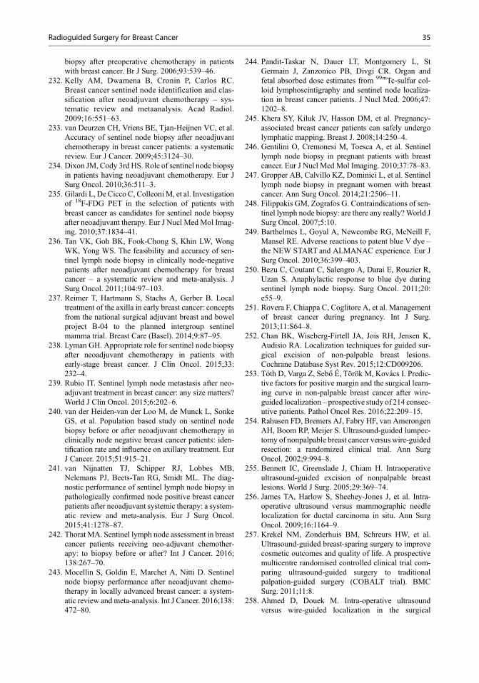
biopsy after preoperative chemotherapy in patientswith breast cancer. Br J Surg. 2006;93:539–46.
232. Kelly AM, Dwamena B, Cronin P, Carlos RC.Breast cancer sentinel node identification and clas-sification after neoadjuvant chemotherapy – sys-tematic review and metaanalysis. Acad Radiol.2009;16:551–63.
233. van Deurzen CH, Vriens BE, Tjan-Heijnen VC, et al.Accuracy of sentinel node biopsy after neoadjuvantchemotherapy in breast cancer patients: a systematicreview. Eur J Cancer. 2009;45:3124–30.
234. Dixon JM, Cody 3rd HS. Role of sentinel node biopsyin patients having neoadjuvant chemotherapy. Eur JSurg Oncol. 2010;36:511–3.
235. Gilardi L, De Cicco C, Colleoni M, et al. Investigationof 18F-FDG PET in the selection of patients withbreast cancer as candidates for sentinel node biopsyafter neoadjuvant therapy. Eur J Nucl Med Mol Imag-ing. 2010;37:1834–41.
236. Tan VK, Goh BK, Fook-Chong S, Khin LW, WongWK, Yong WS. The feasibility and accuracy of sen-tinel lymph node biopsy in clinically node-negativepatients after neoadjuvant chemotherapy for breastcancer – a systematic review and meta-analysis. JSurg Oncol. 2011;104:97–103.
237. Reimer T, Hartmann S, Stachs A, Gerber B. Localtreatment of the axilla in early breast cancer: conceptsfrom the national surgical adjuvant breast and bowelproject B-04 to the planned intergroup sentinelmamma trial. Breast Care (Basel). 2014;9:87–95.
238. Lyman GH. Appropriate role for sentinel node biopsyafter neoadjuvant chemotherapy in patients withearly-stage breast cancer. J Clin Oncol. 2015;33:232–4.
239. Rubio IT. Sentinel lymph node metastasis after neo-adjuvant treatment in breast cancer: any size matters?World J Clin Oncol. 2015;6:202–6.
240. van der Heiden-van der Loo M, de Munck L, SonkeGS, et al. Population based study on sentinel nodebiopsy before or after neoadjuvant chemotherapy inclinically node negative breast cancer patients: iden-tification rate and influence on axillary treatment. EurJ Cancer. 2015;51:915–21.
241. van Nijnatten TJ, Schipper RJ, Lobbes MB,Nelemans PJ, Beets-Tan RG, Smidt ML. The diag-nostic performance of sentinel lymph node biopsy inpathologically confirmed node positive breast cancerpatients after neoadjuvant systemic therapy: a system-atic review and meta-analysis. Eur J Surg Oncol.2015;41:1278–87.
242. Thorat MA. Sentinel lymph node assessment in breastcancer patients receiving neo-adjuvant chemother-apy: to biopsy before or after? Int J Cancer. 2016;138:267–70.
243. Mocellin S, Goldin E, Marchet A, Nitti D. Sentinelnode biopsy performance after neoadjuvant chemo-therapy in locally advanced breast cancer: a system-atic review and meta-analysis. Int J Cancer. 2016;138:472–80.
244. Pandit-Taskar N, Dauer LT, Montgomery L, StGermain J, Zanzonico PB, Divgi CR. Organ andfetal absorbed dose estimates from 99mTc-sulfur col-loid lymphoscintigraphy and sentinel node localiza-tion in breast cancer patients. J Nucl Med. 2006;47:1202–8.
245. Khera SY, Kiluk JV, Hasson DM, et al. Pregnancy-associated breast cancer patients can safely undergolymphatic mapping. Breast J. 2008;14:250–4.
246. Gentilini O, Cremonesi M, Toesca A, et al. Sentinellymph node biopsy in pregnant patients with breastcancer. Eur J Nucl Med Mol Imaging. 2010;37:78–83.
247. Gropper AB, Calvillo KZ, Dominici L, et al. Sentinellymph node biopsy in pregnant women with breastcancer. Ann Surg Oncol. 2014;21:2506–11.
248. Filippakis GM, Zografos G. Contraindications of sen-tinel lymph node biopsy: are there any really?World JSurg Oncol. 2007;5:10.
249. Barthelmes L, Goyal A, Newcombe RG, McNeill F,Mansel RE. Adverse reactions to patent blue V dye –the NEW START and ALMANAC experience. Eur JSurg Oncol. 2010;36:399–403.
250. Bezu C, Coutant C, Salengro A, Darai E, Rouzier R,Uzan S. Anaphylactic response to blue dye duringsentinel lymph node biopsy. Surg Oncol. 2011;20:e55–9.
251. Rovera F, Chiappa C, Coglitore A, et al. Managementof breast cancer during pregnancy. Int J Surg.2013;11:S64–8.
252. Chan BK, Wiseberg-Firtell JA, Jois RH, Jensen K,Audisio RA. Localization techniques for guided sur-gical excision of non-palpable breast lesions.Cochrane Database Syst Rev. 2015;12:CD009206.
253. Tóth D, Varga Z, Sebő É, Török M, Kovács I. Predic-tive factors for positive margin and the surgical learn-ing curve in non-palpable breast cancer after wire-guided localization – prospective study of 214 consec-utive patients. Pathol Oncol Res. 2016;22:209–15.
254. Rahusen FD, Bremers AJ, Fabry HF, van AmerongenAH, Boom RP, Meijer S. Ultrasound-guided lumpec-tomy of nonpalpable breast cancer versus wire-guidedresection: a randomized clinical trial. Ann SurgOncol. 2002;9:994–8.
255. Bennett IC, Greenslade J, Chiam H. Intraoperativeultrasound-guided excision of nonpalpable breastlesions. World J Surg. 2005;29:369–74.
256. James TA, Harlow S, Sheehey-Jones J, et al. Intra-operative ultrasound versus mammographic needlelocalization for ductal carcinoma in situ. Ann SurgOncol. 2009;16:1164–9.
257. Krekel NM, Zonderhuis BM, Schreurs HW, et al.Ultrasound-guided breast-sparing surgery to improvecosmetic outcomes and quality of life. A prospectivemulticentre randomised controlled clinical trial com-paring ultrasound-guided surgery to traditionalpalpation-guided surgery (COBALT trial). BMCSurg. 2011;11:8.
258. Ahmed D, Douek M. Intra-operative ultrasoundversus wire-guided localization in the surgical
Radioguided Surgery for Breast Cancer 35
management of non-palpable breast cancers: system-atic review and meta-analysis. Breast Cancer ResTreat. 2013;140:435–46.
259. Yu CC, Chiang KC, Kuo WL, Shen SC, Lo YF, ChenSC. Low re-excision rate for positive margins inpatients treated with ultrasound-guided breast-con-serving surgery. Breast. 2013;22:698–702.
260. De Cicco C, Pizzamiglio M, Trifirò G, Luini A,Ferrari M, Prisco G, Galimberti V, Cassano E,Viale G, Intra M, Veronesi P, Paganelli G. Radio-guided occult lesion localisation (ROLL) and surgicalbiopsy in breast cancer. Technical aspects. Q J NuclMed. 2002;46:145–51.
261. Paganelli G, Luini A, Veronesi U. Radioguided occultlesion localization (ROLL) in breast cancer: maximiz-ing efficacy, minimizing mutilation. Ann Oncol.2002;13:1839–40.
262. Paganelli G, Veronesi U. Innovation in early breastcancer surgery: radio-guided occult lesion localiza-tion and sentinel node biopsy. Nucl Med Commun.2002;23:625–7.
263. Landman J, Kulawansa S, McCarthy M, et al. Radio-guided localisation of impalpable breast lesions using99m-technetium macroaggregated albumin: lessonslearnt during introduction of a new technique toguide preoperative localisation. J Med Radiat Sci.2015;62:6–14.
264. Rovera F, Frattini F, Marelli M, Corben AD, Vanoli C,Dionigi G, Boni L, Dionigi R. Radio-guided occultlesion localization versus wire-guided localization innon-palpable breast lesions. Int J Surg. 2008;1:S101–3.
265. van der Ploeg IM, Hobbelink M, van den Bosch MA,Mali WP, Borel Rinkes IH, van HillegersbergR. ‘Radioguided occult lesion localisation’ (ROLL)for non-palpable breast lesions: a review of the rele-vant literature. Eur J Surg Oncol. 2008;34:1–5.
266. Sajid MS, Parampalli U, Haider Z, Bonomi R. Com-parison of radioguided occult lesion localization(ROLL) and wire localization for non-palpable breastcancers: a meta-analysis. J Surg Oncol. 2012;105:852–8.
267. Ahmed M, Douek M. Sentinel node and occult lesionlocalization (SNOLL): a systematic review. Breast.2013;22:1034–40.
268. Aydogan F, Velidedeoglu M, Kilic F, Yilmaz H.Radio-guided localization of clinically occult breastlesions: current modalities and future directions.Expert Rev Med Devices. 2014;11:53–63.
269. Monti S, Galimberti V, Trifirò G, et al. Occult breastlesion localization plus sentinel node biopsy(SNOLL): experience with 959 patients at theEuropean Institute of Oncology. Ann Surg Oncol.2007;14:2928–31.
270. Mariscal Martínez A, SolàM, de Tudela AP, Julián JF,Fraile M, Vizcaya S, Fernández J. Radioguided local-ization of nonpalpable breast cancer lesions: random-ized comparison with wire localization in patients
undergoing conservative surgery and sentinel nodebiopsy. AJR Am J Roentgenol. 2009;193:1001–9.
271. Vernet-Tomas Mdel M, Ortega M, Vidal S,Corominas JM, Carreras R. Factors affecting surgicalmargins in nonpalpable breast tumors excised withthe radioguided occult lesion localization approach. JObstet Gynaecol Res. 2011;37:422–7.
272. Ahmed M, van Hemelrijck M, Douek M. Systematicreview of radioguided versus wire-guided localizationin the treatment of non-palpable breast cancers. BreastCancer Res Treat. 2013;140:241–52.
273. Postma EL, Koffijberg H, Verkooijen HM, WitkampAJ, van den Bosch MA, van Hillegersberg R. Cost-effectiveness of radioguided occult lesion localization(ROLL) versus wire-guided localization (WGL) inbreast conserving surgery for nonpalpable breast can-cer: results from a randomized controlled multicentertrial. Ann Surg Oncol. 2013;20:2219–26.
274. Rampaul RS, Dudley NJ, Thompson JZ, Burrell H,Evans AJ, Wilson AR, Macmillan RD. Radioisotopefor occult lesion localisation (ROLL) of the breastdoes not require extra radiation protection procedures.Breast. 2003;12:150–2.
275. Duarte GM, dos Santos CC, Torresan RZ, et al.Radioguided surgery using intravenous 99mTcsestamibi associated with breast magnetic resonanceimaging for guidance of breast cancer resection.Breast J. 2006;12:202–7.
276. De Cicco C, Trifirò G, IntraM, et al. Optimised nuclearmedicine method for tumour marking and sentinelnode detection in occult primary breast lesions. Eur JNucl Med Mol Imaging. 2004;31:349–54.
277. Fraile M, Mariscal A, Lorenzo C, et al. Radio-guidedoccult lesion localization combined with sentinelnode biopsy in women with breast cancer. Cir Esp.2005;77:36–9.
278. Strnad P, Rob L, Halaska MG, Chod J, Zuntova A,Moravcova Z. Radioguided occult lesion localisationin combination with detection of the sentinel lymphnode in non-palpable breast cancer tumours. Eur JGynaecol Oncol. 2006;27:236–8.
279. Intra M, De Cicco C, Gentilini O, Luini A, PaganelliG. Radioguided localisation (ROLL) of non-palpablebreast lesions and simultaneous sentinel lymph nodebiopsy (SNOLL): the experience of the EuropeanInstitute of Oncology. Eur J Nucl Med Mol Imaging.2007;34:957–8.
280. van Rijk MC, Tanis PJ, Nieweg OE, et al. Sentinelnode biopsy and concomitant probe-guided tumorexcision of nonpalpable breast cancer. Ann SurgOncol. 2007;14:627–32.
281. Lavoué V, Nos C, Clough KB, et al. Simplified tech-nique of radioguided occult lesion localization (ROLL)plus sentinel lymph node biopsy (SNOLL) in breastcarcinoma. Ann Surg Oncol. 2008;15:2556–61.
282. Thind CR, Tan S, Desmond S, et al. SNOLL. Sentinelnode and occult (impalpable) lesion localization inbreast cancer. Clin Radiol. 2011;66:833–9.
36 F. Giammarile et al.
283. Bordea C, Plesca M, Condrea I, Gherghe M,Gociman A, Blidaru A. Occult breast lesion localiza-tion and concomitant sentinel lymph node biopsy inearly breast cancer (SNOLL). Chirurgiae (Bucur).2012;107:722–9.
284. Follacchio GA, Monteleone F, Anibaldi P, et al. Amodified sentinel node and occult lesion localization(SNOLL) technique in non-palpable breast cancer: apilot study. J Exp Clin Cancer Res. 2015;34:113.
285. Jakub JW, Gray RJ, Degnim AC, Boughey JC,Gardner M, Cox CE. Current status of radioactiveseed for localization of non palpable breast lesions.Am J Surg. 2010;199:522–8.
286. Sung JS, King V, Thornton CM, Brooks JD, Fry CW,El-Tamer M, Dauer LT, Brogi E, St Germain JM,Morris EA. Safety and efficacy of radioactive seedlocalization with I-125 prior to lumpectomy and/orexcisional biopsy. Eur J Radiol. 2013;82:1453–7.
287. Jackson L, Bourke AG, Abdul Aziz F, Taylor D.Radioactive seed localisation to guide removal ofimpalpable lymph nodes (radioguided occult lesionlocalisation using iodine-125 seeds, “ROLLIS”).BMJ Case Rep. 2014; pii: bcr-2013-203267.doi: 10.1136/bcr-2013-203267.
288. Parvez E, Cornacchi SD, Hodgson N, et al. Acosmesis outcome substudy in a prospective, random-ized trial comparing radioguided seed localizationwith standard wire localization for nonpalpable, inva-sive, and in situ breast carcinomas. Am J Surg.2014;208:711–8.
289. van der Noordaa ME, Pengel KE, Groen E, et al. Theuse of radioactive iodine-125 seed localization inpatients with non-palpable breast cancer: a compari-son with the radioguided occult lesion localizationwith 99m technetium. Eur J Surg Oncol. 2015;41:553–8.
290. Vermeeren L, Valdes Olmos RA, KlopWM, BalmAJ,van den Brekel MW. A portable gamma-camera forintraoperative detection of sentinel nodes in the headand neck region. J Nucl Med. 2010;51:700–3.
291. Bricou A, Duval MA, Charon Y, Barranger E. Mobilegamma cameras in breast cancer care – a review. Eur JSurg Oncol. 2013;39:409–16.
292. Bluemel C, Cramer A, Grossmann C, Kajdi GW, et al.iROLL: does 3-D radioguided occult lesion localiza-tion improve surgical management in early-stagebreast cancer? Eur J Nucl Med Mol Imaging. 2015;42:1692–9.
293. Engelen T, Winkel BM, Rietbergen DD, et al. Thenext evolution in radioguided surgery: breast cancerrelated sentinel node localization using a freehandSPECT-mobile gamma camera combination. Am JNucl Med Mol Imaging. 2015;5:233–45.
294. Lombardi A, Nigri G, Scopinaro F, et al. High-resolution, handheld camera use for occult breastlesion localization plus sentinel node biopsy(SNOLL): a single-institution experience with186 patients. Surgeon. 2015;13:69–72.
295. Taylor D, Landman J. ‘Rolling out radioguided occultlesion localisation for breast tumours’: moving fromROLL to ROLLIS. J Med Radiat Sci. 2015;62:175–6.
296. Bricou A, DuvalMA, Bardet L, et al. Is there a role fora handheld gamma camera (TReCam) in the SNOLLbreast cancer procedure? Q J Nucl Med Mol Imaging.2015 Mar 31 [Epub ahead of print].
297. Bergqvist L, Strand SE, Persson B, HafströmL, JönssonPE. Dosimetry in lymphoscintigraphy of Tc-99mantimonysulfide colloid. JNuclMed.1982;23:698–705.
298. Cremonesi M, Ferrari M, Sacco E, et al. Radiationprotection in radioguided surgery of breast cancer.Nucl Med Commun. 1999;20:919–24.
299. Waddington WA, Keshtgar MRS, Taylor I, LakhaniSR, Short MD, Ell PJ. Radiation safety of the sentinelnode technique in breast cancer. Eur J Nucl Med.2000;27:377–91.
300. Law M, Cheng KC, Wu PM, Ho WY, Chow LW.Patient effective dose from sentinel lymph nodelymphoscintigraphy in breast cancer: a study using afemale humanoid phantom and thermoluminescentdosimeters. Br J Radiol. 2003;76:818–23.
301. Law M, Chow LW, Kwong A, Lam CK. Sentinellymph node technique for breast cancer: radiationsafety issues. Semin Oncol. 2004;31:298–303.
302. Sata S, Knesaurek K, Krynyckyi BR. Effective dosein sentinel lymph node imaging. Br J Radiol. 2004;77:709. author reply 709.
303. Glass EC, Basinski JE, Krasne DL, Giuliano AE.Radiation safety considerations for sentinel nodetechniques. Ann Surg Oncol. 1999;6:10–1.
304. Miner TJ, Shriver CD, Flicek PR, Miner FC, JaquesDP, Maniscalco-Theberge ME, Krag DN. Guidelinesfor the safe use of radioactive materials during local-ization and resection of the sentinel lymph node. AnnSurg Oncol. 1999;6:75–82.
305. Stratmann SL, McCarty TM, Kuhn JA. Radiationsafety with breast sentinel node biopsy. Am J Surg.1999;178:454–7.
306. Morton R, Horton PW, Peet DJ, Kissin MW. Quanti-tative assessment of the radiation hazards and risks insentinel node procedures. Br J Radiol. 2003;76:117–22.
307. de Kanter AY, Arends PP, Eggermont AM, WiggersT. Radiation protection for the sentinel node proce-dure in breast cancer. Eur J Surg Oncol. 2003;29:396–9.
308. Klausen TL, Chakera AH, Friis E, Rank F, Hesse B,Holm S. Radiation doses to staff involved in sentinelnode operations for breast cancer. Clin Physiol FunctImaging. 2005;25:196–202.
309. Nejc D, Wrzesień M, Piekarski J, Olszewski J,Pluta P, Kuśmierek J, Jeziorski A. Sentinel nodebiopsy in patients with breast cancer – evaluation ofexposure to radiation of medical staff. Eur J SurgOncol. 2006;32:133–8.
310. Ckarke RH, Bines W. Evolution of ICRP recommen-dations 1977, 1990, 2007 – Publications 26 to 60 to
Radioguided Surgery for Breast Cancer 37
103. OECD NEA, No 6920, 2011. http://www.oecd-nea.org/rp/reports/2011/nea6920-ICRP-recommendations.pdf
311. Singleton M, Firth M, Stephenson T, Morrison G,Baginska J. Radiation-guided breast sentinel lymphnode biopsies – is a handling delay for radiation pro-tection necessary? Histopathology. 2012;61:277–82.
312. Gentilini O, Cremonesi M, Trifirò G, et al. Safety ofsentinel node biopsy in pregnant patients with breastcancer. Ann Oncol. 2004;15:1348–51.
313. Keleher A, Wendt 3rd R, Delpassand E, StachowiakAM, Kuerer HM. The safety of lymphatic mapping inpregnant breast cancer patients using Tc-99m sulfurcolloid. Breast J. 2004;10:492–5.
314. Spanheimer PM, Graham MM, Sugg SL, Scott-Conner CE, Weigel RJ. Measurement of uterine radi-ation exposure from lymphoscintigraphy indicatessafety of sentinel lymph node biopsy during preg-nancy. Ann Surg Oncol. 2009;16:1143–7.
315. Law M, Ma WH, Leung R, Li S, Wong KK, Ho WY,Kwong A. Evaluation of patient effective dose fromsentinel lymph node lymphoscintigraphy in breast can-cer: a phantom study with SPECT/CT and ICRP-103recommendations. Eur J Radiol. 2012;81:717–20.
316. Winter A, Woenkhaus J, Wawroschek F. A novelmethod for intraoperative sentinel lymph node detec-tion in prostate cancer patients using super-paramagnetic iron oxide nanoparticles and ahandheld magnetometer: the initial clinical experi-ence. Ann Surg Oncol. 2014;21:4390–6.
317. Rubio IT, Diaz-Botero S, Esgueva A, Rodriguez R,Cortadellas T, Cordoba O, Espinosa-Bravo M. Thesuperparamagnetic iron oxide is equivalent to theTc99 radiotracer method for identifying the sentinellymph node in breast cancer. Eur J Surg Oncol.2015;41:46–51.
318. Pitsinis V, Provenzano E, Kaklamanis L, Wishart GC,Benson JR. Indocyanine green fluorescence mappingfor sentinel lymph node biopsy in early breast cancer.Surg Oncol. 2015;24:375–9.
319. Koslow Mautner S, Cody 3rd HS. Sentinel nodebiopsy after neoadjuvant chemotherapy for node-positive breast cancer: does axillary ultrasoundimprove performance? J Clin Oncol. 2015;33:3375–8.
320. Toh U, Iwakuma N, Mishima M, Okabe M,Nakagawa S, Akagi Y. Navigation surgery forintraoperative sentinel lymph node detection usingindocyanine green (ICG) fluorescence real-timeimaging in breast cancer. Breast Cancer Res Treat.2015;153:337–44.
321. Tian P, Zhang W, Zhao H, Lei Y, Cui L, Zhang Y, XuZ. Intraoperative detection of sentinel lymph nodemetastases in breast carcinoma by Fourier transforminfrared spectroscopy. Br J Surg. 2015;102:1372–9.
322. Stoffels I, Dissemond J, Pöppel T, Schadendorf D,Klode J. Intraoperative fluorescence imaging for sen-tinel lymph node detection: prospective clinical trialto compare the usefulness of indocyanine green vs
Technetium Tc 99m for identification of sentinellymph nodes. JAMA Surg. 2015;150:617–23.
323. Piñero-Madrona A, Torró-Richart J, de León-Carrillo J, et al. Superparamagnetic iron oxide as atracer for sentinel node biopsy in breast cancer: acomparative non-inferiority study. Eur J Surg Oncol.2015;41:991–7.
324. Kuijs VJ, Moossdorff M, Schipper RJ, Beets-Tan RG,Heuts EM, Keymeulen KB, Smidt ML, LobbesMB. The role of MRI in axillary lymph node imagingin breast cancer patients: a systematic review. InsightsImaging. 2015;6:203–15.
325. Tsuyuki S, Yamaguchi A, Kawata Y, KawaguchiK. Assessing the effects of neoadjuvant chemother-apy on lymphatic pathways to sentinel lymph nodes incases of breast cancer: usefulness of the indocyaninegreen-fluorescence method. Breast. 2015;24:298–301.
326. Kida K, Ishikawa T, Yamada A, et al. A prospectivefeasibility study of sentinel node biopsy by modifiedindigo carmine blue dye methods after neoadjuvantchemotherapy for breast cancer. Eur J Surg Oncol.2015;41:566–70.
327. Samorani D, Fogacci T, Panzini I, et al. The use ofindocyanine green to detect sentinel nodes in breastcancer: a prospective study. Eur J Surg Oncol.2015;41:64–70.
328. Caudle AS, Yang WT, Mittendorf EA, et al. Selectivesurgical localization of axillary lymph nodescontaining metastases in patients with breast cancer:a prospective feasibility trial. JAMA Surg. 2015;150:137–43.
329. Inoue T, Nishi T, Nakano Y, Nishimae A, Sawai Y,Yamasaki M, Inaji H. Axillary lymph node recurrenceafter sentinel lymph node biopsy performed using acombination of indocyanine green fluorescence andthe blue dye method in early breast cancer. BreastCancer. 2016;23:295–300.
330. Nakagawa M, Morimoto M, Takechi H, Tadokoro Y,Tangoku A. Preoperative diagnosis of sentinel lymphnode (SLN) metastasis using 3D CT lymphography(CTLG). Breast Cancer. 2016;23:519–24.
331. Sugie T, Kinoshita T, Masuda N, et al. Evaluation ofthe clinical utility of the ICG fluorescence methodcompared with the radioisotope method for sentinellymph node biopsy in breast cancer. Ann Surg Oncol.2016;23:44–50.
332. Benson J. Indocyanine green fluorescence for sentinellymph node detection in early breast cancer. Ann SurgOncol. 2016;23:6–8.
333. Pouw JJ, Grootendorst MR, Bezooijen R, et al. Pre-operative sentinel lymph node localization in breastcancer with superparamagnetic iron oxide MRI: theSentiMAGmulticentre trial imaging subprotocol. Br JRadiol. 2015;88:20150634.
334. Li C, Meng S, Yang X, Zhou D, Wang J, Hu J.Sentinel lymph node detection using magnetic reso-nance lymphography with conventional gadolinium
38 F. Giammarile et al.
contrast agent in breast cancer: a preliminary clinicalstudy. BMC Cancer. 2015;15:213.
335. Ahmed M, Anninga B, Goyal S, et al. Magnetic senti-nel node and occult lesion localization in breast cancer(MagSNOLLTrial). Br J Surg. 2015;102:646–52.
336. Ghilli M, Carretta E, Di Filippo F, et al. The super-paramagnetic iron oxide tracer: a valid alternative insentinel node biopsy for breast cancer treatment. Eur JCancer Care (Engl). 2015 Sep 14 [Epub ahead ofprint].
337. Motomura K, Izumi T, Tateishi S, Tamaki Y, Ito Y,Horinouchi T, Nakanishi K. Superparamagnetic ironoxide-enhanced MRI at 3 T for accurate axillary stag-ing in breast cancer. Br J Surg. 2015;17 [Epub aheadof print].
338. Dellaportas D, Koureas A, Contis J, et al. Contrast-enhanced color Doppler ultrasonography forpreoperative evaluation of sentinel lymph node
in breast cancer patients. Breast Care (Basel).2015;10:331–5.
339. Rautiainen S, Sudah M, Joukainen S, Sironen R,Vanninen R, Sutela A. Contrast-enhanced ultra-sound-guided axillary lymph node core biopsy: diag-nostic accuracy in preoperative staging of invasivebreast cancer. Eur J Radiol. 2015;84:2130–6.
340. Ahmed M, Purushotham AD, Douek M. Novel tech-niques for sentinel lymph node biopsy in breast cancer:a systematic review. Lancet Oncol. 2014;15:e351–62.
341. Chung A, Gangi A, Amersi F, Zhang X, Giuliano AE.Not performing a sentinel node biopsy for olderpatients with early-stage invasive breast cancer.JAMA Surg. 2015;150:683–4.
342. Jacobson GM, Partin JF, Salkeni MA. Optimal man-agement of sentinel lymph node positive biopsypatients in early breast cancer. Ann Transl Med.2015;3:87.
Radioguided Surgery for Breast Cancer 39