The Role of Electron Transfer and Dithiol-Disulfide Interchange in Solute Transport in Bacteria
Upload
independentCategory
view
0download
0
Eur. J. Mineral. 1998, 10, 1097-1108
This paper was presented at the ECROFI XIV Symposium in Nancy, France (July 1997)
Quantitative solute analysis of single fluid inclusions in halite by LA-ICP-MS and cryo-SEM-EDS:
complementary microbeam techniques
THOMAS J. SHEPHERD1, CARLOS AYORA2, DIΟNI I. CENDON2 , SIMON R. CHENERY1
and ALAIN MOISSETTE3
'British Geological Survey, Keyworth, Nottingham, NG12 5GG, UK 2 Institut de Ciencies de la Terra, CSIC, Marti I Franques, E-08028 Barcelona, Spain
3CREGU, BP 23, F-54501 Vandoeuvre les Nancy, France
Abstract: An inter laboratory study was carried out to assess the complementariness of laser ablation-induc-tively coupled plasma-mass spectrometry (LA-ICP-MS) and cryogenic SEM-energy dispersive analysis (cryo-SEM-EDS) for the quantitative solute analysis of single inclusions in halite. The halite samples were selected from diverse ancient and recent evaporite environments to give a wide range of solute compositions, and were characterized by abundant, homogeneous populations of large (1—100 µm diameter) primary brine inclusions. Homogeneity of the inclusions was important because both techniques are destructive and had to be carried out on different individual inclusions. Cryogenic SEM-EDS was chosen because it is one of the few, well established fluid inclusion techniques to provide absolute concentrations for the major solutes (Na, K, Mg, Ca, Cl, SO4), and could be used to convert the relative element concentration ratios determined by LA-ICP-MS (K, Mg, Ca, Cl, Br, Sr, Li, B) into true concentration units. Normalization was achieved by referring to chlorine.
The results confirm that LA-ICP-MS and cryo-SEM-EDS are perfectly complementary and yield values for Mg and K that agree within \a standard deviation. Calcium concentrations were generally too low to be measured by cryo-SEM-EDS, but values determined by LA-ICP-MS are consistent with the hypothesis that the brines are saturated with respect to anhydrite. Overall analytical precision (relative standard deviation) for the major solutes is 2-3 times better for cryo-SEM-EDS than for LA-ICP-MS (5-15% vs. 10-35% respectively). Internal consistency of results was checked by charge mass balance estimates and halite saturation indices. Though the study utilized halite-saturated inclusions, cryo-SEM-EDS and LA-ICP-MS can be applied equally to lower salinity fluids and, with care, to inclusions that contain daughter minerals.
Key-words: quantitative chemical analysis, single fluid inclusions, laser ablation ICP-MS, cryogenic SEM-EDS, halite.
Introduction
Recent developments in instrumental methods of analysis, in particular microbeam techniques, have presented fluid inclusion researchers with an unparalleled opportunity to investigate the solute chemistry of individual fluid inclusions. No longer is the small size of an inclusion an overriding constraint since the majority of these techniques (synchrotron X-ray fluorescence (SXRF),
proton-induced X-ray excitation (PIXE), secondary ion mass spectrometry (SIMS), laser ablation-inductively coupled plasma-mass spectrometry (LA-ICP-MS), cryogenic SEM-energy dispersive spectroscopy (cryo-SEM-EDS) and micro-laser Raman spectroscopy (LRS)) have beam sizes less than a few micrometres. The limiting problems are now (1) the heterogeneous (solid-liquid-vapour) nature of inclusions, (2) their sub-surface location within the sample, (3) the ability to
0935-1221/98/0010-1097 $ 3.00 © 1998 E. Schweizerbart'sche Verlagsbuchhandlung. D-70176 Stuttgart

1098 T.J. Shepherd et al.
measure trace elements {e.g., metals) at low solute concentrations, and (4) whether destructive analysis is acceptable. Of these four, the first and second probably impose the greatest constraint. This is especially true in the case of SXRF and PIXE where data reduction procedures are designed for surface or thin layer samples. Modelling the SXRF and PIXE excitation of heterogeneous subsurface inclusions is extremely complex and requires a gross simplification of inclusion geometry to quantify the spectra. For LA-ICP-MS and cryo-SEM-EDS, the two main destructive techniques used for single inclusion analysis, the depth of an inclusion beneath the surface is less critical (see below). In the case of LA-ICP-MS the greatest uncertainties are the efficiency of fluid release during laser ablation, transfer of vapourised material to the plasma without chemical fraction-ation, and calibration. Such uncertainties do not apply to cryo-SEM-EDS but unlike the other techniques referred to above, one cannot pre-select the inclusion for analysis (see below).
Nevertheless, despite these difficulties an increasing number of papers report quantitative analyses for the solute chemistry of individual aqueous fluid inclusions by one or more micro-beam techniques (Diamond et al., 1990; Ramsey et al, 1992; Vanko et al., 1993; Ghazi et al., 1996). This marks an important watershed in inclusion research and heralds the possibility of carrying out quantitative geofluid analysis. Applications are too numerous to mention but key areas of future research include 1) direct measurement of mineral-fluid trace element distribution coefficients, 2) influence of diagenetic fluids on the cementation of hydrocarbon reservoirs, 3) fluid fluxes associated with subduction and thrust tectonics, 4) secular variations in ocean water chemistry, and 5) the metal-carrying capacity of hydro-thermal fluids. However, to exploit these new techniques it must first be proven that the analyses are fit for purpose. Until now, inclusion analyses have been treated with some degree of scepticism. This is not entirely unjustified since many of the older published inclusion analyses (pre-1975) do not stand up to inspection and are at best qualitative. To date, the most reliable solute analyses refer either to exceptional large inclusions (> 100 µm) or milligram quantities of hand-picked, carefully selected fluid inclusion-rich monomineralic samples that have been analysed using crush-leach techniques. To demonstrate the complete
ness of analysis, analyses are often recalculated to show cation-anion charge balances. Values close to charge neutrality indicate a high degree of confidence in the bulk inclusion analysis. If analyses for single inclusions obtained by microbeam techniques are to be regarded with the same degree of confidence they must be accompanied by similar figures of merit (precision, accuracy, etc.). Work in this direction has already begun but there remains the need to establish the extent of agreement between different microbeam techniques (Rankin et al., 1992). At the moment, no one technique provides a complete anion-cation analysis of individual <50 µm inclusions. It is important therefore that in pooling data, any systematic technique-specific biases that may influence the quality of the analysis are clearly recognized.
Research objectives
This paper presents the results of an interlabora-tory study to assess the use of two complementary microbeam techniques, LA-ICP-MS (Shepherd & Chenery, 1995; Moissette et al., 1996) and cryo-SEM-EDS (Ayora & Fontarnau, 1990; GarcRa-Veigas, 1993; Ayora et al, 1994a and b), to obtain quantitative data for the solute chemistry of fluid inclusions in halite. Since the latter is one of the few well established single inclusion techniques that yields absolute element concentrations, it afforded an excellent benchmark for normalizing and interpreting the relative element concentration ratios obtained by LA-ICP-MS. Brief descriptions of each technique are given below. Only a limited number of elements were selected for analysis and were chosen principally to satisfy the strengths of cryo-SEM-EDS (Na, K, Mg, Ca, S and Cl), whilst at the same time testing the extended trace element capability of LA-ICP-MS (Li, B, Br, Sr). Halite was chosen as the host mineral because it provided the ideal matrix for cryo-SEM-EDS analysis and because it typically contains growth zones with large populations of primary brine inclusions. The inclusions were considered sufficiently abundant and chemically homogeneous to compare the capabilities of both techniques even though different inclusions were analysed. The use of halite also has other advantages. Under most evaporitic conditions, NaCl-saturated brines are saturated with respect to anhydrite which allows Ca concentrations to be independently calculated from solubility data.

Quantitative solute analysis of single fluid inclusions 1099
Samples selected for analysis
To present a wide range of brine compositions, halite samples were selected from a number of well documented, contrasting, ancient and recent evaporite environments (Table 1).
BI-425 and BI-389 are from the Biurrun borehole (Navarra, Spain), located in the Upper Eocene evaporite sequence of the South Pyrenean foreland basin. This sequence, approximately 100 m in thickness, contains four units (Rosell, 1983): a) basal anhydrite unit, b) lower halite unit, c) potash unit (sylvite and carnallite), and d) upper halite unit. Sample BI-425 belongs to the lower halite unit and was precipitated in an evolved marine evaporitic environment prior to potash-salt precipitation. Sample BI-389 comes from the upper halite unit and was precipitated from continental waters which had recycled previous evaporites.
L-212 and L-147 are from borehole S-4 located in the Upper Miocene (Messinian) evaporite sequence of the Lorca Basin (SE Spain). This sequence, 250 m in thickness (Garcia-Veigas et ai, 1990), contains a lower halite unit sub-divided into Lower and Upper sections and an overlying gypsum unit. Sample L-212 is from the Lower section of that unit and corresponds to a shallow non-potash marine environment. Sample L-147 is located in the transition zone between the Lower and Upper sections and shows evidence of recycling of previous evaporites by continental waters.
A3 and A8/3 are from evaporites of the Upper Miocene (Messinian) Gessoso Solifera Formation (Zone C) of the Central Sicilian Basin (Italy). Samples were collected at the Realmonte Mine at a depth of 128 m below sea level. Sample A3 is a secondary halite precipitate within the mine workings.
SR-825 is from a Neogene halite bed exposed at the margin of the Salar del Rincon (Argentina). This halite was precipitated within a closed basin, continental environment.
DS1B is from the floor of the northern basin of the Dead Sea and represents a recent halite precipitate.
DSW provided a comparative sample of Dead Sea water collected at a depth of 150 m.
Except for DSW, the samples were sliced under paraffin and, zones of chevron and hopper halite (Shearman, 1970) removed, and prepared as 1 mm thick polished wafers. Potential variability of inclusions within the same sample was minimized by preparing duplicate 'mirror image' slices. Samples that could not be prepared in du-
Table 1. Halite and related brine samples selected for analysis.
Sample Material Locality
no.
other details
BI-389 Halite Biurrun, Spain Upper Eocene halite. Borehole sample
BI-425 Halite Biurrun, Spain Upper Eocene halite.
Borehole sample
L-212 Halite Lorca, Spain Upper Miocene halite.
Borehole sample
L-147 Halite Lorca, Spain Upper Miocene halite.
Borehole sample
A8/3 Halite Realmonte Upper Miocene Mine, (Messinian) halite.
Sicily, Italy In situ sample
SR-825* Halite Salar del Neogene halite. Rincon, In situ sample
Argentina
DS1B Halite Dead Sea, Recent halite from Israel the floor of the Dead
Sea. In situ sample
DSW Brine Dead Sea, Dead Sea water collect-Israel ed at a depth of 150 m
* samples not prepared as duplicate wafers
plicate are identified in Table 1. The transparency of the wafers was perfectly adequate to locate inclusion-rich growth bands and to identify individual inclusions within the chevron halite. Typically the inclusions had a negative cuboid habit and ranged from 100 to < l µ m in diameter (edge dimension). All were liquid dominant (i.e. one phase liquid inclusions). Where a vapour bubble was observed the inclusion was normally adjacent to a fracture or cleavage plane and had probably leaked during sample preparation. These were excluded from analysis. No daughter minerals were evident, although the presence of sub-micrometre solid phases could not be absolutely discounted.
Methodologies
Laser ablation-inductively coupled plasma-mass spectrometry (LA-ICP-MS)
Instrumentation and calibration strategy
Comprehensive accounts of the technique have already been published (Shepherd & Chenery, 1995; Ghazi et ai, 1996; Moissette et al., 1996; Gunther

l loo T.J. Shepherd et al.
et al., 1997) and only specific aspects are highlighted here. Laser ablation ICP-MS is based upon a well established analytical technique that has been modified to allow the determination of the major, minor and trace element constituents of individual brine inclusions >10 µm in size. The technique utilizes a laser to drill into selected inclusions and vapourise the contents for ICP-MS analysis. The micrometre sampling capability of the BGS laser microprobe is achieved using a Spectron Nd.YAG laser coupled to a Leitz Aris-tomet microscope with Cassegrain objective lenses. This configuration allows excellent optical resolution of the inclusions before, during and after ablation. The laser is operated in the Q-switched mode with frequency quadrupling to produce a 266 nm beam in the UV. At this wavelength most minerals absorb strongly, ensuring controlled and reproducible ablation. Samples are contained within a Perspex ablation cell (working volume 25 cm3), the inner profile of which is aerodynamically shaped to minimize the residence time of ablated material; thus improving the signal/background ratio of the transient peak. After selecting the inclusion, the laser beam is focused onto the surface of the sample to a diffraction-limited point (<1 µm) and allowed to drill down to the inclusion. Depending upon the laser conditions, inclusions within 5 µm of the surface are opened almost immediately while those at a depth of 50 µm may take up to 100 s before breakthrough is achieved. Operating parameters are given in Table 2. [N.B. Voltages to the laser oscillator and amplifier flash tubes were varied according to the diameter (typically 10 µm at the surface) and depth of the ablation crater, and rate of ablation required.]
The ICP-MS instrument was operated in a dual gas flow mode whereby a nebulized solution was introduced into the plasma simultaneously with the laser-ablated material (Chenery & Cook, 1993; Querol & Chenery, 1995; Chenery et al., 1996). Using a simple tuning solution, this permits rapid optimization of the system for inclusion analysis (Shepherd & Chenery, 1995). To enhance the light element response, gas flow and ion lens voltages were adjusted to maximize the 59Co+ signal whilst at the same time minimizing the l4(Ce+/15fX2eO+ ratio to reduce potential oxide interferences. During analysis of the inclusions a 0.1 % HNO3 blank solution was run continuously through the nebulizer. Gas flow rates and operating conditions for the ICP-mass spectrometer are
Table 2. Operating conditions for laser ablation micro-probe.
Laser Wavelength Maximum energy
Mode Pulse Length Laser repetition rate Microscope Laser focusing objective
Spectron SL803 Nd:YAG 266 nm (quadrupled from 1064 nm) 70 mJ (energy changed with size of crater desired) Fully Q-switched, TEMπo 10 ns 10 Hz Leitz Aristomet Ealing Electro-Optics 36X reflecting lens coated for 266 nm
Table 3. Operating conditions for ICP-mass spectrometer.
Spectrometer Forward power Coolant gas flow rate Auxiliary gas flow rate Ablation cell gas flow rate Nebulizer gas flow rate Nebulizer Total Injector gas flow rate Data acquisition mode Points per peak Dwell time per peak Integration Mode Integration time for all isotopes per time slice
VG PlasmaQuad 2+ 1100W 13 L min~' 0.8 L min-1
0.6 L min-1
0.4 L min-1
de Galaan V-groove 1 L min-1
Peak jumping 3 5 ms Time resolved analysis 1-2 s
shown in Table 3. The r.f. power applied to the ICP was 1100 W rather than 1350 W as for conventional solution analysis, since this provided better signal/background ratios for many first row transition elements (Querol & Chenery, 1995 ).
Data were acquired in a time-resolved fashion using peak-jumping mode for a limited number of isotopes to maximize the signal/background ratio and minimize any isotope discrimination from the transient nature of the signal (Table 3). The acquisition of data in a time-resolved form has several advantages: subtraction of matrix blanks immediately prior to opening the inclusion; observation of any contamination from the matrix; integration of the signal from the inclusion only while it was significantly above background (the larger the inclusion volume, the longer the transient signal); observation of any time-dependent fractionation of the signal for different elements.

Quantitative solute analysis of single fluid inclusions 1101
60
"E 40
-Ω (5 30
c o α CO 2 20 o
I p~v"5Γ
1 ! M g E 1
1
Δ R
Γl Li L • 1 1 1
1 1 1 1 1 1 1 r* —
If I T 1
I"-"-*-'"" 1
400
3
TO
200 &
100
50
2
0 20 40 60 80 100 120 140 160 180 200 Time (s)
Fig. I. Time-resolved LA-ICP-MS traces for Cl and Mg for the analysis of a 50 µm diameter brine inclusion in halite at a depth of 30 µm below the sample surface. Time segment A (0-50 s) refers to the instrumental background before ablation; segment B (50-140 s) the halite background during laser ablation but before inclusion rupture; segment C (140-170 s) the signal due to laser vapourisation of the inclusion contents. Cl and Mg responses are in arbitrary concentration units.
Due to the composition of the matrix and the need to normalize to the Cl concentration of the fluid, particular care was taken to monitor any contribution of Cl from the matrix to the fluid signal. Fig. 1 shows the time-resolved traces for Cl and Mg for a 50 µm diameter inclusion at a depth of 30 µm. Since Mg is absent from the matrix, the strong Mg signal corresponds to the instance of brine vapourisation. Prior to inclusion rupture the trace may be divided into two segments. Time interval A (1-50 s) corresponds to the instrumental background before ablation, whereas time interval B (51-140 s) corresponds to the matrix background during ablation. It is evident that the two segments for Mg are statistically indistinguishable, thus confirming the absence of Mg in the solid halite. By comparison, the matrix background for Cl is slightly higher than the instrumental background during the same ablation period. However, the difference is very small and several orders of magnitude less than the fluid signal (time interval C, 141-165 s). Thus by integrating the Cl signal over the same time interval as for
Mg, a value can be derived for the fluid phase. Where the fluid signal is low (i.e. a small inclusion) a minor background correction can be applied. A simple calculation indicates that a 50 µm cuboid inclusion (125000 µm3) containing 200 000 ppm Cl contains approximately 1.5 ×10² times more Cl than an equivalent 5µm cube (125µm3) of solid halite (p 2.16 g cm-3). Because the laser is continuously ablating the salt, it is considered that 125 µm3 represents the maximum amount of halite likely to contribute to the fluid signal at the moment of inclusion rupture.
Calibration was carried out using the micro-well-solution method described by Moissette et al. (1996). By ablating solutions of known concentration and inclusions of unknown composition under identical conditions, sensitivity factors were calculated for each element. This allowed count ratios (counts per sec) to be converted into relative concentration ratios. To allow for variable amounts of material in the inclusion (i.e. inclusion size) and variable amounts of solution ablated from the micro-wells, an internal standard element

I 102 TJ. Shepherd et ah
was used to calculate absolute concentration ratios from the relative concentration ratios. In this study the data were ratioed to Cl (35C1+ isotope). Though the determination of chlorine by ICP-MS is difficult due to poor ionization, the naturally occurring high level of chloride in these brines gives rise to an almost ideal signal level. Using the average absolute concentrations of Cl as determined by cryo-SEM-EDS, it was possible to convert the LA-ICP-MS concentration ratios to true concentration units for all elements (major, minor and trace).
Detection limits are controlled by the mass of analyte extracted from the inclusion and by polyatomic interferences generated in the ICP source, and generally decrease with increasing atomic number. Smaller inclusions (<l0µm) can be analysed but with considerable reduction in the number of elements measured due to much lower signal/background ratios. Any analytical uncertainty due to chemical heterogeneity of the inclusions was considered negligible.
Cryogenic SEM-energy dispersive analysis
Instrumentation and calibration strategy
Cryogenic SEM-EDS is a modification of conventional Scanning Electron Microscopy-Energy Dispersive Analysis in which the sample is held at cryogenic temperatures during EDS analysis. The technique was pioneered by Ayora, Fontarnau and
Garcia-Veigas (Ayora & Fontarnau, 1990; Garcfa-Veigas, 1993; Ayora et ah, 1994a and b) and involves placing a cleavage fragment of halite and droplets of standard aqueous solutions onto a sample holder, freezing them in liquid nitrogen and then quickly transferring the frozen assembly into the SEM chamber which is held at -170°C. Whilst still frozen, the halite is broken and the fractured surface coated with aluminium. Using this procedure, inclusions as small as 15 µm diameter can be analysed quantitatively for Na, K, Mg, Ca, Cl and SO4 (as S). For inclusions greater than 15 µm there is no X-ray interference between the host mineral and the frozen inclusion. Operating parameters are given in Table 4. Analytical precision varies for each element and for each analytical run, but for this study was consistently <10%. Detection limits for Ca and K were typically 0.01 mol/kg (400 ppm), SO4 0.05 mol/kg (4800 ppm), Mg 0.31 mol/kg (7500 ppm) and Na from 0.54 to 0.97 mol/kg (14900 ppm), depending upon the Mg content of the inclusion. Internal consistency of results was checked against the cat-ion/anion charge balance ([Σcation - Σanion]/ [(Σcation + Σanion)/2]) and the calculated halite saturation index (log[aNa × ac]/Kha]ite]). For details see Ayora et ah (1994a). Analyses with charge imbalances <l±0.20l and a halite-saturation index lower than l±0.15l were considered to be within the range of analytical error. The technique however is unsuitable for the measurement of trace elements, and for most evaporite brines the con-
Table 4. Operating conditions for cryo-SEM-EDS.
Approximate sample size Standard droplets
Cryogenic chamber (CC) : CT 1500 Oxford Instruments Ltd
CC temperature and pressure Thickness of sample Al coating N2 slush temperature -160°C and 10_i torr
Microscope : Scanning Electron Microscope JSM-840 Jeol
Accelerating voltage Beam current intensity Working temperature and pressure Working distance
X-ray detector : Solid state Si(Li) detector AN 10000 Link Analytical Ltd
Detector window Counting time for each analysis Resolution
10 x 8 × 1 mm 4
-160°Cand 10-2torr 50-100 nm <-200°C
15 KV 1.5 nA
-170to-190°Cand 10"6torr 39 mm
8µm beryllium 200 s 166 eV (Mn peak width half height)

Quantitative solute analysis of single fluid inclusions 1103
centrations of B, Br, Li and Sr (using the X-ray detectors installed at the Serveis Cientific-Tècnics, Universidad de Barcelona) are below detection.
Results
Analytical data for the brine inclusions are summarized in Tables 5 and 6. Table 5 shows the average solute concentrations (in ppm), standard deviation ( lc) and number of individual inclusions (n) analysed by LA-ICP-MS and cryo-SEM-EDS. Table 5 summarises the mean solute values (mol/kg) obtained by cryo-SEM-EDS. Conventionally, cryo-SEM-EDS analyses are expressed as mol/kg H2O. To allow comparison with LA-ICP-MS, these were transformed into ppm concentration units using the density of the brine as calculated from the Mg content of the inclusion. For this transformation an empirical relationship was used where
p(kg/l) = cMg(kg/l)× 1.2+ 1.1828 (McCaffrey et ciL, 1987).
Although the numerical model of Monnin (1989) yields more accurate density measurements for Na-Ca-Cl-SO4 brines, the McCaffrey method is better suited to marine, Mg-rich samples. Any differences noted, however, are in the second decimal place and one to two orders of magnitude below the standard deviation of the chemical analyses. All cryo-SEM-EDS analyses are within the accepted charge balance error range of l±0.20l. Though strictly qualitative, equivalent charge balances for the LA-ICP-MS data based on a sulphate-free brine also satisfy the same criteria.
As described above, cryo-SEM-EDS is restricted to the measurement of Na, Mg, K, Ca, Cl and S (SO4). Unfortunately, with the exception of sample DS1B, Ca concentrations were close to or below the limit of detection (0.01 mol/Kg H2O). In Table 4, the Ca values enclosed by brackets were derived from numerical calculations (Ayora et ai, 1994b) which equilibrated the brine with anhydrite, a common accessory mineral throughout the halite sequences studied. For LA-ICP-MS, no values are reported for Na and S. This is because polyatomic interferences at m/e 32 and 34 (32S and 34S) preclude the measurement of sulphur, whilst for sodium the poor signal/background ratio adversely affects the acquisition of reliable measurements.
Discussion
Due to differences in the detection limits for each technique and limitations imposed by the nature of the halite and size of inclusion amenable to analysis, not all elements were analysed by both methods. It is not possible therefore to make a full two-way comparison between LA-ICP-MS and cryo-SEM-EDS. Nevertheless important conclusions can be drawn (Table 5). Ideally, to compare both techniques, an independent estimate of the chlorine concentration is required. However, for the following reasons, this proved impracticable. Microthermometric measurements of ice melting or halite daughter mineral dissolution as used by Heinrich et al. (1992) for aqueous inclusions in quartz are not applicable to halite. On freezing brine inclusions in halite, the brine reacts with the host to form hydrohalite (NaCl • 2H2O) which melts incongruently at + 0.1 °C. Thus hydrohalite melting temperatures do not provide a measure of salinity (i.e., NaCl equivs.) and cannot be used to estimate the chlorine concentration. Similarly, it is not possible to estimate salinity from known halite saturation curves. The Na content of evap-orite brines is extremely dependent on other ion concentrations, and especially on the activity coefficient of divalent cations such as Mg (Cen-don et al, 1998). Thus Cl and Na estimates derived from halite saturation at room temperature in the system NaCl-H2O can lead to serious analytical errors when applied to chemically complex brines. In view of these constraints, the cryo-SEM-EDS chlorine estimates were considered to offer the best measure of normalization.
Comparison of LA-ICP-MS and cryo-SEM-EDS
The first conclusion to be drawn from the analyses given in Table 5 is that the absolute and estimated concentrations of Mg and K by LA-ICP-MS and cryo-SEM-EDS are in good agreement and overlap at the 1 α level. Agreement is especially close for those samples where duplicate slices were prepared (L-147, L-212, BI-425, DS1B). Values for calcium tend to show greater variability. This is not entirely unexpected since the values are either close to or below the detection limits for LA-ICP-MS and cryo-SEM-EDS. Only for the Dead Sea halite sample (DS1B) was the Ca concentration high enough to be measured by cryo-SEM-EDS.
1104 TJ. Shepherd et al.
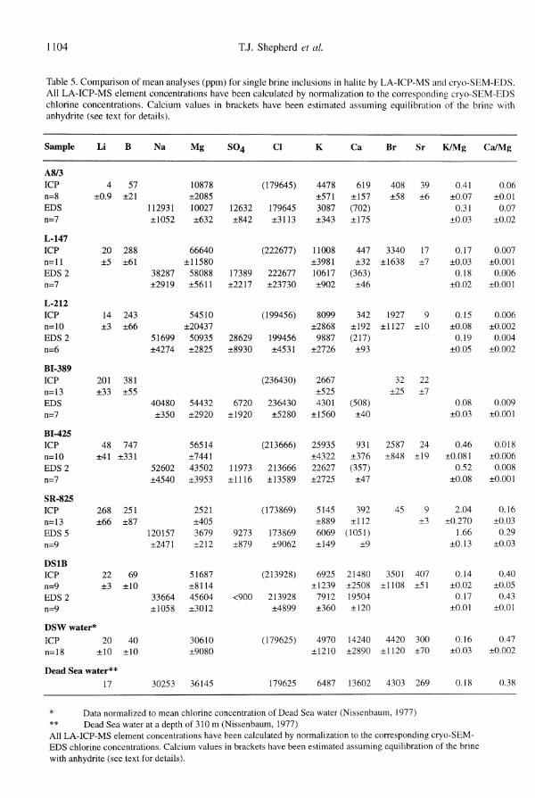
Table 5. Comparison of mean analyses (ppm) for single brine inclusions in halite by LA-ICP-MS and cryo-SEM-EDS. All LA-ICP-MS element concentrations have been calculated by normalization to the corresponding cryo-SEM-EDS chlorine concentrations. Calcium values in brackets have been estimated assuming equilibration of the brine with anhydrite (see text for details).
Sample
A8/3
ICP n=8 EDS n=7
L-147
ICP n=ll
EDS 2
n=7
L-212
ICP n=10
EDS 2
n=6
BI-389
ICP n=13
EDS n=7
BI-425
ICP n=10
EDS 2
n=7
SR-825
ICP n=13
EDS 5
n=9
DS1B
ICP n=9 EDS 2
n=9
Li
4 ±0.9
20 ±5
14 ±3
201 ±33
48 ±41
268 ±66
22 ±3
D S W water*
ICP n=18
20 ±10
Dead Sea water**
17
B
57 ±21
288 ±61
243 ±66
381 ±55
747 ±331
251 ±87
69 ±10
40 ±10
Na
112931
±1052
38287
±2919
51699
±4274
40480
±350
52602
±4540
120157
±2471
33664
±1058
30253
Mg
10878
±2085
10027
±632
66640
±11580
58088
±5611
54510
±20437
50935
±2825
54432
±2920
56514
±7441
43502
±3953
2521
±405
3679
±212
51687
±8114
45604
±3012
30610
±9080
36145
S0 4
12632
±842
17389
±2217
28629
±8930
6720
±1920
11973
±1116
9273
±879
<900
Cl
(179645)
179645
±3113
(222677)
222677
±23730
(199456)
199456
±4531
(236430)
236430
±5280
(213666)
213666
±13589
(173869)
173869
±9062
(213928)
213928
±4899
(179625)
179625
K
4478
±571
3087
±343
11008
±3981
10617
±902
8099
±2868
9887
±2726
2667
±525
4301
±1560
25935
±4322
22627
±2725
5145
±889
6069
±149
6925
±1239
7912
±360
4970
±1210
6487
Ca
619 ±157
(702)
±175
447 ±32
(363)
±46
342 ±192
(217)
±93
(508)
±40
931 ±376
(357)
±47
392 ±112
(1051)
±9
21480
±2508
19504
±120
14240
±2890
13602
Br
408 ±58
3340
±1638
1927
±1127
32 ±25
2587
±848
45
3501
±1108
4420
±1120
4303
Sr
39 ±6
17 ±7
9 ±10
22 ±7
24 ±19
9 ±3
407 ±51
300 ±70
269
K/Mg
0.41
±0.07
0.31
±0.03
0.17
±0.03
0.18
±0.02
0.15
±0.08
0.19
±0.05
0.08
±0.03
0.46
±0.081
0.52
±0.08
2.04
±0.270
1.66
±0.13
0.14
±0.02
0.17
±0.01
0.16
±0.03
0.18
Ca/Mg
0.06
±0.01
0.07
±0.02
0.007
±0.001
0.006
±0.001
0.006
±0.002
0.004
±0.002
0.009
±0.001
0.018
±0.006
0.008
±0.001
0.16
±0.03
0.29
±0.03
0.40
±0.05
0.43
±0.01
0.47
±0.002
0.38
* Data normalized to mean chlorine concentration of Dead Sea water (Nissenbaum, 1977) ** Dead Sea water at a depth of 310 m (Nissenbaum, 1977) All LA-ICP-MS element concentrations have been calculated by normalization to the corresponding cryo-SEM-EDS chlorine concentrations. Calcium values in brackets have been estimated assuming equilibration of the brine with anhydrite (see text for details).

Quantitative solute analysis of single fluid inclusions 1105
Table 6. Mean solute values (mol/kg) determined by cryo-SEM-EDS for single brine inclusions in halite.
Sample
A8/3
L-147
L-212
BI-389
BI-425
SR-825
DS1B
Na
5.61
1.84
2.49
1.95
2.57
5.99
1.60
Mg
0.47
2.64
2.32
2.47
2.01
0.17
2.05
so4
0.15
0.20
0.33
0.08
0.14
0.11
Cl
5.79
6.94
6.23
7.37
6.77
5.62
6.59
K
0.09
0.30
0.28
0.12
0.65
0.18
0.22
Ca
0.02
b.d
b.d
b.d
b.d
0.03
0.53
Σcations
6.68
7.44
7.42
7.04
7.26
6.57
6.98
Σanions
6.09
7.34
6.89
7.52
7.05
5.84
6.61
CB
0.09
0.01
0.07
-0.07
0.03
0.12
0.05
ISHUI
0.005
0.008
0.002
0.041
0.001
-0.035
-0.147
p g/cm3
1.19
1.25
1.24
1.25
1.24
1.19
1.24
CB = charge balance ([Σcations - Σanions/(Σcations + Σanions)/2]). ISHal = saturation index for halite, p = density of brine. b.d refers to Ca values that are close to or below the detection limit (0.01 mol/kg).
For DS1B the mean Ca values by LA-ICP-MS and cryo-SEM-EDS overlap at the 1 α level. It is satisfying to note the high degree of correspondence between the theoretical (numerically modelled) and measured Ca concentrations suggesting that Ca is indeed controlled by anhydrite saturation.
In keeping with previous studies, the relative standard deviations (RSD) for K and Mg indicate that the overall analytical precision for cryo-SEM-EDS (Mg 7%, K 14%) is better than LA-ICP-MS (Mg 20%, K 22%) by a factor of 2 to 3. Also shown in Table 5 are the mean LA-ICP-MS values for Dead Sea water (sample DSW). These have been normalized to the mean chlorine value for Dead Sea water (179625 ppm) and it can be seen that there is very close agreement (within 10-20%) between the normalized solute concentrations estimated for DSW and the absolute concentrations measured by Nissenbaum (1977). Since the estimated element concentrations for DS1B are systematically higher than both water samples, it is thought that the Cl value of 179 625 ppm for DSW is an underestimate of the present day halite-saturated bottom waters referred to by Anati & Stiller (1991). Further discussion of this topic is outside the scope of this paper.
As a further illustration of comparability between LA-ICP-MS and cryo-SEM-EDS, Fig. 2 and 3 show the correspondence for simple element concentration ratios. This approach cancels
0.40-,
0.00 0.10 0.20 0.30 0.40 Ca/Mg (cryo-SEM-EDS)
0.50
Fig. 2. Comparison of measured Ca/Mg ratios for single fluid inclusions in halite by cryo-SEM-EDS and LA-ICP-MS. Values refer to sample means. The solid triangle is a measured data point using both methods; the open triangles refer to Ca/Mg ratios where the values for Ca have been obtained by (a) direct LA-ICP-MS and (b) estimated assuming saturation of the brine with respect to anhydrite (see text for details). The dashed curve is the ideal 1:1 relationship between the two methods.
out any errors due to the measurement of chlorine. For Ca/Mg and K/Mg, the regressions (r = 0.96 and 0.99, respectively) are highly significant indi-

1106 T.J. Shepherd et al.
l
PH
y i
<
1
2.00-
1.50-
_
1.00-
0.50-
0.00-
r = 0 . 9 9 /
//
Λ s/
^ ' 1 ' 1 ' Γ
/ / / Theoretical
K/Mg
i | i |
0.00 0.40 0.8 1.20 1.60 2.00 K/Mg(cryoSEM-EDS)
Fig. 3. Comparison of measured K/Mg ratios for single fluid inclusions in halite by cryo-SEM-EDS and LA-ICP-MS. Values refer to sample means. The dashed curve is the ideal 1:1 relationship between the two methods.
eating excellent agreement between the two techniques. With the exception of sample SR-825, for which the anhydrite-saturated Ca estimate is almost twice that of the measured LA-ICP-MS value, the slope of the regression line is very close to 1. Thus for Ca and Mg, no systematic technique bias is evident. For K/Mg, the slope of the regression is slightly higher suggesting a slight overestimate by LA-ICP-MS. However, to be statistically proven one would need to analyse additional samples with high K/Mg ratios. The accuracy of LA-ICP-MS is further substantiated by the good agreement between analyses obtained for DSW (Table 5, Dead Sea water) and the analyses for deep Dead Sea water published by Nissen-baum (1977). This is first ICP-MS analysis of its kind obtained by direct laser ablation of a brine and the results strongly justify further investigation of this methodology as a means of analysing high TDS solutions (>50000 µg ml1) . In conventional ICP-MS analysis, the nebulization of solutions containing more than 5000 µg ml"1 is not advisable and causes serious degradation of analyte precision and sensitivity (Jarvis et al., 1992). However, new simultaneous analogue/pulse counting detectors may allow the analysis of high TDS solutions, assuming of course that problems of sampler cone blockage can be overcome.
Conclusions
The study clearly shows that LA-ICP-MS and cryogenic SEM-EDS are perfectly complementary and well suited for the analysis of single brine inclusions in halite. Values for Mg and K are in very good statistical agreement (±1 α s.d.) and data for Ca by LA-ICP-MS are consistent with theoretical estimates of Ca in the brines. The ability to convert relative concentrations ratios determined by LA-ICP-MS into absolute element concentrations is especially valuable, and allows easy quantification of important process-sensitive trace elements such as Li, B, Cs, Rb, Sr and Br. Furthermore, the ability to measure Cl/Br ratios by LA-ICP-MS offers an exciting potential for investigating the origins of salinity in many types of basinal brine (Kesler et aL9 1995).
At present, both techniques have difficulty in measuring low concentrations of Ca (<400 ppm). This is seen as a temporary problem for LA-ICP-MS since new developments in instrument design should overcome or reduce the polyatomic interferences which complicate the analysis of Ca and preclude the detection of sulphur. With continued improvements in detector design for the light elements, detection limits for Li, B and Br by cryo-SEM-EDS can also be expected to decrease. Cryo-SEM-EDS is not restricted to the analysis of brine inclusions and analyses have been obtained for other types of inclusion (aqueous!variants daughter minerals) in other mineral matrices (Ayora & Fontarnau, 1990; Garcfa-Veigas, 1993; Tritlla & Cardellach, 1997). Its principal limitation however is that it does not allow pre-selection of inclusions and, for inclusion-poor samples or mineral with poor cleavage, much patience and effort is required. Finally, for LA-ICP-MS there remains the question of converting relative element concentrations into absolute concentrations. Ultimately it is hoped to calibrate and quantify the ICP signals as illustrated by Ghazi et al (1996). For the moment we are restricted to various kinds of external normalization. Where we have low or high salinity inclusions in quartz, one can use microthermometric salinity measurements to estimate Cl concentrations. Although this method has an uncertainty of ±10-20% it is easy to apply and is compatible with the overall level of analytical precision for LA-ICP-MS. In general, this is more than adequate for many trace element investigations. Notwithstanding these limitations, the present study demonstrates that cryo-SEM-EDS and

Quantitative solute analysis of single fluid inclusions 1107
LA-ICP-MS are capable of providing high quality, quantitative data for a large range of cation and anion species in single inclusions, and sets a benchmark against which other techniques may be assessed. It is hoped to extend this exercise to other microbeam techniques (PIXE and synchrotron XRF) to provide fluid inclusion researchers with a practical guide for selecting the best 'fit for purpose' single inclusion technique based on precision, accuracy and limits of detection. A subsidiary but nevertheless important conclusion to be drawn from the study is that inclusions in halite provide a working alternative to synthetic fluid inclusions for calibration and development.
Acknowledgments: The authors would like to thank their respective Institutes for encouraging the study and to the British Council and Ministe-rio de Educacion y Ciencia for providing essential funding (Acciones Integradas Grant no. 1898) for exchange visits between Spain and the UK. We also wish thank Prof. A. Starinsky for providing samples of halite and sea water from the Dead Sea. The authors gratefully acknowledge Dr. C. Ramboz (University of Orleans, France) and Dr. A. Audetat (ETH Zhrich, Switzerland) for their constructive comments on the paper which did much to improve its value and presentation. Publication for TJS and SRC is by permission of the Director, British Geological Survey (NERC).
References
Anati, D.A. & Stiller, M. (1991): Thermohaline structure of the Dead Sea and the role of double-diffusive mixing. Limnol. Oceanog., 36, 342-354.
Ayora, C. & Fontarnau, R. (1990): X-ray microanalysis of frozen fluid inclusions. Chem. Geoi, 89, 135-148.
Ayora, C, Garcia-Veigas, J., Pueyo, J.-J. (1994a): X-ray microanalysis of fluid inclusions and its application to the geochemical modelling of evaporite basins. Geochim. Cosmochim. Acta, 58, 43-55. (1994b): The chemical and hydrological evolution
of an ancient potash-forming evaporite basin as constrained by mineral sequence, fluid inclusion composition, and numerical simulation. Geochim. Cosmochim. Acta, 58, 3379-3394.
Cendon, D.I., Ayora, C, Pueyo, J.J. (1998): The origin of barren bodies in the Subiza potash deposit, Na-varra, Spain: Implications for sylvite formation. J. Sedimen. Res., 68, 43-52.
Chenery, S. & Cook, J.M. (1993): Determination of rare earth elements in single mineral grains by laser ablation microprobe inductively coupled plasma mass
spectrometry - preliminary study. J. Anal. At. Spec-trom., 8, 299-303.
Chenery, S., Williams, C.T., Elliot, T.A., Forey, P.L., Werdelin, L. (1996): Determination of Rare Earth Elements in Biological and Mineral Apatite by EPMA and LAMP-ICP-MS. Mikrochim. Acta [SuppL], 13, 259-269.
Diamond, L.W., Marshall, D.D., Jackman, J.A., Skippen, G.B. (1990): Elemental analysis of individual fluid inclusions in minerals by Secondary Ion Mass Spectrometry (SIMS): application to cation ratios of fluid inclusions in an Archaean mesothermal gold-quartz vein. Geochim. Cosmochim. Acta, 54, 545-552.
Garcia-Veigas, J., (1993): Geoquimica de inclusiones fluidas en formaciones salinas, Microanálisis Cryo-SEM-EDS, Universidad de Barcelona.
Garcia-Veigas, J., Rosell, L., Utrilla, R., Orti, F. (1990): Aportaciones geoquimicas al conocimiento de las evaporitas messinienses: cuencas de Palma (Mal-lorca), San Miguel de Salinas (Alicante) y Lorca (Murcia). in "Formaciones evaporiticas de la Cuenca del Ebro y cadenas perifericas, y de la zona de Levante", F. Orti & J. M. Salvany, eds. Barcelona, 257-266.
Ghazi, A.M., McCandless, T.E., Vanko, D.A., Ruiz, J. (1996): New quantitative approach to trace element analysis of single fluid inclusions: applications of laser ablation inductively coupled plasma mass spectrometry (LA-ICP-MS). J. Anal. At. Spectrom., 11, 667-674.
Gunther, D., Frischknecht, R., Muschenborn, H.J., Hein-rich, C.A. (1997): Direct liquid ablation: a new calibration strategy for laser ablation ICP-MS micro-analysis of solids and liquids. Fres. J. Anal. Chem., 359, 390-393.
Heinrich, C.A., Ryan, C.G., Mernagh, T.P., Eadington, P.J. (1992): Segregation of ore metals between magmatic brine and vapour - a fluid inclusion study using PIXE microanalysis. Econ. Geoi, 87, 1566-1583.
Jarvis, I., Gray, A.L., Houk, R.S. (1992): Handbook of Inductively Coupled Plasma Mass Spectrometry. Blackie, New York, 380 p.
Kesler, S.E., Appold, M.S., Martini, A.M., Walter, L.M., Huston, T.J., Kyle, J.R. (1995): Na-Cl-Br syste-matics of mineralizing brines in Mississippi Valley-type deposits. Geology, 23, 7, 641-644.
McCaffrey, M., Lazar, B.', Holland, H.D. (1987): The evaporation path of seawater and the coprecipita-tion of Br- and K+ with halite. J. Sediment. Petrol., 57, 928-937.
Moissette, A., Shepherd, T.J., Chenery, S.R. (1996): Calibration strategies for the elemental analysis of individual aqueous fluid inclusions by laser ablation inductively coupled plasma mass spectrometry. J. Anal. At. Spectrom., 11, 177-185
Monnin, C. (1989): An ion interaction-model for the volumetric properties of natural-waters - density of

1108 T.J. Shepherd et al.
the solution and partial molal volumes of electrolytes to high-concentrations at 25 °C. Geochim. Cosmochim. Acta, 53, 1177-1188.
Nissenbaum, A. (1977): Minor and trace elements in Dead Sea water. Chem. Geol., 19, 99-111.
Querol, X. & Chenery, S. (1995): The determination of trace element affinities in coal by laser ablation microprobe - inductively coupled plasma - mass spectrometry. in "European Coal Geology", M. K. G. Whateley & D. A. Spears, eds. Geol. Soc. Spec. Publ. 82, 147-155.
Ramsey, M.H., Coles, B.J., Wilkinson, J.J. (1992): Single fluid inclusion analysis by laser ablation inductively coupled plasma atomic emission spectrometry: quantification and validation. J. Anal At. Spectrom., 7, 587-593.
Rankin, A.H., Ramsey, M.H., Coles, B., Van Lange-velde, E, Thomas, C.R. (1992): The composition of hypersaline, iron-rich granitic fluids based on laser-ICP and Synchrotron-XRF microprobe analysis of individual fluid inclusions in topaz, Mole granite, eastern Australia. Geochim. Cosmochim. Acta, 56, 67-79.
Rosell, L. (1983): Estudi petrologic, sedimentologic i geoquimic de la formacio de sals potàssiques de Navarra (Eocè superior). Memoria Tesis Doctoral, Universidad de Barcelona.
Shearman, D.J. (1970): Recent halite rock, Baja California, Mexico. Trans. Inst. Min. MetalL, B79, 155-162.
Shepherd, TJ. & Chenery, S.R. (1995): Laser ablation ICP-MS elemental analysis of individual fluid inclusions: An evaluation study. Geochim. Cosmochim. Acta, 59, 3997-4007.
Tritlla, J. & Cardellach, E„ (1997): Fluid inclusions in pre-ore minerals from the carbonate-hosted mercury deposits in the Espadán Ranges (eastern Spain). Chem. Geol., 137, 91-106.
Vanko, D.A., Sutton, S.R., Rivers, M.L., Bodnar, R.J. (1993): Major element ratios in synthetic fluid inclusions by synchrotron X-ray fluorescence microprobe. Chem. Geol., 109, 125-134.
Received 10 October 1997 Modified version received 20 March 1998 Accepted 14 May 1998
Copyright © 2022 FDOKUMEN