The Uremic Retention Solute p-Cresyl Sulfate and Markers of Endothelial Damage
11
The Uremic Retention Solute p-Cresyl Sulfate and Markers of Endothelial Damage Björn K.I. Meijers, MD, 1 Soetkin Van kerckhoven, 2 Kristin Verbeke, PharmD, PhD, 3 Wim Dehaen, PhD, 4 Yves Vanrenterghem, MD, PhD, 1 Marc F. Hoylaerts, PhD, 2 and Pieter Evenepoel, MD, PhD 1 Background: Cardiovascular disease is highly prevalent in patients with chronic kidney disease. In hemodialysis patients, the protein-bound uremic retention solute p-cresol is independently associated with cardiovascular disease. The underlying mechanisms have not been elucidated. Study Design: (1) Prospective observational study of humans and (2) in vitro study in human umbilical vein endothelial cells. Setting: Hemodialysis patients. Factor: p-Cresol and its main derivative p-cresyl sulfate. Outcomes: Endothelial dysfunction. Measurements: We studied: (1) the relation between p-cresol and blood markers of endothelial dysfunction, including soluble P-selectin and endothelial microparticles; and (2) direct effects of p-cresol and p-cresyl sulfate on endothelial cell cultures. Results: (1) In a cohort of 100 maintenance hemodialysis patients, free serum p-cresol concentra- tions (median, 11.7 mol/L; interquartile range, 15.2) were directly associated with circulating endothe- lial microparticles (P 0.007), but not with soluble P-selectin (mean, 37.7 14.4 [SD] pg/mL). Other independent determinants of the degree of circulating microparticles were greater serum phosphorus (mean, 4.8 1.5 mg/dL; P 0.008) and serum calcium concentrations (mean, 9.3 0.8 mg/dL; P 0.03), whereas treatment with active vitamin D (P 0.008) and vintage (median, 25 months; P 0.04) were inversely associated. (2) In vitro, p-cresyl sulfate induced a dose-dependent increase in the shedding of endothelial microparticles (P 0.001) by human umbilical vein endothelial cells. Shedding was reduced, but not completely aborted, in the presence of albumin, whereas the selective Rho kinase inhibitor Y-27632 abrogated the p-cresyl sulfate–induced generation of endothelial microparticles. Limitations: The relationship between p-cresyl sulfate and shedding of endothelial microparticles in vivo was not mechanistically explored. Conclusion: p-Cresyl sulfate induces shedding of endothelial microparticles in the absence of overt endothelial damage in vitro and is independently associated with the number of endothelial micropar- ticles in hemodialysis patients. These findings suggest that p-cresyl sulfate alters endothelial function in hemodialysis patients. Am J Kidney Dis 54:891-901. © 2009 by the National Kidney Foundation, Inc. INDEX WORDS: p-Cresol; p-cresyl sulfate; 4-cresol; endothelium; cell-deprived microparticles; uremia; hemodialysis. Editorial, p. 792 C ardiovascular disease (CVD) is highly prevalent in patients with chronic kidney disease (CKD). 1,2 In hemodialysis (HD) pa- tients, mortality from CVD is 5- to 20-fold greater compared with the general population, even after stratification for age, sex, race, and diabetes. Traditional cardiovascular risk factors are insufficient to accurately predict cardiovascu- lar risk in patients with CKD. 3 Uremic retention solutes, particularly those that are protein bound, are implicated in the excess CVD. A major prototypic representative of the group of protein- bound uremic retention solutes is p-cresol. We have recently shown that free serum concentrations of p-cresol (and its sulfate and glucuronide conju- From the 1 Department of Medicine, Division of Nephrol- ogy, University Hospitals Leuven, 2 Center for Molecular and Vascular Biology, Catholic University Leuven, 3 Depart- ment of Gastro-intestinal Research, University Hospital Leu- ven; and 4 Department of Chemistry, Catholic University Leuven, Leuven, Belgium. Received December 8, 2008. Accepted in revised form April 3, 2009. Originally published online as doi: 10.1053/ j.ajkd.2009.04.022 on July 21, 2009. Address correspondence to Pieter Evenepoel, MD, PhD, Dienst nefrologie, Universitair Ziekenhuis Gasthuisberg, Herestraat 49, B-3000 Leuven, Belgium. E-mail: pieter. [email protected] © 2009 by the National Kidney Foundation, Inc. 0272-6386/09/5405-0014$36.00/0 doi:10.1053/j.ajkd.2009.04.022 American Journal of Kidney Diseases, Vol 54, No 5 (November), 2009: pp 891-901 891
-
Upload
independent -
Category
Documents
-
view
0 -
download
0
Transcript of The Uremic Retention Solute p-Cresyl Sulfate and Markers of Endothelial Damage

Cdtge
oamvL
Aj
A
The Uremic Retention Solute p-Cresyl Sulfate and Markersof Endothelial Damage
Björn K.I. Meijers, MD,1 Soetkin Van kerckhoven,2 Kristin Verbeke, PharmD, PhD,3
Wim Dehaen, PhD,4 Yves Vanrenterghem, MD, PhD,1 Marc F. Hoylaerts, PhD,2
and Pieter Evenepoel, MD, PhD1
Background: Cardiovascular disease is highly prevalent in patients with chronic kidney disease. Inhemodialysis patients, the protein-bound uremic retention solute p-cresol is independently associatedwith cardiovascular disease. The underlying mechanisms have not been elucidated.
Study Design: (1) Prospective observational study of humans and (2) in vitro study in humanumbilical vein endothelial cells.
Setting: Hemodialysis patients.Factor: p-Cresol and its main derivative p-cresyl sulfate.Outcomes: Endothelial dysfunction.Measurements: We studied: (1) the relation between p-cresol and blood markers of endothelial
dysfunction, including soluble P-selectin and endothelial microparticles; and (2) direct effects of p-cresoland p-cresyl sulfate on endothelial cell cultures.
Results: (1) In a cohort of 100 maintenance hemodialysis patients, free serum p-cresol concentra-tions (median, 11.7 �mol/L; interquartile range, 15.2) were directly associated with circulating endothe-lial microparticles (P � 0.007), but not with soluble P-selectin (mean, 37.7 � 14.4 [SD] pg/mL). Otherindependent determinants of the degree of circulating microparticles were greater serum phosphorus(mean, 4.8 � 1.5 mg/dL; P � 0.008) and serum calcium concentrations (mean, 9.3 � 0.8 mg/dL; P �0.03), whereas treatment with active vitamin D (P � 0.008) and vintage (median, 25 months; P � 0.04)were inversely associated. (2) In vitro, p-cresyl sulfate induced a dose-dependent increase in theshedding of endothelial microparticles (P � 0.001) by human umbilical vein endothelial cells. Sheddingwas reduced, but not completely aborted, in the presence of albumin, whereas the selective Rho kinaseinhibitor Y-27632 abrogated the p-cresyl sulfate–induced generation of endothelial microparticles.
Limitations: The relationship between p-cresyl sulfate and shedding of endothelial microparticles invivo was not mechanistically explored.
Conclusion: p-Cresyl sulfate induces shedding of endothelial microparticles in the absence of overtendothelial damage in vitro and is independently associated with the number of endothelial micropar-ticles in hemodialysis patients. These findings suggest that p-cresyl sulfate alters endothelial function inhemodialysis patients.Am J Kidney Dis 54:891-901. © 2009 by the National Kidney Foundation, Inc.
INDEX WORDS: p-Cresol; p-cresyl sulfate; 4-cresol; endothelium; cell-deprived microparticles; uremia;hemodialysis.
dalsapbho
Editorial, p. 792
ardiovascular disease (CVD) is highlyprevalent in patients with chronic kidney
isease (CKD).1,2 In hemodialysis (HD) pa-ients, mortality from CVD is 5- to 20-foldreater compared with the general population,ven after stratification for age, sex, race, and
From the 1Department of Medicine, Division of Nephrol-gy, University Hospitals Leuven, 2Center for Molecularnd Vascular Biology, Catholic University Leuven, 3Depart-ent of Gastro-intestinal Research, University Hospital Leu-
en; and 4Department of Chemistry, Catholic Universityeuven, Leuven, Belgium.Received December 8, 2008. Accepted in revised form
pril 3, 2009. Originally published online as doi: 10.1053/
.ajkd.2009.04.022 on July 21, 2009.merican Journal of Kidney Diseases, Vol 54, No 5 (November), 2
iabetes. Traditional cardiovascular risk factorsre insufficient to accurately predict cardiovascu-ar risk in patients with CKD.3 Uremic retentionolutes, particularly those that are protein bound,re implicated in the excess CVD. A majorrototypic representative of the group of protein-ound uremic retention solutes is p-cresol. Weave recently shown that free serum concentrationsf p-cresol (and its sulfate and glucuronide conju-
Address correspondence to Pieter Evenepoel, MD, PhD,ienst nefrologie, Universitair Ziekenhuis Gasthuisberg,erestraat 49, B-3000 Leuven, Belgium. E-mail: pieter.
[email protected]© 2009 by the National Kidney Foundation, Inc.0272-6386/09/5405-0014$36.00/0doi:10.1053/j.ajkd.2009.04.022
DHe
009: pp 891-901 891

gTlst
ttbirlEi(isscdtCrw
asscfisfhal
sstEtc
S
etcI
ituttp
i9mfsae
BP
atgpssppTdwuvgttwmus
clipD
M
pan�itC2(ar
Meijers et al892
ates) are associated with CVD in HD patients.4
his study provides evidence for the use of p-cresolevel as a cardiovascular risk marker, especially inuch patients with lower absolute risk of CVD ashose who do not have diabetes.
The endothelium is considered a key player inhe triggering and development of CVD.5 Endo-helial dysfunction, frequently defined as an im-alance between vasodilating and vasoconstrict-ng substances,6 is abundant in patients withenal failure.5 Effects of p-cresol on the endothe-ium have been shown repeatedly in vitro.7-9
xposure to uremic concentrations of p-cresolnduced shedding of endothelial microparticlesEMPs) by cultured endothelial cells.8 Circulat-ng EMPs selectively impair the nitric oxideignaling pathway,10 and observational studieshowed a direct relationship between degree ofirculating EMPs in HD patients and endothelialysfunction.11 Shedding is believed to be a pro-ective mechanism against cellular toxicity.12
ombined, these studies suggest that p-cresol iselated to endothelial dysfunction in patientsith CKD.However, the relationship between p-cresol
nd endothelial dysfunction in vivo has not beentudied to date. Moreover, we and others havehown that in vivo, p-cresol mainly (�95%)irculates as its sulfate conjugate (p-cresyl sul-ate).13,14 Sulfate conjugation is one of the mostmportant detoxification pathways.15 Cautionhould be taken to extrapolate in vitro findingsrom studies with unconjugated p-cresol. Thisas been emphasized by the finding that p-cresolnd p-cresyl sulfate exert different effects oneukocytes.16
The aim of this study is to explore the relation-hip between serum p-cresol, most of which isulfate conjugated, and selected markers of endo-helial dysfunction, including soluble P-selectin,MPs, and platelet microparticles, in HD pa-
ients. The associations found were mechanisti-ally explored further in vitro.
METHODS
tudyPopulation andSampleCollection
Patients on dialysis therapy for more than 3 months wereligible for inclusion. The study was performed according tohe Declaration of Helsinki and approved by the ethicsommittee of the University Hospital Leuven, Belgium.
nformed consent was obtained from all patients. fiAt inclusion, blood was obtained from the dialysis accessmmediately before the midweek HD session. Samples ob-ained at the end of the dialysis session were collected bysing the slow-flow/stop-pump technique according to Na-ional Kidney Foundation guidelines. Residual renal func-ion was estimated from an interdialysis urine collection asart of the baseline evaluation.For measurement of microparticles, blood was collected
n citrate tubes (0.109 mol/L citrate/2.7 mL; VacutainerNC, BD, Plymouth, UK) and centrifuged at 2,000g for 10inutes. The supernatant was centrifuged again at 2,000g
or 20 minutes. Supernatant from the second centrifugationtep was stored for batch analysis at �80°C within 1 hourfter sampling. The centrifuge protocol was validated tonsure the absence of platelets.
iochemical Analysis andBaseline andEndoint Evaluation
Hemoglobin, calcium, phosphorus, C-reactive protein,nd cholesterol were measured by using standard laboratoryechniques. Albumin was measured by using the bromcresolreen method. Total and free (not bound to proteins) serum-cresol was measured by using gas chromatography masspectrometry (GC-MS), as described previously.14 In brief,erum was acidified and heated to denature serum bindingroteins and hydrolyze p-cresol conjugates. Subsequently,-cresol was extracted in ethyl acetate and injected on arace GC-MS (Thermo Finnigan, San Jose, CA); 2,6-imethyl-phenol was used as internal standard. Free p-cresolas measured in serum ultrafiltered at room temperature bysing 30,000-Da molecular cutoff filters (Centifree UF de-ices; Amicon, Beverly, MA). Both p-cresol and its conju-ated metabolites are measured by using this analyticalechnique.14 Intra-assay and interassay coefficients of varia-ion were 3.3% and 5.3%, respectively. Thromboxane B2
as measured by using an enzyme immunoassay kit (Cay-an, Ann Arbor, MI). Soluble P-selectin was measured by
sing an enzyme-linked immunosorbent assay (Bender Med-ystems, Vienna, Austria).
Dialysis efficacy was calculated as single-pool Kt/V ac-ording to Daugirdas. Residual renal function was calcu-ated from collected volume and urea nitrogen concentrationn urine and serum and expressed as clearance (milliliterser minute). Baseline comorbidity was scored according toavies et al.17
icroparticleAnalysis
Samples were thawed at 37°C for 30 minutes. Platelet-oor plasma (20 �L) or cell culture supernatant (40 �L) wasdded to phosphate-buffered saline (PBS), together withonspecific mouse immunoglobulin G1 (IgG1) antibodies (5L; concentration, 2 mg/mL) to a final volume of 70 �L and
ncubated at room temperature for 10 minutes. Ten microli-ers of fluorescein isothiocyanate–labeled mouse IgG1 anti-D42a (cat 348083; Becton-Dickinson, San José, CA) and0 �L of phycoerythrin-labeled mouse IgG1 anti-CD105cat 12-1057; Immunosource, Halle-Zoersel, Belgium) weredded. After incubation at room temperature in the dark on aocking platform for 30 minutes, samples were diluted to a
nal volume of 500 �L with PBS containing 0.5% human
sgaS
estklpb(msCCapflm(aE0pnmccnrEvw
C
C
ufu3wB(wwp(cw
wi
i
R
AMDSRBS
PDC
CHAPCCCTFE
PTS
SC�r�
EMP and p-Cresyl Sulfate 893
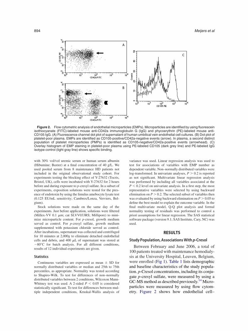
erum albumin (Hibumine, 200 g/L; Baxter, Lessines, Bel-ium) before analysis. PBS solutions and nonspecific IgGntibodies were filtered (Millex-VV 0.1 �m; catLVV033RS; Millipore, Brussels, Belgium).Samples were analyzed using a FACSCalibur flow cytom-
ter (Becton-Dickinson). Events were gated according toize in a forward/side scatter dot plot. Size cutoff values (0.2o 1.0 �m) were determined by using latex beads withnown size. Subsequently, gated events were plotted onogarithmic scaled fluorescence channel 1 and channel 2 dotlots. Thresholds for specific fluorescence were determinedy using mouse fluorescein isothiocyanate–IgG1 isotypecat 555748; BD Biosciences, Erembodegem, Belgium) andouse phycoerythrin-IgG1 isotype (cat 555749; BD Bio-
ciences) controls. EMPs were identified as CD105-positive/D42a-negative events. It is known that a small fraction ofD105-positive events originates from activated monocytesnd bone marrow cell subsets. For validation purposes, weerformed double staining for EMPs by using CD144-uorescein isothiocyanate and CD105-phycoerythrin. Al-ost all CD105-positive events were also positive for CD144
Fig S1, provided as online supplementary materials avail-ble with this article at www.ajkd.org). Measurement ofMPs after filtration of platelet-poor plasma through a.1-�m filter (Millex-VV 0.1 �m, cat SLVV033RS; Milli-ore) served as negative control. CD105-positive/CD42a-egative events were counted at medium flow rate over 5inutes. Measured EMPs were expressed as absolute EMP
ounts. Stability of the flow rate was controlled by usingontrols with known concentrations of latex beads. Becauseo technique currently is available to measure EMP recoveryates,18 we chose not to recalculate absolute counts towardMP concentrations. Intraday and interday coefficients ofariation of analysis of patient microparticles in plasmaere 8.7% and 13%, respectively.
ell Culture andExposure Experiments
Human umbilical vein endothelial cells (HUVECs; cat2517A; Lonza Ltd, Verviers, Belgium) were grown by
sFigure 1. Flow chart shows patient screening and
nclusion.
sing endothelial cell medium-2 supplemented with growthactors and cytokines (Bulletkit; cat CC3162; Lonza Ltd)nder standard culture conditions (humidified atmosphere,7°C; 5% carbon dioxide). For exposure experiments, cellsere plated on 24-well plates (Cellstar; cat 662160; Greinerio-one, Wemmel, Belgium) and grown to near-confluency
�90%). Supernatant was aspirated and cells were washedith PBS. Cells were incubated for 24 hours with 500-�L/ell solutions of p-cresol (cat 42429; Fluka, Geel, Belgium),-cresyl sulfate (potassium salt), or potassium chlorideMerck, Darmstadt, Germany) in growth medium. Finaloncentrations of p-cresol, p-cresyl sulfate, and potassiumere 0.1, 0.5, and 1.0 mmol/L.In a second series of experiments, HUVECs were cultured
ith and without 1.0 mmol/L of p-cresyl sulfate for 24 hoursn basal medium without growth factors or serum. For a third
Table 1. Patient Demographics of 100 HemodialysisPatients
enal diagnosisGlomerulonephritis 20 (20)Hypertensive/large vessel 11 (11)Polycystic kidney disease 9 (9)Tubulointerstitial nephritis 4 (4)Diabetic nephropathy 17 (17)Other/miscellaneous 39 (39)
ge (y) 71 (59-78)en/women 64/36 (64/36)ialysis vintage (mo) 25.0 (12-47)ingle-pool Kt/V 1.57 � 0.34esidual urea clearance (mL/min) 0.0 (0.0-0.38)ody mass index (kg/m2) 22.9 (21.0-25.8)ystolic/diastolic blood
pressure (mm Hg) 131 � 22/66 � 15ulse pressure (mm Hg) 65.5 � 17.7iabetes (% yes/no) 28/72urrent smoker (%
yes/no/unknown) 15/84/1holesterol (mg/dL) 151.5 � 35.7ematocrit (%) 37 � 3lbumin (g/dL) 39.3 � 3.5hosphorus (mg/dL) 4.8 � 1.5alcium (mg/dL) 9.3 � 0.8reatinine (mg/dL) 7.6 � 2.4-Reactive protein (mg/L) 6 (3-12)otal p-cresol (�mol/L) 185.7 (119.0-282.3)ree p-cresol (�mol/L) 11.7 (6.1-21.3)ndothelial microparticle count
(/5 min) 375 (292-534)latelet microparticle count (/5 min) 4,488 (2,960-7,146)hromboxane B2 (pg/mL) 4,714 (2,960-7,146)oluble P-selectin (pg/mL) 37.7 � 14.4
Note: Values expressed as number (percent), mean �D, or median (25th to 75th percentile), as appropriate.onversion factors for units: cholesterol in mg/dL to mmol/L,0.02586; calcium in mg/dL to mmol/L, �0.2495; phospho-
us in mg/dL to mmol/L, �0.3229; creatinine in mg/dL tomol/L, �88; albumin in g/dL to g/L, �10.
eries of experiments, growth medium was supplemented
w(uieBbee(g
e(mssAfc�r
S
nptdWst
vtdlawPrewdfinpsu
S
1swatgGp
iCppO a usingi
Meijers et al894
ith 30% vol/vol uremic serum or human serum albuminHibumine; Baxter) at a final concentration of 40 g/L. Wesed pooled serum from 8 maintenance HD patients notncluded in the original observational study cohort. Forxperiments testing the blocking effect of Y-27632 (Tocris,ristol, UK), cells were incubated with Y-27632 for 2 hoursefore and during exposure to p-cresyl sulfate. In a subset ofxperiments, exposition solutions were tested for the pres-nce of endotoxin by using the limulus amebocyte lysate test0.125 EU/mL sensitivity; Cambrex/Lonza, Verviers, Bel-ium).Stock solutions were made on the same day of the
xperiments. Just before application, solutions were filteredMillex-VV 0.1 �m; cat SLVV033RS; Millipore) to mini-ize microparticle content. For p-cresol, growth medium
erved as control. For p-cresyl sulfate, growth mediumupplemented with potassium chloride served as control.fter incubations, supernatant was collected and centrifuged
or 10 minutes at 2,000g to eliminate detached endothelialells and debris, and 400 �L of supernatant was stored at80°C for batch analysis. For all different conditions,
esults of 12 individual experiments are given.
tatistics
Continuous variables are expressed as mean � SD forormally distributed variables or median and 25th to 75thercentiles, as appropriate. Normality was tested accordingo Shapiro-Wilk. To test for differences of non–normallyistributed variables between 2 conditions, Wilcoxon Mann-hitney test was used. A 2-sided P � 0.05 is considered
tatistically significant. To test for differences between mul-
Figure 2. Flow cytometric analysis of endothelial micropsothiocyanate (FITC)-labeled mouse anti-CD42a immunD105 IgG. (A) Fluorescence channel dot plot of supernatalatelet-poor plasma. EMPs are identified as CD105-positopulation of platelet microparticles (PMPs) is identifiedverlay histogram of EMP staining in platelet-poor plasm
sotype control (light gray line) shows specific binding.
iple independent conditions, Kruskal-Wallis analysis of e
ariance was used. Linear regression analysis was used toest for associations of variables with EMP number asependent variable. Non–normally distributed variables wereog-transformed. In univariate analyses, P � 0.2 is reporteds not significant. Multivariate linear regression analysisas performed by including all variables associated at the� 0.2 level on univariate analysis. In a first step, the most
epresentative variables were selected by using backwardlimination on P � 0.2. The selected subset of variables thenas evaluated by using backward elimination on P � 0.05 toefine the best model to explain the outcome variable. In thenal multivariate model, Q-Q plot analysis and formalormality testing of residuals was performed to control ariori assumptions for linear regression. The SAS statisticaloftware package (version 9.1; SAS Institute, Cary, NC) wassed.
RESULTS
tudyPopulation, AssociationsWithp-Cresol
Between February and June 2006, a total of00 patients treated with maintenance hemodialy-is at the University Hospital, Leuven, Belgium,ere enrolled (Fig 1). Table 1 lists demographic
nd baseline characteristics of the study popula-ion. p-Cresol concentrations, including its conju-ate p-cresyl sulfate, were measured by using aC-MS method as described previously.14 Micro-articles were measured by using flow cytom-
s (EMPs). Microparticles are identified by using fluoresceinlin G (IgG) and phycoerythrin (PE)-labeled mouse anti-man umbilical vein endothelial cell cultures. (B) Dot plot of
42a-negative events (arrow). In plasma, a second distinctD105-negative/CD42a-positive events (arrowhead). (C)PE-labeled CD105 (dark grey line) and PE-labeled IgG
articleoglobunt of huive/CD
as C
try. Figure 2 shows how endothelial cell–

dblC
tEattt3ps0p
sl
RB
spteiifw0
Mpa(cEtgstatt
ecipa
I
tcsIumpet
dp
saitd
EMP and p-Cresyl Sulfate 895
erived microparticles could be analyzed on theasis of their CD105 (endoglin) positivity. Plate-et microparticles were identified based on theirD42a positivity.Associations of p-cresol with markers of endo-
helial function, including degree of circulatingMPs, were analyzed by using Spearman ranknalysis. We observed a significant direct associa-ion both between free (r � 0.28; P � 0.004) andotal p-cresol (r � 0.35; P � 0.001) concentra-ions in patient serum and numbers of EMPs (Fig). However, a significant association between-cresol and soluble P-selectin could not behown. Additionally, a borderline significant (P �.05) correlation between free p-cresol and
Figure 3. Dot plot of the relationship between (A) freeerum p-cresol and (B) total serum p-cresol concentrationsnd number of circulating endothelial microparticles (EMPs)
n 100 patients treated with hemodialysis. Serum concentra-ions of p-cresol (micromolar) and EMPs were not normallyistributed and data were log-transformed.
lasma thromboxane B2 levels was observed. A r
ignificant association between p-cresol and plate-et microparticles could not be shown.
elationshipof EMPsWithClinical andiochemical Variables
We performed multivariate regression analy-is to test the strength of the association between-cresol and EMPs. First, we explored the rela-ionship between clinical and biochemical param-ters (Table 2) and numbers of circulating EMPsn univariate linear regression analyses. In univar-ate analysis, both total (bound and unbound) andree (unbound) serum p-cresol concentrationsere associated with EMPs (P � 0.005 and P �.02, respectively).We constructed 2 multivariate models (seeethods section) by including either total or free
-cresol serum concentrations as explanatory vari-ble. In the final model (Table 3), both totalbound and unbound) and free (unbound) serumoncentrations of p-cresol were associated withMP count (P � 0.001 and P � 0.007, respec-
ively). Other independent determinants of de-ree of circulating microparticles were greatererum phosphorus and serum calcium concentra-ions, whereas treatment with active vitamin Dnd vintage were associated inversely. Together,hese factors explain 20% (model R2 � 0.20) ofhe variation in circulating EMPs.
The patient with the highest EMP count repeat-dly was identified as an important outlier. Ex-luding this patient from univariate and multivar-ate analysis, both total (P � 0.001) and free-cresol concentrations (P � 0.002) remainedssociated with circulating EMPs.
nVitro Effects ofp-Cresol andp-Cresyl Sulfate
To further study whether the association be-ween p-cresol and EMPs reflected endothelialell injury in vitro, p-cresyl sulfate was synthe-ized according to Feigenbaum and Neuberg.19
dentity and purity (�99%) were confirmed bysing nuclear magnetic resonance, high-perfor-ance liquid chromatography, and GC-MS. The
resence of endotoxin in the final product wasxcluded by using the limulus amebocyte lysateest.
Because p-cresyl sulfate is the main (�95%)erivative of p-cresol circulating in vivo, theotency of both products was analyzed with
espect to generation of EMPs by HUVECs.
Ifdt41(wc
spsmstamr
ASDCHCPCCTSFTCNAAA
sKc
triol.
TSA
FASCV
V
Meijers et al896
ncreasing concentrations of p-cresol, rangingrom 0.1 to 1.0 mmol/L, induced a dose-depen-ent increase in number of EMPs (P � 0.001 forrend) in the supernatant of these cultures (FigA). Numbers of EMPs shed in the presence of.0 mmol/L of p-cresol were significantly higherP � 0.001) compared with growth mediumithout supplementation. A dose-dependent in-
rease in EMPs also was induced by p-cresyl
Table 2. Clinical and Biochemical Variables Associa
Variable U
ge yex Men v wialysis vintage moholesterol mg/dLematocrit %alcium mg/dLhosphorus mg/dLreatinine mg/dL-Reactive protein (ln)* mg/Lhromboxane B2 (ln)* pg/mLoluble P-selectin pg/mLree p-cresol (ln)* �mol/Lotal p-cresol (ln)* �mol/Lalcium-based phosphate binders Yes (79)on–calcium-based phosphate binders Yes (36)ctive vitamin D† Yes (56)ngiotensin-converting enzyme inhibitor Yes (18)ngiotensin-II receptor blocker Yes (2) v
*Non–normally distributed variables were natural log-trignificantly associated with endothelial microparticles at tt/V, residual urea clearance, systolic blood pressure, pholesterol, albumin, and parathyroid hormone.†Active vitamin D consisted of either alfacalcidiol or calci
Table 3. Multivariate Linear Regression Models W
Variable Unit
Total p-cresol serum conceotal p-cresol (ln)* �mol/L 0erum phosphorus mg/dL 0ctive vitamin D therapy Yes v no 0
Free p-cresol serum conceree p-cresol (ln)* �mol/L 0ctive vitamin D therapy Yes v no 0erum phosphorus mg/dL 0alcium mg/dL 0intage y 0
Note: Multivariate linear regression with backward elimiariables retained in the final model are shown.
*Natural log-transformed.ulfate (P � 0.001 for trend; Fig 4B). Because-cresyl sulfate was synthesized as a potassiumalt, cells exposed to culture medium supple-ented with equimolar concentrations of potas-
ium chloride were used as controls. The addi-ion of 1.0 mmol/L of potassium chloride did notlter the number of EMPs compared with growthedium alone. Compared with potassium chlo-
ide–supplemented growth medium, the addition
Univariate Analysis With Endothelial Microparticles
� Coefficient (confidence interval) P
0.003 (�0.001 to 0.009) 0.50.092 (�0.099 to 0.284) 0.3
�0.002 (�0.004 to 0.001) 0.20.002 (�0.001 to 0.005) 0.10.028 (�0.04 to 0.060) 0.080.086 (�0.036 to 0.208) 0.10.054 (�0.007 to 0.115) 0.080.032 (�0.007 to 0.070) 0.1
�0.028 (�0.111 to 0.056) 0.50.007 (�0.083 to 0.096) 0.90.001 (�0.001 to 0.004) 0.30.099 (0.015 to 0.182) 0.020.134 (0.042 to 0.227) 0.005
1) 0.020 (�0.207 to 0.247) 0.94) 0.097 (�0.160 to 0.225) 0.74) �0.157 (�0.340 to 0.026) 0.092) �0.059 (�0.299 to 0.181) 0.6) 0.478 (�0.174 to 1.130) 0.1
ed (ln) for regression analysis. Additional variables not0.2 level included Davies comorbidity score, single-pool
essure, diabetes, smoking habit, low-density lipoprotein
ndothelial Microparticles as Dependent Variable
� Coefficient (confidence interval) R2
ns as explanatory variable0.151 (0.061 to 0.241) 0.0780.076 (0.018 to 0.135) 0.045
�0.198 (�0.371 to �0.024) 0.044Total 0.167
s as explanatory variable0.109 (0.029 to 0.189) 0.053
�0.236 (�0.410 to �0.063) 0.0370.078 (0.021 to 0.136) 0.0510.131 (0.013 to 0.248) 0.027
�0.002 (�0.004 to �0.001) 0.035Total 0.203
at P � 0.2 followed by backward elimination at P � 0.05.
ted in
nit
omen
v no (2v no (6v no (4v no (8no (98
ansformhe P �ulse pr
ith E
P
ntratio.001.01.03
ntration.007.008.008.03.04
nation

os0s1c
E
swirp
aigc
mmapiwt5fv
twewditl[50
R
tfcu2btpr�mm
lacs
plcosccdt
EMP and p-Cresyl Sulfate 897
f 1.0 mmol/L of p-cresyl sulfate resulted in aignificantly higher number of shed EMPs (P �.001). EMP number in the supernatant was notignificantly different between incubation with.0 mmol/L of p-cresol and 1.0 mmol/L of p-resyl sulfate.
ffect of Culture Conditions
To investigate the toxic effects of p-cresylulfate in more detail, various culture conditionsere tested. HUVECs that had been stressed by
ncubation with growth factor–deprived and se-um-deficient medium shed more EMPs com-
Figure 4. Scatter plots of number of endothelial micro-articles (EMPs) shed from human umbilical vein endothe-
ial cells exposed to p-cresol and p-cresyl sulfate. (A) Cellsultured in the presence of p-cresol at a final concentrationf 0.1, 0.5, and 1.0 mmol/L. A dose-dependent increase inhed EMPs is found (P � 0.001 for trend). (B) Cellsultured in the presence of p-cresyl sulfate at a finaloncentration of 0.1, 0.5, and 1.0 mmol/L. A dose-ependent increase in shed EMP is found (P � 0.001 forrend). *P � 0.05, **P � 0.001.
ared with control conditions (P � 0.001). The i
ddition of 1 mmol/L of p-cresyl sulfate furtherncreased shed EMPs (P � 0.01) to levels 3-foldreater than released in cultures grown in serum-ontaining medium (Fig 5A).
Uremic conditions were mimicked by growthedium supplemented with 30% (vol/vol) ure-ic serum. Uremic serum was filtered before
pplication to prevent contamination with micro-articles originating from patients. EMP releasen uremic conditions was increased comparedith baseline (P � 0.0001), but remained lower
han in the growth factor–deficient medium (FigB). The addition of 1 mmol/L of p-cresyl sulfateurther increased the number of shed EMPs toery high values (P � 0.003; Fig 5B).Because p-cresol belongs to the group of pro-
ein-bound uremic retention solutes, we testedhether EMP shedding was altered in the pres-
nce of albumin. When HUVECs were culturedith albumin (40 g/L)-supplemented growth me-ium (Fig 5C), p-cresyl sulfate still resulted inncreased EMP shedding (P � 0.02), althoughhe absolute number of EMPs was substantiallyower in the presence of albumin (median, 75minimum, 37; maximum, 188] versus median,52 [minimum, 298; maximum, 1,188]; P �.001).
ole of theRhoKinasePathway
Rho kinase pathway inhibitors are widely usedo block the generation of microparticles. We there-ore tested whether the increased shedding of EMPould be blocked by using Y-27632, a frequentlysed inhibitor of both Rho-associated kinase 1 and(ROCK1 and ROCK2). HUVECs were incu-
ated with Y-27632 for 2 hours before and duringhe 24-hour incubation period with 1 mmol/L of-cresyl sulfate. At 25 �mol/L, Y-27632 (Fig 5D)esulted in EMP generation similar to baseline (25mol/LofY-27632: median, 50 [minimum to maxi-um, 31 to 64] versus baseline: median, 45 [mini-um to maximum, 31 to 70]; P � 0.50).
DISCUSSION
The major findings of this study are as fol-ows: (1) free serum concentrations of p-cresolre independently associated with the number ofirculating EMPs in HD patients; (2) p-cresylulfate, the main in vivo conjugate of p-cresol,
nduces EMP shedding in vitro; and (3) the
pbY
vtwmPlrhOspfpvbd
ditnsEpcaiipam
4
pccba(isrsbhatsei
Meijers et al898
-cresyl sulfate–induced shedding of EMPs cane blocked by using the Rho-kinase inhibitor-27632.Microparticles are a class of small membrane
esicles originating from different parent cellypes.20 EMP levels are increased in patientsith acute coronary syndromes,10,21 acute ische-ic stroke,22 and venous thromboembolism.23
atients with CKD have persistently increasedevels of EMPs.8,11 EMP levels decrease afterenal transplantation and resemble those ofealthy controls by 1 year after transplantation.24
bservational studies showed a direct relation-hip between degree of circulating EMPs in HDatients and endothelial dysfunction11; however,actors contributing to increased EMP levels inatients with CKD are understood only partly. Initro, several uremic retention solutes, includingoth indoxyl sulfate and p-cresol, induce shed-ing of EMPs.8
This is the first study to show a gradual andirect relation between p-cresol (�95% of whichs p-cresyl sulfate), a prototypic representative ofhe protein-bound uremic retention solutes, andumber of EMPs in vivo. We also identifiedeveral other factors associated with circulatingMPs in HD patients. We show that serumhosphorus (P � 0.008) and serum calciumoncentrations (P � 0.03) are directly associ-ted, whereas use of active vitamin D therapy isnversely associated (P � 0.008), with circulat-ng EMPs. These findings provide an additionalotential clue to the mechanisms underlying thessociations of hyperphosphatemia25 and vita-in D use26 with mortality. Clearly, drug therapy
™™™™™™™™™™™™™™™™™™™™™™™™™™™™™™Figure 5. Scatter plots of number of endothelial micro-
articles (EMPs) shed by human umbilical vein endothelialells (HUVECs) under different culture conditions. To mimiconditions of increased stress, HUVECs were grown in (A)asal medium lacking serum and growth factors in thebsence or presence of 1.0 mmol/L of p-cresyl sulfate, andB) culture medium supplemented with 30% uremic serumn the absence or presence of 1.0 mmol/L of p-cresylulfate. In both conditions, the addition of p-cresyl sulfateesulted in increased EMP shedding in the supernatant. Totudy whether p-cresyl sulfate–induced shedding could belocked, HUVECs were grown in the presence of (C)uman serum albumin at a final concentration of 40 g/Lnd (D) the Rho kinase inhibitor Y-27632 at a final concen-ration of 25 �mol/L. In the presence of albumin, p-cresylulfate significantly increased EMP shedding. In the pres-
nce of 25 �mol/L of Y-27632, EMP shedding was notncreased.

mscceoadsuoi
dglmfiTpfwEipt0rctagE
ippnt�RCPkmkgeoM
sPda
otrtteisasppaico
avptamsEioHbaowcb
aturattAm
EMP and p-Cresyl Sulfate 899
ight affect these associations, and interventiontudies are needed to show causality. Finally, inontrast to Amabile et al,11 pulse pressure in ourohort is not associated with EMP levels. Differ-nces in study design preclude direct comparisonf findings between these studies. In their study,ntihypertensive drug therapy was stopped 10ays before blood sampling, whereas in ourtudy, antihypertensive drug therapy was contin-ed. Whether this is the sole explanation for thebserved differences or other factors are equallynvolved is unclear.
Faure et al8 demonstrated that p-cresol in-uced EMP shedding in vitro. However, severalroups found that in vivo, p-cresol largely circu-ates as p-cresyl sulfate, and in vivo measure-ents of p-cresol mainly represent p-cresyl sul-
ate.13,14 Sulfate conjugation is one of the mostmportant endogenous detoxification pathways.15
his has been emphasized by the finding that-cresol and p-cresyl sulfate exert different ef-ects on leukocytes.16 We therefore studiedhether p-cresyl sulfate increased shedding ofMPs in vitro. We observed a dose-dependent
ncrease in EMPs (P � 0.0002 for trend) with-cresyl sulfate (0.1 to 1.0 mmol/L) at concentra-ions similar to those seen in HD patients (up to.5 mmol/L in this cohort) and in line withecommendations for in vitro research.16 Be-ause p-cresol is protein bound, we also studiedhe effect of p-cresyl sulfate in the presence oflbumin. Albumin attenuated, but did not abro-ate, the p-cresyl sulfate–induced shedding ofMPs (P � 0.02).Y-27632, at 25 �mol/L, blocks EMP shedding
n endothelial cells.12,27 At lower concentrations,-cresyl sulfate–induced shedding was not com-letely blocked (data not shown). It should beoted that this does not provide direct evidencehat the Rho kinase pathway is involved. At 25mol/L, Y-27632 is not only an inhibitor ofOCK1 and ROCK2, but also of protein kinase–related protein kinase 2 (PRK2; encoded by theKN2 gene), mitogen- and stress-activated proteininase 1 (MSK1; encoded by the MSK1 gene), anditogen-activated protein kinase–activated protein
inase 1(MAPKAP-K1; encoded by the RSK2ene).28 However, inhibition by Y-27632 providesvidence that p-cresyl sulfate induces generationf EMPs through an actively regulated pathway.
oreover, correlations between p-cresol and eoluble P-selectin and between EMP and soluble-selectin could not be shown. Combined, theseata suggest that shedding is actively regulatednd not a mere effect of overt cell toxicity.
From these observations, the question arisesf whether p-cresol results in vascular dysfunc-ion through circulating EMPs. It has been shownepeatedly that EMPs selectively impair the endo-helial nitric oxide signal transduction pathway,hus causing endothelial dysfunction.10 How-ver, recent findings suggest that EMP sheddings a response of healthy endothelium to noxiousolutes. Inhibition of EMP shedding resulted inn increased rate of caspase-3–induced apopto-is.12 This suggests that EMP shedding is arotective response at the cellular level, with thisathway therefore reflective of vascular stressnd vascular dysfunction. Clearly, more researchs needed to completely elucidate the role ofirculating EMPs in the genesis and progressionf CVD.A limitation of our study is that we were not
ble to directly prove causality in vivo. Thealidity of rodent models to study the effects of-cresol can be questioned because p-cresol me-abolism substantially differs between rodentsnd humans.29 Proof of a causal relationshipight be provided by showing that a decrease in
erum p-cresol concentrations results in reducedMP numbers. Although efforts are mounting to
mprove the blood clearance of p-cresol andther protein-bound uremic retention solutes inD patients, until now, these studies have noteen successful.30-32 A second limitation, whichlso could be seen as an advantage, is that thebserved relationship between p-cresol and EMPsas present in patients during routine patient
are, which might have affected associationsetween some variables and EMPs.In conclusion, retention of p-cresol results in
ctivation of endothelial cells and contributes tohe increased concentrations of EMPs found inremia. These findings agree with the previouslyeported finding that p-cresyl sulfate is associ-ted with CVD in HD patients. Our data suggesthat p-cresyl sulfate directly contributes to endo-helial dysfunction and CVD in HD patients.dditional studies are needed to elucidate whicholecular pathways are involved. These findings
mphasize the need to develop treatment to de-

cl
AwSSP
FGMCfis
C
aw
rS
Cl1
ddCPa
Vwn
d1
faWt2
pa
if
rv
Ci2
efN
Ne9
RN
Vsl2
sT
Emd
is1
ml
p6
ta2
lw2
CsT
taC
Io
Meijers et al900
rease serum concentrations of p-cresol and re-ated uremic retention solutes.
ACKNOWLEDGEMENTSWe thank M. Dekens, H. De Loor, E. Vanhalewyck, and
. Van Esch for excellent technical assistance. Part of thisork was presented at the 45th ERA-EDTA Congress intockholm, Sweden, May 10-13, 2008, and the Americanociety of Nephrology Renal Week, November 4-9, 2008,hiladelphia, PA.Support: FWO-Vlaanderen (Research Foundation
landers) provided grant support for this research (Project.0408.06) and Fellowship Grant No. 1.1.382.06N to Dreijers. The Center for Molecular and Vascular Biology,atholic University Leuven, is supported by “excellentienanciering KULeuven” (EF/05/013), and the project isupported by the University of Leuven (GOA/2004/09).
Financial Disclosure: None.
SUPPLEMENTARY MATERIALFigure S1: Flow cytometric analysis of microparticles by
D105/CD42a and CD105/CD144 staining.Note: The supplementary material accompanying this
rticle (doi:10.1053/j.ajkd.2009.04.022) is available atww.ajkd.org.
REFERENCES1. Sarnak M, Levey A: Cardiovascular disease and chronic
enal disease: A new paradigm. Am J Kidney Dis 35:S117-131, 2000 (suppl 1)2. Go AS, Chertow GM, Fan D, McCulloch CE, Hsu CY:
hronic kidney disease and the risks of death, cardiovascu-ar events, and hospitalization. N Engl J Med 351:1296-305, 20043. Sarnak MJ, Levey AS, Schoolwerth AC, et al: Kidney
isease as a risk factor for development of cardiovascularisease: A statement from the American Heart Associationouncils on Kidney in Cardiovascular Disease, High Bloodressure Research, Clinical Cardiology, and Epidemiologynd Prevention. Circulation 108:2154-2169, 2003
4. Meijers BKI, Bammens B, De Moor B, Verbeke K,anrenterghem Y, Evenepoel P: Free p-cresol is associatedith cardiovascular disease in hemodialysis patients. Kid-ey Int 73:1173-1180, 20085. Schiffrin EL, Lipman ML, Mann JFE: Chronic kidney
isease: Effects on the cardiovascular system. Circulation16:85-97, 2007
6. Deanfield J, Donald A, Ferri C, et al: Endothelialunction and dysfunction. Part I: Methodological issues forssessment in the different vascular beds: A statement by theorking Group on Endothelin and Endothelial Factors of
he European Society of Hypertension. J Hypertens 23:7-17,0057. Dou L, Bertrand E, Cerini C, et al: The uremic solutes
-cresol and indoxyl sulfate inhibit endothelial proliferationnd wound repair. Kidney Int 65:442-451, 2004
8. Faure V, Dou L, Sabatier F, et al: Elevation of circulat-ng endothelial microparticles in patients with chronic renal
ailure. J Thromb Haemost 3:566-573, 2006 89. Cerini C, Dou L, Anfosso F, et al: p-Cresol, an uremicetention solute, alters the endothelial barrier function initro. Thromb Haemost 92:140-150, 200410. Boulanger CM, Scoazec A, Ebrahimian T, et al:
irculating microparticles from patients with myocardialnfarction cause endothelial dysfunction. Circulation 104:649-2652, 200111. Amabile N, Guerin AP, Leroyer A, et al: Circulating
ndothelial microparticles are associated with vascular dys-unction in patients with end-stage renal failure. J Am Socephrol 16:3381-3388, 200512. Abid Hussein MN, Böing AN, Sturk A, Hau CM,
ieuwland R: Inhibition of microparticle release triggersndothelial cell apoptosis and detachment. Thromb Haemost8:1096-1107, 200713. Martinez AW, Recht NS, Hostetter TH, Meyer TW:
emoval of p-cresol sulfate by hemodialysis. J Am Socephrol 16:3430-3436, 200514. de Loor H, Bammens B, Evenepoel P, De Preter V,
erbeke K: Gas chromatographic-mass spectrometric analy-is for measurement of p-cresol and its conjugated metabo-ites in uremic and normal serum. Clin Chem 51:1535-1538,00515. Gamage N, Barnett A, Hempel N, et al: Human
ulfotransferases and their role in chemical metabolism.oxicol Sci 90:5-22, 200616. Schepers E, Meert N, Glorieux G, Goeman J, Van der
ycken J, Vanholder R: p-Cresylsulphate, the main in vivoetabolite of p-cresol, activates leucocyte free radical pro-
uction. Nephrol Dial Transplant 22:592-596, 200717. Davies SJ, Phillips L, Naish PF, Russell GI: Quantify-
ng comorbidity in peritoneal dialysis patients and its relation-hip to other predictors of survival. Nephrol Dial Transplant7:1085-1092, 200218. Enjeti A, Lincz L, Seldon M: Detection and measure-ent of microparticles: An evolving research tool for vascu-
ar biology. Semin Thromb Hemost 33:771-779, 200719. Feigenbaum J, Neuberg C: Simplified method for the
reparation of aromatic sulfuric acid esters. J Am Chem Soc3:3529-3530, 194120. Simak J, Gelderman MP: Cell membrane micropar-
icles in blood and blood products: Potentially pathogenicgents and diagnostic markers. Transfus Med Rev 20:1-26,00621. Bernal-Mizrachi L, Jy W, Jimenez J, et al: High
evels of circulating endothelial microparticles in patientsith acute coronary syndromes. Am Heart J 145:962-970,00322. Simak J, Gelderman MP, Yu H, Wright V, Baird A:
irculating endothelial microparticles in acute ischemictroke: A link to severity, lesion volume and outcome. Jhromb Haemost 4:1296-1302, 200623. Chirinos JA, Heresi GA, Velasquez H, et al: Eleva-
ion of endothelial microparticles, platelets, and leukocytectivation in patients with venous thromboembolism. J Amoll Cardiol 45:1467-1471, 200524. Al-Massarani G, Vacher-Coponat H, Paul P, et al:
mpact of immunosuppressive treatment on endothelial bi-markers after kidney transplantation. Am J Transplant
:2360-2367, 2008
LaN
ic
Of3
ip
etT
T2
ct
op
EMP and p-Cresyl Sulfate 901
25. Block GA, Klassen PS, Lazarus JM, Ofsthun N,owrie EG, Chertow GM: Mineral metabolism, mortality,nd morbidity in maintenance hemodialysis. J Am Socephrol 15:2208-2218, 200426. Teng M, Wolf M, Ofsthun MN, et al: Activated
njectable vitamin D and hemodialysis survival: A historicalohort study. J Am Soc Nephrol 16:1115-1125, 2005
27. Coleman ML, Sahai EA, Yeo M, Bosch M, Dewar A,lson MF: Membrane blebbing during apoptosis results
rom caspase-mediated activation of ROCK I. Nat Cell Biol:339-345, 200128. Davies SP, Reddy H, Caivano M, Cohen P: Specific-
ty and mechanism of action of some commonly used
rotein kinase inhibitors. Biochem J 351:95-105, 2000 229. Lesaffer G, De Smet R, Belpaire FM, et al: Urinaryxcretion of the uraemic toxin p-cresol in the rat: Contribu-ion of glucuronidation to its metabolization. Nephrol Dialransplant 18:1299-1306, 200330. Depner T, Himmelfarb J: Uremic retention solutes:
he free and the bound. J Am Soc Nephrol 18:675-676,00731. Meyer TW, Peattie JW, Miller JD, et al: Increasing the
learance of protein-bound solutes by addition of a sorbent tohe dialysate. J Am Soc Nephrol 18:868-874, 2007
32. Meijers BKI, Weber V, Bammens B, et al: Removalf the uremic retention solute p-cresol using fractionatedlasma separation and adsorption. Artif Organs 32:214-219,
008