M Barber, D Field, P Topping (2000) Grime's Graves, Norfolk. RCHME Survey Report.
This PDF file of your paper in From Mine to Microscope belongs
to the publishers Oxbow Books and it is their copyright.
As author you are licenced to make up to 50 offprints from it,
but beyond that you may not publish it on the World Wide Web
or in any other form.
an offprint from
From Mine to Microscope
Advances in the Study of Ancient Technology
edited by
Andrew J. Shortland, Ian C. Freestone
and Thilo Rehren
© Oxbow Books 2009
ISBN 978 1 84217 259 9
Introduction ........................................................................................................................................................ vii
Apology ................................................................................................................................................................ ix
M.S. Tite Bibliography ........................................................................................................................................ xi
1 Lead frits in Islamic and Hispano-Moresque glazed productions .................................................. 1
J. Molera, T. Pradell, N. Salvadó and M. Vendrell-Saz
2 The emergence of ceramic technology and its evolution as revealed with the use
of scientific techniques .......................................................................................................................... 11
Y. Maniatis
3 Neolithic pottery from Switzerland: raw materials and manufacturing processes ................... 29
M. Maggetti
4 Low-tech in Amalfi: provenance and date assignation of medieval Middle-Eastern
pottery by application of eyeball technique ...................................................................................... 43
R.B.J. Mason
5 Some implications of the use of wood ash in Chinese stoneware glazes
of the 9th–12th centuries ....................................................................................................................... 51
N. Wood
6 The Hispano-Moresque tin glazed ceramics produced in Teruel, Spain:
a technology between two historical periods, 13th to 16th c. AD................................................. 61
J. Pérez-Arantegui, J. Ortega and C. Escriche
7 Beads beyond number: faience from the ‘Isis Tomb’ at Vulci, Italy .............................................. 69
A.P. Middleton
8 Egyptian blue in Greek painting between 2500 and 50 BC............................................................. 79
I. Kakoulli
9 Links between glazes and glass in mid-2nd millennium BC Mesopotamia and Egypt ............ 93
S. Paynter
10 The fish’s tale: a foreign glassworker at Amarna? .......................................................................... 109
A.J. Shortland
11 Ancient copper red glasses: investigation and analysis by microbeam techniques
D.J. Barber, I.C. Freestone and K.M. Moulding ..................................................................................... 115
Contents

iv
12 The provenance of archaeological plant ash glasses ..................................................................... 129
J. Henderson
13 Microanalysis of glass by Laser Induced Plasma Spectroscopy .................................................. 139
M.S. Walton
14 New thoughts on niello ....................................................................................................................... 145
P. Northover and S. La Niece
15 From mine to microbe – the Neolithic copper melting crucibles from Switzerland ............... 155
Th. Rehren
16 Across the wine dark seas... sailor tinkers and royal cargoes in the Late Bronze Age
eastern Mediterranean ......................................................................................................................... 163
Z.A. Stos
17 What a long, strange trip it’s been: lead isotopes and archaeology ........................................... 181
A.M. Pollard
A response to the paper of A.M. Pollard: What a long, strange trip it’s been:
lead isotopes and archaeology ........................................................................................................... 191
N.H. Gale
18 The juice of the pomegranate: processing and quality control of alumen in antiquity,
and making sense of Pliny’s Phorimon and Paraphoron .................................................................. 197
A.J. Hall and E. Photos-Jones
19 Finding the Floorstone......................................................................................................................... 207
P.T. Craddock and M.R. Cowell
20 ‘Sweet waste’: The industrial waste from the medieval sugar refinery
at the Tawahin es-Sukkar in Jordan ................................................................................................... 223
E. Photos-Jones, A.J. Hall , R. Jones and E. Pantos
Contents
Ancient copper red glasses 115
Chapter 11
Ancient copper red glasses:investigation and analysis by microbeam techniques
D.J. Barber1, I.C. Freestone2 and K.M. Moulding3
Abstract
Four ancient red glasses ranging from the 14th century BC to the 11th century AD have beenstudied by electron micro-beam methods, in order to ascertain the origins of their colours.Analytical transmission electron microscopy was the main tool employed, but scanningelectron microscopy, electron spectroscopy and time-of-flight imaging secondary ion massspectrometry were also used.
The red colours of two of the glasses, a mosaic tessera from Hadrian’s Villa (Tivoli, Italy)and an ingot from Nimrud, Mesopotamia, are mainly attributed to light-scattering by particlesof cuprite, Cu
2O. The principal scattering particles in the other two glass samples, a Byzantine
tessera from a church at the Parthenon, Greece and a cane from Tell el Amarna, Egypt are sub-micron-sized colloids of metallic copper. All four glasses contain exsolved crystals of halite,NaCl and they also have other inhomogeneities. The halite crystals and other features arethought to have little effect on the coloration of the glasses but have potential implications forthe investigation of early glass technology.
Our findings suggest that the copper red colorations achieved by ancient artisans are notattributable to any single mechanism of chromatic dispersion or absorption. In particular, theoccurrence of primary cuprite particles on a sub-micron scale in the Tivoli sample suggeststhat the nature of the precipitated phase may not be always directly related to copper and leadcontents, leading to some unpredictability in the final colour.
Introduction
Sometime in the late 1980s, Mike Tite recognised thattransmission electron microscopy had significantpotential in the investigation of early materials. Inthe course of establishing a project on the microscopyof ceramic bodies, he introduced two of the presentauthors, a colleague from his time at the Universityof Essex and a member of his staff at the BritishMuseum. This introduction prompted an on-goingcollaboration on the microscopy of glazes and glassesand it is a pleasure to present this paper in hisFestschrift.
The use of copper red as a pigment for thecoloration of glass appears to originate at about thesame time as the earliest glass vessels, in the Near
East in around the sixteenth century BC (e.g. Vandiver1982). Since that time, copper-coloured opaque redglass has been utilised more-or-less continuously inthe production of vessels, beads and enamels. Whilethe production of translucent (“ruby”) copper redglass was very challenging to early glass makers, atranslucent red effect was sometimes obtained byflashing a thin layer of opaque red glass onto acolourless glass substrate, as in the ecclesiastical glasswindows of medieval Europe. Copper has also beenwidely used in the production of translucent redceramic glazes, from Ming Dynasty China through tothe modern period (Scott 1992). Virtually all red glassbefore the seventeenth century AD is based uponcopper coloration, with the exception of a handful of
D.J. Barber, I.C. Freestone and K.M. Moulding116
Roman glasses that were coloured by a dispersion ofgold alloy colloids, such as the Lycurgus Cup (Barberand Freestone 1990).
Red glass has held a particular fascination forspecialists in early glass technology, and there is asubstantial literature on ancient red glass(summarised by Freestone et al. 2003). Red glass oftenhas a special symbolic significance (e.g. Scott 1992;Ram and Prasad 1965), and has been used on objectsof high status, such as Celtic enamelwork. Further-more, the need to maintain reducing conditions inthe production of copper red glass meant that evenin modern times it has been considered the mostchallenging of coloured glasses (Weyl 1951), and ittends to occur less frequently than other colours.Thus it is likely to have been a relatively rare andvalued commodity.
The mechanism of colour generation in copper redglass and glaze has also been of interest to modernmaterials technologists, and there is a substantialbody of published information on this subject, butsurprisingly there has not been a clear consensus onthe nature of the copper-rich phase responsible. It isclear that two phases, copper metal and cuprousoxide (“cuprite”) can impart a red colour. Pettenkofer(1857) reported long ago that dendritic crystals ofcuprous oxide were responsible for the red colour ofsome ancient glasses and this has been confirmed inlater studies by X-ray diffraction of the bulk sample(Turner 1954; Bimson and Freestone 1985; Brill andCahill 1988; Harding et al. 1989), while the presenceof copper metal has been reported from other glasses(Brill and Cahill 1988; Bimson and Freestone 1985).Weyl (1951) claimed that coloration of copper ruby isalways caused by a distribution of crystals of metalliccopper, and this view has been widely held (e.g.Williams 1914). However, more recently red glazeand glass colours have been attributed to bothmetallic copper and cuprite in modern laboratoryand industrial glasses (Ram and Prasad 1965;Bannerjee and Paul 1974; Paul 1982; Ishida et al. 1987;Wakamatsu et al. 1989). Similarly, while Cu-metalcolloid particles are claimed to be the main source ofcoloration of Chinese ceramic glazes (Wood 1992),both Cu-metal particles (Freestone and Barber 1992)and cuprous oxide (Yusuke 1992) have been identifiedin such glazes, and both phases have been identifiedwithin the same glaze (Yusuke op. cit.), withindications that the state of oxidation varies withdistance from the surface.
The identity of the particles that give colour in aparticular glass is often not easy to prove: the sizes ofparticles and their distribution densities can varywidely, even within a sample; old coloured glasses
can be inhomogeneous and it is common for there tobe more than one second phase. The small sizes ofthe colourant particles usually necessitate the use oftransmission electron microscopy (TEM) which can,however, bring its own problems (see below).
Brun et al. (1991) have studied a number of ancientred opaque glasses and enamels from France, usingTEM. They reported the finding of mostly dendriticcuprous oxide in a Celtic enamel and mostly particlesof copper metal in Gallo-Roman mosaic glasstesserae, although the two types of glass showed thepresence of both phases. This is broadly consistentwith compositional and morphological studies,which in general show that glasses such as Celticenamel, with high lead contents (around 15 wt% PbOor greater) and high copper contents (c. 6 wt% orgreater) contain dendritic crystals of cuprite, whileglasses with low lead and low copper (typicallyaround 5 wt% and 3 wt% respectively), such as thetesserae, contain sub-micrometre particles of acopper-rich phase, variously assumed to be eithercopper or cuprite (Hughes 1972; Bimson andFreestone 1985; Brill and Cahill 1988). The difficultiesin identification of the fine particles in these earlierstudies stemmed from the limitations of X-raydiffraction of bulk samples, where the abundance ofthe particles was near to or below the detection limitsof the method and where both metal and oxideparticles occurred in the same sample.
Microbeam instruments are valuable tools in theanalysis of archaeological materials. Electron probemicroanalysis and scanning electron microscopy arewidely used (Tite 1992) but, as mentioned, TEMmethods are currently the best means of proving thenature of the very small particles that generally occurin coloured glasses. Such particles are typically ~50–100 nm diameter and may be as small as 10–20 nm, asis stated to be the case of the metal colloids in silverand gold ruby red glasses (Bamford 1977; Volf 1984).Even TEM does not always give unequivocal resultsbecause of the possibility of phase changes or thebreakdown of some mineral species during electronirradiation. In particular, halides (e.g. halite andfluorite) decompose readily and the tendency ofcuprite to alter to metallic copper is a potentialproblem. The process of decomposition of cupriteunder electron irradiation in TEM has beendocumented by Veblen and Post (1983) using thefibrous mineral variety called chalcotrichite.
The identification of early glass colourants is ofinterest in that it is a reflection of the raw materials,the chaîne operatoire and the choices made by thecraftsperson. In this study we investigated fourancient glasses with a wide chronological and

Ancient copper red glasses 117
geographical range, to determine the nature of thephases present and in particular to discriminatebetween the metallic and sub-oxide phases. Our aimwas to evaluate the likely occurrence of the colourantspecies and whether there was significant variationworthy of more detailed study on materials of specificperiods.
Glasses analysed
The glasses selected for analysis had been previouslyanalysed by Freestone and co-workers using reflectedlight microscopy and scanning electron microscopywith energy dispersive X-ray analysis. Compositionsdetermined by energy-dispersive X-ray analysis inthe SEM are provided in Table 1.
1. Fragment of a plano-convex opaque red glassingot from the Burnt Palace, Nimrud, Iraq (BritishMuseum ANE 1957.2–9.10). Dated to 8–9thcenturies BC. Analysis from Freestone (1988),previously discussed by Turner (1954), Bimsonand Freestone (1985), Brill and Cahill (1987),Moorey (1994). This was selected because we canbe confident that the colourant phase is cuprite.Essentially the type glass of the high-lead, highcopper variety, containing about 4% antimonyoxide, which may have been added to enhancethe growth of cuprite. The base glass is soda-lime-silica with elevated potash and magnesia (“plantash” type).
2. Opaque red mosaic tessera, Hadrian’s Villa, Tivoli(BMRL 14122V). Second century AD. Low-lead,low-copper, soda-lime-silica glass fairly typicalof the Roman period (previously unpublished).
3. Opaque red cylindrical cane from Tell el Amarna,Middle Egypt (British Museum AES 1934.8–16.193or EA 67883). 18th Dynasty, Amarna period, circa1340 BC. Analysis from Freestone (1987).Essentially no lead or antimony, but moderateamounts of copper. A soda-lime-silica glass withhigh potash and magnesia.
4. Opaque red mosaic tessera from the Byzantinechurch in the Parthenon, Athens (BMRL 33909V).11th century AD. Low copper and very low lead(analysis from Freestone, Bowman and Stapletonforthcoming).
Experimental methods
Small fragments of each glass were prepared as small-area thin sections; these were mounted on TEMsupport grids and ion-milled to perforation. Wemostly cemented the pieces of thin section on carbon
single-aperture-type grids to avoid possible sourcesof confusion when using analytical electron micro-scopy (ATEM). Ion-milling was completed at lowangles of incidence (~10o), low kV (2–3) and lowcurrents in order to minimise both surface relief andradiation damage. Beam milling of one glass(Hadrian’s villa) initially introduced artefacts, whichwe soon suspected. In order to confirm that theresults obtained from all the beam-milled specimenswere reliable, additional specimens were made bycrushing each of the glasses and mounting theresulting fragments between carbon-films supportedby 200-mesh gold grids.
The analytical transmission electron microscopesused were a Philips CM20 equipped with awindowless energy dispersive X-ray (EDX) detectorand a JEOL 200–CX fitted with a Be-window highangle X-ray detector. A JEOL 2010 high resolutioninstrument without EDX was also used on theNimrud specimens. To analyse the small particlesfound in the glasses the microscopes were focused togive small probes, ~0.01–0.1 μm dia. Generally, wedid not attempt quantitative analysis because of the
1 2 3 4
Nimrud Tivoli Amarna Parthenon
1957.2-9.10
14122V
67883
33909V
SiO2
42.28
58.39
59.58
66.1
Al2O3
0.68
2.69
0.47
1.7
FeO
0.43
0.99
0.36
2.2
MnO
bd
0.32
bd
0.9
MgO
2.84
0.85
3.32
1.4
CaO
3.82
6.56
8.87
6.7
Na2O
9.46
16.28
17.15
14.8
K2O
1.43
0.75
1.74
1.1
Cu2O
8.58
2.87
5.18
2.5
PbO
24.96
6.44
bd
0.4
SnO2
bd
bd
bd
bd
Sb2O3
4.19
0.73
bd
bd
SO2
bd
0.85
0.17
0.2
Cl
0.45
0.88
1.10
1.0
Table 1. Compositions of Opaque Red Glasses Examined.
D.J. Barber, I.C. Freestone and K.M. Moulding118
errors that would have been introduced because mostparticles were partially or completely embeddedwithin the glasses. The ATEM work was supportedwith optical microscopy and SEM, as necessary.Investigation of the Nimrud glass by X-rayphotoelectron spectroscopy (XPS) and Auger electronspectroscopy (AES) was carried out using a PhysicalElectronics PHI 5600 multitechnique surface analysissystem. More extensive and revealing results on theNimrud glass were obtained using a PhysicalElectronics PHI 7200 time-of-flight secondary ionmass spectrometry (ToF-SIMS) system with imagingcapabilities.
Results
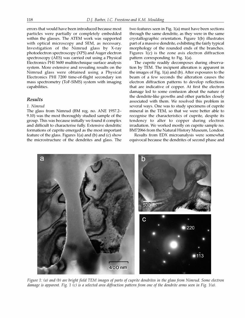
1. NimrudThe glass from Nimrud (BM reg. no. ANE 1957.2–9.10) was the most thoroughly studied sample of thegroup. This was because initially we found it complexand difficult to characterise fully. Extensive dendriticformations of cuprite emerged as the most importantfeature of the glass. Figures 1(a) and (b) and (c) showthe microstructure of the dendrites and glass. The
two features seen in Fig. 1(a) must have been sectionsthrough the same dendrite, as they were in the samecrystallographic orientation. Figure 1(b) illustratespart of a massive dendrite, exhibiting the fairly typicalmorphology of the rounded ends of the branches.Figures 1(c) is the zone axis electron diffractionpattern corresponding to Fig. 1(a).
The cuprite readily decomposes during observa-tion by TEM. The incipient alteration is apparent inthe images of Fig. 1(a) and (b). After exposures to thebeam of a few seconds the alteration causes theelectron diffraction patterns to develop reflectionsthat are indicative of copper. At first the electrondamage led to some confusion about the nature ofthe dendrite-like growths and other particles closelyassociated with them. We resolved this problem inseveral ways. One was to study specimens of cupritemineral in the TEM, so that we were better able torecognise the characteristics of cuprite, despite itstendency to alter to copper during electronirradiation. We worked mostly on cuprite sample no.BM72066 from the Natural History Museum, London.
Results from EDX microanalysis were somewhatequivocal because the dendrites of second phase and
Figure 1: (a) and (b) are bright field TEM images of parts of cuprite dendrites in the glass from Nimrud. Some electrondamage is apparent. Fig. 1 (c) is a selected area diffraction pattern from one of the dendrite arms seen in Fig. 1(a).
Ancient copper red glasses 119
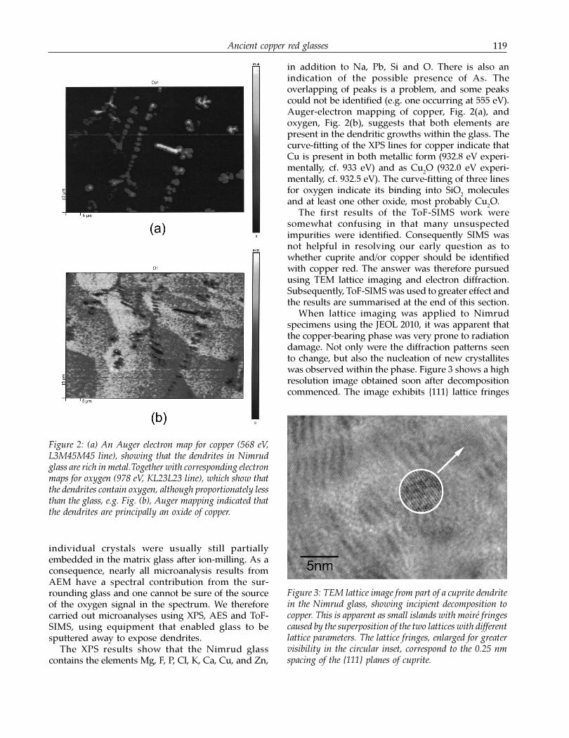
individual crystals were usually still partiallyembedded in the matrix glass after ion-milling. As aconsequence, nearly all microanalysis results fromAEM have a spectral contribution from the sur-rounding glass and one cannot be sure of the sourceof the oxygen signal in the spectrum. We thereforecarried out microanalyses using XPS, AES and ToF-SIMS, using equipment that enabled glass to besputtered away to expose dendrites.
The XPS results show that the Nimrud glasscontains the elements Mg, F, P, Cl, K, Ca, Cu, and Zn,
Figure 2: (a) An Auger electron map for copper (568 eV,L3M45M45 line), showing that the dendrites in Nimrudglass are rich in metal.Together with corresponding electronmaps for oxygen (978 eV, KL23L23 line), which show thatthe dendrites contain oxygen, although proportionately lessthan the glass, e.g. Fig. (b), Auger mapping indicated thatthe dendrites are principally an oxide of copper.
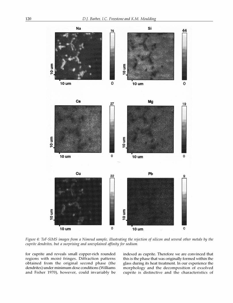
Figure 3: TEM lattice image from part of a cuprite dendritein the Nimrud glass, showing incipient decomposition tocopper. This is apparent as small islands with moiré fringescaused by the superposition of the two lattices with differentlattice parameters. The lattice fringes, enlarged for greatervisibility in the circular inset, correspond to the 0.25 nmspacing of the {111} planes of cuprite.
in addition to Na, Pb, Si and O. There is also anindication of the possible presence of As. Theoverlapping of peaks is a problem, and some peakscould not be identified (e.g. one occurring at 555 eV).Auger-electron mapping of copper, Fig. 2(a), andoxygen, Fig. 2(b), suggests that both elements arepresent in the dendritic growths within the glass. Thecurve-fitting of the XPS lines for copper indicate thatCu is present in both metallic form (932.8 eV experi-mentally, cf. 933 eV) and as Cu
2O (932.0 eV experi-
mentally, cf. 932.5 eV). The curve-fitting of three linesfor oxygen indicate its binding into SiO
2 molecules
and at least one other oxide, most probably Cu2O.
The first results of the ToF-SIMS work weresomewhat confusing in that many unsuspectedimpurities were identified. Consequently SIMS wasnot helpful in resolving our early question as towhether cuprite and/or copper should be identifiedwith copper red. The answer was therefore pursuedusing TEM lattice imaging and electron diffraction.Subsequently, ToF-SIMS was used to greater effect andthe results are summarised at the end of this section.
When lattice imaging was applied to Nimrudspecimens using the JEOL 2010, it was apparent thatthe copper-bearing phase was very prone to radiationdamage. Not only were the diffraction patterns seento change, but also the nucleation of new crystalliteswas observed within the phase. Figure 3 shows a highresolution image obtained soon after decompositioncommenced. The image exhibits {111} lattice fringes
D.J. Barber, I.C. Freestone and K.M. Moulding120
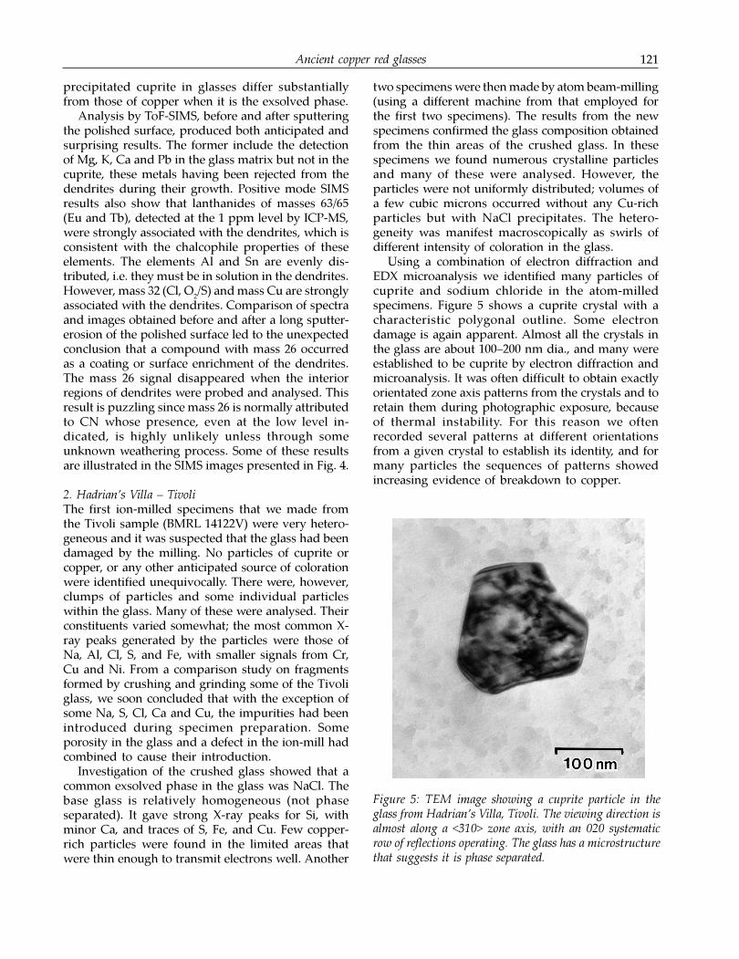
for cuprite and reveals small copper-rich roundedregions with moiré fringes. Diffraction patternsobtained from the original second phase (thedendrites) under minimum dose conditions (Williamsand Fisher 1970), however, could invariably be
indexed as cuprite. Therefore we are convinced thatthis is the phase that was originally formed within theglass during its heat treatment. In our experience themorphology and the decomposition of exsolvedcuprite is distinctive and the characteristics of
Figure 4: ToF-SIMS images from a Nimrud sample, illustrating the rejection of silicon and several other metals by thecuprite dendrites, but a surprising and unexplained affinity for sodium.
Ancient copper red glasses 121
precipitated cuprite in glasses differ substantiallyfrom those of copper when it is the exsolved phase.
Analysis by ToF-SIMS, before and after sputteringthe polished surface, produced both anticipated andsurprising results. The former include the detectionof Mg, K, Ca and Pb in the glass matrix but not in thecuprite, these metals having been rejected from thedendrites during their growth. Positive mode SIMSresults also show that lanthanides of masses 63/65(Eu and Tb), detected at the 1 ppm level by ICP-MS,were strongly associated with the dendrites, which isconsistent with the chalcophile properties of theseelements. The elements Al and Sn are evenly dis-tributed, i.e. they must be in solution in the dendrites.However, mass 32 (Cl, O
2/S) and mass Cu are strongly
associated with the dendrites. Comparison of spectraand images obtained before and after a long sputter-erosion of the polished surface led to the unexpectedconclusion that a compound with mass 26 occurredas a coating or surface enrichment of the dendrites.The mass 26 signal disappeared when the interiorregions of dendrites were probed and analysed. Thisresult is puzzling since mass 26 is normally attributedto CN whose presence, even at the low level in-dicated, is highly unlikely unless through someunknown weathering process. Some of these resultsare illustrated in the SIMS images presented in Fig. 4.
2. Hadrian’s Villa – TivoliThe first ion-milled specimens that we made fromthe Tivoli sample (BMRL 14122V) were very hetero-geneous and it was suspected that the glass had beendamaged by the milling. No particles of cuprite orcopper, or any other anticipated source of colorationwere identified unequivocally. There were, however,clumps of particles and some individual particleswithin the glass. Many of these were analysed. Theirconstituents varied somewhat; the most common X-ray peaks generated by the particles were those ofNa, Al, Cl, S, and Fe, with smaller signals from Cr,Cu and Ni. From a comparison study on fragmentsformed by crushing and grinding some of the Tivoliglass, we soon concluded that with the exception ofsome Na, S, Cl, Ca and Cu, the impurities had beenintroduced during specimen preparation. Someporosity in the glass and a defect in the ion-mill hadcombined to cause their introduction.
Investigation of the crushed glass showed that acommon exsolved phase in the glass was NaCl. Thebase glass is relatively homogeneous (not phaseseparated). It gave strong X-ray peaks for Si, withminor Ca, and traces of S, Fe, and Cu. Few copper-rich particles were found in the limited areas thatwere thin enough to transmit electrons well. Another
two specimens were then made by atom beam-milling(using a different machine from that employed forthe first two specimens). The results from the newspecimens confirmed the glass composition obtainedfrom the thin areas of the crushed glass. In thesespecimens we found numerous crystalline particlesand many of these were analysed. However, theparticles were not uniformly distributed; volumes ofa few cubic microns occurred without any Cu-richparticles but with NaCl precipitates. The hetero-geneity was manifest macroscopically as swirls ofdifferent intensity of coloration in the glass.
Using a combination of electron diffraction andEDX microanalysis we identified many particles ofcuprite and sodium chloride in the atom-milledspecimens. Figure 5 shows a cuprite crystal with acharacteristic polygonal outline. Some electrondamage is again apparent. Almost all the crystals inthe glass are about 100–200 nm dia., and many wereestablished to be cuprite by electron diffraction andmicroanalysis. It was often difficult to obtain exactlyorientated zone axis patterns from the crystals and toretain them during photographic exposure, becauseof thermal instability. For this reason we oftenrecorded several patterns at different orientationsfrom a given crystal to establish its identity, and formany particles the sequences of patterns showedincreasing evidence of breakdown to copper.
Figure 5: TEM image showing a cuprite particle in theglass from Hadrian’s Villa, Tivoli. The viewing direction isalmost along a <310> zone axis, with an 020 systematicrow of reflections operating. The glass has a microstructurethat suggests it is phase separated.
D.J. Barber, I.C. Freestone and K.M. Moulding122
A few of the cuprite precipitates were considerablylarger than those seen in Fig. 5, ranging up to 0.5 μmdia. Most particles were polyhedral in form and theywere invariably single crystals. We suspect that someof the cuprite crystals may have been partiallyconverted to copper in situ, prior to any exposure toelectron irradiation. A low concentration of sulphurwas invariably associated with the cuprite. The glassfrom Tivoli lacked any extensive or massive cupritedendrites, which were prevalent in the Nimrud glass.The differences in the occurrences of cuprite inNimrud and Tivoli glasses are attributed to the largedifferences in the concentrations of copper oxide andlead oxide (Table 1). Halite crystals were easy torecognize because of their tendency to decomposevery rapidly under the action of the electron beam,leaving geometrically-shaped voids in the glass,unless care was taken.
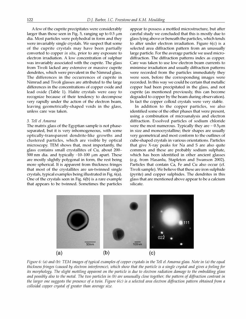
3. Tell el AmarnaThe matrix glass of the Egyptian sample is not phase-separated, but it is very inhomogeneous, with someoptically-transparent dendrite-like growths andclustered particles, which are visible by opticalmicroscopy. TEM shows that, most importantly, theglass contains small crystallites of Cu, about 200–500 nm dia. and typically ~10–100 μm apart. Theseare mostly slightly polygonal in form, the rest beingmore spherical. It is apparent from thickness fringesthat most of the crystallites are un-twinned singlecrystals, typical examples being illustrated in Fig. 6(a).One of the crystals seen in Fig. 6(b) is a rare examplethat appears to be twinned. Sometimes the particles
appear to possess a mottled microstructure, but aftercareful study we concluded that this is mostly due toglass lying above or beneath the particles, which tendsto alter under electron irradiation. Figure 6(c) is aselected area diffraction pattern from an unusuallylarge particle. For the average particle we used micro-diffraction. The diffraction patterns index as copper.Care was taken to use low electron beam currents tominimise irradiation and usually diffraction patternswere recorded from the particles immediately theywere seen, before the corresponding images wererecorded. In this way we could be certain that metalliccopper had been precipitated in the glass, and notcuprite (as mentioned previously, this can becomedegraded to copper by the beam during observation).In fact the copper colloid crystals were very stable.
In addition to the copper particles, we alsoidentified some of the other phases that were present,using a combination of microanalysis and electrondiffraction. Exsolved particles of sodium chloridewere the most numerous. Typically they are ~ 0.5μmin size and monocrystalline; their shapes are usuallyvery geometrical and most conform to the outlines ofcube-shaped crystals in various orientations. Particlesthat give X-ray peaks for Na and S are also quitecommon and these are probably sodium sulphate,which has been identified in other ancient glasses(e.g. from Hasanlu, Stapleton and Swanson 2002).Particles that contain Ca, Fe and Cu also occur (cf.Tivoli sample). We believe that these are iron sulphide(pyrite) and copper sulphides. The dendrites in thisglass that are mentioned above appear to be a calciumsilicate.
Figure 6: (a) and (b): TEM images of typical examples of copper crystals in the Tell el Amarna glass. Note in (a) the equalthickness fringes (caused by electron interference), which show that the particle is a single crystal and gives a feeling forits morphology. The slight mottling apparent on the particle is due to electron radiation damage to the embedding glassand possibly also to the metal. The two particles in (b) are unusually close together; the pattern of diffraction contrast inthe larger one suggests the presence of a twin. Figure 6(c) is a selected area electron diffraction pattern obtained from acolloidal copper crystal of greater than average size.
Ancient copper red glasses 123
4. ParthenonThe matrix glass from the Parthenon church is notsingle phase but is phase-separated (presumably dueto metastable liquid-liquid immiscibility). We havenot studied this effect in any detail.
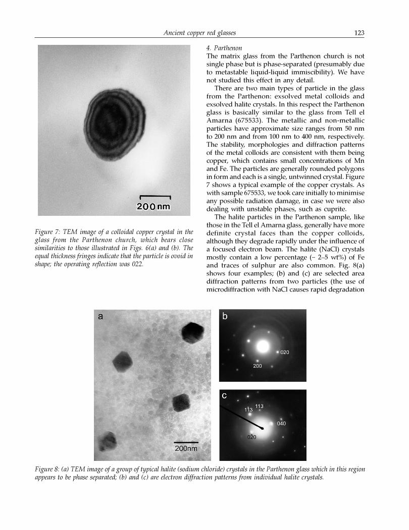
There are two main types of particle in the glassfrom the Parthenon: exsolved metal colloids andexsolved halite crystals. In this respect the Parthenonglass is basically similar to the glass from Tell elAmarna (675533). The metallic and non-metallicparticles have approximate size ranges from 50 nmto 200 nm and from 100 nm to 400 nm, respectively.The stability, morphologies and diffraction patternsof the metal colloids are consistent with them beingcopper, which contains small concentrations of Mnand Fe. The particles are generally rounded polygonsin form and each is a single, untwinned crystal. Figure7 shows a typical example of the copper crystals. Aswith sample 675533, we took care initially to minimiseany possible radiation damage, in case we were alsodealing with unstable phases, such as cuprite.
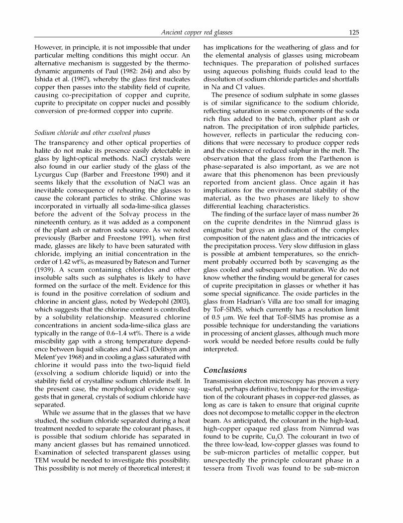
The halite particles in the Parthenon sample, likethose in the Tell el Amarna glass, generally have moredefinite crystal faces than the copper colloids,although they degrade rapidly under the influence ofa focused electron beam. The halite (NaCl) crystalsmostly contain a low percentage (~ 2–5 wt%) of Feand traces of sulphur are also common. Fig. 8(a)shows four examples; (b) and (c) are selected areadiffraction patterns from two particles (the use ofmicrodiffraction with NaCl causes rapid degradation
Figure 7: TEM image of a colloidal copper crystal in theglass from the Parthenon church, which bears closesimilarities to those illustrated in Figs. 6(a) and (b). Theequal thickness fringes indicate that the particle is ovoid inshape; the operating reflection was 022.
Figure 8: (a) TEM image of a group of typical halite (sodium chloride) crystals in the Parthenon glass which in this regionappears to be phase separated; (b) and (c) are electron diffraction patterns from individual halite crystals.

D.J. Barber, I.C. Freestone and K.M. Moulding124
and loss of sodium). We also found individual crystalsthat appear to be iron sulphide (probably pyrite) orpossibly sulphate and some more massive K-feldspar.
Discussion
Origins of Colour
The four glass samples that we have studied all havered coloration, but two contain particles of metalliccopper while the other two contain principallycuprite. Many exsolved NaCl (halite) particles alsooccur in the glasses, along with some other minorimpurity phases, but none of these phases are thoughtto contribute significantly to the coloration.
The average distances between particles in thevarious samples can only be estimated roughly byTEM because no information about the third dimen-sion is obtained. The average distance betweenparticles in the essentially two-dimensional speci-mens of the Parthenon and Tell el Amarna glassestypically appears to be between 1 and 10 μm.However, the probability of the specimen planepassing through the small particles is low and theactual average spatial separation of the particles inthese glasses is probably of the order of 500 nm.Cuprite crystals in Tivoli are typically separated by afew microns. The cuprite precipitates in Nimrudcannot be characterized in the same ways as themetallic-copper bearing glasses because of theirdendritic morphologies.
The samples containing copper nano-sizedparticles owe their colour to the relative contributionsof the incident light scattered and absorbed by thecopper particles, the principles of which are dis-cussed for example by Bamford (1977). The mechan-ism of coloration due to cuprite is likely to differgreatly from that of copper, at least in the Nimrudglass. Cuprite crystals in Nimrud are too large to beeffective scatterers of light. Here, the coloration isdue to the intrinsic colour (i.e. the absorptionspectrum) of cuprite, which is a skeletal-type micro-structure embedded in the colourless glass matrix.
It is surprising that the copper-rich nano-sizedparticles in the mosaic tessera from Tivoli are mainlyof cuprite. It is unclear that colloidal cuprite andcopper should yield the same colour. Furthermore,there are indications from observations on earlyglasses that fine cuprite particles impart a moreyellow or orange colour to the glass (e.g. Cable andSmedley 1987). It may be that the red colour of thisglass is due to contributions from both the cupriteand the subordinate copper particles.
Comparing our results with those of Brun et al.(1991) there is a level of consistency, in that all threeglasses that they studied containing sub-micrometreparticles appear to have been coloured by metalliccopper, while two of the three glasses of this typethat we studied were similar. However, precautionstaken to prevent the decomposition of cuprite in theelectron beam allowed us to identify commonparticles of cuprite in the glass from Tivoli. In orderto understand this, it is necessary to consider themechanisms of precipitation of copper and cupritein the glasses.
Metallic copper is essentially insoluble in silicatemelts and glasses. It was dissolved in the glass ascopper oxide and precipitation was induced, forexample, by heat treatment and/or adding carbon oriron, which reduced the oxide to metal. In this way,the formation of copper particles could be inducedin glasses that contain one percent or less of copper.Indeed, excessive concentrations of copper would nothave been helpful using such an approach, becauseparticles would have grown too large, spoiling thecolour. In contrast, as an oxide, Cu
2O is significantly
more soluble. In order to induce the precipitation ofcuprite, the glass melt was usually more-or-lesssaturated with high concentrations of Cu
2O, in the
range 5–10%. The benefits of lead oxide are several,but it appears that this component shifts the CuO/Cu
2O equilibrium towards Cu
2O thus increasing the
tendency of cuprite to crystallise (Edwards et al. 1977;see also Ahmed and Ashour 1984). Furthermore,according to the experiments of Williams (1914) onruby glasses and by Ahmed and Ashour (1974) oncuprite reds, lead improves the rates of nucleationand growth.
Thus there exists in the behaviour of the glass meltsan explanation for the compositional and micro-structural contrasts between the two fundamentaltypes of opaque red glass. Low-lead, low-copperglasses do not necessarily need lead, and require lowlevels of copper as they are generally coloured bycopper metal nano-particles that are exsolved due tothe reduction of the melt. High-lead, high-copperglasses require high copper oxide contents and highlead contents to promote the formation of cuprite;with too little copper, cuprite would be unlikely toform, while with too little lead, it would not form thedendritic structures which produce the brightest reds.
The glass from Tivoli is apparently an exception,as it is a low-copper, low-lead glass which pre-cipitated nano-sized particles of cuprite, with sub-ordinate copper. It is surprising that the 3% copperoxide in this glass was sufficient to cause cuprite tonucleate, in the presence of only 6.4% PbO (Table 1).
Ancient copper red glasses 125
However, in principle, it is not impossible that underparticular melting conditions this might occur. Analternative mechanism is suggested by the thermo-dynamic arguments of Paul (1982: 264) and also byIshida et al. (1987), whereby the glass first nucleatescopper then passes into the stability field of cuprite,causing co-precipitation of copper and cuprite,cuprite to precipitate on copper nuclei and possiblyconversion of pre-formed copper into cuprite.
Sodium chloride and other exsolved phases
The transparency and other optical properties ofhalite do not make its presence easily detectable inglass by light-optical methods. NaCl crystals werealso found in our earlier study of the glass of theLycurgus Cup (Barber and Freestone 1990) and itseems likely that the exsolution of NaCl was aninevitable consequence of reheating the glasses tocause the colorant particles to strike. Chlorine wasincorporated in virtually all soda-lime-silica glassesbefore the advent of the Solvay process in thenineteenth century, as it was added as a componentof the plant ash or natron soda source. As we notedpreviously (Barber and Freestone 1991), when firstmade, glasses are likely to have been saturated withchloride, implying an initial concentration in theorder of 1.42 wt%, as measured by Bateson and Turner(1939). A scum containing chlorides and otherinsoluble salts such as sulphates is likely to haveformed on the surface of the melt. Evidence for thisis found in the positive correlation of sodium andchlorine in ancient glass, noted by Wedepohl (2003),which suggests that the chlorine content is controlledby a solubility relationship. Measured chlorineconcentrations in ancient soda-lime-silica glass aretypically in the range of 0.6–1.4 wt%. There is a widemiscibility gap with a strong temperature depend-ence between liquid silicates and NaCl (Delitsyn andMelent’yev 1968) and in cooling a glass saturated withchlorine it would pass into the two-liquid field(exsolving a sodium chloride liquid) or into thestability field of crystalline sodium chloride itself. Inthe present case, the morphological evidence sug-gests that in general, crystals of sodium chloride haveseparated.
While we assume that in the glasses that we havestudied, the sodium chloride separated during a heattreatment needed to separate the colourant phases, itis possible that sodium chloride has separated inmany ancient glasses but has remained unnoticed.Examination of selected transparent glasses usingTEM would be needed to investigate this possibility.This possibility is not merely of theoretical interest; it
has implications for the weathering of glass and forthe elemental analysis of glasses using microbeamtechniques. The preparation of polished surfacesusing aqueous polishing fluids could lead to thedissolution of sodium chloride particles and shortfallsin Na and Cl values.
The presence of sodium sulphate in some glassesis of similar significance to the sodium chloride,reflecting saturation in some components of the sodarich flux added to the batch, either plant ash ornatron. The precipitation of iron sulphide particles,however, reflects in particular the reducing con-ditions that were necessary to produce copper redsand the existence of reduced sulphur in the melt. Theobservation that the glass from the Parthenon isphase-separated is also important, as we are notaware that this phenomenon has been previouslyreported from ancient glass. Once again it hasimplications for the environmental stability of thematerial, as the two phases are likely to showdifferential leaching characteristics.
The finding of the surface layer of mass number 26on the cuprite dendrites in the Nimrud glass isenigmatic but gives an indication of the complexcomposition of the natent glass and the intricacies ofthe precipitation process. Very slow diffusion in glassis possible at ambient temperatures, so the enrich-ment probably occurred both by scavenging as theglass cooled and subsequent maturation. We do notknow whether the finding would be general for casesof cuprite precipitation in glasses or whether it hassome special significance. The oxide particles in theglass from Hadrian’s Villa are too small for imagingby ToF-SIMS, which currently has a resolution limitof 0.5 μm. We feel that ToF-SIMS has promise as apossible technique for understanding the variationsin processing of ancient glasses, although much morework would be needed before results could be fullyinterpreted.
Conclusions
Transmission electron microscopy has proven a veryuseful, perhaps definitive, technique for the investiga-tion of the colourant phases in copper-red glasses, aslong as care is taken to ensure that original cupritedoes not decompose to metallic copper in the electronbeam. As anticipated, the colourant in the high-lead,high-copper opaque red glass from Nimrud wasfound to be cuprite, Cu
2O. The colourant in two of
the three low-lead, low-copper glasses was found tobe sub-micron particles of metallic copper, butunexpectedly the principle colourant phase in atessera from Tivoli was found to be sub-micron
D.J. Barber, I.C. Freestone and K.M. Moulding126
particles of cuprite. However, this is consistent withprevious reports of the coexistence of copper andcuprite in this type of glass, based upon X-raydiffraction of bulk samples (Brill and Cahill 1988).
The occurrence of the two phases is explained bythe existence of two distinct approaches to theproduction of opaque red glass. One approachinvolved the addition of high levels of lead andcopper to a base glass, which favoured the nucleationand growth of cuprite, often producing largedendritic crystals. The other involved the presence ofrelatively low quantities of copper in the base glass,with or without lead, and depended upon theaddition of reducing agents such as iron or copper tothe melt to precipitate sub-micron droplets of metalliccopper. The glass from Tivoli shows characteristicsof the latter group in terms of its very low lead, lowcopper and the sub-micron size of the colourantparticles, but the colourant is predominantly cuprite.This is either because the conditions inside the glasspassed into the cuprite field, after metallic copperhad nucleated, or because the glass nucleated cupritedirectly. Some metallic copper appears to have beenoriginally present but we cannot confidently estimatethe original proportion of copper particles becauseof problems with the decomposition of cuprite in theelectron beam. However, it is likely that some of thesubordinate metallic copper in this glass contributesto the colour.
The colour generation mechanism in early opaquered glass is thus complex. However, it appears thatby the Roman period, the two distinct approaches,high-lead high-copper and low-lead low-copper, hadbecome established and intermediate compositionswere relatively uncommon (e.g. Freestone 1987). Theanalysis of the colourant phases has allowed us toappreciate the underlying technical reasons for thesedistinctive technological traditions.
The apparent ubiquity of halite particles in theglasses that we have so far studied leads us toquestion if this is a phenomenon limited to glasseswhich have been heat-treated, to promote the growthof colourant particles, or if it is a feature of all ancientglasses. If the former, it has potential as an indicatorof heat-treatment. On the other hand, the occurrenceof NaCl particles in all early soda-lime-silica glasseswould be a potential source of error for analyticaltechniques that depend upon the production of asurface prepared using aqueous polishing media.
Acknowledgements
We thank Dr. A.M. Clark of the Natural HistoryMuseum, London, for supplying us with specimens
of mineral cuprite. We are grateful for access tosurface science equipment in the Materials Character-isation and Preparation Facility (MCPF) at the HongKong University of Science and Technology. We aregreatly indebted to Mr. T. Smith of the MCPF forproviding the XPS, AES and ToF-SIMS results. DavidBuckton, formerly of the Department of Medieval andLater Antiquities, Jeffrey Spencer, Department ofAncient Egypt and Sudan and Mavis Bimson, Depart-ment of Conservation and Science, The BritishMuseum, kindly made archaeological samples avail-able for analysis.
Notes1 Physics Centre and Department of Biological Sciences,
University of Essex, Colchester CO4 3SQ; Wolfson Centrefor Materials Processing, Brunel University, Kingston Lane,Uxbridge, Middlesex UB8 3PH, UK
2 Cardiff School of History and Archaeology, CardiffUniversity, Colum Drive, Cardiff CF10 3EU, UK
3 Raith Asia Ltd., 1103 Two Chinachem Exchange Square,338 King’s Road, North Point, Hong Kong
ReferencesAhmed, A.A., and Ashour, G.M., 1974, The effect of glass composition
on the crystallisation of copper and cuprous oxide in glass,Proceedings of the 10th International Congress on Glass 3, Kyoto, 34–8.
Ahmed, A.A., and Ashour, G.M., 1984, Effect of melting conditionson the crystallisation of cuprous oxide and copper in glass,Proceedings of the 11th International Congress on Glass 2, Prague,177–87.
Bamford, C.R., 1977, Colour generation and control in glass, glass science
and technology, Elsevier, Amsterdam.Bannerjee, S., and Paul, A., 1974, Thermodynamics of the system Cu-
O and ruby formation in borate glass, Journal of the American Ceramic
Society 57, 286–90.Barber, D.J., and Freestone, I.C., 1990, An investigation of the origin
of the colour of the Lycurgus Cup by analytical transmissionelectron microscopy, Archaeometry 32, 33–45.
Bateson, H.M., and Turner, W.E.S., 1939, A note on the solubility ofsodium chloride in a soda-lime-silica glass, Journal of Society of Glass
Technology 23, 265–7.Bimson, M., and Freestone I.C., 1985, Scientific examination of opaque
red glass of the second and first millennia BC, in Catalogue of Western
Asiatic Glass in the British Museum Volume 1, (ed. D. Barag), 119–22,British Museum Press, London.
Brill, R.H., and Cahill, N.D., 1988, A red opaque glass from Sardis andsome thoughts on red opaques in general, Journal of Glass Studies
30, 16–27.Brun, N., Mazerolles, L., and Pernot, M., 1991, Microstructure of
opaque red glass containing copper, Journal of Materials Science
Letters 10, 1418–20.Cable, M., and Smedley, J., 1987, The replication of an opaque red
glass from Nimrud, in Early Vitreous Materials, (eds. M. Bimson andI.C. Freestone), 151–64, British Museum Occasional Paper 56,London.
Delitsyn, L.M., and Melent’yev, B.N., 1968, Coexistence of two liquidphases at high temperatures: the system sodium chloride-albite
Ancient copper red glasses 127
glass, Doklady Akademia Nauk SSSR, Earth Science sections 180, 208–10 (American Geological Institute).
Edwards, R.J., Paul, A., and Douglas, R.W., 1977, Spectroscopy andoxidation-reduction of iron and copper in Na2O-PbO-SiO2 glasses,Physics and Chemistry of Glasses 13, 131–4.
Freestone, I.C., 1987, Composition and microstructure of early opaquered glass, in Early Vitreous Materials, (eds. M. Bimson and I.C.Freestone), 173–91, British Museum Occasional Paper 56, London.
Freestone, I.C., and Barber, D.J., 1992, The development of the colourof Sacrificial Red glaze with special reference to a Qing dynastysaucer dish, in Chinese Copper Red Wares, (ed. R. Scott), 53–62,Percival David Foundation of Chinese Art, Monograph Series 3,School of Oriental and African Studies, University of London,London.
Freestone, I.C., Stapleton, C.P., and Rigby, V., 2003, The production ofred glass and enamel in the Late Iron Age, Roman and Byzantineperiods, in Through a Glass Brightly: Studies in Byzantine and medieval
Art and Archaeology presented to David Buckton, (ed. C. Entwistle),142–54, Oxbow Books, Oxford.
Freestone, I.C., Bowman, S.G.E., and Stapleton, C.P., forthcoming,Composition and origins of Byzantine and early medieval enamelglasses, in Catalogue of Medieval Enamels Vol. 1, (ed. D. Buckton),British Museum, London.
Harding, R.R., Hornytskyj, S., and Date, A.R., 1989, The compositionof an opaque red glass used by Fabergé, Journal of Gemmology 21,275–87.
Hughes, M.J., 1972, A technical study of opaque red glass of the IronAge in Britain, Proceedings of the Prehistoric Society 38, 98–107.
Ishida, S., Takeuchi, N., Hayashi, M., and Wakamatsu, M., 1987, Roleof Sn2+ in development of red colour during reheating of copperglass, Journal of Non-Crystalline Solids 95/96, 793–800.
Moorey, P.R.S., 1994, Ancient Mesopotamian Materials and Industries,Clarendon Press, Oxford.
Paul, A., 1982, The Chemistry of Glass, Chapman and Hall, London.Pettenkofer, M., 1857, Haematinon (an ancient red paste) and
adventurine glass, Abhandlungen der Bayerischen Akademie der
Wissenschaften – Mathematisch-Naturwissenschaftliche Klasse 1, 123.Ram, A., and Prasad, S.N., 1965, Principles underlying the production
of copper ruby glass, in Coloured Glass, 130–5, CzechoslovakScientific Society for the Silicate Industries, Prague.
Scott, R.E., 1992, Chinese Copper Red Wares, Percival David Foundationof Chinese Art, Monograph Series 3, School of Oriental and AfricanStudies, University of London, London.
Stapleton, C.P., and Swanson, S.E., 2002, Chemical analysis of glassartefacts from Iron Age levels at Hasanlu, northwestern Iran, Glass
Technology 43C, 151–7.Tite, M.S., 1992, The impact of electron microscopy on ceramic studies,
Proceedings of the British Academy 77, 111–31.Turner, W.E.S., 1954, Studies in ancient glass and glass making
processes. Part II. The composition, weathering characteristics andhistorical significance of some Assyrian glasses of the eighth tosixth centuries B.C. from Nimrud, Transactions of the Society of Glass
Technology 40, 277–300.Vandiver, P.B., 1982, Glass technology at the mid-second millennium
B.C. Hurrian site of Nuzi, Journal of Glass Studies 25, 239–47.Veblen, D.R., and Post, J.E., 1983, A TEM study of fibrous cuprite
(chalcotrichite): microstructures and growth mechanisms, American
Mineralogist 68, 790–803.Volf, M.B., 1984, Chemical Approach to Glass, Elsevier, Amsterdam.Wakamatsu, M., Takeuchi, N., Nagai, H., and Ishida, S., 1989, Chemical
states of copper and tin in copper glazes fired under variousatmospheres, Journal of the American Ceramic Society 72, 16–9.
Wedepohl, K.H., 2003, Glas in Antike und Mittelalter, E.Schweizerbart’sche Verlagsbuchhandlung, Stuttgart.
Weyl, W.A., 1951, Coloured Glasses, Society of Glass Technology,Sheffield.
Williams, A.E., 1914, Notes on the development of the ruby colour inglass, Transactions of the American Ceramic Society 16, 284–306.
Williams, R.C., and Fisher, H.W., 1970, Electron microscopy of tobaccomosaic virus under conditions of minimal beam exposure, Journal
of Molecular Biology 52, 121–3.Wood, N., 1992, The evolution of Chinese copper red, in Chinese
Copper Red Wares, (ed. R. Scott), 11–35, Percival David Foundationof Chinese Art, Monograph Series 3, School of Oriental and AfricanStudies, University of London, London.
Yusuke, M., 1992, The microstructure of Sacrificial Red glaze, inChinese Copper Red Wares, (ed. R. Scott), 63–75, Percival DavidFoundation of Chinese Art, Monograph Series 3, School of Orientaland African Studies, University of London, London.

Copyright © 2022 FDOKUMEN