Characteristics of airflow in a CT-based ovine lung: a numerical study
Upload
independentCategory
view
0download
0
RESEARCH ARTICLE Open Access
Quantitative assessment of microbicide-inducedinjury in the ovine vaginal epithelium usingconfocal microendoscopyGracie Vargas1,2*, Igor Patrikeev1, Jingna Wei1, Brent Bell1, Kathleen Vincent3,4, Nigel Bourne3 andMassoud Motamedi1,5
Abstract
Background: The development of safe topical microbicides that can preserve the integrity of cervicovaginal tractepithelial barrier is of great interest as this may minimize the potential for increased susceptibility to STI infections.High resolution imaging to assess epithelial integrity in a noninvasive manner could be a valuable tool forpreclinical testing of candidate topical agents.
Methods: A quantitative approach using confocal fluorescence microendoscopy (CFM) for assessment ofmicrobicide-induced injury to the vaginal epithelium was developed. Sheep were treated intravaginally with one offive agents in solution (PBS; 0.02% benzalkonium chloride (BZK); 0.2% BZK) or gel formulation (hydroxyethylcellulose (HEC); Gynol II nonoxynol-9 gel (N-9)). After 24 hours the vaginal tract was removed, labeled withpropidium iodide (PI), imaged, then fixed for histology. An automated image scoring algorithm was developed forquantitative assessment of injury and applied to the data set. Image-based findings were validated withhistological visual gradings that describe degree of injury and measurement of epithelial thickness.
Results: Distinct differences in PI staining were detected following BZK and N-9 treatment. Images from controlshad uniformly distributed nuclei with defined borders, while those after BZK or N-9 showed heavily stained anddisrupted nuclei, which increased in proportion to injury detected on histology. The confocal scoring systemrevealed statistically significant scores for each agent versus PBS controls with the exception of HEC and wereconsistent with histology scores of injury.
Conclusions: Confocal microendoscopy provides a sensitive, objective, and quantitative approach for non-invasiveassessment of vaginal epithelial integrity and could serve as a tool for real-time safety evaluation of emergingintravaginal topical agents.
Keywords: Benzalkonium chloride, Confocal endomicroscopy, Epithelial disruption, Imaging, Microbicides, Nonoxy-nol-9
BackgroundEffective preclinical microbicide safety testing is of greatimportance to insure that only the most promising can-didates are advanced to clinical trials. This is particularlyimportant in light of the fact that several clinical trialsof emerging microbicides have been terminated due tosafety concerns [1-3]. Several early microbicide
candidates were shown to increase susceptibility toinfection in vivo [4,5] and were associated with surfaceepithelial disruption and inflammation [4-6]. Addition-ally, undetected changes in the epithelial integrity belowthe detection threshold of traditional safety assessmenttechniques such as colposcopy may yet increase suscept-ibility to infection [2]. Tools which aid in noninvasivevisualization of the vaginal mucosa and effects of agentsat the microscopic level in real time could greatly bene-fit the microbicide field. Such capabilities could facilitatethe design of safe and effective topical agents.
* Correspondence: [email protected] for Biomedical Engineering, The University of Texas Medical Branch,Galveston, TX, USAFull list of author information is available at the end of the article
Vargas et al. BMC Infectious Diseases 2012, 12:48http://www.biomedcentral.com/1471-2334/12/48
© 2012 Vargas et al; licensee BioMed Central Ltd. This is an Open Access article distributed under the terms of the Creative CommonsAttribution License (http://creativecommons.org/licenses/by/2.0), which permits unrestricted use, distribution, and reproduction inany medium, provided the original work is properly cited.

A number of methods are currently used for assessingmicrobicide effects on the cervicovaginal tract. Colpo-scopy is easily implemented and allows repeated evalua-tion. Disadvantages are it only allows visual assessmentof the tissue surface albeit with magnification, colpo-scopy training requirements are intensive, and interpre-tation is subjective and only modestly quantitative.Other currently used methods to detect epithelialdamage and inflammation do not provide immediatefeedback regarding the effect of an agent. Histopathol-ogy requires biopsy and processing of tissue and resultsin days to weeks of delay. Thus, it is both cumbersomeand raises safety issues when used clinically in high-riskpopulations. Cytokine mapping following vaginal lavagerequires offline testing, but has the advantage of beingcompatible with other methods and can be repeated inlongitudinal studies.Recently, noninvasive imaging has been investigated
for the in vivo near microscopic visualization of thevaginal epithelium in small and large animal models.Optical coherence tomography (OCT), an imaging toolapplied to the mouse and ovine vaginal epithelium, pro-vides quantitative measurement of epithelial thicknesschanges at a resolution of approximately 10-15 μm andhas been used to detect microbicide-induced changes inepithelial thickness and morphology following topicalapplication of BZK to the ovine vaginal tract [7].Although OCT allows for imaging of the full vaginalmucosa and submucosa, the resolution does not allowfor evaluation of subcellular changes or cell viabilityassessment.CFM is a high resolution noninvasive imaging method
commercially available as an endoscopic system [8]. Ithas been used in a number of clinical cancer trials pri-marily in the gastrointestinal tract and lung in studiesusing systemic or topically applied fluorophores toreveal morphological or cytological abnormalities on theepithelial surface during real-time endoscopy [8-10]. Inthe colorectum, CFM detected architectural differencesbetween the normal crypt structure and neoplasia[11,12]. In the esophagus it is being investigated as anadjunct tool in the management of precancerous lesions[13,14]. Although CFM has not been used in clinicalstudies in the cervicovaginal tract it has been used inpreclinical studies in sheep to examine cellular morphol-ogy changes on the cervical surface following progester-one and estradiol hormonal treatment [15]. In thecurrent study, we investigate the ability of a fixed-planesurface imaging CFM to visualize and quantify epithelialinjury in the ovine cervicovaginal tract following treat-ment with solutions and gels commonly studied inmicrobicide research.
MethodsAnimal model and in vivo agent applicationStudies were approved by the Institutional Animal Careand Use Committee at the University of Texas MedicalBranch and conformed to the Guide for the Care andUse of Laboratory Animals. Virginal Rambouillet femalesheep (n = 17 weighing 25-35 kg) were sedated with 10mg/kg ketamine IM followed by IV administration of 10mg/kg ketamine and 0.1-0.2 mg/kg Diazepam to relaxthe vaginal sphincter. The animals were placed dorsalsupine on a V-tilt table and intubated for delivery of iso-flurane throughout microbicide treatment and imagingprocedures.The cervicovaginal tract was examined using a pedia-
tric Graves speculum and the animals were administered8 cc of one of the following test agents: PBS (n = 3),Gynol II, an over the counter spermicidal gel containing2% N-9 (n = 4), HEC regarded as a universal placebogel (n = 4) [16], 0.02% BZK solution (n = 3), or 0.2%BZK solution (n = 3). Test agents were coded to maskinvestigators to treatments, then were delivered intra-vaginally using a ball-tip catheter. After twenty-fourhours, the animal was sacrificed and the vaginal tractimmediately removed for imaging.
ImagingThe CFM used in these studies provided excitation at488 nm through a fiber bundle with fluorescence emis-sion collected in the spectral band of 505-700 nm (Cell-vizio Lab, Mauna Kea, France). The system lateralresolution is 3.3 μm and field of view is 600 × 600 μm.The 1.5 mm diameter endoscopic probe requires contactto obtain images of the surface in a plane parallel to thesurface (en face) and has an axial resolution of 15 μm. Asingle optically sectioned plane was obtained at the sur-face. Images were acquired at a rate of 12 frames persecond. Individual frames were extracted in TIFFformat.Each freshly excised vaginal tract was opened longi-
tudinally along the anterior wall and laid flat with themucosal surface exposed. The surface was rinsed withPBS and 20 μM propidium iodide (PI) applied evenly tothe surface using a bulb pipet. PI is a fluorescent dyethat binds to DNA and is not permeant to healthy cells.Thus damaged or dead cells were identified by fluores-cence from the cell nuclei. After 5 minutes the PI solu-tion was rinsed off using PBS. The CFM probe wasplaced directly on the vaginal surface and an image ser-ies collected site-by-site in a predetermined 5 × 5 matrixon the proximal vagina surface above the sphincter witha 1 cm separation between image sites. Following ima-ging, the entire tract was fixed in 10% formalin for 24
Vargas et al. BMC Infectious Diseases 2012, 12:48http://www.biomedcentral.com/1471-2334/12/48
Page 2 of 8

hours and then the imaged sites which had been markedwere biopsied (2 mm), paraffin embedded, sectioned,and stained with hemotoxylin and eosin.
Image analysisAn algorithm was developed to quantify the effects oftopical agents on epithelial surface integrity based onthe PI staining pattern. The algorithm incorporated fea-ture size information through calculation of the mean-to-median size of all individual objects within an image.The basis for such an approach is that in the nativeepithelium, the nuclear staining pattern is expected toconsist of intact nuclei of uniform size across the vagi-nal surface (these nuclei are considered the objects inthe algorithm). In the case where there is no variationin size of objects, the calculated median size and meansize of objects (nuclei) are equal, or very closely matchif there is a small variation in size. However, if theimage is comprised of features which vary significantlyin size, as might be expected if nuclear material aggre-gates following lysis, the median size of objects willincrease relative to the mean size.All analyses were performed with Matlab software ver-
sion R2006a (Mathworks, Natick, MA) using standardoperations of the image processing toolbox. A represen-tative image was selected for each of the 25 vaginal sitesper animal. Each raw image consisted of a range ofgrayscale pixel values with those belonging to the objectbright in comparison to the surrounding dark pixels.Images were thresholded using a predetermined globalthreshold based on unstained control samples (in thisprocess pixels with a value greater than the thresholdvalue were assigned as ‘object’ pixels while those withvalues smaller than the threshold value as dark or sub-threshold pixels). Binary images were created with pixelsassigned a value of 0 or 1 to identify sub-threshold andabove threshold (object) pixels, respectively. A Matlablabeling operation (bwlabel) was used to identify all indi-vidual objects within the image. This operation deter-mines all clusters of interconnected pixels with a valueof 1 and assigns each cluster (each object) a number.Using region statistics, the size (area) of each object wasthen determined. The median size of objects within theimage and the mean size was then determined and amean-to-median ratio calculated for the image. This wasperformed for all 25 image sites per animal, PBS treatedcontrols were used to determine a typical value formean-to-median ratio and a threshold value was setabove this to differentiate ‘normal’ from ‘abnormal’. Theaverage mean-to-median value in PBS controls wasfound to be 1.5 and 90% of images had a mean-to-med-ian ratio < 2.0. The threshold value differentiating nor-mal from abnormal value was set to 2.0. The number ofimages in each animal having a mean-to-median ratio >
2.0 was then enumerated and an average determinedper treatment group.
Histological assessmentPhotographs of H&E stained sections from biopsies ofthe 25 sampled regions were obtained using a colorCCD mounted on an IX71 Olympus inverted micro-scope. Photographs were randomized and graded by atrained grader familiar with the assessment of BZK-induced damage in sheep vaginal epithelium but maskedto the treatments in this study [7]. H&E sections withfully intact epithelium having a cornified stratified squa-mous epithelium of full thickness were given a score of0. Samples exhibiting partial denuding, such as a visuallythinned epithelium and evidence of inflammation deter-mined by the presence of leukocytes, were given a scoreof 1. Samples with fully denuded epithelium were givena score of 2. A score was given to each of the 25 biopsysites and used to determine the average score for eachanimal. These individual animal scores were then usedto generate mean scores for each treatment group.Epithelial thickness was measured at three predeter-
mined locations on photographs of an H&E sectionfrom each imaged site. A single average per animal andper treatment group was then determined.
Statistical analysisStatistical analysis of confocal data for comparisonbetween treatment groups was by single factor ANOVAfollowed by Tukey’s post hoc test. A value of p < 0.05was considered statistically significant. Single factorANOVA with Tukey’s post hoc test was also applied toepithelial thickness data. For statistical analysis of histol-ogy scores (an ordinal variable -0,1,2) the mixed modelwas used to test the treatment effects and perform mul-tiple comparisons. Tukey adjustment was used for thep-values in the multiple comparisons.
ResultsRepresentative CFM and histology images for each treat-ment are shown in Figure 1. The confocal images areshown in pseudocolor rather than grayscale for ease ofvisualization. The intensity is represented by a green-blue fire look-up table color scale (black represents zerointensity progressing to blue, green, yellow, white formaximum intensity as shown in the intensity scale ofFigure 1). Each representative layout shows confocalimages of 9 of 25 sampled regions (positions1,3,5,11,13,15,21,23,25 of the 5 × 5 image matrix) from asingle animal in the left column, a single larger confocalimage in the center column, and a corresponding H&Eimage in the right column. Confocal images from PBStreated animals showed uniformly-sized nuclei distribu-ted throughout the surface. Nuclei had well defined
Vargas et al. BMC Infectious Diseases 2012, 12:48http://www.biomedcentral.com/1471-2334/12/48
Page 3 of 8

borders. Fluorescent staining in the PBS controls isattributed to the cornified surface layer from which cellsare shed throughout the estrus cycle. Histology fromPBS treated sheep confirmed the presence of a cornifiedstratified squamous epithelium (Figure 1). The PI stain-ing pattern and histology following treatment with HECplacebo gel were similar to that in PBS controls (Figure1e f and 1g). In contrast, treatment with BZK (0.02% or0.2%) or N-9 resulted in a different staining pattern inconfocal images and altered epithelium in histology ascan be visualized in Figure 1 and shown quantitativelyin Figures 2 and 3. Following treatment with 0.02% BZKthere was an increase in the total area of staining, andalthough individual nuclei could still be identified, theywere more closely spaced and merged in areas likelydue to cell lysis (Figure 1h and 1i). Corresponding his-tology (Figure 1j) showed a thinned epithelium andinflammatory infiltrates. Treatment with 0.2% BZK pro-duced increased staining with disruption of the regularnuclear spacing and presence of nuclear aggregate/deb-ris in confocal micrographs (Figure 1k and 1l). Corre-sponding histology revealed extensive epithelial damagewith complete denuding in most samples (Figure 1m).Results following N-9 treatment were somewhat similarto 0.02% BZK in that there was increased staining andsome disruption to the regular distribution of nucleiacross the sampled regions (Figure 1n and 1o). Much ofthe epithelium became denuded as well (Figure 1p).Figure 2 shows the results from the mean-to-median
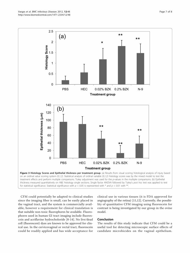
ratio analysis per treatment group. In PBS animals, theaverage mean-to-median value was found to be 1.5.Results in HEC treated animals were comparable. How-ever, N-9, 0.02% BZK, and 0.2% BZK, treatment allresulted in statistically significant changes. Correspond-ing histology scores and epithelial thicknesses are shownin the plots of Figure 3a and 3b. In both cases, statisti-cally significant differences were also detected betweenPBS and N-9, 0.02% BZK, and 0.2% BZK, but notbetween PBS and HEC.
DiscussionIn evaluating safety of candidate microbicides, there is aneed to identify indicators of epithelial damage whichmay signify increased susceptibility to cervicovaginalinfection with STI pathogens. There is evidence to sug-gest that disruption of the epithelial barrier is linked toincreased susceptibility [5]. However, susceptibility stu-dies are not practical for initial agent testing in largeanimal models due to the high cost of studies. Thus,direct imaging that provides feedback regarding epithe-lial integrity/disruption could be valuable in preclinicaltesting of candidate topical agents. In the current studyCFM provided real-time high resolution single-planevisualization of the vaginal surface epithelium and
utilizing an automated analysis algorithm developed forthis work, provided assessment of damage based ondegree of positive staining by a cell impermeant nucleicacid dye. To our knowledge, this is the first study toinvestigate this imaging modality for this purpose.Two agents were evaluated that have no known effect
on epithelial structure (PBS and HEC) and two agents(BZK and N-9) that have been shown previously toinduce cervicovaginal epithelial disruption in small andlarge animal models [5-7,17]. Because the use of 2%BZK has been shown to lead to immediate widespreadepithelial exfoliation and complete denuding of theepithelium [5,7], we chose to use lower dilutions of BZKat 0.02% and 0.2% for this study. For the N-9 gel wechose an over-the-counter formulation shown to pro-duce epithelial disruption [1,2]. CFM revealed differ-ences in the PI staining patterns of vaginal tissue treatedwith N-9 and BZK compared to PBS. While there was aregular baseline staining pattern of nuclei across thesurface in PBS controls (attributed to cornified cellsabout to be shed from the surface and permeant to PI),there was a clear increase in nuclear density with smalldegrees of damage (e.g. 0.02% BZK; Figure 1h and 1i)and evidence of exfoliated material that formed largeaggregated nuclear material in confocal micrographs (e.g. 0.2% BZK; Figure 1k and 1l). These findings of epithe-lial damage were confirmed by the presence of disruptedepithelium in H&E stained histology (Figure 1). It isinteresting that CFM detected changes with the subcli-nical concentration of BZK (0.02%), particularly giventhat in a recent study changes in the ovine epitheliumdue to 0.02% BZK were not detected by colposcopy [7].Both CFM and histology from animals treated withHEC were comparable to those of PBS treated animals.HEC gel is recognized as a nontoxic topical agent and iswidely used as a universal placebo in microbicide safetystudies [16]. Repeated dosing with HEC for seven daysresulted in no discernable difference from untreatedcontrols by histology in mice [18].A critical aspect of this study was the development of
an algorithm based on a mean-to-median object sizeanalysis to measure epithelial damage in confocal micro-graphs. Observation of confocal micrographs in whichepithelial damage occurred as confirmed by histologyrevealed the increased presence of aggregated materialor nuclei that could not be clearly differentiated fromneighboring nuclei. Preliminary tests (not shown) indi-cated that although visually there was an increase instaining pattern with damage, the degree of damage didnot necessarily relate with percent area of staining. Thuswe chose a metric that would take into account the factthat images of epithelium went from uniformly sizedobjects (single nuclei) to larger objects when degree ofstaining increased and individual nuclei could not be
Vargas et al. BMC Infectious Diseases 2012, 12:48http://www.biomedcentral.com/1471-2334/12/48
Page 4 of 8

Figure 1 Representative confocal microendoscopy and histology images. For each treatment group, a 3 × 3 layout of 9 confocal imagesrepresenting positions 1,3,5,11,13,15,21,23,25 of the sampled 5 × 5 image matrix from a single animal is shown (left column), followed by asingle larger confocal image (center column), and corresponding H&E section (right column). (a) 5 × 5 matrix representing the positions of the25 imaged sites per animal. (b-d) representative PBS case; (e-g) representative HEC treated case, (h-j) representative 0.02% BZK treated case; (k-m)representative 0.2% BZK treated case; (n-p) representative N-9 treated case. Confocal images are shown in pseudocolor for ease of visualizationwith the intensity represented by the shown color (black represents a zero intensity value, progressing to yellow for maximum intensity). Scalebar 100 μm.
Vargas et al. BMC Infectious Diseases 2012, 12:48http://www.biomedcentral.com/1471-2334/12/48
Page 5 of 8

readily separated. The quantitative algorithm based onthe mean-to-median object size assessment provideddiscriminatory information regarding level of damageinduced by the applied agents and results were consis-tent with histology findings, validating the ability of thistechnique to detect microbicide-induced changes inepithelial morphology. We chose to image ex vivo speci-mens to facilitate development of an automated algo-rithm for determination of damage to the epithelium.We intend to expand these studies for real time in vivoand longitudinal evaluation of microbicide toxicity inthe ovine model. It is expected that many more sitescan be assessed by CFM than is feasible to biopsy inchronic studies.Another advantage of CFM is that it can be coupled
with any number of fluorescent indicators of cellular ormolecular injury. It is noted that the ensuing analysis ofconfocal micrographs and assessment of damage is depen-dent upon the fluorophore used for image contrast andlocalization and staining patterns. PI stains nuclei ofdamaged cells. Other fluorescent dyes typically used withCFM are nonspecific in that they either stain all nuclei (e.g. acriflavine hydrochloride) or both intracellular andextracellular features (e.g. fluorescein) [8]. Assessment of
damage in these cases may be based on morphologychanges such as in a study by Bott et al., evaluating differ-ences in morphology of the ovine epithelium due to hor-monal treatment that used acriflavine orange for contrast[15]. An interesting possibility in preclinical studies maybe the use of molecular-specific fluorophores as is beingpursued in gastrointestinal imaging [19,20], that indicateinflammation or damage that does not result in cell lysis.Several biomarkers of inflammation related to microbicidesafety assessment are being investigated in vaginal mucosa[21,22] and could in the future be targeted with fluoro-phores for imaging with CFM. The ability of CFM todetect cellular/molecular indicators of damage will in partdepend on location of fluorescent signals. The system uti-lized in this study is restricted to signals at or near the sur-face (working distance from 0-80 μm depending on theimaging probe used) with a similar clinical version allow-ing maximal depth at 130 μm [8]; CFM with dynamicdepth-sectioning is also available providing optical sectionsto depths 250 μm using larger diameter imaging probes(12.8 mm), that are suitable for large animal and humanstudies, with the advantage of improved resolution [8] butthe challenge of determining the actual depth beingimaged when using a handheld probe.
**
20
25
regi
ons
out o
f 25 **
**
**
10
15
umbe
r of d
amag
ed
0
5
PBS HEC 0.02% BZK 0.2% BZK N-9
Nu
Treatment groupFigure 2 Number of damaged regions based on mean-to-median ratio. Images with a mean-to-median ratio were enumerated per animaland averaged within treatment groups. No statistical significance occurred between PBS and HEC cases. Statistically significant increase in thenumber of damaged regions occurred with 0.02% BZK, 0.2% BZK, and N-9. Results in the 0.02% BZK case were similar to N-9, and both werestatistically different from those at 0.2% BZK. Statistical analysis for comparison between treatment groups was by single factor ANOVA followedby Tukey’s post hoc test for significance. Statistical significance with p < 0.05 is represented with * and p <0.01 with **.
Vargas et al. BMC Infectious Diseases 2012, 12:48http://www.biomedcentral.com/1471-2334/12/48
Page 6 of 8
CFM could potentially be adapted to clinical studiessince the imaging fiber is small, can be easily placed inthe vaginal tract, and the system is commercially avail-able, however a requirement for clinical translation isthat suitable non-toxic fluorophores be available. Fluoro-phores used in human GI tract imaging include fluores-cein and acriflavine hydrocholoride [8-14]. No live/deadcell (fluorescent) dyes are known to be approved for clin-ical use. In the cervicovaginal or rectal tract, fluoresceincould be readily applied and has wide acceptance for
clinical use in various tissues (it is FDA approved forangiography of the retina) [11,12]. Currently, the possibi-lity of quantitative CFM imaging using fluorescein forcontrast is being investigated by our group in the ovinemodel.
ConclusionThe results of this study indicate that CFM could be auseful tool for detecting microscopic surface effects ofcandidate microbicides on the vaginal epithelium.
2.5
****(a)
1
1.5
2H
isto
logy
Sco
re*
0
0.5
PBS HEC 0.02% BZK 0.2% BZK N-9Treatment group
**
(b)
80
100
120
140
ness
(m
)
**
**
20
40
60
80
Epith
elia
l thi
ckn
0PBS HEC 0.02% BZK 0.2% BZK N-9
Treatment groupFigure 3 Histology Scores and Epithelial thickness per treatment group. (a) Results from visual scoring histological analysis of injury basedon an ordinal value scoring system (0,1,2). Statistical analysis of ordinal variable (0,1,2) histology scores was by the mixed model to test thetreatment effects and perform multiple comparisons. Tukey adjustment was used for the p-values in the multiple comparisons. (b) Epithelialthickness measured quantitatively on H&E histology single sections. Single factor ANOVA followed by Tukey’s post hoc test was applied to testfor statistical significance. Statistical significance with p < 0.05 is represented with * and p < 0.01 with **.
Vargas et al. BMC Infectious Diseases 2012, 12:48http://www.biomedcentral.com/1471-2334/12/48
Page 7 of 8

Agents in solution and gel formulations were tested suc-cessfully; gel formulations did not interfere with ima-ging. Results were consistent with those of histology andwith findings reported in the literature for each of theagents studied. Future efforts will include translation ofthe techniques into in vivo studies and evaluation of themethods for use with microbicide candidates that arebelieved to have minimal cytotoxic effects but maynonetheless alter the vaginal epithelium.
AcknowledgementsWe thank Dr. Alai Tan, Department of Statistics, University of Texas MedicalBranch for statistical consultation.Sources of financial support (includinggrant numbers): this work was supported in part by the NIAID(R21AI07606202/R33AI076062 and UC7AI070083) and R01CA127429. Reprints,proofs or correspondence. Mailing address: 301 University Blvd., Rt. 0456, TheUniversity of Texas Medical Branch, Galveston, TX 77555. Phone: (409)772-8363. Fax: (409) 772-0751. E-mail: http://[email protected].
Author details1Center for Biomedical Engineering, The University of Texas Medical Branch,Galveston, TX, USA. 2Department of Neuroscience and Cell Biology, TheUniversity of Texas Medical Branch, Galveston, TX, USA. 3Department ofPediatrics, The University of Texas Medical Branch, Galveston, TX, USA.4Department of Obstetrics and Gynecology, The University of Texas MedicalBranch, Galveston, TX, USA. 5Department of Opthalmology, The University ofTexas Medical Branch, Galveston, TX, USA.
Authors’ contributionsGV, study concept and design, acquisition of data, algorithmconceptualization with IP, analysis and interpretation of data, statistics,drafting of the manuscript. KV, study concept and design, coordination andacquisition of samples, interpretation of data, drafting of manuscript. MM,study concept and design, interpretation of data, provided expertise onimaging/optics, drafting of the manuscript. NB, study concept and design,analysis and interpretation of data, expertise on microbicides and epithelialinjury, drafting of the manuscript. IP, image processing and algorithmdevelopment, data analysis. JW and BBell, sample collection, acquisition ofdata, data analysis and scoring. All authors read and approved the finalmanuscript.
Competing interestsThe authors declare that they have no competing interests.
Received: 26 July 2011 Accepted: 29 February 2012Published: 29 February 2012
References1. Kreiss J, Ngugi E, Holmes K, Ndinya-Achola J, Waiyaki P, Roberts PL, et al:
Efficacy of nonoxynol 9 contraceptive sponge use in preventingheterosexual acquisition of HIV in Nairobi prostitutes. JAMA 1992,268(4):477-482.
2. Van Damme L, Ramjee G, Alary M, Vuylsteke B, Chandeying V, Rees H, et al:Effectiveness of COL-1492, a nonoxynol-9 vaginal gel, on HIV-1transmission in female sex workers: a randomised controlled trial. Lancet2002, 360(9338):971-977.
3. Ramjee G, Govinden R, Morar NS, Mbewu A: South Africa’s experience ofthe closure of the cellulose sulphate microbicide trial. PLoS Med 2007,4(7):e235.
4. Hillier SL, Moench T, Shattock R, Black R, Reichelderfer P, Veronese F: Invitro and in vivo: the story of nonoxynol 9. J Acquir Immune Defic Syndr2005, 39(1):1-8.
5. Cone RA, Hoen T, Wong X, Abusuwwa R, Anderson DJ, Moench TR: Vaginalmicrobicides: detecting toxicities in vivo that paradoxically increasepathogen transmission. BMC Infect Dis 2006, 6:90.
6. Catalone BJ, Kish-Catalone TM, Budgeon LR, Neely EB, Ferguson M,Krebs FC, et al: Mouse model of cervicovaginal toxicity and inflammation
for preclinical evaluation of topical vaginal microbicides. AntimicrobAgents Chemother 2004, 48(5):1837-1847.
7. Vincent KL, Bourne N, Bell BA, Vargas G, Tan A, Cowan D, et al: Highresolution imaging of epithelial injury in the sheep cervicovaginal tract:a promising model for testing safety of candidate microbicides. SexTransm Dis 2009, 36(5):312-318.
8. Neumann H, Kiesslich R, Wallace MB, Neurath MF: Confocal LaserEndomicroscopy: Technical Advances and Clinical Applications.Gastroenterology 2010, 139(2):388-392.
9. De Palma GD: Confocal laser endomicroscopy in the “in vivo” histologicaldiagnosis of the gastrointestinal tract. World J Gastroenterol 2009,15(46):5770-5775.
10. Thiberville L, Salaün M, Lachkar S, Dominique S, Moreno-Swirc S, Vever-Bizet C, et al: Confocal fluorescence endomicroscopy of the humanairways. Proc Am Thorac Soc 2009, 6(5):444-449.
11. Smith LA, Tiffin N, Thomson M, Cross SS, Hurlstone DP: Chromoscopicendomicroscopy: in vivo cellular resolution imaging of the colorectum. JGastroenterol Hepatol 2008, 23(7 Pt 1):1009-1023.
12. Hurlstone DP, Baraza W, Brown S, Thomson M, Tiffin N, Cross SS: In vivoreal-time confocal laser scanning endomicroscopic colonoscopy for thedetection and characterization of colorectal neoplasia. Br J Surg 2008,95(5):636-645.
13. Konda VJ, Chennat JS, Hart J, Waxman I: Confocal laser endomicroscopy:potential in the management of Barrett’s esophagus. Dis Esophagus 2010,23(5):E21-E31.
14. Wallace MB, Sharma P, Lightdale C, Wolfsen H, Coron E, Buchner A, et al:Preliminary accuracy and interobserver agreement for the detection ofintraepithelial neoplasia in Barrett’s esophagus with probe-basedconfocal laser endomicroscopy. Gastrointest Endosc 2010, 72(1):19-24.
15. Bott EM, Young IR, Jenkin G, McLaren WJ: Detection of morphologicalchanges of the ovine cervix in response to sex steroids using afluorescence confocal endomicroscope. Am J Obstet Gynecol 2006,194(1):105-112.
16. Tien D, Schnaare RL, Kang F, Cohl G, McCormick TJ, Moench TR, Doncel G,Watson K, Buckheit RW, Lewis MG, Schwartz J, Douville K, Romano JW: Invitro and in vivo characterization of a potential universal placebodesigned for use in vaginal microbicide clinical trials. AIDS Res HumRetroviruses 2005, 21(10):845-853.
17. Patton DL, Kidder GG, Sweeney YC, Rabe LK, Hillier SL: Effects of multipleapplications of benzalkonium chloride and nonoxynol 9 on the vaginalepithelium in the pigtailed macaque (Macaca nemestrina). Am J ObstetGynecol 1999, 180(5):1080-1087.
18. Galen BT, Martin AP, Hazrati E, Garin A, Guzman E, Wilson SS, et al: Acomprehensive murine model to evaluate topical vaginal microbicides:mucosal inflammation and susceptibility to genital herpes as surrogatemarkers of safety. J Infect Dis 2007, 195(9):1332-1339.
19. Goetz M, Wang TD: Molecular imaging in gastrointestinal endoscopy.Gastroenterology 2010, 138(3):828-833, e1.
20. Miller SJ, Joshi BP, Feng Y, Gaustad A, Fearon ER, Wang TD: In vivofluorescence-based endoscopic detection of colon dysplasia in themouse using a novel peptide probe. PLoS One 2011, 6(3):e17384.
21. Trifonova RT, Bajpai M, Pasicznyk JM, Chandra N, Doncel GF, Fichorova RN:Biomarkers of leukocyte traffic and activation in the vaginal mucosa.Biomarkers 2007, 12(6):608-622.
22. Cummins JE, Doncel GF: Biomarkers of cervicovaginal inflammation forthe assessment of microbicide safety. Sex Transm Dis 2009, 36(3 Suppl):S84-S91.
Pre-publication historyThe pre-publication history for this paper can be accessed here:http://www.biomedcentral.com/1471-2334/12/48/prepub
doi:10.1186/1471-2334-12-48Cite this article as: Vargas et al.: Quantitative assessment of microbicide-induced injury in the ovine vaginal epithelium using confocalmicroendoscopy. BMC Infectious Diseases 2012 12:48.
Vargas et al. BMC Infectious Diseases 2012, 12:48http://www.biomedcentral.com/1471-2334/12/48
Page 8 of 8
Copyright © 2022 FDOKUMEN