Quantitative analysis of low molecular weight polar compounds by continuous flow liquid secondary...
10
FOCUS:PROTEOMICS AND DISEASE Quantitative Analysis of the Low Molecular Weight Serum Proteome Using 18 O Stable Isotope Labeling in a Lung Tumor Xenograft Mouse Model Brian L. Hood, David A. Lucas, Grace Kim, King C. Chan, Josip Blonder, Haleem J. Issaq, Timothy D. Veenstra, and Thomas P. Conrads Laboratory of Proteomics and Analytical Technologies, National Cancer Institute at Frederick, SAIC-Frederick, Inc., Frederick, Maryland, USA Ingrid Pollet and Aly Karsan British Columbia Cancer Research Center, Vancouver, British Columbia, Canada With advancements in the analytical technologies and methodologies in proteomics, there is great interest in biomarker discovery in biofluids such as serum and plasma. Current hypotheses suggest that the low molecular weight (LMW) serum proteome possesses an archive of clipped and cleaved protein fragments that may provide insight into disease development. Though these biofluids represent attractive samples from which new and more accurate disease biomarkers may be found, the intrinsic person-to-person variability in these samples complicates their discovery. Mice are one of the most extensively used animal models for studying human disease because they represent a highly controllable experimental model system. In this study, the LMW serum proteome was compared between xenografted tumor-bearing mice and control mice by differential labeling utilizing trypsin-mediated incorporation of the stable isotope of oxygen, 18 O. The digestates were combined, fractionated by strong cation exchange chromatography, and analyzed by nanoflow reversed-phase liquid chromatography coupled online with tandem mass spectrometry, resulting in the identifica- tion of 6003 proteins identified by at least a single, fully tryptic peptide. Almost 1650 proteins were identified and quantitated by two or more fully tryptic peptides. The methodology adopted in this work provides the means for future quantitative measurements in comparative animal models of disease and in human disease cohorts. (J Am Soc Mass Spectrom 2005, 16, 1221–1230) © 2005 American Society for Mass Spectrometry B iomarker investigations are increasingly exploit- ing the developing technologies and methodolo- gies of proteomics with the hope of discovering better indicators of the onset or progression of diseases. Many investigations aim to discover biomarkers in various biofluids (i.e., serum, plasma, and cerebrospinal fluid) as these samples are easily obtainable, contain high concentrations of proteins, and perfuse key tissues, undoubtedly endowing these fluids with protein spe- cies indicative of diseased states [1– 4]. Hence, detection of changes in the abundance or characteristics of differ- ent proteins in biofluids may provide the means for disease detection, enabling earlier intervention in dis- ease progression. Much of the promise of proteomics for identification of biomarkers in complex biological fluids can be traced to seminal studies performed in John Yates’ laboratory. This laboratory was the first to develop a high-throughput method for large-scale proteome analysis, which com- bined multidimensional liquid chromatography, tandem mass spectrometry (MS/MS), and database searching by the SEQUEST algorithm [5]. This method became popular under the acronym MudPIT (i.e., multidimensional pro- tein identification technology). MudPIT, or variations thereof, has been used in countless studies to characterize complex proteome samples, with the typical goal of ob- taining as much possible information concerning the sam- ple of interest. This technology has ushered in an era Published online June 24, 2005 Address reprint requests to Dr. T. D. Veenstra, Laboratory of Proteomics and Analytical Technologies, National Cancer Institute at Frederick, SAIC- Frederick, Inc., P.O. Box B, Frederick, MD 21702, USA. E-mail: [email protected]: [email protected] or Dr. T. P. Conrads, Mass Spectrometry Center, Laboratory of Proteomics and Analytical Technologies, National Cancer Institute at Frederick, SAIC- Frederick, Inc., P.O. Box B, Frederick, MD 21702, USA. © 2005 American Society for Mass Spectrometry. Published by Elsevier Inc. Received November 24, 2004 1044-0305/05/$30.00 Revised February 2, 2005 doi:10.1016/j.jasms.2005.02.005 Accepted February 2, 2005
Transcript of Quantitative analysis of low molecular weight polar compounds by continuous flow liquid secondary...

FOCUS: PROTEOMICS AND DISEASE
Quantitative Analysis of the Low MolecularWeight Serum Proteome Using 18O StableIsotope Labeling in a Lung TumorXenograft Mouse Model
Brian L. Hood, David A. Lucas, Grace Kim, King C. Chan,Josip Blonder, Haleem J. Issaq, Timothy D. Veenstra,and Thomas P. ConradsLaboratory of Proteomics and Analytical Technologies, National Cancer Institute at Frederick,SAIC-Frederick, Inc., Frederick, Maryland, USA
Ingrid Pollet and Aly KarsanBritish Columbia Cancer Research Center, Vancouver, British Columbia, Canada
With advancements in the analytical technologies and methodologies in proteomics, there isgreat interest in biomarker discovery in biofluids such as serum and plasma. Currenthypotheses suggest that the low molecular weight (LMW) serum proteome possesses anarchive of clipped and cleaved protein fragments that may provide insight into diseasedevelopment. Though these biofluids represent attractive samples from which new and moreaccurate disease biomarkers may be found, the intrinsic person-to-person variability in thesesamples complicates their discovery. Mice are one of the most extensively used animal modelsfor studying human disease because they represent a highly controllable experimental modelsystem. In this study, the LMW serum proteome was compared between xenograftedtumor-bearing mice and control mice by differential labeling utilizing trypsin-mediatedincorporation of the stable isotope of oxygen, 18O. The digestates were combined, fractionatedby strong cation exchange chromatography, and analyzed by nanoflow reversed-phase liquidchromatography coupled online with tandem mass spectrometry, resulting in the identifica-tion of 6003 proteins identified by at least a single, fully tryptic peptide. Almost 1650 proteinswere identified and quantitated by two or more fully tryptic peptides. The methodologyadopted in this work provides the means for future quantitative measurements in comparativeanimal models of disease and in human disease cohorts. (J Am Soc Mass Spectrom 2005, 16,1221–1230) © 2005 American Society for Mass Spectrometry
Biomarker investigations are increasingly exploit-ing the developing technologies and methodolo-gies of proteomics with the hope of discovering
better indicators of the onset or progression of diseases.Many investigations aim to discover biomarkers invarious biofluids (i.e., serum, plasma, and cerebrospinalfluid) as these samples are easily obtainable, containhigh concentrations of proteins, and perfuse key tissues,undoubtedly endowing these fluids with protein spe-cies indicative of diseased states [1–4]. Hence, detection
Published online June 24, 2005
Address reprint requests to Dr. T. D. Veenstra, Laboratory of Proteomicsand Analytical Technologies, National Cancer Institute at Frederick, SAIC-Frederick, Inc., P.O. Box B, Frederick, MD 21702, USA. E-mail:[email protected]: [email protected] Dr. T. P. Conrads, Mass Spectrometry Center, Laboratory of Proteomics
and Analytical Technologies, National Cancer Institute at Frederick, SAIC-Frederick, Inc., P.O. Box B, Frederick, MD 21702, USA.© 2005 American Society for Mass Spectrometry. Published by Elsevie1044-0305/05/$30.00doi:10.1016/j.jasms.2005.02.005
of changes in the abundance or characteristics of differ-ent proteins in biofluids may provide the means fordisease detection, enabling earlier intervention in dis-ease progression.
Much of the promise of proteomics for identification ofbiomarkers in complex biological fluids can be traced toseminal studies performed in John Yates’ laboratory. Thislaboratory was the first to develop a high-throughputmethod for large-scale proteome analysis, which com-bined multidimensional liquid chromatography, tandemmass spectrometry (MS/MS), and database searching bythe SEQUEST algorithm [5]. This method became popularunder the acronym MudPIT (i.e., multidimensional pro-tein identification technology). MudPIT, or variationsthereof, has been used in countless studies to characterizecomplex proteome samples, with the typical goal of ob-taining as much possible information concerning the sam-
ple of interest. This technology has ushered in an erar Inc. Received November 24, 2004Revised February 2, 2005
Accepted February 2, 2005
1222 HOOD ET AL. J Am Soc Mass Spectrom 2005, 16, 1221–1230
where upwards of 4000 proteins can be characterized in asingle sample [6, 7] and enables proteomics with thetechnology to live up to its expectations of comprehensivecoverage of cellular proteins.
Perhaps the largest challenge posed in characterizingthe serum proteome arises from its inherently widedynamic range of protein concentration. Indeed, pro-teins in serum have been estimated to be present across�9 orders of magnitude in concentration, in which only22 species comprise almost 99% of the total proteinmass [8]. Logically, depletion of one or more of the highabundance, high molecular weight proteins would as-sist the detection and identification of low abundancespecies. In an effort to decrease this dynamic range ofprotein concentration, numerous affinity purificationand extraction methods have been developed to enrichthe smaller, less-abundant proteins and peptides incomplex biofluids [9, 10]. Recently, a simple methodol-ogy was developed to deplete serum of high molecularweight proteins by size partitioning through the use ofcentrifugal ultrafilters [2]. The result was recovery ofwhat was coined the low molecular weight (LMW)serum proteome. Microcapillary reversed-phase liquidchromatography (�RPLC) coupled online with tandemmass spectrometry (MS/MS) enabled the identificationof greater than 880 peptides in this fraction, correspond-ing to �340 proteins, which demonstrated the efficacyof this method for the removal of large abundantproteins and the enrichment of the LMW serum pro-teome.
The work described herein expands upon the LMWserum proteome enrichment methodology previouslydeveloped in our laboratory to enable quantitativemeasurements of peptide relative abundances betweenserum samples utilizing trypsin-mediated 18O-labeling.Specifically a quantitative analysis of the LMW serumproteome from a murine model of human lung carci-noma utilizing 18O stable isotope labeling and MS hasresulted in the identification and quantitation of 6003proteins from 8992 unique tryptic peptides. This anal-ysis establishes the basis for high throughput quantita-tive analysis of the LMW serum proteome for diseasebiomarker discovery.
Experimental
Materials
Ammonium bicarbonate (NH4HCO3), ammonium for-mate (NH4HCO2), formic acid (HCOOH), trifluoroace-tic acid (TFA), and dithiothreitol (DTT) were purchasedfrom Sigma (St. Louis, MO). Porcine sequencing grademodified trypsin was obtained from Promega (Madi-son, WI). High performance liquid chromatography(HPLC)-grade acetonitrile (CH3CN) was obtained fromEMD Chemicals Inc. (Gibbstown, NJ). All buffers andreagents were used as supplied from the manufacturerand prepared in double distilled water using a NANO-Pure Diamond water system (Barnstead International,
Dubuque, IA).Mouse Tumor Xenograft Model
Mice were xenografted subcutaneously with 2 � 106
Lewis lung carcinoma cells in 100 �l of medium or withmedium alone. Thirteen days following tumor implan-tation or sham injection, blood was obtained by cardiacpuncture from 5 tumor-bearing mice and 5 control(medium injected) mice, placed into Microtainer tubes(Becton Dickinson, Franklin Lakes, NJ), allowed to clot,and then centrifuged at 2500 � g for 10 min. Serum wasaliquoted and stored at �80 °C until further use. Tu-mors ranged in size from 250 to 300 mm at the time ofserum collection.
Low Molecular Weight Fractionation of PooledMouse Serum Samples
Serum samples from each group of mice were pooledand 500 �l of each sample was diluted 20-fold with 50mM NH4HCO3, pH 8.4, 20% CH3CN and incubated onice for 90 min with occasional gentle mixing. The lowmolecular weight proteins were obtained by centrifugalultrafiltration using Centriplus ultrafilters (Milipore,Billerica, MA) at 750 � g at 4 °C until approximately80% (8 mL) of the sample filtered through the mem-brane. The filtrates were removed and stored at �20 °Cuntil further use. The retentates (approximately 2 mL)were further diluted to 10 mL with 50 mM NH4HCO3,pH 8.4, 20% CH3CN and incubated on ice for anadditional 2 h with occasional mixing. These sampleswere fractionated as above overnight at 500 � g at 4 °C.Filtrates from both fractionations were pooled, lyophi-lized and stored at �80 °C.
Tryptic Digestion
Total protein in each LMW serum sample was quanti-fied using the BCA protein assay (Pierce, Rockford, IL)and removal of the high molecular weight proteins wasconfirmed by SDS-PAGE analysis of the pooled filtratesand retentates. Equivalent amounts of each sample(approximately 300 �g) were diluted to 500 �L with 25mM NH4HCO3, pH 8.4. Freshly prepared 1 M DTT wasadded to a final concentration of 10 mM and thesamples were boiled for 10 min. Trypsin was added atan enzyme to protein ratio of 1:50 and each sample wasincubated for 16 h at 37 °C after which a second aliquotof trypsin was added (enzyme to protein ratio of 1:50)and the samples were incubated an additional 8 h at37 °C.
Trypsin-Mediated 18O Labeling of MouseLMW Serum
The tryptically digested LMW serum proteins werelyophilized and resuspended in 145 �L of either 16O(control) or 18O (lung carcinoma) water and 40 �L ofmethanol (20% (vol/vol) final concentration). Fifteen�L of trypsin, resuspended in the appropriately labeled
water, was added to each sample at an enzyme to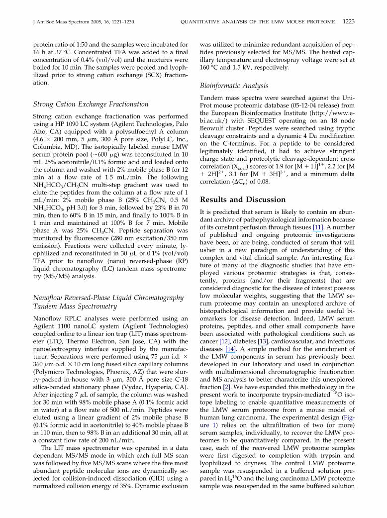
1223J Am Soc Mass Spectrom 2005, 16, 1221–1230 QUANTITATIVE ANALYSIS OF THE LMW MOUSE PROTEOME
protein ratio of 1:50 and the samples were incubated for16 h at 37 °C. Concentrated TFA was added to a finalconcentration of 0.4% (vol/vol) and the mixtures wereboiled for 10 min. The samples were pooled and lyoph-ilized prior to strong cation exchange (SCX) fraction-ation.
Strong Cation Exchange Fractionation
Strong cation exchange fractionation was performedusing a HP 1090 LC system (Agilent Technologies, PaloAlto, CA) equipped with a polysulfoethyl A column(4.6 � 200 mm, 5 �m, 300 Å pore size, PolyLC, Inc.,Columbia, MD). The isotopically labeled mouse LMWserum protein pool (�600 �g) was reconstituted in 10mL 25% acetonitrile/0.1% formic acid and loaded ontothe column and washed with 2% mobile phase B for 12min at a flow rate of 1.5 mL/min. The followingNH4HCO3/CH3CN multi-step gradient was used toelute the peptides from the column at a flow rate of 1mL/min: 2% mobile phase B (25% CH3CN, 0.5 MNH4HCO3, pH 3.0) for 3 min, followed by 23% B in 70min, then to 60% B in 15 min, and finally to 100% B in1 min and maintained at 100% B for 7 min. Mobilephase A was 25% CH3CN. Peptide separation wasmonitored by fluorescence (280 nm excitation/350 nmemission). Fractions were collected every minute, ly-ophilized and reconstituted in 30 �L of 0.1% (vol/vol)TFA prior to nanoflow (nano) reversed-phase (RP)liquid chromatography (LC)-tandem mass spectrome-try (MS/MS) analysis.
Nanoflow Reversed-Phase Liquid ChromatographyTandem Mass Spectrometry
Nanoflow RPLC analyses were performed using anAgilent 1100 nanoLC system (Agilent Technologies)coupled online to a linear ion trap (LIT) mass spectrom-eter (LTQ, Thermo Electron, San Jose, CA) with thenanoelectrospray interface supplied by the manufac-turer. Separations were performed using 75 �m i.d. �360 �m o.d. � 10 cm long fused silica capillary columns(Polymicro Technologies, Phoenix, AZ) that were slur-ry-packed in-house with 3 �m, 300 Å pore size C-18silica-bonded stationary phase (Vydac, Hysperia, CA).After injecting 7 �L of sample, the column was washedfor 30 min with 98% mobile phase A (0.1% formic acidin water) at a flow rate of 500 nL/min. Peptides wereeluted using a linear gradient of 2% mobile phase B(0.1% formic acid in acetonitrile) to 40% mobile phase Bin 110 min, then to 98% B in an additional 30 min, all ata constant flow rate of 200 nL/min.
The LIT mass spectrometer was operated in a datadependent MS/MS mode in which each full MS scanwas followed by five MS/MS scans where the five mostabundant peptide molecular ions are dynamically se-lected for collision-induced dissociation (CID) using a
normalized collision energy of 35%. Dynamic exclusionwas utilized to minimize redundant acquisition of pep-tides previously selected for MS/MS. The heated cap-illary temperature and electrospray voltage were set at160 °C and 1.5 kV, respectively.
Bioinformatic Analysis
Tandem mass spectra were searched against the Uni-Prot mouse proteomic database (05-12-04 release) fromthe European Bioinformatics Institute (http://www.e-bi.ac.uk/) with SEQUEST operating on an 18 nodeBeowulf cluster. Peptides were searched using trypticcleavage constraints and a dynamic 4 Da modificationon the C-terminus. For a peptide to be consideredlegitimately identified, it had to achieve stringentcharge state and proteolytic cleavage-dependent crosscorrelation (Xcorr) scores of 1.9 for [M � H]1�, 2.2 for [M� 2H]2�, 3.1 for [M � 3H]3�, and a minimum deltacorrelation (�Cn) of 0.08.
Results and Discussion
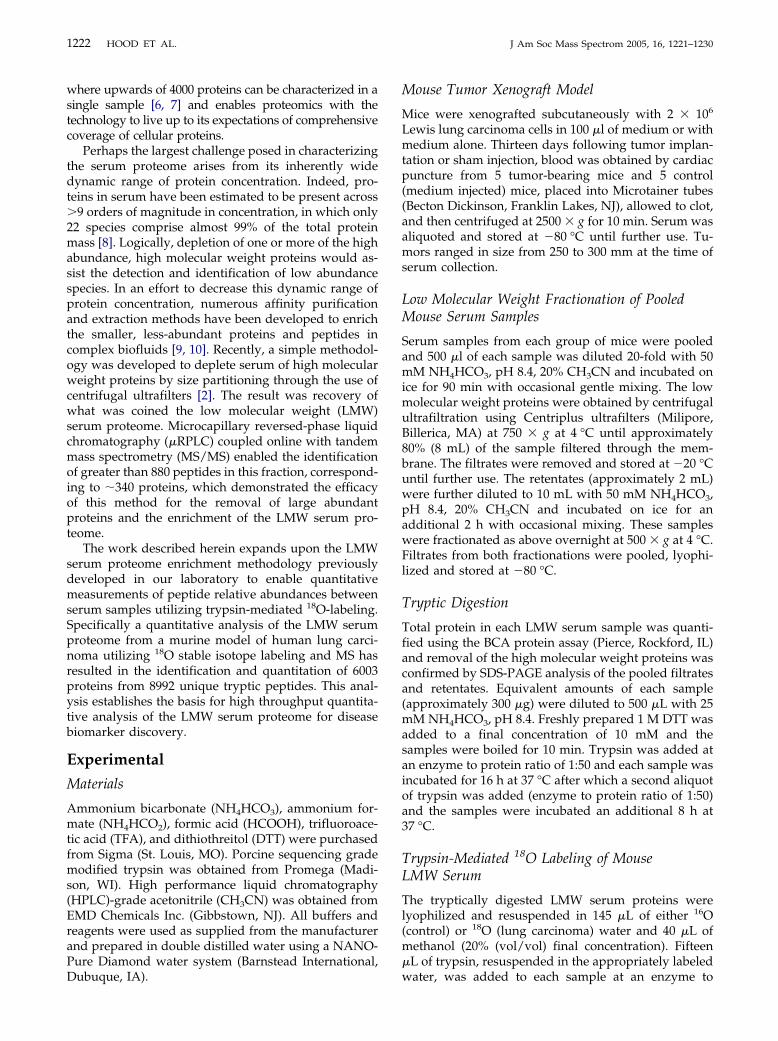
It is predicted that serum is likely to contain an abun-dant archive of pathophysiological information becauseof its constant perfusion through tissues [11]. A numberof published and ongoing proteomic investigationshave been, or are being, conducted of serum that willusher in a new paradigm of understanding of thiscomplex and vital clinical sample. An interesting fea-ture of many of the diagnostic studies that have em-ployed various proteomic strategies is that, consis-tently, proteins (and/or their fragments) that areconsidered diagnostic for the disease of interest possesslow molecular weights, suggesting that the LMW se-rum proteome may contain an unexplored archive ofhistopathological information and provide useful bi-omarkers for disease detection. Indeed, LMW serumproteins, peptides, and other small components havebeen associated with pathological conditions such ascancer [12], diabetes [13], cardiovascular, and infectiousdiseases [14]. A simple method for the enrichment ofthe LMW components in serum has previously beendeveloped in our laboratory and used in conjunctionwith multidimensional chromatographic fractionationand MS analysis to better characterize this unexploredfraction [2]. We have expanded this methodology in thepresent work to incorporate trypsin-mediated 18O iso-tope labeling to enable quantitative measurements ofthe LMW serum proteome from a mouse model ofhuman lung carcinoma. The experimental design (Fig-ure 1) relies on the ultrafiltration of two (or more)serum samples, individually, to recover the LMW pro-teomes to be quantitatively compared. In the presentcase, each of the recovered LMW proteome sampleswere first digested to completion with trypsin andlyophilized to dryness. The control LMW proteomesample was resuspended in a buffered solution pre-pared in H2
16O and the lung carcinoma LMW proteome
sample was resuspended in the same buffered solution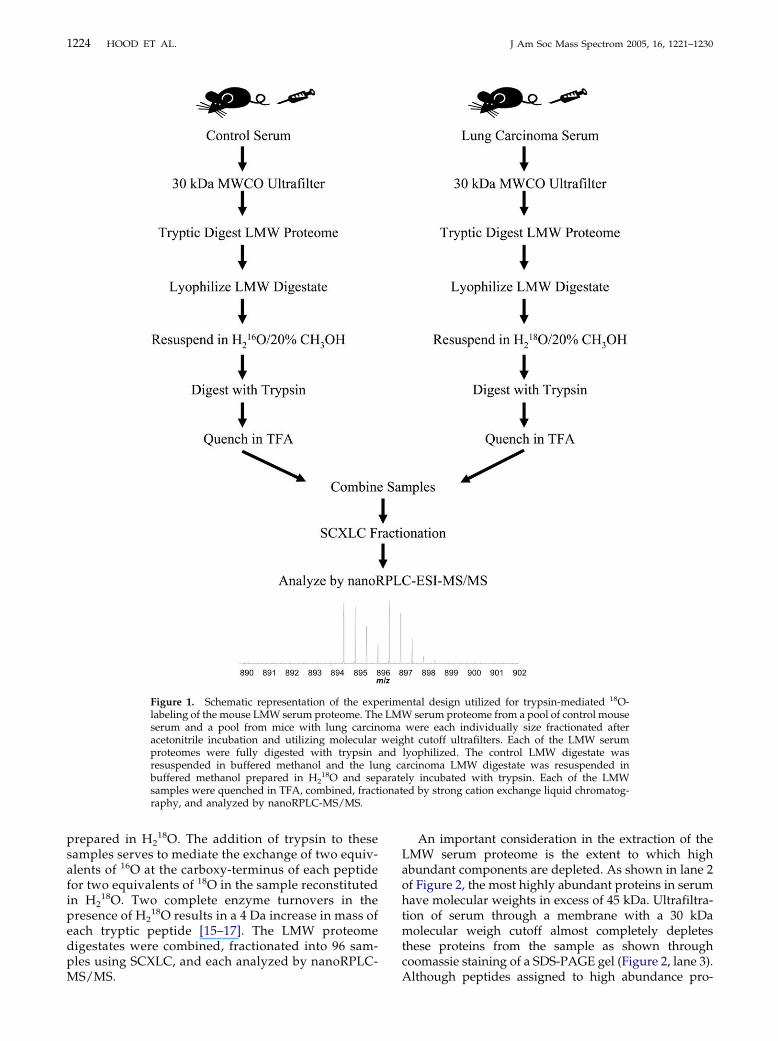
1224 HOOD ET AL. J Am Soc Mass Spectrom 2005, 16, 1221–1230
prepared in H218O. The addition of trypsin to these
samples serves to mediate the exchange of two equiv-alents of 16O at the carboxy-terminus of each peptidefor two equivalents of 18O in the sample reconstitutedin H2
18O. Two complete enzyme turnovers in thepresence of H2
18O results in a 4 Da increase in mass ofeach tryptic peptide [15–17]. The LMW proteomedigestates were combined, fractionated into 96 sam-ples using SCXLC, and each analyzed by nanoRPLC-
Figure 1. Schematic representation of the explabeling of the mouse LMW serum proteome. Theserum and a pool from mice with lung carcinacetonitrile incubation and utilizing molecularproteomes were fully digested with trypsinresuspended in buffered methanol and the lubuffered methanol prepared in H2
18O and sepsamples were quenched in TFA, combined, fractraphy, and analyzed by nanoRPLC-MS/MS.
MS/MS.
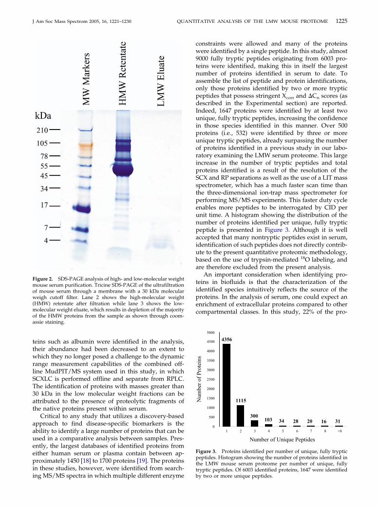
An important consideration in the extraction of theLMW serum proteome is the extent to which highabundant components are depleted. As shown in lane 2of Figure 2, the most highly abundant proteins in serumhave molecular weights in excess of 45 kDa. Ultrafiltra-tion of serum through a membrane with a 30 kDamolecular weigh cutoff almost completely depletesthese proteins from the sample as shown throughcoomassie staining of a SDS-PAGE gel (Figure 2, lane 3).
ntal design utilized for trypsin-mediated 18O-serum proteome from a pool of control mouse
were each individually size fractionated afterht cutoff ultrafilters. Each of the LMW serumlyophilized. The control LMW digestate wasrcinoma LMW digestate was resuspended inly incubated with trypsin. Each of the LMWed by strong cation exchange liquid chromatog-
erimeLMW
omaweig
andng caarate
ionat
Although peptides assigned to high abundance pro-
1225J Am Soc Mass Spectrom 2005, 16, 1221–1230 QUANTITATIVE ANALYSIS OF THE LMW MOUSE PROTEOME
teins such as albumin were identified in the analysis,their abundance had been decreased to an extent towhich they no longer posed a challenge to the dynamicrange measurement capabilities of the combined off-line MudPIT/MS system used in this study, in whichSCXLC is performed offline and separate from RPLC.The identification of proteins with masses greater than30 kDa in the low molecular weight fractions can beattributed to the presence of proteolytic fragments ofthe native proteins present within serum.
Critical to any study that utilizes a discovery-basedapproach to find disease-specific biomarkers is theability to identify a large number of proteins that can beused in a comparative analysis between samples. Pres-ently, the largest databases of identified proteins fromeither human serum or plasma contain between ap-proximately 1450 [18] to 1700 proteins [19]. The proteinsin these studies, however, were identified from search-
Figure 2. SDS-PAGE analysis of high- and low-molecular weightmouse serum purification. Tricine SDS-PAGE of the ultrafiltrationof mouse serum through a membrane with a 30 kDa molecularweigh cutoff filter. Lane 2 shows the high-molecular weight(HMW) retentate after filtration while lane 3 shows the low-molecular weight eluate, which results in depletion of the majorityof the HMW proteins from the sample as shown through coom-assie staining.
ing MS/MS spectra in which multiple different enzyme
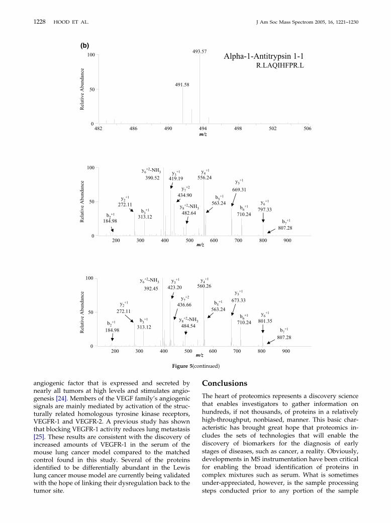
constraints were allowed and many of the proteinswere identified by a single peptide. In this study, almost9000 fully tryptic peptides originating from 6003 pro-teins were identified, making this in itself the largestnumber of proteins identified in serum to date. Toassemble the list of peptide and protein identifications,only those proteins identified by two or more trypticpeptides that possess stringent Xcorr and �Cn scores (asdescribed in the Experimental section) are reported.Indeed, 1647 proteins were identified by at least twounique, fully tryptic peptides, increasing the confidencein those species identified in this manner. Over 500proteins (i.e., 532) were identified by three or moreunique tryptic peptides, already surpassing the numberof proteins identified in a previous study in our labo-ratory examining the LMW serum proteome. This largeincrease in the number of tryptic peptides and totalproteins identified is a result of the resolution of theSCX and RP separations as well as the use of a LIT massspectrometer, which has a much faster scan time thanthe three-dimensional ion-trap mass spectrometer forperforming MS/MS experiments. This faster duty cycleenables more peptides to be interrogated by CID perunit time. A histogram showing the distribution of thenumber of proteins identified per unique, fully trypticpeptide is presented in Figure 3. Although it is wellaccepted that many nontryptic peptides exist in serum,identification of such peptides does not directly contrib-ute to the present quantitative proteomic methodology,based on the use of trypsin-mediated 18O labeling, andare therefore excluded from the present analysis.
An important consideration when identifying pro-teins in biofluids is that the characterization of theidentified species intuitively reflects the source of theproteins. In the analysis of serum, one could expect anenrichment of extracellular proteins compared to othercompartmental classes. In this study, 22% of the pro-
Figure 3. Proteins identified per number of unique, fully trypticpeptides. Histogram showing the number of proteins identified inthe LMW mouse serum proteome per number of unique, fullytryptic peptides. Of 6003 identified proteins, 1647 were identified
by two or more unique peptides.
1226 HOOD ET AL. J Am Soc Mass Spectrom 2005, 16, 1221–1230
teins identified by at least two unique tryptic peptideswere classified as extracellular (Figure 4a). An addi-tional 27% of the proteins were localized to the mem-brane, with the remainder (i.e., 51%) originated fromintracellular compartments. Unfortunately, only about30% of the proteins identified in this study by two ormore unique tryptic peptides could be linked to geneontology classifications. Gene ontology of the entiremouse proteome (18,887 out of 27,459 possible proteins)predicts that 14% of the proteins will be extracellular(Figure 4b). Why there was a substantial increase in thepercentage of extracellular proteins observed in thisstudy compared to the mouse proteome is not clear.Coincidentally, this same phenomenon was observed ina gene ontology comparison of experimentally identi-fied human serum proteins and the entire humanproteome [20].
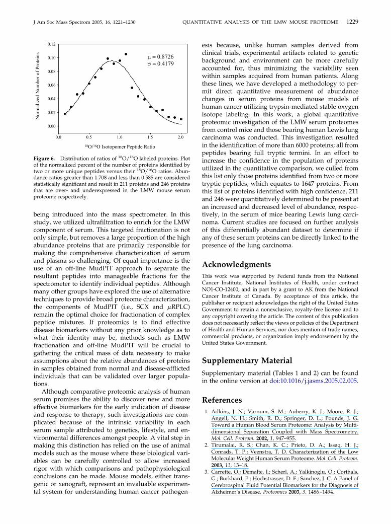
An important factor when using 16O/18O labeling forquantitative measurements is the completeness of en-zyme-mediated isotope exchange. Shown in Figure 5aand b are selected mass spectra of differentially labeledLMW serum 16O/18O peptides from control mice andthose bearing Lewis lung carcinoma, indicating com-plete 18O incorporation of peptides within serum ob-tained from the mouse lung cancer model. These two
Figure 4. Gene ontology classification of cellular location ofmouse LMW serum proteins. (a) Cellular compartmentalization oflow molecular weight mouse serum proteins (�30% mapped byGO) reveal that 22% of these are classified as extracellular. (b)Gene ontology classification predicts only 14% of the entire mouseproteome encodes for extracellular proteins.
sets of peptides were identified as apolipoprotein A1,
which has recently been reported as a biomarker for thedetection of early stage ovarian cancer [21], and �-1-antitrypsin 1-1, which has been observed to have in-creased levels in lung cancer patients [22], demonstrat-ing the facile ability of the present methodology toprovide both quantitation and identification of putativebiomarkers in the same high-throughput experiment.We hypothesize that efficient 18O incorporation, asindicated by the mass spectra in Figure 5, may beattributed to the observed increase (�20%) in trypsinactivity in 20% (vol/vol) CH3OH as previously demon-strated by a N-benzoyl-L-arginine ethyl ester trypsinactivity assay [23].
The identified peptides were quantified by calcu-lating the relative abundances (18O/16O, in this dataset) of peptides based on the area of their extractedion chromatograms (XIC) reconstructed from themolecular ion MS scans. There is no standard forwhat change in relative abundance, as measured byquantitative proteome analyses, represent statisti-cally significant changes in protein abundance ratios.This lack of a standard is because the determinationof isotopomeric peptide ratios depends directly onmany experimental factors, such as the equivalence ofthe initial sample mixing, the relative efficiency ofrecovery of each of the peptide pairs, the inherentresolution and accuracy of the MS instrumentation,and the number of molecular ion scans across eachpeptide pair nanoRPLC peak. We sought to deter-mine the threshold above and below which the mea-sured protein abundance ratios were statistically sig-nificant within this dataset. This analysis wasconducted by plotting the normalized percent of thenumber of 18O/16O labeled proteins identified withinbinned abundance ratios (e.g., 1.0-1.09, 1.1-1.19, etc.)as determined from integration of each of the respec-tive isotopomeric peptide pair XIC (Figure 6). Theo-retically, the distribution of the protein abundanceratios should be a normal distribution, defined as,
P �1
��2�e�(x � �)2⁄(2�2) (1)
where P is the normalized number of proteins identifiedat a given protein abundance ratio x, � is defined as theexperimentally measured mean value of the total pop-ulation of the protein abundance ratios and � is stan-dard deviation. The z score is defined as,
z �x � �
�(2)
Using non-linear least squares regression method, �and � can be obtained from the data series of nor-malized number of proteins identified and the exper-imentally determined ratios of the protein abun-dances. The protein abundance ratio above which a
statistically significant increase in abundance should
1227J Am Soc Mass Spectrom 2005, 16, 1221–1230 QUANTITATIVE ANALYSIS OF THE LMW MOUSE PROTEOME
be considered is 2� above the measured mean ratio,while the protein abundance ratio below which astatistically significant decrease in abundance shouldbe considered 1/(2� � �). Using these criteria, thethreshold above which a measured protein abun-dance ratio reflects a statistically significant increasein abundance should be considered to be �1.708 andthe threshold below which a measured abundanceratio reflects a statistically significant decrease inabundance should be considered to �0.585, for thesedata. The present data indicate that 211 proteins and
Figure 5. Selected mass spectra of differentiallmass spectra of the 16O/18O molecular ion pairs wfragmentation ion series showing �4 Da ([Mapolipoprotein A-1, and (b) �-1-antitrypsin.
246 proteins are significantly increased and de-
creased in abundance in serum from mice bearingLewis lung carcinoma, respectively. Hence, of theproteins identified by two or more unique fullytryptic peptides, approximately 28%, are observed tochange in abundance in mice with Lewis lung carci-noma.
Several of the proteins that were found to be upregu-lated in the serum obtained from the lung carcinomamouse model have been implicated in cancer progres-sion. Vascular endothelial growth factor receptor 1(VEGFR-1) was identified to be upregulated over 7-fold
eled LMW serum 16O/18O peptides. Full range[M � H]�1) or 2 Da their corresponding MS/MSH]�2) mass shifts in the y-type ions for (a)
y labith (� 2
in the mouse lung carcinoma model. VEGF is a key
(con
1228 HOOD ET AL. J Am Soc Mass Spectrom 2005, 16, 1221–1230
angiogenic factor that is expressed and secreted bynearly all tumors at high levels and stimulates angio-genesis [24]. Members of the VEGF family’s angiogenicsignals are mainly mediated by activation of the struc-turally related homologous tyrosine kinase receptors,VEGFR-1 and VEGFR-2. A previous study has shownthat blocking VEGFR-1 activity reduces lung metastasis[25]. These results are consistent with the discovery ofincreased amounts of VEGFR-1 in the serum of themouse lung cancer model compared to the matchedcontrol found in this study. Several of the proteinsidentified to be differentially abundant in the Lewislung cancer mouse model are currently being validatedwith the hope of linking their dysregulation back to the
Figure 5
tumor site.
Conclusions
The heart of proteomics represents a discovery sciencethat enables investigators to gather information onhundreds, if not thousands, of proteins in a relativelyhigh-throughput, nonbiased, manner. This basic char-acteristic has brought great hope that proteomics in-cludes the sets of technologies that will enable thediscovery of biomarkers for the diagnosis of earlystages of diseases, such as cancer, a reality. Obviously,developments in MS instrumentation have been criticalfor enabling the broad identification of proteins incomplex mixtures such as serum. What is sometimesunder-appreciated, however, is the sample processing
tinued)
steps conducted prior to any portion of the sample
1229J Am Soc Mass Spectrom 2005, 16, 1221–1230 QUANTITATIVE ANALYSIS OF THE LMW MOUSE PROTEOME
being introduced into the mass spectrometer. In thisstudy, we utilized ultrafiltration to enrich for the LMWcomponent of serum. This targeted fractionation is notonly simple, but removes a large proportion of the highabundance proteins that are primarily responsible formaking the comprehensive characterization of serumand plasma so challenging. Of equal importance is theuse of an off-line MudPIT approach to separate theresultant peptides into manageable fractions for thespectrometer to identify individual peptides. Althoughmany other groups have explored the use of alternativetechniques to provide broad proteome characterization,the components of MudPIT (i.e., SCX and �RPLC)remain the optimal choice for fractionation of complexpeptide mixtures. If proteomics is to find effectivedisease biomarkers without any prior knowledge as towhat their identity may be, methods such as LMWfractionation and off-line MudPIT will be crucial togathering the critical mass of data necessary to makeassumptions about the relative abundances of proteinsin samples obtained from normal and disease-afflictedindividuals that can be validated over larger popula-tions.
Although comparative proteomic analysis of humanserum promises the ability to discover new and moreeffective biomarkers for the early indication of diseaseand response to therapy, such investigations are com-plicated because of the intrinsic variability in eachserum sample attributed to genetics, lifestyle, and en-vironmental differences amongst people. A vital step inmaking this distinction has relied on the use of animalmodels such as the mouse where these biological vari-ables can be carefully controlled to allow increasedrigor with which comparisons and pathophysiologicalconclusions can be made. Mouse models, either trans-genic or xenograft, represent an invaluable experimen-
Figure 6. Distribution of ratios of 18O/16O labeled proteins. Plotof the normalized percent of the number of proteins identified bytwo or more unique peptides versus their 18O/16O ratios. Abun-dance ratios greater than 1.708 and less than 0.585 are consideredstatistically significant and result in 211 proteins and 246 proteinsthat are over- and underexpressed in the LMW mouse serumproteome respectively.
tal system for understanding human cancer pathogen-
esis because, unlike human samples derived fromclinical trials, experimental artifacts related to geneticbackground and environment can be more carefullyaccounted for, thus minimizing the variability seenwithin samples acquired from human patients. Alongthese lines, we have developed a methodology to per-mit direct quantitative measurement of abundancechanges in serum proteins from mouse models ofhuman cancer utilizing trypsin-mediated stable oxygenisotope labeling. In this work, a global quantitativeproteomic investigation of the LMW serum proteomesfrom control mice and those bearing human Lewis lungcarcinoma was conducted. This investigation resultedin the identification of more than 6000 proteins; all frompeptides bearing full tryptic termini. In an effort toincrease the confidence in the population of proteinsutilized in the quantitative comparison, we culled fromthis list only those proteins identified from two or moretryptic peptides, which equates to 1647 proteins. Fromthis list of proteins identified with high confidence, 211and 246 were quantitatively determined to be present atan increased and decreased level of abundance, respec-tively, in the serum of mice bearing Lewis lung carci-noma. Current studies are focused on further analysisof this differentially abundant dataset to determine ifany of these serum proteins can be directly linked to thepresence of the lung carcinoma.
AcknowledgmentsThis work was supported by Federal funds from the NationalCancer Institute, National Institutes of Health, under contractNO1-CO-12400, and in part by a grant to AK from the NationalCancer Institute of Canada. By acceptance of this article, thepublisher or recipient acknowledges the right of the United StatesGovernment to retain a nonexclusive, royalty-free license and toany copyright covering the article. The content of this publicationdoes not necessarily reflect the views or policies of the Departmentof Health and Human Services, nor does mention of trade names,commercial products, or organization imply endorsement by theUnited States Government.
Supplementary Material
Supplementary material (Tables 1 and 2) can be foundin the online version at doi:10.1016/j.jasms.2005.02.005.
References1. Adkins, J. N.; Varnum, S. M.; Auberry, K. J.; Moore, R. J.;
Angell, N. H.; Smith, R. D.; Springer, D. L.; Pounds, J. G.Toward a Human Blood Serum Proteome: Analysis by Multi-dimensional Separation Coupled with Mass Spectrometry.Mol. Cell. Proteom. 2002, 1, 947–955.
2. Tirumalai, R. S.; Chan, K. C.; Prieto, D. A.; Issaq, H. J.;Conrads, T. P.; Veenstra, T. D. Characterization of the LowMolecular Weight Human Serum Proteome. Mol. Cell. Proteom.2003, 13, 13–18.
3. Carrette, O.; Demalte, I.; Scherl, A.; Yalkinoglu, O.; Corthals,G.; Burkhard, P.; Hochstrasser, D. F.; Sanchez, J. C. A Panel ofCerebrospinal Fluid Potential Biomarkers for the Diagnosis of
Alzheimer’s Disease. Proteomics 2003, 3, 1486–1494.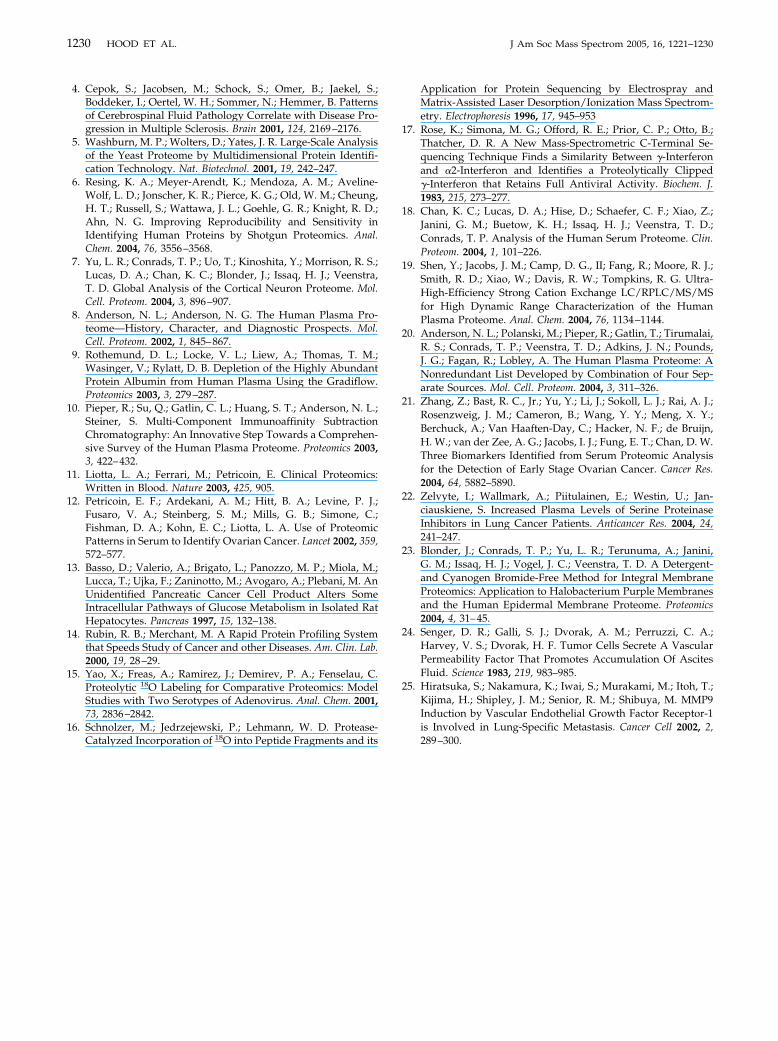
1230 HOOD ET AL. J Am Soc Mass Spectrom 2005, 16, 1221–1230
4. Cepok, S.; Jacobsen, M.; Schock, S.; Omer, B.; Jaekel, S.;Boddeker, I.; Oertel, W. H.; Sommer, N.; Hemmer, B. Patternsof Cerebrospinal Fluid Pathology Correlate with Disease Pro-gression in Multiple Sclerosis. Brain 2001, 124, 2169–2176.
5. Washburn, M. P.; Wolters, D.; Yates, J. R. Large-Scale Analysisof the Yeast Proteome by Multidimensional Protein Identifi-cation Technology. Nat. Biotechnol. 2001, 19, 242–247.
6. Resing, K. A.; Meyer-Arendt, K.; Mendoza, A. M.; Aveline-Wolf, L. D.; Jonscher, K. R.; Pierce, K. G.; Old, W. M.; Cheung,H. T.; Russell, S.; Wattawa, J. L.; Goehle, G. R.; Knight, R. D.;Ahn, N. G. Improving Reproducibility and Sensitivity inIdentifying Human Proteins by Shotgun Proteomics. Anal.Chem. 2004, 76, 3556–3568.
7. Yu, L. R.; Conrads, T. P.; Uo, T.; Kinoshita, Y.; Morrison, R. S.;Lucas, D. A.; Chan, K. C.; Blonder, J.; Issaq, H. J.; Veenstra,T. D. Global Analysis of the Cortical Neuron Proteome. Mol.Cell. Proteom. 2004, 3, 896–907.
8. Anderson, N. L.; Anderson, N. G. The Human Plasma Pro-teome—History, Character, and Diagnostic Prospects. Mol.Cell. Proteom. 2002, 1, 845–867.
9. Rothemund, D. L.; Locke, V. L.; Liew, A.; Thomas, T. M.;Wasinger, V.; Rylatt, D. B. Depletion of the Highly AbundantProtein Albumin from Human Plasma Using the Gradiflow.Proteomics 2003, 3, 279–287.
10. Pieper, R.; Su, Q.; Gatlin, C. L.; Huang, S. T.; Anderson, N. L.;Steiner, S. Multi-Component Immunoaffinity SubtractionChromatography: An Innovative Step Towards a Comprehen-sive Survey of the Human Plasma Proteome. Proteomics 2003,3, 422–432.
11. Liotta, L. A.; Ferrari, M.; Petricoin, E. Clinical Proteomics:Written in Blood. Nature 2003, 425, 905.
12. Petricoin, E. F.; Ardekani, A. M.; Hitt, B. A.; Levine, P. J.;Fusaro, V. A.; Steinberg, S. M.; Mills, G. B.; Simone, C.;Fishman, D. A.; Kohn, E. C.; Liotta, L. A. Use of ProteomicPatterns in Serum to Identify Ovarian Cancer. Lancet 2002, 359,572–577.
13. Basso, D.; Valerio, A.; Brigato, L.; Panozzo, M. P.; Miola, M.;Lucca, T.; Ujka, F.; Zaninotto, M.; Avogaro, A.; Plebani, M. AnUnidentified Pancreatic Cancer Cell Product Alters SomeIntracellular Pathways of Glucose Metabolism in Isolated RatHepatocytes. Pancreas 1997, 15, 132–138.
14. Rubin, R. B.; Merchant, M. A Rapid Protein Profiling Systemthat Speeds Study of Cancer and other Diseases. Am. Clin. Lab.2000, 19, 28–29.
15. Yao, X.; Freas, A.; Ramirez, J.; Demirev, P. A.; Fenselau, C.Proteolytic 18O Labeling for Comparative Proteomics: ModelStudies with Two Serotypes of Adenovirus. Anal. Chem. 2001,73, 2836–2842.
16. Schnolzer, M.; Jedrzejewski, P.; Lehmann, W. D. Protease-
Catalyzed Incorporation of 18O into Peptide Fragments and itsApplication for Protein Sequencing by Electrospray andMatrix-Assisted Laser Desorption/Ionization Mass Spectrom-etry. Electrophoresis 1996, 17, 945–953
17. Rose, K.; Simona, M. G.; Offord, R. E.; Prior, C. P.; Otto, B.;Thatcher, D. R. A New Mass-Spectrometric C-Terminal Se-quencing Technique Finds a Similarity Between -Interferonand �2-Interferon and Identifies a Proteolytically Clipped-Interferon that Retains Full Antiviral Activity. Biochem. J.1983, 215, 273–277.
18. Chan, K. C.; Lucas, D. A.; Hise, D.; Schaefer, C. F.; Xiao, Z.;Janini, G. M.; Buetow, K. H.; Issaq, H. J.; Veenstra, T. D.;Conrads, T. P. Analysis of the Human Serum Proteome. Clin.Proteom. 2004, 1, 101–226.
19. Shen, Y.; Jacobs, J. M.; Camp, D. G., II; Fang, R.; Moore, R. J.;Smith, R. D.; Xiao, W.; Davis, R. W.; Tompkins, R. G. Ultra-High-Efficiency Strong Cation Exchange LC/RPLC/MS/MSfor High Dynamic Range Characterization of the HumanPlasma Proteome. Anal. Chem. 2004, 76, 1134–1144.
20. Anderson, N. L.; Polanski, M.; Pieper, R.; Gatlin, T.; Tirumalai,R. S.; Conrads, T. P.; Veenstra, T. D.; Adkins, J. N.; Pounds,J. G.; Fagan, R.; Lobley, A. The Human Plasma Proteome: ANonredundant List Developed by Combination of Four Sep-arate Sources. Mol. Cell. Proteom. 2004, 3, 311–326.
21. Zhang, Z.; Bast, R. C., Jr.; Yu, Y.; Li, J.; Sokoll, L. J.; Rai, A. J.;Rosenzweig, J. M.; Cameron, B.; Wang, Y. Y.; Meng, X. Y.;Berchuck, A.; Van Haaften-Day, C.; Hacker, N. F.; de Bruijn,H. W.; van der Zee, A. G.; Jacobs, I. J.; Fung, E. T.; Chan, D. W.Three Biomarkers Identified from Serum Proteomic Analysisfor the Detection of Early Stage Ovarian Cancer. Cancer Res.2004, 64, 5882–5890.
22. Zelvyte, I.; Wallmark, A.; Piitulainen, E.; Westin, U.; Jan-ciauskiene, S. Increased Plasma Levels of Serine ProteinaseInhibitors in Lung Cancer Patients. Anticancer Res. 2004, 24,241–247.
23. Blonder, J.; Conrads, T. P.; Yu, L. R.; Terunuma, A.; Janini,G. M.; Issaq, H. J.; Vogel, J. C.; Veenstra, T. D. A Detergent-and Cyanogen Bromide-Free Method for Integral MembraneProteomics: Application to Halobacterium Purple Membranesand the Human Epidermal Membrane Proteome. Proteomics2004, 4, 31–45.
24. Senger, D. R.; Galli, S. J.; Dvorak, A. M.; Perruzzi, C. A.;Harvey, V. S.; Dvorak, H. F. Tumor Cells Secrete A VascularPermeability Factor That Promotes Accumulation Of AscitesFluid. Science 1983, 219, 983–985.
25. Hiratsuka, S.; Nakamura, K.; Iwai, S.; Murakami, M.; Itoh, T.;Kijima, H.; Shipley, J. M.; Senior, R. M.; Shibuya, M. MMP9Induction by Vascular Endothelial Growth Factor Receptor-1is Involved in Lung-Specific Metastasis. Cancer Cell 2002, 2,
289–300.