Species diversity of heterotrophic flagellates in White Sea littoral sites
Upload
independentCategory
view
0download
0
AQUATIC MICROBIAL ECOLOGYAquat Microb Ecol
Vol. 61: 105–117, 2010doi: 10.3354/ame01440
Published online October 1
INTRODUCTION
Cyanobacterial symbionts (cyanobionts) of non-photosynthetic dinophysoids (Dinophyceae) were firstobserved more than 100 yr ago (Schütt 1895). They arethought to function as photosynthetic partners in theirrelationship with the host (Taylor 1982), but despitemany different types of cyanobacterial and bacterial
symbionts having been described for several dino-physoid genera (such as Amphisolenia, Histioneis,Ornithocercus and Parahistioneis), little is known aboutthe identity and diversity of these symbionts. Further,their ecological significance is essentially unknown.
Recently, we found that symbiont-bearing dino-flagellates were most common in the photic zone of theIndian Ocean characterized by low nutrients and low
© Inter-Research 2010 · www.int-res.com*Corresponding author. E-mail: [email protected]
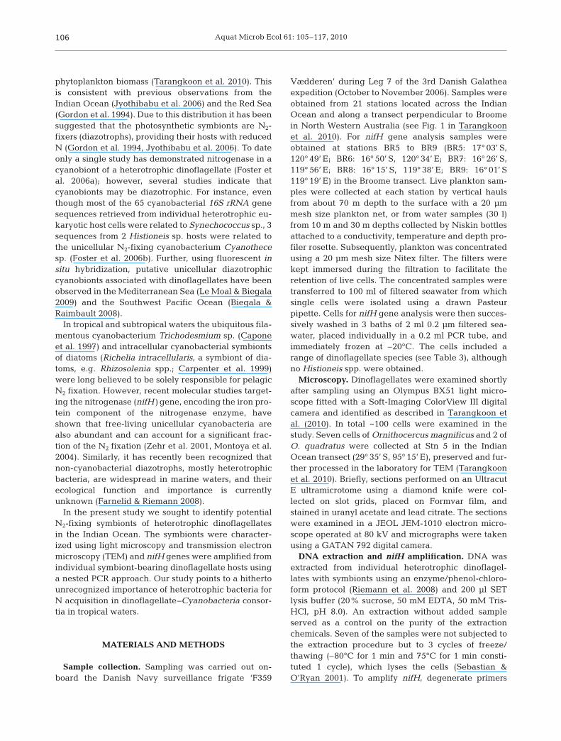
Putative N2-fixing heterotrophic bacteriaassociated with dinoflagellate–Cyanobacteriaconsortia in the low-nitrogen Indian Ocean
Hanna Farnelid1, Woraporn Tarangkoon2, 3, Gert Hansen4, Per Juel Hansen2, Lasse Riemann1, 5,*
1Department of Natural Sciences, Linnaeus University, 39182 Kalmar, Sweden2Section for Marine Biology, Strandpromenaden 5, 3000 Helsingør, Denmark
3Faculty of Science and Fisheries Technology, Rajamangala University of Technology Srivijaya, 92150 Trang, Thailand4Department of Phycology, Ø. Farimagsgade 2D, 1353, Copenhagen, Denmark
5Present address: Section for Marine Biology, Strandpromenaden 5, 3000 Helsingør, Denmark
ABSTRACT: Heterotrophic dinoflagellates bearing unicellular cyanobacterial symbionts are commonwithin the order Dinophysiales. However, the ecological role of these symbionts is unclear. Due to theoccurrence of such consortia in oceanic waters characterized by low nitrogen concentrations, wehypothesized that the symbionts fix gaseous nitrogen (N2). Individual heterotrophic dinoflagellatescontaining cyanobacterial symbionts were isolated from the open Indian Ocean and off Western Aus-tralia, and characterized using light microscopy, transmission electron microscopy (TEM), and nitro-genase (nifH) gene amplification, cloning, and sequencing. Cyanobacteria, heterotrophic bacteriaand eukaryotic algae were recognized as symbionts of the heterotrophic dinoflagellates. nifH genesequences were obtained from 23 of 37 (62%) specimens of dinoflagellates (Ornithocercus spp. andAmphisolenia spp.). Interestingly, only 2 specimens contained cyanobacterial nifH sequences, while21 specimens contained nifH genes related to heterotrophic bacteria. Of the 137 nifH sequencesobtained 68% were most similar to Alpha-, Beta-, and Gammaproteobacteria, 8% clustered with an-aerobic bacteria, and 5% were related to second alternative nitrogenases (anfH). Twelve sequencesfrom 5 host cells formed a discrete cluster which may represent a not yet classified nifH cluster. Eightdinoflagellates contained only 1 type of nifH sequence (>99% sequence identity) but overall theputative N2-fixing symbionts did not appear host specific and mixed assemblages were often foundin single host cells. This study provides the first insights into the nifH diversity of dinoflagellate sym-bionts and suggests a symbiotic co-existence of non-diazotrophic cyanobacteria and N2-fixingheterotrophic bacteria in heterotrophic dinoflagellates.
KEY WORDS: Symbionts · Nitrogen fixation · Nitrogenase · Heterotrophic bacteria · Ornithocercus ·Amphisolenia · Histioneis · Dinoflagellates · Indian Ocean · Galathea 3
Resale or republication not permitted without written consent of the publisher
Aquat Microb Ecol 61: 105–117, 2010
phytoplankton biomass (Tarangkoon et al. 2010). Thisis consistent with previous observations from theIndian Ocean (Jyothibabu et al. 2006) and the Red Sea(Gordon et al. 1994). Due to this distribution it has beensuggested that the photosynthetic symbionts are N2-fixers (diazotrophs), providing their hosts with reducedN (Gordon et al. 1994, Jyothibabu et al. 2006). To dateonly a single study has demonstrated nitrogenase in acyanobiont of a heterotrophic dinoflagellate (Foster etal. 2006a); however, several studies indicate thatcyanobionts may be diazotrophic. For instance, eventhough most of the 65 cyanobacterial 16S rRNA genesequences retrieved from individual heterotrophic eu-karyotic host cells were related to Synechococcus sp., 3sequences from 2 Histioneis sp. hosts were related tothe unicellular N2-fixing cyanobacterium Cyanothecesp. (Foster et al. 2006b). Further, using fluorescent insitu hybridization, putative unicellular diazotrophiccyanobionts associated with dinoflagellates have beenobserved in the Mediterranean Sea (Le Moal & Biegala2009) and the Southwest Pacific Ocean (Biegala &Raimbault 2008).
In tropical and subtropical waters the ubiquitous fila-mentous cyanobacterium Trichodesmium sp. (Caponeet al. 1997) and intracellular cyanobacterial symbiontsof diatoms (Richelia intracellularis, a symbiont of dia-toms, e.g. Rhizosolenia spp.; Carpenter et al. 1999)were long believed to be solely responsible for pelagicN2 fixation. However, recent molecular studies target-ing the nitrogenase (nifH) gene, encoding the iron pro-tein component of the nitrogenase enzyme, haveshown that free-living unicellular cyanobacteria arealso abundant and can account for a significant frac-tion of the N2 fixation (Zehr et al. 2001, Montoya et al.2004). Similarly, it has recently been recognized thatnon-cyanobacterial diazotrophs, mostly heterotrophicbacteria, are widespread in marine waters, and theirecological function and importance is currentlyunknown (Farnelid & Riemann 2008).
In the present study we sought to identify potentialN2-fixing symbionts of heterotrophic dinoflagellatesin the Indian Ocean. The symbionts were character-ized using light microscopy and transmission electronmicroscopy (TEM) and nifH genes were amplified fromindividual symbiont-bearing dinoflagellate hosts usinga nested PCR approach. Our study points to a hithertounrecognized importance of heterotrophic bacteria forN acquisition in dinoflagellate–Cyanobacteria consor-tia in tropical waters.
MATERIALS AND METHODS
Sample collection. Sampling was carried out on-board the Danish Navy surveillance frigate ‘F359
Vædderen’ during Leg 7 of the 3rd Danish Galatheaexpedition (October to November 2006). Samples wereobtained from 21 stations located across the IndianOcean and along a transect perpendicular to Broomein North Western Australia (see Fig. 1 in Tarangkoonet al. 2010). For nifH gene analysis samples wereobtained at stations BR5 to BR9 (BR5: 17° 03’ S,120° 49’ E; BR6: 16° 50’ S, 120° 34’ E; BR7: 16° 26’ S,119° 56’ E; BR8: 16° 15’ S, 119° 38’ E; BR9: 16° 01’ S119° 19’ E) in the Broome transect. Live plankton sam-ples were collected at each station by vertical haulsfrom about 70 m depth to the surface with a 20 µmmesh size plankton net, or from water samples (30 l)from 10 m and 30 m depths collected by Niskin bottlesattached to a conductivity, temperature and depth pro-filer rosette. Subsequently, plankton was concentratedusing a 20 µm mesh size Nitex filter. The filters werekept immersed during the filtration to facilitate theretention of live cells. The concentrated samples weretransferred to 100 ml of filtered seawater from whichsingle cells were isolated using a drawn Pasteurpipette. Cells for nifH gene analysis were then succes-sively washed in 3 baths of 2 ml 0.2 µm filtered sea-water, placed individually in a 0.2 ml PCR tube, andimmediately frozen at –20°C. The cells included arange of dinoflagellate species (see Table 3), althoughno Histioneis spp. were obtained.
Microscopy. Dinoflagellates were examined shortlyafter sampling using an Olympus BX51 light micro-scope fitted with a Soft-Imaging ColorView III digitalcamera and identified as described in Tarangkoon etal. (2010). In total ~100 cells were examined in thestudy. Seven cells of Ornithocercus magnificus and 2 ofO. quadratus were collected at Stn 5 in the IndianOcean transect (29° 35’ S, 95° 15’ E), preserved and fur-ther processed in the laboratory for TEM (Tarangkoonet al. 2010). Briefly, sections performed on an UltracutE ultramicrotome using a diamond knife were col-lected on slot grids, placed on Formvar film, andstained in uranyl acetate and lead citrate. The sectionswere examined in a JEOL JEM-1010 electron micro-scope operated at 80 kV and micrographs were takenusing a GATAN 792 digital camera.
DNA extraction and nifH amplification. DNA wasextracted from individual heterotrophic dinoflagel-lates with symbionts using an enzyme/phenol-chloro-form protocol (Riemann et al. 2008) and 200 µl SETlysis buffer (20% sucrose, 50 mM EDTA, 50 mM Tris-HCl, pH 8.0). An extraction without added sampleserved as a control on the purity of the extractionchemicals. Seven of the samples were not subjected tothe extraction procedure but to 3 cycles of freeze/thawing (–80°C for 1 min and 75°C for 1 min consti-tuted 1 cycle), which lyses the cells (Sebastian &O’Ryan 2001). To amplify nifH, degenerate primers
106
Farnelid et al.: Diazotrophic symbionts of heterotrophic dinoflagellates
purified by high performance liquid chromatographyand polyacrylamide gel electrophoresis (Sigma-Aldrich; Zehr & McReynolds 1989, Zani et al. 2000)were used according to a nested PCR protocol (Zehr &Turner 2001) using Pure Taq Ready-To-Go PCR Beads(GE Healthcare). A negative control reaction withUV-treated water was included in each PCR batch. Tominimize the risk of contamination, mixing of re-agents was done in a UV-treated sterile flow bench ina UV-treated room, template was added in a PCR/UVworkstation in a separate room, and single tubes (notstrips) were used. For the initial PCR reaction, 3 to6 µl of the extracted DNA or freeze/thawed solutionwas added as template and 1 µl PCR product wastransferred to the subsequent PCR reaction. Five µlfrom the second PCR reaction was examined on a 1%agarose gel, and for samples that produced a ~359 bpproduct the remaining 20 µl was gel purified (E.Z.N.AGel extraction kit, VWR). The negative PCR controland the negative extraction control were alwaysblank. For the negative control PCR reaction, al-though there was no visible product, the gel regioncorresponding to the correct product size was excised,gel purified and cloned. All purified products werecloned using the TOPO TA Cloning Kit (Invitrogen).Plasmid DNA was obtained using the R.E.A.L Prep96Plasmid Kit (Qiagen) according to manufacturer’s pro-tocol and sequencing was done commercially (Macro-gen, Korea).
Sequence and phylogenetic analysis. Vector se-quences and primers were removed manually and thesequences were translated and aligned using theLasergene 7 package (DNASTAR). The most similaruncultured and cultured relatives as identified fromBLASTN comparisons from the NCBI GenBank data-base were added to the dataset and a neighbor-joiningphylogenetic tree was constructed in MEGA4 (Tamuraet al. 2007). The partial nifH sequences have beendeposited in GenBank under accession numbersGU196835 to GU196971.
RESULTS
Microscopy analyses of symbionts
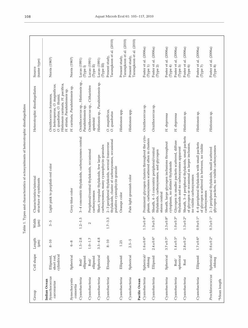
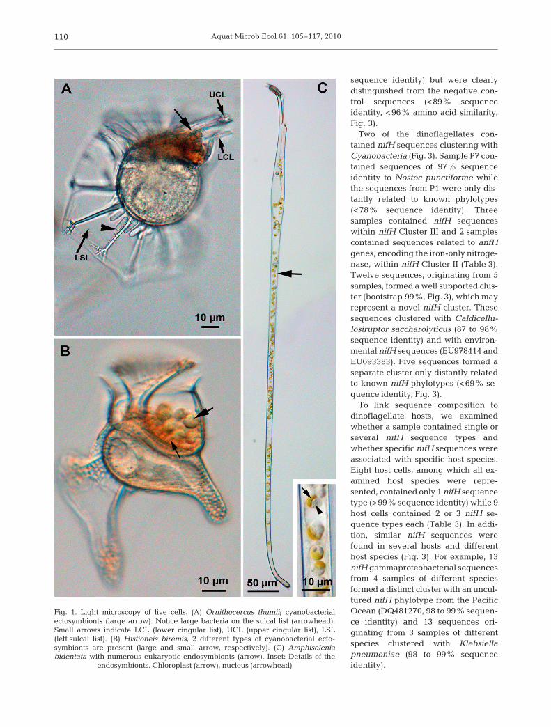
The morphologies (e.g. color, shape and size) of sym-bionts of heterotrophic dinoflagellates (~100 cells)were compared to published data on ectosymbionts(Table 1) and endosymbionts (Table 2). All Ornithocer-cus spp. cells had orange and elongated cyanobacter-ial ectosymbionts located within the cingulum, whilesome also had large rod-shaped non-cyanobacterialprokaryotes on their sulcal lists (Fig. 1A, Table 1). His-tioneis spp. contained 2 other types of cyanobacterial
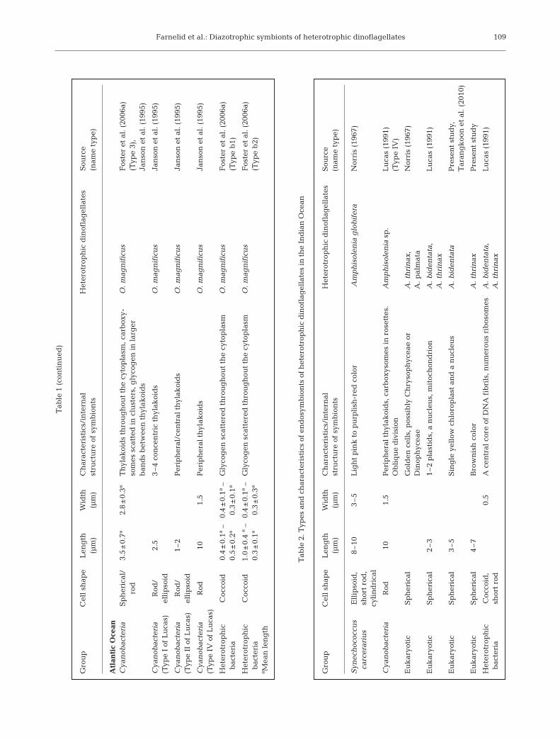
ectosymbionts (Fig. 1B, Table 1). In Amphisolenia spp.only endosymbiotic spheres of 3 to 7 µm were found(Fig. 1C, Table 2). The endosymbionts in A. bidentatacontained a single yellow chloroplast and a nucleusdemonstrating its eukaryotic origin. The symbionts ofA. thrinax had a more brownish color, but whetherthese symbionts were of a eukaryotic origin is unclear(not shown).
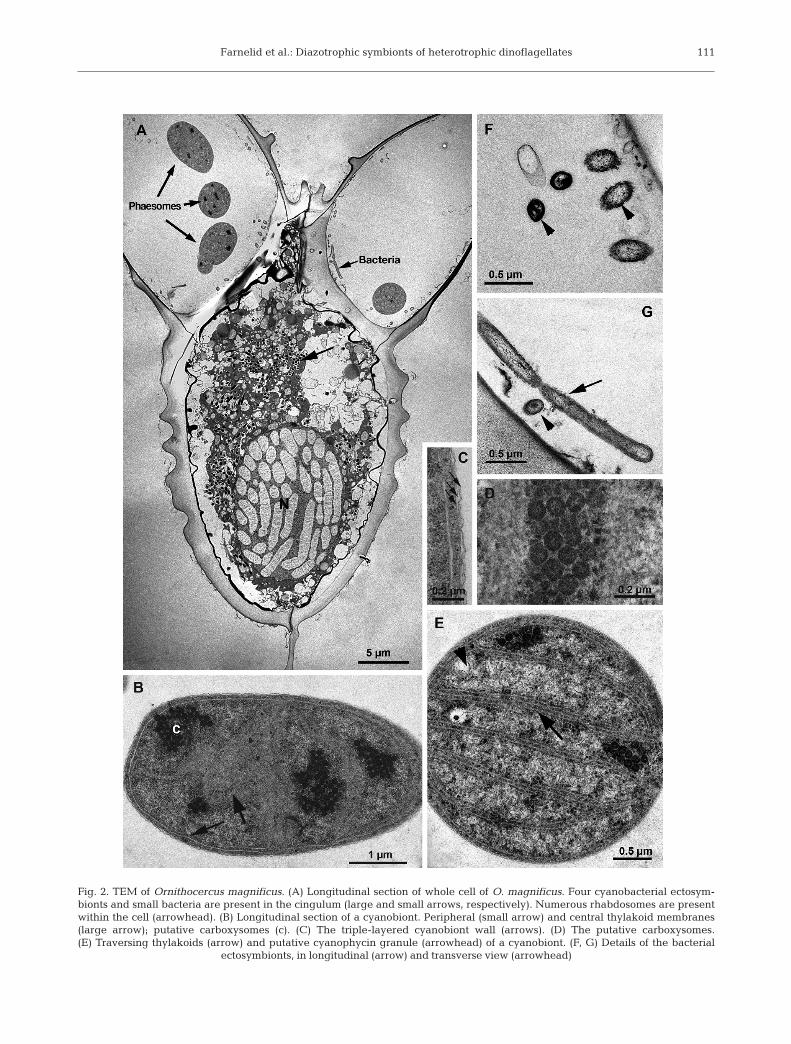
TEM revealed that the ectosymbionts of Ornitho-cercus magnificus and O. quadratus were Cyanobac-teria and heterotrophic bacteria (Fig. 2A). Cyanobacte-rial ectosymbionts were observed in the cingulum ofboth species (Fig. 2A), though most were lost duringthe preparation process for TEM. These all appearedto be of the same type, i.e. containing 1 to 3 peripheralthylakoid bands in addition to several bands traversingthe cell (Fig. 2B,E). Clusters of polyhedral granules,carboxysomes (Lucas 1991), were present in all thecyanobacterial ectosymbionts examined (Fig. 2B,D). Insome, electron translucent granules were present(Fig. 2E), similar to putative cyanophycin granules(Lucas 1991). A typical eubacterial Gram-negativewall, consisting of a thin wall inbetween 2 membranes,surrounded the cyanobiont cells (Fig. 2C). Small rod-shaped, 1.5 × 0.2 µm, heterotrophic bacteria wereobserved in the cingulum of some Ornithocercus cells(Fig. 2A,F,G). Unfortunately, the large rod-shaped het-erotrophic bacteria seen on the sulcal list by lightmicroscopy (Fig. 1A) were lost during the TEM prepa-ration procedure.
nifH sequence composition and phylogeny
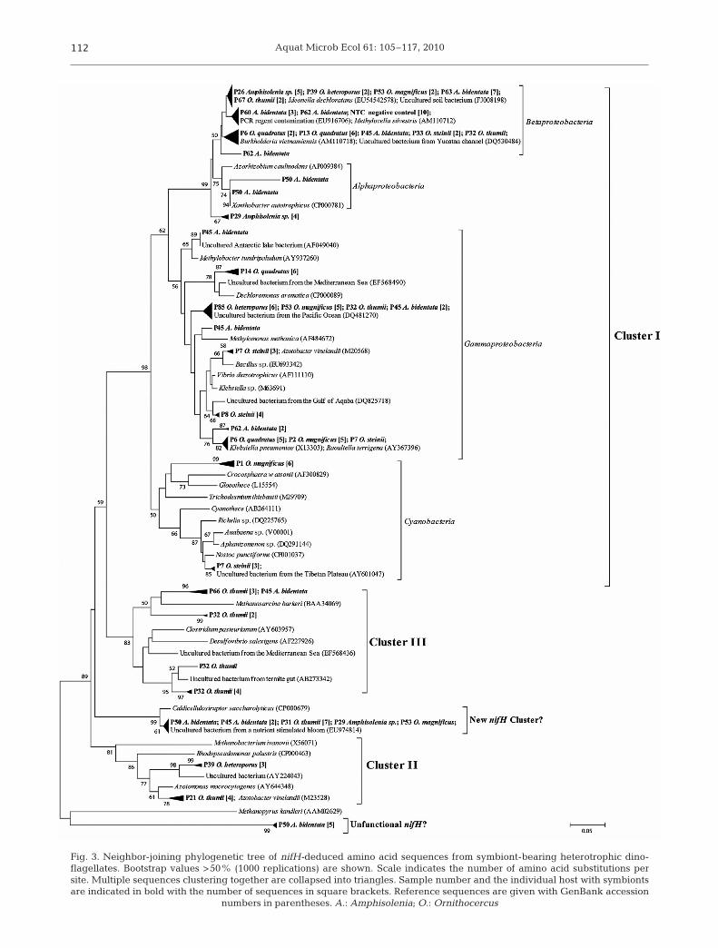
nifH amplicons were obtained from 23 of the 37analyzed symbiont-bearing heterotrophic dinoflagel-lates. The 137 nifH sequences obtained were relatedto nifH Cluster I (Cyanobacteria and Alpha-, Beta-,and Gammaproteobacteria), Cluster II (alternativenitrogenases; anfH) and Cluster III (anaerobic bacte-ria) as defined by Chien & Zinder (1996). Sixty-eightpercent of the sequences (originating from 17 sam-ples) were most similar to proteobacterial nifH se-quences (Fig. 3). All 10 sequences from the negativecontrol formed a cluster within Betaproteobacteria(>98% within-cluster sequence identity, Fig. 3) re-lated to a previously reported PCR reagent contami-nant sequence. Four sample sequences (A. bidentata,samples P60 and P62, Table 3) were affiliated withthis cluster, but were not identical to the negativecontrol sequences. Other sequences clustering withBetaproteobacteria were most similar to Ideonelladechloratans (18 sequences from 5 samples; 91 to92% sequence identity) and Burkholderia vietnam-iensis (12 sequences from 5 samples; 97 to 98%
107
Aquat Microb Ecol 61: 105–117, 2010108
Gro
up
Cel
l sh
ape
Len
gth
Wid
thC
har
acte
rist
ics/
inte
rnal
H
eter
otro
ph
ic d
inof
lag
ella
tes
Sou
rce
(µm
)(µ
m)
stru
ctu
re o
f sy
mb
ion
ts
(nam
e ty
pe)
Ind
ian
Oce
anS
ynec
hoc
occu
s E
llip
soid
,8
–10
3–
5L
igh
t p
ink
to
pu
rpli
sh-r
ed c
olor
Orn
ith
ocer
cus
form
osu
s,N
orri
s (1
967)
carc
erar
ius
shor
t ro
d,
O. h
eter
opor
us,
O. m
agn
ific
us,
cyli
nd
rica
lO
. qu
adra
tus,
O. t
hu
mii
,H
isti
onei
s ca
rin
ata,
H. p
acif
ica,
H. s
tria
ta, P
arah
isti
onei
ssp
.
Syn
ech
ocys
tis
Sp
her
ical
6–
8G
rey-
blu
e co
lor
H. c
arin
ata,
Par
ahis
tion
eis
sp.
Nor
ris
(196
7)co
nso
rtia
Cya
nob
acte
ria
Rod
/1.
5–2
.81.
2–1
.53
–4
con
cen
tric
th
ylak
oid
s, c
arb
oxys
omes
cen
tral
Orn
ith
ocer
cus
sp.,
His
tion
eis
sp.,
Lu
cas
(199
1)el
lip
soid
Par
ahis
tion
eis
sp.
(Typ
e I)
Cya
nob
acte
ria
Rod
/1.
0–1
.72
Per
iph
eral
/cen
tral
th
ylak
oid
s, o
ccas
ion
al
Orn
ith
ocer
cus
sp.,
Cit
har
iste
s L
uca
s (1
991)
elli
pso
idca
rbox
ysom
es
apst
ein
ii(T
ype
II)
Cya
nob
acte
ria
Sp
her
ical
3.5
–4.
8S
hor
t, i
rreg
ula
r th
ylak
oid
s, f
ew l
arg
e H
isti
onei
ssp
., P
arah
isti
onei
ssp
.L
uca
s (1
991)
carb
oxys
omes
, man
y cy
anop
hyc
in g
ran
ule
s(T
ype
III)
Cya
nob
acte
ria
Elo
ng
ate
8–1
01.
7–
3.3
2–
3 p
erip
her
al t
hyl
akoi
ds,
sev
eral
tra
nsv
erse
O
. mag
nif
icu
s,P
rese
nt
stu
dy,
th
ylak
oid
s, c
lust
er o
f ca
rbox
ysom
es, o
ccas
ion
al
O. q
uad
ratu
sT
aran
gk
oon
et
al. (
2010
)p
uta
tive
cya
nop
hyc
in g
ran
ule
Cya
nob
acte
ria
Ell
ipso
id1.
25O
ran
ge
colo
rH
isti
onei
ssp
p.
Pre
sen
t st
ud
y,T
aran
gk
oon
et
al. (
2010
)
Cya
nob
acte
ria
Sp
her
ical
2.5
–5
Pal
e li
gh
t g
reen
ish
col
orH
isti
onei
ssp
p.
Pre
sen
t st
ud
y,T
aran
gk
oon
et
al. (
2010
)
Pac
ific
Oce
an
Cya
nob
acte
ria
Sp
her
ical
/1.
6±
0.6a
1.3
±0.
4aP
rom
inen
t g
lyco
gen
clu
ster
s th
rou
gh
out
the
cyto
-O
rnit
hoc
ercu
ssp
.F
oste
r et
al.
(20
06a)
oblo
ng
pla
sm, c
arb
oxys
omes
sca
tter
ed o
ften
in
clu
ster
s(T
ype
1)
Cya
nob
acte
ria
Ell
ipso
id2.
4±
0.6a
1.9
±0.
5aS
hea
th, 4
–5
con
cen
tric
per
iph
eral
/cen
tral
O
rnit
hoc
ercu
ssp
.F
oste
r et
al.
(20
06a)
thyl
akoi
ds,
car
box
ysom
es, a
nd
gly
cog
en
(Typ
e 2)
stor
es c
entr
al
Cya
nob
acte
ria
Sp
her
ical
3.7
±0.
7a2.
3±
0.8a
Sh
eath
, lar
ge
gly
cog
en i
ncl
usi
ons
thro
ug
hou
t H
. dep
ress
aF
oste
r et
al.
(20
06a)
cyto
pla
sm, n
o d
isti
nct
th
ylak
oid
s(T
ype
4)
Cya
nob
acte
ria
Rod
/1.
4±
0.5a
1.0
±0.
3aG
lyco
gen
in
sm
alle
r p
ack
ets
scat
tere
d, d
iffu
se
H. d
epre
ssa
Fos
ter
et a
l. (
2006
a)sp
her
ical
thyl
akoi
ds
and
no
carb
oxys
omes
ap
par
ent
(Typ
e 5)
Cya
nob
acte
ria
Rod
2.8
±0.
2a1.
3±
0.2a
Sh
eath
, 3–
4 p
erip
her
al t
hyl
akoi
ds,
sm
all
pac
ket
s H
isti
onei
ssp
.F
oste
r et
al.
(20
06a)
of g
lyco
gen
or
occa
sion
al a
s la
rger
in
clu
sion
, (T
ype
6)n
o vi
sib
le c
arb
oxys
omes
Cya
nob
acte
ria
Ell
ipso
id1.
7±
0.6a
0.9
±0.
1a4
–6
per
iph
eral
th
ylak
oid
s w
ith
sm
all
pac
ket
sH
isti
onei
ssp
.F
oste
r et
al.
(20
06a)
of g
lyco
gen
sca
tter
ed i
n b
etw
een
, no
visi
ble
(T
ype
7)ca
rbox
ysom
es
Pro
chlo
roco
ccu
sS
ph
eric
al/
0.6
±0.
2a0.
3±
0.1a
2–
3 p
erip
her
al t
hyl
akoi
ds,
sm
all
scat
tere
d
His
tion
eis
sp.
Fos
ter
et a
l. (
2006
a)ob
lon
gg
lyco
gen
pac
ket
s, n
o vi
sib
le c
arb
oxys
omes
(Typ
e 8)
a Mea
n l
eng
th
Tab
le 1
. Typ
es a
nd
ch
arac
teri
stic
s of
ect
osym
bio
nts
of
het
erot
rop
hic
din
ofla
gel
late
s
Farnelid et al.: Diazotrophic symbionts of heterotrophic dinoflagellates 109
Gro
up
Cel
l sh
ape
Len
gth
Wid
thC
har
acte
rist
ics/
inte
rnal
H
eter
otro
ph
ic d
inof
lag
ella
tes
Sou
rce
(µm
)(µ
m)
stru
ctu
re o
f sy
mb
ion
ts
(nam
e ty
pe)
Atl
anti
c O
cean
Cya
nob
acte
ria
Sp
her
ical
/3.
5±
0.7a
2.8
±0.
3aT
hyl
akoi
ds
thro
ug
hou
t th
e cy
top
lasm
, car
box
y-
O. m
agn
ific
us
Fos
ter
et a
l. (
2006
a)ro
dso
mes
sca
tted
in
clu
ster
s, g
lyco
gen
in
lar
ger
(Typ
e 3)
, b
and
s b
etw
een
th
ylak
oid
sJa
nso
n e
t al
. (19
95)
Cya
nob
acte
ria
Rod
/2.
53
–4
con
cen
tric
th
ylak
oid
sO
. mag
nif
icu
sJa
nso
n e
t al
. (19
95)
(Typ
e I
of L
uca
s)el
lip
soid
Cya
nob
acte
ria
Rod
/1
–2P
erip
her
al/c
entr
al t
hyl
akoi
ds
O. m
agn
ific
us
Jan
son
et
al. (
1995
)(T
ype
II o
f L
uca
s)el
lip
soid
Cya
nob
acte
ria
Rod
101.
5P
erip
her
al t
hyl
akoi
ds
O. m
agn
ific
us
Jan
son
et
al. (
1995
)(T
ype
IV o
f L
uca
s)
Het
erot
rop
hic
C
occo
id0.
4±
0.1a
– 0.
4±
0.1a
– G
lyco
gen
sca
tter
ed t
hro
ug
hou
t th
e cy
top
lasm
O. m
agn
ific
us
Fos
ter
et a
l. (
2006
a)b
acte
ria
0.5
±0.
2a0.
3±
0.1a
(Typ
e b
1)
Het
erot
rop
hic
C
occo
id1.
0±
0.4
a–
0.4
±0.
1a–
Gly
cog
en s
catt
ered
th
rou
gh
out
the
cyto
pla
smO
. mag
nif
icu
sF
oste
r et
al.
(20
06a)
bac
teri
a0.
3±
0.1a
0.3
±0.
3a(T
ype
b2)
a Mea
n l
eng
th
Tab
le 1
(co
nti
nu
ed)
Gro
up
Cel
l sh
ape
Len
gth
Wid
thC
har
acte
rist
ics/
inte
rnal
H
eter
otro
ph
ic d
inof
lag
ella
tes
Sou
rce
(µm
)(µ
m)
stru
ctu
re o
f sy
mb
ion
ts
(nam
e ty
pe)
Syn
ech
ococ
cus
Ell
ipso
id,
8–1
03
–5
Lig
ht
pin
k t
o p
urp
lish
-red
col
orA
mp
his
olen
ia g
lob
ifer
aN
orri
s (1
967)
carc
erar
ius
shor
t ro
d,
cyli
nd
rica
l
Cya
nob
acte
ria
Rod
101.
5P
erip
her
al t
hyl
akoi
ds,
car
box
ysom
es i
n r
oset
tes.
Am
ph
isol
enia
sp.
Lu
cas
(199
1)O
bli
qu
e d
ivis
ion
(Typ
e IV
)
Eu
kar
yoti
cS
ph
eric
alG
old
en c
ells
, pos
sib
ly C
hry
sop
hyc
eae
orA
. th
rin
ax,
Nor
ris
(196
7)D
inop
hyc
eae
A. p
alm
ata
Eu
kar
yoti
cS
ph
eric
al2
–3
1–2
pla
stid
s, a
nu
cleu
s, m
itoc
hon
dri
onA
. bid
enta
ta,
Lu
cas
(199
1)A
. th
rin
ax
Eu
kar
yoti
cS
ph
eric
al3
–5
Sin
gle
yel
low
ch
loro
pla
st a
nd
a n
ucl
eus
A. b
iden
tata
Pre
sen
t st
ud
y,
Tar
ang
koo
n e
t al
. (20
10)
Eu
kar
yoti
cS
ph
eric
al4
–7
Bro
wn
ish
col
orA
. th
rin
axP
rese
nt
stu
dy
Het
erot
rop
hic
C
occo
id,
0.5
A c
entr
al c
ore
of D
NA
fib
rils
, nu
mer
ous
rib
osom
esA
. bid
enta
ta,
Lu
cas
(199
1)b
acte
ria
shor
t ro
dA
. th
rin
ax
Tab
le 2
. Typ
es a
nd
ch
arac
teri
stic
s of
en
dos
ymb
ion
ts o
f h
eter
otro
ph
ic d
inof
lag
ella
tes
in t
he
Ind
ian
Oce
an
Aquat Microb Ecol 61: 105–117, 2010
sequence identity) but were clearlydistinguished from the negative con-trol sequences (<89% sequenceidentity, <96% amino acid similarity,Fig. 3).
Two of the dinoflagellates con-tained nifH sequences clustering withCyanobacteria (Fig. 3). Sample P7 con-tained sequences of 97% sequenceidentity to Nostoc punctiforme whilethe sequences from P1 were only dis-tantly related to known phylotypes(<78% sequence identity). Threesamples contained nifH sequenceswithin nifH Cluster III and 2 samplescontained sequences related to anfHgenes, encoding the iron-only nitroge-nase, within nifH Cluster II (Table 3).Twelve sequences, originating from 5samples, formed a well supported clus-ter (bootstrap 99%, Fig. 3), which mayrepresent a novel nifH cluster. Thesesequences clustered with Caldicellu-losiruptor saccharolyticus (87 to 98%sequence identity) and with environ-mental nifH sequences (EU978414 andEU693383). Five sequences formed aseparate cluster only distantly relatedto known nifH phylotypes (<69% se-quence identity, Fig. 3).
To link sequence composition todinoflagellate hosts, we examinedwhether a sample contained single orseveral nifH sequence types andwhether specific nifH sequences wereassociated with specific host species.Eight host cells, among which all ex-amined host species were repre-sented, contained only 1 nifH sequencetype (>99% sequence identity) while 9host cells contained 2 or 3 nifH se-quence types each (Table 3). In addi-tion, similar nifH sequences werefound in several hosts and differenthost species (Fig. 3). For example, 13nifH gammaproteobacterial sequencesfrom 4 samples of different speciesformed a distinct cluster with an uncul-tured nifH phylotype from the PacificOcean (DQ481270, 98 to 99% sequen-ce identity) and 13 sequences ori-ginating from 3 samples of differentspecies clustered with Klebsiellapneumoniae (98 to 99% sequenceidentity).
110
Fig. 1. Light microscopy of live cells. (A) Ornithocercus thumii; cyanobacterialectosymbionts (large arrow). Notice large bacteria on the sulcal list (arrowhead).Small arrows indicate LCL (lower cingular list), UCL (upper cingular list), LSL(left sulcal list). (B) Histioneis biremis; 2 different types of cyanobacterial ecto-symbionts are present (large and small arrow, respectively). (C) Amphisoleniabidentata with numerous eukaryotic endosymbionts (arrow). Inset: Details of the
endosymbionts. Chloroplast (arrow), nucleus (arrowhead)
Farnelid et al.: Diazotrophic symbionts of heterotrophic dinoflagellates 111
Fig. 2. TEM of Ornithocercus magnificus. (A) Longitudinal section of whole cell of O. magnificus. Four cyanobacterial ectosym-bionts and small bacteria are present in the cingulum (large and small arrows, respectively). Numerous rhabdosomes are presentwithin the cell (arrowhead). (B) Longitudinal section of a cyanobiont. Peripheral (small arrow) and central thylakoid membranes(large arrow); putative carboxysomes (c). (C) The triple-layered cyanobiont wall (arrows). (D) The putative carboxysomes. (E) Traversing thylakoids (arrow) and putative cyanophycin granule (arrowhead) of a cyanobiont. (F, G) Details of the bacterial
ectosymbionts, in longitudinal (arrow) and transverse view (arrowhead)
Aquat Microb Ecol 61: 105–117, 2010112
Fig. 3. Neighbor-joining phylogenetic tree of nifH-deduced amino acid sequences from symbiont-bearing heterotrophic dino-flagellates. Bootstrap values >50% (1000 replications) are shown. Scale indicates the number of amino acid substitutions persite. Multiple sequences clustering together are collapsed into triangles. Sample number and the individual host with symbiontsare indicated in bold with the number of sequences in square brackets. Reference sequences are given with GenBank accession
numbers in parentheses. A.: Amphisolenia; O.: Ornithocercus
Farnelid et al.: Diazotrophic symbionts of heterotrophic dinoflagellates 113
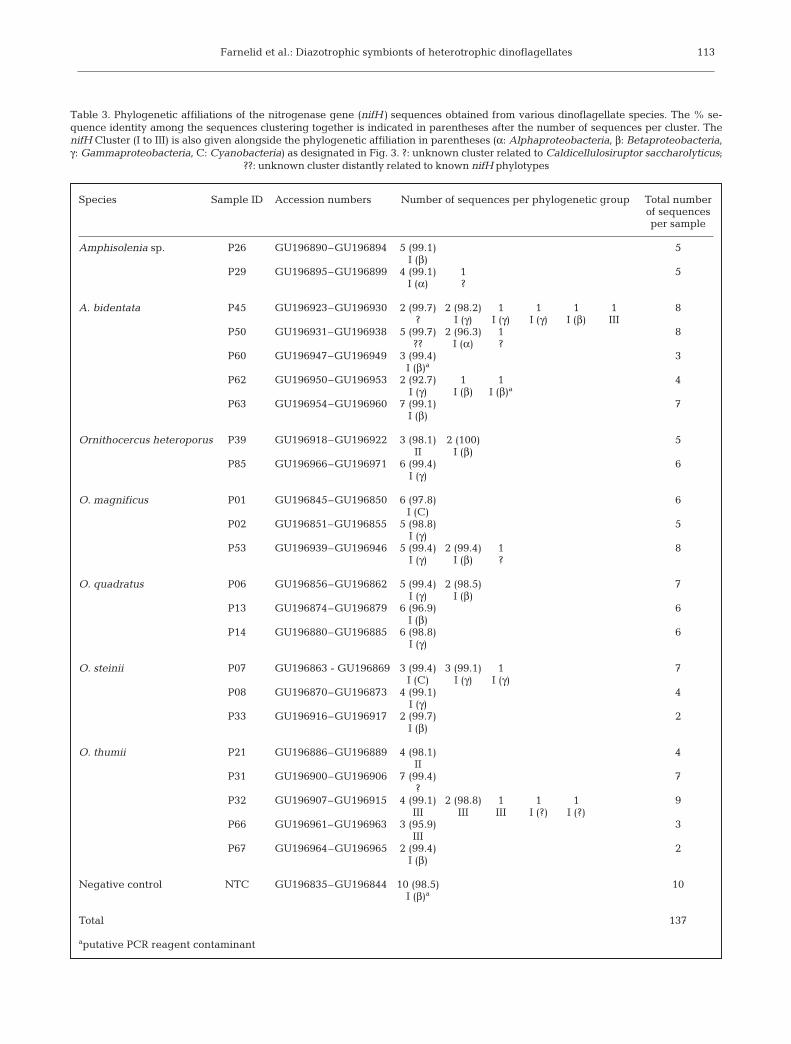
Species Sample ID Accession numbers Number of sequences per phylogenetic group Total number of sequences per sample
Amphisolenia sp. P26 GU196890–GU196894 5 (99.1) 5I (β)
P29 GU196895–GU196899 4 (99.1) 1 5I (α) ?
A. bidentata P45 GU196923–GU196930 2 (99.7) 2 (98.2) 1 1 1 1 8? I (γ) I (γ) I (γ) I (β) III
P50 GU196931–GU196938 5 (99.7) 2 (96.3) 1 8?? I (α) ?
P60 GU196947–GU196949 3 (99.4) 3I (β)a
P62 GU196950–GU196953 2 (92.7) 1 1 4I (γ) I (β) I (β)a
P63 GU196954–GU196960 7 (99.1) 7I (β)
Ornithocercus heteroporus P39 GU196918–GU196922 3 (98.1) 2 (100) 5II I (β)
P85 GU196966–GU196971 6 (99.4) 6I (γ)
O. magnificus P01 GU196845–GU196850 6 (97.8) 6I (C)
P02 GU196851–GU196855 5 (98.8) 5I (γ)
P53 GU196939–GU196946 5 (99.4) 2 (99.4) 1 8I (γ) I (β) ?
O. quadratus P06 GU196856–GU196862 5 (99.4) 2 (98.5) 7I (γ) I (β)
P13 GU196874–GU196879 6 (96.9) 6I (β)
P14 GU196880–GU196885 6 (98.8) 6I (γ)
O. steinii P07 GU196863 - GU196869 3 (99.4) 3 (99.1) 1 7I (C) I (γ) I (γ)
P08 GU196870–GU196873 4 (99.1) 4I (γ)
P33 GU196916–GU196917 2 (99.7) 2I (β)
O. thumii P21 GU196886–GU196889 4 (98.1) 4II
P31 GU196900–GU196906 7 (99.4) 7?
P32 GU196907–GU196915 4 (99.1) 2 (98.8) 1 1 1 9III III III I (?) I (?)
P66 GU196961–GU196963 3 (95.9) 3III
P67 GU196964–GU196965 2 (99.4) 2I (β)
Negative control NTC GU196835–GU196844 10 (98.5) 10I (β)a
Total 137
aputative PCR reagent contaminant
Table 3. Phylogenetic affiliations of the nitrogenase gene (nifH) sequences obtained from various dinoflagellate species. The % se-quence identity among the sequences clustering together is indicated in parentheses after the number of sequences per cluster. ThenifH Cluster (I to III) is also given alongside the phylogenetic affiliation in parentheses (α: Alphaproteobacteria, β: Betaproteobacteria,γ: Gammaproteobacteria, C: Cyanobacteria) as designated in Fig. 3. ?: unknown cluster related to Caldicellulosiruptor saccharolyticus;
??: unknown cluster distantly related to known nifH phylotypes
Aquat Microb Ecol 61: 105–117, 2010
DISCUSSION
The role of heterotrophic dinoflagellate symbiontshas been a mystery for many years. Due to the appar-ent restriction of these consortia to marine watersdeplete of inorganic reduced N, it has been suggestedthat the cyanobacterial symbionts provide their hostswith N through N2 fixation (Gordon et al. 1994, Jyoth-ibabu et al. 2006, Tarangkoon et al. 2010). In this firstreport of nifH genes from dinoflagellate–Cyanobac-teria consortia, we show that 23 of the 37 investigateddinoflagellate cells carried putative diazotrophs, andthat 21 of these carried nifH genes exclusively relatedto heterotrophic bacteria. Hence, our analysis suggeststhat heterotrophic diazotrophs rather than Cyanobac-teria supply the dinoflagellates with reduced N.
Identification of symbionts through microscopy
The identification of symbionts of heterotrophicdinoflagellates has so far primarily been based on size,shape, pigmentation, and in some cases, ultrastructure.Heterotrophic bacterial ectosymbionts and/or cyano-bacterial ectosymbionts of heterotrophic dinoflagel-lates have been described from the Indian, Pacific andAtlantic Oceans (Table 1). The types of ectosymbiontsthat we observed are similar to those previously de-scribed from the Indian Ocean (Table 1). For instance,the fairly large ectosymbionts (8–10 × 3–5 µm) inOrnithocercus magnificus and O. quadratus were sim-ilar in pigmentation, size and shape to Synechococcuscarcerarius (Norris 1967; our Table 1). This was sup-ported by a molecular study targeting cyanobacterial16S rRNA gene sequences from symbionts of eukary-otic hosts where the majority of the cyanobacterialsequences were closely related to Synechococcus(>96% similarity; Foster et al. 2006b). Further, theectosymbionts of Histioneis carinata and H. biremisboth contained at least 2 types of reddish cyanobac-terial ectosymbionts; a large one (2.5 to 5 µm) anda smaller one (1.25 µm, Fig. 1B), similar to Types IIIand I, respectively (Table 2 in Lucas 1991).
In accordance with previous observations from theIndian Ocean, we found cyanobacterial and eukaryoticendosymbionts in Amphisolenia spp. (our Table 2; Ta-rangkoon et al. 2010). Photosynthetic endosymbiontswere observed in both A. bidentata (Fig. 1C) and A.thrinax. So far only one type of prokaryotic endo-symbiont, S. carcerarius, has been reported; originatingfrom A. globifera (Norris 1967, Hallegraeff & Jeffrey1984, Lucas 1991), while eukaryotic endosymbiontshave been reported from different species of Am-phisolenia (Norris 1967, Lucas 1991). We observed anadditional type of eukaryotic symbiont in A. bidentata.
Interestingly, Foster et al. (2006b) also recovered 16SrRNA genes <92% identical to eukaryotic plastids froman A. bidentata host, which could represent the eukary-otic symbionts as reported by Lucas (1991) and/or inthis study (Table 2).
Heterotrophic bacteria have previously been re-ported as both ecto- and endosymbionts of hetero-trophic dinoflagellates (Tables 1 & 2). For instance,Foster et al. (2006a) detected 2 morphotypes of hetero-trophic bacteria associated with Ornithocercus mag-nificus. Likewise, we observed heterotrophic bacterialectosymbionts of Ornithocercus spp. (Fig. 1A) and bothcyanobacterial and heterotrophic bacterial ectosym-bionts for O. magnificus and O. quadratus (Fig. 2A). InO. magnificus and O. steinii, Janson et al. (1995) ob-served heterotrophic bacteria between the upper andthe lower girdle list of the cingular groove in all sam-ples examined by TEM. Also, groups of heterotrophicbacterial endosymbionts have been observed in thecytoplasm of A. thrinrax and A. bidentata (Lucas 1991).Interestingly, although using primers targeting cyano-bacteria, 26% of the 16S rRNA sequences recoveredfrom eukaryotic marine hosts by Foster et al. (2006b)originated from heterotrophic bacteria; however, noneof these could be directly linked to diazotrophic spe-cies (based on BLASTN search results on unpublishedsequences provided by R. A. Foster). Thus, the occur-rence of heterotrophic ecto- and endosymbionts ofheterotrophic dinoflagellates is not unusual but to ourknowledge, there has been no previous documentationof a N2-fixing potential in these symbionts.
Identities of nifH genes obtained fromsymbiont-bearing dinoflagellates
Although cyanobacterial symbionts were visible inall examined dinoflagellates, only 2 of the 23 cells,which yielded nifH sequences, had sequences relatedto Cyanobacteria. Intriguingly, sample P7 (Ornithocer-cus steinii) contained nifH sequences of 97% sequenceidentity with the filamentous heterocystous cyano-bacterium Nostoc punctiforme, which is known fromfreshwater and for its endosymbiotic associations withplants (Meeks et al. 2002). Similarly, 16S rRNA genesequences 92% identical to Nostoc spp. were found inan Amphisolenia bidentata host (Foster et al. 2006b).Taken together, these results may suggest that symbio-sis facilitates the survival of Nostoc spp. in the salinemarine environment. nifH sequences related to Cyano-bacteria were also obtained from sample P1 (O. mag-nificus) but these were only distantly related to knownphylotypes (Fig. 3). Since very few nifH sequencesrelated to Cyanobacteria were found, we find itunlikely that the role of the Cyanobacteria in the
114
Farnelid et al.: Diazotrophic symbionts of heterotrophic dinoflagellates
symbiosis should be to supply the host with reducedN. Importantly, in a parallel study (F. Farnelid & L.Riemann unpubl.) using the same primer sets, wedetected representatives from the major groups of uni-cellular Cyanobacteria (e.g. Crocosphaera watsonii,Cyanothece sp. and Group A; Bergman et al. 1997, Stal& Zehr 2008). Thus, the lack of these known Cyano-bacteria in the present data set is not due to a primermismatch.
Twenty-one samples contained sequences clusteringin nifH Cluster I, with Alpha-, Beta-, and Gamma-proteobacteria (Fig. 3, Table 3). nifH gene contamina-tion of PCR reagents, particularly with alpha- andbetaproteobacterial sequences, may occur in thenested PCR (Zehr et al. 2003, Goto et al. 2005). How-ever, the 10 nifH sequences we obtained from non-vis-ible negative control samples clustered with only 4sample sequences (Fig. 3, Table 3) and were not iden-tical to any sample sequences. Hence, reagent conta-mination appeared negligible in the present study.However, hypothetical sources of error such as ampli-fication of nifH genes derived from bacteria ingestedby the host or from free-living bacteria which mayhave been passed through the 3 washing steps with0.2 µm filtered seawater cannot be ruled out. Inaddition, as the detection limit of the nested nifH assayis unknown, samples which did not yield a nifHproduct could theoretically have contained putativediazotrophs.
Diverse Proteobacteria within nifH Cluster I arecommonly detected in marine waters (e.g. Zehr et al.1998, Church et al. 2005, Langlois et al. 2005, Hewsonet al. 2007 Moisander et al. 2008). Associated withdinoflagellates, we found 8 diverse clusters of nifH se-quences related to Gammaproteobacteria while only2 sequences were related to Alphaproteobacteria(Fig. 3). Interestingly, 30 sequences (22% of all se-quences) were affiliated with 2 betaproteobacterialclusters, distinct from the negative control sequences(Fig. 3). Similarly, bacteria associated with the photo-synthetic dinoflagellate Gyrodinium instriatum weredominated by Betaproteobacteria (Alverca et al. 2002).Hence, although rare in marine ecosystems (Barberán& Casamayor 2010), Betaproteobacteria appear com-mon as symbionts of dinoflagellates.
Sequences clustering in nifH Cluster II were ob-tained from 2 samples (Ornithocercus heteroporus P39and O. thumii P21, Table 3). Molybdenum-indepen-dent nitrogenases are present in a diverse group ofdiazotrophs and second alternative nitrogenases areexpressed under molybdenum- and vanadium-defi-cient conditions (Betancourt et al. 2008). Bacteria con-taining alternative nitrogenase genes have been iso-lated from diverse marine environments (Loveless etal. 1999) but interestingly, anfH related genes seem to
be absent in sub-tropical and tropical open waters (e.g.Zehr et al. 1998, Church et al. 2005, Langlois et al.2005, Hewson et al. 2007, Moisander et al. 2008). Thus,the recovery of anfH related genes suggests that sym-bionts of dinoflagellates may be an environmentalniche in open water where second alternative nitroge-nase genes can be used.
Sequences from Cluster III, which contains nifHgenes from anaerobic bacteria, have been detected(Church et al. 2005) but appear uncommon in the openocean (Langlois et al. 2005, 2008). The presence ofCluster III sequences in 3 dinoflagellates thereforesuggests that the dinoflagellate–Cyanobacteria con-sortia provide low oxygen (O2) habitats required for N2
fixation (Paerl & Prufert 1987). Similarly, Cluster IIIsequences from strict anaerobes and nitrogenaseactivity have been detected in association with zoo-plankton (Braun et al. 1999). However, since our Clus-ter III sequences were only distantly related to culti-vated anaerobic bacteria (76 to 87% sequenceidentity), the phenotypes they represent are rather un-certain. Survival of strict anaerobes associated withdinoflagellates would presumably require verticalinheritance of these symbionts as the dinoflagellatehost divides. However, given the observed non-hostspecificity for the symbionts (see below), it may bemore likely that the obtained Cluster III sequencesderive from facultatively anaerobic bacteria.
Twelve sequences originating from 5 samplesformed a discrete nifH cluster separate from the knownnifH Clusters I to IV (Chien & Zinder 1996; our Fig. 3).These sequences had a sequence identity of 87 to 98%to the nifH gene of Caldicellulosiruptor saccharolyticus(van de Werken et al. 2008), which can grow in the ab-sence of reduced N (van Niel et al. 2002). It is surprisingto find sequences closely related to an anaerobic ex-treme thermophile in the pelagic zone; however, thecluster also contains nifH sequences from a marinebloom and from symbionts of corals. Hence, surface-associated growth in the marine environment, such asin association with dinoflagellates, may be characteris-tic for these bacteria.
Eleven clone libraries yielded 2 or 3 different nifHsequence types per dinoflagellate. This suggests thepresence of mixed assemblages of diazotrophic sym-bionts in host cells (Table 3), consistent with previousmicroscopy observations of mixed populations ofcyanobionts and/or bacterial cell types in one host cell(Foster et al. 2006a). In addition, observations of sev-eral specimens of the same dinoflagellate species withdiverging nifH sequences and different species ofdinoflagellates hosting identical nifH sequences sug-gested that the nifH phylotypes were not host specific.Similar patterns of non-host specific 16S rRNA genephylotypes were also observed for cyanobacterial sym-
115

Aquat Microb Ecol 61: 105–117, 2010
bionts in tintinnids, dinoflagellates, and radiolarians(Foster et al. 2006b). In contrast, in the Richelia intra-cellularis-diatom symbiosis a divergence of hetR andnifH sequences of symbionts from different host gen-era was interpreted as an indication of host specificity(Janson et al. 1999, Foster & Zehr 2006). Thus, itappears that at any one time dinoflagellate hosts maycontain multiple symbionts but the low degree ofspecificity also indicates that their dependence on spe-cialized symbionts is not fundamental.
Putative ecological roles of the consortia
In dinoflagellate–Cyanobacteria consortia, the host’srequirement for fixed carbon as well as N is presum-ably the driving force for the relationship. Our resultsshow that heterotrophic bacterial symbionts ratherthan cyanobionts have the genetic potential for fixingN2. Consequently, we speculate that the widespread,and somewhat counter-intuitive distribution of theselarge (50 to 1000 µm) species of heterotrophic dinofla-gellates in the oligotrophic subtropical and tropicaloceans is partly made possible by symbiont-mediatedphotosynthesis (Cyanobacteria) and N2 fixation (het-erotrophic bacteria).
Acknowledgements. Danish Galathea expedition and theCaptain of HMDS ‘Vædderen’, C. Smidt, and his crew arethanked for excellent assistance in connection with sampling.We thank R. A. Foster for generously providing unpublishedsequence data and the anonymous reviewers for helpful com-ments on an earlier version of the manuscript. The project wassupported by grants from Knud Højgaards Fond, Danish Nat-ural Sciences Research Council (272-05-0333 and 272-06-0485 to P.J.H. and 277-05-0421 to G.H.) and Dansk Expedi-tions fond. The work of H.F. was supported by the SwedishResearch Council FORMAS (217-2006-342 to L.R.). The pre-sent work was carried out as part of the Galathea 3 expeditionunder the auspices of the Danish Expedition Foundation. Thisis Galathea 3 contribution no. P67.
LITERATURE CITED
Alverca E, Biegala IC, Kennaway GM, Lewis J, Franca S(2002) In situ identification and localization of bacteriaassociated with Gyrodinium instriatum (Gymnodiniales,Dinophyceae) by electron and confocal microscopy. Eur JPhycol 37:523–530
Barberán A, Casamayor EO (2010) Global phylogenetic com-munity structure and beta-diversity patterns in surfacebacterioplankton metacommunities. Aquat Microb Ecol59:1–10
Bergman B, Gallon JR, Rai AN, Stal LJ (1997) N2-fixation bynon-heterocystous cyanobacteria. FEMS Microbiol Rev19:139–185
Betancourt DA, Loveless TM, Brown JW, Bishop PE (2008)Characterization of diazotrophs containing Mo-indepen-dent nitrogenases, isolated from diverse natural environ-ments. Appl Environ Microbiol 74:3471–3480
Biegala IC, Raimbault P (2008) High abundance of diazo-trophic picocyanobacteria (<3 µm) in a Southwest Pacificcoral lagoon. Aquat Microb Ecol 51:45–53
Braun ST, Proctor LM, Zani S, Mellon MT, Zehr JP (1999)Molecular evidence for zooplankton-associated nitrogen-fixing anaerobes based on amplification of the nifH gene.FEMS Microbiol Ecol 28:273–279
Capone DG, Zehr JP, Paerl HW, Bergman B, Carpenter EJ(1997) Trichodesmium, a globally significant marinecyanobacterium. Science 276:1221–1229
Carpenter E, Montoya JP, Burns JA, Mulholland MR, Subra-maniam A, Capone DG (1999) Extensive bloom of a N2-fixing diatom/cyanobacterial association in the tropicalAtlantic Ocean. Mar Ecol Prog Ser 185:273–283
Chien YT, Zinder SH (1996) Cloning, functional organization,transcript studies, and phylogenetic analysis of the com-plete nitrogenase stuctural genes (nifHDK2) and associ-ated genes in the Archaeon Methanosarcina barkeri 227.J Bacteriol 178:143–148
Church MJ, Jenkins BD, Karl DM, Zehr JP (2005) Vertical dis-tributions of nitrogen-fixing phylotypes at Stn ALOHA inthe oligotrophic North Pacific Ocean. Aquat Microb Ecol38:3–14
Farnelid H, Riemann L (2008) Heterotrophic N2-fixing bacte-ria: Overlooked in the marine nitrogen cycle? In: CoutoGN (ed) Nitrogen fixation research progress. Nova Sci-ence Publishers, New York, NY, p 409–423
Foster RA, Zehr JP (2006) Characterization of diatom-cyano-bacteria symbioses on the basis of nifH, hetR and 16SrRNA sequences. Environ Microbiol 8:1913–1925
Foster RA, Carpenter EJ, Bergman B (2006a) Unicellularcyanobionts in open ocean dinoflagellates, radiolarians,and tintinnids: ultrastructure characterization and immuno-localization of phycoerythrin and nitrogenase. J Phycol 42:453–463
Foster RA, Collier JL, Carpenter EJ (2006b) Reverse transcrip-tion PCR amplification of cyanobacterial symbiont 16SrRNA sequences from single non-photosynthetic eukary-otic marine planktonic host cells. J Phycol 42:243–250
Gordon N, Angel DL, Neorl A, Kress N, Kimor B (1994) Het-erotrophic dinoflagellates with symbiotic cyanobacteriaand nitrogen limitation in the Gulf of Aqaba. Mar EcolProg Ser 107:83–88
Goto M, Ando S, Hachisuka Y, Yoneyama T (2005) Contami-nation of diverse nifH and nifH-like DNA into commercialPCR primers. FEMS Microbiol Lett 246:33–38
Hallegraeff GM, Jeffrey SW (1984) Tropical phytoplanktonspecies and pigments of continental shelf waters of Northand North-West Australia. Mar Ecol Prog Ser 20:59–74
Hewson I, Moisander PH, Achilles KM, Carlson CA and oth-ers (2007) Characteristics of diazotrophs in surface toabyssopelagic waters of the Sargasso Sea. Aquat MicrobEcol 46:15–30
Janson S, Carpenter EJ, Bergman B (1995) Immunolabellingof phycoerythrin, ribulose 1,5-biphosphate carboxylase/oxygenase and nitrogenase in the unicellular cyanobiontsof Ornithocercus spp. (Dinophyceae). Phycologia 34:171–176
Janson S, Wouters J, Bergman B, Carpenter EJ (1999) Hostspecificity in the Richelia-diatom symbiosis revealed byhetR gene sequence analysis. Environ Microbiol 1:431–438
Jyothibabu R, Madhu NV, Maheswaran PA, Asha Devi CR,Balasubramanian T, Nair KKC, Achuthankutty CT (2006)Environmentally-related seasonal variation in symbioticassociations of heterotrophic dinoflagellates with cyano-bacteria in the western Bay of Bengal. Symbiosis 42:51–58
Langlois RJ, LaRoche J, Raab PA (2005) Diazotrophic diver-
116
Farnelid et al.: Diazotrophic symbionts of heterotrophic dinoflagellates
sity and distribution in the tropical and subtropicalAtlantic Ocean. Appl Environ Microbiol 71:7910–7919
Langlois RJ, Hümmer D, La Roche J (2008) Abundances anddistributions of the dominant nifH phylotypes in theNorthern Atlantic Ocean. Appl Environ Microbiol 74:1922–1931
Le Moal M, Biegala IC (2009) Diazotrophic unicellular cyano-bacteria in the northwestern Mediterranean Sea: a sea-sonal cycle. Limnol Oceanogr 54:845–855
Loveless TM, Saah JR, Bishop PE (1999) Isolation of nitrogen-fixing bacteria containing Molybdenum-independentnitrogenases from natural environments. Appl EnvironMicrobiol 65:4223–4226
Lucas IAN (1991) Symbionts of the tropical dinophysiales.Ophelia 33:213–224
Meeks JC, Campbell EL, Summers ML, Wong FC (2002) Cel-lular differentiation in the cyanobacterium Nostoc puncti-forme. Arch Microbiol 178:395–403
Moisander PH, Beinart RA, Voss M, Zehr JP (2008) Diversityand abundance of diazotrophic microorganisms in theSouth China Sea during intermonsoon. ISME J 2:954–967
Montoya JP, Holl CM, Zehr JP, Hansen A, Villareal TA,Capone DG (2004) High rates of N2 fixation by unicellulardiazotrophs in the oligotrophic Pacific Ocean. Nature 430:1027–1031
Norris RE (1967) Algal consortisms in marine plankton. In:Krishnamurthy V (ed) Proc Seminar on Sea, Salt andPlants 1965. Central Salt and Marine Chemicals ResearchInstitute, Bhavnagar, India. Catholic Press, Ranchi (Bihar),p 178–179
Paerl HW, Prufert LE (1987) Oxygen-poor microzones as poten-tial sites of microbial N2 fixation in nitrogen-depleted aero-bic marine waters. Appl Environ Microbiol 53:1078–1087
Riemann L, Leitet C, Pommier T, Simu K, Holmfeldt K, Lars-son U, Hagström Å (2008) The native bacterioplanktoncommunity in the central Baltic Sea is influenced by fresh-water bacterial species. Appl Environ Microbiol 74:503–515
Schütt F (1895) Die Peridineen der Plankton-Expedition.Ergebnisse Plankton-Expedition der Humboldt Stiftung,Band 4. Lipsius & Tischer, Kiel
Ruiz Sebastian C, O’Ryan C (2001) Single-cell sequencing ofdinoflagellate (Dinophyceae) nuclear ribosomal genes.Mol Ecol Notes 1:329–331
Stal LJ, Zehr JP (2008) Cyanobacterial nitrogen fixation in theOcean: Diversity, regulation, and ecology. In: Herrero A,Flores E (eds) The Cyanobacteria molecular biology,genomics and evolution. Caister Academic Press, Bodmin
Tamura K, Dudley J, Nei M, Kumar S (2007) MEGA4: Molec-ular Evolutionary Genetics Analysis (MEGA) softwareversion 4.0. Mol Biol Evol 24:1596–1599
Tarangkoon W, Hansen G, Hansen P (2010) Spatial distribu-tion of symbiont-bearing dinoflagellates in the IndianOcean in relation to oceanographic regimes. AquatMicrob Ecol 58:197–213
Taylor FJR (1982) Symbioses in marine microplankton. AnnInst Oceanogr Paris 58:61–90
van de Werken HJG, Verhaart MRA, VanFossen AL, WillquistK and others (2008) Hydrogenomics of the extremely ther-mophilic bacterium Caldicellulosiruptor saccharolyticus.Appl Environ Microbiol 74:6720–6729
van Niel EWJ, Budde MAW, de Haas GG, van der Wal FJ,Claassen PAM, Stams AJM (2002) Distinctive propertiesof high hydrogen producing extreme thermophiles,Caldicellusiruptor saccharolyticus and Thermotoga elfii.Int J Hydrogen Energy 27:1391–1398
Zani S, Mellon MT, Collier JL, Zehr JP (2000) Expression ofnifH genes in natural microbial assemblages in LakeGeorge, New York, detected by reverse transcriptasePCR. Appl Environ Microbiol 66:3119–3124
Zehr JP, McReynolds LA (1989) Use of degenerate oligonu-cleotides for amplification of the nifH gene from themarine cyanobacterium Trichodesmium thiebautii. ApplEnviron Microbiol 55:2522–2526
Zehr JP, Turner PJ (2001) Nitrogen fixation: nitrogenasegenes and gene expression. In: Paul JH (ed) Methods inmicrobiology. Academic Press, New York, NY, p 271–286
Zehr JP, Mellon MT, Zani S (1998) New nitrogen-fixingmicroorganisms detected in oligotrophic oceans by ampli-fication of nitrogenase (nifH) genes. Appl Environ Micro-biol 64:3444–3450
Zehr JP, Waterbury JB, Turner PJ, Montoya JP and others(2001) Unicellular cyanobacteria fix N2 in the subtropicalNorth Pacific Ocean. Nature 412:635–638
Zehr JP, Crumbliss LL, Church MJ, Omoregie EO, Jenkins BD(2003) Nitrogenase genes in PCR and RT-PCR reagents:implications for studies of diversity of functional genes.Biotechniques 35:996–1005
117
Editorial responsibility: Douglas Capone, Los Angeles, California, USA
Submitted: November 30, 2009; Accepted: July 26, 2010Proofs received from author(s): September 17, 2010
Copyright © 2022 FDOKUMEN