Implementation of Industrial Waste Water Purification & Bottle ...
Upload
khangminh22Category
view
3download
0
NRl 1990 Oxford University Press 1407
Purification and characterization of the in vitro activity ofl-Sce 1, a novel and highly specific endonuclease encodedby a group I intron
Claude Monteilhet+, Arnaud Perrin, Agnes Thierry, Laurence Colleaux and Bernard Dujon*Unite de Gen6tique moleculaire des Levures, Institut Pasteur, 25 rue du Docteur Roux, F-75724Paris-Cedex 15, France
Received December 29, 1989; Revised and Accepted January 23, 1990
ABSTRACT
Group I intron encoded proteins represent a novel classof site specific double strand endonucleases. Theendonuclease activity of this class of proteins has beenfirst demonstrated in vivo for l-Sce I which is encodedby a mitochondrial intron of Saccharomyces cerevisiae.Assays using crude cell extracts have shown that I-SceI can be used in vitro as a restriction endonucleasepotentially useful for recombinant DNA technologyowing to its large recognition sequence (18nucleotides). We report here the purification and thefirst detailed analysis of the in vitro activity andproperties of l-Sce 1.
INTRODUCTIONIt has recently been recognized that a number of group I intronsof various organisms exhibit genetic mobility in crosses (reviewedin 1). Such introns encode double strand endonucleases that cleaveintron-less genes in a sequence specific manner. In each case,the intron propagates itself by insertion into the double strandcut generated by the action of its endonuclease. The insertionis associated with the coconversion of flanking exons in a mannerconsistent with the mechanism of double strand break repair asproposed for yeast.The optional group I intron of the mitochondrial LSU gene
of Saccharomyces cerevisiae (intron Sc LSU *I) belongs to thiscategory of introns and was, indeed, the first mobile intron tobe discovered (2). It contains a 235 codon long open readingframe encoding the endonuclease I-Sce I (formerly called w-transposase; for recent nomenclature conventions refer to 3). Thisprotein remains below detection level in mitochondria. However,it can be overexpressed in E. coli cells after modification of thenon-universal codons of the intron ORF (4). The endonucleaseI-Sce I is active in vivo in E. coli cells indicating that it is notthe subject of important post-translational modifications inmitochondria. Partially purified fractions from E. coli also showthat I-Sce I is an active endonuclease in vitro conserving theexpected specificity (5).
Aside from its interest in the molecular genetics of introns,a most interesting aspect of the I-Sce I endonuclease lies in thatits recognition site, which is a non-symmetrical sequence, extendsover 18 bp, making it a potentially useful restriction endonucleasewith very high sequence specificity. I-Sce I generates a four basepairs staggered cut with 3' OH overhangs (6). We report herethe purification, properties and analysis of the in vitro enzymaticactivity of I-Sce I.
MATERIALS AND METHODSPlasmids and strainsI-Sce I was purified from JM1O1 E. coli cells transformed bythe plasmid pSCM930 as previously described (4). This plasmidcontains the universal code equivalent of the intronic ORF, placedunder tac promoter control.
BuffersBuffer Ao was: Tris/HCl 0.05 M, Ethylene diamine tetraaceticacid (EDTA) 0.003 M, phenylmethylsulfonyl fluoride (PMSF)l0-4 M, glycerol 5% (v/v) at pH 7.5 . Buffer Ho was: Hepes0.02 M, EDTA 0.003 M, glycerol 5% (v/v) at pH 7.8. FinalNaCl concentrations in the previous buffers are indicated by theindex (eg. A0.2 is buffer A containing NaCl at 0.2 M, Ho.01 isbuffer H containing NaCl at 0.15 M). TBE buffer was: Tris/HCl0.1 M, boric acid 0.1M and EDTA 0.025 M at pH 8.3.
Extraction procedurePurifications were routinely carried out using 2 to 4 liters ofcultures. E. coli cells were grown at 37°C in LBA medium (LBmedium containing 50 yg/ml of ampicilline) until the OD6Wreaches 0.8. Isopropylthio-(3-galactoside (IPTG) was then addedat a final concentration of 5 x l0-3 M and the cultures werefurther incubated for 1 to 2 hours. Cells were collected bycentrifugation and the washed pellets (7 to 12 g) were frozento -20°C before extraction (we noticed that pellets can be keptfrozen for long periods without significant loss of activity). Afterthawing, the pellets were redispersed in 30 ml of buffer A0.2 and
* To whom correspondence should be addressed
+ Permanent address: Centre de Gn&tique moleculaire du CNRS, F-91198 Gif sur Yvette, France
Nucleic Acids Research, Vol. 18, No. 6
1408 Nucleic Acids Research
E
-SceI
/
so HC 6 +
7 6 5 4 3 2 1
/AH
2 3ITV
2249 bp cl1518 bp 41a
731 bp 41b517 bp396 bp IIII
4 5 6 7 8 9 10 11
5' T A G G G A T A A C A G G G T A A T 3'3' A T C C C T A T T G T C C C A T T A 5'
T T G C A GA AC G T C
331 328 842 750 320 147
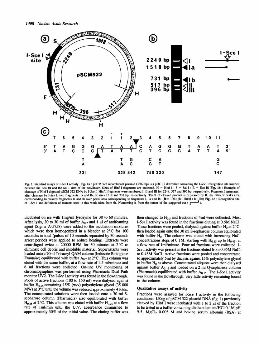
Fig. 1. Standard assays of I-Sce I activity. Fig. la: pSCM 522 recombinant plasmid (3302 bp) is a pUC 12 derivative containing the I-Sce I recognition site insertedbetween the Eco RI and the Sal I sites of the polylinker. Sizes of Hinf I fragments are indicated. H = Hinf I; S = Sal I; E = Eco RI Fig. lb: Example ofcleavage of Hinf I digested pSCM 522 DNA by I-Sce I. Hinf I fragments were numbered I, II and III for 2249, 517 and 396 bp, respectively. Fragment I generates,after cleavage by I-Sce I, two fragments, Ia and Ib, of sizes 1518 and 731 bp, respectively. The% of cleaved product is expressed by R, the ratio of peaks areacorresponding to cleaved fragments Ia and Ib over peaks area corresponding to fragments I, Ia and lb. (R= 1OOx(Ia+Ib)/(I+Ia+Ib)) Fig. lc: Recognition siteof I-Sce I and definition of mutants used in this work (data from 6). Numbering is from the center of the staggered cut (
incubated on ice with lmg/ml lysozyme for 30 to 60 minutes.After lysis, 20 to 30 ml of buffer A0.2 and 1 yd of antifoamingagent (Sigma A-5758) were added to the incubation mixtureswhich were then homogenized in a blender at 2°C for 100secondes in total (pulses of 10 seconds separated by 30 secondsarrest periods were applied to reduce heating). Extracts werecentrifuged twice at 20000 RPM for 30 minutes at 2°C toeliminate cell debris and insoluble material. Supematants wereloaded onto a 70ml Trisacryl-QAM column (Industrie BiologiqueFranlaise) equilibrated with buffer A0.2 at 2°C. This column waseluted with the same buffer, at a flow rate of 1.5 ml/minute and6 ml fractions were collected. On-line UV monitoring ofchromatographies was performed using Pharmacia Dual Pathmonitor UV2. The I-Sce I activity was found in the flowthrough.Pools of active fractions (100 to 150 ml) were dialysed againstbuffer H0.25 containing 15% (w/v) polyethylene glycol (35 000MW) at 0°C until the volume was reduced approximately 4 folds.The concentrated solutions were then loaded onto a 30 ml S-sepharose column (Pharmacia) also equilibrated with bufferH0.25 at 2°C. This column was eluted with buffer H0.25 at a flowrate of lml/min until the U.V. absorbance diminished toapproximately 30% of the initial value. The eluting buffer was
then changed to Ho.5 and fractions of 4ml were collected. MostI-Sce I activity was found in the fractions eluting at 0.5M NaCl.These fractions were pooled, dialysed against buffer Ho at 2°C,then loaded again onto the 30 ml S-sepharose column equibratedwith buffer Ho. The column was eluted with increasing NaClconcentrations steps of 0. IM, starting with HO.s up to Ho045, ata flow rate of iml/minute. Four ml fractions were collected. I-Sce I activity was present in the fractions eluted from 0.35M NaClto 0.45M NaCl. Active fractions were pooled and concentratedto approximately 5ml by dialysis against 15% polyethylene glycolin buffer Ho as above. Concentrated aliquots were then dialysedagainst buffer A0.15 and loaded on a 2 ml Q-sepharose column(Pharmacia) equilibrated with buffer Ao.15. The I-Sce I activitywas found in the flowthrough, very little activity remaining boundto the column.
Qualitative assays of activityFractions were assayed for I-Sce I activity in the followingconditions: 150ng ofpSCM 522 plasmid DNA (fig. 1) previouslycleaved by Hinf I were incubated with 1 to 2 Al of the fractionto be tested in a buffer containing diethanolamine/HCl 0. 1M pH9.5, MgCl2 0.005 M and bovine serum albumin (BSA) at
I-Sce Isite
I

Nucleic Acids Research 1409
Tablel. Quantitative parameters of the purification strategy employed
Fractionation Total protein Total activity Specific activity Purification Yieldstep content (mg) (units) (units/mg) factor (%)
Crude cell 730 26000 35.6 1 100supernatantTrisacryl-QAM 140 18000 128.6 3.6 69eluateS-sepharose 5 6000 1200 34 23eluateQ-sepharose 1.7 2500 1470 41 10eluate
Fractions were purified from 7.5g of cells (wet weight) according to Material and Methods. Total protein contents have been calculated using the method ofBradford (7). An arbitrary unit of activity is defined as the amount of enzyme needed for complete cleavage at the I-Sce I site of 150 ng of Hinf I digestedpSCM 522 DNA (equivalent to 7 x 10-14 moles of recognition site) during a 20 minutes reaction at 37°C in a buffer containing 0. IM diethanolamine/HCI,0.005 M MgCl2 and 100 ,Ag/ml of BSA at pH 9.5 and a total reaction volume of 15 X1.
100 jig/ml in a total reaction volume of 15 1l. Reactions werecarried out at 37°C for 20 minutes then stopped by addition of'I l of EDTA at 0.5 M and analyzed by agarose gelelectrophoresis. When crude fractions were tested, 10 jg ofproteinase K (Boehringer) was added and assays were left at roomtemperature for 10 minutes before electrophoresis.
Quantitative measurements of activitySamples of reactions were analyzed by electrophoresis on 1%agarose gels in TBE buffer. After migration, gels were incubatedin a solution of 0.1 g/ml of ethidium bromide (EB) in H20with gentle agitation at room temperature for lhr to ensurehomogenous staining. EB fluorescence at 254nm was photo-graphed on Polaroid 665 films. Relative amounts of DNAin the different bands of each sample were then determined fromthe scanning of the negative films using a Bio-Rad Model 620video densitometer coupled to a Shimadzu chromatopac C-R3Aintegrator. Quantitation of I-Sce I activity in each reaction iscalculated from the% of cleaved product expressed as in fig. 1.
fractionnumber
I-Scel-
40 45 50 52 54 56 58 60 62 64 66 68 70 75 80 90
14I+,III
Amaximum activity
4.0
3.8
3.6
3A4
3.2
3.0
2.8- 0.3
In (1 - KD)-0.6
RESULTSPurification of I-Sce I activityThe purification procedure is given by Table I. Chromatographyof crude E.coli cell supematants using Trisacryl-QAM anionicexchange column yields I-Sce I activity in the flowthrough. Nosignificant activity can be recovered after stepwise elutions ofthe column with buffers A0o4 to Al .This chromatographyeliminates most of the nucleic acids as well as unspecific nucleicacid binding proteins. In the second chromatography, using theS-sepharose cationic exchange column, the I-Sce I activityremains bound to the column in buffer H025. Elution of thiscolumn with buffer Hos allows the recovery of the activity.Some activity is also found in the fractions eluted with H0o25 butthese fractions contain contaminating nucleic acids and non
specific nucleases which have not been removed by the previouschromatography. On the second passage on S-sepharose with 0.1M NaCl steps, I-Sce I activity elutes from 0.35 to 0.45 M NaCl.The last chromatography, using Q-sepharose anionic exchangecolumn contributes to the elimination of a few more contaminants.This procedure gives a ca. 40 fold purification of I-Sce I witha 10% recovery. I-Sce I enzyme prepared as above shows specificcleavage activity without significant non specific nucleasedegradation of DNA (fig. lb).
Fig.2. Size-exclusion chromatography of I-Sce I. A 89 x2.5 cm Sephacryl-HR200 column (Pharmacia) was calibrated using aldolase, bovin serum albumin,ovalbumin, (3-lactoglobulin, soybean trypsin inhibitor and cytochrome c asstandards. Elutions were performed with a flow rate of 0.74 ml/mn in bufferAO 15 at 2°C and monitored by UV absorbance. A 5 ml active fraction of I-SceI was then loaded on this column and chromatographed in identical conditions.Elution of I-Sce I was monitored by activity in standard assays (fig 2a). Relationshipbetween the Stocks radii of the standards (taken from 8) and the partitioncoefficients were represented according to (9) (fig 2b). Rs = Stocks radius inA; KD = partition coefficient. Open squares = standard proteins ; full square= I-Sce I
I-Sce I purifies as a monomeric globular protein.Gel exclusion chromatography was used to determine the sizeand shape of I-Sce I (fig.2). The activity of I-Sce I in the elutedfractions corresponds, on the plot drawn with the proteinstandards, to an apparent molecular weight of 26 kD and a Stokesradius of 24 A. This figure is close to the molecular weight of27.6 kD calculated from the amino acid composition deducedfrom the DNA sequence (2), hence I-Sce I appears as amonomeric globular protein.
IV
cC

1410 Nucleic Acids Research
3- Z--,V ~~~~~~~~~~~~~~~14IV
12 0
v
1020
VWim (sec 8/ M1 1
o
E .- 6
Z 4~~~~~~~~~~~~~
la.
0
10 30 50 70 1 23time (sec) 1/S (M-1) xilO 8
Fig. 3. Kinetics of reactions and determination of Km value. Samples of various amounts of Hinf I digested pSCM 522 DNA were incubated in the presence of1 unit of I-Sce I under standard conditions at pH 7.5 or pH 9.5 in final volumes of 66 AI or 88.5 1d, respectively. Aliquots were taken at intervals of 10 sec (pH9.5) or 2 minutes (pH 7.5) and reactions were stopped by the addition of 1 ,ul of EDTA at 0.5 M and freezing at -70°C. Aliquots were analyzed by agarose gelelectrophoresis and the reactions were quantified as described in Material and Methods. Fig. 3a: Examples of kinetics for samples of 0.5 ( * ) or 2 (v) picomolesof DNA. Fig. 3b: Double reciprocal representation of the initial rates of the cleavage reactions. Initial rates were measured for substrate concentrations varyingfrom 3.2x 10⁻⁹ M to 22.6x 10⁻⁹ M for the reactions at pH 9.5 (*) and from 9.7 x 10⁻⁹ M to 77 x 10-9 M for the reactions at pH 7.5 (LI). (S: concentrationof sites; V: initial rates)
Kinetics, Km value and optimal conditions.Initial rates of the cleavage reaction catalyzed by I-Sce I weredetermined from assays using various concentrations of substratein two different pH conditions. Examples are given in fig.3a.Interpretation of this analysis (fig.3b) shows linear relationshipsas expected of a Michaelian reaction. Km values areapproximately 4.3 x 10⁻⁸M (pH 7.5) or 3.4 x 10⁻⁸ M (pH 9.5)and Vmax are 0.4x10⁻¹⁰ M/sec. (pH 7,5) or 2.8x10⁻¹⁰M/sec.(pH 9.5).The effects of temperature, ionic strength and pH have been
analyzed to further characterize the in vitro reaction and tooptimize the assay conditions (fig.4). It appears from our resultsthat the pH has a major effect on the reaction. The endonucleaseactivity is limited at low pH values and increases rapidly abovepH 7 to reach a maximum between 9 and 10. Temperature canbe changed from ca. 30°C to 45°C without major effect on thereaction but a significant decrease occurs beyond these limits.Our results also show that the reaction requires no monovalentcations and is even inhibited when the NaCl concentration raisesabove 0.05M. Magnesium (0.003 to 0.01M) is absolutelyrequired, no reaction takes place in EDTA (data not shown).Manganese in similar concentrations can replace magnesium,although less efficiently. Other divalent cations such as: zinc,calcium, cobalt or copper have been tested but none was foundable to replace magnesium.
In vitro stability of I-Sce IThe catalytic activity of I-Sce I in purified fractions can bemaintained almost constant for long periods of time (up to 4
months) provided that the enzyme is stored in 50% glycerolsolutions at -20°C or simply in the buffer AO at 0°C. Wenoticed, however, that the activity of I-Sce I rapidly decreasesduring incubation in the reaction buffer at 37°C. We have,therefore, examined the stability of I-Sce I in variouspreincubation conditions prior to the addition of substrate. Figure5 shows typical results of such assays. It can be seen that ca.80% of activity is lost during the first 1-2 minutes ofpreincubation in the presence of magnesium. In the absence ofmagnesium, only ca. 50% of activity is lost during the same time.The pH range tested shows little effect in the stability.
Interactions between I-Sce I and mutants of the recognitionsiteWe noticed that the addition to the previous assays of plasmidDNA containing a mutant of the recognition site for I-Sce Icontributes to increase the stability of the enzyme. On thecontrary, non-specific DNA (eg. pEMBL18+) has no effect(Fig.6). We have analyzed this protection as a means toinvestigate the interaction between I-Sce I and the mutants of itssite. Several mutants of the site have been previously tested fortheir cleavability by I-Sce I in in vitro assays at pH 7.5 (6).Depending upon the position and the nature of the mutation, someof these mutants were completely uncleavable (called 0), otherswere cleaved as efficiently as the wild type while others werecleaved much less efficiently (called e). In the present study wehave reexamined the cleavability of a set of mutants underoptimum conditions and, at the same time, their degree ofprotection on the enzyme (Fig. 7). In each case, the remaining

Nucleic Acids Research 1411
.o
0 25 5 10 20
8.0 9.00 2 5 5 10 20
8>
I
.100.-- --.. t
O 801- 60t
r 40
20'0
0 25 5 10time (mnm)
I ' '
Te. m0 .(X 0r trC
10C;
Od
©~~~~~~~ ~~~.-tI00 0:1 0. {.
NaCI Molarity
100
80
C}80
C) 40
1)
i-< v-
'., 6 fi .:)
pHt of itICUblhon buttffer
Fig. 4. Determination of optimum conditions. Fig. 4a: Kinetics of reaction at various pHs: 1.5 jig of Hinf I digested pSCM 522 DNA (0.7 picomoles of I-SceI sites) were incubated with 10 units of I-Sce I in a buffer containing 0.005 M MgCl2 and 0.1 M Tris/HCl at pH 7.0, 8.0 or 9.0 as indicated (total reaction volumeswere 100 pl). Incubation was carried out at 37°C and 20 1l aliquots were taken at time intervals (times are indicated in minutes). The reactions were stopped byaddition of 2 A1 ofEDTA at 0.5 M and quick freezing at -70°C. Percent of cleaved product is calculated as in Materials and Methods. Fig. 4b: Effect of temperature: 150 ng of Hinf I digested pSCM 522 DNA were incubated with 1 unit of I-Sce I in a buffer containing 0.1 M diethanolamine-CI at pH 9.5 and 0.005 M MgCl2.Incubations were carried out at various temperatures for 20 minutes. Fig. 4c: Effect of ionic strength: 150 ng of Hinf I digested pSCM 522 DNA were incubatedwith 1 unit of I-Sce I in a buffer containing 0.1 M diethanolamine/HCI at pH 9.5, 0.005 M MgCl2 and various NaCI concentrations as indicated. Incubations were
carried out at 37°C for 20 minutes. Fig. 4d : Effect of pH :150 ng of Hinf I digested pSCM 522 DNA were incubated with 1 unit of I-Sce I in a buffer containing0.005 M MgCI2 and 0.03 M MES (at pH 5.0 or 6.0), 0.1 M Tris/HCI (at pH 7.0, 8.0 or 8.6) or 0.1 M diethanolamine/HCI (at pH 9.5; 10.0 or 11.0) in a totalvolume of 15 A1. Incubations were carried out at 37°C for 20 minutes.
enzyme activity is estimated from the% of wild-type substratecleaved upon completion of the reaction. In such experimentsthe time 0 (preincubation) represents a direct competition betweenthe wild type and mutant sites since both DNAs were added at
the same time. Times 15, 30 and 60 min. indicate simultaneously:-i- the cleavage of mutants sites alone during the preincubation,-ii- the degree of protection of the enzyme by the various mutantssites and -iii- the competition between wild-type and mutants sitesafter the addition of wild-type DNA. Figure 7a shows the amountof cleavage of mutant sites obtained at the end of reactions.Mutants 320 and 328 are not cleaved, mutants 147 and 331 showlittle cleavage and mutants 750 and 842 are the most efficientlycleaved. Figure 7b shows the amount of wild type sites cleavedat the end of the reaction after various preincubation times. Asalready demonstrated, the enzyme alone is extremely instable.Presence of mutants increases the stability of the enzyme tovarious degree, 842 being the most effective.
DISCUSSION
In this paper we have described a protocol for the isolation ofactive fractions of I-Sce I, a new endonuclease encoded by a groupI intron and responsible in vivo for the cleavage of intron-lessgenes resulting in the insertion of the intron into the cleaved site.
got0
60
o
0 1 2 3 4 5 6 7 8 9
Preincubatlon time (min.)
Fig.5. Spontaneous inactivation of I-See I. 10 units of I-Sce I were incubated
at 37°C in a buffer containing 0.1 M Tris/HCl (at pH 7.0 or 9.0) with or without
0.O1M MgCl2 in total sample volumes of 120 pL4. Aliquots of 15 were taken
at various time intervals and added with 15 samples containing 150 ng of Hinf
I digested pSCM 522 DNA in 0.1 M Tris/HCI pH 9.0 with or without 0.01 M
MgCI2 (MgCI2 is added if absent from preincubation). Incubation was carried
out at 37°C for another 30 minutes and reactions were stopped by addition of
1 of EDTA at 0.5 M and freezingat9-70C.(*) pH 9 without magnesium;
(o) pH 9 with magnesium; (U) pH 7 without magnesium and (i) pH 7 with
magnesium
100
80 -
.....*.....0.....
20
Uc30o.C)
(I.DO
C)C)U0
60 -
40 -
20
....>r.... '1-
2I
..t0

1412 Nucleic Acids Research
The demonstration that this protein is endowed with endonucleaseactivity was first obtained by in vivo assays in E. coli (4). Wehave now demonstrated that the enzyme is active in vitro ,showingthat it is sufficient by itself to recognize a specific DNA sequenceand catalyze a double strand break within the recognition sitedetermined previously (6). This work represents the firstenzymatic characterization of an intron encoded nuclease.Our procedure allows the rapid preparation of several thousands
units of enzyme from a relatively small quantity of E. coli cells.Active fractions show no significant contamination by non-specific nucleases under the conditions described and, therefore,can be used to cleave DNA in a clean and reproducible fashion.No cleavage occurs on sequences other than the recognition site.Upon systematic examination of the optimum conditions for
the in vitro activity of I-Sce I, we observed a strong and
0 10' 20 30 0 10 20303 0 10'20'30
ab'io
B
Fig. 6. Protection of I-Sce I by its recognition site. Three units of I-Sce I wereincubated at 37°C in Tris/HCl buffer (pH 9.0) 0.1 M; MgCl2 0.005 M, duringvarious times (from 0 to 30 minutes) in the absence of DNA (A), in the presenceof 400 ng of pEMBL 18+ DNA (B) or in presence of 400 ng of a pUC 13recombinant clone containing mutant 331 (C) in a total volume of 80 $1. Aliquotsof 20 IL were taken at different time intervals, added with 150 ng of Hinf I digestedpSCM 522 DNA and incubated at pH 9.0 and 37°C during 30 minutes. Reactionswere stopped and analyzed as described in Materials and Methods. Times areindicated in minutes. (D> and . ): Fragments of pSCM 522 DNA; (-)Fragments of pEMBL 18+ or mutant 331.
40
C)
V0° 30Q
Cu10
,, 200C4-
04 100lo0.
00 1 0 20 30 40 50
Preincubatlon time (min.)
unexpected pH dependance of the reaction with optimum for veryhigh pH values.
This may be interpreted in several ways: differential stabilityof the enzyme, involvment of a charged lateral chain in the activesite or direct effect of high pH on the substrate structure. Becausein our experiments the stabilities of the enzyme were similar atpH 7 and pH 9, we exclude the first possibility. Kineticsexperiments showed that the Vmax of the reaction is pHdependant. The higher efficiency at high pH indicates probablya higher turn over number of the enzyme. On the contrary, theapparent Km values remain similar at different pHs and are ofthe order of 3 to 4 x 10⁻⁸ M. These figures compare favorablywith the estimated concentration of recognition sites in themitochondria in vivo which is 8 x l0-7 M, assuming 30mitochondrial DNA copies per cell (10) and a total mitochondrialvolume of 5 Itm3 (11).The second unexpected observation of this study is that the
enzyme proves very instable in the absence of substrate underconditions of activity. This instability depends upon temperatureand the presence of magnesium. The enzyme can be partiallyprotected in the presence of specific substrate while random DNAsequences have no effect.The most remarkable aspect of the I-Sce I endonuclease is the
extent of its recognition site (18 bp) which should confer it avery high sequence specificity if the degree of internal degeneracyis not too high. It has already been shown that some mutantsin the recognition site are tolerated which only partially affectthe cleavage reaction at pH 7.5. Our results with a purifiedfraction now confirm and extend this observation in the optimumin vitro conditions. The present protection experiments also showthat some mutants, although uncleavable or poorly cleavable, bindto the enzyme and increase its stability. Binding constants of themutants would be interesting to determine in the future.
100
C)00la-0.CU
CLI
100.
80
60
40
20
060 0 1 0 20 30 40 50 60
Preincubation time (min)
Fig. 7. Interactions of I-Sce I with various mutants of the recognition site. Three units of I-Sce I were incubated at 37°C in a buffer containing 0.1 M Tris/HClpH 9,0; 0.005 M MgCl2 in the absence of DNA or in presence of 200 to 300ng of ScaI digested pUC 13 recombinant plasmids containing mutants of the recognitionsite in a total volume of 80/1. Aliquots of 20 yd were taken at various time intervals (0, 15, 30 and 60 minutes) and added to 2sld containing l50ng of Hinf I digestedpSCM 522 DNA in 0.1 M Tris/HCl pH 9.0. Incubations were carried out at 37°C during 30 minutes. Reactions were stopped and analyzed as described as inMaterials and Methods. Fig. 7a: Cleavage of mutants sites by I-Sce I: The graph represents the extent of cleavage of the different mutants DNA (see fig. 1),after incubation with wild-type DNA as described above. Fig. 7b: Residual activity of I-Sce I: The graph shows the residual activity on the wild-type site observedafter incubation of I-Sce I in the absence of DNA (V) or in the presence of various DNAs mutants of the site: ( 0 ): 842; (O): 750; ( * ): 331; (O): 147; (O):328 and (A): 320.

Nucleic Acids Research 1413
Other potential activities of I-Sce I, suggested by its role invivo, have been tested using the purified fractions. Neither anyexonucleolytic activity on the cleaved substrate nor anyrecombinase activity between two cleaved sites or between onecleaved site and uncleaved homologous DNA could be detectedin vitro. In addition, no double strand cleavage activity couldbe found on the exon-intron junctions, although each sequencecontains half of the recognition site (5). We have also tested theaction of I-Sce I on single strand DNA containing the recognitionsite and found no activity. The absence of activities other thanthe site specific endonuclease supports the notion that the roleof I-Sce I (and probably other group I intron encodedendonucleases as well) in vivo is simply to introduce a doublestrand break in the intron-less gene. Subsequent steps leadingto intron insertion necessitate, therefore, the action of otherproteins, possibly involved in a general double strand break repairmechanism in mitochondria.
I-Sce I is the first group I intron encoded endonucleases thein vitro activity of which has now been characterized in somedetails. Recently, the activity of five other group I intron encodedendonucleases have been reported based on in vivo assays, cellextracts or in vitro synthesized proteins: I-Sce II encoded by theyeast intron Sc coxlb4 (12, 13), I-Tev I and I-Tev II encodedby two introns of bacteriophage T4 (14, 15, 16), I-Ceu I encodedby intron Ce LSU 5 of Chlamydomonas chloroplast (17) and I-Ppo I encoded by the nuclear group I intron Pp LSU 3 ofPhysarum (18). So far, data on the in vitro properties of suchendonucleases are only available for I-Tev I (16). Although notsystematically examined, the properties of this last enzyme seemto differ from those of I-Sce I. However, I-Tev I belongs to agroup of proteins different from that of the endonucleases ofchloroplast or mitochondrial origins. Those, although not closelyrelated in terms of their primary amino acid sequences or in termsof their recognition sites, share common structural motives(dodecapeptides or LAGLI-DADG). In addition, allendonucleases from this last group generate four base pairstaggered cuts with 3'OH overhangs (reviewed in 1). The sameproperties are also shared by HO, the nuclear encoded yeastendonuclease responsible for mating type switching (19, 20).Although not of intron origin, that endonuclease also possessesthe dodecapeptide motif and has a non symmetrical recognitionsite which extends over 18 bp.A major limitation in the use of I-Sce I as a restriction enzyme
is the relatively low yield of our purification procedure (assuminga 1 to 1 molar ratio of enzyme to substrate, 2500 units-the totalpreparation-represent 4.7 /tg of the protein, and it is clear thatthe actual ratio is much below this value). Part of this low yieldresults from the low production of the enzyme by E. coli, the otherfrom the probable instability of the enzyme in E. coli, as inferredfrom its properties in vitro. We have constructed otherrecombinant plasmids to try to solve the first problem.Experiments are now in progress with such constructions.
REFERENCES1. Dujon, B. (1989) Gene 82, 91-114.2. Dujon, B. (1980) Cell 20, 185-197.3. Dujon, B., Belfort, M., Butow, R.A., Jacq, C., Lemieux, C., Perlman,
P.S. and Vogt, V.M. (1989) Gene 82, 115-118.4. Colleaux, L., d'Auriol, L., Betermier, M., Cottarel, G., Jacquier, A.,
Galibert, F., Dujon, B. (1986) Cell 44, 521-533.5. Dujon, B., Colleaux, L., Jacquier, A., Michel, F., Monteilhet, C. (1986)
In R. B. Wickner, A. Hinnebush, A. M. Lambowitz, I. C. Gunsalus andA. Hollaender (Eds), Extrachromosomal elements in lower eucaryotes.Plenum Press pp 5-27.
6. Colleaux, L., d'Auriol, L., Galibert, F., Dujon, B. (1988) Proc. NatI. Acad.Sc. 85, 6022-6026.
7. Bradford, M.M. (1976) Anal. Biochem. 72, 248-254.8. Le Maire, M., Aggerbeck, L.P., Monteilhet, C., Andersen, J.P. and Moller,
J.V. (1986) Anal. Biochem. 154, 525-535.9. Le Maire, M., Ghazi, A., Martin, M. and Brochard, F. (1989) J. Biochem.
106, 814-817.10. De Zamaroczy, M. and Bernardi, G. (1985) Gene 37, 1-17.11. Stevens, B. (1981) In Strathern, J.N, Jones, E.W. and Broach, J.R. (Eds).
The Molecular Biology of the Yeast Saccharomyces, Life Cycle andInheritance Cold Spring Harbor Laboratory pp 471-504.
12. Delahodde, A., Goguel, V., Becam, A.M., Creusot, F., Perea, J., Banroques,J., Jacq, C. (1989) Cell 56, 431-441.
13. Wenzlau, J.M., Saldanha, R.J, Butow, R.A., Perlman, P.S. (1989) Cell56, 421-430.
14. Quirk, S.M., Bell-Pedersen, D., Belfort, M. (1989) Cell 56, 455-465.15. Bell-Pedersen, D., Quirk, S.M., Aubrey, M. and Belfort, M. (1989) Gene
82, 119-126.16. Lemieux, B., Tumel, M., Lemieux, C. (1988) Mol. Gen. Genet. 212, 48-5517. West, D.K., Changchien, L-M, Maley, G.F. and Maley, F. (1989) J. Biol.
Chem. 264, 10343-10346.18. Muscarella, D.E. and Vogt, V.M. (1989) Cell 56, 443-454.19. Kostriken, R., Strathern, J.N., Klar, A.J.S., Hicks, J.B. and Heffron, F.
(1983) Cell 35, 167-174.20. Kostriken, R. and Heffron, F. (1984) Cold Spring Harbor Symposia on
Quantitative Biology. 49, 89-96.
ACKNOWLEDGMENTS
We thank A. Jacquier, E. Luzi, A. Plessis, A. Spassky and I.Stroke for stimulating discussions. C.M. is indebted to V. Luzzatifor his interest in this work. This work was supported by CNRS(URA 0152), by University Pierre and Marie Curie and by agrant 861007 from INSERM to B.D.
Copyright © 2022 FDOKUMEN