Purification and Characterization of Bacteriocin Produced by ...
17
RESEARCH ARTICLE Purification and Characterization of Bacteriocin Produced by Weissella confusa A3 of Dairy Origin Hweh Fen Goh, Koshy Philip* Microbiology Division, Institute of Biological Sciences, Faculty of Science, University of Malaya, 50603, Kuala Lumpur, Malaysia * [email protected] Abstract A dramatic increase in bacterial resistance towards currently available antibiotics has raised worldwide concerns for public health. Therefore, antimicrobial peptides (AMPs) have emerged as a promisingly new group of therapeutic agents for managing infectious dis- eases. The present investigation focusses on the isolation and purification of a novel bacte- riocin from an indigenous sample of cow milk and it’s mode of action. The bacteriocin was isolated from Weissella confusa A3 that was isolated from the sample and was shown to have inhibitory activity towards pathogenic bacteria namely Bacillus cereus, Escherichia coli, Pseudomonas aeruginosa and Micrococcus luteus. The bacteriocin was shown to be heat stable and functioned well at low pH (2 to 6). Reduction of activity was shown after treatment with proteinase K, trypsin and peptidase that confirmed the proteinaceous nature of the compound. MALDI-TOF analysis of the sample gave a mass approximating 2.7 kDa. The membrane of the bacteria was disrupted by the bacteriocin causing SYTOX 1 green dye to enter the cell and bind to the bacterial DNA giving fluorescence signal. Bacterial cell treated with the bacteriocin also showed significant morphological changes under transmis- sion electron microscope. No virulence and disease related genes can be detected from the genome of the strain. Introduction Antimicrobial peptides (AMPs) are ubiquitous and natural antibiotics generated by a diverse range of microorganisms, plants, insect and mammalian cells. Recent attention has been drawn to AMPs as new antimicrobials to combat harmful microbes especially those resistant to con- ventional antibiotics. In the search for new antimicrobial agents, these may be used as tem- plates for the design of novel drugs [1,2]. AMPs are divided into different groups based on their variable structural characteristics [3]. Those AMPs produced by bacteria to kill or inhibit other bacteria are known as bacteriocins [4,5]. In an effort to establish a new antimicrobial agent, attention was focused on lactic acid bac- teria (LAB) in this study. This was chosen as some bacteriocins from LAB are well studied in PLOS ONE | DOI:10.1371/journal.pone.0140434 October 16, 2015 1 / 17 OPEN ACCESS Citation: Goh HF, Philip K (2015) Purification and Characterization of Bacteriocin Produced by Weissella confusa A3 of Dairy Origin. PLoS ONE 10(10): e0140434. doi:10.1371/journal.pone.0140434 Editor: George-John Nychas, Agricultural University of Athens, GREECE Received: July 29, 2015 Accepted: September 26, 2015 Published: October 16, 2015 Copyright: © 2015 Goh, Philip. This is an open access article distributed under the terms of the Creative Commons Attribution License, which permits unrestricted use, distribution, and reproduction in any medium, provided the original author and source are credited. Data Availability Statement: All relevant data are within the paper. Funding: The study was funded by University of Malaya - Ministry of Higher Education High Impact Research Grant number UM.C/HIR/MOHE/SC/08 (F0008-21001) awarded to the Principal Investigator Koshy Philip who is the corresponding author of the manuscript. Competing Interests: The authors have declared that no competing interests exist.
-
Upload
khangminh22 -
Category
Documents
-
view
0 -
download
0
Transcript of Purification and Characterization of Bacteriocin Produced by ...
RESEARCH ARTICLE
Purification and Characterization ofBacteriocin Produced byWeissella confusa A3of Dairy OriginHweh Fen Goh, Koshy Philip*
Microbiology Division, Institute of Biological Sciences, Faculty of Science, University of Malaya, 50603, KualaLumpur, Malaysia
AbstractA dramatic increase in bacterial resistance towards currently available antibiotics has raised
worldwide concerns for public health. Therefore, antimicrobial peptides (AMPs) have
emerged as a promisingly new group of therapeutic agents for managing infectious dis-
eases. The present investigation focusses on the isolation and purification of a novel bacte-
riocin from an indigenous sample of cow milk and it’s mode of action. The bacteriocin was
isolated fromWeissella confusa A3 that was isolated from the sample and was shown to
have inhibitory activity towards pathogenic bacteria namely Bacillus cereus, Escherichiacoli, Pseudomonas aeruginosa andMicrococcus luteus. The bacteriocin was shown to be
heat stable and functioned well at low pH (2 to 6). Reduction of activity was shown after
treatment with proteinase K, trypsin and peptidase that confirmed the proteinaceous nature
of the compound. MALDI-TOF analysis of the sample gave a mass approximating 2.7 kDa.
The membrane of the bacteria was disrupted by the bacteriocin causing SYTOX1 green
dye to enter the cell and bind to the bacterial DNA giving fluorescence signal. Bacterial cell
treated with the bacteriocin also showed significant morphological changes under transmis-
sion electron microscope. No virulence and disease related genes can be detected from the
genome of the strain.
IntroductionAntimicrobial peptides (AMPs) are ubiquitous and natural antibiotics generated by a diverserange of microorganisms, plants, insect and mammalian cells. Recent attention has been drawnto AMPs as new antimicrobials to combat harmful microbes especially those resistant to con-ventional antibiotics. In the search for new antimicrobial agents, these may be used as tem-plates for the design of novel drugs [1,2]. AMPs are divided into different groups based ontheir variable structural characteristics [3]. Those AMPs produced by bacteria to kill or inhibitother bacteria are known as bacteriocins [4,5].
In an effort to establish a new antimicrobial agent, attention was focused on lactic acid bac-teria (LAB) in this study. This was chosen as some bacteriocins from LAB are well studied in
PLOSONE | DOI:10.1371/journal.pone.0140434 October 16, 2015 1 / 17
OPEN ACCESS
Citation: Goh HF, Philip K (2015) Purification andCharacterization of Bacteriocin Produced byWeissella confusa A3 of Dairy Origin. PLoS ONE10(10): e0140434. doi:10.1371/journal.pone.0140434
Editor: George-John Nychas, Agricultural Universityof Athens, GREECE
Received: July 29, 2015
Accepted: September 26, 2015
Published: October 16, 2015
Copyright: © 2015 Goh, Philip. This is an openaccess article distributed under the terms of theCreative Commons Attribution License, which permitsunrestricted use, distribution, and reproduction in anymedium, provided the original author and source arecredited.
Data Availability Statement: All relevant data arewithin the paper.
Funding: The study was funded by University ofMalaya - Ministry of Higher Education High ImpactResearch Grant number UM.C/HIR/MOHE/SC/08(F0008-21001) awarded to the Principal InvestigatorKoshy Philip who is the corresponding author of themanuscript.
Competing Interests: The authors have declaredthat no competing interests exist.

the past and now used as safe and natural food preservatives. The best known example is nisinproduced by Lactococcus lactis [6]. Except for a few species, LAB are considered as “GenerallyRecognized as Safe” (GRAS) and used in the production of fermented foods and beverages [7].Some LAB produce antimicrobial compounds which may act as bacteriostatic or bactericidalagents. The antimicrobial activities are mainly due to the production of antimicrobial metabo-lites such as bacteriocins, hydrogen peroxide and organic acids [8].
The mechanisms of action of AMPs can generally be grouped into two classes. The first classis membrane disruptive following barrel stave, toroidal, carpet or micellar aggregate mecha-nisms. Second is the non-membrane disruptive class which targets the intracellular components[9]. In the first mechanism, bacteriocins selectively disrupt the cell membranes and the amphi-pathic structural arrangement of the bacteriocins is believed to play a significant role in thismechanism. Membrane disruption can also be caused by bacteriocins produced by milk-associ-ated LAB. For example L. lactis strain which is usually found in milk is used as starter culturesin production of many fermented foods for producing safe biopreservatives [10]. Researchreveals that milk consists of a wide range of bioactive compounds with antimicrobial, antihyper-tensive and antioxidant properties. Most of these bacteriocins are only released during milk fer-mentation implying the milk is presumed to generate these bioactive compounds [11].
The genusWeissella was reclassified in 1990. The speciesWeissella confusa isolated in thecurrent study was formerly called Lactobacillus confusus and has been isolated from severalsources.Weissella confusa is known to produce dextran which is an exopolysaccharide with α-glucans and a linear backbone made of α-(1!6)-linked d-glucopyranosyl units. In situ pro-ducedW. confusa dextran has been reported to be useful in improving the shelf-life, volumeand nutritional value of bread [12–14]. Apart from the production of dextran, the bacteriocinproduced by the genusWeissella received little attention. Until now only few bacteriocinsknown as weissellicin was identified fromWeissella and report of bacteriocin produced byWeissella confusa is even rarer [15].
The aims of this study were to isolate and characterize a novel bacteriocin from LAB isolatedfrom indigenous cow milk sampled in Malaysia and study its mode of action by using real-timePCR and SYTOX1 green dye that binds to nucleic acid of the affected target bacteria. Subse-quently this disrupted the cell membrane. SYTOX1 Green dye has high binding affinitytoward nucleic acid and easily penetrates those cells with compromised membranes but do notcross the intact membranes of live cells [16].
Materials and Methods
Isolation and Identification of Lactic Acid Bacteria from Fermented MilkSampling was done from raw cow milk randomly collected from two different restaurants (Al-Berkat Curry House and Grand City) located in Petaling Jaya, Selangor, Malaysia. The rawmilk samples were pasteurized at 63°C for 30 minutes to kill the harmful bacteria present in theraw milk. The milk was left to ferment in a sterile flask under room temperature for two days.After fermentation process, LAB was isolated by growing on de-Mann, Rogosa and Sharpe(MRS) agar plates (Merck, Germany). These plates were incubated for 24 hours at 37°C. Singlecolonies were picked from the plates and sub-cultured two times to get a pure colony based onmorphology. The isolates were grown in MRS broth for 24 hours and centrifuged at 10,000 x gfor 20 minutes. The supernatant from each isolate was used for preliminary antimicrobial testby using agar well diffusion assay. 50 μl of the supernatant from each isolate was transferred todifferent wells on a Mueller-Hinton agar plate seeded with indicator bacteria. The bacteria iso-late (A3) that produces potent antimicrobial activity was selected for further tests. For identifi-cation of bacteria, five tests were done including Gram-stain, spore stain, catalase test, oxidase
Action of Bacteriocin fromWeissella confusa A3
PLOSONE | DOI:10.1371/journal.pone.0140434 October 16, 2015 2 / 17

test and the ability to grow on bile esculin azide agar (Merck, Germany). Cell morphology andGram-stain were examined by light microscope at 1000X magnification. Schaeffer and Fulton’sspore staining method modified by [17] was followed. Bile esculin azide agar tested the abilityof the bacteria to hydrolyze esculin in the presence of bile. Bacteria positive for esculin hydroly-sis can hydrolyze the glycoside esculin to esculetin and dextrose. The bile in the agar was usedto inhibit Gram-positive bacteria other than enterococci while the sodium azide inhibitedGram-negative bacteria. LAB was identified using API 50CHL (bioMérieux, France) whichtested the ability of the bacteria to metabolize 49 kinds of carbohydrates. Pure colonies of thebacteria were transferred into the test medium and loaded into the test strip. The test strip wasincubated for 48 hours at 37°C. The result obtained was examined for bacterial similarity to thedatabase using the API software provided. The bacteriocin producer strain was identified by16S rRNA gene sequencing using universal primers namely 27F (5'-AGAGTTTGATC(A/C)TGGCTCAG-3') and 1492R (5'-ACGG(C/T)TACCTTGTTACGACTT-3'). PCR reactionmixture (25 μl total) contains 2.5 μl PCR buffer, 2 μl of dNTP mix (2.5mM each), 1μl of eachprimer (20 pmol), 100ng of DNA template, 0.5 μl of i-Taq™ DNA polymerase 5U/μl suppliedfrom iNtRON Biotechnology, Korea. PCR conditions used initial denaturation at 94°C for 5minutes, denaturation at 94°C for 1 minute, annealing at 52°C for 1 minute, extension at 72°Cfor 1.5 minutes for 30 cycles with a final extension at 72°C for 10 minutes.
Effect of Different Media on Bacteriocin ProductionThe bacteriocin producer (A3) was grown in 10 types of broth and tested against B. cereus toexamine and compare the effectiveness of different culture media on the production of the bac-teriocin. The media used was de Man, Rogosa and Sharpe (MRS), M17 supplemented with1.5% glucose, M17 supplemented with 1.5% sucrose, M17 supplemented with 1.5% lactose,Brain-Heart Infusion (BHI), LAPTg [18], tryptic soy broth (TSB) with 1% Tween 80, Brucellabroth, nutrient broth (NB) with 1.5% glucose and Miller's LB broth. After 18 hours incubation,the fermented broth was centrifuged at 10,000 x g for 20 minutes and the supernatant was pre-cipitated with 80% ammonium sulphate overnight at 4°C. Then the pellet was centrifuged anddissolved in minimum amount of water and tested for inhibitory activity by well diffusionassay. Well diffusion assay was conducted by seeding B. cereus on Mueller- Hinton agar platewith sterile cotton swab and 5 mm diameter wells were made with a sterile cork borer. Then50 μl of the crude bacteriocin purified from different media was transferred into separate wells.The plates were incubated at 37°C overnight and the inhibition zones were measured.
Growth Curve and Production of BacteriocinThe growth of the bacteriocin producer was measured using Miles and Misra method [19]. Thegrowth of the bacteria was also monitored by measuring the optical density (O.D) at 600nm.Ten percent of starter culture of the bacteriocin producer at O.D 0.1 was added to 1 litre of ster-ile MRS broth. The colony forming unit (CFU) and the O.D. of the bacteria within 36 hourswas monitored. The production of bacteriocin was monitored during log to stationary phasesby measuring the inhibition zone of the crude bacteriocin. 20ml of the bacteria culture was cen-trifuged at 10,000 x g for 15 minutes and the supernatant was subjected to 80% ammonium sul-phate precipitation. The dissolved precipitate from each interval was tested against B. cereusand the inhibition zone measured.
Purification and Antimicrobial Assay of BacteriocinThe test bacteria used were Bacillus cereus ATCC14579, Escherichia coli UT181, Listeria mono-cytogenes NCTC10890, Pseudomonas aeruginosa PA7, Staphylococcus aureus RF122,
Action of Bacteriocin fromWeissella confusa A3
PLOSONE | DOI:10.1371/journal.pone.0140434 October 16, 2015 3 / 17

Micrococcus luteus ATCC10240, Lactococcus lactis A1 and Enterococcus faecium C1 from theculture collection maintained in the Microbial Biotechnology Laboratory, Microbiology Divi-sion of University of Malaya. All the test bacteria were maintained on Muller-Hinton agar(Merck, Germany) at 37°C. The concentration of the test bacteria was fixed at 0.1 value of O.D.at 600nm wavelength. The test bacteria were first lawn grown on Muller-Hinton agar (Merck,Germany) and wells were made using a cork borer. The test was carried out in triplicate. Theplate was incubated overnight at 37°C and the inhibition zone was measured. For purificationof the bacteriocin, the bacteria was grown in MRS broth for 18 hours and then centrifuged at10,000 x g for 20 minutes. The bacterial pellet was discarded. The resulting cell-free superna-tant was filtered with 0.2μmmembrane filter (Sartorius, Germany). This supernatant was sub-jected to 80% ammonium sulphate precipitation to obtain the crude bacteriocin and thenlyophilized. The lyophilized sample was then run through a column pre-packed with AmberliteXAD16 resin and eluted fractions then collected with increasing concentration gradients ofacetonitrile and tested for antimicrobial activity using well diffusion assay. The active fractionfrom XAD column was further fractionated by size using Vivaspin (Sartorius, Germany) withdifferent molecular weight cut off (MWCO) which is 0.2μm, 1,000,000 MWCO, 50,000MWCO, 30,000 MWCO, 10,000 MWCO, 5,000 MWCO and 2,000 MWCO. Antimicrobial testwas performed by spotting 20μl of the fraction on Mueller-Hinton agar seeded with B cereus.The active fraction collected from Vivaspin column was introduced into reverse-phase high-performance liquid chromatography (RP-HPLC) system (Waters, USA) using RP C18 column(Merck, Germany). The separation was carried out by gradient separation using two solvents:A (95%Mili-Q water (Millipore, USA) and 5% acetonitrile (Merck, Germany) and B (100%acetonitrile). The flow rate of the mobile phase was set at 1ml min-1. The following gradientwas used: 0% solvent B for 3 minutes, 0–40% solvent B for 45 minutes followed by 40–100%solvent B for 5 minutes and then back to 100% solution A. Fractions were evaporated toremove acetonitrile and then tested for antimicrobial activity using B. cereus as a test indicator.Active fraction was collected at the same retention time during different HPLC runs and thenpooled and lyophilized.
Bacteriocin Activity Assay and the Effect of Various Concentrations ofBacteriocin on Optical Density of B. cereusThe minimum inhibitory concentration of the bacteriocin was performed using broth microdi-lution method proposed by Steinberg [20]. The indicator strains were inoculated into 5 ml ofMueller-Hinton broth and incubated at 18 h at 37°C on a shaker. Cultures were diluted withMueller- Hinton broth to give 7 × 10⁵ CFU/ml. Two-fold serial dilutions were performed fromthe stock bacteriocin with Bradford concentration of 1.48 mg/ml in 0.2% BSA and 0.01% aceticacid in Eppendorf tubes (polypropylene). 11 μl of each concentration was added to a well in a96-well poplypropylene plate. Then 100 μl of the diluted indicator strains was added into eachcorresponding well containing each concentration of the bacteriocin. The final concentrationsof the bacteriocin were progressively halved to 74, 37, 18.5 until 1.16 μg/ml. The 96-well platewas incubated for 18 h at 37°C. MIC was taken as the lowest concentration of the bacteriocinthat reduces growth by more than 50% compared to that of the control. 10 μl of the content ofthe well with higher MIC value was plated onto Mueller-Hinton agar and incubated at 37°Covernight. The lowest concentration of bacteriocin that prevents any viable cell growth wasdefined as the minimum bactericidal concentration (MBC). To determine the effect of variousconcentrations on the growth of B. cereus, the protocol of Skandamis [21] was followed withsome modification. Briefly, overnight bacterial culture was diluted in Mueller-Hinton broth togive a final OD615 = 0.1. Then 100 μl of the bacterial suspension was added to a 96-well plate
Action of Bacteriocin fromWeissella confusa A3
PLOSONE | DOI:10.1371/journal.pone.0140434 October 16, 2015 4 / 17

followed by 11 μl of the different bacteriocin concentrations as stated earlier. The final concen-tration of bacteriocin was similar to the concentration used to determine MIC. The plate wasthen read with Multiskan™ GOMicroplate Spectrophotometer (Thermo Scientific, Finland)every 1 hour interval for a period of 24 hours.
Stability Tests of BacteriocinThe bacteriocin A3 was tested for stability in terms of inhibition on target microorganisms byvarying its production in different carbon sources namely glucose, lactose and sucrose. Thiswas repeated for thermal and pH stabilities. For accessing the thermal stability test, the bacteri-ocin was exposed to temperatures 40, 60, 80 and 100°C for 20 minutes. The heated bacteriocinwas cooled to room temperature and tested for inhibition on Bacillus cereus. For the pH stabil-ity test, the bacteriocin was adjusted to a pH value ranging from 2 to 10 by using concentratedNaOH (Merck, Germany) and HCl (Merck, Germany). This was incubated for 2 hours at 25°Cbefore determining the antimicrobial activity by agar well diffusion method. The vulnerabilityof the bacteriocin to breakdown by different enzymes namely proteinase K, lysozyme, lipase,catalase, lyticase, trypsin and peptidase sourced from Sigma- Aldrich was also tested and thenchecked for subsequent inhibition. 500 μl of the bacteriocin was treated with the enzymes with1 mg/ml final concentration and a control without treatment was prepared. All preparationswere incubated at 37°C for 1 hour before antimicrobial measurement with B. cereus.
Molecular Weight Estimation with Sodium Dodecyl Sulfate-Polyacrylamide Gel Electrophoresis (SDS-PAGE) and MALDI-TOFThe molecular weight of the purified bacteriocin obtained from the HPLC fraction was esti-mated using Laemmli SDS-PAGE [22]. The gel used for the separation was 16.5% tris- tricineSDS-PAGE and the ladder used was Precision Plus Protein™ Dual Xtra Standards (Bio-Rad,USA). The gel was subjected to 100mV for one and a half hours and then stained with Simply-Blue™ SafeStain (Invitrogen, UK). The same sample also was subjected to matrix- assisted laserdesorption ionization-time of flight mass spectrometry (MALDI-TOF MS). The targeted platewas spotted with aliquots consisting of 4μl of matrix and 4μl sample cleaned- up with zip-tip.
Membrane Permeability TestReal time PCR was used to monitor the quantity of DNA present when different concentra-tions of bacteriocin were added to the target bacteria. Bacteriocin shearing the membrane ofthe target bacteria can cause the bacterial DNA to be released and then bound with the fluores-cent dye. Hence, higher fluorescence indicates the bacteriocin was more effective in terms ofinhibitory activity on target bacteria. The membrane permeability test was done usingSYTOX1 Green dye following the method used in previous studies with modification [23].The indicator bacteria was grown until attaining an O.D value of 0.6 and then the bacterial pel-lets were washed with 10mM sodium phosphate buffer at pH 7.2 before centrifuging at 2,000 xg for 15 minutes. This step was repeated twice. The pellet was re-suspended in 5ml of 10mMsterile sodium phosphate buffer adjusted to pH 7.2. The bacteria was diluted again with sodiumphosphate buffer and adjusted to an O.D value of 0.6 at 600nm wavelength. Then 5μl ofSYTOX1 Green stock solution was added to 5ml of bacterial solution prepared as mentionedabove. 90μl of the bacteria added with SYTOX1 Green was mixed with 10μl of bacteriocin.The positive control used was 1M sodium hydroxide and negative control contained only bac-terial cells without bacteriocin. The experiment was performed in quadruplicate. The wellswere sealed by using adhesive cover and then the plate was placed in the Step One Plus real-time PCR system (Applied Biosystem, USA).
Action of Bacteriocin fromWeissella confusa A3
PLOSONE | DOI:10.1371/journal.pone.0140434 October 16, 2015 5 / 17

Effects of the Bacteriocin on Bacteria under Transmission ElectronMicroscope (TEM)The test bacteria was treated with the bacteriocin and incubated for 3 hours at 37°C. Bacteriawithout addition of bacteriocin was prepared and used as a negative control. Then the bacterio-cin was washed away thrice by using sodium phosphate buffer by centrifuging at 2,000 x g for15 minutes. The cells were fixed with 4% glutaraldehyde and left overnight at 4°C. The sampleswere washed thrice with cacodylate buffer and post-fixed for 2 h with osmium tetroxide andcacodylate buffer in 1:1 ratio. Then it was washed thrice in cacodylate buffer and incubated incacodylate buffer overnight. The samples were then washed thrice with water, once with uranylacetate and again thrice with water. The samples were dehydrated in a graded ethanol seriesand embedded in Epon. Ultrathin sections (0.1 μm) were prepared and coated on copper gridsand stained with uranyl acetate and lead citrate. The grids were examined using LEO-Libra 120transmission electron microscope (Carl Zeiss, Germany).
Basic Analysis of Genome SequenceThe total genomic DNA of the bacteria was extracted with DNeasy Blood & Tissue Kits (Qia-gen, Valencia) following manufacturer’s instruction. The DNA was fragmented into a sizeranging 600–900bp using the Covaris S220 system (Applied Biosystems, USA). DNA library(using 800ng sonic cleaned DNA) was prepared by Nextflex DNA Sample Prep Kit (Bio scien-tific, Texas, USA) complying with the manufacturer’s instructions. Library QC was performedon Bioanalyzer (Agilent Technologies) to check the fragment size distribution. The QC bench-marked library was sequenced on MiSeq sequencer (Illumina) with MiSeq Reagent Kits v3(300 cycles, paired-end). Fluorescent images were analyzed using the MiSeq Control Softwareand FASTQ-formatted sequence data was created using MiSeq Reporter Analysis. The genomesequence was analyzed by Rapid Annotation Subsystem Technology (RAST) server (http://rast.nmpdr.org/).
Results
Isolation and Identification of Lactic Acid Bacteria from Fermented MilkIn the preliminary antimicrobial test, one LAB was showed to give inhibitory activity againstthe test bacteria. The bacterial colony morphology on MRS agar plate showed as medium sizedand milky-white in colour, circular, convex and elevated with entire margins. The isolate wasnegative for catalase and oxidase and did not produce spores based on staining with malachitegreen. The bacteria appeared rod shaped under light microscope. Bile esculin test showed thatit was able to weakly hydrolyze esculin in the presence of bile. Based on the interpretation ofthe API database, the bacteria colony was shown to be 99.9% similar toWeissella confusa.Genomic analysis 16S rRNA gene sequencing confirmed the isolate wasWeissella confusa. Thesequence obtained from 16S rRNA gene sequencing was deposited in the NCBI gene bankdatabase under accession number KJ476186 and was given the strain designation A3.
Effect of Different Media on the Production of BacteriocinSeveral commercially media available were tested for supporting the growth and production ofbacteriocin. Among the 10 media used, MRS gave the highest activity followed by LAPTg andM17 supplemented with different carbohydrates. The other 5 media (BHI, TSB with 1% tween80, Brucella broth, NB with 1.5% glucose and Miller's LB broth) also can be used to grow thebacteria but the production of bacteriocin was lower and no bacteriocin could be recoveredfrom the culture grown in Brucella broth. The inhibition zone is shown in Table 1.
Action of Bacteriocin fromWeissella confusa A3
PLOSONE | DOI:10.1371/journal.pone.0140434 October 16, 2015 6 / 17

Growth Curve and Production of Bacteriocin StudyThe growth curves (Fig 1) the bacteria reached stationary phase after 18 hours of incubation.According to the inhibition zone from the crude bacteriocin purified from the supernatant, the bac-teriocin started to produce at 8 hours of incubation and reached optimum production at 18 hoursincubation period. The antimicrobial activity remained almost the same between 18 to 24 hours ofgrowth. However the activity of bacteriocin started to decrease at 28 hours of growth (Fig 1). There-fore the bacteriocin was best harvested between 18 to 24 hours to get optimum production.
Purification and Antimicrobial Assay of BacteriocinThe bacteriocin produced by theW. confusa showed inhibition against all test bacteria exceptL.monocytogenes NCTC10890 and S. aureus RF122. The inhibition zone of the ammonium
Table 1. Antimicrobial activity of bacteriocin recovered fromW. confusa cultured in different media againstBacillus cereusATCC14579.
Media MRS M17+1.5%Sucrose
M17+1.5%Glucose
M17+1.5%Lactose
BHI LAPTg TSB+1%tween 80
NB+1.5%glucose
LB+1.5%glucose
Brucellabroth
Inhibitionzone*
11.62 9.68 9.22 7.88 7.02 9.52 6.84 6.18 7.86 -
* Inhibition zone measures in millimeter (mm)
doi:10.1371/journal.pone.0140434.t001
Fig 1. Antimicrobial activity and the growth of the bacteria. The antimicrobial activity started to be detected at 8 hours of incubation period and reachedoptimum at 18 hours of incubation period.
doi:10.1371/journal.pone.0140434.g001
Action of Bacteriocin fromWeissella confusa A3
PLOSONE | DOI:10.1371/journal.pone.0140434 October 16, 2015 7 / 17

sulphate precipitated extract against different test bacteria is shown in Table 2. The crude bac-teriocin was desalted with XAD 16 column to remove the ammonium sulphate. Three fractionswere obtained from the Amberlite XAD 16 column. Only fractions eluted out with 50% and90% of acetonitrile gave activity against B. cereus (Fig 2a). The active fractions were combinedand fractionated with Vivaspin column. The sample fraction of size 2–5kDa showed positiveresult when tested against B. cereus (Fig 2b). The bacteriocin from Vivaspin was injected intoHPLC and the run under the set gradient for 1 hour (Fig 3). The active bacteriocin was elutedout from 36 to 39 min and inhibited B. cereus.
Table 2. Inhibition zones andMIC value of the crude and purified bacteriocin against test bacteria strains.
Test bacteria Ammonium sulphateprecipitation*
Amberlite XAD16*
MIC for HPLC fraction(μg/ml)
MBC for HPLC fraction(μg/ml)
Bacillus cereus ATCC14579 10.67±0.58 11.89±0.06 9.25 37
Escherichia coli UT181 7.98±0.06 8.49±0.25 18.5 74
Listeria monocytogenesNCTC10890
0 0 - -
Pseudomonas aeruginosa PA7 11.83±0.89 11.59±0.26 18.5 74
Staphylococcus aureus RF122 0 0 - -
Micrococcus luteus ATCC10240 8.59±0.03 11.57±0.10 9.25 37
Lactococcus lactis A1 8.72±0.52 8.93±0.10 37 >74
Enterococcus faecium C1 9.25±0.10 9.59±0.28 37 >74
*Inhibition zones measured in ±standard deviation millimeter (mm).
doi:10.1371/journal.pone.0140434.t002
Fig 2. Antimicrobial assay. Antimicrobial activity of different fractions from Amberlite XAD 16 column (a) and two different fractions (< 2kDa and 2–5kDa)from Vivaspin (b).
doi:10.1371/journal.pone.0140434.g002
Action of Bacteriocin fromWeissella confusa A3
PLOSONE | DOI:10.1371/journal.pone.0140434 October 16, 2015 8 / 17

Bacteriocin Activity Assay and the Effect of Various Concentrations ofBacteriocin on Optical Density of B. cereusThe MIC value (Table 2) of the bacteriocin was 9.25 μg/ml against both B.cereus andM. luteus.The MIC value increased to 18.5 μg/ml against E.coli and P. aeruginosa. A higher MIC of37 μg/ml was required to inhibit L. lactis and E. faecium. After plating 10 μl of the overnightbacteriocin with the bacterial solution from the wells but without any bacterial growth afterincubation, the MBC obtained for B.cereus andM. luteus was 37 μg/ml. The MBC for E.coliand P. aeruginosa was 74 μg/ml whereas L. lactis and E. faecium required a concentrationexceeding 74 μg/ml to produce bactericidal effect. The addition of different concentrations ofbacteriocin affected the growth of B. cereus (Fig 4). The growth of bacteria can be observed inconcentrations below 4.63 μg/ml. The growth of B. cereus was extremely slow in concentrationsof 9.25 and 18.5 μg/ml. The optical density of B. cereus remained almost unchanged in concen-trations of bacteriocin exceeding 37 μg/ml.
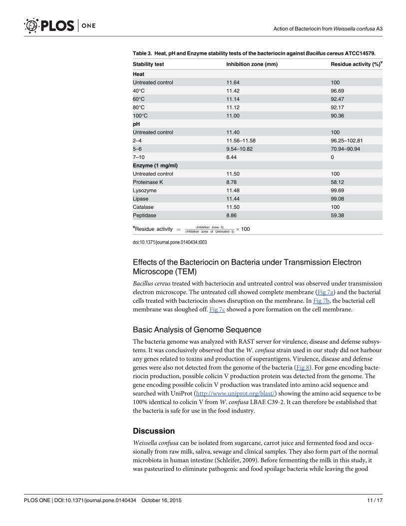
Stability Test of BacteriocinThe effect of the three different carbohydrates as carbon source supplemented in MRS showedthat there was no significant contribution of the different carbohydrates on the inhibition. But2% of glucose gave slightly better inhibition zones compared to sucrose and lactose. The heatstability test showed that the bacteriocin was heat stable and still showed inhibitory activityafter exposing to 100°C for 30 minutes. The bacteriocin functioned well at a low pH of 2 to 6.The antimicrobial activity decreased upon exposure to high pH. For enzyme stability test, thebacteriocin was stable after treatment with lysozyme, lipase, catalase and lyticase but showedreduction of the activity after treatment with proteinase K, trypsin and peptidase. The reduc-tion of activity by proteolytic enzymes confirmed that the antimicrobial substance was not oflipid nature but was proteinaceous in nature. The results of heat, pH and enzyme stability testsare summarized in Table 3.
Fig 3. RP-HPLC Profile.RP-HPLC profile of active faction isolated fromWeissella confusawith antimicrobial activity detected during 36 to 39 minute elutionperiod.
doi:10.1371/journal.pone.0140434.g003
Action of Bacteriocin fromWeissella confusa A3
PLOSONE | DOI:10.1371/journal.pone.0140434 October 16, 2015 9 / 17

Molecular Weight Estimation with Sodium Dodecyl Sulfate-Polyacrylamide Gel Electrophoresis (SDS-PAGE) and MALDI-TOFWhen the HPLC sample was subjected to SDS-PAGE, only a single band was detected confirm-ing its high purity. The purified bacteriocin showed molecular weight approximately 2.5 kDacompared to the marker used (Fig 5). The MALDI-TOF chromatogram (Fig 5) showed thatthe sample had a high density peak at 2706.6855Da. Therefore the possible molecular weight ofthe sample was approximately 2.7 kDa.
Membrane Permeability TestThe real time PCR fluorescence (Fig 6) showed that the bacteria treated with bacteriocin fromW. confusa had highest fluorescence density after positive control (NaOH, 1M). Negative con-trols without adding any bacteriocin and with tetracycline added showed the lowest fluores-cence. This result proved membrane disruption occur when bacteriocin fromW. confusa wasadded into the test bacteria allowed the fluorescence dye to bind to the nucleic acid of the testbacteria and hence giving high fluorescence intensity.
Fig 4. Effect of various concentration of bacteriocin on the growth ofB. cereus at 37°C. The optical density was measured at 615 nm every hourcontinuously for a period of 24 hours.
doi:10.1371/journal.pone.0140434.g004
Action of Bacteriocin fromWeissella confusa A3
PLOSONE | DOI:10.1371/journal.pone.0140434 October 16, 2015 10 / 17
Effects of the Bacteriocin on Bacteria under Transmission ElectronMicroscope (TEM)Bacillus cereus treated with bacteriocin and untreated control was observed under transmissionelectron microscope. The untreated cell showed complete membrane (Fig 7a) and the bacterialcells treated with bacteriocin shows disruption on the membrane. In Fig 7b, the bacterial cellmembrane was sloughed off. Fig 7c showed a pore formation on the cell membrane.
Basic Analysis of Genome SequenceThe bacteria genome was analyzed with RAST server for virulence, disease and defense subsys-tems. It was conclusively observed that theW. confusa strain used in our study did not harbourany genes related to toxins and production of superantigens. Virulence, disease and defensegenes were also not detected from the genome of the bacteria (Fig 8). For gene encoding bacte-riocin production, possible colicin V production protein was detected from the genome. Thegene encoding possible colicin V production was translated into amino acid sequence andsearched with UniProt (http://www.uniprot.org/blast/) showing the amino acid sequence to be100% identical to colicin V fromW. confusa LBAE C39-2. It can therefore be established thatthe bacteria is safe for use in the food industry.
DiscussionWeissella confusa can be isolated from sugarcane, carrot juice and fermented food and occa-sionally from raw milk, saliva, sewage and clinical samples. They also form part of the normalmicrobiota in human intestine (Schleifer, 2009). Before fermenting the milk in this study, itwas pasteurized to eliminate pathogenic and food spoilage bacteria while leaving the good
Table 3. Heat, pH and Enzyme stability tests of the bacteriocin againstBacillus cereusATCC14579.
Stability test Inhibition zone (mm) Residue activity (%)¥
Heat
Untreated control 11.64 100
40°C 11.42 96.69
60°C 11.14 92.47
80°C 11.12 92.17
100°C 11.00 90.36
pH
Untreated control 11.40 100
2–4 11.56–11.58 96.25–102.81
5–6 9.54–10.82 70.94–90.94
7–10 8.44 0
Enzyme (1 mg/ml)
Untreated control 11.50 100
Proteinase K 8.78 58.12
Lysozyme 11.48 99.69
Lipase 11.44 99.08
Catalase 11.50 100
Peptidase 8.86 59.38
¥Residue activity ¼ ðInhibition zone�5ÞðInhibition zone of Untreated�5Þ � 100
doi:10.1371/journal.pone.0140434.t003
Action of Bacteriocin fromWeissella confusa A3
PLOSONE | DOI:10.1371/journal.pone.0140434 October 16, 2015 11 / 17

Fig 5. MALDI-TOFmass spectrometry and SDS-PAGE of purified bacteriocin.MALDI-TOF MS analysis of the HPLC fraction and SDS-PAGE gelpicture. Lane 1 is fraction from HPLC, Lane M is Precision Plus Protein™ Dual Xtra Standards (Bio-Rad, USA).
doi:10.1371/journal.pone.0140434.g005
Fig 6. Membrane permeability test. Real time PCR fluorescence pattern of bacteriocin fromW. confusa, negative control, positive control (NaOH, 1M) andtetracycline.
doi:10.1371/journal.pone.0140434.g006
Action of Bacteriocin fromWeissella confusa A3
PLOSONE | DOI:10.1371/journal.pone.0140434 October 16, 2015 12 / 17

bacteria intact which can withstand high temperature. These good bacteria comprise mostly ofthe LAB. During fermentation, the bacteria utilized the nutrients in the milk to multiply.Hence, this step was also known as the enrichment step.W. confusa can hydrolyze esculin inthe presence of bile. It hydrolyzed the glycoside esculin to form dextrose and esculetin which
Fig 7. Bactericidal effect of Bacteriocin A3 on B. cereus. TEM images of B. cereus cells before (a) and after (b & c) treatment with bacteriocin extractedfromW. confusa. Bar indicates 1μm for (a & c) and 500nm for (b). Arrows indicate destruction of membrane.
doi:10.1371/journal.pone.0140434.g007
Fig 8. Subsystem feature of the genome sequence ofW. confusaA3 analysed with RAST server. The red box indicated absence of genes in toxins andsuperantigens and virulence, disease and defense genes.
doi:10.1371/journal.pone.0140434.g008
Action of Bacteriocin fromWeissella confusa A3
PLOSONE | DOI:10.1371/journal.pone.0140434 October 16, 2015 13 / 17

react with ferric citrate producing a dark brown or black phenolic iron complex. This test iscommonly used to presumptively identify Group D Streptococci but also showed positive reac-tion forW. confusa in our study. Bacteriocin production is always influenced by the carbonand nitrogen sources of the growth media. The maximum bacteriocin production ofW. con-fusa A3 was obtained from MRS media. In the past studies, MRS media was used to cultivateLAB since it was rich in organic carbon and nitrogen sources which enhanced the growth andproduction of bacteriocin.
W. confusa can be isolated from sugarcane, carrot juice, fermented food and occasionallyfrom raw milk, saliva, sewage and clinical samples. They also form part of the normal micro-biota in human intestine [24]. Before fermenting the milk in this study, it was pasteurized toeliminate pathogenic and food spoilage bacteria while leaving the good bacteria intact whichcan withstand high temperature. These good bacteria comprise mostly of the LAB. During fer-mentation, the bacteria utilized the nutrients in the milk to multiply. Hence, this step was alsoknown as the enrichment step.W. confusa can hydrolyze esculin in the presence of bile. Ithydrolyzed the glycoside esculin to form dextrose and esculetin which react with ferric citrateproducing a dark brown or black phenolic iron complex. This test is commonly used to pre-sumptively identify Group D Streptococci but also showed positive reaction forW. confusa inour study. Bacteriocin production is always influenced by the carbon and nitrogen sources ofthe growth media. The maximum production of bacteriocin fromW. confusa A3 was obtainedwith MRS media. In past studies, MRS media was used to cultivate LAB since it was rich inorganic carbon and nitrogen sources which enhanced the growth and production of bacterio-cin [25–27].
The peptides or bacteriocins produced by other genera of LAB such as Lactobacillus, Entero-coccus, Leuconostoc, Streptococcus, and Carnobacterium were reported in previous studies.Nevertheless, there was insufficient studies on bacteriocins fromWeissella sp especiallyW. con-fusa [28]. The bacteriocins isolated fromW. confusa was found to belong to class II bacterio-cins from past research because the antibacterial activity was stable after exposing to low pH,pepsin, proteinase or heat [29]. In this study the antibacterial activity of the bacteriocin alsoshowed similar characteristics.
The bacteriocin extracted from the bacteria in the current study showed good inhibitoryactivity against B. cereus, E. coli, P. aeruginosa,M. luteus, L. lactis and E. faecium. This indicatesthat the bacteriocin produced byW. confusa has a broad spectrum of antimicrobial activityinhibiting both Gram-positive and Gram-negative bacteria used in this study.
The optimal bacteriocin production of the bacteria was obtained at 18 hours of incubationat 37°C that marked the beginning of the stationary growth phase of the bacteria followed bydecrease of production during the rest of the stationary phase. Similar trend of bacteriocin pro-duction was reported in previous studies for nisin and other bacteriocins [30,31]. In this study,4 steps of the purification method were carried out. Firstly, we extracted the total protein andpeptide from the supernatant of the producer strain by ammonium sulphate precipitation.Then, in order to remove the ammonium sulphate residue from the extracted bacteriocin andat the same time remove hydrophilic protein, Amberlite XAD 16 column was used. This isbased on a similar method used with a different type of Amberlite XAD column to purify lanti-biotic from Paenibacillus polymyxa strain [32]. The third step of purification involved the useof Vivaspin centrifugal column to separate the peptides or proteins in the sample into two por-tions based on their molecular weights. Then the fraction from Vivaspin was further fraction-ated with RP-HPLC using C18 column. Vivaspin results in the current study also suggestedthat the bacteriocin was of smaller size ranging from 2 to 5 kDa. This was further confirmed byMALDI-TOF result (Fig 5) indicating the size was approximately 2.7 kDa. The size was differ-ent from weissellin-A which was a 4450 Da class IIa bacteriocin purified fromWeissella
Action of Bacteriocin fromWeissella confusa A3
PLOSONE | DOI:10.1371/journal.pone.0140434 October 16, 2015 14 / 17

paramesenteroides DX [33] and weissellicin 110 which was of 3,487.8 Da size purified fromWeissella cibaria 110 [28]. Bacteriocin with similar molecular weights reported from past stud-ies included pediocin A from Pediococcus acidilactici [34] and bacteriocin ST33LD from Leuco-nostoc mesenteroides subsp.mesenteroides [35].
The mode of action study using real time PCR showed tetracycline used gave low fluores-cence while the bacteriocin fromW. confusa gave higher fluorescence. This was attributed tothe disruption of the bacterial membrane or lack of it. When no DNA is released from the cellthere is reduced fluorescence. The fluorescence dye only binds to the inner DNA of the bacteriain the event of membrane disruption by the bacteriocin. So once the bacteriocin disrupts thecell membrane, the SYTOX1 Green binds to the inner DNA and fluorescence is then detectedby real time PCR. This evidence indicated that the bacteriocin disrupted the cell membrane.SYTOX1 Green appeared to be a reliable molecular probe to detect membrane disruption oftargeted sensitive bacterial strains treated with bacteriocins [23]. The mechanism of action oftetracycline is not by membrane permeabilization but by inhibition of protein synthesis.Hence, low fluorescence was detected in the treatment with tetracycline.
The electron micrograph of the untreated bacterial cells had smooth membranes and uni-form shapes but the treated bacterial cells in our study became shrunken after treatment indi-cating water and ion loss from the cell. After treatment the membrane sloughed off (Fig 7b &7c) but retained the cytoplasmic content of the cell. Besides, spore formation could also be seenafter bacteriocin treatment. Bacillus cereus formed spores under unfavourable external condi-tions. This indicated that the bacteriocin added stress to the Bacillus cereus causing it to formspores to survive harsh conditions.
The strain also proved to be non-virulent and does not produce toxins and superantigensbased on genome analysis from this investigation. This novel bacteriocin isolated from anindigenous milk source has potentials as an antimicrobial in the food industry. Further work isbeing pursued to facilitate its development as a food preservative or as a new antibiotic.
AcknowledgmentsWe would like to express our gratitude to University Malaya (UM) for providing all theresearch facilities. This work was supported by research grant provided under the High ImpactResearch—Ministry of Higher Education (HIR-MOHE) project UM.C/HIR/MOHE/SC/08(F0008-21001), Malaysia under the Principal Investigator Koshy Philip.
Author ContributionsConceived and designed the experiments: KP HFG. Performed the experiments: HFG. Ana-lyzed the data: KP HFG. Contributed reagents/materials/analysis tools: KP. Wrote the paper:HFG KP. Formatting: HFG. Editing: KP.
References1. Fedders H, Podschun R, Leippe M (2010) The antimicrobial peptide Ci-MAM-A24 is highly active
against multidrug-resistant and anaerobic bacteria pathogenic for humans. Int J Antimicrob Agents 36:264–266. doi: 10.1016/j.ijantimicag.2010.04.008 PMID: 20627462
2. Oyston PC, Fox MA, Richards SJ, Clark GC (2009) Novel peptide therapeutics for treatment of infec-tions. J Med Microbiol 58: 977–987. doi: 10.1099/jmm.0.011122-0 PMID: 19528155
3. Reddy KV, Yedery RD, Aranha C (2004) Antimicrobial peptides: premises and promises. Int J Antimi-crob Agents 24: 536–547. doi: 10.1016/j.ijantimicag.2004.09.005 PMID: 15555874
4. Cleveland J, Montville TJ, Nes IF, Chikindas ML (2001) Bacteriocins: safe, natural antimicrobials forfood preservation. Int J Food Microbiol 71: 1–20. doi: 10.1016/S0168-1605(01)00560-8 PMID:11764886
Action of Bacteriocin fromWeissella confusa A3
PLOSONE | DOI:10.1371/journal.pone.0140434 October 16, 2015 15 / 17

5. Sang Y, Blecha F (2008) Antimicrobial peptides and bacteriocins: alternatives to traditional antibiotics.Anim Health Res Rev 9: 227–235. doi: 10.1017/S1466252308001497 PMID: 18983725
6. Delves-Broughton J, Blackburn P, Evans RJ, Hugenholtz J (1996) Applications of the bacteriocin, nisin.Antonie Van Leeuwenhoek 69: 193–202. doi: 10.1007/BF00399424 PMID: 8775979
7. Stiles M, Holzapfel W (1997) Lactic acid bacteria of foods and their current taxonomy. Int J Food Micro-biol 36: 1–29. doi: 10.1016/S0168-1605(96)01233-0 PMID: 9168311
8. Holzapfel W, Wood BJ (2012) The genera of lactic acid bacteria: Springer Science & Business Media.
9. Giuliani A, Pirri G, Nicoletto S (2007) Antimicrobial peptides: an overview of a promising class of thera-peutics. Open Life Sciences 2: 1–33. doi: 10.2478/s11535-007-0010-5
10. Parapouli M, Delbes-Paus C, Kakouri A, Koukkou AI, Montel MC, Samelis J (2013) Characterization ofa wild, novel nisin a-producing Lactococcus strain with an L. lactis subsp. cremoris genotype and an L.lactis subsp. lactis phenotype, isolated from Greek raw milk. Appl Environ Microbiol 79: 3476–3484.doi: 10.1128/AEM.00436-13 PMID: 23542625
11. Hajirostamloo B (2010) Bioactive component in milk and dairy product. World Acad Sci Eng Technol72: 162–166.
12. Katina K, Maina NH, Juvonen R, Flander L, Johansson L, Virkki L, et al. (2009) In situ production andanalysis of Weissella confusa dextran in wheat sourdough. Food Microbiol 26: 734–743. doi: 10.1016/j.fm.2009.07.008 PMID: 19747607
13. Maina NH, TenkanenM, Maaheimo H, Juvonen R, Virkki L (2008) NMR spectroscopic analysis of exo-polysaccharides produced by Leuconostoc citreum andWeissella confusa. Carbohydr Res 343:1446–1455. doi: 10.1016/j.carres.2008.04.012 PMID: 18452899
14. Shukla S, Shi Q, Maina NH, JuvonenM, Goyal A (2014)Weissella confusaCab3 dextransucrase: Prop-erties and in vitro synthesis of dextran and glucooligosaccharides. Carbohydrate polymers 101: 554–564. doi: 10.1016/j.carbpol.2013.09.087 PMID: 24299811
15. Björkroth J, Holzapfel W (2006) Genera Leuconostoc, Oenococcus andWeissella. The prokaryotes:Springer. pp. 267–319.
16. Lebaron P, Catala P, Parthuisot N (1998) Effectiveness of SYTOXGreen stain for bacterial viabilityassessment. Appl Environ Microbiol 64: 2697–2700. PMID: 9647851
17. Ashby GK (1938) Simplified Schaeffer Spore Stain. Science 87: 443. PMID: 17781044
18. Pingitore EV, Salvucci E, Sesma F, Nader-Macias ME (2007) Different strategies for purification of anti-microbial peptides from lactic acid bacteria (LAB). Communicating current research and educationaltopics and trends in applied microbiology 1: 557–568.
19. Miles AA, Misra SS, Irwin JO (1938) The estimation of the bactericidal power of the blood. J Hyg (Lond)38: 732–749. doi: 10.1017/S002217240001158X
20. Steinberg DA, Hurst MA, Fujii CA, Kung AH, Ho JF, Cheng FC, et al. (1997) Protegrin-1: a broad-spec-trum, rapidly microbicidal peptide with in vivo activity. Antimicrob Agents Chemother 41: 1738–1742.PMID: 9257752
21. Skandamis P, Koutsoumanis K, Fasseas K, Nychas GE (2001) Inhibition of oregano essential oil andEDTA on Escherichia coli O157: H7. Ital J of Food Sci 13: 65–75.
22. Schagger H (2006) Tricine-SDS-PAGE. Nat Protoc 1: 16–22. doi: 10.1038/nprot.2006.4 PMID:17406207
23. Barbour A, Philip K, Muniandy S (2013) Enhanced production, purification, characterization and mech-anism of action of salivaricin 9 lantibiotic produced by Streptococcus salivarius NU10. PLoS One 8:e77751. doi: 10.1371/journal.pone.0077751 PMID: 24147072
24. Schleifer K-H (2009) Phylum XIII. Firmicutes Gibbons and Murray 1978, 5 (Firmacutes [sic] Gibbonsand Murray 1978, 5). Bergey’s Manual1 of Systematic Bacteriology: Springer. pp. 19–1317.
25. Ahmad V, Iqbal AN, Haseeb M, Khan MS (2014) Antimicrobial potential of bacteriocin producing Lysini-bacillus jx416856 against foodborne bacterial and fungal pathogens, isolated from fruits and vegetablewaste. Anaerobe 27: 87–95. doi: 10.1016/j.anaerobe.2014.04.001 PMID: 24735603
26. Mataragas M, Drosinos EH, Tsakalidou E, Metaxopoulos J (2004) Influence of nutrients on growth andbacteriocin production by Leuconostoc mesenteroides L124 and Lactobacillus curvatus L442. AntonieVan Leeuwenhoek 85: 191–198. doi: 10.1023/B:ANTO.0000020291.01957.a2 PMID: 15031648
27. Todorov SD, Dicks LM (2006) Effect of medium components on bacteriocin production by Lactobacillusplantarum strains ST23LD and ST341LD, isolated from spoiled olive brine. Microbiol Res 161: 102–108. doi: 10.1016/j.micres.2005.06.006 PMID: 16427512
28. Srionnual S, Yanagida F, Lin LH, Hsiao KN, Chen YS (2007) Weissellicin 110, a newly discovered bac-teriocin fromWeissella cibaria 110, isolated from plaa-som, a fermented fish product from Thailand.Appl Environ Microbiol 73: 2247–2250. doi: 10.1128/AEM.02484-06 PMID: 17293526
Action of Bacteriocin fromWeissella confusa A3
PLOSONE | DOI:10.1371/journal.pone.0140434 October 16, 2015 16 / 17

29. Nam H, Ha M, Bae O, Lee Y (2002) Effect ofWeissella confusa strain PL9001 on the adherence andgrowth of Helicobacter pylori. Appl Environ Microbiol 68: 4642–4645. doi: 10.1128/AEM.68.9.4642-4645.2002 PMID: 12200324
30. de Arauz LJ, Jozala AF, Mazzola PG, Penna TCV (2009) Nisin biotechnological production and appli-cation: a review. Trends in Food Science & Technology 20: 146–154. doi: 10.1016/j.tifs.2009.01.056
31. Zamfir M, Callewaert R, Cornea PC, De Vuyst L (2000) Production kinetics of acidophilin 801, a bacteri-ocin produced by Lactobacillus acidophilus IBB 801. FEMSMicrobiol Lett 190: 305–308. doi: 10.1111/j.1574-6968.2000.tb09303.x PMID: 11034296
32. He Z, Kisla D, Zhang L, Yuan C, Green-Church KB, Yousef AE (2007) Isolation and identification of aPaenibacillus polymyxa strain that coproduces a novel lantibiotic and polymyxin. Appl Environ Microbiol73: 168–178. doi: 10.1128/AEM.02023-06 PMID: 17071789
33. Papagianni M, Papamichael EM (2011) Purification, amino acid sequence and characterization of theclass IIa bacteriocin weissellin A, produced byWeissella paramesenteroides DX. Bioresour Technol102: 6730–6734. doi: 10.1016/j.biortech.2011.03.106 PMID: 21511463
34. Bhunia A, Johnson M, Ray B (1988) Purification, characterization and antimicrobial spectrum of a bac-teriocin produced by Pediococcus acidilactici. J Appl Microbiol 65: 261–268. doi: 10.1111/j.1365-2672.1988.tb01893.x
35. Todorov S, Dicks L (2005) Production of bacteriocin ST33LD, produced by Leuconostoc mesenter-oides subsp.mesenteroides, as recorded in the presence of different medium components. World JMicrobiol Biotechnol 21: 1585–1590. doi: 10.1007/s11274-005-8122-4
Action of Bacteriocin fromWeissella confusa A3
PLOSONE | DOI:10.1371/journal.pone.0140434 October 16, 2015 17 / 17