Purification and characterization of an extracellular, uracil specific ribonuclease from a Bizionia...
9
Microbiology (1 994), 140, 1687-1 695 Printed in Great Britain ~- - Purification and characterization of an extracellular serine protease from the nematode-trapping fungus Arthrobotrys oligospora Anders Tunlid,’ Stefan Rosen,’ Bo Ek2 and Lars Rask2 Xuthor for correspondence: Xnders Tdd. Tel: +46 46109614. Fa.;: +46 46104158. e-ma 11 : A nde r s. Tu n I1 d ((1 1 m IN oc k o I. I u . se. 1 Department of Microbial Ecology, Lund University, Ecology Building, Helgonavagen 5, 5-223 62 Lund, Sweden * Department of Cell Research, Swedish University of Ag r icu It u r al Science, Box 7055, 5-75007 Uppsala, Sweden When grown in liquid cultures allowing the formation of nematode traps, the fungus Arthrobotrys oligospora produced two extracellular proteases hydrolysing the chromogenic substrate Azocoll. The protease activity was separated into two fractions (FI and FII) using anion-exchange chromatography. In bioassays, protease(s) present in FII immobilized the free- living nematode Panagrelhs redivivus indicating that the enzyme(s) might be involved in the infection of nematodes. A protease designated PI1 was purified from FII to apparent homogeneity by hydrophobic interaction and size- exclusion chromatography, resulting in an approximately 15-fold increase in specific activity. The purified enzyme was glycosylated, had a molecular mass of approximately 35 kDa (gel filtration) and an isoelectric point of pH 4.6. PI1 immobilized P. redivivus in bioassays and hydrolysed proteins of the purified cuticle. The enzyme hydrolysed several protein substrates including casein, bovine serum albumin and gelatin, but not native collagen. Examination of substrate specificity with synthetic peptides showed that PI1 readily hydrolysed tripeptides with aromatic or basic amino acids including N-benzoyl- ~-phenylalanyl-~-valyl-~-arginine-4-nitroanilide (Bz-Phe-Val-Arg-NA) and succi n y I-g l ycy I-g l ycy l-~-phen y lalanine-4-nitroani I ide (Suc-G I y-G I y-Phe-N A). Mono-peptides were hydrolysed at considerably slower rates. PI1 had an optimum activity between pH 7 and 9 and was susceptible to autodegradation. PI1 was inhibited by several serine protease inhibitors including phenylmethylsulfonyl fluoride (PMSF), chymostatin and antipain. The protease was N-terminally blocked, but the sequence of one internal peptide showed a high homology with a region containing the active site histidine residue of the subtilisin family of serine proteases. Keywords : .4rthrobotg~.c o/?jcgo.rpor~, extracellular serine protease, nematophagous fungus, subtilisin, cuticle-degrading enzyme INTRODUCTION Nematophagous fungi infect their hosts through a se- quencc of events: attachment to the host surface, pen- etration, followed by invasion and digestion of the host . .... . . . .. . ... . . . . .... , . . .... . . . , .. , . .. , . .. . .. . .... . .. ... . . ., , ... . . , , .,. , .. .. . . . .... . . . . . . .. . . .. . .. ... , , ... . .... . .... . Abbreviations: DCI, 3,4-dichloroisocoumarin; DIG, digoxigenin; E-64, ~-trans-epoxy-succinyl-leucylamide-(4-guanidino)-butane; pCMB, p- hydroxymercuribenzoate; PU, proteolytic units; TFA, trifluoroacetic acid; TLCK, tosyl lysyl chloromethyl ketone; TPCK, tosyl phenylalanyl chloro- methyl ketone. Abbreviations for chromogenic peptides are defined in Methods. ~~ - 0001-8912 0 1994SGM cells (Jansson & Nordbring-Hertz, 1988). The molecular mechanisms of this sequence are not well known, but, on the basis of studies of entomopathogens (St Leger, 1993), it is likely that hydrolytic enzymes are involved in several steps and processes during the infection : releasing nutri- ents for pathogen growth, facilitating penetration by solubilizing the cuticle, inducing cytotoxic effects on the nematode, digesting the host tissue, and inhibiting secondary invasion of micro-organisms. Among the hydrolytic enzymes proteases are of special interest since the nematode cuticle is composed of 1687
-
Upload
independent -
Category
Documents
-
view
5 -
download
0
Transcript of Purification and characterization of an extracellular, uracil specific ribonuclease from a Bizionia...

Microbiology (1 994), 140, 1687-1 695 Printed in Great Britain ~- -
Purification and characterization of an extracellular serine protease from the nematode-trapping fungus Arthrobotrys oligospora
Anders Tunlid,’ Stefan Rosen,’ Bo Ek2 and Lars Rask2
Xuthor for correspondence: Xnders T d d . Tel: +46 46109614. Fa.;: +46 46104158. e-ma 11 : A nde r s. T u n I1 d ((1 1 m IN oc k o I . I u . se.
1 Department of Microbial Ecology, Lund University, Ecology Building, Helgonavagen 5, 5-223 62 Lund, Sweden
* Department of Cell Research, Swedish University of Ag r icu It u r al Science, Box 7055, 5-75007 Uppsala, Sweden
When grown in liquid cultures allowing the formation of nematode traps, the fungus Arthrobotrys oligospora produced two extracellular proteases hydrolysing the chromogenic substrate Azocoll. The protease activity was separated into two fractions (FI and FII) using anion-exchange chromatography. In bioassays, protease(s) present in FII immobilized the free- living nematode Panagrelhs redivivus indicating that the enzyme(s) might be involved in the infection of nematodes. A protease designated PI1 was purified from FII to apparent homogeneity by hydrophobic interaction and size- exclusion chromatography, resulting in an approximately 15-fold increase in specific activity. The purified enzyme was glycosylated, had a molecular mass of approximately 35 kDa (gel filtration) and an isoelectric point of pH 4.6. PI1 immobilized P. redivivus in bioassays and hydrolysed proteins of the purified cuticle. The enzyme hydrolysed several protein substrates including casein, bovine serum albumin and gelatin, but not native collagen. Examination of substrate specificity with synthetic peptides showed that PI1 readily hydrolysed tripeptides with aromatic or basic amino acids including N-benzoyl- ~-phenylalanyl-~-valyl-~-arginine-4-nitroanilide (Bz-Phe-Val-Arg-NA) and succi n y I-g l ycy I-g l ycy l-~-phen y la lani ne-4-n itroani I ide (Suc-G I y-G I y-Phe-N A). Mono-peptides were hydrolysed at considerably slower rates. PI1 had an optimum activity between pH 7 and 9 and was susceptible to autodegradation. PI1 was inhibited by several serine protease inhibitors including phenylmethylsulfonyl fluoride (PMSF), chymostatin and antipain. The protease was N-terminally blocked, but the sequence of one internal peptide showed a high homology with a region containing the active site histidine residue of the subtilisin family of serine proteases.
Keywords : .4rthrobotg~.c o/?jcgo.rpor~, extracellular serine protease, nematophagous fungus, subtilisin, cuticle-degrading enzyme
INTRODUCTION
Nematophagous fungi infect their hosts through a se- quencc of events: attachment to the host surface, pen- etration, followed by invasion and digestion of the host
. . . . . . . . . . . . . . . . . . . . . . , . . . . . . . . . , . . , . . . , . . . . . . . . . . . . . . . . . . . . , , . . . . . , , . , . , . . . . . . . . . . . . . . . . . . . . . . . . . . . . . , , . . . . . . . . . . . . . . Abbreviations: DCI, 3,4-dichloroisocoumarin; DIG, digoxigenin; E-64, ~-trans-epoxy-succinyl-leucylamide-(4-guanidino)-butane; pCMB, p- hydroxymercuribenzoate; PU, proteolytic units; TFA, trifluoroacetic acid; TLCK, tosyl lysyl chloromethyl ketone; TPCK, tosyl phenylalanyl chloro- methyl ketone. Abbreviations for chromogenic peptides are defined in Methods.
~~ -
0001-8912 0 1994SGM
cells (Jansson & Nordbring-Hertz, 1988). The molecular mechanisms of this sequence are not well known, but, on the basis of studies of entomopathogens (St Leger, 1993), it is likely that hydrolytic enzymes are involved in several steps and processes during the infection : releasing nutri- ents for pathogen growth, facilitating penetration by solubilizing the cuticle, inducing cytotoxic effects on the nematode, digesting the host tissue, and inhibiting secondary invasion of micro-organisms.
Among the hydrolytic enzymes proteases are of special interest since the nematode cuticle is composed of
1687

A. ' l 'UN1,ID a n d O T H E R S
proteins, including collagens (Cox e t a/., 1981). Extra- cellular proteases have also been detected and partly characterized f rom a few nematode-trapping fungi (Schenk e t a/., 1980; Aswani & Jaffar, 1992), as well as f rom endoparasites of cyst nematodes (Dackman e t a/., 1989 ; Lopez-Llorca, 1990). However , the importance and function of these proteases in the infection of nematodes are n o t known.
B 'e recently demonstrated that the nematode-trapping fungus A rthro Dotr_vs oligovspo ra p rod u ce s ex t r ace 11 u 1 a r se r i ne proteases when g r o w n in liquid medium allowing the formation of nematode traps. Furthermore, incubating the trap-bearing mycelium with inhibitors against serine proteases significantly decreased the immobilization of captured nematodes, indicating an important function of such proteases dur ing infection (Tunlid 8c Jansson, 1991). I n this study, an extracellular serine protease present in the culture filtrates of A. oligospora has been purified and characterized. The purified protease hydrolysed cuticle proteins and immobilized free-living nematodes, indi- cating that it may be a n important virulence factor for the infection of nematodes by A. oligospora.
Cultures of A. oligospora and Panagrellus redivivus. A. o/iCpo.pora Fres. (ATCC 24927) was maintained on cornmeal agar (Difco) supplemented with 2 g I<H,HPO, 1-'. Liquid cultures of A. o/z@spora were obtained by inoculating 4500 ml medium with a water suspension of conidia prepared from a 2- to 3- week-old culture grown on the cornmeal agar. Trap-containing mycelium [T( +)] was grown in medium containing 0-01 O/O soya peptone (neutralized ; Oxoid) supplemented with 0.05 g phenyl- alanine and 0.05 g valine 1-' (Friman e t a/., 1985). The cultures were grown for 7 d at room temperature with constant aeration, achieved by vigorous air bubbling from the bottom of the vessel (Friman eta/. , 1985). To compare proteases produced by hyphae without traps [T(-)], the fungus was grown in the same medium supplemented with a phosphate buffer to a final concentration of 12 mM (pH 7.0).
The nematode P . redivivzds Id. (Goodey) was grown axenically in a soya peptoneeliver extract medium (Nordbring-Hertz, 1977).
Purification of extracellular serine proteases. Typically, mycelium from four vessels (4 x 4500 ml) was harvested by filtering through a nylon mesh, and washed with 4 x 5 0 ml 10 mM Tris/HCl (pH 7.5).
(i) Binding to Q Sepharose. The pH of the culture filtrate was adjusted to 7.5 by adding Tris/HCl to a final concentration of 10 mM. Approximately 20 ml of a suspension (80% in 0.01 M Tris/HCl pH 7.5) o f Q Sepharose (Pharmacia LKB Biotechnology) was added per 4500 ml of culture filtrate and placed on a magnetic stirrer at 4 "C for 15 min. After sedi- menting for at least 3 h, the supernatant was discarded, and the resin was transferred to a funnel with a Whatman glass microfibre filter (GF/F). The Q Sepharose was washed with 5 x 10 ml 10 mM Tris/HCl (pH 7.5) and the proteases were eluted with 50-100 ml 10 mM Tris/HCl (pH 7.5) and 0.5 hf NaCl (until no protease activity was detected). The extract was concentrated by ultrafiltration (YM-3 membrane, cut-off 3 kDa; Amicon, W. R. Grace, Helsingborg, Sweden). Finally, the extracts were desalted by passing through a Sephadex G-25 PD-10 column (Pharmacia) equilibrated with 10 mM Tris/HCl (pH 7.5). This extract was designated crude protease extract.
(ii) Anion-exchange chromatography. The crude protease extract was applied to a Mono Q HR 5/5 column (Pharmacia) connected to an HPLC system (Pharmacia LKB; pump 2248, detector Variable Wavelength Monitor 2141, fraction collector HeliFrac). Buffers used were: A, 10 mM Tris/HCl (pH 7.5); B, 10 mM Tris/HCl (pH 7.5) and 0.5 M NaCI. The gradient was 0.5 YO B for 5 min, 0.5 YO to 100 YO B in 35 min, and 100 YO B for 5 min with a flow of 1.0 ml min-'. Elution of proteins was followed at 280 nm. Fractions of 1.0 ml were collected and assayed for protease activity (Azocoll). (iii) Hydrophobic interaction chromatography. Fractions con- taining protease activity from the Mono Q column were pooled and mixed with 3.4 M (NH,),SO, in a proportion of 3: 2 ( v / v ) sample : buffer. The sample was applied to a Phenyl Superose HR 5/5 column (Pharmacia) connected to the Pharmacia HPLC system. Buffers used were: A, 50 mM sodium phosphate buffer (pH 7.0) and 1.7 M (NH,),SO,; B, 50 mM sodium phosphate buffer (pH 7.0). The gradient was 0 'YO B for 5 min, 0 to 100 O/O B for 37 min, 100°/o B for 5 min, with a flow of 0-4 ml min-'. Elution of proteins was followed at 280 nm. Fractions of0.4 ml were collected and assayed for protease activity (Azocoll or chromogenic peptides). (iv) Sizeexclusion chromatography. Fractions containing protease activity from the Phenpl Superose column were pooled, concentrated by ultrafiltration (Filtron, cut-off 10 kDa, final volume of sample < 0.35 ml), and applied to a Superdex 75 HR 10/30 size exclusion column (Pharmacia) connected to the Pharmacia HPLC system. The buffer used was 0.100 M NH,HCO, (pH 7.7) with a flow of 0.4 ml min-'. Elution was followed at 280 nm. Fractions of 0.4 ml were collected and assayed for protease activity (Azocoll or chromogenic peptides). The enzyme purified from the Superdex column was designated PII. Before biochemical characterizations, the ammonium carbonate buffer was evaporated (Speedvac) and PI1 was dissolved in the appropriate buffer. Extraction of cell-bound proteases. The filtered mycelium of A. oligospora was washed with 3 x 50 ml PBS (20 mM sodium phosphate buffer pH 7.4 containing 0.15 M NaCl) and cell- bound proteins were extracted using a homogenization sonication procedure previously described (Tunlid e t a/., 1991). The extract was concentrated by ultrafiltration (YM-3 mem- brane), desalted (PD-10 column) and separated chromato- graphically on the Mono Q column as described above. Protease assays. Protease activity was measured in the extracts and HPLC fractions using the chromogenic protein substrate Azocoll (Sigma) (Chavira e t al., 1984; Tunlid & Jansson, 1991). Incubations were performed at 37 O C . The activities were expressed as or as proteolytic units (PU) defined as the increase of A,,,, ml-l min-l. Proteolytic activity versus protein substrates and purified nema- tode cuticle (see below) were assayed by mixing 10 pl PI1 (typically 0.01 pg pl-') with 100 pl 0.1 M Tris/HCl (pH 7.5) containing 2.5 mg substrate ml-'. After appropriate periods of incubation at 30 O C , the reaction was terminated by adding 200 pl5 O/O (w/v) trichloroacetic acid and the tubes were allowed to stand for more than 1 h. Undigested protein was removed by centrifugation and the released peptides were assayed by measuring the absorbance at 280 nm. One unit is defined as the increase in A,,, ml-' min-' (1 cm path length). The following substrates were used : casein (BDH), bovine serum albumin fraction V (Boehringer Mannheim), collagen (Sigma, type I insoluble) and gelatin (Sigma). Fragments of cuticle were prepared from the nematode P. redzviuzas by sonication and treatment with 1 YO (w/v) SDS according to Cox e t al. (1981). Before being used in the assays, the fragments were thoroughly washed with 0.01 M Tris/HCl (pH 7.5) and lyophilized.
1688

Serine protease from A. oligospora
Hydrolysis of chromogenic peptide substrates was measured using peptides conjugated to 4-nitroaniline. The following peptides \'ere used : hr-benzoyl-~-arginine-4-nitroanilide. HCI (Bz- Arg-N , I ) , N-benzoyl-~~-lysine-4-nitroanilide. acetate (Bz- Lys-NA), ,Y-benzoyl-~-tyrosine-4-nitroanilide (Bz-Tyr-NA), N- benzo!,l- L - phenylalanyl- L - valyl- L - arginine-4 -nitroanilide ( B z - P h e - \' 21 1 - A r g - N A ) , N- b e n z o y 1 - p h e n y 1 a 1 an y 1 - 1 e u c y 1 - arginine-4-rii troanilide . acetate (Bz-Phe-Leu- Arg-Nh), suc- cinyl-~-phenylalanine-4-nitroanilide (Suc-Phe-NA), succinyl- ~-alanyl-~-alanyl-~-phenylalanine-4-nitroanilide (Suc-Ala-Ala- Phe-NA), succinyl-~-alanyl-~-alanyl-~-alanine-4-nitroanilide ( S uc- h la- A I a - h la- N A), s u cci n y I-g 1 y c y 1-g 1 y c y 1- L - p hen y la la n i ne- 4-nitroanilide (Suc-Gly-Gly-Phe-Nh). All were obtained from Serva Fei n b iochemica, except Bz-Phe-Leu- A rg-N A, which was from NovaBiochem. Stock solutions (10 mM) o f the substrates \vere prepared in DMSO, and diluted with H 2 0 to 1.0 mM just before use. Ten microlitres of sample (typically 0.01 pg p1-l) was added to 70 pl 100 mM Tris/HCl buffer (pH 7*5), and the reaction was started by adding 10 pl substrate (final concentration 0.125 mM). After a suitable incubation time (5-15 min) a t 25 O C , the reaction was stopped by adding 10 p1 50% ( v / v ) acetic acid, and the absorbance was measured at 405 nm. Specific activities are given in nmol 4-nitroaniline liberated min-' (mg protein)-', using a molar absorption co- efficient f o r 4-nitroaniline of 10500 M cm-' (Sarath e t al., 1989).
The kinetic constants (K,,, k ~ , . ~ , ) were determined from the initial rate of hydrolysis of the substrate Bz-Phe-Val- Arg-N A at five different concentrations by using Lineweaver-Burk plots.
Protease inhibitors and pH. The effects of inhibitors on the protease actil-ity were examined by incubating 0.10 pg PI1 with various inhihitors for 5 min at room temperature, before adding the Tris bufter and the substrate Bz-Phe-Val-Arg-NA. The proteolytic activity was measured as described above. The following inhibitors were used : phenylmethylsulfonyl fluoride (PMSF), tosyl lysyl chloromethyl ketone (TLCK), tosyl phenyl- alanyl chloromethyl ketone (TPCK), A'-((S)-l-carboxy-iso- pent y I ) - c a r b:i m o y 1 -a- (2 - i min o h ex a h y d r o -4(J) - p y rim i d y I) - L - G 1 y -P h e-al ) ( c h y mo s t a t i n) , ( (S ) - 1 -car box y -2- p hen y let h y 1) -car- bamoyl-ArS-Val-Arg-al (antipain), 3,4-dichloroisocoumarin (DCI), p-hydroxymercuribenzoate (pCMB), L-tram-epoxy-suc- cinyl-leucylaniide-(4-guanidino)-butane (E-64), pepstatin A, 1,10-phenanthroline, EDTA, and dithiothreitol (DTT). A11 inhibitors were obtained from Sigma, and they were prepared in stock solutions and applied in concentrations within their effective rang:", as given by Beynon & Salvesen (1989).
The effects o f pH on the proteolytic activity were investigated by mixing enzyme extracts or purified PI1 with the Britton Robinson universal buffer system at pH values between 3 and 12, followed h y protease assay.
Stability of PII. PI1 (10 pl, 0.02 pg PI-') was mixed with 70 pl 0.1 M Tris/HCl (pH 7.5) and incubated at 4, 20, 37 or 55 OC. The protease activity was determined in the samples after 0, 1, 4 and 24 h using the peptide Bz-Phe-Val-Arg-NA.
PAGE, detection of glycoproteins and molecular mass. Discontinuous SDS-PAGE was performed according to Laemmli (1 970) using a Mini-Protean I1 electrophoresis unit (Bio-Rad) and 12 YO running gels. Samples were diluted (1 : 1, by vol.) with 50 mM Tris/HCl buffer (pH 6.8) containing 2 % (w/v) SDS, 11.6 '/o (v/v) glycerol and 0.001 '/o (w/v) bromo- phenol blue in the presence or absence of 5 % (w/v) DTT. The samples were heated at 100 "C for 3 min. The molecular mass was calculated using a Bio-Rad prestained SDS-PAGE standard kit including phosphorylase b (106 kDa), bovine serum albumin (80 kDa), ovalbumin (49.5 kDa), carbonic anhydrase
-. ~
(32.5 kDa), soybean trypsin inhibitor (27.5 kDa) and lysozyme (18.3 kDa). Gels were stained with Coomassie Brilliant Blue.
Glycoproteins were detected on SDS-PAGE blots using a digoxigenin (DIG) glycan detection ki t (Boehringer Mannheim) according to the manufacturer's instructions. Approximately 2 pg PI1 was applied on the gels. Transferrin and creatinase served as positive and negative controls for glycoproteins, respectively . The apparent molecular mass of PI1 was determined by size- exclusion chromatography (using the Superdex 75 HR 10/30 column and conditions described above), and comparing the elution volume of the peak containing the protease activity with those of bovine serum albumin (67 kDa), ovalbumin (45 kDa), cx-chymotrypsinogen (25 kDa) and ribonuclease (1 3.7 kDa) (Pharmacia low molecular weight standard kit).
lsoelectric focusing and chromatofocusing. Isoelectric fo- cusing (Pharmacia Immobiline DryPlate, p H 4-7) was per- formed in a Multiphore I1 system (Pharmacia) according to the manufacturer's instructions with the temperature set at 10 "C. The isoelectric point (PI) was estimated using a broad pI calibration kit (pH 3-10, Pharmacia). The gel was stained with Coomassie Brilliant Blue.
Chromatofocusing was performed with a Mono P HR 5/20 column (Pharmacia) connected to the Pharmacia HPLC system (see above). The column was equilibrated with 0.025 M Bis- Tris/HCl (pH 5.8) and the sample was eluted with Polybuffer 74 (Pharmacia) pH 3.5. The chromatography was performed at 21 "C with a flow rate of 1.0 ml min-l. Elution of proteins was followed at 280 nm. Fractions of 1.0 ml were collected and assayed for protease activity (using the peptide Bz-Phe-Val- Arg-Nil).
Protein content. The protein content was determined by the method of Bradford (1976) using bovine serum albumin as a standard.
Protein cleavage. Purified PI1 (approx. 10 pg) was digested with Achromobacter iyticzls protease (Wako Pure Chemical Industries) in 2 M guanidine hydrochloride essentially as described by Riviere e t al. (1991). The peptides were separated by HPLC on a Brownlee C4 Aquapore column (2.1 x 30 mm). Pumps were System Gold (Beckman) and the detector a photo diode-array 990 (Waters). Buffers used for the reversed-phase chromatography were: A, 0.1 % (v/v) trifluoroacetic acid (TFA) in H,O; and B, 0.1 % TFA (v/v), 10% H,O (v/v), 90% acetonitrile (v/v). The gradient was 2 to 62 % B in 60 min and 62 to 90 YO B in 3 min with a flow rate of 100 pl min-l. Elution was followed at 214 nm and fractions were collected manually.
Peptide sequencing. Peptides were sequenced with a model 470A gas-phase sequenator (Applied Biosystems) equipped with an on-line phenylthiohydantoin (PTH) amino acid analyser (model 120, Applied Biosystems) according to the manu- facturer's protocol.
Bioassay. P . redivivzls nematodes were washed thoroughly with a 10 mM sodium phosphate buffer (pH 7.2) before being used in the assays. Portions of the nematode suspension containing 20- 30 nematodes (100 pl) were transferred to microtitre wells and 5-10 pl protease extracts were added. After incubating (static) the wells at 20 "C for 20-22 h, the numbers of mobile and immobile (i.e. with arrested movements) nematodes were counted in a light microscope. The experiments were performed with five parallels and repeated at least twice. Controls were incubated without protease extracts. In one experiment, the extract FII (from Mono Q) was boiled for 10 min before being added to the nematodes. PMSF-treated FII was obtained by
1689

A. TUN1,ID a n d O T H E R S
T U
0
qN
0.7
0.6
0-5
0.4
0.3
0.2
0.1
0.06
0.05
T W 0.04 0.03
0.02 Q
. . I j o 5 10 15 20 25 30 35 40 45
Time (min)
5 10 15 20 25 30 35 40 45 50 Time (min)
1.5 n
W
1.0 d
2 I"
0-5
h
0.025 [ (c) PI I
W
qg 0.010 i
i o.lo
. 0.05
0.005 1
10 20 30 40 50 60 Time (rnin)
T I
i W
0
< s CL .- > CL
m
.-
U .- - s 0 CL
L P
Figrn 1. Purification of the extracellular serine protease PI1 from Arthrobotrys oligospora. (a) Anion-exchange chromatography on Mono Q of a crude extracellular protease extract concentrated from a culture filtrate of the fungus. The column was equilibrated with Tris/HCI buffer (pH 7.5), and eluted with a gradient of NaCl using a flow of 1 mi min-'. Fractions (1 ml) were collected and assayed for protease activity using the chromogenic substrate Azocoll. (b) Hydrophobic-interaction chromatography on a Phenyl Superose column of fraction FII recovered from the Mono Q column. The column was equilibrated with 1.7 M (NH,),S0,/0.025 M sodium phosphate buffer (pH 7.0), and eluted with a decreasing gradient of 1.7 M (NH,),SO, with- a flow of 0.4 ml min-'. Protease activity was measured in 0.4 ml fractions using Azocoll. (c) Gel chromatography (Superdex column) of the major protease fraction (FII) from the Phenyl Superose column. The sample was eluted with 0.100 M (NH,),HCO, buffer (pH 7-7) with a flow of 0.400 ml min-'. Fractions (0.4 ml) were collected and assayed for protease activity using Azocoll.
adding PMSF to the extract (final concentration 1.0 mM), incubating at room temperature for 30 min, and unbound PMSF was removed by ultrafiltration (Filtron, cut-off 10 kDa).
The statistical significance of the differences in frequency of mobile and immobile nematodes in treated versus control samples was tested by ,yB analysis.
RESULTS
Production of serine proteases
When grown in dilute liquid medium allowing the formation of trap cells [T( + ) I , A. oligospora secreted very low levels of proteins (< 0.1 mg m1-l). Proteolytic activity hydrolysing the chromogenic substrate Azocoll was, however, detected in culture filtrates concentrated by ultrafiltration. Experiments showed that it was possible to bind at least 95% of this activity to the anion exchange resin Q-Sepharose at pH 7.5.
Chromatography on Mono Q of the extracts eluted from the Q-Sepharose separated the proteolytic activity into two fractions, FI and FII (Fig. la). The proteolytic activities in both FI and FII were briefly characterized. The activities in both fractions were completely inhibited by the serine protease inhibitor PMSF, but not by the metalloprotease inhibitor phenanthroline (FI, 107 % of the activity in controls; FII, 101 YO), nor the aspartate protease inhibitor pepstatin (FI, 118 '/o ; FII, 114 YO). The proteolytic activities of FI and FII had their maxima at basic p H values (approx. 7.5-1 0.0). Further purification of FI and FII by size exclusion chromatography (Superdex column) showed that the protease activity of FI eluted in one peak with an apparent molecular mass of 60 kDa and FII in one peak of 35 kDa. Of the 4-nitroanilide peptide substrates tested (see Methods), both FI and FII hydro- lysed the tripeptide Bz-Phe-Val- Arg-NA best.
FI and FII were also the major fractions containing proteolytic activity in extracts purified from fungal cultures containing no traps [T(-)I. There was no significant differences in the ratio of the proteolytic activity of FI and FII between extracts recovered from T( +) and T( -) cultures: the ratio of the maximum .iz,,, of FI to maximum A,,, of FII in T ( +) was 0.334 (mean) (SD, 0.288; n = 3) and in T( -) 0-202 (mean) (SD, 0.168; n = 3 ) .
Chromatography on Mono Q of cell-bound proteases extracted from A. oligospora showed the presence of protease activity in a fraction corresponding to FI of the extracellular proteases, but no activity was detected corresponding to FII. Chromatography of FI from the cell extract on the Superdex column showed that the protease activity eluted as a peak with an apparent molecular mass of approximatrely 60 kDa.
Bioassa ys
Crude extracts from both T(+) and T(-) cultures contained materials that immobilized the nematode P. redivivzls in microtitre assays (Table 1). After chroma- tography on Mono Q, the fraction FII immobilized the nematodes, but FI failed to do so. N o significant effect however, was observed after boiling or treating FII with PMSF, indicating that the effect was due to the presence of
1690

Serine protease from A. oligospora
Table 1. Immobilization of the nematode Panagrellus redivivus by protease extracts from A. oligospora ................................................................................................................................................................................................................................................................7�
Extracts \rere incubated with nematodes in microtitre wells. After 20-22 h, the numbers of mobile and immobile nematodes were counted in a light microscope.
Extract*
~~
Crude T( +) Crudc T( - ) FI (hlono Q) FII (hlono Q) FII, ~ ( J I led FII, P3ISF PI I
No. of nematodes Statistics$
Mobile Immobile 2 P
1.36 4.4 1-93 2.5 10.0 2.1
1.2 10.4 1.2 0-4 1.2 0.0 1.0 17.5
254 87 (25.5)s 204 70 (25.5) 173 51 (22.8) 131 175 (57.2) 275 68 (19.8) 273 72 (20.9)
70 232 (7643)
6.0 < 0.05 5.5 < 0.05
50.7 < 0.001 0 9 NS
3.55 NS
240 < 0.001 2.48 NS
* Crude T( +) and T( -) extracts designate extracellular proteases concentrated from the medium using Q Sepharose. Mono Q FI and FII are two pooled fractions containing protease activity purified by anion exchange chromatography; PI1 is the purified extracellular serine protease (ci. Fig. 1). j- PU, proteolytic units. For definition see Methods. $ x 2 tests (c1.f. = 1) comparing the frequencies of mobilized and immobilized nematodes in treated versus control samples. NS, Not significant.
5 Percentagr (of the total numbers) of nematodes that were immobilized. Values in controls (without enzyme) varied between 17 and 22 YO.
Table 2. Purification of the major extracellular serine protease (PII) from A. oligospora ...............................................................................................................................................................................................................................
The extracts were recovered from trap-containing cultures [T( + ) I . Crude extract was obtained by binding of proteases in culture filtrate (4 x 4500 nil) to Q Sepharose, followed by elution in salt buffer and concentration by ultrafiltration. Crude extract was chromatographed on Mono (2, followed by hydrophobic interaction and size exclusion chromatography as shown in Fig. l(a-c). The purification scheme and calculations were repeated three times with similar results.
Purification step Total Total Specific Purification Yield
(mg) W)* (PU mg-I) protein activity activity (fold) (%)
Crude extract 2.1 8 0-27 0.12 1 *00 100 Mono Q 0-33 0-22 0.67 5.47 82.4 Phenyl Superose 0.060 0.039 1.56 12.74 35.1 Superdex 0.01 3 0.03 1 1.88 15.32 9.29
~~
* PU, protcolytic units. Protease activity was assayed using the substrate Azocoll (see Methods).
serine proteases in FII. On the basis of these data, we decided t o further purify the protease(s) present in FII.
Purification of PI1
A protease designated PI1 was purified from fraction FII by hydrophobic-interaction and size-exclusion chroma- tography (Fig. lb , c ; Table 2). The purification scheme and calculations were repeated three times, giving a mean specific activity of 3-14 PU mg-' (SD, 1*68), a 15.5-fold increase in specific activity (SD, 2*0), and a yield of 15.5 % (SD, 16.7). Further purification of PI1 using chromato- focusing (Mono P) or hydroxylapatite chromatography (Bio-Rad kITP cartridge) did not increase the specific activity of the protease. PI1 was found to be almost homogeneous on SDS-PAGE (Fig. 2). Electrophoresis of the purified protease, in the
presence or absence of DTT, showed a major band at an estimated molecular mass of approximately 40 kDa. The amount of PI1 recovered from 4 x 4500 ml of culture medium [(T+) or T( -)] varied and was relatively small, typically between 3 and 30 pg. Growth in medium with a higher concentration of soya peptone, in medium dialysed to remove low molecular mass peptides and proteins (cut- off 3 o r 12 kDa), or adding nematodes (P . redivivus) to the mycelium did not considerably improve the amount of recovered PI1 (data not shown).
Molecular mass, isoelectric point and glycosylation
Gel filtration of the purified PI1 showed that it corre- sponded to a protein with an apparent molecular mass of approximately 35 kDa. The isoeiectric point of PI1 was 4.6 on the Immobiline gel with the temperature set at
1691

X . T U N J A I D a n d O T H E R S
Fig. 2. SDS-PAGE of samples from the purification of PII. Lanes: A, crude extract; B, Fraction FII from the Mono Q column (cf. Fig. la); C, Fraction FII from the Phenyl Superose column (cf. Fig. lb); D, PI1 from the Superdex column (cf. Fig. lc). Molecular mass (kDa) of marker proteins is indicated a t the left of the figure.
10 "C. Using chromatofocusing at 21 OC, PI1 eluted as a sharp peak at pH 4.4 in which the A,,,, and proteolytic activitv coincided.
PI1 was stained on blots using the D I G kit, indicating that the protease was a glycoprotein.
Hydrolysis of protein substrates and nematode cuticle
PI1 showed high hydrolytic activity against denatured casein and moderate hydrolysis of BSA, gelatin, denatured collagen and the preparation of cuticle from the nematode P. redziiws (Table 3). The activity against native collagen was very low.
Hydrolysis of peptide substrates
The substrate specificity of PI1 was investigated in more detail with a number of synthetic peptide substrates, including those with one o r three amino acids. The substrates were blocked at the N-terminus and bore the chromogenic group at the C-terminus. The most com- pletely cleaved substrate was the tripeptide Bz-Phe-Val- Arg-NA (Table 4). Substituting Val for Leu at position P, in this peptide (for notation see Schechter & Berger, 1967) significantly decreased the rate of hydrolysis. PI1 was also active against the tripeptide Suc-Ala-Ala-Phe- NA. Substitutions at position P, (-Ala-Ala-Ala-) or at P, and P, (-Gly-Gly-Phe-) in this peptide significantly decreased the rate of hydrolysis. The one-peptide sub- strates tested were not cleaved or were active at con- siderably lower rates.
Table 3. Hydrolysis o f various protein substrates by the serine protease PII, purified from A. oligospora
The maximum activity corresponding to 100 YO was 2.79 (SD, 0.21) (units x lo-')), for three replicates.
Substrate Relative activity
(W
Casein (denatured)* 100 Bovine serum albumin 5.99 Gelatin 10.5 Collagen 0.42 Collagen (denatured)* 16.2 Nematode cuticlet 9.84
* Denatured by heating at 100 "C for 15 min. t Fragments of cuticle prepared from the nematode P. rediiliiw.
Table 4. Substrate specificity o f the serine protease PI1
Purified enzyme (0.1 0 pg) was incubated with the substrates (1.0 mM) at 25 "C for 20 min. The maximum proteolytic activity corresponding to 100% was 1370 (SD, 85) nmol NA ml-' min-' (mg protein)-', for three replicates.
Peptide Relative activity
(W
Bz- Arg-NA
Bz-Tyr-N h Suc-Phe-N A Bz-Phe-Val-Arg-N A Rz-Phe-Leu- Arg-N A Suc-Ala- Ala-Phe-N A Suc- Ala- Ala- Ala-N A Suc-Gly-Gly-Phe-N A
Bz-L~s-NA 1.4 1.5 0.0 1.6
100.0 28-9 45.6
2.8 2.4
The mean values (n = 3) of K,, kcat, and catalytic efficiency (kC,JKm) of PI1 for the hydrolysis of the peptide Bz-Phe-Val-Arg-NA were 0.0962 mM (SD, 0-0145), 7.62 s-' (SD, 1*45), and 78900 M-' s-l (SD, 3200), respectively.
Inhibition and pH effects
PI1 was completely inhibited by the serine protease inhibitor PMSF (Table 5). The amino acid aldehydes chymostatin and antipain with a Phe and Arg residue, respectively, were also inhibitory. Other serine protease inhibitors, including the isocoumarin DCI, TLCK and TPCK were less inhibitory or completely ineffective. The thiol reagent pCMB inhibited the activity of the enzyme. The cysteine protease inhibitor E-64, the aspartic protease inhibitor pepstatin as well as DTT did not significantly affect the activity of PII. Minor effects on the proteolytic
1692
Serine protease from A. oligosporu
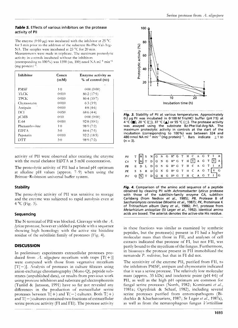
Table 5. Effects of various inhibitors on the protease activity of PI1 . . . . . . . . . . , . , . . . . . . . . . . . . , . . . . . . . . . . . , . . . . . . . . . . . . . . . . . . . . . . . . . . . . . . . . . . . . . . . . . . . , . . . , , . . . . . . . . . . . . . . . . . . . . . . . . . . . . . . . . . . . . .
The enzyme (0.10 pg) was incubated with the inhibitor at 25 "C for 5 min prior to the addition of the substrate Uz-Phe-Val-Arg- NA. The samples were incubated at 25 "C for 20 min. Measurements were made in triplicate. The maximum proteolytic activity in controls incubated without the inhibitors (corresponding t o 100%) was 1590 (SD, 100) nmol N A ml-' min-' (mg protein)- I .
100 0
Inhibitor
~~
PhiSF TLCK TPCK Chymosta tin A ntipai n DCI pCMH E-64 Phenanthroline E D T A Pepstat in DTT
Concn (mM)
1.0 0.0 10 0.0 10 0.0 10 0.010 0.050 0.10 0.010 1.0 5.0 0.010 5.0
Enzyme activity as % of control (SD)
0.00 (0.00) 86.2 (1 7.5) 86.4 (10.7) 6.3 (3.9) 4.8 (4.6)
68.6 (4.4) 0.00 (0.00)
92.8 (10.1)
84.4 (7.5) 98.9 (7.2)
92.2 (18.5) 98.9 (7.2)
incubation time (h)
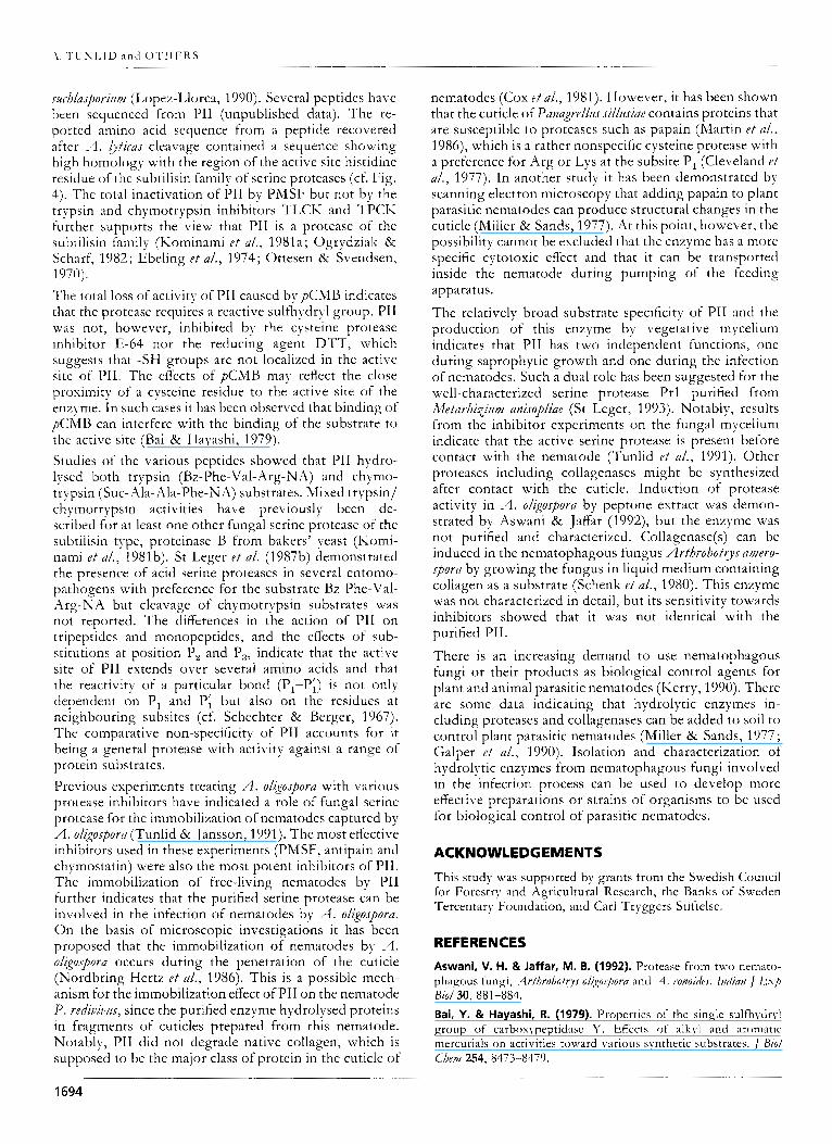
Fig. 3. Stability of Pi1 a t various temperatures. Approximately 0.2 pg Pii was incubated in 0.100 M Tris/HCI buffer (pH 7.5) a t 4 "C (M), 20 "C (a), 37 "C (A) or 55 "C (0). The protease activity was assayed using the substrate Bz-Phe-Val-Arg-NA. The maximum proteolytic activity in controls a t the start of the incubation (corresponding to 100%) was between 324 and 480 nmol NA ml-' min-' (mg protein)-'. Bars indicate 1 SD (n = 3).
activity o f PI1 were observed after treating the enzyme with the metal chelator E D T A at 5 mM concentration.
The proteolytic activity of PI1 had a broad p H optimum at alkaline pH values (approx. 7-9) when using the Britton-Robinson universal buffer system.
PB E D L D
PK S S R D Pri Q T D
G N G H ' G T H C A G T I
G N G H * G T H O A G T B A
Stability
The proteolytic activity of PI1 was sensitive to storage and the enzyme was subjected to rapid autolysis even at 4 "C (Fig. 3).
Sequencing
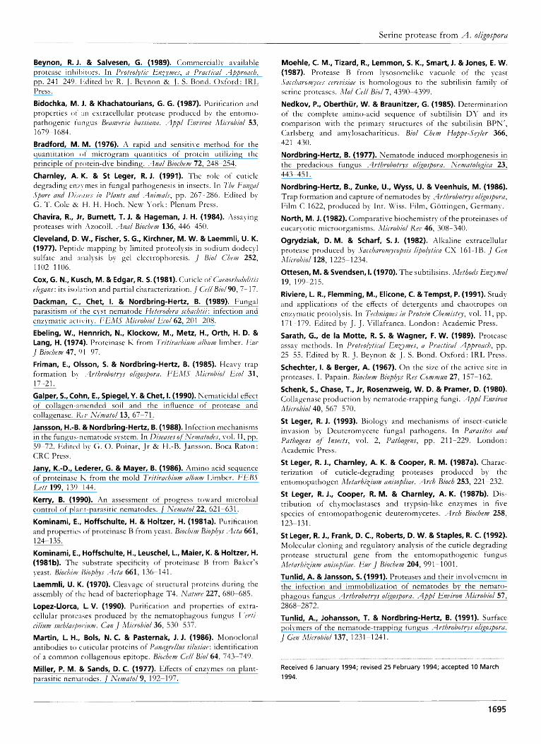
The N-terminal of PI1 was blocked. Cleavage with the ,4. hticzts protease, however yielded a peptide with a sequence showing high homology with the active site histidine residue o f the subtilisin family of proteases (Fig. 4).
DISCUSSION
In preliminary experiments extracellular proteases pro- duced from A. oligospora mycelium with traps [T( +)] were comp;ired with those from vegetative mycelium [T( - ) I . Analysis of proteases in culture filtrates using anion-exchange chromatography (Mono Q), peptide sub- strates (unpublished data), o r results from previous work using protease inhibitors and substrate gel electrophoresis (Tunlid 8c Jansson, 1991) have so far not revealed any differences in the production of extracellular set-ine proteases between T( +) and T( -) cultures. Both T( +) and T( - ) cultures contained two fractions of extracellular serine protease activity (FI and FII). The protease activity
Fig. 4. Comparison of the amino acid sequence of a peptide obtained by cleaving Pii with Achromobacter lyticus protease with those of the subtilisin-family enzyme CAI subtilisin Carlsberg (from Nedkov et a/., 1985); PB, Protease B of Saccharomyces cerevisiae (Moehle et a/., 1987); PK, Proteinase K of Tritirachium album (Jany et a/., 1986); Pr l , protease from Metarhizium anisopliae (St Leger et a/., 1992). identical amino acids are boxed. The asterisk denotes the active-site His residue.
in these fractions was similar as examined by synthetic peptides, but the protease(s) present in FI had a higher molecular mass than those in FII, and analyses of cell extracts indicated that protease of FI, but not FII, was partly bound to the mycelium of the fungus. Furthermore, in bioassays the protease present in FII immobilized the nematode P. redi~ivus, but that in FI did not.
The sensitivity of the enzyme PII, purified from FII, to the inhibitors PMSF, antipain and chymostatin indicated that it was a serine protease. The relatively low molecular mass (approx. 35 kDa) and isoelectric point (pH 4.6) of PII, as well as the high pH optimum are common for fungal serine proteases (North, 1982; Iiominami e t a/ . , 1981a; Ogrydziak & Scharf, 1982), including several serine proteases purified from entomopathogens (Bi- dochka & Khachatourians, 1987; St Leger ef d/., 1987a), as well as from the nematophagous fungus 17erticillizmz
1693
\. ’I’LIKJAID a n d O T H E R S
.rztch/asporizutz (Lopez-Llorca, 1990). Several peptides have been sequenced from PI1 (unpublished data). The re- ported amino acid sequence from a peptide recovered after A. hticas cleavage contained a sequence showing high homology with the region of the active site histidine residue of the subtilisin family of serine proteases (cf. Fig. 4). The total inactivation of PI1 by PMSF but not by the trypsin and chymotrypsin inhibitors TLCK and TPCK further supports the view that PI1 is a protease of the subtilisin family (Kominami e t a/., 1981a; Ogrydziak & Scharf, 1982; Ebeling e t a/., 1974; Ottesen & Svendsen,
The total loss of activity o f PI1 caused bypCMB indicates that the protease requires a reactive sulfhydryl group. PI1 was not, however, inhibited by the cysteine protease inhibitor E-64 nor the reducing agent DTT, which suggests that -SH groups are not localized in the active site of PII. The effects of pCMB may reflect the close proximity of a cysteine residue to the active site of the enzyme. In such cases it has been observed that binding o f pCMB can interfere with the binding of the substrate to the active site (Bai & Hayashi, 1979). Studies of the various peptides showed that PI1 hydro- lpsed both trypsin (Bz-Phe-Val- Arg-NA) and chymo- trypsin (Suc-Xla- Ah-Phe-NA) substrates. Mixed trypsin/ chymotrypsin activities have previously been de- scribed for at least one other fungal serine protease of the subtilisin type, proteinase B from bakers’ yeast (Komi- nami e t a/., 198lb). St Leger e t al. (1987b) demonstrated the presence of acid serine proteases in several entomo- pathogens with preference for the substrate Bz-Phe-Val- Arg-NA but cleavage of chymotrypsin substrates was not reported. The differences in the action o f PI1 on tripeptides and monopeptides, and the effects of sub- stitutions at position P, and P,, indicate that the active site of PI1 extends over several amino acids and that the reactivity of a particular bond (Pt-P;) is not only dependent on P, and P; but also on the residues at neighbouring subsites (cf. Schechter & Berger, 1967). The comparative non-specificity of PI1 accounts for it being a general protease with activity against a range of protein substrates . Previous experiments treating A. oligospora with various protease inhibitors have indicated a role of fungal serine protease for the immobilization of nematodes captured by A. oligospora (Tunlid & Jansson, 1991). The most effective inhibitors used in these experiments (PMSF, antipain and chymostatin) were also the most potent inhibitors of PII. The immobilization of free-living nematodes by PI1 further indicates that the purified serine protease can be involved in the infection of nematodes by A. oligospora. On the basis of microscopic investigations it has been proposed that the immobilization of nematodes by A. oligospora occurs during the penetration of the cuticle (Nordbring-Hertz e t a/., 1986). This is a possible mech- anism for the immobilization effect of PI1 on the nematode P. rediuiuiu, since the purified enzyme hydrolysed proteins in fragments of cuticles prepared from this nematode. Notably, PI1 did not degrade native collagen, which is supposed to be the major class of protein in the cuticle o f
1970).
nematodes (Cox e t a/., 1981). However, it has been shown that the cuticle of Panagrelhs s ihsiae contains proteins that are susceptible to proteases such as papain (Martin r f a/,, 1986), which is a rather nonspecific cysteine protease with a preference for Arg or Lys at the subsite P, (Cleveland e t a/., 1977). In another study it has been demonstrated by scanning electron microscopy that adding papain to plant parasitic nematodes can produce structural changes in the cuticle (Miller & Sands, 1977). At this point, however, the possibility cannot be excluded that the enzyme has a more specific cytotoxic effect and that it can be transported inside the nematode during pumping of the feeding apparatus.
The relatively broad substrate specificity of PI1 and the production of this enzyme by vegetative mycelium indicates that PI1 has two independent functions, one during saprophytic growth and one during the infection of nematodes. Such a dual role has been suggested for the well-characterized serine protease Prl purified from Afetarhixizmz anisopliae (St Ixger, 1993). Notably, results from the inhibitor experiments on the fungal mycelium indicate that the active serine protease is present before contact with the nematode (Tunlid e t a/., 1991). Other proteases including collagenases might be synthesized after contact with the cuticle. Induction of protease activity in A. oligospora by peptone extract was demon- strated by Aswani & Jaffar (1992), but the enzyme was not purified and characterized. Collagenase(s) can be induced in the nematophagous fungus Arthrobotys amero- spora by growing the fungus in liquid medium containing collagen as a substrate (Schenk e t a/., 1980). This enzyme was not characterized in detail, but its sensitivity towards inhibitors showed that it was not identical with the purified PII.
There is an increasing demand to use nematophagous fungi or their products as biological control agents for plant and animal parasitic nematodes (Icerry, 1990). There are some data indicating that hydrolytic enzymes in- cluding proteases and collagenases can be added to soil to control plant parasitic nematodes (Miller & Sands, 1977 ; Galper e t a/., 1990). Isolation and characterization of hydrolytic enzymes from nematophagous fungi involved in the infection process can be used to develop more effective preparations or strains of organisms to be used for biological control of parasitic nematodes.
ACKNOWLEDGEMENTS
This study was supported by grants from the Swedish Council for Forestry and Agricultural Research, the Banks of Sweden Tercentary Foundation, and Carl Tryggers Stiftelse.
REFERENCES
Aswani, V. H. & Jaffar, M. B. (1992). Protease from two nemato- phagous fungi, .4rfhrobofrys oligospora and .+I. conoidcs. Indian J E x p
Bai, Y. & Hayashi, R. (1979). Properties of the single sulfhydryl group of carboxypeptidase Y. Effects of alkyl and aromatic mercurials on activities toward various synthetic substrates. J Biol
Bi0l30, 881-884.
C h ~ l 2 5 4 , 8473-8479.
1694
Serine protease from A. oligospora ~- ~
Beynon, R. J. & Salvesen, G. (1989). Commercially available protease inhihitors. In Protro!ytic Envlms, a Practical <-lppronch, pp. 241 249. 1:dited by R. J . Reynon Ck J . S. Bond. Oxford: IRL Press.
Bidochka, M. 1. & Khachatourians, G. G. (1987). Purification and properties o f :in extracellular protease produced by the entomo- pathogenic t'urigus Braweria bussi~inn. -4ppl Enriro/r 'llicrobiol 53, 1679 1684. Bradford, M. M. (1976). A rapid and sensitive method for the quantitation o f microgram quantities of protein utilizing the principle of protein-dye binding. .gnu/ Biochtvr/ 72, 248-254. Charnley, A. K. & S t Leger, R. J. (1991). The role of cuticle degrading en/! mes in fungal pathogenesis in insects. In 7 % ~ FunAal .$pore and 1)i.riii.re.r in Plants and -4nin/a/s, pp. 267 ~ 286. Edited by G. T. Cole 8: H. H. Hoch. New l ' o r k : Plenum Press. Chavira, R., Jr, Burnett, T. 1. & Hagernan, 1. H. (1984). :\ssaying proteases Lvitti :\zocoll. .4nal Biochtt?) 136, 446 -450. Cleveland, D. W., Fischer, 5. G., Kirchner, M. W. & Laernrnli, U. K. (1977). Peptitlc mapping by limited proteolysis in sodium dodecyl sulfate and analysis by gel electrophoresis. .] B i d Chem 252, 1102 1106. Cox, G. N., Kusch, M. & Edgar, R. 5. (1981). Cuticle of Cuenorhal~ditis rlrgan.r: its isolation and partial characterization. .] CrN Biol90, 7- 17. Dackrnan, C., Chet, I. & Nordbring-Hertz, B. (1989). Fungal parasitism of the cyst nematode Heterodrra schachtii: infection and enzymatic acti\.ity. F'EAIS i2licroDzol k c o l 62, 201-208. Ebeling, W., Hennrich, N., Klockow, M., Metz, H., Orth, H. D. & Lang, H. (1974). Proteinase I< from Tritiruchiutti U/IJI /W limber. LJur J Biocheni 47, 01 97. Frirnan, E., Olsson, 5. & Nordbring-Hertz, B. (1985). Heavy trap formation b! :2rfhrobotrys ol(prpora. FER.1.S Alicrohiol Ecol 31,
Galper, S., Cohn, E., Spiegel, Y. & Chet, 1. (1990). Nematicidal effect of collagen-amended soil and the influence of protease and collagenase. t(i.11 iYin/atol 13, 67-71. Jansson, H.-B. & Nordbring-Hertz, B. (1988). Infection mechanisms in the fungus-nematode system. In Diseases oj-iVr~t/atodt.r, vol. 11, pp. 59-72. Edited b y G . 0. Poinar, J r Ck I-I.-B. Jansson. Roca Raton: CRC Press. Jany, K.-D., Lederer, G. & Mayer, B. (1986). Amino acid sequence of proteinase I\ from the mold Tritirachi14ni album Limber. FEBJ' Lett 199, 130 144. Kerry, B. (1990). An assessment of progress toward microl)ial control of plant-parasitic nematodes. J Nematol22, 621-631. Korninarni, E., Hoffschulte, H. & Holtzer, H. (1981a). Purification and propertie.; o f proteinase B from yeast. Biochi/pf Siopbys -4c fu 661,
Korninarni, E., Hoffschulte, H., Leuschel, L., Maier, K. & Holtzer, H. (1981b). The substrate specificity of proteinase B from Baker's yeast. Biochifti Iliopt5y.r -4ctu 661, 136-141. Laernrnli, U. K. (1970). Cleavage of structural proteins during the assembly of the head of bacteriophage T4. Nufirre 227, 680-685. Lopez-Llorca, L. V. (1990). Purification and properties of extra- cellular proteases produced by the nematophagous fungus I 'erfi- cilium s i~h /a . rpor i i~~~ . Caw J Microhiol36, 530-537. Martin, L. H., Bols, N. C. & Pasternak, 1. 1. (1986). Monoclonal antibodies t o cuticular proteins of Punagrrllz~s silctsiur : identification of a common collagenous epitope. Biochew Cell Biol64, 743-749. Miller, P. M. & Sands, D. C. (1977). Effects of enzymes on plant- parasitic nematodes. J Nematol9, 192-1 97.
17 -21.
124-1 35.
Moehle, C. M., Tizard, R., Lernrnon, 5. K., Smart, J. & Jones, E. W. (1987). Protease B from lysosomelike vacuole of the yeast .5'ucchnro/~ycrs cereiiisiue is homologous to the subtilisin family of serine proteases. Alol Cell Biol7, 4390-4399. Nedkov, P., Oberthur, W. & Braunitzer, G. (1985). Determination of the complete amino-acid sequence of subtilisin D Y and its comparison with the primary structures of the subtilisin BPN', Carlsberg and amylosachariticus. Biol Chem Hoppe-Syler 366, 421 430. Nordbring-Hertz, B. (1977). Nematode induced morphogenesis in the predacious fungus .4rthroliotrys ol(qosporu. iYrt~iatologicu 23, 443-45 1. Nordbring-Hertz, B., Zunke, U., Wyss, U. & Veenhuis, M. (1986). Trap formation and capture of nematodes by .-3rthroDotiys oligospora, Film C 1622, produced by Int. Vc'iss. Film, Giittingen, Germany.
North, M. 1. (1982). Comparative biochemistry of the proteinases of eucaryotic microorganisms. illicrobiol K P P 46, 308-340. Ogrydziak, D. M. & Scharf, S. J. (1982). Alkaline extracellular protease produced by .Saccbaron!ycop.ri.r lipoiyticn CS 161-1 B. J G'rn ibIiCrO/JiO/ 128, 1225-1234. Ottesen, M. & Svendsen, 1. (1 970). The subtilisins. Methods EnZy?//ol 19, 199 215.
Riviere, L. R., Flemrning, M., Elicone, C. &Tempst, P. (1991). Study and applications of the effects of detergents and chaotropes on enzymatic protolysis. In Technipes in Protein Chemistry, vol. 11, pp. 171-179. Edited by J . J . Villafranca. London: Academic Press.
Sarath, G., de la Motte, R. 5. & Wagner, F. W. (1989). Protease assay methods. In l'rotro!ytical Enutms , u Practical z4pproach, pp. 25 55. Edited by R. J . Beynon 2% J . S. Bond. Oxford: IRL Press.
Schechter, 1. & Berger, A. (1967). O n the size of the active site in proteases. I . Papain. Biochem Biopkys Kes Conimun 27, 157-162. Schenk, S., Chase, T., Jr, Rosenzweig, W. D. & Prarner, D. (1980). Collagenase production by nematode-trapping fungi. A p p l Emirom illicrobiol 40, 567 570. S t Leger, R. 1. (1993). Biology and mechanisms of insect-cuticle invasion by Deuteromycete fungal pathogens. In Parasites and Pathogens of' Insects, vol. 2, Pathogens, pp. 21 1-229. London : Academic Press.
S t Leger, R. J., Charnley, A. K. & Cooper, R. M. (1987a). Charac- terization of cuticle-degrading proteases produced by the entomopathogen Aletarhi~inm anisopliae. -4rch Bioch 253, 221 -232. S t Leger, R. J., Cooper, R. M. & Charnley, A. K. (1987b). Dis- tribution of chymoelastases and trypsin-like enzymes in five species of entomopathogenic deuteromycetes. Arch Biochem 258,
S t Leger, R. J., Frank, D. C., Roberts, D. W. & Staples, R. C. (1992). Molecular cloning and regulatory analysis of the cuticle degrading protease structural gene from the entomopathogenic fungus iiletarhixi/i~/ anisopliae. Eur J Biochem 204, 991 - 1001. Tunlid, A. & Jansson, 5. (1991). Proteases and their involvement in the infection and immobilization of nematodes by the nemato- phagous fungus .4 rthrobotrys olkospora. Appl Environ Microbiol 57,
Tunlid, A,, Johansson, T. & Nordbring-Hertz, B. (1991). Surface polymers of the nematode-trapping fungus <4rthrobotrys oligospora. J Gen Alicrobiol 137, 1231-1241.
123-131.
2868-2872.
Received 6 January 1994; revised 25 February 1994; accepted 10 March 1994.
1695