Consistent treatment of inter- and intramolecular polarization in molecular mechanics calculations
Upload
independentCategory
view
0download
0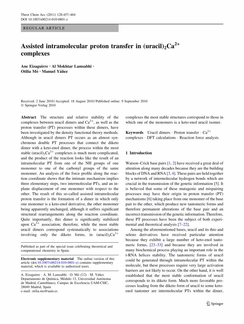
REGULAR ARTICLE
Assisted intramolecular proton transfer in (uracil)2Ca2+
complexes
Ane Eizaguirre • Al Mokhtar Lamsabhi •
Otilia Mo • Manuel Yanez
Received: 2 June 2010 / Accepted: 18 August 2010 / Published online: 9 September 2010
� Springer-Verlag 2010
Abstract The structure and relative stability of the
complexes between uracil dimers and Ca2?, as well as the
proton transfer (PT) processes within these dimers, have
been investigated by the density functional theory methods.
Although in uracil dimers PT occurs as an almost syn-
chronous double PT processes that connect the diketo
dimer with a keto-enol dimer, the process within the most
stable (uracil)2Ca2? complexes is much more complicated,
and the product of the reaction looks like the result of an
intramolecular PT from one of the NH groups of one
monomer to one of the carbonyl groups of the same
monomer. An analysis of the force profile along the reac-
tion coordinate shows that the intimate mechanism implies
three elementary steps, two intermolecular PTs, and an in-
plane displacement of one monomer with respect to the
other. The result of this so-called assisted intramolecular
proton transfer is the formation of a dimer in which only
one monomer is a keto-enol derivative, the other monomer
being apparently unchanged, although it suffers significant
structural rearrangements along the reaction coordinate.
Quite importantly, this dimer is significantly stabilized
upon Ca2? association; therefore, while the most stable
uracil dimers correspond systematically to associations
involving only the diketo forms, in (uracil)2Ca2?
complexes the most stable structures correspond to those in
which one of the monomers is a keto-enol uracil isomer.
Keywords Uracil dimers � Proton transfer � Ca2?
complexes � DFT calculations � Reaction force analysis
1 Introduction
Watson–Crick base pairs [1, 2] have received a great deal of
attention along many decades because they are the building
blocks of DNA and RNA [3, 4]. These pairs are held together
by a network of intermolecular hydrogen bonds which are
crucial in the transmission of the genetic information [5]. It
is believed that some of these mutagenic and mispairing
processes may have their origin in proton transfer (PT)
mechanisms [6] taking place from one monomer of the base
pair to the other, which produce new tautomeric forms and
therefore permanent alterations of the base pair and an
incorrect transmission of the genetic information. Therefore,
these PT processes have been the subject of both experi-
mental and theoretical analysis [7–22].
Among the aforementioned bases, uracil and its thio and
seleno derivatives have received particular attention
because they exhibit a large number of keto-enol tauto-
meric forms, [23–33] and because they are involved in
many biochemical process playing an important role in the
t-RNA helices stability. The tautomeric forms of uracil
could be generated through intramolecular PT within the
molecule, but these processes require very large activation
barriers are not likely to occur. On the other hand, it is well
established that the most stable conformation of uracil
corresponds to its diketo form. Much more favorable pro-
cesses leading from the diketo form of uracil to some keto-
enol tautomer are intermolecular PTs within the dimer,
Published as part of the special issue celebrating theoretical and
computational chemistry in Spain.
Electronic supplementary material The online version of thisarticle (doi:10.1007/s00214-010-0801-z) contains supplementarymaterial, which is available to authorized users.
A. Eizaguirre � A. M. Lamsabhi � O. Mo (&) � M. Yanez
Departamento de Quımica, Modulo 13, Universidad Autonoma
de Madrid, Cantoblanco, Campus de Excelencia UAM-CSIC,
28049 Madrid, Spain
e-mail: [email protected]
123
Theor Chem Acc (2011) 128:457–464
DOI 10.1007/s00214-010-0801-z

which required much lower barriers than the intramolecular
PTs within the monomers, because the mechanisms actu-
ally involves a double PT from one monomer to the other
[34]. It has also been shown that for the particular case of
uracil these double PT are practically synchronous pro-
cesses, whereas for the thiouracil derivatives they exhibit
some degree of asynchronicity [34]. The question we want
to analyze in this paper is, ‘‘are there any possible effects
that the interaction of the base pair with metal ions may
have in these double PT processes?’’ For this purpose, we
have chosen Ca2? because (1) the perturbation of the base
will be more significant when interacting with a doubly
charged species than with singly charged cations (2) Ca2?
is ubiquitous in physiological media, and (3) differently to
other doubly charged metal ions, such as Cu2? [35] or Pb2?
[36], Ca2? [37] does not lead to a spontaneous deproto-
nation of the base.
Several studies on the interaction of base pairs with
doubly charged metal ions and in particular with alkaline-
earth cations have been reported in the literature, [21, 38–
40] but much less attention was paid to the effects of this
association on the tautomerization processes which may
take place within the base pair [21, 32, 36].
2 Computational details
The geometries of the different uracil dimers and their
complexes with Ca2? were optimized by using the hybrid
B3LYP functional. This approach, which combines the
nonlocal correlation function of Lee et al. [41] with the
Becke’s three-parameter nonlocal hybrid exchange func-
tional [42], has been shown to provide very reliable
geometries and harmonic vibrational frequencies for a
great variety of systems. Furthermore, in previous theo-
retical assessments, it has been shown to be a very good
compromise between accuracy and computational effort
when dealing with Ca2? interactions [43]. This approach,
when used with flexible enough basis sets, also performs
quite well in describing both inter- and intramolecular
hydrogen bonds, which in our case are important actors in
the mechanisms to be investigated. It must be mentioned,
however, that in a recent assessment using adenine-thy-
mine and guanine-cytosine base pairs as model system it
was found that B3LYP method slightly overestimated
hydrogen bond distances when compared with MP2 ones
[44]. Geometry optimizations and harmonic vibrational
frequencies were evaluated using a DZ-polarized 6-
31 ? G(d,p) basis set expansion, which includes diffuse
functions on the heavy atoms, required to an appropriate
description of hydrogen bonding and the polarization
effects induced by the doubly charged metal ion on the
base. The harmonic vibrational frequencies so obtained
were used to evaluate the vibrational thermal corrections
and classify the stationary points located on the potential
energy surface as local minima or transition states. To have
reliable final energies, these were obtained in single-point
calculations using a much larger 6-311 ? G(3df,2p) basis
set expansion.
The reaction force analysis [45–47] has been shown to
offer interesting insights into the double PT mechanisms
within uracil dimers and therefore we have decided to
apply the same approach for the analysis of similar pro-
cesses within (uracil)2Ca2? complexes.
The lowest energy path connecting reactants and prod-
ucts in an elementary reaction when projected into the
intrinsic reaction coordinate typically presents a maximum
corresponding to the transition state (TS) relying reactants
and products. The differentiation of this energy curve leads
to the reaction force,
FðnÞ ¼ �dEðnÞdn
ð1Þ
which presents two extrema, n1 (minimum) and n2
(maximum) (See Fig. 1), which in turn permit the
identification of different regions with a well-defined
chemical meaning [48, 49], namely (1) the reactant
region (nR B n B n1) where the reactants begin to start
activated by a negative retarding force, (2) the transition
state region (n1 B n B n2) where a driving force
increasingly balances the aforementioned retarding one,
and (3) the products region (n2 B n B nP) where the
system relaxes and the driving force tends to zero when
approaching nP. This partitioning of the reaction coordinate
in different reaction regions allows dividing the activation
energy into two main contributions, [49, 50]
DE# ¼ W1 þW2 ð2Þ
where,
W1 ¼ �Zn1
nR
FðnÞdn and W2 ¼ �Zn0
n1
FðnÞdn ð3Þ
In our particular case, the first term of Eq. 2 represents the
work to be done to bring the HB donor and the HB acceptor
close enough as to propitiate the PT process, whereas the
second term corresponds to the energy required to reach the
TS, in order to begin the evolution toward the products.
Analogously, the second part of the reaction coordinate can
be split into two components, W3 and W4, so that the
reaction energy can be written as
DE0 ¼ W1 þW2 þW3 þW4 ð4Þ
where W3 represents the energy gaining on the relaxation
of the system from the TS to n2 and W4 the energy to finally
reach the products, and are given by
458 Theor Chem Acc (2011) 128:457–464
123

W3 ¼ �Zn2
n0
FðnÞdn and W4 ¼ �ZnP
n2
FðnÞdn ð5Þ
To carry out this analysis, the necessary calculations along
the intrinsic reaction coordinate (IRC) expressed in mass-
weighted cartesian coordinates, which involve a full
geometry optimization at each step along n, were carried
out as implemented in the Gaussian03 suite of programs.
The bonding perturbations triggered by the metal dica-
tion in the base pair were analyzed by means of the atoms
in molecules (AIM) [51] and the electron localization
function (ELF) [52, 53] theories. Within the framework of
the former, we have evaluated the electron density at the
different bond critical points (BCPs) and we have exam-
ined its evolution along the reaction coordinate by using
the AIMALL series of programs [54]. The bonding chan-
ges in a system triggered by its interaction with a charged
species can be also followed by analyzing the changes in
the energy density
hðr~Þ ¼ vðr~Þ þ gðr~Þ ð6Þ
where vðr~Þ and gðr~Þ are the local densities of the kinetic
energies, respectively. The regions in which this magnitude
is negative or positive correspond to areas in which the
electron density is built up or depleted, respectively, so that
the former can be associated with covalent interactions,
whereas the latter are typically associated with closed-shell
interactions, as in ionic bonds or hydrogen bonds.
The ELF allows the partition of the molecular space into
basins, [53, 55] usually disynaptic and monosynaptic,
which correspond to regions of maximum probability of
finding electron pairs. This description in terms of disy-
naptic basins, associated with bonding pairs, and mono-
synaptic basins, associated with lone-pairs or core-pair
electrons, provides useful information about the nature of
the bonding, even in challenge cases in which other
approaches fail to give an unambiguous bonding picture
[56]. The electron population within these basins can be
obtained by integration of the electron density within the
basins limits. ELF grids and basin integrations have been
evaluated with the TopMod package [57]. For the three-
dimensional plots, an ELF value of 0.8 has been used.
A complementary viewpoint some times crucial to
understand the bonding can be obtained through the use of
the NBO (Natural Bond Orbital) approach [58], which
describes the bonding in terms of localized hybrids
obtained as local block eigenvectors of the one particular
density matrix. A second-order perturbation treatment
permits also to quantify the interaction energies between
occupied and virtual orbitals reflected in a charge transfer
from the former to the latter. These kind of interactions are
typically found in XH���Y hydrogen bonds, which are
characterized by a charge transfer from the lone-pairs of
the HB acceptor Y, to the antibonding rXH* orbital of the
HB donor, XH [58].
3 Results and discussion
As it has been previously shown in the literature, uracil
leads to six different dimers [59] (see Fig. 2), depending on
the relative positions of the two monomers and the kind of
hydrogen bonds connecting them. For instance, if we
identify within the uracil molecule, a urea-like subunit
(HN–CO–NH), and a formamide-like subunit (HN–CO–
CH), it can be observed that for the global minimum, UU1,
and the less stable dimers, UU3 and UU6, the HBs con-
necting both monomers, looking at the carbonyl groups
which act as HB acceptors, correspond to a urea–urea
interaction, the only difference between the three
Fig. 1 Energy (a) and force (b) profiles for an elementary reaction
connecting reactants (R) and products (P)
Theor Chem Acc (2011) 128:457–464 459
123

complexes being the relative orientation of both monomers.
UU2 and UU5 are connected through urea–formamide
interactions and again only differ in the relative orientation
of both monomers. Finally, UU4 dimer involves a form-
amide–formamide interaction.
The number of (uracil)2Ca2? complexes is much larger
since the metal dication can be attached to different basic
sites of the dimer, but more importantly, the relative sta-
bility of the (uracil)2Ca2? complexes does not reflect the
relative stability of the uracil dimers. Actually, as shown in
Fig. 2, the most stable (uracil)2Ca2? complex, namely
UU5-a, is formed by the association of Ca2? to the one of
the less stable uracil dimer (UU5). Conversely, the global
minimum of the neutral dimers, UU1, leads upon Ca2?
association to one of the less stable (uracil)2Ca2? com-
plexes (UU1-a), 172 kJ mol-1 above UU5-a. It is also
worth noting that in UU5-a, Ca2? bridges between the two
formamide-like carbonyl groups. Interestingly, when the
metal dication interacts with the two urea-like carbonyl
groups, the complex formed, UU5-b, is found to be
57 kJ mol-1 less stable. This can be easily explained if one
takes into account that, as has been shown previously in the
literature, the most basic site of the uracil molecule is the
carbonyl group at position 4, so in complex UU5-a, Ca2?
interacts with the most basic site of both monomers,
whereas in UU5-b complex it interacts with the least basic
site centers. Although many other (uracil)2Ca2? complexes
have been calculated in our work, in Fig. 2 only the more
stable ones are shown. Their total energies together with
the zero point energy (ZPE) corrections are reported in
Table S1 of the supporting information. The next important
question to be addressed in this study, once we have
located the global minimum of the (uracil)2Ca2? PES, is
the mechanism of the PT process in this complex, UU5-a.
4 Proton transfer processes
As mentioned in the introduction, in uracil dimers, the most
favorable PT process corresponds to a practically syn-
chronous double PT mechanism, [34] so that the product is
a dimer in which both interacting monomers are enolic
forms of uracil. As illustrated in Fig. 3, the situation seems
to be significantly different upon the interaction with Ca2?,
where the product arises formally from an intramolecular
PT process in which the proton is transferred from a NH
group of one monomer toward the carbonyl group of the
same monomer. The result is that, after the PT, the
Fig. 2 Structure and relative stability of the six uracil dimers and its more stable Ca2? complexes. All values, in kJ mol-1; are referred to the
most stable (uracil)2Ca2? complex, UU5-a
460 Theor Chem Acc (2011) 128:457–464
123

complex corresponds to a dimer between the diketo form of
one of uracil moieties and the enolic form of the other. The
activation barrier for this process is 77 kJ mol-1, which is
significantly lower than the corresponding activation bar-
riers for a similar process in both isolated uracil
(197 kJ mol-1) and uracil-Ca2? complex (154 kJ mol-1)
[37]. This decrease in the activation barrier points appar-
ently to a catalytic effect triggered by Ca2? association.
However, the structure of the transition state found (see
Fig. 3) and the anomalous profile of both the potential
energy curve and the curve of the forces along the reaction
coordinate (See Fig. 4a, b, respectively, for the UU5a
(R) ? UU5a(e) (P) process), clearly show that a more
complicated mechanism than just a single intramolecular
PT must be taking place. The shoulder observed in the
force in the region previous to the TS as well as the almost
linear increase observed after the TS clearly indicate that
the process connecting reactants and products is not an
elementary one.
This is actually seen when looking at the plots of the
energy density and the ELF along the potential energy curve
and the forces profile, respectively. The populations of the
ELF basins are summarized in Table S2 of the supporting
information. In the reactants (n & -2), one clearly
observes an ionic interaction between Ca2? and the two
(fomamide-like) carbonyl oxygens and two NH���O inter-
molecular hydrogen bonds. At a more advanced point along
the reaction coordinate (n = -0.53), a PT within the HB
not interacting with the metal dication is taking place, and
the proton appears almost midway between the N and the O
atoms. At this point, the ELF shows the presence of an OH
disynaptic basin which did not exist in the reactants, and the
disappearance of the NH disynaptic basin. At the same time,
the monosynaptic N lone-pair, which in the reactants has a
clear p-character, becomes now a monosynaptic basin with
a noticeable r character. This PT process would involve a
(hidden) transition state (TS1) which can be qualitatively
represented by the red potential energy curve. A further
evolution of the system to reach (n = 0) leads to the
aforementioned PT to almost completion. Actually, the
transition vector at n = 0 corresponds to the relative dis-
placement of the right monomer with respect to the left one,
to favor the interaction of the (urea-like) carbonyl group of
the left monomer with the OH group formed in the first PT.
Note that this displacement will also favor the interaction of
the NH group of the right monomer with the (deprotonated)
N atom of the left one. The OH���O interaction triggers a PT
Fig. 3 Activation barriers associated with PT processes within the
(uracil)2Ca2? global minimum UU5-a. All values are in kJ mol-1
Fig. 4 Energy profile (a) and force profile (b) for the processes
connecting UU5-a with UU5-a(e). In figure (a), the energy density for
the reactant and the products, as well as for the transition states TS1,
TS, and TS2 are shown. In these plots, positive and negative values of
the energy density are denoted by dashed red lines and solid bluelines, respectively
Theor Chem Acc (2011) 128:457–464 461
123
from the right monomer toward the left one (n = 1.20). At
this point, which will correspond to the hidden transition
state (TS2) associated with the OH���O PT (black curve), the
proton appears almost midway between the two oxygen
atoms. Note that the ELF shows that at this point the PT is
not completed since the OH disynaptic basin, although
polarized toward the left monomer, is still located at the
right monomer. From here, the system evolves toward the
products, by completing the aforementioned PT. The con-
sequence is that the right monomer recovers the diketo
arrangement and only the left monomer presents an enol
group. Hence, although apparently an intramolecular PT
took place from the NH group of the left monomer toward
one of the (urea-like) carbonyl groups of the same mono-
mer, the real process involves three steps: a) a PT from the
NH group of the left monomer toward the (formamide-like)
carbonyl group of the right monomer, b) the relative dis-
placement of the right monomer with respect to the left one
and c) a PT from the OH group of the right monomer,
formed in the first step, toward the (urea-like) carbonyl
group of the left monomer.
This is also nicely reflected by looking at the charac-
teristics of the molecular graphs (See Fig. 5) evaluated at
the critical points in the evolution of the force (see Fig. 3)
and to the work employed in each step.
For the reactant (n & -2), two BCPs are located
between Ca2? and the two (formamide-like) carbonyl
oxygens of UU5 with small electron densities as in typical
ionic interactions. Two NH���O intramolecular HBs are
also observed. The work needed to reach n = -1.29
(23.7 kJ mol-1) is associated with the structural prepara-
tion of the system, so that at n = -1.29 the PT from the
NH group of the left monomer has already started, as
shown by a significant decrease in the electron density at
the N–H BCP and a concomitant increase in the electron
density at the O–H BCP. To reach TS1 (n = -0.53), the
work needed (42.7 kJ mol-1) corresponds to the electronic
reordering and represent 65% of the activation barrier.
When n = -0.53 is reached, the PT transfer process is
practically complete, so that the initial NH���O HB has been
replaced by a N���HO HB. On going from (n = -0.53)
toward (n = 0), the displacement of the right monomer
with respect to the left one starts. Obviously, this structural
rearrangement requires little work (8.3 kJ mol-1), the main
consequence being that the NH group of the right monomer
starts forming a HB also with the deprotonated N atom of
Fig. 5 Molecular graphs along the reaction coordinate connecting UU5-a with UU5-a(e). At the bottom of the figure, the molecular graphs for
the neutral UU5 and UU5(e) compounds are also given for the sake of comparison. Electron densities in a.u
462 Theor Chem Acc (2011) 128:457–464
123

the left monomer. A little further, a second structural
reordering, which requires a work of 11.3 kJ mol-1 takes
place. As a consequence, the OH group of the right
monomer starts to interact with the (urea-like) carbonyl
group of the left monomer, so that when the TS2 is reached
(n = 1.20) the PT from the OH of the right isomer toward
the (urea-like) carbonyl of the left one starts, as evidenced
by the decrease in the electron density at the O–H covalent
bond and the concomitant increase in the electron density
at the OH���O HB. The completion of the PT requires an
electronic reorganization work of 47.7 kJ mol-1 to reach
n = 2.39, and from here the system relax toward the
product, the work involved in this process being
60.4 kJ mol-1.
One question that needs to be answered is what is the
possible role of Ca2? on the stabilization of these enol-
diketo dimers. As shown in Fig. 3, the enol-diketo dimes,
UU5-a(e) is more stable than the diketo-diketo one UU5-a.
The question is, whether the association with Ca2? is
responsible for the enhanced stability of the enol-diketo
form. To answer this question, we have calculated the
energy of the isolated dimer and the corresponding dimer-
Ca2? complex. The results obtained indicate that the iso-
lated enol-diketo form is 64.7 kJ mol-1 less stable than the
diketo-diketo isomer, whereas for the corresponding Ca2?
complexes is the other way around, and UU5-a(e) isomer is
61.3 kJ mol-1 more stable than UU5-a.
In other words, whereas the PT process from UU5-a to
UU5-a(e) is exothermic, the corresponding process for the
isolated dimers would be clearly endothermic. This can be
due to two different effects, the perturbation caused by
Ca2? on the HBs connecting both monomers and/or the
different basicity of the carbonyl groups interacting with
the metal dication. A comparison of the molecular graphs
of UU5 and UU5-a (see Fig. 5) clearly indicates that
association of Ca2? to the former results in a weakening of
both intermolecular hydrogen bonds. This is consistent
with the NBO second-order perturbation energies. As
shown in Table 1, in UU5 there is an interaction between
the two lone-pairs of the carbonyl oxygens with the rNH*
antibonding orbitals, associated with the two intermolecu-
lar HBs that are almost identical. Upon Ca2? attachment
the upper HB becomes weaker, because the donation from
the oxygen lone-pair, which is interacting with Ca2?,
toward the rNH* antibonding orbital drastically decreases.
The strength of the lower HB decreases more, because, on
the one hand, the donating capacity of the carbonyl oxygen
decreases upon Ca2? attachment and the intrinsic acidity of
the NH group also decreases. In UU5(e), the first N2H���O1
HB in UU5 is replaced by a N2H���N1 HB. Similarly, the
N1H���O4 HB is replaced by a stronger O3H���O4 HB,
because the OH group is a stronger HB donor than the NH
one. The important finding however is that the former
becomes reinforced upon Ca2? association due likely to the
enhanced acidity of the NH groups and the latter weakens
only slightly, due likely to a slightly decrease in the basi-
city of the carbonyl group. The second important factor
which may explain the enhanced stability of the enol-keto
dimers upon Ca2? association is that in these complexes
the metal cation interacts with two free carbonyl groups,
whereas in the diketo-diketo complexes, one of the inter-
actions involves the carbonyl group which is already acting
as HB acceptor in one of the NH���O intermolecular HBs.
5 Conclusions
Although in uracil dimers PT occurs as an almost syn-
chronous double PT processes that connect the diketo
dimer with a keto-enol dimer, the process within the most
stable (uracil)2Ca2? complexes is much more complicated,
and the product of the reaction looks like the result of an
intramolecular PT from one of the NH groups of one
monomer to one of the carbonyl groups of the same
monomer. However, an analysis of the force profile shows
that the intimate mechanism implies three elementary
steps, two of which, the first and the third steps, are
intermolecular PT and the second an in-plane displacement
of one monomer with respect to the other. The result of this
so-called assisted intramolecular proton transfer is the
formation of a dimer in which only one monomer is a keto-
enol derivative, the other monomer being apparently
unchanged, although it suffers significant structural rear-
rangements along the reaction coordinate. Quite impor-
tantly, this dimer is significantly stabilized upon Ca2?
association, therefore, while the most stable uracil dimers
correspond systematically to associations involving only
the diketo forms, in (uracil)2Ca2? complexes the most
stable structures correspond to those in which one of the
monomers is a keto-enol uracil isomer. The origin of this
enhanced stability is likely twofolded: (1) a reinforcement
of the HBs connecting the monomers upon Ca2? associa-
tion and (2) a stronger interaction of the doubly charged
metal ion with the dimer, because the interaction involves
carbonyl groups not engaged in the intermolecular hydro-
gen bonds.
Table 1 Second-order interaction energies (in kJ mol-1) between the
HB acceptor lone-pair and the rXH* (X = N, O) antibonding orbital
in UU5, UU5-a, UU5(e), and UU5-a(e) systems
UU5 UU5-a UU5(e) UU5-a(e)
LPO1 ? rN2H* 37/41a 36/12a LPN1 ? rN2H* 67 77
LPO4 ? rN1H* 37/40a 28/13a LPO4 ? rO3H* 57/118 56/100a
a The two values reported correspond to the interactions of the two
lone-pairs of each carbonyl oxygen
Theor Chem Acc (2011) 128:457–464 463
123
The activation barriers, associated with this intradimer
enolization process in (uracil)2Ca2? complexes, are lower
than those calculated for the double proton transfer process
in the neutral (uracil)2 dimers and much lower than those
calculated for the tautomerization within each monomer.
Hence, these assisted intramolecular PT may play a quite
significant role in the tautomerization of uracil and likely
also in the tautomerization of uracil derivatives.
Acknowledgments This work has been partially supported by the
DGI Project No. CTQ2009-13129-C01, by the Project MADRISO-
LAR2, Ref.: S2009PPQ/1533 of the Comunidad Autonoma de
Madrid, by Consolider on Molecular Nanoscience CSC2007-00010,
and by the COST Action CM0702. A generous allocation of com-
puting time at the CCC of the UAM is also acknowledged.
References
1. Watson JD, Crick FHC (1953) Nature 171:964–967
2. Watson JD, Crick FHC (1953) Nature 171:737–738
3. Shibata M, Zielinski TJ, Rein R (1990) Theoretical Biochemistry
and Molecular Biophysics. Adenine Press, New York
4. Saenger W (1984) Principles of Nucleic Acid Structure. Springer
Verlag, New York
5. Jeffrey GA, Saenger W (1991) Hydrogen Bonding in Biological
Structure. Springer Verlag, New York
6. Nelson DL, Cox MM, Lehninger AL (2000) Principles of Bio-
chemistry. Worth Publishers Inc, New York
7. Kierdaszuk B, Stolarski R, Shugar D (1983) Eur J Biochem
130:559–564
8. He L, Kierzek R, Santalucia J, Walter AE, Turner DH (1991)
Biochemistry 30:11124–11132
9. Florian J, Hrouda V, Hobza P (1994) J Am Chem Soc
116:1457–1460
10. Walter AE, Wu M, Turner DH (1994) Biochemistry
33:11349–11354
11. Douhal A, Kim SK, Zewail AH (1995) Nature 378:260–263
12. Florian J, Leszczynski J (1996) J Am Chem Soc 118:3010–3017
13. Chen XY, McDowell JA, Kierzek R, Krugh TR, Turner DH
(2000) Biochemistry 39:8970–8982
14. Jiang LH, Russu IM (2001) Nucleic Acids Res 29:4231–4237
15. Kryachko ES (2002) Int J Quant Chem 90:910–923
16. Gorb L, Podolyan Y, Dziekonski P, Sokalski WA, Leszczynski J
(2004) J Am Chem Soc 126:10119–10129
17. Zoete V, Meuwly M (2004) J Chem Phys 121:4377–4388
18. Noguera M, Sodupe M, Bertran J (2004) Theor Chem Acc
112:318–326
19. Rak J, Makowska J, Voityuk AA (2006) Chem Phys 325:567–574
20. Noguera M, Sodupe M, Bertran J (2007) Theor Chem Acc
118:113–121
21. Noguera M, Bertran J, Sodupe M (2008) J Phys Chem B
112:4817–4825
22. Zhang JD, Chen ZF, Schaefer HF (2008) J Phys Chem A
112:6217–6226
23. Czerminski R, Lesyng B, Pohorille A (1979) Int J Quant Chem
16:605–613
24. Les A, Ortegablake I (1986) Int J Quant Chem 30:225–237
25. Katritzky AR, Karelson M, Harris PA (1991) Heterocycles
32:329–369
26. Leszczynski J (1992) J Phys Chem 96:1649–1653
27. Estrin DA, Paglieri L, Corongiu G (1994) J Phys Chem
98:5653–5660
28. Leszczynski J, Sponer J (1996) J Mol Struct Theochem
388:237–243
29. Lamsabhi M, Alcamı M, Mo O, Bouab W, Esseffar M, Abboud
JLM, Yanez M (2000) J Phys Chem A 104:5122–5130
30. Marino T, Russo N, Sicilia E, Toscano M (2001) Int J Quant
Chem 82:44–52
31. Millefiori S, Alparone A (2004) Chem Phys 303:27–36
32. Trujillo C, Mo O, Yanez M (2007) Org Biomol Chem
5:3092–3099
33. Feyer V, Plekan O, Richter R, Coreno M, Vall-Ilosera G, Prince
KC, Trofimov AB, Zaytseva IL, Moskovskaya TE, Gromov EV,
Schirmer J (2009) J Phys Chem A 113:5736–5742
34. Lamsabhi AM, Mo O, Gutierrez-Oliva S, Perez P, Toro-Labbe A,
Yanez M (2009) J Comput Chem 30:389–398
35. Lamsabhi AM, Alcamı M, Mo O, Yanez M, Tortajada J (2006) J
Phys Chem A 110:1943–1950
36. Gutle C, Salpin JY, Cartailler T, Tortajada J, Gaigeot MP (2006)
J Phys Chem A 110:11684–11694
37. Trujillo C, Lamsabhi AM, Mo O, Yanez M, Salpin JY (2008) Org
Biomol Chem 6:3695–3702
38. Sponer J, Sabat M, Burda JV, Leszczynski J, Hobza P (1999) J
Phys Chem B 103:2528–2534
39. Zhang Y, Huang KX (2007) J Mol Struct Theochem 812:51–62
40. Miyachi H, Matsui T, Shigeta Y, Hirao K (2010) Phys Chem
Chem Phys 12:909–917
41. Lee C, Yang W, Parr RG (1988) Theochem 40:305–313
42. Becke AD (1993) J Chem Phys 98:5648
43. Corral I, Mo O, Yanez M, Scott A, Radom L (2003) J Phys Chem
A 107:10456–10461
44. van der Wijst T, Guerra CF, Swart M, Bickelhaupt FM (2006)
Chem Phys Lett 426:415–421
45. Toro-Labbe A (1999) J Phys Chem A 103:4398–4403
46. Jaque P, Toro-Labbe A (2000) J Phys Chem A 104:995–1003
47. Gutierrrez-Oliva S, Herrera B, Toro-Labbe A, Chermette H
(2005) J Phys Chem A 109:1748–1751
48. Politzer P, Toro-Labbe A, Gutierrez-Oliva S, Herrera B, Jaque P,
Concha MC, Murray JS (2005) J Chem Sci 117:467–472
49. Toro-Labbe A, Gutierrerez-Oliva S, Murray JS, Politzer P (2007)
Mol Phys 105:2619–2625
50. Rincon E, Toro-Labbe A (2007) Chem Phys Lett 438:93–98
51. Bader RFW (1990) Atoms in Molecules. A Quantum Theory.
Clarendon Press, Oxford
52. Becke AD, Edgecombe KE (1990) J Chem Phys 92:5397–5403
53. Silvi B, Savin A (1994) Nature 371:683–686
54. Keith TA (2010) AIMAll (Version 10.05.04).
http://aim.tkgristmill.com
55. Savin A, Nesper R, Wengert S, Fasler TF (1997) Angew Chem
Int Ed Engl 36:1808–1832
56. Mo O, Yanez M, Martın Pendas A, Del Bene JE, Alkorta I,
Elguero J (2007) Phys Chem Chem Phys 9:3970–3977
57. Noury S, Krokidis X, Fuster F, Silvi B (1999) Comput Chem
23:597–604
58. Reed AE, Curtiss LA, Weinhold F (1988) Chem Rev 88:899–926
59. Kelly REA, Kantorovich LN (2006) J Phys Chem B
110:2249–2255
464 Theor Chem Acc (2011) 128:457–464
123
Copyright © 2022 FDOKUMEN