Proteomic identification of differentially expressed proteins during the acquisition of somatic...
12
Proteomic Identification of Differentially Expressed Proteins in the Ligon lintless Mutant of Upland Cotton (Gossypium hirsutum L.) Pi-Ming Zhao, †,‡,§ Li-Li Wang, †,‡,§ Li-Bo Han, †,‡,§ Juan Wang, †,‡,§ Yuan Yao, †,‡,§ Hai-Yun Wang, †,‡,§ Xiong-Ming Du, | Yuan-Ming Luo,* ,† and Gui-Xian Xia* ,†,‡,§ Institute of Microbiology, Chinese Academy of Sciences, Beijing 100101, State Key Laboratory of Plant Genomics, Beijing 100101, National Center for Plant Gene Research, Beijing 100101, and Institute of Cotton, Chinese Academy of Agricultural Sciences, Anyang 455112 Received October 28, 2009 Cotton fiber is an ideal model for studying plant cell elongation. To date, the underlying mechanisms controlling fiber elongation remain unclear due to their high complexity. In this study, a comparative proteomic analysis between a short-lint fiber mutant (Ligon lintless, Li 1 ) and its wild-type was performed to identify fiber elongation-related proteins. By 2-DE combined with local EST database-assisted MS/ MS analysis, 81 differentially expressed proteins assigned to different functional categories were identified from Li 1 fibers, of which 54 were down-regulated and 27 were up-regulated. Several novel aspects regarding cotton fiber elongation can be illustrated from our data. First, over half of the down- regulated proteins were newly identified at the protein level, which is mainly involved in protein folding and stabilization, nucleocytoplasmic transport, signal transduction, and vesicular-mediated transport. Second, a number of cytoskeleton-related proteins showed a remarkable decrease in protein abundance in the Li 1 fibers. Accordingly, the architecture of actin cytoskeleton was severely deformed and the microtubule organization was moderately altered, accompanied with dramatic disruption of vesicle trafficking. Third, the expression of several proteins involved in unfolded protein response (UPR) was activated in Li 1 fibers, indicating that the deficiency of fiber cell elongation was related to ER stress. Collectively, these findings significantly advanced our understanding of the mechanisms associated with cotton fiber elongation. Keywords: proteomics • cotton fiber • Ligon mutant • cytoskeleton • cell elongation Introduction Cotton fiber is a single and highly elongated epidermal cell of the cotton seed coat. Fiber development consists of four overlapping stages: initiation, elongation, secondary wall depo- sition, and maturation. 1 Fiber elongation begins on the day of anthesis and continues for 20-30 days, during which cotton fibers grow to 3-6 cm depending on the cultivars. 2 Due to the advantageous features such as extreme length and long dura- tion of elongation, cotton fiber is regarded as a model system for studying the mechanisms controlling plant cell elongation. 3 Significant progress has been made in large-scale identifica- tion of genes and proteins involved in fiber elongation during recent years. Through transcriptome analysis, Arpat et al. identified 2553 unique ESTs (uniESTs) from fiber cells of the diploid variety Gossypium arboretum, 4 and Shi et al. identified 778 uniESTs expressed preferentially or specifically during the elongation stage of fiber development in the tetroploid cotton variety G. hirsutum. 5 At protein level, Yang et al. studied the protein profiles in different stages of fiber development and identified 106 differently expressed proteins, 6 and Li et al. found 49 proteins accumulated specifically in elongating fiber cells by comparing the proteome profiles of a fuzzless-lintless mutant and its wild-type. 7 In addition to large-scale genes and proteins identification, a number of fiber elongation-related genes have been structurally or functionally characterized. Among them, a sucrose synthase gene (Sus), an actin gene (GhACT1), and a gene encoding a rate-limiting enzyme (GhDET2) in brassinosteroid (BR) biosynthesis were shown to be crucial for fiber elongation. 8-10 Although identification of these genes or proteins has made a substantial contribution to understanding the molecular basis of cotton fiber develop- ment, the underlying mechanisms that regulate fiber elongation are still unclear due to their high complexity. The Ligon lintless 1(Li 1 ) is a mutant of upland cotton (G. hirsutum) and possesses extremely shortened fibers as com- pared to the wild-type plant (6 mm vs 30 mm). It was originally discovered by Griffee and Ligon in 1929 11 and subsequently characterized by several studies, which revealed that: (1) Li 1 is a monogenic, dominant mutant with a defect in fiber elonga- tion and plant growth; (2) the inhibition of Li 1 fiber elongation * Authors for correspondence: Gui-Xian Xia and Yuan-Ming Luo Institute of Microbiology, Chinese Academy of Sciences, Beijing 100101, China. Telephone: +86 10 64845674, Fax: +86 10 64845674, E-mail: [email protected], [email protected]. † Chinese Academy of Sciences. ‡ State Key Laboratory of Plant Genomics. § National Center for Plant Gene Research. | Chinese Academy of Agricultural Sciences. 10.1021/pr900975t XXXX American Chemical Society Journal of Proteome Research XXXX, xxx, 000 A
Transcript of Proteomic identification of differentially expressed proteins during the acquisition of somatic...
Proteomic Identification of Differentially Expressed Proteins in the
Ligon lintless Mutant of Upland Cotton (Gossypium hirsutum L.)
Pi-Ming Zhao,†,‡,§ Li-Li Wang,†,‡,§ Li-Bo Han,†,‡,§ Juan Wang,†,‡,§ Yuan Yao,†,‡,§
Hai-Yun Wang,†,‡,§ Xiong-Ming Du,| Yuan-Ming Luo,*,† and Gui-Xian Xia*,†,‡,§
Institute of Microbiology, Chinese Academy of Sciences, Beijing 100101, State Key Laboratory of PlantGenomics, Beijing 100101, National Center for Plant Gene Research, Beijing 100101, and Institute of Cotton,
Chinese Academy of Agricultural Sciences, Anyang 455112
Received October 28, 2009
Cotton fiber is an ideal model for studying plant cell elongation. To date, the underlying mechanismscontrolling fiber elongation remain unclear due to their high complexity. In this study, a comparativeproteomic analysis between a short-lint fiber mutant (Ligon lintless, Li1) and its wild-type was performedto identify fiber elongation-related proteins. By 2-DE combined with local EST database-assisted MS/MS analysis, 81 differentially expressed proteins assigned to different functional categories wereidentified from Li1 fibers, of which 54 were down-regulated and 27 were up-regulated. Several novelaspects regarding cotton fiber elongation can be illustrated from our data. First, over half of the down-regulated proteins were newly identified at the protein level, which is mainly involved in protein foldingand stabilization, nucleocytoplasmic transport, signal transduction, and vesicular-mediated transport.Second, a number of cytoskeleton-related proteins showed a remarkable decrease in protein abundancein the Li1 fibers. Accordingly, the architecture of actin cytoskeleton was severely deformed and themicrotubule organization was moderately altered, accompanied with dramatic disruption of vesicletrafficking. Third, the expression of several proteins involved in unfolded protein response (UPR) wasactivated in Li1 fibers, indicating that the deficiency of fiber cell elongation was related to ER stress.Collectively, these findings significantly advanced our understanding of the mechanisms associatedwith cotton fiber elongation.
Keywords: proteomics • cotton fiber • Ligon mutant • cytoskeleton • cell elongation
Introduction
Cotton fiber is a single and highly elongated epidermal cellof the cotton seed coat. Fiber development consists of fouroverlapping stages: initiation, elongation, secondary wall depo-sition, and maturation.1 Fiber elongation begins on the day ofanthesis and continues for 20-30 days, during which cottonfibers grow to 3-6 cm depending on the cultivars.2 Due to theadvantageous features such as extreme length and long dura-tion of elongation, cotton fiber is regarded as a model systemfor studying the mechanisms controlling plant cell elongation.3
Significant progress has been made in large-scale identifica-tion of genes and proteins involved in fiber elongation duringrecent years. Through transcriptome analysis, Arpat et al.identified 2553 unique ESTs (uniESTs) from fiber cells of thediploid variety Gossypium arboretum,4 and Shi et al. identified778 uniESTs expressed preferentially or specifically during the
elongation stage of fiber development in the tetroploid cottonvariety G. hirsutum.5 At protein level, Yang et al. studied theprotein profiles in different stages of fiber development andidentified 106 differently expressed proteins,6 and Li et al. found49 proteins accumulated specifically in elongating fiber cellsby comparing the proteome profiles of a fuzzless-lintlessmutant and its wild-type.7 In addition to large-scale genes andproteins identification, a number of fiber elongation-relatedgenes have been structurally or functionally characterized.Among them, a sucrose synthase gene (Sus), an actin gene(GhACT1), and a gene encoding a rate-limiting enzyme(GhDET2) in brassinosteroid (BR) biosynthesis were shown tobe crucial for fiber elongation.8-10 Although identification ofthese genes or proteins has made a substantial contributionto understanding the molecular basis of cotton fiber develop-ment, the underlying mechanisms that regulate fiber elongationare still unclear due to their high complexity.
The Ligon lintless 1 (Li1) is a mutant of upland cotton (G.hirsutum) and possesses extremely shortened fibers as com-pared to the wild-type plant (6 mm vs 30 mm). It was originallydiscovered by Griffee and Ligon in 192911 and subsequentlycharacterized by several studies, which revealed that: (1) Li1 isa monogenic, dominant mutant with a defect in fiber elonga-tion and plant growth; (2) the inhibition of Li1 fiber elongation
* Authors for correspondence: Gui-Xian Xia and Yuan-Ming Luo Instituteof Microbiology, Chinese Academy of Sciences, Beijing 100101, China.Telephone: +86 10 64845674, Fax: +86 10 64845674, E-mail: [email protected],[email protected].
† Chinese Academy of Sciences.‡ State Key Laboratory of Plant Genomics.§ National Center for Plant Gene Research.| Chinese Academy of Agricultural Sciences.
10.1021/pr900975t XXXX American Chemical Society Journal of Proteome Research XXXX, xxx, 000 A
appeared at ∼5-7 DPA (days post anthesis) and the fiber cellspreterminated their growth among 13 DPA; (3) as little differ-ence was found between the wild-type and Li1 fibers duringthe initiation stage of fiber development, the elongation factorsmay account for the difference in cell length of the two typesof fibers.12-15 A striking feature of the Li1 mutant is that whilethe elongation of the lint fibers was strictly impaired, the growthof the fuzz (a type of short fibers coexisting on the ovules withlint fibers) was not affected.16,17 Hence, the elongation defectof lint fibers can be studied independent of the fuzz fibers inLi1 mutant. On the basis of these genetic and phenotypic traits,the Li1 mutant can stand out from the limited number of fiberelongation mutants as a unique cotton plant material viablefor studying fiber elongation.
In the present study, we attempted to use a proteomicapproach to compare the protein profiles between the Li1
mutant and its wild-type to reveal more functional proteinsinvolved in fiber elongation. We successfully identified 81 fiberelongation-related proteins, including a large portion of newlyidentified proteins that are involved in different biologicalprocesses.
Materials and Methods
Plant Materials. Ligon mutant and its wild-type cotton plantswere grown in greenhouse. Cotton bolls were tagged on theday of anthesis. The fresh cotton ovules with fibers wereharvested at 6 and 12 DPA, and fibers were carefully strippedfrom the ovules. All materials were immediately frozen in liquidnitrogen and stored at -80 °C until use.
In Vitro Culture of Cotton Fibers and Treatment withHSP90-Specific Inhibitor. Cotton ovules were cultured in vitrofollowing the protocol of Beasley and Ting.18 Bolls on cottonplants were collected on 2 DPA and ovules were dissected fromthe ovaries and floated on the basal medium containing 5 µMindole-3-acetic acid and 0.5 µM gibberellic acid. Cultures weregrown at 32 °C in the dark. For treatment of fibers with HSP90-specific inhibitor, radicicol (Sigma-Aldrich Corp, St. Louis, MO,USA) with concentrations 0.1 or 1 µM was added to the mediumat 5 DPA and DMSO was added as a mock treatment. Ovuleswere collected at 12 DPA for fibers length measurement.
RNA Extraction and Real-Time PCR. Total RNA of cottonfibers was extracted by ultracentrifugation as previously de-scribed.19 First-strand cDNA was synthesized from 4 µg of totalRNA using the SuperScript III first-strand synthesis system forreverse transcription (Invitrogen, Carlsbad, CA). Aliquots of theRT product were used for quantitative real-time PCR (qRT-PCR). Cotton histone3 gene was used as an internal control.QRT-PCR assays were performed by using SYBR Green RealtimePCR Master Mix (Toyobo, Osaka, Japan) and DNA EngineOpticon 2 Real-Time PCR Detection System (MJ research). Allreactions were performed in triplicate. Primers used in thisstudy were shown in Table 1.
Protein Extraction and Quantification. Cotton fiber proteinswere extracted according to a published protocol with minormodification.20 Briefly, fibers were stripped from the ovules andground with 10% (w/w) PVPP and 10% (w/w) quartz sand inliquid nitrogen. The powder was extracted with 10% (w/v) TCAand 0.07% (v/v) 2-mercaptoethanol (2-ME) in acetone for 1 hat -20 °C, and subsequently centrifuged at 20 000g for 20 minat 4 °C. The pellet was washed twice with cold (-20 °C) acetonecontaining 0.07% 2-ME and dried in air on ice, followed by asecond-round extraction with buffer containing 30% sucrose,0.1 M Tris-HCl (pH 8.8), 2% SDS, 2 mM PMSF and 0.07% 2-ME.An equal volume of phenol saturated with Tris-HCl (pH 8.6)was added to the extract and the mixture was vortexed for 1 hand then centrifuged at 20 000g for 20 min at room tempera-ture. The upper phenolic phase was removed to a new tubeand the proteins were precipitated with five volumes of 0.1 Mammonium acetate in methanol at -20 °C overnight. Aftercentrifugation at 20 000g for 20 min at 4 °C, the pellet wasrinsed twice with ice-cold 0.1 M ammonium acetate in metha-nol and twice with ice-cold 80% acetone. The air-dried pelletwas resuspended in isoelectric focusing (IEF) buffer containing7 M urea, 2 M thiourea, 4% CHAPS, 40 mM DTT, 2% (v/v) IPGbuffer (pH 4-7). Protein concentration was determined with2-D Quant kit (GE Healthcare Life Sciences, NJ, USA). Thesupernatant containing the soluble protein fraction was im-mediately subjected to 2-DE for protein separation.
2-DE and Image Analysis. Total proteins (1 mg) were loadedonto 24 cm IPG (pH 4-7) strips (GE Healthcare Life Science)
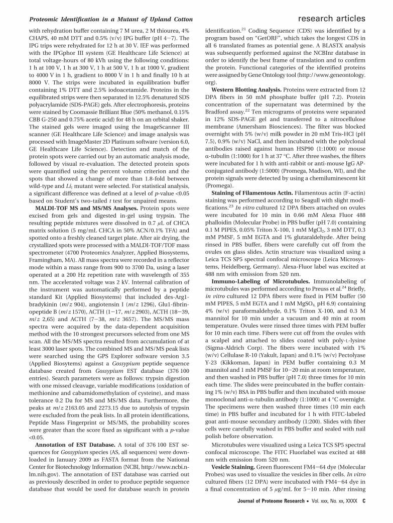
Table 1. Sequences of the Primers Used for Real-Time PCR Analysis
gene accession no. primers
PFN EV489411 F 5′ ACCTTTCTGCCGCTGCTATCG 3′R 5′ CGCCCAACCTCTCCACAACC 3′
TUB1 DT544715 F 5′ AAGGAAGCCGAGAATTGCGATTG 3′R 5′ CGAGGGAATGGAATAAGGTTTACAGC 3′
RanBP1 CO131457 F 5′ GAGGAAGTCGCCGTTACCAC 3′R 5′ TTTGTATCCTCCTTTGTTTCCGTTTC 3′
CaM ES831951 F 5′ ATCTGAGACATATACTGACTAGCATCGG 3′R 5′ AGAAAGAGTGTTACCATTAGTTCCAAAGG 3′
VIN ES832477 F 5′ CGACATAGGCACCGCTACTCAG 3′R 5′ CACCGTCCTCCCTCCTTCTCC 3′
HSP90 ES821537 F 5′ ATTCCGATGACTTGCCGCTTAAC 3′R 5′ GCTTCTTGCCGTTGTATCCCTTC 3′
SNF7 DW245123 F 5′ TGCCAAAGGGAGAAACAAGAGAG 3′R 5′ CCAAGAGGAGTTGACAGTGCTTC 3′
GLP EV485102 F 5′ TCCGTACCGCAGGCAACAC 3′R 5′ GTTCATTCAATAAGTCAGTCGCAAGTC 3′
CRT ES850077 F 5′ TGAAGAAGCAGAGAAAGAACGAAGAG 3′R 5′ ACGACGACAACAATGAGAACAACC 3′
His3 AF024716 F 5′ GCCAAGCGTGTCACAATTATGC 3′R 5′ ACATCACATTGAACCTACCACTACC 3′
research articles Zhao et al.
B Journal of Proteome Research • Vol. xxx, No. xx, XXXX
with rehydration buffer containing 7 M urea, 2 M thiourea, 4%CHAPS, 40 mM DTT and 0.5% (v/v) IPG buffer (pH 4-7). TheIPG trips were rehydrated for 12 h at 30 V. IEF was performedwith the IPGphor III system (GE Healthcare Life Science) attotal voltage-hours of 80 kVh using the following conditions:1 h at 100 V, 1 h at 300 V, 1 h at 500 V, 1 h at 1000 V, gradientto 4000 V in 1 h, gradient to 8000 V in 1 h and finally 10 h at8000 V. The strips were incubated in equilibration buffercontaining 1% DTT and 2.5% iodoacetamide. Proteins in theequilibrated strips were then separated in 12.5% denatured SDSpolyacrylamide (SDS-PAGE) gels. After electrophoresis, proteinswere stained by Coomassie Brilliant Blue (50% methanol, 0.15%CBB G-250 and 0.75% acetic acid) for 48 h on an orbital shaker.The stained gels were imaged using the ImageScanner IIIscanner (GE Healthcare Life Science) and image analysis wasprocessed with ImageMaster 2D Platinum software (version 6.0,GE Healthcare Life Science). Detection and match of theprotein spots were carried out by an automatic analysis mode,followed by visual re-evaluation. The detected protein spotswere quantified using the percent volume criterion and thespots that showed a change of more than 1.8-fold betweenwild-type and Li1 mutant were selected. For statistical analysis,a significant difference was defined at a level of p-value <0.05based on Student’s two-tailed t test for unpaired means.
MALDI-TOF MS and MS/MS Analyses. Protein spots wereexcised from gels and digested in-gel using trypsin. Theresulting peptide mixtures were dissolved in 0.7 µL of CHCAmatrix solution (5 mg/mL CHCA in 50% ACN/0.1% TFA) andspotted onto a freshly cleaned target plate. After air drying, thecrystallized spots were processed with a MALDI-TOF/TOF massspectrometer (4700 Proteomics Analyzer, Applied Biosystems,Framingham, MA). All mass spectra were recorded in a reflectormode within a mass range from 900 to 3700 Da, using a laseroperated at a 200 Hz repetition rate with wavelength of 355nm. The accelerated voltage was 2 kV. Internal calibration ofthe instrument was automatically performed by a peptidestandard Kit (Applied Biosystems) that included des-Arg1-bradykinin (m/z 904), angiotensin I (m/z 1296), Glu1-fibrin-opeptide B (m/z 1570), ACTH (1-17, m/z 2903), ACTH (18-39,m/z 2,65) and ACTH (7-38, m/z 3657). The MS/MS massspectra were acquired by the data-dependent acquisitionmethod with the 10 strongest precursors selected from one MSscan. All the MS/MS spectra resulted from accumulation of atleast 3000 laser spots. The combined MS and MS/MS peak listswere searched using the GPS Explorer software version 3.5(Applied Biosytems) against a Gossypium peptide sequencedatabase created from Gossypium EST database (376 100entries). Search parameters were as follows: trypsin digestionwith one missed cleavage, variable modifications (oxidation ofmethionine and cabamidomethylation of cysteine), and masstolerance 0.2 Da for MS and MS/MS data. Furthermore, thepeaks at m/z 2163.05 and 2273.15 due to autolysis of trypsinwere excluded from the peak lists. In all protein identifications,Peptide Mass Fingerprint or MS/MS, the probability scoreswere greater than the score fixed as significant with a p-value<0.05.
Annotation of EST Database. A total of 376 100 EST se-quences for Gossypium species (AS, all sequences) were down-loaded in January 2009 as FASTA format from the NationalCenter for Biotechnology Information (NCBI, http://www.ncbi.n-lm.nih.gov). The annotation of EST database was carried outas previously described in order to produce peptide sequencedatabase that would be used for database search in protein
identification.21 Coding Sequence (CDS) was identified by aprogram based on “GetORF”, which takes the longest CDS inall 6 translated frames as potential gene. A BLASTX analysiswas subsequently performed against the NCBInr database inorder to identify the best frame of translation and to confirmthe protein. Functional categories of the identified proteinswere assigned by Gene Ontology tool (http://www.geneontology.org).
Western Blotting Analysis. Proteins were extracted from 12DPA fibers in 50 mM phosphate buffer (pH 7.2). Proteinconcentration of the supernatant was determined by theBradford assay.22 Ten micrograms of proteins were separatedin 12% SDS-PAGE gel and transferred to a nitrocellulosemembrane (Amersham Biosciences). The filter was blockedovernight with 5% (w/v) milk powder in 20 mM Tris-HCl (pH7.5), 0.9% (w/v) NaCl, and then incubated with the polyclonalantibodies raised against human HSP90 (1:1000) or mouseR-tubulin (1:1000) for 1 h at 37 °C. After three washes, the filterswere incubated for 1 h with anti-rabbit or anti-mouse IgG AP-conjugated antibody (1:5000) (Promega, Madison, WI), and theprotein signals were detected by using a chemiluminescent kit(Promega).
Staining of Filamentous Actin. Filamentous actin (F-actin)staining was performed according to Seagull with slight modi-fications.23 In vitro cultured 12 DPA fibers attached on ovuleswere incubated for 10 min in 0.66 mM Alexa Fluor 488phalloidin (Molecular Probe) in PBS buffer (pH 7.0) containing0.1 M PIPES, 0.05% Triton X-100, 1 mM MgCl2, 3 mM DTT, 0.3mM PMSF, 5 mM EGTA and 1% glutaraldehyde. After beingrinsed in PBS buffer, fibers were carefully cut off from theovules on glass slides. Actin structure was visualized using aLeica TCS SP5 spectral confocal microscope (Leica Microsys-tems, Heidelberg, Germany). Alexa-Fluor label was excited at488 nm with emission from 520 nm.
Immuno-Labeling of Microtubules. Immunolabeling ofmicrotubules was performed according to Preuss et al.24 Briefly,in vitro cultured 12 DPA fibers were fixed in PEM buffer (50mM PIPES, 5 mM EGTA and 1 mM MgSO4, pH 6.9) containing4% (w/v) paraformaldehyde, 0.1% Triton X-100, and 0.3 Mmannitol for 10 min under a vacuum and 40 min at roomtemperature. Ovules were rinsed three times with PEM bufferfor 10 min each time. Fibers were cut off from the ovules witha scalpel and attached to slides coated with poly-L-lysine(Sigma-Aldrich Corp). The fibers were incubated with 1%(w/v) Cellulase R-10 (Yakult, Japan) and 0.1% (w/v) PectolyaseY-23 (Kikkoman, Japan) in PEM buffer containing 0.3 Mmannitol and 1 mM PMSF for 10-20 min at room temperature,and then washed in PBS buffer (pH 7.0) three times for 10 mineach time. The slides were preincubated in the buffer contain-ing 1% (w/v) BSA in PBS buffer and then incubated with mousemonoclonal anti-R-tubulin antibody (1:1000) at 4 °C overnight.The specimens were then washed three times (10 min eachtime) in PBS buffer and incubated for 1 h with FITC-labeledgoat anti-mouse secondary antibody (1:200). Slides with fibercells were carefully washed in PBS buffer and sealed with nailpolish before observation.
Microtubules were visualized using a Leica TCS SP5 spectralconfocal microscope. The FITC Fluorlabel was excited at 488nm with emission from 520 nm.
Vesicle Staining. Green fluorescent FM4-64 dye (MolecularProbes) was used to visualize the vesicles in fiber cells. In vitrocultured fibers (12 DPA) were incubated with FM4-64 dye ina final concentration of 5 µg/mL for 5-10 min. After rinsing
Proteomic Identification in a Mutant of Upland Cotton research articles
Journal of Proteome Research • Vol. xxx, No. xx, XXXX C
briefly with PBS buffer (pH 7.0), FM4-64 fluorescence wasdetected using a Leica TCS SP5 spectral confocal microscope.The FM4-64 fluorescence was excited at 543 nm with emissionfrom 640 nm.
Microscopy Analyses. For scanning electron microscopy(SEM) analysis, bolls were harvested from the cotton plantgrown in green house at 12 DPA and fibers were fixed in 2.5%glutaraldehyde in phosphate buffer (50 mM, pH 7.2) at 4 °Covernight. After dehydration in gradient ethanol series, thesamples were treated with 100% isopentyl acetate at 4 °Covernight. The materials were then dried using a critical pointdryer (model HCP-2, Hitachi, Tokyo, Japan), sputter coated withgold in an E-1010 ion sputter (Hitachi) and visualized under ascanning electron microscope (Quanta200, FEI Company,Netherlands).
For transmission electron microscopy (TEM) analysis, cottonfibers (12 DPA, grown in green house) were fixed in 2.5%glutaraldehyde in phosphate buffer (50 mM, pH 7.2) at 4 °Covernight. After three washes, the fibers were incubated in 1%osmium tetraoxide for 4 h at room temperature. The specimenswere washed for three times and dehydrated in a series ofethanol and epoxy ethane, and then embedded in Epon 812resin for 48 h at 60 °C. The middle parts of the fibers were cutinto thin sections (50 nm) with an LKB III Ultratome, stainedwith uranyl acetate followed by lead citrate, then examined andphotographed under a JEM-1400 electron microscope (JEDLLTD, Japan).
Results
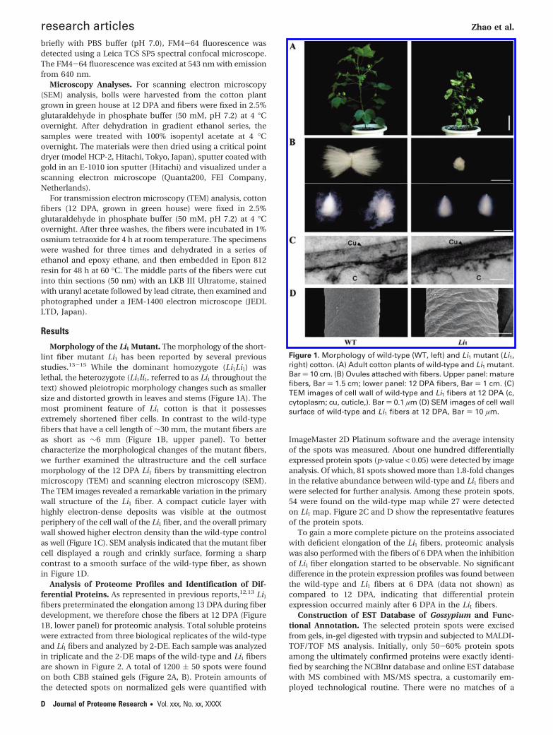
Morphology of the Li1 Mutant. The morphology of the short-lint fiber mutant Li1 has been reported by several previousstudies.13-15 While the dominant homozygote (Li1Li1) waslethal, the heterozygote (Li1li1, referred to as Li1 throughout thetext) showed pleiotropic morphology changes such as smallersize and distorted growth in leaves and stems (Figure 1A). Themost prominent feature of Li1 cotton is that it possessesextremely shortened fiber cells. In contrast to the wild-typefibers that have a cell length of ∼30 mm, the mutant fibers areas short as ∼6 mm (Figure 1B, upper panel). To bettercharacterize the morphological changes of the mutant fibers,we further examined the ultrastructure and the cell surfacemorphology of the 12 DPA Li1 fibers by transmitting electronmicroscopy (TEM) and scanning electron microscopy (SEM).The TEM images revealed a remarkable variation in the primarywall structure of the Li1 fiber. A compact cuticle layer withhighly electron-dense deposits was visible at the outmostperiphery of the cell wall of the Li1 fiber, and the overall primarywall showed higher electron density than the wild-type controlas well (Figure 1C). SEM analysis indicated that the mutant fibercell displayed a rough and crinkly surface, forming a sharpcontrast to a smooth surface of the wild-type fiber, as shownin Figure 1D.
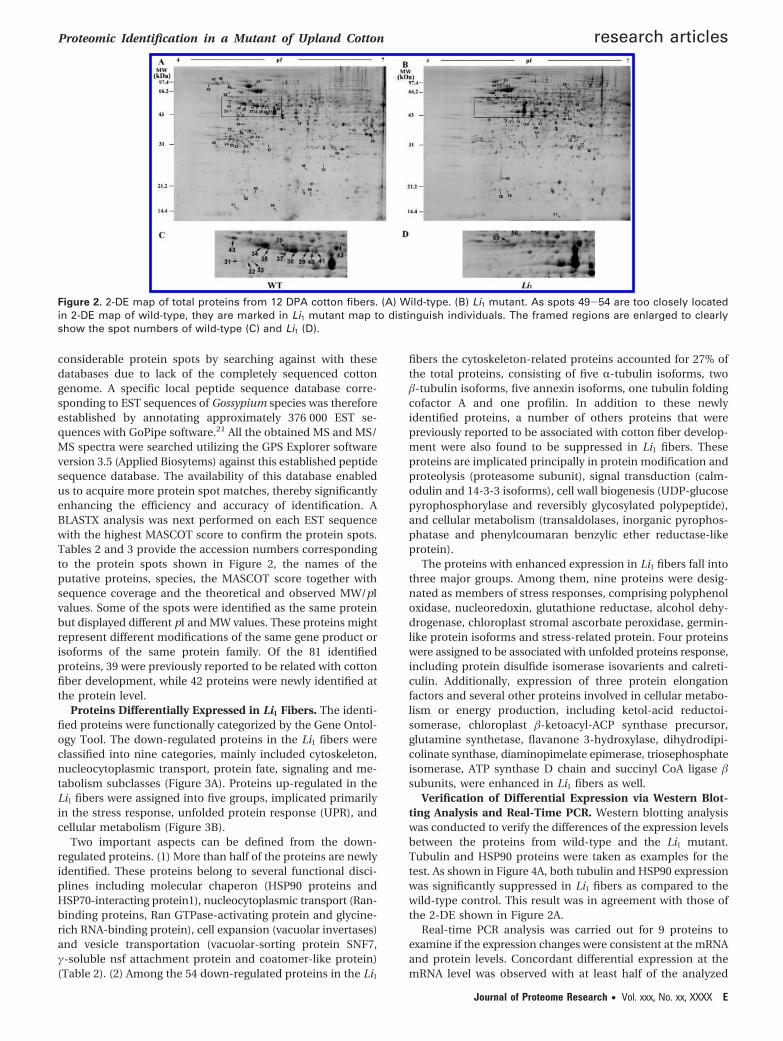
Analysis of Proteome Profiles and Identification of Dif-ferential Proteins. As represented in previous reports,12,13 Li1
fibers preterminated the elongation among 13 DPA during fiberdevelopment, we therefore chose the fibers at 12 DPA (Figure1B, lower panel) for proteomic analysis. Total soluble proteinswere extracted from three biological replicates of the wild-typeand Li1 fibers and analyzed by 2-DE. Each sample was analyzedin triplicate and the 2-DE maps of the wild-type and Li1 fibersare shown in Figure 2. A total of 1200 ( 50 spots were foundon both CBB stained gels (Figure 2A, B). Protein amounts ofthe detected spots on normalized gels were quantified with
ImageMaster 2D Platinum software and the average intensityof the spots was measured. About one hundred differentiallyexpressed protein spots (p-value < 0.05) were detected by imageanalysis. Of which, 81 spots showed more than 1.8-fold changesin the relative abundance between wild-type and Li1 fibers andwere selected for further analysis. Among these protein spots,54 were found on the wild-type map while 27 were detectedon Li1 map. Figure 2C and D show the representative featuresof the protein spots.
To gain a more complete picture on the proteins associatedwith deficient elongation of the Li1 fibers, proteomic analysiswas also performed with the fibers of 6 DPA when the inhibitionof Li1 fiber elongation started to be observable. No significantdifference in the protein expression profiles was found betweenthe wild-type and Li1 fibers at 6 DPA (data not shown) ascompared to 12 DPA, indicating that differential proteinexpression occurred mainly after 6 DPA in the Li1 fibers.
Construction of EST Database of Gossypium and Func-tional Annotation. The selected protein spots were excisedfrom gels, in-gel digested with trypsin and subjected to MALDI-TOF/TOF MS analysis. Initially, only 50-60% protein spotsamong the ultimately confirmed proteins were exactly identi-fied by searching the NCBInr database and online EST databasewith MS combined with MS/MS spectra, a customarily em-ployed technological routine. There were no matches of a
Figure 1. Morphology of wild-type (WT, left) and Li1 mutant (Li1,right) cotton. (A) Adult cotton plants of wild-type and Li1 mutant.Bar ) 10 cm. (B) Ovules attached with fibers. Upper panel: maturefibers, Bar ) 1.5 cm; lower panel: 12 DPA fibers, Bar ) 1 cm. (C)TEM images of cell wall of wild-type and Li1 fibers at 12 DPA (c,cytoplasm; cu, cuticle,). Bar ) 0.1 µm (D) SEM images of cell wallsurface of wild-type and Li1 fibers at 12 DPA, Bar ) 10 µm.
research articles Zhao et al.
D Journal of Proteome Research • Vol. xxx, No. xx, XXXX
considerable protein spots by searching against with thesedatabases due to lack of the completely sequenced cottongenome. A specific local peptide sequence database corre-sponding to EST sequences of Gossypium species was thereforeestablished by annotating approximately 376 000 EST se-quences with GoPipe software.21 All the obtained MS and MS/MS spectra were searched utilizing the GPS Explorer softwareversion 3.5 (Applied Biosytems) against this established peptidesequence database. The availability of this database enabledus to acquire more protein spot matches, thereby significantlyenhancing the efficiency and accuracy of identification. ABLASTX analysis was next performed on each EST sequencewith the highest MASCOT score to confirm the protein spots.Tables 2 and 3 provide the accession numbers correspondingto the protein spots shown in Figure 2, the names of theputative proteins, species, the MASCOT score together withsequence coverage and the theoretical and observed MW/pIvalues. Some of the spots were identified as the same proteinbut displayed different pI and MW values. These proteins mightrepresent different modifications of the same gene product orisoforms of the same protein family. Of the 81 identifiedproteins, 39 were previously reported to be related with cottonfiber development, while 42 proteins were newly identified atthe protein level.
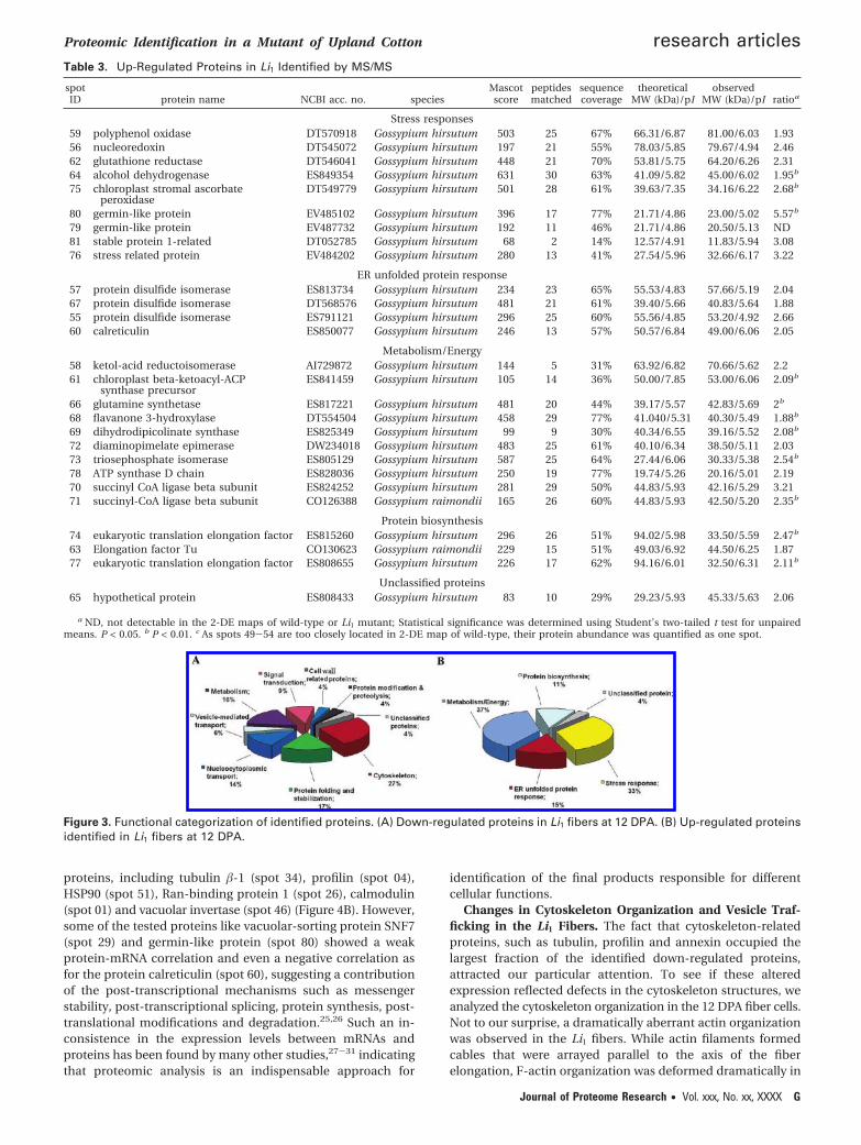
Proteins Differentially Expressed in Li1 Fibers. The identi-fied proteins were functionally categorized by the Gene Ontol-ogy Tool. The down-regulated proteins in the Li1 fibers wereclassified into nine categories, mainly included cytoskeleton,nucleocytoplasmic transport, protein fate, signaling and me-tabolism subclasses (Figure 3A). Proteins up-regulated in theLi1 fibers were assigned into five groups, implicated primarilyin the stress response, unfolded protein response (UPR), andcellular metabolism (Figure 3B).
Two important aspects can be defined from the down-regulated proteins. (1) More than half of the proteins are newlyidentified. These proteins belong to several functional disci-plines including molecular chaperon (HSP90 proteins andHSP70-interacting protein1), nucleocytoplasmic transport (Ran-binding proteins, Ran GTPase-activating protein and glycine-rich RNA-binding protein), cell expansion (vacuolar invertases)and vesicle transportation (vacuolar-sorting protein SNF7,γ-soluble nsf attachment protein and coatomer-like protein)(Table 2). (2) Among the 54 down-regulated proteins in the Li1
fibers the cytoskeleton-related proteins accounted for 27% ofthe total proteins, consisting of five R-tubulin isoforms, two�-tubulin isoforms, five annexin isoforms, one tubulin foldingcofactor A and one profilin. In addition to these newlyidentified proteins, a number of others proteins that werepreviously reported to be associated with cotton fiber develop-ment were also found to be suppressed in Li1 fibers. Theseproteins are implicated principally in protein modification andproteolysis (proteasome subunit), signal transduction (calm-odulin and 14-3-3 isoforms), cell wall biogenesis (UDP-glucosepyrophosphorylase and reversibly glycosylated polypeptide),and cellular metabolism (transaldolases, inorganic pyrophos-phatase and phenylcoumaran benzylic ether reductase-likeprotein).
The proteins with enhanced expression in Li1 fibers fall intothree major groups. Among them, nine proteins were desig-nated as members of stress responses, comprising polyphenoloxidase, nucleoredoxin, glutathione reductase, alcohol dehy-drogenase, chloroplast stromal ascorbate peroxidase, germin-like protein isoforms and stress-related protein. Four proteinswere assigned to be associated with unfolded proteins response,including protein disulfide isomerase isovarients and calreti-culin. Additionally, expression of three protein elongationfactors and several other proteins involved in cellular metabo-lism or energy production, including ketol-acid reductoi-somerase, chloroplast �-ketoacyl-ACP synthase precursor,glutamine synthetase, flavanone 3-hydroxylase, dihydrodipi-colinate synthase, diaminopimelate epimerase, triosephosphateisomerase, ATP synthase D chain and succinyl CoA ligase �subunits, were enhanced in Li1 fibers as well.
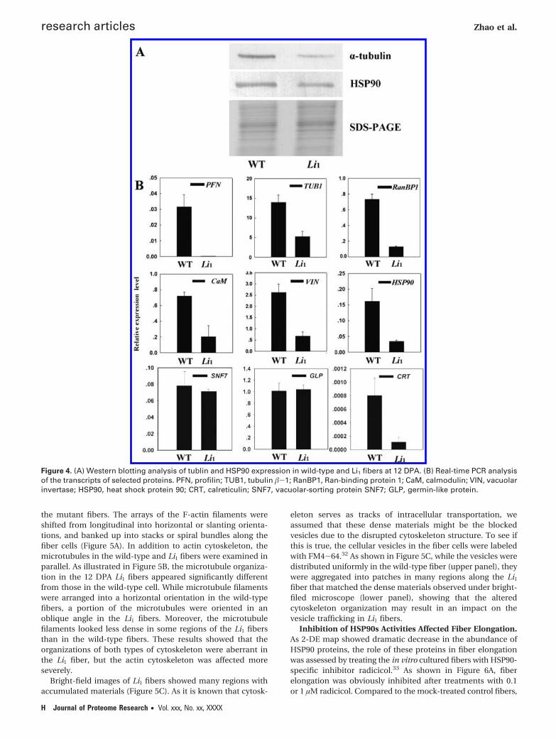
Verification of Differential Expression via Western Blot-ting Analysis and Real-Time PCR. Western blotting analysiswas conducted to verify the differences of the expression levelsbetween the proteins from wild-type and the Li1 mutant.Tubulin and HSP90 proteins were taken as examples for thetest. As shown in Figure 4A, both tubulin and HSP90 expressionwas significantly suppressed in Li1 fibers as compared to thewild-type control. This result was in agreement with those ofthe 2-DE shown in Figure 2A.
Real-time PCR analysis was carried out for 9 proteins toexamine if the expression changes were consistent at the mRNAand protein levels. Concordant differential expression at themRNA level was observed with at least half of the analyzed
Figure 2. 2-DE map of total proteins from 12 DPA cotton fibers. (A) Wild-type. (B) Li1 mutant. As spots 49-54 are too closely locatedin 2-DE map of wild-type, they are marked in Li1 mutant map to distinguish individuals. The framed regions are enlarged to clearlyshow the spot numbers of wild-type (C) and Li1 (D).
Proteomic Identification in a Mutant of Upland Cotton research articles
Journal of Proteome Research • Vol. xxx, No. xx, XXXX E
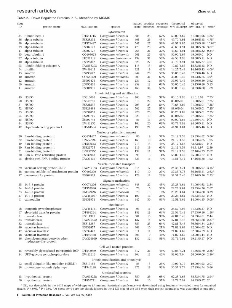
Table 2. Down-Regulated Proteins in Li1 Identified by MS/MS
spotID protein name NCBI acc. no. species
mascotScore
peptidesmatched
sequencecoverage
theoreticalMW (kDa)/pI
observedMW (kDa)/pI ratioa
Cytoskeleton34 tubulin beta-1 DT544715 Gossypium hirsutum 588 23 57% 50.00/4.67 51.20/4.96 4.85b
36 alpha-tubulin ES828302 Gossypium hirsutum 401 26 45% 49.70/4.91 49.10/5.13 4.75b
38 alpha-tubulin DT571427 Gossypium hirsutum 547 27 59% 49.57/4.85 48.33/5.18 10.09b
39 alpha-tubulin ES807127 Gossypium hirsutum 479 25 40% 49.69/4.91 48.68/5.26 3.87b
41 alpha-tubulin ES807127 Gossypium hirsutum 264 21 37% 49.69/4.91 48.68/5.32 9.34b
35 beta-tubulin 1 CO107623 Gossypium raimondii 561 25 48% 50.00/4.67 49.00/5.0 ND37 alpha tubulin EE592712 Gossypium barbadense 432 31 50% 49.58/4.90 48.50/5.11 ND40 alpha-tubulin ES828302 Gossypium hirsutum 328 27 40% 49.70/4.91 48.66/5.27 4.0103 tubulin folding cofactor A DW516203 Gossypium hirsutum 115 13 81% 12.82/4.87 18.33/5.13 ND04 profilin EV489411 Gossypium hirsutum 321 9 42% 14.25/5.48 14.16/5.43 4.09b
13 annexin ES793672 Gossypium hirsutum 244 28 58% 36.05/6.45 37.33/6.46 ND10 annexin CO129429 Gossypium raimondii 609 31 63% 36.05/6.45 40.23/6.75 4.9b
12 annexin ES795476 Gossypium hirsutum 234 12 56% 36.05/6.45 39.50/6.80 3.6209 annexin ES795476 Gossypium hirsutum 259 12 64% 36.05/6.45 35.13/6.80 3.1311 annexin ES804937 Gossypium hirsutum 466 34 59% 36.05/6.45 38.33/6.80 1.89
Protein folding and stabilization49 HSP90 ES810068 Gossypium hirsutum 468 28 37% 80.15/4.90 91.0/5.01 7.25c
50 HSP90 ES848757 Gossypium hirsutum 518 22 55% 88.0/5.01 91.00/5.01 7.25c
51 HSP90 ES821537 Gossypium hirsutum 293 25 54% 79.68/4,97 91.00/5.01 7.25c
52 HSP90 ES828488 Gossypium hirsutum 502 27 57% 88.0/5.04 88.00/5.01 7.25c
53 HSP90 ES837858 Gossypium hirsutum 226 25 54% 80.20/4.89 88.00/5.01 7.25c
54 HSP90 ES795715 Gossypium hirsutum 329 19 41% 89.0/5.07 87.00/5.01 7.25c
45 HSP90 ES797743 Gossypium hirsutum 86 13 34% 90.00/4.85 101.50/4.71 ND44 HSP90 EV491935 Gossypium hirsutum 104 20 68% 80.77/4.99 94.66/5.11 ND42 Hsp70-interacting protein 1 DT564504 Gossypium hirsutum 190 21 47% 44.94/4.84 51.50/5.46 ND
Nucleocytoplasmic transport26 Ran-binding protein 1 CO131457 Gossypium raimondii 86 9 37% 24.12/4.58 35.33/4.82 3.86b
22 Ran-binding protein 1 DN757992 Gossypium hirsutum 165 10 47% 24.12/4.58 33.33/4.85 1.9519 Ran-binding protein 1 DT558543 Gossypium hirsutum 219 13 44% 24.12/4.58 33.33/5.0 ND20 Ran-binding protein 1 ES822775 Gossypium hirsutum 216 16 60% 24.12/4.58 34.3/4.97 2.3921 Ran-binding protein 1 DN757955 Gossypium hirsutum 134 11 37% 24.12/4.58 32.16/4.93 2.6730 Ran GTPase-activating protein ES830968 Gossypium hirsutum 95 3 11% 58.02/4.48 65.00/4.77 ND05 glycine-rich RNA-binding protein DW231397 Gossypium hirsutum 323 15 70% 16.51/8.12 17.16/5.88 1.92
Vesicle-mediated transport29 vacuolar-sorting protein SNF7 DW245123 Gossypium hirsutum 314 17 38% 24.36/4.71 39.00/5.97 3.15b
18 gamma-soluble nsf attachment protein CO102209 Gossypium raimondii 110 10 29% 32.36/4.73 36.16/5.11 2.62b
17 coatomer-like protein ES804905 Gossypium hirsutum 176 12 26% 32.31/5.40 32.16/5.30 2.53b
Signal transduction25 14-3-3 protein CO074226 Gossypium raimondii 648 22 43% 29.23/4.64 31.00/4.63 3.3424 14-3-3 protein DT557996 Gossypium hirsutum 76 5 30% 29.23/4.64 33.33/4.76 2.6723 14-3-3 protein BM359797 Gossypium arboreum 78 3 16% 29.23/4.64 33.33/4.80 3.3927 14-3-3 protein DW485067 Gossypium hirsutum 190 16 38% 29.23/4.64 36.33/4.83 ND01 calmodulin ES831951 Gossypium hirsutum 447 30 86% 16.51/4.64 14.00/4.85 5.08b
Metabolism08 inorganic pyrophosphatase DW484151 Gossypium hirsutum 96 11 31% 24.57/6.08 31.33/6.27 ND07 glycolipid transfer protein DT461234 Gossypium hirsutum 152 18 64% 22.54/6.49 27.33/6.10 1.88b
31 transaldolase ES811387 Gossypium hirsutum 501 15 38% 47.81/5.46 50.33/4.81 2.4433 transaldolase DW225572 Gossypium hirsutum 137 14 55% 47.81/5.46 49.66/4.88 2.37b
32 transaldolase ES811387 Gossypium hirsutum 359 23 64% 47.81/5.46 50.66/4.84 2.34b
46 vacuolar invertase ES832477 Gossypium hirsutum 368 10 21% 71.82/4.69 92.00/4.62 ND47 vacuolar invertase ES832477 Gossypium hirsutum 311 11 24% 71.82/4.69 92.00/4.59 ND48 vacuolar invertase DT049468 Gossypium hirsutum 308 9 48% 71.82/4.69 92.00/4.44 ND16 phenylcoumaran benzylic ether
reductase-like proteinDW226959 Gossypium hirsutum 137 12 51% 33.79/5.92 39.21/5.57 ND
Cell wall related proteins15 reversibly glycosylated polypeptide RGP DT543039 Gossypium hirsutum 317 21 46% 40.85/6.21 41.66/5.70 2.26b
14 UDP-glucose pyrophosphorylase EV482616 Gossypium hirsutum 204 12 40% 52.06/7.0 56.00/6.06 2.30b
Protein modification and proteolysis02 small ubiquitin-like modifier 1(SUM1) ES819189 Gossypium hirsutum 85 3 25% 10.97/4.79 19.00/4.93 2.0728 proteasome subunit alpha type DT549128 Gossypium hirsutum 375 18 53% 30.37/4.79 37.25/4.94 3.66
Unclassified proteins43 hypothetical protein DW008228 Gossypium hirsutum 650 25 68% 67.23/4.65 68.33/4.75 2.94b
06 hypothetical protein DT459771 Gossypium hirsutum 164 10 37% 18.72/5.94 24.83/5.87 2b
a ND, not detectable in the 2-DE maps of wild-type or Li1 mutant; Statistical significance was determined using Student’s two-tailed t test for unpairedmeans. P < 0.05. b P < 0.01. c As spots 49-54 are too closely located in the 2-DE map of the wild-type, their protein abundance was quantified as one spot.
research articles Zhao et al.
F Journal of Proteome Research • Vol. xxx, No. xx, XXXX
proteins, including tubulin �-1 (spot 34), profilin (spot 04),HSP90 (spot 51), Ran-binding protein 1 (spot 26), calmodulin(spot 01) and vacuolar invertase (spot 46) (Figure 4B). However,some of the tested proteins like vacuolar-sorting protein SNF7(spot 29) and germin-like protein (spot 80) showed a weakprotein-mRNA correlation and even a negative correlation asfor the protein calreticulin (spot 60), suggesting a contributionof the post-transcriptional mechanisms such as messengerstability, post-transcriptional splicing, protein synthesis, post-translational modifications and degradation.25,26 Such an in-consistence in the expression levels between mRNAs andproteins has been found by many other studies,27-31 indicatingthat proteomic analysis is an indispensable approach for
identification of the final products responsible for differentcellular functions.
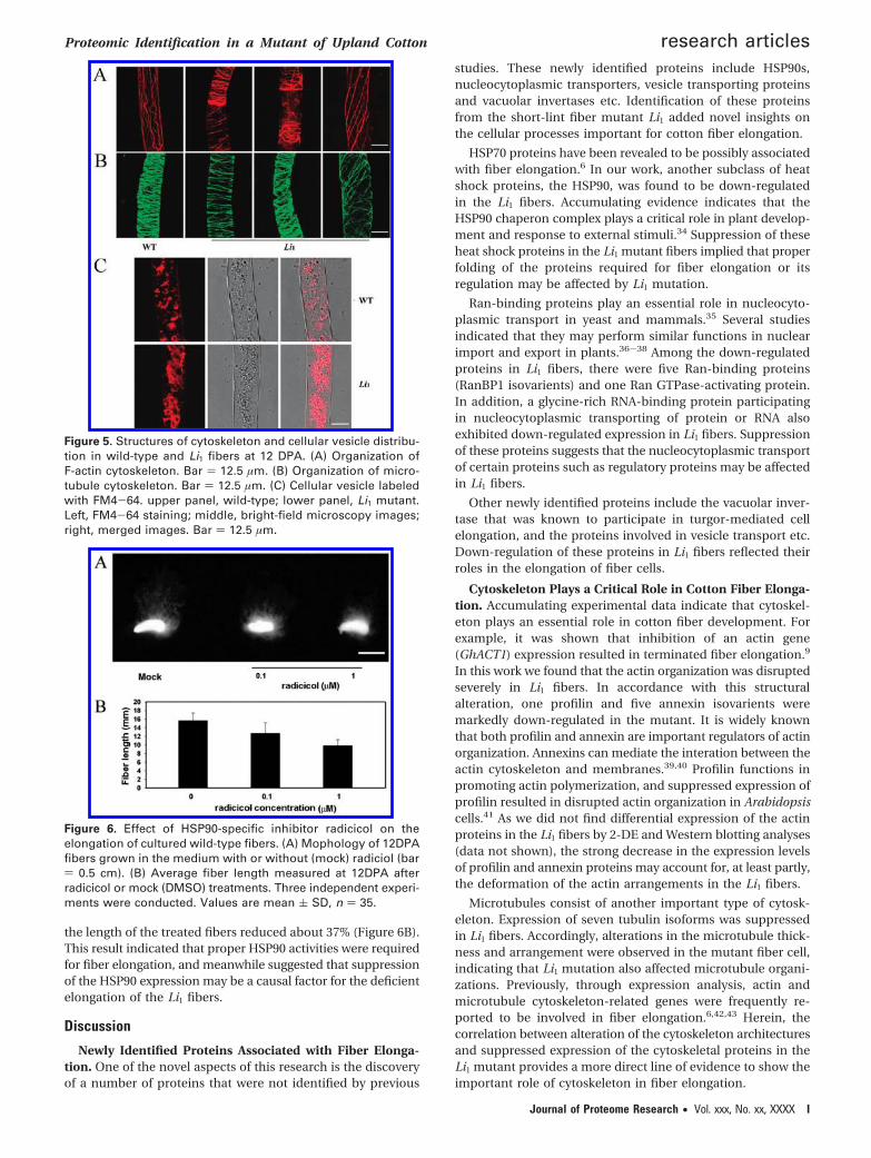
Changes in Cytoskeleton Organization and Vesicle Traf-ficking in the Li1 Fibers. The fact that cytoskeleton-relatedproteins, such as tubulin, profilin and annexin occupied thelargest fraction of the identified down-regulated proteins,attracted our particular attention. To see if these alteredexpression reflected defects in the cytoskeleton structures, weanalyzed the cytoskeleton organization in the 12 DPA fiber cells.Not to our surprise, a dramatically aberrant actin organizationwas observed in the Li1 fibers. While actin filaments formedcables that were arrayed parallel to the axis of the fiberelongation, F-actin organization was deformed dramatically in
Table 3. Up-Regulated Proteins in Li1 Identified by MS/MS
spotID protein name NCBI acc. no. species
Mascotscore
peptidesmatched
sequencecoverage
theoreticalMW (kDa)/pI
observedMW (kDa)/pI ratioa
Stress responses59 polyphenol oxidase DT570918 Gossypium hirsutum 503 25 67% 66.31/6.87 81.00/6.03 1.9356 nucleoredoxin DT545072 Gossypium hirsutum 197 21 55% 78.03/5.85 79.67/4.94 2.4662 glutathione reductase DT546041 Gossypium hirsutum 448 21 70% 53.81/5.75 64.20/6.26 2.3164 alcohol dehydrogenase ES849354 Gossypium hirsutum 631 30 63% 41.09/5.82 45.00/6.02 1.95b
75 chloroplast stromal ascorbateperoxidase
DT549779 Gossypium hirsutum 501 28 61% 39.63/7.35 34.16/6.22 2.68b
80 germin-like protein EV485102 Gossypium hirsutum 396 17 77% 21.71/4.86 23.00/5.02 5.57b
79 germin-like protein EV487732 Gossypium hirsutum 192 11 46% 21.71/4.86 20.50/5.13 ND81 stable protein 1-related DT052785 Gossypium hirsutum 68 2 14% 12.57/4.91 11.83/5.94 3.0876 stress related protein EV484202 Gossypium hirsutum 280 13 41% 27.54/5.96 32.66/6.17 3.22
ER unfolded protein response57 protein disulfide isomerase ES813734 Gossypium hirsutum 234 23 65% 55.53/4.83 57.66/5.19 2.0467 protein disulfide isomerase DT568576 Gossypium hirsutum 481 21 61% 39.40/5.66 40.83/5.64 1.8855 protein disulfide isomerase ES791121 Gossypium hirsutum 296 25 60% 55.56/4.85 53.20/4.92 2.6660 calreticulin ES850077 Gossypium hirsutum 246 13 57% 50.57/6.84 49.00/6.06 2.05
Metabolism/Energy58 ketol-acid reductoisomerase AI729872 Gossypium hirsutum 144 5 31% 63.92/6.82 70.66/5.62 2.261 chloroplast beta-ketoacyl-ACP
synthase precursorES841459 Gossypium hirsutum 105 14 36% 50.00/7.85 53.00/6.06 2.09b
66 glutamine synthetase ES817221 Gossypium hirsutum 481 20 44% 39.17/5.57 42.83/5.69 2b
68 flavanone 3-hydroxylase DT554504 Gossypium hirsutum 458 29 77% 41.040/5.31 40.30/5.49 1.88b
69 dihydrodipicolinate synthase ES825349 Gossypium hirsutum 99 9 30% 40.34/6.55 39.16/5.52 2.08b
72 diaminopimelate epimerase DW234018 Gossypium hirsutum 483 25 61% 40.10/6.34 38.50/5.11 2.0373 triosephosphate isomerase ES805129 Gossypium hirsutum 587 25 64% 27.44/6.06 30.33/5.38 2.54b
78 ATP synthase D chain ES828036 Gossypium hirsutum 250 19 77% 19.74/5.26 20.16/5.01 2.1970 succinyl CoA ligase beta subunit ES824252 Gossypium hirsutum 281 29 50% 44.83/5.93 42.16/5.29 3.2171 succinyl-CoA ligase beta subunit CO126388 Gossypium raimondii 165 26 60% 44.83/5.93 42.50/5.20 2.35b
Protein biosynthesis74 eukaryotic translation elongation factor ES815260 Gossypium hirsutum 296 26 51% 94.02/5.98 33.50/5.59 2.47b
63 Elongation factor Tu CO130623 Gossypium raimondii 229 15 51% 49.03/6.92 44.50/6.25 1.8777 eukaryotic translation elongation factor ES808655 Gossypium hirsutum 226 17 62% 94.16/6.01 32.50/6.31 2.11b
Unclassified proteins65 hypothetical protein ES808433 Gossypium hirsutum 83 10 29% 29.23/5.93 45.33/5.63 2.06
a ND, not detectable in the 2-DE maps of wild-type or Li1 mutant; Statistical significance was determined using Student’s two-tailed t test for unpairedmeans. P < 0.05. b P < 0.01. c As spots 49-54 are too closely located in 2-DE map of wild-type, their protein abundance was quantified as one spot.
Figure 3. Functional categorization of identified proteins. (A) Down-regulated proteins in Li1 fibers at 12 DPA. (B) Up-regulated proteinsidentified in Li1 fibers at 12 DPA.
Proteomic Identification in a Mutant of Upland Cotton research articles
Journal of Proteome Research • Vol. xxx, No. xx, XXXX G
the mutant fibers. The arrays of the F-actin filaments wereshifted from longitudinal into horizontal or slanting orienta-tions, and banked up into stacks or spiral bundles along thefiber cells (Figure 5A). In addition to actin cytoskeleton, themicrotubules in the wild-type and Li1 fibers were examined inparallel. As illustrated in Figure 5B, the microtubule organiza-tion in the 12 DPA Li1 fibers appeared significantly differentfrom those in the wild-type cell. While microtubule filamentswere arranged into a horizontal orientation in the wild-typefibers, a portion of the microtubules were oriented in anoblique angle in the Li1 fibers. Moreover, the microtubulefilaments looked less dense in some regions of the Li1 fibersthan in the wild-type fibers. These results showed that theorganizations of both types of cytoskeleton were aberrant inthe Li1 fiber, but the actin cytoskeleton was affected moreseverely.
Bright-field images of Li1 fibers showed many regions withaccumulated materials (Figure 5C). As it is known that cytosk-
eleton serves as tracks of intracellular transportation, weassumed that these dense materials might be the blockedvesicles due to the disrupted cytoskeleton structure. To see ifthis is true, the cellular vesicles in the fiber cells were labeledwith FM4-64.32 As shown in Figure 5C, while the vesicles weredistributed uniformly in the wild-type fiber (upper panel), theywere aggregated into patches in many regions along the Li1
fiber that matched the dense materials observed under bright-filed microscope (lower panel), showing that the alteredcytoskeleton organization may result in an impact on thevesicle trafficking in Li1 fibers.
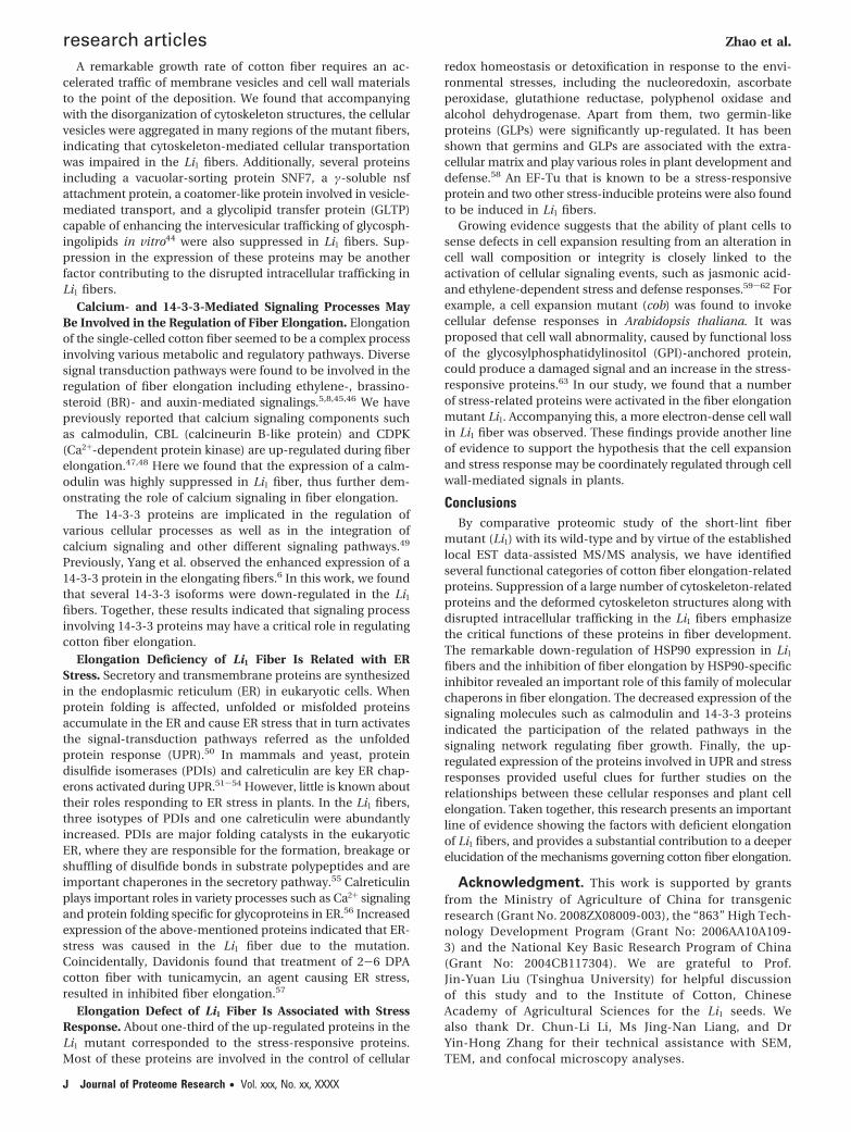
Inhibition of HSP90s Activities Affected Fiber Elongation.As 2-DE map showed dramatic decrease in the abundance ofHSP90 proteins, the role of these proteins in fiber elongationwas assessed by treating the in vitro cultured fibers with HSP90-specific inhibitor radicicol.33 As shown in Figure 6A, fiberelongation was obviously inhibited after treatments with 0.1or 1 µM radicicol. Compared to the mock-treated control fibers,
Figure 4. (A) Western blotting analysis of tublin and HSP90 expression in wild-type and Li1 fibers at 12 DPA. (B) Real-time PCR analysisof the transcripts of selected proteins. PFN, profilin; TUB1, tubulin �-1; RanBP1, Ran-binding protein 1; CaM, calmodulin; VIN, vacuolarinvertase; HSP90, heat shock protein 90; CRT, calreticulin; SNF7, vacuolar-sorting protein SNF7; GLP, germin-like protein.
research articles Zhao et al.
H Journal of Proteome Research • Vol. xxx, No. xx, XXXX
the length of the treated fibers reduced about 37% (Figure 6B).This result indicated that proper HSP90 activities were requiredfor fiber elongation, and meanwhile suggested that suppressionof the HSP90 expression may be a causal factor for the deficientelongation of the Li1 fibers.
Discussion
Newly Identified Proteins Associated with Fiber Elonga-tion. One of the novel aspects of this research is the discoveryof a number of proteins that were not identified by previous
studies. These newly identified proteins include HSP90s,nucleocytoplasmic transporters, vesicle transporting proteinsand vacuolar invertases etc. Identification of these proteinsfrom the short-lint fiber mutant Li1 added novel insights onthe cellular processes important for cotton fiber elongation.
HSP70 proteins have been revealed to be possibly associatedwith fiber elongation.6 In our work, another subclass of heatshock proteins, the HSP90, was found to be down-regulatedin the Li1 fibers. Accumulating evidence indicates that theHSP90 chaperon complex plays a critical role in plant develop-ment and response to external stimuli.34 Suppression of theseheat shock proteins in the Li1 mutant fibers implied that properfolding of the proteins required for fiber elongation or itsregulation may be affected by Li1 mutation.
Ran-binding proteins play an essential role in nucleocyto-plasmic transport in yeast and mammals.35 Several studiesindicated that they may perform similar functions in nuclearimport and export in plants.36-38 Among the down-regulatedproteins in Li1 fibers, there were five Ran-binding proteins(RanBP1 isovarients) and one Ran GTPase-activating protein.In addition, a glycine-rich RNA-binding protein participatingin nucleocytoplasmic transporting of protein or RNA alsoexhibited down-regulated expression in Li1 fibers. Suppressionof these proteins suggests that the nucleocytoplasmic transportof certain proteins such as regulatory proteins may be affectedin Li1 fibers.
Other newly identified proteins include the vacuolar inver-tase that was known to participate in turgor-mediated cellelongation, and the proteins involved in vesicle transport etc.Down-regulation of these proteins in Li1 fibers reflected theirroles in the elongation of fiber cells.
Cytoskeleton Plays a Critical Role in Cotton Fiber Elonga-tion. Accumulating experimental data indicate that cytoskel-eton plays an essential role in cotton fiber development. Forexample, it was shown that inhibition of an actin gene(GhACT1) expression resulted in terminated fiber elongation.9
In this work we found that the actin organization was disruptedseverely in Li1 fibers. In accordance with this structuralalteration, one profilin and five annexin isovarients weremarkedly down-regulated in the mutant. It is widely knownthat both profilin and annexin are important regulators of actinorganization. Annexins can mediate the interation between theactin cytoskeleton and membranes.39,40 Profilin functions inpromoting actin polymerization, and suppressed expression ofprofilin resulted in disrupted actin organization in Arabidopsiscells.41 As we did not find differential expression of the actinproteins in the Li1 fibers by 2-DE and Western blotting analyses(data not shown), the strong decrease in the expression levelsof profilin and annexin proteins may account for, at least partly,the deformation of the actin arrangements in the Li1 fibers.
Microtubules consist of another important type of cytosk-eleton. Expression of seven tubulin isoforms was suppressedin Li1 fibers. Accordingly, alterations in the microtubule thick-ness and arrangement were observed in the mutant fiber cell,indicating that Li1 mutation also affected microtubule organi-zations. Previously, through expression analysis, actin andmicrotubule cytoskeleton-related genes were frequently re-ported to be involved in fiber elongation.6,42,43 Herein, thecorrelation between alteration of the cytoskeleton architecturesand suppressed expression of the cytoskeletal proteins in theLi1 mutant provides a more direct line of evidence to show theimportant role of cytoskeleton in fiber elongation.
Figure 5. Structures of cytoskeleton and cellular vesicle distribu-tion in wild-type and Li1 fibers at 12 DPA. (A) Organization ofF-actin cytoskeleton. Bar ) 12.5 µm. (B) Organization of micro-tubule cytoskeleton. Bar ) 12.5 µm. (C) Cellular vesicle labeledwith FM4-64. upper panel, wild-type; lower panel, Li1 mutant.Left, FM4-64 staining; middle, bright-field microscopy images;right, merged images. Bar ) 12.5 µm.
Figure 6. Effect of HSP90-specific inhibitor radicicol on theelongation of cultured wild-type fibers. (A) Mophology of 12DPAfibers grown in the medium with or without (mock) radiciol (bar) 0.5 cm). (B) Average fiber length measured at 12DPA afterradicicol or mock (DMSO) treatments. Three independent experi-ments were conducted. Values are mean ( SD, n ) 35.
Proteomic Identification in a Mutant of Upland Cotton research articles
Journal of Proteome Research • Vol. xxx, No. xx, XXXX I
A remarkable growth rate of cotton fiber requires an ac-celerated traffic of membrane vesicles and cell wall materialsto the point of the deposition. We found that accompanyingwith the disorganization of cytoskeleton structures, the cellularvesicles were aggregated in many regions of the mutant fibers,indicating that cytoskeleton-mediated cellular transportationwas impaired in the Li1 fibers. Additionally, several proteinsincluding a vacuolar-sorting protein SNF7, a γ-soluble nsfattachment protein, a coatomer-like protein involved in vesicle-mediated transport, and a glycolipid transfer protein (GLTP)capable of enhancing the intervesicular trafficking of glycosph-ingolipids in vitro44 were also suppressed in Li1 fibers. Sup-pression in the expression of these proteins may be anotherfactor contributing to the disrupted intracellular trafficking inLi1 fibers.
Calcium- and 14-3-3-Mediated Signaling Processes MayBe Involved in the Regulation of Fiber Elongation. Elongationof the single-celled cotton fiber seemed to be a complex processinvolving various metabolic and regulatory pathways. Diversesignal transduction pathways were found to be involved in theregulation of fiber elongation including ethylene-, brassino-steroid (BR)- and auxin-mediated signalings.5,8,45,46 We havepreviously reported that calcium signaling components suchas calmodulin, CBL (calcineurin B-like protein) and CDPK(Ca2+-dependent protein kinase) are up-regulated during fiberelongation.47,48 Here we found that the expression of a calm-odulin was highly suppressed in Li1 fiber, thus further dem-onstrating the role of calcium signaling in fiber elongation.
The 14-3-3 proteins are implicated in the regulation ofvarious cellular processes as well as in the integration ofcalcium signaling and other different signaling pathways.49
Previously, Yang et al. observed the enhanced expression of a14-3-3 protein in the elongating fibers.6 In this work, we foundthat several 14-3-3 isoforms were down-regulated in the Li1
fibers. Together, these results indicated that signaling processinvolving 14-3-3 proteins may have a critical role in regulatingcotton fiber elongation.
Elongation Deficiency of Li1 Fiber Is Related with ERStress. Secretory and transmembrane proteins are synthesizedin the endoplasmic reticulum (ER) in eukaryotic cells. Whenprotein folding is affected, unfolded or misfolded proteinsaccumulate in the ER and cause ER stress that in turn activatesthe signal-transduction pathways referred as the unfoldedprotein response (UPR).50 In mammals and yeast, proteindisulfide isomerases (PDIs) and calreticulin are key ER chap-erons activated during UPR.51-54 However, little is known abouttheir roles responding to ER stress in plants. In the Li1 fibers,three isotypes of PDIs and one calreticulin were abundantlyincreased. PDIs are major folding catalysts in the eukaryoticER, where they are responsible for the formation, breakage orshuffling of disulfide bonds in substrate polypeptides and areimportant chaperones in the secretory pathway.55 Calreticulinplays important roles in variety processes such as Ca2+ signalingand protein folding specific for glycoproteins in ER.56 Increasedexpression of the above-mentioned proteins indicated that ER-stress was caused in the Li1 fiber due to the mutation.Coincidentally, Davidonis found that treatment of 2-6 DPAcotton fiber with tunicamycin, an agent causing ER stress,resulted in inhibited fiber elongation.57
Elongation Defect of Li1 Fiber Is Associated with StressResponse. About one-third of the up-regulated proteins in theLi1 mutant corresponded to the stress-responsive proteins.Most of these proteins are involved in the control of cellular
redox homeostasis or detoxification in response to the envi-ronmental stresses, including the nucleoredoxin, ascorbateperoxidase, glutathione reductase, polyphenol oxidase andalcohol dehydrogenase. Apart from them, two germin-likeproteins (GLPs) were significantly up-regulated. It has beenshown that germins and GLPs are associated with the extra-cellular matrix and play various roles in plant development anddefense.58 An EF-Tu that is known to be a stress-responsiveprotein and two other stress-inducible proteins were also foundto be induced in Li1 fibers.
Growing evidence suggests that the ability of plant cells tosense defects in cell expansion resulting from an alteration incell wall composition or integrity is closely linked to theactivation of cellular signaling events, such as jasmonic acid-and ethylene-dependent stress and defense responses.59-62 Forexample, a cell expansion mutant (cob) was found to invokecellular defense responses in Arabidopsis thaliana. It wasproposed that cell wall abnormality, caused by functional lossof the glycosylphosphatidylinositol (GPI)-anchored protein,could produce a damaged signal and an increase in the stress-responsive proteins.63 In our study, we found that a numberof stress-related proteins were activated in the fiber elongationmutant Li1. Accompanying this, a more electron-dense cell wallin Li1 fiber was observed. These findings provide another lineof evidence to support the hypothesis that the cell expansionand stress response may be coordinately regulated through cellwall-mediated signals in plants.
ConclusionsBy comparative proteomic study of the short-lint fiber
mutant (Li1) with its wild-type and by virtue of the establishedlocal EST data-assisted MS/MS analysis, we have identifiedseveral functional categories of cotton fiber elongation-relatedproteins. Suppression of a large number of cytoskeleton-relatedproteins and the deformed cytoskeleton structures along withdisrupted intracellular trafficking in the Li1 fibers emphasizethe critical functions of these proteins in fiber development.The remarkable down-regulation of HSP90 expression in Li1
fibers and the inhibition of fiber elongation by HSP90-specificinhibitor revealed an important role of this family of molecularchaperons in fiber elongation. The decreased expression of thesignaling molecules such as calmodulin and 14-3-3 proteinsindicated the participation of the related pathways in thesignaling network regulating fiber growth. Finally, the up-regulated expression of the proteins involved in UPR and stressresponses provided useful clues for further studies on therelationships between these cellular responses and plant cellelongation. Taken together, this research presents an importantline of evidence showing the factors with deficient elongationof Li1 fibers, and provides a substantial contribution to a deeperelucidation of the mechanisms governing cotton fiber elongation.
Acknowledgment. This work is supported by grantsfrom the Ministry of Agriculture of China for transgenicresearch (Grant No. 2008ZX08009-003), the “863” High Tech-nology Development Program (Grant No: 2006AA10A109-3) and the National Key Basic Research Program of China(Grant No: 2004CB117304). We are grateful to Prof.Jin-Yuan Liu (Tsinghua University) for helpful discussionof this study and to the Institute of Cotton, ChineseAcademy of Agricultural Sciences for the Li1 seeds. Wealso thank Dr. Chun-Li Li, Ms Jing-Nan Liang, and DrYin-Hong Zhang for their technical assistance with SEM,TEM, and confocal microscopy analyses.
research articles Zhao et al.
J Journal of Proteome Research • Vol. xxx, No. xx, XXXX
Note Added after ASAP Publication. This paper waspublished on the Web on Dec 3, 2009, with errors on Table 1and Figure 3. The corrected version was reposted on Jan 6,2010.
References(1) Basra, A. S.; Malik, C. P. Development of the cotton fiber. Int. Rev.
Cytol. 1984, 89, 65–113.(2) Ryser, U., Cotton fiber initiation and histodifferentiation. In Cotton
Fibers: Developmental Biology, Quality Improvement, and TextileProcessing; Basra, A. S., Ed.; Food Products Press: New York, 1999;pp 1-45.
(3) Kim, H. J.; Triplett, B. A. Cotton fiber growth in planta and in vitro.Models for plant cell elongation and cell wall biogenesis. PlantPhysiol. 2001, 127 (4), 1361–1366.
(4) Arpat, A. B.; Waugh, M.; Sullivan, J. P.; Gonzales, M.; Frisch, D.;Main, D.; Wood, T.; Leslie, A.; Wing, R. A.; Wilkins, T. A. Functionalgenomics of cell elongation in developing cotton fibers. Plant Mol.Biol. 2004, 54 (6), 911–929.
(5) Shi, Y. H.; Zhu, S. W.; Mao, X. Z.; Feng, J. X.; Qin, Y. M.; Zhang, L.;Cheng, J.; Wei, L. P.; Wang, Z. Y.; Zhu, Y. X. Transcriptomeprofiling, molecular biological, and physiological studies reveal amajor role for ethylene in cotton fiber cell elongation. Plant Cell2006, 18 (3), 651–664.
(6) Yang, Y. W.; Bian, S. M.; Yao, Y.; Liu, J. Y. Comparative ProteomicAnalysis Provides New Insights into the Fiber Elongating Processin Cotton. J. Proteome Res. 2008, 7 (11), 4623–4637.
(7) Li, H. B.; Qin, Y. M.; Pang, Y.; Song, W. Q.; Mei, W. Q.; Zhu, Y. X.A cotton ascorbate peroxidase is involved in hydrogen peroxidehomeostasis during fibre cell development. New Phytol. 2007, 175(3), 462–471.
(8) Luo, M.; Xiao, Y.; Li, X.; Lu, X.; Deng, W.; Li, D.; Hou, L.; Hu, M.;Li, Y.; Pei, Y. GhDET2, a steroid 5alpha-reductase, plays animportant role in cotton fiber cell initiation and elongation. PlantJ. 2007, 51 (3), 419–430.
(9) Li, X. B.; Fan, X. P.; Wang, X. L.; Cai, L.; Yang, W. C. The cottonACTIN1 gene is functionally expressed in fibers and participatesin fiber elongation. Plant Cell 2005, 17 (3), 859–875.
(10) Ruan, Y. L.; Llewellyn, D. J.; Furbank, R. T. Suppression of sucrosesynthase gene expression represses cotton fiber cell initiation,elongation, and seed development. Plant Cell 2003, 15 (4), 952–964.
(11) Griffee, Fred.; Ligon, L. L. Occurrence of “lintless” cotton plantsand the inheritance of the character “lintless. J. Am. Soc. Agron.1929, 21, 711–717.
(12) Kohel, R. J.; Narbuth, E. V.; Benedict, C. R. Fiber development ofligon lintless-2 mutant of cotton. Crop Sci. 1992, 32 (3), 733–735.
(13) Kohel, R. J.; Quisenbe, Je.; Benedict, C. R. Fiber elongation anddry weight changes in mutant lines of cotton. Crop Sci. 1974, 14(3), 471–474.
(14) Kohel, R. J. Linkage tests in upland cotton Gossypium hirsutumL0.2. Crop Sci. 1972, 12 (1), 66–69.
(15) Karaca, M.; Saha, S.; Jenkins, J. N.; Zipf, A.; Kohel, R.; Stelly, D. M.Simple sequence repeat (SSR) markers linked to the Ligon lintless(Li-1) mutant in cotton. J. Hered. 2002, 93 (3), 221–224.
(16) Du, X.-M. Early development of the fiber and inheritance of fibermutants in upland cotton (Gossypium hirsutum L.). PhD thesisNanjing Agricultural University, Nanjing, China, 1998.
(17) Triplett, B. A.; Busch, W. H.; Goynes, W. R. Ovule and suspension-culture of a cotton fiber development mutant. In Vitro Cellular &Developmental Biology 1989, 25 (2), 197–200.
(18) Beasley, C. A.; Ting, I. P. Effects of plant-growth substances onin-vitro fiber development from fertilized cotton ovules. Am. J. Bot.1973, 60 (2), 130–139.
(19) Gao, P.; Zhao, P.-M.; Wang, J.; Wang, H.-Y.; Wu, X.-M.; Xia, G.-X.Identification of genes preferentially expressed in cotton fibers: Apossible role of calcium signaling in cotton fiber elongation. PlantSci. (Oxford) 2007, 173 (1), 61–69.
(20) Yao, Y.; Yang, Y. W.; Liu, J. Y. An efficient protein preparation forproteomic analysis of developing cotton fibers by 2-DE. Electro-phoresis 2006, 27 (22), 4559–4569.
(21) Chen, Z. Z.; Xue, C. H.; Zhu, S.; Zhou, F. F.; Ling, X. F. B.; Liu,G. P.; Chen, L. B. GoPipe: Streamlined Gene Ontology annotationfor batch anonymous sequences with statistics. Prog. Biochem.Biophys. 2005, 32 (2), 187–190.
(22) Bradford, M. M. Rapid and sensitive method for quantitation ofmicrogram quantities of protein utilizing principle of protein-dyebinding. Anal. Biochem. 1976, 72 (1-2), 248–254.
(23) Seagull, R. W. The effects of microtubule and microfilamentdisrupting agents on cytoskeletal arrays and wall deposition indeveloping cotton fibers. Protoplasma 1990, 159 (1), 44–59.
(24) Preuss, M. L.; Delmer, D. P.; Liu, B. The cotton kinesin-likecalmodulin-binding protein associates with cortical microtubulesin cotton fibers. Plant Physiol. 2003, 132 (1), 154–160.
(25) Pradet-Balade, B.; Boulme, F.; Beug, H.; Mullner, E. W.; Garcia-Sanz, J. A. Translation control: bridging the gap between genomicsand proteomics. Trends Biochem. Sci. 2001, 26 (4), 225–229.
(26) Greenbaum, D.; Colangelo, C.; Williams, K.; Gerstein, M. Compar-ing protein abundance and mRNA expression levels on a genomicscale. Genome Biol. 2003, 4 (9), 8.
(27) Yan, S. P.; Zhang, Q. Y.; Tang, Z. C.; Su, W. A.; Sun, W. N.Comparative proteomic analysis provides new insights into chillingstress responses in rice. Mol. Cell. Proteomics 2006, 5 (3), 484–496.
(28) Greenbaum, D.; Colangelo, C.; Williams, K.; Gerstein, M. Compar-ing protein abundance and mRNA expression levels on a genomicscale. Genome Biol. 2003, 4 (9), 117.
(29) Chen, G.; Gharib, T. G.; Huang, C. C.; Taylor, J. M.; Misek, D. E.;Kardia, S. L.; Giordano, T. J.; Iannettoni, M. D.; Orringer, M. B.;Hanash, S. M.; Beer, D. G. Discordant protein and mRNA expres-sion in lung adenocarcinomas. Mol. Cell. Proteomics 2002, 1 (4),304–313.
(30) Gygi, S. P.; Rochon, Y.; Franza, B. R.; Aebersold, R. Correlationbetween protein and mRNA abundance in yeast. Mol. Cell. Biol.1999, 19 (3), 1720–1730.
(31) Anderson, L.; Seilhamer, J. A comparison of selected mRNA andprotein abundances in human liver. Electrophoresis 1997, 18 (3-4), 533–537.
(32) Bolte, S.; Talbot, C.; Boutte, Y.; Catrice, O.; Read, N. D.; Satiat-Jeunemaitre, B. FM-dyes as experimental probes for dissectingvesicle trafficking in living plant cells. J. Microsci.-Oxf. 2004, 214,159–173.
(33) Schulte, T. W.; Akinaga, S.; Soga, S.; Sullivan, W.; Stensgard, B.;Toft, D.; Neckers, L. M. Antibiotic radicicol binds to the N-terminaldomain of Hsp90 and shares important biologic activities withgeldanamycin. Cell Stress Chaperones 1998, 3 (2), 100–108.
(34) Sangster, T. A.; Queitsch, C. The HSP90 chaperone complex, anemerging force in plant development and phenotypic plasticity.Curr. Opin. Plant Biol. 2005, 8 (1), 86–92.
(35) Sorokin, A. V.; Kim, E. R.; Ovchinnikov, L. P. Nucleocytoplasmictransport of proteins. Biochemistry (Mosc) 2007, 72 (13), 1439–57.
(36) Vernoud, V.; Horton, A. C.; Yang, Z.; Nielsen, E. Analysis of thesmall GTPase gene superfamily of Arabidopsis. Plant Physiol. 2003,131 (3), 1191–1208.
(37) Kim, S. H.; Arnold, D.; Lloyd, A.; Roux, S. J. Antisense expressionof an Arabidopsis ran binding protein renders transgenic rootshypersensitive to auxin and alters auxin-induced root growth anddevelopment by arresting mitotic progress. Plant Cell 2001, 13 (12),2619–2630.
(38) Kim, S. H.; Roux, S. J. An Arabidopsis Ran-binding protein,AtRanBP1c, is a co-activator of Ran GTPase-activating protein andrequires the C-terminus for its cytoplasmic localization. Planta2003, 216 (6), 1047–1051.
(39) Mortimer, J. C.; Laohavisit, A.; Macpherson, N.; Webb, A.; Brown-lee, C.; Battey, N. H.; Davies, J. M. Annexins: multifunctionalcomponents of growth and adaptation. J. Exp. Bot. 2008, 59 (3),533–544.
(40) Konopka-Postupolska, D. Annexins: putative linkers in dynamicmembrane-cytoskeleton interactions in plant cells. Protoplasma2007, 230 (3-4), 203–215.
(41) Ramachandran, S.; Christensen, H. E.; Ishimaru, Y.; Dong, C. H.;Chao-Ming, W.; Cleary, A. L.; Chua, N. H. Profilin plays a role incell elongation, cell shape maintenance, and flowering in Arabi-dopsis. Plant Physiol. 2000, 124 (4), 1637–1647.
(42) Li, X. B.; Cai, L.; Cheng, N. H.; Liu, J. W. Molecular characterizationof the cotton GhTUB1 gene that is preferentially expressed in fiber.Plant Physiol. 2002, 130 (2), 666–674.
(43) He, X. C.; Qin, Y. M.; Xu, Y.; Hu, C. Y.; Zhu, Y. X. Molecular cloning,expression profiling, and yeast complementation of 19 beta-tubulin cDNAs from developing cotton ovules. J Exp Bot 2008, 59(10), 2687–2695.
(44) West, G.; Viitanen, L.; Alm, C.; Mattjus, P.; Salminen, T. A.; Edqvist,J. Identification of a glycosphingolipid transfer protein GLTP1 inArabidopsis thaliana. FEBS J. 2008, 275 (13), 3421–3437.
(45) Gou, J. Y.; Wang, L. J.; Chen, S. P.; Hu, W. L.; Chen, X. Y. Geneexpression and metabolite profiles of cotton fiber during cellelongation and secondary cell wall synthesis. Cell Res. 2007, 17(5), 422–434.
Proteomic Identification in a Mutant of Upland Cotton research articles
Journal of Proteome Research • Vol. xxx, No. xx, XXXX K
(46) Sun, Y.; Veerabomma, S.; Abdel-Mageed, H. A.; Fokar, M.; Asami,T.; Yoshida, S.; Allen, R. D. Brassinosteroid regulates fiber develop-ment on cultured cotton ovules. Plant Cell Physiol. 2005, 46 (8),1384–1391.
(47) Gao, P.; Zhao, P. M.; Wang, J.; Wang, H. Y.; Du, X. M.; Wang, G. L.;Xia, G. X. Co-expression and preferential interaction between twocalcineurin B-like proteins and a CBL-interacting protein kinasefrom cotton. Plant Physiol. Biochem. 2008, 46 (10), 935–940.
(48) Huang, Q. S.; Wang, H. Y.; Gao, P.; Wang, G. Y.; Xia, G. X. Cloningand characterization of a calcium dependent protein kinase geneassociated with cotton fiber development. Plant Cell Rep. 2008,27 (12), 1869–1875.
(49) Chow, C. W.; Davis, R. J. Integration of calcium and cyclic AMPsignaling pathways by 14-3-3. Mol. Cell. Biol. 2000, 20 (2), 702–712.
(50) Urade, R. Cellular response to unfolded proteins in the endoplas-mic reticulum of plants. FEBS J. 2007, 274 (5), 1152–1171.
(51) Ni, M.; Lee, A. S. ER chaperones in mammalian development andhuman diseases. FEBS Lett. 2007, 581 (19), 3641–3651.
(52) Xu, P.; Raden, D.; Doyle, F. J.; Robinson, A. S. Analysis of unfoldedprotein response during single-chain antibody expression inSaccaromyces cerevisiae reveals different roles for BiP and PDI infolding. Metab. Eng. 2005, 7 (4), 269–279.
(53) Dorner, A. J.; Wasley, L. C.; Raney, P.; Haugejorden, S.; Green,M.; Kaufman, R. J. The stress response in chinese-hamster ovarycells - regulation of ERP72 and protein disulfide isomeraseexpression and secretion. J. Biol. Chem. 1990, 265 (35), 22029–22034.
(54) Uehara, T.; Nakamura, T.; Yao, D. D.; Shi, Z. Q.; Gu, Z. Z.; Ma,Y. L.; Masliah, E.; Nomura, Y.; Lipton, S. A. S-Nitrosylated protein-disulphide isomerase links protein misfolding to neurodegenera-tion. Nature 2006, 441 (7092), 513–517.
(55) Lees, W. J. Small-molecule catalysts of oxidative protein folding.Curr. Opin. Chem. Biol. 2008, 12 (6), 740–745.
(56) Williams, D. B. Beyond lectins: the calnexin/calreticulin chaperonesystem of the endoplasmic reticulum. J. Cell Sci. 2006, 119 (4),615–623.
(57) Davidonis, G. Cotton fiber growth and development invitro -effects of tunicamycin and monensin. Plant Sci. 1993, 88 (2),229–236.
(58) Berna, A.; Bernier, F. Regulation by biotic and abiotic stress of awheat germin gene encoding oxalate oxidase, a H2O2-producingenzyme. Plant Mol. Biol. 1999, 39 (3), 539–549.
(59) Tao, Y.; Xie, Z. Y.; Chen, W. Q.; Glazebrook, J.; Chang, H. S.; Han,B.; Zhu, T.; Zou, G. Z.; Katagiri, F. Quantitative nature of Arabi-dopsis responses during compatible and incompatible interactionswith the bacterial pathogen Pseudomonas syringae. Plant Cell2003, 15 (2), 317–330.
(60) Zhong, R. Q.; Kays, S. J.; Schroeder, B. P.; Ye, Z. H. Mutation of aChitinase-like gene causes ectopic deposition of lignin, aberrantcell shapes, and overproduction of ethylene. Plant Cell 2002, 14(1), 165–179.
(61) Ellis, C.; Turner, J. G. The Arabidopsis mutant cev1 hasconstitutively active jasmonate and ethylene signal pathwaysand enhanced resistance to pathogens. Plant Cell 2001, 13 (5),1025–1033.
(62) Ellis, C.; Karafyllidis, I.; Wasternack, C.; Turner, J. G. The Arabi-dopsis mutant cev1 links cell wall signaling to jasmonate andethylene responses. Plant Cell 2002, 14 (7), 1557–1566.
(63) Ko, J. H.; Kim, J. H.; Jayanty, S. S.; Howe, G. A.; Han, K. H. Loss offunction of COBRA, a determinant of oriented cell expansion,invokes cellular defence responses in Arabidopsis thaliana. J. Exp.Bot. 2006, 57 (12), 2923–2936.
PR900975T
research articles Zhao et al.
L Journal of Proteome Research • Vol. xxx, No. xx, XXXX