Conserved quantities, optimal systems and explicit solutions ...
Upload
teknologimalaysiaCategory
view
5download
0
Protein engineering of selected residuesfrom conserved sequence regions of anovel Anoxybacillus a-amylaseVelayudhan Ranjani1, Stefan Janecek2,3, Kian Piaw Chai1, Shafinaz Shahir1,Raja Noor Zaliha Raja Abdul Rahman4, Kok-Gan Chan5 & Kian Mau Goh1
1Faculty of Biosciences and Medical Engineering, Universiti Teknologi Malaysia, Skudai, 81310 Johor, Malaysia, 2Laboratory ofProtein Evolution, Institute of Molecular Biology, Slovak Academy of Sciences, SK-84551 Bratislava, Slovakia, 3Department ofBiology, Faculty of Natural Sciences, University of SS. Cyril and Methodius, SK-91701 Trnava, Slovakia, 4Enzyme and MicrobialTechnology Research Centre, Faculty of Biotechnology and Biomolecular Science, Universiti Putra Malaysia, 43400 Serdang,Selangor, Malaysia, 5Division of Genetics and Molecular Biology, Institute of Biological Sciences, Faculty of Science, University ofMalaya, 50603 Kuala Lumpur, Malaysia.
The a-amylases from Anoxybacillus species (ASKA and ADTA), Bacillus aquimaris (BaqA) and Geobacillusthermoleovorans (GTA, Pizzo and GtamyII) were proposed as a novel group of the a-amylase family GH13.An ASKA yielding a high percentage of maltose upon its reaction on starch was chosen as a model to studythe residues responsible for the biochemical properties. Four residues from conserved sequence regions(CSRs) were thus selected, and the mutants F113V (CSR-I), Y187F and L189I (CSR-II) and A161D (CSR-V)were characterised. Few changes in the optimum reaction temperature and pH were observed for allmutants. Whereas the Y187F (t1/2 43 h) and L189I (t1/2 36 h) mutants had a lower thermostability at 656Cthan the native ASKA (t1/2 48 h), the mutants F113V and A161D exhibited an improved t1/2 of 51 h and53 h, respectively. Among the mutants, only the A161D had a specific activity, kcat and kcat/Km higher (1.23-,1.17- and 2.88-times, respectively) than the values determined for the ASKA. The replacement of theAla-161 in the CSR-V with an aspartic acid also caused a significant reduction in the ratio of maltose formed.This finding suggests the Ala-161 may contribute to the high maltose production of the ASKA.
Most a-amylases (EC 3.2.1.1) belong to family 13 of glycoside hydrolases (GH13)1,2. The polyspecific familyGH13 covering more than 30 different amylolytic and related enzyme specificities with nearly 16,000sequences ranks among the largest of the GH families2. It is also the main a-amylase family of the a-amylase
CAZy clan GH-H that is formed by the families GH13, GH70 and GH773–7. The family GH13 has been divided intocurator-based subfamilies (currently 40)8, although some of them were previously established9,10, and others have notyet been established11. a-Amylase is a very common enzyme produced by most living organisms3. Because it isnecessary for the digestion of complex starchy substrates in living systems12, it is also important in various branchesof industrial production13,14. In addition to other bacterial producers, several Anoxybacillus species have been shown toproduce a-amylases with potentially industry-desired properties including the ability to digest raw starch15–19.
Recently, we described the biochemical properties of two Anoxybacillus a-amylases produced by the closelyrelated Anoxybacillus sp. SK3-4 (ASKA) and Anoxybacillus sp. DT3-1 (ADTA)18. Interestingly, although boththese a-amylases belong to the maina-amylase family GH13, the phylogenetic tree clearly demonstrates that theircluster is independent of all existing GH13 subfamilies18. A few other a-amylases exhibiting close relatedness toASKA and ADTA were reported, i.e., those from B. aquimaris11 and G. thermoleovorans20–22. This novel, currentlyundefined GH13 subfamily of a-amylases may be most closely related to subfamily GH13_1 of extracellularfungal a-amylases2. It, however, can be distinguished from other GH13 subfamilies with the a-amylase specificityby the exclusive presence of two consecutive tryptophan residues (Trp-201 and Trp-202; B. aquimaris a-amylasenumbering) positioned at helix a3 of the catalytic TIM-barrel domain11. Of the three a-amylases from G.thermoleovorans, two of them are identical (amyA from the strain PizzoT and GTA from the strain CCB US3UF5)21,22 and share 94% sequence identity with the third one (Gt-amyII from the strain MTCC 4220)20.
A preliminary inspection of the CSRs revealed that several positions are not conserved in ASKA whencompared with a-amylase counterparts from other GH13 subfamilies. The main goal of the present study wasto determine the function of some distinct amino acid residues in the ASKA CSRs using site-directed mutagen-
OPEN
SUBJECT AREAS:BIOCATALYSIS
PROTEIN DESIGN
Received29 April 2014
Accepted8 July 2014
Published28 July 2014
Correspondence andrequests for materials
should be addressed toK.M.G.
SCIENTIFIC REPORTS | 4 : 5850 | DOI: 10.1038/srep05850 1
esis. These residues including Phe-113 in CSR-I, Tyr-187 and Leu-189 in CSR-II, and Ala-161 in CSR-V (ASKA numbering) weresubstituted with valine, phenylalanine, isoleucine, and aspartic acid,respectively, which are more conserved in other GH13 subfamiliescontaining the a-amylase specificity.
ResultsAnalysis of the conserved sequence regions of a-amylases. ASKA,ADTA, Pizzo, GtamyII, GTA and BaqA a-amylases phylogenetically
cluster together, creating a new GH13 subfamily. Other homologs ofthis subfamily are summarised in Table 1, based on the BLASTresults (see the Methods section). Most of these a-amylases are notwell characterised. Using the thirty-six a-amylases shown in Table 1,seven conserved sequence regions (CSRs) typical for the members ofthis new a-amylase family GH1323 were identified in these sequencesand the weblogo covering the CSRs was created (Figure 1).Compared with the representatives of this new GH13 subfamily,ASKA and all its closely homologous subfamily counterparts have
Table 1 | Sequences of the GH13 a-amylase subfamily represented in this study by ASKA. The length shown is the total number of aminoacids
No. Organism GenBank UniProt Length CAZy
1 Anoxybacillus sp. SK3-418 AFI49455.1 I1VWH9 505 Yes2 Anoxybacillus sp. DT3-118 AFI49456.1 I1VWI0 505 Yes3 Anoxybacillus sp. GXS-BL AEQ38578.1 G4Y5W9 505 Yes4 A. flavithermus AK1 EMT46721.1 M8D7E8 505 No5 A. kamchatkensis WP_019417912.1 UPI0002F2C1C0 504 No6 A. flavithermus TNO-09.006 ELK21122.1 M5JAR3 504 No7 A. flavithermus NBRC 109594 GAC90171.1 R4F9M5 505 No8 A. flavithermus DSM 21510/WK1 ACJ34547.1 B7GLU6 505 Yes9 Bacillus sp. m3-13 WP_010192073.1 UPI0001E8973B 517 No10 Paenisporosarcina sp. TG-14 WP_017381610.1 UPI0003096FF2 514 No11 Geobacillus sp. GHH01 AGE21200.1 L7ZU32 511 Yes12 G. caldoxylosilyticus WP_017436518.1 UPI0002F1817B 510 No13 Geobacillus sp. WCH70 ACS23590.1 C5D6S3 510 Yes14 Geobacillus sp. POT5 ABL77406.1 A8QL62 514 Yes15 Ornithinibacillus scapharcae WP_010094264.1 UPI000225AA66 506 No16 B. oceanisediminis WP_019381460.1 UPI0002D7E70C 512 No17 B. infantis NRRL B-14911 AGX03407.1 U5L9V5 513 Yes18 Bacillus sp. 2_A_57_CT2 EFV78894.1 E5WEA3 512 No19 G. kaustophilus GBlys GAD14452.1 U2X6P3 513 No20 Geobacillus sp. G11MC16 EDY07654.1 B4BIQ2 511 No21 Geobacillus sp. GXS1 ACK58047.1 B7UDC2 513 Yes22 Geobacillus sp. Y412MC61 ACX78152.1 C9RXK9 511 Yes23 B. firmus DS1 EWG12544.1 UPI0003EB955E 512 No24 Geobacillus sp. MAS1 ESU72338.1 V6VD05 513 No25 G. thermoleovorans CCB_US3_UF521 AEV18110.1 G8N704 511 Yes26 Clostridiaceae bacterium L21-TH-D2 EOD00789.1 R1CVX1 496 No27 Geobacillus sp. C56-T3 ADI27796.1 D7D2F9 511 Yes28 Geobacillus sp. JF8 AGT31044.1 S5YWG2 511 Yes29 G. thermoleovorans NP5420 AFK08971.1 I3QII4 513 Yes30 Geobacillus sp. WSUCF1 EPR28542.1 S7SUP4 513 No31 G. kaustophilus HTA426 BAD74992.1 Q5L238 513 Yes32 Bacillus coahuilensis WP_010171518.1 UPI0001850CB5 445 No33 G. thermodenitrificans NG80-2 ABO65996.1 A4IKZ2 511 Yes34 B. boroniphilus JCM 21738 GAE45850.1 W4RN42 510 No35 B. aquimaris MKSC 6.211 AER68125.1 G8IJA7 512 Yes36 Spirochaeta sp. L21-RPul-D2 AHC13589.1 V5WDG2 490 Yes
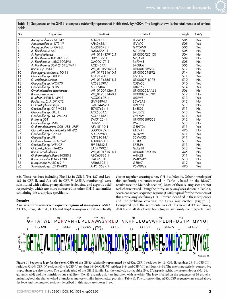
Figure 1 | Sequence logo for the seven CSRs of the GH13 subfamily represented by ASKA. CSR-I, residues 10–15; CSR-II, residues 23–31; CSR-III,
residues 32–39; CSR-IV, residues 40–45; CSR-V, residues 16–20; CSR-VI, residues 1–9; and CSR-VII, residues 46–54. The two characteristic, consecutive
tryptophans are also shown. The catalytic triad of the GH13 family, i.e., the catalytic nucleophile (No. 27, aspartic acid), the proton donor (No. 36,
glutamic acid) and the transition-state stabiliser (No. 45, aspartic acid) are indicated with asterisks. The logo is based on the sequences of 36 proteins
including both the characterised a-amylases and very similar hypothetical proteins (Table 1). The corresponding ASKA CSR sequences are stated above
the logo and the mutated residues described in this study are shown in red.
www.nature.com/scientificreports
SCIENTIFIC REPORTS | 4 : 5850 | DOI: 10.1038/srep05850 2
similar CSRs (Figure 1). A separate weblogo with a-amylasesequences from earlier known GH13 subfamilies was built andcompare with ASKA’s CSRs (Figure 2). ASKA clearly containsdifferent residues in several positions of the CSRs (Figure 2). Toinvestigate their functional role, Phe-113, Ala-161, Tyr-187, andLeu-189 of ASKA were thus selected and replaced with valine,aspartic acid, phenylalanine, and isoleucine, respectively. A total offour single-point mutant a-amylases were designed and denoted asF113V, A161D, Y187F, and L189I (Table 2).
Figure 3a illustrates the position of CSR-I to CSR-VII in silico. Phe-113 is the second residue of CSR-I (Figure 1; position 11 in the logo),which is located on strand b3 of the catalytic (b/a)8-barrel domainA23. This position was chosen for the mutagenesis study because avaline or other aliphatic hydrophobic residue has frequently beenobserved at the corresponding position, e.g., in the a-amylases fromsubfamilies GH13_1 (fungi), GH13_5 (liquefying enzymes from bac-teria), GH13_6 (plants) and GH13_7 (archaeons)23,24. The next twomutant positions, Tyr-187 and Leu-189 (logo positions 24 and 26,respectively; Figure 1), are residues from CSR-II, which covers strandb4 of the catalytic barrel23. While the former position is well-conserved as an aromatic one throughout the entire a-amylase familyGH13, it is mostly occupied by phenylalanine or tryptophan23,24 andthe presence of the tyrosine (Figure 2) here in ASKA seems to beunique for the GH13 subfamily11. Because tryptophan is highly char-acteristic of the GH13 subfamilies 6 and 7, which include plants,archaeons and flavobacteria23–25, phenylalanine, which is found inthe GH13 subfamilies 15 and 24 (animals) as well as 27, 28, 32 and36 (various groups of bacteria), was chosen as the substitute for Tyr-187 in the mutant. The latter mutant position in CSR-II, Leu-189, is aconserved position, but if it is replaced then isoleucine (Figure 2) is
often found23,24. The fourth residue selected for mutation, Ala-161, ispositioned in CSR-V near the C terminal of domain B, i.e., in the loopconnecting stand b3 to helix a3 of catalytic (b/a)8-barrel domain A23.Although various residues can be seen at that position, aspartic acidor asparagine may represent a consensus23,24. It should therefore be ofinterest to investigate the role the alanine may play in ASKA(Figure 1; position 20 in the logo) because an alanine is typicallypresent in the corresponding position only in the animal a-amylases(from vertebrates) from the subfamily GH13_2423.
Characterisation of the mutant a-amylases. The purified enzymeswere subjected to sodium dodecyl sulphate polyacrylamide gelelectrophoresis (SDS-PAGE) to determine the purity of theproteins (Figure 4). The mutant enzymes were pure with singlebands at the molecular mass of 50 kDa in agreement with that ofnative a-amylase18. The zymogram analysis was performed usingnative PAGE, and the gel was stained with iodine solution. For allthe enzymes, a single, clear band was observed upon staining. Thezymogram of the purified proteins shows that the mutant enzymesretained amylolytic activity (Figure 4a). After the purificationprocedures, the enzyme activity and the protein concentration forall a-amylase variants were determined, and the specific activity ofeach a-amylase variant was calculated (Table 2).
The effect of temperature on the activity of the ASKA and mutantvariants was tested at temperatures ranging from 30 to 90uC at pH 8.0(Supplemental file). The optimum reaction temperature for all the a-amylase variants was 60uC. None of the amino acid substitutionsaffected the optimum temperature activity. At 70uC, the enzymesmaintained 54–87% of the initial activity. As the incubation temper-ature rises, the activity of the enzymes drops to 13–24% at 80uC and10–18% at 90uC. Compared to ASKA, mutants Y187F and L189I hada slight decline in activity of approximately 32% and 23%, respect-ively at a temperature of 70uC. The thermostability of the enzymeswas studied at the temperatures of 60uC and 65uC for 72 h. Theactivity half-life of all mutants at their optimum temperature of60uC is unaffected by the mutation (Supplemental file). The mutantA161D retained 94.6% of its total activity after 72 h of incubation at60uC, and this value is slightly higher compared to ASKA, whichretained 91% of its activity. The activity half-life at 65uC is displayedin Table 2, the ASKA had a half-life of 48 h, while Y187F and L189Ihad a slight reduction in thermostability with half-lives of 43 and36 h, respectively. The activity half-lives at 65uC for the mutantsF113V and A161D were 51 and 53 h, respectively.
The effect of pH on a-amylase activity and stability were tested at apH range of 4.0 to 11.0 using sodium acetate buffer (pH 4–5), sodiumphosphate buffer (pH 6–7), Tris-HCl buffer (pH 8–9) and glycine-NaOH buffer (pH 10–11) at a temperature of 60uC. ASKA and allmutant a-amylases exhibited maximum activity at pH 8.0. Theoptimum pH is thus unaffected by the amino acid substitutions.This result is expected because very frequently the optimum pH ofan enzyme is not easily affected by a single residue replacement. TheASKA and mutant a-amylases were stable within a wide pH range of7.0 to 10.0 for 30 min, and the enzyme activities were at least 90% ofthe initial enzyme activity at these pH values. The pH stability of themutants was similar to the ASKA. The pH stability of ASKA wasunaffected by the mutations.
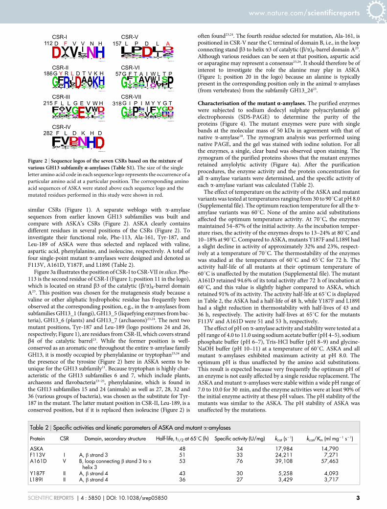
Figure 2 | Sequence logos of the seven CSRs based on the mixture ofvarious GH13 subfamily a-amylases (Table S1). The size of the single
letter amino acid code in each sequence logo represents the occurrence of a
particular amino acid at a particular position. The corresponding amino
acid sequences of ASKA were stated above each sequence logo and the
mutated residues performed in this study were shown in red.
Table 2 | Specific activities and kinetic parameters of ASKA and mutant a-amylases
Protein CSR Domain, secondary structure Half-life, t1/2 at 65uC (h) Specific activity (U/mg) kcat (s21) kcat/Km (ml mg21 s21)
ASKA 48 34 17,984 14,790F113V I A, b strand 3 51 33 24,211 7,271A161D V B, loop connecting b stand 3 to a
helix 353 76 39,108 57,463
Y187F II A, b strand 4 43 30 5,258 4,093L189I II A, b strand 4 36 27 3,429 3,717
www.nature.com/scientificreports
SCIENTIFIC REPORTS | 4 : 5850 | DOI: 10.1038/srep05850 3
The specific activities, turnover rate (kcat) and catalytic efficiency(kcat/Km) of ASKA were compared to its mutant variants (Table 2).ASKA had a specific activity of 34 U/mg-protein. Of the mutant a-amylases, the mutant A161D had the highest specific activity at76 U/mg with an increase of approximately 1.23 times. The Vmax
as well as the kcat/Km were drastically changed by the A161 mutation.The mutant A161D exhibited the highest kcat of 39,108 s21, which is1.17-times higher than the kcat value of ASKA. Moreover, the kcat/Km
of A161D was also higher than that of ASKA by 2.88-times, at57,463 ml mg21 s21. Of the four mutants constructed using site-directed mutagenesis, only mutant A161D exhibited enhanced kcat
and kcat/Km values (Table 2). The L189I substitution had an undesir-able effect as the kcat and kcat/Km values were reduced by 81% and74%, respectively. Such deterioration was the worst observed of themutants. The adjacent mutant Y187F also exhibited a reduced effi-ciency in the catalytic reaction. Both of these residues were locatednear the catalytic Asp-190 (Figure 3b), and their mutations havecaused a detrimental effect to the catalytic property of the enzyme.These results are in agreement with the findings of earlier reports26,27,which stated that any mutations near the catalytic residue will mostlikely influence the catalytic property of enzyme and likely cause anoverall negative effect on enzyme catalysis.
All mutant enzymes were subjected to an enzymatic reaction tostudy the effect of the mutation on product specificity. Hydrolysis of1% (w/v) and 4% (w/v) gelatinised soluble-, tapioca-, potato-, corn-,and sago-starch was carried out at 60uC for 24 h. The total sugarproduction (mg/ml) is shown in Table 3. At the lower starch con-centration of 1%, the yield of total sugars generated by mutantsY187F and L189I was significantly lower than that of the ASKA;however, this effect was less significant at the higher concentrationof starch (4%). With the 1% starch solution, the total sugars yieldedby mutant A161D were quite similar to the ASKA most likely becausethe amount of substrate was not in excess. Interestingly, at a highersubstrate concentration, for instance 4% corn-starch, the total sugarformed by mutant A161D was 27.8 mg/ml, which is higher than thatof the ASKA (16.3 mg/ml) (Table 3). The kcat and kcat/Km values formutant A161D are better than for ASKA, and the HPLC analysisconfirms the outperformance of A161D.
The percentages of individual sugars that were formed after 24-hreactions in 1% (Figure 5a) and 4% corn-starch (Figure 5b) werestudied. Previously, we reported that ASKA is a maltose-forminga-amylase18, and a negligible percentage of glucose (G1), maltohex-
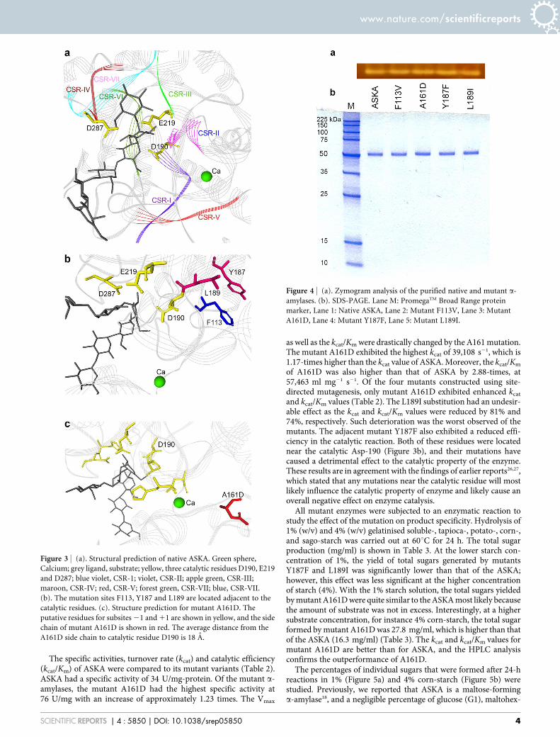
Figure 3 | (a). Structural prediction of native ASKA. Green sphere,
Calcium; grey ligand, substrate; yellow, three catalytic residues D190, E219
and D287; blue violet, CSR-1; violet, CSR-II; apple green, CSR-III;
maroon, CSR-IV; red, CSR-V; forest green, CSR-VII; blue, CSR-VII.
(b). The mutation sites F113, Y187 and L189 are located adjacent to the
catalytic residues. (c). Structure prediction for mutant A161D. The
putative residues for subsites 21 and 11 are shown in yellow, and the side
chain of mutant A161D is shown in red. The average distance from the
A161D side chain to catalytic residue D190 is 18 A.
Figure 4 | (a). Zymogram analysis of the purified native and mutant a-
amylases. (b). SDS-PAGE. Lane M: PromegaTM Broad Range protein
marker, Lane 1: Native ASKA, Lane 2: Mutant F113V, Lane 3: Mutant
A161D, Lane 4: Mutant Y187F, Lane 5: Mutant L189I.
www.nature.com/scientificreports
SCIENTIFIC REPORTS | 4 : 5850 | DOI: 10.1038/srep05850 4

aose (G6) and maltoheptaose (G7) were usually detected in 1% gela-tinised corn-starch (Figure 5a). However, when the ASKA wasreacted with 4% corn-starch for 24 h, a small percentage of G6 andG7 were detected. The product specificity spectrum for mutantA161D changed significantly for the reaction that was carried outin 4% starch. The percentage of maltose forming dropped, while asubstantial increase in the percentage of glucose was observed(Figure 5b).
Discussiona-Amylases, in addition to all members of the a-amylase GH13family, contain four to seven conserved sequence regions (CSRs),roughly covering strands b2, b3, b4, b5, b7 and b8 of the catalyticTIM-barrel and a short stretch near the C-terminus of domain Bprotruding out of the barrel in the b3-a3 loop. The individual a-amylases often possess unique sequence features in their respectiveCSRs that distinguish them from each other, and thus these regionsmay be connected to the specific biochemical properties of eachprotein, such as substrate preferences, product specificities, andthermostability9–11.
Anoxybacillus sp. SK3-4 was isolated from a Malaysian hotspring28
and its genome was sequenced using Illumina MiSeq29. Most of theglycoside hydrolases of the cell are expressed intracellularly; how-ever, ASKA was described as an extracellular hydrolase18. Comparedwith well-recognised a-amylases, the ASKA protein sequence has alow similarity of 49% to the G. stearothermophilus30 (BSTA, subfam-ily GH13_5, UniProt Accession number P06279), 47% to theAspergillus oryzae31 (TAKA, GH13_1, P0C1B3) and 37% similarityto the B. licheniformis32 (BLA, GH13_5, P06278) a-amylases. Despitethe fact that ASKA is a high maltose producing a-amylase, it is agenuine a-amylase. In fact, the two types of family GH13 amylasesforming mainly maltose (EC 3.2.1.133) should be distinguished fromeach other, i.e., maltogenic amylases and maltogenic a-amylases23.Whereas the former enzymes are members of the so-called neopul-lulanase subfamily (subfamily GH13_20), the latter ones are closelyrelated to cyclodextrin glucanotransferases (CGTase) (GH13_2). Atypical GH13_20 maltogenic amylase possesses in its CSR-V thesignature MPKLN9, contains at its N-terminus a carbohydrate-binding module from the family CBM3433 and is able to hydrolysecyclodextrins34. Because the ASKA has a different CSR-V (157_LPDLA; Figure 1), lacks the CBM34 N-terminal domain and is notactive on cyclodextrins18, it is likely not related to this type of mal-togenic amylase. For the maltogenic a-amylases from the subfamilyGH13_2, the CSR-V signature (LADLS) from B. stearothermophi-lus23 is more similar to that seen in ASKA, but it typically shares itsfive-domain organisation with CGTase35; i.e., it has two C-terminaldomains in addition to domains A, B and C that are present in theASKA. Thus, ASKA and most likely the entire newly established a-amylase family GH13 group11 (i.e., ADTA, BaqA and homologuesfrom G. thermoleovorans) may represent a novel type of high maltoseproducing amylase.
ASKA shares 69% similarity with the a-amylase from G. thermo-leovorans subsp. stromboliensis Pizzo22, 69% to G. thermoleovoransGtamyII20 and 60% to a marine B. aquimaris MKSC 6.2 a-amylase(BaqA)11,36. Recently, the crystal structure of the a-amylase G. ther-moleovarans (GTA) was reported21. The sequence similarity betweenASKA and GTA is as high as 69%. ASKA, Pizzo, GtamyII, GTA, andBaqA as well as another Anoxybacillus a-amylase ADTA from ourlaboratory18 can be classified as a new sub-group of the a-amylasefamily GH13. Taken together, the reported experimental data ofthese a-amylases, this new subgroup has at least three unique char-acteristics. Firstly, they are high maltose producing enzymes as seenin ASKA, ADTA and Pizzo and potential in GTA since Pizzo sharesan identical protein sequence with GTA21. Secondly, these enzymesare able to degrade raw starches as reported for the Pizzo, GtamyII,and BaqA amylases11,20,22. Although the two consecutive tryptophanswere proposed to be the putative raw starch binding residues11, thebinding action has yet to be experimentally proven. Thirdly, the C-terminal of the ASKA protein sequence was predicted to be a putativetransmembrane region that could possibly anchor to the wild typeAnoxybacillus sp. SK3-419. Recent work on the deletion of this trans-membrane region of GtamyII elucidated its role in raw starchadsorption20.
Table 3 | Total sugar production (mg/ml) from the hydrolysis of different starches by ASKA and mutant a-amylases using 1% (w/v) and 4%(w/v) of starches
Type of starches
1% (w/v) of starches 4% (w/v) of starches
ASKA F113V A161D Y187F L189I ASKA F113V A161D Y187F L189I
Corn 12.5 12.3 13.3 10.9 11.1 16.3 17.0 27.8 15.0 15.7Soluble 12.1 10.4 12.1 9.5 10.5 16.3 15.8 24.9 14.9 15.1Tapioca 11.0 11.4 12.8 9.5 10.5 16.2 16.9 25.8 14.2 14.7Potato 11.2 11.7 12.0 10.0 10.4 16.1 16.7 24.5 14.2 14.9Sago 11.8 12.0 12.9 11.4 10.7 16.1 16.4 27.4 15.5 15.5
Figure 5 | End product distribution of ASKA and mutant a-amylasesfrom the hydrolysis of a) 1% (w/v) and b) 4% (w/v) corn-starch. The data
are shown as the percentage (%) hydrolysed. Black: glucose (G1), grey:
maltose (G2), blue: maltotriose (G3), green: maltotetraose, (G4), purple:
maltopentaose (G5), yellow: maltohexaose (G6) and red: maltoheptaose
(G7).
www.nature.com/scientificreports
SCIENTIFIC REPORTS | 4 : 5850 | DOI: 10.1038/srep05850 5
The main goal of this current work is to address the reason that theenzymes from this new GH13 subgroup, for instance ASKA, are ahigh maltose forming. The CSRs of ASKA were compared to otherreported a-amylases, and four single point mutations were carriedout. Interestingly, the product specificity of mutant A161D wasaltered causing the overall ratio of maltose to drop, while the per-centage of glucose increased 28-times in 4% corn-starch. Based onthese findings, Ala-161 is proposed to be responsible for the highmaltose-producing characteristic of ASKA. In the maltose producingADTA, Pizzo and GTA a-amylases, Ala is well conserved at anidentical position. The other homologs, i.e., GtamyII and BaqA alsocontained Ala, but their product specificity has yet to be determined.
Such drastic change was unexpected because the location of 161Din the predicted 3D structure is 18 A from the nearest catalyticresidue, Asp-190 (Figure 3c). Moreover, the superimposition of themutant A161D model with the G. thermoleovorans a-amylase(4E2O) elucidated that 161D has no direct interaction with thepseudo substrate acarbose. At this stage, it is not clear the true reasonwhy product specificity is associated with Ala-161. In CGTase, theproduct specificity is often related to the type of amino acids at thesubsite interacting residues37–39, yet A161 is not likely to be a part ofany subsites. The in silico position of residue 161D is located close tothe putative calcium (Ca21)-binding site (Figure 3c). A161D maystabilise the secondary structure or the folding of this Ca21-bindingpocket, therefore the half-life of this mutant was higher than that ofASKA (Table 2). Nevertheless, at this point, we are unable to deter-mine why A161D can alter the product specificity in a drastic man-ner. We are now conducting further examination of this site bycreating other mutations to examine the effects of various amino acidside chains on product specificity. Besides X-ray crystallisation will beperformed in the near future. We hope that these experiments willlead to more insight to the importance of A161 in product specificity.
Several mutations have previously been made to the CSRs of othera-amylases. For instance, Chen et al. (2010) mutated Asp-236 of theBacillus sp. strain TS-23 a-amylase40. This mutated residue corre-sponds to Ala-161 in ASKA numbering, in CSR-V. The replacementof Asp-236 with asparagine resulted in a 33% reduction in the specificactivity of the enzyme. Interestingly, this finding contradicts theA161D mutation results obtained in this study. In addition, Chenet al. (2010) mutated Asp-234, which is also located in CSR-V. Thissubstitution resulted in an 86% reduction in the specific activity ofthe enzyme. It seemed from that study that two residues in CSR-V(Asp-234 and Asp-236) played a significant role in the catalyticproperties of the enzyme40. Unfortunately, both trials disrupted thespecific activity, and the authors suggested that Asp-234 and Asp-236 were involved in Ca21-binding, and any changes to these Ca21-binding residues will affect the enzyme’s structural integrity, andstability as well as its a-amylase activity. In another study, the sub-stitution of Asp-233 of the B. amyloliquefaciens a-amylase (corre-sponds to ASKA’s Ala-161) significantly affected the specific activityand thermostability of the enzyme. There was an 84.4% reduction inspecific activity compared to the ASKA. From this result, the authorsdeduced that Asp-233 is important for the catalytic activity of theenzyme41.
Despite mutant F113V exhibiting a similar specific activity toASKA, the kcat value for the mutant was 35% higher. ASKA and itshomologues ADTA, Pizzo, GtamyII, BaqA, and GTA have a Phe atthis particular position of CSR-I, while most other a-amylases haveanother type of amino acid (Figure 2). Several mutations of CSR-Iresidues were performed previously, for instance, H137L (His-117 inASKA) of the Bacillus sp. TS-23 a-amylase resulted in a complete lossof amylolytic activity42. Chang et al. (2003) explained that His-137could be the essential catalytic residue in the amylolytic reaction. Thesite directed mutagenesis of another CSR-I residue, Asn-104 of BLA(corresponds to ASKA’s Asn-116) was replaced with Asp. Thismutant had an 11% decrease in specific activity at 30uC. But, inter-
estingly, the mutant had a 30% higher specific activity than the ASKAat the higher temperature of 70uC43.
This study found that the mutation of CSR-II, i.e., mutant Y187Fand L189I caused a more than 70% reduction in the kcat and kcat/Km
(Table 2). For mutant Y187F, the chemical structures of tyrosine andphenylalanine are structurally close except that phenylalanine lacks ahydroxyl -OH group at end of the aromatic side chain. The removalof the hydroxyl group caused a severe disturbance during the starchhydrolysis process. The hydroxyl group is likely involved in stabilis-ing of the intermediate state during the catalytic mechanism. Theseresults elucidate that the amino acid residues Tyr-187 and Leu-189are associated with ASKA activity, most likely because they arelocated adjacent to catalytic residue 190 (Figure 3b). Similarly, themutation of the Bacillus sp. TS-23 a-amylase, in CSR-II at His-269(TS-23 numbering), which is adjacent to catalytic Asp-265, led to thecomplete loss of amylolytic activity. The report proposed that thisconserved histidine may play a substrate binding role37, or possibly,this stretch of CSR-II is sensitive to alteration.
MethodsBioinformatics analysis. The 36 amino acid sequences (Table 1) that are currentlythought to form the GH13 subfamily represented by ASKA were retrieved fromGenBank database44. The sequences were collected from a BLAST search45 using asthe query the stretch of 89 residues from the ASKA sequence18 spanning thepolypeptide chain covering CSR-I (strand b3) and CSR-II (strand b4), including theentire domain B (GenBank Acc. No.: AFI49455.1: Asp-129-His-217). A sequence wasconsidered to belong to this GH13 subfamily if it contains the features ascribed to theclosely related a-amylase from B. aquimaris11, especially the two consecutivetryptophan residues in helix a3 of the catalytic TIM-barrel and the specific sequencein CSR-V positioned in domain B.
The sequences were aligned using the program Clustal-Omega46 available on theEuropean Bioinformatics Institute’s server (http://www.ebi.ac.uk/). For the whole setof 36 sequences, a sequence logo for their seven CSRs typical of the members of the a-amylase family GH1323 was created using the WebLogo 3.0 server47 (http://weblogo.berkeley.edu/).
Preparation of starting structure and molecular dynamics simulation. The proteinstructure of ASKA was constructed using the SWISS-MODEL48 program (http://swissmodel.expasy.org/), and the X-ray crystal structure of G. thermoleovorans a-amylase (PDB code: 4E2O)21 was selected as the template. Refinements of the modelwere performed using energy minimisation and dynamic simulation (MD). Prior tothe MD simulation using the GROMACS software package49, calcium and glucosewere removed from the structure. The system was solvated with SPC/E water in adodecahedral box with 10 A between the protein structure and the box. The systemwas subsequently neutralised by the addition of sodium ions. The MD simulation wasperformed using an OPLS-AA force field. The system was energy minimised andsubsequently simulated in NVT and NPT ensembles. The temperature wasmaintained at 333 K. Upon completing the system equilibration, a production run of10 ns was performed.
Mutagenesis. The a-amylase mutant gene was generated using the OverlapExtension PCR (OE-PCR) method. The external forward primer contained an EcoRIrestriction site, and the external reverse primer contained an XhoI restriction site. Thechange in nucleotide sequence was introduced by incorporating nucleotide changesinto the overlapping oligo primers, as shown in Supplemental file. During the cloningprocess, the native signal peptide DNA sequence was excluded, as the pET-22b(1)contains a pelB leading sequence. PhusionH PCR Master Mix (NEB, Ipswich,Massachusetts, USA) was used to perform the PCR. PCR reaction mixturescontaining 25 ng ASKA DNA template, 25 ml 23 PhusionTM Flash PCR Master Mix,and 0.5 mM each primer were added to a final volume of 50 ml. A SpinPrepTM PCRClean-Up kit (Novagen, Darmstadt, Germany) was used to purify the PCR products.Purified PCR products and pET-22b (1) (Novagen, Darmstadt, Germany) weredigested with EcoRI and XhoI (NEB, Ipswich, Massachusetts, USA) and subsequentlypurified. The ligation was performed using T4 DNA ligase (NEB, Ipswich,Massachusetts, USA) and then transformed into E. coli DH5a. The recombinantplasmid was confirmed by sequencing. The recombinant plasmid was thentransformed into the expression host E. coli BL21 (DE3) for later analysis.
Protein expression and purification. The recombinant E. coli BL21 (DE3) wascultured overnight in LB medium supplemented with 100 mg/ml ampicillin at 37uCwith agitation at 200 rpm. Induction was performed by adding 0.01 mM IPTG, andthe cultivation was continued at 25uC with agitation at 200 rpm for 48 h50. The cellfree enzyme was harvested after centrifugation (10000 3 g, 20 min, and 4uC). Thecrude enzyme solution was concentrated using the Vivaflow 50 crossflow systemfrom Sartorius Stedim Biotech (Gottingen, Germany) using the polyethersulfone(PES) membrane with a 10 kDa molecular weight cut-off (MWCO). The retentatewas loaded onto affinity columns, and chromatography was conducted using the
www.nature.com/scientificreports
SCIENTIFIC REPORTS | 4 : 5850 | DOI: 10.1038/srep05850 6
AKTAprime plus system (GE Healthcare, Uppsala, Sweden) according to the methodreported previously18.
Characterisation of ASKA and mutant enzymes. The protocols used to characterisethe mutant a-amylases were also performed using ASKA unless otherwise stated18.One unit of a-amylase was defined as the amount of enzyme that releases 1 mmolmaltose per min per ml under the assay conditions specified. The kinetic parametersof the ASKA and mutant a-amylases were determined by measuring the amount ofmaltose production using the standard assay conditions. The samples were reactedwith soluble starch (Kanto Chemical, Tokyo, Japan) at concentrations from 0.1 to2.0 mg/ml in 0.1 M Tris-HCl buffer (pH 8.0). SigmaPlot v11.0 was used to build theHanes plot.
The products of starch hydrolysis by a-amylase were analysed using High-performance liquid chromatography (HPLC). First, 0.1 ml purified enzyme wasincubated with 1.0 ml 1% gelatinised starch in 0.1 M Tris-HCl buffer (pH 8.0) at60uC for 24 h. The efficiency of degradation on various starches (soluble-, tapioca-,potato-, sago- and corn-starch) was compared. After 24 h of incubation, the reactionmixture was boiled for 15 min to stop the reaction and cooled to room temperature.The mixture was filtered through a 0.45 mm nylon syringe filter (Whatman, LittleChalfront, United Kingdom) to remove insoluble particles. The hydrolysis productswere analysed using a Waters HPLC system with a Zorbax Carbohydrate analysiscolumn, 4.6 3 250 mm (Agilent, Santa Clara, California, USA). An acetonitrile(HPLC grade, Merck, White House Station, New Jersey, USA) to water mixture(70530, v/v) was used as the mobile phase, and the flow rate was maintained at 1.0 ml/min. The column was kept at 30uC, and a Waters 2414 refractive index detector wasused to detect the reaction products. The data were recorded by the integratedcomputer system attached to the HPLC. Glucose, maltose, maltotriose, maltotetraose,maltopentaose, maltohexaose and maltoheptaose (Sigma-Aldrich. St Louis, Missouri,USA) were used as standards. Additionally, the efficiency of starch concentration onthe hydrolysis products was investigated by incubating the ASKA and mutantenzymes in a 4% gelatinised starch in 0.1 M Tris-HCl buffer (pH 8.0) at 60uC for 24 h.The end products were analysed using HPLC. The assays and data collection wereperformed at least in triplicate, unless otherwise specified.
1. Cantarel, B. L. et al. The Carbohydrate-Active EnZymes database (CAZy): anexpert resource for glycogenomics. Nucleic Acids Res. 37, D233–D238,doi:10.1093/nar/gkn663 (2009).
2. Lombard, V., Golaconda Ramulu, H., Drula, E., Coutinho, P. M. & Henrissat, B.The Carbohydrate-Active EnZymes database (CAZy) in 2013. Nucleic Acids Res.42, D490–D495, doi:10.1093/nar/gkt1178 (2014).
3. Janecek, S., Svensson, B. & MacGregor, E. A. a-Amylase: an enzyme specificityfound in various families of glycoside hydrolases. Cell. Mol. Life Sci. 71,1149–1170, doi:10.1007/s00018-013-1388-z (2014).
4. Svensson, B. Protein engineering in the a-amylase family: catalytic mechanism,substrate specificity, and stability. Plant Mol. Biol. 25, 141–157, doi:10.1007/bf00023233 (1994).
5. Janecek, S.a-amylase family: molecular biology and evolution. Prog. Biophys. Mol.Biol. 67, 67–97, doi:10.1016/S0079-6107(97)00015-1 (1997).
6. Kuriki, T. & Imanaka, T. The concept of thea-amylase family: structural similarityand common catalytic mechanism. J. Biosci. Bioeng. 87, 557–565, doi:10.1016/S1389-1723(99)80114-5 (1999).
7. MacGregor, E. A., Janecek, S. & Svensson, B. Relationship of sequence andstructure to specificity in the a-amylase family of enzymes. Biochim. Biophys.Acta, Protein Struct. Mol. Enzymol. 1546, 1–20, doi:10.1016/S0167-4838(00)00302-2 (2001).
8. Stam, M. R., Danchin, E. G. J., Rancurel, C., Coutinho, P. M. & Henrissat, B.Dividing the large glycoside hydrolase family 13 into subfamilies: towardsimproved functional annotations of a-amylase-related proteins. Protein Eng. Des.Sel. 19, 555–562, doi:10.1093/protein/gzl044 (2006).
9. Oslancova, A. & Janecek, S. Oligo-1,6-glucosidase and neopullulanase enzymesubfamilies from the a-amylase family defined by the fifth conserved sequenceregion. Cell. Mol. Life Sci. 59, 1945–1959, doi:10.1007/pl00012517 (2002).
10. Majzlova, K., Pukajova, Z. & Janecek, S. Tracing the evolution of the a-amylasesubfamily GH13_36 covering the amylolytic enzymes intermediate betweenoligo-1,6-glucosidases and neopullulanases. Carbohydr. Res. 367, 48–57,doi:10.1016/j.carres.2012.11.022 (2013).
11. Puspasari, F. et al. Raw starch-degrading a-amylase from Bacillus aquimarisMKSC 6.2: isolation and expression of the gene, bioinformatics and biochemicalcharacterization of the recombinant enzyme. J. Appl. Microbiol. 114, 108–120,doi:10.1111/jam.12025 (2013).
12. Koropatkin, N. M., Cameron, E. A. & Martens, E. C. How glycan metabolismshapes the human gut microbiota. Nat. Rev. Microbiol. 10, 323–335, doi:10.1038/nrmicro2746 (2012).
13. Leveque, E., Janecek, S., Haye, B. & Belarbi, A. Thermophilic archaeal amylolyticenzymes. Enzyme Microb. Technol. 26, 3–14, doi:10.1016/S0141-0229(99)00142-8 (2000).
14. van der Maarel, M. J. E. C., van der Veen, B., Uitdehaag, J. C. M., Leemhuis, H. &Dijkhuizen, L. Properties and applications of starch-converting enzymes of the a-amylase family. J. Biotechnol. 94, 137–155, doi:10.1016/S0168-1656(01)00407-2 (2002).
15. Tawil, G., Viksø-Nielsen, A., Rolland-Sabate, A., Colonna, P. & Buleon, A.Hydrolysis of concentrated raw starch: a new very efficient a-amylase from
Anoxybacillus flavothermus. Carbohydr. Polym. 87, 46–52, doi:10.1016/j.carbpol.2011.07.005 (2012).
16. Matpan Bekler, F. & Guven, K. Isolation and production of thermostable a-amylase from thermophilic Anoxybacillus sp. KP1 from Diyadin hot spring inAgri, Turkey. Biologia 69, 419–427, doi:10.2478/s11756-014-0343-2 (2014).
17. Aguloglu Fincan, S., Enez, B., Ozdemir, S. & Matpan Bekler, F. Purification andcharacterization of thermostable a-amylase from thermophilic Anoxybacillusflavithermus. Carbohydr. Polym. 102, 144–150, doi:10.1016/j.carbpol.2013.10.048(2014).
18. Chai, Y. Y., Rahman, R. N. Z. R. A., Illias, R. M. & Goh, K. M. Cloning andcharacterization of two new thermostable and alkalitolerant a-amylases from theAnoxybacillus species that produce high levels of maltose. J. Ind. Microbiol.Biotechnol. 39, 731–741, doi:10.1007/s10295-011-1074-9 (2012).
19. Kahar, U., Chan, K.-G., Salleh, M., Hii, S. & Goh, K. M. A high molecular-massAnoxybacillus sp. SK3-4 amylopullulanase: characterization and its relationshipin carbohydrate utilization. Int. J. Mol. Sci. 14, 11302–11318, doi:10.3390/ijms140611302 (2013).
20. Mehta, D. & Satyanarayana, T. Domain C of thermostable a-amylase ofGeobacillus thermoleovorans mediates raw starch adsorption. Appl. Microbiol.Biotechnol. 1–17, doi:10.1007/s00253-013-5459-8 (2014).
21. Mok, S.-C., Teh, A.-H., Saito, J. A., Najimudin, N. & Alam, M. Crystal structure ofa compact a-amylase from Geobacillus thermoleovorans. Enzyme Microb.Technol. 53, 46–54, doi:10.1016/j.enzmictec.2013.03.009 (2013).
22. Finore, I. et al. Purification, biochemical characterization and gene sequencing of athermostable raw starch digesting a-amylase from Geobacillus thermoleovoranssubsp. stromboliensis subsp. nov. World J. Microbiol. Biotechnol. 27, 2425–2433,doi:10.1007/s11274-011-0715-5 (2011).
23. Janecek, S. How many conserved sequence regions are there in the a-amylasefamily? Biologia - Section Cellular and Molecular Biology 57 (Suppl. 11, ), 29–41(2002).
24. Janecek, S., Leveque, E., Belarbi, A. & Haye, B. Close evolutionary relatedness of a-amylases from Archaea and plants. J. Mol. Evol. 48, 421–426, doi:10.1007/PL00006486 (1999).
25. Li, C. et al. Close relationship of a novel Flavobacteriaceae a-amylase with archaeala-amylases and good potentials for industrial applications. Biotechnol. Biofuels 7,18, doi:10.1186/1754-6834-7-18 (2014).
26. Nielsen, J. E. et al. Electrostatics in the active site of an a-amylase. Eur. J. Biochem.264, 816–824, doi:10.1046/j.1432-1327.1999.00664.x (1999).
27. Wind, R. D., Uitdehaag, J. C. M., Buitelaar, R. M., Dijkstra, B. W. & Dijkhuizen, L.Engineering of cyclodextrin product specificity and pH optima of thethermostable cyclodextrin glycosyltransferase from Thermoanaerobacteriumthermosulfurigenes EM1. J. Biol. Chem. 273, 5771–5779 (1998).
28. Chai, Y. Y., Kahar, U. M., Salleh, M. M., Illias, R. M. & Goh, K. M. Isolation andcharacterization of pullulan-degrading Anoxybacillus species isolated fromMalaysian hot springs. Environ. Technol. 33, 1231–1238, doi:10.1080/09593330.2011.618935 (2011).
29. Goh, K. M. et al. Analysis of Anoxybacillus genomes from the aspects of lifestyleadaptations, prophage diversity, and carbohydrate metabolism. PLoS ONE 9,e90549, doi:10.1371/journal.pone.0090549 (2014).
30. Wind, R. D., Buitelaar, R. M., Eggink, G., Huizing, H. J. & Dijkhuizen, L.Characterization of a new Bacillus stearothermophilus isolate: a highlythermostable a-amylase-producing strain. Appl. Microbiol. Biotechnol. 41, 155-162, doi:10.1007/bf00186953 (1994).
31. Matsuura, Y., Kusunoki, M., Harada, W. & Kakudo, M. Structure and possiblecatalytic residues of Taka-amylase A. J. Biochem. 95, 697-702 (1984).
32. Ivanova, V. N., Dobreva, E. P. & Emanuilova, E. I. Purification andcharacterization of a thermostable a-amylase from Bacillus licheniformis.J. Biotechnol. 28, 277-289, doi:10.1016/0168-1656(93)90176-N (1993).
33. Kim, J.-S. et al. Crystal structure of a maltogenic amylase provides insights into acatalytic versatility. J. Biol. Chem. 274, 26279–26286 (1999).
34. Cha, H.-J. et al. Molecular and enzymatic characterization of a maltogenic amylasethat hydrolyzes and transglycosylates acarbose. Eur. J. Biochem. 253, 251–262,doi:10.1046/j.1432-1327.1998.2530251.x (1998).
35. Dauter, Z. et al. X-ray structure of Novamyl, the five-domain ‘‘maltogenic’’ a-amylase from Bacillus stearothermophilus: maltose and acarbose complexes at1.7A resolution. Biochemistry 38, 8385–8392, doi:10.1021/bi990256l (1999).
36. Puspasari, F. et al. Characteristics of raw starch degrading a-amylase from Bacillusaquimaris MKSC 6.2 associated with soft coral Sinularia sp. Starch - Starke 63,461–467, doi:10.1002/star.201000127 (2011).
37. Goh, K. M., Mahadi, N. M., Hassan, O., Abdul Rahman, R. N. Z. R. & Illias, R. M.The effects of reaction conditions on the production of c-cyclodextrin fromtapioca starch by using a novel recombinant engineered CGTase. J. Mol. Catal. B-Enzym. 49, 118–126, doi:10.1016/j.molcatb.2007.09.011 (2007).
38. Goh, K. M., Mahadi, N. M., Hassan, O., Rahman, R. N. Z. R. A. & Illias, R. M. Apredominant b-CGTase G1 engineered to elucidate the relationship betweenprotein structure and product specificity. J. Mol. Catal. B-Enzym. 57, 270–277,doi:10.1016/j.molcatb.2008.09.016 (2009).
39. Han, R. et al. Recent advances in discovery, heterologous expression, andmolecular engineering of cyclodextrin glycosyltransferase for versatileapplications. Biotechnol. Adv. 32, 415–428, doi:10.1016/j.biotechadv.2013.12.004(2014).
www.nature.com/scientificreports
SCIENTIFIC REPORTS | 4 : 5850 | DOI: 10.1038/srep05850 7
40. Chen, Y.-H. et al. Mutational analysis of the proposed calcium-binding aspartatesof a truncated a-amylase from Bacillus sp. strain TS-23. Ann. Microbiol. 60,307–315, doi:10.1007/s13213-010-0042-3 (2010).
41. Liu, Y., Shen, W., Shi, G.-Y. & Wang, Z.-X. Role of the calcium-binding residuesAsp231, Asp233, and Asp438 in a-amylase of Bacillus amyloliquefaciens asrevealed by mutational analysis. Curr. Microbiol. 60, 162–166, doi:10.1007/s00284-009-9517-5 (2010).
42. Chang, C.-T. et al. Identification of essential histidine residues in a recombinant a-amylase of thermophilic and alkaliphilic Bacillus sp. strain TS-23. Extremophiles 7,505–509, doi:10.1007/s00792-003-0341-8 (2003).
43. Priyadharshini, R. & Gunasekaran, P. Site-directed mutagenesis of the calcium-binding site of a-amylase of Bacillus licheniformis. Biotechnol. Lett. 29,1493–1499, doi:10.1007/s10529-007-9428-0 (2007).
44. Benson, D. A. et al. GenBank. Nucleic Acids Res. 42, D32–D37, doi:10.1093/nar/gkt1030 (2014).
45. Altschul, S. F., Gish, W., Miller, W., Myers, E. W. & Lipman, D. J. Basic localalignment search tool. J. Mol. Biol. 215, 403–410, doi:10.1016/S0022-2836(05)80360-2 (1990).
46. Sievers, F. et al. Fast, scalable generation of high-quality protein multiple sequencealignments using Clustal Omega. Mol. Syst. Biol. 7, 539–544, doi:10.1038/msb.2011.75 (2011).
47. Crooks, G. E., Hon, G., Chandonia, J. M. & Brenner, S. E. WebLogo: a sequencelogo generator. Genome Res. 14, 1188–1190, doi:10.1101/gr.849004 (2004).
48. Arnold, K., Bordoli, L., Kopp, J. & Schwede, T. The SWISS-MODEL workspace: aweb-based environment for protein structure homology modelling.Bioinformatics 22, 195–201, doi:10.1093/bioinformatics/bti770 (2006).
49. Pronk, S. et al. GROMACS 4.5: a high-throughput and highly parallel open sourcemolecular simulation toolkit. Bioinformatics 29, 845–854, doi:10.1093/bioinformatics/btt055 (2013).
50. Muniandy, K. et al. Application of statistical experimental design for optimizationof novel a-amylase production by Anoxybacillus species. J. Biol. Sci. 13, 605–613,doi:10.3923/jbs.2013.605.613 (2013).
AcknowledgmentsThis study was financially supported by the Universiti Teknologi Malaysia GUP Grant04H00 and the University of Malaya-Ministry of Higher Education High Impact ResearchGrant (UM MOHE HIR Grant No. H-50001-A000027). S.J. thanks the Slovak GrantAgency VEGA for financial support with grant No. 2/0150/14.
Author contributionsS.J., S.S., R.N.Z.R.A.R., K.G.C. and K.M.G. conceived and designed the experiments. V.R.,K.P.C. and K.G.C. performed the experiments. V.R., S.J., S.S. and K.M.G. analyzed the data.S.J., R.N.Z.R.A.R., K.G.C. and K.M.G. contributed reagents/materials/analysis tools. V.R.,S.J., K.P.C. and K.M.G. wrote the paper. V.R., S.J., K.P.C. and K.M.G. prepared figures 1–5.All authors reviewed the manuscript.
Additional informationSupplementary information accompanies this paper at http://www.nature.com/scientificreports
Competing financial interests: The authors declare no competing financial interests.
How to cite this article: Ranjani, V. et al. Protein engineering of selected residues fromconserved sequence regions of a novel Anoxybacillus a-amylase. Sci. Rep. 4, 5850;DOI:10.1038/srep05850 (2014).
This work is licensed under a Creative Commons Attribution-NonCommercial-ShareAlike 4.0 International License. The images or other third party material in thisarticle are included in the article’s Creative Commons license, unless indicatedotherwise in the credit line; if the material is not included under the CreativeCommons license, users will need to obtain permission from the license holderin order to reproduce the material. To view a copy of this license, visit http://creativecommons.org/licenses/by-nc-sa/4.0/
www.nature.com/scientificreports
SCIENTIFIC REPORTS | 4 : 5850 | DOI: 10.1038/srep05850 8
Copyright © 2022 FDOKUMEN