Protein Analysis by 31 P NMR Spectroscopy in Ionic Liquid: Quantitative Determination of...
11
rXXXX American Chemical Society A dx.doi.org/10.1021/jf102973d | J. Agric. Food Chem. XXXX, XXX, 000–000 ARTICLE pubs.acs.org/JAFC Protein Analysis by 31 P NMR Spectroscopy in Ionic Liquid: Quantitative Determination of Enzymatically Created Cross-Links Evanthia Monogioudi,* ,† Perttu Permi, ‡ Ilari Filpponen, §,|| Michael Lienemann, † Bin Li, §,^ Dimitris Argyropoulos, §,# Johanna Buchert, † and Maija-Liisa Mattinen † † VTT, P.O. Box 1000, FI-02044 VTT, Finland ‡ Institute of Biotechnology, FI-00014 University of Helsinki, Finland § Organic Chemistry of Wood Components Laboratory, Department of Forest Biomaterials, North Carolina State University, Raleigh, North Carolina 27695-8005, United States ) Department of Forest Products Technology, FI-00076 Aalto University, Finland ^ Department of Wood Science and Forest Products, Virginia Tech, Blacksburg, Virginia 24060, United States # Laboratory of Organic Chemistry, Department of Chemistry, P.O. Box 55, FI-00014 University of Helsinki, Finland ABSTRACT: Cross-linking of β-casein by Trichoderma reesei tyrosinase (TrTyr) and Streptoverticillium mobaraense transglutaminase (Tgase) was analyzed by 31 P nuclear magnetic resonance (NMR) spectroscopy in ionic liquid (IL). According to 31 P NMR, 91% of the tyrosine side chains were cross-linked by TrTyr at high dosages. When Tgase was used, no changes were observed because a different cross-linking mechanism was operational. However, this verified the success of the phosphitylation of phenolics within the protein matrix in the IL. Atomic force microscopy (AFM) in solid state showed that disk-shaped nanoparticles were formed in the reactions with average diameters of 80 and 20 nm for TrTyr and Tgase, respectively. These data further advance the current understanding of the action of tyrosinases on proteins on molecular and chemical bond levels. Quantitative 31 P NMR in IL was shown to be a simple and efficient method for the study of protein modification. KEYWORDS: Tyrosinase, transglutaminase, β-casein, cross-linking, 31 P NMR, AFM ’ INTRODUCTION Enzymatic modification of proteins via cross-linking is an attractive way to alter and improve the chemical and physico- chemical properties of protein-based products such as yogurt and cheese in the food industry as well as of foams and emulsions in cosmetics and pharmaceuticals. 1 In food applications improved texture (mouthfeel), low fat content (energy content), and stable gel formation (sensation of satiety) are properties that consu- mers typically appreciate. 2,3 Milk proteins, such as caseins and whey proteins, are food components of particular interest in the dairy industry due to their importance in human nutrition and allergenicity. 4 Enzymatic cross-linking of caseins may be an attractive way to overcome milk-derived health problems in addition to improving the quality of products. 5 When enzymatic modifications are combined with chemical (organic solvents, salts, pH) and physical (heat, milling, pressure) treatments, the applicability of the technology may be further expanded. 6,7 The effects of various cross-linking enzymes, for example, tyrosinases, laccases, peroxidases, and transglutaminases on proteins have been extensively reported in various publica- tions. 2,8-10 In addition to direct cross-linking, laccases and peroxidases can modify protein material 9,11 using small phenolic molecules as mediators. 2 Cross-linking of proteins by enzymes is challenging to study at the chemical bond level, although several serious attempts have been performed recently using different analytical 2,8,9 and rheological methods. 2 The type, position, and extent of individual cross-links produced by various enzymes in proteins are still largely unknown due to the complexity of the cross-linked protein material and the lack of sensitive analytical techniques capable of detecting individual cross-links of different types. However, the chemical structure and the amount of individual cross-links are crucial criteria for understanding how cross-linking of proteins affects, for example, digestibility, allergenicity, processing, and other functional properties. NMR spectroscopy is widely used to study protein struc- tures, 12,13 folding, 14 and modifications 15,16 in solution. In addi- tion to structures, noncovalent protein-protein interactions 13 have also been studied extensively by this technique. 31 P NMR is a widely used method for structural and chemical characteriza- tion of various biopolymers such as cellulose and lignin in wood. 17 In addition, the method has been exploited in food applications, for example, to analyze mono- and diglycerides in olive oils. 18 Typically, a phosphitylating reagent such as 2-chloro- 4,4,5,5-tetramethyl-1,3,2-dioxaphospholane (PR[II]) has been employed in derivatization of the free hydroxyl groups, making them detectable by NMR as shown in Scheme 1. 19 An ether bond is formed in the case of phenolic and aliphatic hydroxyl groups, whereas in the case of carboxylic acid hydroxyl group, an ester bond is formed. This reagent allows excellent separation of aliphatic, Received: August 2, 2010 Accepted: December 16, 2010 Revised: December 9, 2010
Transcript of Protein Analysis by 31 P NMR Spectroscopy in Ionic Liquid: Quantitative Determination of...
rXXXX American Chemical Society A dx.doi.org/10.1021/jf102973d | J. Agric. Food Chem. XXXX, XXX, 000–000
ARTICLE
pubs.acs.org/JAFC
Protein Analysis by 31P NMR Spectroscopy in Ionic Liquid:Quantitative Determination of Enzymatically Created Cross-LinksEvanthia Monogioudi,*,† Perttu Permi,‡ Ilari Filpponen,§,|| Michael Lienemann,† Bin Li,§,^
Dimitris Argyropoulos,§,# Johanna Buchert,† and Maija-Liisa Mattinen†
†VTT, P.O. Box 1000, FI-02044 VTT, Finland‡Institute of Biotechnology, FI-00014 University of Helsinki, Finland§Organic Chemistry of Wood Components Laboratory, Department of Forest Biomaterials, North Carolina State University,Raleigh, North Carolina 27695-8005, United States
)Department of Forest Products Technology, FI-00076 Aalto University, Finland^Department of Wood Science and Forest Products, Virginia Tech, Blacksburg, Virginia 24060, United States#Laboratory of Organic Chemistry, Department of Chemistry, P.O. Box 55, FI-00014 University of Helsinki, Finland
ABSTRACT: Cross-linking of β-casein by Trichoderma reesei tyrosinase (TrTyr) and Streptoverticillium mobaraense transglutaminase(Tgase) was analyzed by 31P nuclear magnetic resonance (NMR) spectroscopy in ionic liquid (IL). According to 31P NMR, 91% ofthe tyrosine side chains were cross-linked by TrTyr at high dosages. When Tgase was used, no changes were observed because adifferent cross-linking mechanism was operational. However, this verified the success of the phosphitylation of phenolics within theprotein matrix in the IL. Atomic force microscopy (AFM) in solid state showed that disk-shaped nanoparticles were formed in thereactions with average diameters of 80 and 20 nm for TrTyr and Tgase, respectively. These data further advance the currentunderstanding of the action of tyrosinases on proteins on molecular and chemical bond levels. Quantitative 31P NMR in IL wasshown to be a simple and efficient method for the study of protein modification.
KEYWORDS: Tyrosinase, transglutaminase, β-casein, cross-linking, 31P NMR, AFM
’ INTRODUCTION
Enzymatic modification of proteins via cross-linking is anattractive way to alter and improve the chemical and physico-chemical properties of protein-based products such as yogurt andcheese in the food industry as well as of foams and emulsions incosmetics and pharmaceuticals.1 In food applications improvedtexture (mouthfeel), low fat content (energy content), and stablegel formation (sensation of satiety) are properties that consu-mers typically appreciate.2,3 Milk proteins, such as caseins andwhey proteins, are food components of particular interest in thedairy industry due to their importance in human nutrition andallergenicity.4 Enzymatic cross-linking of caseins may be anattractive way to overcome milk-derived health problems inaddition to improving the quality of products.5 When enzymaticmodifications are combined with chemical (organic solvents,salts, pH) and physical (heat, milling, pressure) treatments, theapplicability of the technology may be further expanded.6,7
The effects of various cross-linking enzymes, for example,tyrosinases, laccases, peroxidases, and transglutaminases onproteins have been extensively reported in various publica-tions.2,8-10 In addition to direct cross-linking, laccases andperoxidases can modify protein material9,11 using small phenolicmolecules as mediators.2 Cross-linking of proteins by enzymes ischallenging to study at the chemical bond level, although severalserious attempts have been performed recently using differentanalytical2,8,9 and rheological methods.2 The type, position, andextent of individual cross-links produced by various enzymes in
proteins are still largely unknown due to the complexity of thecross-linked protein material and the lack of sensitive analyticaltechniques capable of detecting individual cross-links of differenttypes.However, the chemical structure and the amount of individualcross-links are crucial criteria for understanding how cross-linking ofproteins affects, for example, digestibility, allergenicity, processing,and other functional properties.
NMR spectroscopy is widely used to study protein struc-tures,12,13 folding,14 and modifications15,16 in solution. In addi-tion to structures, noncovalent protein-protein interactions13
have also been studied extensively by this technique. 31P NMR isa widely used method for structural and chemical characteriza-tion of various biopolymers such as cellulose and lignin inwood.17 In addition, the method has been exploited in foodapplications, for example, to analyze mono- and diglycerides inolive oils.18 Typically, a phosphitylating reagent such as 2-chloro-4,4,5,5-tetramethyl-1,3,2-dioxaphospholane (PR[II]) has beenemployed in derivatization of the free hydroxyl groups, makingthem detectable byNMR as shown in Scheme 1.19 An ether bondis formed in the case of phenolic and aliphatic hydroxyl groups,whereas in the case of carboxylic acid hydroxyl group, an esterbond is formed. This reagent allows excellent separation of aliphatic,
Received: August 2, 2010Accepted: December 16, 2010Revised: December 9, 2010

B dx.doi.org/10.1021/jf102973d |J. Agric. Food Chem. XXXX, XXX, 000–000
Journal of Agricultural and Food Chemistry ARTICLE
phenolic, and carboxylic acid hydroxyl groups in the NMRspectrum.18,20 However, until now this method has not beenapplied to protein material containing the corresponding func-tionalities. Thus, it is assumed that after the peak integration ofphosphitylated hydroxyls of the various amino acid side chains inpeptides and proteins, the amount of phopsphitylated hydroxylscould be quantitatively assessed against a stable internal standardsuch as endo-N-hydroxy-5-norbornene-2,3-dicarboximide (e-HNDI).21
As the phenolic hydroxyls enzymatically cross-linked are notaccessible to modification by PR[II], the amount of cross-linksmay be quantified from the decrease of the integrated signal.
Inorganic salts, buffers,22 and organic solvents23 have com-monly been used to improve the solubility of proteins. Duringrecent years a number of different ionic liquids (ILs) have alsobeen tested for solubilization of proteins due to their attractivechemical and physical properties. They are thermally stable,nonvolatile, and nonflammable solvents,24 which have replacedorganic solvents, for example, in studies of recombinant spidersilk proteins25 and in analyses of wood components such ascellulose17,26,27 and suberin.28 Usually, 1-allyl-3-methylimidazo-lium chloride ([amim]Cl) has been used due to its low meltingpoint and viscosity. In addition, it is easy to synthesize due to itshigh chloride content.17 Clearly, ILs are attractive novel potentialmedia for the characterization of enzymatically cross-linkedproteins.
In this study the potential of the 31P NMR method wasevaluated for the quantitative analysis of free reactive functionalgroups in native and enzymatically cross-linked proteins dis-solved in ILs. Trichoderma reesei tyrosinase (TrTyr) was used as across-linking enzyme forming covalent bonds via phenolicgroups. Transglutaminase (Tgase), forming isopeptide bondsbetween lysine and glutamine side chains, was used as a referenceenzyme to assess the quantitativity of the phosphitylation. Inaddition, atomic force microscopy (AFM) was used to study themorphology of the enzymatically cross-linked proteins.
’MATERIALS AND METHODS
Chemicals. Serine and glutamic acid as well as 4-hydroxybenzoic acidand 2-hydroxyethanoic acid were obtained from Fluka (Taufkirchen,Germany). Tyrosine was purchased from Merck (Darmstadt, Germany).GYG peptide was obtained from Bachem (Bubendorf, Switzerland) andEGVYVHPV peptide from AnaSpec (Fremont, CA). Bovine β-casein
(purity g 85%) was obtained from Sigma-Aldrich (St. Louis, MO) andfurther purified as described under Results and Discussion. Urea wasobtained from Merck, and imidazole and NaCl were obtained fromSigma-Aldrich (St. Louis, MO).Characterization of Purified β-Casein. Fractions of chromatog-
raphically purified β-casein were collected and analyzed by sodium dodecylsulfate-polyacrylamide gel electrophoresis (SDS-PAGE; Bio-RadLaboratories, Richmond, CA) to estimate the molecular mass (MM)of the main β-casein component after fractionation of 85% pureβ-casein by
::Akta purifier (Amersham Pharmacia Biotech, Piscataway,
NJ). The gels used for the analysis were 10-20% Tris-HCl CriterionPreCast gels (Bio-Rad Laboratories, Richmond, CA). CoomassieBrilliant Blue staining (Pharmacia, Uppsala, Sweden) was used forvisualization of the bands. The MM estimation was based on themethod developed by L€aemmli29 using prestained MM standards(MM, 6.6-203 kDa) from Bio-Rad Laboratories (Hercules, CA).
The more accurateMM of the main β-casein fraction was determinedby matrix-assisted laser desorption ionization time-of-flight mass spec-trometry (MALDI-TOF MS) using Autoflex II (Bruker Daltonics,Bremen, Germany) equipped with a UV/N2-laser (337 nm/100 μJ).Sinapic acid (Bruker Daltonics) solubilized in Milli-Q water (Millipore,Molsheim, France) containing 50% acetonitrile and 0.1% TFA was usedas matrix solution. The purified β-casein fraction was mixed with the matrixsolution (1:1 v/v), and 1μLof themixture was applied on the stainless steelMALDI target plate. The sample spot was dried under a gentle air stream atambient temperature. The mass spectrum (10-25 kDa) was measured inlinear positive-ion mode. Protein standard solution I (206355, BrukerDaltonics) was used for the external molecular mass calibration.Enzymes. Fungal tyrosinase from Trichoderma reeseiwas produced,
purified, and characterized at VTT as described by Selinheimo et al.30
Microbial transglutaminase from Streptoverticillium mobaraense waspurchased fromAjinomoto Co. Inc. (Tokyo, Japan) and further purified,according to the method of Lantto et al.31 TrTyr activity was measuredspectrophotometrically using 15 mM L-3,4-dihydroxyphenylalanine(L-DOPA, Sigma-Aldrich) as substrate at pH 7.0 according to themethod developed by Robb.32 The activity of Tgase was measured usingthe N-carbobenzoxy (CBZ)-glutaminyl-glycine/hydroxylamine assaydescribed by Folk.33 The specific activities of the enzyme stock solutionswere 380 and 420 nkat mg-1, respectively.Cross-Linking Reactions. For the cross-linking of β-casein by
TrTyr and Tgase, purified β-casein was dissolved in Milli-Q (Millipore)water at 1 mg mL-1 concentration and incubated with the enzymesusing three different enzyme dosages: 10, 100, and 1000 nkat g-1. In thecase of TrTyr a 24 h incubation time was used, whereas in the case of the
Scheme 1
C dx.doi.org/10.1021/jf102973d |J. Agric. Food Chem. XXXX, XXX, 000–000
Journal of Agricultural and Food Chemistry ARTICLE
reference enzyme, Tgase, a 2 h incubation time was sufficient to produceβ-casein with various degrees of cross-linking as described by Mono-gioudi et al.8 The enzymatic reactions were performed at 40 �C34 andpH 7.0. Constant stirring and the presence of ample oxygen, essential forthe action of TrTyr, were ensured using open vessels during incubationof the samples. All enzymatic reactions were terminated by boiling for10 min prior to characterization of the reaction products. Referencesamples without enzyme addition were analyzed in parallel with theactual samples. All reactions were performed in duplicate.
31PNMRSpectroscopy ofModel Compounds andProtein-Based Materials. For the solubilization of selected model com-pounds and cross-linked proteins, two ILs, [amim]Cl and 1-butyl-3-methylimidazolium chloride ([bmim]Cl), were synthesized accordingto the methods of Granstr€om et al.26 and Dyson et al.,35 respectively.Other chemicals such as chloroform, deuterated chloroform (CDCl3),chromium acetylacetonate (Cr(acac)3), e-HNDI, and PR[II] used in thephosphitylation were purchased from Sigma-Aldrich. Pyridine wasobtained from Fluka.
For the analyses, the accurately weighed samples (ca. 40 mg) werestirred in [amim]Cl (0.5 mL, 500 mg) in a 5 mL screw-top glass samplebottle and gently warmed (∼60 �C) until a clear homogeneous solutionwas obtained. Then pyridine (150 μL) was added in one portion, and thesamples were vortexed for ca. 30 s until visibly homogeneous. After thesamples had cooled to ambient temperature, PR[II] reagent (200 μL,1.26 mmol) was added in one portion and vortexed for ca. 30 s until avisibly homogeneous, white to yellow, cream paste was formed. Then, asolution of Cr(acac)3/CDCl3 (1 mg mL-1, 500 μL) was added in fourportions (125 μL) with vortexing for ca. 30 s between each addition.Internal standard, that is, e-HNDI solution (121.5 mM in 3:2 pyridine/CDCl3), was added (125 μL) in one portion,36 and the solution wasvortexed for ca. 30 s. Finally, 500 μL of Cr(acac)3/CDCl3 solution wasadded to further decrease the viscosity.
Quantitative 31P NMR spectra were recorded from the samples in5 mm o.d. tubes using a Varian Unity INOVA 600 MHz spectrometerequipped with a quadruple probe to detect 31P nuclei in the purified andenzymatically cross-linked and phosphitylated β-casein. In the case ofsmall model compounds a Bruker 300MHz spectrometer (Newark, DE)was used to measure the 31P NMR spectra. In all cases a total of256 scans were acquired for each sample, with a relaxation delay time of5.0 s and an acquisition time of 5.24 s, ensuring sufficient resolution anda good signal-to-noise ratio. All spectra were acquired at 25 �C. Typical90� pulse lengths were 11-12 μs determined using lower molecularweight e-HNDI resonance recorded under standard conditions. Theoptimized long recycle times (5 s) in addition to an excess of Cr(acac)3as a relaxation agent ensured the quantitative measurements.37 Thechemical shifts of 31P NMR spectra were calibrated according to thesignal derived from the phosphitylated e-HNDI at 152.0 ppm. Thephosphitylated hydroxyls of the various amino acid side chains inpeptides and proteins as well as in single amino acids were quantifiedagainst the same internal standard after peak integration.AFM of Enzymatically Cross-Linked β-Casein. To further
study the structure, that is, size and morphology, of the enzymaticallycross-linked β-caseins, square-shaped mica substrates (area = 1 cm2)were attached onto steel supports with double-sided tape. A NanoScopeIIIa Multimode AFM (Digital Instruments/Veeco Instruments, SantaBarbara, CA) equipped with an “E” scanner was used for the measure-ments. Silicon cantilevers (NanoSensors GmbH, Wetzlar, Germany)with a nominal resonance frequency of ca. 300 kHz and a tip radius of ca.10 nm were used in the analyses. For the actual experiments, 10 μL ofcross-linked β-casein solution (1 mg mL-1) was pipetted onto freshlycleaved mica surfaces and left to dry at ambient temperature. Imaging ofthe surfaces was performed in tappingmodewith scan rates around 1Hz.The ratio of set-point amplitude (Asp) to free amplitude (Af) was 0.7-0.85. For the image data analysis, a NanoScopeIII offline workstation
and Scanning Probe Imaging Processor software (SPIP, Image Metrol-ogy, Hørsholm, Denmark) were used to visualize the data. The imageprocessing step included flattening the baseline of the image, setting of athreshold level at 1 nm, and calculation of some specific parameters.Imaging was reproducible, as similar structures were visible at certainareas of all protein films prepared according to the method describedabove. Measurements were performed in duplicate.
’RESULTS AND DISCUSSION
Selection Criteria of Model Compounds and ProteinMaterials. Enzymatic cross-linking of β-casein by TrTyr andTgase, under the experimental conditions used in this study, hasbeen evidenced in our previous studies.8,37 However, in contrastto those studies, chromatographically purified β-casein was nowused as a substrate to quantitatively study enzymatic cross-linkingreactions. Thus, to facilitate interpretation and discussion of thedata, the purified β-casein was modified by TrTyr and Tgaseusing the same reaction conditions as before. The use of 10 nkat g-1
enzyme dosage for cross-linking reactions whether by TrTyr orby Tgase did not have a significant effect on the polymerization ofβ-casein, whereas the use of a 10-fold higher enzyme dosageresulted in intermediately polymerized material. When the highenzyme dosage of 1000 nkat g-1 was used, β-casein was fullypolymerized.8
Clearly, these two sets of cross-linked β-caseins provided auseful series of model polymers to be used as protein matrix, inaddition to the individual organic model compounds, aminoacids (tyrosine, serine, and glutamic acid), peptides (GYG andEGVYVHPV), and non-cross-linked β-casein, for the develop-ment of a 31P NMR method for the analysis of protein modifica-tions via various hydroxyl groups. To ensure the performance of thewidely accepted 31P NMR method in the selected reaction condi-tions for protein-based materials, 4-hydroxybenzoic acid and 2-hydro-xyethanoic acid, containing all possible reactive hydroxyl groupsfor the PR[II] reagent, were used as single model compounds.Purification of β-Casein for the Enzymatic Cross-Linking
Reactions. For the quantitative determination of cross-linkscreated by TrTyr, as well as to ensure that 31P NMR in ILs isapplicable for the analysis of modifications in the protein matrixin general, impure β-casein was fractionated according to aprotocol of Kauf and Kensinger38 and further developed andoptimized in this study as described below. Prior to chromato-graphic fractionation by the
::Akta purifier system equipped with
6mLResourceQ anion-exchange column (GEHealthcare, ChalfontSt. Giles, U.K.) and a UV detector operating at λ ∼ 280 nm(Amersham Pharmacia Biotech), impure β-casein was dissolvedin the eluent buffer (mixture of 1 M urea and 0.01 M imidazole,pH 6.75) at 100mgmL-1 concentration. The optimized amountof injected protein was 10 mg. The optimized flow rate for thesuccessful fractionation was 6 mL min-1 when using a stepwisegradient of NaCl in eluent buffer: 30 mL at 75 mM, 30 mL at175 mM, and finally 60 mL at 500 mM NaCl concentration.The chromatogram of the fractionated β-casein is shown in
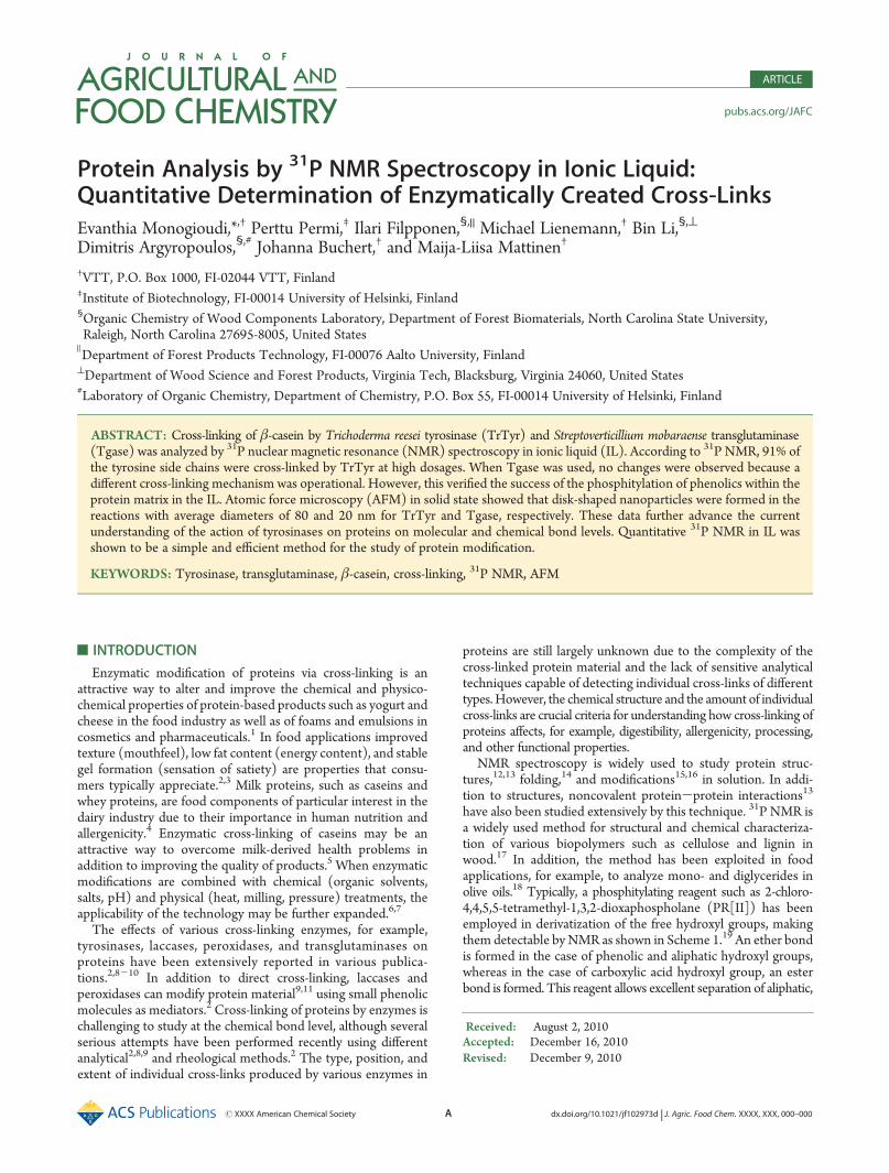
Figure 1A. Four well-resolved peaks were detected in addition tothe main component (elution volume = 120 mL). After that, allfractions were analyzed by SDS-PAGE for the MM evaluation(data not shown). The SDS-PAGE of the purest β-casein fractionis shown in Figure 1B (lane 3). The bands of the unpurifiedβ-casein are shown in lane 2. On the basis of the analysis, themain component of the fractionated β-casein is almost 100%pure β-casein. Comparison of the bands in lanes 2 and 3 to the
D dx.doi.org/10.1021/jf102973d |J. Agric. Food Chem. XXXX, XXX, 000–000
Journal of Agricultural and Food Chemistry ARTICLE
known MM marker (lane 1) confirmed this conclusion, as thetheoretical MM of β-casein is 24 kDa. To further confirm thepurity and MM of the purified fraction, it was analyzed byMALDI-TOF MS. As shown in Figure 1C, the correct mass,that is, 23.96 kDa for single-charged ion and 11.98 kDa fordouble-charged ion, was detected from the spectrum of purifiedβ-casein. No additional peaks were detected from the spectrum,verifying the purity of the main β-casein fraction.Solubilization of the Model Compounds and Proteins in
ILs. Although various organic solvents are commonly used toimprove the solubilization of different polymers,23 they are notapplicable for the solubilization of large proteins. In our studiesthe singe amino acids and short peptides could be solubilized inadequate concentrations, that is, 30 mg mL-1, but in the case ofβ-casein only ca. 10 mg could be solubilized in 1 mL of chloro-form in order to perform phosphitylation and subsequent31P NMR measurements. However, it is well-known that31P NMR is not a sensitive method and that it requires largeramounts of sample, typically at least ca. 40 mg (depending on thecontent of reactive groups) in a 1 mL volume to be effective.39
Thus, to overcome this problem, all model compounds as well asenzymatically polymerized β-caseins were dissolved in a solventsystem based on chloride anion and imidazolium cation, that is,in [amim]Cl, which has successfully been used to dissolve woodand other plant-based materials.17,21,28 Moreover, ILs have beenreported to stabilize the three-dimensional structure of variousproteins.40 However, Swatloski et al.27 have speculated that ILswith high chloride content may break the hydrogen bondspresent in cellulose, and hence it is believed that the samephenomenon may occur in the case of proteins such as β-casein.If that is the case, in our studies it can be considered as anadvantage, because the aim was to phosphitylate all nonreactedhydroxyl groups in cross-linked networked protein matrix. Allamino acids, peptides, and proteins used in this study were easilydissolved in [amim]Cl in adequate, that is, 80 mg in 1 mL,
concentration. However, [bmim]Cl, also tested for this purpose,was not suitable for this application due to its high melting pointand high viscosity (data not shown).Thus, it can be summarized that in the presence of imidazole-
based ILs, high concentrations of protein material can be dissolvedfor further chemical modifications. This provides an attractive basisalso for protein-based applications in addition to those described forwood components. In addition, ILs have high potential as greensolvents due to their ability to dissolve a wide variety of biopolymerscompared with conventional organic solvents.41
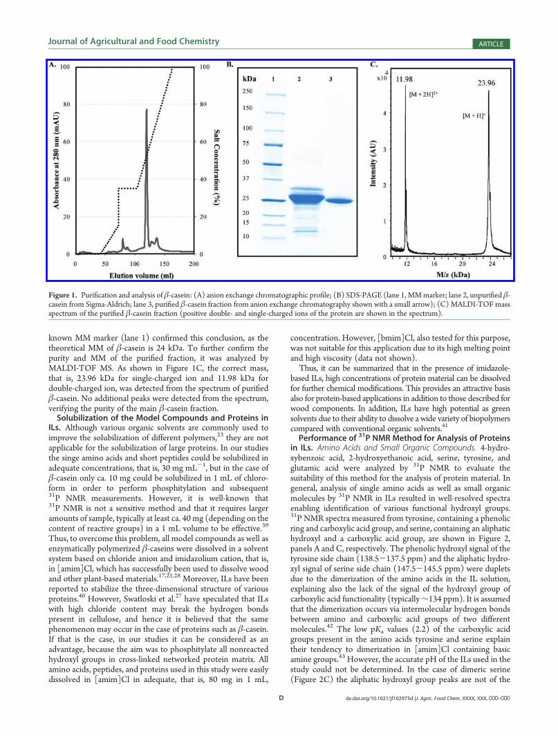
Performance of 31P NMR Method for Analysis of Proteinsin ILs. Amino Acids and Small Organic Compounds. 4-hydro-xybenzoic acid, 2-hydroxyethanoic acid, serine, tyrosine, andglutamic acid were analyzed by 31P NMR to evaluate thesuitability of this method for the analysis of protein material. Ingeneral, analysis of single amino acids as well as small organicmolecules by 31P NMR in ILs resulted in well-resolved spectraenabling identification of various functional hydroxyl groups.31P NMR spectra measured from tyrosine, containing a phenolicring and carboxylic acid group, and serine, containing an aliphatichydroxyl and a carboxylic acid group, are shown in Figure 2,panels A and C, respectively. The phenolic hydroxyl signal of thetyrosine side chain (138.5-137.5 ppm) and the aliphatic hydro-xyl signal of serine side chain (147.5-145.5 ppm) were dupletsdue to the dimerization of the amino acids in the IL solution,explaining also the lack of the signal of the hydroxyl group ofcarboxylic acid functionality (typically∼134 ppm). It is assumedthat the dimerization occurs via intermolecular hydrogen bondsbetween amino and carboxylic acid groups of two differentmolecules.42 The low pKa values (2.2) of the carboxylic acidgroups present in the amino acids tyrosine and serine explaintheir tendency to dimerization in [amim]Cl containing basicamine groups.43 However, the accurate pH of the ILs used in thestudy could not be determined. In the case of dimeric serine(Figure 2C) the aliphatic hydroxyl group peaks are not of the
Figure 1. Purification and analysis of β-casein: (A) anion exchange chromatographic profile; (B) SDS-PAGE (lane 1, MMmarker; lane 2, unpurified β-casein from Sigma-Aldrich; lane 3, purified β-casein fraction from anion exchange chromatography shown with a small arrow); (C) MALDI-TOF massspectrum of the purified β-casein fraction (positive double- and single-charged ions of the protein are shown in the spectrum).
E dx.doi.org/10.1021/jf102973d |J. Agric. Food Chem. XXXX, XXX, 000–000
Journal of Agricultural and Food Chemistry ARTICLE
same intensity. This difference could be attributed to the pre-sence of serine monomer having an internal hydrogen bondbetween amino and carboxylic acid groups. The peak ofthe monomeric aliphatic hydroxyl group is overlapping withthe peaks that have resulted from the dimer. The error for thequantitative results of the phenolic group of tyrosine andthe aliphatic group of serine side chains was <5%. The samephenomenon of dimerization was observed also in the case ofglutamic acid (Figure 2E) containing two carboxylic acid groups
having different pKa values, that is, 4.3 for the group in the aminoacid side chain and 2.2 for the group located next to the aminogroup. Also in this case, the signal resulting from the side-chaincarboxylic acid group was a duplet (135-133 ppm) yieldingquantitative results and having an average error of <5%. As in thiscase the dimerization via other carboxylic acid group was notstable and complete. Hence, a somewhat smaller duplet signal(∼135 ppm) appeared next to the side-chain peak (Figure 3A,mixture of different amino acids).
Figure 2. 31P NMR spectra measured from (A) tyrosine, (B) 4-hydroxybenzoic acid, (C) serine, (D) 2-hydroxyethanoic acid, and (E) glutamic acidsolubilized in ionic liquid. In addition to the chemical structures of the small model compounds, the assignments of the various phosphitylated functionalhydroxyl groups (aliphatic, phenolic, carboxylic acid, water, and internal standard, I.S.) are shown above the corresponding peaks. The positions of thecarboxylic acid hydroxyls that are not present due to dimerization are marked with an arrow. In the case of glutamic acid (E), the positions of the peaks ofthe carboxylic acid hydroxyl groups are marked by 1 and 2.
F dx.doi.org/10.1021/jf102973d |J. Agric. Food Chem. XXXX, XXX, 000–000
Journal of Agricultural and Food Chemistry ARTICLE
To verify the somewhat unexpected results described above, twosmall organicmolecules, that is, 4-hydroxybenzoic acid and 2-hydro-xyethanoic acid, were chosen as reference compounds for com-parison to tyrosine and serine. In the case of 4-hydroxybenzoic acidacid both reactive groups, that is, the phenolic hydroxyl and thecarboxylic acid hydroxyl group, could be successfully derivatizedby PR[II] reagent in the selected solvent system as shown inFigure 2B. A singlet peak was detected for each functional hydroxylgroup. The intensities of the peaks were almost identical, indicatingthat both functional groups reacted to an equal extent with thePR[II] reagent. No splitting was observed, due to the lack of anamino group in the molecule. In the case of 2-hydroxyethanoic acidboth of the reactive functional hydroxyl groups became visible in thespectrum as singlets at equal intensities, indicating no preferencebetween the hydroxyl groups as shown in Figure 2D. The quanti-fication of the various hydroxyl peaks revealed excellent results withan error of <1%. In contrast to tyrosine, serine, and glutamic acid, thecarboxylic acid groups of 4-hydroxybenzoic acid and 2-hydroxyetha-noic acid have much higher pKa values (4.48 and 3.83, respectively)than carboxylic acid groups next to amino groups in selected aminoacids. Thus, no dimerization occurred and the hydroxyls of thecarboxylic groups could be successfully derivatized by PR[II]reagent and detected as singlets in the spectra. In all spectra(Figures 2A-D), signals deriving from the phosphitylated residualwater and internal standard were detected.
31P NMR was also used for analyzing a mixture of threeselected amino acids (in equal concentrations) representingdifferent functionalities in the amino acid side chain. Thus,tyrosine, serine, and glutamic acid were dissolved efficiently inthe IL and analyzed by 31P NMR, as shown in Figure 3A. Thesignals in the spectrum measured from the mixture of amino
acids were identical to those measured for the individual aminoacids. The variation between the theoretical and experimentalvalues for the amount of reactive groups was again found to besmall, that is, <5% for amino acid side chains. In general, thechemical shifts of the functional groups derivatized by PR[II] inprotein material are in good agreement with the studies con-ducted by Wroblewski on the 31P NMR analysis of organiccompounds containing phenols, alcohols, and acids.44
Overall, with an average error of <5%, the quantitative 31P NMRof the amino acids and small molecules presents strong evidence forthe potential of the IL solvent system for quantification of phenolicstructures aswell as other hydroxyl groups in largemolecules such aspeptides and proteins as described below.Peptides. To further evaluate the possibility of using the IL
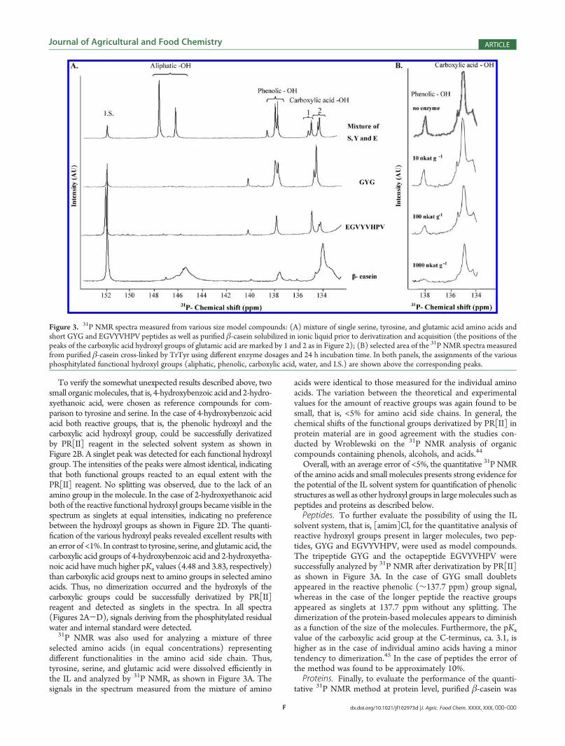
solvent system, that is, [amim]Cl, for the quantitative analysis ofreactive hydroxyl groups present in larger molecules, two pep-tides, GYG and EGVYVHPV, were used as model compounds.The tripeptide GYG and the octapeptide EGVYVHPV weresuccessfully analyzed by 31P NMR after derivatization by PR[II]as shown in Figure 3A. In the case of GYG small doubletsappeared in the reactive phenolic (∼137.7 ppm) group signal,whereas in the case of the longer peptide the reactive groupsappeared as singlets at 137.7 ppm without any splitting. Thedimerization of the protein-based molecules appears to diminishas a function of the size of the molecules. Furthermore, the pKa
value of the carboxylic acid group at the C-terminus, ca. 3.1, ishigher as in the case of individual amino acids having a minortendency to dimerization.45 In the case of peptides the error ofthe method was found to be approximately 10%.Proteins. Finally, to evaluate the performance of the quanti-
tative 31P NMR method at protein level, purified β-casein was
Figure 3. 31P NMR spectra measured from various size model compounds: (A) mixture of single serine, tyrosine, and glutamic acid amino acids andshort GYG and EGVYVHPV peptides as well as purified β-casein solubilized in ionic liquid prior to derivatization and acquisition (the positions of thepeaks of the carboxylic acid hydroxyl groups of glutamic acid are marked by 1 and 2 as in Figure 2); (B) selected area of the 31P NMR spectra measuredfrom purified β-casein cross-linked by TrTyr using different enzyme dosages and 24 h incubation time. In both panels, the assignments of the variousphosphitylated functional hydroxyl groups (aliphatic, phenolic, carboxylic acid, water, and I.S.) are shown above the corresponding peaks.
G dx.doi.org/10.1021/jf102973d |J. Agric. Food Chem. XXXX, XXX, 000–000
Journal of Agricultural and Food Chemistry ARTICLE
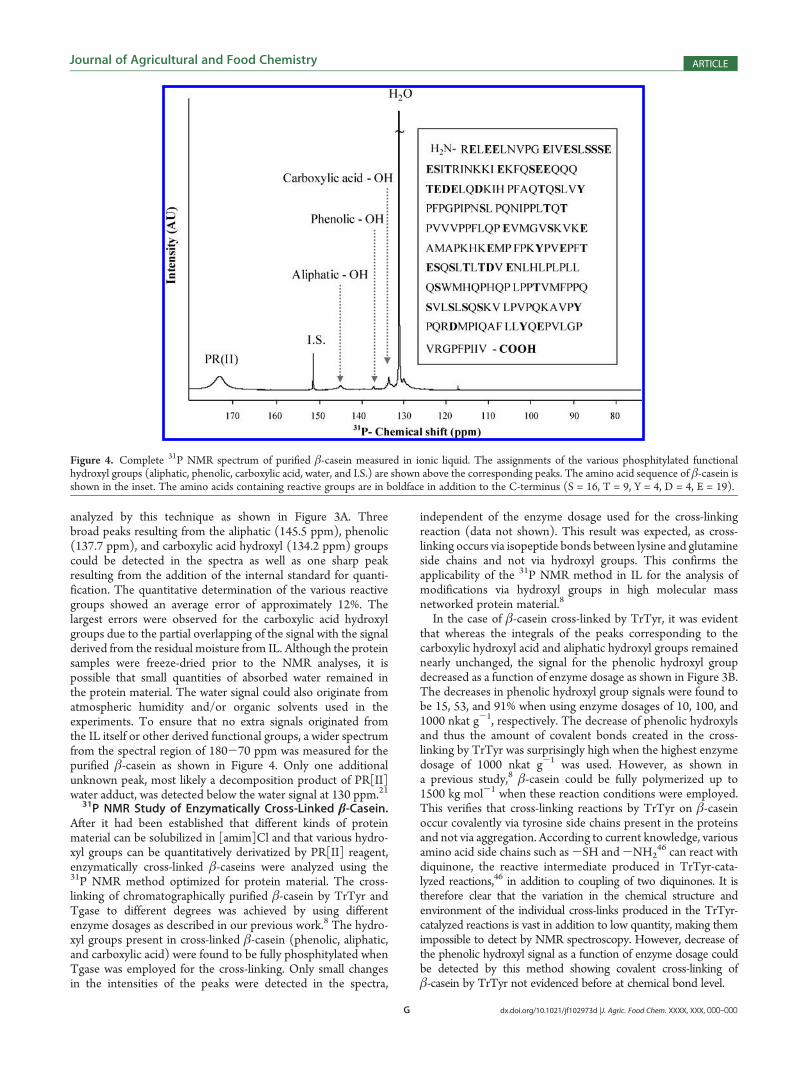
analyzed by this technique as shown in Figure 3A. Threebroad peaks resulting from the aliphatic (145.5 ppm), phenolic(137.7 ppm), and carboxylic acid hydroxyl (134.2 ppm) groupscould be detected in the spectra as well as one sharp peakresulting from the addition of the internal standard for quanti-fication. The quantitative determination of the various reactivegroups showed an average error of approximately 12%. Thelargest errors were observed for the carboxylic acid hydroxylgroups due to the partial overlapping of the signal with the signalderived from the residual moisture from IL. Although the proteinsamples were freeze-dried prior to the NMR analyses, it ispossible that small quantities of absorbed water remained inthe protein material. The water signal could also originate fromatmospheric humidity and/or organic solvents used in theexperiments. To ensure that no extra signals originated fromthe IL itself or other derived functional groups, a wider spectrumfrom the spectral region of 180-70 ppm was measured for thepurified β-casein as shown in Figure 4. Only one additionalunknown peak, most likely a decomposition product of PR[II]water adduct, was detected below the water signal at 130 ppm.21
31P NMR Study of Enzymatically Cross-Linked β-Casein.After it had been established that different kinds of proteinmaterial can be solubilized in [amim]Cl and that various hydro-xyl groups can be quantitatively derivatized by PR[II] reagent,enzymatically cross-linked β-caseins were analyzed using the31P NMR method optimized for protein material. The cross-linking of chromatographically purified β-casein by TrTyr andTgase to different degrees was achieved by using differentenzyme dosages as described in our previous work.8 The hydro-xyl groups present in cross-linked β-casein (phenolic, aliphatic,and carboxylic acid) were found to be fully phosphitylated whenTgase was employed for the cross-linking. Only small changesin the intensities of the peaks were detected in the spectra,
independent of the enzyme dosage used for the cross-linkingreaction (data not shown). This result was expected, as cross-linking occurs via isopeptide bonds between lysine and glutamineside chains and not via hydroxyl groups. This confirms theapplicability of the 31P NMR method in IL for the analysis ofmodifications via hydroxyl groups in high molecular massnetworked protein material.8
In the case of β-casein cross-linked by TrTyr, it was evidentthat whereas the integrals of the peaks corresponding to thecarboxylic hydroxyl acid and aliphatic hydroxyl groups remainednearly unchanged, the signal for the phenolic hydroxyl groupdecreased as a function of enzyme dosage as shown in Figure 3B.The decreases in phenolic hydroxyl group signals were found tobe 15, 53, and 91% when using enzyme dosages of 10, 100, and1000 nkat g-1, respectively. The decrease of phenolic hydroxylsand thus the amount of covalent bonds created in the cross-linking by TrTyr was surprisingly high when the highest enzymedosage of 1000 nkat g-1 was used. However, as shown ina previous study,8 β-casein could be fully polymerized up to1500 kg mol-1 when these reaction conditions were employed.This verifies that cross-linking reactions by TrTyr on β-caseinoccur covalently via tyrosine side chains present in the proteinsand not via aggregation. According to current knowledge, variousamino acid side chains such as-SH and-NH2
46 can react withdiquinone, the reactive intermediate produced in TrTyr-cata-lyzed reactions,46 in addition to coupling of two diquinones. It istherefore clear that the variation in the chemical structure andenvironment of the individual cross-links produced in the TrTyr-catalyzed reactions is vast in addition to low quantity, making themimpossible to detect by NMR spectroscopy. However, decrease ofthe phenolic hydroxyl signal as a function of enzyme dosage couldbe detected by this method showing covalent cross-linking ofβ-casein by TrTyr not evidenced before at chemical bond level.
Figure 4. Complete 31P NMR spectrum of purified β-casein measured in ionic liquid. The assignments of the various phosphitylated functionalhydroxyl groups (aliphatic, phenolic, carboxylic acid, water, and I.S.) are shown above the corresponding peaks. The amino acid sequence of β-casein isshown in the inset. The amino acids containing reactive groups are in boldface in addition to the C-terminus (S = 16, T = 9, Y = 4, D = 4, E = 19).
H dx.doi.org/10.1021/jf102973d |J. Agric. Food Chem. XXXX, XXX, 000–000
Journal of Agricultural and Food Chemistry ARTICLE
The 31P NMR spectra of nonpurified β-casein cross-linked byTrTyr were also analyzed (data not shown). As expected, theoverall pattern of the spectra was identical to that of the purifiedβ-casein cross-linked by TrTyr. However, the calculated cross-linking efficiencies were different, as the commercial β-caseincontains some impurities such as whey proteins that cannot becross-linked by this enzyme.8 Specifically, the data for the phenolicgroups in impure β-casein cross-linked using TrTyr dosages of 10,100, and 1000 nkat g-1 were 12, 23, and 68%, respectively. In thecase of the lowest enzyme dosage the results with the nonpurifiedand purified β-casein were almost identical. However, when using
the highest TrTyr dosage and nonpurified β-casein, the phenolicsignal was only 75% of that obtained using chromatographicallypurified protein as substrate. This is most probably due to thepresence of the above-mentioned whey proteins that could not becross-linked by TrTyr.8 Overall, the results obtained by 31P NMRmethod are compatible with our previous findings.8
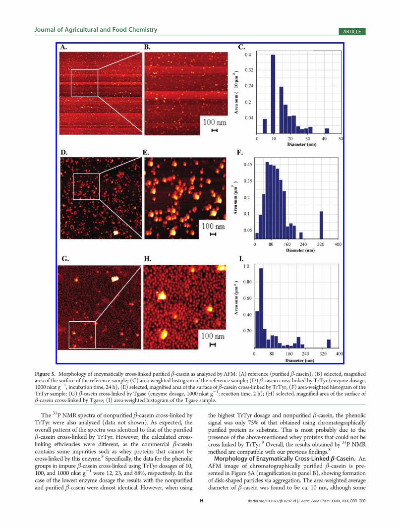
Morphology of Enzymatically Cross-Linked β-Casein. AnAFM image of chromatographically purified β-casein is pre-sented in Figure 5A (magnification in panel B), showing formationof disk-shaped particles via aggregation. The area-weighted averagediameter of β-casein was found to be ca. 10 nm, although some
Figure 5. Morphology of enzymatically cross-linked purified β-casein as analyzed by AFM: (A) reference (purified β-casein); (B) selected, magnifiedarea of the surface of the reference sample; (C) area-weighted histogram of the reference sample; (D) β-casein cross-linked by TrTyr (enzyme dosage,1000 nkat g-1; incubation time, 24 h); (E) selected, magnified area of the surface of β-casein cross-linked by TrTyr; (F) area-weighted histogram of theTrTyr sample; (G) β-casein cross-linked by Tgase (enzyme dosage, 1000 nkat g-1; reaction time, 2 h); (H) selected, magnified area of the surface ofβ-casein cross-linked by Tgase; (I) area-weighted histogram of the Tgase sample.

I dx.doi.org/10.1021/jf102973d |J. Agric. Food Chem. XXXX, XXX, 000–000
Journal of Agricultural and Food Chemistry ARTICLE
larger particles with maximum diameter ca. 42 nm were alsodetected as shown in Figure 5C. The rms roughness fornontreated purified β-casein was 0.2 nm, indicating the forma-tion of a smooth protein surface on the mica.When β-casein was cross-linked by TrTyr using the lowest
enzyme dosage (10 nkat g-1), disk-shaped particles resemblingthe control sample were formed, and no linear polymerizedstructures as in the case of small phenolic compounds47 weredetected from the AFM surface (data not shown). However, theaverage diameter of these particles was approximately 2-foldhigher, that is, 19 nm, than that of the control sample (non-cross-linked β-casein), showing activity of the enzyme. Unexpectedly, a10-fold increase in the enzyme dosage (100 nkat g-1) resulted inslightly smaller particles having an average diameter of around15 nm (data not shown), suggesting nonlinear proceeding of theenzymatic reaction. When β-casein was cross-linked with thehighest enzyme dosage (1000 nkat g-1), large particles wereformed as shown in Figure 5D. The formation of disk-shapedcross-linked particles is evident in the magnified Figure 5E. Inthis case the average diameter of the dried cross-linked β-caseinparticles was 80 nm. However, larger particles were also formedwith a diameter of approximately 340 nm (Figure 5F). Theroughnesses rms values were 0.2, 1.3, and 2.0 nm, respectively,when TrTyr dosages of 10, 100, and 1000 nkat g-1 were used forthe cross-linking of β-casein. The rms roughness of β-caseincross-linked by 10 nkat g-1 of TrTyr was equal to that ofuntreated β-casein. This correlates well with our previous results,showing that only some polymerization occurred when using thelowest enzyme dosage.8
When β-casein was cross-linked by the reference enzyme,Tgase, the lowest enzyme dosage of 10 nkat g-1 also resulted indisk-shaped particles of 22 nm average diameter (data notshown). Use of the intermediate enzyme dosage (100 nkatg-1) resulted in somewhat larger particles with an averagediameter of 39 nm (data not shown). The analysis of β-caseincross-linked by Tgase using an enzyme dosage of 1000 nkat g-1
resulted in an average particle diameter of 20 nm as shown inFigure 5G (magnification in Figure 5H). In addition to theseuniform and rather homogeneous nanoparticles, as also reportedin our previous studies,8 larger aggregates of approximately360 nmwere formed in the presence of the highest Tgase dosage.The rms roughness of the sample surfaces appeared to increasealong with increasing enzyme dosage, that is, 0.3, 0.9, and 1.5 nm.In the case of 10 nkat g-1 of Tgase, the rms roughness is slightlyhigher than that of untreated β-casein, correlating with ourprevious data according to which β-casein cross-linked by Tgase(10 nkat g-1 for 2 h) was only slightly polymerized.8 In general,the cross-linking of β-casein by Tgase resulted in small particleswith an average diameter of 20 nm regardless of the enzymedosage used for the cross-linking reaction. Tight cross-linking ofβ-casein into spherical nanoparticles by Tgase can easily beunderstood, as β-casein contains many reactive sites for trans-glutaminase, that is, 11 lysines and 20 glutamines, enablingextensive cross-linking as evidenced earlier.8 The radius ofgyration obtained for the cross-linked protein particles was rathersmall for such a large cross-linked polymer. The elution order ofcross-linked β-casein in size exclusion chromatography (SEC)obtained using 1000 nkat g-1 enzyme dosage and 2, 6, and 24 hincubation times also supported the above conclusions, as thecross-linked material produced in the extreme reaction condi-tions eluted last. Thus, it can be postulated that the protein net-work formed after Tgase treatment is very tight, also indicating
the formation of a large number of intramolecular cross-links inaddition to intermolecular cross-links responsible for the poly-merization reactions.In general, the apparent radii of gyration (Rg) were smaller for
all enzymatically cross-linked samples when analyzed by AFM incomparison to the values obtained in our previous studies bySEC-UV/vis-MALLS.8 In SEC-MALLS the hydrodynamic dia-meter of the polymer is determined by dynamic light scattering inthe solution. By contrast, in AFM the polymer samples are driedon the mica surface, and enzymatically cross-linked protein parti-cles may shrink during the drying. A similar phenomenon wasreported by Martin et al. for casein micelles analyzed by SEM.48
It has also been shown that cross-linking of β-casein by Tgaseusing a dosage of 1000 nkat g-1 forms products which are slowlydigested by the pepsin, in comparison to β-casein cross-linkedby TrTyr or native β-casein.37 These observations supportthe above AFM results as the protein particles formed in thecross-linking by Tgase had much smaller diameter and tightermorphology when compared to the particles formed by TrTyr.Thus, it is suggested that the cross-linking by Tgase largely occursvia intramolecular bonding. By contrast, the cross-linking ofβ-casein by TrTyr produced particles with larger diameter, thusindicating the predominant role of intermolecular bonding asβ-casein contains only four reactive sites, that is, tyrosine residuesfor cross-linking by TrTyr.It has been shown that various protein materials can be
dissolved in chloride-based IL for further analysis of enzymatic(as well as chemical) modifications by 31PNMR spectroscopy. Inaddition, it was verified that the method can be applied forquantitative determination of aliphatic, phenolic, and carboxylicacid hydroxyls present in native and cross-linked proteins. Theextent of cross-linked phenolics in β-casein, that is, 91% pro-duced by TrTyr, was surprisingly high, showing on a chemicalbond level that nearly all tyrosine residues are involved in thecross-linking reactions. AFM results indicated on a molecularlevel that the cross-linking of β-casein by TrTyr largely occurs viaintermolecular bonding, whereas Tgase was found to formlargely also via intramolecular cross-links. The data are importantfor exploitation of tyrosinases in different applications as com-plete cross-linking of random coil protein was confirmed.
’ABBREVIATIONS USED
AFM, atomic force microscopy; [amim]Cl, 1-allyl-3-methyli-midazolium chloride; [bmim]Cl, 1-butyl-3-methylimidazoliumchloride; 31P NMR, phosphorus nuclear magnetic resonance;Cr(acac)3, chromium(III) acetylacetonate; PR[II], 2-chloro-4,4,5,5-tetramethyl-1,3,2-dioxaphospholane; IL, ionic liquid;K, lysine; Q, glutamine; S, serine; E, glutamic acid; Y, tyrosine;D, aspartate;MALDI-TOFMS,matrix-assisted laser desorption/ionization-time-of-flight mass spectroscopy; MM, molecularmass; L-DOPA, L-3,4-dihydroxyphenylalanine; Rg, radius ofgyration; rms, root-mean-square; SDS-PAGE, sodium dodecylsulfate-polyacrylamide gel electrophoresis; SEC, size exclusionchromatography; SEM; scanning electron microscopy; VTT,Technical Research Center of Finland.
’AUTHOR INFORMATION
Corresponding Author*Phone: þ358 400 35 7279; fax: þ358 20 722 7071; e-mail:[email protected].
J dx.doi.org/10.1021/jf102973d |J. Agric. Food Chem. XXXX, XXX, 000–000
Journal of Agricultural and Food Chemistry ARTICLE
Funding SourcesThe study was financed by Marie Curie mobility actions as partof the EU project MEST-CT-2005-020924. The work was alsosupported by LIPFUN (“Discovery and exploitation of novellipid modifying enzymes in industrial processes”), funded byTekes, and SA-CrossFold (“Enzymatic cross-linking of foodproteins: impact of food protein folding on the mode of actionof cross-linking enzymes”), funded by the Academy of Finlandand the ABS graduate school at the University of Helsinki(Finland).
’ACKNOWLEDGMENT
We thank Dr. Jarmo Ropponen (VTT, Finland) for thesynthesis of ionic liquid and acknowledge Dr. Alistair King fortechnical assistance relating to the NMR experiments performedin the organic solvents. Furthermore, we warmly thank Olli Aitiofor technical assistance with the various NMR softwares.
’REFERENCES
(1) Buchert, J.; Selinheimo, E.; Kruus, K.; Mattinen, M. L.; Lantto,R.; Autio, K. Using cross-linking enzymes to improve textural and otherproperties of food. In Novel Enzyme Technology for Food Applications;Rastall, R., Ed.; Woodhead Publishing: Cambridge, U.K., 2007; pp 118.(2) Ercili Cura, D.; Lantto, R.; Lille, M.; Andberg, M.; Kruus, K.;
Buchert, J. Laccase-aided protein modification: effects on the struc-tural properties of acidified sodium caseinate gels. Int. Dairy J. 2009, 19,737–745.(3) Nieuwenhuizen, W. F.; Dekker, H. L.; deKoning, L. J.;
Groneveld, T.; deKoster, C. G.; deJong, G. A. H. Modification ofglutamine and lysine residues in holo and apo R-lactalbumin withmicrobial transglutaminase. J. Agric. Food Chem. 2003, 51, 7132–7139.(4) Stanic, D.; Monogioudi, E.; Ercili, D.; Radosavljevic, J.;
Atanaskovic-Markovic, M.; Vuckovic, O.; Raija, L.; Mattinen, M.;Buchert, J.; Velickovic, T. C. Digestibility and allergenicity assessmentof enzymatically crosslinked β-casein. Mol. Nutr. Food Res. 2010,54, 1–12.(5) Buchert, J.; Ercili-Cura, D.; Ma, H.; Gasparetti, C.; Monogioudi,
E.; Faccio, G.; Mattinen, M.; Boer, H.; Partanen, R.; Selinheimo, E.;Lantto, R.; Kruus, K. Crosslinking food proteins for improved function-ality. Annu. Rev. Food Sci. Technol. 2010, 1, 113–138.(6) Hern�andez-Balada, E.; Taylor, M. M.; Phillips, J. G.; Marmer,
W. N.; Brown, E. M. Properties of biopolymers produced by trans-glutaminase treatment of whey protein isolate and gelatin. Bioresour.Technol. 2009, 100, 3638–3643.(7) Silveira, W. B.; Passos, F. J. V.; Mantovani, H. C.; Passos, F. M. L.
Ethanol production from cheese whey permeate by Kluyveromycesmarxianus UFV-3: a flux analysis of oxido-reductive metabolism as afunction of lactose concentration and oxygen levels. Enzyme Microb.Technol. 2005, 36, 930–936.(8) Monogioudi, E.; Creusot, N.; Kruus, K.; Gruppen, H.; Buchert,
J.; Mattinen, M. Cross-linking of β-casein by Trichoderma reesei tyrosi-nase and Streptoverticillium mobaraense transglutaminase followed bySEC-MALLS. Food Hydrocolloids 2009, 23, 2008–2015.(9) Mattinen, M.; Hellman, M.; Steffensen, C. L.; Selinheimo, E.;
Permi, P.; Kalkkinen, N.; Kruus, K.; Buchert, J. Laccase and tyrosinasecatalysed polymerization of proteins and peptides. J. Biotechnol. 2008,136, S318–S318.(10) Steffensen, C.; Mattinen, M.; Andersen, H.; Kruus, K.; Buchert,
J.; Nielsen, J. Cross-linking of tyrosine-containing peptides by hydrogenperoxide-activated Coprinus cinereus peroxidase. Eur. Food Res. Technol.2008, 227, 57–67.(11) Oudgenoeg, G.; Hilhorst, R.; Piersma, S. R.; Boeriu, C. G.;
Gruppen, H.; Hessing, M.; Voragen, A. G. J.; Laane, C. Peroxidase-mediated cross-linking of a tyrosine-containing peptide with ferulic acid.J. Agric. Food Chem. 2001, 49, 2503–2510.
(12) Ma, Z.; Dunn, B. C.; Turpin, G. C.; Eyring, E. M.; Ernst, R. D.;Pugmire, R. J. Solid state NMR investigation of silica aerogel supportedFischer-Tropsch catalysts. Fuel Process. Technol. 2007, 88, 29–33.
(13) Zuiderweg, E. R. P. Mapping protein-protein interactions insolution by NMR spectroscopy. Biochemistry 2002, 41, 1–7.
(14) Sakurai, K.; Konuma, T.; Yagi, M.; Goto, Y. Structural dynamicsand folding of β-lactoglobulin probed by heteronuclear NMR. Biochim.Biophys. Acta (BBA): Gen. Subjects 2009, 1790, 527–537.
(15) Scheidt, H. A.; Huster, D. Structure and dynamics of themyristoyl lipid modification of Src peptides determined by 2H solid-state NMR spectroscopy. Biophys. J. 2009, 96, 3663–3672.
(16) Vogel, A.; Reuther, G.; Roark, M. B.; Tan, K.; Waldmann, H.;Feller, S. E.; Huster, D. Backbone conformational flexibility of the lipidmodified membrane anchor of the human N-Ras protein investigated bysolid-state NMR and molecular dynamics simulation. Biochim. Biophys.Acta (BBA): Biomembranes 2010, 1798, 275–285.
(17) King, A. W. T.; Zoia, L.; Filpponen, I.; Olszewska, A.; Xie,H.; Kilpel€ainen, I.; Argyropoulos, D. S. In situ determination of ligninphenolics and wood solubility in imidazolium chlorides using 31P NMR.J. Agric. Food Chem. 2009, 57, 8236–8243.
(18) Spyros, A.; Dais, P. Application of 31P NMR spectroscopyin food analysis. 1. Quantitative determination of the mono- anddiglyceride composition of olive oils. J. Agric. Food Chem. 2000, 48,802–805.
(19) Granata, A.; Argyropoulos, D. S. 2-Chloro-4,4,5,5-tetramethyl-1,3,2-dioxaphospholane, a reagent for the accurate determination of theuncondensed and condensed phenolic moieties in lignins. J. Agric. FoodChem. 1995, 43, 1538–1544.
(20) Jiang, Z. H.; Argyropoulos, D. S.; Granata, A. Correlationanalysis of 31P NMR chemical shifts with substituent effects of phenols.Magn. Reson. Chem. 1995, 33, 375–382.
(21) King, A. W. T.; Kilpel€ainen, I.; Heikkinen, S.; J€arvi, P.;Argyropoulos, D. S. Hydrophobic interactions determining functiona-lized lignocellulose solubility in dialkylimidazolium chlorides, as probedby 31P NMR. Biomacromolecules 2009, 10, 458–463.
(22) Guilloteau, J.; Rie’s-Kautt, M. M.; Ducruix, A. F. Variation oflysozyme solubility as a function of temperature in the presence oforganic and inorganic salts. J. Cryst. Growth 1992, 122, 223–230.
(23) Serebryakova, L. T.; Zorin, N. A.; Karyakin, A. A. Improvementof hydrogenase enzyme activity by water-miscible organic solvents.Enzyme Microb. Technol. 2009, 44, 329–333.
(24) Huddleston, J. G.; Visser, A. E.; Reichert, W. M.; Willauer,H. D.; Broker, G. A.; Rogers, R. D. Characterization and comparison ofhydrophilic and hydrophobic room temperature ionic liquids incorpor-ating the imidazolium cation. Green Chem. 2001, 3, 156.
(25) Junghans, F.; Morawietz, M.; Conrad, U.; Scheibel, T.;Heilmann, A.; Spohn, U. Preparation and mechanical properties oflayers made of recombinant spider silk proteins and silk from silk worm.Appl. Phys. A: Mater. Sci. Process. 2006, 82, 253–260.
(26) Granstr€om, M.; Kavakka, J.; King, A.; Majoinen, J.; M€akel€a, V.;Helaja, J.; Hietala, S.; Virtanen, T.; Maunu, S.; Argyropoulos, D.;Kilpel€ainen, I. Tosylation and acylation of cellulose in 1-allyl-3-methy-limidazolium chloride. Cellulose 2008, 15, 481–488.
(27) Swatloski, R. P.; Spear, S. K.; Holbrey, J. D.; Rogers, R. D.Dissolution of cellose with ionic liquids. J. Am. Chem. Soc. 2002, 124,4974–4975.
(28) Mattinen, M.; Filpponen, I.; J€arvinen, R.; Li, B.; Kallio, H.;Lehtinen, P.; Argyropoulos, D. Structure of the polyphenolic com-ponent of suberin isolated from potato (Solanum tuberosum var. Nikola).J. Agric. Food Chem. 2009, 57, 9747–9753.
(29) L€aemmli, U. K. Cleavage of structural proteins during theassembly of the head of bacteriophage T4. Nature 1970, 227, 680–685.
(30) Selinheimo, E.; Saloheimo, M.; Ahola, E.; Westerholm-Parvinen,A.; Kalkkinen,N.; Buchert, J.; Kruus, K. Production and characterization of asecreted, C-terminally processed tyrosinase from the filamentous fungusTrichoderma reesei. FEBS J. 2006, 273, 4322–4335.
(31) Lantto, R.; Puolanne, E.; Kalkkinen, N.; Buchert, J.; Autio, K.Enzyme-aided modification of chicken-breast myofibril proteins: effect
K dx.doi.org/10.1021/jf102973d |J. Agric. Food Chem. XXXX, XXX, 000–000
Journal of Agricultural and Food Chemistry ARTICLE
of laccase and transglutaminase on gelation and thermal stability. J. Agric.Food Chem. 2005, 53, 9231–9237.(32) Robb, D. A. Tyrosinase. InCopper Proteins and Copper Enzymes;
Lontie, R., Ed.; CRC Press: Boca Raton, FL, 1984; Vol. II, pp 207-240.(33) Folk, J. E. [127] Transglutaminase (guinea pig liver). In
Methods in Enzymology, Herbert Tabor and; Tabor, H., Tabor, C. W.,Eds.; Academic Press: New York, 1970; Vol. 17, Part 1, pp 889-894.(34) Selinheimo, E.; NiEidhin, D.; Steffensen, C.; Nielsen, J.;
Lomascolo, A.; Halaouli, S.; Record, E.; O’Beirne, D.; Buchert, J.; Kruus,K. Comparison of the characteristics of fungal and plant tyrosinases.J. Biotechnol. 2007, 130, 471–480.(35) Dyson, P. J.; Grossel, M. C.; Srinivasan, N.; Vine, T.; Welton,
T.; Williams, D. J.; White, A. J. P.; Zigras, T. Organometallic synthesisin ambient temperature chloroaluminate(III) ionic liquids. Ligandexchange reactions of ferrocene. Dalton Trans. 1997, 3465–3469.(36) Xia, Z.; Akim, L. G.; Argyropoulos, D. S. Quantitative 13CNMR
analysis of lignins with internal standards. J. Agric. Food Chem. 2001, 49,3573–3578.(37) Monogioudi, E.; Faccio, G.; Lille, M.; Poutanen, K.; Buchert, J.;
Mattinen, M. Effect of enzymatic cross-linking of β-casein on proteolysisby pepsin. Food Hydrocolloids 2011, 25 (1), 71–81.(38) Kauf, A. C.W.; Kensinger, R. S. Purification of porcine β-casein,
N-terminal sequence, quantification in mastitic milk. J. Anim. Sci. 2002,80, 1863–1870.(39) Veronesi, M.; Dalvit, C. 19F NMR spectroscopy for functional
and binding. High-throughput screening. InModernMagnetic Resonance;Graham, A. W., Ed.; Springer: Dordrecht, The Netherlands, 2008;Vol. 5, subpart 28, pp 1393-1399.(40) Fujita, K.; Forsyth, M.; MacFarlane, D. R.; Reid, R. W.; Elliott,
G. D. Unexpected improvement in stability and utility of cytochrome cby solution in biocompatible ionic liquids. Biotechnol. Bioeng. 2006,94, 1209–1213.(41) Yang, Z.; Pan, W. Ionic liquids: green solvents for nonaqueous
biocatalysis. Enzyme Microb. Technol. 2005, 37, 19–28.(42) Panyushkin, V. T.; Vashchuk, A. V.;Marusov, M. A. Application
of NMR to the investigation of intramolecular hydrogen bonds in aminoacids. J. Struct. Chem. 1996, 37, 343–346.(43) Thar, J.; Brehm, M.; Seitsonen, A. P.; Kirchner, B. Unexpected
hydrogen bond dynamics in imidazolium-based ionic liquids. J. Phys.Chem. B 2009, 113, 15129–15132.(44) Wroblewski, A. E.; Lensink, C.; Markuszewski, R.; Verkade,
J. G. 31P NMR spectroscopic analysis of coal pyrolysis condensates andextracts for heteroatom functionalities possessing labile hydrogen.Energy Fuels 1988, 2, 765–774.(45) Stryer, L. Biochemistry, 4th ed.; W. H. Freeman: New York,
1997.(46) Ito, S.; Kato, T.; Shinpo, K.; Fujita, K. Oxidation of tyrosine
residues in proteins by tyrosinase. Formation of protein-bonded 3,4-dihydroxyphenylalanine and 5-S-cysteinyl-3,4-dihydroxyphenylalanine.Biochem. J. 1984, 222, 407–411.(47) Desentis-Mendoza, R.; Hernandez-Sanchez, H.; Moreno, A.;
Rojas, D. C.; Chel-Guerrero, L.; Tamariz, J.; Jaramillo-Flores, M.Enzymatic polymerization of phenolic compounds using laccase andtyrosinase fromUstilago maydis. Biomacromolecules 2006, 7, 1845–1854.(48) Martin, A. H.; Douglas Goff, H.; Smith, A.; Dalgleish, D. G.
Immobilization of casein micelles for probing their structure andinteractions with polysaccharides using scanning electron microscopy(SEM). Food Hydrocolloids 2006, 20, 817–824.
’NOTE ADDED AFTER ASAP PUBLICATION
Figure 2 was modified in the version of this paper publishedJanuary 10, 2011. The correct version published January 19,2011.