Proprioceptive Guidance of Saccades in Eye-Hand Coordination
63
1 Proprioceptive Guidance of Saccades in Eye-Hand Coordination L. Ren 1,2,5 , AZ. Khan 1,3,5 , G. Blohm 1,2,5 , DYP. Henriques 1,2,5 , LE. Sergio 1,2,5 , and JD. Crawford 1,2,3,4,5 1 Centre for Vision Research, 2 Departments of Kinesiology and Health Science, 3 Psychology and 4 Biology, York University, 4700 Keele Street, Toronto, ON, Canada, M3J1P3, 5 CIHR group for action and perception, Ottawa, ON, Canada, K1A 0W9. Running head: Saccades and Limb Proprioception The total number of pages: 63 The number of pages of main text: 31 The number of tables: 2 The number of figures: 11 Corresponding author: Dr. J.D. Crawford Rm. 0003F, Computer Science and Engineering Bldg, Centre for Vision Research, 4700 Keele Street, York University, Toronto, ON, Canada, M3J 1P3 Tel: 416-736-2100 ext. 88621 Fax: 416-736-5814 E-mail: [email protected] Page 1 of 63 Articles in PresS. J Neurophysiol (May 17, 2006). doi:10.1152/jn.01012.2005 Copyright © 2006 by the American Physiological Society.
Transcript of Proprioceptive Guidance of Saccades in Eye-Hand Coordination

1
Proprioceptive Guidance of Saccades in Eye-Hand Coordination L. Ren1,2,5, AZ. Khan1,3,5, G. Blohm1,2,5, DYP. Henriques1,2,5, LE. Sergio1,2,5, and JD. Crawford1,2,3,4,5
1 Centre for Vision Research, 2 Departments of Kinesiology and Health Science, 3 Psychology
and 4 Biology, York University, 4700 Keele Street, Toronto, ON, Canada, M3J1P3, 5 CIHR
group for action and perception, Ottawa, ON, Canada, K1A 0W9.
Running head: Saccades and Limb Proprioception
The total number of pages: 63
The number of pages of main text: 31
The number of tables: 2
The number of figures: 11
Corresponding author: Dr. J.D. Crawford
Rm. 0003F, Computer Science and Engineering Bldg, Centre for Vision Research, 4700 Keele Street, York University, Toronto, ON, Canada, M3J 1P3 Tel: 416-736-2100 ext. 88621 Fax: 416-736-5814 E-mail: [email protected]
Page 1 of 63 Articles in PresS. J Neurophysiol (May 17, 2006). doi:10.1152/jn.01012.2005
Copyright © 2006 by the American Physiological Society.

2
ABSTRACT
The saccade generator updates memorized target representations for saccades during eye
and head movements. Here, we tested if proprioceptive feedback from the arm can also update
hand-held object locations for saccades, and what intrinsic coordinate system(s) are used in this
transformation. We measured radial saccades beginning from a central light-emitting diode to
sixteen target locations arranged peripherally in eight directions and two eccentricities on a
horizontal plane in front of subjects. Target locations were either indicated by 1) a visual flash, 2)
by subject actively moving the hand-held central target to a peripheral location, 3) by the
experimenter passively moving the subject’s hand, 4) through a combination of the above
proprioceptive and visual stimuli. Saccade direction was relatively accurate, but subjects showed
task-dependent systematic overshoots and variable errors in radial amplitude. Visually-guided
saccades showed the smallest overshoot, followed by saccades guided by both vision and
proprioception, while proprioceptively-guided saccades showed the largest overshoot. In most
tasks, the overall distribution of saccade endpoints was shifted and expanded in a gaze- or head-
centered cardinal coordinate system. However, the active proprioception task produced a tilted
pattern of errors, apparently weighted toward a limb-centered coordinate system. This suggests
the saccade generator receives an efference copy of the arm movement command but fails to
compensate for the arm’s inertia-related directional anisotropy. Thus, the saccade system is able
to transform hand-centered somatosensory signals into oculomotor coordinates and combine
somatosensory signals with visual inputs, but it appears to have a poorly calibrated internal
model of limb properties.
KEY WORDS: reference frame, feedback, efference copy, parietal cortex, optimal inference.
Page 2 of 63

3
INTRODUCTION
Human behavior consists largely of a succession of actions that require eye-hand
coordination. Saccade target locations can be derived from a variety of sensory inputs, such as
visual, auditory, and somatosensory information (Bridgeman 1995; Deubel et al. 1998, Pouget et
al. 2002), which provide the brain with moment-to-moment information about the location and
motion of external targets. Since these internal representations appear to be mainly stored in
egocentric coordinates, they must also be updated when the body itself moves (Helmholtz 1962;
Stark and Bridgeman 1983). For example, it is well known that gaze-centered representations of
remembered target locations for subsequent saccades are updated constantly during eye
movements (Colby et al., 1995; Duhamel et al., 1992; Gottlieb et al., 1998; Khan et al. 2005;
Medendorp et al., 2003). However, this is not the only situation where the target estimate within
the oculomotor system needs to be updated due to self motion. For example, during manual
manipulation of an object, our own hand movements often displace the target with respect to the
eye. Currently, it is not known if, or how accurately, humans can use limb proprioception to
internally update oculomotor representations in this situation.
Gaze behavior is closely intertwined with hand movements (Abrams et al., 1990; Helsen
et al., 2000; Neggers & Bekkering, 2000, 2001), but it can be temporarily dissociated from hand-
held objects to gather information elsewhere during the manipulation of multiple objects, for
example in construction tasks or tool use (Johansson et al., 2001). Several studies have suggested
that the kinematics of saccades associated with simultaneous movements of the hand is different
from those when the eyes move alone (Synder et al. 2002; Van Donkelaar et al. 2004).
Moreover, two recent studies indicated that perturbations of limb movements are incorporated
Page 3 of 63

4
into the internal models for saccade generation (Nanayakkara et al., 2003; Scheidt et al., 2005).
But, the spatial accuracy and neural substrates of these feedback signals are unknown.
Cognition is also involved in manually guided saccades: the subject must know that the
target is held in the hand and moves along with the hand (Flanagan and Johansson, 2003;
Johansson et al., 2001; Nanayakkara and Shadmehr, 2003; Scheidt et al., 2005). When this
information is unambiguous and the position of body and head is stationary, errors associated
with saccades to handheld targets might result from two sources: baseline errors associated with
the oculomotor system itself, and errors specific to the transformation of somatosensory signals
of the limb into oculomotor coordinates.
Saccade accuracy to visual and remembered visual targets has been measured in many
studies (Henriques and Crawford, 2001). With the exception of initial eye position effects,
saccade errors are generally small (Henriques and Crawford, 2001; White et al. 1994). However,
saccades based on somatosensory input from the limbs would depend on a more complex set of
transformations and thus might be expected to show a more complex set of errors (Groh and
Sparks, 1996; Neggers and Bekkering, 1999). In order to transform somatosensory input into
oculomotor coordinates, it has been suggested that the system has to have knowledge of the
location of the target in the hand relative to the direction of gaze (Andersen and Buneo, 2002;
Batista et al., 1999; Buneo et al. 2002). This calculation in turn requires a series of reference
frame transformations, which depend on knowledge of relative finger, hand, arm, head, and eye
configurations; potentially derived from both proprioceptive and efference copy signals
(Blakemore et al., 1998; Leube et al., 2003; Lewis et al., 1998; Nanayakkara and Shadmehr,
2003; Nelson, 1996).
Given the above transformations, efference copies of limb commands could potentially
provide an accurate measure of limb movements and online limb locations, if the system
Page 4 of 63

5
possesses an accurate model of limb biomechanics in the efference feedback loop (Ariff et al.,
2002; Nanayakkara and Shadmehr, 2003). Hogan (1985) has shown that the inertial resistance (a
component of limb biomechanics) for hand movements is not uniform, but varies depending on
the direction of hand movement. According to his two-segment model, initial inertial resistance
varies with initial hand position and, in the horizontal plane, the maximum inertia of the arm
aligns approximately along the extension-flexion axis (Fig. 1A), whereas the direction of
minimum inertia aligns approximately along the adduction-abduction axis.
Flanagan and Lolley (2001) showed that the inertial anisotropy of the arm was accurately
predicted during movement planning. And, these findings have been expanded to three-
dimensional (3-D) arm positions (Sabes, et al., 1998; Soechting, et al., 1995). These studies show
that a model of the inertial properties of the arm is incorporated into the planning of hand
trajectories during reaching and pointing movements. Specifically, since the “power” of required
neuromuscular activation depends on the direction of movement, this directional anisotropy must
be incorporated into the motor command (Scott et al. 1997; Sergio et al. 2005), and an internal
model of limb inertia is also required when efferent copies of the motor outflow are used to
estimate the size and direction of the movement. Interestingly, the smooth pursuit system
(Vercher et al., 2003) appears to make use of this internal model of limb inertia (when pursuing a
self-generated hand movement), but this has yet to be tested for the saccade generator.
In this study, we hypothesized that spatial representations of hand-held targets can be
updated by the saccade generator using limb proprioceptive signals and/or efference copies of
hand movements. We also hypothesized that saccades show their largest systematic and variable
errors in amplitude rather than direction, as shown previously for visually guided saccades (Klier
and Crawford 1998; Niemeier et al., 2003; Vindras et al., 2005). However, here this pattern of
amplitude errors could take several forms, depending on their source (Fig. 1B-D). If the
Page 5 of 63

6
amplitude errors are independent of direction (Fig.1B), this would provide little information
about the source of the errors. If there are errors of gain or bias in the spatial coordinates
associated with saccades, patterns like those depicted in Figure 1C might be expected. i.e., shifts
or distortions along one or both cardinal axes. Since the oblique movement directions in our
experiment coincided with forearm posture in our experiment, extension-flexion and adduction-
abduction will form two coordinate axes with different levels of limb inertia. If the saccade
generator uses an efference copy of these movements from the limb system but fails to
compensate for the inertial anisotropy, shifts or gain distortions will be expected along the
oblique coordinates during active hand movement (Fig. 1D).
Page 6 of 63

7
MATERIALS AND METHODS
Subjects
Six healthy volunteers (three males and three females) were recruited in this study, whose
ages range from 22 to 44 (mean=29). All subjects were healthy with normal or corrected to
normal vision. Subjects were naive to the purpose of the experiment, and signed informed
consent forms for their participation in this study, which was approved by the York University
Human Participants Review Subcommittee.
Equipment
Subjects sat in a dark room at the center of a 2-meter magnetic search-coil system, with
their head immobilized and tilted 45° downward through the use of a personalized dental
impression bar (Fig. 2A). Subjects were directed to look down at a horizontal target board that
was indented with linear grooves to guide arm movements. The center of the board was aligned
to the subjects’ midline, at a vertical distance of 30 cm and a horizontal distance of 20 cm from
the center of the two eyes. The subject’s right hand rested on a hand plate (5 x 10 x 1 cm) with a
spring-loaded guide pin placed underneath to slide smoothly along the grooves of the target
board. Subjects learned to recognize the central position when the guide-pin slid over a deep
indentation at the center of the board. Four grooves were carved along the four perpendicular
cardinal X-Y axes, with removable stoppers on each side at 5 and 10 cm from the central
indentation. The target board could be rotated 45º to re-align the grooves with the oblique axes,
therefore providing 16 peripheral positions in total (Fig. 2A). In addition, a dowel projecting 2
cm upward from the hand plate was held between the subject’s right thumb and index finger. A
green light-emitting diode (LED) attached to the top of the dowel served as the central target and
Page 7 of 63

8
the peripheral visual cue in the proprioceptive + visual tasks (Fig. 2A). For the visual controls, a
set of LEDs were mounted on the target board at exactly the same positions as the dowel/LED in
the proprioceptive and proprioceptive + visual tasks (see next section).
Eye movements were measured using a two-dimensional (2-D) search coil as described
previously (Henriques et al. 1998). A scleral 2-D coil (Skalar, Delft, Netherlands) was inserted
into the right eye of the subject. The movements of the target and right hand were recorded using
an Optotrak 3020 digitizing and motion analysis system (Northern Digital, Waterloo, Ontario)
with infrared emitting diode markers attached to the target and fingertips of index finger and
thumb of the right hand. An additional marker was placed at the right temple. Two Optotrak
position sensors placed to the left and right of the subject enabled us to record the positions of all
markers. Data was sampled from both the Optotrak and search coils at a frequency of 100 Hz,
and saved on a personal computer for offline analysis.
Experimental Paradigms
In each of the following paradigms, five trials were made to each of the 16 peripheral
target locations in computer-randomized order:
Paradigm 1: Visual Saccade (VS) (Fig. 2B, 3A). Subjects began by looking at the central
LED which was illuminated for 500 ms, and then were instructed to maintain fixation at this
position in the dark for another 500 ms until a peripheral LED flashed for 100 ms. Subjects were
directed to saccade immediately toward the peripheral LED, and then return fixation back to the
center position to await the next trial. Each trial lasted 2500ms.
Paradigm 2: Memory Saccade (MS) (Fig. 2B and 3B). This task was similar to paradigm
1 except that subjects were required to continue fixating the center for 900ms after the peripheral
target was flashed and extinguished. At the end of this delay interval a spatially-irrelevant
Page 8 of 63

9
auditory ‘beep’ signaled subjects to saccade to the remembered location of the peripheral target.
After another 700 ms, a second beep signaled subjects to return their gaze to the center and wait
for the next trial. The complete time span of this trial was 3000 ms.
Paradigm 3: Active Proprioception (AP) (Fig. 2C, 3C). Here, the hand-held central LED
was flashed for 500ms, and then a computer-generated voice command instructed the subject to
slide the hand plate (as fast as possible) in one of the four cardinal or oblique directions to the
end-stopper. The first four cardinal directions were tested in one block, then the grooved board
was rotated horizontally by 45° and the four diagonal directions were tested in a second block.
Within each of these two blocks, we measured a sub-block that contained short-amplitude
movements and another sub-block that contained long-amplitude movements. The order of these
blocks and sub-blocks was counter-balanced equally across subjects. During hand motion
subjects were required to maintain fixation at the center of the board. An auditory beep signaled
subjects to saccade toward and fixate the perceived location of the hand-held target (specifically,
the top of the dowel where the LED was located) 3500 ms after the central fixation LED was
extinguished. 1000 ms later a second beep signaled subjects to return gaze and hand position
back to the center location. These trials lasted 7000ms.
Paradigm 4: Active Proprioception with Visual Memory (AP+VM) (Fig. 2D, 3D). This
task was similar to paradigm 3 except that the handheld peripheral target was flashed again for
100 ms after the hand plate was stopped at an end-stopper, precisely 2500 ms after the central
fixation LED was extinguished and 1000 ms before the saccade ‘beep’ instruction. This
produced a memory delay of 900 ms between the peripheral visual flash and the saccade,
comparable to the memory delay in the Visual Memory Control (paradigm 2).
Paradigm 5: Passive Proprioception (PP) (Fig. 2C, 3E). This task was similar to
paradigm 3 (Active proprioception) but here, the subject’s hand was moved passively by the
Page 9 of 63

10
experimenter. The experimenter received a computer-generated voice command via headphones
(inaudible to the subject) to move the hand along one groove to the stopper. This movement was
accomplished manually using a handle attached to the subject’s hand-rest plate.
Paradigm 6: Passive Proprioception with Visual Memory (PP+VM) (Fig. 2D, 3F). This
task was similar to paradigm 4 except that the hand was moved passively as in paradigm 5.
Fixation Control: Subjects fixated illuminated LEDs at each of the 16 target locations for
two seconds. These fixation controls established the appropriate gaze directions for each target
and ensured a standard calibration across the two sessions.
Subjects were provided with a short practice session one day before the experiment in
order to familiarize themselves with the tasks and instructions. Data collection was limited by the
time allowed to safely wear scleral search coils (approximately 30 minutes). Each paradigm
comprised 80 trials (5 trials x 16 targets), and a 30 second rest was provided between paradigms
to instruct subjects about the next paradigm. The six paradigms were implemented in two
sessions: session one included the following sequence: AP, AP+VM, and a fixation control at the
end; session two included: PP, PP+VM, MS, VS, and a fixation control. Paradigms involving
visual signals (AP+VM, PP+VM, VS, and MS) were always performed after the proprioceptive-
only paradigms to avoid unintentional memorization of the targets, and subjects were provided
with no other feedback about their performance during the experiment.
The sources of information about target location during these conditions, which include
proprioceptive and tactile feedback, limb efference copy signals, and visual signals, were
summarized in Table 1. In brief, proprioceptive information was derived from the receptors in
the limb muscles, joints and skin, and visual information was derived from retinal stimulation
(from the flashing LEDs). Again, in the proprioceptive tasks (AP & PP), subjects were instructed
to wait for an auditory ‘beep’, and then make a saccade to the extinguished LED target, which
Page 10 of 63

11
was held between the thumb and index finger of the right hand. This LED target also flashed at
the final hand position in the combination paradigms (AP+VM and PP+VM).
Calibration and Data Analysis
The Optotrak system was calibrated according to the manufacturer’s instructions. Eye-
coil signals were pre-calibrated using a method that has been described previously (Tweed et al.,
1990). This initial calibration was further adjusted by aligning average fixation directions with
the actual directions of the 16 target directions (Fig. 2B), calculated from the known locations in
space and the measured distance and angles of the subject’s eyes from the center of the target
display. The search coil provided a 2-D orientation signal relative to the head upright straight-
ahead position. In order to convert these parameters in the standard ‘oculomotor coordinates’, we
then rotated these signals by the opposite amount of the measured downward orientation of the
head (Fig. 2A), which resulted in 2-D gaze vectors defined relative to the frontal plane of the
face (Fig. 3, 4).
In order to evaluate gaze accuracy with respect to hand position in a common coordinate
system, we also transformed the eye orientation signals into ‘table coordinates’. This was done
by calculating the intersection point between the line of gaze and the plane of the table top on the
level of the LED targets. The result was gaze coded in a Cartesian coordinate system aligned
with the forward and lateral axes of the table top. Since our objective was to evaluate the role of
arm proprioception in guiding saccades and to compare gaze and hand position, most of our
quantitative analysis on saccade accuracy was performed in this coordinate system (Fig. 5-10).
We removed all trials where subjects did not move their eyes or hand, where they did not
move their hand in the correct direction, or if the hand did not reach the stopper. For data
analysis, saccade onset was defined to be the time at which the velocity of the primary saccade
Page 11 of 63

12
rose above 30 deg/s, and the final saccade endpoint was defined as the eye position 400ms after
the primary saccade, no matter how many corrective saccades subjects made (typically 0 or 1).
Quantitative analysis of saccade accuracy was performed on the final eye position (after
corrective saccades) as defined above.
Because we did not provide a constant central fixation after the first 500ms central
fixation stage, eye position often drifted (with small saccades) before the beginning of the main
saccade, especially in paradigms when the arm moved (Fig. 3). Because this deviation was
caused by a series of small saccades, we called this ‘microsaccadic drift’. To account for this in
our analysis, we defined the vector of microsaccadic drift as the difference between the initial
position of the eye when the LED was extinguished, and the eye position before the primary
saccade. Correlations were calculated using Pearson correlation and two-tailed pairwise t-tests
with Bonferroni corrections were used (Graphpad Prism software, San Diego, CA, USA).
Factors were analyzed using one-way repeated ANOVAs, followed by a Newman-Keuls post-
hoc tests.
Optimal inference model
To determine how the brain combines visual and proprioceptive information in our
combined proprioception and visual tasks, we built a sensory integration model based on the
Bayesian Optimal Inference principle (equivalent to the Maximum Likelihood Estimation or the
Minimum Variance Method). This model was used to quantify our behavioural findings by
estimating saccade amplitude along the radial direction from the visual (V) and (active or
passive) proprioceptive (P) information available.This was carried out for each condition and
each target separately. The equations for this model are provided in the APPENDIX.
x̂
Page 12 of 63

13
RESULTS
Saccade Trajectories in Oculomotor Coordinates
Each of the six subjects successfully performed all six tasks with the proper timing and
kinematics, except that they often showed a microsaccadic drift during the initial memory
interval, especially in the tasks that involved arm movements (Fig. 3). Figure 4 shows examples
of 2-D saccade trajectories toward the eight far targets ( ) and a typical pattern of end-point
errors ( ) in one subject. Not surprisingly, visually-guided saccades (VS) were highly accurate
(Fig. 4A). Visual memory-guided saccades (MS) were only slightly more hypermetric and
variable (Fig. 4B). Figure 4C and 4D show saccade trajectories from the active proprioception
task without a visual cue (AP) and with visual cue (AP+VM), respectively. In the AP task,
saccades were highly hypermetric (Fig. 4C), overshooting the target much more, and with much
greater variability than in the MS paradigm. This overshoot was somewhat reduced when an
additional visual cue was provided (Fig. 4D), but not to the level of the MS paradigm. Note that
in these active proprioception tasks, as well as the passive proprioception tasks shown in the
bottom two panels of Fig. 4, saccades did not begin perfectly from center because of the
microsaccadic drift described above.
Finally, Figures 4E and 4F show saccade trajectories during the passive proprioception
(PP) and passive proprioception with visual cue (PP+VM) paradigms. These trajectories showed
the same trends as observed in the active proprioception tasks. Saccades were more hypermetric
in paradigm PP than in paradigm PP+VM, and saccades in both of these paradigms were less
accurate and more variable than in the memory saccade paradigm (MS). The following sections
quantify these and other observations in much greater detail.
Page 13 of 63

14
Effect of Initial Eye Position
As noted above, eye position tended to deviate from the center during the initial
memory interval, especially in the tasks that involved hand movements. Because this deviation
included many small saccades, we called it “microsaccadic drift”. In theory, the saccade
generator should know about these changes in eye position (Becker and Jurgen, 1979; Mays and
Sparks, 1980; Klier and Crawford, 1998) and therefore, take them into account when calculating
the main saccade, but it is still possible that they influenced the pattern of saccade end-point
errors. To test this possibility, we performed multiple linear regression analyses to test the effects
of microsaccadic drifts on final saccade errors. As shown in Table 2, we found that (first) the
relationship between saccade errors and microsaccadic drifts was only significant for the
horizontal component of the saccade errors in the AM, AP, PM and PP paradigms, not for the
vertical component or for any component of the VS or MS paradigms. Second, within the
horizontal component of these four paradigms, we also found a significant relationship between
saccade errors and target locations, and the slope and r2 values for this relationship were much
higher than those seen in the comparison between saccade errors and microsaccadic drifts. Third,
the Pearson correlation between microsaccadic drift and target location was also quite high in the
above four conditions (-0.440 – -0.719). We observed that in these proprioceptive tasks, these
microsaccadic drifts tended to be in the opposite direction as the hand movement which was
directed to a target position. Therefore, target position and microsaccadic drift are both related to
the hand movement direction.
We therefore used multiple linear regression to disentangle the relative contributions of
target location and microsaccadic drift on saccade error. We found that, although there were
significant r2 changes when adding the microsaccadic drift as an independent variable into the
equation already containing target position (p<0.05, N≈480, see Table 2), the actual contribution
Page 14 of 63

15
of microsaccadic drift to saccade error was quite small, only accounting for 0.1% - 3% (r2
changes when adding microsaccadic drift to the equation) of the variability in saccade error (r2
changes were significant basically because of the large number of trials (n≈480)). Thus, we
conclude that microsaccadic drift does not account for the final saccade error patterns seen in our
tasks. These results suggest that deviations in initial eye position provided only a very minor
contribution to the amplitude and direction of the saccade errors, especially compared to the
highly task-dependent factors described below.
Gaze Accuracy in Table Coordinates
In order to quantify the pattern of saccade errors, we plotted the data from each of the six
paradigms in “table coordinates”: a Cartesian coordinate system aligned with the forward-
backward and transverse axes of the target board (Fig. 5, 6). Our quantification confirmed the
qualitative observations: noted above. Absolute errors of saccades were significantly different
among the paradigms (F (5,75) = 6.55, P<0.0001). Specifically, saccades made immediately
toward a visual target (VS) were significantly more accurate than saccades made after a short
memory delay (MS) (p<0.01). Saccades in MS were significantly more accurate than the
saccades made in the paradigms of proprioception only tasks (AP and PP) and passive
proprioception with visual information (PP+VM) (all p<0.01). In contrast, although there was a
trend for MS saccades to be more accurate than saccades made to hand-held targets that were
briefly seen after the hand was actively moved (AP+VM), this trend was not significant
(p=0.254). Furthermore, saccadic movements during AP+VM were significantly more accurate
than those during AP (p<0.001) and saccades made during PP+VM were significantly more
accurate than those during PP alone (p<0.01). Thus, saccade accuracy was significantly
Page 15 of 63

16
increased with visual information, and saccades made toward a non-visual target moved by the
hand (AP and PP) were least accurate, as illustrated qualitatively in Figure 5 and 6.
To characterize the systematic accuracy of saccades and its variability between subjects
in table coordinates, we fitted 68% confidence interval ellipses to the distribution of the average
saccadic responses of each subject (Fig. 5). In terms of accuracy, subjects performed most
consistently in the vision-only tasks, i.e. with the smallest inter-subject variation (Fig 5A, 5B). In
contrast, the distribution of saccade endpoints was largest in the non-visualized proprioceptive
updating tasks (Fig. 5C, 5E), with intermediate levels of variation in the tasks combining visual
memory and proprioception (Fig 5D, 5F). Moreover, inter-subject variability was greatest in the
radial direction of eye and arm movement (the long axis of an ellipse).
To illustrate the intra-individual variability (precision) of the saccades, we fitted 95%
confidence interval ellipses to the saccade endpoints of each subject, and then averaged the
parameters of these ellipses across subjects, including the length of the major and minor axes and
the direction of the major axis (Fig. 6). This showed that intra-subject variability was largest in
the non-visual paradigms (Fig. 6C, 6E), intermediate in the combined sensory paradigms (Fig.
6D, 6F) and lowest in the vision paradigms (Fig. 6A, 6B). Again, the main source of variability
within subjects (the long axes of the ellipses) was along the radial direction in all paradigms.
To quantify the error distribution across subjects and within subjects, we calculated the
lengths of the major and minor axes of the saccade end-point ellipses (now calculated from 95%
confidence intervals for both inter- and intra-subject variability). Overall, the saccade
distributions were broader across the subjects (Fig. 7A, 7B) compared to those within the
subjects (Fig. 7D, 7E). In all cases, the length of the long axis was significantly different than the
length of the short axis (p < 0.01), and on average was 1.36 times greater. However, there was no
significant difference among the paradigms in the ratio between the long and the short axes of
Page 16 of 63

17
the ellipses for 16 targets in the across subjects analysis (F(5,75) = 1.28, p=0.28). There was
only a significant difference in this ratio for the AP and AP+VM paradigms (p<0.05) in the
within subjects analysis (F(5,75)=3.00, p<0.0001). Thus, with this one exception, the tasks did
not affect the shape of the error distribution, only the size of the error distribution.
What was the orientation of these ellipses? To quantitatively confirm our observation that
the long axes of the error ellipses were aligned with target directions (i.e., the direction of
saccades and hand movement), we defined the rightward direction of the horizontal axis in the
table coordinates (Fig. 5 and 6) as 0° (in a radial coordinate system). The coordinate system was
based on a counterclockwise rotation from 0° to 360°. In this way, we defined the right, up-right,
up, up-left, left, down-left, down, down-right target locations as 0°, 45°, 90°, 135°, 180°, 225°,
270°, 315° respectively. The basic ellipse fitting algorithm will only provide the orientations of
the major axes of ellipses between 0° and 180° (since one cannot distinguish between an ellipse
orientation of 0 and 180°). In order to correct for target locations greater than 180°, we added
180° to the final orientations of the corresponding ellipses. The bottom row of Figure 7 shows
these angles for the major axes of saccade error distributions as a function of the angles of target
locations, both across subjects and within subjects. The orientation of the long axes of the error
ellipses increased linearly as a function of target direction in both the across-subject (Fig. 7C)
and within-subject distributions (Fig. 7F). The average slope for across-subject distributions was
0.94 ± 0.12 (mean ± SD) (Fig. 7C) and the average slope for within-subject distributions was
0.95 ± 0.03 (mean ± SD) (Fig. 7F). The angle of the major axis of ellipsis was highly correlated
with target angle in both the across-subject data (r= 0.96 ± 0.01 (mean ± SD)) and in the within-
subject data (r=0.967 ± 0.01 (mean ± SD)) paradigms.
Thus, in all tasks, saccades were more accurate and precise in their direction than in their
amplitude. This pattern suggests a dissociation between the control of the direction and
Page 17 of 63

18
amplitude of saccades, as shown previously in reaching movements by Messier and Kalaska
(1997), and in visually-guided saccades (Niemeier et al. 2003).
Weighting of visual and proprioceptive inputs in the combination tasks
In the AP+VM and PP+VM paradigms, subjects used both visual and proprioceptive
inputs to update target representations for saccades. In order to investigate how these different
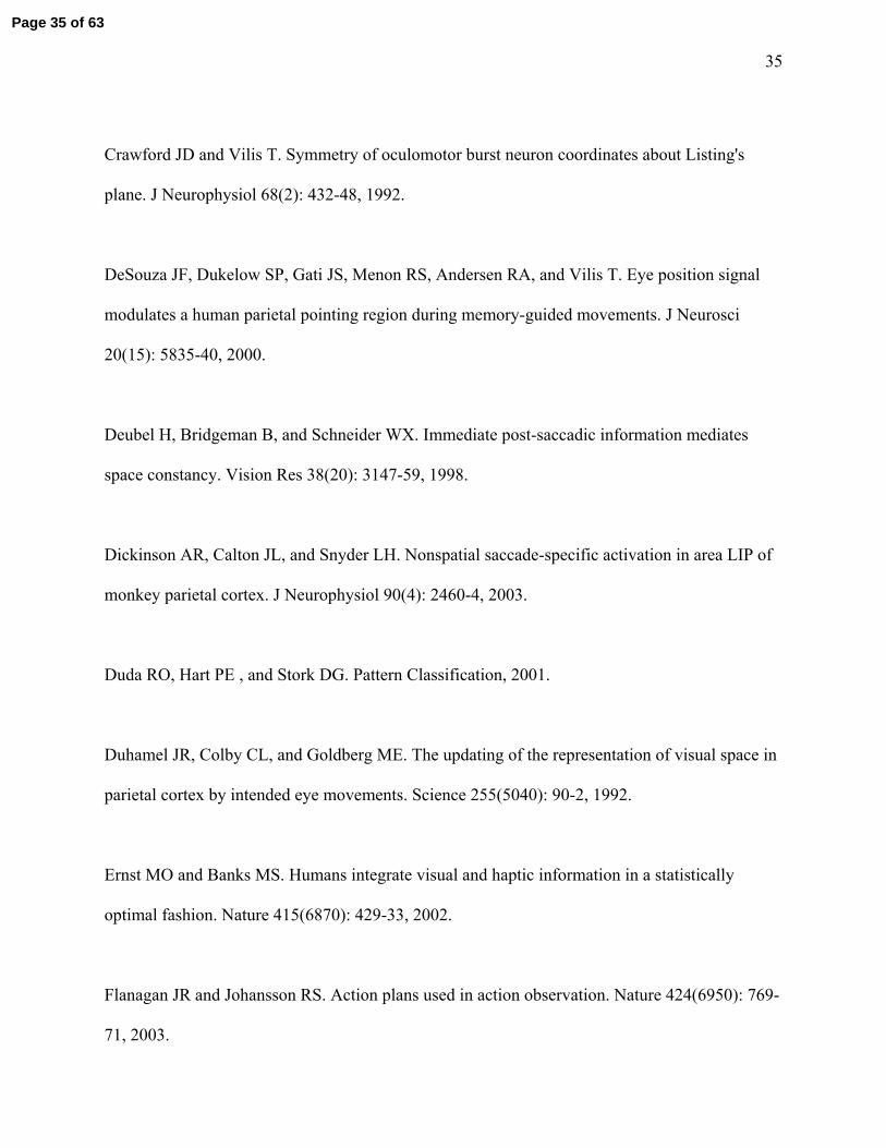
inputs were weighted, we built an optimal inference model (see METHODS and APPENDIX)
(Fig. 8). We tested this model and showed that it predicted the data well both for the constant
(Fig. 8A and D) and variable errors (Fig. 8B and E). The close match between this simple
optimal signal combination model and the observed data occurred despite the few number of
trials for each target (n=5).
We then analysed the different weights of visual and proprioceptive signals in Fig. 8C
and F. Weight = 0 means that the variable was not used in the estimate at all, and weight = 1
means that the estimate relied completely on this one variable. In the AP+VM paradigm, the
average weight of visual signal was 0.68 (range for individual subjects: 0.40-0.78), and the
average weight of proprioceptive feedback was 0.32 (range for individual subjects: 0.22-0.60). In
the PP+VM paradigm, the average weight of visual signal was 0.69 (range: 0.42-0.80), whereas
the average weight of proprioceptive feedback was 0.31 (range: 0.20-0.58). There was no
significant difference in the multi-sensory weighting between the active and passive hand
movement paradigms. Thus, in these proprioceptive + visual tasks, it appears that although visual
information plays a greater role in determining the location of the saccade goal compared to
proprioceptive information, proprioceptive information is not completely overwritten by the
visual information.
Page 18 of 63

19
Coordinate Systems and Sources of Error
In order to test the coordinate system(s) in which the error patterns arose, we investigated
the effect of target direction on amplitude errors for each paradigm. To do this, we fitted ellipses
to the averaged across-subject saccade endpoints separately for the eight near targets and for the
eight far targets, for each task (Fig. 9). Despite the symmetrical circular distribution of the targets,
the distribution of average saccade endpoints showed distinctive patterns in the different
paradigms. The ellipses for the visual paradigms (VS and MS) appeared to align symmetrically
around the central point (Fig. 9A, 9B), whereas in all of the proprioception and proprioception
plus vision paradigms, there was a shift of saccade endpoints away from the subjects (Fig. 9C-F),
especially in the passive proprioception paradigm (PP) (Fig. 9E).
In the vision-only tasks (Fig. 9A, 9B) and in the PP+VM tasks (Fig. 9F), the elliptical fits
appeared to be nearly circular, but in the other tasks (Fig. 9C-E), the elliptical fits were
somewhat elongated. In the AP+VM and PP tasks (Fig. 9D-E), the ellipses were only slightly
elongated with a slight tilt. However, the active proprioception ellipses (Fig. 9C) showed both a
greater elongation and a greater tilt in the direction predicted by errors in a proprioceptive
coordinate system, with larger errors in the flexion-extension dimension (Fig. 1A). These tilts
were also observed in individual subjects, as shown by the plots of the long axes of ellipses fit to
individual data, superimposed in Fig. 9C. These patterns of shifting and tilting are consistent
with the idea of an anisotropy in hand inertia (Hogan, 1985; Gordon et al., 1994).
To quantify the displacement of the centers of overall saccadic ellipses (Fig. 9) from the
center of the table coordinates, we analyzed the distributions (mean ± SD) of the averaged
position of the centers of the two ellipses from each individual (Fig. 10). Only the centers of the
PP and PP+VM ellipses were shifted significantly from the target center (p<0.05) along the Y
(forward) axis. Only the center of the average PP ellipse was significantly displaced from zero
Page 19 of 63

20
(p<0.01) along the x (transverse) axis. Therefore, the passive proprioception tasks (PP) showed
the largest shift, where the center of ellipses of errors shifted significantly forward and rightward
compared to visually controlled tasks (VS and MS). In addition, there was a significant
difference (p<0.05) in the center location on the y axis between AP (1.69±3.87cm) and PP
(4.06±4.39cm). There was no significant difference (p=0.144) in the center location on the x axis
between AP (1.37±1.26cm) and PP (2.17±2.24cm). And there was no significant difference
(p=0.070) in the elongation of ellipses between AP (0.50±0.22) and PP (0.38±0.17).
In order to quantify the task-specific alteration of the coordinate frames for saccade errors
(Fig. 1), we first characterized the elongation of the ellipses. There is an inherent computational
problem with determining the tilt: the closer the ellipse is to being circular, the less certain the
measure of tilt is and the more the data are subject to noise. In order to determine if a given
ellipse was sufficiently elongated, we applied the following formula:
Elongation = (long axis / short axis) – 1
This provides both a measure of elongation and also a certainty score for measuring ellipses
ranging from zero (circular ellipse) to infinite certainty (a line). We judged that ellipses that
were significantly elongated relative to the visual paradigms (VS and MS) could be reliably used
to judge the tilt of the overall error distribution in the proprioceptive updating tasks.
Elongation scores for all subjects (mean ± SD) and two fitted ellipses (near and far
targets) were calculated (F(5,55)=3.95, p<0.05) and plotted in Fig.10C. Note that this score does
not average linearly to equal the score for the overall average data (superimposed black bars)
derived from the plots in Figure 9. The overall score is lower because individual subjects show
elongations in different directions that tend to cancel out in averaging. However, both measures
show the same pattern, i.e. that the AP paradigm produced the greatest elongation. The PP task
showed an intermediate elongation, whereas the addition of a visual cue in the AP+VM and
Page 20 of 63

21
PP+VM paradigms reduced elongation in both tasks. Importantly, the elongation score for the
AP task was the only score that was significantly different from both the non-proprioceptive VS
(p<0.05) and MS (p<0.01) paradigms. For this reason we concluded that the AP ellipses
provided the only reliable measure of ellipse rotation in the proprioceptive tasks.
We next examined the tilt of the AP ellipses to determine if they were significantly
rotated toward a limb/joint coordinate system. For reference, the average (across subjects) angle
between the line of the forearm (at the center resting position) and the leftward direction of the
horizontal axis in table coordinates was 42.05°. The average (±SD) rotations of the major axes of
the ellipses fit to the saccade endpoints in AP condition for individual subjects were 30.05° ±
19.73° for the outer ellipse (plotted as lines in Fig. 9 C) and 35.18° ± 28.47° for the inner ellipse
(compared to 24.68° and 21.29° respectively for the ellipse fits to overall average data that are
shown in Fig. 9). These rotations (for individual subjects) were significantly greater than zero
(p=0.003), but were not significantly different from the angle of forearm (p=0.287). For
comparison, the mean overall rotations of the AP+VM, PP, and PP+VM tasks (which did not
meet our tests for reliability) were 7.99°, 9.35°, and -3.88° respectively. We found no significant
correlations between the tilt of these ellipses and the direction of the shifts shown in Figure 10A
and 10B. In summary, the AP paradigm was the only condition that produced significant
elongation of the overall error pattern relative to the visual only paradigms, and these ellipses
were rotated by approximately 2/3 the angle of the forearm at its resting position.
Page 21 of 63

22
DISCUSSION
Our results demonstrate several new findings: First, somatosensory information can be
used to update the internal representation of handheld target locations for saccades. However,
these somatosensory-based saccades are much less accurate and less precise than visually guided
or visual memory saccades. Additional visual cues improved performance, but the reverse was
not true, i.e., additional somatosensory inputs not only failed to improve on the vision-only tasks,
they even degraded the performance. These sources of somatosensory error equally increased the
variability of saccade amplitude and direction, with amplitude always greater than direction.
Reliance on somatosensory input also produced systematic (average) errors: saccades were
hypermetric in all directions, but especially so in the forward (away from the body) and
rightward directions. Furthermore, saccade hypermetria was most pronounced along an axis
rotated toward the arm flexion-extension direction in the active proprioception task.
Spatial updating
We found that the saccade generator can use proprioceptive feedback from the limb to
update hand-held target locations. Such an updating facility would be useful, for example, for
keeping track of the location of a handheld tool while one re-directs gaze toward other potential
objects of interest. This shows that there is an interplay between the eye and hand systems not
only in the usual visuomotor eye-to-hand sense, but also in the reverse hand-to-eye sense
(Nanayakkara and Shadmehr, 2003; Scheidt et al., 2005). Such two-way communication between
the limb system and oculomotor-visual system is probably important when subjects are engaged
in complex, natural eye-hand coordination tasks (Johansson et al., 2001).
Page 22 of 63

23
Updating across gaze movements of the eyes, head, and body has been observed in many
visual memory tasks (Bloomberg et al., 1988; Hallett and Lightstone, 1976; Israel and Berthoz,
1989; Maurer et al., 1997; Mays and Sparks, 1980; Medendorp et al., 2003; Mergner et al., 1998;
Mergner et al., 1992; Pelisson et al., 1989). In these experiments, the updating problem arose
because self-generated eye motion changed the location of the sensor (the retina) relative to a
space-fixed target, whereas in the current experiment the updating problem arose because self-
generated motion of the arm changed the location of the target relative to a space-fixed retina.
In either case, the fundamental updating problem occurs because some type of self-motion
changes the spatial relationship between the remembered target and the sensor, and both of these
problems could occur simultaneously in natural eye-hand coordination tasks.
Error patterns in proprioceptive updating
Four paradigms (AP, AP+VM, PP and PP+VM) in our experiment involved composite
actions of both the eye and the hand. Therefore, the sources of errors in these paradigms may
have derived from the eyes, the hand, or both. In our visual tasks, not surprisingly, the pattern of
errors can be explained simply within an eye- or head-centered coordinate system. However, in
non-visual tasks (PP and especially AP), the errors of saccades showed a pattern that was
suggestive of a body, limb, or joint coordinate system (Nanayakkara and Shadmehr, 2003). First,
saccades were more hypermetric toward far targets than toward near targets, especially in the
passive proprioception task, producing an overall shift in the saccade error pattern away from the
body (i.e., the forward and rightward shift of the ellipses in Fig. 9). Second, in the active
proprioception task the overall pattern of errors was elongated along an axis that was tilted
toward the flexion-extension dimension of arm movement.
Page 23 of 63

24
It is possible that some of the errors observed in the proprioceptive tasks derive directly
from errors specific to the proprioceptive system (Van Beers et al., 1998 and 2002b). However,
this does not explain why the errors in the saccade task are so much larger than errors observed
in other tasks requiring accurate limb proprioception (Henriques and Soechting, 2003; Van Beers
et al., 1998, 2002a). These appear to be specific to proprioceptively guided saccades. Therefore,
the observed hypermetria of the saccades might be due to an imperfect calibration of
proprioceptive signals used to remap the target. This seems to be a reasonable assumption given
that in everyday life, proprioceptive signals are normally overwritten by visual information
(spatially more accurate). Moreover, the errors specific to the proprioceptive system do not
account for the differences observed between the active and passive proprioceptive tasks.
So, although we did not measure the axes of limb inertia in the experiment, the saccade
error patterns that we observed appear to be consistent with previous reports of directional
anisotropy in limb inertia (Gordon et al., 1994; Hogan, 1985). This suggests that in the
proprioception tasks, especially the active proprioception task, a predominant source of saccade
error originated from joint or limb-centered coordinates. The specific reasons for these errors
will be discussed below when we consider the neurophysiology of these eye-hand systems.
Sensory integration and target representation
Recent studies have suggested that the senses usually provide redundant information, and
when integrating redundant signals, the brain forms a statistically optimal (i.e., minimum-
variance) estimate by weighting each modality according to its relative precision. Minimum-
variance models have been shown to account for human performance when subjects integrate
vision and touch (Ernst and Banks, 2002), vision and audition (Battaglia et al., 2003), and other
combinations of sensory input (Jacobs, 1999; Van Beers et al., 1999; Welch et al., 1979).
Page 24 of 63

25
Similarly, in our AP + VM and PP + VM tasks, subjects received both visual and proprioception
(afferent and/or efferent proprioceptive signals) estimates of the target location. The integration
of visual and proprioceptive information has been studied extensively (Haggard et al., 2000;
Plooy et al., 1998; Welch 1978; Welch and Warren 1986). In our experiment, most of the errors
associated with proprioception were significantly reduced when an additional visual cue was
provided, even after a memory interval. This appears to be an example of the brain weighting
different inputs according to their reliability (Sober and Sabes, 2003; Van Beers et al., 1996).
According to the optimal inference model, the weight of the visual signal was 0.68 in the
active proprioception (AP) paradigm, and 0.69 in the passive proprioception (PP) paradigm (thus,
the complimentary weight of the proprioceptive signal was only 0.32 for AP and 0.31 for PP).
This supports the idea that the brain performs an optimal combination of all available sensory
information, here with visual memory being the more reliable of the two sources. The relative
weights were very similar for AP and PP conditions, even on a subject-by-subject basis,
presumably because the ratio of reliability between proprioceptive and visual inputs is normally
constant in everyday active behavior. In other words, the oculomotor system appears to rely on
an internal model of hand position that is derived from both afferent and efferent proprioceptive
signals, and being upstream from this model, it is not able to disentangle the earlier inputs. Thus,
the weighting of visual and somatosensory inputs (Fig. 11, * symbol indicates the points at
which these inputs are weighted in the optimal inference model) probably occurs at a late stage.
Interestingly, proprioception did not improve on vision alone. Indeed, the Bayesian
model says it should not since proprioception was less accurate than vision. Why then weight in
proprioception at all? This is likely a product of the memory interval, where visual reliability
‘fades’ compared to the reliability of sustained proprioceptive inputs: eventually proprioception
is better, as in the case where there is no vision at all. The relatively poor performance of the
Page 25 of 63

26
proprioceptive system in guiding eye movements is not entirely surprising, because the primary
task of the eye-hand coordination system is to guide hand movements using visual gaze (Abrams
et al., 1990; Scheidt et al., 2005), not the other way around. Nevertheless, it is possible that this
sense is better calibrated in individuals who work with their hands professionally.
Neurophysiological Models for proprioceptive updating of saccades
The eye and hand seem to be controlled by parallel but interacting mechanisms (Snyder
et al., 2002). Lazzari et al. (1997) proposed a model in which both motor systems are completely
independent but exchange information, mediated by sensory signals (e.g., visual from the eye,
proprioceptive from the arm) and efference copies of motor commands. Our experiment
identified a new kind of interaction – the spatial updating of handheld target representations in
the saccade generator based on proprioceptive feedback from the limb.
Such signals could enter the saccade generator at a number of points, such as the
cerebellum, which possesses both oculomotor and limb proprioception signals (Lynch and Tian,
2005; Shadmehr, 2004). But our hypothesis is that proprioceptive updating acts at the same early
level as the previously observed signals associated with gaze-centered updating across saccades.
In other words, we propose that the common buffer for target storage and updating is an eye-
centered coordinate system (Anderson and Buneo, 2002; Batista et al. 1999; Colby and Goldberg,
1999; Duhamel et al. 1992; Henriques et al., 1998; Medendorp et al., 2003). The scheme in
Figure 11 shows how this might occur. The schematic depicts a simple visuomotor
transformation for saccades (e.g., Crawford and Guitton, 1997) and other several means of
updating the target location. In brief, the gaze-centered visuospatial memory buffer can receive
and synthesize information from 1) the visual system itself; 2) the oculomotor system to update
targets in eye-centered coordinates; and 3) the somatosensory updating system. Clearly the latter
Page 26 of 63

27
would require several reference frame transformations in order to correctly transform
proprioceptive signals from limb-based coordinates into gaze-centered coordinates (Buneo et al.,
2002).
The latter supposition would implicate a number of cortical and subcortical saccade areas
as the targets for proprioceptive updating, including lateral intraparietal area (LIP) (Duhamel et
al., 1992; Medendorp et al., 2003; Nakamura and Colby, 2002), the superior colliculus
(Nakamura and Colby, 2002; Walker et al., 1995), and the frontal eye fields (Heide et al., 2001;
Nakamura and Colby, 2002; Umeno and Goldberg, 1997). Buneo et al. (2002) have recently
found evidence that hand position may be represented in gaze-centered coordinates in posterior
parietal cortex. Similarly, hand position signals could be used for the calculations required in our
tasks. Oculomotor and limb signals are closely associated in parietal cortex (Baker et al., 1999;
Batista et al., 1999; DeSouza et al., 2000). Conversely, LIP preferentially encodes targets for
upcoming eye movements, but also possesses responses related to limb movements (Dickinson et
al., 2003; Snyder et al., 1997). Therefore, we suggest that the transformations, required for
proprioceptive-oculomotor updating, occur through a mechanism similar to that proposed by
Buneo et al. (2002) for arm movements, but targeting specifically the saccade-related network in
areas like LIP.
Our results from the active proprioceptive tasks suggest that the saccade generator may
receive an efference copy that originates from a level at or downstream from primary motor
cortex. Recent unit recording experiments suggest that the output of M1 (but not parietal cortex)
may compensate for the anisotropy observed in limb inertia (Scott et al. 1997), producing greater
discharge for extension-flexion than adduction-abduction (Sergio et al., 2005). If the saccade
generator received this command as an efference copy of the limb movement, it would have to
transform these anisotropic signals through a “forward model” of limb inertia to correctly
Page 27 of 63

28
calculate the limb kinematics. The error pattern that we observed in the active proprioceptive
tasks suggests that the saccade generator does receive such a signal, but fails to completely
account for its anisotropy. Consistent with this, this particular pattern of error disappeared in the
passive proprioception tasks (i.e., because here there is neither a motor command nor an
efference copy of the motor command). The precise directions of the anisotropy in our data do
not perfectly match the anisotropies observed in the primate neurophysiological data (Sergio et
al., 2005), but this could simply represent a species or task difference. Furthermore, any
difference in the saccade anisotropy and the actual mechanical anisotropy in the human limb
could result from a partially, but not properly, calibrated forward model.
Finally, one can see that the variability of saccade endpoints in our experiment is inherent
in all saccades, especially in the amplitude. The changes in variability could be due to noise in
the internal estimate of the hand movement vector, or in the estimate of the difference between
the initial eye position and the target. These two possibilities cannot be disentangled in the
current experiment because they were always correlated (i.e., initial eye and hand position were
always at center). To distinguish between these possibilities will require further experiments in
the future.
To conclude, the saccade generator can update hand-held target locations for saccades by
using proprioceptive information from the limb. However, proprioceptive input is not as accurate
as visual input in guiding saccades. In addition, our results indicate that the saccade generator
cannot fully compensate for the anisotropy of limb inertia. During the process of active
proprioceptive updating, saccade errors are likely to originate from limb-centered coordinates, a
hypothesis that could be confirmed by systematically dissociating these coordinates from other
gaze-related coordinate systems. Finally, our proposed model offers potential pathways of the
spatial updating of handheld target locations for saccades that may be tested physiologically in
Page 28 of 63

29
the future.
Page 29 of 63
30
APPENDIX: Optimal Inference Model
In order to compute (estimated saccade amplitude) from the available visual (V) and
(active or passive) proprioceptive (P) information, we used an optimal inference model (Duda et
al., 2001):
x̂
( )( )∫
∫⋅
⋅⋅=
VPxpdx
VPxpxdxx̂
( VPxp ) is the probability of the target being at location x, given the visual (V) and
proprioceptive (P) inputs. Then, we used standard statistical rules (Duda et al., 2001) to compute
( ) ( )VPxp as: ( ) ( ) ( )xpxPpxVpxVPp ⋅⋅= .
In our case, the prior was simply a uniform distribution. The conditional
probabilities
( )xp
( )xVp and ( )xPp were Gaussians with independent means ( )PV µµ , and
noise ( )PV σσ , . We estimated the parameters of these Gaussians from our data, i.e. from the
visual memory (VM) and active or passive proprioception only (AP or PP) conditions. We then
computed the posterior probability density function ( )VPxp to make predictions for the
combined vision and (active or passive) proprioception tasks. Since ( ) ( ) ( )xPpxVpVPxp ⋅∝ , we
calculated the predicted means ( µ̂ ) and standard deviations (σ̂ ) for the combined
proprioception and visual memory task from the multiplication of the two Gaussians as follows:
( )2222
222 ,minˆ PV
PV
PV σσσσσσσ ≤+⋅
=
Page 30 of 63

31
[ ]PVP
P
V
V µµσµ
σµσµ ,ˆˆ 22
2 ∈⎟⎟⎠
⎞⎜⎜⎝
⎛+⋅=
We also computed the relative weights of the visual and proprioceptive information, i.e.
what was the proportion of visual and proprioceptive information used to compute the optimal
estimate. These are given by the following expression:
22
2
11
1
PV
PVPVw
σσ
σ
+=
Page 31 of 63

32
ACKNOWLEDGEMENTS:
We thank S. Sun and H. Wang for technical support. This work is supported by the
Canadian Institutes of Health Research (CIHR). LR is supported by Ontario Graduate
Scholarship, AZK is supported by an NSERC Postgraduate Award, GB is supported by a Marie
Curie International fellowship within the 6th European Community Framework Program and
CIHR (Canada), and JDC holds a Canada Research Chair.
Page 32 of 63

33
REFERENCES
Abrams RA, Meyer DE, and Kornblum S. Eye-hand coordination: oculomotor control in rapid
aimed limb movements. J Exp Psychol Hum Percept Perform 16(2): 248-67, 1990.
Andersen RA and Buneo CA. Intentional maps in posterior parietal cortex. Annu Rev Neurosci
25: 189-220, 2002.
Ariff G, Donchin O, Nanayakkara T and Shadmehr R. A real-time state predictor in motor
control: study of saccadic eye movements during unseen reaching movements. J Neurosci 22(17):
7721-9, 2002.
Baker JT, Donoghue JP, and SanesJN. Gaze direction modulates finger movement activation
patterns in human cerebral cortex. J Neurosci 19(22): 10044-52, 1999.
Batista AP, Buneo CA, Snyder LH, and Andersen RA. Reach plans in eye-centered coordinates.
Science 285(5425): 257-60, 1999.
Battaglia PW, Jacobs RA, and Aslin RN. Bayesian integration of visual and auditory signals for
spatial localization. J Opt Soc Am A Opt Image Sci Vis 20(7): 1391-7, 2003.
Becker W and Jurgens R. An analysis of the saccadic system by means of double step stimuli. Vision Res 19(9): 967-83, 1979.
Page 33 of 63

34
Blakemore SJ, Goodbody SJ, and Wolpert DM. Predicting the consequences of our own actions:
the role of sensorimotor context estimation. J Neurosci 18(18): 7511-8, 1998.
Bloomberg J, Jones GM, Segal B, McFarlane S, and Soul J. Vestibular-contingent voluntary
saccades based on cognitive estimates of remembered vestibular information. Adv
Otorhinolaryngol 41: 71-5, 1988.
Bridgeman B. A review of the role of efference copy in sensory and oculomotor control systems.
Ann Biomed Eng 23(4): 409-22, 1995.
Buneo CA, Jarvis MR, Batista AP, and Andersen RA. Direct visuomotor transformations for
reaching. Nature 416(6881): 632-6, 2002.
Colby CL, Duhamel JR, and Goldberg ME. Oculocentric spatial representation in parietal cortex.
Cereb Cortex 5(5): 470-81, 1995.
Colby CL and Goldberg ME. Space and attention in parietal cortex. Annu Rev Neurosci 22: 319-
49, 1999.
Cordo P, Gurfinkel VS, Bevan L, and Kerr GK. Proprioceptive consequences of tendon vibration
during movement. J Neurophysiol 74(4): 1675-88, 1995.
Crawford JD and Guitton D. Visual-motor transformations required for accurate and
kinematically correct saccades. J Neurophysiol 78(3): 1447-67, 1997.
Page 34 of 63
35
Crawford JD and Vilis T. Symmetry of oculomotor burst neuron coordinates about Listing's
plane. J Neurophysiol 68(2): 432-48, 1992.
DeSouza JF, Dukelow SP, Gati JS, Menon RS, Andersen RA, and Vilis T. Eye position signal
modulates a human parietal pointing region during memory-guided movements. J Neurosci
20(15): 5835-40, 2000.
Deubel H, Bridgeman B, and Schneider WX. Immediate post-saccadic information mediates
space constancy. Vision Res 38(20): 3147-59, 1998.
Dickinson AR, Calton JL, and Snyder LH. Nonspatial saccade-specific activation in area LIP of
monkey parietal cortex. J Neurophysiol 90(4): 2460-4, 2003.
Duda RO, Hart PE , and Stork DG. Pattern Classification, 2001.
Duhamel JR, Colby CL, and Goldberg ME. The updating of the representation of visual space in
parietal cortex by intended eye movements. Science 255(5040): 90-2, 1992.
Ernst MO and Banks MS. Humans integrate visual and haptic information in a statistically
optimal fashion. Nature 415(6870): 429-33, 2002.
Flanagan JR and Johansson RS. Action plans used in action observation. Nature 424(6950): 769-
71, 2003.
Page 35 of 63

36
Flanagan JR and Lolley S. The inertial anisotropy of the arm is accurately predicted during
movement planning. J Neurosci 21(4): 1361-9, 2001.
Gordon J, Ghilardi MF, Cooper SE and Ghez C. Accuracy of planar reaching movements. Exp
Brain Res 99: 112-130, 1994.
Gottlieb JP, Kusunoki M, and Goldberg ME. The representation of visual salience in monkey
parietal cortex. Nature 391(6666): 481-4, 1998.
Graf W. Spatial coordination of compensatory eye movements in vertebrates: form and function.
Acta Biol Hung 39(2-3): 279-90, 1988.
Graf W and Wilson VJ. Afferents and efferents of the vestibular nuclei: the necessity of context-
specific interpretation. Prog Brain Res 80: 149-57; discussion 127-8, 1989.
Groh JM and Sparks DL. Saccades to somatosensory targets. I. behavioral characteristics. J.
Neurophysiol. 75(1): 412-27, 1996.
Grill SE and Hallett M. Velocity sensitivity of human muscle spindle afferents and slowly
adapting type II cutaneous mechanoreceptors. J Physiol. 489 ( Pt 2): 593-602, 1995.
Haggard P, Newman C, Blundell J, and Andrew H. The perceived position of the hand in space.
Percept Psychophys. 62(2): 363-77, 2000.
Page 36 of 63

37
Hallett PE and Lightstone AD. Saccadic eye movements towards stimuli triggered by prior
saccades. Vision Res. 16(1): 99-106, 1976.
Heide W, Binkofski F, Seitz RJ, Posse S, Nitschke MF, Freund HJ, and Kompf D. Activation of
frontoparietal cortices during memorized triple-step sequences of saccadic eye movements: an
fMRI study. Eur J Neurosci 13(6): 1177-89, 2001.
Helmholtz HV. Helmholtz's Treatise on Physiological Optics. New York:: Dover Press 1: 143-
172,375-415, 1962.
Helsen WF, Elliott D, Starkes JL, and Ricker KL. Coupling of eye, finger, elbow, and shoulder
movements during manual aiming. J Mot Behav. 32(3): 241-8, 2000.
Henriques, D. Y, and Crawford, J. D. Testing the three-dimensional reference frame
transformation for express and memory-guided saccades. Neurocomputing. 38-40: 1267-80,
2001.
Henriques DY and Soechting JF. Bias and sensitivity in the haptic perception of geometry. Exp
Brain Res. 150(1): 95-108, 2003
Henriques DY, and Soechting JF. Haptic synthesis of shapes and sequences. J Neurophysiol.
91(4): 1808-21, 2004.
Page 37 of 63

38
Henriques DY, Klier EM, Smith MA, Lowy D, and Crawford JD. Gaze-centered remapping of
remembered visual space in an open-loop pointing task. J Neurosci 18(4): 1583-94, 1998.
Hogan N. The mechanics of multi-joint posture and movement control. Biol Cybern 52: 315-331,
1985.
Israel I. and Berthoz A. Contribution of the otoliths to the calculation of linear displacement. J
Neurophysiol 62(1): 247-63, 1989.
Jacobs RA. Optimal integration of texture and motion cues to depth. Vision Res 39(21): 3621-9,
1999.
Johansson RS, Westling G, Backstrom A, and Flanagan JR. Eye-hand coordination in object
manipulation. J Neurosci 21(17): 6917-32, 2001.
Khan AZ, Pisella L, Rossetti Y, Vighetto A, and Crawford JD. Impairment of Gaze-centered
Updating of Reach Targets in Bilateral Parietal-Occipital Damaged Patients. Cereb Cortex
15(10):1547-60, 2005.
Klier EM and Crawford JD. Human oculomotor system accounts for 3-D eye orientation in the
visual-motor transformation for saccades. J Neurophysiol 80(5): 2274-94, 1998.
Lazzari S, Vercher JL, and Buizza A. Manuo-ocular coordination in target tracking. I. A model
simulating human performance. Biol Cybern 77(4): 257-66, 1997.
Page 38 of 63

39
Leube DT, Knoblich G, Erb M, Grodd W, Bartels M, and Kircher TT. The neural correlates of
perceiving one's own movements. Neuroimage 20(4): 2084-90, 2003.
Lewis RF, Gaymard BM and Tamargo RJ. Efference copy provides the eye position information
required for visually guided reaching. J Neurophysiol 80(3):1605-8, 1998.
Lynch JC and Tian JR. Cortico-cortical networks and cortico-subcortical loops for the higher
control of eye movements. Prog Brain Res 151: 461-501, 2005.
Maurer C, Kimmig H, Trefzer A, and Mergner T. Visual object localization through vestibular
and neck inputs. 1: Localization with respect to space and relative to the head and trunk mid-
sagittal planes. J Vestib Res 7(2-3): 119-35, 1997.
Mays LE and Sparks DL. Saccades are spatially, not retinocentrically, coded. Science 208(4448):
1163-5, 1980.
Medendorp WP, Goltz HC, Vilis T, and Crawford JD. Gaze-centered updating of visual space in
human parietal cortex. J Neurosci 23(15): 6209-14, 2003.
Mergner T, Nasios G, and Anastasopoulos D. Vestibular memory-contingent saccades involve
somatosensory input from the body support. Neuroreport 9(7): 1469-73, 1998.
Page 39 of 63

40
Mergner T, Rottler G, Kimmig H, and Becker W. Role of vestibular and neck inputs for the
perception of object motion in space. Exp Brain Res 89(3): 655-68, 1992.
Messier J. and Kalaska JF. Differential effect of task conditions on errors of direction and extent
of reaching movements. Exp Brain Res 115(3): 469-78, 1997.
Murphy JT, Wong YC, and Kwan HC. Afferent-efferent linkages in motor cortex for single
forelimb muscles. J Neurophysiol 38(4): 990-1014, 1975.
Mussa-Ivaldi FA, Hogan N, and Bizzi E. Neural, mechanical, and geometric factors subserving
arm posture in humans. J Neurosci 5(10): 2732-43, 1985.
Nakamura K and Colby CL. Updating of the visual representation in monkey striate and
extrastriate cortex during saccades. Proc Natl Acad Sci USA 99(6): 4026-31, 2002.
Nanayakkara T and Shadmehr R. Saccade adaptation in response to altered arm dynamics. J
Neurophysiol 90(6): 4016-21, 2003.
Neggers SF and Bekkering H. Integration of visual and somatosensory target information in
goal-directed eye and arm movements. Exp Brain Res. 125(1): 97-107, 1999.
Neggers SF and Bekkering H. Ocular gaze is anchored to the target of an ongoing pointing
movement. J Neurophysiol. 83(2): 639-51, 2000
Page 40 of 63

41
Neggers SF and Bekkering H. Gaze anchoring to a pointing target is present during the entire
pointing movement and is driven by a non-visual signal. J Neurophysiol. 86(2): 961-70, 2001
Nelson RJ. Interactions between motor commands and somatic perception in sensorimotor cortex.
Curr Opin Neurobiol. 6(6): 801-10, 1996.
Niemeier M, Crawford JD, and Tweed DB. Optimal transsaccadic integration explains distorted
spatial perception. Nature 422(6927): 76-80, 2003.
Pelisson D, Guitton D, and Munoz DP. Compensatory eye and head movements generated by the
cat following stimulation-induced perturbations in gaze position. Exp Brain Res 78(3): 654-8,
1989.
Plooy A, Tresilian JR, Mon-Williams M, and Wann JP. The contribution of vision and
proprioception to judgements of finger proximity. Exp Brain Res 118(3): 415-20, 1998.
Pouget A, Ducom JC, Torri J, and Bavelier D. Multisensory spatial representations in eye-
centered coordinates for reaching. Cognition 83(1): B1-11, 2002.
Pozzo T, Levik Y, and Berthoz A. Head and trunk movements in the frontal plane during
complex dynamic equilibrium tasks in humans. Exp Brain Res 106(2): 327-38, 1995.
Sabes PN, Jordan MI and Wolpert DM. The role of inertial sensitivity in motor planning. J
Neurosci 18(15): 5948-57, 1998.
Page 41 of 63

42
Shadmehr R. Generalization as a behavioral window to the neural mechanisms of learning
internal models. Hum Mov Sci 23(5): 543-68, 2004.
Scheidt RA, Conditt MA, Secco EL, and Mussa-Ivaldi FA. Interaction of visual and
proprioceptive feedback during adaptation of human reaching movements. J Neurophysiol
93(6):3200-13, 2005.
Scherberger H, Cabungcal JH, Hepp K, Suzuki Y, Straumann D, and Henn V. Ocular counterroll
modulates the preferred direction of saccade-related pontine burst neurons in the monkey. J
Neurophysiol 86(2): 935-49, 2001.
Scott SH, Sergio LE, and Kalaska JF. Reaching movements with similar hand paths but different
arm orientations. II. Activity of individual cells in dorsal premotor cortex and parietal area 5. J
Neurophysiol 78(5): 2413-26, 1997.
Sergio LE, Hamel-Paquet C, and Kalaska JF. Motor Cortex Neural Correlates of Output
Kinematics and Kinetics During Isometric-Force and Arm-Reaching Tasks. J Neurophysiol
94(4):2353-78, 2005.
Snyder LH, Batista AP, and Andersen RA. Coding of intention in the posterior parietal cortex.
Nature 386(6621): 167-70, 1997.
Page 42 of 63

43
Snyder LH, Calton JL, Dickinson AR, and Lawrence BM. Eye-hand coordination: saccades are
faster when accompanied by a coordinated arm movement. J Neurophysiol 87(5): 2279-86, 2002.
Sober SJ and Sabes PN. Multisensory integration during motor planning. J Neurosci 23(18):
6982-92, 2003.
Soechting JF, Buneo CA, Herrmann U and Flanders M. Moving effortlessly in three dimensions:
does Donders’ law apply to arm movement? J Neurosci 15(9): 6271-80, 1995.
Stahl JS and Simpson JI. Dynamics of rabbit vestibular nucleus neurons and the influence of the
flocculus. J Neurophysiol 73(4): 1396-413, 1995.
Stark L and Bridgeman B. Role of corollary discharge in space constancy. Percept Psychophys
34(4): 371-80, 1983.
Suzuki Y, Straumann D, Simpson JI, Hepp K, Hess BJ, and Henn V. Three-dimensional
extraocular motoneuron innervation in the rhesus monkey. I: Muscle rotation axes and on-
directions during fixation. Exp Brain Res 126(2): 187-99, 1999.
Tweed D, Cadera W, and Vilis T. Computing three-dimensional eye position quaternions and
eye velocity from search coil signals. Vision Res 30(1): 97-110, 1990.
Umeno MM and Goldberg ME. Spatial processing in the monkey frontal eye field. I. Predictive
visual responses. J Neurophysiol 78(3): 1373-83, 1997.
Page 43 of 63

44
Van Beers RJ, Sittig AC, and Denier van der Gon JJ. How humans combine simultaneous
proprioceptive and visual position information. Exp Brain Res 111(2): 253-61, 1996.
Van Beers, R. J, Sittig, A. C, and Gon, J. J. Integration of proprioceptive and visual position-
information: An experimentally supported model. J Neurophysiol 81(3): 1355-64, 1999.
Van Beers RJ, Baraduc P and Wolpert DM. Role of uncertainty in sensorimotor control. Philos
Trans R Soc Lond B Biol Sci. 357(1424): 1137-45, 2002a.
Van Beers RJ, Wolpert DM and Haggard P. When feeling is more important than seeing in
sensorimotor adaptation. Curr Biol. 12(10): 834-7, 2002b.
Van Donkelaar P, Siu KC, and Walterschied J. Saccadic output is influenced by limb kinetics
during eye-hand coordination. J Mot Behav 36(3): 245-52, 2004.
Van Opstal AJ, Hepp K, Hess BJ, Straumann D, and Henn V. Two- rather than three-
dimensional representation of saccades in monkey superior colliculus. Science 252(5010): 1313-
5, 1991.
Vercher JL, Sares F, Blouin J, Bourdin C, and Gauthier G. Role of sensory information in
updating internal models of the effector during arm tracking. Prog Brain Res 142:203-22, 2003.
Page 44 of 63

45
Vindras P, Desmurget M and Viviani P. Error parsing in visuomotor pointing reveals
independent processing of amplitude and direction. J. Neurophysiol. 94(2): 1212-24
Walker MF, Fitzgibbon EJ, and Goldberg ME. Neurons in the monkey superior colliculus predict
the visual result of impending saccadic eye movements. J Neurophysiol 73(5): 1988-2003, 1995.
Welch RB. Perceptual Modification. New York: Academic Press, 1978.
Welch, R. B, Widawski, M. H, Harrington, J, and Warren, D. H. An examination of the
relationship between visual capture and prism adaptation. Percept Psychophys 25(2): 126-32,
1979.
Welch RB and Warren DH. Intersensory interactions. In Handbook of Perception and Human
Performance.Volume 1. K. R. Boff, L. Kaufman, and J. P. Thomas, des. (New York: Wiley). pp.
25.1-25.36, 1986.
White JM, Sparks DL, and Stanford TR. Saccades to remembered target locations: an analysis of
systematic and variable errors. Vision Res 34(1): 79-92, 1994.
Zalkind VI. [Spontaneous discharges of muscle receptors of different functional types]. Fiziol Zh
SSSR Im I M Sechenova 65(4): 565-74, 1979.
Page 45 of 63

46
TABLES
Table 1: Sources of information about target locations
Paradigms Efference copy Visual signal Proprioception + tactile sensation
Visual Saccade (VS) x (no memory delay) x Memory Saccade (MS) x (with memory delay) x Active Prop. (AP) x Active Prop. + Visual Mem. (AP+VM) Passive Prop. (PP) x x Passive Prop. + Visual Mem. (PP+VM) x
Table 2: Multiple Linear Regression Condition Direction Model 1
Slope (r2,p)
Model 2 Slope (r2, p)
Pearson Correlation (Between IVs)
Model 3
r2 Slope 1 Slope 2
r2 change (M3-M1) (p)
H 0.299 (0.089,*)
0.258 (0.047,-)
0.006 0.155 0.297 0.256 0.066 (*) VS
V 0.106 (0.011,-)
0.199 (0.040,-)
-0.051 0.053 0.116 0.213 0.042 (*)
H 0.685 (0.469,*)
0.126 (0.016,-)
-0.036 0.492 0.690 0.150 0.023 (*) MS
V 0.477 (0.228,*)
0.268 (0.042,-)
0.027 0.303 0.470 0.274 0.075 (*)
H 0.623 (0.389,*)
-0.438 (0.192,*)
-0.440 0.422 0.534 -0.203 0.033 (*) AP
V 0.466 (0.217,*)
0.006 (0.000,-)
-0.284 0.238 0.509 -0.151 0.021 (*)
H 0.667 (0.446,*)
-0.568 (0.322,*)
-0.719 0.461 0.537 -0.182 0.015 (*) AP+VM
V 0.374 (0.140,*)
-0.072 (0.005,-)
-0.393 0.146 0.408 -0.088 0.006 (*)
H 0.759 (0.576,*)
-0.438 (0.192,*)
-0.534 0.577 0.734 -0.046 0.001 (-) PP
V 0.597 (0.357,*)
-0.037 (0.001,-)
-0.229 0.367 0.621 0.105 0.010 (*)
H 0.666 (0.443,*)
-0.393 (0.154,*)
-0.539 0.445 0.640 -0.048 0.002 (-) PP+VM
V 0.472 (0.223,*)
0.032 (0.001,-)
-0.357 0.269 0.554 0.229 0.046 (*)
Model 1: The dependent variable (DV) is saccade error, the independent variable (IV) is target location. Model 2: The DV is saccade error, the IV is microsaccadic drift. Model 3: The DV is saccade error, the IVs are target location and microsaccadic drift. H: Horizontal component; V: Vertical component. *: p<0.05 ; -: p>=0.05
Page 46 of 63

47
LEGENDS
Figure 1. Potential error patterns for saccades in table coordinates. A, Experimental setup.
Subjects’ arms were positioned so that the oblique movement direction aligned with a limb-
based coordinate system (extension-flexion vs. abduction-adduction: <------->), B-D, Possible
patterns of saccade errors in table coordinates when a saccade is guided by vision and/or
proprioception. : target locations; unfilled circles and ellipses are potential error patterns. B,
Saccade errors in amplitude only with no overall bias (shift). C, Saccade errors with amplitude
depending on direction in cardinal coordinates, plus an overall bias relative to the center position
or body. D, Error due to greater saccade amplitude effects either along the extension-flexion or
abduction-adduction axes of the limb coordinates (as shown in A) would lead to tilted elliptical
patterns.
Figure 2. Experimental setup and paradigms. A, Left panel: Subjects sat in a dark room in front
of a horizontal table, with the head fixed (using a bite-bar) at a 45° downward orientation toward
the table. Subjects held a short dowel topped by an LED between the right index finger and
thumb, with the hand resting on a plate that could slide on a grooved board. The right panel
shows the 16 radial targets (ο) viewed from above the table (8 directions, 5cm and 10cm from
the center, at visual angles 7° and 13° relative to the center). B, Visual control paradigms. Left
panel: subjects first fixated on the illuminated center LED. Middle panel: a peripheral LED
flashed when the center LED disappeared. Right panel: subjects made a saccade to the peripheral
target with no memory delay (VS) or after a memory delay (MS). Dashed lines are binocular
gaze lines. C, Active/passive proprioception tasks. Left panel: subjects viewed the hand-held
flashed LED at the center position. Middle panel: when the center target was extinguished,
Page 47 of 63

48
either a computer voice command instructed the subject to move the hand to one of the
peripheral positions (Active Proprioception or AP task) or the hand was passively moved to that
location by the experimenter moving the hand plate (passive proprioception or PP task). Right
panel: an auditory beep signaled the subject to saccade toward the perceived location of the non-
illuminated LED between their fingers. D, Active/passive hand movements with additional
visual cues. Hand and eye movements were the same as in Figure 1C with the exception that the
LED flashed briefly at the final dowel position before subjects made a saccade (AP+VM /
PP+VM tasks).
Figure 3. Sample data from a typical subject: horizontal trajectories of the eye and hand toward
the far-right target location. For each of the six paradigms, 5 saccades (black traces) are plotted
in oculomotor coordinates as a function of time. A, Visual saccade (VS) paradigm. B, Memory
saccade (MS) paradigm. C, Active proprioception (AP) paradigm. D, Active proprioception with
visual cue (AP + VM). E, Passive proprioception (PP) paradigm. F, Passive proprioception with
visual cue (PP + VM). Legend: : location and duration of central fixation (F) light; : location
and duration of saccade target (T) light (13° rightward target here); : beep cue for beginning of
saccade; black lines: eye movement trajectories; gray lines: hand movement trajectories.
Figure 4. Sample data from a typical subject: two-dimensional saccade (black traces) and hand
movement (gray traces) trajectories plotted in oculomotor coordinates. For each of the paradigms
(A-F) five movements are shown to each of the eight far targets to qualitatively show intra-
individual variability and task-dependent saccade accuracy. A, Visual Saccade (VS) task. B,
Memory Saccade (MS) task. C, Active Proprioception (AP) task. D, Active Proprioception plus
Visual Memory (AP+VM) task. E, Passive Proprioception (PP) task. F,. Passive Proprioception
Page 48 of 63

49
plus Visual Memory (PP+VM) task. Legend: : target locations; black lines: saccade
trajectories; : saccade end-points; gray lines, trajectory of hand movement. Saccade trajectories
did not always begin at center because of the microsaccadic drift after the extinction of the center
LED, particularly in the hand movement paradigms.
Figure 5. Error distributions of average saccade end-points between subjects. Data were
represented in 2-D table coordinates, and then fit with the 68% confidence interval ellipses
plotted here. A, Visual saccade (VS) paradigm. B, Memory saccade (MS) paradigm. C, Active
proprioception (AP) paradigm. D, Active proprioception with visual cue (AP + VM). E, Passive
proprioception (PP) paradigm. F, Passive proprioception with visual cue (PP + VM). Legend: :
target locations; : mean saccade endpoint (across subject means); black unfilled ellipses: error
distribution with 68% confidence interval across subjects.
Figure 6. Error distributions of saccades within subjects. The distribution ellipse was fit to each
subject’s saccade end-points in table coordinates, then these ellipse parameters were averaged
across the six subjects to provide the ellipses plotted here. A, Visual saccade (VS) paradigm. B,
Memory saccade (MS) paradigm. C, Active proprioception (AP) paradigm. D, Active
proprioception with visual cue (AP + VM). E, Passive proprioception (PP) paradigm. F, Passive
proprioception with visual cue (PP + VM). Legend: : target locations; : mean saccade
endpoint; black unfilled ellipses: error distribution with 95% confidence intervals of intra-subject
variability, averaged across subjects.
Figure 7. Saccade error distribution: parameters from 95% confidence interval ellipses. Bars
show parameters averaged across targets and then across subjects (± SD) for both inter-subject
Page 49 of 63
50
variability (left column) and intra-subject variability (right column). Top row shows lengths of
the major ellipse axes and the middle row shows lengths of the minor axes. Bottom row: scatter
plots (ο) and linear regressions of the major-axis angles as a function of the target location angles
(see text for explanation).
Figure 8. Comparison between optimal inference model predictions and data. Each data point
corresponds to one target. The data and regression lines for all 6 subjects are shown. A perfect
prediction of the data by the model would yield a slope = 1 (black lines). A, B, Predicted location
(mean, µ, panel A) and variability (SD, σ, panel B) of target location for the combined active
proprioception and visual memory task (AM). C, Relative weights of visual (black) and
proprioceptive (white) information for the combined active proprioception and visual memory
task (AM). The histograms show the number of weights within each bin (width = 0.1) for each
target position across all subjects. On the x-axis, weight = 0 means that this variable is not used
at all; weight = 1 means that the optimal estimate only relies on this variable. Solid black lines
are population means (visual = 0.68, proprioception = 0.32), other coloured lines are means for
individual subjects (visual = 0.40…0.78, proprioception = 0.22…0.60). D, E, Location (mean, µ,
panel D) and variability (SD, σ, panel E) of the predicted target location for the passive
proprioception and visual memory (PM) task. F, Relative weights for the combined passive
proprioception and visual memory task (PM). Population means (visual = 0.69,
proprioception = 0.31) and means for individual subjects (visual = 0.42…0.80,
proprioception = 0.20…0.58).
Page 50 of 63

51
Figure 9. Shape of overall saccade error patterns. Ellipses were fit to the overall saccade
endpoints (across all subjects) for the near target locations and for the far target locations
respectively in table coordinates. A, Visual Saccade (VS) task. B, Memory Saccade (MS) task. C,
Active Proprioception (AP task). This panel also shows the directions of major ellipse axes for
each individual. D, Active proprioception with visual cue (AP +VM) task. E, Passive
proprioception (PP) task. F, Passive proprioception with visual cue (PP + VM) task. Legend: :
target locations; : overall mean saccade endpoints; dashed-line ellipses: fit to saccades for near
targets; solid-line ellipses: fit to saccades for far targets; dashed lines in C: directions of major
axis of far-target ellipses for each subject; solid lines in C: direction of major axis of near-target
ellipses for each subject.
Figure 10. Shift and elongation parameters of the error pattern ellipses (like those in Fig. 9) fit to
individual subjects. Bars show averages between fits to near and far targets, averaged (±SD)
across subjects (n = 12). A, Shifts of ellipse centers along the Y-axis (forward-backward) of table
coordinates. B, Shifts of ellipse centers along the X-axis (leftward-rightward) of table
coordinates. C, Elongation scores of ellipses. These scores were calculated as described in the
Results section and plotted for each paradigm. Pattern-filled bars: average (±SD) values of the
elongation scores from each individual subject. Superimposed black bars: elongation scores of
the overall average near-target and far-target ellipses shown in Figure 9.
Figure 11. Proposed model for the spatial updating of handheld targets. The ‘Target in Eye
Coordinates’ representation can be updated based on 1) visual information, 2) movements of the
eyes themselves, and 3) somatosensory inputs (including proprioception, tactile sensation and
efference copy of the command of arm movement). Arrows: the direction of signal transduction;
Page 51 of 63

52
labeled boxes: key steps in the updating process and required transformations; dashed box:
missing internal model that we propose explains the elongation and tilt of error patterns in the
AP paradigm. *: points where inputs are weighted in the optimal inference model.
Page 52 of 63

53
Page 53 of 63

54
Page 54 of 63

55
Page 55 of 63

56
Page 56 of 63

57
Page 57 of 63

58
Page 58 of 63

59
Page 59 of 63
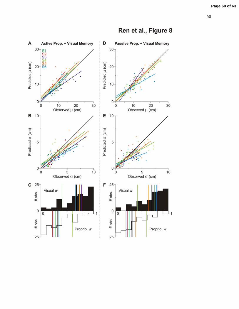
60
Page 60 of 63

61
Page 61 of 63

62
Page 62 of 63

63
Page 63 of 63