Properly Oriented Heparin−Decasaccharide-Induced Dimers Are the Biologically Active Form of Basic...
10
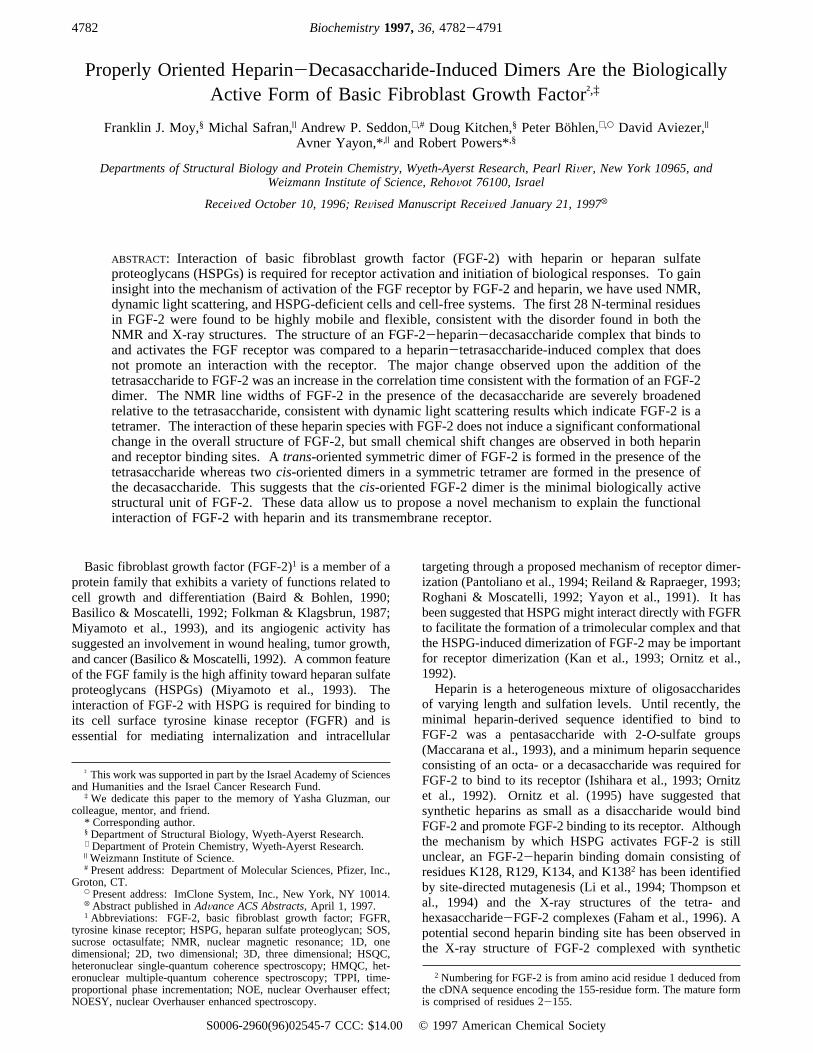
Properly Oriented Heparin-Decasaccharide-Induced Dimers Are the Biologically Active Form of Basic Fibroblast Growth Factor ²,‡ Franklin J. Moy, § Michal Safran, | Andrew P. Seddon, ⊥,# Doug Kitchen, § Peter Bo ¨hlen, ⊥,O David Aviezer, | Avner Yayon,* ,| and Robert Powers* ,§ Departments of Structural Biology and Protein Chemistry, Wyeth-Ayerst Research, Pearl RiVer, New York 10965, and Weizmann Institute of Science, RehoVot 76100, Israel ReceiVed October 10, 1996; ReVised Manuscript ReceiVed January 21, 1997 X ABSTRACT: Interaction of basic fibroblast growth factor (FGF-2) with heparin or heparan sulfate proteoglycans (HSPGs) is required for receptor activation and initiation of biological responses. To gain insight into the mechanism of activation of the FGF receptor by FGF-2 and heparin, we have used NMR, dynamic light scattering, and HSPG-deficient cells and cell-free systems. The first 28 N-terminal residues in FGF-2 were found to be highly mobile and flexible, consistent with the disorder found in both the NMR and X-ray structures. The structure of an FGF-2-heparin-decasaccharide complex that binds to and activates the FGF receptor was compared to a heparin-tetrasaccharide-induced complex that does not promote an interaction with the receptor. The major change observed upon the addition of the tetrasaccharide to FGF-2 was an increase in the correlation time consistent with the formation of an FGF-2 dimer. The NMR line widths of FGF-2 in the presence of the decasaccharide are severely broadened relative to the tetrasaccharide, consistent with dynamic light scattering results which indicate FGF-2 is a tetramer. The interaction of these heparin species with FGF-2 does not induce a significant conformational change in the overall structure of FGF-2, but small chemical shift changes are observed in both heparin and receptor binding sites. A trans-oriented symmetric dimer of FGF-2 is formed in the presence of the tetrasaccharide whereas two cis-oriented dimers in a symmetric tetramer are formed in the presence of the decasaccharide. This suggests that the cis-oriented FGF-2 dimer is the minimal biologically active structural unit of FGF-2. These data allow us to propose a novel mechanism to explain the functional interaction of FGF-2 with heparin and its transmembrane receptor. Basic fibroblast growth factor (FGF-2) 1 is a member of a protein family that exhibits a variety of functions related to cell growth and differentiation (Baird & Bohlen, 1990; Basilico & Moscatelli, 1992; Folkman & Klagsbrun, 1987; Miyamoto et al., 1993), and its angiogenic activity has suggested an involvement in wound healing, tumor growth, and cancer (Basilico & Moscatelli, 1992). A common feature of the FGF family is the high affinity toward heparan sulfate proteoglycans (HSPGs) (Miyamoto et al., 1993). The interaction of FGF-2 with HSPG is required for binding to its cell surface tyrosine kinase receptor (FGFR) and is essential for mediating internalization and intracellular targeting through a proposed mechanism of receptor dimer- ization (Pantoliano et al., 1994; Reiland & Rapraeger, 1993; Roghani & Moscatelli, 1992; Yayon et al., 1991). It has been suggested that HSPG might interact directly with FGFR to facilitate the formation of a trimolecular complex and that the HSPG-induced dimerization of FGF-2 may be important for receptor dimerization (Kan et al., 1993; Ornitz et al., 1992). Heparin is a heterogeneous mixture of oligosaccharides of varying length and sulfation levels. Until recently, the minimal heparin-derived sequence identified to bind to FGF-2 was a pentasaccharide with 2-O-sulfate groups (Maccarana et al., 1993), and a minimum heparin sequence consisting of an octa- or a decasaccharide was required for FGF-2 to bind to its receptor (Ishihara et al., 1993; Ornitz et al., 1992). Ornitz et al. (1995) have suggested that synthetic heparins as small as a disaccharide would bind FGF-2 and promote FGF-2 binding to its receptor. Although the mechanism by which HSPG activates FGF-2 is still unclear, an FGF-2-heparin binding domain consisting of residues K128, R129, K134, and K138 2 has been identified by site-directed mutagenesis (Li et al., 1994; Thompson et al., 1994) and the X-ray structures of the tetra- and hexasaccharide-FGF-2 complexes (Faham et al., 1996). A potential second heparin binding site has been observed in the X-ray structure of FGF-2 complexed with synthetic ² This work was supported in part by the Israel Academy of Sciences and Humanities and the Israel Cancer Research Fund. ‡ We dedicate this paper to the memory of Yasha Gluzman, our colleague, mentor, and friend. * Corresponding author. § Department of Structural Biology, Wyeth-Ayerst Research. ⊥ Department of Protein Chemistry, Wyeth-Ayerst Research. | Weizmann Institute of Science. # Present address: Department of Molecular Sciences, Pfizer, Inc., Groton, CT. O Present address: ImClone System, Inc., New York, NY 10014. X Abstract published in AdVance ACS Abstracts, April 1, 1997. 1 Abbreviations: FGF-2, basic fibroblast growth factor; FGFR, tyrosine kinase receptor; HSPG, heparan sulfate proteoglycan; SOS, sucrose octasulfate; NMR, nuclear magnetic resonance; 1D, one dimensional; 2D, two dimensional; 3D, three dimensional; HSQC, heteronuclear single-quantum coherence spectroscopy; HMQC, het- eronuclear multiple-quantum coherence spectroscopy; TPPI, time- proportional phase incrementation; NOE, nuclear Overhauser effect; NOESY, nuclear Overhauser enhanced spectroscopy. 2 Numbering for FGF-2 is from amino acid residue 1 deduced from the cDNA sequence encoding the 155-residue form. The mature form is comprised of residues 2-155. 4782 Biochemistry 1997, 36, 4782-4791 S0006-2960(96)02545-7 CCC: $14.00 © 1997 American Chemical Society
-
Upload
johnjanovy -
Category
Documents
-
view
0 -
download
0
Transcript of Properly Oriented Heparin−Decasaccharide-Induced Dimers Are the Biologically Active Form of Basic...
Properly Oriented Heparin-Decasaccharide-Induced Dimers Are the BiologicallyActive Form of Basic Fibroblast Growth Factor†,‡
Franklin J. Moy,§ Michal Safran,| Andrew P. Seddon,⊥,# Doug Kitchen,§ Peter Bo¨hlen,⊥,O David Aviezer,|
Avner Yayon,*,| and Robert Powers*,§
Departments of Structural Biology and Protein Chemistry, Wyeth-Ayerst Research, Pearl RiVer, New York 10965, andWeizmann Institute of Science, RehoVot 76100, Israel
ReceiVed October 10, 1996; ReVised Manuscript ReceiVed January 21, 1997X
ABSTRACT: Interaction of basic fibroblast growth factor (FGF-2) with heparin or heparan sulfateproteoglycans (HSPGs) is required for receptor activation and initiation of biological responses. To gaininsight into the mechanism of activation of the FGF receptor by FGF-2 and heparin, we have used NMR,dynamic light scattering, and HSPG-deficient cells and cell-free systems. The first 28 N-terminal residuesin FGF-2 were found to be highly mobile and flexible, consistent with the disorder found in both theNMR and X-ray structures. The structure of an FGF-2-heparin-decasaccharide complex that binds toand activates the FGF receptor was compared to a heparin-tetrasaccharide-induced complex that doesnot promote an interaction with the receptor. The major change observed upon the addition of thetetrasaccharide to FGF-2 was an increase in the correlation time consistent with the formation of an FGF-2dimer. The NMR line widths of FGF-2 in the presence of the decasaccharide are severely broadenedrelative to the tetrasaccharide, consistent with dynamic light scattering results which indicate FGF-2 is atetramer. The interaction of these heparin species with FGF-2 does not induce a significant conformationalchange in the overall structure of FGF-2, but small chemical shift changes are observed in both heparinand receptor binding sites. Atrans-oriented symmetric dimer of FGF-2 is formed in the presence of thetetrasaccharide whereas twocis-oriented dimers in a symmetric tetramer are formed in the presence ofthe decasaccharide. This suggests that thecis-oriented FGF-2 dimer is the minimal biologically activestructural unit of FGF-2. These data allow us to propose a novel mechanism to explain the functionalinteraction of FGF-2 with heparin and its transmembrane receptor.
Basic fibroblast growth factor (FGF-2)1 is a member of aprotein family that exhibits a variety of functions related tocell growth and differentiation (Baird & Bohlen, 1990;Basilico & Moscatelli, 1992; Folkman & Klagsbrun, 1987;Miyamoto et al., 1993), and its angiogenic activity hassuggested an involvement in wound healing, tumor growth,and cancer (Basilico & Moscatelli, 1992). A common featureof the FGF family is the high affinity toward heparan sulfateproteoglycans (HSPGs) (Miyamoto et al., 1993). Theinteraction of FGF-2 with HSPG is required for binding toits cell surface tyrosine kinase receptor (FGFR) and isessential for mediating internalization and intracellular
targeting through a proposed mechanism of receptor dimer-ization (Pantoliano et al., 1994; Reiland & Rapraeger, 1993;Roghani & Moscatelli, 1992; Yayon et al., 1991). It hasbeen suggested that HSPGmight interact directly with FGFRto facilitate the formation of a trimolecular complex and thatthe HSPG-induced dimerization of FGF-2 may be importantfor receptor dimerization (Kan et al., 1993; Ornitz et al.,1992).Heparin is a heterogeneous mixture of oligosaccharides
of varying length and sulfation levels. Until recently, theminimal heparin-derived sequence identified to bind toFGF-2 was a pentasaccharide with 2-O-sulfate groups(Maccarana et al., 1993), and a minimum heparin sequenceconsisting of an octa- or a decasaccharide was required forFGF-2 to bind to its receptor (Ishihara et al., 1993; Ornitzet al., 1992). Ornitz et al. (1995) have suggested thatsynthetic heparins as small as a disaccharide would bindFGF-2 and promote FGF-2 binding to its receptor. Althoughthe mechanism by which HSPG activates FGF-2 is stillunclear, an FGF-2-heparin binding domain consisting ofresidues K128, R129, K134, and K1382 has been identifiedby site-directed mutagenesis (Li et al., 1994; Thompson etal., 1994) and the X-ray structures of the tetra- andhexasaccharide-FGF-2 complexes (Faham et al., 1996). Apotential second heparin binding site has been observed inthe X-ray structure of FGF-2 complexed with synthetic
† This work was supported in part by the Israel Academy of Sciencesand Humanities and the Israel Cancer Research Fund.
‡ We dedicate this paper to the memory of Yasha Gluzman, ourcolleague, mentor, and friend.* Corresponding author.§ Department of Structural Biology, Wyeth-Ayerst Research.⊥ Department of Protein Chemistry, Wyeth-Ayerst Research.| Weizmann Institute of Science.# Present address: Department of Molecular Sciences, Pfizer, Inc.,
Groton, CT.O Present address: ImClone System, Inc., New York, NY 10014.X Abstract published inAdVance ACS Abstracts,April 1, 1997.1 Abbreviations: FGF-2, basic fibroblast growth factor; FGFR,
tyrosine kinase receptor; HSPG, heparan sulfate proteoglycan; SOS,sucrose octasulfate; NMR, nuclear magnetic resonance; 1D, onedimensional; 2D, two dimensional; 3D, three dimensional; HSQC,heteronuclear single-quantum coherence spectroscopy; HMQC, het-eronuclear multiple-quantum coherence spectroscopy; TPPI, time-proportional phase incrementation; NOE, nuclear Overhauser effect;NOESY, nuclear Overhauser enhanced spectroscopy.
2 Numbering for FGF-2 is from amino acid residue 1 deduced fromthe cDNA sequence encoding the 155-residue form. The mature formis comprised of residues 2-155.
4782 Biochemistry1997,36, 4782-4791
S0006-2960(96)02545-7 CCC: $14.00 © 1997 American Chemical Society
heparin-derived di- and trisaccharides (Ornitz et al., 1995).Heparin has also been shown to protect FGF-2 frominactivation from exposure to low pH, elevated temperature,or proteases and to restore bioactivity to inactive growthfactor (Gospodarowicz & Cheng, 1986; Pineda-Lucena etal., 1994; Saksela et al., 1988; Sommer & Rifkin, 1989;Westall et al., 1983).Elucidation of the mechanism of heparin-induced aug-
mentation of FGF-2 binding to its cell surface receptor hasbeen hampered by often contradictory reports most likelyrelated to the complexity and heterogeneous nature of thesystem. Several mechanisms based on interpretation ofexperimental evidence have been proposed with a consensusthat heparin induces oligomerization of FGF-2 which in turnfacilitates receptor dimerization and transmembrane signaling(Mach et al., 1993; Ornitz et al., 1992, 1995; Spivak-Kroizman et al., 1994; Venkataraman et al., 1996). Whathas eluded our understanding are data that definitivelydistinguish between an oligomerization event such as adding“beads on a string versus” a defined structural unit requiredfor receptor binding and activation. In previous papers (Moyet al., 1995, 1996) we presented the nearly complete1H, 15N,13CO, and13C assignments, the solution secondary structure,and the high-resolution NMR structure of FGF-2 to comprisethe essential foundation for such a study. In this paper, wepresent the first direct experimental evidence obtained byNMR, independently confirmed by dynamic light scatteringand biological relevance established in cell-based and cell-free assays, to propose a specifically oriented heparin-FGF-2complex.
MATERIALS AND METHODS
Materials. The heparin-derived tetrasaccharide was kindlyprovided by Robert J. Linhardt, Division of Medicinal andNatural Products Chemistry, University of Iowa (Pervin etal., 1995), and the heparin-derived decasaccharide was kindlyprovided by Carl Svahn, Department of Organic Chemistry,Pharmacia AB (Aviezer et al., 1994).NMR Sample Preparation.A 1 mM 15N-FGF-2 sample
was prepared as described previously (Moy et al., 1995) andwas titrated with a stock solution of heparin-derived tet-rasaccharide at a concentration of 3 mg/150µL in 50 mMpotassium phosphate, 2 mM NaN3, and 90% H2O/10% D2Oat pH 5.5. 1D1H and 2D 15N HSQC (Bodenhausen &Ruben, 1980) were collected at a molar ratio of FGF-2 totetrasaccharide of 1:0.25, 1:0.5, 1:0.75, and 1:1. The titrationof each quarter increment was carried out by addition of 9µL a of tetrasaccharide stock solution to an initial volumeof FGF-2 of 600µL. The concentration of the tetrasaccha-ride was determined by dry weight, and the concentrationof FGF-2 was determined spectrophotometrically [absorbance(0.1% at 280 nm)) 0.964 (Thompson et al., 1994)].Similarly, a 1 mM15N-FGF-2 sample was titrated with a
decasaccharide at a molar ratio of FGF-2 to the decasac-charide of 1:0.25, 1:0.5, and 1:1. 1D1H and 2D15N HSQCwere collected at these concentrations.NMR Data Collection. All spectra were recorded at 25
°C on a Bruker AMX600 spectrometer using a gradient-enhanced triple-resonance1H/13C/15N probe. All hetero-nuclear NOE,T1, andT2 relaxation time measurements werecarried out in duplicate for FGF-2. Water suppression inthe NOE,T1, andT2 experiments was carried out with the
WATERGATE sequence and water flip-back pulses (Grz-esiek & Bax, 1993; Piotto et al., 1992) . All 2D spectrawith 15N indirectly detected dimensions were recorded withthe States-TPPI hypercomplex phase increment (Marion etal., 1989) and collected with appropriate refocusing delaysto allow for spectra without any phase correction. Theheteronuclear15N NOE,T1, andT2 relaxation times for FGF-2and the FGF-2-tetrasaccharide complex were measured with1HN and 15N spectral widths of 8064 and 1642 Hz,respectively, with maximum acquisition times of 127 ms (t2)and 97.4 ms (t1).
For FGF-2, both theT1 andT2 data were collected with32 transients per increment. TheT1 inversion recovery timeswere 20, 60, 140, 240, 360, 520, 720, and 1200 ms and theT2 Carr-Purcell-Meiboom-Gill (CPMG) (Meiboom &Gill, 1958) trains were 8, 24, 40, 56, 72, 104, 152, and 184ms in duration (Kay et al., 1992; Markley et al., 1971).Recycle delays forT1 and T2 experiments were 1.7 and 1.3s, respectively. Since the NOE measurement requires anequilibrated15N magnetization for accurate analysis, therecycle time was extended to more than 6 s while 48transients per increment were collected. In the NOEexperiment with presaturation, the proton saturation periodwas 3 s. 1H saturation was carried out with the use of 180°1H pulses applied every 5 ms.
For FGF-2 complexed with the tetrasaccharide in a 1:1ratio, theT1 inversion recovery times were 20, 80, 180, 300,420, 600, 860, and 1300 ms, and theT2 CPMG periods were8, 24, 40, 56, and 72 ms. AllT1 experiments were collectedwith 64 transients, and the 20 ms time point was collectedtwice. Since theT2 experiment had a lower signal to noiseratio, 96 scans were used and the time point at 40 ms wascollected twice.
The 15N-edited NOESY-HMQC experiments were col-lected as described previously (Moy et al., 1995).
Data Processing and Analysis. Spectra were processedas described previously (Moy et al., 1995) using theNMRPipe software package (Delaglio et al., 1995) andanalyzed with PIPP (Garrett et al., 1991) and NMRPipe. Peakheights were automatically assigned for each residue in all2D spectra after semiautomatically peak picking one 2Dspectrum using NMRPipe.T1 andT2 values were determinedby fitting the measured peak heights to the two-parameterprofile I(t) ) I0 exp(-t/Tn). The Levenberg-Marquardtalgorithm (Press et al., 1986) was used to determine theoptimum values ofTn by minimizing the goodness of fitparameterø2 ) ∑(Ic(t) - Ie(t))2/σ, where Ic(t) are theintensities calculated from the fitting parameters,Ie(t) arethe experimental intensities, andσ is the standard deviationof the experimental intensities. For the FGF-2 dataσ is thestandard deviation of the differences between the heights ofcorresponding peaks in duplicate spectra divided by thesquare root of 2 (Palmer et al., 1991) whereas for the FGF-2-tetrasaccharide complexσ was set to the root-mean-squarebaseline noise in the spectra as determined from NMRDraw.The two methods for determining experimental uncertaintywere very similar. Uncertainties inT1 and T2 measurementswere obtained from the covariance matrix generated in theLevenberg-Marquardt algorithm and were used in MonteCarlo simulations for determining the standard deviationsfor fitting parameters (Farrow et al., 1994; Kamath & Shriver,1989; Palmer et al., 1991).
Biologically Active Heparin-Induced FGF-2 Dimers Biochemistry, Vol. 36, No. 16, 19974783

The steady-state NOE values for FGF-2 were determinedfrom the ratios of the average intensities of the peaks withand without proton saturation. Since the data for the FGF-2-tetrasaccharide complex were not recorded in duplicate,the steady-state NOE values were determined from the ratiosof the intensities of the peaks with and without protonsaturation. The standard deviation of the NOE value wasdetermined by the baseline noise (Farrow et al., 1994). Theoverall correlation time was determined by using residuesthat had15N T1/T2 ratios within one standard deviation andNOE values greater than 0.6 (Clore et al., 1990a; Clubb etal., 1995; Kay et al., 1989). Three models of the spectraldensity functions were used to classify five classes ofoptimized parameters (Clore et al., 1990a,b; Farrow et al.,1994; Kay et al., 1989). Selection of the appropriate spectraldensity function was determined by initially fitting the datato the simplest spectral density function and only selectingthe more complicated spectral density function as requiredto fit the data (Clore et al., 1990a; Clubb et al., 1995; Powerset al., 1992).Dynamic Light Scattering.Dynamic light scattering data
were collected and analyzed with a DynaPro-801 instrument(Protein Solutions, Inc., Charlottesville, VA). Initially, 150µL aliquots from free FGF-2, the FGF-2-tetrasaccharide (1:1) complex, and the FGF-2-decasaccharide (1:0.25) complexNMR samples were used for dynamic light scatteringanalysis. The titration curves were determined by addingaliquots of a 10-fold excess oligosaccharide stock solutionto a 0.5 mM FGF-2 sample. The observed molecular weightat each titration step was averaged over 10 data points.Binding and Cross-Linking of125I-FGF-2 to Soluble
FGFR-1 and to Cells. Conditioned medium from cellssecreting FGF receptor-1-AP fusion protein (0.24 OD units/min) was incubated for 45 min at room temperature withrabbit anti-human placental AP antibodies prebound toagarose-protein A beads (Pierce, Rockford, IL). The FGFR-1-coupled beads were washed three times with 1 mL of 20mM HEPES, pH 7.5, 150 mM NaCl, 1% Triton X-100, and10% glycerol (HNTG) and incubated with 2 ng/mL125I-FGF-2 and heparin-oligosacchrides at increasing concentra-tions for 90 min at room temperature. High-affinity-bound125I-FGF-2 was determined after three cycles of washing withHNTG and counted in aγ-counter. High-affinity bindingof FGF-2 to CHO cells was performed as described by Yayonet al. (1991). Briefly,125I-FGF-2 and heparin-oligosac-chrides at increasing concentrations were incubated with cellsfor 90 min at 40°C. Low-affinity-bound FGF-2 was releasedfrom the cell surface by a 5 min incubation with a coldsolution containing 1.6 M NaCl and 20 mM HEPES, pH7.4. High-affinity-bound FGF-2 was determined by a 2 MNaCl and 20 mM acetate buffer, pH 4.0, extraction.Calculation of the FGF-2-Heparin Molecular Model.
The FGF-2 tetramer model was generated by starting withthe monomer of FGF-2 from a recently solved FGF-2-SOScrystal structure. The dimer of FGF-2 was created byrotating about the Cys78 Sγ-Câ axis to permit a minimumdistance between 128 and 138 heparin binding regions ondifferent FGF-2 units. A heparin model consisting ofalternating sulfated glucosamine-glucose structure (obtainedfrom 1hpn.pdb) was positioned on the dimer roughlyspanning the hexasaccharide and tetrasaccharide structuresof the recent FGF-2-heparin crystal structure (1bfb.pdb and1bfc.pdb). This dimer model was minimized and subjected
to 50 ps of molecular dynamics using the AMBER 4.0parameter set. The heparin was used as an axis of rotationto form the tetramer of FGF-2. Thus, the tetramer wasformed by a rotation around the heparin with some translationto eliminate interatomic distances which were too short andto maintain salt bridges between heparin and the four copiesof FGF-2. The tetramer model was subjected to 500 stepsof conjugate gradient minimization.
RESULTS AND DISCUSSION
Titration of FGF-2 with a Decasaccharide and Tetrasac-charide. 2D 15N HSQC spectra of FGF-2 and FGF-2 titratedwith both the tetrasaccharide and decasaccharide are shownin Figure 1. A severe increase in line widths results uponaddition of heparin to FGF-2 (15N T2 < 36.6 ms). It isimportant to note that the results are independent of the orderof addition. FGF-2 aliquots added to a concentrated (3 mg/150 µL) and 4-fold excess tetrasaccharide sample broughtto the same FGF-2 concentration yielded the same broadenedNMR spectra.The major difference between the tetra- and decasaccharide
was the comparable extent of their effect on the FGF-2spectra. While the decasaccharide caused extensive broad-ening of the 2D15N HSQC spectra at a 0.25 molar ratio ofheparin to FGF-2, it was possible to titrate the tetrasaccharideto a 1:1 molar ratio with FGF-2 and acquire a 2D15N HSQCspectrum of acceptable quality. A 1D spectrum shows anapproximate 50% decrease in signal intensity at a 1:1 molarratio of the tetrasaccharide-FGF-2 complex relative to thefree FGF-2 spectrum. Similarly, a 2D15N HSQC shows anapproximate 80% decrease in signal intensity.Chemical shifts are sensitive to local changes in protein
structure and provide a qualitative analysis of the structuraleffects of protein-ligand interactions. From the 2D15NHSQC spectra of FGF-2 and the complex with the tetrasac-charide and decasaccharide (Figure 1) it is apparent that thepresence of heparin does not induce any significant structuralchanges in FGF-2 since most of the observed chemical shiftsare nearly identical between the two samples. Somerelatively small chemical shift changes or significant per-turbations in signal intensity, however, did occur in bothFGF-2-oligosaccharide complexes (0.1-0.3 ppm; vector ofboth 15N and 1HN chemical shift changes). Residues thatincurred small chemical shift changes were clustered in tworegions of the FGF-2 structure: N36, G37, L127, K128,R129, G131, K134, G136, T139, G140, Q143, K144, andA145, which are in the vicinity of the heparin binding domainas identified by site-directed mutagenesis and the tetra- andhexasaccharide-FGF-2 complex X-ray structures (Faham etal., 1996; Li et al., 1994; Thompson et al., 1994), and E105,L107, S109, N110, N111, and L149, which are in the vicinityof the putative receptor binding domain (Springer et al., 1994;Zhu et al., 1991).3D 15N-edited HMQC-NOESY spectra were collected for
both free FGF-2 and the FGF-2-tetrasaccharide complex.Figure 2 shows strips taken from both the free and complex15N-edited NOESY spectra, parallel to theF1 axis, for theamides of residues Thr-130 through Ser-137 which cor-respond to the FGF-2-heparin binding site. While the datafor the FGF-2-tetrasaccharide complex are only marginal,it is still apparent that the NOE data are identical for bothspectra. This supports the conclusion that heparin does not
4784 Biochemistry, Vol. 36, No. 16, 1997 Moy et al.

induce any significant conformational change in FGF-2 uponbinding and is consistent with the X-ray structures of thetetra- and hexasaccharide-FGF-2 complexes (Faham et al.,1996).
Although the severe increase in line widths in the FGF-2NMR spectra upon addition of either the tetra- or decasac-charide implies the formation of high molecular weight formsof FGF-2, it is also possible that the line width increase is aresult of a dynamic exchange process. To distinguishbetween these two possibilities,5N T1, T2, and heteronuclearNOEs were measured for FGF-2 alone and the FGF-2-tetrasaccharide complex to measure the overall correlationtime (τr) for both samples. Correlation time generallyincreases linearly with an increase in molecular weight;however, if the effect on line width is a result of dynamicexchange, no effect will be observed on the correlation timeand a significant exchange term will be required to fit therelaxation data.T1, T2, and NOE Relaxation Parameters.Relaxation data
were obtained for 141 and 125 residues for FGF-2 and theFGF-2-tetrasaccharide complex, respectively, out of 145possible15N-1H backbone correlations. Resonance overlapwas the main cause of difficulties in measuring peak intensityespecially in the random coil region of theT1, T2, and NOEspectra. For FGF-2, L92 is overlapped with the intense E14peak, the peak intensity was assigned to E14, and the
FIGURE1: Comparison of the1H-15N HSQC spectra for (A) FGF-2, (B) the 2:1 FGF-2-tetrasaccharide complex, and (C) the 4:1FGF-2-decasaccharide complex. Residues which incurred a chemi-cal shift change upon addition of the tetrasaccharide are labeledand boxed.
FIGURE 2: Composite of amide strips taken from the 100 msmixing time 3D15N-edited NOESY-HMQC spectrum of (A)FGF-2 and(B) the FGF-2-tetrasaccharide complex for thestretch of sequence from Thr-130 to Ser-137 correspondingto missingâ-strand XI in the NMR structure (Moy et al.,1995). The diagonal peaks are indicated by an asterisk, NH-(i)-NH(i(1), NH(i+1)-HR(i), NH(i)-NH(i(1), and NH-(i+1)-Hâ(i) NOEs are indicated by solid arrows, andNH(i)-NH(i(2), NH(i+2,3,4)-HR(i), and NH(i+2,3,4)-Hâ(i) NOEs are indicated by dashed arrows. IntraresidueHR-NH and Hâ-NH NOEs are boxed.
Biologically Active Heparin-Induced FGF-2 Dimers Biochemistry, Vol. 36, No. 16, 19974785
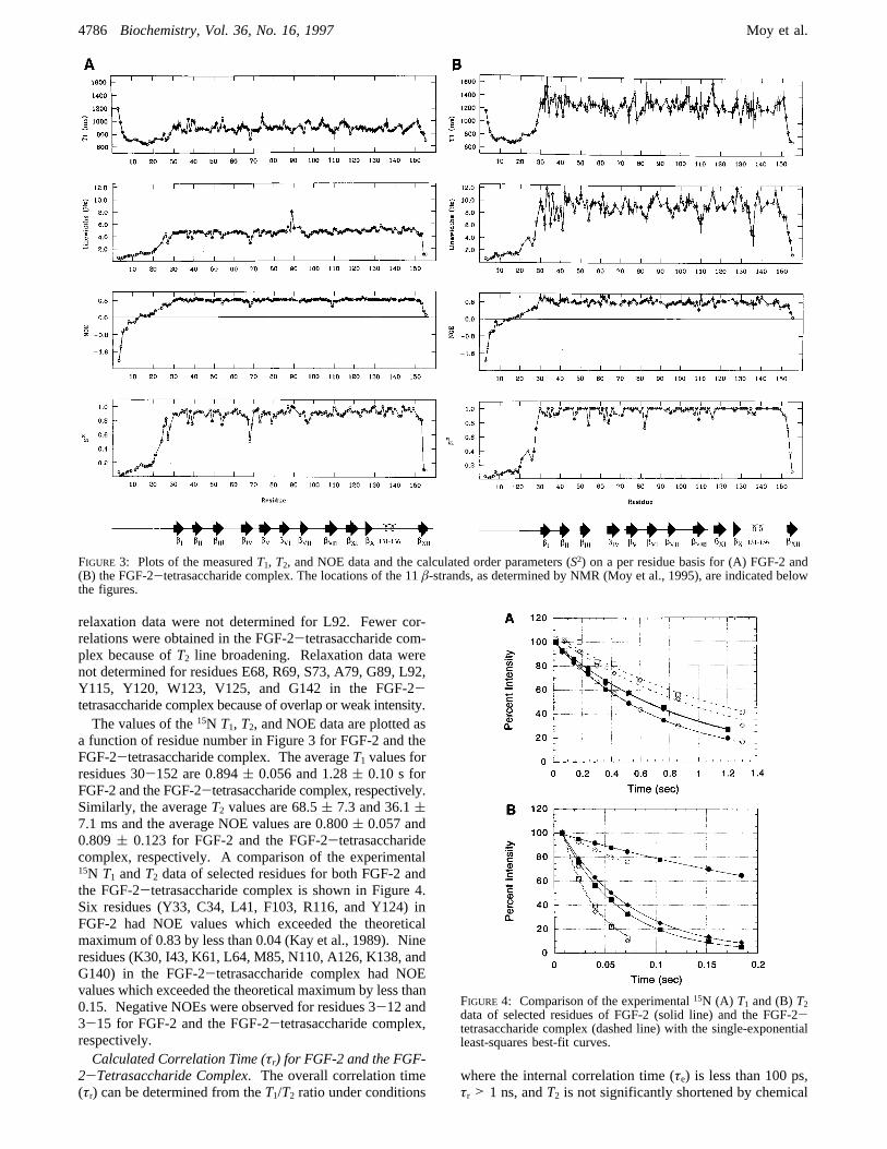
relaxation data were not determined for L92. Fewer cor-relations were obtained in the FGF-2-tetrasaccharide com-plex because ofT2 line broadening. Relaxation data werenot determined for residues E68, R69, S73, A79, G89, L92,Y115, Y120, W123, V125, and G142 in the FGF-2-tetrasaccharide complex because of overlap or weak intensity.The values of the15N T1, T2, and NOE data are plotted as
a function of residue number in Figure 3 for FGF-2 and theFGF-2-tetrasaccharide complex. The averageT1 values forresidues 30-152 are 0.894( 0.056 and 1.28( 0.10 s forFGF-2 and the FGF-2-tetrasaccharide complex, respectively.Similarly, the averageT2 values are 68.5( 7.3 and 36.1(7.1 ms and the average NOE values are 0.800( 0.057 and0.809( 0.123 for FGF-2 and the FGF-2-tetrasaccharidecomplex, respectively. A comparison of the experimental15N T1 andT2 data of selected residues for both FGF-2 andthe FGF-2-tetrasaccharide complex is shown in Figure 4.Six residues (Y33, C34, L41, F103, R116, and Y124) inFGF-2 had NOE values which exceeded the theoreticalmaximum of 0.83 by less than 0.04 (Kay et al., 1989). Nineresidues (K30, I43, K61, L64, M85, N110, A126, K138, andG140) in the FGF-2-tetrasaccharide complex had NOEvalues which exceeded the theoretical maximum by less than0.15. Negative NOEs were observed for residues 3-12 and3-15 for FGF-2 and the FGF-2-tetrasaccharide complex,respectively.Calculated Correlation Time (τr) for FGF-2 and the FGF-
2-Tetrasaccharide Complex. The overall correlation time(τr) can be determined from theT1/T2 ratio under conditions
where the internal correlation time (τe) is less than 100 ps,τr > 1 ns, andT2 is not significantly shortened by chemical
FIGURE 3: Plots of the measuredT1, T2, and NOE data and the calculated order parameters (S2) on a per residue basis for (A) FGF-2 and(B) the FGF-2-tetrasaccharide complex. The locations of the 11â-strands, as determined by NMR (Moy et al., 1995), are indicated belowthe figures.
FIGURE 4: Comparison of the experimental15N (A) T1 and (B)T2data of selected residues of FGF-2 (solid line) and the FGF-2-tetrasaccharide complex (dashed line) with the single-exponentialleast-squares best-fit curves.
4786 Biochemistry, Vol. 36, No. 16, 1997 Moy et al.

exchange (Clore et al., 1990a; Kay et al., 1989). Thusresidues which are suspected to have significant contributionsto their experimentalT1 and T2 values from τe and/orchemical exchange are removed from theτr calculation.Typically, residues with NOE values less than 0.6 orT1/T2ratios which lie either 1 SD above or below the mean wereremoved from theτr calculation. For FGF-2, 23 residueshad NOE values less than 0.6, and 27 residues were outside1 SD of theT1/T2 mean. The overall correlation time forFGF-2 was determined to be 11.4( 0.3 ns by fitting theT1,T2, and NOE data for each of the 91 residues to the Lipariand Szabo “model-free” formalism (Lipari & Szabo, 1982).The optimal correlation time (τr), order parameter (S2), andeffective internal correlation time (τe) were calculatedindividually for each residue, and the overall correlation timefor FGF-2 was the average from all the residues. For theFGF-2-tetrasaccharide complex, 27 residues had NOEvalues less than 0.6, 20 residues were outside 1 SD of theT1/T2 mean, and an additional 19 residues did not convergeduring theτr calculation. The optimal overall correlationtime for the FGF-2-tetrasaccharide complex was determinedto be 20.2 ns by simultaneously fitting theT1, T2, and NOEdata for 49 residues to the Lipari and Szabo model-freeformalism (Lipari & Szabo, 1982). For the FGF-2-tetrasaccharide complex an optimal overall correlation timeinstead of an average value is reported because of an apparentlimitation in the model. A plot ofø2 versus correlation times(Supplementary Information, Figure 1S) is very asymmetricand flat about the minima. This suggests that certainparameters have a minimal contribution to reducing the valueof ø2 at higher values ofτr. This results in the meancorrelation time of 21.3( 1.9 ns being skewed toward ahigher value relative to the optimal correlation time of 20.2ns (minima on theø2 versus correlation time plot). This wasnot the case for free FGF-2 where the mean correlation timeof 11.4 ( 0.3 ns was nearly identical to the optimalcorrelation time of 11.3 ns and theø2 versus correlation timeplot was symmetrical about the minima. Correlation timesof 11.4 and 20.2 ns were used in all subsequent calculationsfor FGF-2 and the FGF-2-tetrasaccharide complex, respec-tively.FGF-2 Dynamics.The generalized order parameterS2 is
plotted as a function of residue number for FGF-2. Theaverage value of the order parameterS2 for residues 30-152 for free FGF-2 is 0.90( 0.05. Other proteins of similarsize, including interleukin-4 (Redfield et al., 1992) and p53(Clubb et al., 1995), haveS2 ≈ 0.90, indicating limitedconformational flexibility. The15N relaxation data of FGF-2indicate that the first 28 N-terminal residues are highlyflexible and mobile (S2 < 0.6). Negative NOEs wereobserved for residues 3-12 and 2-25, and 27 residues werebest fitted to the extended spectral density model (Clore etal., 1990a,b) in which there are two internal motions. Thedynamic analysis supports the previous observation that twodistinct conformations exist for the first 28 residues causedby acis-trans isomerization of prolines 10 and 13 (Moy etal., 1995) and that the first 17-28 residues are disorderedin the NMR structure of FGF-2 (Moy et al., 1996). This isalso consistent with the observation that the first 17-19residues in the X-ray structures of FGF-2 (10-155) are notvisible in the electron density map and have been identifiedas disordered (Ago et al., 1991; Eriksson et al., 1991, 1993;Zhang et al., 1991; Zhu et al., 1991). Aside from the N-
and C-terminal regions, FGF-2 is well ordered with allS2values being greater than 0.80 except for residues G37, F39,E54, E68, and G70, which are in or near loop regions.Residues G37 and E54 reside in loop regions betweenâ-strands 1 and 2 andâ-strands 3 and 4, respectively.Residues E68 and G70 are in the loop betweenâ-strands 4and 5, and F39 is at the start ofâ-strand 2. Five otherresidues in loop regions which were not assigned becauseof overlap or lack of an observable cross peak were L91,L92, and Y120. All residues exhibit very fast internalmotions on time scalese20 ps except for residues E54, E67,E68, S96, E108, S109, N111, and Y112, which exhibitinternal motions of 20-111 ps, and the mobile N- andC-terminal residues 1-29 and 152-154, which exhibit asecond slower motion on a time scale of 0.3-1.4 ns. Theaverage calculatedte relative error estimate was 28%.FGF-2-Heparin Dynamics. The 15N relaxation data
(Figures 3 and 4) from the FGF-2-tetrasaccharide complexindicated that the presence of heparin bound to FGF-2induced a distinct effect on the dynamics of FGF-2. It isquite apparent that the observedT1 and line width valuesincreased dramatically for residues 30-152 in the FGF-2-tetrasaccharide complex while the NOE values and orderparameters were quite similar. The FGF-2-tetrasaccharidecomplex exhibits the same mobility as free FGF-2 for theN- and C-terminal residues as indicated by order parametersbelow 0.8. The average value of the order parameterS2
observed for residues 30-152 for FGF-2 increased in thepresence of the tetrasaccharide to 0.96( 0.06. An increasein S2 is not unexpected upon formation of a complex, butthere is no indication of a local effect on mobility since theorder parameter increase is observed throughout the protein.Therefore, the major effect of the tetrasaccharide on FGF-2dynamics is the substantial increase in the protein’s overallcorrelation time. Since a correlation time of 20.2 ns wasmeasured for the complex compared to 11.4 ns for free FGF-2, this indicates that the presence of the tetrasaccharideinduces the formation of FGF-2 dimers and the observedincrease in FGF-2 line widths is not a result of exchangebroadening. In the FGF-2-tetrasaccharide complex a totalof 18 residues exhibit internal motions of 20-111 pscompared to 8 residues in free FGF, and 9 residues inaddition to the mobile N- and C-terminal residues exhibit asecond slower motion on a time scale of 0.3-1.4 ns. Inaddition, an exchange term was required to fit 32 residuesfor the complex, but only 5 residues in free FGF-2 requiredthis additional exchange term. The average exchange valuein the FGF-2-tetrasaccharide complex was 5.4( 2.7 Hz.Since an additional exchange term was required by residuesthroughout the protein structure, this suggests a possible slowmonomer-dimer exchange where the dimer is the predomi-nant species.In the case of the FGF-2-decasaccharide complex, a
greater effect was observed on the broadening of FGF-2 linewidths compared to the tetrasaccharide. Since the dynamicsdata for the tetrasaccharide support the formation of a dimer,it is reasonable to conclude that the greater increase in linewidths associated with the decasaccharide at a lower molarratio of decasaccharide to FGF-2 (0.25:1) is consistent withthe formation of a higher order oligomer.FGF-2-Heparin Dynamic Light Scattering.To indepen-
dently confirm the NMR results, dynamic light scatteringdata were collected on 150µL aliquots of the NMR samples
Biologically Active Heparin-Induced FGF-2 Dimers Biochemistry, Vol. 36, No. 16, 19974787

of FGF-2 and the FGF-2-oligosaccharide complexes. Themolecular weights determined indicated that FGF-2 ismonomeric, the FGF-2-tetrasaccharide complex is dimeric,and the FGF-2-decasaccharide is tetrameric, which is inaccord with the additional overall increase in complexationobserved by NMR (Figure 5). This supports the previousconclusion that the observed increase in the FGF-2 NMRline widths caused by the addition of the oligosaccharidesis a result of the formation of high molecular weight formsof FGF-2 and not a result of exchange broadening. This isalso consistent with the observation that the FGF-2 bindingaffinity for heparin is relatively strong (KD in the nanomolarrange) and an exchange between free and complexed FGF-2is not expected on the NMR time scale (Moscatelli, 1987;Nugent & Edelman, 1992; Li & Seddon, 1994; Spivak-Kroizman et al., 1994).No evidence to suggest an FGF-FGF dimer interface for
the tetrasaccharide-containing complex was observed, andthe interaction between FGF-2 and the oligosaccharides wasdetermined to occur predominantly in the heparin bindingdomain. Since the NMR spectra of FGF-2 in the presenceand absence of the oligosaccharides are almost identical, thisrequires the structures of the FGF-2 dimer and tetramer tobe composed of symmetric protein units arranged in aconformation such that the backbone atoms are in almostidentical environments. Given these constraints, a “sand-wich” motif of the FGF-2-tetrasaccharide dimer is indicatedwhich is repeated in the decasaccharide complex. Consistentwith a sandwich model, dynamic light scattering data ofFGF-2 titrated with both the tetra- and decasaccharide (Figure5) indicate that complete dimerization of FGF-2 occurs atapproximately a 1:0.5 ratio of FGF-2 to the tetrasaccharideand the FGF-2 tetramer occurs at approximately a 1:0.25ratio.In ViVo and in Vitro Heparin-Induced FGF-2 Receptor
Binding. The ability of both the tetrasaccharide and thedecasaccharide to induce high-affinity FGF-2 receptor bind-ing was examined by utilizing two experimental systems:Chinese hamster ovary (CHO) mutant cells deficient inHSPGs and genetically engineered to express FGF receptor-1(FGFR-1) (Aviezer et al., 1994) and a cell-freein Vitrosystem employing the soluble ectodomain of FGFR-1 geneti-cally fused to alkaline phosphatase (Ornitz et al., 1992;Yayon et al., 1992). As shown in Figure 6, both experi-mental systems demonstrate that the heparin-derived tet-rasaccharide was unable to augment125I-FGF-2 binding to
the high-affinity receptor while the decasaccharide restored80-90% of FGF-2 high-affinity receptor binding inducedby native heparin. The tetrasaccharide was inactive in bothsystems even at extremely high concentrations of thetetrasaccharide. Moreover, the “nonactive” tetrasaccharidemarkedly inhibited in a dose-dependent manner the inducedbinding of FGF-2 to soluble FGFR-1 at subsaturatingconcentrations (100 ng/mL) of the decasaccharide (inset,Figure 6B). These results clearly indicate that both oli-gosaccharides can efficiently bind FGF-2 but only thedecasaccharide can further induce high-affinity receptorbinding and activation.Model of a Biologically ActiVe FGF-2-Heparin Complex.
The use of NMR in conjunction with dynamic light scatteringand receptor binding assays allows us to propose a novelmechanism for the interaction of FGF-2 with heparin andits receptor as illustrated in Figure 7. Since the FGF-2-tetrasaccharidetrans-dimer is inactive in receptor bindingand initiation of the biological response, we conclude thatthe minimum active structural unit of the FGF-2-heparincomplex is the properly orientedcis-dimer component of thetetramer sandwich motif induced by the decasaccharide(Figure 7). It is still plausible that the tetramer structure isrequired for biological activity; in fact, a recent paper byMohammadi et al. (1996) suggests that autophosphorylationof FGFR requires an association of receptor dimers inducedby a multivalent heparin-FGF complex, which is consistentwith the tetramer structure induced by the decasaccharide.The observed sandwich motif may be attributed to thepresence of both 2-O-sulfate and 6-O-sulfate groups in the
FIGURE 5: Dynamic light scattering results of FGF-2 titrated withthe (O) tetrasaccharide and (9) decasaccharide. At higher decasac-charide concentrations significant precipitation occurs.
FIGURE 6: 125I-FGF-2 binding to the high-affinity receptor in (A)a cell-freein Vitro system and (B) CHO mutant cells deficient inHSPG and expressing FGFR-1 in the presence of (2) heparin, (n)heparin-derived decasaccharide, and (O) heparin-derived tetrasac-charide. (B) The inset shows the effect of the tetrasaccharide inthe presence of subsaturating concentrations (100 ng/mL) ofdecasaccharide-induced FGF-2 binding to the soluble receptor.
4788 Biochemistry, Vol. 36, No. 16, 1997 Moy et al.

oligosaccharide, resulting in two distinct charged surfacesfor FGF-2 binding. Since the high-affinity binding of heparinto FGF-2 and resulting biological activity are dependentmainly on the presence of 2-O-sulfate groups (Maccaranaet al., 1993), the observed tetramer structure may simply bea result of the presence of 6-O sulfate groups and, therefore,not crucial for activity.The direct experimental evidence presented here is in full
agreement with the previously observed heparin length andcharge dependency for FGF-2 receptor mediated activity.Heparin species smaller than an octasaccharide are unableto form the FGF-2cis-dimer or tetramer complex (Aviezeret al., 1994; Ishihara et al., 1993; Ornitz et al., 1992). Onthe basis of this scheme an FGF-2-decasaccharide tetramermodel was generated from an FGF-2-sucrose octasulfatecrystal structure, a heparin NMR structure, and the tetra- andhexasaccharide-FGF-2 complex X-ray structures usingmolecular modeling and molecular dynamics techniques(Figure 8) (Xu et al., 1996). This model is consistent withthe chemical shift perturbations observed for the binding ofthe decasaccharide to the FGF-2-heparin binding site whilemaintaining a symmetric tetramer structure without modi-fications to the backbone structure of FGF-2. The modelalso supports the observation that a decasaccharide wouldbe the shortest oligosaccharide capable of facilitating anactive, high-affinity complex of two correctly oriented dimersin a tetramer arrangement by spanning adjacent monomerheparin binding sites. Additionally, residues previouslyidentified as important in receptor binding (Springer et al.,1994; Zhu et al., 1991) are located on exposed surfaces inthe FGF-2-decasaccharide tetramer model. These residuesare apparently not involved in the FGF-FGF dimer interfaceor heparin binding site but are adjacent to the FGF-2-heparinbinding domain and incur a chemical shift perturbation uponbinding heparin. Mutation of E105 (Zhu et al., 1991) andN110 and L149 (Springer et al., 1994) to alanine has beenshown to dramatically impact receptor binding affinity.Interestingly, oligosaccharide binding to FGF-2 induceschemical shift perturbations in a region of the protein thatforms a receptor binding face but is not directly involved in
heparin-FGF-2 interactions. It is tempting to speculate thatthese local changes in FGF-2 structure contribute to theheparin-dependent ligand-receptor recognition process.
Formation of disulfide-linked monomers in the presenceof heparin may be a mechanism to enhance the stability ofthe active heparin-FGF-2 complex. It is interesting to notethat C78 in the FGF-2-decasaccharide tetramer model iscapable of forming an intermolecular disulfide bond betweenadjacent monomers, but direct evidence for this disulfidebond was not available since a C78S,C96S-FGF-2 mutantwas used for the current study. C78 is also surfaceaccessible, which is consistent with the observation thatmodification of C78 does not impair receptor binding oractivation (Lappi et al., 1991). This suggests thatif a C78intermolecular disulfide bond is formed in the presence ofheparin, this bond is not crucial or necessary for FGF-2activity.
FGF-2 dimers have been observed in cross-linking experi-ments but at very high heparin to FGF-2 concentrationswhere the major species present was a monomer (Ornitz etal., 1992, 1995). Dimers have been inferred from crystal-lographic data based on unit cell contacts (Venkataraman etal., 1996) even though X-ray structures of FGF-2 indicatethat the protein is a monomer (Ago et al., 1991; Eriksson etal., 1991; 1993; Zhang et al., 1991; Zhu et al., 1991).Additionally, the amount of heparin required to induce apositive FGF-2 response (ng/ml) was well below the quantityreported to induce an FGF-2 dimer (µg/mL) (Ornitz et al.,1992, 1995). Therefore, the role of an FGF-2 oligomer inFGF-2 activity has been based purely on speculation. Ourdata clearly indicate that themajor form of FGF-2 in thepresence of equimolar amounts of a tetrasaccharide or adecasaccharide is a dimer or tetramer, respectively, andprovide strong experimental evidence to support theseprevious speculations.
FIGURE 7: Proposed model for the interaction of FGF-2 with thetetrasaccharide to form the inactivetrans-dimer and with thedecasaccharide to form the activecis-dimer complexed with theFGF-2-tyrosine kinase receptor dimer.
FIGURE 8: FGF-2-decasaccharide tetramer model. The residuesinvolved in heparin binding (128, 129, 134, and 138) are indicatedby the presence of side-chain atoms, the potential intermoleculardisulfide bond by C78 is colored yellow, the residues that incurchemical shift changes in the heparin binding site are colored blue,and those in the putative receptor binding site are colored red.
Biologically Active Heparin-Induced FGF-2 Dimers Biochemistry, Vol. 36, No. 16, 19974789

Interestingly, the three tetrasaccharide structures examinedby Ornitz et al. (1995), one of which contains one of themost active trisaccharide sequences, had a 1000-fold decreasein FGF-2 binding relative to native heparin and did notfacilitate binding to the soluble FGF receptor. It seemsplausible that the mitogenetic activity exhibited by the di-and trisaccharides does not involve direct binding to FGF-2. But on the basis of these observations two distinct andopposed models were proposed by the authors where the roleof heparin is either to stabilize self-associated FGF-2oligomers by spanning adjacent primary heparin binding sites(Venkataraman et al., 1996) (beads-on-a-string model) or tostabilize self-associated FGF-2 dimers by binding to multipleheparin binding sites where the beads-on-a-string model isnot the essential component of FGFR activation (Ornitz etal., 1995).
Our data indicate that the low levels of self-associatedFGF-2 dimers present are not a significant component ofthe receptor active form of FGF-2 and heparin-inducedimproperly oriented FGF-2 dimers are not sufficient forbiological activity. This is evident from the ability of thetetrasaccharide to dimerize FGF-2 without inducing a formof FGF-2 that binds to FGFR-1 and the fact that the self-associated dimer speculated to form in the presence of thedi- and trisaccharides is not observed with the tetrasaccharide(Ornitz et al., 1995). Although the tetrasaccharide is tooshort to stabilize a preexisting FGF-2 dimer by binding toadjacent monomer heparin binding sites as proposed byVenkataraman et al. (1996), the tetrasaccharide is capableof inducing the sandwich motif dimer depicted in Figure 7.This sandwich dimer is completely unrelated to any self-associated FGF-2 dimer. Lack of evidence to indicate anassociation of the tetrasaccharide-induced sandwich dimerby FGF-2 self-association to form an “active” FGF-2 tetramerimplies that spontaneous FGF-2 association may not play asignificant role in the activation of FGF-2. This suggeststhat the functional role of heparin is to correctly orient andstabilize FGF-2 into the proper high-affinity dimer ortetramer configuration.
Our model also provides an interpretation to the observa-tion that there are specific saccharide sequences that do notpromote FGF-2-FGFR interaction, that is, failure to formthe properly oriented FGF-2 dimer or tetramer complex(Aviezer et al., 1994). It is also in complete agreement withthe isothermal titration calorimetry results for acidic fibroblastgrowth factor (FGF-1), where FGF-1 was shown to oligo-merize in the presence of heparin and the extent of theoligomerization was dependent on the size of heparin. Inaddition, FGFR dimerization was only observed with heparinthat caused oligomerization of FGF-1 (Spivak-Kroizman etal., 1994). The current study begins to define the nature ofthe FGF-2-HSPG-receptor complex, suggesting strategiesfor the design of chemical entities directed at specificallymodulating the activity of the FGF-2 ligand-receptor system.This provides a mechanism to explain the functional interac-tions of heparin-HSPGs with FGF-2 which may be funda-mental to this class of heparin binding growth factors. Whatis still undetermined for the FGF-2-HSPG-receptor com-plex is the relative orientation and specific interactions ofthe FGF-2-heparin dimer or tetramer complex with itsreceptor and the role of receptor-heparin interactions inFGF-2 activity.
ACKNOWLEDGMENT
We thank Dr. Dan Garrett and Frank Delaglio for the useof their programs for NMR data analysis and processing,Dr. Neil Farrow for the use of his programs for analysis of15N relaxation data, Dr. Robert J. Linhardt for providing theheparin-derived tetrasaccharide sample, and Dr. Carl Svahnfor providing the heparin-derived decasaccharide.
SUPPORTING INFORMATION AVAILABLE
Figures showing the plot ofø2 versus correlation time forfree FGF-2 and the FGF-2-tetrasaccharide complex (1 page).Ordering information is given on any current masthead page.
REFERENCES
Ago, H., Kitagawa, Y., Fujishima, A., Matsuura, Y., & Katsube,Y. (1991)J. Biochem. 110, 360-363.
Aviezer, D., Levy, E., Safran, M., Svahn, C., Buddecke, E.,Schmidt, A., David, G., Vlodavsky, I., & Yayon, A. (1994)J.Biol. Chem. 269, 114-121.
Baird, A., & Bohlen, P. (1990) inPeptide Growth Factors andtheir Receptors(Sporn, M., & Roberts, A., Eds.)Handbook ofExperimental Pharmacology, vol. 95, No. 1, pp 369-418,Springer-Verlag, New York.
Basilico, C., & Moscatelli, D. (1992)AdV. Cancer Res. 59, 115-165.
Bodenhausen, G., & Ruben, D. J. (1980)Chem. Phys. Lett. 69,185.
Clore, G. M., Driscoll, P. C., Wingfield, P. T., & Gronenborn, A.M. (1990a)Biochemistry 29, 7387-7401.
Clore, G. M., Szabo, A., Bax, A., Kay, L. E., Driscoll, P. C., &Gronenborn, A. M. (1990b)J. Am. Chem. Soc 112, 4989-4991.
Clubb, R. T., Omichinski, J. G., Sakaguchi, K., Appella, E.,Gronenborn, A. M., & Clore, G. M. (1995)Protein Sci. 4, 855.
Delaglio, F., Grzesiek, S., Vuister, G. W., Zhu, G., Pfeifer, J., &Bax, A. (1995)J. Biomol. NMR 6, 277-293.
Eriksson, A. E., Cousens, L. S., Weaver, L. H., & Matthews, B.W. (1991)Proc. Natl. Acad. Sci. U.S.A 88, 3441-3445.
Eriksson, A. E., Cousens, L. S., & Matthews, B. W. (1993)ProteinSci. 2, 1274-1284.
Faham, S., Hileman, R. E., Fromm, J. R., Linhardt, R. J., & Rees,D. C. (1996)Science 271, 1116-1120.
Farrow, N. A., Muhandiram, R., Singer, A. U., Pascal, S. M., Kay,C. M., Gish, G., Shoelson, S. E., Pawson, T., Forman-Kay, J.D., & Kay, L. E. (1994)Biochemistry 33, 5984-6003.
Folkman, J., & Klagsbrun, M. (1987)Science 235, 442-447.Garrett, D. S., Powers, R., Gronenborn, A. M., & Clore, G. M.(1991)J. Magn. Reson. 95, 214-220.
Gospodarowicz, D., & Cheng, J. (1986)J. Cell. Physiol. 128, 475-484.
Grzesiek, S., & Bax, A. (1993)J. Am. Chem. Soc 115, 12593-12594.
Ishihara, M., Tyrrell, D. J., Stauber, G. B., Brown, S., Cousens, L.S., & Stack, R. J. (1993)J. Biol. Chem. 268, 4675-4683.
Kamath, U., & Shriver, J. W. (1989)J. Biol. Chem. 264, 5586.Kan, M., Wang, F., Xu, J., Crabb, J. W., Hou, J., & McKeehan,W. L. (1993)Science 259, 1918-1921.
Kay, L. E., Torchia, D. A., & Bax, A. (1989)Biochemistry 28,8972-8979.
Kay, L. E., Nicholson, L. K., Delaglio, F., Bax, A., & Torchia, D.A. (1992)J. Magn. Reson. 97, 359-375.
Lappi, D. A., Maher, P. A., Martineau, D., & Baird, A. (1991)J.Cell. Physiol. 147, 17-26.
Li, L. Y., & Seddon, A. P. (1994)Growth Factors 11, 1-7.Li, L. Y., Safran, M., Aviezer, D., Boehlen, P., Seddon, A. P., &Yayon, A. (1994)Biochemistry 33, 10999-11007.
Lipari, G., & Szabo, A. (1982)J. Am. Chem. Soc. 104, 4546-4559.
Maccarana, M., Casu, B., & Lindahl, U. (1993)J. Biol. Chem. 268,23898-23905.
Mach, H., Volkin, D. B., Burke, C. J., & Middaugh, C. R. (1993)Biochemistry 32, 5480-5489.
4790 Biochemistry, Vol. 36, No. 16, 1997 Moy et al.

Marion, D., Ikura, M., Tschudin, R., & Bax, A. (1989b)J. Magn.Reson. 85, 393-399.
Markley, J. L., Horsley, W. J., & Klein, M. P. (1971)J. Chem.Phys. 55, 3604.
Meiboom, S., & Gill, D. (1958)ReV. Sci. Instrum 29, 688.Miyamoto, M., Naruo, K.-I., Seko, C., Matsumoto, S., Kondo, T.,& Kurokawa, T. (1993)Mol. Cell. Biol. 13, 4251-4259.
Mohammadi, M., Schlessinger, J., & Hubbard, S. R. (1996)Cell86, 577-587.
Moscatelli, D. (1987)J. Cell. Physiol. 131, 123-130.Moy, F. J., Seddon, A. P., Campbell, E. B., Bo¨hlen, P., & Powers,a. R. (1995)J. Biomol. NMR 6, 245-254.
Moy, F. J., Seddon, A. P., Bo¨hlen, P., & Powers, R. (1996)Biochemistry 35, 13552-13561.
Nugent, M. A., & Edelman, E. R. (1992)Biochemistry 31, 8876-8883.
Ornitz, D. M., Yayon, A., Flanagan, J. G., Svahn, C. M., Levi, E.,& Leder, P. (1992)Mol. Cell. Biol. 12, 240-247.
Ornitz, D. M., Herr, A. B., Nilsson, M., Westman, J., Svahn, C.-M., & Waksman, G. (1995)Science 268, 432-436.
Palmer, A. G. I., Rance, M., & Wright, P. E. (1991)J. Am. Chem.Soc. 113, 4371.
Pantoliano, M. W., Horlick, R. A., Springer, B. A., Van Dyk, D.E., Tobery, T., Wetmore, D. R., Lear, J. D., Nahapetian, A. T.,Bradley, J. D., & Sisk, W. P. (1994)Biochemistry 33, 10229-10248.
Pervin, A., Gallo, C., Jandik, K. A., Han, X. J., & Linhardt, R. J.(1995)Glycobiology 5, 83-95.
Pineda-Lucena, A., Nunez de Castro, I., Lozano, R. M., Munoz-Willery, I., & Zazo, M. (1994)Eur. J. Biochem. 222, 425-431.
Piotto, M., Saudek, V., & Sklenar, V. (1992)J. Biomol. NMR 2,661-665.
Powers, R., Clore, G. M., Stahl, S. J., Wingfield, P. T., &Gronenborn, A. (1992)Biochemistry 31, 9150-9157.
Press, W. M., Flannery, B. P., Teukolsky, S. A., & Vetterling, W.
T. (1986) Numerical Recipes, Cambridge University Press,Cambridge.
Redfield, C., Boyd, J., Smith, L. J., Smith, R. A. G., & Dobson, C.M. (1992)Biochemistry 31, 10431.
Reiland, J., & Rapraeger, A. C. (1993)J. Cell Sci. 105, 1085-1093.
Roghani, M., & Moscatelli, D. (1992)J. Biol. Chem. 267, 22156-22162.
Saksela, O., Moscatelli, D., Sommer, A., & Rifkin, D. B. (1988)J.Cell Biol. 107, 743-751.
Sommer, A., & Rifkin, D. B. (1989)J. Cell. Physiol. 138, 215-220.
Spivak-Kroizman, T., Lemmon, M. A., Dikic, I., Ladbury, J. E.,Pinchasi, D., Huang, J., Jaye, M., Crumley, G., Schlessinger, J.,& Lax, I. (1994)Cell 79, 1015-1024.
Springer, B. A., Pantoliano, M. W., Barbera, F. A., Gunyuzlu, P.L., Thompson, L. D., Herblin, W. F., Rosenfeld, S. A., & Book,G. W. (1994)J. Biol. Chem. 269, 26879-26884.
Thompson, L. D., Pantoliano, M. W., & Springer, B. A. (1994)Biochemistry 33, 3831-3840.
Venkataraman, G., Sasisekharan, V., Herr, A. B., Ornitz, D. M.,Waksman, G., Cooney, C. L., Langer, R., & Sasisekharan, R.(1996)Proc. Natl. Acad. Sci. U.S.A. 93, 845-850.
Westall, F. C., Rubin, R., & Gospodarowicz, D. (1983)Life Sci.33, 2425-2429.
Xu, Z., Seddon, A., Kitchen, D., Bohlen, P., & Venkataraghavan,R. (1996)Structure(in preparation).
Yayon, A., Klagsbrun, M., Esko, J. D., Leder, P., & Ornitz, D. M.(1991)Cell 64, 841-848.
Yayon, A., Zimmer, Y., Shen, G. H., Avivi, A., Yarden, Y., &Givol, D. (1992)EMBO J. 11, 1885-1890.
Zhang, J., Cousens, L. S., Barr, P. J., & Sprang, S. R. (1991)Proc.Natl. Acad. Sci. U.S.A 88, 3446-3450.
Zhu, X., Komiya, H., Chirino, A., Faham, S., Fox, G. M., Arakawa,T., Hsu, B. T., & Rees, D. C. (1991)Science 251, 90-93.
BI9625455
Biologically Active Heparin-Induced FGF-2 Dimers Biochemistry, Vol. 36, No. 16, 19974791