Cleavage of Histone 3 by Cathepsin D in the Involuting Mammary Gland
Upload
independentCategory
view
1download
0
Heparin Modulates the Endopeptidase Activity ofLeishmania mexicana Cysteine Protease Cathepsin L-LikerCPB2.8Wagner A. S. Judice1, Marcella A. Manfredi1, Gerson P. Souza1, Thiago M. Sansevero1, Paulo C. Almeida2,
Claudio S. Shida3, Tarsis F. Gesteira4, Luiz Juliano5, Gareth D. Westrop6, Sanya J. Sanderson6,
Graham H. Coombs6, Ivarne L. S. Tersariol1,2*
1 Centro Interdisciplinar de Investigacao Bioquımica, Universidade de Mogi das Cruzes, Mogi das Cruzes, Brazil, 2 Departamentos de Bioquımica, Universidade Federal de
Sao Paulo, Sao Paulo, Brazil, 3 Instituto de Ciencia e Tecnologia, Universidade Federal de Sao Paulo, Sao Jose dos Campos, Brazil, 4 Division of Developmental Biology,
Cincinnati Children’s Hospital Medical Center, Cincinnati, Ohio, United States of America, 5 Departamentos de Biofısica, Universidade Federal de Sao Paulo, Sao Paulo,
Brazil, 6 Strathclyde Institute of Pharmacy and Biomedical Sciences, University of Strathclyde, Glasgow, Scotland
Abstract
Background: Cysteine protease B is considered crucial for the survival and infectivity of the Leishmania in its human host.Several microorganism pathogens bind to the heparin-like glycosaminoglycans chains of proteoglycans at host-cell surfaceto promote their attachment and internalization. Here, we have investigated the influence of heparin upon Leishmaniamexicana cysteine protease rCPB2.8 activity.
Methodology/Principal Findings: The data analysis revealed that the presence of heparin affects all steps of the enzymereaction: (i) it decreases 3.5-fold the k1 and 4.0-fold the k21, (ii) it affects the acyl-enzyme accumulation with pronounceddecrease in k2 (2.7-fold), and also decrease in k3 (3.5-fold). The large values of DG = 12 kJ/mol for the association anddissociation steps indicate substantial structural strains linked to the formation/dissociation of the ES complex in thepresence of heparin, which underscore a conformational change that prevents the diffusion of substrate in the rCPB2.8active site. Binding to heparin also significantly decreases the a-helix content of the rCPB2.8 and perturbs the intrinsicfluorescence emission of the enzyme. The data strongly suggest that heparin is altering the ionization of catalytic (Cys25)-S2/(His163)-Im+ H ion pair of the rCPB2.8. Moreover, the interaction of heparin with the N-terminal pro-region of rCPB2.8significantly decreased its inhibitory activity against the mature enzyme.
Conclusions/Significance: Taken together, depending on their concentration, heparin-like glycosaminoglycans can eitherstimulate or antagonize the activity of cysteine protease B enzymes during parasite infection, suggesting that thisglycoconjugate can anchor parasite cysteine protease at host cell surface.
Citation: Judice WAS, Manfredi MA, Souza GP, Sansevero TM, Almeida PC, et al. (2013) Heparin Modulates the Endopeptidase Activity of Leishmania mexicanaCysteine Protease Cathepsin L-Like rCPB2.8. PLoS ONE 8(11): e80153. doi:10.1371/journal.pone.0080153
Editor: Photini Sinnis, Johns Hopkins Bloomberg School of Public Health, United States of America
Received June 22, 2013; Accepted September 28, 2013; Published November 21, 2013
Copyright: � 2013 Tersariol et al. This is an open-access article distributed under the terms of the Creative Commons Attribution License, which permitsunrestricted use, distribution, and reproduction in any medium, provided the original author and source are credited.
Funding: ILST acknowledges financial support from Fundacao de Amparo a Pesquisa do Estado de Sao Paulo Grants 2012/50219-6, and Conselho Nacional deDesenvolvimento Cientıfico e Tecnologico Grants acknowledges financial support from 303843/2009-8, Brazil. GHC acknowledges financial support from theMedical Research Council grant G0700127. The funders had no role in study design, data collection and analysis, decision to publish, or preparation of themanuscript.
Competing Interests: The authors have declared that no competing interests exist.
* E-mail: [email protected]
Introduction
Papain-like cysteine proteases have been identified in
parasitic organisms, such as T. cruzi (cruzain), T. brucei
(trypanopain, TbCatB) and different Leishmania spp. (CPA,
CPB, CPC) [1]. The cysteine proteinase (CP) activity of
Leishmania mexicana is considerably greater in the mammalian
amastigote form than in the promastigote forms [2]. These
classes of CPs exist as multiple isoenzymes [3–7], which are
encoded by a tandem array of 19 similar CPB genes [7–11].
The CPs, together with homologues from either leishmanias
[12,13] and trypanosomatids such as Trypanosoma cruzi (cruzi-
pain or cruzain) [14,15] and Trypanosoma brucei [16] are
cathepsin L-like and are characterized by the presence of an
unusual 100 amino acid C-terminal extension, which in some
cases is highly glycosylated. Like cruzain, CPB from L. mexicana
are abundant and stage-regulated and can occur on the surface
of parasite [10–13].
CPB are thought to be crucial for the survival and infectivity of
the parasite in its human host and have been involved in successful
invasion of host macrophages by promastigotes, the transforma-
tion of parasitic forms, parasitic nutrition, and evasion of hosts
immune system [10,11,17–19]. Because of the importance of
cysteine proteases in the survival and in the life cycle of Leishmania,
they have been targets for development of inhibitors as
antileishmanial drugs [10,20,21].
PLOS ONE | www.plosone.org 1 November 2013 | Volume 8 | Issue 11 | e80153

A recombinant form of the enzyme encoded by CPB2.8 but
lacking the C-terminal extension (known as CPB2.8DCTE) was
expressed [22], and its substrate specificity has been studied
extensively [23–26,27] and several peptide inhibitors have also
been reported for it [28–30]. The rCPB2.8 presents the amino
acids Asn60, Asp61, Asp64 in the a-helices that form the wall of the
active site cleft [27,31].
Heparan sulfate proteoglycans are ubiquitous components of
cell surface of animal cells. They are components of plasma
membranes and also the extracellular matrix (ECM). Heparan
sulfate is sulfated glycosaminoglycans composed of disaccharides
containing uronic acid and glucosamine with N- and 6-O-sulfates
and N-acetyl substitutions. These molecules are located to regulate
the cell interactions with the environment. The interaction of
heparan sulfate or heparin-like glycosaminoglycans with proteins
control a large spectrum of biological processes including cell
homeostasis, growth factor activity, cell adhesion and parasitic
infection [32,33].
Several microorganism pathogens bind to the glycosaminogly-
can chains of proteoglycans at the host cell surface to promote
their attachment and internalization [34]. Heparan sulfate
proteoglycan-binding mediates the interaction of the amastigote
form of Leishmania amazonensis to mammalian cells, this interaction
seems to be an important first step in the host cell invasion by
Leishmania [35]. Recently, it has been suggest that a cell surface
metalloproteinase from promastigote form of L.(V.) braziliensis can
interact with heparin-like glycosaminoglycans of vector Lulo cells
[36]. Interesting, secreted cysteine protease (CPB) can participate
in Leishmania infection by degradation of fibronectin of the host’s
extracellular matrix (ECM), thereby facilitating the local spread of
the parasite [37].
We have shown that glycosaminoglycans (GAGs), especially
heparin-like compounds, can modulate the catalytic activity some
papain-like enzymes [38,39]. Heparan sulfate and heparin are able
to interact with cathepsin B specifically; this interaction promotes
the stabilization of the enzyme in neutral/alkaline pH, favoring
the endopeptidasic activity of cathepsin B at neutral pH [39]. Also,
the release of kinin by T. cruzi trypomastigotes was increased 10-
fold in the presence of heparan sulfate. Previous data showed that
heparan sulfate markedly potentiates the kininogenasic activity of
cruzipain by forming a heparan sulfate-kininogen-cruzipain
ternary complex [40]. Also, it has been shown that chondroitin
sulfate proteoglycans are able to activate the collagenolytic activity
of cathepsin K [41].
Therefore, the interaction of Leishmania cysteine protease with
GAGs of the host cell surface may be of significant interest for
understanding the biological role of this class of enzyme in
degradation of host ECM components in parasite infection. This
study addressed this possibility.
Materials and Methods
Enzyme rCPB2.8The recombinant CPB2.8DCTE (designated rCPB2.8 through-
out this manuscript is a recombinant cysteine protease type B
truncated in the C-terminal) was obtained and purified as earlier
described [22,25]. The rCPB2.8 expressed in E. coli show 38000-
Mr and it is converted to 26000-Mr activity mature form after
incubation in acidic medium at 37 uC in the presence of 100 mM
sodium acetate buffer, pH 5.5, 0.9 M NaCl, 2 mM EDTA
(Ethylenediaminetetraacetic acid), and 10 mM DTT (Dithiothre-
itol) until full conversion was observed (approx. 2 — 4 h). N-
terminal Pro-region peptides released during the activation process
are removed by gel filtration at room temperature using a 50 mL
Sephadex G-50 column (Amersham Pharmacia Biotech) equili-
brated in 100 mM sodium acetate, pH 5.5, 2 mM EDTA, 10 mM
DTT, 0.45 M NaCl, and 0.01% (v/v) Triton X-100 at a flow rate
of 0.75 mL/min over 55 mL.
The enzyme concentration was determined by the titration of
the active site using the irreversible inhibitor E64 (1-[[(L-trans-
epoxysuccinyl),L,leucyl]amino]-4-guanidino-butane). Aliquots
of the rCPB2.8 were incubated in the presence of different
concentrations of E64 in 100 mM sodium acetate buffer, 5 mM
DTT, 20% glycerol (included in the buffer assay to increase the
enzyme stability), pH 5.5 at 35uC for 10 min. The uninhibited
remaining enzyme was monitored by the hydrolysis of Z-FR-MCA
(Carbobenzoxyl-L-phenylalanil-L-arginine-4-methylcoumarinyl-7-
amide) in the wavelengths set at 360 nm for excitation and 480 nm
for emission on a thermostatic Hitachi F-2500 spectrofluorometer.
Cloning, expression and purification of the N-terminalpro-region of Leishmania mexicana CPB2.8
The entire N-terminal pro-region of CPB2.8 was generated
using PCR (Polymerase Chain Reaction) and the plasmid
containing the N-terminally His6 tagged rCPB2.8 sequence as
template [22]. The primers used were 59GCCATAT-
GACGCCGGCTGCTGCGCTGTTCG39 and 59TAACTC-
GAGCGACAGGTCTGC GCGCGCCTTG39. The PCR prod-
uct was cloned into pET21a+ (Novagen) with NdeI and XhoI to
yield a construct to express the N-terminal pro-region with a C-
terminal His6 tag. The vector was transformed into Escherichia coli
BL21DE3 for expression overnight at 37uC with expression being
induced with 0.5 mM isopropyl b-D-thiogalactoside. The protein
was purified from inclusion bodies and soluble phase using nickel
agarose chromatography under denaturing conditions as described
in [22]. Following dialysis of the sample from 8 M urea to PBS
(Phosphate Buffered Saline) pH 7.4 the majority of the protein
sample remained in solution and was used for analysis. The
purified protein gave a single major band on SDS/PAGE (Sodium
Dodecyl Sulphate/Poliacrylamide gel electrophoresis).
Enzyme assaysThe influence of heparin upon rCPB2.8 endopeptidase activity
was monitored fluorometrically using the fluorogenic substrate Z-
FR-MCA. The fluorescence intensity was monitored on a
thermostatic Hitachi F-2500 spectrofluorometer. The steady-state
kinetic assays with fluorogenic substrate were performed in
100 mM sodium acetate buffer (pH 5.5) containing 20% glycerol
and 5 mM DTT at 35uC. The enzyme was activated by its pre-
incubation in the assay buffer for 5 min at 35uC before the
substrate addition. The concentration of active rCPB2.8 was
determined by titration with its irreversible inhibitor E-64. All
reactions were done in 161 cm cross section quartz cuvette. For
the Z-FR-MCA (0.1 — 10 mM) substrate assays, the excitation and
emission wavelengths were set at 360 and 480 nm, respectively.
The kinetic parameters were determined by measuring the initial
rate of hydrolysis at various substrate concentrations in the
absence or in the presence of heparin (0 — 60 mM). In the
experiments we have used a size-defined (10 kDa) bovine lung
heparin (The Upjohn Co), prepared by using size exclusion
column approach [32]. The fluorescence of 7-amino-4-methyl-
coumarin and ortho-aminobenzoic acid were determined for the
calculation of precise rate constants. All kinetic experiments were
performed in triplicate. In order to calculate the concentration of
the released product calibration curves of fluorescence versus
concentration were constructed. The data obtained were analyzed
by nonlinear regression using the program GraFit 5.0 (Erithacus
Software Ltd.). The data were analyzed in steady-state kinetic
L. mexicana Cysteine Protease B Binds to Heparin
PLOS ONE | www.plosone.org 2 November 2013 | Volume 8 | Issue 11 | e80153
.
system and the values for the constants were determined by using
nonlinear regression to non-competitive Equation 1.
v~Vmax:½S�
KM :1
1z½I �=Ki
� �z½S�: 1
1z½I �=Ki
� � ð1Þ
The influence of others high sulfated GAGs namely heparan
sulfate, chondroitin sulfate, and dematan sulfate were analyzed by
determination of inhibitory potential IC50. The enzyme rCPB2.8
activity was performed in 100 mM sodium acetate buffer (pH 5.5)
containing 20% glycerol and 5 mM DTT at 35uC pre-activated
for 5min. The substrate Z-FR-MCA hydrolyses was followed in a
specrofluorometer F2500 in the excitation and emission wave-
lengths set at 360 and 480 nm, respectively, in the absence and in
the presence of different concentrations of GAGs. The residual
enzyme activity was registered and the rate values were plotted
against GAGs concentrations and the data analyzed by nonlinear
regression using the program GraFit 5.0 (Erithacus Software Ltd.)
using the equation 2. The GAG effects on the enzyme activity
were monitored at different concentrations (0 — 60 mM). All
kinetic experiments were performed in triplicate.
y~Range
1z xIC50
� �s ð2Þ
where Range is fitted uninhibited value and s is a slope factor. The
equation assumes that y falls with increasing x.
Determination of Individual Rate Constants for theHydrolysis of Z-FR-MCA by rCPB2.8
Although the three-step mechanism for cysteine protease-
catalyzed hydrolysis of peptides is widely accepted [59–61], the
individual rate constants that describe the steps have not been
determined in the studies of rCPB2.8 inhibition. Since
kcat = (k2.k3)/(k2+k3); KS = (k21+k2)/k1 and KM = KS.k3/(k2+k3) are
composite parameters of the rate constants, the Michaelis-Menten
kinetic parameters kcat and KM, determined by steady-state analysis
do not give a detailed picture about the kinetic mechanism of a
cysteine protease. Clearly, the only way to get an accurate picture
of rCPB2.8 inhibition by heparin is through the determination of
the heparin effect upon the mechanistic kinetic parameters k1, k21,
k2 and k3.
Based on the temperature dependence upon the kinetics
parameters kcat/KM and kcat for hydrolysis of Z-FR-MCA by the
rCPB2.8 enzyme, we evaluated the individual constants k1, k21, k2
and k3 of the hydrolytic reactions in the absence or in the presence
of 40 mM heparin using the procedure reported by Ayala and Di
Cera [42] and Judice et al. [43,44] which was based on the
assumption that the hydrolytic process of a cysteine protease
occurs as described below:
The values of the kinetic parameters kcat and KM for rCPB2.8
was determined using 100 mM sodium acetate buffer, 20%
glycerol, 5 mM DTT, pH 5.5 activating the enzyme for 10 min
in the temperature range 10uC to 35uC. In the temperature above
of 35uC the only modifications were: 1) each reaction at
temperatures higher than 35uC up to 55uC were started by
addition to the reaction mixture of enzyme previously activated at
a lower temperature using 5 mM DTT, temperature and the
initial velocity was registered in the first 60 sec; 2) the buffer pH
was corrected for the temperature used.
The following equations were developed as earlier described
[42–44]. The temperature dependence of rate constants obeys the
Arrhenius law (Equation 3):
k~ko exp {E
R
1
T{
1
To
� �� �ð3Þ
where E is the activation energy associated with the rate
constant k, R is the gas constant, T the absolute temperature and ko
the value of k at the temperature To = 298,15 K. The equations
that define the Michaelis-Menten parameters kcat /KM and kcat,
which are based on the reaction of Figure 1, are as follows:
kcat
KM
~k1k2
k{1zk2ð4Þ
kcat~k2k3
k2zk3(5)
The substitution of Equation 3 in the Eq. 4 and Eq. 5 results in
the following expressions:
kcat
KM
~
k1,0k2,0 exp { E1zE2R
1T
{ 1T0
� �k{1,0 exp { E{1
R1T
{ 1T0
� �zk2,0 exp { E2
R1T
{ 1T0
� � ð6Þ
kcat~k2,0k3,0 exp { E2zE3
R1T
{ 1T0
� �k2,0 exp { E2
R1T
{ 1T0
� �zk3,0 exp { E3
R1T
{ 1T0
� � ð7Þ
in which k1,0, k21,0, k2,0 and k3,0 are the rate constants k1
(association constant), k21 (dissociation constant), k2 (acylation
constant) and k3 (deacylation constant), respectively at the
temperature T0 = 298,15 K and E1, E21, E2 and E3 are the
corresponding activation energies.
From the plot of ln s versus 1/T and ln kcat versus 1/T, as
indicated in the equations above (Equations 5 and 6), the
parameters k1,0, k–1,0, k2,0, k3,0, E1, E21, E2 and E3 can be
determined for the temperature T0 = 298,15 K.
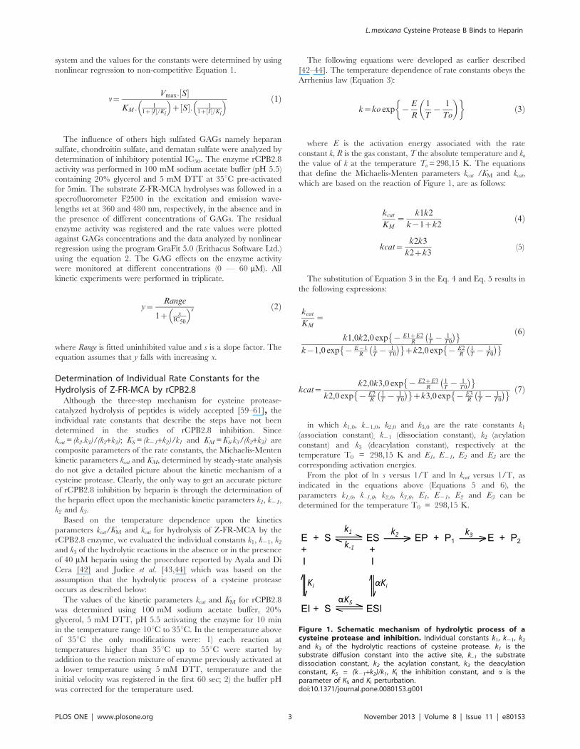
Figure 1. Schematic mechanism of hydrolytic process of acysteine protease and inhibition. Individual constants k1, k21, k2
and k3 of the hydrolytic reactions of cysteine protease. k1 is thesubstrate diffusion constant into the active site, k–1 the substratedissociation constant, k2 the acylation constant, k3 the deacylationconstant, KS = (k21+k2)/k1, Ki the inhibition constant, and a is theparameter of KS and Ki perturbation.doi:10.1371/journal.pone.0080153.g001
L. mexicana Cysteine Protease B Binds to Heparin
PLOS ONE | www.plosone.org 3 November 2013 | Volume 8 | Issue 11 | e80153
.
The Eyring transition-state theory allows the calculation of the
entropy and enthalpy of activation using the following equation
[43–47]:
k~RT
Nhexp
DS
R
� �exp
{DH
RT
� �ð8Þ
The substitution of Equation 8 in the Eq. 4 and Eq. 5 and
calculating kcat/KM.T and kcat /T results in the following
expressions:
kcat
KM :T~
R
Nh
exp DS1zDS2R
� exp { DH1zDH2
RT
� exp DS{1
R
� exp { DH{1
RT
� z exp DS2
R
� exp { DH2
RT
� " # ð9Þ
kcat
T~
R
Nh
exp DS2zDS3R
� exp { DH2zDH3
RT
� exp DS2
R
� exp { DH2
RT
� z exp DS3
R
� exp { DH3
RT
� " #
ð10Þ
in which N is Avogrado’s number and h is Planck’s constant and
DS1, DS21, DS2 and DS3 are corresponding activation entropies
and DH1, DH21, DH2 and DH3 are corresponding activation
enthalpies. DS1, DS21, DS2, DS3, DH1, DH21, DH2 and DH3 can
be obtained from parameters using the Eyring plot ln(kcat /KM.T)
versus 1/T and ln(kcat /T) versus 1/T.
The values of the kinetic parameters kcat and KM were
determined for rCPB2.8 in the temperature range from 10uC to
55uC were: 1) each reaction at temperatures higher than 35uC was
started by addition to the reaction mixture of enzyme previously
activated at a lower temperature using 5 mM DTT, and the initial
velocity was registered in the first 60 sec; 2) the buffer pH was
corrected for the temperature used.
rCPB2.8 pH Activity ProfileFor the determination of pH activity profiles, the kinetics of Z-
FR-MCA substrate hydrolysis were performed in absence or in
presence of 40 mM heparin at 35 uC in four-component buffer
system of constant ionic strength, consisting of 25 mM glycine,
25 mM acetic acid, 25 mM Mes and 75 mM Tris, containing
20% glycerol and 5 mM DTT, the pH of buffers were adjusted
using HCl or NaOH diluted solutions. The substrate concentra-
tions were kept 20-fold above the KM values. The progress of the
reaction was continuously monitored by the fluorescence of the
released product and the initial rates were determined. It is
important to mention that the enzyme was stable at pH range
studied, and the pH values did not affect the ionic form of
substrate. The pH activity profiles data were fitted according to
Equation 11 by using non-linear regression software system
(GraFit version 5.0, Erithacus Software Ltd) as follows:
Vmax~Vmaxlim it
1z10pKES1{pHz10pH{pKES2ð Þ ð11Þ
where Vmaxlim itstands for the pH-independent maximum reac-
tion rate, and pKES1 and pKES2 are the dissociation constants of a
catalytically competent base and acid in the presence of the
substrate respectively.
Effect of Heparin upon Intrinsic Fluorescence of therCPB2.8
The intrinsic fluorescence of tryptophan residues of the
rCPB2.8 was monitored in 50 mM sodium phosphate buffer
(pH 7) containing 20% glycerol at 35uC by measuring the
emission of fluorescence between at 300 — 450 nm (10 nm slit)
after excitation at lex = 290 nm (5 nm slit) in a Hitachi F-2500
spectrofluorimeter in the absence or in the presence of different
concentrations of heparin (0 — 167 mM). The 1 cm path-length
cuvette containing 1 mL of the buffered enzyme solution (1 mM)
was placed in a thermostatically controlled cell compartment
under constant magnetic stirring in the spectrofluorimeter for 5
min prior to the addition of small aliquots of a highly concentrated
heparin solution with minimal dilution (less than 5%) and the
decrease in fluorescence signal was read. The dependence of the
relative fluorescence change, i.e., DF = (Fobs – F0) where F0 is the
initial rCPB2.8 solution fluorescence value and Fobs is the observed
fluorescence value after each addition of heparin, was analyzed by
nonlinear least-squares data fitting by the binding Equation 11
according to [48,49], using Grafit 5.0 Software. The same
procedure described above was used to measure the effect of
heparin upon the intrinsic fluorescence of the N-terminal pro-
region domain of CPB2.8.
DF~DF max :½(PznHzKd )|
ffiffiffiffiffiffiffiffiffiffiffiffiffiffiffiffiffiffiffiffiffiffiffiffiffiffiffiffiffiffiffiffiffiffiffiffiffiffiffiffiffiffiffiffiffiffiffiffiffiffi(PznHzKd )2{4:P:nH
q�
2Pð12Þ
where P is the total protein concentration, H represents the
added heparin fragments concentration, n the stoichiometry, Kd
the dissociation constant and DFmax is the maximum fluorescence
change.
Circular Dichroism ExperimentsCircular dichroism (CD) measurements in far ultraviolet regions
(190 — 260 nm) of rCPB2.8-heparin interactions were conducted
in a JASCO J-810 spectropolarimeter scanning at rate of 50 nm/
min at 35 uC. Cells of 0.1 cm for the far UV were used. The
experiments were done in 5 mM sodium phosphate buffer pH 5.8
containing 2 mM of enzyme rCPB2.8. The observed ellipticity was
normalized to units of degrees.cm2.dmol21. All dichroic spectra
were corrected by subtraction of the background for the spectrum
obtained with buffer alone or buffer containing heparin fragments.
The CD spectra for the rCPB2.8 was analyzed for the relative
amount in percentage, of the secondary structural elements by a
program based on comparison to the spectra obtained for the
structures of known proteins according to [50].
Effects of heparin on the inhibitory activity of N-terminalpro-region of the enzyme rCPB2.8
The inhibitory effect of the N-terminal pro-region on the
endopeptidase activity of the enzyme rCPB2.8 was determined in
the absence or in the presence of 40 mM heparin. The enzyme
rCPB2.8 was pre-activated by incubation in 50 mM sodium
acetate buffer (pH 5.5), containing 5 mM DTT, 20% glycerol for
10 min at 35uC. The rCPB2.8 aliquots were incubated at different
concentrations of pro-region (0 — 0.24 mM) and or with N-
terminal pro-region previously pre-incubated for 30 min with
heparin, the final concentration of heparin at enzyme assay was
12 mM. The endopeptidase activity of the enzyme was continu-
ously monitored by the hydrolysis of fluorogenic substrate Z-FR-
MCA in a spectrofluorometer F-2500 Hitachi, set at wavelengths
L. mexicana Cysteine Protease B Binds to Heparin
PLOS ONE | www.plosone.org 4 November 2013 | Volume 8 | Issue 11 | e80153
.

360 nm for excitation and 480 nm for emission. The remaining
activities of the enzyme inhibited by N-terminal pro-region in the
absence or in the presence of heparin were analyzed by nonlinear
regression using the software GraFit 5.0 (Erithacus Software Ltda).
Results
Effect of Heparin upon the rCPB2.8 EndopeptidaseActivity
The effect of heparin upon the rCPB2.8 endopeptidase activity
was monitored with the aid of its fluorogenic substrate Z-FR-
MCA, covering the rCPB2.8 subsites from S2 to S91 [51,52]. The
HPLC and Maldi-TOF mass spectrometry analysis showed that
Arg-MCA is the only peptide bond cleaved by rCPB2.8 on this
substrate; also, heparin did not change the pattern of cleavage of
this substrate by rCPB2.8. No interaction between Z-FR-MCA
substrate with heparin was detected by studying the molar
absorption of substrate in function of heparin concentration (data
not shown). The possible effect of other high sulfated GAGs,
namely heparan sulfate, chondroitin sulfate, and dermatan sulfate
were also investigated upon rCPB2.8-catalysed hydrolysis of the
fluorogenic substrate Z-FR-MCA. The results showed that the
chondroitin sulfate, and dermatan sulfate are not able to inhibit
the activity of rCPB2.8 in the concentration range from 0 – 60 mM
(data not shown), and only heparin and heparan sulfate were
capable of inhibit the endopeptidase activity of rCPB2.8. These
results show that the interaction of heparin and heparan sulfate
polysaccharides to rCPB2.8 is specific to heparin-like GAGs
similar results have been observed previously [38,39].
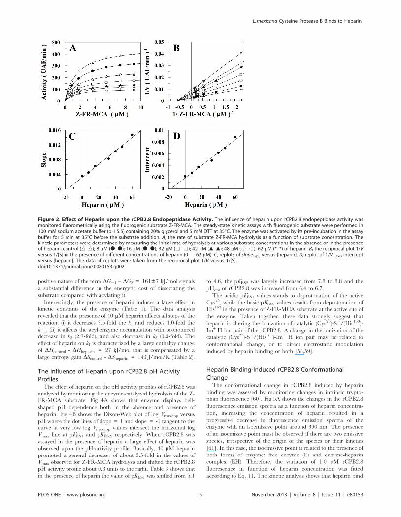
The interaction of heparin with rCPB2.8 perturbs its catalytic
activity upon Z-FR-MCA substrate (Fig. 2). The efficiency of the
system for the hydrolysis of Z-FR-MCA can be altered by
changing either KM value (a parameter) or kcat value (b parameter).
Figs 2A and 2B show that the presence of heparin results in a large
decrease in Vmax value without changing the apparent binding
affinity of the enzyme for its substrate Z-FR-MCA (KS =
1.660.1 mM). It was observed that heparin binds to free rCPB2.8
(E) and to the enzyme-substrate complex (ES) with the same
dissociation constant KH = 1761 mM (Figs 2D and 2E). The data
analysis show that heparin inhibited rCPB2.8 by a linear non-
competitive inhibition mechanism, a = 1.060.1 and b = 0 as
depicted in Figure 1, where the ternary complex enzyme-
substrate-inhibitor (ESI) cannot form product and can only be
converted back to the ES complex or the EI complex.
Interesting, heparin showed a very similar kinetic pattern of
inhibition upon papain when this enzyme was also assayed with
the same substrate Z-FR-MCA. Heparin promoted a large
decrease of 5.5-fold in Z-FR-MCA hydrolysis second-order rate
without changing the affinity of papain for the Z-FR-MCA,
showing a partial non-competitive inhibition behavior [38]. On
the other hand, it has been demonstrated that the inhibitory effects
of heparin on other cysteine proteases from the papain family may
be related to the substrate structure [44,53].
Seminal works of Schneck et al., 2008, and Steiner et al., 1987,
have shown that the steady-state kinetic parameters kcat, KM, and
kcat/KM do not give a clear picture of cysteine-proteases [54] and
serine-proteases [55] substrate specificity, because kcat, KS, and KM
are composite parameters of the intrinsic rate constants. There-
fore, the determination of the steady-state parameters is not
sufficient to describe the kinetic mechanism in the case of rCPB2.8
inhibition by heparin. In order to get an accurate picture of
rCPB2.8 inhibition by heparin, we also analyzed the effect of
heparin upon the individual rate constants k1, k21, k2 and k3 for the
substrate hydrolysis.
Determination of the Individual Rate Constants in therCPB2.8 Kinetic Mechanism
We have used temperature studies of kinetic parameters to
resolve the individual rate constants k1, k21, k2 and k3 in the kinetic
mechanism of rCPB2.8 in the absence or in the presence of 40 mM
heparin as previously described [42–47]. Measurements of k /Kcat M
and kcat as a function of temperature can resolve all the parameters
depicted in Equations 5, 6, 8 and 9.
The temperature dependence of kcat/KM and kcat for Z-FR-
MCA hydrolysis by rCPB2.8 is shown in Fig. 3, in the absence or
in the presence of 40 mM heparin. Figs 3A and 3B show the
Arrhenius plots of the specificity constant kcat /KM (Fig. 3A) and
kcat (Fig. 3B) and Figs 3C and 3D show the Eyring plots of the
specificity constant kcat /KM (Fig. 3C) and kcat (Fig. 3D) in the
temperature range from 10 to 55uC. The observed curvature in
the plots 3A and 3C is indicative of a change in the rate-limiting
step for substrate hydrolysis due to the shift from k2.. k21 at low
temperatures and k21.. k2 at high temperatures. The shift is
caused by the drastic difference in activation energies for substrate
acylation and dissociation when E21.. E2 or E21- E2..0. On
the other hand, the observed linearity in the plots in Figs 3B and
3D is a strong indicative that k3..k2 [42,56]. Taking together
these data, we resolved the kinetic rate constants k1, k21, k2 and k3
at the reference temperature (298.15 K), and the thermodynamic
parameters Ea, DH, DS and DG for this enzymatic reaction in the
absence or in the presence of 40 mM heparin (Table 1).
Table 1 shows that the hydrolysis of substrate Z-FR-MCA by
rCPB2.8 can be satisfactorily described by a three-step kinetic
mechanism, where KS = (k21+k2)/k1 stands for the formation of
ES complex, k2 stands for the rCPB2.8 acylation step and k3 stands
for the rCPB2.8 deacylation step [54]. As expected, the value of k3
(2061 s21) was much higher than the value of k2 (2.760.1 s21)
and the kcat constant is mainly governed by k2, these values agree
with the linearity of the plots (Figs. 3B and 3D), indicating that
substrate acylation is rate limiting over the entire temperature
range examined. Because k2 (2.760.1 s21) is higher than k21
(1.660.1 s21), the hydrolysis of Z-FR-MCA substrate by rCPB2.8
can be considered a diffusion controlled process; the constant of
specificity kcat/KM (1.760.1 mM21.s21) is essentially defined by the
association rate constant k1 (2.760.1 mM21.s21). Despite of k3 ..
k2 and consequently KS = KM; as k2 is 1.7-fold higher than k21, KS
(1.660.2 mM) cannot be considered a true dissociation constant.
The association constant k1 can be limited either by diffusion or
conformational rearrangements that facilitate the enzyme-sub-
strate interaction. As diffusion-limited enzyme substrate encoun-
ters feature k1 values . 107 M21.s21 are linked to small activation
energies E1 , 42 kJ/mol [57], the information on k1 and E1 is
quite valuable in establishing mechanisms of enzyme-substrate
interaction [56]. The present data (Table 2) show that the large
value of E1 (6265 kJ/mol) and k1 (2.7 106 M21.s21) ,
107 M21.s21 in the absence of heparin indicates substantial
structural strains linked to the formation of the enzyme-substrate
complex. Also, during the formation of enzyme-substrate complex
there is a gain of entropy (DS1 = 4763 J/mol.K) and it indicates
that rCPB2.8 can assume a more open conformation [45]. As the
entropy of activation is negative for the acylation and deacylation
step [45], a substantial loss of entropy is observed during the
acylation step as expected (DS2 = –7563 J/mol.K) - strongly
suggesting a refolding of the enzyme [45]. The ‘‘stickiness’’ of
substrate, defined as the rate at which a substrate reacts to give
products relative to the rate at which that substrate dissociates, in
the absence of heparin k2/k21 = 2.060.2, shows that the
propensity of the ES complex to undergo acylation is 2.0-fold
larger than its propensity to decouple in E + S forms. The large
L. mexicana Cysteine Protease B Binds to Heparin
PLOS ONE | www.plosone.org 5 November 2013 | Volume 8 | Issue 11 | e80153
.
positive nature of the term DG21 – DG2 = 16167 kJ/mol signals
a substantial difference in the energetic cost of dissociating the
substrate compared with acylating it.
Interestingly, the presence of heparin induces a large effect in
kinetic constants of the enzyme (Table 1). The data analysis
revealed that the presence of 40 mM heparin affects all steps of the
reaction: (i) it decreases 3.5-fold the k1 and reduces 4.0-fold the
k21, (ii) it affects the acyl-enzyme accumulation with pronounced
decrease in k2 (2.7-fold), and also decrease in k3 (3.5-fold). The
effect of heparin on k1 is characterized by a large enthalpy change
of DHcontrol - DHheparin = 27 kJ/mol that is compensated by a
large entropy gain DScontrol - DSheparin = 143 J/mol/K (Table 2).
The influence of Heparin upon rCPB2.8 pH ActivityProfiles
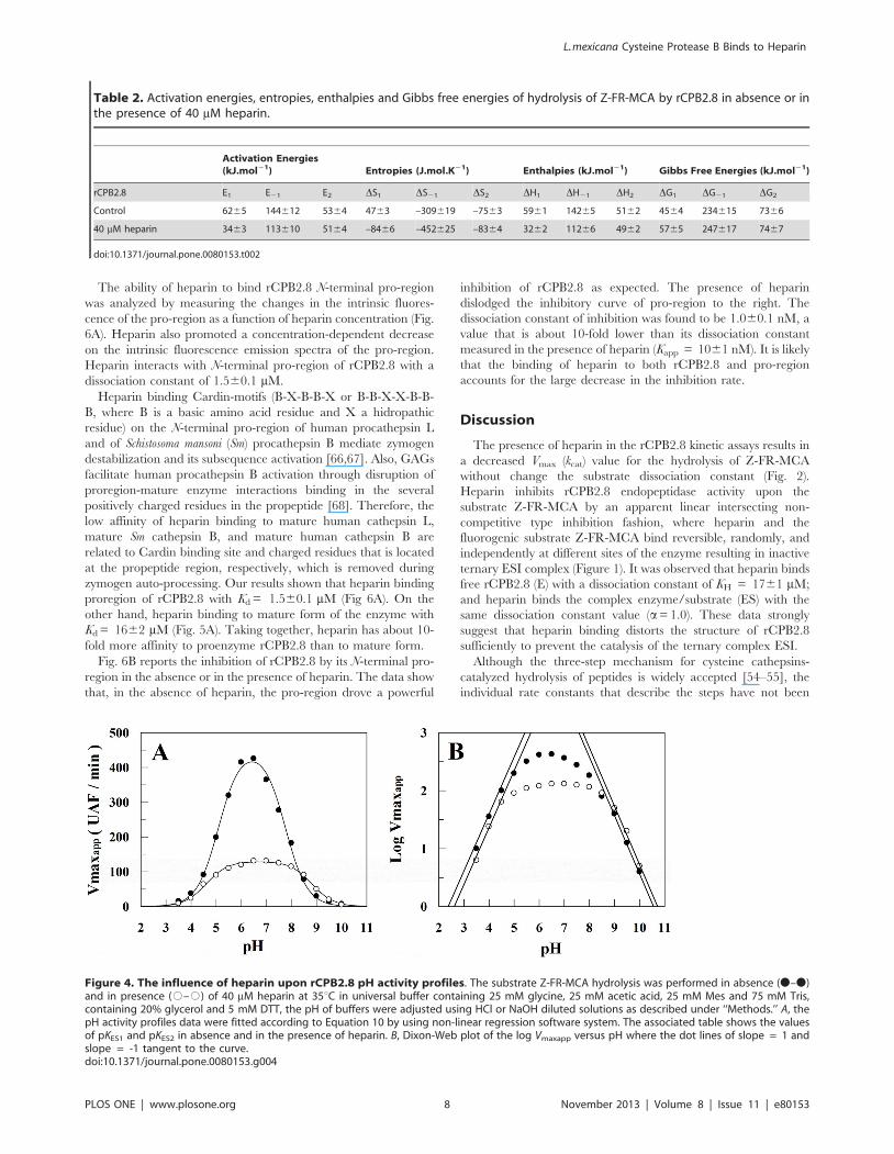
The effect of heparin on the pH activity profiles of rCPB2.8 was
analyzed by monitoring the enzyme-catalyzed hydrolysis of the Z-
FR-MCA substrate. Fig 4A shows that enzyme displays bell-
shaped pH dependence both in the absence and presence of
heparin. Fig 4B shows the Dixon-Web plot of log Vmaxapp versus
pH where the dot lines of slope = 1 and slope = -1 tangent to the
curve at very low log Vmaxapp values intersect the horizontal log
Vmax line at pKES1 and pKES2, respectively. When rCPB2.8 was
assayed in the presence of heparin a large effect of heparin was
observed upon the pH-activity profile. Basically, 40 mM heparin
promoted a general decreases of about 3.5-fold in the values of
Vmax observed for Z-FR-MCA hydrolysis and shifted the rCPB2.8
pH activity profile about 0.3 units to the right. Table 3 shows that
in the presence of heparin the value of pKES1 was shifted from 5.1
to 4.6, the pKES2 was largely increased from 7.8 to 8.8 and the
pHopt of rCPB2.8 was increased from 6.4 to 6.7.
The acidic pKES1 values stands to deprotonation of the active
Cys25, while the basic pKES2 values results from deprotonation of
His163 in the presence of Z-FR-MCA substrate at the active site of
the enzyme. Taken together, these data strongly suggest that
heparin is altering the ionization of catalytic (Cys25)-S2/(His163)-
Im+ H ion pair of the rCPB2.8. A change in the ionization of the
catalytic (Cys25)-S2/(His163)-Im+ H ion pair may be related to
conformational change, or to direct electrostatic modulation
induced by heparin binding or both [58,59].
Heparin Binding-Induced rCPB2.8 ConformationalChange
The conformational change in rCPB2.8 induced by heparin
binding was assessed by monitoring changes in intrinsic trypto-
phan fluorescence [60]. Fig 5A shows the changes in the rCPB2.8
fluorescence emission spectra as a function of heparin concentra-
tion, increasing the concentration of heparin resulted in a
progressive decrease in fluorescence emission spectra of the
enzyme with an isoemissive point around 390 nm. The presence
of an isoemissive point must be observed if there are two emissive
species, irrespective of the origin of the species or their kinetics
[61]. In this case, the isoemissive point is related to the presence of
both forms of enzyme: free enzyme (E) and enzyme-heparin
complex (EH). Therefore, the variation of 1.0 mM rCPB2.8
fluorescence in function of heparin concentration was fitted
according to Eq. 11. The kinetic analysis shows that heparin bind
Figure 2. Effect of Heparin upon the rCPB2.8 Endopeptidase Activity. The influence of heparin upon rCPB2.8 endopeptidase activity wasmonitored fluorometrically using the fluorogenic substrate Z-FR-MCA. The steady-state kinetic assays with fluorogenic substrate were performed in100 mM sodium acetate buffer (pH 5.5) containing 20% glycerol and 5 mM DTT at 35uC. The enzyme was activated by its pre-incubation in the assaybuffer for 5 min at 35uC before the substrate addition. A, the rate of substrate Z-FR-MCA hydrolysis as a function of substrate concentration. Thekinetic parameters were determined by measuring the initial rate of hydrolysis at various substrate concentrations in the absence or in the presenceof heparin, control (n–n); 8 mM (N–N); 16 mM (N–N); 32 mM (%–%); 42 mM (m–m); 48 mM (#–#); 62 mM (*–*) of heparin. B, the reciprocal plot 1/Vversus 1/[S] in the presence of different concentrations of heparin (0 — 62 mM). C, replots of slope1/[S] versus [heparin]. D, replot of 1/V2axis interceptversus [heparin]. The data of replots were taken from the reciprocal plot 1/V versus 1/[S].doi:10.1371/journal.pone.0080153.g002
L. mexicana Cysteine Protease B Binds to Heparin
PLOS ONE | www.plosone.org 6 November 2013 | Volume 8 | Issue 11 | e80153
.

rCPB2.8 by a saturable bimolecular reaction with an apparent Kd
of 1662 mM (Fig. 5A, insert).
The effect of heparin on rCPB2.8 conformation was also
analyzed by CD spectroscopy. Fig 5B shows that the presence of
40 mM heparin causes a significant change in the spectral envelope
of the rCPB2.8, leading a positive increase of the ellipticity value at
[h]222 nm, suggesting that heparin decreases the helicity of
rCPB2.8. Indeed, Table 4 exhibits the secondary structure content
of enzyme in the absence or in presence of 40 mM heparin
concentration. The fractions of the different rCPB 2.8 structural
types, a, b, and remainder (R), were computed from the CD
spectra (197–260 nm) as described previously (38). The data show
a large decrease of 27% in the rCPB2.8 a-helix content induced by
heparin, whereas b-structure and remainder content increased.
These changes are likely to reflect the enzyme-heparin interaction.
Heparin Decreases the Rate of Inhibition of rCPB2.8 by itsN-terminal Pro-Region
CPB2.8 and other lysosomal cysteine proteases are synthesized
as inactive zymogens due to the presence of an N-terminal pro-
region; this domain is a potent inhibitor of the cysteine proteases
[62,63]. Cleavage and dissociation of the N-terminal propeptide
domain with concomitant activation of the cysteine protease occur
as the result of an autocatalytic processing under acidic conditions
[64] or in the presence of glycosaminoglycans [65–67].
Figure 3. Arrhenius and Eyring plots of the kcat/KM and kcat for the Z-FR-MCA hydrolisys by rCPB2.8. A, Arrhenius plot of the ln (kcat/KM)versus 1000/T. B, Arrhenius plot of the ln kcat versus 1000/T. C, Eyring plot of the ln (kcat/KM.T) versus 1000/T. D, Eyring plot of the ln (kcat/T) versus1000/T. The experimental conditions were: the values of the kinetic parameters kcat and KM for rCPB2.8 was determined using 100 mM sodiumacetate buffer, 20% glycerol, 5 mM DTT, pH 5.5 activating the enzyme for 10 min in the temperature range 10uC to 55uC as described under‘‘Methods.’’ The data were obtained in the absence (N–N) or in the presence (#–#) of 40 mM heparin. The continuous lines (slops) were drawnaccording the Equations 6, 7, 9 and 10, with the best-fit parameter values listed in Table 1 and 2.doi:10.1371/journal.pone.0080153.g003
Table 1. Kinetic rate constants of hydrolysis of Z-FR-MCA by rCPB2.8 in absence or in the presence of 40 mM heparin.
ENZYME Kinetic rate constants (298.15 K)
rCPB2.8 k1(mM21.s21) k21 (s21) k2 (s21) k3 (s21) KS (mM) kcat (s21)
Control 26546168 1.5860.03 2.6960.03 2062 1.660.2 2.460.1
40 mM heparin 768611 0.4060.01 1.0060.01 5.760.6 1.860.2 0.8560.08
doi:10.1371/journal.pone.0080153.t001
L. mexicana Cysteine Protease B Binds to Heparin
PLOS ONE | www.plosone.org 7 November 2013 | Volume 8 | Issue 11 | e80153
.
The ability of heparin to bind rCPB2.8 N-terminal pro-region
was analyzed by measuring the changes in the intrinsic fluores-
cence of the pro-region as a function of heparin concentration (Fig.
6A). Heparin also promoted a concentration-dependent decrease
on the intrinsic fluorescence emission spectra of the pro-region.
Heparin interacts with N-terminal pro-region of rCPB2.8 with a
dissociation constant of 1.560.1 mM.
Heparin binding Cardin-motifs (B-X-B-B-X or B-B-X-X-B-B-
B, where B is a basic amino acid residue and X a hidropathic
residue) on the N-terminal pro-region of human procathepsin L
and of Schistosoma mansoni (Sm) procathepsin B mediate zymogen
destabilization and its subsequence activation [66,67]. Also, GAGs
facilitate human procathepsin B activation through disruption of
proregion-mature enzyme interactions binding in the several
positively charged residues in the propeptide [68]. Therefore, the
low affinity of heparin binding to mature human cathepsin L,
mature Sm cathepsin B, and mature human cathepsin B are
related to Cardin binding site and charged residues that is located
at the propeptide region, respectively, which is removed during
zymogen auto-processing. Our results shown that heparin binding
proregion of rCPB2.8 with Kd = 1.560.1 mM (Fig 6A). On the
other hand, heparin binding to mature form of the enzyme with
Kd = 1662 mM (Fig. 5A). Taking together, heparin has about 10-
fold more affinity to proenzyme rCPB2.8 than to mature form.
Fig. 6B reports the inhibition of rCPB2.8 by its N-terminal pro-
region in the absence or in the presence of heparin. The data show
that, in the absence of heparin, the pro-region drove a powerful
inhibition of rCPB2.8 as expected. The presence of heparin
dislodged the inhibitory curve of pro-region to the right. The
dissociation constant of inhibition was found to be 1.060.1 nM, a
value that is about 10-fold lower than its dissociation constant
measured in the presence of heparin (Kapp = 1061 nM). It is likely
that the binding of heparin to both rCPB2.8 and pro-region
accounts for the large decrease in the inhibition rate.
Discussion
The presence of heparin in the rCPB2.8 kinetic assays results in
a decreased Vmax (kcat) value for the hydrolysis of Z-FR-MCA
without change the substrate dissociation constant (Fig. 2).
Heparin inhibits rCPB2.8 endopeptidase activity upon the
substrate Z-FR-MCA by an apparent linear intersecting non-
competitive type inhibition fashion, where heparin and the
fluorogenic substrate Z-FR-MCA bind reversible, randomly, and
independently at different sites of the enzyme resulting in inactive
ternary ESI complex (Figure 1). It was observed that heparin binds
free rCPB2.8 (E) with a dissociation constant of KH = 1761 mM;
and heparin binds the complex enzyme/substrate (ES) with the
same dissociation constant value (a= 1.0). These data strongly
suggest that heparin binding distorts the structure of rCPB2.8
sufficiently to prevent the catalysis of the ternary complex ESI.
Although the three-step mechanism for cysteine cathepsins-
catalyzed hydrolysis of peptides is widely accepted [54–55], the
individual rate constants that describe the steps have not been
Figure 4. The influence of heparin upon rCPB2.8 pH activity profiles. The substrate Z-FR-MCA hydrolysis was performed in absence (N–N)and in presence (#–#) of 40 mM heparin at 35uC in universal buffer containing 25 mM glycine, 25 mM acetic acid, 25 mM Mes and 75 mM Tris,containing 20% glycerol and 5 mM DTT, the pH of buffers were adjusted using HCl or NaOH diluted solutions as described under ‘‘Methods.’’ A, thepH activity profiles data were fitted according to Equation 10 by using non-linear regression software system. The associated table shows the valuesof pKES1 and pKES2 in absence and in the presence of heparin. B, Dixon-Web plot of the log Vmaxapp versus pH where the dot lines of slope = 1 andslope = -1 tangent to the curve.doi:10.1371/journal.pone.0080153.g004
Table 2. Activation energies, entropies, enthalpies and Gibbs free energies of hydrolysis of Z-FR-MCA by rCPB2.8 in absence or inthe presence of 40 mM heparin.
Activation Energies(kJ.mol21) Entropies (J.mol.K21) Enthalpies (kJ.mol21) Gibbs Free Energies (kJ.mol21)
rCPB2.8 E1 E21 E2 DS1 DS21 DS2 DH1 DH21 DH2 DG1 DG21 DG2
Control 6265 144612 5364 4763 –309619 –7563 5961 14265 5162 4564 234615 7366
40 mM heparin 3463 113610 5164 –8466 –452625 –8364 3262 11266 4962 5765 247617 7467
doi:10.1371/journal.pone.0080153.t002
L. mexicana Cysteine Protease B Binds to Heparin
PLOS ONE | www.plosone.org 8 November 2013 | Volume 8 | Issue 11 | e80153
.

determined in the studies of rCPB2.8’s inhibition. More, if k2
contributes significantly to the KS = (k21 + k2)/k1 relationship,
then an inhibitor that affects the kcat = k2.k3/k2+k3 value may also
affect the KS value. Consequently, the only way to get an accurate
picture of rCPB2.8 inhibition by heparin is through the
determination of the effects of heparin upon each individual rate
constant k1, k21, k2 and k3.
In order to determine the values for the kinetic constants k1, k21,
k2 and k3 of the Z-FR-MCA hydrolysis by rCPB2.8, we studied the
effect of temperature dependence upon kcat and kcat/KM for the
enzyme inhibition (Fig. 3). As expected, heparin affected the acyl-
Table 3. pKES and pHopt values obtained by the monitoringthe hydrolysis of Z-FR-MCA by the enzyme rCPB2.8.in absenceor in the presence of 40 mM heparin.
Control Heparin 40 mM
pKES1 5.160.1 4.66 0.1
pKES2 7.860.1 8.860.1
pKES1 6.460.1 6.760.1
doi:10.1371/journal.pone.0080153.t003
Figure 6. Effects of heparin on the N-terminal pro-region ofrCPB2.8. A, the influence of heparin concentration upon N-terminalpro-region segment intrinsic fluorescence emission. The insert in Ashows the variation of the intrinsic fluoresce emission DF (F-F0) of the N-terminal pro-region as a function of heparin concentration (0 —167 mM) as described under ‘‘Methods.’’ B, heparin prevents theinhibitory activity of N-terminal pro-region upon rCPB2.8. Theremaining activity of the enzyme was plotted as a function of N-terminal pro-region concentration in the absence (#–#) or in thepresence (N–N) of 40 mM heparin.doi:10.1371/journal.pone.0080153.g006
Figure 5. Heparin binding-induced rCPB2.8 conformationalchange. A, the intrinsic fluorescence of tryptophan residues of therCPB2.8 was monitored in 50 mM sodium phosphate buffer containing20% glycerol at 35 uC by measuring the emission of fluorescencebetween at 300 — 450 nm after excitation at lex = 290 nm in theabsence or in the presence of different concentrations of heparin (0 —167 mM). The insert in A shows the variation of the intrinsic fluoresceemission DF (F-F0) as a function of heparin concentration. B, effects ofheparin on rCPB2.8 circular dichroism spectra. About 2 mM rCPB2.8were determined in 5 mM sodium phosphate buffer pH 5.8 in theabsence (N–N) or in the presence (%–%) of 40 mM heparin.doi:10.1371/journal.pone.0080153.g005
Table 4. Far UV (197 – 260 nm) CD spectra of rCPB2.8.
a-Helix % b-Sheet % Remaining %
rCPB2.8 – pH 5.0 1861 3861 4461
rCPB2.8 + 100 mM Heparin 1361 4361 4461
The CD analysis of rCPB2.8 was proceeded at pH 5.0 in the absence or in thepresence of 40 mM heparin as describe under ‘‘Methods.’’doi:10.1371/journal.pone.0080153.t004
L.. mexicana Cysteine Protease B Binds to Heparin
PLOS ONE | www.plosone.org 9 November 2013 | Volume 8 | Issue 11 | e80153
.

enzyme formation with pronounced decrease in k2 (2.7-fold), and
also decreased the rate of the hydrolysis of the acyl-enzyme
complex k3 3.5-fold (Table 1). Interesting, the magnitude of the
effect of heparin on the substrate association rate constant step (k1)
was basically the same as the heparin-induced decrease the
substrate dissociation constant step (k21+ k2); in both cases heparin
decreased 3.5-fold its values (Table 1). In other words, the
apparent dissociation constant of the substrate KS was the same in
the absence (1.660.2 mM) or in the presence of heparin
(1.860.2 mM). These data corroborate the previously results
depicted in Fig. 2 which describe heparin as an apparent non-
competitive inhibitor of the rCPB2.8.
The binding of heparin with rCPB2.8 was characterized by a
large enthalpy-entropy energetic compensation (Table 2), the data
proves that heparin decreases diffusion of Z-FR-MCA into the
rCPB2.8 active site by 3.5-fold and that this effect is linked to
significant conformational change that increases the energetic
barrier for formation of the productive ES complex by DG = 12
kJ/mol. As expected, heparin also increases the energetic barrier
for the substrate dissociation from the ES complex by decreasing
4.0-fold the dissociation rate k21. In the presence of heparin the
energetic cost (DG21 – DG2) is also increased 13 kJ/mol. Both
large values of DG of the association and dissociation steps indicate
substantial structural strains linked to the formation/dissociation
of the ES complex in the presence of heparin, which underscore a
conformational change that prevents the diffusion of substrate in
the rCPB2.8 active site.
The binding of heparin to rCPB2.8 also perturbs its intrinsic
fluorescence. The binding of heparin induced a saturable suppres-
sion upon the intrinsic fluorescence emission spectra of the enzyme
(Fig. 5A). Interestingly, the estimated affinity of heparin for rCPB2.8
measured by intrinsic fluorescence assays (Kd = 1662 mM) was
very similar to the values of KI found for the inhibition of its
endopeptidase activity (KI = 1761 mM). These results indicate that
heparin binding is perturbing the rCPB2.8 structure in a similar
manner.
The quenching in the intrinsic fluorescence emission spectra
of the enzyme indicates that the buried tryptophan residues
became exposed to a more polar environment in the presence
of heparin [60]. Because rCPB2.8 contains numerous trypto-
phan residues, the change in fluorescence could not be ascribed
to specific residues. It is important to note that rCPB2.8 has 8
tryptophan residues in its composition [22], and that heparin
binding produced a decrease of 25% in the intrinsic
fluorescence of tryptophan residues present in it. Interesting,
the docking analysis show that the interaction of heparin with
Trp185 at subsite S19 and with Trp189 residues at subsite S2’ of
the rCPB2.8 active site suppresses the intrinsic fluorescence of
rCPB2.8. The present data are consistent with a conforma-
tional change induced by heparin binding that affects both
enzyme activity and the intrinsic fluorescence of tryptophan
residues.
Binding to heparin also significantly decreases the a-helix
content of rCPB2.8. This binding was marked by significant
changes in the shape and position of the CD spectral envelope
(Fig. 5B). Table 4 shows that heparin decreases the helical content
of rCPB2.8 by decreasing the number of amino acids residues in
the helical conformation; and this effect seems to be related to the
increase of the b-sheet content. These data strongly suggest that
the conformational change induced by heparin binding leads to a
decrease in the catalysis of the rCPB2.8 for the substrate Z-FR-
MCA.
The presence of heparin disturbed the pH activity profile of the
enzyme (Fig. 4), in the presence of heparin the value of the pKES1
was shifted from 5.1 to 4.6, and the pKES2 was increased from
7.8 to 8.8 (Table 3). For members of the papain family, the ionic
thiolate-imidazole (Cys25)-S2 by (His159)-Im+H interaction of
the active site is predicted to result in low nucleophilic reactivity
of the ion pair S atom as in the case for an un-ionized thiol
group. It has been suggested that the release of nucleophilic
reactivity in Cys25 requires decrease in the solvation of (Cys25)-
S2 by (His159)-Im+H occasioned by movement of the cleft
around Trp177 (papain numbering). This Trp177 movement
results in His159 being exposed to solvent with consequent
decrease in its solvation of the thiolate anion of Cys25. More, the
Trp177 movement is correlated with the interruption of Trp177-
Gln19 hydrogen bond across pKa 4; the rupture of the hydrogen
bond between Trp177-Gln19 seems to be essential to establish the
oxyanion hole in binding the developing tetrahedral species
during the acylation process [58,59]. In rCPB2.8, the function
of Trp177 residue of papain in catalysis is assumed by Trp185
[29].
Recently, it has been demonstrated that heparin and several
other GAGs can increase the rate of pro-cysteine cathepsins
maturation by disrupting the interactions between its N-terminal
pro-region and mature enzymes [22,65,68]. Indeed, our results
clearly show that heparin interacts with the N-terminal pro-region
of rCPB2.8 showing dissociation constant of 1.560.1 mM, a value
that is about 10-fold lower than its dissociation constant for mature
rCPB2.8 (Kd = 1662 mM). The results strongly suggest that
heparin interacts preferentially with the inactive pro-enzyme
rather than its mature form. It is also important to mention that
the interaction of heparin with the N-terminal pro-region of
rCPB2.8 significantly decreased its inhibitory activity against the
mature enzyme (Fig. 6B). Despite of the exact molecular
mechanism for rCPB2.8 zymogen activation is unknown yet, the
procathepsin B activation shows that this mechanism involves
unimolecular conformational change followed by a bimolecular
proteolytic removal of the propeptide, which can be accomplished
in one or more steps. This activation is also facilitated by
glycosaminoglycans or by binding to negatively charged surfaces
[65].
Taken together, the present results suggest that heparin, at
low concentrations (below 2 mM), can both weaken the
effectiveness of the N-terminal pro-region inhibitor and poten-
tiate the conversion of pro-enzyme into mature CPB form and,
conversely, higher concentrations of heparin (above 20 mM) can
inhibit this conversion. The large amounts of proteoglycans and
free chains of GAG present in the extracellular matrix can
overcome the high rate of conversion pro-enzyme into mature
forms of CPB2.8, thereby decreasing the amount of the active
enzyme present.
Thus the results suggest that GAGs from the ECM of host cells
may be important binding sites for the parasite cysteine proteases.
In the ECM, GAGs are covalently bound to core proteins, forming
a dense network of fixed negative charges available for interaction
with CPB enzymes released extracellularly by Leishmania during its
process of infection. It has been shown that Leishmania can secrete
cysteine protease enzymes (CPB) to the ECM of host cells; this
process is related to parasite infection by proteolysis of fibronectin
from host ECM [37]. Also, it is well known that during ECM
remodeling or pathological degradation mediated by several
proteolytic enzymes, GAG chains are released following hydrolysis
of core proteins [69,70].
GAG chains released from ECM proteoglycans by the action
of proteolytic enzymes during parasite infection and inflam-
matory response may contribute to the regulation of CPB
enzymes by themselves and in association with inhibitors such
L. mexicana Cysteine Protease B Binds to Heparin
PLOS ONE | www.plosone.org 10 November 2013 | Volume 8 | Issue 11 | e80153
.
as its N-terminal pro-region. So, depending on their concen-
tration, GAG can either stimulate or antagonize the activity of
CPB enzymes during parasite infection. Similar GAG control
has also been described for other classes of proteolytic enzymes
[71].
Author Contributions
Conceived and designed the experiments: ILST WASJ. Performed the
experiments: WASJ MAM GPS TMS PCA CSS TFG. Analyzed the data:
WASJ GHC ILST. Contributed reagents/materials/analysis tools: LJ
GDW SJS. Wrote the paper: WASJ GHC ILST.
References
1. Sajid M, McKerrow JH (2002) Cysteine proteases of parasitic organisms. Mol
Biochem Parasitol 120: 1-21. Review. Erratum in: Mol Biochem Parasitol 121:
159.
2. Coombs GH (1982) Proteinases of Leishmania mexicana and other flagellate
protozoa. Parasitology 84: 149–155.
3. Pupkis MF, Coombs GH (1984) Purification and characterization of proteolytic
enzymes of Leishmania mexicana amastigotes and promastigotes. J Gen Microbiol
130: 2375–2383.
4. Robertson CD, Coombs GH (1990) Characterization of three groups of cysteine
proteinases in the amastigotes of Leishmania mexicana mexicana. Mol Biochem
Parasitol 42: 269–276.
5. Robertson CD, Coombs GH (1992) Stage-specific proteinases of Leishmania
mexicana promastigotes. FEMS Microbiol Lett 94: 127–132.
6. Bates PA, Robertson CD, Coombs GH (1994) Expression of cysteine proteinases
by metacyclic promastigotes of Leishmania mexicana. J Euk Microbiol 41: 199–203.
7. Mottram JC, Frame MC, Brooks DR, Tetley L, Hutchison JE, et al. (1997) The
multiple cpb cysteine proteinase genes of Leishmania mexicana encode isoenzymes
that differ in their stage regulation and substrate preferences. J Biol Chem 272:
14285–14293.
8. Souza AE, Waugh S, Coombs GH, Mottram JC (1992) Characterization of a
multicopy gene for a major stage-specific cysteine proteinase of Leishmania
mexicana. FEBS Lett 311: 124–127.
9. Robertson CD, Coombs GH (1994) Multiple high activity cysteine proteases of
Leishmania mexicana are encoded by the lmcpb gene array. Microbiology 140: 417–
424.
10. Mottram JC, Souza AE, Hutchison JE, Carter R, Frame MJ, et al. (1996).
Evidence from disruption of the lmcpb gene array of Leishmania mexicana that
cysteine proteinases are virulence factors. Proc Natl Acad Sci USA 93: 6008–
6013.
11. Coombs GH, Mottram JC (1997) Proteinases of trypanosomes and Leishmania.
In: Hide, G, Mottram, JC, Coombs, GH, Holmes, PH, editors. Trypanosomiasis
and leishmaniasis: biology and control. Oxford: Oxford-CAB International. pp.
177–197.
12. Traub-Cseko YM, Duboise M, Boukai LK, McMahon-Pratt D (1993)
Identification of two distinct cysteine proteinase genes of Leishmania pifanoi
axenic amastigotes using the polymerase chain reaction. Mol Biochem Parasitol
57: 101–116.
13. Sakanari JA, Nadler SA, Chan VJ, Engel JC, Leptak C, et al. (1997) Leishmania
major: comparison of the cathepsin L- and B-like cysteine protease genes with
those of other trypanosomatids. Exp Parasitol 85: 63–76.
14. Campetella O, Henriksson J, Aslund L, Frasch ACC, Pettersson U, et al. (1992)
The major cysteine proteinase (cruzipain) from Trypanosoma cruzi is encoded by
multiple polymorphic tandemly organized genes located on different chromo-
somes. Mol Biochem Parasitol 50: 225–234.
15. Murta ACM, Persechini PM, Padron TS, Souza W, Guimaraes JA, et al. (1990)
Structural and functional identification of GP57/51 antigen of Trypanosoma cruzi
as a cysteine proteinase. Mol Biochem Parasitol 43: 27–38.
16. Mottram JC, North MJ, Barry JD, Coombs GH (1989) A cysteine proteinase
cDNA from Trypanosoma brucei predicts an enzyme with an unusual C-terminal
extension. FEBS Lett 258: 211–215.
17. Coombs GH, Mottram JC (1997) Parasite Proteinases and Amino Acid
Metabolism: Possibilities for Chemotherapeutic Exploitation. Parasitology 114:
S61–S80.
18. DeSouza LS, Lang T, Prina E, Hellio R, Antoine JC (1995) Intracellular
Leishmania amazonensis Amastigotes Internalize and Degrade MHC Class II
Molecules of Their Host Cells. J Cell Sci 108: 3219–3231.
19. Alexander J, Coombs GH, Mottram JC (1998) Leishmania mexicana Cysteine
Proteinase- Deficient Mutants Have Attenuated Virulence for Mice and
Potentiate a Th1 Response. J Immunol161: 6794–6801.
20. Barrett MP, Mottram JC, Coombs GH (1999) Recent advances in identifying
and validating drug targets in trypanosomes and leishmanias. Trends Microbiol
7: 82–88.
21. Caffrey CR, Scory S, Steverding D (2000) Cysteine proteinases of trypanosome
parasites: novel targets for chemotherapy. Curr Drug Targets 1: 155–162.
22. Sanderson SJ, Pollock KG, Hilley JD, Meldal M, St Hilaire PM, et al. (2000)
Expression and characterization of a recombinant cysteine proteinase of
Leishmania mexicana. Biochem J 347: 383–388.
23. Alves LC, Melo RL, Sanderson SJ, Mottram JC, Coombs GH, et al. (2001) S1
subsite specificity of a recombinant cysteine proteinase, CPB, of Leishmania
Mexicana compared with cruzain, human cathepsin L and papain using
substrates containing non-natural basic amino acids. Eur J Biochem 268:
1206–1212.
24. Alves LC, Judice WA, St Hilaire PM, Meldal M, Sanderson SJ, et al. (2001)
Substrate specificity of recombinant cysteine proteinase, CPB, of Leishmania
mexicana. Mol Biochem Parasitol 116: 1–9.
25. Alves LC, Melo RL, Cezari MH, Sanderson SJ, Mottram JC, et al. (2001)
Analysis of the S2 subsite specificities of the recombinant cysteine proteinases
CPB of Leishmania mexicana, and cruzain of Trypanosoma cruzi, using fluorescentsubstrates containing non-natural basic amino acids. Mol Biochem Parasitol 117:
137–143.
26. St Hilaire PM, Alves LC, Sanderson SJ, Mottram JC, Juliano MA, et al. (2000)The substrate specificity of a recombinant cysteine protease from Leishmania
mexicana: application of a combinatorial peptide library approach. Chem BiolChem 1: 115–122.
27. Juliano MA, Brooks DR, Selzer PM, Pandolfo HL, Judice WA, et al (2004)
Differences in substrate specificities between cysteine protease CPB isoforms of
Leishmania mexicana are mediated by a few amino acid changes. Eur J Biochem271: 3704–3714.
28. Alves LC, St Hilaire PM, Meldal M, Sanderson SJ, Mottram JC, et al. (2001)
Identification of peptides inhibitory to recombinant cysteine proteinase, CPB, ofLeishmania mexicana. Mol Biochem Parasitol 114: 81–88.
29. St Hilaire PM, Alves LC, Herrera F, Renil M, Sanderson S, et al (2002) Solid-
Phase library synthesis, screening and selection of tight-binding reduced peptidebond inhibitors of a recombinant Leishmania mexicana cysteine protease B. J Med
Chem 45: 1971–1982.
30. Graven A, St Hilaire PM, Sanderson SJ, Mottram JC, Coombs GH, et al. (2001)Combinatorial library of peptide isosters based on Diels-Alder reactions:
identification of novel inhibitors against a recombinant cysteine protease from
Leishmania mexicana. J Comb Chem 3: 441–452.
31. Stoka V, McKerrow JH, Cazzulo JJ, Turk V (1998) Substrate inhibition ofcruzipain is not affected by the C-terminal domain. FEBS Lett 429: 129–133.
32. Conrad HE (1998) Heparin-Binding Proteins. Academic Press Inc., New York.
33. Del Nery E, Juliano MA, Lima APCA, Scharfstein J, Juliano L (1997)
Kininogenase activity by the major cysteinyl proteinase (cruzipain) fromTrypanosoma cruzi. J Biol Chem 272: 25713–25718.
34. Bartlett AH, Park PW (2010) Proteoglycans in host-pathogen interactions:
molecular mechanisms and therapeutic implications. Expert Rev Mol Med 12:e5.
35. Love DC, Esko JD, Mosser DM (1993) A heparin-binding activity on
Leishmania amastigotes which mediates adhesion to cellular proteoglycans. JBiol Chem 123: 759–766.
36. de Castro Cortes LM, de Souza Pereira MC, da Silva FS, Pereira BA, de
Oliveira Junior FO, et al. (2012) Participation of heparin binding proteins fromthe surface of Leishmania (Viannia) braziliensis promastigotes in the adhesion of
parasites to Lutzomyia longipalpis cells (Lulo) in vitro. Parasit Vectors 5: 142.
37. Kulkarni MM, Jones EA, McMaster WR, McGwire BS (2008) Fibronectinbinding and proteolytic degradation by Leishmania and effects on macrophage
activation. Infect Immun 76: 1738–1747
38. Almeida PC, Nantes IL, Rizzi CCA, Judice WAS, Chagas JR, et al. (1999)
Cysteine proteinase activity regulation. A possible role of heparin and heparin-like glycosaminoglycans. J Biol Chem 274: 30433–30438.
39. Almeida PC, Nantes IL, Chagas JR, Rizzi CCA, Faljoni-Alario A, et al. (2001)
Cathepsin B activity regulation. Heparin-like glycosaminogylcans protect humancathepsin B from alkaline pH-induced inactivation. J Biol Chem 276: 944–951.
40. Lima APCA, Almeida PC, Tersariol ILS, Schmitz V, Schmaier AH, et al. (2002)
Heparan sulfate modulates kinin release by Trypanosoma cruzi through theactivity of cruzipain. J Biol Chem 277: 5875–5881.
41. Li Z, Hou WS, Bromme D (2000) Collagenolytic activity of cathepsin K is
specifically modulated by cartilage-resident chondroitin sulfates. Biochemistry39: 529–536.
42. Ayala YM, Di Cera E (2000) A simple method for the determination of
individual rate constants for substrate hydrolysis by serine proteases. Protein Sci9: 1589–1593.
43. Judice WA, Cezari MH, Lima AP, Scharfstein J, Chagas JR, et al. (2001)
Comparison of the specificity, stability and individual rate constants with
respective activation parameters for the peptidase activity of cruzipain and itsrecombinant form, cruzain, from Trypanosoma cruzi. Eur J Biochem 268:
6578–6586.
44. Judice WA, Mottram JC, Coombs GH, Juliano MA, Juliano L (2005) Specificnegative charges in cysteine protease isoforms of Leishmania mexicana are
highly influential on the substrate binding and hydrolysis. Mol Biochem Parasitol144: 36–43.
45. Laidler KJ, Peterman BF (1979) Temperature effects in enzyme kinetics.
Methods in Enzymology 63: 234–257.
46. Cornish-Bowden A (1999) Fundaments of Enzyme Kinetics. Portland Press,London, UK.
L. mexicana Cysteine Protease B Binds to Heparin
PLOS ONE | www.plosone.org 11 November 2013 | Volume 8 | Issue 11 | e80153
.
47. Polgar L (1999) Oligopeptidase B: a new type of serine peptidase with unique
substrate-dpendent temperature sensitivity. Biochemistry 38: 15548–1555548. Pimenta DC, Nantes IL, de Souza ES, Le Bonniec B, Ito AS, et al. (2002)
Interaction of heparin with internally quenched fluorogenic peptides derived
from heparin-binding consensus sequences, kallistatin and anti-thrombin III.Biochem J 366: 435–446.
49. Shinjo SK, Tersariol IL, Oliveira V, Nakaie CR, Oshiro ME, et al. (2002)Heparin and heparan sulfate disaccharides bind to the exchanger inhibitor
peptide region of Na+/Ca2+ exchanger and reduce the cytosolic calcium of
smooth muscle cell lines. Requirement of C4-C5 unsaturation and 1 4 glycosidiclinkage for activity. J Biol Chem 277: 48227–48233
50. Sreerama N, Woody RW (2004) Computation and Analysis of Protein CircularDichroism Spectra. Methods in Enzymology 383: 318–351
51. Nagler DK, Storer AC, Portaro FC, Carmona E, Juliano L, et al.(1997) Majorincrease in endopeptidase activity of human cathepsin B upon removal of
occluding loop contacts. Biochemistry 36: 12608–12615.
52. Barrett AJ (1980) Fluorimetric assays for cathepsin B and cathepsin H withmethylcoumarylamide substrates. Biochem J 187: 909–912.
53. Nunes GLC, Simoes A, Dyszy FH, Shida CS, Juliano MA, et al. (2011)Mechanism of Heparin Acceleration of Tissue Inhibitor of Metalloproteases-1
(TIMP-1) Degradation by the Human Neutrophil Elastase. PLoS One 6: e21525
54. Schneck JL, Villa JP, McDevitt P, McQueney MS, Thrall SH, et al. (2008)Chemical Mechanism of a Cysteine Protease, Cathepsin C, As Revealed by
Integration of both Steady-State and Pre-Steady-State Solvent Kinetic IsotopeEffects. Biochemistry 47: 8697–8710
55. Stein RL, Strimpler AM, Hori H, Powers JC (1987) Catalysis by humanleukocyte elastase. Aminolysis of acyl-enzymes by amino acid amides and
peptides. Biochemistry 26: 2238–2242.
56. Xu H, Bush LA, Pineda AO, Caccia S, Di Cera E (2005) ThrombomodulinChanges the Molecular Surface of Interaction and the Rate of Complex
Formation between Thrombin and Protein C. J Biol Chem 280: 7956–7961.57. van Holde KE (2002) A hypothesis concerning diffusion-limited protein-ligand
interactions. Biophys Chem 101–102: 249–254.
58. Hussain S, Khan A, Gul S, Resmini M, Verma CS, et al. (2011) Identification ofinteractions involved in the generation of nucleophilic reactivity and of catalytic
competence in the catalytic site Cys/His ion pair of papain. Biochemistry 50:10732–10742.
59. Hussain S, Pinitglang S, Bailey TS, Reid JD, Noble MA, et al. (2003) Variationin the pH-dependent pre-steady-state and steady-state kinetic characteristics of
cysteine-proteinase mechanism: evidence for electrostatic modulation of
catalytic-site function by the neighbouring carboxylate anion. Biochem J 372:
735–746.
60. Gesteira TF, Coulson-Thomas VJ, Taunay-Rodrigues A, Oliveira V, Thacker
BE, et al. (2011) Inhibitory peptides of the sulfotransferase domain of the
heparan sulfate enzyme, N-deacetylase-N-sulfotransferase-1. J Biol Chem 286:
5338–4536.
61. Koti ASR, Krishna MMG, Periasamy N (2001) Time-resolved area-normalized
emission spectroscopy (TRANES): a novel method for confirming emission from
two excited states, J Phys Chem A 105: 1767–1771.
62. Carmona E, Dufour E, Plouffe C, Takebe S, Mason P, et al. (1996) Potency and
selectivity of the cathepsin L propeptide as an inhibitor of cysteine proteases.
Biochemistry 35: 8149–8157.
63. Coulombe R, Grochulski P, Sivaraman J, Menard R, Mort JS, et al. (1996)
Structure of human procathepsin L reveals the molecular basis of inhibition by
the prosegment. EMBO J 15: 5492–5503.
64. Mason RW, Gal S, Gottesman MM (1987) The identification of the major
excreted protein (MEP) from a transformed mouse fibroblast cell line as a
catalytically active precursor form of cathepsin L. Biochem J 248: 449–454.
65. Pungercar JR, Caglic D, Sajid M, Dolinar M, Vasiljeva O, et al. (2009)
Autocatalytic processing of procathepsin B is triggered by proenzyme activity.
FEBS J 276: 660–668.
66. Fairhead M, Kelly SM, van der Walle CF (2008) A heparin binding motif on the
pro-domain of human procathepsin L mediates zymogen destabilization and
activation. Biochem Biophys Res Commun 366: 862–867.
67. Horn M, Jılkova A, Vondrasek J, Maresova L, Caffrey CR, et al. (2011)
Mapping the Pro-Peptide of the Schistosoma mansoni Cathepsin B1 Drug Target:
Modulation of Inhibition by Heparin and Design of Mimetic Inhibitors. ACS
Chem Biol 6: 609–617.
68. Caglic D, Pungercar JR, Pejler G, Turk V, Turk B (2007) Glycosaminoglycans
Facilitate Procathepsin B Activation through Disruption of Propeptide-Mature
Enzyme Interactions. J Biol Chem 282: 33076–33085.
69. Novinec M, Grass RN, Stark WJ, Turk V, Baici A, et al. (2007) Interaction
between human cathepsins K, L and S and elastins: mechanism of elastinolysis
and inhibition by macromolecular inhibitors. J Biol Chem 282: 7893–7902.
70. Roughley PJ, Barrett AJ (1977) The degradation of cartilage proteoglycans by
tissue proteinases. Proteoglycan structure and its susceptibility to proteolysis.
Biochem J 167: 629–637.
71. Schenker P, Baici A (2010) Paradoxical interactions between modifiers and
elastase-2. FEBS J 277: 2486–2495.
L. mexicana Cysteine Protease B Binds to Heparin
PLOS ONE | www.plosone.org 12 November 2013 | Volume 8 | Issue 11 | e80153
.
Copyright © 2022 FDOKUMEN