Monitoring parliamentarians: promises, barriers and dilemmas of parliamentary openness
Upload
khangminh22Category
view
0download
0
REVIEW ARTICLE
Promises and challenges of adoptive T-cell therapies forsolid tumoursMatteo Morotti 1,20, Ashwag Albukhari2, Abdulkhaliq Alsaadi1, Mara Artibani1, James D. Brenton 3, Stuart M. Curbishley4,Tao Dong5,6, Michael L. Dustin7, Zhiyuan Hu1, Nicholas McGranahan8, Martin L. Miller 9, Laura Santana-Gonzalez1,Leonard W. Seymour10, Tingyan Shi11, Peter Van Loo 12, Christopher Yau13,14, Helen White15, Nina Wietek1, David N. Church 16,17,David C. Wedge 17,18 and Ahmed A. Ahmed1,17,19
Cancer is a leading cause of death worldwide and, despite new targeted therapies and immunotherapies, many patients withadvanced-stage- or high-risk cancers still die, owing to metastatic disease. Adoptive T-cell therapy, involving the autologous orallogeneic transplant of tumour-infiltrating lymphocytes or genetically modified T cells expressing novel T-cell receptors or chimericantigen receptors, has shown promise in the treatment of cancer patients, leading to durable responses and, in some cases, cure.Technological advances in genomics, computational biology, immunology and cell manufacturing have brought the aspiration ofindividualised therapies for cancer patients closer to reality. This new era of cell-based individualised therapeutics challenges thetraditional standards of therapeutic interventions and provides opportunities for a paradigm shift in our approach to cancertherapy. Invited speakers at a 2020 symposium discussed three areas—cancer genomics, cancer immunology and cell-therapymanufacturing—that are essential to the effective translation of T-cell therapies in the treatment of solid malignancies. Keyadvances have been made in understanding genetic intratumour heterogeneity, and strategies to accurately identify neoantigens,overcome T-cell exhaustion and circumvent tumour immunosuppression after cell-therapy infusion are being developed.Advances are being made in cell-manufacturing approaches that have the potential to establish cell-therapies as credibletherapeutic options. T-cell therapies face many challenges but hold great promise for improving clinical outcomes for patients withsolid tumours.
British Journal of Cancer https://doi.org/10.1038/s41416-021-01353-6
BACKGROUNDPersonalised, or precision, medicine aims to identify the optimaltreatment for each patient in order to maximise benefit andminimise toxicity. In the context of cancer treatment, advancesin genomics have enabled the identification of germline andsomatic genomic alterations that can be matched to therapeu-tics on an individual patient basis1—in some cases, such as theuse of imatinib for the treatment of chronic myeloid leukaemiato great effect.2 However, most cancer genomes lack drivermutations for which a molecularly targeted agent is available.3
The unprecedented results obtained using novel cancerimmunotherapies such as immune checkpoint blockade (ICB)have revealed the potential of leveraging the immune system in
cancer treatment.4 Unfortunately, despite durable responses in asubset of patients, particularly those with melanoma or non-small cell lung cancer (NSCLC),5–7 most patients do not respondto the immunotherapies in current use;8,9 in fact, it has beenestimated that responders to ICB might constitute around only13% of cancer patients.10
Advanced therapy medicinal products (ATMPs) hold promise totransform cancer treatment11—cell-based vaccines,12,13 engi-neered T cells14 or autologous tumour-infiltrating lymphocytes(TILs), for example, represent highly personalised modes of cancertreatment. However, the application of cell-therapy approaches tolarge numbers of cancer patients presents major challenges, asgood manufacturing practice (GMP) is a complex and expensive
www.nature.com/bjc
Received: 9 November 2020 Revised: 22 February 2021 Accepted: 4 March 2021
1Ovarian Cancer Cell Laboratory, MRC Weatherall Institute of Molecular Medicine, University of Oxford, Oxford, UK; 2Biochemistry Department, Faculty of Science, King AbdulazizUniversity, Jeddah, Saudi Arabia; 3Functional Genomics of Ovarian Cancer Laboratory, Cancer Research UK Cambridge Institute, University of Cambridge, Cambridge, UK;4Advanced Therapies Facility and National Institute for Health Research (NIHR) Biomedical Research Centre, University of Birmingham, Birmingham, UK; 5Medical ResearchCouncil (MRC) Human Immunology Unit, MRC Weatherall Institute of Molecular Medicine, University of Oxford, Oxford, UK; 6Chinese Academy of Medical Sciences (CAMS) OxfordInstitute, University of Oxford, Oxford, UK; 7Kennedy Institute of Rheumatology, University of Oxford, Oxford, UK; 8Cancer Genome Evolution Research Group, University CollegeLondon Cancer Institute, London, UK; 9Cancer System Biology Group, Cancer Research UK Cambridge Institute, University of Cambridge, Cambridge, UK; 10Gene Therapy Group,Department of Oncology, University of Oxford, Oxford, UK; 11Department of Gynecological Oncology, Zhongshan Hospital, Fudan University, Shanghai, China; 12Cancer GenomicsLaboratory, The Francis Crick Institute, London, UK; 13Division of Informatics, Imaging and Data Sciences, Faculty of Biology Medicine and Health, University of Manchester,Manchester, UK; 14The Alan Turing Institute, London, UK; 15Patient Representative, Endometrial Cancer Genomics England Clinical Interpretation Partnership (GeCIP) Domain,London, UK; 16Wellcome Centre for Human Genetics, University of Oxford, Oxford, UK; 17Oxford NIHR Biomedical Research Centre, Oxford, UK; 18Manchester Cancer ResearchCentre, University of Manchester, Manchester, UK and 19Nuffield Department of Women’s & Reproductive Health, University of Oxford, Oxford, UKCorrespondence: David N. Church ([email protected]) or David C. Wedge ([email protected]) or Ahmed A. Ahmed ([email protected])20Present address: Department of Oncology, Ludwig Institute for Cancer Research Lausanne, Lausanne University Hospital (CHUV) and University of Lausanne (UNIL), Lausanne,Switzerland
© The Author(s) 2021 Published by Springer Nature on behalf of Cancer Research UK

process. Moreover, to improve the effectiveness of ATMPs, weneed to gain a better and more comprehensive understanding ofthe interaction between cancer and the immune system. Thisreview will discuss what we believe are the three most importantareas for advancing the field of personalised cell immunothera-pies: cancer genomics; cancer–immune system interactions; andATMP manufacturing. We will focus on discussing the promisesand challenges of each area for solid cancers and highlightpotential factors for improvement, particularly in the field of T-celltherapy (Box 1).
ATMPS IN THE ERA OF PERSONALISED MEDICINEAn ATMP can be a gene therapy medicinal product (e.g.Holoclar®, a stem-cell treatment used for limbal stem-celldeficiency in the eye); a somatic cell therapy medicinal product(e.g. TILs for the treatment of tumours); a tissue-engineeredproduct (e.g. anti-CD19 CAR-T therapies); or a combination ofany of the above (Table 1). Personalised cell-therapy treatmentsare a type of ATMP, manufactured specifically for each patientusing their own cellular material (e.g. immune cells). They areintended as long-term or permanent therapeutic solutions toacute or chronic human diseases such as cancer. The ability toextract and grow immune cells—in particular, T-cells—in vitropaved the way for the use of adoptive cell transfer (ACT; thetransfer of cells to a patient) in cancer immunotherapy.15
Broadly, ACT can be carried out using three different T-cellapproaches. In the first, TIL-ACT, endogenous TILs are expandedex vivo from a patient’s tumour before being infused back intothe patient. The second approach uses T-cell receptors (TCRs)that have been engineered to recognise specific tumourantigens, although this approach is limited to major histocom-patibility complex (MHC)-expressed antigens. Chimeric antigenreceptors (CARs) comprise an extracellular antigen recognitiondomain, a transmembrane domain, and a cytoplasmic signallingdomain, and so can recognise a variety of cell-surface antigens,by contrast, independently of MHCs.16,17
Scientific advances throughout the entire pipeline frombiopsy to the manufacturing of cell therapy products havemade ACT potentially more accessible to growing numbers ofpatients. Accordingly, cell therapy trials in cancer are steadilyincreasing in number worldwide, with the second largestnumber of active trials (after ICB) in the immuno-oncologyspace.18 However, despite ~90% of cancer incidences globallybeing caused by solid tumours, only around half of these trialsare targeting solid tumours, and they have rarely been extendedto non-melanoma cancers, due to a lower immunogenicity ofthese tumours.19–23 Moreover, although clinical trials using celltherapy products directed against cancer neoantigens derivedfrom somatic mutations hold promise, evidence of success is stilllimited to case reports of patients with particular tumour typessuch as cervical cancer,24 cholangiocarcinoma,25 metastaticmelanoma,26–29 colorectal cancer,30 breast cancer31 and thy-moma.32 Further studies are thus needed to advance thepotentially curative approach of ACT.
CANCER GENOMICSThe decreasing costs and technological advances in massivelyparallel sequencing techniques have enabled the identification ofsomatic mutations in cancer on a large scale and facilitated theclinical implementation of genomic medicine.33–35
Neoantigens and personalisation of therapyThe cellular immune response to cancer largely depends on T cellsthat specifically target cancer/testis antigens (CTA) or cancerneoantigens that are derived from somatic genomic alterationsthat lead to the expression of immunogenic neoepitopes.36–38 Inthis article, we focus on cancer neoantigens, which result fromgenomic perturbations, exhibit entirely novel amino acidsequences and, importantly, are rarely shared among patients.These mutant peptides that bind to human leukocyte antigen(HLA) class I or HLA class II molecules are capable of generating arobust and durable immune response, and high mutational andpredicted neoantigen load are significantly associated withimproved progression-free and overall survival in melanomapatients treated with TIL-ACT.39 Furthermore, evidence frommelanoma patients treated with TILs suggests that the evokedimmune response comprises both CD4+ and CD8+ T cells specificfor mutant epitopes,25,27,40 with cases of off-target immuneresponse against the wild-type, non-mutated peptide beingexceptionally rare.41,42
The combination of genomics and cellular immunotherapypermits the identification of somatic alterations and the predictionof potential neoantigens that could be utilised as targets fortherapy by vaccination, adoptive TIL transfer, or engineered T-cells.42,43 The pipeline for neoantigen-directed cell-therapy ispresented in Fig. 1a. Significant efforts have been made toadvance the individual steps, particularly the detectionof mutations and prediction of neoepitopes, which isprincipally determined by the probability that mutant peptidesbind HLA-I.44–47 The current state-of-the-art and challenges ofbioinformatic analysis of the cancer mutanome for ACT has beenextensively reviewed elsewhere.47,48
Tumour heterogeneity and evolutionData from international collaborative studies have shed light onmany aspects of cancer genomics and cancer evolution, whichcould potentially inform the implementation of bioinformaticspipelines for cancer mutanome discovery.33,49 Genomic studieshave demonstrated how tumour heterogeneity and evolutiondrive resistance to systemic anti-cancer and targeted therapy50–52
such as epidermal growth factor receptor (EGFR) amplification andmutations in the MET or PIK3CA gene resistance in EGFR+tumours after EGFR inhibitors treatment.53 Cancer heterogeneitysimilarly represents a pivotal challenge for the development ofneoantigen-directed-TIL therapies.54
The expansion of TILs from multiregional biopsy samples, fromdistinct cancerous lesions within the same patient or fromdifferent regions within the same tumour, could give a moreaccurate snapshot of intratumour heterogeneity at a single time-point (Fig. 1b) and, therefore, enable the more successful designof TIL therapies targeting a substantial proportion of tumour cellsin any given cancer.55 Neoantigen-specific CD8+ and CD4+
lymphocytes have also been detected in peripheral blood,56–58
which could overcome the problem of limited specimenavailability in certain tumours. However, the low levels of theseneoantigen-specific lymphocytes from peripheral blood and theirdiscordant TCR repertoire compared with TILs collected from thesame patients could represent a challenge for their suitabilityfor ACT.59
Clonal mutations and neoantigen quality. A 2020 publicationlooking at the timescales of tumour development showed that‘first driver’ events seemingly occurred up to decades before
Box 1 Generation of this conference report
This Oxford meeting ‘Advanced Personalised Therapeutics in Solid Cancers’ inFebruary 2020 convened experts to discuss our current understanding ofchallenges in advance medicinal therapeutic products (ATMPs) in the treatmentof solid cancer. The emphasis was on discussing challenges in the field of cancergenomics, cancer–immune system interaction and manufacturing of ATMPs. Theidea of summarising the outputs from the meeting in a report was proposed andunanimously approved before the meeting. Following the talks, at the end ofeach topic session, an open discussion with all meeting participants was held tocollate ideas and future perspectives. A draft statement was then circulated to allauthors for feedback and refinement, leading to agreement with the viewsexpressed in this manuscript.
Promises and challenges of adoptive T-cell therapies for solid tumoursM Morotti et al.
2
1234567890();,:
diagnosis, demonstrating how cancer genomes are shaped by anear-lifelong process of somatic evolution.60 The evolution ofmost cancers usually involves common early mutations in cancerdriver genes, followed by diverse trajectories generated byindividually rare driver mutations, and by copy number altera-tions.60 Intriguingly, for some cancers, genomic sequencing isunable to identify driver mutations.49 A notable advantage ofneoantigen-driven immunotherapy is that the issue of whether ornot a mutation in a cancer gene occurs is irrelevant; what isimportant is that the mutation is clonal—that is, it is shared by allcancer cells—which increases the number of therapeutic targetsin a way that is not feasible using traditional small-moleculetargeted therapies. Predicting neoantigens that are sharedamongst a substantial proportion of the target cell population isvital for the success of ACT, irrespective of which gene a mutationresides in. Mutations that occur early are important to consider forimmunotherapy as they would be clonal and are, therefore, at thetrunk of a tumour’s phylogenetic tree61–63 (Fig. 1b) and mightconsequently generate a more effective anti-cancer T-cellresponse compared with later mutations (subclonal) that arelimited to subpopulations of cancer cells.64–66 The identification ofclonal mutations relies on the accurate computation of theprevalence of mutations in a tumour.67 This is not a trivial task andis often confounded by normal cell contamination, substantialheterogeneity and copy number alterations.68 However, advancesin sequencing and computational technologies, particularly in thefield of linked-read methods, have enabled improved estimationof the prevalence of mutations by incorporating phase informa-tion. This allows the alleles identification on maternal and paternalchromosomes which is important for understanding geneexpression patterns in tumours.69,70 Whether subclonal neoanti-gens developing in a rapidly evolving tumour or in response totreatment pressure71 can actively distract the immune responsefrom effectively targeting clonal neoantigens is unclear. Thus,therapeutic efforts might need to be oriented toward thetargeting of multiple clonal neoantigens to optimise diseasecontrol and minimise the potential for immune escape.
Predicting neoantigens beyond somatic mutations. Another keychallenge is that only a few predicted neoantigens encoded bysomatic non-synonymous mutations are actually immuno-genic.55,72–74 Therefore, expanding the search for immunogenicantigens to include different categories of genomic alterationsbeyond non-synonymous mutations is essential. Single-nucleotidevariants (SNVs), insertions and deletions (indels) causing
frameshifts, chromosome alterations and splice variants could allpotentially generate neoantigens (Fig. 1c).75 Data from cancergenomics studies also shed insights into the timing of acquisitionof structural variants and76,77 copy number gains78–80 and theincidence and timing of punctuated events (such as chromo-thripsis, chromoplexy, kataegis) in cancer evolution.49,81 Owing totheir high prevalence in cancer genomes and their near absencein healthy tissues, complex chromosomal rearrangements repre-sent an exciting potential target for the identification ofneoantigens. Frameshifts resulting from indels, if they evadenonsense-mediated decay, have been shown to generateapproximately three times as many high-affinity neoantigens asnon-synonymous SNVs.82,83 Interestingly, mitochondrial-localisedpeptides were shown to induce a more robust immune responsethan cytoplasmic peptides, owing to their increased stability,indicating that the subcellular localisation of peptides might beimportant in determining their immunogenicity, which couldpotentially be exploited during the identification of predictedneoantigens.84
As shown in Fig. 1a, the neoantigens predicted from genomesequencing data (whole-genome and exome sequencing) areusually filtered by gene expression (RNA-seq) to assess theexpression (yes or no) of that given gene. However, single-celltranscriptomic sequencing (scRNA-seq) data from ovarian andlung cancer documented the presence of non-genetic hetero-geneity amongst cancer cells, with evidence of remarkableplasticity and capability of transitioning from one cell state toanother.85–88 This phenomenon might be relevant for theselection of cancer neoantigens. For example, knowing thepredominant cellular state of a tumour (e.g., stemness pro-gramme88) would make it possible to prioritise neoantigens thatare encoded by genes that are expressed in that particular cellstate. Not surprisingly, promoter hypermethylation has beenshown to affect the expression of ~23% of the predictedneoantigens studied in a large cohort of lung cancer,89 high-lighting the occurrence of transcriptional repression mechanismsin cancer and their potential significance in neoantigenidentification.90
Improving the prediction of neoantigen immunogenicity. Oncepotential neoepitope-generating mutations have been identified,T cells are assessed for their reactivity against tandem minigenes(TMGs) or peptides containing the potential neoantigens (Fig. 1a).Challenges in determining the anti-tumour reactivity ofneoantigen-specific T-cells have been reviewed elsewhere47 but,
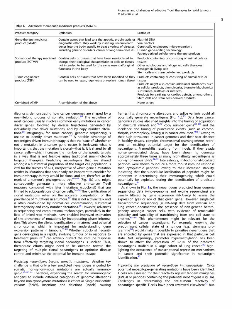
Table 1. Advanced therapeutic medicinal products (ATMPs).
Product category Definition Examples
Gene-therapy medicinalproduct (GTMP)
Contain genes that lead to a therapeutic, prophylactic ordiagnostic effect. They work by inserting ‘recombinant’genes into the body, usually to treat a variety of diseases,including genetic disorders, cancer or long-term diseases.
Plasmid DNAViral vectorsGenetically engineered micro-organismsHuman gene-editing technologyPatient-derived cellular gene therapy products
Somatic-cell therapy medicinalproduct (SCTMP)
Contain cells or tissues that have been manipulated tochange their biological characteristics or cells or tissuesnot intended to be used for the same essential/originalfunctions in the body.
Products containing or consisting of animal cells ortissuesOther autologous and allogeneic cells therapiesXenogeneic living cellsStem cells and stem cell-derived products
Tissue-engineeredproduct (TEP)
Contain cells or tissues that have been modified so theycan be used to repair, regenerate or replace human tissue.
Products containing or consisting of animal cells ortissuesProducts might also contain additional substances, suchas cellular products, biomolecules, biomaterials, chemicalsubstances, scaffolds or matricesProducts for cartilage or cardiac defects, among othersStem cells and stem cells-derived products
Combined ATMP A combination of the above None as yet
Promises and challenges of adoptive T-cell therapies for solid tumoursM Morotti et al.
3

in addition to technological limitations, it is important to note thatnot every specific neoepitope gives an immune response.91 Aninherent bias in this pipeline is that we can only test theimmunogenicity of the neoepitope-generating mutations that weare able to predict bioinformatically. In addition, neoantigenoverlap predictions of the top 100 ranked peptide-bound MHCsfrom the same tumour samples between different teams of a
global community has been shown to be low (less than 20%) inthe majority of the cases. This lack of consensus might be partiallydriven by differences in epitope filtering and/or ranking betweenthe different teams and suggest that efforts to harmonizeneoantigen-prediction will be necessary for future clinical cross-comparison of neoantigen-based TIL trials between differentcentres.92 Moreover, despite mutations that poorly bind to HLA-II
a
b
Genetic heterogeneity
Early events Late events
Truncal
Met
Primary
Non-genetic heterogeneity
Intertumour heterogeneity Intratumour heterogeneity
Normal cell Tumour growth Treatment Metastasis Relapse
X driver mutations
X X X X X XX
XX
Time
Met
Blood/Biopsy
DNA and RNAcell sequencing
Tetramers or tandem minigenes
TIL APC
TCR-HLA
TIL culture
Assay for T-cell activation
CD
137
CD4 or CD8
Rapid expansion protocol
Co-culture
Infusion
NeoAgs discovery
c
Single nucleotide variants Indels
DNA
PROTEIN
HLA-I
Normal
Missense Nonsense
Post-translationalmodifications
Nucleus CytoplasmProtein
Immunogenic
Nonimmunogenic
Not presented
Proteasome Peptide
ImmunogenicMitochondria
Fig. 1 Targeting cancer neoantigens using cell therapy. a Using autologous tumour-infiltrating lymphocytes in autologous cell transfer. Theresected specimen is divided into multiple tumour fragments that are individually grown in IL-2 for 7–10 days. For the ‘non-specific’ TILtherapy (dashed line) the individual cultures are then moved to a rapid expansion protocol before reinfusion into patients. Neoantigen-TILtherapy involves the sequencing of exomic or whole-genome DNA from tumour cells and healthy cells to identify tumour-specific mutations,before RNA-sequencing is used to check for the expression of mutations. Corresponding minigenes or peptides encoding each mutatedamino acid are synthesised and expressed in or pulsed into a patient’s autologous antigen-presenting cells (APCs) for presentation in thecontext of a patient’s HLA. Individual mutations responsible for tumour recognition are identified by analysing activation of a T-cell co-stimulatory marker, such as 41BB/CD137 (CD8+ T cells), in response to cognate target antigen recognition. b Genetic and genomicheterogeneity and evolution of clonal populations. Upper panel: Genetic and phenotypic variations are observed between tumours ofdifferent tissues (inter-tumour heterogeneity). Within a tumour, subclonal diversity can be observed (intra-tumour heterogeneity, differentcolours of tumour clones). Clonal alterations occurring early in tumorigenesis are represented by the blue trunk of the phylogenetic tree(truncal mutations); later alterations could be shared by tumour cells in some regions of the tumour (light blue and pink branches of the tree)or present in only one region of the tumour (yellow branches of the tree) in a branched cancer evolution model. Tumour subclones can alsoshow differential gene expression due to non-genetic heterogeneity. Lower row: Unique clones (represented by different colours) emerge as aconsequence of accumulating driver mutations in the progeny of a single most recent common ancestor cell. Ongoing linear and branchingevolution results in multiple simultaneous subclones that can individually give rise to episodes of disease relapse and metastasis. c Overviewof the neoantigen landscape. The sources of potential neoantigens for HLA class I ligands are shown. In tumours, mutated or aberrantlyexpressed proteins are processed via the proteasome into peptides. The cross-priming abilities of peptides are also linked to non-geneticfactors such as protein stability, which can be modulated by several factors, including their localisation in the mitochondria. These peptidescan be loaded onto HLA class I molecules and might or might not elicit a CD8+ T-cell response, depending on several factors, includingpeptide sequence or T-cell receptor (TCR) sequences. In general, most of the neoantigens derived from single-nucleotide variants gain theirimmunogenicity through altered amino acids involved in direct T-cell contact.
Promises and challenges of adoptive T-cell therapies for solid tumoursM Morotti et al.
4
being positively selected for during tumorigenesis, thus empha-sising the importance of CD4+ T cells in anti-tumour immunity,93
the computational prediction and analysis of HLA-II remains anongoing challenge owing to the highly polymorphic nature of theHLA-II and poorly characterised endosomal HLA-II peptideprocessing, which limits the development of HLA-II peptideprocessing algorithms.45,94,95 Furthermore, despite the corebinding motif of both HLA molecules comprising peptides ofapproximately nine amino acids, HLA-II-restricted ones have awider length range (11–20 amino acids) compared with HLA-I-restricted ones (8–11 amino acids)96 which can make bioinfor-matics prediction task challenging.The use of mass spectrometry for the direct discovery of
tumour-specific HLA peptides (immunopeptidomics) holds pro-mise for defining targets for immunotherapy (Table 2). Promisesand challenges of this approach have been reviewed else-where.97,98 The use of data-driven machine-learning approachesto leverage information from established sets of HLA-I and,notably, HLA-II ligandomic data has provided new hope forimproving the ability to predict a broader range of neoantigens,including those derived from post-translational modification aswell as from the cancer-specific translation of products arising as aconsequence of alternative splicing and intron retention, RNAediting, novel open reading frames and endogenous retroviruselements99–105 (Fig. 1c). The need to incorporate these ‘unconven-tional’ tumour antigens into current approaches for predictingneoantigens has been reviewed.38 The increase in available dataon peptide immunogenicity and TCR binding prediction alongwith machine-based learning extrapolation could have the powerto improve the quality of neoantigen identification106 by
incorporating structural and associated physical principles intoapproaches for evaluating immunogenicity107 and to potentiallyidentify immunogenic hotspots for directed neoantigen target-ing.108,109
In conclusion, despite being reliable and extremely promising,personalised immunotherapy that targets unique mutations facesmany challenges. Apart from the technical and logistical hurdlesfor this highly personalised approach, which will be discussedbelow, a better understanding of cancer trajectories frompreneoplastic lesions to invasive cancer and of the simultaneouspressure of the immune system (immunoediting) is warranted toimprove the identification of neoantigens.65
CANCER–IMMUNE SYSTEM INTERACTIONSCancer immunoediting and immunoevasionScientific advances over the past decades have demonstratedthat the immune system can paradoxically both constrain andpromote tumour development and progression. This process isreferred to as cancer immunoediting and, in its most complexform, involves three phases—elimination, equilibrium andescape—that ultimately result in the advent of immune-resistant variants.110,111 The constant pressure from the adaptiveimmune system coupled with the genetic instability of tumourcells can select for tumour subclones with reduced immuno-genicity that can evade immune recognition and destruc-tion.112–114 The immunological control of tumour progressionand sculpting of tumour genomes has been shown in severalsolid cancers, with tumour regions that harbour the highestlevels of immune infiltrates exhibiting the lowest cancer cell
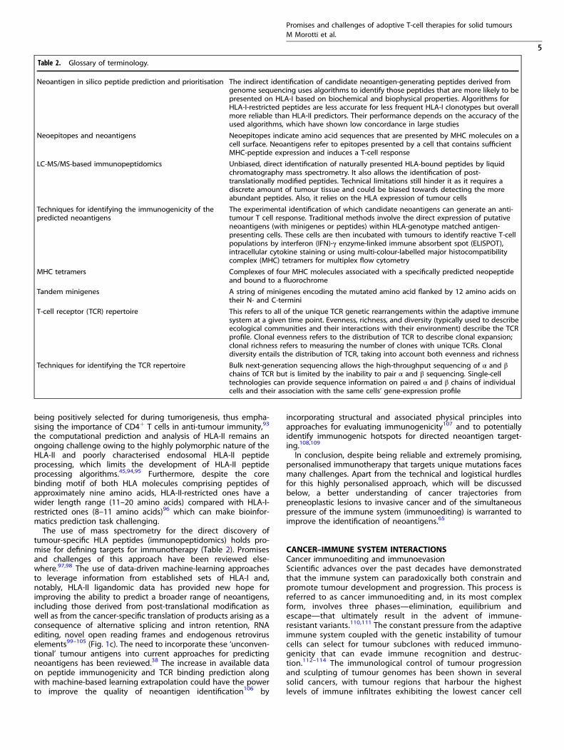
Table 2. Glossary of terminology.
Neoantigen in silico peptide prediction and prioritisation The indirect identification of candidate neoantigen-generating peptides derived fromgenome sequencing uses algorithms to identify those peptides that are more likely to bepresented on HLA-I based on biochemical and biophysical properties. Algorithms forHLA-I-restricted peptides are less accurate for less frequent HLA-I clonotypes but overallmore reliable than HLA-II predictors. Their performance depends on the accuracy of theused algorithms, which have shown low concordance in large studies
Neoepitopes and neoantigens Neoepitopes indicate amino acid sequences that are presented by MHC molecules on acell surface. Neoantigens refer to epitopes presented by a cell that contains sufficientMHC-peptide expression and induces a T-cell response
LC-MS/MS-based immunopeptidomics Unbiased, direct identification of naturally presented HLA-bound peptides by liquidchromatography mass spectrometry. It also allows the identification of post-translationally modified peptides. Technical limitations still hinder it as it requires adiscrete amount of tumour tissue and could be biased towards detecting the moreabundant peptides. Also, it relies on the HLA expression of tumour cells
Techniques for identifying the immunogenicity of thepredicted neoantigens
The experimental identification of which candidate neoantigens can generate an anti-tumour T cell response. Traditional methods involve the direct expression of putativeneoantigens (with minigenes or peptides) within HLA-genotype matched antigen-presenting cells. These cells are then incubated with tumours to identify reactive T-cellpopulations by interferon (IFN)-γ enzyme-linked immune absorbent spot (ELISPOT),intracellular cytokine staining or using multi-colour-labelled major histocompatibilitycomplex (MHC) tetramers for multiplex flow cytometry
MHC tetramers Complexes of four MHC molecules associated with a specifically predicted neopeptideand bound to a fluorochrome
Tandem minigenes A string of minigenes encoding the mutated amino acid flanked by 12 amino acids ontheir N- and C-termini
T-cell receptor (TCR) repertoire This refers to all of the unique TCR genetic rearrangements within the adaptive immunesystem at a given time point. Evenness, richness, and diversity (typically used to describeecological communities and their interactions with their environment) describe the TCRprofile. Clonal evenness refers to the distribution of TCR to describe clonal expansion;clonal richness refers to measuring the number of clones with unique TCRs. Clonaldiversity entails the distribution of TCR, taking into account both evenness and richness
Techniques for identifying the TCR repertoire Bulk next-generation sequencing allows the high-throughput sequencing of α and βchains of TCR but is limited by the inability to pair α and β sequencing. Single-celltechnologies can provide sequence information on paired α and β chains of individualcells and their association with the same cells’ gene-expression profile
Promises and challenges of adoptive T-cell therapies for solid tumoursM Morotti et al.
5

clonal diversity, which is likely to be a reflection of neoantigendepletion and loss of heterozygosity (LOH) at the HLA loci.115,116
The timing of initiation of immunological sculpting is animportant question in cancer biology. The number of expandedTCRs found ubiquitously across all tumour samples in early-stage lung cancer or paired metastatic breast and ovarian cancerimplies that some level of immune surveillance directed againstclonal neoantigens can be initiated early and maintainedthrough all levels of cancer development, including metastaticprogression.115,117,118 However, as is evident through the lack of
cancer cell elimination, immune escape mechanisms capable ofpreventing T-cell mediated death might be an extremely earlyevent in cancer evolution.
Potential immune escape mechanisms. The immunogenicity ofneoantigens has been challenged. A study using data from TheCancer Genome Atlas (TCGA) showed that neoantigen depletion,detected using HLA affinity predictions, is weak or absent in theuntreated cancer genome overall.119 This observation is consistentwith the fact that the vast majority of non-synonymous mutations
a
c
Tumour microenvironment
Treg
TEMCD4+
MDSCIL-10TGFβ
VEGFPGE2
ARG-1I-NOSROSTGFβ
CD8+
TAM
ADE
CCL5CXCL9
Ky
TGFβ,TNFα
Tie-2
HLA-LOH, HLA hypermethylation
WNT/β-catenin
PD-L1
CD8+
T-cell exhaustion
Exhaustion/Inhibitory receptors/
Effector function/Proliferation
Cancer cell/APC
pHLA
TCR
Memory-likeExhausted
DifferentiatedExhausted
PDhi
TCFhi
T-bethi
TIM3low
PDhi
T-betlow
EOMEShi
TIM3low
Rep
rogr
amm
abili
tyR
esis
tanc
e to
ICB
TIM3CTL4
TME
IFN-γ, GrzBIL2 sensitivity
NFAT TOXCD8+
Ag stimulation
T-cell modulation and engineering
TCR PD1X TCR PD1
X
Cas9+
gRNA
Ex vivo drug modulation Modifications of culturing conditions
T-cell engineering Protein
engineering
T-cell activationNeoAgs load
TCRoIL-2R
oIL-2
SMAPsCD8+CD8+ CD8+
Stem-like/TRM-TILCD8+ component
PD1
PDL1
b
TAM
CAF
Fig. 2 T cell conditioning to overcome the immunosuppressive tumour microenvironment. a Immuno-evasion mechanisms in the tumourmicroenvironment. A representative example of an ‘excluded’ (cold) T-cell tumour is shown. Some of the most studied immune cells alongwith their ligand–receptor and secreted growth factors (chemokines and cytokines) known to promote immunoevasion are shown. In theblack box, examples are given of cancer genetic alterations linked to an immuno-evasive tumour microenvironment (ADE adenosine, ARG1Arginase 1, CAFs cancer-associated fibroblasts, DC dendritic cell, IL interleukin, iNOS inducible nitric oxide synthase, KYN: kynurenine, MDSCmyeloid-derived suppressor cells, NO nitric oxide, PGE2 prostaglandin E2, ROS reactive oxygen species TAMs tumour-associated macrophages,TGF-β transforming growth factor-β, Treg regulatory T cell, VEGF vascular endothelial growth factor). In the black box are highlighted geneticmechanisms linked to a cold TME. b T cell exhaustion in solid tumours. A representative image of transitions from an effector (Teff) to anexhausted (Tex) T cell is shown. Chronic antigen exposure and the TME pressure promote the activity of transcription factors (such as NFAT,TOX), which increases the expression of exhaustion-associated molecules such as PD1, LAG3, and TIM3, and the downregulation of effectorcytokines such as IFNγ, GrzB and IL-2 sensitivity. GrzB Granzyme B, IL2 interleukin-2, TME tumour microenvironment, TRM tissue-residentmemory. c Potential interventions to increase TIL efficacy during the expansion of T cells for ACT. The tumour microenvironment (TME) can bemodulated ex vivo with different drugs (such as epigenetics, immunometabolic drugs) or interleukins (e.g. IL-2, IL-15) to boost the growth andactivation of tumour-infiltrating lymphocytes, to increase the number of neoantigens (epigenetic drugs) or to preferentially expand TIL-specific subtypes such as tissue resident memory T cells (TRM-TILs). CRISPR–Cas9 ribonuclear protein complexes loaded with single-guideRNAs can be electroporated into TILs or normal T cells for gene editing. Shown here is an example of deletion of TCRs (off-the-shelf T cells)and of inhibiting immune checkpoint receptors such as programmed death 1 (PD-1) in T cells. Protein engineering can be used for thecreation of orthogonal IL-2–IL-2 receptor pairs, which consist of a mutant orthogonal IL-2 cytokine (oIL-2) and mutant IL-2 receptor (oIL-2R)that interact specifically with each other but do not interact with their wild-type counterparts. SMAPs (supramolecular attack particles) can beproduced in vitro and grafted to relevant specific TCRs for adoptive transfer.
Promises and challenges of adoptive T-cell therapies for solid tumoursM Morotti et al.
6

across 29 cancer types are not subject to selection and that only aminority of mutations (∼5%) were positively selected whenevaluating the selection pressures exerted on mutant somaticcancer alleles (nonsense versus missense mutations).120 Similarly,few differences in the immunoediting of clonal neoantigens wereseen in a large cohort of lung cancer patients.89 Therefore, eitheronly very few predicted driver neoantigens are immunogenic, ordriver mutations possess the ability to evolve efficient earlyimmune evasion mechanisms. This could potentially be linked toindividual variation at the HLA locus,121 LOH in the HLAregion or by inducing other mechanisms such as amplificationof the immune checkpoint molecule programmed deathligand-1,119,122,123 as was demonstrated by comparing preinvasivelesions of the lung that were immune-competent (and thereforeregressed) with those that progressed.124 Additionally, hyper-methylation of the HLA region was also commonly observed124
(Fig. 2a).Interestingly, several studies have demonstrated the wide-
spread appearance of cancer mutations in healthy tissues,including the oesophagus, skin, liver, lung, endometrium andcolon.125–130 The concept of normal cell mutagenesis hasrevolutionised our understanding of cancer development, asdriver mutations are not only limited to carcinogenesis but arealso common events throughout life, occurring as early asembryogenesis.131 The presence of these widespread mutationsin healthy tissue might also influence our understanding of cancerimmunotherapy and cell-therapy development, as they couldprovide insight into those potential neoepitopes against whichthe immune system has been already tolerized (e.g. neoepitopesgenerated by mutations in cancer genes early in life). This mightguide future prioritization of neoepitopes for neoantigen-basedtherapies.Another explanation for the lack of negative selection of
mutated cells is that the pervasive presence of driver mutations inhealthy tissue starting even at the embryonic level is tolerated bythe immune system until other insults, such as the potentialinvasion of the basal epithelial layers or metabolic changes,occur.120 In addition, some cancer-associated driver mutationshave been reported to attenuate immune responses. For example,mutations in the genes KRAS and BRAF or other mutationalactivations in components of the mitogen-activated protein kinase(MAPK) pathway can decrease the transcription of HLA class Imolecules as well as the expression of other genes encodingmolecules that are essential for peptide loading.132–135 Whetherand how these driver mutations in healthy tissues are surveyed,selected and removed by the immune system is a crucialbiological question and could significantly enhance our under-standing of cancer–immune interactions and the immunogenicproperties of a given mutation.Increasing our understanding will entail re-focusing on the
importance of tissue-specific immunity/‘structural immunity’136
and how each organ’s immune system can affect cancerdevelopment and treatment.137,138 In the same way thatcomprehensive DNA profiling of tumours has revealed thegenetics of cancers and the significant variation amongst tumoursand individual patients,49,60,81,139,140 knowing the difference in theimmunological composition and peripheral fitness selection ofT cells141 between different organs might provide insights into therole of the early and late events of immunosurveillance in cancerdevelopment in a given tumour microenvironment (TME).142,143
The tumour microenvironment and TILsT-cell infiltration into the TME has been extensively demonstratedto be clinically relevant, with the quantity, quality and location ofthe immune infiltrate (known as the ‘immune contexture’) ofcytotoxic and memory T cells within the solid tumour accuratelypredicting clinical outcome144–147 as well as a positive response toICI therapy.148,149 The density of the T-cell infiltrate has been
linked to cancer genomic characteristics, with tumours that aregenetically more heterogeneous showing less immune infiltra-tion.150,151 Analysis of synchronous metastases in patients withmelanoma has revealed not only genetic heterogeneity but alsoimmune-infiltration heterogeneity in terms of different immunecell types and T-cell clonality between different sites in the samepatient.152
T-cell infiltration, activation and exhaustion. Relatively little isknown about the cancer-intrinsic mechanisms, alterations andoncogenic signalling programmes that underlie TME heterogene-ity.153 The dysregulation of specific pathways, such as WNT–β-catenin signalling, and tumoral amplification of genes that encodeproteins associated with immunosuppression, such as PD-L1, thearachidonate lipoxygenases, and indoleamine-2,3-dioxygenase-1(IDO-1) and IDO-2, have been linked with low T-cell infiltration andlower cytolytic activity.154–156 The association between WNT–β-catenin signalling and TME modulation is further supported by theobservation that immune-excluded tumours are enriched formutations in negative regulators of the WNT–β-catenin pathwayin treatment-naive high-grade serous ovarian cancer157 (Fig. 2a).Several biochemical pathways and cell types in the TME have
been reported to lead to a decrease in T-cell infiltration (T-cellhoming) and activation, as well as increased T-cell exhaustion,thereby increasing immunoevasion in different tumour types158–161
(Fig. 2a). It appears evident that an understanding of the biology ofhow the TME shapes the pattern and levels of immune-cellinfiltration and exhaustion will be a key factor for the success ofACT.162 This understanding could provide the biological knowledgeto pharmacologically modulate the key pathways of immunoevasionprior to or during the administration of ACT to the patients.Furthermore, drugs could be administered to tumour fragmentsex vivo during the manufacturing of the cell product to increase TILexpansion and activation. In addition, the cell product could beengineered to enhance persistence and activation in vivo.163 Theseaspects will be discussed in further detail below.
Therapeutic approaches to increase T-cell infiltration. The ability ofthe TME to show a myriad of pathway redundancies and developmetabolic adaptations precludes, from a clinical point of view,multiple simultaneous targeting to avoid patient toxicity. How-ever, the importance of establishing inflammation in the TME andthus overcoming immune exclusion and increasing T-cell homingin solid tumours (turning the tumours ‘hot’) has stimulatedresearch into the discovery of new therapeutic options.164,165 Suchan approach will be crucial for ACT success as the presence of TILs(indicative of an inflamed tumour) is a prerequisite for TILexpansion. Radiotherapy, which is capable of inducing immuno-genic cell death by exposing tumour-associated antigens/neoanti-gens that can be recognised by antigen-presenting cells (APCs)166
and then presented to CD8+ T cells, is currently being tested inclinical trials in combination with ICB.167
The combination of ICB and ACT has been shown to be feasibleand safe in patients with ovarian cancer.168 Another interestingstrategy to render tumours ‘hot’ is the use of oncolytic viruses—native or genetically modified viruses that selectively infect andreplicate within tumour cells, eventually leading to tumour celllysis.169 Direct injection into cancer cells of the oncolyticvirotherapy agent talimogene laherparepvec, which is alsoengineered to produce granulocyte/macrophage colony-stimulating factor (GM-CSF) to induce an immune response,increased T-cell infiltration and the response to ICIs in melanomapatients.170 Similarly, administration of a dendritic cell (DC)-basedvaccine has been shown to increase T-cell infiltration and induceT-cell responses to autologous tumour antigens in ovarian cancerwhole cell lysate.166 These therapeutic options could constitute apriming step to stimulate an immune response and thus createthe basis for T-cell interrogation of neoantigens for subsequent TIL
Promises and challenges of adoptive T-cell therapies for solid tumoursM Morotti et al.
7

therapy, particularly in those patients whose tumours have a lownumber of TILs.171
These therapies (virotherapy, low-dose radiotherapy) can alsoinduce a systemic increase of chemokines (such as CCL2 and CCL5)known to promote T-cell homing and T-cell infiltration172–174
(Fig. 2b). Indeed, a 2019 publication reported that the cooperationof chemokines such as chemokine ligand 5 (CCL5), which isconstitutively expressed by tumour cells, with IFN-γ-induciblechemokines such as chemokine ligand 9 (CXCL9), plays a key anduniversal role in the orchestration of T-cell responses in tumoursand facilitates the establishment of the T-cell-inflamed pheno-type.175 The loss of tumour-intrinsic chemokines (such as CCL5, forexample) that support T-cell recruitment is a common mechanismof immunoevasion. These results could open opportunities for themanipulation, using genome-engineering techniques, of theseT-cell homing ligands in expanding TIL populations to enhanceT-cell engraftment.
The preferential expansion of TIL subtypes. The intrinsic capacityof intratumoural T cells to recognise adjacent tumour tissue canbe rare and variable (∼10% in the case of melanoma, ovarian andcolorectal cancer CD8+ TILs41,176); the majority of TILs arebystander T cells.177 Thus, strategies to enrich a predefinedneoantigen-specific subpopulation or tumour-specific cells wouldincrease the chances of obtaining a final TIL product withadequate tumour reactivity as well as persistence in vivo.178,179
The preferential expansion, via cytokine modulation or usingbioengineering materials, of specific T-cell subtypes, such as thetissue-resident memory (TRM) T cells, is an interestingapproach.180,181 TRM T cells are particularly attractive for TIL-ACTas they are associated with better survival outcomes in many solidtumours and have an inherent capacity to home, owing to theexpression of specific integrins on their cell surface.182–186
Moreover, the CD8+PD1+CD103+ TRM subpopulation representsthe predominant proportion of TILs with expanded intratumouralubiquitous TCRs (thus recognising clonal neoantigens), and theseTCRs been demonstrated to be expressed on tumour-reactiveT cells in lung cancer patients.187
As mentioned, the success of ACT depends not only on thehoming capability of the cell product, but also on additionalspecific properties such as the differentiation state and ability topersist in vivo, along with the capacity to exert effector functionsagainst cancer cells in the host (Fig. 2b). Therefore, a betterunderstanding of the physiological mechanism that couples cellexpansion and differentiation in T cells could improve the efficacyof ACT.188 An increased understanding might facilitate in vitrostrategies to increase the percentage of tumour-reactive stem-likeTILs, for example, which have been shown to be capable of self-renewal, expansion, persistence and a superior anti-tumourresponse in patients with melanoma.189
Maintaining T-cell activity/responses. The persistence of highamounts of antigen and the immunosuppressive nature of theTME push the majority of cancer-specific T cells towards anexhausted phenotype176,177,190 (Fig. 2c). As these cancer-specificT cells become less responsive to interleukin-2 (IL-2), whichmediates T-cell expansion, they might become diluted bybystander T cells during the process of expansion in TIL-ACT,with an overall loss of TCR repertoire.15,191,192 However, despitetheir functional impairment, it is widely appreciated thatexhausted T cells are often tumour-specific and can still retainsome control over tumour growth, as shown by the great clinicaleffect of blocking programmed death protein 1 (PD-1) orprogrammed cell death ligand 1 (PD-L1) axis in solidtumours.193,194 Exhausted T cells express increased levels ofinhibitory immune checkpoint molecules, such as PD-1, CTLA-4,LAG3 and TIM3,195 and decreased levels of the adhesion/costimulatory molecule CD2, both of which might attenuate
anti-tumour T-cell responses in tumours.196 However, somesubsets of exhausted T cells that express the transcription factorTCF1, also known as precursor exhausted T cells,197 display self-renewing capacity and are essential for the long-term main-tenance of persistent T-cell responses in different solidtumours.198–200 The enrichment of this subpopulation during TILexpansion could therefore enhance the efficacy of ACT thera-pies.189,201
Future efforts to rapidly sort tumour-reactive cells (e.g. thoseexpressing cell co-stimulatory molecules such as PD-1, 4-1BB orOX40)59,191,202 or metabolically ‘fit’ T cells203 might reduce the lossof efficacy seen in response to high-dose IL-2 culture conditionsand improve the persistence of the T-cell product in vivo. Futuretechnological advances that can integrate sensitivity into high-throughput approaches are warranted for the selection of a TILproduct with better anticancer characteristics.48,204,205
So far, T cells in the earliest stages of differentiation (naive orcentral memory) have shown the highest efficacy and persistencein TIL-ACT regimens, as progressive terminal T-cell differentiationor exhaustion causes loss of anti-tumour power through impair-ments in TCR signalling and/or via reductions in either cytolyticactivities or adhesion.206,207 However, although the isolation frompatients’ blood of less-differentiated T-cell subsets can be aneffective strategy for generating superior TCR or CAR-engineeredT-cell products, it is more challenging when using TILs, which areoften found in a state of senescence and functional exhaus-tion.208,209 Moreover, early states of T-cell differentiation mightalso coincide with a decreased expression of tissue homingmolecules and trafficking potential, and a less preferentialexpansion of TRM T cells.210 Thus, preventing this kind of cellularfatigue (exhaustion), alongside manipulation of other molecularpathways, will help to unleash the potential of TIL-ACT for thetreatment of solid tumours.211
Ex-vivo modulation of the TME. As mentioned above, theenrichment of TILs with better functional activity could potentiallybe obtained during the ex vivo expansion stage of the process.Notably, the TME was also able to be manipulated ex vivo by thedirect application to cultured fragments of breast and ovariancancer of the agonistic anti-CD137 antibody, which increased therate of TRM-TIL outgrowth; and ICB has been shown to increasethe polyclonal expansion of infiltrating CD8+ TILs and activationlevels of the final T-cell product.212,213 Furthermore, the additionto expanding TILs of synthetic peptide pools of all predicted HLA-class I neoantigens improved conventional methods of TILgeneration by enriching for neoantigen-specific CD8+ TILs.59
The use of epigenetic therapies in this ex vivo setting couldincrease the expression of transcriptionally repressed neoantigensand CTA,214 which could potentially enhance the recognition ofcancer cells by adaptive immune cells.215,216 Similarly, epigenetictherapies could reverse the T-cell chromatin conformation andDNA methylation that are linked to decreased chemokineproduction, T-cell differentiation and exhaustion.217–219 Potentialstrategies to reprogramme the T-cell metabolic state ex vivo toimprove T-cell phenotypes also exist.220,221 Treating expandingT cells with increased levels of extracellular potassium and acetateresulted in the generation of T cells with retained stemness,evidenced by self-renewal and multipotency.222 Other strategieshave explored the inhibition of T-cell exhaustion by reducingmitochondrial oxidative stress,223,224 the reversal of T-cell senes-cence by inhibiting sestrin complexes.225
Immune-cell engineering. Notably, the time required to expandTILs could also be used as a ‘window of opportunity’ for immune-cellengineering with viral vectors226–228 or for gene editing withCRISPR–Cas9 technology.229 Loss-of-function studies in T cells haveidentified genes that, when deleted, can enhance T-cellresponses,230–234 such as p38 MAPK (MAPK14).235 Many groups
Promises and challenges of adoptive T-cell therapies for solid tumoursM Morotti et al.
8

are also investigating whether candidate transcription factors, suchas c-Jun,236 or synthetic cell receptors,237,238 such as the IL-2receptor,239 can be overexpressed to reprogramme T cells, preventexhaustion, or convert suppressive extracellular signals into activat-ing signals. A first-in-human Phase 1 clinical trial of multiplexCRISPR–Cas9 gene editing in T cells from three patients withadvanced, refractory cancer (two patients with myeloma and onewith sarcoma) demonstrated that this approach is safe andfeasible.240 T cells engineered using CRISPR–Cas9 editing to expressa synthetic, cancer-specific TCR transgene and to lack expression ofPD-1 persisted for longer than T cells retaining the expression of theendogenous TCR and PD-1. The gene editing and syntheticimmunology areas are rapidly expanding, and universal approachessuch as ‘off-the-shelf’ T cells (using T cells from healthy donors)generated through the editing of TCR could substantially advancethe field and dramatically increase the number of patients eligiblefor cell therapies241 (Fig. 2d). Advances in protein engineering alsohold promise. The expression of an orthogonal mutant IL-2 receptorand administration of its paired orthogonal mutant IL-2 ligandactivates only the engineered T cells and not the IL-2 dependentCD4+Foxp3+ subpopulation of regulatory T (TREG) cells,
242,243 which
have been shown to decrease the therapeutic effect of ACT244
(Fig. 2d). Similarly, an engineered IL-2R agonist that preferentiallyreduces IL-2 binding to CD25 (low-affinity IL-2R, highly expressed inTREG cells) over CD122/CD132 (high-affinity IL2R)245 was able toselectively expand intratumoural effector T cells over TREG cells andsynergise with anticancer vaccination.246
The 2020 description of the structure and composition ofcytotoxic multiprotein complexes called supramolecular attackparticles (SMAPs) released by CD8+ effector T cells (cytotoxic Tlymphocytes; CTLs) and natural killer (NK) cells might openimportant avenues for ACT.247 SMAPs, which are composed of acore of cytotoxic proteins such as granzyme B and perforin and ashell of glycoproteins including thrombospondin-1, are a distincttype of extracellular particle of ~100 nm in diameter released byimmune cells.247,248 Ideally, the innate specificity of these particleswould be re-engineered towards an individual tumour andpotentially HLA type, enabling the targeting of neoantigen-expressing tumour cells, potentially by grafting relevant TCRs ontotheir surfaces.Several approaches exist that might, therefore, allow the
development of optimised next-generation T cells that display
MANUFACTURING
TRANSPORT/TRACKINGTRANSPORT/PRESERVATION/DISTRIBUTION
CELL THERAPY TRAINING
CENTRALIZED/DECENTRALIZED FACILITY
PATIENT
CELL THERAPY HOSPITAL
INVENTORY
STORAGE
DA
TA
PR
OT
EC
TIO
N DA
TA
CO
LLEC
TIO
N
QC OPEN SYSTEM CLOSED SYSTEM
MANUFACTURING SPECIALIST
CELL THERAPY TEAM
REIMBURSEMENT
REGULATORY
GOVERNANCE/ADMINISTRATION
RECEIPT/ANONYMIZATION
Fig. 3 ATMP manufacturing chain and challenges. The chain starts with the cell-therapy team providing direct patient care. After apheresisor biopsy collection at the cell-therapy hospital, the material is anonymised, and the relevant setting is required to maintain a chain-of-custody tracking. The governance and administration team oversee the cell-therapy programme, the development and management ofstandard operating procedures, the outcomes of auditing processes as well as assess resource allocation and business planning. The biopsysample is processed and transported to the manufacturing facility with a system to ensure that the integrity and chain-of-custody of the initialcellular material are maintained. At the manufacturing facility (centralised or decentralised), manufacturing specialists in GMP procedures withexpertise in the standardisation of cell-therapy protocols are needed. Clean room requirements are determined by the use of open versusclosed systems. A cell-product storage facility along with electronic database infrastructure for health record documentation and qualityreporting preparation of manufactured products with a chain-of-custody are required. Transport of the cell product back to the hospitalrequires temperature and storage controls to maintain viability. In the hospital/organisation, a financial service dealing with single-caseinsurance agreements and institutional payer relations is required. A legal and compliance team overseeing the manufacturer contractualagreements, the interfaces with commercial manufacturers, as well as regulatory assessment for potential international trials, is also required.Similarly, staff education to provide proper clinical training and scientific and regulatory competencies (cell-therapy fellowships) are required.
Promises and challenges of adoptive T-cell therapies for solid tumoursM Morotti et al.
9

better tumour selectivity, better tumour access capabilities, andincreased activity in an immunosuppressive context. However, theseapproaches must meet rigorous quality control and regulatoryqualification processes that account for the risk of off-targetgenome-editing modifications.
CHALLENGES IN DELIVERING PERSONALISED ATMPSThe promise of cell therapeutics for cancer treatment comes withnew challenges in the form of reproducible manufacturing and inadministering the product to thousands of patients,249 whichrequires the development of high-throughput and robustmethodologies with high fidelity in a timely and GMP mannerfor therapeutic applications.250 Therefore, beyond the success ofcurrent clinical trials, commercial-scale cell therapeutics in cancermight not be available for many patients, simply due to a lack ofcapacity to deliver cell therapies. The associated logistical andeconomic factors—including physical space, production time,human resources, consumables and waste generation (and itsenvironmental impact)—as well as other direct costs, are nottrivial. All these factors must be integrated into the long-term viewof a manufacturing blueprint.251
The application of cell therapy requires not only the manu-facturing but also the distribution within a regulatory frameworkto ensure safety and efficacy. This framework also includesupstream events such as procurement of starting materials andthe downstream storage and distribution of the product. Theefficacy and stability of the final product are dependent on theseprocesses, as they are on the rest of the key manufacturingprocess.252
Commercial production of cell-therapy productsTraditional manufacturing operations for human therapeutics arebased on the ‘batch’ concept, in which the therapeutic is available‘off the shelf’ for a large well-defined patient population. ATMPsinstead target specific groups of patients or even individualpatients using a sensitive live-cell therapeutic product. Efficientcommercial production cannot therefore be achieved using thetraditional processes implemented for biopharmaceuticals.Although many unit operations are similar, generating a cell-therapy product requires additional processing steps, such as cellselection, purification, formulation, preservation and distribution,all of which pose different technical challenges to those neededfor the production of a molecular agent, especially in light of thenumber of modifications that cells are required to undergo(Fig. 3).253
The first consideration is that the recruitment of suitablepatients to receive ACT is critical, particularly to TIL-therapy. Thetrial population should be carefully defined with a benefit–riskbalance that should be positive at both a trial and an individuallevel. Patient recruitment for cell-based therapy treatments at anearlier stage, potentially before the use of ICB or otherimmunotherapies to ensure the presence of a polyclonal TCRrepertoire in TILs or less senescent TILs,199 could facilitate thedelivery of improved cellular products and minimise complicatingco-morbidities that are associated with advancing metastaticcancer.47
Manufacturing challenges in the ATMP pipelineThe issue of centralised versus small-scale/hospital manufacturinghas been discussed elsewhere.254–256 However, in both cases, theproduction of patient-specific cell therapies presents uniquechallenges not seen in the manufacture of pharmaceuticals orbiologicals.Several academic hospitals worldwide have started to build
their own cell-therapy programmes along with their own in-housemanufacturing capacity.256,257 However, as only pharmaceuticalcompanies have historically possessed the required
manufacturing expertise to drive late-stage product developmentand manufacturing, government-funded initiatives to support thegrowth of cell-therapy organisations by bridging the gap betweenscientific research (academia) and full-scale commercialisation(industry) are warranted.
Analytics. As mentioned, rather than scaling-up to increase thebatch size (and thus gain efficiency in the process), the processessupporting cell therapies need to be ‘scaled out’ to deliver a largenumber of individual batches.258 Therefore, with each doserepresenting a separate batch, the requirements for records,quality control (QC) testing, and quality assurance (QA) releasemust be repeated for every patient. The QC steps for cell therapyaim to ensure that the cell product is maintained bioburden-freeand to verify the characteristics of the therapeutic product, such asidentity, purity, activity, functionality, geno-/pheno-typic changesof the cells and the biocompatibility of the cell products, as well asother factors such as the culture medium. Developing newanalytical technologies that will support in-process and the releaseof a high number of batches (by multiplexing QC tests) will beneeded.259
Biological safety and standards. The provision of a sufficientlyhigh degree of assurance that the environment does notcontaminate each batch is of course necessary. A comprehensiveanalysis of previously unpublished industry-wide viral contamina-tion information showed how viral infection of cell culture poses areal risk, particularly for cell therapy. In the context of ATMPs,safety relies almost exclusively on preventing contamination byusing rigorous process controls.260 For these reasons, as celltherapies continue to advance quickly towards the clinic, the needto engineer robust, sustainable and cost-effective manufacturingprocesses becomes increasingly important.261–263
The biological activity of the raw materials used for cellexpansion (e.g. cytokines) is of paramount importance as thiscan significantly alter the potency and safety profile of cellproducts and have significant implications on clinical outcomes.Indeed, as previously mentioned, various groups are testingdifferent sources of materials and cytokine mixture to improve cellexpansion and isolation. However, the range of approaches andassays to test T-cell efficacy against autologous tumours, thevariation in processes and the various equipment used couldhamper comparability and cross-validation of data.259 Consensusbest practices and measurement assurance guidelines must thusbe developed along with eventual standards.
Automation. Several closed automated systems have beendeveloped to reduce the need for a higher class of cleanroomand labour-intensive processes that limit the supply, demand highproduction costs, and ultimately hinder investments. Functionallyclosed systems enable the appropriate degree of sterility andautomation needed and allow the implementation and modula-tion of the product on a GMP level.264 The industrialisation ofthese operations on a small-scale level through robotics and noveltypes of equipment will provide, on a single platform, thenecessary scalability, by parallel processing of multiple patientbatches, for large clinical trials263,265 It will also allow themanufacturing of multiple personalised products within a smallsized GMP facility.256 With the need to select a high number ofspecific cell subtypes for neoantigen-TIL therapy, future techno-logical improvement of cell-sorting instruments at GMP grade isalso required.
Qualified personnel and accredited centres. One of the mainchallenges for the success of a cell therapy programme will be theavailability of qualified personnel sufficiently trained in theprocesses and operations of a GMP facility. A 2017 surveyestablished there were approximately 500 roles in bioprocessing
Promises and challenges of adoptive T-cell therapies for solid tumoursM Morotti et al.
10

in the UK, while a similar 2019 survey demonstrated significantgrowth beyond the expected forecast, with current bioprocessingemployment at >1,700 roles; this figure is forecast to reach over3,800 by 2024 in the UK.266 Existing academic initiatives, as well asnew bridges between academia and industry for training, need tobe expanded to meet the current demand for GMP manufacturingspecialists.266,267
Another critical point is the designation of cell-therapyaccredited centres that have shared and agreed clinical andlaboratory standards of excellence.268 Cell-therapy programmesrequire specialised professional multidisciplinary teams that arefocussed on addressing the complex infrastructure and patientcare needs inherent to such programmes.269 The incorporation ofa structured audit as a requirement for accreditation is warranteddue to the high standards required for successful cell-therapyprogrammes.270
Transport and storage. A key consideration is transport, whichcan have a significant impact on the product’s viability. Thetransport of newly harvested biological materials to a manufactur-ing centre close to the collection point simplifies the logistics andin parallel decreases the risks related to the deterioration ofcellular product quality and integrity.267,271 In addition, fortransportation to a central manufacturing facility, the issues fordata inventory, packaging, and tracking become even morecritical.The need to store cellular material or conserve particular cellular
attributes, often at several points during the manufacturing andtransportation processes, necessitates the optimisation of cryo-preservation of cell-therapy products, as these products are highlysusceptible to temperature fluctuations. Suboptimal cryopreserva-tion can lead to batch-to-batch variation, lowered cellularfunctionality and reduced cell yield.272,273 Further research anddevelopment, as well as the establishment of regulatory guide-lines, will be required to ensure robust and reproducibleapproaches to the freezing, storage and thawing of theproduct.274
Challenges for the widespread availability of ATMPsThe Food and Drug Administration projected that by 2025 itwould be approving between 10 and 20 new cell and genetherapy products a year with more than 40 of these therapiespotentially available on the market in the next five years. Theincorporation of cell and gene therapy as a new therapeuticstrategy, along with surgery, chemo and radiotherapy, willdetermine critical new challenges in the clinical practice. In fact,the complexity of these treatments is not limited only bymanufacturing issues, but also by a new paradigm shift in cancertreatment, where the patients and hospital personal are part ofthe supply chain with all the training, ethical and legislative issuesconnect it.
Personnel, networks and communication. The transition fromdrug-based clinical trials to a growing number of cell therapytrials will also increase the need for skilled clinicians in the fieldof ATMPs, both from a scientific and regulatory point of view.Cellular therapy fellowships are starting to be advertised in theUSA and will probably be implemented worldwide. Theseschemes should not be restricted to immuno-oncologists but,rather, should aim to create multidisciplinary teams comprisingsurgeons, interventional radiologists and other allied specialistsdedicated to cell therapy programmes. A critical step in thisdirection would be to establish specialised treatment centres forATMPs that enable networking activities between manufactur-ing units, specialised contractors, academic research, clinicalcentres, patients and caregivers. These networked clinicalenvironments would also facilitate new business models, inwhich decision-making, cost, and risk of establishing efficacy,
safety and quality are shared through an infrastructure that linksdata across cell-therapy professionals and stakeholders at allstages of development.275 These networked clinical environ-ments will also provide an opportunity to address the evidencerequirements for licensure and reimbursement, allowing stake-holder connectivity around post-authorisation commitments.The need to actively involve/engage patients/patient groups askey stakeholders in discussions around the development ofATMPs will be crucial. Support and information should beavailable for patients—and caregivers—undergoing treatmentwith ATMPs. Patients should be fully informed about thetreatment and its effects through discussion with a healthcareprofessional and provision of patient resources.
Access to ATMPs. It should be acknowledged that access toATMPs is likely to be a particular challenge for patients,healthcare professionals and national health systems, owing totheir expected high costs. In particular, the cost of ACT usingmutation-reactive T cells can be several times that of CAR-T cell-based immunotherapy.47 Not surprisingly, access to haemato-poietic stem-cell transplantation is still associated with higher-income countries only.276 Although a discussion of thereimbursement aspect of ATMPs is outside the scope of thisreview, ensuring equitable access to cell therapies, from afinancial and geographical point of view, and managingexpectations and patient demands for access to therapeuticswith high potential but as-yet-unproven interventions areimportant topics.277
Ethics and regulatory requirements. Professional communitiesshould also promote the adoption of standards that meet thearising associated ethical considerations. An integrated ethicalapproach that aims for transparency and regulation of devel-opment processes, the support of independent judgement andthe elimination of unregulated and uncontrolled grey areas ofaction are necessary to move cell therapy forward.278 Therefore,national and international societies should make readilyavailable up-to-date information on accredited centres tominimise the risk of unproven and unethical interventions topatients.279
International co-ordination in the harmonisation of regulatoryrequirements is warranted because of potential future multi-centre ATMP trials. In fact, specific regulations still vary bynation: for example, in the European Union and Switzerland,GMP requirements are expected for all stages of clinical trials(including Phase 1 trials where most novel ATMPs are located),but this is not the case in the USA.253,280 Regulations andstandards must also evolve to reflect products that arepersonalised and sometimes administered in settings of urgentmedical need.281 Ideally, these initiatives should be led by anumbrella of international societies to avoid duplication ofeffort.282–284
CONCLUSIONSThe field of ACT is growing exponentially. However, despite thecomplete and durable responses seen in some patients with late-stage and treatment-refractory diseases, a subset of patients donot respond, might relapse, or do not have an adequate ACTstrategy for their disease.285 New strategies and ideas are neededto enhance T-cell efficacy and reduce exhaustion, to circumventTME immunosuppression and to optimise T-cell manufacturing.Future translational efforts might open the way for combinationalor sequential therapies with ACT-TIL. Parallel processes arerequired to establish treatment centres, modernise regulatoryand commercialisation approaches and to effectively deal witharising ethical considerations. The involvement of all stakeholders,including patient representatives, will be essential to ensure the
Promises and challenges of adoptive T-cell therapies for solid tumoursM Morotti et al.
11

successful translation of cell-therapy research to clinicalimplementation.Here, we have reviewed the three pillars of ACT—cancer
genomics, cancer–immune system interactions and cell therapymanufacturing—and discussed ongoing research efforts in thesefields that will hopefully broaden the application of ACT to manycancer types. The integration of new genome-engineering orgenome-editing technologies such as CRISPR into cell therapiesmight lead to significant progress.286
Issues of scale-up, automation and commercialisation that areunique to ACT need to be addressed to create the infrastructurerequired for the widespread availability of cell-therapeuticsstrategies. Finally, new models of academia–industry collaborationwill be required. We argue/believe that overcoming the challengesfacing ACT in such processes has the potential to improve clinicaloutcome for patients with a wide variety of cancer types.
ACKNOWLEDGEMENTSNot applicable.
AUTHOR CONTRIBUTIONSM.M. and A.A.A. conceptualised the review. M.M researched data for the article, M.M.,A.A.A., D.N.C. and D.C.W. contributed to writing early drafts of the paper. M.M., As.A.,Aa.A., M.A., J.D.B., S.M.C., T.D., M.L.D., S.M.C., Z.H., N.M., M.L.M., L.S.G., L.W.S., T.S., P.V.L.,C.Y., H.W. and N.W. made substantial contributions to discussion of content of thereview and edited the manuscript before submission. M.M., As.A., Aa.A., M.A., J.D.B., S.M.C., T.D., M.L.D., S.M.C., Z.H., N.M., M.L.M., L.S.G., L.W.S, T.S., P.V.L., C.Y., H.W., N.W., D.N.C., D.C.W. and A.A.A. revised and approved the final version.
ADDITIONAL INFORMATIONEthics approval and consent to participate Not applicable.
Consent to publish Not applicable.
Data availability Not applicable.
Competing interests J.D.B. has received honoraria from consulting/honoraria withAstraZeneca and GSK, receives research support from Aprea AB and holds equity inInivata and Tailor Bio. T.D has received honoraria from consulting with OxfordImmunotec Limited. M.L.D. is a scientific advisory board member of Adaptimmune.N.M. has received consultancy fees and has stock options in Achilles Therapeutics.N.M. holds European patents relating to targeting neoantigens (PCT/EP2016/059401), identifying patient response to immune checkpoint blockade (PCT/EP2016/071471), determining HLA LOH (PCT/GB2018/052004), predicting survivalrates of patients with cancer (PCT/GB2020/050221). M.L.M. has received honorariafrom GSK which are not related to this work. L.W.S. is founder, consultant and holdsequity in two oncolytic virus companies (Psioxus Therapeutics Ltd and TheolyticsLtd). C.Y. receives enumeration for consultancy for Singula Bio Ltd. D.N.C. is ascientific advisory board member of MSD. A.A.A. is a founder, shareholder andconsultant for Singula Bio Ltd. M.M., As.A., Aa.A., M.A., S.M.C., Z.H., L.S.G., T.S., P.V.L.,H.W. and N.W. declare no conflict of interest.
Funding information This work was supported by the following grants: OvarianCancer Action HER000762 and the National Institute for Health Research (NIHR)Oxford Biomedical Research Centre IS-BRC-0211-10025 to A.A.A. NIHR ResearchClinical Fellow award RCF18/046 to M.M. T.D. is supported by Medical ResearchCouncil, UK and Chinese Academy of Medical Sciences (CAMS) Innovation Fund forMedical Science (CIFMS), China (grant number: 2018-I2M-2-002). N.M. is a Sir HenryDale Fellow, jointly funded by the Wellcome Trust and the Royal Society (GrantNumber 211179/Z/18/Z), and also receives funding from Cancer Research UK LungCancer Centre of Excellence, Rosetrees, and the NIHR BRC at University CollegeLondon Hospitals. P.V.L. is supported by the Francis Crick Institute, which receives itscore funding from Cancer Research UK (FC001202), the UK Medical Research Council(FC001202), and the Wellcome Trust (FC001202). P.V.L. is a Winton Group Leader inrecognition of the Winton Charitable Foundation’s support towards the establish-ment of The Francis Crick Institute. The views expressed are those of the authors andnot necessarily those of the NIHR or the Department of Health.
Publisher’s note Springer Nature remains neutral with regard to jurisdictional claimsin published maps and institutional affiliations.
REFERENCES1. Letai, A. Functional precision cancer medicine-moving beyond pure genomics.
Nat. Med. 23, 1028–1035 (2017).2. Carlisle, B. G., Zheng, T. & Kimmelman, J. Imatinib and the long tail of targeted
drug development. Nat. Rev. Clin. Oncol. 17, 1–3 (2020).3. de Bono, J. S. & Ashworth, A. Translating cancer research into targeted ther-
apeutics. Nature 467, 543–549 (2010).4. Galon, J. & Bruni, D. Tumor immunology and tumor evolution: intertwined
histories. Immunity 52, 55–81 (2020).5. Hodi, F. S., O’Day, S. J., McDermott, D. F., Weber, R. W., Sosman, J. A., Haanen, J. B.
et al. Improved survival with ipilimumab in patients with metastatic melanoma.N. Engl. J. Med. 363, 711–723 (2010).
6. Garon, E. B., Rizvi, N. A., Hui, R., Leighl, N., Balmanoukian, A. S., Eder, J. P. et al.Pembrolizumab for the treatment of non-small-cell lung cancer. N. Engl. J. Med.372, 2018–2028 (2015).
7. Larkin, J., Chiarion-Sileni, V., Gonzalez, R., Grob, J. J., Cowey, C. L., Lao, C. D. et al.Combined nivolumab and ipilimumab or monotherapy in untreated melanoma.N. Engl. J. Med. 373, 23–34 (2015).
8. Cogdill, A. P., Andrews, M. C. & Wargo, J. A. Hallmarks of response to immunecheckpoint blockade. Br. J. Cancer 117, 1–7 (2017).
9. Kalbasi, A. & Ribas, A. Tumour-intrinsic resistance to immune checkpointblockade. Nat. Rev. Immunol. 20, 25–39 (2020).
10. Haslam, A. & Prasad, V. Estimation of the percentage of US patients with cancerwho are eligible for and respond to checkpoint inhibitor immunotherapy drugs.JAMA Netw. Open 2, e192535 (2019).
11. Khalil, D. N., Smith, E. L., Brentjens, R. J. & Wolchok, J. D. The future of cancertreatment: immunomodulation, CARs and combination immunotherapy. Nat.Rev. Clin. Oncol. 13, 394 (2016).
12. Mastelic-Gavillet, B., Balint, K., Boudousquie, C., Gannon, P. O. & Kandalaft, L. E.Personalized dendritic cell vaccines-recent breakthroughs and encouragingclinical results. Front. Immunol. 10, 766 (2019).
13. Carreno, B. M., Magrini, V., Becker-Hapak, M., Kaabinejadian, S., Hundal, J., Petti,A. A. et al. Cancer immunotherapy. A dendritic cell vaccine increases thebreadth and diversity of melanoma neoantigen-specific T cells. Science 348,803–808 (2015).
14. Yang, J. C. & Rosenberg, S. A. Adoptive T-cell therapy for cancer. Adv. Immunol.130, 279–294 (2016).
15. Rosenberg, S. A. & Restifo, N. P. Adoptive cell transfer as personalized immu-notherapy for human cancer. Science 348, 62–68 (2015).
16. June, C. H., Riddell, S. R. & Schumacher, T. N. Adoptive cellular therapy: a race tothe finish line. Sci. Transl. Med. 7, 280ps287 (2015).
17. Met, O., Jensen, K. M., Chamberlain, C. A., Donia, M. & Svane, I. M.Principles of adoptive T cell therapy in cancer. Semin. Immunopathol. 41,49–58 (2019).
18. Xin, Yu, J., Hubbard-Lucey, V. M. & Tang, J. The global pipeline of cell therapiesfor cancer. Nat. Rev. Drug Discov. 18, 821–822 (2019).
19. Fousek, K. & Ahmed, N. The evolution of T-cell therapies for solid malignancies.Clin. Cancer Res. 21, 3384–3392 (2015).
20. Rosenberg, S. A., Restifo, N. P., Yang, J. C., Morgan, R. A. & Dudley, M. E. Adoptivecell transfer: a clinical path to effective cancer immunotherapy. Nat. Rev. Cancer8, 299–308 (2008).
21. van den Berg, J. H., Heemskerk, B., van Rooij, N., Gomez-Eerland, R., Michels, S.,van Zon, M. et al. Tumor infiltrating lymphocytes (TIL) therapy in metastaticmelanoma: boosting of neoantigen-specific T cell reactivity and long-term fol-low-up. J. Immunother. Cancer 8, e000848 (2020).
22. Dafni, U., Michielin, O., Lluesma, S. M., Tsourti, Z., Polydoropoulou, V., Karlis, D.et al. Efficacy of adoptive therapy with tumor-infiltrating lymphocytes andrecombinant interleukin-2 in advanced cutaneous melanoma: a systematicreview and meta-analysis. Ann. Oncol. 30, 1902–1913 (2019).
23. Upadhaya, S., Hubbard-Lucey, V. M. & Yu, J. X. Immuno-oncology drug devel-opment forges on despite COVID-19. Nat. Rev. Drug Discov. https://doi.org/10.1038/d41573-020-00166-1 (2020).
24. Stevanovic, S., Pasetto, A., Helman, S. R., Gartner, J. J., Prickett, T. D., Howie, B.et al. Landscape of immunogenic tumor antigens in successful immunotherapyof virally induced epithelial cancer. Science 356, 200–205 (2017).
25. Tran, E., Turcotte, S., Gros, A., Robbins, P. F., Lu, Y. C., Dudley, M. E. et al. Cancerimmunotherapy based on mutation-specific CD4+ T cells in a patient withepithelial cancer. Science 344, 641–645 (2014).
Promises and challenges of adoptive T-cell therapies for solid tumoursM Morotti et al.
12

26. Lu, Y. C., Yao, X., Li, Y. F., El-Gamil, M., Dudley, M. E., Yang, J. C. et al. MutatedPPP1R3B is recognized by T cells used to treat a melanoma patient whoexperienced a durable complete tumor regression. J. Immunol. 190, 6034–6042(2013).
27. Robbins, P. F., Lu, Y. C., El-Gamil, M., Li, Y. F., Gross, C., Gartner, J. et al. Miningexomic sequencing data to identify mutated antigens recognized by adoptivelytransferred tumor-reactive T cells. Nat. Med. 19, 747–752 (2013).
28. Prickett, T. D., Crystal, J. S., Cohen, C. J., Pasetto, A., Parkhurst, M. R., Gartner, J. J.et al. Durable complete response from metastatic melanoma after transfer ofautologous T cells recognizing 10 mutated tumor antigens. Cancer Immunol. Res.4, 669–678 (2016).
29. Yee, C., Thompson, J. A., Byrd, D., Riddell, S. R., Roche, P., Celis, E. et al. Adoptive Tcell therapy using antigen-specific CD8+ T cell clones for the treatment ofpatients with metastatic melanoma: in vivo persistence, migration, and anti-tumor effect of transferred T cells. Proc. Natl Acad. Sci. USA 99, 16168–16173(2002).
30. Tran, E., Robbins, P. F., Lu, Y. C., Prickett, T. D., Gartner, J. J., Jia, L. et al. T-celltransfer therapy targeting mutant KRAS in cancer. N. Engl. J. Med. 375,2255–2262 (2016).
31. Zacharakis, N., Chinnasamy, H., Black, M., Xu, H., Lu, Y. C., Zheng, Z. et al. Immunerecognition of somatic mutations leading to complete durable regression inmetastatic breast cancer. Nat. Med. 24, 724–730 (2018).
32. Chen, F., Zou, Z., Du, J., Su, S., Shao, J., Meng, F. et al. Neoantigen identificationstrategies enable personalized immunotherapy in refractory solid tumors. J. Clin.Invest. 129, 2056–2070 (2019).
33. Hutter, C. & Zenklusen, J. C. The cancer genome atlas: creating lasting valuebeyond its data. Cell 173, 283–285 (2018).
34. Fittall, M. W. & Van Loo, P. Translating insights into tumor evolution to clinicalpractice: promises and challenges. Genome Med. 11, 20 (2019).
35. International Cancer Genome, C., Hudson, T. J., Anderson, W., Artez, A., Barker, A.D., Bell, C. et al. International network of cancer genome projects. Nature 464,993–998 (2010).
36. Castle, J. C., Kreiter, S., Diekmann, J., Lower, M., van de Roemer, N., de Graaf, J.et al. Exploiting the mutanome for tumor vaccination. Cancer Res. 72,1081–1091 (2012).
37. Kreiter, S., Vormehr, M., van de Roemer, N., Diken, M., Lower, M., Diekmann, J.et al. Mutant MHC class II epitopes drive therapeutic immune responses tocancer. Nature 520, 692–696 (2015).
38. Leko, V. & Rosenberg, S. A. Identifying and targeting human tumor antigens forT Cell-based immunotherapy of solid tumors. Cancer Cell https://doi.org/10.1016/j.ccell.2020.07.013 (2020).
39. Lauss, M., Donia, M., Harbst, K., Andersen, R., Mitra, S., Rosengren, F. et al.Mutational and putative neoantigen load predict clinical benefit of adoptive Tcell therapy in melanoma. Nat. Commun. 8, 1738 (2017).
40. Linnemann, C., van Buuren, M. M., Bies, L., Verdegaal, E. M., Schotte, R., Calis, J. J.et al. High-throughput epitope discovery reveals frequent recognition of neo-antigens by CD4+ T cells in human melanoma. Nat. Med. 21, 81–85 (2015).
41. Stronen, E., Toebes, M., Kelderman, S., van Buuren, M. M., Yang, W., van Rooij, N.et al. Targeting of cancer neoantigens with donor-derived T cell receptorrepertoires. Science 352, 1337–1341 (2016).
42. Tran, E., Robbins, P. F. & Rosenberg, S. A. ‘Final common pathway’ of humancancer immunotherapy: targeting random somatic mutations. Nat. Immunol. 18,255–262 (2017).
43. Schumacher, T. N. & Schreiber, R. D. Neoantigens in cancer immunotherapy.Science 348, 69–74 (2015).
44. Luksza, M., Riaz, N., Makarov, V., Balachandran, V. P., Hellmann, M. D., Solovyov,A. et al. A neoantigen fitness model predicts tumour response to checkpointblockade immunotherapy. Nature 551, 517–520 (2017).
45. Schumacher, T. N., Scheper, W. & Kvistborg, P. Cancer neoantigens. Annu. Rev.Immunol. 37, 173–200 (2019).
46. Castle, J. C., Uduman, M., Pabla, S., Stein, R. B. & Buell, J. S. Mutation-derivedneoantigens for cancer immunotherapy. Front. Immunol. 10, 1856 (2019).
47. Yamamoto, T. N., Kishton, R. J. & Restifo, N. P. Developing neoantigen-targeted Tcell-based treatments for solid tumors. Nat. Med. 25, 1488–1499 (2019).
48. Arnaud, M., Duchamp, M., Bobisse, S., Renaud, P., Coukos, G. & Harari, A. Bio-technologies to tackle the challenge of neoantigen identification. Curr. Opin.Biotechnol. 65, 52–59 (2020).
49. ICGC/TCGA Pan-Cancer Analysis of Whole Genomes Consortium Pan-canceranalysis of whole genomes. Nature 578, 82–93 (2020).
50. Swanton, C. Intratumor heterogeneity: evolution through space and time.Cancer Res. 72, 4875–4882 (2012).
51. Burrell, R. A., McGranahan, N., Bartek, J. & Swanton, C. The causes and con-sequences of genetic heterogeneity in cancer evolution. Nature 501, 338–345(2013).
52. Dagogo-Jack, I. & Shaw, A. T. Tumour heterogeneity and resistance to cancertherapies. Nat. Rev. Clin. Oncol. 15, 81–94 (2018).
53. Sequist, L. V., Waltman, B. A., Dias-Santagata, D., Digumarthy, S., Turke, A. B.,Fidias, P. et al. Genotypic and histological evolution of lung cancers acquiringresistance to EGFR inhibitors. Sci. Transl. Med. 3, 75ra26 (2011).
54. McGranahan, N. & Swanton, C. Clonal heterogeneity and tumor evolution: past,present, and the future. Cell 168, 613–628 (2017).
55. Parkhurst, M. R., Robbins, P. F., Tran, E., Prickett, T. D., Gartner, J. J., Jia, L. et al.Unique neoantigens arise from somatic mutations in patients with gastro-intestinal cancers. Cancer Discov. 9, 1022–1035 (2019).
56. Gros, A., Parkhurst, M. R., Tran, E., Pasetto, A., Robbins, P. F., Ilyas, S. et al. Pro-spective identification of neoantigen-specific lymphocytes in the peripheralblood of melanoma patients. Nat. Med. 22, 433–438 (2016).
57. Peng, S., Zaretsky, J. M., Ng, A. H. C., Chour, W., Bethune, M. T., Choi, J. et al.Sensitive detection and analysis of neoantigen-specific T cell populations fromtumors and blood. Cell Rep. 28, 2728–2738 e2727 (2019).
58. Gros, A., Tran, E., Parkhurst, M. R., Ilyas, S., Pasetto, A., Groh, E. M. et al. Recog-nition of human gastrointestinal cancer neoantigens by circulating PD-1+lymphocytes. J. Clin. Invest. 129, 4992–5004 (2019).
59. Bobisse, S., Genolet, R., Roberti, A., Tanyi, J. L., Racle, J., Stevenson, B. J. et al.Sensitive and frequent identification of high avidity neo-epitope specific CD8(+) T cells in immunotherapy-naive ovarian cancer. Nat. Commun. 9, 1092(2018).
60. Gerstung, M., Jolly, C., Leshchiner, I., Dentro, S. C., Gonzalez, S., Rosebrock, D.et al. The evolutionary history of 2,658 cancers. Nature 578, 122–128 (2020).
61. Turajlic, S., Xu, H., Litchfield, K., Rowan, A., Horswell, S., Chambers, T. et al.Deterministic evolutionary trajectories influence primary tumor growth: TRA-CERx renal. Cell 173, 595–610.e511 (2018).
62. Reiter, J. G., Makohon-Moore, A. P., Gerold, J. M., Heyde, A., Attiyeh, M. A.,Kohutek, Z. A. et al. Minimal functional driver gene heterogeneity amonguntreated metastases. Science 361, 1033–1037 (2018).
63. McGranahan, N., Favero, F., de Bruin, E. C., Birkbak, N. J., Szallasi, Z. & Swanton, C.Clonal status of actionable driver events and the timing of mutational processesin cancer evolution. Sci. Transl. Med. 7, 283ra254 (2015).
64. McGranahan, N., Furness, A. J., Rosenthal, R., Ramskov, S., Lyngaa, R., Saini, S. K.et al. Clonal neoantigens elicit T cell immunoreactivity and sensitivity toimmune checkpoint blockade. Science 351, 1463–1469 (2016).
65. McGranahan, N. & Swanton, C. Neoantigen quality, not quantity. Sci. Transl. Med.11, eaax7918 (2019).
66. Gejman, R. S., Chang, A. Y., Jones, H. F., DiKun, K., Hakimi, A. A., Schietinger, A.et al. Rejection of immunogenic tumor clones is limited by clonal fraction. Elife7, e41090 (2018).
67. Roth, A., Khattra, J., Yap, D., Wan, A., Laks, E., Biele, J. et al. PyClone: statisticalinference of clonal population structure in cancer. Nat. Methods 11, 396–398(2014).
68. Dentro, S. C., Wedge, D. C. & Van Loo, P. Principles of reconstructing the sub-clonal architecture of cancers. Cold Spring Harb. Perspect. Med. 7, a026625(2017).
69. Tewhey, R., Bansal, V., Torkamani, A., Topol, E. J. & Schork, N. J. The importance ofphase information for human genomics. Nat. Rev. Genet. 12, 215–223 (2011).
70. KaramiNejadRanjbar, M., Sharifzadeh, S., Wietek, N. C., Artibani, M., El-Sahhar, S.,Sauka-Spengler, T. et al. A highly accurate platform for clone-specific mutationdiscovery enables the study of active mutational processes. Elife 9, e55207(2020).
71. Pich, O., Muinos, F., Lolkema, M. P., Steeghs, N., Gonzalez-Perez, A. & Lopez-Bigas, N. The mutational footprints of cancer therapies. Nat. Genet. 51,1732–1740 (2019).
72. Yadav, M., Jhunjhunwala, S., Phung, Q. T., Lupardus, P., Tanguay, J., Bumbaca, S.et al. Predicting immunogenic tumour mutations by combining mass spectro-metry and exome sequencing. Nature 515, 572–576 (2014).
73. Assadipour, Y., Zacharakis, N., Crystal, J. S., Prickett, T. D., Gartner, J. J., Somerville,R. P. T. et al. Characterization of an immunogenic mutation in a patient withmetastatic triple-negative breast cancer. Clin. Cancer Res. 23, 4347–4353 (2017).
74. Tran, E., Ahmadzadeh, M., Lu, Y. C., Gros, A., Turcotte, S., Robbins, P. F. et al.Immunogenicity of somatic mutations in human gastrointestinal cancers. Sci-ence 350, 1387–1390 (2015).
75. Yang, W., Lee, K. W., Srivastava, R. M., Kuo, F., Krishna, C., Chowell, D. et al.Immunogenic neoantigens derived from gene fusions stimulate T cell respon-ses. Nat. Med. 25, 767–775 (2019).
76. Cmero, M., Yuan, K., Ong, C. S., Schroder, J., Evolution, P., Heterogeneity Working, G.et al. Inferring structural variant cancer cell fraction. Nat. Commun. 11, 730 (2020).
77. Li, Y., Roberts, N. D., Wala, J. A., Shapira, O., Schumacher, S. E., Kumar, K. et al.Patterns of somatic structural variation in human cancer genomes. Nature 578,112–121 (2020).
Promises and challenges of adoptive T-cell therapies for solid tumoursM Morotti et al.
13

78. Durinck, S., Ho, C., Wang, N. J., Liao, W., Jakkula, L. R., Collisson, E. A. et al.Temporal dissection of tumorigenesis in primary cancers. Cancer Discov. 1,137–143 (2011).
79. Nik-Zainal, S., Van Loo, P., Wedge, D. C., Alexandrov, L. B., Greenman, C. D., Lau,K. W. et al. The life history of 21 breast cancers. Cell 149, 994–1007 (2012).
80. Jolly, C. & Van Loo, P. Timing somatic events in the evolution of cancer. GenomeBiol. 19, 95 (2018).
81. Cortes-Ciriano, I., Lee, J. J., Xi, R., Jain, D., Jung, Y. L., Yang, L. et al. Comprehensiveanalysis of chromothripsis in 2,658 human cancers using whole-genomesequencing. Nat. Genet. https://doi.org/10.1038/s41588-019-0576-7 (2020).
82. Turajlic, S., Litchfield, K., Xu, H., Rosenthal, R., McGranahan, N., Reading, J. L. et al.Insertion-and-deletion-derived tumour-specific neoantigens and the immuno-genic phenotype: a pan-cancer analysis. Lancet Oncol. 18, 1009–1021 (2017).
83. Litchfield, K., Reading, J. L., Lim, E. L., Xu, H., Liu, P., Al-Bakir, M. et al. Escape fromnonsense-mediated decay associates with anti-tumor immunogenicity. Nat.Commun. 11, 3800 (2020).
84. Prota, G., Gileadi, U., Rei, M., Lechuga-Vieco, A. V., Chen, J. L., Galiani, S. et al.Enhanced immunogenicity of mitochondrial-localized proteins in cancer cells.Cancer Immunol. Res. 8, 685–697 (2020).
85. Suva, M. L. & Tirosh, I. Single-cell RNA sequencing in cancer: lessons learned andemerging challenges. Mol. Cell 75, 7–12 (2019).
86. Sharma, A., Merritt, E., Hu, X., Cruz, A., Jiang, C., Sarkodie, H. et al. Non-geneticintra-tumor heterogeneity is a major predictor of phenotypic heterogeneity andongoing evolutionary dynamics in lung tumors. Cell Rep. 29, 2164–2174 e2165(2019).
87. Hu, Z., Artibani, M., Alsaadi, A., Wietek, N., Morotti, M., Shi, T. et al. The repertoireof serous ovarian cancer non-genetic heterogeneity revealed by single-cellsequencing of normal fallopian tube epithelial cells. Cancer Cell 37, 226–242e227 (2020).
88. Izar, B., Tirosh, I., Stover, E. H., Wakiro, I., Cuoco, M. S., Alter, I. et al. A single-celllandscape of high-grade serous ovarian cancer. Nat. Med. https://doi.org/10.1038/s41591-020-0926-0 (2020).
89. Rosenthal, R., Cadieux, E. L., Salgado, R., Bakir, M. A., Moore, D. A., Hiley, C. T. et al.Neoantigen-directed immune escape in lung cancer evolution. Nature 567,479–485 (2019).
90. Dunn, J. & Rao, S. Epigenetics and immunotherapy: the current state of play.Mol. Immunol. 87, 227–239 (2017).
91. Platten, M., Schilling, D., Bunse, L., Wick, A., Bunse, T., Riehl, D. et al. A mutation-specific peptide vaccine targeting IDH1R132H in patients with newly diagnosedmalignant astrocytomas: a first-in-man multicenter phase I clinical trial of theGerman Neurooncology Working Group (NOA-16). J. Clin. Oncol. 36, https://doi.org/10.1200/JCO.2018.36.15_suppl.2001 (2018).
92. Wells, D. K., van Buuren, M. M., Dang, K. K., Hubbard-Lucey, V. M., Sheehan, K. C.F., Campbell, K. M. et al. Key parameters of tumor epitope immunogenicityrevealed through a consortium approach improve neoantigen prediction. Cellhttps://doi.org/10.1016/j.cell.2020.09.015 (2020).
93. Marty Pyke, R., Thompson, W. K., Salem, R. M., Font-Burgada, J., Zanetti, M. &Carter, H. Evolutionary pressure against MHC class II binding cancer mutations.Cell 175, 416–428.e413 (2018).
94. Gfeller, D. & Bassani-Sternberg, M. Predicting antigen presentation-what couldwe learn from a million peptides? Front. Immunol. 9, 1716 (2018).
95. Nielsen, M., Lund, O., Buus, S. & Lundegaard, C. MHC class II epitope predictivealgorithms. Immunology 130, 319–328 (2010).
96. Lundegaard, C., Lund, O., Kesmir, C., Brunak, S. & Nielsen, M. Modeling theadaptive immune system: predictions and simulations. Bioinformatics 23,3265–3275 (2007).
97. Bassani-Sternberg, M. & Coukos, G. Mass spectrometry-based antigen discoveryfor cancer immunotherapy. Curr. Opin. Immunol. 41, 9–17 (2016).
98. Creech, A. L., Ting, Y. S., Goulding, S. P., Sauld, J. F. K., Barthelme, D., Rooney, M. S.et al. The role of mass spectrometry and proteogenomics in the advancement ofHLA epitope prediction. Proteomics 18, e1700259 (2018).
99. Hosen, N., Matsunaga, Y., Hasegawa, K., Matsuno, H., Nakamura, Y., Makita, M.et al. The activated conformation of integrin beta7 is a novel multiple myeloma-specific target for CAR T cell therapy. Nat. Med. 23, 1436–1443 (2017).
100. Racle, J., Michaux, J., Rockinger, G. A., Arnaud, M., Bobisse, S., Chong, C. et al.Robust prediction of HLA class II epitopes by deep motif deconvolution ofimmunopeptidomes. Nat. Biotechnol. 37, 1283–1286 (2019).
101. Chen, B., Khodadoust, M. S., Olsson, N., Wagar, L. E., Fast, E., Liu, C. L. et al.Predicting HLA class II antigen presentation through integrated deep learning.Nat. Biotechnol. 37, 1332–1343 (2019).
102. Sarkizova, S., Klaeger, S., Le, P. M., Li, L. W., Oliveira, G., Keshishian, H. et al. Alarge peptidome dataset improves HLA class I epitope prediction across most ofthe human population. Nat. Biotechnol. 38, 199–209 (2020).
103. Abelin, J. G., Harjanto, D., Malloy, M., Suri, P., Colson, T., Goulding, S. P. et al.Defining HLA-II ligand processing and binding rules with mass spectrometryenhances cancer epitope prediction. Immunity 51, 766–779 e717 (2019).
104. Chong, C., Muller, M., Pak, H., Harnett, D., Huber, F., Grun, D. et al. Integratedproteogenomic deep sequencing and analytics accurately identify non-canonical peptides in tumor immunopeptidomes. Nat. Commun. 11, 1293(2020).
105. Zhang, M., Fritsche, J., Roszik, J., Williams, L. J., Peng, X., Chiu, Y. et al. RNA editingderived epitopes function as cancer antigens to elicit immune responses. Nat.Commun. 9, 3919 (2018).
106. Mosch, A., Raffegerst, S., Weis, M., Schendel, D. J. & Frishman, D. Machinelearning for cancer immunotherapies based on epitope recognition by T cellreceptors. Front. Genet. 10, 1141 (2019).
107. Devlin, J. R., Alonso, J. A., Ayres, C. M., Keller, G. L. J., Bobisse, S., Vander Kooi, C.W. et al. Structural dissimilarity from self drives neoepitope escape fromimmune tolerance. Nat. Chem. Biol. https://doi.org/10.1038/s41589-020-0610-1(2020).
108. Balachandran, V. P., Luksza, M., Zhao, J. N., Makarov, V., Moral, J. A., Remark, R.et al. Identification of unique neoantigen qualities in long-term survivors ofpancreatic cancer. Nature 551, 512–516 (2017).
109. Finn, O. J. Human tumor antigens yesterday, today, and tomorrow. CancerImmunol. Res. 5, 347–354 (2017).
110. Schreiber, R. D., Old, L. J. & Smyth, M. J. Cancer immunoediting: integratingimmunity’s roles in cancer suppression and promotion. Science 331, 1565–1570(2011).
111. O’Donnell, J. S., Teng, M. W. L. & Smyth, M. J. Cancer immunoediting andresistance to T cell-based immunotherapy. Nat. Rev. Clin. Oncol. 16, 151–167(2019).
112. Riaz, N., Havel, J. J., Makarov, V., Desrichard, A., Urba, W. J., Sims, J. S. et al. Tumorand microenvironment evolution during immunotherapy with nivolumab. Cell171, 934–949.e916 (2017).
113. Angelova, M., Mlecnik, B., Vasaturo, A., Bindea, G., Fredriksen, T., Lafontaine, L.et al. Evolution of metastases in space and time under immune selection. Cell175, 751–765.e716 (2018).
114. Matsushita, H., Vesely, M. D., Koboldt, D. C., Rickert, C. G., Uppaluri, R., Magrini, V.J. et al. Cancer exome analysis reveals a T-cell-dependent mechanism of cancerimmunoediting. Nature 482, 400–404 (2012).
115. Zhang, A. W., McPherson, A., Milne, K., Kroeger, D. R., Hamilton, P. T., Miranda, A.et al. Interfaces of malignant and immunologic clonal dynamics in ovariancancer. Cell 173, 1755–1769.e1722 (2018).
116. Messaoudene, M., Mourikis, T. P., Michels, J., Fu, Y., Bonvalet, M., Lacroix-Trikki, M.et al. T-cell bispecific antibodies in node-positive breast cancer: novel ther-apeutic avenue for MHC class I loss variants. Ann. Oncol. 30, 934–944 (2019).
117. De Mattos-Arruda, L., Sammut, S. J., Ross, E. M., Bashford-Rogers, R., Greenstein,E., Markus, H. et al. The genomic and immune landscapes of lethal metastaticbreast cancer. Cell Rep. 27, 2690–2708.e2610 (2019).
118. Joshi, K., Robert de Massy, M., Ismail, M., Reading, J. L., Uddin, I., Woolston, A.et al. Spatial heterogeneity of the T cell receptor repertoire reflects the muta-tional landscape in lung cancer. Nat. Med. 25, 1549–1559 (2019).
119. Van den Eynden, J., Jimenez-Sanchez, A., Miller, M. L. & Larsson, E. Lack ofdetectable neoantigen depletion signals in the untreated cancer genome. Nat.Genet. 51, 1741–1748 (2019).
120. Martincorena, I., Raine, K. M., Gerstung, M., Dawson, K. J., Haase, K., Van Loo, P.et al. Universal patterns of selection in cancer and somatic tissues. Cell 173,1823 (2018).
121. Marty, R., Kaabinejadian, S., Rossell, D., Slifker, M. J., van de Haar, J., Engin, H. B.et al. MHC-I genotype restricts the oncogenic mutational landscape. Cell 171,1272–1283.e1215 (2017).
122. Bakhoum, S. F. & Landau, D. A. Cancer evolution: no room for negative selection.Cell 171, 987–989 (2017).
123. McGranahan, N., Rosenthal, R., Hiley, C. T., Rowan, A. J., Watkins, T. B. K., Wilson,G. A. et al. Allele-specific HLA Loss and Immune escape in lung cancer evolution.Cell 171, 1259–1271.e1211 (2017).
124. Pennycuick, A., Teixeira, V. H., AbdulJabbar, K., Raza, S. E. A., Lund, T., Akarca, A.U. et al. Immune surveillance in clinical regression of pre-invasive squamous celllung cancer. Cancer Discov. https://doi.org/10.1158/2159-8290.CD-19-1366(2020).
125. Martincorena, I., Roshan, A., Gerstung, M., Ellis, P., Van Loo, P., McLaren, S. et al.Tumor evolution. High burden and pervasive positive selection of somaticmutations in normal human skin. Science 348, 880–886 (2015).
126. Martincorena, I., Fowler, J. C., Wabik, A., Lawson, A. R. J., Abascal, F., Hall, M. W. J.et al. Somatic mutant clones colonize the human esophagus with age. Science362, 911–917 (2018).
Promises and challenges of adoptive T-cell therapies for solid tumoursM Morotti et al.
14

127. Brunner, S. F., Roberts, N. D., Wylie, L. A., Moore, L., Aitken, S. J., Davies, S. E. et al.Somatic mutations and clonal dynamics in healthy and cirrhotic human liver.Nature 574, 538–542 (2019).
128. Lee-Six, H., Olafsson, S., Ellis, P., Osborne, R. J., Sanders, M. A., Moore, L. et al. Thelandscape of somatic mutation in normal colorectal epithelial cells. Nature 574,532–537 (2019).
129. Moore, L., Leongamornlert, D., Coorens, T. H. H., Sanders, M. A., Ellis, P., Dentro, S.C. et al. The mutational landscape of normal human endometrial epithelium.Nature 580, 640–646 (2020).
130. Yoshida, K., Gowers, K. H. C., Lee-Six, H., Chandrasekharan, D. P., Coorens, T.,Maughan, E. F. et al. Tobacco smoking and somatic mutations in humanbronchial epithelium. Nature 578, 266–272 (2020).
131. Ju, Y. S., Martincorena, I., Gerstung, M., Petljak, M., Alexandrov, L. B., Rahbari, R.et al. Somatic mutations reveal asymmetric cellular dynamics in the early humanembryo. Nature 543, 714–718 (2017).
132. Seliger, B., Harders, C., Wollscheid, U., Staege, M. S., Reske-Kunz, A. B. & Huber, C.Suppression of MHC class I antigens in oncogenic transformants: associationwith decreased recognition by cytotoxic T lymphocytes. Exp. Hematol. 24,1275–1279 (1996).
133. Seliger, B., Harders, C., Lohmann, S., Momburg, F., Urlinger, S., Tampe, R. et al.Down-regulation of the MHC class I antigen-processing machinery afteroncogenic transformation of murine fibroblasts. Eur. J. Immunol. 28, 122–133(1998).
134. Atkins, D., Breuckmann, A., Schmahl, G. E., Binner, P., Ferrone, S., Krummenauer,F. et al. MHC class I antigen processing pathway defects, ras mutations anddisease stage in colorectal carcinoma. Int. J. Cancer 109, 265–273 (2004).
135. Bradley, S. D., Chen, Z., Melendez, B., Talukder, A., Khalili, J. S., Rodriguez-Cruz, T.et al. BRAFV600E Co-opts a conserved MHC class I internalization pathway todiminish antigen presentation and CD8+ T-cell recognition of melanoma.Cancer Immunol. Res. 3, 602–609 (2015).
136. Krausgruber, T., Fortelny, N., Fife-Gernedl, V., Senekowitsch, M., Schuster, L. C.,Lercher, A. et al. Structural cells are key regulators of organ-specific immuneresponses. Nature 583, 296–302 (2020).
137. Fu, H., Ward, E. J. & Marelli-Berg, F. M. Mechanisms of T cell organotropism. CellMol. Life Sci. 73, 3009–3033 (2016).
138. Pao, W., Ooi, C. H., Birzele, F., Ruefli-Brasse, A., Cannarile, M. A., Reis, B. et al.Tissue-specific immunoregulation: a call for better understanding of the"Immunostat" in the context of cancer. Cancer Discov. 8, 395–402 (2018).
139. Alexandrov, L. B., Kim, J., Haradhvala, N. J., Huang, M. N., Tian Ng, A. W., Wu, Y.et al. The repertoire of mutational signatures in human cancer. Nature 578,94–101 (2020)..
140. Rheinbay, E., Nielsen, M. M., Abascal, F., Wala, J. A., Shapira, O., Tiao, G. et al.Analyses of non-coding somatic drivers in 2,658 cancer whole genomes. Nature578, 102–111 (2020).
141. Qi, Q., Liu, Y., Cheng, Y., Glanville, J., Zhang, D., Lee, J. Y. et al. Diversity and clonalselection in the human T-cell repertoire. Proc. Natl Acad. Sci. USA 111,13139–13144 (2014).
142. Koebel, C. M., Vermi, W., Swann, J. B., Zerafa, N., Rodig, S. J., Old, L. J. et al.Adaptive immunity maintains occult cancer in an equilibrium state. Nature 450,903–907 (2007).
143. Gatenby, R. A. & Gillies, R. J. A microenvironmental model of carcinogenesis. Nat.Rev. Cancer 8, 56–61 (2008).
144. Galon, J., Costes, A., Sanchez-Cabo, F., Kirilovsky, A., Mlecnik, B., Lagorce-Pages,C. et al. Type, density, and location of immune cells within human colorectaltumors predict clinical outcome. Science 313, 1960–1964 (2006).
145. Zhang, L., Conejo-Garcia, J. R., Katsaros, D., Gimotty, P. A., Massobrio, M.,Regnani, G. et al. Intratumoral T cells, recurrence, and survival in epithelialovarian cancer. N. Engl. J. Med. 348, 203–213 (2003).
146. Galon, J., Angell, H. K., Bedognetti, D. & Marincola, F. M. The continuum of cancerimmunosurveillance: prognostic, predictive, and mechanistic signatures.Immunity 39, 11–26 (2013).
147. Thorsson, V., Gibbs, D. L., Brown, S. D., Wolf, D., Bortone, D. S., Ou Yang, T. H. et al.The Immune Landscape of Cancer. Immunity 48, 812–830.e814 (2018).
148. Chen, D. S. & Mellman, I. Elements of cancer immunity and the cancer-immuneset point. Nature 541, 321–330 (2017).
149. Egen, J. G., Ouyang, W. & Wu, L. C. Human anti-tumor immunity: insights fromimmunotherapy clinical trials. Immunity 52, 36–54 (2020).
150. Senbabaoglu, Y., Gejman, R. S., Winer, A. G., Liu, M., Van Allen, E. M., de Velasco,G. et al. Tumor immune microenvironment characterization in clear cell renalcell carcinoma identifies prognostic and immunotherapeutically relevant mes-senger RNA signatures. Genome Biol. 17, 231 (2016).
151. AbdulJabbar, K., Raza, S. E. A., Rosenthal, R., Jamal-Hanjani, M., Veeriah, S.,Akarca, A. et al. Geospatial immune variability illuminates differential evolutionof lung adenocarcinoma. Nat. Med. 26, 1054–1062 (2020).
152. Reuben, A., Spencer, C. N., Prieto, P. A., Gopalakrishnan, V., Reddy, S. M., Miller, J.P. et al. Genomic and immune heterogeneity are associated with differentialresponses to therapy in melanoma. NPJ Genom. Med. 2, 10 (2017).
153. Spranger, S. & Gajewski, T. F. Impact of oncogenic pathways on evasion ofantitumour immune responses. Nat. Rev. Cancer 18, 139–147 (2018).
154. Rooney, M. S., Shukla, S. A., Wu, C. J., Getz, G. & Hacohen, N. Molecular andgenetic properties of tumors associated with local immune cytolytic activity. Cell160, 48–61 (2015).
155. Spranger, S., Bao, R. & Gajewski, T. F. Melanoma-intrinsic beta-catenin signallingprevents anti-tumour immunity. Nature 523, 231–235 (2015).
156. Luke, J. J., Bao, R., Sweis, R. F., Spranger, S. & Gajewski, T. F. WNT/beta-cateninpathway activation correlates with immune exclusion across human cancers.Clin. Cancer Res. 25, 3074–3083 (2019).
157. Jimenez-Sanchez, A., Cybulska, P., Mager, K. L., Koplev, S., Cast, O., Couturier, D.L. et al. Unraveling tumor-immune heterogeneity in advanced ovarian canceruncovers immunogenic effect of chemotherapy. Nat. Genet. 52, 582–593 (2020).
158. Dunn, G. P., Bruce, A. T., Ikeda, H., Old, L. J. & Schreiber, R. D. Cancer immu-noediting: from immunosurveillance to tumor escape. Nat. Immunol. 3, 991–998(2002).
159. Melero, I., Rouzaut, A., Motz, G. T. & Coukos, G. T-cell and NK-cell infiltration intosolid tumors: a key limiting factor for efficacious cancer immunotherapy. CancerDiscov. 4, 522–526 (2014).
160. Joyce, J. A. & Fearon, D. T. T cell exclusion, immune privilege, and the tumormicroenvironment. Science 348, 74–80 (2015).
161. Sharma, P., Hu-Lieskovan, S., Wargo, J. A. & Ribas, A. Primary, adaptive, andacquired resistance to cancer immunotherapy. Cell 168, 707–723 (2017).
162. Wildes, T. J., Dyson, K. A., Francis, C. P., Wummer, B. M., Yang, C., Yegorov, O.et al. Immune escape after adoptive T cell therapy for malignant gliomas. Clin.Cancer Res. https://doi.org/10.1158/1078-0432.CCR-20-1065 (2020).
163. Binnewies, M., Roberts, E. W., Kersten, K., Chan, V., Fearon, D. F., Merad, M. et al.Understanding the tumor immune microenvironment (TIME) for effectivetherapy. Nat. Med. 24, 541–550 (2018).
164. Galon, J. & Bruni, D. Approaches to treat immune hot, altered and cold tumourswith combination immunotherapies. Nat. Rev. Drug Discov. 18, 197–218 (2019).
165. Ochoa de Olza, M., Navarro Rodrigo, B., Zimmermann, S. & Coukos, G. Turningup the heat on non-immunoreactive tumours: opportunities for clinical devel-opment. Lancet Oncol. 21, e419–e430 (2020).
166. Formenti, S. C., Rudqvist, N. P., Golden, E., Cooper, B., Wennerberg, E., Lhuillier, C.et al. Radiotherapy induces responses of lung cancer to CTLA-4 blockade. Nat.Med. 24, 1845–1851 (2018).
167. Herrera, F. G., Irving, M., Kandalaft, L. E. & Coukos, G. Rational combinations ofimmunotherapy with radiotherapy in ovarian cancer. Lancet Oncol. 20,e417–e433 (2019).
168. Kverneland, A. H., Pedersen, M., Westergaard, M. C. W., Nielsen, M., Borch, T. H.,Olsen, L. R. et al. Adoptive cell therapy in combination with checkpoint inhibi-tors in ovarian cancer. Oncotarget 11, 2092–2105 (2020).
169. Kaufman, H. L., Kohlhapp, F. J. & Zloza, A. Oncolytic viruses: a new class ofimmunotherapy drugs. Nat. Rev. Drug Discov. 15, 660 (2016).
170. Ribas, A., Dummer, R., Puzanov, I., VanderWalde, A., Andtbacka, R. H. I. Michielin,O. et al. Oncolytic virotherapy promotes intratumoral T cell infiltration andimproves anti-PD-1 immunotherapy. Cell 174, 1031–1032 (2018).
171. Harari, A., Graciotti, M., Bassani-Sternberg, M. & Kandalaft, L. E. Antitumourdendritic cell vaccination in a priming and boosting approach. Nat. Rev. DrugDiscov. https://doi.org/10.1038/s41573-020-0074-8 (2020).
172. Kalkavan, H., Sharma, P., Kasper, S., Helfrich, I., Pandyra, A. A., Gassa, A. et al.Spatiotemporally restricted arenavirus replication induces immune surveillanceand type I interferon-dependent tumour regression. Nat. Commun. 8, 14447(2017).
173. Yu, J., Green, M. D., Li, S., Sun, Y., Journey, S. N., Choi, J. E. et al. Liver metastasisrestrains immunotherapy efficacy via macrophage-mediated T cell elimination.Nat. Med. 27, 152–164 (2021).
174. Sackstein, R., Schatton, T. & Barthel, S. R. T-lymphocyte homing: an under-appreciated yet critical hurdle for successful cancer immunotherapy. Lab. Invest.97, 669–697 (2017).
175. Dangaj, D., Bruand, M., Grimm, A. J., Ronet, C., Barras, D., Duttagupta, P. A. et al.Cooperation between constitutive and inducible chemokines enables T cellengraftment and immune attack in solid tumors. Cancer Cell 35, 885–900.e810(2019).
176. Scheper, W., Kelderman, S., Fanchi, L. F., Linnemann, C., Bendle, G., de Rooij, M.A. J. et al. Low and variable tumor reactivity of the intratumoral TCR repertoire inhuman cancers. Nat. Med. 25, 89–94 (2019).
177. Simoni, Y., Becht, E., Fehlings, M., Loh, C. Y., Koo, S. L., Teng, K. W. W. et al.Bystander CD8(+) T cells are abundant and phenotypically distinct in humantumour infiltrates. Nature 557, 575–579 (2018).
Promises and challenges of adoptive T-cell therapies for solid tumoursM Morotti et al.
15
178. Lu, Y. C., Jia, L., Zheng, Z., Tran, E., Robbins, P. F. & Rosenberg, S. A. Single-celltranscriptome analysis reveals gene signatures associated with T-cell persis-tence following adoptive cell therapy. Cancer Immunol. Res. 7, 1824–1836(2019).
179. Bianchi, V., Harari, A. & Coukos, G. Neoantigen-specific adoptive cell therapiesfor cancer: making T-cell products more personal. Front. Immunol. 11, 1215(2020).
180. Stephan, S. B., Taber, A. M., Jileaeva, I., Pegues, E. P., Sentman, C. L. & Stephan, M.T. Biopolymer implants enhance the efficacy of adoptive T-cell therapy. Nat.Biotechno. 33, 97–101 (2015).
181. Schluck, M., Hammink, R., Figdor, C. G., Verdoes, M. & Weiden, J. Biomaterial-based activation and expansion of tumor-specific T cells. Front. Immunol. 10,931 (2019).
182. Webb, J. R., Milne, K., Watson, P., Deleeuw, R. J. & Nelson, B. H. Tumor-infiltratinglymphocytes expressing the tissue resident memory marker CD103 are asso-ciated with increased survival in high-grade serous ovarian cancer. Clin. CancerRes. 20, 434–444 (2014).
183. Malik, B. T., Byrne, K. T., Vella, J. L., Zhang, P., Shabaneh, T. B., Steinberg, S. M.et al. Resident memory T cells in the skin mediate durable immunity to mela-noma. Sci. Immunol. 2, eaam6346 (2017).
184. Egelston, C. A., Avalos, C., Tu, T. Y., Rosario, A., Wang, R., Solomon, S. et al.Resident memory CD8+ T cells within cancer islands mediate survival in breastcancer patients. JCI Insight 4, e130000 (2019).
185. Dijkgraaf, F. E., Matos, T. R., Hoogenboezem, M., Toebes, M., Vredevoogd, D. W.,Mertz, M. et al. Tissue patrol by resident memory CD8(+) T cells in human skin.Nat. Immunol. 20, 756–764 (2019).
186. Duhen, T., Duhen, R., Montler, R., Moses, J., Moudgil, T., de Miranda, N. F. et al.Co-expression of CD39 and CD103 identifies tumor-reactive CD8 T cells inhuman solid tumors. Nat. Commun. 9, 2724 (2018).
187. Joshi, K., de Massy, M. R., Ismail, M., Reading, J. L., Uddin, I., Woolston, A. et al.Spatial heterogeneity of the T cell receptor repertoire reflects the mutationallandscape in lung cancer. Nat. Med. 25, 1549–1559 (2019).
188. Crompton, J. G., Sukumar, M. & Restifo, N. P. Uncoupling T-cell expansion fromeffector differentiation in cell-based immunotherapy. Immunol. Rev. 257,264–276 (2014).
189. Krishna, S., Lowery, F. J., Copeland, A. R., Bahadiroglu, E., Mukherjee, R., Jia, L.et al. Stem-like CD8 T cells mediate response of adoptive cell immunotherapyagainst human cancer. Science 370, 1328–1334 (2020).
190. Jerby-Arnon, L., Shah, P., Cuoco, M. S., Rodman, C., Su, M. J. Melms, J. C. et al. Acancer cell program promotes T cell exclusion and resistance to checkpointblockade. Cell 175, 984–997.e924 (2018).
191. Yossef, R., Tran, E., Deniger, D. C., Gros, A., Pasetto, A., Parkhurst, M. R. et al.Enhanced detection of neoantigen-reactive T cells targeting unique and sharedoncogenes for personalized cancer immunotherapy. JCI Insight 3, e122467(2018).
192. Poschke, I. C., Hassel, J. C., Rodriguez-Ehrenfried, A., Lindner, K. A. M., Heras-Murillo, I., Appel, L. M. et al. The outcome of ex vivo TIL expansion is highlyinfluenced by spatial heterogeneity of the tumor T-cell repertoire and differ-ences in intrinsic in vitro growth capacity between T-cell clones. Clin. Cancer Res.26, 4289–4301 (2020).
193. Philip, M. & Schietinger, A. Heterogeneity and fate choice: T cell exhaustion incancer and chronic infections. Curr. Opin. Immunol. 58, 98–103 (2019).
194. Li, H., van der Leun, A. M., Yofe, I., Lubling, Y., Gelbard-Solodkin, D., van Akkooi,A. C. J. et al. Dysfunctional CD8 T cells form a proliferative, dynamically regu-lated compartment within human melanoma. Cell 176, 775–789.e718 (2019).
195. van der Leun, A. M., Thommen, D. S. & Schumacher, T. N. CD8(+) T cell states inhuman cancer: insights from single-cell analysis. Nat. Rev. Cancer 20, 218–232(2020).
196. Demetriou, P., Abu-Shah, E., Valvo, S., McCuaig, S., Mayya, V., Kvalvaag, A. et al. Adynamic CD2-rich compartment at the outer edge of the immunologicalsynapse boosts and integrates signals. Nat. Immunol. 21, 1232–1243 (2020).
197. Kallies, A., Zehn, D. & Utzschneider, D. T. Precursor exhausted T cells: key tosuccessful immunotherapy? Nat. Rev. Immunol. 20, 128–136 (2020).
198. Hinrichs, C. S., Borman, Z. A., Gattinoni, L., Yu, Z., Burns, W. R., Huang, J. et al.Human effector CD8+ T cells derived from naive rather than memory subsetspossess superior traits for adoptive immunotherapy. Blood 117, 808–814 (2011).
199. Miller, B. C., Sen, D. R., Al Abosy, R., Bi, K., Virkud, Y. V., LaFleur, M. W. et al.Subsets of exhausted CD8(+) T cells differentially mediate tumor control andrespond to checkpoint blockade. Nat. Immunol. 20, 326–336 (2019).
200. Siddiqui, I., Schaeuble, K., Chennupati, V., Fuertes Marraco, S. A., Calderon-Copete, S., Pais Ferreira, D. et al. Intratumoral Tcf1(+)PD-1(+)CD8(+) T cells withstem-like properties promote tumor control in response to vaccination andcheckpoint blockade immunotherapy. Immunity 50, 195–211.e110 (2019).
201. Martinez-Usatorre, A., Carmona, S. J., Godfroid, C., Yacoub Maroun, C., Labiano, S.& Romero, P. Enhanced phenotype definition for precision isolation of precursorexhausted tumor-infiltrating CD8 T cells. Front. Immunol. 11, 340 (2020).
202. Lu, Y. C., Zheng, Z., Robbins, P. F., Tran, E., Prickett, T. D., Gartner, J. J. et al. Anefficient single-cell RNA-seq approach to identify neoantigen-specific T cellreceptors. Mol. Ther. 26, 379–389 (2018).
203. Sukumar, M., Liu, J., Mehta, G. U., Patel, S. J., Roychoudhuri, R., Crompton, J. G.et al. Mitochondrial membrane potential identifies cells with enhanced stem-ness for cellular therapy. Cell Metab. 23, 63–76 (2016).
204. Newell, E. W., Sigal, N., Nair, N., Kidd, B. A., Greenberg, H. B. & Davis, M. M.Combinatorial tetramer staining and mass cytometry analysis facilitate T-cellepitope mapping and characterization. Nat. Biotechnol. 31, 623–629 (2013).
205. Bentzen, A. K., Marquard, A. M., Lyngaa, R., Saini, S. K., Ramskov, S., Donia, M.et al. Large-scale detection of antigen-specific T cells using peptide-MHC-Imultimers labeled with DNA barcodes. Nat. Biotechnol. 34, 1037–1045 (2016).
206. Gattinoni, L., Klebanoff, C. A., Palmer, D. C., Wrzesinski, C., Kerstann, K., Yu, Z.et al. Acquisition of full effector function in vitro paradoxically impairs thein vivo antitumor efficacy of adoptively transferred CD8+ T cells. J. Clin. Invest.115, 1616–1626 (2005).
207. Koneru, M., Monu, N., Schaer, D., Barletta, J. & Frey, A. B. Defective adhesion intumor infiltrating CD8+ T cells. J. Immunol. 176, 6103–6111 (2006).
208. Gros, A., Robbins, P. F., Yao, X., Li, Y. F., Turcotte, S., Tran, E. et al. PD-1 identifiesthe patient-specific CD8(+) tumor-reactive repertoire infiltrating human tumors.J. Clin. Invest. 124, 2246–2259 (2014).
209. Thommen, D. S., Koelzer, V. H., Herzig, P., Roller, A., Trefny, M., Dimeloe, S. et al. Atranscriptionally and functionally distinct PD-1(+) CD8(+) T cell pool with pre-dictive potential in non-small-cell lung cancer treated with PD-1 blockade. Nat.Med. 24, 994–1004 (2018).
210. Schenkel, J. M. & Masopust, D. Tissue-resident memory T cells. Immunity 41,886–897 (2014).
211. Dolgin, E. Immunotherapy takes aim at exhausted T cells. Nat. Biotechnol. 38,3–5 (2020).
212. Chacon, J. A., Sarnaik, A. A., Chen, J. Q., Creasy, C., Kale, C., Robinson, J. et al.Manipulating the tumor microenvironment ex vivo for enhanced expansion oftumor-infiltrating lymphocytes for adoptive cell therapy. Clin. Cancer Res. 21,611–621 (2015).
213. Friese, C., Harbst, K., Borch, T. H., Westergaard, M. C. W., Pedersen, M., Kverne-land, A. et al. CTLA-4 blockade boosts the expansion of tumor-reactive CD8(+)tumor-infiltrating lymphocytes in ovarian cancer. Sci. Rep. 10, 3914 (2020).
214. Nielsen, A. Y., Ormhoj, M., Traynor, S. & Gjerstorff, M. F. Augmenting engineeredT-cell strategies in solid cancers through epigenetic priming. Cancer Immunol.Immunother. https://doi.org/10.1007/s00262-020-02661-1 (2020).
215. Topper, M. J., Vaz, M., Chiappinelli, K. B., DeStefano Shields, C. E., Niknafs, N., Yen,R. C. et al. Epigenetic therapy ties MYC depletion to reversing immune evasionand treating lung cancer. Cell 171, 1284–1300.e1221 (2017).
216. Scharer, C. D., Bally, A. P., Gandham, B. & Boss, J. M. Cutting edge: chromatinaccessibility programs CD8 T cell memory. J Immunol. 198, 2238–2243 (2017).
217. Peng, D., Kryczek, I., Nagarsheth, N., Zhao, L., Wei, S., Wang, W. et al. Epigeneticsilencing of TH1-type chemokines shapes tumour immunity and immunother-apy. Nature 527, 249–253 (2015).
218. Ghoneim, H. E., Fan, Y., Moustaki, A., Abdelsamed, H. A., Dash, P. Dogra, P. et al.De novo epigenetic programs inhibit PD-1 blockade-mediated T cell rejuvena-tion. Cell 170, 142–157.e119 (2017).
219. Henning, A. N., Roychoudhuri, R. & Restifo, N. P. Epigenetic control of CD8(+) Tcell differentiation. Nat. Rev. Immunol. 18, 340–356 (2018).
220. Eil, R., Vodnala, S. K., Clever, D., Klebanoff, C. A., Sukumar, M., Pan, J. H. et al. Ionicimmune suppression within the tumour microenvironment limits T cell effectorfunction. Nature 537, 539–543 (2016).
221. Klein Geltink, R. I., Edwards-Hicks, J., Apostolova, P., O’Sullivan, D., Sanin, D. E.,Patterson, A. E. et al. Metabolic conditioning of CD8(+) effector T cells foradoptive cell therapy. Nat Metab 2, 703–716 (2020).
222. Vodnala, S. K., Eil, R., Kishton, R. J., Sukumar, M., Yamamoto, T. N., Ha, N. H. et al. Tcell stemness and dysfunction in tumors are triggered by a commonmechanism. Science 363, eaau0135 (2019).
223. Vardhana, S. A., Hwee, M. A., Berisa, M., Wells, D. K., Yost, K. E., King, B. et al.Impaired mitochondrial oxidative phosphorylation limits the self-renewal ofT cells exposed to persistent antigen. Nat. Immunol. https://doi.org/10.1038/s41590-020-0725-2 (2020).
224. Leone, R. D. & Powell, J. D. Metabolism of immune cells in cancer. Nat. Rev.Cancer https://doi.org/10.1038/s41568-020-0273-y (2020).
225. Lanna, A., Gomes, D. C., Muller-Durovic, B., McDonnell, T., Escors, D., Gilroy, D. W.et al. A sestrin-dependent Erk-Jnk-p38 MAPK activation complex inhibitsimmunity during aging. Nat. Immunol. 18, 354–363 (2017).
Promises and challenges of adoptive T-cell therapies for solid tumoursM Morotti et al.
16

226. Li, C. & Samulski, R. J. Engineering adeno-associated virus vectors for genetherapy. Nat. Rev. Genet. 21, 255–272 (2020).
227. Dunbar, C. E., High, K. A., Joung, J. K., Kohn, D. B., Ozawa, K. & Sadelain, M. Genetherapy comes of age. Science 359, eaan4672 (2018).
228. Lundstrom, K. Viral vectors in gene therapy. Diseases 6, 42 (2018).229. Roth, T. L. Editing of endogenous genes in cellular immunotherapies. Curr.
Hematol. Malig. Rep. 15, 235–240 (2020).230. Shifrut, E., Carnevale, J., Tobin, V., Roth, T. L., Woo, J. M. Bui, C. T. et al. Genome-
wide CRISPR screens in primary human T cells reveal key regulators of immunefunction. Cell 175, 1958–1971.e1915 (2018).
231. Zhu, J., Powis de Tenbossche, C. G., Cane, S., Colau, D., van Baren, N., Lurquin, C.et al. Resistance to cancer immunotherapy mediated by apoptosis of tumor-infiltrating lymphocytes. Nat. Commun. 8, 1404 (2017).
232. Patel, S. J., Sanjana, N. E., Kishton, R. J., Eidizadeh, A., Vodnala, S. K., Cam, M. et al.Identification of essential genes for cancer immunotherapy. Nature 548,537–542 (2017).
233. Wei, J., Long, L., Zheng, W., Dhungana, Y., Lim, S. A., Guy, C. et al. TargetingREGNASE-1 programs long-lived effector T cells for cancer therapy. Nature 576,471–476 (2019).
234. Dong, M. B., Wang, G., Chow, R. D., Ye, L., Zhu, L., Dai, X. et al. Systematicimmunotherapy target discovery using genome-scale in vivo CRISPR screens inCD8 T cells. Cell 178, 1189–1204.e1123 (2019).
235. Gurusamy, D., Henning, A. N., Yamamoto, T. N., Yu, Z., Zacharakis, N., Krishna, S.et al. Multi-phenotype CRISPR-Cas9 screen identifies p38 kinase as a target foradoptive immunotherapies. Cancer Cell 37, 818–833.e819 (2020).
236. Lynn, R. C., Weber, E. W., Sotillo, E., Gennert, D., Xu, P., Good, Z. et al. c-Junoverexpression in CAR T cells induces exhaustion resistance. Nature 576,293–300 (2019).
237. Kloss, C. C., Lee, J., Zhang, A., Chen, F., Melenhorst, J. J., Lacey, S. F. et al.Dominant-negative TGF-beta receptor enhances PSMA-targeted human CAR Tcell proliferation and augments prostate cancer eradication. Mol. Ther. 26,1855–1866 (2018).
238. Yamamoto, T. N., Lee, P. H., Vodnala, S. K., Gurusamy, D., Kishton, R. J., Yu, Z. et al.T cells genetically engineered to overcome death signaling enhance adoptivecancer immunotherapy. J. Clin. Invest. 129, 1551–1565 (2019).
239. Parisi, G., Saco, J. D., Salazar, F. B., Tsoi, J., Krystofinski, P., Puig-Saus, C. et al.Persistence of adoptively transferred T cells with a kinetically engineered IL-2receptor agonist. Nat. Commun. 11, 660 (2020).
240. Stadtmauer, E. A., Fraietta, J. A., Davis, M. M., Cohen, A. D., Weber, K. L., Lancaster,E. et al. CRISPR-engineered T cells in patients with refractory cancer. Science 367,eaba7365 (2020).
241. Depil, S., Duchateau, P., Grupp, S. A., Mufti, G. & Poirot, L. ‘Off-the-shelf’ allo-geneic CAR T cells: development and challenges. Nat. Rev. Drug Discov. 19,185–199 (2020).
242. Roychoudhuri, R., Eil, R. L. & Restifo, N. P. The interplay of effector and regulatoryT cells in cancer. Curr. Opin. Immunol. 33, 101–111 (2015).
243. Sockolosky, J. T., Trotta, E., Parisi, G., Picton, L., Su, L. L., Le, A. C. et al. Selectivetargeting of engineered T cells using orthogonal IL-2 cytokine-receptor com-plexes. Science 359, 1037–1042 (2018).
244. Wrangle, J. M., Patterson, A., Johnson, C. B., Neitzke, D. J., Mehrotra, S., Denlinger,C. E. et al. IL-2 and beyond in cancer immunotherapy. J Interferon Cytokine Res.38, 45–68 (2018).
245. Diab, A., Tannir, N. M., Bentebibel, S. E., Hwu, P., Papadimitrakopoulou, V.,Haymaker, C. et al. Bempegaldesleukin (NKTR-214) plus nivolumab in patientswith advanced solid tumors: phase I dose-escalation study of safety, efficacy,and immune activation (PIVOT-02). Cancer Discov. 10, 1158–1173 (2020).
246. Sharma, M., Khong, H., Fa’ak, F., Bentebibel, S. E., Janssen, L. M. E., Chesson, B. C.et al. Bempegaldesleukin selectively depletes intratumoral Tregs and potenti-ates T cell-mediated cancer therapy. Nat. Commun. 11, 661 (2020).
247. Balint, S., Muller, S., Fischer, R., Kessler, B. M., Harkiolaki, M., Valitutti, S. et al.Supramolecular attack particles are autonomous killing entities released fromcytotoxic T cells. Science 368, 897–901 (2020).
248. Saliba, D. G., Cespedes-Donoso, P. F., Balint, S., Compeer, E. B., Korobchevskaya, K.,Valvo, S. et al. Composition and structure of synaptic ectosomes exporting antigenreceptor linked to functional CD40 ligand from helper T cells. Elife 8, e47528 (2019).
249. Consortium, N. C. M. Achieving large-scale, cost-effective, reproducible manu-facturing of high-quality cells: a technology road map to 2025. (Office of Sci-ence, Technology, and Policy, 2016).
250. Rohaan, M. W., Wilgenhof, S. & Haanen, J. Adoptive cellular therapies: the cur-rent landscape. Virchows Arch. 474, 449–461 (2019).
251. Aijaz, A., Li, M., Smith, D., Khong, D., LeBlon, C., Fenton, O. S. et al. Biomanufacturingfor clinically advanced cell therapies. Nat. Biomed. Eng. 2, 362–376 (2018).
252. McGrath, E. & Chabannon, C. Regulatory Aspects of ATMP Versus MinimallyManipulated Immune Cells. (ed. th et al.). The EBMT Handbook: Hematopoietic
Stem Cell Transplantation and Cellular Therapies https://doi.org/10.1007/978-3-030-02278-5_62 461–464 (Cham (CH), 2019).
253. Bedford, P., Jy, J., Collins, L. & Keizer, S. Considering cell therapy product "GoodManufacturing Practice" status. Front. Med. (Lausanne) 5, 118 (2018).
254. Harrison, R. P., Rafiq, Q. A. & Medcalf, N. Centralised versus decentralisedmanufacturing and the delivery of healthcare products: A United Kingdomexemplar. Cytotherapy 20, 873–890 (2018).
255. Harrison, R. P., Ruck, S., Rafiq, Q. A. & Medcalf, N. Decentralised manufacturing ofcell and gene therapy products: Learning from other healthcare sectors. Bio-technol. Adv. 36, 345–357 (2018).
256. Iancu, E. M. & Kandalaft, L. E. Challenges and advantages of cell therapy man-ufacturing under good manufacturing practices within the hospital setting. Curr.Opin. Biotechnol. 65, 233–241 (2020).
257. Digiusto, D. L., Melsop, K., Srivastava, R. & Tran, C. T. Proceedings of the firstacademic symposium on developing, qualifying and operating a cell and genetherapy manufacturing facility. Cytotherapy 20, 1486–1494 (2018).
258. Campbell, A., Brieva, T., Raviv, L., Rowley, J., Niss, K., Brandwein, H. et al. Concisereview: process development considerations for cell therapy. Stem Cells Transl.Med. 4, 1155–1163 (2015).
259. Riviere, I. & Roy, K. Perspectives on manufacturing of high-quality cell therapies.Mol. Ther. 25, 1067–1068 (2017).
260. Barone, P. W., Wiebe, M. E., Leung, J. C., Hussein, I. T. M., Keumurian, F. J., Bouressa,J. et al. Viral contamination in biologic manufacture and implications for emergingtherapies. Nat Biotechnol https://doi.org/10.1038/s41587-020-0507-2 (2020).
261. Mount, N. M., Ward, S. J., Kefalas, P. & Hyllner, J. Cell-based therapy technologyclassifications and translational challenges. Philos. Trans. R Soc. Lond. B Biol. Sci.370, 20150017 (2015).
262. Levine, B. L., Miskin, J., Wonnacott, K. & Keir, C. Global manufacturing of CAR Tcell therapy. Mol. Ther. Methods Clin. Dev. 4, 92–101 (2017).
263. Kaiser, A. D., Assenmacher, M., Schroder, B., Meyer, M., Orentas, R., Bethke, U.et al. Towards a commercial process for the manufacture of genetically modifiedT cells for therapy. Cancer Gene Ther. 22, 72–78 (2015).
264. Lipsitz, Y. Y., Milligan, W. D., Fitzpatrick, I., Stalmeijer, E., Farid, S. S., Tan, K. Y. et al.A roadmap for cost-of-goods planning to guide economic production of celltherapy products. Cytotherapy 19, 1383–1391 (2017).
265. McCoy, R. H. J., Ward, S. & Gaddum, N. The necessity of automated manufacturefor cell-based immunotherapies: a cost-based analysis. Cell Gene Ther. Insights 6,673–690 (2020).
266. Therapy, C. C. a. G. UK cell and gene therapy skills demand report 2019 (2019).267. Greenberg-Worisek, A. J., Runge, B. K., Solyntjes, S. A., St Helene-Kraft, J., Glass, S.
L., Waletzki, B. E. et al. Establishing a current good manufacturing practicefacility for biomaterials and biomolecules in an academic medical center. TissueEng. Part B Rev. 24, 493–498 (2018).
268. LeMaistre, C. F., Wacker, K. K., Akard, L. P., Al-Homsi, A. S., Gastineau, D. A.,Godder, K. et al. Integration of publicly reported center outcomes into standardsand accreditation: the FACT model. Biol. Blood Marrow Transplant. 25,2243–2250 (2019).
269. Taylor, L., Rodriguez, E. S., Reese, A. & Anderson, K. Building a program: impli-cations for infrastructure, nursing education, and training for CAR T-cell therapy.Clin. J. Oncol. Nurs. 23, 20–26 (2019).
270. Weber, J., Atkins, M., Hwu, P., Radvanyi, L., Sznol, M., Yee, C. et al. White paper onadoptive cell therapy for cancer with tumor-infiltrating lymphocytes: a report ofthe CTEP subcommittee on adoptive cell therapy. Clin. Cancer Res. 17,1664–1673 (2011).
271. Vigano, M., Giordano, R. & Lazzari, L. Challenges of running a GMP facilityfor regenerative medicine in a public hospital. Regen. Med. 12, 803–813(2017).
272. Stacey, G. N., Connon, C. J., Coopman, K., Dickson, A. J., Fuller, B., Hunt, C. J. et al.Preservation and stability of cell therapy products: recommendations from anexpert workshop. Regen. Med. 12, 553–564 (2017).
273. Baust, J. M., Campbell, L. H. & Harbell, J. W. Best practices for cryopreserving,thawing, recovering, and assessing cells. In Vitro Cell Dev. Biol. Anim. 53, 855–871(2017).
274. Hunt, C. J. Technical considerations in the freezing, low-temperature storageand thawing of stem cells for cellular therapies. Transfus Med. Hemother 46,134–150 (2019).
275. Papadaki, M. Adaptation through Collaboration: developing novel platforms toadvance the delivery of advanced therapies to patients. Front. Med. (Lausanne)4, 56 (2017).
276. Gratwohl, A., Pasquini, M. C., Aljurf, M., Atsuta, Y., Baldomero, H., Foeken, L. et al.One million haemopoietic stem-cell transplants: a retrospective observationalstudy. Lancet Haematol. 2, e91–e100 (2015).
277. Hermeren, G. Ethical challenges for using human cells in clinical cell therapy.Prog. Brain Res. 200, 17–40 (2012).
Promises and challenges of adoptive T-cell therapies for solid tumoursM Morotti et al.
17
278. Riva, L. & Petrini, C. A few ethical issues in translational research for gene andcell therapy. J. Transl. Med. 17, 395 (2019).
279. Elverum, K. & Whitman, M. Delivering cellular and gene therapies to patients:solutions for realizing the potential of the next generation of medicine. GeneTher. https://doi.org/10.1038/s41434-019-0074-7 (2019).
280. Pearce, K. F., Hildebrandt, M., Greinix, H., Scheding, S., Koehl, U., Worel, N. et al.Regulation of advanced therapy medicinal products in Europe and the role ofacademia. Cytotherapy 16, 289–297 (2014).
281. Levine, B. L. & June, C. H. Perspective: assembly line immunotherapy. Nature498, S17 (2013).
282. Andrews, P. W., Cavagnaro, J., Deans, R., Feigal, E., Horowitz, E., Keating, A. et al.Harmonizing standards for producing clinical-grade therapies from pluripotentstem cells. Nat. Biotechnol. 2, 724–726 (2014).
283. Rosemann, A., Bortz, G., Vasen, F. & Sleeboom-Faulkner, M. Global regulatorydevelopments for clinical stem cell research: diversification and challenges tocollaborations. Regen. Med. 11, 647–657 (2016).
284. Shukla, V., Seoane-Vazquez, E., Fawaz, S., Brown, L. & Rodriguez-Monguio, R.The landscape of cellular and gene therapy products: authorization,discontinuations, and cost. Hum. Gene Ther. Clin. Dev. 30, 102–113(2019).
285. Guedan, S., Ruella, M. & June, C. H. Emerging cellular therapies for cancer. Annu.Rev. Immunol. 37, 145–171 (2019).
286. Klebanoff, C. A., Rosenberg, S. A. & Restifo, N. P. Prospects for gene-engineered Tcell immunotherapy for solid cancers. Nat. Med. 22, 26–36 (2016).
Open Access This article is licensed under a Creative CommonsAttribution 4.0 International License, which permits use, sharing,
adaptation, distribution and reproduction in anymedium or format, as long as you giveappropriate credit to the original author(s) and the source, provide a link to the CreativeCommons license, and indicate if changes were made. The images or other third partymaterial in this article are included in the article’s Creative Commons license, unlessindicated otherwise in a credit line to the material. If material is not included in thearticle’s Creative Commons license and your intended use is not permitted by statutoryregulation or exceeds the permitted use, you will need to obtain permission directlyfrom the copyright holder. To view a copy of this license, visit http://creativecommons.org/licenses/by/4.0/.
© The Author(s) 2021
Promises and challenges of adoptive T-cell therapies for solid tumoursM Morotti et al.
18
Copyright © 2022 FDOKUMEN