Prolonged rote learning produces delayed memory facilitation and metabolic changes in the...
17
BioMed Central Page 1 of 17 (page number not for citation purposes) BMC Neuroscience Open Access Research article Prolonged rote learning produces delayed memory facilitation and metabolic changes in the hippocampus of the ageing human brain Richard AP Roche* 1,2 , Sinéad L Mullally 1 , Jonathan P McNulty 3 , Judy Hayden 1 , Paul Brennan 4 , Colin P Doherty 4 , Mary Fitzsimons 4 , Deirdre McMackin 5 , Julie Prendergast 4 , Sunita Sukumaran 4 , Maeve A Mangaoang 5 , Ian H Robertson 1 and Shane M O'Mara 1 Address: 1 School of Psychology & Trinity College Institute of Neuroscience, University of Dublin, Trinity College, Dublin 2, Ireland, 2 Dept of Psychology, National University of Ireland, Maynooth, Co. Kildare, Ireland, 3 School of Medicine & Medical Science, University College Dublin, Dublin 4, Ireland, 4 Dept of Radiology & Diagnostic Imaging, Beaumont Hospital, Dublin 9, Ireland and 5 St Patrick's Hospital, PO Box 136, James's St, Dublin 8, Ireland Email: Richard AP Roche* - [email protected]; Sinéad L Mullally - [email protected]; Jonathan P McNulty - [email protected]; Judy Hayden - [email protected]; Paul Brennan - [email protected]; Colin P Doherty - [email protected]; Mary Fitzsimons - [email protected]; Deirdre McMackin - [email protected]; Julie Prendergast - [email protected]; Sunita Sukumaran - [email protected]; Maeve A Mangaoang - [email protected]; Ian H Robertson - [email protected]; Shane M O'Mara - [email protected] * Corresponding author Abstract Background: Repeated rehearsal is one method by which verbal material may be transferred from short- to long-term memory. We hypothesised that extended engagement of memory structures through prolonged rehearsal would result in enhanced efficacy of recall and also of brain structures implicated in new learning. Twenty-four normal participants aged 55-70 (mean = 60.1) engaged in six weeks of rote learning, during which they learned 500 words per week every week (prose, poetry etc.). An extensive battery of memory tests was administered on three occasions, each six weeks apart. In addition, proton magnetic resonance spectroscopy ( 1 H-MRS) was used to measure metabolite levels in seven voxels of interest (VOIs) (including hippocampus) before and after learning. Results: Results indicate a facilitation of new learning that was evident six weeks after rote learning ceased. This facilitation occurred for verbal/episodic material only, and was mirrored by a metabolic change in left posterior hippocampus, specifically an increase in NAA/(Cr+Cho) ratio. Conclusion: Results suggest that repeated activation of memory structures facilitates anamnesis and may promote neuronal plasticity in the ageing brain, and that compliance is a key factor in such facilitation as the effect was confined to those who engaged fully with the training. Published: 20 November 2009 BMC Neuroscience 2009, 10:136 doi:10.1186/1471-2202-10-136 Received: 22 December 2008 Accepted: 20 November 2009 This article is available from: http://www.biomedcentral.com/1471-2202/10/136 © 2009 Roche et al; licensee BioMed Central Ltd. This is an Open Access article distributed under the terms of the Creative Commons Attribution License (http://creativecommons.org/licenses/by/2.0 ), which permits unrestricted use, distribution, and reproduction in any medium, provided the original work is properly cited.
-
Upload
independent -
Category
Documents
-
view
3 -
download
0
Transcript of Prolonged rote learning produces delayed memory facilitation and metabolic changes in the...
BioMed CentralBMC Neuroscience
ss
Open AcceResearch articleProlonged rote learning produces delayed memory facilitation and metabolic changes in the hippocampus of the ageing human brainRichard AP Roche*1,2, Sinéad L Mullally1, Jonathan P McNulty3, Judy Hayden1, Paul Brennan4, Colin P Doherty4, Mary Fitzsimons4, Deirdre McMackin5, Julie Prendergast4, Sunita Sukumaran4, Maeve A Mangaoang5, Ian H Robertson1 and Shane M O'Mara1Address: 1School of Psychology & Trinity College Institute of Neuroscience, University of Dublin, Trinity College, Dublin 2, Ireland, 2Dept of Psychology, National University of Ireland, Maynooth, Co. Kildare, Ireland, 3School of Medicine & Medical Science, University College Dublin, Dublin 4, Ireland, 4Dept of Radiology & Diagnostic Imaging, Beaumont Hospital, Dublin 9, Ireland and 5St Patrick's Hospital, PO Box 136, James's St, Dublin 8, Ireland
Email: Richard AP Roche* - [email protected]; Sinéad L Mullally - [email protected]; Jonathan P McNulty - [email protected]; Judy Hayden - [email protected]; Paul Brennan - [email protected]; Colin P Doherty - [email protected]; Mary Fitzsimons - [email protected]; Deirdre McMackin - [email protected]; Julie Prendergast - [email protected]; Sunita Sukumaran - [email protected]; Maeve A Mangaoang - [email protected]; Ian H Robertson - [email protected]; Shane M O'Mara - [email protected]
* Corresponding author
AbstractBackground: Repeated rehearsal is one method by which verbal material may be transferred fromshort- to long-term memory. We hypothesised that extended engagement of memory structuresthrough prolonged rehearsal would result in enhanced efficacy of recall and also of brain structuresimplicated in new learning. Twenty-four normal participants aged 55-70 (mean = 60.1) engaged insix weeks of rote learning, during which they learned 500 words per week every week (prose,poetry etc.). An extensive battery of memory tests was administered on three occasions, each sixweeks apart. In addition, proton magnetic resonance spectroscopy (1H-MRS) was used to measuremetabolite levels in seven voxels of interest (VOIs) (including hippocampus) before and afterlearning.
Results: Results indicate a facilitation of new learning that was evident six weeks after rote learningceased. This facilitation occurred for verbal/episodic material only, and was mirrored by ametabolic change in left posterior hippocampus, specifically an increase in NAA/(Cr+Cho) ratio.
Conclusion: Results suggest that repeated activation of memory structures facilitates anamnesisand may promote neuronal plasticity in the ageing brain, and that compliance is a key factor in suchfacilitation as the effect was confined to those who engaged fully with the training.
Published: 20 November 2009
BMC Neuroscience 2009, 10:136 doi:10.1186/1471-2202-10-136
Received: 22 December 2008Accepted: 20 November 2009
This article is available from: http://www.biomedcentral.com/1471-2202/10/136
© 2009 Roche et al; licensee BioMed Central Ltd. This is an Open Access article distributed under the terms of the Creative Commons Attribution License (http://creativecommons.org/licenses/by/2.0), which permits unrestricted use, distribution, and reproduction in any medium, provided the original work is properly cited.
Page 1 of 17(page number not for citation purposes)
BMC Neuroscience 2009, 10:136 http://www.biomedcentral.com/1471-2202/10/136
BackgroundThe hippocampal formation is a key structure in episodicand spatial memory in humans. Since the original casestudy of patient H.M. fifty years ago [1], a vast literaturehas implicated medial temporal lobe structures in mem-ory in both humans and animals [2-4]. Recent decadeshave seen a delineation of the functions of left and righthippocampi, with left primarily associated with verbally-mediated episodic memory, while the right hippocampusseems crucial for visuo-spatial information [5]. Two fea-tures in particular make the hippocampus a unique struc-ture: it was the first region of the brain in which thephenomenon of long-term potentiation [6] was demon-strated in response to pulsed electrical stimulation. LTPremains the most popular neural model of memory for-mation, and though it has since been shown in other partsof the cortex (E.g. visual cortex: [7,8]; somatosensory cor-tex: [9]), the hippocampus is still the area in which it ismost readily induced [10]. Secondly, the hippocampus isone of the few areas of the brain in which adult neurogen-esis occurs; Eriksson et al. [11] have shown that new cellscan grow in the dentate gyrus of the hippocampal forma-tion under certain conditions (see also [12,13]). Takentogether, these factors suggest the hippocampus may alsobe a crucial site of plasticity and growth in the mamma-lian brain (However, several studies point to the impor-tance of entorhinal cortex (EC) rather than hippocampalvolume in age-related memory decline (e.g. [14,15]).
Memory problems are the most common cognitive com-plaint of the elderly to their physicians [16]. The normaldecline in memory performance that accompanies old ageis thought to be related to cell loss in the hippocampusand prefrontal cortex, two crucial areas for memoryencoding and recall [17,18]. Furthermore, extensive neu-ronal death is observed in these same areas in Alzheimer'sDisease [19] although the relationship between memoryand structural volume is less clear in healthy adults[14,15]. Paradoxically, older adults display the greatestvariation in memory performance over the lifespan, withsome older adults capable of performing as well as thosein their thirties, while others show evidence of severememory impairment on standard tasks (e.g. the Califor-nia Verbal Learning Test (CVLT): [20]). Converging evi-dence suggests that one mediating factor in this variationmay be levels of mental activity. Snowdon and colleagueshave shown, in the widely cited "Nuns Studies" [21-23],that continued mental activity in older adulthood is asso-ciated with numerous cognitive and health benefitsincluding lower rates of memory decline and neurodegen-erative disorders. The nuns in these studies were found toengage in mental activities such as crossword puzzles,Scrabble, word games and other mental exercises. There isa strong implication that repeated activation of cognitive
apparatus may be beneficial for brain health and viability,and may even be prophylactic for neural degeneration.
Other evidence supports the role of repetitive cognitiveactivity in cortical plasticity in the hippocampus. Maguireet al. [24] showed that experienced London taxi-drivershad larger right hippocampi than matched controls, sug-gesting that repeated accessing of (right hippocampus-mediated) spatial information over a prolonged periodmay lead to extensive cell growth. Further, Bremner [25]reported Vietman veterans suffering from post-traumaticstress disorder (PTSD, characterised by repeated re-experi-encing of traumatic events) had shrunken hippocampi,with on average an 8% loss of volume. The accompani-ment of stress (as measured by increases in blood cortisol)with re-experiencing may have been neurotoxic, resultingin hippocampal shrinkage. When coupled with the largeLTP literature on altered neuronal firing following electri-cal stimulation, this evidence may imply that repeated useof memory processes, with the concomitant repetitiveactivation of their neural substrates such as the hippocam-pus, leads to changes in both neural signalling and cellstructure. However, the above studies suffer from the seri-ous limitation of being cross-sectional in design, therebynot allowing for any experience-based plastic changes tobe tracked over a period of time. The current studyemploys a longitudinal design wherein any plasticchanges may be observed over a period of three months.
Repeated rehearsal of verbal material is termed "rotelearning", and is a common method by which smallamounts of information can be transferred from short-term to long-term memory (e.g. telephone numbers,email addresses etc.; [26]). Rote rehearsal activates a dis-tributed neural circuit consisting of left inferior prefrontalcortex, supplementary motor area (SMA), bilateral poste-rior parietal cortex, lateral cerebellum and medial tempo-ral lobe (including hippocampus); furthermore, activitylevels in these structures during rehearsal predict subse-quent recall of the rehearsed material [27]. Rausch andBabb [28] examined rote-learned word-pairs and hippoc-ampal cell loss in epilepsy patients pre- and post-surgery;they found a significant relationship between degree ofcell loss and rote learning in left, but not right, hippocam-pus. Rote learning might be considered a means by whichrepetitive activation of particular memory structures(especially the medial temporal lobe, prefrontal cortexand the fibre pathway connecting them, the uncinate fas-cicle; [29]) can be accomplished.
Here we attempt to enhance memory function in healthyaged participants through the use of prolonged rote learn-ing by repetitively activating memory structures in thebrain. We predict that this repetitive activation may effectcortical plasticity (most likely dendritic growth processes)
Page 2 of 17(page number not for citation purposes)
BMC Neuroscience 2009, 10:136 http://www.biomedcentral.com/1471-2202/10/136
or promote cell health/viability, as indexed by single voxelproton magnetic resonance spectroscopy (1H-MRS).Valenzuela et al. [30] have shown that mnemonic mem-ory training using a spatial strategy produced improve-ments on word-list recall and metabolic changes in thehippocampus. They found training-related alterations inconcentrations of creatine (Cr) and choline (Cho), butnot N-acetylaspartate (NAA) after five weeks of trainingusing the Method of Loci (MoL) to recall lists of words.These biochemical changes related to cellular energy andcell-membrane metabolism rather than number andhealth of neurons [31,32]. Such memory-related meta-bolic change was predicted over 50 years ago: Hebb [33],in stating his theory of synaptic plasticity, referred to"repeated and persistent" activation of synapses leading to"some growth process or metabolic change" taking placesuch that the efficiency of that cell pairing is increased. Ina study of nondemented older adults Zimmerman et al.[34] demonstrated that participants with reduced hippoc-ampal NAA/Cr ratio performed more poorly on a test ofverbal memory. Based on these findings they suggestedthat the integrity of both the structure and metabolism ofthe hippocampus may underlie verbal memory function.A further study of younger healthy participants found thatan NAA increase was associated with overall neuropsycho-logical performance which may be related to mitochon-drial function [35]. Huang et al. [36] associated areduction in NAA with the level of cognitive dysfunctionand Riederer et al. [37] found a correlation betweenincreased NAA concentrations in the temporal lobe andverbal memory. Charlton et al. [38] found that reducedNAA, increased Cho and increased Cr were associatedwith decline in executive function performance/cognitivefunction consistent with their role of axonal integrity andcellular energy metabolism. They found that beyond theeffects of age and estimated intelligence, only NAA signif-icantly contributed to explaining the variance in executivefunction. This has been supported by Kantarci et al.(2002) and Olson et al. [39,40]. However Valenzuela etal. [30] reported an unexplained reduction in NAA/Crmeasures in the hippocampus following memory train-ing. This last finding may be explained by increased neu-ral efficiency resulting from the memory training, as noneof the other studies mentioned above employed a specifictraining régime. As such, while increases in NAA (or NAA/Cr or Cho ratio) may be seen as indices of general memoryor cognitive function, decreases in NAA (or derived ratios)might be a more likely indicator of cognitive change fol-lowing memory training.
We predict postlearning memory enhancement, specifi-cally on verbal memory tasks, accompanied by metabolicchanges in memory-related brain structures, specificallyprefrontal cortex and/or MTL areas including the hippoc-ampal formation and EC. Specifically, in the event of gen-
eralised training effects manifested across all tasks, wewould predict decreases in NAA/(Cr+Cho) ratio, consist-ent with Valenzuela et al [30] above and supporting theclaim of increased neural efficiency in a reduced numberof cortical circuits. However, performance gains specific toverbal memory tasks (e.g. Rivermead Short Story test,CVLT) should be associated with increases in NAA con-centration and/or NAA/(Cr+Cho) ratio, consistent withZimmerman et al. [34] and Riederer et al. [37]. Finally,any such changes are predicted to be evident in brainregions specifically associated with memory - hippocam-pus and/or prefrontal cortex - and not in the control voxel,placed at midline prefrontal cortex, a region not typicallyassociated with memory function.
MethodsParticipantsTwenty-four participants (9 male) between the ages of 55and 70 years (mean = 60.1) were recruited by means of anewspaper advertisement requesting volunteers for astudy on memory. Exclusion criteria were any self-reported history of neurological, medical or psychiatricdisorder, alcohol or drug addiction, epilepsy, heart prob-lems, serious head injury resulting in loss of conscious-ness or psychoactive medication. None of the participantsreported any known vascular risk factors such as hyperten-sion or diabetes, as such were included in the exclusioncriteria. Claustrophobia or the presence of any bodily fer-rous metals (pacemakers, aneurism clips, metal pins/plates) excluded participants from the MRS aspect of thestudy. All structural MRS scans were inspected for abnor-malities by a trained radiographer. Participants were paidonly for full completion of the study. Written andinformed consent were obtained prior to commencement.The experiment conformed to the Declaration of Helsinkiand was approved by the Trinity College PsychologyDepartment Ethics Committee and that of BeaumontHospital, Dublin.
DesignStratified random sampling was carried out to allocate theparticipants into two groups of twelve, using the strataGender and Age. The groups were also matched for intel-ligence, based on NART score (National Adult ReadingTest) and absentmindedness, based on CFQ score [41]. Acrossover design was used; all participants completed abattery of learning and memory tests at baseline, beforelearning commenced (Battery 1). Group A then spent thefirst six weeks engaged in intensive rote learning of verbalmaterial (see below) while Group B remained at baseline.At the end of the six weeks, a second battery was com-pleted by all participants (Battery 2) containing alternateforms of the learning and memory tests. The groups thenreversed, with Group B spending six weeks learning whileGroup A ceased rote rehearsal. At the end of this six week
Page 3 of 17(page number not for citation purposes)
BMC Neuroscience 2009, 10:136 http://www.biomedcentral.com/1471-2202/10/136
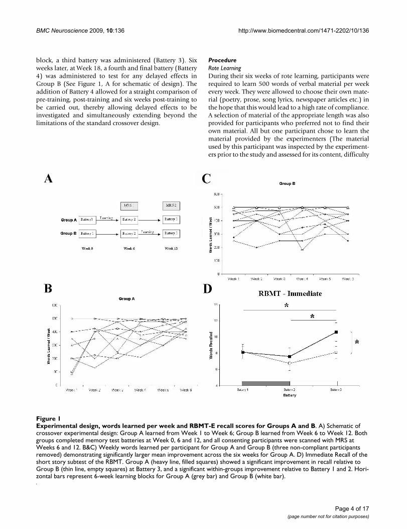
block, a third battery was administered (Battery 3). Sixweeks later, at Week 18, a fourth and final battery (Battery4) was administered to test for any delayed effects inGroup B (See Figure 1, A for schematic of design). Theaddition of Battery 4 allowed for a straight comparison ofpre-training, post-training and six weeks post-training tobe carried out, thereby allowing delayed effects to beinvestigated and simultaneously extending beyond thelimitations of the standard crossover design.
ProcedureRote LearningDuring their six weeks of rote learning, participants wererequired to learn 500 words of verbal material per weekevery week. They were allowed to choose their own mate-rial (poetry, prose, song lyrics, newspaper articles etc.) inthe hope that this would lead to a high rate of compliance.A selection of material of the appropriate length was alsoprovided for participants who preferred not to find theirown material. All but one participant chose to learn thematerial provided by the experimenters (The materialused by this participant was inspected by the experiment-ers prior to the study and assessed for its content, difficulty
Experimental design, words learned per week and RBMT-E recall scores for Groups A and BFigure 1Experimental design, words learned per week and RBMT-E recall scores for Groups A and B. A) Schematic of crossover experimental design: Group A learned from Week 1 to Week 6; Group B learned from Week 6 to Week 12. Both groups completed memory test batteries at Week 0, 6 and 12, and all consenting participants were scanned with MRS at Weeks 6 and 12. B&C) Weekly words learned per participant for Group A and Group B (three non-compliant participants removed) demonstrating significantly larger mean improvement across the six weeks for Group A. D) Immediate Recall of the short story subtest of the RBMT. Group A (heavy line, filled squares) showed a significant improvement in recall relative to Group B (thin line, empty squares) at Battery 3, and a significant within-groups improvement relative to Battery 1 and 2. Hori-zontal bars represent 6-week learning blocks for Group A (grey bar) and Group B (white bar).
Page 4 of 17(page number not for citation purposes)
BMC Neuroscience 2009, 10:136 http://www.biomedcentral.com/1471-2202/10/136
and volume, and was judged to be appropriate for thestudy). Recall of the material was tested every week, and arecord kept of the number of words learned each week.No instruction was given as to what learning strategy touse, but it was suggested that the participants learn part ofthe material each day rather than all in one session byreading over it repeatedly. Participants were asked aftertheir learning whether they had used any particular strat-egy, and all reported the use of rote repetition.
Behavioural TestingAt each of the behavioural batteries, an array of tests oflearning and memory (both verbal and visuo-spatial) andexecutive function were administered. These are describedbriefly.
Two measures of verbal learning and memory were used.The Short Story Recall task of the Rivermead BehaviouralMemory Test - Extended version (RBMT-E) consists of ashort paragraph of prose (5 to 6 lines) which is read aloudto the participant for recall. The passage is divided into 21ideational units, and each unit correctly recalled scoresone point. This task involves both immediate (directlyafter it is read aloud) and delayed recall (25-30 minuteslater), and has 6 alternate forms which are equated for dif-ficulty. The California Verbal Learning Test (CVLT) is aword-list learning task. A list of sixteen words is readaloud for immediate recall (the words are organized intofour semantic categories). The list is then read again for asecond recall trial, and repeated for five recall trials. Aninterference list is then read, also of sixteen words, forimmediate recall. The participant must then recall theoriginal list without hearing it read again. There then fol-lows a cued recall test. After a delay of 25-30 minutes, theparticipant must again recall as many words from the orig-inal list as possible. Cued recall and a yes/no recognitiontask then follow. The CVLT provides indices of short- andlong-term free and cued recall, as well as learning slope,clustering, proactive and retroactive interference.
A Visuo-spatial Learning task (VSL) was also used. Par-ticipants were presented with a 10 × 10 cell grid patternconsisting of eleven shapes (filled in blue squares) againstyellow empty squares. The grid was presented on screenfor 2 minutes, during which participants were instructedto study the shapes. They were then presented with anempty grid on a response sheet, and instructed to repro-duce the pattern by filling in the blue squares with pen.Four versions of this task were used, equated for thenumber of shapes. Scoring yielded a total VSL score, visualscore (number of shapes recalled) and spatial score(number of locations correctly recalled).
A measure of episodic memory for everyday events, theMundane Memory Questionnaire (MMQ) was also
given. The MMQ is a pen and paper measure of recall oftwelve commonplace everyday events over the last fourdays (E.g. "Do you recall getting dressed on this day? If so,what did you wear?"). One point is scored for responding"yes" and a second point given for supplying details. Ascore is obtained for each of the four past days. This testhas been shown to reliably differentiate hippocampecto-mied patients from normals (Mangaoang M, McMackinD, Fitzsimons M, Delanty N, Phillips J, Quigley J, O'MaraSM: Unilateral hippocampal damage impairs spoken andwritten language, Submitted). A sustained attention task,the SART (Sustained Attention to Response Task; [42]),was also given. The SART requires participants to make abutton-press response to a stream of rapidly-presentedrandom digits (1-9) with the task rule that response mustbe withheld for the number 3. This task is known to besubserved by the right hemisphere prefrontal-parietalattention network [43] and is a measure of frontal execu-tive function and attention.
In addition to the NART and the CFQ, three other controlmeasures were administered: the Hospital Anxiety andDepression Scale (HADS) was given at each battery tomonitor levels of depression and anxiety throughout thestudy. In addition, the Perceived Stress Inventory [44]was also included, given the known deleterious effects ofstress on learning (Mullally SL, Brotons JR, Cowley TR,Gobbo O, Shaw KN, O'Dwyer AM, O'Mara SM: Com-bined, but not separate, physical exercise and fluoxetinetreatment ameliorates the effect of repeated moderatestress on hippocampal-dependent spatial learning, Sub-mitted). In the light of recent findings regarding exerciseand learning, an Exercise Questionnaire (adapted from[45]) was also given to all participants at the end of thestudy.
Behavioural batteries lasted approximately 90 minutes,and were split into two sessions with a ten-minute breakbetween sessions. Tests of verbal learning and memorywere divided between the sessions to control for any pos-sible interference effects of list content, and it was madeclear at the start of the second session that no materialfrom the previous session would be requested.
Magnetic Resonance SpectroscopyIn addition to behavioural testing, participants who weresuitable and willing for MR scanning (Group A, n = 9,Group B, n = 6) underwent proton spectroscopy (1H-MRS) at Week 6 (immediately post-learning for Group Aand pre-learning for Group B) and Week 12 of the study(six weeks after learning ceased for Group A and immedi-ately post-learning for Group B).
Single voxel 1H MRS studies were conducted using PROBEsoftware on a GE Signa 1.5 Tesla system (GE, Milwaukee,
Page 5 of 17(page number not for citation purposes)
BMC Neuroscience 2009, 10:136 http://www.biomedcentral.com/1471-2202/10/136
WI, USA). Subjects were positioned in the supine positionso that the centering lights passed along their median sag-ittal plane and along their inter-pupilary line. Distancemeasurements were taken from the outer canthus of eacheye to the base of the headrest to ensure both sides wereequidistant. A protractor was then used to measure theangle between the subject's anthropomorphic baselineand the vertical. Voxels of interest (VOIs) were located ona set of coronal T2- weighted images acquired from theanterior-most portion of the brain to the posterior aspectof the lateral ventricles. Metabolite levels of N-acetylaspar-tate (NAA), creatine + phosphocreatine (tCr) and choline(Cho) were obtained from seven VOI locations: right andleft prefrontal cortex, midline prefrontal, and right andleft anterior and posterior hippocampus. VOIs measured2 cmx2 cmx2 cm (8 cm3) each. Precise positioning ofVOIs at first and repeat MRS acquisitions was determinedby distance measurement from anatomical markers visi-ble on the T2 image set. Spectra were acquired using point-resolved spectroscopy (PRESS; TR, 1500; TE, 35).
Using the automated shimming methods available on theGE Signa system, two approaches were employed to cor-rect for field inhomogeneity as previously described byKegeles et al. [46]. Phase differences between two echoesof different echo time are used to adapt shim currents tocorrect for inhomogeneity using the phase map method.Voxel shimming which calculates the optimal shim cur-rent to reduce the line width of water and correct for inho-mogeneity was the second method employed. The latter isunique to magnetic resonance spectroscopy sequences. Inthe current study such techniques were essential due tothe location of hippocampal voxels in the temporal lobein close proximity to areas of potential inhomogeneity,the skull base and sphenoid sinus
As highlighted by Kreis [47] and Taylor [48] qualitychecks on MRS data are essential. Thus for each examina-tion the location of VOIs was checked pre- and post-MRS,individual raw data files were stored to facilitate checks formotion, system instabilities and SNR, water referencescans were used to correct for eddy currents using SAGE(Spectroscopy Analysis of GE, GE Healthcare, Milwaukee,WI, USA) using the Probe/SVQ automatic single voxelprocessing routine, spectral resolution was improved byusing a dual shimming technique, and spectra wereassessed for abnormalities (failure of water saturation,ghosts, peak doubling, unidentified metabolites, and sig-nal from outer volume). Spatial saturation bands werealso positioned to completely surround the VOI.
After acquisition the Probe/SVQ software conducts anautomatic analysis. Using the phase corrected water refer-ence signal and the phase corrected suppressed signal, apure metabolite signal is computed through a water sub-
traction technique. A narrow frequency window aroundeach of the metabolite resonances is analyzed by curve fit-ting, using the Marquardt-Levenberg method. Beforecurve fitting, the spectral line shape is manipulated toimprove fitting by removing any errors associated withline width variations. There are three line shape manipu-lations: line broadening, line width normalization, andline shape transformation. Each of these manipulationsare combined into a single apodization step. All Probe/SVQ numerical analysis is based on peak amplitude butby normalizing the line widths of the peaks, the analysiseffectively measures areas and ratios of areas. The algo-rithm first determines the line width of the creatine peak,a (partial) Lorentzian to Gaussian transformation is per-formed, the line width normalization and the first part ofthe line shape transformation are combined into a singleexponential apodization, and the second half of the lineshape transformation is performed by a Gaussian apodi-zation.
Signal intensities of the metabolite peaks are determinedfrom which the ratio NAA/(tCr+Cho) is established. Pro-ducing an image containing a Pure Absorption (Real PartFFT) Spectrum covering a spectral range from -0.4 to 4.3parts per million (ppm), and an associated chemical shiftscale axis with tick marks in ppm.
Data AnalysisBehavioural data were analysed using mixed factorialANOVAs with within-groups factor BATTERY (Battery 1 -Battery 3) for crossover analyses or PRE-POST (Pre-train-ing, Post-training and 6 Week delay) for delayed effectsanalysis, and between groups factor GROUP (Group A,Group B). The DV in each analysis was the dependentmeasure obtained from each behavioural test. Lack ofsphericity was compensated for, where appropriate, usinga Greenhouse-Geisser correction (indicated by "GG" afterthe p-value). Within-groups main effects were exploredusing Bonferroni multiple comparisons. All means arereported ± SEM; due to the large number of analyses, onlysignificant F and p-values for main effects/interactions arereported. Pearson correlations were carried out betweenbehavioural performance measures and Total WordsLearned (TWL) for each participant, as well as the increasein words learned from Week 1 to Week 6 ("delta words",or words).
Spectroscopy data were analysed by computing the ratioof NAA to the sum of creatine and choline (i.e. NAA/(tCr+Cho)); this ratio was used because creatine andcholine are considered relatively stable metabolites,thereby allowing changes in NAA to be highlighted. Thisratio was computed at each of the seven VOIs at Week 6and Week 12. A series of mixed factorial ANOVAs werecarried out for the seven voxels of interest with GROUP (2
Page 6 of 17(page number not for citation purposes)
BMC Neuroscience 2009, 10:136 http://www.biomedcentral.com/1471-2202/10/136
levels) as the between groups factor and PRE-POST (2 lev-els) as the within groups factor; NAA/(tCr+Cho) ratio wasthe DV in each case. Greenhouse-Geisser and Bonferronicorrections were again employed, where appropriate.Pearson correlations were also computed between thechange in metabolite ratios and behavioural measures ofmemory performance.
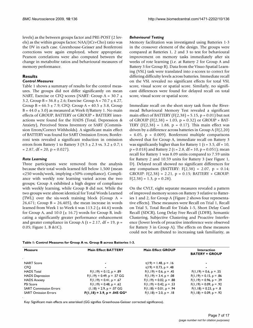
ResultsControl MeasuresTable 1 shows a summary of results for the control meas-ures. The groups did not differ significantly on meanNART, Exercise or CFQ scores (NART: Group A = 30.7 ±3.2, Group B = 36.8 ± 2.6; Exercise: Group A = 70.7 ± 4.27,Group B = 66.5 ± 7.9; CFQ: Group A = 40.5 ± 3.8, GroupB = 44.0 ± 3.0) as measured at Week 0/Battery 1. No maineffects of GROUP, BATTERY or GROUP × BATTERY inter-actions were found for the HADS (Total, Depression &Anxiety), Perceived Stress Inventory or SART (Commis-sion Errors/Correct Withholds). A significant main effectof BATTERY was found for SART Omission Errors; Bonfer-roni tests revealed a significant reduction in omissionerrors from Battery 1 to Battery 3 (9.5 ± 2.3 vs. 3.2 ± 0.7; t= 2.87, df = 20, p = 0.027).
Rote LearningThree participants were removed from the analysisbecause their total words learned fell below 1,500 (mean<250 words/week, implying <50% compliance). Compli-ance with weekly rote learning varied across the twogroups; Group A exhibited a high degree of compliancewith weekly learning, while Group B did not. While thetwo groups were almost identical for Total Words Learned(TWL) over the six-week training block (Group A =26,671; Group B = 26,405), the mean increase in wordslearned from Week 1 to Week 6 was 113.2 (± 44.6) wordsfor Group A, and 10.0 (± 16.7) words for Group B, indi-cating a significantly greater performance enhancementand greater compliance in Group A (t = 2.17, df = 19, p =0.05; Figure 1, B &1C).
Behavioural TestingMemory facilitation was investigated using Batteries 1-3in the crossover element of the design. The groups werecompared at Batteries 1, 2 and 3 to test for behaviouralimprovement on memory tasks immediately after sixweeks of rote learning (i.e. at Battery 2 for Group A andBattery 3 for Group B). Data from the Visuo-Spatial Learn-ing (VSL) task were translated into z-scores to correct fordiffering difficulty levels across batteries. Immediate recallon the VSL revealed no significant effects for total VSLscore, visual score or spatial score. Similarly, no signifi-cant differences were found for delayed recall on totalscore, visual score or spatial score.
Immediate recall on the short story task from the River-mead Behavioural Memory Test revealed a significantmain effect of BATTERY (F(2,38) = 5.15, p = 0.01) but notof GROUP (F(2,38) = 1.03, p = 0.32) or GROUP × BAT-TERY (F(2,38) = 1.88, p = 0.17). This main effect wasdriven by a difference across batteries in Group A (F(2,20)= 6.05, p = 0.009). Bonferroni multiple comparisonsshowed that for Group A, immediate recall on Battery 3was significantly higher than for Battery 1 (t = 3.5, df = 10,p = 0.018) and Battery 2 (t = 2.8, df = 10, p = 0.051); meanrecall for Battery 1 was 8.09 units compared to 7.59 unitsfor Battery 2 and 10.59 units for Battery 3 (see Figure 1,D). Delayed recall showed no significant differences forany comparison (BATTERY: F(2,38) = 2.07, p = 0.14;GROUP: F(2,38) = 2.21, p = 0.15; BATTERY × GROUP:F(2,38) = 1.3, p = 0.28).
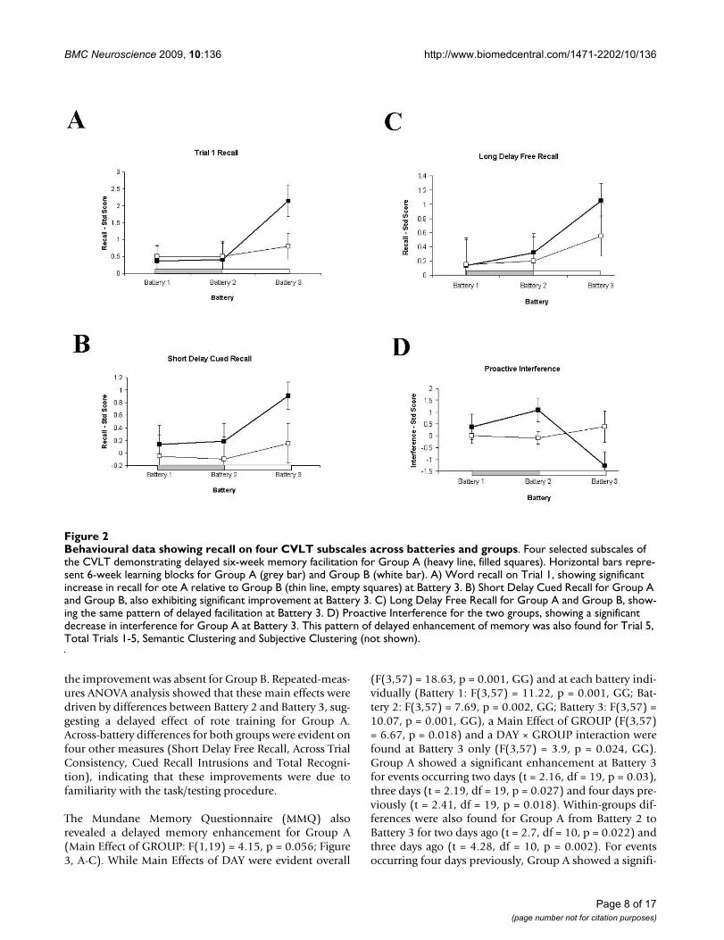
On the CVLT, eight separate measures revealed a patternof improved memory scores on Battery 3 relative to Batter-ies 1 and 2, for Group A (Figure 2 shows four representa-tive effects). These measures were Recall on Trial 1, Recallon Trial 5, Total Recall for Trials 1-5, Short Delay CuedRecall (SDCR), Long Delay Free Recall (LDFR), SemanticClustering, Subjective Clustering and Proactive Interfer-ence (lower levels of proactive interference were observedfor Battery 3 in Group A). The effects on these measurescould not be attributed to increasing task familiarity, as
Table 1: Control Measures for Group A vs. Group B across Batteries 1-3.
Measure Main Effect BATTERY Main Effect GROUP InteractionBATERY × GROUP
NART Score - t(19) = 1.48, p = .16 -CFQ - t(19) = 0.73, p = .48 -HADS Total F(1,19) = 0.12, p = .89 F(1,19) = 0.6, p = .45 F(1,19) = 0.6, p = .55HADS Depression F(1,19) = 0.49, p = .57 GG F(1,19) = 3.4, p = .08 F(1,19) = 0.15, p = .86HADS Anxiety F(1,19) = 0.41, p = .67 F(1,19) = 0.02, p = .88 F(1,19) = 0.96, p = .39PSI Score F(1,19) = 0.48, p = .62 F(1,19) = 0.42, p = .53 F(1,19) = 0.09, p = .92SART Commission Errors (1,18) = 2.9, p = .07 GG F(1,18) = 0.01, p = .94 F(1,18) = 0.23, p = .8SART Omission Errors F(1,18) = 3.9, p = .045 GG* F(1,18) = 2.0, p = .18 F(1,18) = 0.09, p = .92
Key: Significant main effects are asterisked (GG signifies Greenhouse-Geisser corrected significance).
Page 7 of 17(page number not for citation purposes)
BMC Neuroscience 2009, 10:136 http://www.biomedcentral.com/1471-2202/10/136
the improvement was absent for Group B. Repeated-meas-ures ANOVA analysis showed that these main effects weredriven by differences between Battery 2 and Battery 3, sug-gesting a delayed effect of rote training for Group A.Across-battery differences for both groups were evident onfour other measures (Short Delay Free Recall, Across TrialConsistency, Cued Recall Intrusions and Total Recogni-tion), indicating that these improvements were due tofamiliarity with the task/testing procedure.
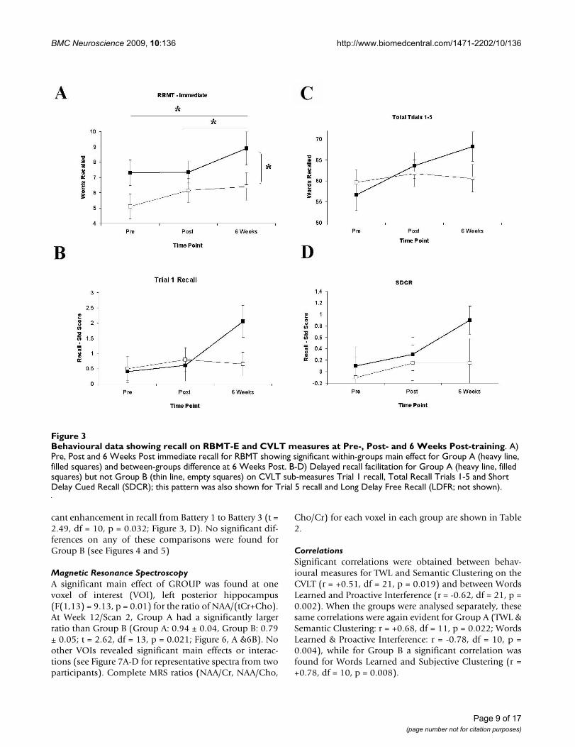
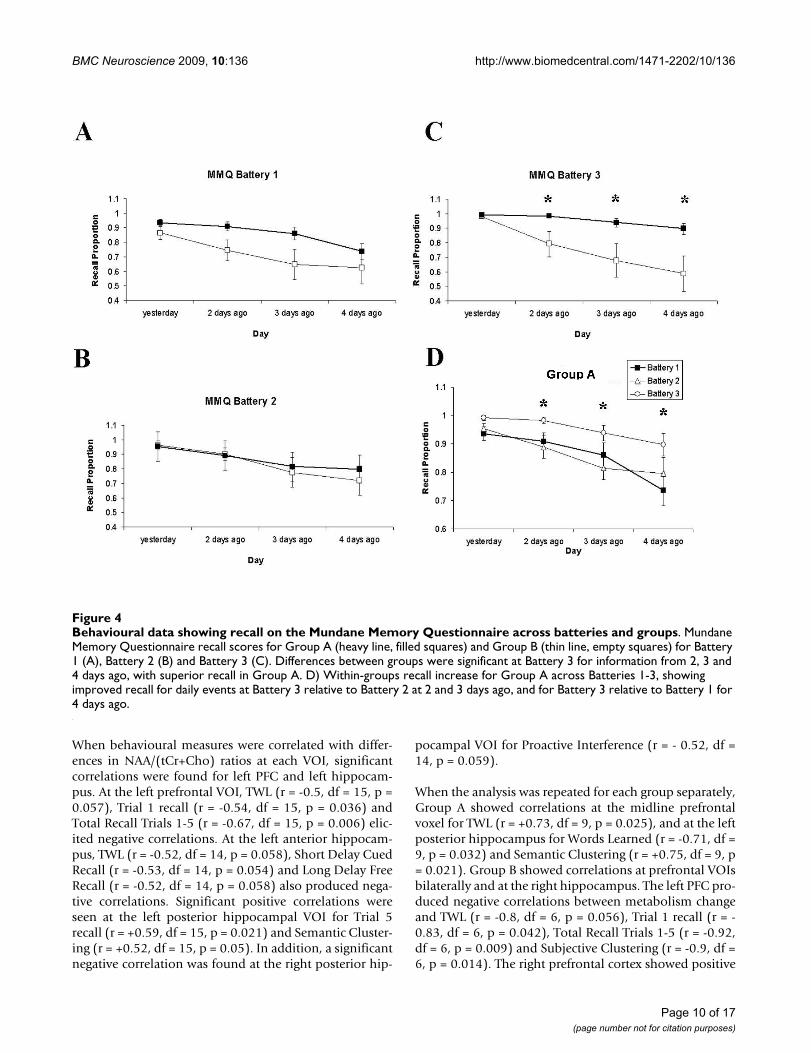
The Mundane Memory Questionnaire (MMQ) alsorevealed a delayed memory enhancement for Group A(Main Effect of GROUP: F(1,19) = 4.15, p = 0.056; Figure3, A-C). While Main Effects of DAY were evident overall
(F(3,57) = 18.63, p = 0.001, GG) and at each battery indi-vidually (Battery 1: F(3,57) = 11.22, p = 0.001, GG; Bat-tery 2: F(3,57) = 7.69, p = 0.002, GG; Battery 3: F(3,57) =10.07, p = 0.001, GG), a Main Effect of GROUP (F(3,57)= 6.67, p = 0.018) and a DAY × GROUP interaction werefound at Battery 3 only (F(3,57) = 3.9, p = 0.024, GG).Group A showed a significant enhancement at Battery 3for events occurring two days (t = 2.16, df = 19, p = 0.03),three days (t = 2.19, df = 19, p = 0.027) and four days pre-viously (t = 2.41, df = 19, p = 0.018). Within-groups dif-ferences were also found for Group A from Battery 2 toBattery 3 for two days ago (t = 2.7, df = 10, p = 0.022) andthree days ago (t = 4.28, df = 10, p = 0.002). For eventsoccurring four days previously, Group A showed a signifi-
Behavioural data showing recall on four CVLT subscales across batteries and groupsFigure 2Behavioural data showing recall on four CVLT subscales across batteries and groups. Four selected subscales of the CVLT demonstrating delayed six-week memory facilitation for Group A (heavy line, filled squares). Horizontal bars repre-sent 6-week learning blocks for Group A (grey bar) and Group B (white bar). A) Word recall on Trial 1, showing significant increase in recall for ote A relative to Group B (thin line, empty squares) at Battery 3. B) Short Delay Cued Recall for Group A and Group B, also exhibiting significant improvement at Battery 3. C) Long Delay Free Recall for Group A and Group B, show-ing the same pattern of delayed facilitation at Battery 3. D) Proactive Interference for the two groups, showing a significant decrease in interference for Group A at Battery 3. This pattern of delayed enhancement of memory was also found for Trial 5, Total Trials 1-5, Semantic Clustering and Subjective Clustering (not shown).
Page 8 of 17(page number not for citation purposes)
BMC Neuroscience 2009, 10:136 http://www.biomedcentral.com/1471-2202/10/136
cant enhancement in recall from Battery 1 to Battery 3 (t =2.49, df = 10, p = 0.032; Figure 3, D). No significant dif-ferences on any of these comparisons were found forGroup B (see Figures 4 and 5)
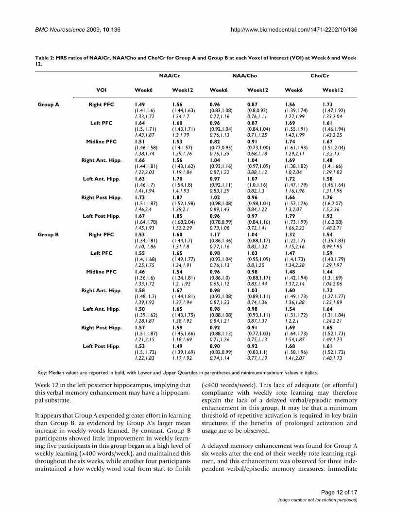
Magnetic Resonance SpectroscopyA significant main effect of GROUP was found at onevoxel of interest (VOI), left posterior hippocampus(F(1,13) = 9.13, p = 0.01) for the ratio of NAA/(tCr+Cho).At Week 12/Scan 2, Group A had a significantly largerratio than Group B (Group A: 0.94 ± 0.04, Group B: 0.79± 0.05; t = 2.62, df = 13, p = 0.021; Figure 6, A &6B). Noother VOIs revealed significant main effects or interac-tions (see Figure 7A-D for representative spectra from twoparticipants). Complete MRS ratios (NAA/Cr, NAA/Cho,
Cho/Cr) for each voxel in each group are shown in Table2.
CorrelationsSignificant correlations were obtained between behav-ioural measures for TWL and Semantic Clustering on theCVLT (r = +0.51, df = 21, p = 0.019) and between WordsLearned and Proactive Interference (r = -0.62, df = 21, p =0.002). When the groups were analysed separately, thesesame correlations were again evident for Group A (TWL &Semantic Clustering: r = +0.68, df = 11, p = 0.022; WordsLearned & Proactive Interference: r = -0.78, df = 10, p =0.004), while for Group B a significant correlation wasfound for Words Learned and Subjective Clustering (r =+0.78, df = 10, p = 0.008).
Behavioural data showing recall on RBMT-E and CVLT measures at Pre-, Post- and 6 Weeks Post-trainingFigure 3Behavioural data showing recall on RBMT-E and CVLT measures at Pre-, Post- and 6 Weeks Post-training. A) Pre, Post and 6 Weeks Post immediate recall for RBMT showing significant within-groups main effect for Group A (heavy line, filled squares) and between-groups difference at 6 Weeks Post. B-D) Delayed recall facilitation for Group A (heavy line, filled squares) but not Group B (thin line, empty squares) on CVLT sub-measures Trial 1 recall, Total Recall Trials 1-5 and Short Delay Cued Recall (SDCR); this pattern was also shown for Trial 5 recall and Long Delay Free Recall (LDFR; not shown).
Page 9 of 17(page number not for citation purposes)
BMC Neuroscience 2009, 10:136 http://www.biomedcentral.com/1471-2202/10/136
When behavioural measures were correlated with differ-ences in NAA/(tCr+Cho) ratios at each VOI, significantcorrelations were found for left PFC and left hippocam-pus. At the left prefrontal VOI, TWL (r = -0.5, df = 15, p =0.057), Trial 1 recall (r = -0.54, df = 15, p = 0.036) andTotal Recall Trials 1-5 (r = -0.67, df = 15, p = 0.006) elic-ited negative correlations. At the left anterior hippocam-pus, TWL (r = -0.52, df = 14, p = 0.058), Short Delay CuedRecall (r = -0.53, df = 14, p = 0.054) and Long Delay FreeRecall (r = -0.52, df = 14, p = 0.058) also produced nega-tive correlations. Significant positive correlations wereseen at the left posterior hippocampal VOI for Trial 5recall (r = +0.59, df = 15, p = 0.021) and Semantic Cluster-ing (r = +0.52, df = 15, p = 0.05). In addition, a significantnegative correlation was found at the right posterior hip-
pocampal VOI for Proactive Interference (r = - 0.52, df =14, p = 0.059).
When the analysis was repeated for each group separately,Group A showed correlations at the midline prefrontalvoxel for TWL (r = +0.73, df = 9, p = 0.025), and at the leftposterior hippocampus for Words Learned (r = -0.71, df =9, p = 0.032) and Semantic Clustering (r = +0.75, df = 9, p= 0.021). Group B showed correlations at prefrontal VOIsbilaterally and at the right hippocampus. The left PFC pro-duced negative correlations between metabolism changeand TWL (r = -0.8, df = 6, p = 0.056), Trial 1 recall (r = -0.83, df = 6, p = 0.042), Total Recall Trials 1-5 (r = -0.92,df = 6, p = 0.009) and Subjective Clustering (r = -0.9, df =6, p = 0.014). The right prefrontal cortex showed positive
Behavioural data showing recall on the Mundane Memory Questionnaire across batteries and groupsFigure 4Behavioural data showing recall on the Mundane Memory Questionnaire across batteries and groups. Mundane Memory Questionnaire recall scores for Group A (heavy line, filled squares) and Group B (thin line, empty squares) for Battery 1 (A), Battery 2 (B) and Battery 3 (C). Differences between groups were significant at Battery 3 for information from 2, 3 and 4 days ago, with superior recall in Group A. D) Within-groups recall increase for Group A across Batteries 1-3, showing improved recall for daily events at Battery 3 relative to Battery 2 at 2 and 3 days ago, and for Battery 3 relative to Battery 1 for 4 days ago.
Page 10 of 17(page number not for citation purposes)
BMC Neuroscience 2009, 10:136 http://www.biomedcentral.com/1471-2202/10/136
correlations between metabolism and Trial 1 recall (r =+0.87, df = 6, p = 0.026) and Subjective Clustering (r =+0.88, df = 6, p = 0.014). There was a positive correlationat the right anterior hippocampus for Short Delay CuedRecall (r = +0.89, df = 5, p = 0.044), and at the right pos-terior hippocampus for TWL (r = -0.86, df = 6, p = 0.027).
DiscussionIn this study, two groups of normal older adults weretrained with prolonged rote learning over six weeks, withmemory performance and brain metabolism measuresassessed pre- and post-training. We predicted that suchprolonged learning would, by virtue of the repeated acti-
vation of memory structures, bring about performancebenefits on behavioural memory measures, and that theseeffects would be confined to verbal-based tests. We foundthat this training regime did produce an enhancement inmemory function in the compliant training Group A, butnot in the non-compliant training Group B. These mem-ory gains were specific to three independent verbally-based measures (RBMT, CVLT and MMQ), and could notbe attributed to extraneous factors such as anxiety, depres-sion or attention. The absence of such an effect in GroupB suggests that compliance with weekly learning was thecrucial determinant of this facilitation. In addition, GroupA displayed a metabolic change relative to Group B at
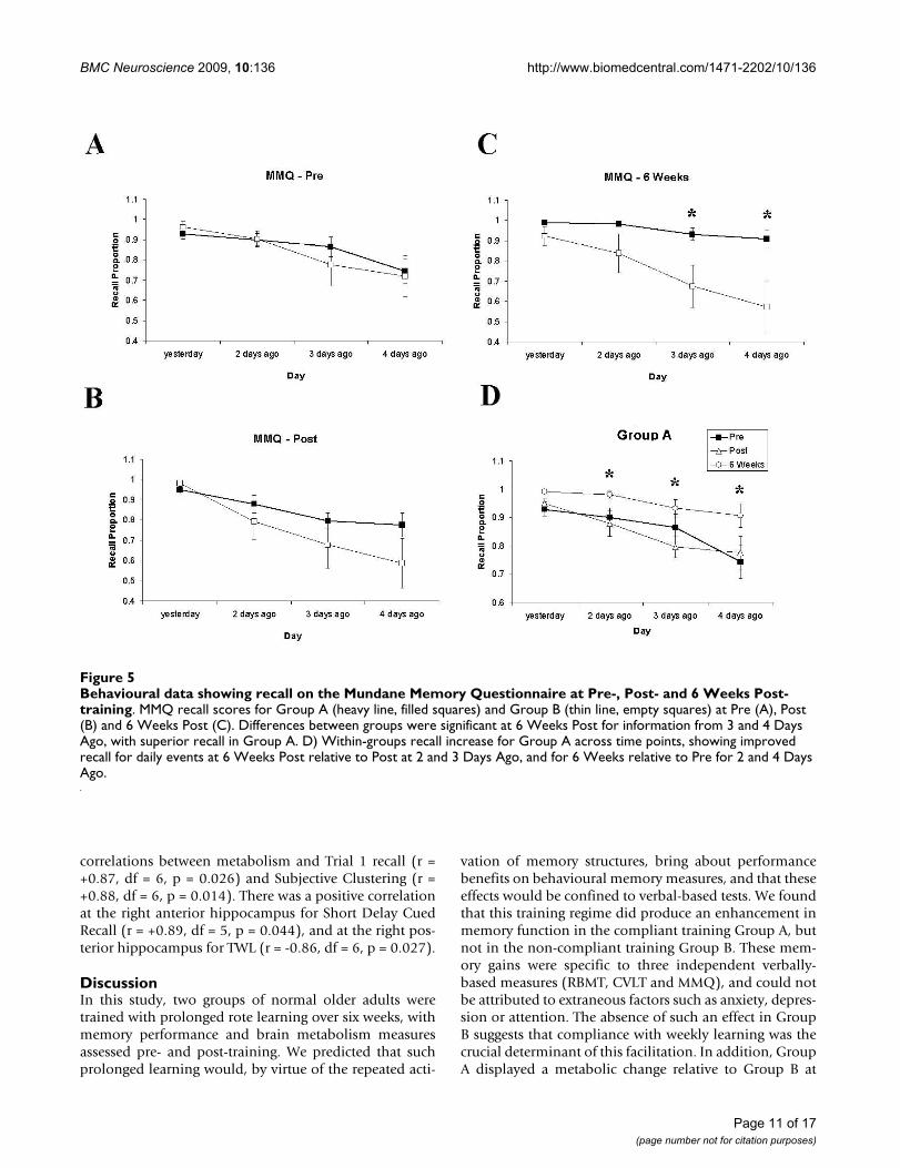
Behavioural data showing recall on the Mundane Memory Questionnaire at Pre-, Post- and 6 Weeks Post-trainingFigure 5Behavioural data showing recall on the Mundane Memory Questionnaire at Pre-, Post- and 6 Weeks Post-training. MMQ recall scores for Group A (heavy line, filled squares) and Group B (thin line, empty squares) at Pre (A), Post (B) and 6 Weeks Post (C). Differences between groups were significant at 6 Weeks Post for information from 3 and 4 Days Ago, with superior recall in Group A. D) Within-groups recall increase for Group A across time points, showing improved recall for daily events at 6 Weeks Post relative to Post at 2 and 3 Days Ago, and for 6 Weeks relative to Pre for 2 and 4 Days Ago.
Page 11 of 17(page number not for citation purposes)
BMC Neuroscience 2009, 10:136 http://www.biomedcentral.com/1471-2202/10/136
Week 12 in the left posterior hippocampus, implying thatthis verbal memory enhancement may have a hippocam-pal substrate.
It appears that Group A expended greater effort in learningthan Group B, as evidenced by Group A's larger meanincrease in weekly words learned. By contrast, Group Bparticipants showed little improvement in weekly learn-ing; five participants in this group began at a high level ofweekly learning (>400 words/week), and maintained thisthroughout the six weeks, while another four participantsmaintained a low weekly word total from start to finish
(<400 words/week). This lack of adequate (or effortful)compliance with weekly rote learning may thereforeexplain the lack of a delayed verbal/episodic memoryenhancement in this group. It may be that a minimumthreshold of repetitive activation is required in key brainstructures if the benefits of prolonged activation andusage are to be observed.
A delayed memory enhancement was found for Group Asix weeks after the end of their weekly rote learning regi-men, and this enhancement was observed for three inde-pendent verbal/episodic memory measures: immediate
Table 2: MRS ratios of NAA/Cr, NAA/Cho and Cho/Cr for Group A and Group B at each Voxel of Interest (VOI) at Week 6 and Week 12.
NAA/Cr NAA/Cho Cho/Cr
VOI Week6 Week12 Week6 Week12 Week6 Week12
Group A Right PFC 1.49(1.41,1.6)1.33,1.72
1.56(1.44,1.63)1.24,1.7
0.96(0.83,1.08)0.77,1.16
0.87(0.8,0.93)0.76,1.11
1.56(1.39,1.74)1.22,1.99
1.73(1.47,1.92)1.33,2.04
Left PFC 1.64(1.5, 1.71)1.43,1.87
1.60(1.43,1.71)1.3,1.79
0.96(0.92,1.04)0.76,1.13
0.87(0.84,1.04)0.71,1.25
1.69(1.55,1.91)1.43,1.99
1.61(1.46,1.94)1.43,2.25
Midline PFC 1.51(1.46,1.58)1.38,1.74
1.53(1.4,1.57)1.29,1.76
0.82(0.77,0.95)0.75,1.35
0.91(0.73,1.00)0.68,1.18
1.74(1.61,1.93)1.29,2.11
1.67(1.51,2.04)1.3,2.13
Right Ant. Hipp. 1.66(1.44,1.81)1.22,2.03
1.56(1.43,1.62)1.19,1.84
1.04(0.93,1.16)0.87,1.22
1.04(0.97,1.09)0.88,1.12
1.69(1.38,1.82)1.0,2.04
1.48(1.4,1.66)1.29,1.82
Left Ant. Hipp. 1.63(1.46,1.7)1.41,1.94
1.70(1.54,1.8)1.4,1.93
0.97(0.92,1.11)0.83,1.29
1.07(1.0,1.16)0.82,1.3
1.72(1.47,1.79)1.16,1.96
1.58(1.46,1.64)1.31,1.96
Right Post Hipp. 1.73(1.51,1.87)1.46,2.4
1.87(1.52,1.98)1.39,2.1
1.02(0.98,1.08)0.89,1.43
0.96(0.98,1.01)0.84,1.22
1.66(1.53,1.76)1.3,2.07
1.76(1.6,2.07)1.5,2.36
Left Post Hipp. 1.67(1.64,1.78)1.45,1.93
1.85(1.68,2.04)1.52,2.29
0.96(0.78,0.99)0.73,1.08
0.97(0.84,1.16)0.72,1.41
1.79(1.73,1.99)1.66,2.22
1.92(1.6,2.08)1.48,2.71
Group B Right PFC 1.53(1.34,1.81)1.10, 1.86
1.60(1.44,1.7)1.31,1.8
1.17(0.86,1.36)0.77,1.16
1.04(0.88,1.17)0.85,1.32
1.32(1.22,1.7)1.15,2.16
1.54(1.35,1.83)0.99,1.95
Left PFC 1.55(1.4, 1.68)1.25,1.75
1.65(1.49,1.77)1.34,1.91
0.98(0.92,1.04)0.76,1.13
1.03(0.95,1.09)0.8,1.20
1.47(1.4,1.73)1.34,2.28
1.59(1.43,1.79)1.29,1.97
Midline PFC 1.46(1.36,1.6)1.33,1.72
1.54(1.24,1.81)1.2, 1.92
0.96(0.86,1.0)0.65,1.12
0.98(0.88,1.17)0.83,1.44
1.48(1.42,1.94)1.37,2.14
1.44(1.3,1.69)1.04,2.06
Right Ant. Hipp. 1.58(1.48, 1.7)1.39,1.92
1.67(1.44,1.81)1.37,1.94
0.98(0.92,1.08)0.87,1.23
1.03(0.89,1.11)0.74,1.36
1.60(1.49,1.73)1.36,1.88
1.72(1.27,1.77)1.25,1.89
Left Ant. Hipp. 1.50(1.39,1.62)1.28,1.87
1.65(1.43,1.75)1.38,1.92
0.98(0.88,1.08)0.84,1.21
0.98(0.93,1.11)0.87,1.37
1.54(1.31,1.72)1.2,2.1
1.64(1.31,1.84)1.24,2.21
Right Post Hipp. 1.57(1.51,1.87)1.21,2.15
1.59(1.45,1.66)1.18,1.69
0.92(0.88,1.13)0.71,1.26
0.91(0.77,1.03)0.75,1.13
1.69(1.64,1.73)1.54,1.87
1.65(1.52,1.73)1.49,1.73
Left Post Hipp. 1.53(1.5, 1.72)1.22,1.83
1.49(1.39,1.69)1.17,1.92
0.90(0.82,0.99)0.74,1.14
0.92(0.83,1.1)0.77,1.19
1.68(1.58,1.96)1.41,2.07
1.61(1.52,1.72)1.48,1.73
Key: Median values are reported in bold, with Lower and Upper Quartiles in parentheses and minimum/maximum values in italics.
Page 12 of 17(page number not for citation purposes)
BMC Neuroscience 2009, 10:136 http://www.biomedcentral.com/1471-2202/10/136
recall on the RBMT short story, four sub-measures of theCVLT, and recall of everyday events on the MMQ. No suchimprovement was found for the visuo-spatial task, or anyof the control measures employed, and can therefore notbe explained by generalised increases in arousal or atten-tion, fluctuations in depression or anxiety, or other con-founding variables. These effects may be thus attributableto the prolonged period of rote rehearsal of verbal mate-rial. This verbally-mediated memory gain emerged sixweeks after a period of no learning. This suggests that thelearning benefits accrued by sustained rote rehearsalcould require a latent period before they emerge. Theduration of this latent period is unknown - it is possiblethat the six-week follow-up used in the present studyrevealed these memory gains at their greatest magnitude,
but it is equally possible that the data collected at this timepoint represent a position on either an upward or down-ward curve of facilitation. Further study will be required todetermine the exact time course of this delayed facilitationeffect. Irrespective of this, the fact that these effects wereobserved on a range of laboratory measures and the recallof everyday real-life events demonstrates that this is ahighly robust effect, and appears specific to the verbal/epi-sodic memory domain.
The behavioural memory improvement was accompaniedby a metabolic difference between groups difference atonly one voxel of interest (VOI), the left posterior hippoc-ampus. The left hippocampal region has been reliablyassociated with verbal and episodic memory performance
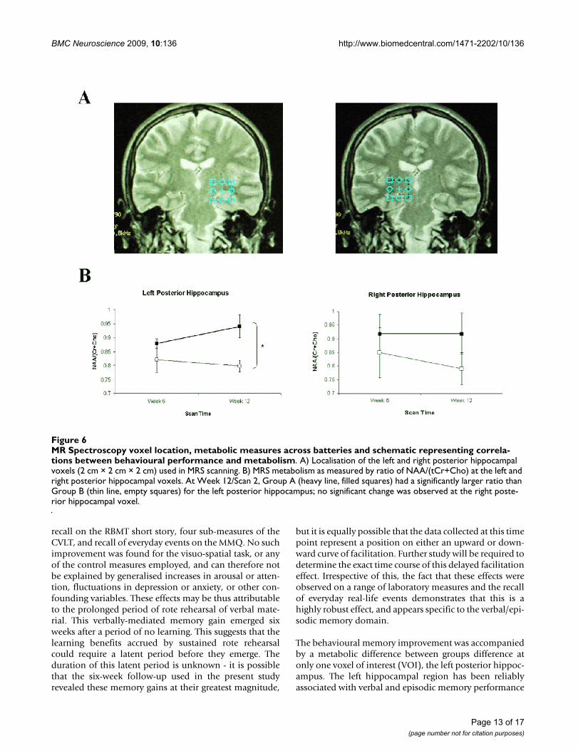
MR Spectroscopy voxel location, metabolic measures across batteries and schematic representing correlations between behav-ioural performance and metabolismFigure 6MR Spectroscopy voxel location, metabolic measures across batteries and schematic representing correla-tions between behavioural performance and metabolism. A) Localisation of the left and right posterior hippocampal voxels (2 cm × 2 cm × 2 cm) used in MRS scanning. B) MRS metabolism as measured by ratio of NAA/(tCr+Cho) at the left and right posterior hippocampal voxels. At Week 12/Scan 2, Group A (heavy line, filled squares) had a significantly larger ratio than Group B (thin line, empty squares) for the left posterior hippocampus; no significant change was observed at the right poste-rior hippocampal voxel.
Page 13 of 17(page number not for citation purposes)
BMC Neuroscience 2009, 10:136 http://www.biomedcentral.com/1471-2202/10/136
in normal humans using imaging, and in lesionedpatients (see [5]). As such, it is not unreasonable toattribute the behavioural effects to alterations in the met-abolic activity of this region as a result of the rote learningregime. A comparable effect was reported in Valenzuela etal. [30], where a five week training period with a spatially-based strategy produced memory enhancement andchanges in metabolite levels in right hippocampus, how-ever they reported an unexplained reduction in NAA/Crmeasures in the hippocampus following memory train-ing. The present results may represent evidence of a com-plimentary process for verbal learning, subserved by theleft hippocampal formation. At present we can onlyhypothesise as to the mechanism of action of this process;however, the repeated activation of brain structures asso-
ciated with rote learning, particularly the projection fromleft PFC to hippocampus, may have led to increases inneuronal viability and health in these regions, as indexedby increased NAA/(Cho+tCr) ratio. These benefits may beslow to emerge, but have the effect of facilitating futurelearning of new information, in a manner akin to the ele-vated memory performance of the mentally-active nuns inSnowdon's studies [21-23]. Furthermore, Riby et al. [49]recently demonstrated that glucose ingestion facilitatedepisodic memory performance in a sample of healthy eld-erly participants. Prolonged learning with its concomitantincreased bloodflow to memory structures may have hada comparable glucose-mediated effect. The exact neu-rometabolic mechanism behind this effect notwithstand-ing, it can be cautiously asserted that an extended period
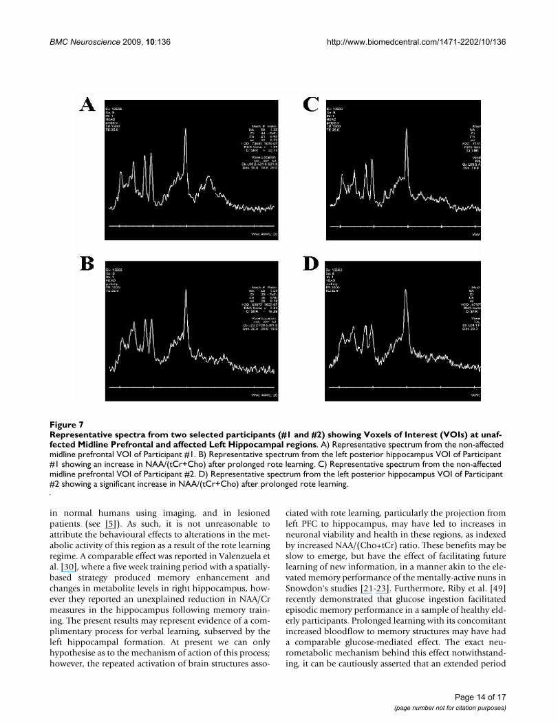
Representative spectra from two selected participants (#1 and #2) showing Voxels of Interest (VOIs) at unaffected Midline Prefrontal and affected Left Hippocampal regionsFigure 7Representative spectra from two selected participants (#1 and #2) showing Voxels of Interest (VOIs) at unaf-fected Midline Prefrontal and affected Left Hippocampal regions. A) Representative spectrum from the non-affected midline prefrontal VOI of Participant #1. B) Representative spectrum from the left posterior hippocampus VOI of Participant #1 showing an increase in NAA/(tCr+Cho) after prolonged rote learning. C) Representative spectrum from the non-affected midline prefrontal VOI of Participant #2. D) Representative spectrum from the left posterior hippocampus VOI of Participant #2 showing a significant increase in NAA/(tCr+Cho) after prolonged rote learning.
Page 14 of 17(page number not for citation purposes)
BMC Neuroscience 2009, 10:136 http://www.biomedcentral.com/1471-2202/10/136
of rote rehearsal leads to enhanced future learning accom-panied by changes in the markers of cell health in a keymemory structure of the brain.
When changes in metabolism for Week 6 to Week 12 werecorrelated with changes in behavioural measures, signifi-cant correlations tended to cluster at specific VOIs. Whenthe groups were collapsed, two regions, left PFC and lefthippocampus (anterior and posterior) were associatedwith changes in seven of the eight measures tested. This isfurther support for the suggestion that, on the whole, thewell-established anatomical connection between left PFCand left hippocampal formation (the uncinate fascicle;[29]) may have been activated in this study. An anatomi-cal dissociation was evident when the groups were consid-ered separately:
Group A's behavioural measures were correlated withchanges primarily in left posterior hippocampus, whilefor Group B left and right PFC were most strongly impli-cated.
Due to limitations of the MR system employed in thisstudy the minimal VOI available was 8 cm3. Thus the hip-pocampal VOIs also included extrahippocampal tissuessuch as the amygdale, the entorhinal cortex, the parahip-pocampal gyrus and CSF in the temporal horn of the lat-eral ventricle in keeping with previous studies employingsingle-voxel MRS [50-56]. In a previous study utilising sin-gle voxel proton MRS to evaluate hippocampal sclerosis,Chang et al. [51] utilised small and large VOIs and dis-cussed the partial volume effects of the larger voxels andthe inadequate signal-to-noise ratio of smaller voxelsresulting in inadequate spectra. For the current study spe-cial care was taken when positioning the VOIs to ensuremaximal hippocampal coverage, to minimise partial vol-ume effects from other tissues, to minimise potential mag-netic susceptibility artifacts from the skull base andsphenoid sinus, and to minimise potential spectral con-tamination from fat in the skull base. The use of two-dimensional chemical shift imaging, which allowsimproved sampling of smaller VOIs, the measurement ofthe absolute metabolite levels, the use of within voxel tis-sue segmentation, and higher field strength magnetswould improve the MRS data in future studies as the cur-rent approach was somewhat limited by the technologyavailable [51,56]. However Waldman and Rai [55] suggestthat the determination of metabolite ratios rather thanabsolute concentrations circumvents the necessity for seg-mentation to correct for CSF in the acquisition voxel andthat such processes inevitably introduce errors.
Although these data, along with those of previous studies,suggest some promise for MRS in the evaluation of mem-ory it must be noted that the relatively small sample size
is a limitation and extrapolation of observations requirescaution. Further limitations include the lack of a true com-parison group and the curtailment of the age range.Strengths included the recruitment of a diverse sample ofcommunity based older participants which reduces thepossibility of sampling bias and the use of a detailed andstandardised neuropsychological protocol.
ConclusionIn conclusion, we have here demonstrated that, whencompliance is high, a prolonged period of repetitive rotelearning may lead to improvements in verbal/episodicmemory which emerge in the weeks following learningcessation, and that these benefits are not confined to lab-oratory-based indices of memory. Furthermore, thesebenefits appear to be associated with metabolic changesin the left posterior hippocampus, which may reveal thehealth implications for key brain structures of repetitiveactivation and regular usage in healthy ageing.
Authors' contributionsRAPR carried out behavioural testing and participantrecruitment, analysed the behavioural data, plotted thefigures and wrote the manuscript. JMcN carried out partic-ipant scanning, assisted with data analysis, and contrib-uted to writing of manuscript; SLM and JH carried outbehavioural testing and participant recruitment, and con-tributed to analysis. PB, CPD, MF and DMcM facilitatedaccess to and testing with the MR scanner; JP carried outparticipant scanning and assisted with data analysis. SSand MAM provided specific test batteries during partici-pant testing and assisted with data analysis and interpre-tation; IHR and SOM provided the initial impetus for thestudy and oversaw the execution of the study. All authorshave read and approved the final manuscript, with theexception of DMcM, who passed away in 2006.
AcknowledgementsThis paper is dedicated to the memory of Dr Deirdre McMackin, (1965-2006), whose passing is a huge loss professionally and personally to all who knew her. This research was supported by the Higher Education Authority Research (HEA) Programme for Research in Third Level Institutions (PRTLI).
References1. Scoville WB, Milner B: Loss of recent memory after bilateral
hippocampal lesions. J Neurol, Neurosurg Psychiat 1957, 20:11-21.2. Squire LR: Memory and the hippocampus: A synthesis from
findings with rats, monkeys, and humans. Psychol Rev 1992,99:195-231.
3. Rolls ET, O'Mara SM: Neurophysiological and theoretical anal-ysis of how the hippocampus functions in memory. In BrainMechanisms of Perception and Memory: From Neuron to Behavior Editedby: Ono T, Squire LR, Raichle ME, Perrett DI, Fukuda M. New York:Oxford University Press; 1993.
4. Eichenbaum H, Cohen NJ: From conditioning to conscious rec-ollection: memory systems of the brain. Oxford: Oxford Uni-versity Press; 2001.
5. Burgess N, Maguire EA, O'Keefe J: The Human Hippocampusand Spatial and Episodic Memory. Neuron 2002, 35:625-641.
Page 15 of 17(page number not for citation purposes)
http://www.ncbi.nlm.nih.gov/entrez/query.fcgi?cmd=Retrieve&db=PubMed&dopt=Abstract&list_uids=1594723
BMC Neuroscience 2009, 10:136 http://www.biomedcentral.com/1471-2202/10/136
6. Bliss TV, Lomo T: Long-lasting potentiation of synaptic trans-mission in the dentate area of the anaesthetized rabbit fol-lowing stimulation of the perforant path. J Physiol 1973,232:331-56.
7. Berry RL, Teyler TJ, Taizhen H: Induction of LTP in rat primaryvisual cortex: tetanus parameters. Brain Res 1989, 481:221-227.
8. Aroniadou VA, Teyler TJ: Induction of NMDA receptor inde-pendent long-term potentiation (LTP) in visual cortex ofadult rats. Brain Res 1992, 584:169-173.
9. Zhang Z, Nguyen K, Krnjevic K: 2-deoxyglucose induces LTP inlayer I of rat somatosensory cortex in vitro. Brain Res 2000,876:103-111.
10. Lynch MA: Long-term potentiation and memory. Physiol Rev2004, 84:87-136.
11. Eriksson PS, Perfilieva E, Björk-Eriksson T, Alborn AM, Nordborg C,Peterson DA, Gage FH: Neurogenesis in the adult human hip-pocampus. Nature Med 1998, 4:1313-1317.
12. Ramirez JJ: The functional significance of lesion-induced plas-ticity of the hippocampal formation. In Brain Plasticity: Advancesin Neurology Volume 73. Edited by: Freund HJ, Sabel BA, Witte OW.Philadelphia: Lippencott- Raven Publishers; 1997:61-78.
13. Sisken BF, Kanje M, Lundborg G, Kurtz W: Pulsed electromag-netic fields stimulate nerve regeneration in vitro and in vivo.Restor Neurol Neurosci 1990, 1:303-309.
14. Rodrigue KM, Raz N: Shrinkage of the Entorhinal Cortex overFive Years Predicts Memory Performance in Healthy Adults.J Neurosci 2004, 24(4):956-963.
15. de Toledo-Morrell L, Goncharova I, Dickerson B, Wilson RS, BennettDA: From healthy aging to early Alzheimer's disease: in vivodetection of entorhinal cortex atrophy. Ann NY Acad Sci 2000,911:240-253.
16. Levy-Cushman J, Abeles N: Memory complaints in the able eld-erly. Clin Gernotol 1998, 19:3-24.
17. Small SA: Age-related memory decline. Archiv Neurol 2001,58:360-364.
18. Pasquier F: Early diagnosis of dementia: neuropsychology. JNeurol 1999, 246:6-15.
19. Janowsky JS, Carper RA, Kaye JA: Asymmetrical memory declinein normal aging and dementia. Neuropsychologia 1996,34:527-535.
20. Lamar M, Resnick SM, Zonderman AB: Longitudinal changes inverbal memory in older adults. Neurol 2003, 60:82-86.
21. Snowdon DA, Kemper SJ, Mortimer JA, Greiner LH, Wekstein DR,Markesbery WR: Linguistic ability in early life and cognitivefunction and AD in late life; Findings from the nun study. JAm Med Assoc 1996, 275:528-532.
22. Snowdon DA: Nun study: brain infarction and expression ofAD. J Am Med Assoc 1997, 277:813-817.
23. Snowdon DA, Tully C, Smith C, Riley K, Markesbery W: Serumfolate and the severity of atrophy of the neocortex in Alzhe-imer disease: findings from the Nun Study. Am J Clin Nutrit2000, 71:993-998.
24. Maguire EA, Franckowiak RSJ, Frith CD: Recalling routes aroundLondon: activation of the right hippocampus in taxi-drivers.J Neurosci 1997, 17:7103-7110.
25. Bremner JD: Does stress damage the brain? Biol Psychiat 1999,45:797-805.
26. Benjamin AS, Bjork RA: On the Relationship Between Recogni-tion Speed and Accuracy for Words Rehearsed Via RoteVersus Elaborative Rehearsal, J. Exper Psychol: Learning, Memory& Cog 2000, 26:638-648.
27. Davachi L, Maril A, Wagner AD: When keeping in mind supportslater bringing to mind: neural markers of phonologicalrehearsal predict subsequent remembering. J Cog Neurosci2001, 13:1059-1070.
28. Rausch R, Babb TL: Hippocampal neuron loss and memoryscores before and after temporal lobe surgery for epilepsy.Archiv Neurol 1993, 50:812-817.
29. Kuypers HGJM, Scwarcbart MK, Mishkin M, Rosvold HE: Occipito-temporal corticocortical connection in the rhesus monkey.Exp Neurol 1965, 11:245-262.
30. Valenzuela MJ, Jones M, Wen W, Rae C, Graham S, Shnier R, SachdevP: Memory training alters hippocampal neurochemistry inhealthy elderly. Neuro Report 2003, 14:1333-1337.
31. Block W, Jessen F, Träber F, Flacke S, Manka C, Lamerichs R, KellerE, Heun R, Schild H: Regional N-acetylaspartate reduction in
the hippocampus detected with fast proton magnetic reso-nance spectroscopic imaging in patients with Alzheimer dis-ease. Arch Neurol 2002, 59(5):828-34.
32. Simmons ML, Frondoza CG, Coyle JT: Immunocytochemicallocalization of N-acetyl-aspartate with monoclonal antibod-ies. Neuroscience 1991, 45(1):37-45.
33. Hebb D: The Organization of Behavior. New York: Wiley; 1949. 34. Zimmerman ME, Pan JW, Hetherington HP, Katz MJ, Verghese J,
Buschke H, Derby CA, Lipton RB: Hippocampal neurochemistry,neuromorphometry, and verbal memory in nondementedolder adults. Neurology 2008, 70(18):1594-600.
35. Jung RE, Yeo RA, Chiulli SJ, Sibbitt WL Jr, Weers DC, Hart BL, BrooksWM: Biochemical markers of cognition: a proton MR spec-troscopy study of normal human brain. Neuroreport 1999,10(16):3327-31.
36. Huang W, Alexander GE, Chang L, Shetty HU, Krasuski JS, RapoportSI, Schapiro MB: Brain metabolite concentration and dementiaseverity in Alzheimer's disease: a (1)H MRS study. Neurology2001, 57(4):626-32.
37. Riederer F, Bittsanský M, Lehner-Baumgartner E, Baumgartner C,Mlynárik V, Gruber S, Moser E, Kaya M, Serles W: Decrease ofNAA with aging outside the seizure focus in mesial temporallobe epilepsy--a proton-MRS study at 3 Tesla. Brain Res 2007,1179:131-9.
38. Charlton RA, McIntyre DJ, Howe FA, Morris RG, Markus HS: Therelationship between white matter brain metabolites andcognition in normal aging: the GENIE study. Brain Res 2007,1164:108-16.
39. Kantarci K, Smith GE, Ivnik RJ, Petersen RC, Boeve BF, Knopman DS,Tangalos EG, Jack CR Jr: 1H magnetic resonance spectroscopy,cognitive function, and apolipoprotein E genotype in normalaging, mild cognitive impairment and Alzheimer's disease. JInt Neuropsychol Soc 2002, 8(7):934-42.
40. Olson BL, Holshouser BA, Britt W, Mueller C, Baqai W, Patra S,Petersen F, Kirsch WM: Longitudinal metabolic and cognitivechanges in mild cognitive impairment patients. Alzheimer DisAssoc Disord 2008, 22(3):269-77.
41. Broadbent DE, Cooper PF, FitzGerald P, Parkes KR: The CognitiveFailures Questionnaire (CFQ) and its correlates. Brit J Clin Psy-chol 1982, 21:1-16.
42. Robertson IH, Manly T, Andrade J, Baddeley BT, Yiend J: Oops!: Per-formance correlates of everyday attentional failures: theSustained Attention to Response Task (SART). Neuropsycho-logia 1997, 35:747-758.
43. Manly T, Owen AM, Datta A, Lewis G, Scott S, Rorden C, Pickard J,Robertson IH: Busy doing nothing?: Increased right frontal andparietal activation associated with self-sustained attentionto an unchallenging' task. NeuroImage 2001, 13:S331-S331. Part 2
44. Cohen S, Kamarck T, Mermelstein R: A global measure of per-ceived stress. J Health Soc Behav 1983, 24:385-96.
45. Allied Dunbar National Fitness Survey: A report on activity pat-terns and fitness levels. London: Sports Council and Health Edu-cation Authority; 1992.
46. Kegeles LS, Shungu DC, Anjilvel S, Chan S, Ellis SP, Xanthopoulos E,Malaspina D, Gorman JM, Mann JJ, Laruelle M, Kaufmann CA: Hip-pocampal pathology in schizophrenia: magnetic resonanceimaging and spectroscopy studies. Psychiatry Res 2000,98(3):163-75.
47. Kreis R: Issues of spectral quality in clinical 1H-magnetic res-onance spectroscopy and a gallery of artifacts. NMR Biomed2004, 17(6):361-81.
48. Taylor JS: The trouble with spectroscopy papers, 15 yearslater. NMR Biomed 2006, 19(4):409-10.
49. Riby LM, Meikle A, Glover C: The effects of are, glucose inges-tion and gluco-regulatory control on episodic memory. Age& Ageing 2004, 33:483-487.
50. Caserta MT, Ragin A, Hermida AP, Ahrens RJ, Wise L: Single voxelmagnetic resonance spectroscopy at 3 Tesla in a memorydisorders clinic: Early right hippocampal NAA/Cr loss inmildly impaired subjects. Psychiatry Res 2008, 164(2):154-9.
51. Chang KH, Kim HD, Park SW, Song IC, Yu IK, Han MH, Lee SK,Chung CK, Park YH: Usefulness of single voxel proton MR spec-troscopy in the evaluation of hippocampal sclerosis. Korean JRadiol 2000, 1(1):25-32.
Page 16 of 17(page number not for citation purposes)
http://www.ncbi.nlm.nih.gov/entrez/query.fcgi?cmd=Retrieve&db=PubMed&dopt=Abstract&list_uids=4727084
http://www.ncbi.nlm.nih.gov/entrez/query.fcgi?cmd=Retrieve&db=PubMed&dopt=Abstract&list_uids=4727084
http://www.ncbi.nlm.nih.gov/entrez/query.fcgi?cmd=Retrieve&db=PubMed&dopt=Abstract&list_uids=4727084
http://www.ncbi.nlm.nih.gov/entrez/query.fcgi?cmd=Retrieve&db=PubMed&dopt=Abstract&list_uids=2720377
http://www.ncbi.nlm.nih.gov/entrez/query.fcgi?cmd=Retrieve&db=PubMed&dopt=Abstract&list_uids=2720377
http://www.ncbi.nlm.nih.gov/entrez/query.fcgi?cmd=Retrieve&db=PubMed&dopt=Abstract&list_uids=1387580
http://www.ncbi.nlm.nih.gov/entrez/query.fcgi?cmd=Retrieve&db=PubMed&dopt=Abstract&list_uids=1387580
http://www.ncbi.nlm.nih.gov/entrez/query.fcgi?cmd=Retrieve&db=PubMed&dopt=Abstract&list_uids=1387580
http://www.ncbi.nlm.nih.gov/entrez/query.fcgi?cmd=Retrieve&db=PubMed&dopt=Abstract&list_uids=9809557
http://www.ncbi.nlm.nih.gov/entrez/query.fcgi?cmd=Retrieve&db=PubMed&dopt=Abstract&list_uids=9809557
http://www.ncbi.nlm.nih.gov/entrez/query.fcgi?cmd=Retrieve&db=PubMed&dopt=Abstract&list_uids=9987708
http://www.ncbi.nlm.nih.gov/entrez/query.fcgi?cmd=Retrieve&db=PubMed&dopt=Abstract&list_uids=8736566
http://www.ncbi.nlm.nih.gov/entrez/query.fcgi?cmd=Retrieve&db=PubMed&dopt=Abstract&list_uids=8736566
http://www.ncbi.nlm.nih.gov/entrez/query.fcgi?cmd=Retrieve&db=PubMed&dopt=Abstract&list_uids=9278544
http://www.ncbi.nlm.nih.gov/entrez/query.fcgi?cmd=Retrieve&db=PubMed&dopt=Abstract&list_uids=9278544
http://www.ncbi.nlm.nih.gov/entrez/query.fcgi?cmd=Retrieve&db=PubMed&dopt=Abstract&list_uids=1754068
http://www.ncbi.nlm.nih.gov/entrez/query.fcgi?cmd=Retrieve&db=PubMed&dopt=Abstract&list_uids=1754068
http://www.ncbi.nlm.nih.gov/entrez/query.fcgi?cmd=Retrieve&db=PubMed&dopt=Abstract&list_uids=1754068
http://www.ncbi.nlm.nih.gov/entrez/query.fcgi?cmd=Retrieve&db=PubMed&dopt=Abstract&list_uids=9204482
http://www.ncbi.nlm.nih.gov/entrez/query.fcgi?cmd=Retrieve&db=PubMed&dopt=Abstract&list_uids=9204482
http://www.ncbi.nlm.nih.gov/entrez/query.fcgi?cmd=Retrieve&db=PubMed&dopt=Abstract&list_uids=9204482
http://www.ncbi.nlm.nih.gov/entrez/query.fcgi?cmd=Retrieve&db=PubMed&dopt=Abstract&list_uids=6668417
BMC Neuroscience 2009, 10:136 http://www.biomedcentral.com/1471-2202/10/136
Publish with BioMed Central and every scientist can read your work free of charge
"BioMed Central will be the most significant development for disseminating the results of biomedical research in our lifetime."
Sir Paul Nurse, Cancer Research UK
Your research papers will be:
available free of charge to the entire biomedical community
peer reviewed and published immediately upon acceptance
cited in PubMed and archived on PubMed Central
yours — you keep the copyright
Submit your manuscript here:http://www.biomedcentral.com/info/publishing_adv.asp
BioMedcentral
52. Fukuzako H: Heritability heightens brain metabolite differ-ences in schizophrenia. J Neuropsychiatry Clin Neurosci 2000,12(1):95-7.
53. Kikuchi S, Kubota F, Akata T, Shibata N, Hattori S, Oya N, TakahashiA: A study of the relationship between the seizure focus and1H-MRS in temporal lobe epilepsy and frontal lobe epilepsy.Psychiatry Clin. Neurosci 2000, 54(4):455-9.
54. Riederer F, Bittsanský M, Lehner-Baumgartner E, Baumgartner C,Mlynárik V, Gruber S, Moser E, Kaya M, Serles W: Decrease ofNAA with aging outside the seizure focus in mesial temporallobe epilepsy--a proton-MRS study at 3 Tesla. Brain Res 2007,1179:131-9.
55. Waldman AD, Rai GS: The relationship between cognitiveimpairment and in vivo metabolite ratios in patients withclinical Alzheimer's disease and vascular dementia: a protonmagnetic resonance spectroscopy study. Neuroradiology 2003,45(8):507-12.
56. Frederick BD, Lyoo IK, Satlin A, Ahn KH, Kim MJ, Yurgelun-Todd DA,Cohen BM, Renshaw PF: In vivo proton magnetic resonancespectroscopy of the temporal lobe in Alzheimer's disease.Prog. Neuropsychopharmacol. Biol Psychiatry 2004,28(8):1313-22.
Page 17 of 17(page number not for citation purposes)