Progressive iron accumulation induces a biphasic change in the glutathione content of neuroblastoma...
8
Original Contribution PROGRESSIVE IRON ACCUMULATION INDUCES A BIPHASIC CHANGE IN THE GLUTATHIONE CONTENT OF NEUROBLASTOMA CELLS MARCO T. NU ´ N ˜ EZ,* ,y VIVIAN GALLARDO,* P ATRICIA MUN ˜ OZ, y VICTORIA TAPIA, y ANDRE ´ S ESPARZA, y JULIO SALAZAR,* and HERNA ´ N SPEISKY z *Department of Biology, Faculty of Sciences, and z Micronutrients Unit, Instituto de Nutricio ´n y Tecnologı ´a de los Alimentos, Universidad de Chile, Santiago, Chile; and y Millennium Institute for Advanced Studies in Cell Biology and Biotechnology, Santiago, Chile Abstract—Glutathione (GSH) constitutes the single most important antioxidant in neurons, whereas iron causes oxidative stress that leads to cell damage and death. Although GSH and iron produce opposite effects on redox cell status, no mechanistic relationships between iron and GSH metabolism are known. In this work, we evaluated in SH- SY5Y neuroblastoma cells the effects of iron accumulation on intracellular GSH metabolism. After 2 d exposure to increasing concentrations of iron, cells underwent concentration-dependent iron accumulation and a biphasic change in intracellular GSH levels. Increasing iron from 1 to 5 AM resulted in a marked increase in intracellular oxidative stress and increased GSH levels. Increased GSH levels were due to increased synthesis. Further increases in iron concentration led to significant reduction in both reduced (GSH) and total (GSH + (2 GSSG)) glutathione. Cell exposure to high iron concentrations (20 – 80 AM) was associated with a marked decrease in the GSH/GSSG molar ratio and the GSH half-cell reduction potential. Moreover, increasing iron from 40 to 80 AM resulted in loss of cell viability. Iron loading did not change GSH reductase activity but induced significant increases in GSH peroxidase and GSH transferase activities. The changes in GSH homeostasis reported here recapitulate several of those observed in Parkinson’s disease substantia nigra. These results support a model by which progressive iron accumulation leads to a progressive decrease in GSH content and cell reduction potential, which finally results in impaired cell integrity. Keywords—SH-SY5Y cells, Oxidative stress, Redox potential, Viability, Parkinson’s Disease, Free radicals INTRODUCTION Brain cells have a highly active oxidative metabolism with elevated levels of ATP consumption, yet they contain only low to moderate superoxide dismutase, catalase, nd glutathione peroxidase (GPx) activities [1,2]. Thus, their antioxidant defenses rely mainly on cellular glutathione (GSH) levels [3–5]. GSH reacts nonenzymatically with free radicals, serves as electron donor for GPx-catalyzed reduction of peroxides, and interacts with sulfhydryl groups of proteins to keep them in the reduced state. These functions are funda- mental to maintain the intracellular thiol redox potential [6] and involve the oxidation of GSH into its disulfide form, GSSG. GSH is regenerated from GSSG in a reaction catalyzed by GSH reductase (GR), which transfers electrons from NADPH to GSSG. In addition, GSH is nonoxidatively consumed via generation of glutathione S-conjugates by glutathione S-trans- ferase (GT) [7]. GSH also reacts with nitric oxide and peroxynitrite [8]. Brain antioxidant defenses function properly during most of human life. However, a number of neurodegen- erative processes, which involve reactive oxygen (ROS) and nitrogen species, become evident with age [9]. In particular, changes in GSH levels occur in Alzheimer’s disease (AD) and Parkinson’s disease (PD). Elevated GSH levels in hippocampus and midbrain were reported in AD [10], whereas treatment of NT2 cells with Ah-amyloid Address correspondence to: Marco T. Nu ´n ˜ez, Departamento de Biologı ´a, Facultad de Ciencias, Universidad de Chile, Casilla 653, Santiago, Chile. Fax: +56 2 2712983; E-mail: [email protected]. 953
-
Upload
independent -
Category
Documents
-
view
0 -
download
0
Transcript of Progressive iron accumulation induces a biphasic change in the glutathione content of neuroblastoma...

Original Contribution
PROGRESSIVE IRON ACCUMULATION INDUCES A BIPHASIC CHANGE IN THE
GLUTATHIONE CONTENT OF NEUROBLASTOMA CELLS
MARCO T. NUNEZ,*,y VIVIAN GALLARDO,* PATRICIA MUNOZ,y VICTORIA TAPIA,y ANDRES ESPARZA,y
JULIO SALAZAR,* and HERNAN SPEISKYz
*Department of Biology, Faculty of Sciences, and zMicronutrients Unit, Instituto de Nutricion y Tecnologıa de los Alimentos,Universidad de Chile, Santiago, Chile; and yMillennium Institute for Advanced Studies in Cell Biology and Biotechnology,
Santiago, Chile
Addr
Biologı
Santiag
Abstract—Glutathione (GSH) constitutes the single most important antioxidant in neurons, whereas iron causes
oxidative stress that leads to cell damage and death. Although GSH and iron produce opposite effects on redox cell
status, no mechanistic relationships between iron and GSH metabolism are known. In this work, we evaluated in SH-
SY5Y neuroblastoma cells the effects of iron accumulation on intracellular GSH metabolism. After 2 d exposure to
increasing concentrations of iron, cells underwent concentration-dependent iron accumulation and a biphasic change in
intracellular GSH levels. Increasing iron from 1 to 5 AM resulted in a marked increase in intracellular oxidative stress and
increased GSH levels. Increased GSH levels were due to increased synthesis. Further increases in iron concentration led
to significant reduction in both reduced (GSH) and total (GSH + (2 � GSSG)) glutathione. Cell exposure to high iron
concentrations (20–80 AM) was associated with a marked decrease in the GSH/GSSG molar ratio and the GSH half-cell
reduction potential. Moreover, increasing iron from 40 to 80 AM resulted in loss of cell viability. Iron loading did not
change GSH reductase activity but induced significant increases in GSH peroxidase and GSH transferase activities. The
changes in GSH homeostasis reported here recapitulate several of those observed in Parkinson’s disease substantia nigra.
These results support a model by which progressive iron accumulation leads to a progressive decrease in GSH content
and cell reduction potential, which finally results in impaired cell integrity.
Keywords—SH-SY5Y cells, Oxidative stress, Redox potential, Viability, Parkinson’s Disease, Free radicals
INTRODUCTION
Brain cells have a highly active oxidative metabolism
with elevated levels of ATP consumption, yet they
contain only low to moderate superoxide dismutase,
catalase, nd glutathione peroxidase (GPx) activities
[1,2]. Thus, their antioxidant defenses rely mainly on
cellular glutathione (GSH) levels [3–5]. GSH reacts
nonenzymatically with free radicals, serves as electron
donor for GPx-catalyzed reduction of peroxides, and
interacts with sulfhydryl groups of proteins to keep
them in the reduced state. These functions are funda-
ess correspondence to: Marco T. Nunez, Departamento de
a, Facultad de Ciencias, Universidad de Chile, Casilla 653,
o, Chile. Fax: +56 2 2712983; E-mail: [email protected].
953
mental to maintain the intracellular thiol redox potential
[6] and involve the oxidation of GSH into its disulfide
form, GSSG. GSH is regenerated from GSSG in
a reaction catalyzed by GSH reductase (GR), which
transfers electrons from NADPH to GSSG. In addition,
GSH is nonoxidatively consumed via generation
of glutathione S-conjugates by glutathione S-trans-
ferase (GT) [7]. GSH also reacts with nitric oxide and
peroxynitrite [8].
Brain antioxidant defenses function properly during
most of human life. However, a number of neurodegen-
erative processes, which involve reactive oxygen (ROS)
and nitrogen species, become evident with age [9]. In
particular, changes in GSH levels occur in Alzheimer’s
disease (AD) and Parkinson’s disease (PD). Elevated GSH
levels in hippocampus and midbrain were reported in AD
[10], whereas treatment of NT2 cells with Ah-amyloid

M. T. Nunez et al.954
peptide decreases GSH and GR activity [11]. Similarly,
decreased activity of antioxidant enzymes occur in AD
brains [12]. Altered GSH metabolism seems to be an
important factor contributing to the pathogenesis of neuro-
degeneration in PD [13]. Initial studies reported a com-
plete absence of GSH in the substantia nigra of PD patients
[14]. Later studies found a 30–40% decrease in GSH
concentrations, without a corresponding increase in the
levels of GSSG [15]. Interestingly, because nigral GSH
levels are decreased to the same extent in presymptomatic
PD (incidental Lewy body disease), this decrease seems to
be a primary event in the pathogenesis of PD [16]. In
addition, GSH levels in PD are specifically decreased in
substantia nigra pars compacta (SNpc), without a con-
comitant increase in the levels of GSSG [17]. The decrease
in GSH content correlates with the severity of neuro-
degeneration [18,19]. Thus, substantial evidence points
to decreased levels of GSH in PD and, probably, in AD.
Transition metals such as iron and copper are fre-
quently associated with eurodegenerative processes
[20–23]. Iron, because of unpaired electrons in its 3d
orbital, participates in a myriad of electron transfer
processes, including the electron transport chain [24].
Through the Fenton reaction, Fe2+ transforms the mildly
oxidant hydrogen peroxide into hydroxyl radical (HO�),
one of the most reactive species in nature [21]. The
Fenton reaction follows mass action law, so HO� pro-
duction is proportional to reactive Fe2+ concentration.
There are no known specific mechanisms to detoxify
HO�, so this species quickly reacts and modifies lipids,
proteins, and DNA [21,25].
Iron accumulates with age in redox-sensitive tissues
such as substantia nigra [26] and hippocampal neurons
[27]. Postmortem studies describe higher levels of iron
in normal SNpc than in other brain regions and increased
iron accumulation in SNpc of patients with PD [19,26–
30]. These findings led to the hypothesis that excess iron
is the cause of dopaminergic neuronal death in PD [13].
Similarly, iron has been involved in the etiology of AD
[31]. Redox-active iron is found associated with senile
plaques and neurofibrillary tangles [22]. Abundant
evidence indicates that neurons from Alzheimer brains
are subject to a high oxidative load. Postmortem analysis
of Alzheimer patient brains revealed activation of heme
oxygenase [32] and NADPH oxidase [33], two enzy-
matic indicators of cellular oxidative stress. The preva-
lence of oxidative stress in Alzheimer brains led to the
proposal that production of free radicals is an early event
in the generation of the disease [9,31–33]. Increased
iron supply could indeed accelerate neurodegenerative
changes. Population studies which indicate that hemo-
chromatosis patients have an earlier onset of Alz-
heimer’s disease than normal individuals [34] support
this proposal.
Under the hypothesis that iron accumulation may alter
GSH homeostasis and thus impair cell viability, in this
work we analyzed the interrelationships between iron and
GSH metabolism. To that end, SH-SY5Y neuroblastoma
cells were loaded with varied contents of iron and both
GSH levels and the activity of enzymes involved in GSH
metabolism were studied. The results show that after an
initial increase in GSH synthesis, iron accumulation
produced decreased levels of GSH and increased levels
of GSSG, to a point at which cell viability was impaired.
These results indicate that cell GSH levels are a target of
iron accumulation.
MATERIALS AND METHODS
Materials
Fetal bovine serum, MEM and F12 culture media,
protease inhibitors, buffers, and salts were purchased
from Sigma Chemical Co. (St. Louis, MO, USA). 3-
(4,5-Dimethylthiazol-2-yl)-2,5-diphenyltetrazolium bro-
mide (MTT) and 2V,7V-dichlorodihydrofluorescin diace-
tate (DCDHF-DA) were from Molecular Probes
(Eugene, OR, USA). 55Fe in the ferric chloride form
was obtained from New England Nuclear (Boston, MA,
USA). Culture plasticware and Transwell bicameral
inserts were from Corning Costar (Cambridge, MA,
USA).
Cell culture and iron challenge
Human neuroblastoma SH-SY5Y cells (CRL-2266;
American Type Culture Collection, Rockville, MD,
USA) were seeded at 1 � 105 cells in 2 cm2 plastic wells
and cultured in a 5% CO2 incubator in MEM/F12 medium
supplemented with 10% fetal bovine serum and 5 mM
glutamine. The medium was replaced every 2 d. Under
these conditions, doubling time was about 48 h. After 8 d
in culture the culture reached a steady-state number of
cells. At this time, cells were challenged with iron for the
next 2 d. To that end, culture medium was changed to
MEM, 10% low-iron FBS (iron-depleted fetal bovine
serum; total iron content <0.2 AM) [35], supplemented
with 1, 5, 10, 20, 40, or 80 AMFe3+ as the complex FeCl3–
sodium nitrilotriacetate (NTA, 1:2.2, mol:mol) [36]. One
micromolar Fe was considered the basal condition, be-
cause it corresponds to the reported iron concentration of
cerebral spinal fluid [21]. This model of iron loading
intends to recapitulate neuronal iron accumulation which
may occur during long (years) periods of time [37].
55Fe uptake and cell viability
Cells grown for 8 d as described above were changed to
low-Fe medium supplemented with 1, 5, 10, 20, 40, or
80 AM Fe3+ containing 55Fe–NTA as a tracer. After 2 d,

Fig. 1. Progressive iron loading of SH-SY5Y cells. (A) Relationshipbetween extracellular and intracellular iron content. Cells were seededand grown for 8 d in standard culture medium and then cultured for 2 din a medium containing 1, 5, 10, 20, 40, or 80 AM Fe with 55Fe added astracer. The iron content taken up by the cells was expressed as pmol/mgof cell extract protein. Note the continuous increase in cell iron uptakein the 1–80 AM extracellular iron range. (B) Cell viability. Cell viabilityunder the culture conditions described above was determined by theMTT method. Culture for 2 d in 80 AM Fe resulted in a significantdecrease in cell viability (p < 0.01 compared to the 1 AM value).
Cell iron modifies GSH homeostasis 955
cells from triplicate wells were harvested, cell extracts
were prepared [36], and their 55Fe content was deter-
mined. Iron uptake was expressed as picomoles of 55Fe
per milligram of cell protein.
Cell viability was quantified by the MTT assay
following the manufacturer’s instructions. This assay
determines the mitochondrial-dependent formation of a
colored product [38].
Assessment of oxidative stress
Generation of reactive oxygen species was determined
using the membrane-permeable fluorescent dye DCDHF-
DA. DCDHF-DA is hydrolyzed inside cells to the non-
fluorescent compound 2V,7V-dichlorodihydrofluorescin,which emits fluorescence when oxidized to 2V,7V-dichloro-fluorescein (DCF). Thus the fluorescence emitted by DCF
directly reflects the overall oxidative status of a cell [39].
Coverslip-grown cells were cultured for 2 d in medium
with varied iron concentrations as described above. The
medium was made 10 AM in DCDHF-DA and incubation
continued for 45 min. The cells were washed three times
with saline and DCF fluorescence was determined in a
Zeiss Axiovert 200M epifluorescence microscope
equipped with temperature and CO2 controllers. Temper-
ature was maintained at 37jC and CO2 at 5%. Low-
magnification fields from each culture were imaged with
an Axiocam HR camera attached to a PC equipped with
Axiovision software. Shutters and a neutral density filter
were used to minimize photobleaching. Fluorescence
intensity from at least 100 cells from the captured images
was determined using the Quantity One (Bio-Rad) soft-
ware. Fluorescence values were expressed as arbitrary
fluorescence intensity units.
Determination of GSH and GSSG levels and enzyme
activities
GSH and GSSG levels were assayed as described
[40,41]. GSH reductase was determined using a com-
mercial kit (359962; Calbiochem). Established methods
were used to determine the activity of GSH peroxidase
[42] and GSH transferase [43].
Estimation of the GSH/GSSG redox potential
The redox potential for the GSH/GSSG half-cell was
determined as described [6], using the equation
Ehc ¼ �240� ð59:1=2Þlogð½GSH2=½GSSGÞ;mV;
where �240 (in mV) corresponds to the standard
reduction potential of the GSH/GSSG couple.
Data analysis
Variables were tested in triplicate, and experiments
were repeated at least twice. Variability among experi-
ments was <20%. One-way ANOVA was used to test
differences in mean values, and Tukey’s post hoc test
was used for comparisons (In Stat program from Graph-
Pad Prism). Differences were considered significant if
p < .05.
RESULTS
Increasing the concentration of iron in the culture
medium from 1 to 80 AM resulted in a concentration-
dependent increase in the intracellular iron content
of SH-SY5Y cells (Fig. 1A). Cell viability remained
over 90% up to 40 AM Fe, dropping to 59% (p < .01)
in cells exposed for 2 d to 80 AM Fe (Fig. 1B). A
direct relationship was found between iron load and
M. T. Nunez et al.956
DCF fluorescence in the 5 to 40 AM Fe range, with
DCF fluorescence distributed evenly throughout the
cells (Fig. 2A). High-magnification images revealed
occasional zones of higher fluorescence in a thread-like
pattern (not shown). Quantification of the oxidative load
in these cells revealed that ROS increased markedly in
the 1–40 AM Fe range (Fig. 2B). Thus, a direct
relationship was found between iron load and ROS
production in SH-SY5Y cells.
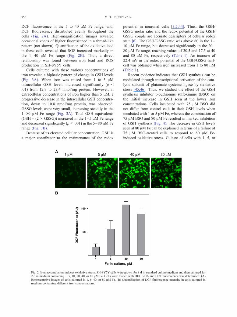
Cells cultured with these various concentrations of
iron revealed a biphasic pattern of change in GSH levels
(Fig. 3A). When iron was raised from 1 to 5 AMintracellular GSH levels increased significantly (p <
.01) from 12.9 to 23.4 nmol/mg protein. However, at
extracellular concentrations of iron higher than 5 AM, a
progressive decrease in the intracellular GSH concentra-
tion, down to 10.8 nmol/mg protein, was observed.
GSSG levels were very small, increasing steadily in the
1–80 AM Fe range (Fig. 3A). Total GSH equivalents
(GSH + (2 � GSSG)) increased in the 1–5 AM Fe range
and decreased significantly (p < .001) in the 5–80 AM Fe
range (Fig. 3B).
Because of its elevated cellular concentration, GSH is
a major contributor to the maintenance of the redox
Fig. 2. Iron accumulation induces oxidative stress. SH-SY5Y cells wer2 d in medium containing 1, 5, 10, 20, 40, or 80 AM Fe. Cells were loaRepresentative images of cells cultured in 1, 5, 40, or 80 AM Fe. (B)medium containing different iron concentrations.
potential in neuronal cells [3,5,44]. Thus, the GSH/
GSSG molar ratio and the redox potential of the GSH/
GSSG couple are accurate descriptors of cellular redox
state [6]. The GSH/GSSG ratio was above 60 in the 1–
10 AM Fe range, but decreased significantly in the 20–
80 AM Fe range, reaching values of 30.5 and 17.5 at 40
and 80 AM Fe, respectively (Table 1). An increase of
22.4 mV in the redox potential of the GSH/GSSG half-
cell was obtained when iron increased from 1 to 80 AM(Table 1).
Recent evidence indicates that GSH synthesis can be
modulated through transcriptional activation of the cata-
lytic subunit of glutamate cysteine ligase by oxidative
stress [45,46]. Thus, we studied the effect of the GSH
synthesis inhibitor L-buthionine sulfoximine (BSO) on
the initial increase in GSH seen at the lower iron
concentrations. Cells incubated with 75 AM BSO did
not differ from control cells in their GSH levels when
incubated with 1 or 5 AM Fe, whereas the combination of
75 AM BSO and 80 AM Fe resulted in marked inhibition
of GSH synthesis (Fig. 4). The decrease in GSH levels
seen at 80 AM Fe can be explained in terms of a failure of
75 AM BSO-treated cells to respond to 80 AM Fe-
induced oxidative stress. Culture of cells with 1, 5, or
e grown for 8 d in standard culture medium and then cultured forded with DHCF-DA and DCF fluorescence was determined. (A)Quantification of DCF fluorescence intensity in cells cultured in
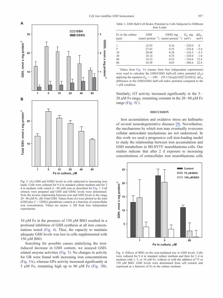
Fig. 3. (A) GSH and GSSG levels in cells subjected to increasing ironloads. Cells were cultured for 8 d in standard culture medium and for 2d in medium with varied (1–80 AM) iron as described for Fig. 1. Cellextracts were prepared and GSH and GSSG levels were determined.Note the inverse relationship between iron and GSH levels in the range20–80 AM Fe. (B) Total GSH. Values from (A) were plotted as the total(GSH plus 2 � GSSG) glutathione content as a function of extracellulariron concentration. Values are means F SD from four independentexperiments.
Table 1. GSH Half-Cell Redox Potential in Cells Subjected to DifferentIron Loads
Fe in the culture
(Am)
GSH
(nmol protein�1)
GSSG mg
(nmol protein�1)
Ehc mg
(mV)
DEhc
(mV)
1 12.93 0.16 �329.0 05 23.43 0.35 �334.4 �5.410 20.68 0.28 �334.3 �5.320 18.14 0.35 �328.0 1.040 14.32 0.53 �316.6 12.480 10.58 0.63 �306.6 22.4
Values from Fig. 3A (means form four independent experiments)
were used to calculate the GSH/GSSG half-cell redox potential (Ehc)
applying the equation Ehc = �240 – (59.1/2)log([GSH]2/[GSSG]). DEhc
difference in the GSH/GSSG half-cell redox potential compared to the
1 AM condition.
Fig. 4. Effects of BSO on the iron-mediated rise in GSH levels. Cellswere cultured for 8 d in standard culture medium and then for 2 d inmedium with 1, 5, or 10 AM Fe, without or with the addition of 75 or150 AM BSO. GSH levels were determined from cell extracts andexpressed as a function of Fe in the culture medium.
Cell iron modifies GSH homeostasis 957
10 AM Fe in the presence of 150 AM BSO resulted in a
profound inhibition of GSH synthesis at all iron concen-
trations tested (Fig. 4). Thus, the capacity to maintain
adequate GSH levels was lost in cells supplemented with
150 AM BSO.
Searching for possible causes underlying the iron-
induced decrease in GSH content, we assayed GSH-
related enzyme activities (Fig. 5). No changes in activity
for GR were found with increasing iron concentrations
(Fig. 5A), whereas GPx activity increased significantly at
5 AM Fe, remaining high up to 80 AM Fe (Fig. 5B).
Similarly, GT activity increased significantly in the 5–
20 AM Fe range, remaining constant in the 20–80 AM Fe
range (Fig. 5C).
DISCUSSION
Iron accumulation and oxidative stress are hallmarks
of several neurodegenerative diseases [9]. Nevertheless,
the mechanisms by which iron may eventually overcome
cellular antioxidant mechanisms are not understood. In
this work we used a progressive cell iron-loading model
to study the relationship between iron accumulation and
GSH metabolism in SH-SY5Y neuroblastoma cells. Our
studies indicate that after 2 d exposure to increasing
concentrations of extracellular iron neuroblastoma cells

Fig. 5. GSH enzyme activities in cells subjected to increasing iron load.Cells were cultured for 8 d in standard culture medium and then for 2 din medium with varied (1–80 AM) Fe as described for Fig. 1. Cellextracts were prepared and GR, GPx, and GT activities were determinedas described under Materials and Methods. Changes in GR werenonsignificant compared to the 1 AM Fe value. Changes in GPx and GTactivities were significantly different (*p < .05, **p < .01) compared tothe 1 AM Fe value.
M. T. Nunez et al.958
underwent a concentration-dependent accumulation of
iron, accompanied by a biphasic change in the intracel-
lular concentration of GSH and a decrease in the GSH/
GSSG ratio. Whereas exposure to 5 AM Fe resulted in a
marked increase in GSH levels compared to 1 AM Fe,
exposure to higher Fe concentrations, up to 80 AM,
decreased GSH levels and increased GSSG levels. Simi-
lar results, i.e., decreased GSH and increased GSSH,
were observed in the brain of mice subjected to a high-
iron diet [47], whereas in PD GSH levels were found
specifically decreased in SNpc [15–17].
Increasing extracellular iron concentration from 40 to
80 AM was associated with a marked loss of cell
viability. Because iron accumulation increases oxidative
stress (Fig. 2 this work; [36]), the initial rise in GSH
synthesis may be interpreted as an adaptive response of
the cells to iron-mediated oxidative stress. In turn, the
subsequent decrease in GSH may correspond to a nega-
tive balance between GSH synthesis and Fe-induced
GSH consumption. Indeed, inhibition of GSH synthesis
by BSO produced increased sensitivity of GSH levels to
iron load.
The GSH/GSSG ratio was above 60 in the 1–10 AMFe range, but decreased to 30.5 and 17.5 at 40 and 80 AMFe, respectively. Because 80 AM Fe induced considerable
loss of cell viability, it is likely that GSH/GSSG ratios
below 30 become critical for cell survival. Similarly,
changes in the GSH reduction potential to more positive
values indicated that iron-induced GSH consumption
resulted in a more oxidant cell environment. In opposi-
tion to these findings, no increase in GSSG levels was
reported in Parkinsonian SNpc [17]. A possible expla-
nation for this difference may reside in the fact that our
GSSG determinations were undertaken in freshly har-
vested neuroblastoma cells, as opposed to postmortem
brain tissue. In the latter situation, O2/Fe-mediated GSH
oxidation may overestimate GSSH content. Despite the
differences in GSSG content, the marked decrease in
GSH found in parkinsonian brains argues for decreased
GSH/GSSG ratios, as are reported in this work.
GR activity did not change in the 1–80 AM iron
range, suggesting that GR is either already maximally
expressed under basal conditions or not inducible by
iron. In contrast, GPx and GT activities increased. As
observed here, the activity of GR was found unchanged
in SNpc of PD, whereas the activity of GT had a
tendency to increase [48]. The finding that both GPx
and GT activity increased in SH-SY5Y cells exposed to
iron agrees with recent reports on gene expression in the
MPTP model of PD, in which increases in GPx and GT
mRNAwere reported [49], but does not agree with earlier
findings indicating no change in GT activity in substantia
nigra of Parkinson’s disease patients [15]. Thus, it is
possible that increase in GT may not be a characteristic
feature of PD.
The observed increases in GPx and GT may represent
defense mechanisms against oxidative stress. We propose
that the increase in GSH synthesis, in conjunction with
increased GPx and GT activities, is an important element

Cell iron modifies GSH homeostasis 959
in the antioxidant response to iron accumulation. In this
context, our data support a mechanism by which iron
accumulation in SNpc results in decreased GSH content
and cell death. However, no increase in iron before GSH
depletion was found in substantia nigra of patients with
incidental Lewis body disease [16], and initial death of
tyrosine hydroxylase-positive cells preceded iron accu-
mulation in monkeys injected with MPTP [50], an
indication that iron accumulation could be indeed secon-
dary to dopaminergic cell death.
In summary, we found that iron accumulation in
neuroblastoma cells resulted in a biphasic change in the
levels of GSH levels and a decrease in cellular redox
potential, thus establishing a mechanistic link between
iron and GSH homeostasis. Considering that adequate
levels of GSH are necessary for neuronal survival, these
results point to the relevance of iron accumulation as a
primary cause for diminished redox potential leading to
neurodegeneration in diseases such as Alzheimer’s and
Parkinson’s disease.
Acknowledgments—This work was supported by Project P99-031 ofthe Millennium Institute for Advanced Studies in Cell Biology andBiotechnology, Santiago, Chile.
REFERENCES
[1] Cooper, A. J. L. Glutathione in the brain: disorders of gluta-thione metabolism. In: Rosenberg, R. N.; Prusiner, S. B.; Di-Mauro, S; Barchi, R. L.; Kunk, L. M., eds. The molecular andgenetic basis of neurological disease. Boston: Butterworth–Hei-nemann; 1997:1195–1230.
[2] Clarke, D. D.; Sokoloff, L. Circulation and energy metabolism ofthe brain. In: Siegel, G. J.; Agranoff, B. W.; Albers, R. W.; Fisher,S. K.; Uhler, M. D., eds. Basic neurochemistry: molecular, cellu-lar and medical aspects. Philadelphia: Lippincott – Raven;1999:637–669.
[3] Meister, A. Selective modification of glutathione metabolism.Science 220:472–477; 1983.
[4] Drukarch, B.; Schepens, E.; Jongenelen, C. A.; Stoof, J. C.;Langeveld, C. H. Astrocyte mediated enhancement of neuronalsurvival is abolished by glutathione deficiency. Brain Res. 770:123–130; 1997.
[5] Wang, X. F.; Cynader,M. S. Astrocytes provide cysteine to neuronsby releasing glutathione. J. Neurochem. 74:1434–1442; 2000.
[6] Schafer, F. Q.; Buettner, G. R. Redox environment of the cell asviewed through the redox state of the glutathione disulfide/gluta-thione couple. Free Radic. Biol. Med. 30:191–212; 2001.
[7] Salinas, A. E.; Wong, M. G. Glutathione S-transferases—a review.Curr. Med. Chem. 6:279–309; 1999.
[8] Schrammel, A.; Gorren, A. C.; Schmidt, K.; Pfeifferm, S.;Mayerm,B. S-nitrosation of glutathione by nitric oxide, peroxynitrite, andSNO/O2
S�. Free Radic. Biol. Med. 34:1078–1088; 2003.[9] Sayre, L. M.; Perry, G.; Atwood, C. S.; Smith, M. A. The role
of metals in neurodegenerative diseases. Cell. Mol. Biol. 46:731–741; 2000.
[10] Adams, J. D., Jr.; Klaidman, L. K.; Odunze, I. N.; Shen, H. C.;Miller, C. A. Brain levels of glutathione, glutathione disulfide, andvitamin E. S. Mol. Chem. Neuropathol. 14:213–226; 1991.
[11] Cardoso, S. M.; Oliveira, C. R. Glutathione cycle impairmentmediates A beta-induced cell toxicity. Free Radic. Res. 37:241–250; 2003.
[12] Omar, R. A.; Chyan, Y. J.; Andorn, A. C.; Poeggeler, B.; Robakis,N. K.; Pappolla, M. A. Increased expression but reduced activityof antioxidant enzymes in Alzheimer’s disease. J. Alzheimer’s Dis.1:39–145; 1999.
[13] Double, K. L.; Gerlach, M.; Youdim, M. B.; Riederer, P. Impairediron homeostasis in Parkinson’s disease. J. Neural Transm. Suppl.60:37–58; 2000.
[14] Perry, T. L.; Godin, D. V.; Hansen, S. Parkinson’s disease: adisorder due to nigral glutathione deficiency? Neurosci. Lett.33:305–310; 1982.
[15] Perry, T. L.; Yong, V. W. Idiopathic Parkinson’s disease, pro-gressive supranuclear palsy and glutathione metabolism in thesubstantia nigra of patients. Neurosci. Lett. 67:269– 274;1986.
[16] Dexter, D. T.; Sian, J.; Rose, S.; Hindmarsh, J. G.; Mann, V. M.;Cooper, J. M.; Wells, F. R.; Daniel, S. E.; Lees, A. J.; Schapira,A. H., et al. Indices of oxidative stress and mitochondrial func-tion in individuals with incidental Lewy body disease. Ann.Neurol. 35:38–44; 1994.
[17] Sofic, E.; Lange, K. W.; Jellinger, K.; Riederer, P. Reduced andoxidized glutathione in the substantia nigra of patients with Par-kinson’s disease. Neurosci. Lett. 142:128–130; 1992.
[18] Youdim, M. B.; Ben-Shachar, D.; Riederer, P. Is Parkinson’s dis-ease a progressive siderosis of substantia nigra resulting in ironand melanin induced neurodegeneration? Acta Neurol. Scand.126:47–54; 1989.
[19] Riederer, P.; Sofic, E.; Rausch,W. D.; Schmidt, B.; Reynolds, G. P.;Jellinger, K.; Youdim, M. B. Transition metals, ferritin, gluta-thione, and ascorbic acid in parkinsonian brains. J. Neurochem.52:515–520; 1989.
[20] Gerlach, M.; Ben-Shachar, D.; Riederer, P.; Youdim, M. B. H.Altered brain metabolism of iron as a cause of neurodegenerativediseases? J. Neurochem. 63:793–807; 1994.
[21] Symons, M. C. R.; Gutteridge, J. M. C., eds. Free radicals andiron: chemistry, biology and medicine. New York: Oxford Univ.Press; 1998.
[22] Sayre, L. M.; Perry, G.; Harris, P. L. R.; Liu, Y.; Schubert, K. A.;Smith, M. A. In situ oxidative catalysis by neurofibrillary tanglesand senile plaques in Alzheimer’s disease: a central role for boundtransition metals. J. Neurochem. 74:270–279; 2000.
[23] Perry, G.; Taddeo, M. A.; Petersen, R. B.; Castellani, R. J.; Harris,P. L. R.; Siedlak, S. L.; Cash, A. D.; Liu, Q.; Nunomura, A.;Atwood, C. S.; Smith, M. A. Adventiously-bound redox activeiron and copper are at the center of oxidative damage in Alz-heimer disease. Biometals 16:70–81; 2003.
[24] Gutteridge, J. M.; Halliwell, B. Free radicals and antioxidants inthe year 2000: a historical look to the future. Ann. N. Y. Acad. Sci.899:136–147; 2000.
[25] Hauptmann, N.; Cadenas, E. The oxygen paradox: biochemistryof active oxygen. In: Scandalios, J. G., ed. Oxidative stress andthe molecular biology of antioxidant defenses. New York: ColdSpring Harbor Laboratory Press; 1997.
[26] Youdim, M. B.; Riederer, P. The role of iron in senescence ofdopaminergic neurons in Parkinson’s disease. J. Neural Transm.Suppl. 40:57–67; 1993.
[27] Shoham, S.; Youdim, M. B. The effects of iron deficiency andiron and zinc supplementation on rat hippocampus ferritin.J. Neural Transm. 109:1241–1256; 2002.
[28] Lotharius, J.; Brundin, P. Pathogenesis of Parkinson’s disease: dop-amine, vesicles anda-synuclein.Nat. Rev. Neurosci. 3:1–11; 2002.
[29] Gotz, M. E.; Double, K.; Gerlach, M.; Youdim, M. B.; Riederer, P.The relevance of iron in the pathogenesis of Parkinson’s disease.Ann. N. Y. Acad. Sci. 1012:193–208; 2004.
[30] Hirsch, E. C.; Brandel, J. P.; Galle, P.; Javoy-Agid, F.; Agid, Y.Iron and aluminum increase in the substantia nigra of patients withParkinson’s disease: an X-ray microanalysis. J. Neurochem. 56:446–451; 1991.
[31] Perry, G.; Raina, A. K.; Nunomura, A.; Wataya, T.; Sayre, L. M.;Smith, M. A. How important is oxidative damage? Lessons fromAlzheimer’s disease. Free Radic. Biol. Med. 28:831–834;2000.
[32] Takeda, A.; Smith, M. A.; Avila, J.; Nunomura, A.; Siedlak,S. L.; Zhu, X.; Perry, G.; Sayre, L. M. In Alzheimer’s dis-ease, heme oxygenase is coincident with Alz50, an epitope oftau induced by 4-hydroxy-2-nonenal modification. J. Neuro-chem. 75:1234–1241; 2000.

M. T. Nunez et al.960
[33] Shimohama, S.; Tanino, H.; Kawakami, N.; Okamura, N.; Koda-ma, H.; Yamaguchi, T.; Hayakawa, T.; Nunomura, A.; Chiba, S.;Perry, G.; Smith, M. A.; Fujimoto, S. Activation of NADPHoxidase in Alzheimer’s disease brains. Biochem. Biophys. Res.Commun. 273:5–9; 2000.
[34] Connor, J. R.; Milward, E. A.; Moalem, S.; Sampietro, M.;Boyer, P.; Percy, M. E.; Vergani, A.; Chorney, M. Is hemochro-matosis a risk factor for Alzheimer disease? J. Alzheimer’s Dis.3:471–477; 2001.
[35] Tapia, V.; Arredondo, M.; Nunez, M. T. Regulation of ironabsorption by cultures of epithelia (Caco-2) cells monolayers withvaried iron status. Am. J. Physiol. 271:G443–G447; 1996.
[36] Nunez-Millacura, C.; Tapia, V.; Munoz, P.; Maccioni, R. B.;Nunez, M. T. An oxidative stress-mediated positive-feedbackiron uptake loop in neuronal cells. J. Neurochem. 82:240–248;2002.
[37] Zecca, L.; Gallorini, M.; Schunemann, V.; Alfred, X.; Traut-wein, A. X.; Gerlach, M.; Riederer, P.; Vezzoni, P.; Tampellini,D. Iron, neuromelanin and ferritin content in the substantianigra of normal subjects at different ages: consequences foriron storage and neurodegenerative processes. J. Neurochem.76:1766–1773; 2001.
[38] Mossman, T. Rapid colorimetric assay for cellular growth andsurvival: application to proliferation and cytotoxicity assays.J. Immunol. Methods 65:55–63; 1986.
[39] Reynolds, I. J.; Hastings, T. Glutamate induces the production ofreactive oxygen species in cultured forebrain neurons followingNMDA receptor activation. J. Neurosci. 15:3318–3327; 1995.
[40] Griffith, O. W. Determination of glutathione and glutathione di-sulfide using glutathione reductase and 2-vinylpyridine. Anal.Biochem. 106:207–212; 1980.
[41] Anderson, M. E. Determination of glutathione and glutathionedisulfide in biological samples. Methods Enzymol. 113:548–555;1985.
[42] Paglia, D. E.; Valentine, W. N. Studies on the quantitative andqualitative characterization of erythrocyte glutathione peroxidase.J. Lab. Clin. Med. 70:158–169; 1967.
[43] Veal, E. A.; Toone, W. M.; Jones, N.; Morgan, B. A. Distinct rolesfor glutathione S-transferases in the oxidative stress response inSchizosaccharomyces pombe. J. Biol. Chem. 277:35523–35531;2002.
[44] Dexter, D. T.; Wells, F. R.; Lees, A. J.; Agid, F.; Agid, Y.; Jenner,P.; Marsden, C. D. Increased nigral iron content and alterationsin other metal ions occurring in brain in Parkinson’s disease.J. Neurochem. 52:1830–1836; 1989.
[45] Forman, H. J.; Dickinson, D. A.; Iles, K. E. HNE-signaling path-ways leading to its elimination. Mol. Aspects Med. 24:189–194;2003.
[46] Krzywanski, D. M.; Dickinson, D. A.; Iles, K. E.; Wigley, A. F.;Franklin, C. C.; Liu, R. M.; Kavanagh, T. J.; Forman, H. J. Var-iable regulation of glutamate cysteine ligase subunit proteins af-fects glutathione biosynthesis in response to oxidative stress.Arch. Biochem. Biophys. 423:116–125; 2004.
[47] Lan, J.; Jiang, D. H. Excessive iron accumulation in the brain: apossible potential risk of neurodegeneration in Parkinson’s dis-ease. J. Neural Transm. 104:649–660; 1997.
[48] Sian, J.; Dexter, D. T.; Lees, A. J.; Daniel, S.; Jenner, P.; Marsden,C. D. Alterations in glutathione levels in Parkinson’s disease andother neurodegenerative disorders affecting basal ganglia. Ann.Neurol. 36:348–355; 1994.
[49] Mandel, S.; Grunblatt, E.; Maor, G.; Youdim, M. B. Early and lategene changes in MPTP mice model of Parkinson’s disease em-ploying cDNA microarray. Neurochem. Res. 27:1231–1243;2002.
[50] He, Y.; Thong, P. S.; Lee, T.; Leong, S. K.; Mao, B. Y.; Dong, F.;Watt, F. Dopaminergic cell death precedes iron elevation inMPTP-injected monkeys. Free Radic. Biol. Med. 35:540–547;2003.