Progress in Medicinal Chemistry 32 - National Academic ...
352
Progress in Medicinal Chemistry 32 Editors G.P. ELLIS, D.SC.,PH.D.,F.R.S.C. Department of Chemistry, University of Wales, PO. Box 912, CardijJ CFl3TB, United Kingdom and D.K. LUSCOMBE, B.PHARM., PH.D., F.I.BIOL., F.R.PHARM.S. Welsh School of Pharmacy, University of Wales, P 0. Box 13, CardifJ; CFl 3XE United Kingdom 1995 ELSEVIER AMSTERDAM. LONDON. NEW YORK . TOKYO
-
Upload
khangminh22 -
Category
Documents
-
view
0 -
download
0
Transcript of Progress in Medicinal Chemistry 32 - National Academic ...
Progress in Medicinal Chemistry 32
Editors
G.P. ELLIS, D.SC. ,PH.D. ,F .R .S .C .
Department of Chemistry, University of Wales, PO. Box 912, CardijJ CFl3TB, United Kingdom
and
D.K. LUSCOMBE, B.PHARM., PH.D. , F.I.BIOL., F .R.PHARM.S.
Welsh School of Pharmacy, University of Wales, P 0. Box 13, CardifJ; CFl 3XE United Kingdom
1995
ELSEVIER AMSTERDAM. LONDON. NEW YORK . TOKYO
Elsevier Science BV P.O. Box 211 1000 AE Amsterdam The Netherlands
Library of Congress Cataloging in Publication Data:
Please refer to card number 62-2712 for this series.
ISBN 0-444-82057-4 ISBN Series 0-7204-7400-0
0 1995 Elsevier Science BV. All rights reserved
No part of this publication may be reproduced, stored in a retrieval system or transmitted in any form or by any means, electronic, mechanical, photocopying, recording or otherwise without the prior written permission of the Publisher, Elsevier Science B.V., Copyright and Permissions Department, P.O. Box 521, 1000 AM Amsterdam, The Netherlands.
No responsibility is assumed by the Publisher for any injury andor damage to persons or property as a matter of products liability, negligence or otherwise, or from any use or operation of any methods, products, instructions or ideas contained in the material herein. Because of rapid advances in the medical sciences, the Publisher recommends that independent verification of diagnoses and drug dosages should be made.
Special regulations for readers in the USA: This publication has been registered with the Copyright Clearance Center Inc. (CCC), Salem, Massachusetts. Information can be obtained from the CCC about conditions under which the photocopying of parts of this publication may be made in the USA. All other copyright questions, including photocopying outside of the USA, should be referred to the Publisher.
This book is printed on acid-free paper
Printed in The Netherlands

V
Preface Six topics of interest to medicinal chemists, microbiologists, pharmacolo- gists and clinicians are reviewed in this volume. Chapter 1 updates our knowledge of a promising class of antitumour drugs, the heterocyclic aldehyde thiosemicarbazones. In the last few years, much progress has been made in the treatment of hypertension by inhibiting components of the renin angiotensin system; this is reviewed in Chapter 2. Another approach to the treatment of cardiovascular disease, using 4-quinolones as vasodilators, is discussed in Chapter 3.
Although good progress has been made to counteract bacterial resistance to /I-lactam antibiotics (see Volume 31), this problem is of increasing importance in the efficacy of most other antibiotics, as is demonstrated in Chapter 4. The search for drugs to treat the ever increasing number of people infected by the human immunodeficiency virus (HIV) continues unabated. Research into the inhibition of HIV proteinase (Chapter 5) gives rise to optimism that such agents may play an important role in the treatment of AIDS.
This volume ends with an account (Chapter 6) of a promising but as yet not finalised therapy for tumours which are refractory to other drugs, namely, the use of compounds derived from taxol which react with tubulin rather than DNA.
We thank our authors for providing us with the present state of knowledge in these important fields. We are grateful for permission to reproduce material which is protected by copyright, and for the support and encouragement from the staff of our publishers.
July 1994 G.P. Ellis D.K. Luscombe
Progress in Medicinal Chemistry - Vol. 32, edited by G.P. Ellis and D.K. Luscombe 0 1995 Elsevier Science B.V. All rights reserved.
1 Chemical and Biological Properties of Cyto t oxic a-(N)-He terocyclic Carboxaldehyde Thiosemicarbazones
MAO-CHIN LIU, Ph.D., TAI-SHUN LIN, Ph.D. and ALAN C. SARTORELLI, Ph.D.
Department of Pharmacology and Developmental Therapeutics Program, Comprehensive Cancer Center, Yale University School of Medicine, New Haven, CT 06520, U. S. A.
INTRODUCTION 2
BASIC STRUCTURE AND HETEROCYCLIC RING SYSTEMS
MODIFICATIONS OF PYRIDINE-2-CARBOXALDEHYDE THIOSEMICARBAZONE
MODIFICATIONS OF ISOQUINOLINE-1-CARBOXALDEHYDE THIOSEMICARBAZONE
MODIFICATIONS OF THE THIOSEMICARBAZONE SIDE-CHAIN
METAL COMPLEXES OF a-(N)-HETEROCYCLIC CARBOXALDEHYDE THIOSEMICARBAZONES
BIOCHEMICAL MECHANISMS OF ACTION
STRUCTURE-ACTIVITY RELATIONSHIPS
CONCLUSIONS
3
5
17
19
21
24
27
28
30
30
ACKNOWLEDGEMENT
REFERENCES
1
2 CYTOTOXIC IT,-(N)-HETEROCYCLIC THIOSEMICARBAZONES
INTRODUCTION
Ribonucleoside diphosphate reductase is a critical enzyme in the de novo synthesis of the deoxyribonucleotide precursors of DNA and, as such, is essential for cellular replication. Thus, its presence and activity is closely correlated with cellular growth rates [ 1, 21. Since deoxyribonucleotides are present in extremely low levels in mammalian cells, Cory and Chiba [3] have presented arguments that an inhibitor of ribonucleoside diphosphate reductase should be more effective than an inhibitor of DNA polymerase in blocking DNA synthesis. Hence, it seems reasonable that a strong inhibitor of ribonucleoside diphosphate reductase would be a useful weapon in the therapeutic armamentarium against cancer. Several different classes of agents are relatively specific inhibitors of ribonucleoside diphosphate reductase. These have included a-(N)-heterocyclic carboxaldehyde thiosemi- carbazones (HCTs), hydroxyurea [4], N-hydroxy-N’-aminoguanidine de- rivatives [5-71 and polyhydroxybenzohydroxamates [8, 91. The HCTs, as a class, are among the most potent known inhibitors of ribonucleoside diphosphate reductase, being 80-5000 times more effective, depending upon the HCT, than hydroxyurea, a clinically useful anticancer agent [lo, 111. Members of this class have shown anticancer activity against a wide spectrum of transplanted rodent neoplasms, including sarcoma 180, Ehrlich carcinoma, leukaemia L12 10, Lewis lung carcinoma, hepatoma 129, hepatoma 134, adenocarcinoma 755, and B16 melanoma. In addition, spontaneous lymphomas of dogs have shown susceptibility to HCTs [12, 131. Such broad spectrum activity denotes clinical potential and suggests that a drug of this class may well have utility in cancer therapy.
A variety of nucleoside analogues are also active as inhibitors of ribonucleoside diphosphate reductase. These include 2,2’-difluoro-2’- deoxycytidine [ 14-1 61, 2’-fluoroadenine arabinoside [ 171 and 2-chloro-2’- deoxyadenosine [ 181. However, the 5’-triphosphate of each of these compounds appears to be a more potent inhibitor of DNA polymerase than of ribonucleoside diphosphate reductase, making DNA polymerase the more probable primary target of these nucleoside analogues [17-191.
Some HCTs, especially derivatives of 2-acetylpyridine thiosemicarba- zone, selectively inhibit herpes simplex virus type-1 and -2 specified ribonucleoside diphosphate reductase [20-221, which is biologically distinct from the mammalian enzyme [23, 241. A discussion of the antiviral activity of these compounds is not included in this review.
The work in our laboratory and in those of others which has identified ribonucleoside diphosphate reductase as the major target of the HCTs has been reviewed previously [25-271. This paper will emphasize relatively

M.-C. LIU ET AL. 3
recent investigations, incorporating older findings to comprehensively discuss the structure-activity relationships of this class of compounds.
BASIC STRUCTURE AND HETEROCYCLIC RING SYSTEMS
The basic structure of this class of compounds is shown in (1) [13,25]. The minimum requirement for biological activity is the attachment of the carboxaldehyde thiosemicarbazone side-chain a to an unencumbered ring nitrogen of heteroaromatic character. Compounds in which the thiosemi- carbazone side-chain is attached at positions B or y to the heterocyclic N-atom are inactive. A conjugate N*-N*-S* tridentate ligand system has been found to be a common feature of compounds with carcinostatic activity [28-321. For this reason, the structure of the HCTs may be written in the SH form and metal complexes of the HCTs may be readily formed. The characteristics of the ligand are indicated in (2). The formation of two five-membered chelate rings of a partially conjugate character favours octahedral coordination of two ligands to one divalent metal ion. The ring nitrogen atom is a reasonably good donor to transition metals, allowing the formation of coordination compounds (chelates). Thus, quarternization of the ring nitrogen atom completely eliminates biological activity.
A large number of different ring systems has been used instead of the pyridine ring of pyridine-2-carboxaldehyde thiosemicarbazone (3) (PT) [ 13, 30,311, Replacement of the pyridine ring with benzene, furan, or thiophene ring systems led to loss of antitumour activity [30, 33, 341. Furthermore, replacement of the pyridine ring of PT with five-membered ring systems such as imidazole, pyrazole, pyrrole, or triazole, also resulted in a decrease or complete loss of antineoplastic activity [13, 30, 35-37]. A variety of six-membered heterocyclic ring systems carrying the thiosemicarbazone side-chain a to the heterocyclic nitrogen (3-9) are active antineoplastic agents [13, 321.

4 CYTOTOXIC a-(N)-HETEROCYCLIC THIOSEMICARBAZONES
S S II
a C H = N N H C N H z II
S
NI'
Compounds (4-6), however, can be envisioned as analogues of PT (3) in which the carbon has been replaced by a nitrogen at the ortho, meta, and para positions of the nitrogen atom of the pyridine ring of PT, respectively; these PT analogues exhibited antitumour activity comparable to that of PT.
Three approaches have been employed to introduce an additional aromatic ring onto the pyridine ring of PT, connecting one side of the aromatic ring onto the 3,4-, 4,5- or 5,6-positions of the pyridine ring of PT. The first approach led to the formation of isoquinoline- 1 -carboxaldehyde thiosemicarbazone (7) (IQ-1), one of the most potent ribonucleoside diphosphate reductase inhibitors of the HCT class. The second led to the formation of isoquinoline-3-carboxaldehyde thiosemicarbazone (lo), which showed marginal antitumour activity. The third method of connection produced quinoline-2-carboxaldehyde thiosemicarbazone (1 l), which was devoid of carcinostatic activity. These findings also apply to other ring systems. For example, compound (8) can be envisioned to be formed from compound (5) by introducing a second aromatic ring by the first approach; compound (8) is more active than compound (5) as both an inhibitor of ribonucleoside diphosphate reductase and as an antitumour agent [ 131. In contrast, compound (4), which is active as an antineoplastic agent, generates (by the addition of an additional aromatic ring by the second approach) compound (12), which is inactive [ 131. The pyridine and isoquinoline rings

M.-C. LIU ET AL. 5
are the two heterocyclic ring systems which have been most extensively investigated; the structural modifications of these two series of compounds that have been carried out will be described subsequently.
8 ; = a N N CH=NNH-C-NH2 CHzNNH-C-NH2
(7) S ; = a c H = N N H - C - N H 2 II
N CH=NNH-C-NHz
II activity
MODIFICATIONS OF PYRIDINE-2-CARBOXALDEHYDE THIOSEMICARBAZONE
Since the &st report that pyridine-2-carboxaldehyde thiosemicarbazone (3) (PT) had antileukaemic activity in mice [38], various substituted pyridine-2- carboxaldehyde thiosemicarbazones have been synthesized and evaluated for antineoplastic activity in an effort to find a more efficacious compound [13, 251. Derivatives of PT, in which all of the four unsubstituted positions, 3,4, 5 and 6, were substituted by one, two, or more functional groups, have been synthesized. In early studies, the 3-position of PT was substituted by Me [39], OH, OAc [30, 401, OMe, OEt, COOH, F [13, 411, OCH,C6H,-rn-

6 CYTOTOXIC a-(N)-HETEROCYCLIC THIOSEMICARBAZONES
NH,, OCH&H,-m-OH [42], C&-m-NO,, and C6H4-m-NHz [43]. The agent which appeared to have the most therapeutic potential in this series was 3-hydroxypyridine-2-carboxaldehyde thiosemicarbazone (1 3) (3-HP), which gave a longer duration of inhibition of DNA synthesis in neoplastic cells than that produced by 5-hydroxypyridine-2-carboxaldehyde thiosemi- carbazone (14) (5-HP), when molar equivalent doses of these agents were administered to tumour-bearing mice and the effects on DNA replication were measured in malignant cells [33]. Consistent with these findings, 3-HP was more active than 5-HP as an anticancer agent against the L5178Y lymphoma, Lewis lung carcinoma, and adenocarcinoma 755 [13, 321. However, it was less active than 5-HP against the L1210 leukaemia and sarcoma 180. The 4-position of PT has been substituted by a series of groups including Me, NMe,, N(CH,),, N(CH,CH,)NMe, N(CH,CH,OH),,
C,H,-p-NH, [45], and NHPh [46]. The most active compounds in this series were 4-(3-aminophenyl)pyridine-2-carboxaldehyde thiosemicarbazone (1 5) and 4-morpholinopyridine-2-carboxaldehyde thiosemicarbazone (1 6), both being superior to 5-HP as anticancer agents in mice bearing sarcoma 180 ascites cells [44,45]. The effects of substituents at the 5-position of PT were also extensively studied; 5-substituted substituents synthesized included F, C1, Br, I, Me, Et, OH, OAc, OCF,, CF,, N(Me),, NHAc, O,SMe, OC,H4NMe,, O(C,H,O),Et, OOCR [41, 47, 481, CH,Ph [46], CH,OH, CH,OCOR [49], OCH2C6H4-m-NH,, and OCH,C,H,-m-OH [42]. 5-HP was the most active compound in the 5-substituted series. Only a few derivatives of PT substituted in the 6-position have been reported, with the substituents being Me, C,H,-m-NO,, C,H,-m-NH, [39,43], and CSNH, [50]; all of these derivatives were inactive as anticancer agents. A few di-and multi- substituted derivatives of PT have been reported in the past; these have included 3-OH, 4-Me; 3-OH, 6-Me; 3-C1, 5-OH [13]; 3-Me, 4-morpholino [44]; 3-OH, 4-CH,OH, 5-CH,OH [51]. In this group of agents, 3-hydroxy- 4,5-bis(hydroxymethyl)pyridine-2-carboxaldehyde thiosemicarbazone (17) showed significant antitumour activity against sarcoma 180 and L1210 leukaemia in mice [51].
5-HP is the only member of the HCT series that has been administered to humans in a Phase I study. The selection several years ago of 5-HP for clinical trial was based upon (a) its activity against a spectrum of transplanted tumours and spontaneous dog lymphomas and (b) its ease of parenteral administration as the sodium salt. The results of two independent Phase I studies conducted at Yale University and the Sloan-Kettering Cancer Center [52, 531 showed that transient decreases in blast counts occurred in 6 of 25 patients with leukaemia, none being of a sufficient
N(CH,CH,),O [39, 441, C,H,-m-NO,, C,H,-m-NH, [43], Ph, C,H,-o-NH,,

M.-C. LIU ET AL.
CH=NNHCNHz CH=NNHCNHz
S II
S II
CH=NNHCNHz CH=NNHCNHz
magnitude to constitute a remission, and no antitumour effects were observed in 18 patients with solid tumours. The dose-limiting toxicity of 5-HP in humans was gastrointestinal, being manifested by severe nausea, vomiting, and diarrhoea. In addition, the most aggressive drug regimens also produced myelosuppression, haemolysis, anaemia, hypertension, and hypotension. The lack of demonstrable antineoplastic activity of 5-HP observed in the Phase I trial was attributed in part to the relatively short biological half-life (t,,3 of 5-HP in humans, which was due to the rapid formation and elimination of the 0-glucuronide conjugate [52]. Thus, the tlR of 5-HP in the blood of mice was 15 min, while the drug had a t1,2 in humans of 2.5 to 10.5 min, depending upon the patient. Twenty percent of a therapeutic dose of 5-HP was excreted in the urine of the mouse within 24 h, whereas a therapeutic dose of 5-HP was excreted 2- to 3.5-times faster in man. Approximately 75% of the material found in the urine of patients was in the form of an 0-glucuronide, which had no inhibitory activity against ribonucleoside diphosphate reductase.
Recently, 4-methyl substituted derivatives of 3-HP and 5-HP have been synthesized in our laboratory [54] to explore the possibility that a methyl group adjacent to the ring hydroxyl could protect the 3- or 5-hydroxy substituent from enzymatic 0-glucuronidation and, thereby, prevent inactivation of antitumour activity. The syntheses of these agents are described in Schemes 1.1 and 1.2.
2,CLutidine (18) was nitrated to give the two isomers, 2,4-dimethyl-3- and 2,4-dimethyl-5-nitropyridine (( 19) and (20), respectively), in approxi- mately equal amounts [55]. Catalytic hydrogenation of compounds (19) and (20) over 5% PdC in absolute ethanol gave the corresponding amino derivatives (21) and (22). Diazotization of compounds (21) and (22) with sodium nitrite in 10% sulphuric acid, followed by hydrolysis of the resulting

8 CYTOTOXIC ct-(N)-HETEROCYCLIC THIOSEMICARBAZONES
products, gave the respective hydroxy compounds (23) and (24). Treatment of (23) and (24) with 30% hydrogen peroxide in glacial acetic acid produced the N-oxides, (25) and (26), which were then refluxed with acetic anhydride to give the acetates, (27) and (28). A repeat of the N-oxidation procedure with compound (27), followed by rearrangement of the resulting N-oxide (29) by refluxing with acetic anhydride, yielded the corresponding 2-pyridine aldehyde diacetate derivative (30). Treatment of (30) with thiosemicarbazide in the presence of hydrochloric acid produced 3-hydroxy- 4-methylpyridine-2-carboxaldehyde thiosemicarbazone (3 1) (3-HMP).
Me Me Me Me
21 R=3-NH2 I 1 22 R=5-NH2
(a) Fuming H2SO4, K2NO3; (b) H2, Pd/C, EtOH; (c) NaN02,10% H2S04;
(d) HOAc, 30% H202; (e) AQO; (9 H2NNHC(S)NH2, HCI; (9) NaHC03
Scheme 1.1.
Hydrolysis of the acetate (28) with hydrochloric acid (Scheme 1.2) gave 5-hydroxy-2-hydroxythyl-4-methylpyridine (32). Oxidation of (32) with manganese dioxide in ethanol yielded the corresponding aldehyde, (33), which was then condensed with thiosemicarbazide [43, 441 to afford the 5-hydroxy-4-methylpyridine-2-carboxaldehyde thiosemicarbazone (34) (5- HMP).

M.-C. LIU ET AL. 9
(a-d)
CH~OAC
(a) HCI; (b) MnOp, EtOH; (c) H2NNHC(S)NHz, HCI; (d) NaHC03
Scheme 1.2.
The prolongation of the life-span of mice bearing the L1210 leukaemia produced by the maximum effective daily dose of the hydroxyl-substituted derivatives administered intraperitoneally in solution in dimethylsulphoxide (DMSO) or as a fine suspension is shown in Table 1.1. The 4-methyl substituted derivatives, 3- and 5-HMP, were both equivalent to, or more effective than, their corresponding parent compounds, 3-HP (1 3) and 5-HP (14). Greater antitumour activity occurred when the HCTs were adminis- tered in suspension, presumably resulting from slow solubilization in the peritoneal cavity, which provides a long-lasting effect. The greater activity of 3-HMP (31) and 5-HMP (34) compared with their non-methylated counterparts is consistent with previous findings that the addition of methyl or other hydrophobic groups onto the 3-, 4-, or 5-carbon atoms of the pyridine ring increased activity as inhibitors of ribonucleoside diphosphate reductase, a phenomenon speculated to be due to a hydrophobic binding region in the target enzyme molecule [56]. French and Blanz [13] also reported the synthesis of 3-HMP (31) by a different method; however, neither the synthetic procedure nor spectroscopic data to confirm the structure of this compound were presented. Furthermore, in contrast to our test results, which showed that 3-HMP had antitumour activity against the L1210 leukaemia, they reported that this agent was inactive against this tumour cell-line [ 131.
Various 3-amino-, 5-amino-, and 5-nitro-substituted pyridine-2-carboxal- dehyde thiosemicarbazones (51-56) and their derivatives (60-62,65,66, and 68) were synthesized [57] to further evaluate the effects of various substituents on the pyridine ring on antitumour activity (Schemes 1.3, 1.5, and 1.6). Oxidation [58] of 3-nitro- [59, 601, 5-nitro-[59, 611, 4-methyl-3- nitro- and 4-methyl-5-nitro-2-picolines [55] (35-38) with selenium dioxide in refluxing dioxan yielded the corresponding pyridine-2-carboxaldehydes

10 CYTOTOXIC u-(N)-HETEROCYCLIC THIOSEMICARBAZONES
Table 1.1. COMPARATIVE EFFECTS OF 3-HP, 5-HP, 3-HMP, AND 5-HMP ON MICE BEARING THE L1210 LEUKAEMIA
Compd Injection Optimum Av A wt,b TIC form daily dosage." (%)
(mglkg)
3-HP DMSO soln. 40 + 1.5 114 5-HP DMSOsoln. 40 + 1.8 132 3-HMP DMSOsoln. 40 + 0.5 135 5-HMP DMSOsoln. 40 - 1.4 138
5-HP suspension 60 + 4.6 146 3-HMP suspension 50 + 0.9 168 5-HMP suspension 40 - 3.4 186
"Administered once daily for six consecutive days, beginning 24 h after tumour implantation. bAverage weight change of mice from onset to termination of drug treatment. "% TIC represents the ratio of the survival time of treated to control mice x 100. The average survival time of untreated L1210 tumour-bearing control animals was 8.2 days.
Reprinted with permission from (1992) J. Med. Chem. 35, 3667-3671. Copyright 0 1992 American Chemical Society.
(3942). The aldehydes were protected by conversion to the cyclic ethylene acetals (43-46), which were then reduced by catalytic hydrogenation using Pd/C as a'catalyst to give the corresponding amino acetals (47-50) [62]. Treatment of compounds (47-50) with thiosemicarbazide in ethanol containing 10% concentrated hydrochloric acid gave the desired thiosemi- carbazone hydrochlorides; the free bases (5 1-54) were liberated by treatment with aqueous sodium bicarbonate solution. Condensation of 5-nitropyridine-2-caboxaldehyde and 4-methyl-5-nitropyridine-2-carboxal- dehyde [(40) and (42), respectively] with thiosemicarbazide in aqueous ethanol, yielded the corresponding 5-nitro-substituted thiosemicarbazones ( 5 5 ) and (56) (Scheme Z.3).
It is interesting to note that the 2-methyl groups in compounds (37) and (38) were considerably more sensitive to selenium dioxide oxidation than their 4-methyl counterparts. 4-Methyl-3-nitropyridine-2-carboxaldehyde (41) and 4-methyl-5-nitropyridine-2-carboxaldehyde (42) were isolated in 20% and 55% yields, respectively, by silica gel column chromatography after oxidation. In addition to the unreacted starting material, both 4-methyl-3-nitro-2-pyridine carboxylic acid and 4-methyl-5-nitro-2-pyri- dine carboxylic acid were also isolated. When the reaction time was prolonged, the amount of acidic by-products was increased; however, no detectable quantities of 2-methyl-3-nitropyridine-4-carboxaldehyde and

M.-C. LIU ET AL. 11
R R
(42) R = Me, X = 5 4 0 2
R
(d, e)
(a) Se02, Dioxane; (b) HOCH2CH20H, pTSA; (c) H2, PdC, EtOH;
(d) H2NNHC(S)NH2, HCI; (e) NaHC03
70% EtOH P.,A,,, (40) R = H
(42) R =Me
Scheme 1.3.
2-methyl-5-nitropyridine-4-carboxaldehyde were found. A cyclic mecha- nism is proposed for the oxidation of 5-nitro-2,4-lutidine, which is analogous to the mechanism proposed by Corey and Schaefer [63] for the oxidation of 7-methylquinoline, except that a cyclic transition state is suggested (Scheme 1.4). Such an intermediate may account for the selective oxidation of the 2-methyl group. A similar, but more hindered, cyclic transition state may be formed for the oxidation of 3-nitro-2,4-lutidine, which might explain why the 2-methyl group in the 3-nitro derivative is more difficult to oxidize than its 5-nitro counterpart.
The acetamide and alkylsulphonamide derivatives of 3-amino and

12 CYTOTOXIC ct-(N)-HETEROCYCLIC THIOSEMICARBAZONES
H
Me
Scheme 1.4.
5-aminopyridine-2-carboxaldehyde thiosemicarbazone, (60-62) and (65), (66), respectively, were prepared as described in Scheme 1.5. Acetylation of compounds (48-50) with acetic anhydride in anhydrous pyridine gave the acetamide derivatives (57-59), which were then condensed with thiosemicar- bazide to produce 5-acetylaminopyridine-2-carboxaldehyde thiosemicarba- zone (60) and 3- and 5-acetylamino-4-methylpyridine-2-carboxaldehyde thiosemicarbazones, (61) and (62), respectively. During the process of acidic hydrolysis of the ethylene acetal groups, some hydrolysis of the acetamide functions occurred, even though the reaction conditions were carefully controlled. The desired compounds were obtained in pure form, however, by recrystallization from ethanol or by silica gel chromatography. Treatment of (50) with methanesulphonyl chloride or p-toluenesulphonyl chloride in anhydrous pyridine afforded the corresponding 5-methane- sulphonylamino and, p-toluenesulphonylamino derivatives, (63) and (a), respectively, which were then treated with thiosemicarbazide in the presence of concentrated hydrochloric acid to afford the corresponding 5-methane- sulphonylamino and 5-p-toluenesulphonylamino derivatives of 4-meth- ylpyridine-2-carboxaldehyde thiosemicarbazone, (65) and (66). 5-Hydroxylamino-4-methylpyridine-2-carboxaldehyde thiosemicarba-
zone (68) was synthesized by the procedure described in Scheme 1.6.
M.-C. LIU ET AL.
R
13
R
RSOZNH h0 0
(a) H2NNHC(S)NH2, HCI; (b) NaHC03
Scheme 1.5.
Hydrogenation of compound (46) in ethanol using Pd(OH),/C as a catalyst under 50 psi of hydrogen yielded the 5-hydroxylamino derivative (67). Condensation of (67) with thiosemicarbazide in the presence of concen- trated hydrochloric acid, followed by treatment with sodium bicarbonate, afforded the desired 5-hydroxylamino-4-methylpyridine-2-carboxaldehyde thiosemicarbazone (68) (5-HAP).
The 5-nitro derivatives, (55) and (56), the acetyl derivatives, (60-62), and the alkylsulphonamide derivatives, (65) and (66), were inactive as antineo- plastic agents against the L1210 leukaemia. In contrast the 3-amino derivatives, (51) (3-AP) and (53) (3-AMP), possessed anticancer activity, being comparable in their antitumour efficacy against the L1210 leukaemia;
a CH=NNHCNHz
(46) (67) (68)
(a) H2, Pd(OH)dC, EtOH; (b) H2NNHC(S)NH2, HCI; (c) NaHC03
Scheme 1.6.

14 CYTOTOXIC a-(N)-HETEROCYCLIC THIOSEMICARBAZONES
these two agents were among the most effective of all of the HCTs synthesized to date in prolonging the survival time of mice bearing this neoplasm (Table 1.2). The 5-amino derivatives (52) (5-AP) and (54) (5-AMP) and the 5-hydroxyamino derivative (68) were comparable to 5-HP (14) in the L1210 test system. The % T/C value for 5-HP against the L1210 leukaemia obtained in these experiments was similar to previous reported results from this laboratory with this HCT [64]; however, a significant difference existed between the value for this agent that we have obtained (% T/C = 133) and that (% T/C = 268) reported by French and Blanz [13]. These dissimilar findings may be due to differences between the L1210 leukaemia cell lines employed and/or to differences in the schedule of drug administration. Although 5-HP (14) was administered daily by intraperi- toneal injection starting 24 h after tumour inoculation in both studies, our experiments employed 6 daily treatments, while French and Blanz [13] used daily treatments that were continued until 50% of the animals had died.
The four most active amino-substituted pyridine-containing HCTs synthesized to date, i.e., the 3-amino derivatives, 3-AP (51) and 3-AMP (53) and the 5-amino derivatives, 5-AP (52) and 5-AMP (54), were further evaluated against the L1210 leukaemia employing a schedule of drug administration of twice a day at approximately 12 h intervals for 6 consecutive days. The results of these investigations are supmarized in
Table 1.2. EFFECTS OF PYRIDINE-2-CARBOXALDEHYDE THIOSEMICARBAZONE DERIVATIVES ON THE SURVIVAL TIME OF MICE
BEARING THE L1210 LEUKAEMIA
Compd Optimum Av A w ? , ~ Av survival, TIC daily dosage," (%) days (%) (mglkg)
5-HP 40 + 2.0 10.4 133 3-AP 40 - 5.9 14.6 187 5-AP 20 - 2.8 11.0 140 3-AMP 60 + 2.0 14.9 190 5-AMP 20 - 7.0 10.8 138 5-HMP 10 - 2.7 10.6 136
"Drugs were administered in suspension by intraperitoneal injection, beginning 24 h after
bAverage change in body weight from onset to termination of therapy. '% TIC represents the ratio of the survival time of treated to control mice x 100. The average survival time of untreated L1210 tumour-bearing control animals was 7.8 days.
Reprinted with permission from (1992) J. Med. Chem. 35, 3672-3677. Copyright 0 1992 American Chemical Society.
tumour implantation, once daily for 6 consecutive days, with 5-10 mice per group.
M.-C. LIU ET AL. 15
Table 1.3. 3-AMP exhibited the least toxicity in this series with the group of animals that received two daily doses of 40 mg/kg of 3-AMP having a % T/C value of 255 and 40% long-term survivors. Conversely, the groups receiving two daily doses of 30 mg/kg of 3-AP or of 5-AP died after an average of 7.3 days (% T/C = 96) and 6.9 days (% T/C = 91), respectively. The pronounced loss in body weight post drug treatment (an average decrease of 12.8% and 16.0% from original body weights, respectively) suggests that the early
Table 1.3. EFFECTS OF 3-AP, 5-AP, 3-AMP AND 5-AMP ADMINISTERED TWICE DAILY ON THE SURVIVAL TIME OF MICE BEARING THE
L1210 LEUKAEMIA
Compd Optimum Av A wt,b Av survival, Long-term daily (%) (days c, (%) survivors" dosage, a
(mnlknl
3-AP 10x2 15x2 20 x 2 30x2
5-AP 1 0 x 2 1 5 x 2 20x2 30x2
3-AMP 10x2 20 x 2 30 x 2 40 x 2
5-AMP 10x2 15x2 20x2 30x2
- 6.4 - 3.3 - 12.4 - 12.8
- 3.8 - 7.7 - 12.3 - 16.0
+ 1.4 - 3.8 - 2.4 - 8.1
+ 1.4 - 7.1 - 12.1 - 12.4
18.7 19.8 14.6 7.3
17.6 16.8 14.0 6.9
14.8 16.9 17.6 19.4
18.6 21.0 17.9 14.4
246 262 192 96
232 22 1 185 91
195 222 232 255
245 276 236 190
4110 015 1/10 015
1/10 015 1/10 015
2110 0110 1/10 215
1/10 0110 0110 015
"Drugs were administered in suspension by intraperitoneal injection, beginning 24 h after
bAverage change in body weight from onset to termination of therapy. "Average survival time includes only those mice that died prior to day 60. dT/C represents the ratio of the survival time of treated to control animals x 100. The average survival time of untreated tumour-bearing control animals was 7.6 days.
'Long-term survivors are the number of mice that survived for > 60 days relative to the total number of treated mice.
Reprinted with permission from (1992) J. Med. Chem. 35,3672-3677. Copyright 0 1992 American Chemical Society.
tumour implantation, twice daily for 6 consecutive days, with 5-10 mice per group.
16 CYTOTOXIC ct-(N)-HETEROCYCLIC THIOSEMICARBAZONES
deaths of the mice were the result of drug toxicity rather than the leukaemic process. However, 3-AP and 5-AP exhibited much better antitumour activity at lower dosage levels and the groups of animals that received two daily doses of 10 mg/kg of 3-AP or of 5-AP gave % T/C values of 246 and 232, and produced 40% and 10% long-term survivors, respectively. 5-AMP appeared to be somewhat less toxic than 3-AP and 5-AP, with antitumour activity comparable to that of 5-AP. Comparison of the results listed in Tables 1.2 and 1.3 indicates that the activity of compounds 3-AP, 5-AP, 3-AMP and 5-AMP are schedule-dependent with much better therapeutic effects being obtained by twice daily administration than by a once daily regimen. It appears that 3-AP and 3-AMP are superior in their activities against the L1210 leukaemia to any other agent in this series of HCTs reported to date [54, 641.
The primary metabolic lesion created by the HCTs is interference with the biosynthesis of DNA, an action resulting from the potent inhibition of ribonucleotide reductase activity. For this reason, the capacity of amino- and hydroxy-substituted pyridine-2-carboxaldehyde thiosemicarbazones to inhibit cytidine diphosphate (CDP) reductase activity has been measured [65]. 3-AP (51) and 3-AMP (53) were found to be more potent than the 5-amino-substituted HCTs as inhibitors of CDP reductase activity, each causing 50% inhibition (IC50) of enzymatic activity at a concentration of 0.3pM. As such, they were about 4 and 6 times more potent than the hydroxy-substituted derivatives, 3-HP (ICs0 = 1.2 pM) and 5-HP (IC50 = 1.75 pM), respectively, and approximately 4 and 3 times more potent than 5-AP (IC50 = 1.25 pM) and 5-AMP (IC50 = 0.90 pM).
The comparative effects of these HCTs on the growth of wild-type and hydroxyurea-resistant L1210 cell-lines were measured [65]. Consistent with the relative activities of these HCTs against the target enzyme, ribonucleo- tide reductase, 3-AP (51) and 3-AMP (53) were the most cytotoxic with ICsO values of 1.3 pM and 1.5 pM for the L1210 parental cell-line and 1.6 pM and 2.3 pM for hydroxyurea-resistant L1210 cells, respectively. In accord with interference with ribonucleotide reductase activity being the major site of action of the HCTs, 3-AP and 3-AMP inhibited the incorporation of [3H]thymidine into DNA without affecting the rate of incorporation of [3H]uridine into RNA. Furthermore, the incorporation of ['4C]cytidine into cellular ribonucleotides and RNA was not decreased by 3-AP and 3-AMP; however, the incorporation of cytidine into deoxyribonucleotides facilitated by ribonucleotide reductase and the subsequent incorporation into DNA was markedly inhibited. Thus, the cytodestructive effects of 3-AP and 3-AMP appeared to result from the specific inhibition of DNA biosynthesis [65]. In contrast to most of the HCTs, formulation of 3-AP would appear to
M.-C. LIU ET AL. 17
be easily obtainable since the hydroxyethylsulphonic acid salt is relatively water-soluble, with a saturated water-solubility of 40 mg/mL. Therefore, further evaluation of 3-AP as a potential anticancer drug would appear to be particularly warranted.
MODIFICATIONS OF ISOQUINOLINE- 1 -CARBOXALDEHYDE THIOSEMICARBAZONE
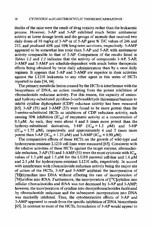
Isoquinoline- 1 -carboxaldehyde thiosemicarbazone (7) (IQ- 1) is an exceed- ingly potent agent, both as an inhibitor of ribonucleoside diphosphate reductase and as an antitumour agent [6669]. The clinical utility of IQ-1 as an antineoplastic agent in humans, however, is limited by its inability to be formulated because of extremely low water-solubility . A large number of structural modifications of the isoquinoline nucleus have been made [70-741, for example, inserting hydrophilic groups such as amino or hydroxy onto the isoquinoline ring system to increase water-solubility as either an acid or a sodium salt, respectively. Substitution of an amino group onto the 5-position of the isoquinoline ring of IQ-1 had no adverse effects on activity against the target enzyme. Thus, for example, 5-amino IQ-1 (69) (5-AIQ-1) at a concentration of 0.03 pM produced 50% inhibition of ribonucleoside diphosphate reductase from the Novikoff hepatoma, which makes it equal to IQ-1 as the most potent known HCT inhibitor of this enzyme [74]. 5-AIQ-1 has, however, an advantage over IQ-1 in that it can be rendered relatively water-soluble as an acid salt. However, since the 5-acetylamino derivative of IQ-1 was found to be devoid of carcinostatic activity [70] and N-acetylation is a relatively ubiquitous metabolic reaction in vivo, 4-methyl-5-aminoisoquinoline- 1 -carboxaldehyde thiosemicarba- zone (70) (MAIQ- 1) was designed and synthesized to create steric hindrance to the enzymatic substitution of the 5-amino function by insertion of a bulky methyl group at the adjacent 4-position of the isoquinoline ring [62, 751. MAIQ-1 was found to be an effective antineoplastic agent against transplanted animal tumours and 60-fold more potent than 5-HP as an inhibitor of ribonucleoside diphosphate reductase [62, 641. MAIQ-1 is also active against human colon carcinoma HT-29 cells in culture, with an ICs0 value of 3.2 pM [76]. Williams et al. [77] reported that an enzyme(s) in hepatic microsomes obtained from rats and mice, which is absent in the microsomes from Ehrlich tumour cells, can inactivate MAIQ-1 as an inhibitor of ribonucleoside diphosphate reductase. These investigations, therefore, suggested a mechanism of selectivity for MAIQ- 1 which resulted
18 CYTOTOXIC a-(N)-HETEROCYCLIC THIOSEMICARBAZONES
in the maintenance of inhibitory activity in tumour cells while producing inactivation by the liver.
OH OH
In contrast to amino derivatives, substitution of a hydroxyl moiety at either the 4- or 5-position of the isoquinoline ring of IQ-1 decreased inhibitory activity in vitro against ribonucleoside diphosphate reductase, and also decreased host toxicity significantly with retention of antineoplas- tic activity, thereby improving the therapeutic index in tumour-bearing mice [70, 721. 5-Hydroxyisoquinoline- 1 -carboxaldehyde thiosemicarbazone (7 1) (5-HIQ- 1) and 4-hydroxyisoquinoline- 1 -carboxaldehyde thiosemicarba- zone (72) (4-HIQ-1) showed antitumour activity in mice bearing sarcoma 180 ascites cells comparable to that of IQ-1 [70, 721. Substitution of more polar groups such as 5-SO3H and 5-COOH, however, led to the loss of antitumour activity [70, 781. A 4-hydroxymethyl derivative of IQ-1 was reported to be more active than the parent compound as an antitumour agent, but was found to be more toxic [49].
A series of additional substitutions on the isoquinoline ring of IQ-1 have been reported [74, 78, 791, which include the 2-oxide, 4-Me, 5-substituted derivatives (OAc, F, C1, NO,, CF3, n-C3F,, Me, succinimido, pyrrolidinyl), 6-Me, 7-substituted derivatives (OAc, F, C1, OH, OMe), and 8-F. The biological activity of these agents varied depending upon the tumour system employed. However, none of these derivatives was found to be superior to the parent compound IQ-1 as an antitumour agent.
M.-C. LIU ET AL. 19
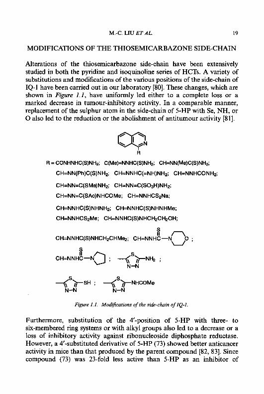
MODIFICATIONS OF THE THIOSEMICARBAZONE SIDE-CHAIN
Alterations of the thiosemicarbazone side-chain have been extensively studied in both the pyridine and isoquinoline series of HCTs. A variety of substitutions and modifications of the various positions of the side-chain of IQ-1 have been carried out in our laboratory [80]. These changes, which are shown in Figure 1.1, have uniformly led either to a complete loss or a marked decrease in tumour-inhibitory activity. In a comparable manner, replacement of the sulphur atom in the side-chain of 5-HP with Se, NH, or 0 also led to the reduction or the abolishment of antitumour activity [Sl].
R
R = CONHNHC(S)NHz; C(Me)=NNHC(S)NH2; CH=NN(Me)C(S)NH2;
CH=NN(Ph)C(S)NHz; CH=NNHC(=NH)NH2; CH=NNHCONHp;
CH=NN=C(SMe)NH2; CH=NN=C(S03H)NH2;
CH=NN=C(SAc)NHCOMe; CH=NNHCS2Na;
CH=NNHC(S)NHNHz; CH=NNHC(S)NHNHMe;
CH=NNHCS2Me; CH=NNHC(S)NHCH2CH20H;
S
CH=NNHC(S)NHCH~CHMQ; CH=NNHt-NnO . U '
; +'>NHCOMe N-N N-N
Figure 1.1. Modifications of the side-chain of IQ-I.
Furthermore, substitution of the #-position of 5-HP with three- to six-membered ring systems or with alkyl groups also led to a decrease or a loss of inhibitory activity against ribonucleoside diphosphate reductase. However, a 4'-substituted derivative of 5-HP (73) showed better anticancer activity in mice than that produced by the parent compound [82, 831. Since compound (73) was 23-fold less active than 5-HP as an inhibitor of
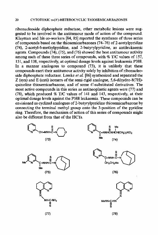
20 CYTOTOXIC a-(N)-HETEROCYCLIC THIOSEMICARBAZONES
ribonucleoside diphosphate reductase, other metabolic lesions were sug- gested to be involved in the antitumour mode of action of the compound. Klayman and his co-workers [84, 851 reported the syntheses of three series of compounds based on the thiosemicarbazones (74-76) of 2-acetylpyridine (74), 2-acetyl-6-methylpyridine, and 2-butyrylpyridine, as antileukaemic agents. Compounds (74), (75), and (76) showed the best antitumour activity among each of these three series of compounds, with % T/C values of 157, 151, and 138, respectively, at optimal dosage levels against leukaemia P388. In a manner analogous to compound (73), it is unlikely that these compounds exert their antitumour activity solely by inhibition of ribonucleo- side diphosphate reductase. Lemke et al. [86] synthesized and separated the Z (syn) and E (anti) isomers of the semi-rigid analogue, 5,6-dihydro-8(7H)- quinoline thiosemicarbazone, and of some 4‘-substituted derivatives. The most active compounds in this series as antineoplastic agents were (77) and (78), which produced % T/C values of 141 and 143, respectively, at their optimal dosage levels against the P388 leukaemia. These compounds can be envisioned as cyclized analogues of 2-butyrylpyridine thiosemicarbazone by connecting the terminal methyl group onto the 3-position of the pyridine ring. Therefore, the mechanism of action of this series of compounds might also be different from that of the HCTs.
S
CH=NNH-C-N C=NNHCNHCH(CH2)3NEt2 I Me
(74) U
(73)
Me C=NNH-C -I
Lv I Me
u/ \ \7 I CH2CH2Me
(75) (76)
/N MeHN-C-HN
! (77) (78)
M.-C. LIU ET AL. 21
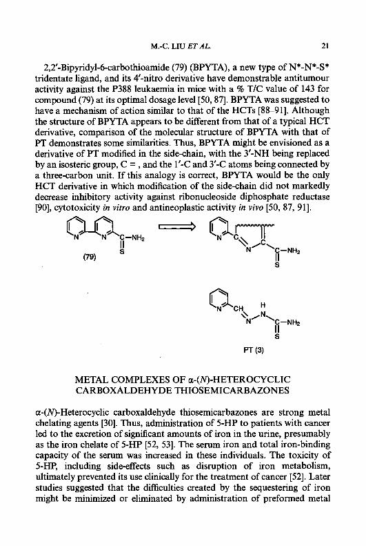
2,2’-Bipyridyl-6-carbothioamide (79) (BPYTA), a new type of N*-N*-S* tridentate ligand, and its 4’-nitro derivative have demonstrable antitumour activity against the P388 leukaemia in mice with a % T/C value of 143 for compound (79) at its optimal dosage level [50,87]. BPYTA was suggested to have a mechanism of action similar to that of the HCTs [88-911. Although the structure of BPYTA appears to be different from that of a typical HCT derivative, comparison of the molecular structure of BPYTA with that of F T demonstrates some similarities. Thus, BPYTA might be envisioned as a derivative of PT modified in the side-chain, with the 3’-NH being replaced by an isosteric group, C = , and the 1’-C and 3’-C atoms being connected by a three-carbon unit. If this analogy is correct, BPYTA would be the only HCT derivative in which modification of the side-chain did not markedly decrease inhibitory activity against ribonucleoside diphosphate reductase [90], cytotoxicity in vitro and antineoplastic activity in vivo [50, 87, 911.
METAL COMPLEXES OF a-(N)-HETEROCYCLIC CARBOXALDEHYDE THIOSEMICARBAZONES
a-(N)-Heterocyclic carboxaldehyde thiosemicarbazones are strong metal chelating agents [30]. Thus, administration of 5-HP to patients with cancer led to the excretion of significant amounts of iron in the urine, presumably as the iron chelate of 5-HP [52, 531. The serum iron and total iron-binding capacity of the serum was increased in these individuals. The toxicity of 5-HP, including side-effects such as disruption of iron metabolism, ultimately prevented its use clinically for the treatment of cancer [52]. Later studies suggested that the difficulties created by the sequestering of iron might be minimized or eliminated by administration of preformed metal
22 CYTOTOXIC a-(N)-HETEROCYCLIC THIOSEMICARBAZONES
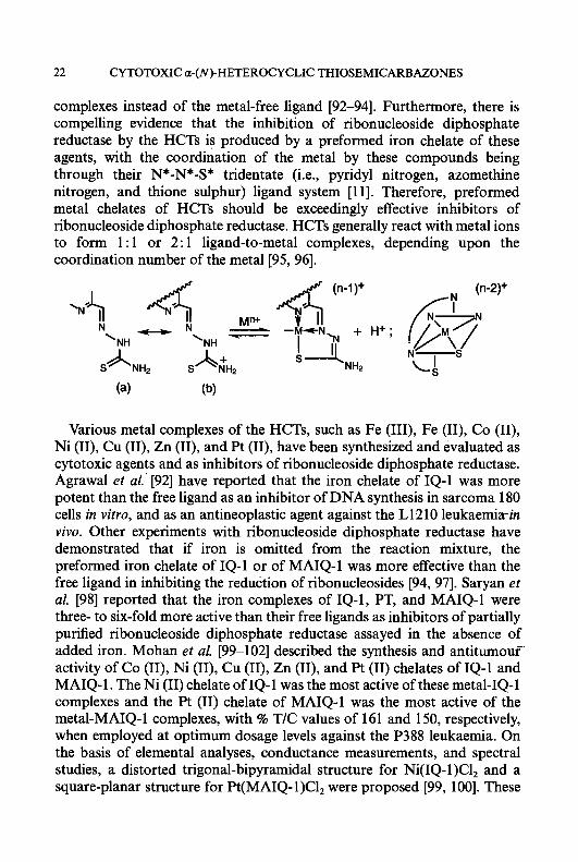
complexes instead of the metal-free ligand [92-941. Furthermore, there is compelling evidence that the inhibition of ribonucleoside diphosphate reductase by the HCTs is produced by a preformed iron chelate of these agents, with the coordination of the metal by these compounds being through their N*-N*-S* tridentate (i.e., pyridyl nitrogen, azomethine nitrogen, and thione sulphur) ligand system [ 1 11. Therefore, preformed metal chelates of HCTs should be exceedingly effective inhibitors of ribonucleoside diphosphate reductase. HCTs generally react with metal ions to form 1 : 1 or 2: 1 ligand-to-metal complexes, depending upon the coordination number of the metal [95, 961.
(n-2)+
Various metal complexes of the HCTs, such as Fe (111), Fe (11), Co (11), Ni (11), Cu (11), Zn (11), and Pt (11), have been synthesized and evaluated as cytotoxic agents and as inhibitors of ribonucleoside diphosphate reductase. Agrawal et al.’ [92] have reported that the iron chelate of IQ-1 was more potent than the free ligand as an inhibitor of DNA synthesis in sarcoma 180 cells in vitro, and as an antineoplastic agent against the L1210 leukaemiain vivo. Other experiments with ribonucleoside diphosphate reductase have demonstrated that if iron is omitted from the reaction mixture, the preformed iron chelate of IQ-1 or of MAIQ-1 was more effective than the free ligand in inhibiting the reduction of ribonucleosides [94, 971. Saryan et al. [98] reported that the iron complexes of IQ-1, PT, and MAIQ-1 were three- to six-fold more active than their free ligands as inhibitors of partially purified ribonucleoside diphosphate reductase assayed in the absence of added iron. Mohan et al. [99-1021 described the synthesis and antitumouf activity of Co (11), Ni (11), Cu (11), Zn (11), and Pt (11) chelates of IQ-1 and MAIQ-1. The Ni (11) chelate of IQ-1 was the most active of these metal-IQ-1 complexes and the Pt (11) chelate of MAIQ-1 was the most active of the metal-MAIQ-1 complexes, with % T/C values of 161 and 150, respectively, when employed at optimum dosage levels against the P388 leukaemia. On the basis of elemental analyses, conductance measurements, and spectral studies, a distorted trigonal-bipyramidal structure for Ni(1Q- 1)C1, and a square-planar structure for Pt(MA1Q- 1)C1, were proposed [99, 1001. These
M.-C. LIU ET AL. 23
authors also reported the synthesis and antitumour activity of Fe (11) and Fe (111) chelates of IQ-1, MAIQ- 1, and 4-(m-aminophenyl)pyridine-2-carbox- aldehyde thiosemicarbazone. The highest level of activity was shown by the Fe(MA1Q-1)C1, complex, with a % T/C value of 154 at the optimum dosage against the P388 leukaemia [103].
The synthesis and antitumour activity of a number of copper and iron complexes of PT and of 5-substituted pyridine-2-carboxaldehyde thiosemi- carbazones where the 5-substituents which were OH, OAc, NMe,, H, Me, C1, and CF, have been reported by several laboratories [93, 104-1161. The copper complex of PT, [CuL(MeCO,)], (HL = PT), was demonstrated to be a potent inhibitor of ribonucleoside diphosphate reductase, being slightly more active than the free ligand, presumably because it was readily taken up and bound as shown with Ehrlich carcinoma cells. Cellular DNA synthesis was inhibited by this copper complex at low concentrations, whereas RNA synthesis was much less sensitive [98, 1161. Furthermore, this copper complex was also more potent than the free ligand as an inhibitor of the growth of the Ehrlich carcinoma, sarcoma 180, and Chinese hamster ovary cells [93, 98, 107-1 101.
Recently, the molecular structures of [CuL(MeCO,)],(HL = PT) and [CuHL(SO,)], were determined by single-crystal X-ray diffraction tech- niques [117]. Both complexes were shown to consist of discrete centrosym- metric dimers, the monomeric units being bridged by two acetato or sulphato ligands. The copper atoms have a distorted square-pyramidal coordination geometry with three donor atoms (NNS) coming from L or HL to form a tricyclic ligation system. The fourth donor atom (oxygen) comes from the bridging MeCOO- or SO,-ions. The fifth coordination position is occupied by a less strongly bound oxygen from the second bridging anion. Ainscough et al. [118, 1191 determined the structure of a Lewis-base adduct of CuL+, [CuL(bipy)]ClO, (bipy = bipyridine), and neutral ligand-copper complexes, such as [Cu(HL)(H2O)(ClO,)~2H,O (HL = PT) by single-crystal X-ray methodology.
Cristalli et al. [120] reported the synthesis and the antitumour and antifungal activities of Fe (11), Co (11), Ni (11), Cu (11), Cd (11), Pd (11), Zn (11), and Pt (11) complexes of 2,2’-bipyridyl-6-carbothioamide (77) (BPYTA). The copper (11) complex was found to be 12-fold more active than the parent compound in decreasing the incorporation of [’251]5- iododeoxyuridine incorporation into the DNA of P388 leukaemia cells, with the 50% inhibitory concentration being 25 pM.
Two hydrazone-copper complexes, pyridine-2-carboxaldehyde-2’- pyridylhydrazone-copper (11) (80) and salicylaldehydebenzoylhydrazonato- copper (81), have been reported to have cytotoxic activity in vitro [121, 1221.
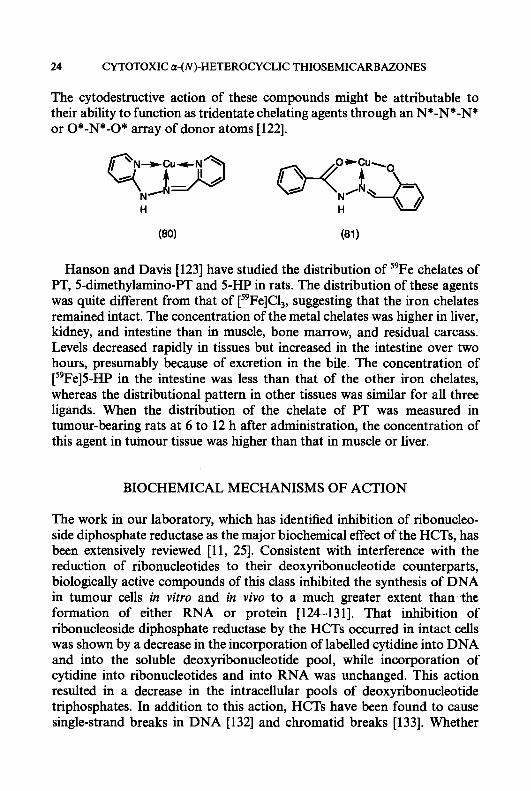
24 CYTOTOXIC CZ-(N)-HETEROCYCLIC THIOSEMICARBAZONES
The cytodestructive action of these compounds might be attributable to their ability to function as tridentate chelating agents through an N*-N*-N* or 0*-N*-O* array of donor atoms [122].
Hanson and Davis [123] have studied the distribution of '9Fe chelates of PT, 5-dimethylamino-PT and 5-HP in rats. The distribution of these agents was quite different from that of [59Fe]C1,, suggesting that the iron chelates remained intact. The concentration of the metal chelates was higher in liver, kidney, and intestine than in muscle, bone marrow, and residual carcass. Levels decreased rapidly in tissues but increased in the intestine over two hours, presumably because of excretion in the bile. The concentration of ["Fe]5-HP in the intestine was less than that of the other iron chelates, whereas the distributional pattern in other tissues was similar for all three ligands. When the distribution of the chelate of PT was measured in tumour-bearing rats at 6 to 12 h after administration, the concentration of this agent in tuinour tissue was higher than that in muscle or liver.
BIOCHEMICAL MECHANISMS OF ACTION
The work in our laboratory, which has identilied inhibition of ribonucleo- side diphosphate reductase as the major biochemical effect of the HCTs, has been extensively reviewed [l l , 251. Consistent with interference with the reduction of ribonucleotides to their deoxyribonucleotide counterparts, biologically active compounds of this class inhibited the synthesis of DNA in tumour cells in vitro and in vivo to a much greater extent than the formation of either RNA or protein [124-1311. That inhibition of ribonucleoside diphosphate reductase by the HCTs occurred in intact cells was shown by a decrease in the incorporation of labelled cytidine into DNA and into the soluble deoxyribonucleotide pool, while incorporation of cytidine into ribonucleotides and into RNA was unchanged. This action resulted in a decrease in the intracellular pools of deoxyribonucleotide triphosphates. In addition to this action, HCTs have been found to cause single-strand breaks in DNA [132] and chromatid breaks [133]. Whether

M.-C. LIU ET AL. 25
these lesions are associated with the inhibition of ribonucleotide reductase or are due to a direct effect at the level of DNA remains to be determined.
It was first assumed that the inhibition of ribonucleoside diphosphate reductase was simply the result of chelation of iron, which was required for full activity of the mammalian enzyme. This mechanism, however, was ruled out by studies with IQ-1 which demonstrated that enzyme inhibition by 0.04 pM IQ-1 was not reversed, and even seemed to be enhanced, by up to 43 pM ferrous ion [134]. The inhibition of ribonucleotide reductase by some of the less potent analogues, however, such as isoquinoline- 1-carboxaldehyde guanylhydrazone and 5-HP, was partially reversed by the addition of iron. Dithiothreitol, used as a substitute for the natural reducing agent, thioredoxin, also caused partial reversal of enzyme inhibition by some of the HCTs. The nucleotide substrate and enzyme activators had no effect on the inhibition produced by members of this class. These findings have been summarized previously [ 1 11.
It was also initially hypothesized [13, 1341 that the HCTs might bind directly to the iron present in the active site of ribonucleoside diphosphate reductase. However, experimentation led to the conclusion that the active form of the inhibitor was in fact a preformed iron chelate [ 1 11, and this was corroborated by calculations which showed that the formation of an enzyme-iron-HCT complex as initially envisioned was thermodynamically unlikely [107].. In support of these conclusions, Agrawal et al. [92] reported that the iron chelate of IQ-1 was more potent than the free ligand as an inhibitor of DNA synthesis in sarcoma 180 cells in vitro and as an antineoplastic agent against the L1210 leukaemia in vivo. Furthermore, the binuclear iron centre of the M2 subunit of ribonucleoside diphosphate reductase is ‘buried’ in the core of the protein [135], suggesting that the iron present in this enzyme is not readily available for chelation, except after modification of the conformation of the protein.
Mammalian ribonucleotide reductase, like that of the enzyme from E. coli, is composed of two nonidentical subunits, M1 and M2, both of which are necessary for activity [136-1401. Subunit M1 contains the nucleotide binding-sites for the substrates and the allosteric effectors, as well as for the dithiodisulphide groups which participate in the redox reaction. Subunit M2 contains a tyrosyl free radical, which is necessary for enzyme activity, and a binuclear ferric iron centre [ 1351. Hydroxyurea destroys the free radical; therefore, in E. coli, inhibition by hydroxyurea was shown to be irreversible unless the free radical was restored either by removal of the hydroxyurea and replacement of the iron, or by an enzymatic mechanism involving iron, oxygen, NADPH, flavin mononucleotide, superoxide dismutase, and two unidentified proteins [141]. Inhibition of the mammalian enzyme by

26 CYTOTOXIC a-(N)-HETEROCY CLIC THIOSEMICARBAZONES
hydroxyurea, however, was easily reversible. We have speculated that this phenomenon is due to the fact that the iron of the mammalian enzyme can be easily replaced under the usual reaction conditions [97]. Graslund et al. [142] have demonstrated that this is in fact the case. These investigators used a partially purified extract from hydroxyurea-resistant mouse fibroblasts which overproduce protein M2 in an amount large enough to give a detectable EPR signal. The EPR signal of the free radical was absent after treatment with hydroxyurea, but was restored if dithiothreitol was added. The presence of iron and of oxygen was required for recovery of the signal. Thelander et al. [143] further demonstrated that the free radical is unstable with a half-life of about ten minutes under anaerobic conditions, so that the presence of iron and oxygen is necessary to maintain enzyme activity in vitro.
Thelander and Graslund [ 1441 have demonstrated that the preformed iron-chelate of MAIQ- 1, in the presence of a dithiol and oxygen, generates a radical scavenger which destroys the free radical essential for enzymatic activity. They proposed that the iron chelate of the HCT binds at the active site of the enzyme, with the ferrous form of the chelate then reacting with molecular oxygen in a redox process that, via a 1-electron reduction, leads to destruction of the tyrosyl radical of the M2 subunit. These findings appear to explain both the necessity for iron for inhibition of enzyme activity by the HCTs, and the partial reversal of inhibition by dithiol and iron. The results also imply that the basic mechanism of inhibition of the enzyme by the HCTs is the same as for drugs such as hydroxyurea and guanazole. Liermann et al. [145] reported that the inhibition of ribonucleo- side diphosphate reductase of intact Ehrlich ascites tumour cells by different antitumour agents, including hydroxyurea, pyrogallol, and IQ-1, can be determined by EPR spectroscopy. The inactivation of the M2 subunit of ribonucleotide reductase was measured by the quenching of the functionally essential tyrosyl radical. The results obtained were comparable with those observed with the isolated enzyme. Furthermore, the chemical behaviour of the iron chelate of IQ-1 may well be similar to that of the iron-bleomycin complex, which causes strand breakage of DNA. This may explain the production of DNA and chromatid breaks produced by this agent [132, 1331. Recently, Nocentini et al. [90] reported that BPYTA, which may be envisioned as a side-chain modified analogue of the HCTs, has a mechanism of action similar to that of the HCTs. Consistent with this concept is the finding that the BPYTA-iron complex has a high affinity for the M2 subunit of ribonucleoside diphosphate reductase and destroys the tyrosyl free radical of the subunit. However, free BPYTA was more active than BPYTA-Fe as an inhibitor of both cell growth and enzyme activity when
M.-C. LIU ET AL. 21
tested in intact cells. It was suggested that a slower cellular uptake of BPYTA-Fe was responsible for the lower degree of activity of the metal complex.
STRUCTURE-ACTIVITY RELATIONSHIPS
Ribonucleoside diphosphate reductase is the primary target of the HCTs; however, a drug must pass through several membrane barriers, survive alternate sites of attachment and storage, and avoid significant metabolic destruction before it reaches its site of action. Therefore, it is not surprising that the inhibitory activity of the HCTs against the partially purified enzyme does not strictly correspond to antitumour activity in v i v a Thus, for example, 5-AP and its 5-acetyl derivative (5-AAP) have similar activity as inhibitors of CDP reductase, both agents producing 50% inhibition of enzymatic activity at a concentration of 2.0 pM [65]. However, 5-AP exhibits significant anticancer activity against the L 12 10 leukaemia, whereas 5-AAP is inactive [57].
French and Blanz [ 131 compared the inhibitory activity against ribonucleo- side diphosphate reductase, as well as the tumour-inhibitory activity against three murine neoplasms of 61 pyridine and 36 other related HCTs. A correlation between the inhibitory activity of the HCTs against partially purified human ribonucleoside diphosphate reductase and the sensitivity of the three tumours employed was not observed. However, among those compounds that were highly active against the enzyme (ICso 5 M), 25 of 27 exhibited antineoplastic activity in one or more of the tumour systems employed, while 33 of 51 and only 2 of 19 of those with intermediate or low activity, respectively, against the enzyme had antitumour activity. A high proportion of the compounds that had the capacity to interfere with tumour growth consisted of the esters and ethers of 3-hydroxy- (3-HP) and 5-hydroxypyridine-2-carboxaldehyde thiosemicarbazone (5-HP), even though these compounds had only intermediate activity (IC50 values between and 5 x M) against ribonucleoside diphosphate reductase. The authors concluded that highly ionic or readily metabolizable substitu- ents were undesirable. The E (anti) isomer of PT was reported to be the major form [146], and was the more inhibitory isomer towards ribonucleo- side diphosphate reductase.
Two groups have made theoretical calculations of the electronic effects of substituents in a small subset of 5-substituted derivatives of PT and have compared these computations with previous data on biological activity. Knight et al. [ 1 151 found correlations between Hammet substituent
28 CYTOTOXIC a-(N)-HETEROCYCLIC THIOSEMICARBAZONES
constants and half-wave reduction potentials, copper-complex formation constants, protonation constants, and other measured factors. There also appeared to be a correlation between Hammet constants and cytotoxic activity, as well as with the capacity of the HCTs to inhibit ribonucleoside diphosphate reductase; however, these relationships were not absolute.
Miertus et al. [147, 1481 made theoretical calculations on a similar but not identical group of compounds, and concluded that activity correlated with the ability to form metal chelates and reactivity at the C = N bond of the side-chain. These workers assumed that at least two steps existed in the inhibitory effects of the HCTs. In the first step, a complex with Fe (11) was formed, and the complex formation activated the C (7) = N (8) bond with respect to nucleophilic attack. According to their calculations, a direct relationship existed between inhibitory activity and reactivity of the C (7) nucleophilic centre. On the basis of this hypothesis, they predicted that 5-nitropyridine-2-carboxaldehyde thiosemicarbazone (55) would be the most potent compound in the pyridine series as an inhibitor of ribonucleo- side diphosphate reductase. However, compound ( 5 9 , which was recently synthesized in our laboratory, was inactive as an antitumour agent against the L1210 leukaemia [57]. In addition, Biyushkin and Chumakov [149] made theoretical calculations of the electronic structure of a number of pyridine-2-carboxaldehyde thiosemicarbazones that was required for anti- tumour activity. They concluded that a longer C = S bond (> 1692 A) and a shorter C-NH, bond (< 1320 A) were necessary for the antitumour activity of the HCTs. Based upon molecular orbital and spectroscopic studies, our laboratory found that Fe(I1) bound to HCTs in a covalent manner, whereas Fe(II1) appeared to interact ionically [150].
Gupta et al. [l51] compared hydrophobicity and Van der Waals volume with previously reported inhibitory activity against ribonucleoside di- phosphate reductase for several series of HCTs. They concluded that, in each group, activity correlated better with size than with hydrophobicity. However, these authors did not consider the location of the substituents in their calculations.
CONCLUSIONS
Phase I clinical trials of a member of the HCTs have been conducted [52, 531. These investigations carried out with 5-HP did not demonstrate significant anticancer activity in humans. The findings did not correspond to those observed in tumour-bearing mice [13] and dogs [67], where 5-HP exhibited relatively good antineoplastic activity, although it must be
M.-C. LIU ET AL. 29
stressed that a Phase I1 evaluation of 5-HP has not been conducted. Extensive excretion of iron complexed with 5-HP was observed in all patients. The dose-limiting toxicity exhibited in the Phase I trial was severe nausea, vomiting, and diarrhoea, although several other toxic side-effects were observed, including myleosuppression, haemolysis, and anaemia. The majority of the drug was excreted in the urine as the 0-glucuronide, with little or no metabolism of the side-chain being detected.
The newly reported HCTs, 3-AP and 3-AMP, are about 6 times more potent than 5-HP as inhibitors of ribonucleoside diphosphate reductase [65]; furthermore, they are not susceptible to glucuronidation, a conjugation that appeared to inactivate 5-HP. In addition, these two amino substituted HCTs exhibited significant cytotoxicity to neoplastic cells in vitro and antitumour activity in vivo. Thus, we believe 3-AP and 3-AMP, and metal complexes of these agents, have clinical potential for the treatment of rapidly growing cancers.
In particular, the HCTs would appear to be good candidates for use in combination chemotherapy. Thus, Cory and his group [152, 1531 found synergism when the HCTs, IQ-1 or MAIQ-1, were employed in admixture with other inhibitors of ribonucleoside diphosphate reductase that affected the nucleotide binding subunit. Gale et al. [ 1541 reported synergistic inhibition of the growth of the L1210 leukaemia by MAIQ-1, as well as by hydroxyurea and guanazole, when used in combination with either cyclophosphamide or a platinum-containing antineoplastic agent. The results with MAIQ- 1 were less schedule-dependent than those with hydroxyurea. This is not surprising since the effects of the similar agent, IQ-1, on DNA synthesis was relatively long-lasting [143]. Bhuyan et al. [155] found increased cell kill by 5-HP when it was used on L1210 leukaemia cells in the S-phase of the cell cycle following synchronization with low levels of 5-fluorouracil. Schabel ' et al. [ 1561 reported supra-additivity between arabinosylcytosine and PT against the L1210 leukaemia. Grindey et al. [157] also described a synergistic interaction between arabinosylcytosine and PT against the L1210 leukaemia. Antholine et al. [lo81 tested combinations of iron and copper complexes of PT with X-irradiation on cultured CHO cells. The copper chelate was much more effective than the iron complex or the free ligand when used in conjunction with X-rays, and the PT-copper complex was synergistic with X-irradiation when exposure was simultane-
In conclusion, the mechanism by which the HCTs inhibit ribonucleoside diphosphate reductase activity appears to be due to the destruction of the tyrosyl free radical in the active site of the enzyme by the iron complex of the drug. The structural features required for maximum activity have been
ous.
30 CYTOTOXIC a-(N)-HETEROCYCLIC THIOSEMICARBAZONES
established. Furthermore, several active compounds are known which appear to be excellent candidates for clinical trial. Since it seems reasonable to assume that a potent inhibitor of ribonucleoside diphosphate reductase would be a useful addition to our clinical armamentarium, particularly to use in combination chemotherapy, it is important to note that several combinations with other anticancer drugs have been reported to give additive or synergistic activity against transplanted animal tumours. Thus, it would seem appropriate to select an agent of this class for extensive clinical trial as an anticancer agent.
ACKNOWLEDGEMENT
Supported in part by United States Public Health Service Grant CA-53340 from the National Cancer Institute.
REFERENCES
1
2 3
4
5 6 7
8 9
10 11
12
13 14
15
16
17
Elford, H.L., Freese, M., Passamani, E. and Morris, H.P. (1970) J. Biol. Chem. 245,
Weber, G. (1977) N. Engl. J. Med. 296,486493. Cory, J.G. and Chiba, P. (1989) in Inhibitors of Ribonucleoside Diphosphate Reductase Activity (Cory, J.G. and Cory, A.M., eds.), pp. 245-264, Pergamon Press, New York. Krakoff, I.H. (1975) in Antineoplastic and Immunosuppressive Agents (Sartorelli, A.C. and Johns, D.G., eds.), Part 11, pp. 789-792, Springer-Verlag, New York. Tai, A.W., Lien, E.J., Lai, M.M.C. and Khwaja, T.A. (1984) J. Med. Chem. 27,236-238. Weckbecker, G., Lien, E.J. and Cory, J.G. (1988) Biochem. Pharmacol. 37,529-534. Cory, J.G., Cory, A.H., Raber, N.K., Narayanan, A. and Schneller, S.W. (1993) Adv. Enzyme Regul. 33, 129-140. Elford, H.L., Wampler, G.L. and van’t Riet, B. (1979) Cancer Res. 39, 8 6 8 5 1 . van’t Riet, B., Wampler, G.L. and Elford, H.L. (1979) J. Med. Chem. 22,589-593. Sartorelli, A.C. (1969) Cancer Res. 29,2292-2299. Sartorelli, A.C., Agrawal, K.C., Tsiftsoglou, A.S. and Moore, E.C. (1 977) Adv. Enzyme Regul. 15, 117-139. Sartorelli, A.C. and Agrawal, K.C. (1976) ACS Symposium Series, No. 30, Cancer Chemotherapy 1-14. French, F.A. and Blanz Jr., E.J. (1974) J. Med. Chem. 17, 172-181. Sunkara, P.S., Lippert, B.J., Snyder, R.D., Jarvi, E.T. and Farr, R.A. (1988) Proc. Am. Assoc. Cancer Res. 29,324. Heinemann, V., Xu, Y.Z., Chubb, S., Sen, A., Hertel, L.W., Grindey, G.B. and Plunkett, W. (1990) Mol. Pharmacol. 38,567-572. Baker, C.H., Banzon, J., Bollinger, J.M., Stubbe, J., Samano, V., Robins, M.J., Lippert, B., Jarvi, E. and Resvick, R. (1991) J. Med. Chem. 34, 1879-1884. Tseng, W.C., Derse, D., Cheng, Y.C., Brockman, R.W. and Bennett Jr., L.L. (1982) Mol. Pharmacol. 21,47&477.
5228-5233.

M.-C. LIU ETAL. 31
18 Huang, M.C., Ashmun, R.A., Avery, T.L., Kuehl, M. and Blakley, R.L. (1986) Cancer Res. 46,2362-2368.
19 Huang, P., Chubb, S., Hertel, L.W., Grindey, G.B. and Plunkett, W. (1991) Cancer Res. 51,611M117.
20 Shipman Jr., C., Smith, S.H., Darch, J.C. and Klayman, D.L. (1986) Antiviral Res. 6,
21 Turk, S.R., Shipman Jr., C. and Darch, J.C. (1986) J. Gen. Virol. 67, 1625-1632. 22 Turk, S.R., Shipman Jr., C. and Darch, J.C. (1986) Biochem. Pharmacol. 35, 1539-1545. 23 Dutia, B.M. (1983) J. Gen. Virol. 64,513-521. 24 Huszar, D., Beharry, S. and Bacchetti, S. (1983) J. Gen. Virol. 64, 1327-1335. 25 Agrawal, K.C. and Sartorelli, A.C. (1978) Prog. Med. Chem. 15,321-356. 26 Moore, E.C. and Sartorelli, A.C. (1984) Pharmacol. Ther. 24,439-447. 27 Moore, E.C. and Sartorelli, A.C. (1989) in Ref. 3, pp. 203-215. 28 French, F.A. and Freedlander, B.L. (1958) Cancer Res. 18,1290-1300. 29 French, F.A., Lewis, A.E., Sheena, A.H. and Blanz Jr., E.J. (1965) Fed. Proc., Fed. Am.
SOC. Exp. Biol. 24,402. 30 French, F.A. and Blanz Jr., E.J. (1966) J. Med. Chem. 9,585-589. 31 French, F.A. and Blanz Jr., E.J. (1967) Gann 2,51-57. 32 French, F.A. and Blanz Jr., E.J. (1971) Cancer Chemother. Rep., Part 2,2, 199-235. 33 Booth, B.A., Moore, E.C. and Sartorelli, A.C. (1971) Cancer Res. 31,228-234. 34 Mohan, M., Gupta, N.S., Chandra, L. and Jha, N.K. (1987) J. Inorg. Biochem. 31,7-27. 35 Antonini, I., Claudi, F., Cristalli, G., Franchetti, P., Grifantini, M. and Martelli, S.
(1984) Eur. J. Med. Chem. 19,285-287. 36 El-Dine, S.A.S., Kassem, M.G., Soliman, F.S.G., Saudi, M.N.S. and Kader, 0. (1991)
Farmaco 46,1179-1 193. 37 Iradyan, M.A., Iradyan, N.S., Stepanyan, G.M., Arsenyan, F.G., Garibdzhanyan, B.T.,
Paronikyan, G.M. and Darbinyan, G.A. (1991) Khim.-Farm. Zh. 25,22-24. 38 Brockman, R.W., Thomson, J.R., Bell, M.J. and Skipper, H.E. (1956) Cancer Res. 16,
39 Agrawal, K.C. and Sartorelli, A.C. (1969) Abstr. Am. Chem. Soc., April 1969, MEDI-16. 40 French, F.A. and Blanz Jr., E.J. (1966) Cancer Res. 26, 1638-1640. 41 Blanzk, E.J., French, F.A., DoAmaral, J.R. and French, D.A. (1970) J. Med. Chem. 13,
1124-1 130. 42 Lin, A.J., Agrawal, K.C. and Sartorelli, A.C. (1972) J. Med. Chem. 15,615-618. 43 Agrawal, K.C., Lin, A.J., Booth, B.A., Wheaton, J.R. and Sartorelli, A.C. (1974) J. Med.
Chem. 17,631435. 44 Agrawal, K.C., Booth, B.A., DeNuzzo, S.M. and Sartorelli, A.C. (1976) J. Med. Chem.
45 Agrawal, K.C., Booth, B.A., DeNuzzo, S.M. and Sartorelli, A.C. (1975) J. Med. Chem.
46 Chumakov, Y.M., Biyushkin, V.N. and Malinovskii, T.I. (1988) Izv. Akad. Nauk Mold. SSR, Ser, Fiz.-Tekh. Mat. Nauk ( I ) , 36-38.
47 Blam Jr., E.J. and French, F.A. (1968) Cancer Res. 28,2419-2421. 48 Booth, B.A., Agrawal, K.C., Moore, E.C. and Sartorelli, A.C. (1974) Cancer Res. 34,
49 Iigo, M., Hoshi, A., Kuretani, K., Natsume, M. and Wada, M. (1977) Gann, 68,221-225. 50 Antonini, I., Claudi, F., Cristalli, G., Franchetti, P., Grifantini, M. and Martelli, S.
(1981) J. Med. Chem. 24,1181-1184. 51 Agrawal, K.C., Clayman, S. and Sartorelli, A.C. (1976) J. Pharm. Sci. 65,297-300.
197-222.
167-1 70.
19, 1209-1214.
18,368-371.
1308-1 3 14.
32 CYTOTOXIC a-(N)-HETEROCYCLIC THIOSEMICARBAZONES
52 DeConti, R.C., Toftness, B.R., Agrawal, K.C., Tomchick, R., Mead, J.A.R., Bertino, J.R., Sartorelli, A.C. and Creasey, W.A. (1972) Cancer Res. 32,1455-1462.
53 Krakoff, I.H., Etcubanas, E., Tan, C., Mayer, K., Bethune, V. and Burchenal, J.H. (1974) Cancer Chemother. Rep. 58,207-212.
54 Wang, Y., Liu, M.C., Lin, T.S. and Sartorelli, A.C. (1992) J. Med. Chem. 35,3667-3671. 55 Furukawa, S. (1956) J. Pharm. SOC. Jpn. 76,900-902. 56 Sartorelli, A.C., Agrawal, K.C. and Moore, E.C. (1971) Biochem. Pharmacol. 20,
57 Liu, M.C., Lin, T.S. and Sartorelli, A.C. (1992) J. Med. Chem. 35, 3672-3677. 58 Dodd, R.H., OuannBs, C., Robert-Gbro, M. and Potier, P. (1989) J. Med. Chem. 32,
59 Liu, M.C., Lin, T.S. and Sartorelli, A.C. (1990) Synth. Commun. 20,2965-2970. 60 Brown, E.V. (1954) J. Am. Chem SOC. 76,3167-3168. 61 Baumgarten, H.E. and Su, H.C. (1952) J. Am. Chem SOC. 74,3828-3831. 62 Agrawal, K.C., Mooney, P.D. and Sartorelli, A.C. (1976) J. Med. Chem. 19,970-972. 63 Corey, E.J. and Schaefer, J.P. (1960) J. Am. Chem. SOC. 82,918-929. 64 Agrawal, K.C., Schenkman, J.B., Denk, H., Mooney, P.D., Moore, E.C., Wodinsky, I.
and Sartorelli, A.C. (1977) Cancer Res. 37, 1692-1696. 65 Cory, J.G., Cory, A.H., Rappa, G., Lorico, A,, Liu, M.C., Lin, T.S. and Sartorelli, A.C.
(1994) Biochem. Pharmacol. 48,335-344. 66 French, F.A. and Blanz Jr., E.J. (1965) Cancer Res. 25,1454-1458. 67 Creasey, W.A:, Agrawal, K.C., Capizzi, R.L., Stinson, K.K. and Sartorelli, A.C. (1972)
Cancer Res. 32,565-572. 68 Agrawal, K.C. and Sartorelli, A.C. (1975) in Handbook of Experimental Pharmacology,
Vol. 38, Part I1 (Sartorelli, A.C. and Johns, D.G., eds.), pp. 793-807, Springer-Verlag. Berlin.
69 Creasey, W.A., Agrawal, K.C., Stinson, K.K. and Sartorelli, A.C. (1970) Fed. Proc., Fed. Am. SOC. Exp. Biol. 29,681.
70 Agrawal, K.C., Booth, B.A. and Sartorelli, A.C. (1968) J. Med. Chem. 11,700-703. 71 Agrawal, K.C. and Sartorelli, A.C. (1968) J. Pharm. Sci. 1948-1951. 72 Agrawal, K.C., Cushley, R.J., McMurray, W.J. and Sartorelli, A.C. (1970) J. Med.
Chem. 13,431-434. 73 Agrawal, K.C., Booth, B.A., Moore, E.C. and Sartorelli, A.C. (1972) J. Med. Chem. 15,
1154-1 158. 74 Mooney, P.D., Booth, B.A., Moore, E.C., Agrawal, K.C. and Sartorelli, A.C. (1974) J.
Med. Chem. 17, 1145-1150. 75 Agrawal, K.C., Mooney, P.D., Schenkman, J.B., Denk, H., Moore, E.C. and Sartorelli,
A.C. (1975) Pharmacologist, 17,201. 76 Matsumoto, M., Tihan, T. and Cory, J.G. (1990) Cancer Chemother. Pharmacol. 26,
32S329. 77 Williams, M.T., Simonet, L., Cory, A.H. and Cory, J.G. (1988) Cancer Res. 48,
6375-6378. 78 French, F.A., Blanz Jr., E.J., DoAmaral, J.R. and French, D.A. (1970) J. Med. Chem. 13,
79 Agrawal, K.C., Cushley, R.J., Lipsky, S.R., Wheaton, J.R. and Sartorelli, A.C. (1972) J. Med. Chem. 15,192-195.
80 Agrawal, K.C. and Sartorelli, A.C. (1969) J. Med. Chem. 12,771-774. 81 Agrawal, K.C., Booth, B.A., Michaud, R.L., Moore, E.C. and Sartorelli, A.C. (1974)
Biochern. Pharmacol. 23,2421-2429. 82 Agrawal, K.C., Booth, B.A. and Sartorelli, A.C. (1973) J. Med. Chem. 16,715-717.
3 1 19-3 123.
1272-1276.
1 117-1 124.

M.-C. LIU ET AL. 33
83
84
85 86
87
88
89
90 91
92
93 94
95
96 97
98
99 100 101 102 103
104 105 106 107
108
109 110
111 112 113 114
Agrawal, K.C., Lee, M.H., Booth, B.A., Moore, E.C. and Sartorelli, A.C. (1974) J. Med. Chem. 17,934-938. Klayman, D.L, Scovill, J.P., Mason, C.J., Bartosevich, J.F., Bruce, J. and Lin, A.J. (1983) Arzneim.-Forsch. 33 (11), 909-912. Scovill, J.P., Klayman, D.L. and Franchino, C.F. (1982) J. Med. Chem. 25, 1261-1264. Lemke, T.L., Shek, T.W., Cates, L.A., Smith, L.K., Cosby, L.A. and Sartorelli, A.C. (1977) J. Med. Chem. 20,1351-1354. Antonhi, I., Cristalli, G., Franchetti, P., Grifantini, M., Martelli, S. and Filippeschi, S. (1986) Farmaco Ed. Sci. 41,346354. Nocentini, G., Federici, F., Menconi, E., Armelhi, R., Franchetti, P. and Barzi, A. (1990) Eur. J. Pharmacol. 183,585-586. Nocentini, G., Federici, F., Armellini, R., Franchetti, P. and Barzi, A. (1990) Anticancer
Nocentini, G., Federici, F., Franchetti, P. and Barzi, A. (1993) Cancer Res. 53, 19-26. Lepri, E., Nocentini, G., Barzi, A., Cristalli, G. and Franchetti, P. (1988) Pharmacol. Res. Commun. 20 (Suppl. 11), 205-206. Agrawal, K.C., Booth, B.A., Moore, E.C. and Sartorelli, A.C. (1974) Proc. Am. Assoc. Cancer Res. 15,73. Antholine, W.E., Knight, J.M. and Petering, D.H. (1976) J. Med. Chem. 19,339-341. Moore, E.C., Agrawal, K.C. and Sartorelli, A.C. (1975) Proc. Am. Assoc. Cancer Res. 16, 160. Petering, D.H. (1980) in Metal Ions in Biological Systems, Vol. I1 (Siegel, H., ed.), pp. 197-223, Marcel Dekker, Inc., New York. Petering, D.H., Antholine, W.E. and Saryan, L.A. (1984) Drugs Pharm. Sci. 24,203-246. Preidecker, P.J., Agrawal, K.C., Sartorelli, A.C. and Moore, E.C. (1980) Mol. Pharmacol. 18,507-512. Saryan, L A . , Ankel, E., Krishnamurti, C., Petering, D.H. and Elford H. (1979) J. Med. Chem. 22,1218-1221. Mohan, M., Sharma, P. and Jha, N.K. (1985) Inorg. Chim. Acta 106, 117-121. Mohan, M., Sharma, P. and Jha, N.K. (1985) Inorg. Chim. Acta 106, 197-201. Mohan, M., Sharma, P. and Jha, N.K. (1985) Inorg. Chim. Acta 107,91-95. Mohan, M., Sharma, P. and Jha, N.K. (1986) Inorg. Chim. Acta 125,9-15. Mohan, M., Kumar, M., Kumar, A., Madhuranath, P.H. and Jha, N.K. (1988) J. Inorg. Biochem. 32,239-249, Ablov, A.V. and Belichuk, N.J. (1969) Russ. J. Inorg. Chem. 14, 179-185. Antholine, W. and Petering, D.H. (1974) Proc. Am. Assoc. Cancer Res. 15, 16. Antholine, W.E., Knight, J.M. and Petering, D.H. (1977) Inorg. Chem. 16,569-574. Antholine, W., Knight, J.M., Whelan, H. and Petering, D.H. (1977) Mol. Pharmacol. 13,
Antholine, W.E., Gum, P. and Hopwood, L.E. (1981) Int. J. Radiat. Oncol. Biol. Phys. 7,491495. Antholine, W. and Taketa, F. (1982) J. Inorg. Biochem. 16, 145-154. Antholine, W.E., Kalyanaraman, B. and Petering, D.H. (1985) Environ. Health Perspect.
Cano, J.M., Benito, D.P. and Pino, P. (1971) An. Quim. 67,299-307. Mathew, M. and Palenik, G.J. (1971) Acta Cryst. Sect. B, 27, 5966. Sartorelli, A.C. and Creasey, W.A. (1969) Annu. Rev. Pharmacol. 9,51-72. Palenik, G.J., Rendle, D.F. and Crater, W.S. (1974) Acta Cryst. Sect. B, 30, 2390-2395.
Drugs 1, 171-177.
89-98.
64, 19-35.
115 Knight, J.M., Whelan, H. and Petering, D.H. (1979) J. Inorg. Chem. 11,327-338.
34 CYTOTOXIC a-(N)-HETEROCYCLIC THIOSEMICARBAZONES
116 Saryan, L.A., Mailer, K., Krishnamurti, C., Antholine, W. and Petering, D.H. (1981) Biochem. Pharmacol. 30, 1595-1604.
117 Bingham, A.G., Bogge, H. and Miiller, A. (1987) J. Chem. Soc., Dalton Trans. 493499. 118 Ainscough, E.W., Brodie, A.M., Ranford, J.D. and Waters, J.M. (1991) J. Chem. Soc.,
Dalton Trans. 1737-1742. 119 Ainscough, E.W., Brodie, A.M., Ranford, J.D. and Waters, J.M. (1991) J. Chem. SOC.,
Dalton Trans. 2125-2131. 120 Cristalli, G., Franchetti, P., Nasini, E., Vittori, S., Grifantini, M., Barzi, A., Lepri, E. and
Ripa, S. (1988) Eur. J. Med. Chem. 23,301-305. 121 Pickart, L., Goodwin, W.H., Murphy, T.B. and Johnson, D.K. (1982) J. Cell Biochem.
Suppl. 6, 172. 122 Pickart, L., Goodwin, W.H., Burgua, W., Murphy, T.B. and Johnson, D.K. (1983)
Biochem. Pharmacol. 32,3868-3871. 123 Hanson, R.N. and Davis, M.A. (1982) Int. J. Nucl. Med. Biol. 9,97-103. 124 Sartorelli, A.C. (1967) Biochem. Biophys. Res. Commun. 27,2632. 125 Sartorelli, A.C. (1967) Pharmacologist, 9, 192. 126 Sartorelli, A.C., Zedeck, M.S., Agrawal, K.C. and Moore, E.C. (1968) Fed. Proc., Fed.
Am. SOC. Exp. Biol. 27, 650. 127 Sartorelli, A.C. and Booth, B.A. (1968) Proc. Am. Assoc. Cancer Res. 9,61. 128 Sartorelli, A.C., Booth, B.A. and Moore, E.C. (1969) Proc. Am. Assoc. Cancer Res. 10,
76. 129 Sartorelli, A.C., Hilton, J., Booth, B.A., Agrawal, K.C., Donnelly Jr., T.E. and Moore,
E.C. (1972) Adv. Biol. Skin 12,271-285. 130 Hochman, H.I., Agrawal, K.C. and Sartorelli, A.C. (1972) Biochem. Pharmacol. 21,
131 Hochman, H.I., Agrawal, K.C. and Sartorelli, A.C. (1972) Biochem. Pharmacol. 21, 3223-3233.
132 Tsiftsoglou, AS., Hwang, K.M., Agrawal, K.C. and Sartorelli, A.C. (1975) Biochem. Pharmacol. 24, 1631-1633.
133 Karon, M. and Benedict, W.F. (1972) Science (Washington, D.C.) 178, 62. 134 Moore, E.C., Zedeck, M.S., Agrawal, K.C. and Sartorelli, A.C. (1970) Biochemistry 9,
4492498. 135 Nordlund, P., Sjoberg, B.M. and Eklund, H. (1990) Nature (London) 345,593-598. 136 Moore, E.C. (1977) Adv. Enzyme Regul. 15, 101-1 14. 137 Thelander, L. and Reichard, P. (1979) Annu. Rev. Biochem. 48, 133-158. 138 Thelander, L., Eriksson, S. and Akerman, M. (1980) J. Biol. Chem. 255, 74267432. 139 Sunkara, P.S., Libbert, B.J., Snyder, R.D., Javie, E.Y. and Farr, R.A. (1988) Proc. Am.
Assoc. Cancer Res. 29,324. 140 Mann, G.J., Graslund, A., Ochiai, E.I., Ingemarson, R. and Thelander, L. (1991)
Biochemistry 30, 1939-1947. 141 Eliasson, R., Jornvall, H. and Reichard, P. (1986) Proc. Natl. Acad. Sci., U.S.A. 83,
142 Graslund, A., Ehrenberg, G. and Thelander, L. (1982) J. Biol. Chem. 257,5711-5715. 143 Thelander, L., Graslund, A. and Thelander, M. (1983) Biochem. Biophys. Res. Commun.
144 Thelander, L. and Graslund, A. (1983) J. Biol. Chem. 258,4063-4066. 145 Liermann, B., Lassmann, G. and Langen, P. (1990) Free Radical Biol. Med. 9, 1 4 . 146 Antonini, I., Claudi, F., Franchetti, P., Grifantini, M. and Martelli, S. (1977) J. Med.
Chem. 20,44749. 147 Miertus, S. and Filipovic, P. (1982) Eur. J. Med. Chem. 17, 145-148.
321 2-3221.
2373-2377.
110,859-865.
M.-C. LIU ET AL. 35
148 149 150 151
152 153 154
155
156
157
Miertus, S., Miertusova, J. and Filipovic, P. (1985) Pharmacochem. Libr. 8, 143-152. Biyushkin, V.N. and Chumakov, Y.M. (1982) Khim.-Farm. Zh. 16,1090-1091. Spingarn, N.E. and Sartorelli, A.C. (1980) Int. J. Quant. Chem. 18,493-500. Gupta, S.P., Prabhakar, Y.S., Bindal, M.C. and Handa, A. (1984) Arzneh-Forsch. 34, 147-150. Cory, J.G. and Fleischer, A.E. (1979) Cancer Res. 39,4600-4604. Cory, J.G. and Chiba, P. (1985) Pharmacol. Ther. 29,111-127. Gale, G.R., Atkins, L.M., Meischen, S.J. and Schwarz, P. (1979) Cancer Treat. Rep. 63, 449456. Bhuyan, B.K., Blowers, C.L., Neil, G.L., Bono, V.H. and Day, K.L. (1977) Cancer Res. 37,3204-3208. Schabel Jr., EM., Laster, Jr., W.R. and Trader, M.W. (1971) Proc. Am. Assoc. Cancer Res. 12,67. Grindey, G.B., Mihich, E. and Nichol, C.A. (1972) Cancer Res. 32, 522-526.
Progress in Medicinal Chemistry - Vol. 32, edited by G.P. Ellis and D.K. Luscombe 0 1995 Elsevier Science B.V. All rights reserved.
2 Renin Inhibitors
SAUL H. ROSENBERG, Ph.D.
Aging and Degenerative Disease Research, Pharmaceutical Products Division, Abbott Laboratories, Abbott Park, I L 60064, U. S. A.
INTRODUCTION Early antihypertensive therapies
RENIN-ANGIOTENSIN SYSTEM Renin Renin and blood pressure ACE inhibitors and A11 antagonists Mechanism and early inhibitors In vitro assays In vivo evaluation
Animal models Plasma renin activity
IN VZTRU AND IN VZVU RENIN INHIBITION Statine-derived inhibitors
S R 43845 ES-6864 ES-8891 P,-site mod@ations
CGP 38 560 and related structures Ditekiren and related structures P,-site modifications
Inhibitors incorporating the hydroxyethylene isostere
Norstatine-derived inhibitors Inhibitors incorporating a C-terminal glycol
Enalkiren Remikiren Zankiren PD I32002 and PD I34672 S 2864, B W-175, and related structures
C-Terminal alkyl groups: FK906 C- Terminal heterocycle-substituted alkyl groups C- Terminal heterocycles
Novel transition-state mimics incorporating a single hydroxyl
38 39
39 39 40 41 42 44 41 41 41
48 48 48 49 50 51 52 52 53 55 56 5 1 5 1 58 60 61 62 63 63 64 65
3 1
38 RENIN INHIBITORS
Other transition-state mimics Phosphom-containing Fluorine-con taining Miscellaneous structures
Isosteric replacements Atom insertions Atom deletions: A-74273 Heterocyclic N-termini Other modified P,-site residues
Macrocyclic renin inhibitors Structures not derived from angiotensinogen Activity against other enzymes
Peptide backbone modifications
Proteinase specificity Species specificity
Renal effects Cardiac effects Other in vivo effects
DELIVERY AND BIOAVAILABILITY OF RENIN INHIBITORS Intestinal absorption and oral bioavailability Hepatic extraction, biliary excretion, and metabolism Intrapulmonary delivery
CONCLUSION
REFERENCES,
70 70 71 72 73 73 77 79 81 84 85 89 90 90 92 94 95 96
97 97
100 102
102
103
INTRODUCTION
Hypertension is a chronic condition that has been shown to be a significant risk factor for the development of cerebrovascular disease, renal failure, and a variety of cardiovascular disorders including myocardial infarction, angina pectoris, and congestive heart failure [l-51. While no clear demarcation exists at which a given blood-pressure measurement will define a patient as hypertensive [6], a significant percentage of the population is considered to have elevated blood pressure [7,8]. Accordingly, over the last four decades there has been a concerted effort to identify and treat patients with this condition [8]. The control of blood pressure is a multiregulated process involving both the sympathetic nervous system and (at least) one endocrine complex.
Together, these systems modulate a dynamic balance between vascular tone and plasma volume [9]. Thus, even though the underlying causes of hypertension remain unknown for the majority of patients, there exist numerous points for pharmacological intervention [lo].
S.H. ROSENBERG 39
EARLY ANTIHYPERTENSIVE THERAPIES
The first modern-day antihypertensive agents were discovered in the mid-1950s. These drugs were diuretics and were introduced to control the plasma volume component of the disease. While their exact mechanism of action remains unclear, diuretics not only promote sodium and water loss, but over time also decrease peripheral resistance [l 11. Direct vasodilators were subsequently developed. These drugs affect the sympathetic nervous system and can be divided into two classes: agents that act as antagonists of the a,-adrenergic receptor (for example, terazosin, prazosin) and agents that antagonize the p-adrenergic receptor (for example, proptanolol, timolol). It is only relatively recently that drugs have become available that interact with specific endocrine components responsible for the regulation of blood pressure, such as those of the renin angiotensin system (RAS).
RENIN-ANGIOTENSIN SYSTEM
RENIN
Renin (EC 3.4.23.15, previously 3.4.99.19) is the first and rate-limiting enzyme in the RAS. It is a 3740 Kd glycoprotein [12] that is secreted primarily by the juxtaglomerular cells in the kidney in response to decreased perfusion pressure [ 131 or a fall in plasma sodium concentration [ 141. Renin is released as either inactive prorenin or as the fully active enzyme [15]. Although prorenin is the major form of circulating renin, the site at which it is converted into active renin by removal of the N-terminal43-amino acid prosegment has not been resolved. An enzyme believed to be the correct processing enzyme has been identified in the human kidney [ 161; however, enzymes capable of activating prorenin have also been found in vascular endothelial cells and in human neutrophils [15].
Isoelectric focusing has shown that active renin exists as at least seven isoforms [17]. This heterogeneity is due in part to variable glycosylation [ 18,191 and does not appear to affect the active site of the enzyme [ 191. While differential rates of secretion [18, 201 and hepatic clearance [20, 211 have been attributed to the various isoforms, the physiological relevance of renin’s heterogeneity has not been fully established. Human renin has been cloned [22] and expressed [23], permitting the determination of crystal structures for both the glycosylated [24] and nonglycosylated [25] enzyme.
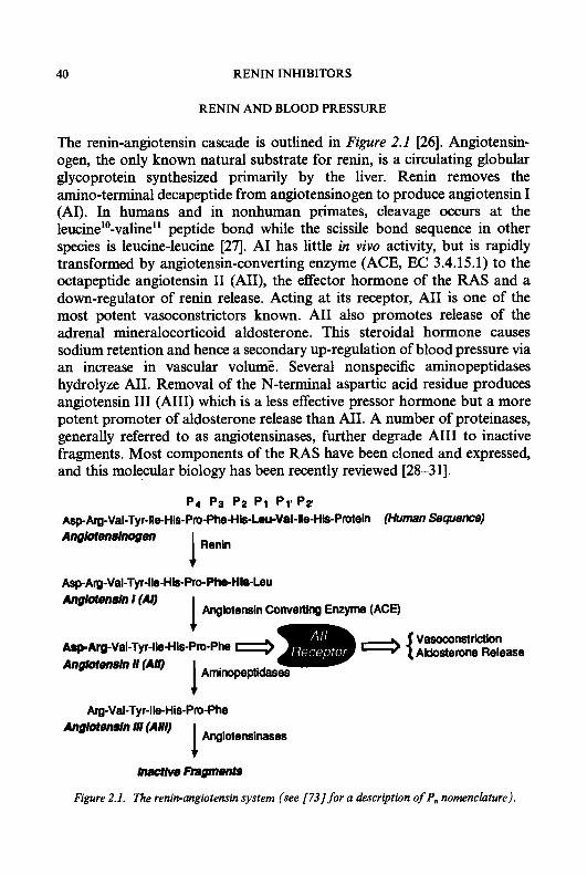
40 RENIN INHIBITORS
RENIN AND BLOOD PRESSURE
The renin-angiotensin cascade is outlined in Figure 2.1 [26]. Angiotensin- ogen, the only known natural substrate for renin, is a circulating globular glycoprotein synthesized primarily by the liver. Renin removes the amino-terminal decapeptide from angiotensinogen to produce angiotensin I (AI). In humans and in nonhuman primates, cleavage occurs at the leucine'O-valine'l peptide bond while the scissile bond sequence in other species is leucine-leucine [27]. A1 has little in vivo activity, but is rapidly transformed by angiotensin-converting enzyme (ACE, EC 3.4.15.1) to the octapeptide angiotensin I1 (AII), the effector hormone of the RAS and a down-regulator of renin release. Acting at its receptor, A11 is one of the most potent vasoconstrictors known. A11 also promotes release of the adrenal mineralocorticoid aldosterone. This steroidal hormone causes sodium retention and hence a secondary up-regulation of blood pressure via an increase in vascular volume. Several nonspecific aminopeptidases hydrolyze AII. Removal of the N-terminal aspartic acid residue produces angiotensin I11 (AIII) which is a less effective pressor hormone but a more potent promoter of aldosterone release than AII. A number of proteinases, generally referred to as angiotensinases, further degrade A111 to inactive fragments. Most components of the RAS have been cloned and expressed, and this molecular biology has been recently reviewed [28-3 11.
P4 p3 p2 p1 Pl*PZ Asp-Arg-Val-Tyr-lleHis-Pro-Phe-His~Val-lle-His-P~ein (Human SequenCe)
Ang'rotenslnogen Renin I
Asp-Am-Val-Tyr-lle-His-Pro-Phe-HIS-Leu Ang'rotendn I (A9 I Angktensin convetting E- (ACE)
Vasoconstriction Aklosterone Release
+ Alp-Arg-Val-Tyr-lle-His-Pro-Phe Anglotmsln I1 (All) Amino+,,.,ptidases I +
Arg-Val-Tyr-ile-His-Pro-Phe
Angiotensinases 1 Ang'rotendn Ill (AM)
inactive Fngments
Figure 2.1. The renin-angiotensin system (see [73] for a description of P, nomenclature).
S.H. ROSENBERG 41
ACE INHIBITORS AND A11 ANTAGONISTS
Conceptually, effective antihypertensive drugs could be derived from obstruction of the RAS at any of three sites: inhibition of renin; inhibition of ACE; or blockade of the A11 receptor. Recently, a number of nonpeptide A11 receptor antagonists have been developed and one (DuP753, losartan) has lowered blood pressure in preliminary clinical trials [32-341. The elevation of circulating A11 levels [32], the existence of multiple A11 receptor subtypes [35, 361, and the recent report that losartan may act through a secondary mechanism unrelated to the RAS [37] raise safety concerns and suggest that the therapeutic potential of A11 antagonists has yet to be confirmed. Conversely, ACE inhibitors have proven to be both potent hypotensive drugs [38-41] and effective agents for the treatment and prevention of congestive heart failure [42]. ACE, however, is a nonspecific enzyme that cleaves bradykinin, substance P, enkephalin and other endogenous peptides in addition to A1 [43-46]. Certain side-effects seen with ACE inhibitors including cough in 614% of patients [43,47-49], skin rash [50], and angioneurotic oedema [43, 48, 511, may result from the accumulation of bradykinin and hence may be inseparable from ACE inhibition (bradykinin has also been implicated in the cardioprotective behaviour of ACE inhibitors [43, 52, 531). Renin inhibitors will also block the formation of both A11 and AIII. Moreover, since angiotensinogen is the single naturally occurring substrate for renin, inhibitors of renin are expected to exhibit an improved side-effect profile.
Renin belongs to the aspartic proteinase family of proteolytic enzymes, other members of which include the mammalian enzymes pepsin, cathepsin D, cathepsin E, gastricsin, and chymosin, the fungal proteinases penicil- lopepsin, endothiapepsin, and rhizopuspepsin, as well as plant and retroviral proteinases [5&56]. Renin is unique among aspartic proteinases in two respects. First, its pH optimum of 6.0 [12] is significantly less acidic than that exhibited by other members of this family and in fact it is active in plasma (PH = 7.4). Second, and more relevant to the task of designing enzyme inhibitors, renin demonstrates an unusual substrate specificity. Angiotensinogen is the only known natural substrate for renin, and this stringent substrate requirement is reflected in the fact that the minimum angiotensinogen-derived sequence cleaved by renin is an octapeptide [57]. In contrast, ACE (a zinc metalloproteinase) accepts substrates as small as tripeptides [41]. The fact that ACE binds smaller substrates than renin has had profound effects on the drug discovery process. The report of the first ACE inhibitor in 1971 [58] led to the development of captopril, an orally active, currently marketed drug, by 1977 [59]. Because of renin’s substrate
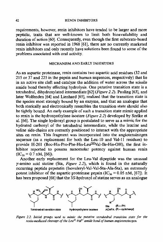
42 RENIN INHIBITORS
requirements, however, renin inhibitors have tended to be larger and more peptidic, traits that are well-known to limit both bioavailability and duration of action [60]. Consequently, even though the first substrate-based renin inhibitor was reported in 1968 [61], there are no currently marketed renin inhibitors and only recently have solutions been found to some of the problems associated with oral activity.
MECHANISM AND EARLY INHIBITORS
As an aspartic proteinase, renin contains two aspartic acid residues (32 and 21 5 or 37 and 225 in the pepsin and human sequences, respectively) that lie in an active site cleft and catalyze the addition of water across the scissile amide bond thereby effecting hydrolysis. One putative transition state is a tetrahedral, dihydroxylated intermediate [62] (Figure 2.2). Pauling [63], and later Wolfenden [64] and Lienhard [65], realized that the transition state is the species most strongly bound by an enzyme, and that an analogue that both sterically and electronically resembles the transition state should also be tightly bound. An early example of such a transition state mimic applied to renin is the hydroxyethylene isostere (Figure 2.2) developed by Szelke et al. [66]. The single hydroxyl group is postulated to serve as a mimic for the hydrated carbonyl of the tetrahedral intermediate, while the leucine and valine side-chains are correctly positioned to interact with the appropriate sites on renin. This fragment was incorporated into the angiotensinogen sequence (as a replacement for both the Leu-10 and Val-11 residues) to provide H-261 (Boc-His-Pro-Phe-His-LeumVal-Ile-His-OH), the first in- hibitor reported to possess nanomolar potency against human r e i n (ICs0 = 0.7 nM, [66]).
Another early replacement for the Leu-Val dipeptide was the unusual y-amino acid statine (Sta, Figure 2.2), which is found in the naturally occurring peptide pepstatin (Isovaleryl-Val-Val-Sta-Ala-Sta), an extremely potent inhibitor of the aspartic proteinase pepsin (ICs0 = 0.05 nM, [67]). It has been proposed [68] that the 3s-hydroxyl of statine serves as an analogue
0 0 R/ Y sta (R=iPr)
Y i
Y Tetrahedral transition state Hydroxyethylene lsostere ACHPA (R = cyclohexyl)
Figure 2.2. Initial groups used to mimic the putative tetrahedral transition state for the renin-mediated cleavage of the Leu''- Val" amide bond of human angiotensinogen.

S.H. ROSENBERG 43
of the tetrahedral transition state. Alternatively, Rich [69] suggested that pepstatin and related species such as the hydroxyethylene isostere bind tightly to the enzyme through displacement of an enzyme-bound water to bulk solvent thereby increasing entropy with little net enthalpic change. An inhibitor acting by this mechanism would mimic both the peptide substrate and the water molecule that participates in proteolysis and would be classified as a collected-substrate inhibitor.
While pepstatin itself was a weak inhibitor of renin (ICs0 = 22pM), Boger et al. [70] incorporated statine into the angiotensinogen sequence to provide SCRIP (Statine-Containing Renin-Inhibitory Peptide: Isovaleryl-His-Pro- Phe-His-Sta-Leu-Phe-NH,, ICso = 16 nM). The importance of the 3S- hydroxyl in statine was demonstrated by the 500-fold reduction in inhibitory activity upon changing to the 3R-configuration. Subsequently, the Merck researchers developed the more potent transition-state analogue (3S,4S)-4-amino-5-cyclohexyl-3-hydroxypentanoic acid (ACHPA) in which the isobutyl side-chain of statine was replaced with cyclohexylmethyl[7 11.
In an excellent reviews, Greenlee has summarized the history of renin inhibition as it appeared in the literature published through 1988 [72]. The development of tightly binding transition-state mimics such as the hydroxyethylene isostere, Sta, and ACHPA was quickly followed by the discovery that truncation of up to two amino acid residues from both the C-and N-termini was compatible with good renin inhibitory activity. Thus, rather than being a large peptide, a renin inhibitor could simply embody an N-blocked dipeptide, representing the P,-P, Phe-His of angiotensinogen (for a description of P, nomenclature see ref. [73]) and a transition-state mimic replacing the scissile dipeptide. Subsequently, new, more potent surrogates for the scissile Leu-Val dipeptide were designed to more closely mimic the putative tetrahedral transition state (Figure 2.3). The vicinal diols and the electron-deficient carbonyl of the difluoroketone, which exists predomi- nately as the hydrated species, both embody two hydroxyls capable of hydrogen bonding to the active site. Incorporation of features that protected certain amide bonds from proteolytic cleavage and attachment of polar residues to either terminus afforded potent renin inhibitors with molecular weights of 600-800 and sufficient aqueous solubility for intravenous administration.
In order to be a viable drug, a renin inhibitor must not only be potent but also orally active. There exist multiple barriers to oral absorption for peptide-based drugs [60]. The presence of peptide bonds renders the compounds susceptible to degradative enzymes, high molecular weight limits intestinal absorption and enhances hepatic elimination, and poor aqueous solubility can further restrict bioavailability. While some of these

44 RENIN INHIBITORS
Norstatine Dlhydroxyethylene Isosteres
I Diiuomgtatone I
Figure 2.3. Fragments designed to more closely mimic the putative tetrahedral transition state for the renin-mediated amide bond hydrolysis.
issues were addressed in the design of the b s t generation of renin inhibitors, these molecules were still largely peptidic and possessed negligible oral bioavailability. The various approaches that were taken to address the problems associated with oral activity are the subject of this review.
IN VITRO ASSAYS
Several methods have been employed to evaluate the in vitro potency of renin inhibitors. All share the common feature that they determine the ability of a test compound to block the renin-mediated conversion of a substrate to AI, which is generally quantified by radioimmunoassay (one recent process utilizes instead the displacement of a fluorescent inhibitor to measure binding [74]). A number of parameters differ among the various assays including the source of the renin, the nature of the substrate, and the pH at which the assay Is conducted. These differences can affect the outcome of the renin inhibition assay, and these effects can vary depending upon the nature of the test compound.
Activity has been determined against both purified human renal renin and the endogenous renin present in human plasma. In addition to renin and angiotensinogen, plasma contains proteins that can bind to a renin inhibitor thereby reducing its effective concentration and consequently its measured potency [75]. Other effects of plasma components on renin inhibition have also been observed [76]. Detection of newly formed A1 in plasma requires the addition of angiontensinase inhibitors to prevent the degradation of A1 by endogenous proteinases. These inhibitors can further affect the assay [76]. Exact kinetic parameters cannot be determined in a
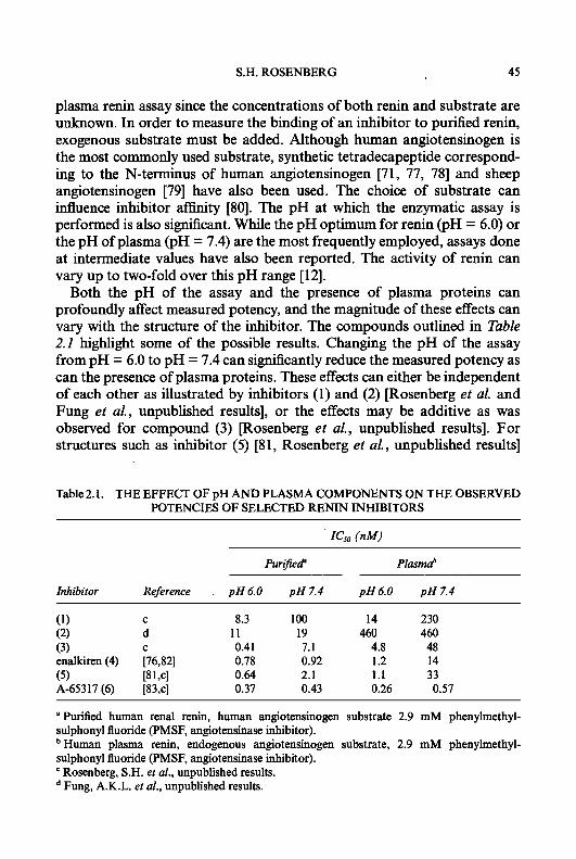
S.H. ROSENBERG 45
plasma renin assay since the concentrations of both renin and substrate are unknown. In order to measure the binding of an inhibitor to purified renin, exogenous substrate must be added. Although human angiotensinogen is the most commonly used substrate, synthetic tetradecapeptide correspond- ing to the N-terminus of human angiotensinogen [71, 77, 781 and sheep angiotensinogen [79] have also been used. The choice of substrate can influence inhibitor afhity [80]. The pH at which the enzymatic assay is performed is also significant. While the pH optimum for renin (PH = 6.0) or the pH of plasma (PH = 7.4) are the most frequently employed, assays done at intermediate values have also been reported. The activity of r e i n can vary up to two-fold over this pH range [12].
Both the pH of the assay and the presence of plasma proteins can profoundly affect measured potency, and the magnitude of these effects can vary with the structure of the inhibitor. The compounds outlined in Table 2.2 highlight some of the possible results. Changing the pH of the assay from pH = 6.0 to pH = 7.4 can sigtllscantly reduce the measured potency as can the presence of plasma proteins. These effects can either be independent of each other as illustrated by inhibitors (1) and (2) [Rosenberg et al. and Fung et al., unpublished results], or the effects may be additive as was observed for compound (3) [Rosenberg et al., unpublished results]. For structures such as inhibitor (5) [81, Rosenberg et al., unpublished results]
Table 2.1. THE EFFECT OF pH AND PLASMA COMPONENTS ON THE OBSERVED POTENCIES OF SELECTED RENIN INHIBITORS
Go (nM)
PurijiecP Plmmd
Inhibitor Reference . p H 6 . 0 pH 7.4 p H 6 . 0 p H 7.4
(1) C 8.3 100 14 230 (2) d 1 1 19 460 460 (3) C 0.41 7.1 4.8 48 enalkiren (4) [76,82] 0.78 0.92 1.2 14
A-65317 (6) [83,~] 0.37 0.43 0.26 0.57 (5) 18 1 ,cl 0.64 2.1 1.1 33
a Purified human renal renin, human angiotensinogen substrate 2.9 mM phenylmethyl- sulphonyl fluoride (PMSF, angiotensinase inhibitor). ' Human plasma renin, endogenous angiotensinogen substrate, 2.9 mM phenylmethyl- sulphonyl fluoride (PMSF, angiotensinase inhibitor).
Rosenberg, S.H. et al., unpublished results. Fung, A.K.L. et al., unpublished results.
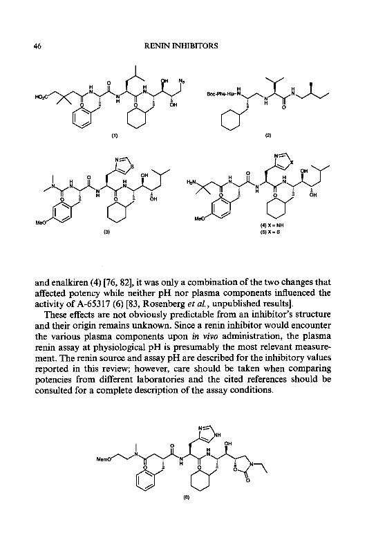
46 RENIN INHIBITORS
(4) X = NH (5)X=S
and enalkiren (4) [76, 821, it was only a combination of the two changes that affected potency while neither pH nor plasma components influenced the activity of A-65317 (6) [83, Rosenberg et al., unpublished results].
These effects are not obviously predictable from an inhibitor’s structure and their origin remains unknown. Since a renin inhibitor would encounter the various plasma components upon in vivo administration, the plasma renin assay at physiological pH is presumably the most relevant measure- ment. The renin source and assay pH are described for the inhibitory values reported in this review; however, care should be taken when comparing potencies from different laboratories and the cited references should be consulted for a complete description of the assay conditions.

S.H. ROSENBERG
IN VIVO EVALUATION
47
Animal Models
The various in vivo models used to study renin inhibitors have been summarized [72]. Renin inhibitors designed to inhibit human renin are generally more active against primate renin than renin from other species (vide infra). Consequently, in vivo activity has most often been demon- strated in primate models. Sodium depletion through a low-salt diet and/or the administration of a diuretic is commonly used to elevate plasma renin thereby rendering blood pressure more renin dependent.
Anaesthesia, employed in some protocols, can also affect plasma renin activity [84]. Because a variety of different primate species (cynomolgus, rhesus, and squirrel monkeys; marmosets) have been employed, and since the response to renin inhibition is dependent upon the degree of sodium depletion, it is dficult to compare the reported in vivo activities for different renin inhibitors. Newer models include the cyclosporine-induced hyperten- sive monkey [85] and the recombinant human renin-infused rat [86]; however, the relevance of these new models, or of the sodium-depleted monkey, to human hypertension remains to be established.
Plasma renin. activity
In addition to the determination of a hypotensive response, in vivo evaluation of a renin inhibitor generally involves the measurement of one or more biochemical parameters such as plasma renin activity (PRA) and, less frequently, A1 and A11 levels. Often, inhibition of PRA is of greater magnitude and longer duration than the observed blood pressure effects, and conversely, blood pressure can continue to fall even after PRA has been completely suppressed [87]. The origin of this dissociation has not been resolved. It has been argued that the traditional radioimmunoassay used to measure PRA is inaccurate due to the liberation of plasma-bound renin inhibitor by exogenous proteinase inhibitors employed in the assay [MI. Several studies have shown a closer correlation between PRA and blood pressure or A11 levels when an alternate antibody trapping technique was used to determine PRA [88-901. It appears, however, that dissociation is not solely due to the use of angiotensinase inhibitors and that results from the antibody-trapping method are themselves highly dependent upon the exact assay conditions [76].
An alternate explanation for the lack of correlation between blood pressure and PRA is that a renin inhibitor must interact with a second,
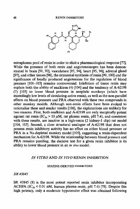
48 RENIN INHIBITORS
extraplasma pool of renin in order to elicit a pharmacological response [77. While the presence of both renin and angiotensinogen has been demon- strated in brain [91, 921, vasculature [93, 941, heart [95, 961, adrenal gland [97], and other tissues [98], the extrarenal synthesis of renin [99, 1001 and the significance of locally produced angiotensins for the regulation of blood pressure [101-1031 remains controversial. Inhibition of tissue renin may explain both the ability of enalkiren (4) [lo41 and the tendency of A-62198 (7) [lo51 to lower blood pressure in anephric monkeys (which have exceedingly low levels of circulating active renin), as well as the non-parallel effects on blood pressure and PRA observed with these two compounds in other monkey models. Although non-renin effects have been evoked to rationalize these and similar results [106], the explanations are unlikely for two reasons. First, both enalkiren and A-62198 are only marginally potent against rat renin (ICs0 > 10 pM, rat plasma renin, pH 7.4), and consistent with these results, are inactive in a high-renin (2 kidney-1 clip) rat model [104, 1051. Second, a close structural analogue of A-62198 that does not possess renin inhibitory activity has no effect on either blood pressure or PRA in a Na-depleted monkey model [105], suggesting a renin-dependent mechanism for A-62198. While the relationship between blood pressure and PRA remains puzzling, the decisive test for a given renin inhibitor is its ability to lower blood pressure in an in vivo model.
IN VITRO AND IN VIVO RENIN INHIBITION
STATINE-DERIVED INHIBITORS
SR 43845
SR 43845 (8) is the most potent reported renin inhibitor incorporating ACHPA (ICs0 c 0.01 nM, human plasma renin, pH 7.4) [78]. Despite this high potency, only a moderate hypotensive effect was obtained following

S.H. ROSENBERG 49
oral administration to Na-depleted marmosets. A dose of 30 mglkg reduced mean arterial pressure (MAP) significantly for 90 min with a maximum response of 16 k 3 mm Hg. A lower dose (10 m a g ) did not elicit a hypotensive response. At 1 h, PRA was 100% and 68% inhibited at the 30 mgkg and 10 mgkg doses, respectively.
In salt-replete, resting hypertensive patients, SR 43845 was given as a 30 min i.v. infusion at 30,100, and 300 puglkg (n = 4 each group) which reduced diastolic blood pressure (DBP) 4.5, 7.5, and 3.9 mm Hg respectively [107]. Furosemide (frusemide)-treated hypertensive patients (n = 8) were also treated with the highest dose causing a 10.8 mm Hg fall in DBP. In all patients, DBP returned to baseline by 3 h. The lack of dose-dependence with the salt-replete patients was attributed in part to heterogeneity in baseline plasma active renin levels. PRA was dissociated from the changes in DBP. It was almost completely suppressed in all groups by 10 min, and remained extremely low for up to 6 h, while A1 and A11 levels more closely followed the changes in DBP.
ES-6864
Although less potent than SR 43845 (IC5,, = 6.9 nM, human plasma renin, pH 7.3), ES-6864 (9) appears to be more efficacious in vivo [108]. This inhibitor incorporates (4-thiazoly1)alanine in place of the P,-site histidine, a
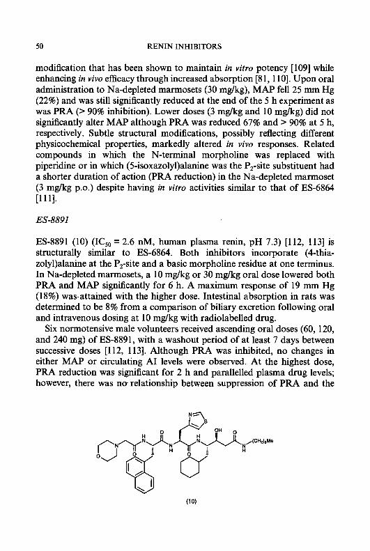
50 RENIN INHIBITORS
modification that has been shown to maintain in v i m potency [lo91 while enhancing in vivo efficacy through increased absorption [81, 1 lo]. Upon oral administration to Na-depleted marmosets (30 mglkg), MAP fell 25 mm Hg (22%) and was still significantly reduced at the end of the 5 h experiment as was PRA (> 90% inhibition). Lower doses (3 mglkg and 10 mg/kg) did not significantly alter MAP although PRA was reduced 67% and > 90% at 5 h, respectively. Subtle structural modifications, possibly reflecting different physicochemical properties, markedly altered in vivo responses. Related compounds in which the N-terminal morpholine was replaced with piperidine or in which (5-isoxazoly1)alanine was the P,-site substituent had a shorter duration of action (PRA reduction) in the Na-depleted marmoset (3 mglkg p.0.) despite having in vitro activities similar to that of ES-6864 [ l l l ] .
ES-8891
ES-8891 (10) (IC5,, = 2.6 nM, human plasma renin, pH 7.3) [112, 1131 is structurally similar to ES-6864. Both inhibitors incorporate (4-thia- zoly1)alanine at the P,-site and a basic morpholine residue at one terminus. In Na-depleted marmosets, a 10 mglkg or 30 mglkg oral dose lowered both PRA and MAP significantly for 6 h. A maximum response of 19 mm Hg (18%) was.attained with the higher dose. Intestinal absorption in rats was determined to be 8% from a comparison of biliary excretion following oral and intravenous dosing at 10 mglkg with radiolabelled drug.
Six normotensive male volunteers received ascending oral doses (60, 120, and 240 mg) of ES-8891, with a washout period of at least 7 days between successive doses [112, 1131. Although PRA was inhibited, no changes in either MAP or circulating A1 levels were observed. At the highest dose, PRA reduction was significant for 2 h and parallelled plasma drug levels; however, there was no relationship between suppression of PRA and the
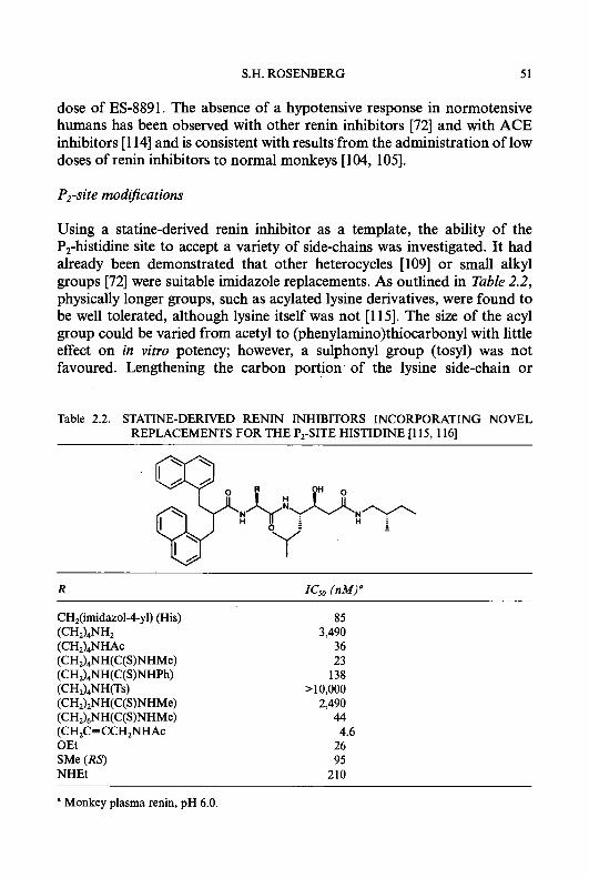
S.H. ROSENBERG 51
dose of ES-8891. The absence of a hypotensive response in normotensive humans has been observed with other renin inhibitors [72] and with ACE inhibitors [ 1 141 and is consistent with results'from the administration of low doses of renin inhibitors to normal monkeys [104, 1051.
P,-site modijications
Using a statine-derived renin inhibitor as a template, the ability of the P,-histidine site to accept a variety of side-chains was investigated. It had already been demonstrated that other heterocycles [lo91 or small alkyl groups [72] were suitable imidazole replacements. As outlined in Table 2.2, physically longer groups, such as acylated lysine derivatives, were found to be well tolerated, although lysine itself was not [ 11 51. The size of the acyl group could be varied from acetyl to (pheny1amino)thiocarbonyl with little effect on in vitro potency; however, a sulphonyl group (tosyl) was not favoured. Lengthening the carbon portion. of the lysine side-chain or
Table 2.2. STATINE-DERIVED RENIN INHIBITORS INCORPORATING NOVEL REPLACEMENTS FOR THE P2-SITE HISTIDINE 11 15, 1161
CH2(imidazo14-yl) (His)
(CH2),NHAc (CHJ,NH(C(S)NHMe) (CH,),NH(C(S)NHPh)
(CH&NH(C(S)NHMe)
(CH,C=CCH,NHAc OEt SMe (RS) NHEt
(CH2)4NH2
(CH2)4NH(Ts)
(CH2).5NH(C(S)NHMe)
85 3,490
36 23
138 >10,000
2,490 44
26 95
210
4.6
a Monkey plasma renin, pH 6.0.

52 RENIN INHIBITORS
introducing unsaturation also maintained or enhanced activity. A second study established that a-heteroatom substituted amino acids could also replace the P,-site histidine [116].
INHIBITORS INCORPORATING THE HYDROXYETHYLENE ISOSTERE
CGP 38 560 and related structures
Biihlmayer et al. found that an N-terminal tert-butylsulphonyl residue could impart greater potency than standard carbonyl-derived groups [117]. This discovery led to the development of CGP 38 560 (1 1) (ICso = 0.7 nM, human plasma renin, pH 7.0) [118]. Due to its potency, this compound exhibited oral activity in the Na-depleted marmoset despite the fact that bioavailability in this species was determined to be only 0.3% [119]. Oral administration to Na-depleted marmosets at 10 mg/kg reduced MAP 23 k 3 mm Hg and completely suppressed PRA, effects which lasted for at least 2 h [118]. A tendency towards reduced blood pressure (not significant) followed a lower oral dose (3 mg/kg); however, PRA was still completely inhibited.
In normal, Na-replete volunteers (n = 5, successive doses with 10 day washout periods), 50,125, & 250pgkg doses of CGP 38 560 given as 30 min infusions completely suppressed PRA (measured by radioimmunoassay) which remained low for the 3 h duration of the experiment [120]. A11 levels were reduced dose-dependently (50, 80, 90%) and recovered rapidly at the end of the infusion. Blood pressure was not affected. When administered at the same doses to volunteers who were moderately Na-depleted (n = 5 each dose), PRA (determined with an antibody trapping technique) was again completely inhibited, but returned to near control by 3 h [89]. Plasma A11 levels recovered more quickly than PRA. Blood pressure was not reduced despite the fact that the plasma drug concentration attained at the highest dose was 1.35 f: 0.13 pg/mL. Infusion of CGP 38 560 (125 and 250 pgkg, n = 6 each group) to volunteers pretreated 21 h earlier with the ACE
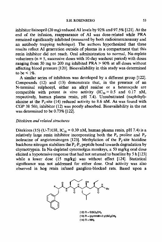
S.H. ROSENBERG 53
inhibitor lisinopril(20 mg) reduced A1 levels by 92% and 97.5% [121]. At the end of the infusion, reappearance of A1 was dose-related while PRA remained significantly inhibited (measured by both radioimmunoassay and an antibody trapping technique). The authors hypothesized that these results reflect A1 generation outside of plasma in a compartment that this renin inhibitor did not reach. Oral administration to normal, Na-replete volunteers (n = 5, successive doses with 10-day washout period) with doses ranging from 50 mg to 200 mg inhibited PRA > 90% at all doses without affecting blood pressure [120]. Bioavailability in this study was determined to be c 1%.
A similar series of inhibitors was developed by a different group [122]. Compounds (12) and (13) demonstrate that, in the presence of an N-terminal sulphonyl, either an alkyl residue or a heterocycle are compatible with potent in vitro activity (IC50 = 0.5 and 0.17 nM, respectively, human plasma renin, pH 7.4). Unsubstituted (naphthy1)- alanine at the P,-site (14) reduced activity to 8.6 nM. As was found with CGP 38 560, inhibitor (12) was poorly absorbed. Bioavailability in the rat was determined to be 0.73% [122].
Ditekiren and related structures
Ditekiren (15) (U-71038, IC5,, = 0.39 nM, human plasma renin, pH 7.4) is a relatively large renin inhibitor incorporating both the P4 proline and Py isoleucine of angiotensinogen [ 1231. Methylation of the P,-site histidine backbone nitrogen stabilizes the P,-P, peptide bond towards degradation by chymotrypsin. In Na-depleted cynomolgus monkeys, a 50 mgkg oral dose elicited a hypotensive response that had not returned to baseline by 5 h [123] while a lower dose (15 mglkg) was without effect [124]. Statistical significance was not addressed for either dose. Oral activity was also observed in hog renin infused ganglion-blocked rats. Based upon a
(12) R = EtS02CH2 (13) R = (pyrimidin-2-yl)SO2CH2 (14) R = NH2

54 RENIN INHIBITORS
(15) R=Boc X = N (16) R = (HOCH&CNHCO (17) R = HSN+CH(CO~-)(CH&CO
X = N+O- X = N+O
comparison of hypotensive effects following oral and intravenous dosing, the authors suggested that bioavailability was > 10% in both species [123]. Subsequently, definitive experiments in the rat showed bioavailability to be only 1.3% demonstrating the danger of estimating absorption from pharmacological responses [ 1251. In Na-depleted cynomolgus monkeys, ditekiren (5 mg/kg i.v.) caused a significant hypotensive response that was not enhanced by treatment with captopril (10 mgkg i.v.) [126]. A similar blood pressure profile was observed when the order of treatment was reversed suggesting that both agents act solely through blockade of the U S .
In normal volunteers, a 3 p&g/min i.v infusion reduced MAP sigmficantly by 8.2 k 1.4 rnm Hg (n = 4) [127]. While a 0.3 pg/kg/min i.v. infusion was ineffective in normal volunteers, in Na-depleted volunteers MAP was significantly lowered 6.8 5 1.5 rnm Hg (n = 4) at this dose [127]. Steady-state plasma drug levels were approximately two-fold higher for all doses in the Na-depleted subjects [128]. Ditekiren was also studied in Na-depleted hypertensive patients. MAP was significantly lowered 17 mm Hg (16%, n = 7) following a 45 min i.v. infusion (7.5 pg/kg/min) [129].
To determine the effects of enhanced aqueous solubility on in vivo activity, analogues of ditekiren were prepared that incorporated a C-terminal pyridine N-oxide and polar groups at the N-terminus [ 124, 1301. Inhibitors U-77436 (16) and (17) (IC50 = 0.58 and 0.32 nM, respectively; human plasma renin, pH 6.0) were essentially equipotent with ditekiren (ICs0 = 0.26 nM, human plasma renin, pH 6.0); however, U-77436 was greater than 20-fold more soluble (20 mg/mL vs. < 1 mg/mL, H,O, 37°C) [130]. In human renin-infused ganglion-blocked rats, a 5 mg/kg oral dose of U-77436 produced a similar hypotensive response to that from ditekiren at the same dose, but the duration of action was significantly longer [130]. Studies in bile duct cannulated rats showed that intestinal absorption of a 5 mg/kg i.d. dose of U-77436 was < 10% [131]. Inhibitor (17) also appeared superior to ditekiren following oral administration to either human renin-infused ganglion-blocked rats or Na-depleted monkeys [ 1241.
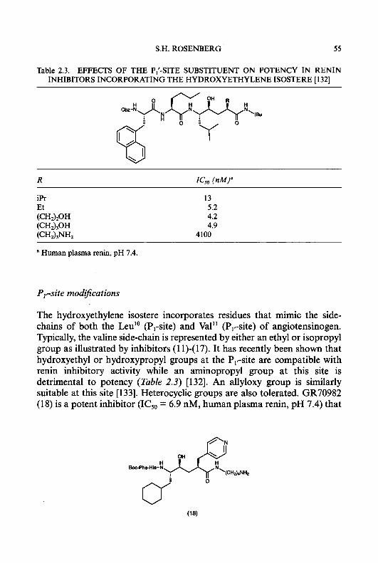
S.H. ROSENBERG 55
Table 2.3. EFFECTS OF THE PI'-SITE SUBSTITUENT ON POTENCY IN RENIN INHIBITORS INCORPORATING THE HYDROXYETHYLENE ISOSTERE [132]
13 5.2 4.2 4.9
4100
a Human plasma renin, pH 7.4.
P,,-site modijications
The hydroxyethylene isostere incorporates residues that mimic the side- chains of both the Leu" (P,-site) and Val" (P,,-site) of angiotensinogen. Typically, the valine side-chain is represented by either an ethyl or isopropyl group as illustrated by inhibitors (1 1)-(17). It has recently been shown that hydroxyethyl or hydroxypropyl groups at the P,,-site are compatible with renin inhibitory activity while an aminopropyl group at this site is detrimental to potency (Table 2.3) [132]. An allyloxy group is similarly suitable at this site [133]. Heterocyclic groups are also tolerated. GR70982 (18) is a potent inhibitor (IC50 = 6.9 nM, human plasma renin, pH 7.4) that
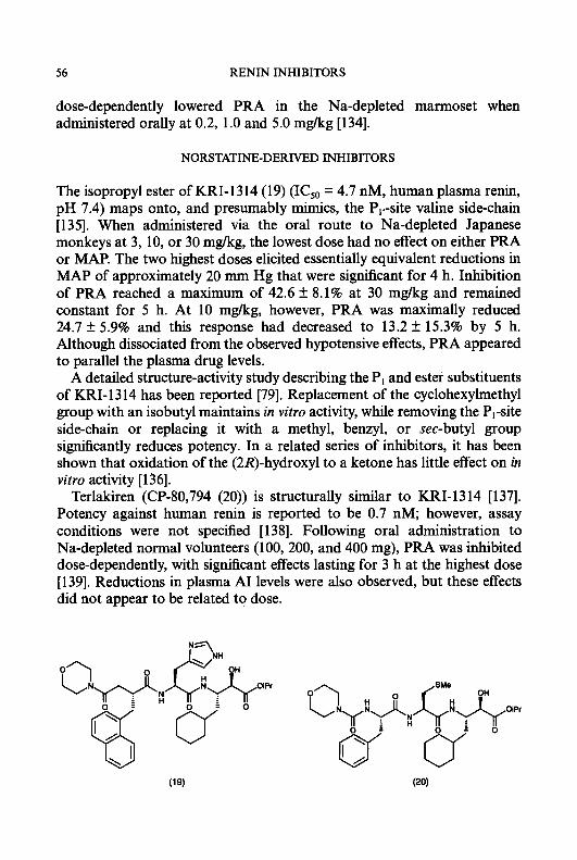
56 RENIN INHIBITORS
dose-dependently lowered PRA in the Na-depleted marmoset when administered orally at 0.2, 1.0 and 5.0 mglkg [134].
NORSTATINE-DERIVED INHIBITORS
The isopropyl ester of KRI-1314 (19) (ICso = 4.7 nM, human plasma renin, pH 7.4) maps onto, and presumably mimics, the Pl.-site valine side-chain [ 1351. When administered via the oral route to Na-depleted Japanese monkeys at 3, 10, or 30 mg/kg, the lowest dose had no effect on either PRA or MAP. The two highest doses elicited essentially equivalent reductions in MAP of approximately 20 mm Hg that were significant for 4 h. Inhibition of PRA reached a maximum of 42.6 f 8.1% at 30 mglkg and remained constant for 5 h. At 10 mglkg, however, PRA was maximally reduced 24.7 f 5.9% and this response had decreased to 13.2 f 15.3% by 5 h. Although dissociated from the observed hypotensive effects, PRA appeared to parallel the plasma drug levels.
A detailed structure-activity study describing the PI and ester substituents of KRI-1314 has been reported [79]. Replacement of the cyclohexylmethyl group with an isobutyl maintains in vitro activity, while removing the PI-site side-chain or replacing it with a methyl, benzyl, or sec-butyl group significantly reduces potency. In a related series of inhibitors, it has been shown that oxidation of the (2R)-hydroxyl to a ketone has little effect on in vitro activity [ 1361.
Terlakiren (CP-80,794 (20)) is structurally similar to KRI-1314 [137]. Potency against human renin is reported to be 0.7 nM; however, assay conditions were not specified [138]. Following oral administration to Na-depleted normal volunteers (100, 200, and 400 mg), PRA was inhibited dose-dependently, with significant effects lasting for 3 h at the highest dose [139]. Reductions in plasma A1 levels were also observed, but these effects did not appear to be related to dose.
S.H. ROSENBERG 57
INHIBITORS INCORPORATING A C-TERMINAL GLYCOL
Enalkiren
Enalkiren (4) (A-64662, IC5,, = 14 nM, human plasma renin, pH 7.4) incorporates the novel dihydroxyethylene isostere transition-state mimic (Figure 2.3) [140]. (0-Methy1)tyrosine serves as a P,-site phenylalanine replacement and stabilizes the P,-P, peptide bond towards degradation by chymotrypsin [109, 1411. The activity of this inhibitor in cynomolgus monkeys has been recently reviewed [82]. Following bolus injections (0.01-10 mg/kg), the magnitude and duration of the hypotensive responses were dose-related. At the highest dose, MAP was maximally reduced approximately 60% in both Na-depleted and normal animals, and was still significantly lowered at the end of the 3 h experiment. PRA was completely inhibited at all doses and the time course of recovery was dose-related, although it did not parallel the changes in blood pressure. Chronic treatment of Na-depleted animals with enalkiren (0.1 mglkg bolus followed by 0.01 mg/kg/min infusion for 7 days) significantly reduced MAP 14 mm Hg (16%) for the duration of the infusion. Blood pressure returned to control values one day after cessation of treatment. Following i.d. administration (10 mg/kg) to Na-depleted animals, MAP and PRA were reduced 15% and 95%, respectively, and these effects persisted for 120-180 minutes although absorption in the monkey was low (i.d. bioavailability = 1.7 f 0.5% [142]). Similar limited bioavailability (< 2.2%) was measured following oral administration (10 m a g ) to dogs [142].
Enalkiren has been studied extensively in man. Normotensive, Na-replete volunteers (n = 8) received ascending 2 min i.v. infusions (0.001,0.003,0.01 mglkg and 0.01, 0.03, 0.1 mg/kg) at 90 min intervals [143]. PRA was inhibited in a dose dependent manner, effects that persisted 90 min after the 0.03 and 0.1 mglkg doses. There was a close correlation between PRA and plasma AII levels. MAP tended to decrease, but these effects were not significant (similar results were obtained in a limited dose-ranging study in hypertensive patients [144]). Na-replete hypertensive patients (n = 18) were given ascending 5 min i.v. infusions of enalkiren (0.03, 0.1, 0.3, and 1.0 mglkg) at 45 min intervals [145]. PRA was significantly inhibited and reductions were still evident 8 h after completion of the dosing. Decreases in blood pressure were dose-related and were sigmiicant for 4 h following administration of the highest dose. PRA was significantly correlated with the changes in blood pressure. Na-depletion (diuretic administration) enhanced both the magnitude and duration of the hypotensive responses in this study. The maximum hypotensive responses in Na-depleted hyperten-
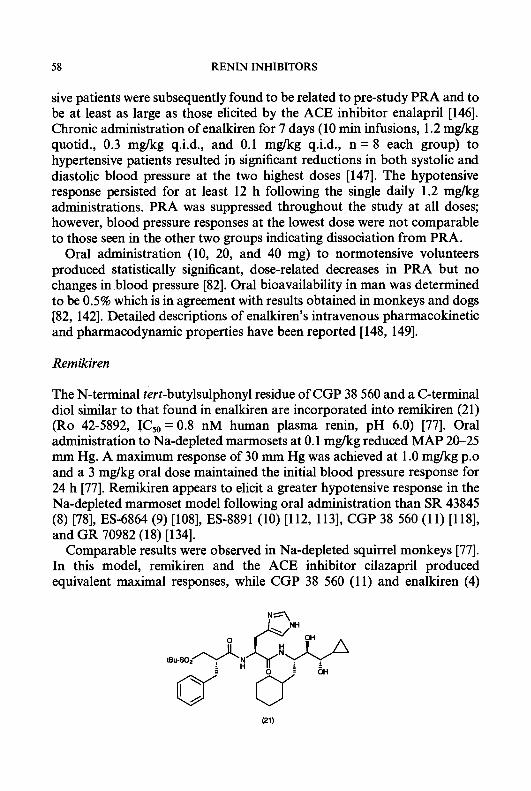
58 RENIN INHIBITORS
sive patients were subsequently found to be related to pre-study PRA and to be at least as large as those elicited by the ACE inhibitor enalapril [146]. Chronic administration of enalkiren for 7 days (10 min infusions, 1.2 mgkg quotid., 0.3 mg/kg q.i.d., and 0.1 mg/kg q.i.d., n = 8 each group) to hypertensive patients resulted in significant reductions in both systolic and diastolic blood pressure at the two highest doses [147]. The hypotensive response persisted for at least 12 h following the single daily 1.2 mgkg administrations. PRA was suppressed throughout the study at all doses; however, blood pressure responses at the lowest dose were not comparable to those seen in the other two groups indicating dissociation from PRA.
Oral administration (10, 20, and 40 mg) to normotensive volunteers produced statistically significant, dose-related decreases in PRA but no changes in blood pressure [82]. Oral bioavailability in man was determined to be 0.5% which is in agreement with results obtained in monkeys and dogs [82, 1421. Detailed descriptions of enalkiren’s intravenous pharmacokinetic and pharmacodynamic properties have been reported [148, 1491.
Remikiren
The N-terminal tert-butylsulphonyl residue of CGP 38 560 and a C-terminal diol similar to that found in enalkiren are incorporated into remikiren (21) (Ro 42-5892, ICs0 = 0.8 nM human plasma renin, pH 6.0) [77]. Oral administration to Na-depleted marmosets at 0.1 mg/kg reduced MAP 20-25 mm Hg. A maximum response of 30 mm Hg was achieved at 1 .O mg/kg p.0 and a 3 mg/kg oral dose maintained the initial blood pressure response for 24 h [77]. Remikiren appears to elicit a greater hypotensive response in the Na-depleted marmoset model following oral administration than SR 43845
and GR 70982 (18) [134]. Comparable results were observed in Na-depleted squirrel monkeys [77].
In this model, remikiren and the ACE inhibitor cilazapril produced equivalent maximal responses, while CGP 38 560 (1 1) and enalkiren (4)
(8) [78], ES-6864 (9) [108], ES-8891 (10) [112, 1131, CGP 38 560 (11) [118],
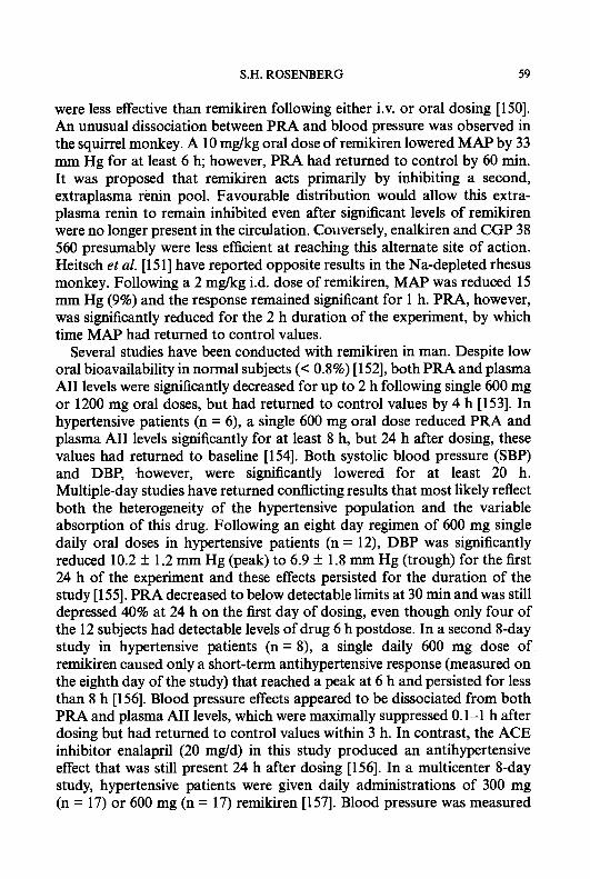
S.H. ROSENBERG 59
were less effective than remikiren following either i.v. or oral dosing [150]. An unusual dissociation between PRA and blood pressure was observed in the squirrel monkey. A 10 mg/kg oral dose of remikiren lowered MAP by 33 mm Hg for at least 6 h; however, PRA had returned to control by 60 min. It was proposed that remikiren acts primarily by inhibiting a second, extraplasma renin pool. Favourable distribution would allow this extra- plasma renin to remain inhibited even after significant levels of remikiren were no longer present in the circulation. Coiiversely, enalkiren and CGP 38 560 presumably were less efficient at reaching this alternate site of action. Heitsch et al. [151] have reported opposite results in the Na-depleted rhesus monkey. Following a 2 mg/kg i.d. dose of remikiren, MAP was reduced 15 mm Hg (9%) and the response remained significant for 1 h. PRA, however, was significantly reduced for the 2 h duration of the experiment, by which time MAP had returned to control values.
Several studies have been conducted with remikiren in man. Despite low oral bioavailability in normal subjects (< 0.8%) [152], both PRA and plasma A11 levels were significantly decreased for up to 2 h following single 600 mg or 1200 mg oral doses, but had returned to control values by 4 h [153]. In hypertensive patients (n = 6), a single 600 mg oral dose reduced PRA and plasma A11 levels significantly for at least 8 h, but 24 h after dosing, these values had returned to baseline [154]. Both systolic blood pressure (SBP) and DBP, .however, were significantly lowered for at least 20 h. Multiple-day studies have returned conflicting results that most likely reflect both the heterogeneity of the hypertensive population and the variable absorption of this drug. Following an eight day regimen of 600 mg single daily oral doses in hypertensive patients (n = 12), DBP was significantly reduced 10.2 f 1.2 mm Hg (peak) to 6.9 f 1.8 mm Hg (trough) for the first 24 h of the experiment and these effects persisted for the duration of the study [155]. PRA decreased to below detectable limits at 30 min and was still depressed 40% at 24 h on the lirst day of dosing, even though only four of the 12 subjects had detectable levels of drug 6 h postdose. In a second 8-day study in hypertensive patients (n = 8), a single daily 600 mg dose of remikiren caused only a short-term antihypertensive response (measured on the eighth day of the study) that reached a peak at 6 h and persisted for less than 8 h [156]. Blood pressure effects appeared to be dissociated from both PRA and plasma A11 levels, which were maximally suppressed 0.1-1 h after dosing but had returned to control values within 3 h. In contrast, the ACE inhibitor enalapril (20 mg/d) in this study produced an antihypertensive effect that was still present 24 h after dosing [156]. In a multicenter 8-day study, hypertensive patients were given daily administrations of 300 mg (n = 17) or 600 mg (n = 17) remikiren [157]. Blood pressure was measured
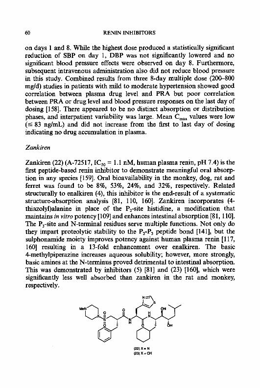
60 RENIN INHIBITORS
on days 1 and 8. While the highest dose produced a statistically significant reduction of SBP on day 1, DBP was not significantly lowered and no significant blood pressure effects were observed on day 8. Furthermore, subsequent intravenous administration also did not reduce blood pressure in this study. Combined results from three 8-day multiple dose (200-800 mdd) studies in patients with mild to moderate hypertension showed good correlation between plasma drug level and PRA but poor correlation between PRA or drug level and blood pressure responses on the last day of dosing [158]. There appeared to be no distinct absorption or distribution phases, and interpatient variability was large. Mean C,, values were low (5 83 nglmL) and did not increase from the first to last day of dosing indicating no drug accumulation in plasma.
Zankiren
Zankiren (22) (A-72517, ICso = 1.1 nM, human plasma re&, pH 7.4) is the first peptide-based renin inhibitor to demonstrate meaningful oral absorp- tion in any species [159]. Oral bioavailability in the monkey, dog, rat and ferret was found to be 8%, 53%, 24%, and 32%, respectively. Related structurally to enalkiren (4), this inhibitor is the end-result of a systematic structure-absorption analysis [81, 1 10, 1601. Zankiren incorporates (4- thiazoly1)alanine in place of the P,-site histidine, a modification that maintains in vitro potency [lo91 and enhances intestinal absorption [81, 1 lo]. The P,-site and N-terminal residues serve multiple functions. Not only do they impart proteolytic stability to the P2-P, peptide bond [141], but the sulphonamide moiety improves potency against human plasma renin [117, 1601 resulting in a 13-fold enhancement over enalkiren. The basic 4-methylpiperazine increases aqueous solubility; however, more strongly, basic amines at the N-terminus proved detrimental to intestinal absorption. This was demonstrated by inhibitors (5) [81] and (23) [160], which were significantly less well absorbed than zankiren in the rat and monkey, respectively.
(Z?)X=N (23) X = CH
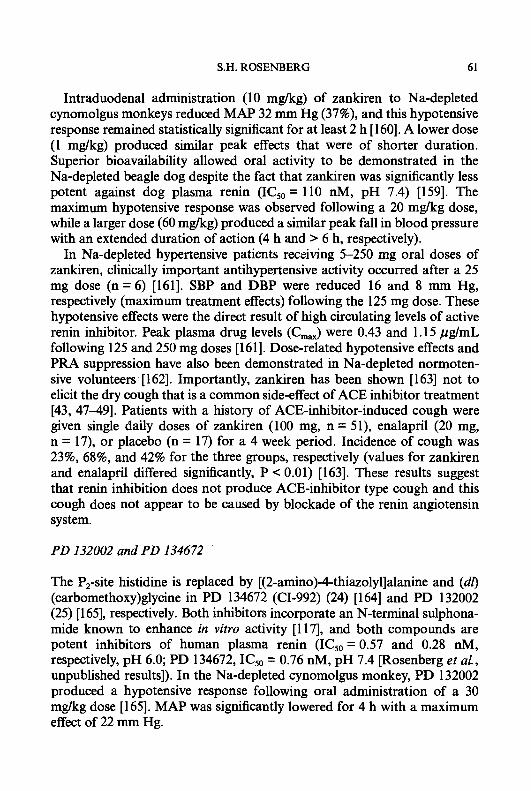
S.H. ROSENBERG 61
Intraduodenal administration (10 mgkg) of zankiren to Na-depleted cynomolgus monkeys reduced MAP 32 mm Hg (37%), and this hypotensive response remained statistically significant for at least 2 h [160]. A lower dose (1 mg/kg) produced similar peak effects that were of shorter duration. Superior bioavailability allowed oral activity to be demonstrated in the Na-depleted beagle dog despite the fact that zankiren was significantly less potent against dog plasma renin (ICs0 = 110 nM, pH 7.4) [159]. The maximum hypotensive response was observed following a 20 mg/kg dose, while a larger dose (60 m a g ) produced a similar peak fall in blood pressure with an extended duration of action (4 h and > 6 h, respectively).
In Na-depleted hypertensive patients recdiving 5-250 mg oral doses of zankiren, clinically important antihypertensive activity occurred after a 25 mg dose (n = 6) [161]. SBP and DBP were reduced 16 and 8 mm Hg, respectively (maximum treatment effects) following the 125 mg dose. These hypotensive effects were the direct result of high circulating levels of active renin inhibitor. Peak plasma drug levels ( C d were 0.43 and 1.15 ,ug/mL following 125 and 250 mg doses [161]. Dose-related hypotensive effects and PRA suppression have also been demonstrated in Na-depleted normoten- sive volunteers [162]. Importantly, zankiren has been shown [163] not to elicit the dry cough that is a common side-effect of ACE inhibitor treatment [43, 47-49]. Patients with a history of ACE-inhibitor-induced cough were given single daily doses of zankiren (100 mg, n = 51), enalapril (20 mg, n = 17), or placebo (n = 17) for a 4 week period. Incidence of cough was 23%, 68%, and 42% for the three groups, respectively (values for zankiren and enalapril differed significantly, P < 0.01) [163]. These results suggest that renin inhibition does not produce ACE-inhibitor type cough and this cough does not appear to be caused by blockade of the renin angiotensin system.
PD 132002 and PD I34672
The P,-site histidine is replaced by [(2-amino)-4-thiazolyl]alanine and (do (carbomethoxy)glycine in PD 134672 (CI-992) (24) [164] and PD 132002 (25) [ 1651, respectively. Both inhibitors incorporate an N-terminal sulphona- mide known to enhance in vitro activity [117], and both compounds are potent inhibitors of human plasma renin (IC,,= 0.57 and 0.28 nM, respectively, pH 6.0; PD 134672, ICso = 0.76 nM, pH 7.4 [Rosenberg et al., unpublished results]). In the Na-depleted cynomolgus monkey, PD 132002 produced a hypotensive response following oral administration of a 30 mg/kg dose [165]. MAP was significantly lowered for 4 h with a maximum effect of 22 mm Hg.

62 RENIN INHIBITORS
(24) R = CH~[(2amino)thiazol4-yl)] (25) R = CaMe (do
Statistically sigtllficant hypotension was not observed following a 10 m a g p.0. dose. PD 134672 proved more active in this model [164]. Administration of 10 mg/kg p.0. reduced MAP 16-18 mm Hg, a response that remained significant for 3 h. Oral bioavailability in the dog and i.d. bioavailability in the rat were 18% and 2%, respectively, for PD 134672 [Rosenberg et al. , unpublished results].
PD 132002 lowered blood pressure following intravenous administration to anaesthetized, Na-depleted dogs. 30 min infusions (0.01,0.1, 1.0, and 10 m a g ) caused dose-related hypotensive responses. At the highest dose, MAP fell 39 f 4 mm Hg (29%), reaching a peak at 30 min and returning to baseline by 4 h [90]. Changes in MAP paralleled plasma A11 levels. PRA measured by radioimmunoassay at pH 6.0 remained suppressed for the duration of the experiment but PRA measured with an antibody trapping method at pH 7.4 paralleled MAP. It is unclear whether these differences are due to the nature of the assay or the pH at which the PRA determination was performed, since the in vitro activity of PD 132002 against dog plasma renin varied 100-fold when the assay was conducted at pH 6.0 (ICsO = 1.7 nM) compared to pH 7.4 (ICsO = 185 nM).
S 2864, B W-I 75, and related structures
S 2864 (26) (ICsO = 0.38 nM, human plasma renin) [151] and BW-175 (27) (ICso = 3.3 nM, human plasma renin, pH 7.4) [166] incorporate glycol-

S.H. ROSENBERG 63
derived transition state mimics and basic C-terminal heterocycles. Both inhibitors have been evaluated in vivo. Following oral administration to Na-depleted marmosets, BW-175 (30 mg/kg) reduced MAP 20 mm Hg. By 4 h, blood pressure had returned to near control values (-5 mm Hg), but PRA was still > 90% inhibited. Oral bioavailability in rats was determined to be 2.8% and 9.7% following 10 mg/kg and 30 mgkg doses indicating an apparent nonlinear dose-absorption relationship [ 1661. In the Na-depleted rhesus monkey, S 2864 was shown to be more effective upon i.d. administration than remikiren (21) [151]. MAP was reduced 22 mm Hg (27%, 90 min duration) following a 2 mg/kg i.d. dose compared with remikiren which at the same dose lowered MAP 15 mm Hg (9%, 60 min duration). With both inhibitors, PRA was significantly inhibited for the 2 h duration of the experiment.
An analogue of remikiren (21) has been reported in which the C-terminal cyclopropyl residue was replaced by CH,SO,NHBu (SQ 33800, IC,, = 0.35 nM, purified human renal renin, pH 7.0) [167]. Following an oral dose of 37 mg/kg in Na-depleted cynomolgus monkeys, MAP was reduced 16 mm Hg. Subsequent treatment with captopril (15 pmoYkg) lowered MAP an additional 27 mm Hg. In contrast, an i.v. dose of SQ 33800 (0.74 mgkg) caused a 35 mm Hg reduction in MAP indicating low oral bioavailability.
The unusual pentahydroxy compound (28) contains a C-terminus that is derived from D-(+)-mannose [168]. Despite the fact that (28) lacks a lipophilic PI.-site side-chain, it is a potent inhibitor of human plasma renin (IC,, = 4.5 nM, pH 7.4). In the Na-depleted rhesus monkey, (28) showed activity similar to S 2864. MAP was lowered 19 mm Hg (75 min duration) following a 2 mg/kg i.d. dose.
NOVEL TRANSITION-STATE MIMICS INCORPORATING A SINGLE HYDROXYL
C- Terminal alkyl groups: FK906
The transition state mimic comprising the C-terminus of FK906 (29) has been described previously [140]. Although the binding affinity of this
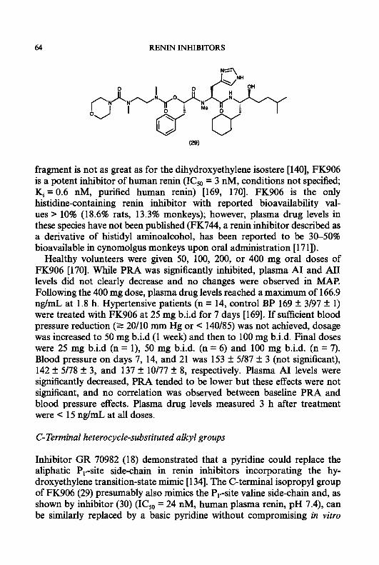
64 RENIN INHIBITORS
fragment is not as great as for the dihydroxyethylene isostere [140], FK906 is a potent inhibitor of human renin (IC50 = 3 nM, conditions not specified; &=0.6 nM, purified human renin) [169, 1701. FK906 is the only histidine-containing renin inhibitor with reported bioavailability val- ues > 10% (18.6% rats, 13.3% monkeys); however, plasma drug levels in these species have not been published (FK744, a renin inhibitor described as a derivative of histidyl aminoalcohol, has been reported to be 30-50% bioavailable in cynomolgus monkeys upon oral administration [ 17 11).
Healthy volunteers were given 50, 100, 200, or 400 mg oral doses of FK906 [170]. While PRA was sigdcantly inhibited, plasma A1 and M I levels did not clearly decrease and no changes were observed in MAP. Following the 400 mg dose, plasma drug levels reached a maximum of 166.9 ng/mL at 1.8 h. Hypertensive patients (n = 14, control BP 169 f 3/97 f 1) were treated with FK906 at 25 mg b.i.d for 7 days [169]. If sufficient blood pressure reduction (2 20/10 mm Hg or < 140/85) was not achieved, dosage was increased to 50 mg b.i.d (1 week) and then to 100 mg b.i.d. Final doses were 25 mg b.i.d (n = l), 50 mg b.i.d. (n = 6) and 100 mg b.i.d. (n = 7). Blood pressure on days 7, 14, and 21 was 153 f 5/87 f 3 (not significant), 142 f 5/78 f 3, and 137 f 10/77 f 8, respectively. Plasma AI levels were significantly decreased, PRA tended to be lower but these effects were not significant, and no correlation was observed between baseline PRA and blood pressure effects. Plasma drug levels measured 3 h after treatment were < 15 ng/mL at all doses.
C- Terminal heterocycle-substituted alkyl groups
Inhibitor GR 70982 (18) demonstrated that a pyridine could replace the aliphatic PI.-site side-chain in renin inhibitors incorporating the hy- droxyethylene transition-state mimic [ 1341. The C-terminal isopropyl group of FK906 (29) presumably also mimics the P,,-site valine side-chain and, as shown by inhibitor (30) (ICS0 = 24 nM, human plasma renin, pH 7.4), can be similarly replaced by a basic pyridine without compromising in vitro
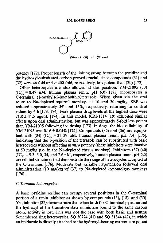
S.H. ROSENBERG 65
(30)n=2 (31)n=1 (32)n=0
potency [172]. Proper length of the linking group between the pyridine and the hydroxyl-substituted carbon proved crucial, since compounds (3 1) and (32) were 46-fold and > 400-fold, respectively, less potent than (30) [172].
Other heterocycles are also allowed at this position. YM-21095 (33) (ICsO = 0.47 nM, human plasma renin, pH 6.0) [173] incorporates a C-terminal (1 -methyl)-2-(methylthio)tetrazole. When given via the oral route to Na-depleted squirrel monkeys at 10 and 30 mglkg, SBP was reduced approximately 5% and 15%, respectively, returning to control values by 6 h [173, 1741. Peak plasma drug levels at the highest dose were 71.8 k 41.5 ngmL [174]. In this model, KRI-1314 (19) exhibited similar effects upon oral administration, but was approximately 5-fold less potent than YM-21095 following i.v. dosing [173]. In dogs, the bioavailability of YM-21095 was 0.16 k 0.04% [174]. Compounds (35) and (36) are equipo- tent with (34) (ICsO = 31-39 nM, human plasma renin, pH 7.4) [175], indicating that the 1-position of the tetrazole can be substituted with basic heterocycles without affecting in vitro potency (these inhibitors were inactive at 50 mgkg p.0. in the Na-depleted rhesus monkey). Inhibitors (37)-(40) (ICso = 9.3, 5.0, 34, and 2.6 nM, respectively, human plasma renin, pH 5.5) are related structures that demonstrate the range of heterocycles accepted at the C-terminus [176]. Moderate but variable hypotension followed oral administration (10 mglkg) of (37) to Na-depleted cynomolgus monkeys [176].
C-Terminal heterocycles
A basic pyridine residue can occupy several positions in the C-terminal portion of a renin inhibitor as shown by compounds (15), (18), and (30). Yet, inhibitor (32) demonstrates that when both the C-terminal pyridine and the hydroxyl of the transition-state mimic are bound to the same carbon atom, activity is lost. This was not the case with both basic and neutral 5-membered ring heterocycles. SQ 30774 (41) and SQ 31844 (42), in which an imidazole is directly attached to the hydroxyl-bearing carbon, are potent

66 RENIN INHIBITORS
(34)R=H (35) R = CHp(pyridin4-yl) (38) R = CHp(morphoiin-4-yl)
(37) R = pyridin-2-yl (38) R = thiazoi-2-yl (39) R = (l-methyl)tetrazoi-5-yl (40) R = (l-methyi-2-hydroxyrn~yl)imid~oi-2-yi
H OH -4-3 R-Phs-His-N
0 (41) R = BCC (42) R = (morphoiin-4-yl)CO

S.H. ROSENBERG 67
(43)R=Et n = l (44)R=H n = 2
inhibitors of human renin (ICso = 8.5 nM and 10 nM, respectively, human kidney renin, pH 7.0) [177]. Oral administration of SQ 31844 (50 mglkg) to the Na-depleted cynomolgus monkey reduced PRA 80% and lowered MAP by 15 mm Hg (17%, not significant). SQ 30774 was less active in this model [177].
Other substituted 5-membered ring heterocycles have yielded even more potent renin inhibitors. The C-terminal oxazolidinone (43) maintains the stereochemistry of the second oxygen of the glycol-derived transition-state mimics and incorporates an N-alkyl group that appears to mimic the P,#-site valine side-chain [178]. The oxazolidinone carbonyl may also participate in hydrogen-bonding to the active site. As outlined in Table 2.4 , ethyl is the optimum side-chain and slight variations in the length of this substituent significantly affect potency. Interestingly, the 6-membered ring cyclic urea
Table 2.4. RENIN INHIBITORS INCORPORATING A C-TERMINAL OXAZOLIDINONE [ 1781
H Me Et (43) OMe Pr (CH320H
25 8.8 2.8 9.2
7.4 12
a Human plasma renin, pH 7.4.
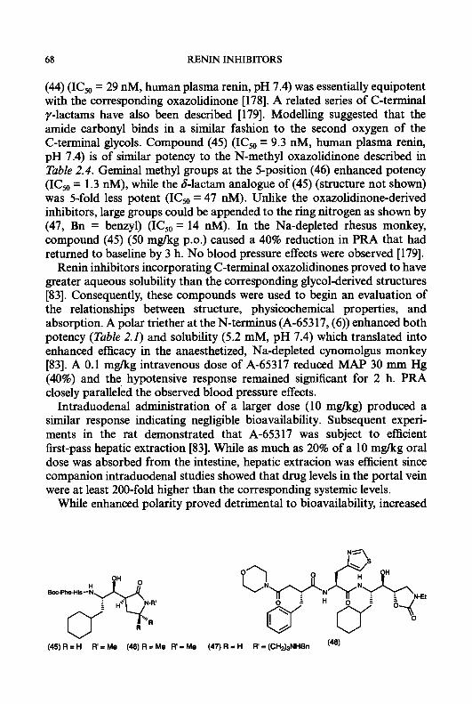
68 RENIN INHIBITORS
(44) (ICsO = 29 nM, human plasma renin, pH 7.4) was essentially equipotent with the corresponding oxazolidinone [178]. A related series of C-terminal y-lactams have also been described [179]. Modelling suggested that the amide carbonyl binds in a similar fashion to the second oxygen of the C-terminal glycols. Compound (45) (IC,O = 9.3 nM, human plasma rein, pH 7.4) is of similar potency to the N-methyl oxazolidinone described in Table 2.4. Geminal methyl groups at the 5-position (46) enhanced potency (ICs0 = 1.3 nM), while the 6-lactam analogue of (45) (structure not shown) was 5-fold less potent (ICsO = 47 nM). Unlike the oxazolidinone-derived inhibitors, large groups could be appended to the ring nitrogen as shown by (47, Bn = benzyl) (IC,,, = 14 nM). In the Na-depleted rhesus monkey, compound (45) (50 mgkg p.0.) caused a 40% reduction in PRA that had returned to baseline by 3 h. No blood pressure effects were observed [179].
Renin inhibitors incorporating C-terminal oxazolidinones proved to have greater aqueous solubility than the corresponding glycol-derived structures [83]. Consequently, these compounds were used to begin an evaluation of the relationships between structure, physicochemical properties, and absorption. A polar triether at the N-terminus (A-65317, (6)) enhanced both potency (Table 2.1) and solubility (5.2 mM, pH 7.4) which translated into enhanced efficacy in the anaesthetized, Na-depleted cynomolgus monkey [83]. A 0.1 mgkg intravenous dose of A-65317 reduced MAP 30 rnm Hg (40%) and the hypotensive response remained significant for 2 h. PRA closely paralleled the observed blood pressure effects.
Intraduodenal administration of a larger dose (10 mgkg) produced a similar response indicating negligible bioavailability. Subsequent experi- ments in the rat demonstrated that A-65317 was subject to efficient first-pass hepatic extraction [83]. While as much as 20% of a 10 mgkg oral dose was absorbed from the intestine, hepatic extracion was efficient since companion intraduodenal studies showed that drug levels in the portal vein were at least 200-fold higher than the corresponding systemic levels.
While enhanced polarity proved detrimental to bioavailability, increased
( 4 5 ) R = H R'=Me (46)R=Me R = M e ( 4 7 ) R = H R=(CH&NHBn
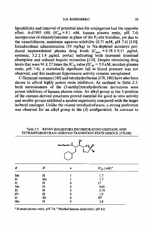
S.H. ROSENBERG 69
lipophilicity and removal of potential sites for conjugation had the opposite effect. A-67993 (48) (ICs0 = 8.1 nM, human plasma renin, pH 7.4) incorporates (4-thiazoly1)alanine in place of the P,-site histidine, yet due to the oxazolidinone, maintains aqueous solubility (0.71 mM, pH 7.4) [110]. Intraduodenal administration (10 mgkg) to Na-depleted monkeys pro- duced unprecedented plasma drug levels (C, = 0.38 f 0.11 pg/mL systemic, 3.2 f 1.4 pg/mL portal) indicating both increased intestinal absorption and reduced hepatic extraction [l lo]. Despite circulating drug levels that were 94 f 27 times the IC50 value (ICS0 = 5.9 nM, monkey plasma renin, pH 7.4), a statistically sigdcant fall in blood pressure was not observed, and this moderate hypotensive activity remains unexplained.
C-Terminal oxetanes [ 1801 and tetrahydrofurans [ 178,180] have also been shown to afford highly potent renin inhibitors. As outlined in Table 2.5, both stereoisomers of the (3-methy1)tetrahydrofuran derivatives were potent inhibitors of human plasma renin. An alkyl group at the 3-position of the oxetane-derived structures proved essential for good in vitro activity and smaller groups exhibited a modest superiority compared with the larger isobutyl analogue. Unlike the related tetrahydrofurans, a strong preference was observed for an alkyl group in the (3 configuration. In contrast to
Table 2.5. RE" INHIBITORS INCORPORATING OXETANE AND TETRAHYDROFURAN-DERIVED TRANSITION STATE MIMICS. [178,180]
Me H H Me Et iPr H iBu
H Me H H H H iPr H
1.7 1.7
0.91 0.79 1 .o 30b 3.8
17
a Human plasma renin, pH 7.4. Purified human renal renin, pH 6.0.
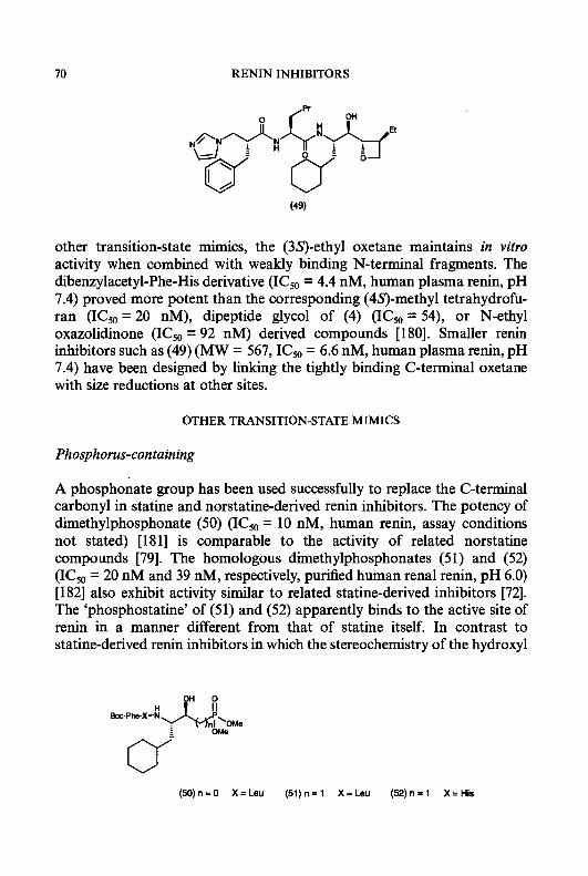
RENIN INHIBITORS
other transition-state mimics, the (3S)-ethyl oxetane maintains in vitro activity when combined with weakly binding N-terminal fragments. The dibenzylacetyl-Phe-His derivative (ICso = 4.4 nM, human plasma renin, pH 7.4) proved more potent than the corresponding (4S)-methyl tetrahydrofu- ran (IC50 = 20 nM), dipeptide glycol of (4) (ICsO = 54), or N-ethyl oxazolidinone (ICs0 = 92 nM) derived compounds [180]. Smaller renin inhibitors such as (49) (MW = 567, IC50 = 6.6 nM, human plasma renin, pH 7.4) have been designed by linking the tightly binding C-terminal oxetane with size reductions at other sites.
OTHER TRANSITION-STATE MIMICS
Phosphorus-containing
A phosphonate group has been used successfully to replace the C-terminal carbonyl in statine and norstatine-derived renin inhibitors. The potency of dimethylphosphonate (50) (ICs0 = 10 nM, human renin, assay conditions not stated) [181] is comparable to the activity of related norstatine compounds [79]. The homologous dimethylphosphonates (5 1) and (52) (ICso = 20 nM and 39 nM, respectively, purified human renal renin, pH 6.0) [ 1821 also exhibit activity similar to related statine-derived inhibitors [72]. The ‘phosphostatine’ of (51) and (52) apparently binds to the active site of renin in a manner different from that of statine itself. In contrast to statine-derived renin inhibitors in which the stereochemistry of the hydroxyl
(50)n=0 x=Leu (51)n=1 X=Leu (52)n=1 X=His
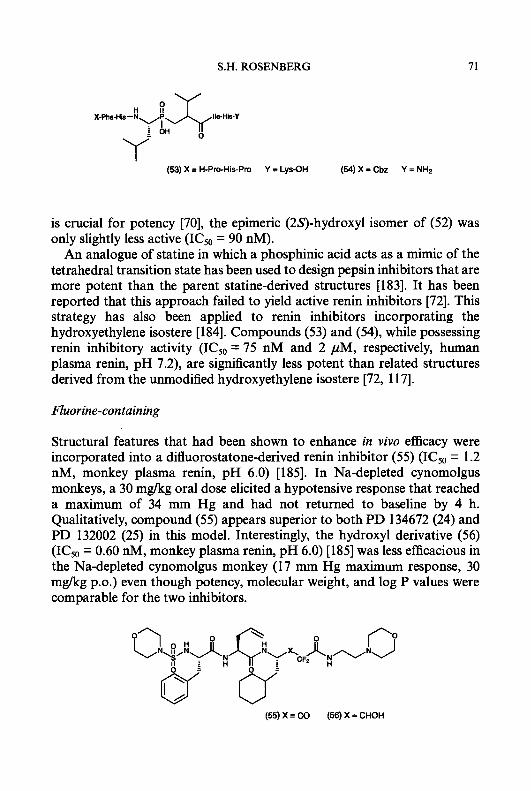
S.H. ROSENBERG 71
(53) X = H-Pro-His-Pro Y = Lys-OH (54) X = Cbz Y = NH2
is crucial for potency [70], the epimeric (2S)-hydroxyl isomer of (52) was only slightly less active (IC50 = 90 nM).
An analogue of statine in which a phosphinic acid acts as a mimic of the tetrahedral transition state has been used to design pepsin inhibitors that are more potent than the parent statine-derived structures [183]. It has been reported that this approach failed to yield active renin inhibitors [72]. This strategy has also been applied to renin inhibitors incorporating the hydroxyethylene isostere [184]. Compounds (53) and (54), while possessing renin inhibitory activity (ICs0 = 75 nM and 2 pM, respectively, human plasma renin, pH 7.2), are significantly less potent than related structures derived from the unmodified hydroxyethylene isostere [72, 11 71.
Fluorine-containing
Structural features that had been shown to enhance in vivo efficacy were incorporated into a difluorostatone-derived renin inhibitor (55 ) (IC50 = 1.2 nM, monkey plasma renin, pH 6.0) [185]. In Na-depleted cynomolgus monkeys, a 30 m a g oral dose elicited a hypotensive response that reached a maximum of 34 mm Hg and had not returned to baseline by 4 h. Qualitatively, compound (55 ) appears superior to both PD 134672 (24) and PD 132002 (25) in this model. Interestingly, the hydroxyl derivative (56) (IC50 = 0.60 nM, monkey plasma renin, pH 6.0) [185] was less efficacious in the Na-depleted cynomolgus monkey (17 mm Hg maximum response, 30 mg/kg p.0.) even though potency, molecular weight, and log P values were comparable for the two inhibitors.
(55) X = CO (56) X = CHOH

12 RENIN INHIBITORS
(57) X = Boc-Phe Y = CO(CH&iPr (58) X = iBuCO(0Me)Tyr Y = H
Compound (57) (ICso = 3.5 nM, human plasma renin, pH 6.0) incorpo- rates difluorostatone that has been retroinverted at the C-terminus [186]. Primary amine (58) (MDL 74147, ICs0 = 16 nM, human plasma renin, pH 6.0), which lacks the C-terminal acyl group, was also a potent renin inhibitor [187]. Other groups have reported similar retroinverted structures [188] and the related nonfluorinated compounds [log]. An unusual difluoro-1,3-diol(59) (ICso = 0.65 nM, purified human renal renin, pH 6.0) has been recently described [189].
Miscellaneous structures
1,3-Diols related to the C-terminus of structure (59), but lacking the geminal difluoro group, are incorporated into compounds (60) and (61) (ICso = 0.3 and 0.2 nM, respectively, monkey plasma renin, pH 6.0) [190]. The potency of these inhibitors suggests that the fluorines of (59) do not contribute significantly to binding. lY4-Diol(62) (ICso = 1.7 nM, monkey plasma renin,
(BO)R=Me (61)R=H

S.H. ROSENBERG 73
0- (63)n=2 (64)n=1 (65)n=0
pH 6.0) was designed based upon a series of renin inhibitor-endothiapepsin crystal structures [191] but is 6-fold less potent than the related glycol PD 134672 (24) (ICso = 0.28 nM, monkey plasma renin, pH 6.0) [164]. The oxidized sulphur atoms in inhibitors (63) and (64) (ICso = 9.4 and 19 nM, respectively, human plasma renin, pH 7.4) are positioned similarly to the C-terminal hydroxyl-bearing carbon of the 1,3-diol structures [192]. Sulphide (65) (ICso = 40 nM) was only moderately less potent, indicating that the sulphone and sulphoxide oxygens do not interact strongly with the active site.
PEPTIDE BACKBONE MODIFICATIONS
A different approach toward developing bioavailable, and hence clinically useful, renin inhibitors has been to design compounds with diminished peptide character. Unfortunately, altering the peptide backbone of a substrate-derived renin inhibitor not only affects side-chain orientation but also the conformation and steric environment of important hydrogen bonding groups within the backbone itself. Consequently, the design of novel amide bond replacements has proceeded at a slower pace than the less complicated modification of side-chains and termini described in the preceding sections.
Isosteric replacements
Molecular modelling and crystallographic studies have suggested that the P, and P,-site carbonyl residues participate in a hydrogen bonding network with the active site cleft of renin [193], and this has been confirmed through structure-activity investigations. Replacement of the amide bond between the P, and P,-sites with the reduced amide (secondary amine) isostere drastically reduced activity thereby demonstrating the importance of a hydrogen-bond accepting group at this site [ 1941. The hydroxyethylene isostere, which incorporates a hydroxyl in place of the carbonyl, can be a
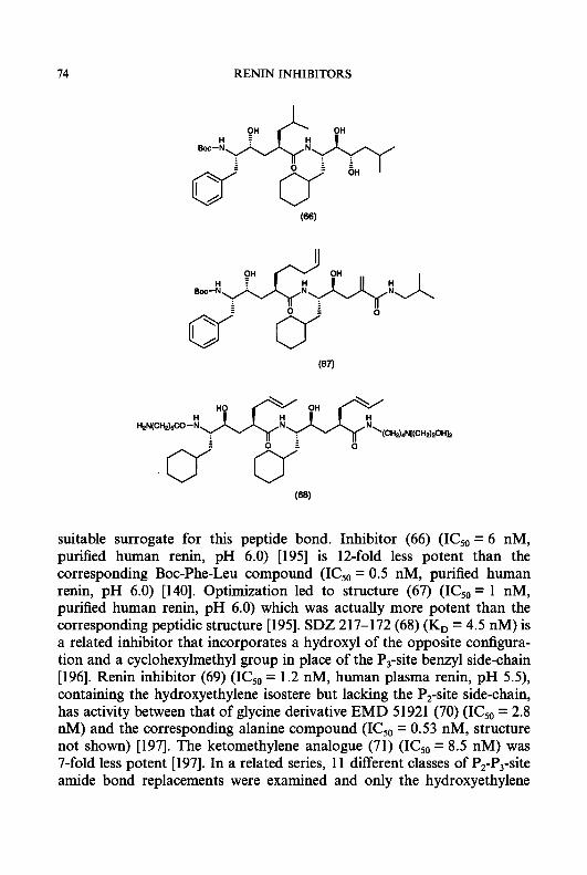
14 RENIN INHIBITORS
suitable surrogate for this peptide bond. Inhibitor (66) (IC,0=6 nM, purified human renin, pH 6.0) [195] is 12-fold less potent than the corresponding Boc-Phe-Leu compound (IC50 = 0.5 nM, purified human renin, pH 6.0) [140]. Optimization led to structure (67) (ICsO = 1 nM, purified human renin, pH 6.0) which was actually more potent than the corresponding peptidic structure [195]. SDZ 217-172 (68) (K, = 4.5 nM) is a related inhibitor that incorporates a hydroxyl of the opposite conligura- tion and a cyclohexylmethyl group in place of the P,-site benzyl side-chain [196]. Renin inhibitor (69) (IC50 = 1.2 nM, human plasma renin, pH 5 . 9 , containing the hydroxyethylene isostere but lacking the P,-site side-chain, has activity between that of glycine derivative EMD 51921 (70) (ICsO = 2.8 nM) and the corresponding alanine compound (IC,,, = 0.53 nM, structure not shown) [197]. The ketomethylene analogue (71) (IC50 = 8.5 nM) was 7-fold less potent [197]. In a related series, 11 different classes of P,-P,-site amide bond replacements were examined and only the hydroxyethylene

S.H. ROSENBERG
(69) X = (s) CHOH Y = CHg (70)X=CO Y=NH (71) X = CO Y = CH2 (72) X = CO Y = NHCHg (73) x = co Y = NH(CHg)g (74) X = CO Y = CHgS
isostere-derived structures approached the activity of parent amide- containing compounds [194].
Oxidized sulphur can also replace the P,-site carbonyl as demonstrated by sulphoxide (75) (SDZ 217-193, K, = 3.5 nM) [196] and sulphone (76) (ICs0 = 40 nM, human plasma renin, pH 7.4) [198]. The sulphone-derived amide bond mimic, however, does not appear optimal at this position since peptide-derived inhibitors (77) and (78) (ICs0 = 1.7 and 1.6 nM, respec- tively, human plasma renin, pH 7.4) are at least 23-fold more potent than (76) [de Lara et al., unpublished results]. Compound (79) (ICs0 = 2.5 nM, human plasma renin, pH 5.5) is representative of a series of renin inhibitors in which a phosphonate replaces the amide bond between the P2 and P,-site residues [ 1991.
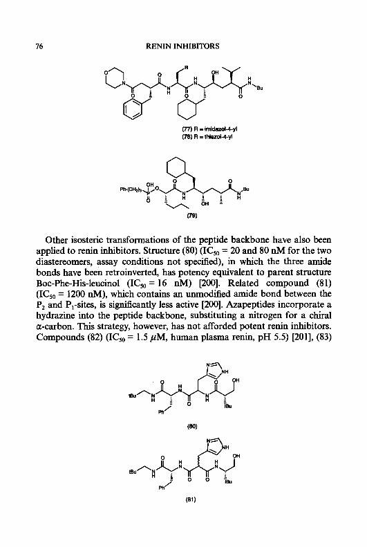
76 RENIN INHIBITORS
(77) R = irnidard4yl (78) R = thlazol4yi
Other isosteric transformations of the peptide backbone have also been applied to renin inhibitors. Structure (80) (ICso = 20 and 80 nM for the two diastereomers, assay conditions not specified), in which the three amide bonds have been retroinverted, has potency equivalent to parent structure Boc-Phe-His-leucinol (ICso = 16 nM) [200]. Related compound (81) (ICso = 1200 nM), which contains an unmodified arnide bond between the P2 and PI-sites, is significantly less active [200]. Azapeptides incorporate a hydrazine into the peptide backbone, substituting a nitrogen for a chiral a-carbon. This strategy, however, has not afforded potent renin inhibitors. Compounds (82) (ICso = 1.5 pM, human plasma renin, pH 5.5) [201], (83)

S.H. ROSENBERG
( 8 2 ) X = N Y = C H g Z=(S)CH (83)X=(S)CH Y = N H Z=(S)CH (84)X=(S)CH Y=CHg Z = N (85)X=(S)CH Y=CHg Z=(S)CH
(ICsO = 245 nM) [202], and (84) (ICso = 100 nM) [203], in which the P,, Pz, and P,-sites respectively have been modified, are at least 35-fold less active than the parent structure (85) (ICsO = 2.8 nM) [197]. A novel 3,5-linked pyrrolin-4-one was designed to mimic the conformation of an amide bond in a /?-strand and was incorporated into structure (86) [204]. While this compound binds to renin (ICso = 600 nM, human plasma renin, pH 7.4), it is a much less potent inhibitor than the unmodified structure [141].
Atom insertions
In this structural type, a /?-amino acid or related species occupies the P,-site thereby increasing the distance between the Pz and P,-site carbonyls. Compound (72) (ICsO = 2.5 nM, human plasma renin, pH 5 . 9 , which incorporates /?-alanine, is equipotent with the corresponding glycine derivative (70) (ICsO = 2.8 nM) [197]. Sulphur-containing inhibitor (74) is also a potent renin inhibitor (ICsO = 2.1 nM), but insertion of two methylene units as in (y-amino)butyric derivative (73) proved detrimental to potency (ICs0 = 28.5 nM) [197]. Replacement of the N-terminal carbamate of compound (72) with a (4-amino)piperidine urea provided inhibitor (87) and enhanced both in vitro potency (ICsO = 0.76 nM) and in vivo efficacy [197]. In the Na-depleted cynomolgus monkey, an oral dose (30 mg/kg) of
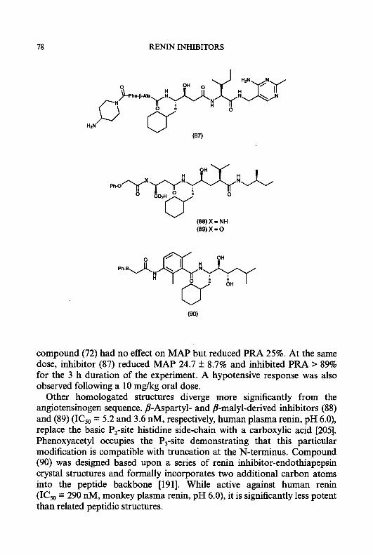
78 RENIN INHIBITORS
(89) x = 0
compound (72) had no effect on MAP but reduced PRA 25%. At the same dose, inhibitor (87) reduced MAP 24.7 f 8.7% and inhibited PRA > 89% for the 3 h duration of the experiment. A hypotensive response was also observed following a 10 mgkg oral dose.
Other homologated structures diverge more significantly from the angiotensinogen sequence. /?-Aspartyl- and /?-malyl-derived inhibitors (88) and (89) (ICso = 5.2 and 3.6 nM, respectively, human plasma renin, pH 6.0), replace the basic P,-site histidine side-chain with a carboxylic acid [205]. Phenoxyacetyl occupies the P,-site demonstrating that this particular modification is compatible with truncation at the N-terminus. Compound (90) was designed based upon a series of renin inhibitor-endothiapepsin crystal structures and formally incorporates two additional carbon atoms into the peptide backbone [191]. While active against human renin (IC50 = 290 nM, monkey plasma renin, pH 6.0), it is significantly less potent than related peptidic structures.

S.H. ROSENBERG 79
(91) x = 0 Y = O (92) X = MeOCHZOCH
(94) X = MeOCH20CH
Y = 0
Y = NH (93) x = 0 Y=NH
Atom deletions: A-74273
Among the renin inhibitors with the least peptide character are those that lack one or more atoms between the P2 and P,-site residues. Fung and co-workers have developed a novel nonpeptide heteroatom linkage at this site that is embodied in representative structure (91) [206]. An ether replaces the amide bond between the P2 and P,-site residues and the resulting shorter backbone required stereochemical inversion of the P,-site side-chain. While potent against purified human renal renin at pH 6.0, (91) was more than 60-fold less active in an assay employing human plasma renin at pH 7.4 (Table 2.6). The nature of the N-terminal substituent had a profound effect on in vitro potency. The corresponding (4-methoxymethoxy)piperidine amide (92) was more potent in both renin inhibition assays and this activity was better maintained against human plasma r e i n at pH 7.4. As shown in
Table 2.6. RENIN INHIBITORS INCORPORATING A NONPEPTIDE HETEROATOM LINKAGE BETWEEN THE P, AND P3-POSITION RESIDUES
G o (nM)
Inhibitor Reference Purified" PIasmab
15
13 1 .o
1.1 2.4 3.3
1000 13
1200 17 2.8 7.3
a Purified human renal renin, pH 6.0. Human plasma renin, pH 7.4.
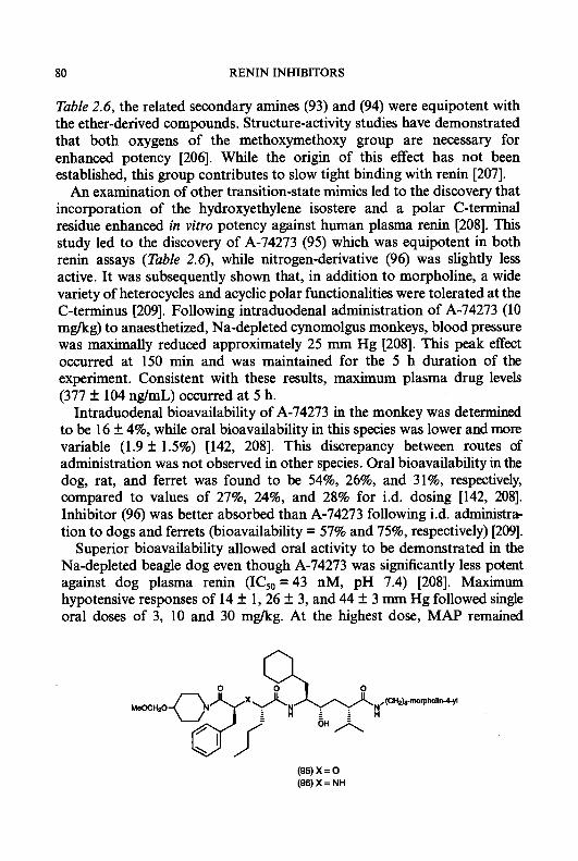
80 RENIN INHIBITORS
Table 2.6, the related secondary amines (93) and (94) were equipotent with the ether-derived compounds. Structure-activity studies have demonstrated that both oxygens of the methoxymethoxy group are necessary for enhanced potency [206]. While the origin of this effect has not been established, this group contributes to slow tight binding with renin [207].
An examination of other transition-state mimics led to the discovery that incorporation of the hydroxyethylene isostere and a polar C-terminal residue enhanced in vitro potency against human plasma renin [208]. This study led to the discovery of A-74273 (95) which was equipotent in both renin assays (Table 2.6), while nitrogen-derivative (96) was slightly less active. It was subsequently shown that, in addition to morpholine, a wide variety of heterocycles and acyclic polar fmctionalities were tolerated at the C-terminus [209]. Following intraduodenal administration of A-74273 (10 m a g ) to anaesthetized, Na-depleted cynomolgus monkeys, blood pressure was maximally reduced approximately 25 mm Hg [208]. This peak effect occurred at 150 min and was maintained for the 5 h duration of the experiment. Consistent with these results, maximum plasma drug levels (377 2 104 ng/mL) occurred at 5 h.
Intraduodenal bioavailability of A-74273 in the monkey was determined to be 16 2 4%, while oral bioavailability in this species was lower and more variable (1.9 f 1.5%) [142, 2081. This discrepancy between routes of administration was not observed in other species. Oral bioavailability in the dog, rat, and ferret was found to be 54%, 26%, and 31%, respectively, compared to values of 27%, 24%, and 28% for i.d. dosing [142, 2081. Inhibitor (96) was better absorbed than A-74273 following i.d. administra- tion to dogs and ferrets (bioavailability = 57% and 75%, respectively) [209].
Superior bioavailability allowed oral activity to be demonstrated in the Na-depleted beagle dog even though A-74273 was significantly less potent against dog plasma renin (ICs0 = 43 nM, pH 7.4) [208]. Maximum hypotensive responses of 14 2 1,26 f 3, and 44 f 3 mm Hg followed single oral doses of 3, 10 and 30 m a g . At the highest dose, MAP remained
(95) x = 0 (96)X=NH

S.H. ROSENBERG 81
significantly suppressed (-22 mm Hg) for the 6 h duration of the experiment. A larger oral dose (60 mglkg) did not elicit a greater peak effect (46 f 5 mm Hg) despite the higher plasma drug concentrations attained at this dose, but preferentially enhanced the duration of the response [210]. This behaviour is not unique to the A-74273 structural class since comparable results have been reported for the peptide-derived renin inhibitor zankiren (22) [159]. MAP remained unchanged following a 10 mglkg i.v. dose in Na-replete dogs, suggesting that A-74273 acts solely by inhibiting the RAS [210].
Heterocyclic N-termini
Renin inhibitors that embody heterocycle-derived N-termini can be divided into two groups, depending upon whether the P2 or P,-site nitrogen is part of the ring. Compound (97) (ICso = 23 nM, recombinant human renin, pH 5.7) is representative of the latter class [211]. Consistent with the significant peptide character maintained by this structure, (97) has activity similar to the unmodified Boc-Phe-Leu compound (ICs0 = 8 nM) but does not reduce MAP in the Na-depleted rhesus monkey (50 mgkg p.0.) [211]. Inclusion of the P2-site nitrogen as part of the N-terminal heterocycle, however, more profoundly alters the peptide backbone. The first reported inhibitors of this type contained y-lactams in which this nitrogen was connected to the &-carbon of the P,-site residue through a two-atom spacer [72, 2121. While inhibitors incorporating the hydroxyethylene isostere were equipotent with
( B ) n = O (BB)n=l
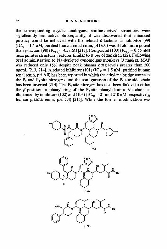
82 RENIN INHIBITORS
the corresponding acyclic analogues, statine-derived structures were significantly less active. Subsequently, it was discovered that enhanced potency could be achieved with the related blactams as inhibitor (99) (ICso = 1.4 nM, pursed human renal renin, pH 6.0) was 3-fold more potent than y-lactam (98) (IC50 = 4.5 nM) [213]. Compound (100) (ICsO = 0.55 nM) incorporates structural features similar to those of zankiren (22). Following oral administration to Na-depleted cynomolgus monkeys (3 mg/kg), MAP was reduced only 15% despite peak plasma drug levels greater than 500 ng/mL [213, 2141. A related inhibitor (101) (IC50 = 1.5 nM, purified human renal renin, pH 6.0) has been reported in which the ethylene bridge connects the P2 and P,-site nitrogens and the configuration of the P,-site side-chain has been inverted [214]. The P2-site nitrogen has also been linked to either the /3-position or phenyl ring of the P,-site phenylalanine side-chain as illustrated by inhibitors (102) and (103) (ICso = 21 and 210 nM, respectively, human plasma renin, pH 7.4) [215]. While the former modification was
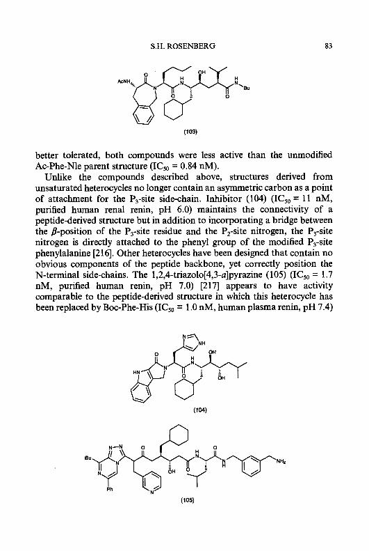
S.H. ROSENBERG 83
better tolerated, both compounds were less active than the unmodified Ac-Phe-Nle parent structure (ICsO = 0.84 nM).
Unlike the compounds described above, structures derived from unsaturated heterocycles no longer contain an asymmetric carbon as a point of attachment for the P,-site side-chain. Inhibitor (104) (ICso = 11 nM, purified human renal renin, pH 6.0) maintains the connectivity of a peptide-derived structure but in addition to incorporating a bridge between the B-position of the P,-site residue and the P,-site nitrogen, the P3-site nitrogen is directly attached to the phenyl group of the modified P3-site phenylalanine [216]. Other heterocycles have been designed that contain no obvious components of the peptide backbone, yet correctly position the N-terminal side-chains. The 1,2,4-triazol0[4,3-a]pyrazine (105) (IC50 = 1.7 nM, purified human renin, pH 7.0) [217] appears to have activity comparable to the peptide-derived structure in which this heterocycle has been replaced by Boc-Phe-His (ICS0 = 1 .O nM, human plasma renin, pH 7.4)

84 RE" INHIBITORS
[218]. Similar inhibitors have been described that incorporate either the hydroxyethylene isostere [219] or difluorostatone [220] transition-state mimics. The latter structure (106) (IC50 = 1.6 nM, purified human renin, pH 7.0) is active despite lacking a P,-site side-chain [220]. Following oral administration to Na-depleted marmosets, 1,2,4-triazolo[4,3-u]pyrazine- containing renin inhibitors elicited at most a moderate hypotensive response (similar to that observed for CGP 38 560 (1 1) [219]) and these results were attributed to poor intestinal absorption [217].
Other modijied P,-site residues
The cyclopropane-derived P,-site residue of inhibitor (107) (IC50 = 20 nM, human plasma renin, pH 7.4) directly links the /?-position of the benzyl side-chain to a carbon atom that replaces the P,-site phenylalanine nitrogen [22 11. The corresponding acyclic benzyl succinate structure (not shown) is slightly more potent (ICsO = 8.3 nM). Interestingly, an isobutyl group is a suitable phenyl replacement in this series as illustrated by inhibitor (108) (ICsO = 40 nM) [221]. Incorporating an N-terminal sulphone, which has previously been shown to enhance in vitro potency [117, 160, 164, 1651,
(107) X = (rnotpholin-l-yl)CO Y = S R = Ph
(roe) x = IPS02 Y = N H R=Ph (108) XI (rnorpholln-l-yl)CO Y = S R = iBu
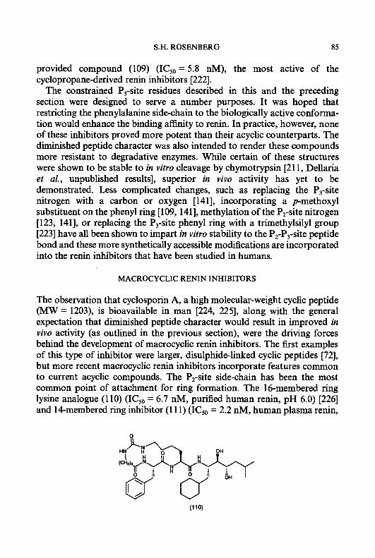
S.H. ROSENBERG 85
provided compound (109) (ICsO = 5.8 nM), the most active of the cyclopropane-derived renin inhibitors [222].
The constrained P,-site residues described in this and the preceding section were designed to serve a number purposes. It was hoped that restricting the phenylalanine side-chain to the biologically active conforma- tion would enhance the binding a f i t y to renin. In practice, however, none of these inhibitors proved more potent than their acyclic counterparts. The diminished peptide character was also intended to render these compounds more resistant to degradative enzymes. While certain of these structures were shown to be stable to in vitro cleavage by chymotrypsin [211, Dellaria et al., unpublished results], superior in vivo activity has yet to be demonstrated. Less complicated changes, such as replacing the P,-site nitrogen with a carbon or oxygen [141], incorporating a p-methoxyl substituent on the phenyl ring [log, 1411, methylation of the P,-site nitrogen [123, 1411, or replacing the P,-site phenyl ring with a trimethylsilyl group [223] have all been shown to impart in vitro stability to the P,-P,-site peptide bond and these more synthetically accessible modifications are incorporated into the renin inhibitors that have been studied in humans.
MACROCYCLIC RENIN INHIBITORS
The observation that cyclosporin A, a high molecular-weight cyclic peptide (MW = 1203), is bioavailable in man [224, 2251, along with the general expectation that diminished peptide character would result in improved in vivo activity (as outlined in the previous section), were the driving forces behind the development of macrocyclic renin inhibitors. The first examples of this type of inhibitor were larger, disulphide-linked cyclic peptides [72], but more recent macrocyclic renin inhibitors incorporate features common to current acyclic compounds. The P,-site side-chain has been the most common point of attachment for ring formation. The 16-membered ring lysine analogue (1 10) (ICsO = 6.7 nM, purified human renin, pH 6.0) [226] and 14-membered ring inhibitor (1 1 1) (ICso = 2.2 nM, human plasma renin,

86 RENIN INHIBITORS
pH 6.0) [227] are examples in which this position has been connected to the N-terminus. It is the structures in which the P,-site side-chain has been linked to the C-terminus, however, that have been the most extensively investigated.
Researchers at Merck have described a series of norACHPA-derived macrocyclic renin inhibitors. The first structures disclosed incorporated a glutamic acid residue at the P,-site that was linked via an aminoethanol group to the norACHPA carbonyl (Table 2.7) [228-2301. Both alkyl and heterocyclic substituents were preferred at the P,,-site (R') and enhanced potency 1 1-€old to 145-fold. Ring size proved critical as the 14-membered ring homologue incorporating aminopropanol (R = Boc-Phe) was 1 1-fold
Table 2.7. NORSTATINE-DERIVED RENIN INHIBITORS GLUTAMIC ACID AT THE P 4 I T E
INCORPORATING
R R' Reference IC, (nM)"
Boc-Phe tBuCH,CONH(CH,),OCO-Phe Boc-Phe Boc-Phe Boc-Phe Boc-Phe Boc-Phe (5') tBuCH,CH(CH,Ph)CO
H H iBu Et Bu n-Hex CH2N(CH2CH,),0 CH2N(CH2CH2),0
610 56
23 25 49 55
4.2
6.9
* Human plasma renin, pH 7.4.
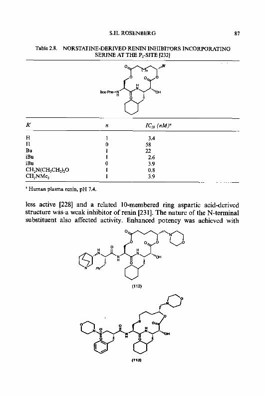
S.H. ROSENBERG 87
Table 2.8. NORSTATINE-DERIVED RENIN INHIBITORS INCORPORATING SERINE AT THE PI-SITE 12321
H 1 3.4 H 0 58 Bu 1 22 iBu 1 2.6 iBu 0 3.9 CH,N(CH,CHJ,O 1 0.8 CH,NMe, 1 3.9
a Human plasma renin, pH 7.4.
less active [228] and a related 10-membered ring aspartic acid-derived structure wasa weak inhibitor of renin [231]. The nature of the N-terminal substituent also affected activity. Enhanced potency was achieved with
(1 12)
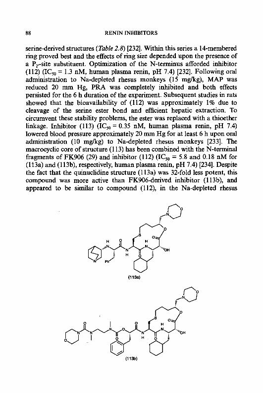
88 RE" INHIBITORS
serine-derived structures (Table 2.8) [232]. Within this series a 14-membered ring proved best and the effects of ring size depended upon the presence of a Pr-site substituent. Optimization of the N-terminus afforded inhibitor (112) (ICsO = 1.3 nM, human plasma renin, pH 7.4) [232]. Following oral administration to Na-depleted rhesus monkeys (15 mglkg), MAP was reduced 20 mm Hg, PRA was completely inhibited and both effects persisted for the 6 h duration of the experiment. Subsequent studies in rats showed that the bioavailability of (112) was approximately 1% due to cleavage of the serine ester bond and efficient hepatic extraction. To circumvent these stability problems, the ester was replaced with a thioether linkage. Inhibitor (113) (ICsO = 0.35 nM, human plasma renin, pH 7.4) lowered blood pressure approximately 20 mm Hg for at least 6 h upon oral administration (10 mgkg) to Na-depleted rhesus monkeys [233]. The macrocyclic core of structure (1 13) has been combined with the N-terminal fragments of FK906 (29) and inhibitor (112) (ICsO = 5.8 and 0.18 nM for (113a) and (1 13b), respectively, human plasma renin, pH 7.4) [234]. Despite the fact that the quinuclidine structure (113a) was 32-fold less potent, this compound was more active than FK906-derived inhibitor (1 13b), and appeared to be similar to compound (1 12), in the Na-depleted rhesus
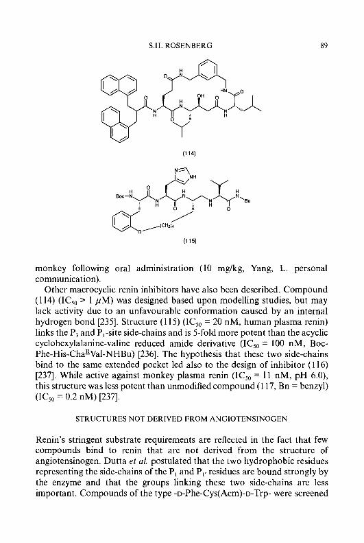
S.H. ROSENBERG 89
monkey following oral administration (10 mg/kg, Yang, L. personal communication).
Other macrocyclic renin inhibitors have also been described. Compound (1 14) (ICs0 > 1 pM) was designed based upon modelling studies, but may lack activity due to an unfavourable conformation caused by an internal hydrogen bond [235]. Structure (1 15) (IC50 = 20 nM, human plasma renin) links the P, and PI-site side-chains and is 5-fold more potent than the acyclic cyclohexylalanine-valine reduced amide derivative (IC5,, = 100 nM, Boc- Phe-His-Cha&Val-NHBu) [236]. The hypothesis that these two side-chains bind to the same extended pocket led also to the design of inhibitor (1 16) [237]. While active against monkey plasma renin (IC50 = 11 nM, pH 6.0), this structure was less potent than unmodified compound (1 17, Bn = benzyl) (IC5,, = 0.2 nM) [237].
STRUCTURES NOT DERIVED FROM ANGIOTENSINOGEN
Renin’s stringent substrate requirements are reflected in the fact that few compounds bind to renin that are not derived from the structure of angiotensinogen. Dutta et al. postulated that the two hydrophobic residues representing the side-chains of the PI and PI, residues are bound strongly by the enzyme and that the groups linking these two side-chains are less important. Compounds of the type -D-Phe-Cys(Acm)-D-Trp- were screened
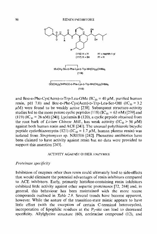
90 RENIN INHIBITORS
(116) R = H (117)R=Bn R'=H
R' = naphth-1-yl
m tBuCHZ-Glu-D-Phe-Lys-D-Trp-NH(CHz)zCHMez
(118)
I I CO(CH2)zCHPhCO-D-Phe-Lys-D-Trp-NH(CHz)zCHMe2
(119)
and Boc-D-Phe-Cys(Acm)-D-Trp-Leu-OMe (IC50 = 40 pM, purified human renin, pH 7.0) and Boc-D-Phe-Cys(Acm)-D-Trp-Leu-Ser-OH (ICs0 = 3.2 pM) were found to be weakly active [238]. Subsequent structure-activity studies led to the more potent cyclic peptides (1 18) (IC50 = 63 nM) [239] and (1 19) (IC," = 26 nM) [240]. Lyciumin B (120), a cyclic peptide obtained from the root bark of Licium Chinese Mill., has weak activity (IC50 > 50 pM) against both human renin and ACE [241]. The unusual polythiazole bicyclic peptide cyclothiazomycin (121) (IC50 = 1.7 pM, human plasma renin) was isolated from Streptornyces sp. NR05 16 [242]. Phenazine antibiotics have been claimed to have activity against renin but no data were provided to support this assertion [243].
ACTIVITY AGAINST OTHER ENZYMES
Proteinnse specijicity
Inhibition of enzymes other than renin could ultimately lead to side-effects that would eliminate the potential advantages of renin inhibitors compared to ACE inhibitors. Early, primarily histidine-containing renin inhibitors exhibited little activity against other aspartic proteinases [72, 2441 and, in general, this behaviour has been maintained with the more recent compounds outlined in Table 2.9. Several trends have become apparent, however. While the nature of the transition-state mimic appears to have little effect (with the exception of certain C-terminal heterocycles), incorporation of lipophilic residues at the P2-site can lead to decreased specificity. Allylglycine structure (60), norleucine compound (1 2), and

S.H. ROSENBERG
PyroGlu-Pro-Trp- -NyCO-Val-Gly-Ser H
/
91
H d
(121)
carbomethoxyglycine derivative (25) are at least 60-fold more potent against pepsin and cathepsin D than the corresponding histidine-containing compounds (entries d, i, and 1) despite equivalent renin inhibitory activity. A similar loss in specificity was observed when the histidine of KRI-1314 (19) was replaced by valine (entry e). Other structural features must also be involved since the presence of a lipophilic residue at the P2-site is not a sufficient condition for diminished specificity as demonstrated by ES-6864 (6), zankiren (22), BW-175 (27), and MDL 74147 which contain (thiazol-4- yl)alanine, norleucine, and valine at the P,-site. Inhibitor (60) has been reported to have appreciable potency against gastricsin. This has been attributed to the nature of the transition-state mimic as the activity was abolished (and potency against cathepsin D was reduced) upon incorpora- tion of an additional C-terminal methyl group (61). Variation of the side-chains in Sta-containing heptapeptides can impart activity against other aspartic proteinases (cathepsins D and E, pepsin, gastricsin, endothiapepsin) but this was often at the expense of renin inhibitory potency [245].
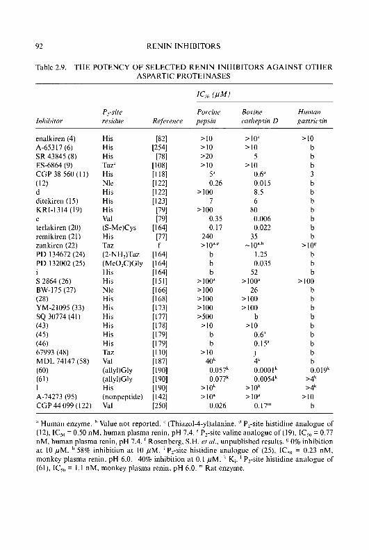
92 RENIN INHIBITORS
Table 2.9. T H E POTENCY OF SELECTED RENIN INHIBITORS AGAINST OTHER ASPARTIC PROTEINASES
Inhibitor
enalkiren (4)
SR 43845 (8) ES-6864 (9) CGP 38 560 (1 1) (12) d ditekiren ( I 5) KRI-1314 (19) e terlakiren (20) remikiren (21) zankiren (22) PD 134672 (24) PD I32002 (25) i S 2864 (26)
(28) YM-21095 (33) SQ 30774 (41) (43) (45) (46) 67993 (48) MDL 74147 (58) (60) (61) 1 A-74273 (95) CGP44 099 (122)
A-65317 (6)
BW-I75 (27)
P,-site residue
His His His Taz' His Nle His His His Val (S-Me)Cys His
(2-NH2)Taz (Me02C)Gly His His Nle His His His His His His Taz Val
(allyl)Gly His (nonpeptide) Val
T d Z
(allyl)Gly
Porcine pepsin
>I0 >I0 >20 >10
5" 0.26
>I00 7
>I00 0.35 0.17
240 >lo"'
b b b
> 100" >I00 >I00 >I00 >so0 >I0
b b
>I0 40' 0.057' 0.077'
>lok >lo"
0.026
Bovine cw tliepsin D
Hutmn gnstricsin
>lo" >I0
5 >I0
0.6" 0.015 8.5 6
80 0.006 0.022
35 -10"
1.25 0.035
52 > 100"
26 >I00 >I00
b >I0
0.6" 0.15" j 4' 0.0001 ' 0.0054k
>lok 210"
0.17"
>I0 b b b 3 b b b b b b b
>lo' b b b
>I00 b b b b b b b b b 0.019'
>4' >4'
>I0 b
Human enzyme. Value not reported. (Thiazol-4-yl)alanine. '' P2-site histidine analogue of (12), IC,,, = 0.50 nM, human plasma renin, pH 7.4. ' P,-site valine analogue of (19). IC,,] = 0.77 nM, human plasma renin, pH 7.4. Rosenberg, S.H. rt nl., unpublished results. 0% inhibition at 10 p M . 58% inhibition at 10 p M . ' P,-site histidine analogue of (25), IC,,, = 0.23 nM, monkey plasma renin, pH 6.0. 40% inhibition at 0.1 p M . ' K,. ' P&e histidine analogue of (61), IC, , = 1.1 nM, monkey plasma renin, pH 6.0. Rat enzyme.
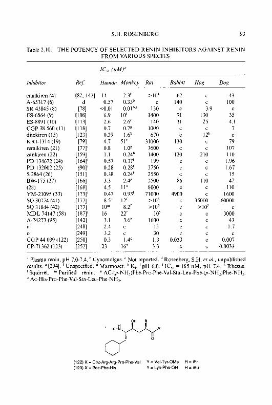
S.H. ROSENBERG 93
T H E POTENCY OF SELECTED RENIN INHIBITORS AGAINST RENIN Table 2.10. FROM VARIOUS SPECIES
ICb (nMi"
Inhibitor Re6 Humcm Monkey Rat Rabbit Hog Dog
enalkiren (4) [82, 1421 14 2.3h A-65317 (6) SR 43845 (8) ES-6864 (9) ES-8891 (10) CGP 38 560 ( 1 1) ditekiren (1 5) KRI-1314 (19) remikiren (21) zankiren (22) PD 134672 (24) PD 132002 (25) S 2864 (26) BW-175 (27) (28) YM-21095 ( 3 3 ) SQ 30774 (41) SQ 3 I844 (42) MDL 74147 (58) A-74273 (95) n
CGP 44 099 (122) 0
CP-71362 (123)
0.57 0.33h <0.01 0.0Ib~"
6.9 10' 2.6 2.6' 0.7 0.7€ 0.39 I.6h 4.7 5 I h 0.8 1.0g 1.1 0.24b 0.57 0.37' 0.28 0.28' 0.38 0.24L 3.3 2.4' 4.5 I l k 0.47 0.93' 8.5'" 12'
10'" 8.2' 16 22' 3.1 3.6h 2.4 c 3.2 c 0.3 1.4'
23 16b
>lo4 62 C 140
130 C
1400 91 140 31
1 0l!O C
670 C
31000 130 3600 C
1400 120 199 C
3750 C
2550 C
3500 86 8000 C
71000 4900 >lo ' C
>lo5 C
1 o5 C
1600 C
15 C
30 C
1.3 0.033 3.3 C
C
C
3.9 130 25
I 2'' C
C
C
210 C
C
C
110 C
C
35000 >lo'
C
C
C
C
C
C
43 100
35 4. I
7
79 107 110 1.96 I .67'
15 42
110 1600
60000
3000 43 1.7
0.007 0.0033
C
C
C
C
Plasma renin, pH 7.0-7.4. Cynomolgus. Not reported. Rosenberg, S.H. et d, unpublished results. ' [294]. ' Unspecified. Marmoset. " K,. ' pH 6.0. IC,,, = 185 nM, pH 7.4. ' Rhesus. ' Squirrel. Purified renin. " AC-@-NHZ)Phe-Pro-Phe-Val-Sta-Leu-Phe-@-NHZ)Phe-NHz. I' Ac-His-Pro-Phe-Val-Sta-Leu-Phe-NHz.
(122) X = Cbz-Arg-Arg-Pro-Phe-Val Y = Val-Tyr-OMe R = iPr (123) X = Boc-Phe-His Y = Lys-Phe-OH R = iBu
94 RENIN INHIBITORS
Species specificity
The effects of renin inhibitors against plasma renin from several species are outlined in Table 2.10. As described in previous sections, these compounds were derived from the sequence of human angiotensinogen. As expected, all are potent inhibitors of human and monkey renin and activities in these two systems are generally similar. These inhibitors bind only weakly to rat renin while potency against dog and rabbit renin varies depending upon structure. Due to this narrow species specificity, in vivo evaluation has generally been performed in primates and for only a few compounds has the combination of superior absorption and acceptable potency against nonprimate renin allowed testing in other species (see discussion of zankiren (22) and A-74273 (95)). Remikiren (21) is a potent inhibitor of guinea-pig renin (IC5,, = 10 nM) [246]. In Na-depleted guinea-pigs, the maximum hypotensive responses following i.v. administration of remikiren and the A11 antagonist EXPl32 were similar. The ACE inhibitor cilazapril, however, caused a greater fall in MAP, and, unlike remikiren, was active in anephric animals. It was concluded that in this animal model ACE inhibitors may have effects not related to the RAS [246]. Others have shown that the renin inhibitor CP-80,794 (20) and captopril elicit similar maximum responses in the guinea-pig; but with submaximal doses the effects of these two agents were synergistic [247].
Renin inhibitors have been optimized for potency against nonprimate renin through incorporation of valine at the P,-site (CGP 44 099 (122), entries n and 0) [248-2501. This particular modification can decrease specificity for other proteinases (CGP 44 099, Table 2.9) and does not invariably enhance binding to nonprimate renin since MDL 74147 (58) is only a weak inhibitor of rat and dog renin. The nature of the groups at the termini can also affect specificity [25 I]. CGP 44 099 is an unusual inhibitor that possesses nanomolar potency against guinea-pig, mouse, and cat renin in addition to renin from the species listed in Tubk 2.10 [250]. In the Na-depleted rat, CGP 44 099 reduced MAP 25 mm Hg following an intravenous infusion (0.03 mg/kg/min) but caused no further reductions in blood pressure in rats pretreated with the converting enzyme inhibitor captopril [250]. CP-71362 (1 23) is a related histidine-containing inhibitor that is highly potent against dog and rat renin and has shown i.v. activity in the former species [252].
RENAL EFFECTS
Renin inhibitors have been shown to directly modulate renal haemodynam-
S.H. ROSENBERG 95
ics. This was demonstrated through intrarenal infusion of sub-pressor doses of the renin inhibitors ACRIP [253] and A-65317 (6) [254] to Na-depleted dogs and monkeys, respectively. Significant increases in glomerular filtration rate (GFR), sodium excretion, and renal blood flow (RBF, A-65317 only) were observed. These early reports have been reviewed by Kleinert et nl. [244]. A novel method for measuring renal clearance in conscious marmosets has been developed and used to show that CGP 29 287 (Z-Arg-Arg-Pro-Phe-His-Sta-Ile-His-Lys(Boc)-OMe, 1 mg/kg i.v.) in- creases RBF but not GFR [255]. More recent studies have compared the renal effects of renin inhibitors with those of other agents that disrupt the RAS. Both ES-8891 (10) and captopril increased the kidney renin content in marmosets, but had differing effects on renin secretion [256]. Na-depleted cynomolgus monkeys were treated with doses of FK906 (29) and captopril that produced equivalent (- 14%) hypotensive responses [257]. Only FK906 significantly increased RBF, GFR, urine volume, and sodium excretion. Blood pressure was reduced 12 mm Hg (14%) following chronic (7 day) infusion (1.1 ,ug/kg/min, below maximum effective dose) of CP-7 1362 ( 123) in Na-depleted dogs [258]. No significant changes were observed in renal haemodynamic or excretory function. Similar infusion of enalapril reduced MAP 21 mm Hg (25%) and increased RBF and sodium excretion while GFR remained unchanged. In a rat model of congestive heart failure, however, both CP-71362 and enalapril had similar beneficial effects on renal function at a dose that lowered MAP 15 mm Hg in normal animals [259]. The effects of i.v. administration of remikiren (21), MK 521 (an ACE inhibitor) and DuP753 (an A11 antagonist) to guinea-pigs were directly compared at doses that reduced MAP 13-15% [260]. All three drugs significantly increased RBF and GFR but the greatest effects were observed with remikiren. In man, increased renal plasma flow was demonstrated following i.v. administration of enalkiren (4) [261] and evidence of intrarenal renin inhibition by remikiren (21) has been suggested [262].
CARDIAC EFFECTS
Previous studies on the haemodynamic mechanism of renin inhibition have demonstrated that blood pressure reduction is the result of vasodilation and not of decreased cardiac output (CO) [244]. This has been confirmed for the orally active renin inhibitors zankiren (22) [263] and A-74273 (95) [264]. In a rat model of congestive heart failure, both CP-7 1362 ( 123) and enalapril were equally effective in reducing left ventricular end diastolic pressure while only enalapril increased cardiac index (+17%) [259]. EMD 52297 (Boc-Phe-His-ACHPA-Ile-NHCH2(tetrazol-2-yl), IC,, = 5.8 nM, sheep
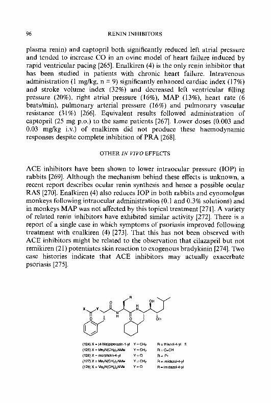
96 RENIN INHIBITORS
plasma renin) and captopril both significantly reduced left atrial pressure and tended to increase CO in an ovine model of heart failure induced by rapid ventricular pacing [265]. Enalkiren (4) is the only renin inhibitor that has been studied in patients with chronic heart failure. Intravenous administration (1 mg/kg, n = 9) significantly enhanced cardiac index (1 7%) and stroke volume index (32%) and decreased left ventricular filling pressure (20%), right atrial pressure (16%), MAP (13%), heart rate (6 beatdmin), pulmonary arterial pressure (1 6%) and pulmonary vascular resistance (3 1 %) [266]. Equivalent results followed administration of captopril (25 mg p.0.) to the same patients [267]. Lower doses (0.003 and 0.03 mg/kg i.v.) of enalkiren did not produce these haemodynamic responses despite complete inhibition of PRA [268].
OTHER IN VIVO EFFECTS
ACE inhibitors have been shown to lower intraocuiar pressure (IOP) in rabbits [269]. Although the mechanism behind these effects is unknown, a recent report describes ocular renin synthesis and hence a possible ocular RAS [270]. Enalkiren (4) also reduces IOP in both rabbits and cynomolgus monkeys following intraocular administration (0.1 and 0.3% solutions) and in monkeys MAP was not affected by this topical treatment [271]. A variety of related renin inhibitors have exhibited similar activity [272]. There is a report of a single case in which symptoms of psoriasis improved following treatment with enalkiren (4) [273]. That this has not been observed with ACE inhibitors might be related to the observation that cilazapril but not remikiren (21) potentiates skin reaction to exogenous bradykinin [274]. Two case histories indicate that ACE inhibitors may actually exacerbate psoriasis [275].
(124) X = (4-Me)piperazin-I-yl Y = CHz R = thiazol-4yl 5
(125) X = MezN(CHz),NMe Y = CHz R = C Z H
(126) X = morpholin-Cyl Y = 0 R = I P ~
(127) X = Me2N(CHz)zNMe Y = C& R = imidazol-4-yl (128) X = MezN(CH2)zNMe Y = 0 R = imidarol-Cyl
S.H. ROSENBERG 97
DELIVERY AND BIOAVAILABILITY OF RENIN INHIBITORS
In order to become a viable drug, a renin inhibitor must possess two attributes: it must be well and predictably absorbed from the gastrointesti- nal tract into the systemic circulation (and therefore must pass safely through the liver) and it must elicit a dose-related lowering of blood pressure when given orally. Thus, the first demonstrations of oral activity represented only the beginning of the quest for oral bioavailability.
INTESTINAL ABSORPTION AND ORAL BIOAVAILABILITY
The mechanisms by which renin inhibitors are absorbed from the intestine are poorly understood. A carrier-mediated process has been suggested [276]. Studies with isolated brush border membrane vesicles prepared from rabbit small intestine indicate that inhibitor (28) [ 1681 and other peptide-derived compounds [277] are actively transported by the proton-dependent uptake system shared by small peptides and B-lactam antibiotics. It has also been shown that (28) interacts with but is not taken up by the Na'-dependent glucose transporter [ 1681.
Strategies to enhance intestinal uptake have been largely empirical and have generally been part of an overall effort to improve bioavailability. A detailed investigation of structure-absorption relationships led to the discovery that incorporation of (thiazol-4-y1)alanine at the P,-site could increase absorption [81, 1 lo]. This study also resulted in a set of guidelines for the design of bioavailable renin inhibitors [S 1, 1601. Optimum structures, exemplified by zankiren (22), A-67993 (48) and (124) (IC50 = 18 nM, human plasma renin, pH 7.4), incorporated a single solubilizing substituent at the C-or N-terminus combined with a lipophilic P,-site residue. Inhibitor (123, reported to be 33% bioavailable in the dog (intragastric administration) [278], along with the well-absorbed nonpeptidic compounds (95) and (96) are consistent with this structural pattern. Physicochemical properties, as measured by the octanol-water partition coefficient and aqueous solubility, proved not to correlate with either intestinal absorption or hepatic extraction [Sl].
Formulation can also influence absorption. While A-74273 was suffi- ciently soluble (0.21 mg/mL, pH 7.4 phosphate buffer) to administer as the unformulated dry base [142], zankiren (solubility = 0.0008 mg/mL, pH 7.4 phosphate buffer) required administration as the more soluble (10 mg/mL, water) hydrochloride salt [160]. SC-46944 (126) (I& = 5 nM, human renin, pH 7.4) [279] was 0.35% bioavailable when administered as an aqueous suspension to rats (20 mg/kg p.0.) [280]. Use of an oleic acid/dilaurin/water
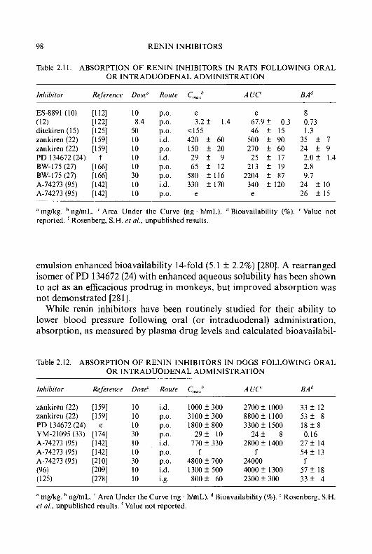
98 RENIN INHIBITORS
Table 2.11. ABSORPTION OF RENIN INHIBITORS IN RATS FOLLOWING ORAL OR INTRADUODENAL ADMINISTRATION
Inhibitor Reference Dose" Route C,,,": A U C BA"
ES-8891 (10) [ I 121 (12) [I221 ditekiren (15) [I251 zankiren (22) [I591 zankiren (22) [I591 PD 134672 (24) f BW-175 (27) [I661 BW-175 (27) [I661 A-74273 (95) [I421 A-74273 (95) [I421
10 p.0. 8.4 p.0.
50 p.0. 10 i.d. 10 p.0. 10 i.d. 10 p.0. 30 p.0. 10 i.d. 10 p.0.
e 3.2 f 1.4
<I55 420 f 60 150 f 20 29 f 9 65 f 12
580 f 116 330 f 170
e
e 61.9 f 0.3 46 f 15
500 f 90 270 f 60
25 f 17 213 f 19
2204 f 87 340 f 120 e
8 0.73 1.3
35 f 7 24 f 9
2.0 f 1.4 2.8 9.7
24 f 10 26 f 15
mg/kg. ng/mL. ' Area Under the Curve (ng . h/mL). reported. Rosenberg, S.H. et al., unpublished results.
Bioavailability (%). ' Value not
emulsion enhanced bioavailability 14-fold (5.1 k 2.2%) [280]. A rearranged isomer of PD 134672 (24) with enhanced aqueous solubility has been shown to act as an efficacious prodrug in monkeys, but improved absorption was not demonstrated [281].
While renin inhibitors have been routinely studied for their ability to lower blood pressure following oral (or intraduodenal) administration, absorption, as measured by plasma drug levels and calculated bioavailabil-
Table 2.12. ABSORPTION OF RENIN INHIBITORS IN DOGS FOLLOWING ORAL OR INTRADUODENAL ADMINISTRATION
Inhibiror Rejerence Dose" Route C,,,,,h A U C BA"
zankiren (22) [I591 10 i.d. I O O O f 300 zankiren (22) [159] 10 p.0. 3100f300 PD 134672 (24) e 10 p.0. 1800 f 800 YM-21095 (33) [I741 30 p.0. 2 9 f 10 A-74273 (95) [I421 10 , i.d. 770 f 330
A-74273 (95) [210] 30 p.0. 4800 f 700 (96) ~ 0 9 1 10 i.d. 1300 f 500 (125) ~ 7 8 1 10 i.g. 800 f 60
A-74273 (95) [I421 10 p.0. f
2700 f 1000 8800 f 1100 3300 f 1500
2 4 f 8 2800 f 1400
24000 4000 f 1300 2300 f 300
f
33 f 12 5 3 f 8 1 8 f 8 0.16
2 7 f 14 5 4 f 13
5 l f 18 3 3 f 4
f
* mg/kg. ng/mL. Area Under the Curve (ng . h/mL). Bioavailability (a). Rosenberg, S.H. et al., unpublished results. 'Value not reported.
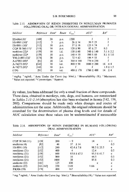
S.H. ROSENBERG 99
Table 2.13. ABSORPTION O F RENIN INHIBITORS IN NONHUMAN PRIMATES FOLLOWING ORAL OR INTRADUODENAL ADMINISTRATION
Inhibitor Rejerence Dose" Route C,,,,,h A U C B A ~
ES-6864 (9)' 11081 30 p.0. 1200 f f ES-8891 (10)' 11 121 3 p.0. 2 6 f 14 50 f 25 f ES-8891 (10)' 11 121 10 p.0. 5 7 2 18 I23 f 7 4 f CGP 38 560 (1 I)' [ I 191 10 p.0. 120f 90 85 f 27 0.3 zankiren (22)g [I591 10 i.d. 130 f 60 340 f 140 5.1 f 2.2 zankiren (22)g [I591 10 p.0. 140f 10 500 f 60 8.1 f 1.0 YM-21095 (33)h [I741 30 p.0. 7 2 2 4 2 106f 55 f A-67993 (48)g [811 10 i.d. 360 f 100 770 f 230 f A-74273 (95)' 11421 10 i.d. 4 0 0 f 9 0 1OOOf200 16 f 4 A-74273 (95)& 11421 10 p.0. f f 1.9 f 1.5 ( 124) P I 1 10 i.d. 480f 170 1700f400 14 f 4
mg/kg. ng/mL. Area Under the Curve (ng . h/mL). Bioavailability (%). Marmoset. 'Value not reported. Cynomolgus. Squirrel.
ity values, has been addressed for only a small fraction of these compounds. These data, obtained in monkeys, rats, dogs, and humans, are summarized in Tables 2.11-2.14 (absorption has also been evaluated in ferrets [142, 159, 2091). Comparisons should be made only when dosages and routes of administration are the same. Additionally, the original references should be consulted for the determination of plasma drug levels and for details of AUC calculation since these values can be underestimated if measurable
Table 2.14. ABSORPTION O F RENIN INHIBITORS IN HUMANS FOLLOWING ORAL ADMINISTRATION
Itdzibitor Reference Dose" c,,,, , A U C B A ~
CGP 38 560 (1 1) [I201 enalkiren (4) [821 ES-8891 (10) [ I 121
remikiren (21) 11581 ,
zdnkiren (22) [I611
remikiren (21) 11521
remikiren (21) 11521 remikiren (21) 11521
FK906 (29) ~ 7 0 1 zankiren (22) [ 1611162]
200 40
240 400 600 800
1600 125 250 400
- 9 37 f 14 42.4 f 7.6 4
83.0 f 47.5 16 38
430 1 I49 167
14 < I e 0.5
98.5 f 33.5 e 12.4 0.2 95.5 f 41.7 e 44.5 0.3 85.8 0.3
e e 1 I66 e
e e
mg. ng/mL. Area Under the Curve (ng h/mL). Bioavailability (%). ' Value not reported.
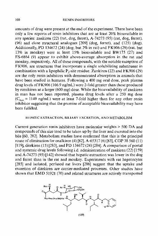
100 RENIN INHIBITORS
amounts of drug were present at the end of the experiment. There have been only a few reports of renin inhibitors that are at least 20% bioavailable in any species: zankiren (22) (rat, dog, ferret), A-74273 (95) (rat, dog, ferret), (96) and close structural analogues [209] (dog, ferret), and (125) (dog). Additionally, PD 134672 (24) (dog, but 2% in rat) and FK906 (29) (rat, but 13% in monkey) were at least 15% bioavailable and BW175 (27) and ES-6864 (9) appear to exhibit above-average absorption in the rat and monkey, respectively. All of these compounds, with the notable exception of FK906, are structures that incorporate a single solubilizing substituent in combination with a lipophilic P,-site residue. Zankiren (22) and FK906 (29) are the only renin inhibitors with demonstrated absorption in animals that have been studied in humans. Following a 400 mg oral dose, peak plasma drug levels of FK906 (166.9 ng/mL) were 2-fold greater than those produced by remikiren at a larger (600 mg) dose. While the bioavailability of zankiren in man has not been reported, plasma drug levels after a 250 mg dose (C,,, = 1149 ng/mL) were at least 7-fold higher than for any other renin inhibitor suggesting that the promise of acceptable bioavailability may have been fulfilled.
HEPATIC EXTRACTION, BlLlARY EXCRETION, AND METABOLISM
Current generation renin inhibitors have molecular weights > 500 DA and compounds of this size tend to be taken up by the liver and excreted into the bile [60, 2821. Metabolism studies have confirmed that this is the principal route of elimination for enalkiren (4) [82], A-65317 (6) [83], CGP 38 560 (1 1) [119], ditekiren (15) [283], and PD 134672 (24) [284]. A comparison of portal and systemic drug levels following i.d. administration of zankiren (22) [159] and A-74273 (95) [142] showed that hepatic extraction was lower in the dog and ferret than in the rat and monkey. Experiments with rat hepatocytes [285] and isolated, perfused rat livers [286] suggest that the uptake and excretion of ditekiren are carrier-mediated processes. Other studies have shown that EMD 51921 (70) and related structures are actively transported
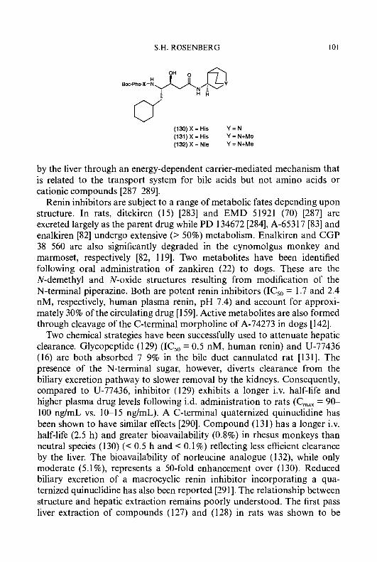
S.H. ROSENBERG 101
(130) X = His Y = N (131) X = His (132) X = Nle
Y = N+Me Y = N+Me
by the liver through an energy-dependent carrier-mediated mechanism that is related to the transport system for bile acids but not amino acids or cationic compounds [287-2891.
Renin inhibitors are subject to a range of metabolic fates depending upon structure. In rats, ditekiren (15) [283] and EMD 51921 (70) [287] are excreted largely as the parent drug while PD 134672 [284], A-653 I7 [83] and enalkiren [82] undergo extensive (> 50%) metabolism. Enalkiren and CGP 38 560 are also significantly degraded in the cynomolgus monkey and marmoset, respectively [82, 1 191. Two metabolites have been identified following oral administration of zankiren (22) to dogs. These are the N-demethyl and N-oxide structures resulting from modification of the N-terminal piperazine. Both are potent renin inhibitors (ICs0 = 1.7 and 2.4 nM, respectively, human plasma renin, pH 7.4) and account for approxi- mately 30% of the circulating drug [159]. Active metabolites are also formed through cleavage of the C-terminal morpholine of A-74273 in dogs [142].
Two chemical strategies have been successfully used to attenuate hepatic clearance. Glycopeptide (129) (IC50 = 0.5 nM, human renin) and U-77436 (16) are both absorbed 7-9% in the bile duct cannulated rat [131]. The presence of the N-terminal sugar, however, diverts clearance from the biliary excretion pathway to slower removal by the kidneys. Consequently, compared to U-77436, inhibitor (129) exhibits a longer i.v. half-life and higher plasma drug levels following i.d. administration to rats (C,,, = 90- 100 ng/mL vs. 10-15 ng/mL). A C-terminal quaternized quinuclidine has been shown to have similar effects [290]. Compound (131) has a longer i.v. half-life (2.5 h) and greater bioavailability (0.8%) in rhesus monkeys than neutral species (130) (< 0.5 h and < 0.1%) reflecting less efficient clearance by the liver. The bioavailability of norleucine analogue (1 32), while only moderate (5.1 %), represents a 50-fold enhancement over (130). Reduced biliary excretion of a macrocyclic renin inhibitor incorporating a qua- ternized quinuclidine has also been reported [29 11. The relationship between structure and hepatic extraction remains poorly understood. The first pass liver extraction of compounds (127) and (128) in rats was shown to be
102 RENIN INHIBITORS
61 f 3% and 8 ? 23%, demonstrating that apparently trivial structural modifications can profoundly affect in vivo behaviour [292].
INTRAPULMONARY DELIVERY
Intrapulmonary administration has been examined as an alternative to oral dosing for poorly absorbed renin inhibitors. Enhanced bioavailability (20 f. 4%) via this route of administration has been demonstrated for SC-46944 (126) in rats [293]. SR 43845 (8) reduced MAP 25% and 39% when given via the intratracheal route (0.1 and 1.0 mg/kg) to Na-depleted cynomolgus monkeys [294]. Maximum plasma drug levels of 6.3 f 1.4 and 117 f 8 ng/mL were achieved with these two doses. The duration of the hypotensive response (7 h) was longer than was observed following i.v. administration. An extended elimination half-life has also been reported following intratracheal administration of enalkiren (4) to dogs [295]. Studies in rats with U-77436 ( 1 6) showed that absorption from the lung was high ( 5 1 %) and that this was a saturable process since plasma drug levels reached a plateau (ca. 100 ng/mL) despite increasing intrapulmonary doses [296].
CONCLUSION
Remarkable progress has been made over the past decade. The design of tightly-binding transition-state mimics led rapidly to renin inhibitors with potencies in the nanomolar range. This was followed by the initial proof of in vivo activity and further studies to assess the scope of allowed substituents at the side-chains and termini. The subsequent development of inhibitors with significant oral bioavailability required an evaluation of structure- activity relationships that entailed several years and many thousand test compounds. This achievement marked the first demonstration that molecular-weight 600-800 peptides could provide high plasma drug levels following oral administration and has paved the way for other peptide- based drugs.
While greater in vivo activity would presumably be attained with even smaller, less peptidic structures, these will most likely require the discovery of an entirely new class of compounds that are not derived from the structure of the natural substrate. Thus, the question of whether a renin inhibitor will prove to be a viable drug will probably be answered by the outcome of clinical trials comparing the current generation of bioavailable compounds with ACE inhibitors with confirmed utility.
S.H. ROSENBERG 103
REFERENCES
1
2
3
4
5 6
7 8 9
10
11 12
13 14 15 16
17 18 19
20 21
22
23
24 25
26 27
28 29 30
Veterans Administration Cooperative Study Group on Antihypertensive Agents (1970) J. Am. Med. Assoc. 213, 1143-1 152. Beevers, D.G., Fairman, M.J., Hamilton, M. and Harpur, J.E. (1973) Lancet i, 1407-1409. Hypertension Detection and Follow-up Program Cooperative Group (1979) J. Am. Med. Assoc. 242,2562-2571. Multiple Risk Factor Intervention Trial Research Group (1982) J. Am. Med. Assoc. 248,
Medical Research Council Working Party (1985) Br. Med. J. 291, 97-104. Pickering, G. (1990) in Hypertension: Pathophysiology, Diagnosis, and Management (Laragh, J.H. and Brenner, B.M., eds.), pp. 3-16, Raven Press, Ltd., New York. Zusman, R.M. (1986) Hypertension 8, 837-842. Moser, M. (1986) Am. J. Med. 80 (Suppl. 5B), 1-1 1. Laragh, J.H. (1973) in Hypertension Manual (Laragh, J.H. ed.), pp. 823-849, Yorke Medical Books, New York. Amadio Jr., P., Cummings, D. M. and Amadio, P.B. (1986) Am. Fam. Phys. 34(2), 157-1 70. Tobian, L. (1967) Annu. Rev. Pharmacol. 7, 399408. Yokosawa, H., Holladay, L.A., Inagami, T., Haas, E. and Murakami, K. (1980) J. Biol. Chem. 255,3498-3502. Skinner, S.L., McCubbin, J.W. and Page, I.H. (1964) Circ. Res. 15, 6476 . Brown, J.J., Davies, D.L., Lever, A.F. and Robertson, J.I.S. (1963) Lancet ii, 278-279. Dzau, V.J., Burt, D.W. and Pratt, R.E. (1988) Am. J. Physiol. 255, F563-FS73. Shinagawa, T., Do, Y.S., Baxter, J.D., Carilli, C., Schilling, J. and Hsueh, W.A. (1990) Proc. Natl. Acad. Sci. U.S.A. 87, 1927-1931. Katz, S.A., Abraham, P.A. and Opsahl, J.A. (1992) Renal Physiol. Biochem. 15,240-248. Hosoi, M., Kim, S., Ikemoto, F. and Yamamoto, K. (1990) Life Sci. 47, 1903-1913. Katz, S.A., Malvin, R.L., Lee, J., Kim, S.H., Murray, R.D., Opsahl, J.A. and Abraham, P.A. (1991) Proc. SOC. Exp. Biol. Med. 197,387-392. Shier, D.N. and Malvin, R.L. (1985) Am. J. Physiol. 249, R79-R84. Malvin, R.L., Katz, S.A., Shier, D.N. and Sessler, F.M. (1984) Clin. Exp. Ther. Pract. A6, 1781-1786. Imai, T., Miyazaki, H., Hirosi, S., Hori, H., Hayashi, T., Kageyama, R., Ohkubo, H., Nakanishi, S. and Murakami, K. (1983) Proc. Natl. Acad. Sci. U S A . 80, 7405-7409. Carilli, C.T., Vigne, J.-L., Wallace, L.C., Smith, L.M., Wong. M.A., Lewicki, J.A. and Baxter, J.D. (1988) Hypertension 11, 713-716. Rahuel, J., Priestle, J.P. and Griitter, M.G. (1991) J. Struct. Biol. 107, 227-236. Sielecki, A.R., Hayakawa, K., Fujinaga, M., Murphy, M.E.P., Fraser, M., Muir, A.K., Carilli, C.T., Lewicki, J.A., Baxter, J.D. and James, M.N.G. (1989) Science (Washington, D.C.) 243, 13461351. Peach, M.J. (1977) Physiol. Rev. 57,313-370. Tewksbury, D.A., Dart, R.A. and Travis, J. (1981) Biochem. Biophys. Res. Commun. 99, 1311-1315. Griendling, K.K., Murphy, T.J. and Alexander, R.W. (1993) Circulation 87, 1816-1828. Morris, B.J. (1992) J. Hypertens. 10, 337-342. Wagner, J., Fernandez-Alfonso, M., Bachmann, J., Bader, M., Paul, M. and Ganten, D. (1993) Nutr. Metab. Cardiovasc. Dis. 3 , 197-203.
1465-1477.
104
31 32
33 34 35
36
37
38 39
40
41 42 43 44 45 46
47 48 49 50 51
52
53
54 55
56
57
58
59
60
61
62
RENIN INHIBITORS
Samani, N.J. (1993) Quart. J. Med. 86, 755-760. Brunner, H.R., Nussberger, J., Burnier, M. and Waeber, B. (1993) Clin. Exp. Hypertens. 15, 1221-1238. Carini, D.J. and Duncia, J.V. (1993) Adv. Med. Chem. 2, 153-195. Steinberg, M.I., Wiest, S.A. and Palkowitz, A.D. (1993) Card. Drug Rev. 11, 312-358. Dudley, D.T., Panek, R.L., Major, T.C., Lu, G.H., Bruns, R.F., Klinkefus, B.A., Hodges, J.C. and Weishaar, R.E. (1990) Mol. Pharmacol. 38, 37C377. de Gasparo, M., Whitebread, S., Mele, M., Motani, AS., Whitcombe, P.J., Ramjoue, H.-P. and Kamber, B. (1990) J. Cardiovasc. Pharmacol. 16 (Suppl. 4), S31-S35. Ohlstein, E.H., Gellai, M., Brooks, D.P., Vickery, L., Jugus, J., Sulpizio, A., Ruffolo Jr., R.R., Weinstock, J. and Edwards, R.M. (1992) J. Pharmacol. Exp. Ther. 262,595-601. Atkinson, A.B. and Robertson, J.I.S. (1979) Lancet ii, 836-839. Gavras, H., Waeber, B., Gavras, I., Biollaz, J., Brunner, H.R. and Davies, R.O. (1981) Lancet ii, 543-546. Rotmensch, H.H., Vlasses, P.H., Swanson, B.N., Irvin, J.D., Harris, K.E., Merrill, D.G. and Ferguson, R.K. (1984) Am. J. Cardiol. 53, 116-1 19. Cushman, D.W. and Ondetti, M.A. (1980) Prog. Med. Chem. 17.41-104. Toher, C.A. and Francis, G.S. (1993) Coron. Artery Dis. 4, 3743. Sunman, W. and Sever, P.S. (1993) Clin. Sci. 85, 661-670. Erdos, E.G. and Skidgel, R.A. (1986) Hypertension 8 (Suppl. I), 1-34-1-37. Erdos, E.G. (1990) Hypertension 16, 363-370. Grafe, M., Bossaller, C., Graf, K., Auch-Schwelk, W., Baumgarten, C.R., Hilderbrandt, A. and Fleck, E. (1993) Am. J. Physiol. 264, H1493-Hl497. Poole, M.D. and Postma, D.S. (1991) Otolaryngol. Head Neck Surg. 150, 714-716. Israili, Z.H. and Hall, W.D. (1992) Ann. Int. Med. 117, 236242. Karlberg, B.E. (1993) J. Hypertens. 1 1 (Suppl. 3), S49-S52. Gavras, H. and Gavras, I. (1988) Hypertension 11 (Suppl. II), 11-37-1141. Slater, E.E., Merrill, D.D., Guess, H.A., Roylance, P.J., Cooper, W.D., Inman, W.H.W. and Ewan, P.W. (1988) J. Am. Med. Assoc. 260,967-970. Noda, K., Sasaguri, M., Ideishi, M., Ikeda, M. and Arakawa, K. (1993) Cardiovasc. Res. 27,334340. Hartman, J.C., Hullinger, T.G., Wall, T.M. and Shebuski, R.J. (1993) Eur. J. Pharmacol. 234,229-236. Kay, J. (1985) Biochem. SOC. Trans. 13, 1027-1029. Kramer, R.A., Schaber, M.D., Skalka, A.M., Ganguly, K., Wong-Staal, F. and Reddy, E.P. (1986) Science (Washington, D.C.) 231, 1580-1584. Tomasselli, A.G., Howe, W.J., Sawyer, T.K., Wlodawer, A. and Heinrikson, R.L. (1991). Chim. Oggi. 6-27, Skeggs, L.T., Lentz, K.E., Kahn, J.R. and Hochstrasser, H. (1968) J. Exp. Med. 128, 13-34. Ondetti, M.A., Williams, N.J., Sabo, E.F., Pluscec, J., Weaver, E.R. and Kocy, 0. (1971), Biochemistry 10,40334039. Ondetti, M.A., Rubin, B. and Cushman, D.W. (1977) Science (Washington, D.C.) 196, 4 4 1 4 . Plattner, J.J. and Norbeck, D.W. (1990) In Drug Discovery Technologies (Clark, C.R. and Moos, W.H., eds.), pp. 92-126, Ellis Horwood Ltd., Chichester. Kokubu, T., Ueda, E., Fujimoto, S., Hiwada, K., Kato, A,, Akutsu, H., Yamamura, Y., Saito, S. and Mizoguchi, T. (1968) Nature (London) 217,45&457. James, M.N.G. and Sielecki, A.R. (1983) J. Mol. Biol. 163, 299-361.
63 Pauling, L. (1946) Chem. Eng. News 24, 1375-1377.
S.H. ROSENBERG 105
64 Wolfenden, R. (1972) Acc. Chem. Res. 5 , 10-18. 65 Lienhard, G.E. (1973) Science (Washington, D.C.) 180, 149-154. 66 Szelke, M., Jones, D.M., Atrash, B., Hallett, A. and Leckie, B.J. (1983) In Peptides,
Structure and Function. Proceedings of the Eighth American Peptide Symposium (Hruby, V.J. and Rich, D.H., eds.), pp. 579-582, Pierce Chemical Co., Rockford.
67 Workman, R.J. and Burkitt, D.W. (1979) Arch. Biochem. Biophys. 194, 157-164. 68 Marciniszyn Jr., J., Hartsuck, J.A. and Tang, J. (1976) J. Biol. Chem. 251, 7088-7094. 69 Rich, D.H. (1985) J. Med. Chem. 28,263-273. 70 Boger, J., Lohr, N.S., Ulm, E.H., Poe, M., Blaine, E.H., Fanelli, G.M., Lin, T.-Y., Payne,
L.S., Schorn, T.W., LaMont, B.I., Vassil, T.C., Stabilito, I . I . , Veber, D.F., Rich, D.H. and Bopari, A.S. (1983) Nature (London) 303,81-84. Boger, J., Payne, L.S., Perlow, D.S., Lohr, N.S., Poe, M., Blaine, E.H., Ulm, E.H., Schorn, T.W., LaMont, B.I., Lin, T.-Y., Kawai, M., Rich, D.H. and Veber, D.F. (1985) J. Med. Chem. 28, 1779-1790.
72 Greenlee, W.J. (1990) Med. Res. Rev. 10, 173-236. 73 Schechter, I. and Berger, A. (1967) Biochem. Biophys. Res. Commun. 27, 157-162. 74 Epps, D.E., Poorman, R.A., Mandel, F. and Schostarez, H.J. (1991) J. Med. Chem. 34,
2107-2112 75 Evans, B.E., Rittle, K.E., Bock, M.G., Bennett, C.D., DiPardo, R.M., Boger, J., Poe, M.,
Ulm, E.H., LaMont, B.I., Blaine, E.H., Fanelli, G.M., Stabilito, 1.1. and Veber, D.F. (1985) J. Med. Chem. 28, 1755-1756.
76 Dayton, B.D., Stein, H.H., Cohen, J., Baker, W.R., Boyd, S.A., Condon, S.L., Donner, B.G., Fung, A.K.L., Luly, J.R., Rosenberg, S.H., Woods, K.W. and Kleinert, H.O. (1992) Clin. Chem. 38,2239-2243.
77 Fischli, W., Clozel, J.-P., Amrani, K.E., Wostl, W., Neidhart, W., Stadler, H. and Branca, Q. (1991) Hypertension 18, 22-31,
78 Nisato, D., Lacour, C., Roccon, A., Gayraud, R., Cazaubon, C., Carlet, C., Plouzane, C., Richaud, J.-P.,Tonnerre, B., Gagnol, J.-P. and Wagnon, J. (1987) J. Hypertens. 5 (Suppl. 5) , S23-S25. Iizuka, K., Kamijo, T., Harada, H., Akahane, K., Kubota, T., Etoh, Y., Shimaoka, I., Tsubaki, A,, Murakami, M., Yamaguchi, T., Iyobe, A., Umeyama, H. and Kiso, Y. (1990) Chem. Pharm. Bull. 38,2487-2493.
80 Stammers, D.K., Dann, J.G., Harris, C.J. and Smith, D.R. (1987) Arch. Biochem. Biophys. 258,413420.
81 Rosenberg, S.H., Spina, K.P., Woods, K.W., Polakowski, J., Martin, D.L., Yao, Z., Stein, H.H., Cohen, J., Barlow, J.L., Egan, D.A., Tricarico, K.A., Baker, W.R. and Kleinert, H.D. (1993) J. Med. Chem. 36,449459.
82 Kleinert, H.D., Luly, J.R., Bopp, B.A., Verburg, K.M., Hoyos, P.A., Karol, M.D., Plattner, J.J., Luther, R.R. and Stein, H.H. (1990) Cardiovasc. Drug Rev. 8, 203-219.
83 Rosenberg, S.H., Woods, K.W., Sham, H.L., Kleinert, H.D., Martin, D.L., Stein, H., Cohen, J., Egan, D.A., Bopp, B., Merits, I., Garren, K.W., Hoffman, D.J. and Plattner, J.J. (1990) J. Med. Chem. 33, 1962-1969.
84 Barrett, J.D., Eggena, P. and Krall, J.F. (1988) Clin. Exp. Ther. Pract. AIO, 757-765. 85 Clozel, J.-P., Fischli, W. and MCnard, J. (1993) J. Hypertens. 1 I , 75-81. 86 Pals, D.T., Lawson, J.A. and Couch, S.J. (1990) J. Pharmacol. Methods 23, 239-245. 87 Blaine, E.H., Schorn, T.W. and Boger, J. (1984) Hypertension 6 (Suppl. I), 1-1 11-1-1 18. 88 Derkx, F.H.M., van den Meiracker, A.H., Fischli, W., Admiraal, P.J.J., Man in 't Veld,
A.J., van Brummelen, P. and Schalekamp, M.A.D.H. (1991) Am. J. Hypertens. 4,
71
79
602-609.
106 RENIN INHIBITORS
89
90
91 92
Nussberger, J., Delabays, A,, De Gasparo, M., Cumin, F., Waeber, B., Brunner, H.R. and Menard, J. (1989) Hypertension 13,948-953. Palmer, R.K., Rapundalo, S.T., Batley, B.L., Barnes, A.E., Singh, S., Ryan, M.J. and Taylor Jr., D.G. (1993) Clin. Exp. Hypertens. 15,663-681. Bunnemann, B., Fuxe, K. and Ganten, D. (1993) Regul. Pept. 46,487-509. Paul, M., Bader, M., Steckelings, U.M., Voigtlander, T. and Ganten, D. (1993) Arzneim.-Forsch. 43 (I), 207-213.
93 Swales, J.D. and Samani, N.J. (1990) Am. J. Hypertens. 3, 890-892. 94 Okamura, T., Aimi, Y., Kimura, H., Murakami, K. and Toda, N. (1992) J. Hypertens.
10.49-53. 95 Lindpaintner, K. and Ganten, D. (1991) Circ. Res. 68,905-921. 96 Lee, Y.-A. and Lindpaintner, K. (1993) Arzneim.-Forsch. 43 (I), 201-206. 97 Mulrow, P.J. (1992) Front. Neuroendocrinol 13,47-60. 98 Mulrow, P.J. (1991) Curr. Opin. Cardiol. 6, 693-696. 99 von Lutterotti, N., Catanzaro, D.F., Sealey, J.E. and Laragh, J.H. (1994) Circulation 89,
458470. 100 Danser, A.H.J., Admiraal, P.J.J., Derkx, F.H.M., Lamers, J.M.J., Verdouw, P.D.,
Saxena, P.R. and Schalekamp, M.A.D.H. (1993) J. Hypertens. 11 (Suppl. 5), S224S225. 101 Inagami, T., Murakami, T., Higuchi, K. and Nakajo, S. (1991) Am. J. Hypertens. 4,
102 Weishaar, R.E., Panek, R.L., Major, T.C., Simmerman, J., Rapundalo, S.T. and Taylor Jr., D.G. (1991) J. Pharmacol. Exp. Ther. 256,568-574.
103 Higashimori, K., Gante, J., Holzemann, G. and Inagami, T. (1991) Hypertension 17, 270-277.
104 Kleinert, H.D., Martin, D., Chekal, M.A., Kadam, J., Luly, J.R., Plattner, J.J., Perun, T.J. and Luther, R.R. (1988) Hypertension 1 I , 613-619.
105 Kleinert, H.D., Martin, D., Chekal, M., Young, G., Rosenberg, S., Plattner, J.J. and Perun, T.J. (1988) J. Pharmacol. Exp. Ther. 246, 975-979.
106 Schaffer, L.W., Schorn, T.W., Winquist, R.J., Strouse, J.F., Payne, L., Chakravarty, P.K., de Laszlo, S.E., tenBroeke, J., Veber, D.F., Greenlee, W.J. and Siegl, P.K.S. (1990) J. Hypertens. 8, 251-259.
107 Denolle, T., Luo, P., Guyene, T.T., Cazaubon, C., Sissmann, J., Corvol, P. and Menard, J. (1993) Arzneim.-Forsch. 43 (I), 255-259.
108 Hiwada, K., Kokubu, T., Murakami, E., Muneta, S., Morisawa, Y., Yabe, Y., Koike, H. and Iijima, Y. (1988) Hypertension 1 I , 708-712.
109 Rosenberg, S.H., Plattner, J.J., Woods, K.W., Stein, H.H., Marcotte, P.A., Cohen, J. and Perun, T.J. (1987) J. Med. Chem. 30, 12241228.
110 Rosenberg, S.H., Kleinert, H.D., Stein, H.H., Martin, D.L., Chekal, M.A., Cohen, J., Egan, D.A., Tricarico, K.A. and Baker, W.R. (1 991) J. Med. Chem. 34,46947 1.
111 Nishi, T., Saito, F., Nagahori, H., Kataoka, M., Morisawa, Y., Yabe, Y., Sakurai, M., Higashida, S., Shoji, M., Matsushita, Y., Iijima, Y., Ohizumi, K. and Koike, H. (1990) Chem. Pharm. Bull. 38, 103-109.
112 Kokubu, T., Hiwada, K., Murakami, E., Muneta, S., Kitami, Y., Oizumi, K., Takahagi, H. and Koike, H. (1991) Cardiovasc. Drug Rev. 9.49-58.
113 Kokubu, T., Hiwada, K., Murakami, E., Muneta, S., Kitami, Y. and Salmon, P.F. (1990) Hypertension 15, 909-91 3.
114 Nussberger, J., Waeber, G., Waeber, B., Bidiville, J. and Brunner, H.R. (1988) J. Cardiovasc. Pharmacol. 1 1, 71 6-721.
15s-22s.

S.H. ROSENBERG 107
115 Doherty, A.M., Kaltenbronn, J.S., Hudspeth, J.P., Repine, J.T., Roark, W.H., Sircar, I . , Tinney, F.J., Connolly, C.J., Hodges, J.C., Taylor, M.D., Batley, B.L., Ryan, M.J., Essenburg, A.D., Rapundalo, S.T., Weishaar, R.E., Humblet, C. and Lunney, E.A. (1991) J. Med. Chem. 34, 1258-1271.
116 Repine, J.T., Kaltenbronn, J.S., Doherty, A.M., Hamby, J.M., Himmelsbach, R.J., Kornberg, B.E., Taylor, M.D., Lunney, E.A., Humblet, C., Rapundalo, S.T., Batley, B.L., Ryan, M.J. and Painchaud, C.A. (1992) J. Med. Chem. 35, 1032-1042. Buhlmayer, P., Caselli, A., Fuhrer, W., Goschke, R., Rasetti, V., Rueger, H., Stanton, J.L., Criscione, L. and Wood, J.W. (1988) J. Med. Chem. 31, 1839-1846. Wood, J.M., Criscione, L., de Gasparo, M., Buhlmayer, P., Riieger, H., Stanton, J.L., Jupp, R.A. and Kay, J. (1989) J. Cardiovasc. Pharmacol. 14, 221-226. Cumin, F., Schnell, C., Richert, P., Raschdorf, F., Probst, A,, Schmid, K., Wood, J.M. and De Gasparo, M. (1990) Drug Metab. Disp. 18, 831-835. De Gasparo, M., Cumin, F., Nussberger, J., Guyenne, T.T., Wood, J.M. and Menard, J. (1989) Br. J. Pharmacol. 27, 587-596. Chauveau, D., Guyenne, T.T., Cumin, F., Chatellier, G., Corvol, P. and Menard, J. (1992) Br. J. Pharmacol. 33,253-260. Atsuumi, S., Nakano, M., Koike, Y., Tanaka, S., Funabashi, H., Matsuyama, K., Nakano, M., Sawasaki, Y., Funabashi, K. and Morishima, H. (1992) Chem. Pharm. Bull. 40, 32163221.
123 Pals, D.T., Thaisrivongs, S., Lawson, J.A., Kati, W.M., Turner, S.R., DeGraaf, G.L., Harris, D.W. and Johnson, G.A. (1986) Hypertension 8, 1105-1 112.
124 Thaisrivongs, S., Pals, D.T., DuCharme, D.W., Turner, S.R., DeGraaf, G.L., Lawson, J.A., Couch, S.J. and Williams, M.V. (1991) J. Med. Chem. 34, 633-642.
125 Rush, B.D., Wilkinson, K.F., Zhong, W.Z., Closson, S.K., Lakings, D.B. and Ruwart, M.J. (1991) Int. J. Pharm. 73, 231-237.
126 Pals, D.T., DeGraaf, G.L., Couch, S.J. and Brunden, M.N. (1991)Clin. Exp. Ther. Pract. A13,425436.
127 Luderer, J., Froeschke, M., Wathen, L., Harrelson, W., Bryan, W., Bokros, J., Mohrland, S. and Metzler, C. (1990) Clin. Pharmacol. Ther. 47, 200.
128 Ferry, J.J., Lakings, D.B., Closson, S.K. and Luderer, J.R. (1990) Clin. Pharmacol. Ther. 47, 157.
129 Taylor, A.A., Piwinski, S.E., Varghese, A., Kitt, T., Luderer, J. and Pool, J.L. (1990) Circulation 82 (Suppl. HI), 111-555.
130 Bundy, G.L., Pals, D.T., Lawson, J.A., Couch, S.J., Lipton, M.F. and Mauragis, M.A. (1990) J. Med. Chem. 33,2276-2283.
131 Fisher, J.F., Harrison, A.W., Bundy, G.L., Wilkinson, K.F., Rush, B.D. and Ruwart, M.J. (1991) J. Med. Chem. 34, 3140-3143.
132 Atsuumi, S., Nakano, M., Koike, Y., Tanakd, S., Matsuyama, K., Nakano, M. and Morishima, H. (1992) Chem. Pharm. Bull. 40, 364370.
133 Metternich, R. and Ludi, W. (1988) Tetrahedron Lett. 29, 3923-3926. 134 Gardner, C.J., Twissell, D.J., Charlton, P.A. and Cherry, P.C. (1991) Eur. J. Pharmacol.
192, 329-335. 135 Etoh, Y., Miyazaki, M., Saitoh, H. and Toda, N. (1993) Jpn. J. Pharmacol. 63, 109-119. 136 Patel, D.V., Rielly-Gauvin, K., Ryono, D.E., Free, C.A., Smith, S.A. and Petrillo Jr.,
E.W. (1993) J. Med. Chem. 36,2431-2447. 137 Fossa, A.A., Weinberg, L.J., Barber, R.L., Rauch, A.L., Nocerini, M.R., Murphy, W.R.
and Swindell, A.C. (1992) FASEB J. 6, A1871. 138 Simpson, K.A., Perkins, T.J., Wilder, D.E., Ellery, S.S., Rauch, A.L., Holt, W.F.,
Murphy, W.R. and Kiron, M.A.R. (1993) FASEB J. 7, A765.
117
118
119
120
121
122
108 RENIN INHIBITORS
139 Abernethy, D.R., Wilner, K.D., Lazar, J.D. and Wilkes, B.M. (1990) Clin. Pharmacol. Ther. 47, 141.
140 Luly, J.R., BaMaung, N., Soderquist, J., Fung, A.K.L., Stein, H., Kleinert, H.D., Marcotte, P.A., Egan, D.A., Bopp, B., Merits, I., Bolis, G., Greer, J., Perun, T.J. and Plattner, J.J. (1988) J. Med. Chem. 31, 22642276. Plattner, J.J., Marcotte, P.A., Kleinert, H.D., Stein, H.H., Greer, J., Bolis, G., Fung, A.K.L., Bopp, B.A., Luly, J.R., Sham, H.L., Kempf, D.J., Rosenberg, S.H., Dellaria, J.F., De, B., Merits, I. and Perun, T.J. (1988) J. Med. Chem. 31,2277-2288. Kleinert, H.D., Stein, H.H., Boyd, S., Fung, A.K.L., Baker, W.R., Verburg, K.M., Polakowski, J.S., Kovar, P., Barlow, J., Cohen, J., Klinghofer, V., Mantei, R., Cepa, S., Rosenberg, S. and Denissen, J.F. (1992) Hypertension 20, 768-775.
143 Delabays, A,, Nussberger, J., Porchet, M., Waeber, B., Hoyos, P., Boger, R., Glassman, H., Kleinert, H.D., Luther, R. and Brunner, H.R. (1989) Hypertension 13,941-947.
144 Bursztyn, M., Gavras, I., Tifft, C.P., Luther, R., Boger, R. and Gavras, H. (1990) J. Cardiovasc. Pharmacol. 15,493-500.
145 Weber, M.A., Neutel, J.M., Essinger, I . , Glassman, H.N., Boger, R.S. and Luther, R. (1990) Circulation 81, 1768-1 774.
146 Neutel, J.M., Luther, R.R., Boger, R.S. and Weber, M.A. (1991) Am. Heart J. 122, 1094-1100.
147 Boger, R.S., Glassman, H.N., Cavanaugh, J.H., Schmitz, P.J., Lamm, J., Moyse, D., Cohen, A,, Kleinert, H.D. and Luther, R.R. (1990) Hypertension 15, 835-840.
148 Gupta, S.K., Granneman, G.R., Boger, R.S., Hollenberg, N.K. and Luther, R.R. (1992) Drug Metab. Dispos. 20, 821-825.
149 Gupta, S.K., Granneman, G.R., Packer, M. and Boger, R.S. (1993) J. Cardiovasc. Pharmacol. 21,834-840.
150 Clozel, J.-P. and Fischli, W. (1993) Hypertension 22, 9-17. 151 Heitsch, H., Henning, R., Kleemann, H.-W., Linz, W., Nickel, W.-U., Ruppert, D.,
Urbach, H. and Wagner, A. (1993) J. Med. Chem. 36,2788-2800. 152 Kleinbloesem, C.H., Weber, C., Fahrner, E., Dellenbach, M., Welker, H., Schroter, V.
and Belz, G.G. (1993) Clin. Pharmacol. Ther. 53, 585-592. 153 Camenzind, E., Nussberger, J., Juillerat, L., Munafo, A,, Fischli, W., Coassolo, P., van
Brummelen, P., Kleinbloesem, C.H., Waeber, B. and Brunner, H.R. (1991) J. Cardiovasc. Pharmacol. 18,299-307.
154 vanden Meiracker, A.H., Admiraal, P.J.J., Man in 't Veld, A.J., Derkx, F.H.M., Ritsema van Eck, H.J., Mulder, P., van Brummelen, P. and Schalekamp, M.A.D.H. (1990) Br. Med. J. 301,205-210.
155 Kobrin, I., Viskoper, R.J., Laszt, A., Bock, J., Weber, C. and Charlon, V. (1993) Am. J. Hypertens. 6, 349-356.
156 van den Meiracker, A.H., Admiraal, P.J.J., Derkx, F.H.M., Kleinbloesem, C., Man in 't Veld, A.J., van Brummelen, P., Mulder, P. and Schalekamp, M.A.D.H. (1993) J. Hypertens. 11, 831-838.
157 Rongen, G.A., Lenders, J.W.M., Kleinbloesem, C.H., Weber, C., Welker, H., Fahrner, E., Pozenel, H., Woittiez, A.J.J., Haug, G., Buchmann, M.S., Hoglund, C.E.K. and Thien, T. (1993) Clin. Pharmacol. Ther. 54, 567-577. Weber, C., Birnbock, H., Luebe, J., Kobrin, I . , Kleinbloesem, C.H. and van Brummelen, P. (1993) Br. J. Clin. Pharmacol. 36, 547-554. Kleinert, H.D., Rosenberg, S.H., Baker, W.R., Stein, H.H., Klinghofer, V., Barlow, J., Spina, K., Polakowski, J., Kovar, P., Cohen, J. and Denissen, J. (1992) Science (Washington, D.C.) 257, 1940-1943.
141
142
158
159
S.H. ROSENBERG 109
160 Rosenberg, S.H., Spina, K.P., Condon, S.L., Polakowski, J., Yao, Z., Kovar, P., Stein, H.H., Cohen, J., Barlow, J.L., Klinghofer, V., Egan, D.A., Tricarico, K.A., Perun, T.J., Baker, W.R. and Kleinert, H.D. (1993) J. Med Chem. 36,46&467.
161 Boger, R.S., Glassman, H.N., Thys, R., Gupta, S.K., Hippensteel, R.L. and Kleinert, H.D. (1993) Am. J. Hypertens. 6, 103A.
162 Boger, R.S., MCnard, J., Manski, R.P., Guyene, T.T., Luther, R.R., Kleinert, H.D. and Glassman, H.N. (1993) Clin. Pharmacol. Ther. 53, 140.
163 Nelson, D.E., Moyse, D.M., O’Neil, J.M., Boger, R.S., Glassman, H.N. and Kleinert, H.D. (1993) Circulation 88, 361.
164 Patt, W.C., Hamilton, H.W., Taylor, M.D., Ryan, M.J., Taylor Jr., D.G., Connolly, C.J.C., Doherty, A.M., Klutchko, S.R., Sircar, I . , Steinbaugh, B.A., Batley, B.L., Painchaud, C.A., Rapundalo, S.T., Michniewicz, B.M. and Olson, S.C. (1992) J. Med. Chem. 35,2562-2572. Repine, J.T., Himmelsbach, R.J., Hodges, J.C., Kaltenbronn, J.S., Sircar, I., Skeean, R.W., Brennan, S.T., Hurley, T.R., Lunney, E., Humblet, C.C., Weishaar, R.E., Rapundalo, S., Ryan, M.J., Taylor Jr., D.G., Olson, S.C., Michniewicz, B.M., Kornberg, B.E., Belmont, D.T. and Taylor, M.D. (1991) J. Med. Chem. 34, 1935-1943. Morishima, H., Koike, Y., Nakano, M., Atsuumi, S., Tanaka, S., Funabashi, H., Hashimoto, J., Sawasaki, Y., Mino, N., Nakano, M., Matsushima, K., Nakamichi, K. and Yano, M. (1989) Biochem. Biophys. Res. Commun. 159,999-1005. Poss, M.A., Reid, J.A., Free, C.A., Rogers, W.L., Weber, H., Ryono, D.E., Dejneka, T., DeForrest, J.M., Waldron, T.L., Brittain, R.J., Weller, H.N., Cimarusti, M.P. and Petrillo, E.W. (1993) Bioorg. Med. Chem. Lett. 3, 2739-2744. Kleemann, H.-W., Heitsch, H., Henning, R., Kramer, W., Kocher, W., Lerch, U., Linz, W., Nickel, W.-U., Ruppert, D., Urbach, H., Utz, R., Wagner, A,, Weck, R. and Wiegand, F. (1992) J. Med. Chem. 35, 559-567.
169 Ogihara, T., Nagano, M., Higaki, J., Higashimori, K., Masuo, K. and Mikami, H. (1993) Clin. Ther. 15, 539-548.
170 Anon. (1993) Drugs Future 18,312-315. 171 Andoh, T.F., Sogabe, K., Nagatomi, I . , Shimazaki, Y., Horiai, H., Mori, J. and
Kohsaka, M. (1991) Am. J. Hypertens. 4,31A. 172 Breipohl, G., Geiger, R., Henke, S., Kleeman, H.W., Knolle, J., Ruppert, D., Scholkens,
B.A., Urbach, H., Wagner, A. and Wegmann, H. (1988) Spec. Publ.-R. SOC. Chem. 65 (top. med. chem.) 101-127.
173 Shibasaki, M., Asano, M., Fukunaga, Y., Usui, T., Ichihara, M., Murakami, Y., Nakano, K. and Fujikura, T. (1991) Am. J. Hypertens. 4, 932-938.
174 Shibasaki, M., Usui, T., Inagaki, O., Asano, M. and Takenaka, T. (1994) J. Pharm. Pharmacol. 46, 68-72.
175 Ashton, W.T., Cantone, C.L., Meurer, L.C., Tolman, R.L., Greenlee, W.J., Patchett, A.A., Lynch, R.J., Schorn, T.W., Strouse, J.F. and Siegl, P.K.S. (1992) J. Med. Chem. 35, 2103-2112.
176 Raddatz, P., Jonczyk, A,, Minck, K.-O., Rippmann, F., Schittenhelm, C. and Schmitges, C.J. (1992) J. Med. Chem. 35, 3525-3536.
177 DeForrest, J.M., Waldron, T.L., Oehl, R.S., Scalese, R.J., Free, C.A., Weller, H.N. and Ryono, D.E. (1989) J. Hypertension (Suppl2), S15-S19.
178 Rosenberg, S.H., Dellaria, J.F., Kempf, D.J., Hutchins, C.W., Woods, K.W., Maki, R.G., de Lara, E., Spina, K.P., Stein, H.H., Cohen, J., Baker, W.R., Plattner, J.J., Kleinert, H.D. and Perun, T.J. (1990) J. Med. Chem. 33, 1582-1590.
165
166
167
168
110 RENIN INHIBITORS
179 Williams, P.D., Perlow, D.S., Payne, L.S., Holloway, M.K., Siegl, P.K.S., Schorn, T.W., Lynch, R.J., Doyle, J.J., Strouse, J.F., Vlasuk, G.P., Hoogsteen, K., Springer, J.P., Bush, B.L., Halgren, T.A., Richards, A.D., Kay, J. and Veber, D.F. (1991) J. Med. Chem. 34, 8 87-900.
180 Rosenberg, S.H., Spina, K.P., Stein, H., Cohen, J., Baker, W.R. and Kleinert, H.D. (1994) Bioorg. Med. Chem. 2,927-937.
181 Patel, D.V., Rielly-Gauvin, K. and Ryono, D.E. (1990) Tetrahedron Lett. 31, 5587-5590. 182 Dellaria Jr., J.F., Maki, R.G., Stein, H.H., Cohen, J., Whittern, D., Marsh, K., Hoffman,
D.J., Plattner, J.J. and Perun, T.J. (1990) J. Med. Chem. 33, 534542. 183 Bartlett, P.A. and Kezer, W.B. (1984) J. Am. Chem. SOC. 106,42824283. 184 Allen, M.C., Fuhrer, W., Tuck, B., Wade, R. and Wood, J.M. (1989) J. Med. Chem. 32,
1652-1 661. 185 Doherty, A.M., Sircar, I., Kornberg, B.E., Quin 111, J., Winters, R.T., Kaltenbronn, J.S.,
Taylor, M.D., Batley, B.L., Rapundalo, S.R., Ryan, M.J. and Painchaud, C.A. (1 992) J. Med. Chem. 35,2-14.
186 Tarnus, C., Jung, M.J., RCmy, J.-M., Baltzer, S. and Schirlin, D.G. (1989) FEBS Lett. 249,47-50.
187 Schirlin, D., Tarnus, C., Baltzer, S. and Remy, J.M. (1992) Bioorg. Med. Chem. Lett. 2, 651-654.
188 Rosenberg, S.H., Dellaria, J., Fung, A.K.L., Kempf, D.J., Luly, J.R. and Plattner, J.J. (1989) U.S. Patent 4,857,507.
189 Sham, H.L., Rempel, C.A., Stein, H. and Cohen, J. (1990) J. Chem. SOC., Chem. Commun. 904905.
190 Rao, C.M., Scarborough, P.E., Kay, J., Batley, B., Rapundalo, S., Klutchko, S., Taylor, M.D., Lunney, E.A., Humblet, C.C. and Dunn, B.M. (1993) J. Med. Chem. 36, 26142620. Lunney, E.A., Hamilton, H.W., Hodges, J.C., Kaltenbronn, J.S., Repine, J.T., Badasso, M., Cooper, J.B., Dealwis, C., Wallace, B.A., Lowther, W.T., Dunn, B.M. and Humblet, C. (1993) J. Med. Chem. 36, 3809-3820.
192 Ashton, W.T., Cantone, C.L., Tolman, R.L., Greenlee, W.J., Lynch, R.J., Schorn, T.W., Strouse, J.F. and Siegl, P.K.S. (1992) J. Med. Chem. 35, 2772-2781.
193 Hutchins, C. and Greer, J. (1991) CRC Crit. Rev. Biochern. Mol. Biol. 26, 77-127. 194 Kaltenbronn, J.S., Hudspeth, J.P., Lunney, E.A., Michniewicz, B.M., Nicolaides, E.D.,
Repine, J.T., Roark, W.H., Stier, M.A., Tinney, F.J., Woo, P.K.W. and Essenburg, A.D. (1990) J. Med. Chem. 33,838-845.
195 Kempf, D.J., de Lara, E., Stein, H.H., Cohen, J., Egan, D.A. and Plattner, J.J. (1990) J. Med. Chem. 33,371-374.
196 Weidmann, B. (1991) Chimia 45,367-376. 197 Raddatz, P., Jonczyk, A,, Minck, K.-O., Schmitges, C.J. and Sombroek, J. (1991) J. Med.
Chem. 34,3267-3280. 198 Rivero, R.A., Greenlee, W.J. and Patchett, A.A. (1991) Tetrahedron Lett. 32, 5263-5264. 199 Raddatz, P., Minck, K.-O., Rippmann, F. and Schmitges, C.-J. (1994) J. Med. Chem. 37,
486-497. 200 Patel, D.V. and Ryono; D.E. (1992) Bioorg. Med. Chem. Lett. 2, 1089-1092. 201 Gante, J. and Kahlenberg, H. (1989) Liebigs Ann. Chem. 1085-1087. 202 Gante, J. and Weitzel, R. (1990) Liebigs Ann. Chem. 349-350. 203 Gante, J. and Kahlenberg, H. (1991) Chem. Z. 115,215-217. 204 Smith 111, A.B., Hirschmann, R., Pasternak, A,, Akaishi, R., Guzman, M.C., Jones,
D.R., Keenan, T.P., Sprengeler, P.A., Darke, P.L., Emini, E.A., Holloway, M.K. and Schleif, W.A. (1994) J. Med. Chem. 37,215-218.
191
S.H. ROSENBERG 111
205 Thaisrivongs, S., Mao, B., Pals, D.T., Turner, S.R. and Kroll, L.T. (1990) J. Med. Chem. 33, 1337-1343.
206 Baker, W.R., Fung, A.K.L., Kleinert, H.D., Stein, H.H., Plattner, J.J., Armiger, Y.-L., Condon, S.L., Cohen, J. , Egan, D.A., Barlow, J.L., Verburg, K.M., Martin, D.L., Young, G.A., Polakowski, J.S., Boyd, S.A. and Perun, T.J. (1992) J. Med. Chem. 35,
207 Stein, H.H., Fung, A.K.L., Cohen, J., Baker, W.R., Rosenberg, S.H., Boyd, S.A., Dayton, B.D., Armiger, Y.-L.S.-T., Condon, S.L., Mantei, R.A. and Kleinert, H.D. (1992) FEBS Lett. 300, 301-304.
208 Boyd, S.A., Fung, A.K.L., Baker, W.R., Mantei, R.A., Armiger, Y.-L., Stein, H.H., Cohen, J., Egan, D.A., Barlow, J., Klinghofer, V., Verburg, K.M., Martin, D.L., Young, G.A., Polakowski, J.S., Hoffman, D.J., Garren, K.W., Perun, T.J. and Kleinert, H.D. (1992) J. Med. Chem. 35, 1735-1746.
209 Boyd, S.A., Fung, A.K.L., Baker, W.R., Mantei, R.A., Stein, H.H., Cohen, J., Barlow, J.L., Klinghofer, V., Wessale, J.L., Verburg, K.M., Polakowski, J.S., Adler, A.L., Calzadilla, S.V., Kovar, P., Yao, Z., Hutchins, C.W., Denissen, J.F., Grabowski, B.A., Cepa, S., Hoffman, D.J., Garren, K.W. and Kleinert, H.D.J. Med. Chem. 37,2991-3007.
210 Verburg, K.M., Polakowski, J.S., Kovar, P.J., Klinghofer, V., Barlow, J.L., Stein, H.H., Mantei, R.A., Fung, A.K.L., Boyd, S.A., Baker, W.R. and Kleinert, H.D. (1993) J. Cardiovasc. Pharmacol. 21, 149-1 55. Ocain, T.D., Deininger, D.D., Russo, R., Senko, N.A., Katz, A,, Kitzen, J.M., Mitchell, R., Oshiro, G., Russo, A., Stupienski, R. and McCaully, R.J. (1992) J. Med. Chem. 35,
212 Thaisrivongs, S., Pals, D.T., Turner, S.R. and Kroll, L.T. (1988) J. Med. Chem. 31, 1369-1 376.
213 Zydowsky, T.M., Dellaria, J.F., de Lara, E., Nellans, H.N., Baker, W.R., Barlow, J . , Calzadilla, S., Cohen, J., Kleinert, H., Klinghofer, V., Polakowski, J., Stein, H., Verburg, K. and Perun, T.J. (1992) Abstracts of Papers, XIIth International Symposium on Medicinal Chemistry, Switzerland, September 13-1 7, p. 66.
214 De, B., Dellaria, J.F., Baker, W.R., Zydowski, T.M., Rosenberg, S.H. and Jae, H.S. (1990) Eur. Patent Appl. 365,992; (1991) Chem. Abstr. 114,43580.
2 15 de Laszlo, S.E., Bush, B.L., Doyle, J. J., Greenlee, W.J., Hangauer, D.G., Halgren, T.A., Lynch, R.J., Schorn, T.W. and Siegl, P.K.S. (1992) J. Med. Chem. 35, 833-846.
216 Kempf, D.J., Condon, S.L., Plattner, J.J., Stein, H.H., Cohen, J., de Lara, E., Baker, W.R. and Kleinert, H.D. (1990) in Peptides, Chemistry, Structure and Biology. Proceedings of the Eleventh American Peptide Symposium (Rivier, J.E. and Marshall, G.R., eds.), pp. 393-395, ESCOM, Leiden. Roberts, D.A., Bradbury, R.H., Brown, D., Faull, A,, Griffiths, D., Major, J.S., Oldham, A.A., Pearce, R.J., Ratcliffe, A.H., Revill, J. and Waterson, D. (1990) J. Med. Chem. 33, 2326-2334. Bock, M.G., DiPardo, R.M., Evans, B.E., Freidinger, R.M., Rittle, K.E., Payne, L.S., Boger, J., Whitter, W.L., LaMont, B.I., Ulm, E.H., Blaine, E.H., Schorn, T.W. and Veber, D.F. (1988) J. Med. Chem. 31, 1918-1923. Bradbury, R.H., Major, J.S., Oldham, A.A., Rivett, J.E., Roberts, D.A., Slater, A.M., Timms, D. and Waterson, D. (1990) J. Med. Chem. 33, 2335-2342. Bradbury, R.H. and Rivett, J.E. (1991) J. Med. Chem. 34, 151-157. Martin, S.F., Austin, R.E., Oalmann, C.J., Baker, W.R., Condon, S.L., de Lara, E., Rosenberg, S.H., Spina, K.P., Stein, H.H., Cohen, J. and Kleinert, H.D. (1992) J. Med. Chem. 35, 1710-1721.
1722-1734.
211
823-832.
217
218
219
220 221

112 RENIN INHIBITORS
222 Baker, W.R., Jae, H.-S., Martin, S.F., Condon, S.L., Stein, H.H., Cohen, J. and Kleinert, H.D. (1992) Bioorg. Med. Chem. Lett. 2, 1405-1410.
223 Weidmann, B. (1992) Chimia 46,312-313. 224 Grevel, J. (1986) Transplant. Proc. 18 (Suppl. 5), 9-15. 225 Tufveson, G., Odlind, B., Sjoberg, O., Lindberg, A., Gabrielsson, J., Lindstrom, B.,
Lithell, H., Selinus, I., Totterman, T. and Wahlberg, J. (1986) Transplant. Proc. 18 (Suppl. 5) , 16-24.
226 Sham, H.L., Rempel, C.A., Stein, H. and Cohen, J. (1990) J. Chem. SOC., Chem. Commun. 666-667.
227 Thaisrivongs, S., Blinn, J.R., Pals, D.T. and Turner, S.R. (1991) J. Med. Chem. 34, 1276-1282.
228 Weber, A.E., Halgren, T.A., Doyle, J.J., Lynch, R.J., Siegl, P.K.S., Parsons, W.H., Greenlee, W.J. and Patchett, A.A. (1991) J. Med. Chem. 34, 2692-2701.
229 Weber, A.E., Steiner, M.G., Yang, L., Dhanoa, D.S., Tata, J.R., Halgren, T.A., Siegl, P.K.S., Parsons, W.H., Greenlee, W.J. and Patchett, A.A. (1992) in Peptides, Chemistry and Biology. Proceedings of the Twelfth American Peptide Symposium (Smith, J. A. and Rivier, J. E. , eds.), pp. 749-751, ESCOM, Leiden.
230 Dhanoa, D.S., Parsons, W.H., Greenlee, W.J. and Patchett, A.A. (1992) Tetrahedron Lett. 33, 1725-1728.
231 Wittenberger, S.J., Baker, W.R., Donner, B.G. and Hutchins, C.W. (1991) Tetrahedron Lett. 32, 7655-7658.
232 Weber, A.E., Steiner, M.G., Krieter, P.A., Colletti, A.E., Tata, J.R., Halgren, T.A., Ball, R.G., Doyle, J.J., Schorn, T.W., Steams, R.A., Miller, R.R., Siegl, P.K.S., Greenlee, W.J. and Patchett, A.A. (1992) J. Med. Chem. 35, 3755-3773.
233 Yang, L., Weber, A.E., Greenlee, W.J. and Patchett, A.A. (1993) Tetrahedron Lett. 34, 7035-7038.
234 Yang, L., Weber, A.E., Greenlee, W.J. and Patchett, A.A. (1994) Abstracts of Papers, 207th National American Chemical Society Meeting, San Diego, March 13-17, MEDI-210.
235 Reily, M.D., Thanabal, V., Lunney, E.A., Repine, J.T., Humblet, C.C. and Wagner, G. (1992) FEBS Lett. 302,97-103.
236 Brotherton-Pleiss, C.E., Newman, S.R., Waterbury, L.D. and Schwartzberg, M.S. (1992) in Ref. 229, pp. 816-817.
237 Plummer, M., Hamby, J.M., Hingorani, G., Batley, B.L. and Papundalo, S.T. (1993) Bioorg. Med. Chem. Lett. 3,2119-2124.
238 Dutta, A.S., Gormley, J.J., McLachlan, P.F. and Major, J.S. (1990) J. Chem. Res. (S)
239 Dutta, A.S., Gormley, J.J., McLachlan, P.F. and Major, J.S. (1990) J. Med. Chem. 33,
240 Dutta, A.S., Gormley, J.J., McLachlan, P.F. and Major, J.S. (1990) J. Med. Chem. 33,
241 Yahara, S., Shigeyama, C., Nohara, T., Okuda, H., Wakamatsu, K. and Yasuhara, T. (1989) Tetrahedron Lett. 30,6041-6042. '
242 Aoki, M., Ohtsuka, T., Yamada, M., Ohba, Y., Yoshizaki, H., Yasuno, H., Sano, T., Watanabe, J. and Yokose, K. (1991) J. Antibiot. 44,582-588.
243 Franco, C.M.M., Maurya, R., Chatterjee, S., Ganguli, B.N. and Blumbach, J. (1993) Eur. Patent Appl. 521,464; (1993) Chem. Abstr. 118, 146248.
244 Kleinert, H.D., Baker, W.R. and Stein, H.H. (1991) Adv. Pharmacol. 22,207-250.
2-3.
2552-2560.
2560-2568.
S.H. ROSENBERG 1 I3
245 Jupp, R.A., Dunn, B.M., Jacobs, J.W., Vlasuk, G., Arcuri, K.E., Veber, D.F., Perlow, D.S., Payne, L.S., Boger, J., de Laszlo, S., Chakravarty, P.K., tenBroeke, J., Hangauer, D.G., Ondeyka, D., Greenlee, W.J. and Kay, J. (1990) Biochem. J. 265, 871-878.
246 Veniant, M., Clozel, J.-P., Hess, P. and Fischli, W. (1992) Hypertension 19, 255-262. 247 Fossa, A.A., Weinberg, L.J., Barber, R.L., Rauch, A.L., Nocerini, M.R., Murphy, W.R.
and Swindell, A.C. (1992) J. Cardiovasc. Pharmacol. 20, 75-82. 248 Hui, K.Y., Siragy, H.M. and Haber, E. (1992) Int. J. Pept. Protein Res. 40, 152-160. 249 Hui, K.Y., Holtzman, E.J., Quinones, M.A., Hollenberg, N.K. and Haber, E. (1988) J.
Med. Chem. 31, 1679-1686. 250 Wood, J.M., Mah, S.C., Baum, H.P., de Gasparo, M., Cumin, F., Rueger, H. and
Nussberger, J. (1990) J. Pharmacol. Exp. Ther. 253, 513-517. 251 Evans,D.B.,Cornette, J.C., Sawyer,T.K., Staples, D.J., deVaux,A.E. andsharma, S.K.
(1990) Biotechnol. Appl. Biochem. 12, 161-175. 252 Murphy, W.R., MacAndrew, J.T., Kleinman, E.F. and Holt, W.F. (1987). Fed. Proc.
Fed. Am. SOC. Exp. Biol. 46,845. 253 Siragy, H.M., Lamb, N.E., Rose Jr., C.E., Peach, M.J. and Carey, R.M. (1988) Am. J .
Physiol. 255, F749-F754. 254 Verburg, K.M., Kadam, J.R.C., Young, G.A., Rosenberg, S.H. and Kleinert, H.D.
(1990) Am. J. Physiol. 259, R7-RI4. 255 Humke, U., Levens, N., Wood, J. and Hofbauer, K. (1991) Am. J. Physiol. 261,
FI 79-Fl86. 256 Ii, Y., Murakami, E. and Hiwada, K. (1991) J. Hypertens. 9, 11 19-1 125. 257 Andoh, T.F., Nagotomi, I . , Horiai, H., Sogabe, K., Shimazaki, Y., Mori, J. and
Kohsaka, M. (1992) J. Am. SOC. Nephrol. 3,557. 258 Hall, J.E. and Mizelle, H.L. (1990) J. Hypertens. 8, 351-359. 259 Mento, P.F., Maita, M.E., Murphy, W.R., Holt, W.F. and Wilkes, B.M. (1993) J.
Cardiovasc. Pharmacol. 21,791-796. 260 El Amrani, A.-I.K., Menard, J., Gonzales, M.F. and Michel, J.-B. (1993) J. Cardiovasc.
Pharmacol. 22,231-239. 261 Cordero, P., Fisher, N.D., Moore, T.J., Gleason, R., Williams, G.H. and Hollenberg,
N.K. (1991) Hypertension 17, 510-516. 262 Menard, J., Guyene, T.-T., Chatellier, G., Kleinbloesem, C.H. and Bernadet, P. (1991)
Hypertension 18,257-265. 263 Wessale, J.L., Kleinert, H.D., Calzadilla, S.V., Kovar, P.J. and Rosenberg, S.H. (1993)
Am. J. Hypertens. 6, 514521. 264 Wessale, J.L., Calzadilla, S.V., Boyd, S.A., Baker, W.R., Stein, H.H., Kovar, P.J.,
Barlow, J., Klinghofer, V., Mantei, R. and Kleinert, H.D. (1993) J. Cardiovasc. Pharmacol. 22,644-652.
265 Fitzpatrick, M.A., Rademaker, M.T., Charles, C.J., Yandle, T.G., Espiner, E.A., Ikram, H. and Nussberger, J. (1992) J. Cardiovasc. Pharmacol. 19, 169-175.
266 Neuberg, G.W., Penn, J., Kukin, M.L., Yushak, M., Medina, N. and Packer, M. (1990) Clin. Res. 38, 253A.
267 Neuberg, G.W., Kukin, M.L., Penn, J., Yushak, M., Medina, N. and Packer, M. (1990) J. Am. Coll. Cardiol. 15 (Suppl. A), 172A.
268 Neuberg, G.W., Kukin, M.L., Penn, J., Pinsky, D.J., Medina, N., Yushak, M. and Packer, M. (1989) Circulation 80, 11-629.
269 Watkins, R.W., Baum, T., Cedeno, K., Smith, E.M., Yuen, P.-H., Ahn, A.-S. and Barnett, A. (1987) J. Ocul. Pharmacol. 3,295-307.
270 Wallow, I.H., Pumfery, A.M., Brandt, C.R., Lyons, G.E., Micales, B.K., Sramek, S.J. and Bindley, C.D. (1994) Invest. Opthalmol. Visual Sci. 35, 1787.
1 I4 RENIN INHIBITORS
271 Giardina, W.J., Kleinert, H.D., Ebert, D.M., Wismer, C.T., Chekal, M.A. and Stein, H.H. (1990) J. Ocul. Pharmacol. 6, 75-83.
272 Stein, H.H., Giardina, W.J., Ebert, D.M., Cohen, J., Condon, S., Fung, A.K.L., Ileinert, H.D., Luly, J.R., Plattner, J.J., Rosenberg, S.H. and Woods, K. (1989) Pharmacologist 31, 124.
273 Boger, R.S. and Crowley, S.R. (1992) U.S. Patent 5,122,514. 274 Veniant, M., Clozel, J.-P. and Fischli, W. (1992) J. Hypertens. 10, 155-160. 275 Gilleaudeau, P., Vallat, V.P., Carter, D.M. and Gottlieb, A.B. (1993) J. Am. Acad.
Dermatol. 28,490492. 276 Stock, S., Hahn, A. and Ziegler, K. (1991) Biol. Chem. Hoppe-Seyler 372, 764. 277 Kramer, W., Girbig, F., Gutjahr, U., Kleemann, H.-W., Leipe, I . , Urbach, H. and
Wagner, A. (1990) Biochim. Biophys. Acta 1027,25-30. 278 Hanson, G.J. and Baran, J.S. (1993) U.S. Patent 5,227,401. 279 Hanson, G.J., Baran, J.S., Lowrie, H.S., Russell, M.A., Sarussi, S.J., Williams, K.,
Babler, M., Bittner, S.E., Papaioannou, S.E., Yang, P.-C. and Walsh, G.M. (1 989) Biochem. Biophys. Res. Commun. 160, 1-5.
280 Kararli, T.T., Needham, T.E., Griffin, M., Schoenhard, G., Ferro, L.J. and Alcorn, L. (1992) Pharm. Res. 9,888-893.
281 Hurley, T.R., Colson, C.E., Hicks, G. and Ryan, M.J. (1993) J. Med. Chem. 36, 1496- 1498.
282 Klaassen, C.D. and Watkins 111, J.B. (1984) Pharmacol. Rev. 36, 1- 67. 283 Greenfield, J.C., Cook, K.J. and OLeary, I.A. (1989) Drug Metab. Dispos. 17,518-525. 284 Schroeder, R.L., Steinbaugh, B.A., Hamilton, H.W., Ryan, M.J., Keiser, J.A. and
Taylor, D.G. (1993) FASEB J. 7, A481. 285 Kim, R.B., Perry, P.R. and Wilkinson, G.R. (1993) Hepatology 18,298A. 286 Adedoyin, A,, Perry, P.R. and Wilkinson, G.R. (1993) Drug Metab. Dispos. 21, 184-188. 287 Bertrams, A.A. and Ziegler, K. (1991) Biochim. Biophys. Acta 1073,213-220. 288 Bertrams, A.A. and Ziegler, K. (1991) Biochim. Biophys. Acta 1091, 337-348. 289 Ziegler, K. and Sanger, U. (1992) Biochim. Biophys. Acta 1103,219-228. 290 Veber, D.F., Payne, L.S., Williams, P.D., Perlow, D.S., Lundell, G.F., Could, N.P.,
Siegl, P.K.S., Sweet, C.S. and Freidinger, R.M. (1990) Biochem. SOC. Trans. 18, 129 1-1294.
291 Sutton, S., Weber, A. and Fix, J. (1992) FASEB J. 6, A993. 292 Kararli, T.T., Farhadieh, B., Bittner, S., Babler, M., Yang, P.-C. and Walsh, G.M. (1994)
Int. J. Pharm. 102, 177-184. 293 Kararli, T.T., Needham, T.E., Schoenhard, G., Baron, D.A., Schmidt, R.E., Katz, B. and
Belonio, B. (1992) Pharm. Res. 9, 10241028. 294 Lacour, C., Roccon, A., Cazaubon, C., Richaud, J.-P., Segondy, D. and Nisato, D.
(1991) J. Hypertens. 9 (suppl 6), S384S385. 295 Garren, J.A., Marsh, K. and Adjei, L. (1990) Pharm. Res. 7, S-134. 296 Sheehy, A.M., Hoover, J.L., Rush, B.D., Wilkinson, K.F., Vidmar, T.J. and Ruwart,
M.J. (1993) Pharm. Res. 10, 1548-1551.
Progress in Medicinal Chemistry - Vol. 32, edited by G.P. Ellis and D.K. Luscombe 0 1995 Elsevier Science B.V. All rights reserved.
3 4-Quinolones as Potential Cardiovascular Agents
ROY V. DAVIES, Ph.D. and DAVID B. YATES, Ph.D.
Boots Pharmaceuticals Research Department, Nottingham, NG23AA, U.K.
INTRODUCTION
TEST SYSTEMS
STRUCTURE-ACTIVITY RELATIONSHIPS Establishment of the lead potential of 4-quinolone-3-sulphoxides Identification of 4-quinolones bearing other 3-substituents Synthesis of other 3-substituted-4-quinolones containing bridging groups in
the 3-position
CHEMISTRY 3-Sulphoxides 3-Sulphonamides 3-Carboxamides 3 -Tet razoles 3-'Bridged' analogues
COMPOUNDS SELECTED FOR DEVELOPMENT Comparison in dogs and cats Further evaluation
DEVELOPMENT OF FLOSEQUINAN Pharmacology
Antihypertensive activity Renal effects Hueinodynainic effects Cardiac efrects
Metabolism Mode of action
Vasorelu.runt activity Cardiac tissue
Clinical studies
1 I6
1 I7
118 1 I8 123
125
126 127 129 131 132 133
136 137 139
139 139 139 141 142 143 146 147 147 149 149
CONCLUSION
115
152
1 I6 4-QUINOLONES AS POTENTIAL CARDIOVASCULAR AGENTS
ACKNOWLEDGEMENT
REFERENCES
INTRODUCTION
152
153
Cardiovascular diseases continue to be the focus of considerable attention from the pharmaceutical industry. Prevalence of the disease is high in Western society and, despite publicity surrounding healthier lifestyles, is likely to continue to be a problem. Thus, hypertensive patients comprise 13% of the population in the United States and 8.5% in the United Kingdom [l]. This variation in incidence is due in part to the definition of hypertension. In the United States, 74% of people with a diastolic blood pressure of > 95 mmHg are classified as hypertensive whereas in the United Kingdom only 60% are thus classified [2]. It is likely that certain factors such as total population growth, increase in the proportion of elderly individuals and improved diagnosis will increase the incidence of hypertension. Hypertension is expected to affect 38.9 million people in the United States by the year 2000 compared with 32.4 million in 1990. In the United Kingdom, comparable figures are 4.9 million rising by the year 2000 to 5.4 million [2]. The majority of patients receive drug treatment for their elevated blood pressure. The cornerstones of this therapy, particularly in the United Kingdom, continue to be the diuretics and beta-blockers although both these drug classes have attracted adverse publicity in recent years [3-51. The introduction of the angiotensin-converting enzyme inhibitors (ACE inhib- itors) has shown that it is possible to lower blood pressure as well as address other cardiovascular problems such as left ventricular and vascular remodelling [6].
Congestive heart failure is a cardiovascular disorder that has been markedly increasing in prevalence in the United States and in the United Kingdom. In the United Kingdom, 170,000 new cases of this serious disease now occur each year. In those over 65 years of age, there is a substantial increase in heart failure. The Framingham study in the United States [7] indicated that 75% of cases involved hypertension and coronary heart disease with an annual incidence of 3 per 1000 in men and women between 35 to 64 years of age. This incidence increased to 10 per 1000 in those aged from 65 to 94. Although hypertension was thought to be the commonest cause of heart failure in the Framingham study, the SOLVD study (Studies on Left Ventricular Disfunction), also in the United States [6], identified ischaemic heart disease as the primary cause of heart failure in 74% of

R.V. DAVIES A N D D.B. YATES 1 I7
patients. Other causes of heart failure include Chagas Disease in South America [8], while the increase in AIDS is also likely to have a significant impact on the disease [9]. The prognosis for mild heart failure is poor and is terminal for severe heart failure. The diagnosis of heart failure is now as frequent as for myocardial infarction.
There is therefore a considerable unmet and increasing need for new therapy in heart failure. Although the ACE inhibitors have been shown to improve exercise tolerance and to reduce mortality [ 10-121, more remains to be done for patients afflicted by this disease.
The 4-quinolone research programme, carried out at Boots Pharmaceuti- cals, was aimed at developing a new vasodilator drug which would prove useful in cardiovascular disease, particularly hypertension. Selected screen- ing of the Boots’ library of compounds led to identification of (1) and (2) as having antihypertensive activity in the spontaneously hypertensive rat.
0 R n No
These original 4-quinolones led to the synthesis and evaluation of approximately 2000 structurally related compounds over a 10 year period and culminated in the progression of flosequinan (3) to extensive trials in both hypertensive and heart failure patients.
TEST SYSTEMS
Initial primary screening for hypotensive activity was carried out in the Aoki-Okamoto strain of spontaneously hypertensive (SH) rat. Blood pressure was measured by a non-invasive sphygmomanometric method whereby pressure was measured in the tail artery of rats restrained at a temperature of 38°C. Blood pressure was measured before and at various periods following oral doses of the compound under investigation and the results analyzed to detect statistically significant effects using data from placebo-treated controls.
Subsequent evaluation of drugs demonstrating hypotensive activity in the rat involved testing in both the conscious renal hypertensive (RH) dog and the normotensive cat. Dogs were prepared by enclosing both kidneys in latex rubber capsules which gave rise to a modest hypertension (15 to

118 4-QUINOLONES AS POTENTIAL CARDIOVASCULAR AGENTS
25 mmHg) which is not progressive [13]. Blood pressure was measured directly via carotid loops [14]. Carotid loops were also used to measure blood pressure from normotensive cats [15]. Cats proved to be quite sensitive to the activity of a range of hypotensive agents so it was not considered necessary to render them hypertensive.
For further evaluation of haemodynamic activity, anaesthetized animals were used in addition to instrumented conscious dogs while extensive use was made of isolated vascular smooth muscle, particularly for mechanism of action studies.
STRUCTURE-ACTIVITY RELATIONSHIPS
The initial target compounds for synthesis and evaluation were 4- quinolones (4) with substituents in the 1-, 2-, and 3-positions and in the benz-ring as shown.
R5 0
The synthetic research programme was carried out in three distinct phases:
Establishment of the lead potential of 4-quinolone analogues. Identification of alternative 3-substituents predicted to mimic those in the original lead compounds. Synthesis of certain 3-position variants containing bridging groups between the 4-quinolone moiety and the previously identified 3- substituents which had given rise to active compounds.
ESTABLISHMENT OF THE LEAD POTENTIAL OF 4-QUINOLONE-3-SULPHOXIDES
Compound ( 1) was originally described in the chemical literature by van Leusen and Taylor [16] and had been synthesized in our laboratories as a potential antibacterial in 1968, though this and the corresponding sulphone (2) were devoid of antibacterial activity. Screening for antihypertensive activity was carried out some 10 years later.
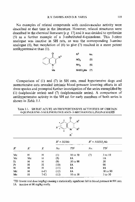
R.V. DAVIES AND D.B. YATES 1 I9
No examples of related compounds with cardiovascular activity were described at that time in the literature. However, related structures were described in the chemical literature [e.g. 171 and it was decided to synthesize ( 5 ) as a further example of a 3-substituted-4-quinolone. This 3-nitro analogue was inactive in SH rats, as was the corresponding 3-amino analogue (6) , but mesylation of ( 6 ) to give (7) resulted in a more potent antihypertensive than (1).
0
I..-ii-r” R3 No.
Me NHS02Me (7)
Comparison of (1) and (7) in SH rats, renal hypertensive dogs and normotensive cats revealed intrinsic blood pressure lowering effects in all three species and prompted further investigation of the series exemplified by (1) (sulphoxide series) and (7) (sulphonamide series). A comparison of antihypertensive activity in the SH rat for early members of both series is shown in Table 3.1.
Table 3.1. SH RAT ACUTE ANTIHYPERTENSIVE ACTIVITIES OF CERTAIN 4-QUINOLONE-3-SULPHOXIDES AND -3-METHANESULPHONAMIDES
0
R’ = SOMr R’ = NHSO’Mr
R‘ R’ X No. TD’ NO. TD’
Me H H (1) 10 to 30 (7) 3 to 10 Me Me H (8) IA 1A Et H H (9) 30 to 90 30
IA 90 (1 1) IA ( lo ) IA
Pr H H H H H Me H 6-CI (12) 1A Me H 7-CI (13) 10 to 30 3 to 10
30 to 90
‘TD lowest oral dose (mg/kg) causing a statistically significant fall in blood pressure in SH rats. IA inactive at 90 mg/kg orally.

120 4-QUINOLONES AS POTENTIAL CARDIOVASCULAR AGENTS
Although the 3-sulphonamide series looked likely to give rise to more potent antihypertensives, it was found to be associated with genotoxicity in the Ames test and caused attention to be switched to the 3-sulphoxide series, which was free of genotoxic problems. The activities of compounds (l), (9) and (1 3) did suggest intrinsic activity associated with these 4-quinolone-3- sulphoxides and a programme of work was therefore initiated to determine whether (1) did indeed represent a good lead compound.
Approximately 120 compounds were synthesized to investigate the effect of substituents R’ to Rs in the 4-quinolone ring system (4) where R3 = S(O),Me and n = 0, 1, or 2.
A particular requirement for activity was minimum bulk in the 1,2-region. Optimum activity was achieved with 1-methyl and 2-H substituents. The 2-methyl analogue (8) was inactive. The 1-ethyl analogue (9) was approximately 3-fold less potent in SH rats than the 1-methyl analogue (1) and the 1-propyl analogue (10) was inactive. All selected higher alkyl homologues that were synthesized up to C,*, 1-phenyl and 1-benzyl (and aromatic ring substituted variants of these, e.g. 4-C1 and 3,4-(OMe),) were inactive. The 1-H analogue (1 1) was also inactive. The 4-OX0 moiety (proton accepting species) in (4), (and l l) , may be essential for activity, and tautomerism of (11) to (l la) may result in loss of activity due to predominance of the 4-hydroxyl group (proton donating species) in (1 la). This 4-hydroxy tautomer may be further stabilized by hydrogen bonding of the 4-hydroxyl proton to the 3-sulphoxide moiety thus holding the sulphoxide group in an unfavourable conformation.
Molecular modelling studies performed on active 4-quinolone-3-sulphox- ides revealed a preferred conformation with the 3-position side-chain methyl group occupying a region of space adjacent to the 4-carbonyl and away from the 2-position, a region of space indicated by structure-activity relationships required to be free of steric bulk. This position is reversed, and fixed, in the inactive (1 la).
The 3-position was also intolerant to the presence of bulk, optimum activity being found for the 3-methylsulphinyl analogue (1). The corre- sponding sulphide (3-SMe) was similarly active and the sulphone (3-S02Me)

R.V. DAVIES A N D D.B. YATES 121
was considerably less active (threshold antihypertensive dose (TD) = 90 mg/kg). Activity was abolished in the ethyl homologue (3-SOEt) and all other higher homologues investigated. 3-Benzyl-, 3-substituted benzyl-, 3-phenyl- and 3-substituted phenylsulphinyl analogues were also inactive.
Most analogues were synthesized in the exploration of benz-ring substituents R5 to RE in (4). Initial studies with halogen, methyl and methoxy monosubstituents (Table 3.2) indicated substitution in the 7-position resulted in retention of activity. Increasing the bulk of the 7-alkyl or 7-alkoxy substituents up to C, did not result in any increase in potency. Of a range of simple disubstituted analogues synthesized, only 6,7- disubstitution showed potential. Further work, therefore, concentrated on the synthesis of other 7-substituted or 6,7-disubstituted analogues where the substituents were of minimum bulk.
When 7-substituents contained an alkyl component, methyl or ethyl was chosen (eg OS0,Me or CO,Et), consistent with minimum bulk. The range of neutral, electron withdrawing and electron donating substituents investi- gated in the 7-position are shown in Table 3.3. No obvious structure-activity relationships for these 7-substituents was revealed although the electron-
Table 3.2. SH RAT ACUTE ANTIHYPERTENSIVE ACTIVITIES FOR CERTAIN BENZ-SUBSTITUTED 4-QUINOLONE-3-SULPHOXIDES
0
Me
TD'
Position of X
X 5- 6- 7- 8-
F 30 90 10 IA Cl 90 IA 10 to 30 90 Me IA IA 30 IA OMe - 90 90 90
'TD lowest oral dose (mg/kg) causing a statistically significant fall in blood pressure in SH rats. IA - analogue not synthesized.
inactive at 90 mg/kg orally.
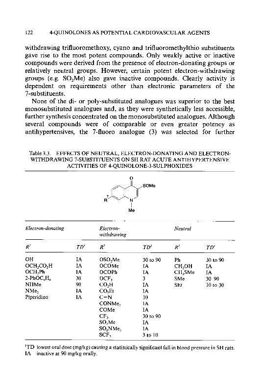
122 4-QUINOLONES AS POTENTIAL CARDIOVASCULAR AGENTS
withdrawing trifluoromethoxy, cyano and trifluoromethylthio substituents gave rise to the most potent compounds. Only weakly active or inactive compounds were derived from the presence of electron-donating groups or relatively neutral groups. However, certain potent electron-withdrawing groups (e.g. S0,Me) also gave inactive compounds. Clearly activity is dependent on requirements other than electronic parameters of the 7-substituents.
None of the di- or poly-substituted analogues was superior to the best monosubstituted analogues and, as they were synthetically less accessible, further synthesis concentrated on the monosubstituted analogues. Although several compounds were of comparable or even greater potency as antihypertensives, the 7-fluoro analogue (3) was selected for further
Table 3.3. EFFECTS OF NEUTRAL, ELECTRON-DONATING AND ELECTRON- WITHDRAWING 7-SUBSTITUENTS ON SH RAT ACUTE ANTIHYPERTENSIVE
ACTIVITIES OF 4-QUINOLONE-3-SULPHOXIDES
Elect ron-dona t ing Electron- withdra w ing
Neutral
R7 TD’ R7 TD’ R7 T D I
OH IA OS0,Me OCH,CO,H IA OCOMe OCH,Ph IA OCOPh 2-PhOC6H4 30 OCF, NHMe 90 CO,H NMez IA C0,Et Piperidino IA C=N
CONMe, COMe
S0,Me SO,NMe,
CF,
SCF,
30to90 Ph 30 to 90 IA CH,OH IA IA CH,SMe IA 3 SMe 30-90 IA SEt 10 to 30 IA 10 IA IA 30 to 90 IA IA 3 to 10
‘TD lowest oral dose (mg/kg) causing a statistically significant fall in blood pressure in SH rats. IA inactive at 90 mg/kg orally.

R.V. DAVIES AND D.B. YATES 123
development as soon as early test results became available and is described in ‘Compounds Selected for Development’.
The promising profile of activity shown by certain 4-quinolone-3- sulphoxides, including (3), in animal models established the true lead potential of 4-quinolone analogues and prompted a major research effort to find other related compounds with potential for development.
IDENTIFICATION OF 4-QUINOLONES bEARING ALTERNATIVE 3-SUBSTITUENTS
To identify alternative 3-substituents use was made of published [18] substituent constants for the methylsulphinyl (SOMe) and methanesulphon- amido (NHS0,Me) groups as shown in Table 3.4. By comparison with these groups, what was thought to be a desirable profile of values for substituents was identified.
As a measure of lipophilicity, the hydrophobic parameter n was selected; as electronic parameters the Hammett and Taft 6, constants and the Swain and Lupton F and R values were chosen; and as a measure of size, molar refractivity (MR), based on the atom-group structure constants of Vogel, was used. ‘Ideal values’ were obtained, i.e. negative n, positive 6,: positive F, zero to negative R and MR approximately 16 (H = 1). 4-Quinolones,
Table 3.4. PHYSICO-CHEMICAL PARAMETERS OF 3-SUBSTITUENTS
Me
R’ n 0, F R M R
SOMe -1.58 0.49 0.52 0.01 13.70 NHS0,Me -1.18 0.03 0.25 -0.20 18.20 CH2CONH2 (i) -1.68 0.07 0.08 -0.01 14.41 S02NH2 (ii) -1.82 0.57 0.41 0.19 12.28 CONHMe (iii) -1.27 0.36 0.34 0.05 14.57
N-N NwN I \ (iv) -1.04 0.50 0.52 0.02 18.33

124 4-QUINOLONES AS POTENTIAL CARDIOVASCULAR AGENTS
with these ‘ideal’ 3-substituents (i to iv, Table3.4), were then targeted for synthesis as potential antihypertensive agents. Results for the target compounds and other closely related analogues which were synthesized are shown in Table 3.5, together with compounds (1) and (7) for comparison.
The carbamoylmethyl analogue (14) was inactive, probably as a consequence of its instability to acid conditions which could result in the formation of the corresponding acetic acid (3-CH2COOH) in vivo, a compound with physico-chemical parameters of the 3-substituent lying outside the target range.
Compared with the 3-methylsulphoxide (1) and the 3-methanesulphon- amide (7) the activity of the 3-sulphamoyl analogue (1 5 ) was reduced and was only partially restored in the 3-N-methylsulphamoyl analogue (16). The 7-chloro analogue (1 7), however, was marginally superior to (1) and the corresponding 7-fluoro analogue (1 8) also exhibited superior potency compared with the parent benz-unsubstituted analogue ( 1 6). This improved potency for 7-halogenated compounds compared with the corresponding parent benz-unsubstituted analogues was somewhat surprising as there was little difference in the potencies of the 7-halogenated and benz-unsubstituted analogues in the original sulphoxide and sulphonamide series. This important observation prompted the syntheses of 7-flUOrO and 7-chloro substituted analogues, as well as the parent unsubstituted compounds, in all future series.
The 3-N-methylcarbamoyl analogue (1 9) exhibited threshold antihy- pertensive activity at 30 mg/kg and this was not enhanced in the 7-flUO1-0 analogue (21). Although 3 to 10-fold more potent, the 7-chloro analogue (20) was toxic in SH rats and attention turned to the primary carboxamide. The benz-unsubstituted analogue (22) was inactive but the corresponding 7-halo primary carboxamides (23) and (24), however, exhibited good potency, again highlighting the potency-enhancing effect of a 7-halogen substituent.
The targeted 3-( 1-tetrazolyl) derivative (25) was inactive and the corresponding 7-halogenated analogues were not synthesized. However, the 5-methyl- 1 H- 1 -tetrazolyl derivatives (26), (27) and (28) showed reasonable potency and the isomeric 1 -methyl-1 H-5-tetrazolyl analogues (29), (30) and (31) were even more active.
Structure-activity relationships for 1 -substituents and benz-ring substitu- ents of these analogues followed broadly similar patterns to those described for the sulphoxide series. Increasing the bulk of the 3-substituent by the introduction, where possible, of additional alkyl substitution generally led to a loss of activity.

R.V. DAVIES AND D.B. YATES 125
Table 3.5. SH RAT ACUTE ANTIHYPERTENSIVE ACTIVITIES OF 4-QUINOLONES IDENTIFIED BY ANALYSIS OF PHYSICO-CHEMICAL PARAMETERS OF
THE 3-SUBSTITUENT
0
Me
R3
SOMe NHS0,Me CH,CONH, S02NH, S0,NHMe CONHMe CONH,
N-N I \
N W N -
N:N -N'
YN
4 Y - y
Me
N"
I Me
R7 = H R7 = CI R7 = F
TD' No. TD' No. TD'
10 to 30 (13) 3 to 10 IA 90 30 to 90 (17) 30 (20) IA (23)
IA
30 to 90 (27)
10 to 30 (30)
10 to 30 - - - 30 30 10
-
30
10
'TD lowest oral dose (mg/kg) causing a statistically significant fall in blood pressure in SH rats. IA - analogue not synthesized.
inactive at 90 mg/kg orally.
SYNTHESIS OF OTHER 3-SUBSTITUTED 4-QUINOLONES CONTAINING BRIDGING GROUPS IN THE 3-POSITION
The strategy for the next phase of the work involved the synthesis of 4-quinolone analogues bearing as 3-substituents groups whose size
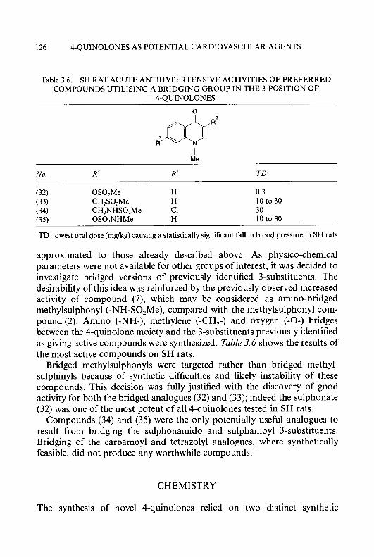
126 4-QUINOLONES AS POTENTIAL CARDIOVASCULAR AGENTS
Table 3.6. SH RAT ACUTE ANTIHYPERTENSIVE ACTIVITIES OF PREFERRED COMPOUNDS UTILISING A BRIDGING GROUP IN THE 3-POSITION OF
4-QUINOLONES
0
Me
No. R3 R7 TD'
(32) OS0,Me H 0.3 (33) CH,SO,Me H 10 to 30 (34) CH,NHSO,Me CI 30 (35) OS0,NHMe H 10 to 30
'TD lowest oral dose (mg/kg) causing a statistically significant fall in blood pressure in SH rats
approximated to those already described above. As physico-chemical parameters were not available for other groups of interest, it was decided to investigate bridged versions of previously identified 3-substituents. The desirability of this idea was reinforced by the previously observed increased activity of compound (7), which may be considered as amino-bridged methylsulphonyl (-NH-SO,Me), compared with the methylsulphonyl com- pound (2). Amino (-NH-), methylene (-CH,-) and oxygen (-0-) bridges between the 4-quinolone moiety and the 3-substituents previously identified as giving active compounds were synthesized. Table 3.6 shows the results of the most active compounds on SH rats.
Bridged methylsulphonyls were targeted rather than bridged methyl- sulphinyls because of synthetic difficulties and likely instability of these compounds. This decision was fully justified with the discovery of good activity for both the bridged analogues (32) and (33); indeed the sulphonate (32) was one of the most potent of all 4-quinolones tested in SH rats.
Compounds (34) and (35) were the only potentially useful analogues to result from bridging the sulphonamido and sulphamoyl 3-substituents. Bridging of the carbamoyl and tetrazolyl analogues, where synthetically feasible, did not produce any worthwhile compounds.
CHEMISTRY
The synthesis of novel 4-quinolones relied on two distinct synthetic
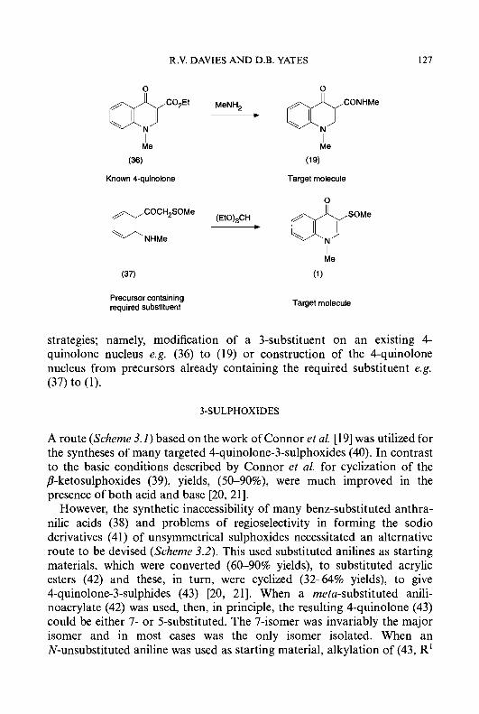
R.V. DAVIES AND D.B. YATES 127
& - MeNH,
I
(36)
Me
Known 4-quinolone
(37)
Precursor containing required substituent
Target molecule
0
Me
(1)
Target molecule
strategies; namely, modification of a 3-substituent on an quinolone nucleus e.g. (36) to (19) or construction of the
existing 4- 4-quinolone
nucleus from precursors already containing the required substituent e.g. (37) to (1).
3-SULPHOXIDES
A route (Scheme 3.1) based on the work of Connor et al. [19] was utilized for the syntheses of many targeted 4-quinolone-3-sulphoxides (40). In contrast to the basic conditions described by Connor et al. for cyclization of the /3-ketosulphoxides (39), yields, (50-90%), were much improved in the presence of both acid and base [20, 211.
However, the synthetic inaccessibility of many benz-substituted anthra- nilic acids (38) and problems of regioselectivity in forming the sodio derivatives (41) of unsymmetrical sulphoxides necessitated an alternative route to be devised (Scheme 3.2). This used substituted anilines as starting materials, which were converted (60-90% yields), to substituted acrylic esters (42) and these, in turn, were cyclized (32-64% yields), to give 4-quinolone-3-sulphides (43) [20, 211. When a meta-substituted anili- noacrylate (42) was used, then, in principle, the resulting 4-quinolone (43) could be either 7- or 5-substituted. The 7-isomer was invariably the major isomer and in most cases was the only isomer isolated. When an N-unsubstituted aniline was used as starting material, alkylation of (43, R'
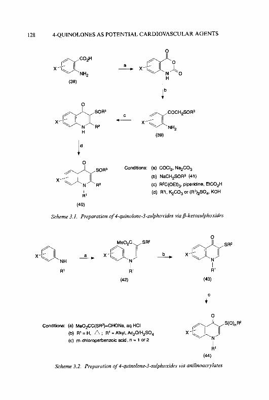
128 4-QUINOLONES AS POTENTIAL CARDIOVASCULAR AGENTS
0
R2
H
0
ld x g s 0 R 3 R2
I R'
(40)
COCH2SOR3
(39)
Conditions: (a) COCI,, Na2C03
(b) NaCH2SOR3 (41)
(c) R2C(OEt)3, piperidine, EtC0,H
(d) R'I, &C03 or (R1),S04, KOH
Scheme 3.1. Preparation of 4-quinolone-3-sulphoxides via b-ketosulphoxides
1
R' Ri
(43)
t
Conditions: (a) MeO,CC(SR?=CHONa, aq HCI S(O),R2
(b) R' = H, ; R' = Alkyl, Ac,O/H2SO4
(c) mshloroperbenzoic acid, n = 1 or 2 I
(44)
R'
Scheme 3.2. Preparation of 4-quinolone-3-sulphoxides via anilinoacrylates

R.V. DAVIES AND D.B. YATES 129
0 n 0
I I
(45) (446) (47)
R’ H
R’
lc
Conditions: (a) R’I, K&03 or (R’),SO,, KOH
(b) cHN03, 100’. 1 h
(c) SnCI,. cHCI, AcOH or HdP&/Me,CO
(d) R2S02CI, pyridine
(e) R31, &C03
Scheme 3.3. Preparation of4-quinolone-3-sulphonamides
= H) or (44, R’ = H) gave the corresponding 1-alkylated derivatives. Oxidation gave the corresponding sulphoxides or sulphones (44).
3-SULPHONAMIDES
3-Sulphonamides (49) and (50) (Scheme 3.3) were readily prepared by treatment of the corresponding 3-amino derivatives (48) with sulphonyl chlorides (1 6 6 4 % yields), followed by optional side-chain alkylation. Although several routes to 3-amino-4-quinolones and their precursor 3-nitro analogues (47) have been described in the literature [17, 22-25], direct nitration (50% yield) of the 3-unsubstituted 4-quinolones (46) was found to be generally applicable. The 3-unsubstituted compounds (46) were obtained from (45) as shown and these, in turn, were prepared by decarboxylation of the 3-carboxylic acids resulting from hydrolysis of the corresponding 3-carboxylate esters (see below). 3-Carboxylate esters (53)

130 4-QUINOLONES AS POTENTIAL CARDIOVASCULAR AGENTS
Ib Conditions: (a) SOCI,. CISO,H, 85‘
@ X’ (b) RWNH, CH,CI,, rt
I , R’
(52)
Scheme 3.4. Preparation of 3-ssulphamoyyC4-quinobnes
are well documented in the literature [e.g. 26301 and have been extensively reviewed by Albrecht [3 11. Alternatively, (47) may be obtained by nitration of (45), followed by N-substitution.
Although a multistep route to 3-sulphamoyl-4-quinolones has been described by Yanagisawa et al. [32], the availability of various benz- substituted 4-quinolones (46), which could be chlorsulphonated in the
X’ &CO2Et a_ X’ &c.
x’&coNHR2
H I (53) (54) R’
b l o r c Conditions: (a) R’I, K&O,
or (R1),SO,, KOH
(b) aq NH, (R2 = H)
(c) R2NHdBF3 etherate (R2 = alkyl)
I (55) R’
Scheme 3.5. Preparation of 3-carbamoyyC4-quinolones
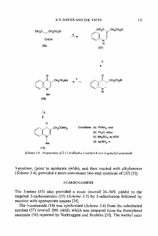
R.V. DAVIES A N D D.B. YATES 131
EtOzCYCH2CO2Et a
CHOH
&cHzcozMe _c ~ c H z c o z E t
I H Me
(58)
Conditions: (a) PhNH,, neat C.rE:,.cH2c0NH2 (b) Ph20. reflux
(c) Me,SO,, aq KOH I (d) aq NH,, rt Me
(14)
Scheme 3.6. Preparation of 2-(1,4-dihydro-I -rnethyl-4-oxo-3-quinolyl)acetamide
3-position, (poor to moderate yields), and then reacted with alkylamines (Scheme 3.4), provided a more convenient two-step synthesis of (52) [33].
3-CARBOXAMIDES
The 3-esters (53) also provided a route (overall 2454% yields) to the targeted 3-carboxamides (55) (Scheme 3.5) by 1 -substitution followed by reaction with appropriate amines [34].
The 3-acetamide (14) was synthesized (Scheme 3.6) from the substituted acrylate (57) (overall 20% yield), which was prepared from the formylated succinate (56) reported by Vorbruggen and Strehlke [35]. The methyl ester
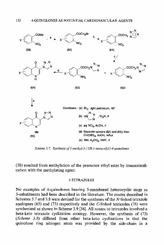
132 4-QUINOLONES AS POTENTIAL CARDIOVASCULAR AGENTS
(59)
Conditions : (a) Br2, light petroleum, 40' I.
N,
\s-N (b) H d . N , Et3N, rt
(c) aq TiCI,, AcOH. rt
(d) Separate isomers (62) and (63); then
(e) Mel. %CO,, DMF, rt
CH(OEt),, AcOH, reflux
Scheme 3.7. Synthesis of I-methyl-3-(1 H-I-tetrazoly1)-4-quinolones
(58) resulted from methylation of the precursor ethyl ester by transesterifi- cation with the methylating agent.
3-TETRAZOLES
No examples of 4-quinolones bearing 5-membered heterocyclic rings as 3-substituents had been described in the literature. The routes described in Schemes 3.7 and 3.8 were devised for the syntheses of the N-linked tetrazole analogues (65) and (73) respectively and the C-linked tetrazoles (76) were synthesized as shown in Scheme 3.9 [36]. All routes to tetrazoles involved a beta-keto tetrazole cyclization strategy. However, the synthesis of (73) (Scheme 3.8) differed from other beta-keto cyclizations in that the quinolone ring nitrogen atom was provided by the side-chain in a
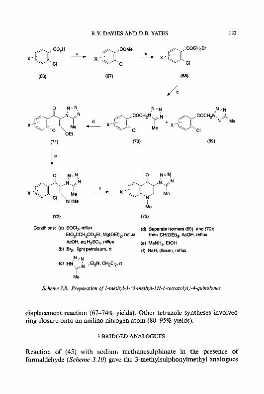
R.V. DAVIES AND D.B. YATES 133
N-N N-N
p.JCHZ”N + X’\ yJIT’.N\Me
.’&M _d X’\ CI Me
x r E E N ; ; Me I
(71 ) (70) (69)
I. 0 N=N, dry
f X’\
(72) (73)
Conditions: (a) SOCI,. reflux (d) Separate isomers (69) and (70); then CH(OEt),, AcOH, reflux
(e) MeNH2, EtOH
(f) NaH, dioxan, reflux
EtOzCCHzCO,Et, Mg(OEt),, reflux
AcOH, aq H,SO,, reflux
(b) Brz, light petroleum, rt
(d HNy’ I EGN, CHzCI,, rt N,N
Me
Scheme 3.8. Preparation o f 1 -methyl-3-(5-methyl-I H-I -tetrazolyl)-4-quinolones
displacement reaction (67-74% yields). Other tetrazole syntheses involved ring closure onto an anilino nitrogen atom (80-95% yields).
3-BRIDGED ANALOGUES
Reaction of (45) with sodium methanesulphinate in the presence of formaldehyde (Scheme 3.20) gave the 3-methylsulphonylmethyl analogues

134 4-QUINOLONES AS POTENTIAL CARDIOVASCULAR AGENTS
t
Conditions: (a) BuLi, hexane, THF
(b) CH(OEt)3, AcOH. reflux Me
I (76) R’
Scheme 3.9. Preparation of 3-(l-methyl-l H-5-tetrazolyl)-4-quinolones
(77) which upon alkylation gave the target compounds (78) in overall good yields [37].
Reaction of (45) with formaldehyde alone gave (79), which was N’-alkylated under basic conditions to (80). Chlorination, followed by reaction with simple amines to give (82) and finally treatment with sulphonyl chlorides, provided an entry (Scheme 3.11) into the series of 3-alkanesulphonamidomethyl analogues (83). Overall yields varied from 8 to 83% depending on the X and R2 substituents.
X’ go a_ X’ &CH2S02Me
Conditions: (a) NaS02Me, aq HCHO, &cH2s02Me
H H
(45) (77)
Ib 95-100‘ X’
I (b) R12S04, aq KOH, rt
(78) R’
Scheme 3.10. Preparation of 3-methylsulphonylmethyl-4-quinolones
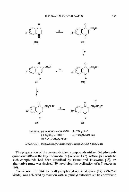
R.V. DAVIES A N D D.B. YATES 135
X’ d!!+ H
(45)
F) CH,CI
x ’ F c I
0
Conditions: (a) aq HCHO, NaOH, 40-50
(b) R’,SO,. aq KOH, rt
(c) SOCI,, CH,CI,, reflux
CH,OH
X’ & H
(79)
ib 0
CH,OH
x J E I R’
(80)
R2
CH2NS02R3 I
X’ & I
(d) R2NH2, THF
(e) R3S0,CI, NaOH aq
Scheme 3.11. Preparation of 3-alkanesulphonamidomethyl-4-quinolones
The preparation of the oxygen-bridged compounds utilized 3-hydroxy-4- quinolones (86) as the key intermediates (Scheme 3.12). Although a route to such compounds had been described by Evans and Eastwood [38], an alternative route was devised [39] involving the cyclization of a /?-ketoester (84).
Conversion of (86) to 3-alkylsulphonyloxy analogues (87) (5675% yields), was achieved by reaction with sulphonyl chlorides whilst conversion

136 4-QUINOLONES AS POTENTIAL CARDIOVASCULAR AGENTS
X,aCOCHzOCOPh a - NCHO
R’ I
(84)
0
I k‘
(87)
Conditions: (a) K.$O,, DMF
(c) R2SOZCI, aq NaOH
(d) PhCH,NEt$I, K&O,,
R2R3NS02CI
0
X’ &OCOPh
I R’
(85)
Ib x/&oH
R’
(86)
X’ &OS02NR2R3
I R’
(88)
Scheme 3.12. Preparation of 3-alkylsulphonyloxy- and 3-alkylsulphamoyloxy-4-quinolones
of (86) to 3-alkylsulphamoyloxy analogues (88) (30-50% yields), was achieved by reaction with sulphamoyl chlorides.
COMPOUNDS SELECTED FOR DEVELOPMENT
Seven analogues, namely compounds (3), (17), (24), (29), (32), (33) and (34) (Tables 3.5 and 3.6), were selected for further development. Selection was dependent on a variety of factors, including potency particularly in the SH rat, and relative ease of synthesis. Before evaluating the safety of these
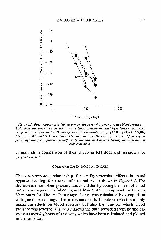
R.V. DAVIES AND D.B. YATES 137
a, k
i? $ 11 k c4
a -5- 0 0 ri
rn -10- 5 a ' -15-
1 ; -20 ld a, k u - 2 5 a, a do - 3 0 3
1 10 100
Dose (mg/kg)
Figure 3. I . Dose-response of quinolone compounds on renal hypertensive dog blood pressure. Data show the percentage change in mean blood pressure of renal hypertensive dogs when compounds are given orally. Dose-responses to compounds (3.0), ( I 7 9 ) . (24, A), (29,m). (32, .), (33,e) and (34,V) are shown. The data points are the means from at least four dogs of percentage changes in pressure at half-hourly intervals for 5 hours following administration of
each compound.
compounds, a comparison of their effects in RH dogs and normotensive cats was made.
COMPARISON IN DOGS AND CATS
The dose-response relationship for antihypertensive effects in renal hypertensive dogs for a range of 4-quinolones is shown in Figure 3. I . The decrease in mean blood pressure was calculated by taking the mean of blood pressure measurements following oral dosing of the compound made every 30 minutes for 5 hours. Percentage change was calculated by comparison with pre-dose readings. These measurements therefore reflect not only maximum effects on blood pressure but also the time for which blood pressure was lowered. Figure 3.2 shows the data recorded from normoten- sive cats over 4y2 hours after dosing which have been calculated and plotted in the same way.
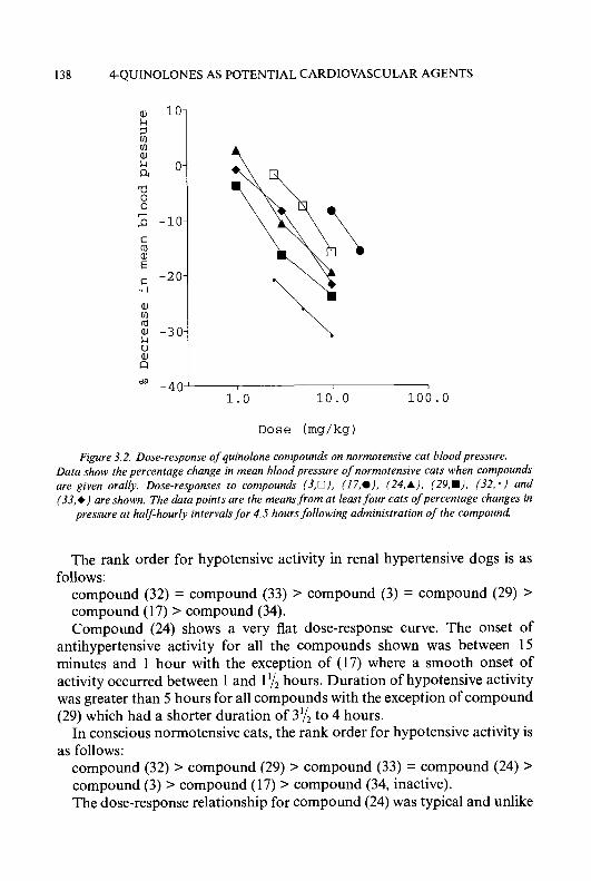
138 4-QUINOLONES AS POTENTIAL CARDIOVASCULAR AGENTS
a, m ld
k U al a
- 4 0 ‘ dp I , I
1.0 10.0 100.0
D o s e (rng/kg)
Figure 3.2. Dose-response of quinolone compounds on normotensive cat blood pressure. Data show the percentage change in mean blood pressure of normotensive cats when compounds are given orally. Dose-responses to compounds (3 ,O) . (17,e). (24,A). (29,W). (32, * ) and (33,+) are shown. The data points are the means from at least four cats of percentage changes in
pressure at half-hourly intervalsfor 4.5 hours following administration of the compound.
The rank order for hypotensive activity in renal hypertensive dogs is as
compound (32) = compound (33) > compound (3) = compound (29) > compound (17) > compound (34). Compound (24) shows a very flat dose-response curve. The onset of
antihypertensive activity for all the compounds shown was between 15 minutes and 1 hour with the exception of (17) where a smooth onset of activity occurred between 1 and 1’/2 hours. Duration of hypotensive activity was greater than 5 hours for all compounds with the exception of compound (29) which had a shorter duration of 3y2 to 4 hours.
In conscious normotensive cats, the rank order for hypotensive activity is as follows:
compound (32) > compound (29) r compound (33) = compound (24) > compound (3) > compound (17) > compound (34, inactive). The dose-response relationship for compound (24) was typical and unlike
follows:

R.V. DAVIES A N D D.B. YATES 139
the flat dose-response curve obtained from dogs. As in dogs, the onset of activity of compound (1 7) was slow and smooth. All the other compounds showed a rapid onset of activity 15 to 30 minutes after oral dosing. Duration of activity of all compounds was greater than 4y2 hours.
FURTHER EVALUATION
Although some compounds offered some potential advantage over the 3-sulphoxide (3) particularly in terms of potency, toxicological evaluation disclosed problems with all compounds other than (3). There was no common toxicological pattern and the development of each compound was halted for a different toxicological reason. Compound (3) progressed through the early toxicological evaluation to further development.
DEVELOPMENT OF FLOSEQUINAN
Pharmacological evaluation of compound (3), subsequently known as flosequinan and later as Manoplax@, disclosed that vasodilatation occurred in both arterial and venous vasculature and thus indicated that there could be an application in the treatment of congestive heart failure.
Clinical evaluation confirmed that the drug possessed antihypertensive activity although this was accompanied by headache. At lower doses both arterial and venous vasodilatation occurred and such haemodynamic activity was consistent with utility for treating congestive heart failure. Clinical development continued with an emphasis on heart failure; flosequinan proved to be particularly effective in improving the exercise capacity of patients in trials carried out in both the United Kingdom and the United States.
The drug was approved for use in both countries in 1992 and 1993, respectively, while a long-term mortality study known as PROFILE (Prospective Randomised Flosequinan Longevity Evaluation) was in progress. In July 1993, continuous monitoring of this study established a statistically significant increase in mortality in flosequinan-treated patients taking 100 mg daily as a single dose in addition to conventional therapy. Following preliminary evaluation of clinical data the drug was withdrawn.
PHARMACOLOGY
Antihypertensive activity
In the Aoki-Okamoto strain of SH rat, oral flosequinan was antihyperten-
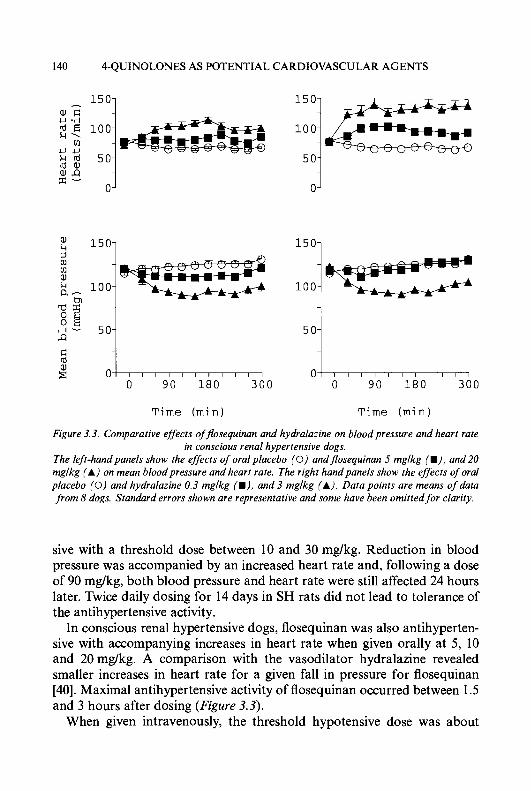
140 4-QUINOLONES AS POTENTIAL CARDIOVASCULAR AGENTS
150- a, u .TI ;< 100-
g f i
vl u u k 50- nlaJ
0-
5 0-
0-
1501 1501
100 I 9 vl rn aJ L i 100 a-
h,
5 0 Q
d 0
0 90 180 3 0 0
T i m e ( m i n )
1 0 1
0 9 0 180 3 0 0
T i m e ( m i n )
Figure 3.3. Comparative effects ofjosequinan and hydralazine on blood pressure and heart rate in conscious renal hypertensive dogs.
The left-hand panels show the effects of oral placebo (0) andjosequinan 5 mglkg (I), and 20 mglkg (A) on mean bloodpressure and heart rate. The right handpanels show the effects of oral placebo (0) and hydralazine 0.3 mglkg (W), and 3 mglkg (A). Data points are means of data from 8 dogs. Standard errors shown are representative and some have been omitted for clarity.
sive with a threshold dose between 10 and 30 mg/kg. Reduction in blood pressure was accompanied by an increased heart rate and, following a dose of 90 mg/kg, both blood pressure and heart rate were still affected 24 hours later. Twice daily dosing for 14 days in SH rats did not lead to tolerance of the antihypertensive activity.
In conscious renal hypertensive dogs, flosequinan was also antihyperten- sive with accompanying increases in heart rate when given orally at 5 , 10 and 20 mg/kg. A comparison with the vasodilator hydralazine revealed smaller increases in heart rate for a given fall in pressure for flosequinan [40]. Maximal antihypertensive activity of flosequinan occurred between 1.5 and 3 hours after dosing (Figure 3.3).
When given intravenously, the threshold hypotensive dose was about
R.V. DAVIES AND D.B. YATES 141
3 mg/kg though peak effects were not present until 65 minutes post-dose, probably indicating an antihypertensive component due to activity of the metabolite [41] (see metabolism of flosequinan).
Normotensive cats were slightly more sensitive to flosequinan with a threshold oral dose of 1 mg/kg. Peak effects were seen between 60 and 160 minutes after dosing and, as in dogs, there was a lesser increase of heart rate for a given hypotensive effect than with hydralazine [40].
Renal effects
Flosequinan was found to be antidiuretic and antinatriuretic in saline- loaded conscious normotensive rats following the oral administration of doses between 10 and 90 mg/kg. This was in contrast to effects in conscious saline-loaded dogs where, following 5, 10 or 20mg/kg, there were no significant changes in sodium or water excretion though there were increases in potassium excretion after 20 mg/kg. Similar effects on potassium in the dog were shown by the higher dose (3 mg/kg) of hydralazine but there were also marked antidiuretic and antinatriuretic effects at all doses with this drug (Table 3.7) [40].
Though plasma renin activity increased in both dogs and cats following flosequinan, these increases were considerably less than those seen after equivalently effective doses of hydralazine (Table 3.8).
Table 3.7. OVERALL MEAN VALUES FOR SODIUM, POTASSIUM AND CHLORIDE EXCRETION AND URINE VOLUME FOLLOWING FLOSEQUINAN OR
HYDRALAZINE IN CONSCIOUS NORMOTENSIVE DOGS LOADED ORALLY WITH SODIUM CHLORIDE SOLUTION
n Na' K+ Cf Urine excretion excretion excretion volume (nmollkg) (nmollkg) (nmollkg) (mllkg)
Placebo 16 0.31 0.069 0.34 3.05 Flosequinan (mg/kg orally) 5.0 8 0.28 0.086 0.33 2.58 10.0 8 0.36 0.089 0.39 2.74 20.0 8 0.39 0.117* 0.41 2.89 Hydralazine (mg/kg orally) 0.3 8 0.25 0.067 0.26 2.71 1 .o 8 0.10** 0.059 0.12** 1.54** 3.0 8 0.11** 0.1 10* 0.12** 1.46**
* p < 0.05; ** p < 0.01 for multiple comparisons against placebo (Williams' test)
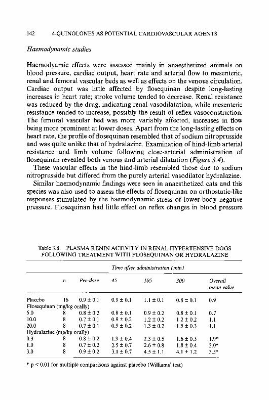
142 4-QUINOLONES AS POTENTIAL CARDIOVASCULAR AGENTS
Haemodynamic studies
Haemodyamic effects were assessed mainly in anaesthetized animals on blood pressure, cardiac output, heart rate and arterial flow to mesenteric, renal and femoral vascular beds as well as effects on the venous circulation. Cardiac output was little affected by flosequinan despite long-lasting increases in heart rate; stroke volume tended to decrease. Renal resistance was reduced by the drug, indicating renal vasodilatation, while mesenteric resistance tended to increase, possibly the result of reflex vasoconstriction. The femoral vascular bed was more variably affected, increases in flow being more prominent at lower doses. Apart from the long-lasting effects on heart rate, the profile of flosequinan resembled that of sodium nitroprusside and was quite unlike that of hydralazine. Examination of hind-limb arterial resistance and limb volume following close-arterial administration of flosequinan revealed both venous and arterial dilatation (Figure 3.4).
These vascular effects in the hind-limb resembled those due to sodium nitroprusside but differed from the purely arterial vasodilator hydralazine.
Similar haemodynamic findings were seen in anaesthetized cats and this species was also used to assess the effects of flosequinan on orthostatic-like responses stimulated by the haemodynamic stress of lower-body negative pressure. Flosequinan had little effect on reflex changes in blood pressure
Table 3.8. PLASMA RENIN ACTIVITY IN RENAL HYPERTENSIVE DOGS FOLLOWING TREATMENT WITH FLOSEQUINAN OR HYDRALAZINE
Time after administration (min)
n Pre-dose 45 I05 300 Overall mean value
Placebo 16 0 .9f0 .1 0 . 9 f 0 . 1 l . l f O . l 0 . 8 f 0 . 1 0.9 Flosequinan (mglkg orally) 5.0 8 0.8 f 0.2 0.8 f 0.1 0.9 f 0.2 0.8 f 0.1 0.7 10.0 8 0 .7 f0 .1 0 . 9 f 0 . 2 1 . 2 f 0 . 2 1 .2 f0 .2 1.1 20.0 8 0 .7f0.1 0 . 9 f 0 . 2 1.3k0.2 1 .5 f0 .3 1.1 Hydralazine (mglkg orally) 0.3 8 0.8 f 0.2 1.9 f 0.4 2 .3f 0.5 1 .6 f 0.3 1.9* 1 .o 8 0 .7 f 0.2 2.5 f 0.7 2 .6 f 0.8 1.8 f 0.4 2.0* 3.0 8 0 . 9 f 0 . 2 3 .1 f0 .7 4 . 5 f 1 . 1 4 .1 f1 .2 3.3*
* p < 0.01 for multiple comparisons against placebo (Williams’ test)

R.V. DAVIES AND D.B. YATES 143
a, U C rd c, (I]
1501 Infusion period
100-
5 0-
0
-50 I I I I
0 30 60 90
Time ( s e c s )
Figure 3.4. Effects offlosequinan on hind-limb volume and femoral arterial resistance following intra-arterial infusion in anaesthetised dogs.
Dara show percentage changes in hind-limb volume for I minute infusions of flosequinan 0.05 mglkg (a). 0.2 mglkg (A), and 0.8 mglkg (+). Open symbols show the effects of the same doses on femoral arterial resistance. Limb volume was measured plethysmographically and allowance made for arterial dilatation effects on limb volume by prior calibration. Arterial resistance was calculated from femoral flow measured via a cuff-type f low probe and flow-meter and systemic mean bloodpressure. Data shown are from 6 dogs. Standard errors are not shown for reasons of
clarity.
and heart rate in contrast to the profound depression of such reflexes encountered with adrenergic neurone blocking agents such as bethanidine.
Cardiac effects
The earliest studies undertaken with flosequinan established that the drug was capable of direct myocardial stimulation. In vitro studies showed that flosequinan had both positive inotropic and chronotropic activity [42]. The effects varied according to the species. Thus rat atria were extremely insensitive to flosequinan while ferret papillary muscle was much more sensitive [43] and guinea-pig atria and papillary muscle were intermediate in sensitivity [42, 441.
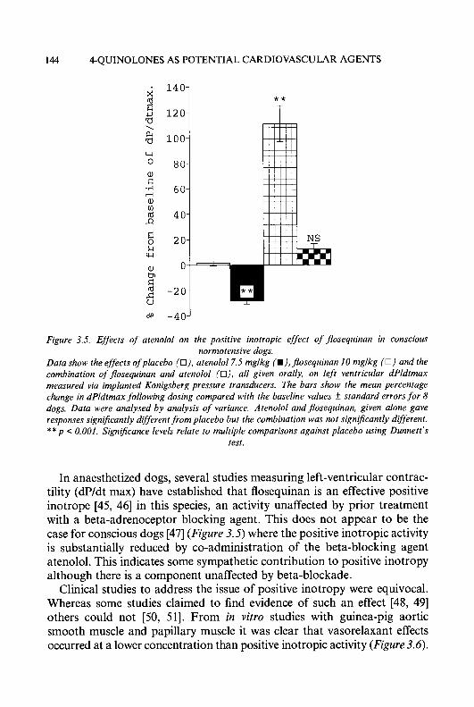
144 4-QUINOLONES AS POTENTIAL CARDIOVASCULAR AGENTS
140 i
? % 100
k w
a, tn C
-20 E -?
* * T
OW -40'
Figure 3.5. Effects of atenolol on the positive inotropic effect of gosequinan in conscious normotensive dogs.
Data show the effects ofplacebo (O), arenolol7.5 mglkg (W),jfosequinan I0 mglkg (0 ) and the combination of jfosequinan and atenolol (o), all given orally, on left ventricular dPldtmax measured via implanted Konigsberg pressure transducers. The bars show the mean percentage change in dPldtmax following dosing compared with the baseline values f standard errors for 8 dogs. Data were analysed by analysis of variance. Atenolol and gosequinan, given alone gave responses significantly different from placebo but the combination was not significantly different. ** p 0.001. Significance levels relate to multiple comparisons against placebo using Dunnett's
test.
In anaesthetized dogs, several studies measuring left-ventricular contrac- tility (dP/dt max) have established that flosequinan is an effective positive inotrope [45, 461 in this species, an activity unaffected by prior treatment with a beta-adrenoceptor blocking agent. This does not appear to be the case for conscious dogs [47] (Figure 3.5) where the positive inotropic activity is substantially reduced by co-administration of the beta-blocking agent atenolol. This indicates some sympathetic contribution to positive inotropy although there is a component unaffected by beta-blockade.
Clinical studies to address the issue of positive inotropy were equivocal. Whereas some studies claimed to find evidence of such an effect [48, 491 others could not [50, 511. From in v i m studies with guinea-pig aortic smooth muscle and papillary muscle it was clear that vasorelaxant effects occurred at a lower concentration than positive inotropic activity (Figure 3.6).
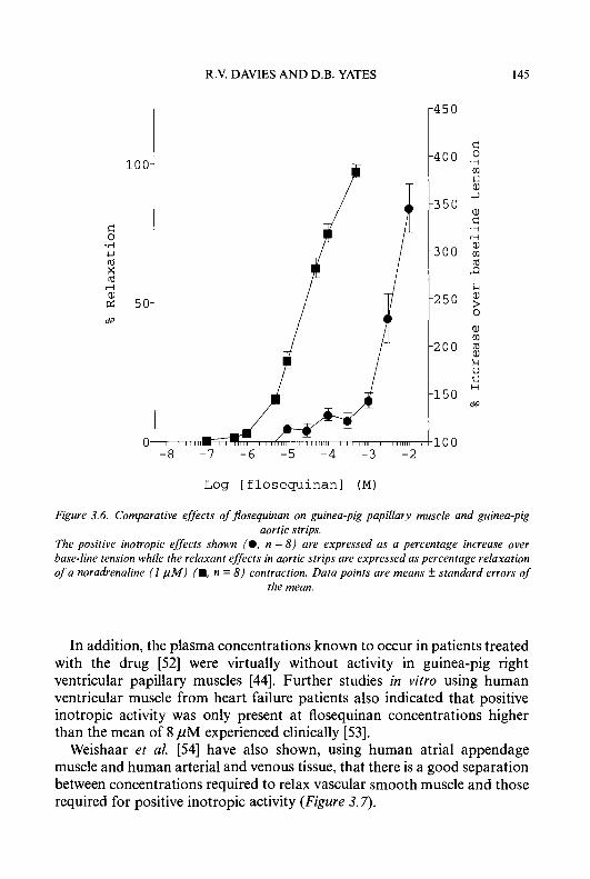
R.V. DAVIES AND D.B. YATES 145
c 0
.A . . U (d X (d
o0 1 -0 0-
5 0-
0 -8
7
d 0 .rl m 8 U
a, d
-A r-
a, 0 ld J2 k a, ? 0
a, m (d a, k V d H
Log [ f losequinan] (M)
Figure 3.6. Comparative effects of’jlosequinan on guinea-pig papillary muscle and guinea-pig aortic strips.
The positive inotropic effects shown (0, n = 8 ) are expressed as a percentage increase over base-line tension while the relaxant effects in aortic strips are expressed as percentage relaxation of a noradrenaline ( I p M ) (M, n = 8 ) contraction. Data points are means f standard errors of
the mean.
In addition, the plasma concentrations known to occur in patients treated with the drug [52] were virtually without activity in guinea-pig right ventricular papillary muscles [44]. Further studies in vitro using human ventricular muscle from heart failure patients also indicated that positive inotropic activity was only present at flosequinan concentrations higher than the mean of 8 pM experienced clinically [53].
Weishaar et al. [54] have also shown, using human atrial appendage muscle and human arterial and venous tissue, that there is a good separation between concentrations required to relax vascular smooth muscle and those required for positive inotropic activity (Figure 3.7).
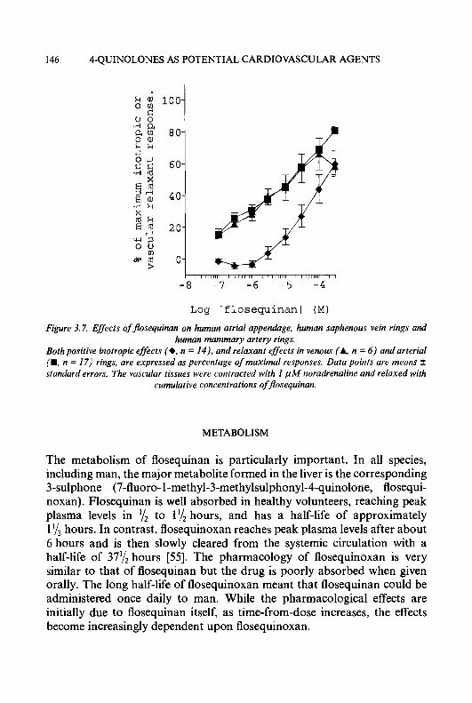
146 4-QUINOLONES AS POTENTIAL CARDIOVASCULAR AGENTS
I , , , , , , , , I , I l l , ,111 i l l l r r m I 1 1 1 1 1 1 1 1 I 1
- 8 -7 -6 -5 -4
Log [ f losequinan] (M)
Figure 3.7. Effects of flosequinan on human atrial appendage, human saphenous vein rings and human mammary artery rings.
Both positive inotropic effects (+, n = 14). and relaxant effects in venous (A, n = 6 ) andarterial (W. n = 17) rings, are expressed as percentage of maximal responses. Data points are means * standard errors. The vascular tissues were contracted with 1 p M noradrenaline and relaxed with
cumulative concentrations offlosequinan.
METABOLISM
The metabolism of flosequinan is particularly important. In all species, including man, the major metabolite formed in the liver is the corresponding 3-sulphone (7-fluoro-l-methyl-3-methylsulphonyl-4-quinolone, flosequi- noxan). Flosequinan is well absorbed in healthy volunteers, reaching peak plasma levels in to l'/Z hours, and has a half-life of approximately 1 '/2 hours. In contrast, flosequinoxan reaches peak plasma levels after about 6 hours and is then slowly cleared from the systemic circulation with a half-life of 371/, hours [55 ] . The pharmacology of flosequinoxan is very similar to that of flosequinan but the drug is poorly absorbed when given orally. The long half-life of flosequinoxan meant that flosequinan could be administered once daily to man. While the pharmacological effects are initially due to flosequinan itself, as time-from-dose increases, the effects become increasingly dependent upon flosequinoxan.

R.V. DAVIES A N D D.B. YATES 147
e 0
u U
c) G 0 60
-d 100-
80-
O L
G 0 -4
rd
2 80- c 0 u 60-
g 100-
O‘O o h -11 -10 - 9 - 8 -7 -6 -11 -10 - 9 - 8 -7
Log [noradrenaline] (M) Log [angiotensin 111 (M)
Figure 3.8. Ejfect of flosequinan on concentration-response curves to noradrenuline and angiotensin II in rat aortic strips.
Aortic strips. maintained in Krebs solution at 37°C. were contracted with noradrenaline or angiotensin to yield concentration-response curves (0). These were repeated in the presence of ~7osequinan 10 p M (A) and 50 p M I+) . The left-/iandpanel shows the ej/kcts on noradrenaline dose-responses (11 = 6 ) while the right-hand panel shows ejyects on angiotensin I I ( n = 23). Data
points are means * SEM.
MODE OF ACTION
Vasorelaxant activity
Flosequinan has been shown to relax isolated smooth muscle of several species, including man. In rat aortic strips, flosequinan was capable of relaxing contractions due to a variety of agonists. It was most potent in relaxing contractions due to receptor-operated mechanisms; thus the EC,, for relaxing noradrenaline or phenylephrine contractions was about 10 pM while the EC,, for relaxing potassium chloride contractions was 80 pM. It was also possible to shift dose-response curves to various agonists to the right, sometimes with reduced maximum responses [56] (Figure 3.8). Other studies have indicated that flosequinan does not bind to any receptors [57] so the drug may affect intracellular mechanisms responsible for contraction. Since extracellular-dependent KCl contractions are less potently affected than contractions due to vasoconstrictors such as phenylephrine, flosequi- nan may be preferentially affecting intracellular Ca” translocation. In addition, because the EC,, for relaxation of a noradrenaline contraction in Ca’+-replete Krebs is similar to that for noradrenaline in Ca2+-free Krebs, it implies that contraction due to extracellular Ca” is little affected [58] . On the other hand, contractions due to BHT-920, an a,-agonist which is
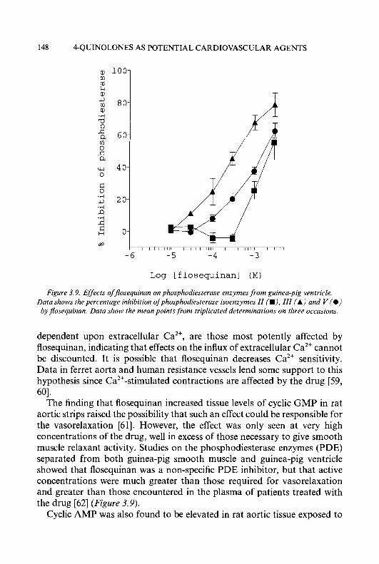
148 4-QUINOLONES AS POTENTIAL CARDIOVASCULAR AGENTS
rd k aJ u cl) aJ
.rl a 0 s a (I) 0 s a
60 1 4 0-
2 0-
0-
I I , 1 1 1 1 1 1 , I I I , , , , , , I , 1 1 , 1 1 1 1 I I I ,
-6 -5 - 4 -3
Log [ f losequinan] (M)
Figure 3.9. Effkcts offlosequinan on phosphodiesterase enzymes,from guinea-pig ventricle. Data shows the percentage inhibition of phosphodiesterase isoenzymes 11 (W), 111 (A) and V (0) by flosequinan. Data show the mean points j i o m triplicated determinations on three occasions.
dependent upon extracellular Ca2', are those most potently affected by flosequinan, indicating that effects on the influx of extracellular Ca2' cannot be discounted. It is possible that flosequinan decreases Ca2+ sensitivity. Data in ferret aorta and human resistance vessels lend some support to this hypothesis since Ca2'-stimulated contractions are affected by the drug [59, 601.
The finding that flosequinan increased tissue levels of cyclic GMP in rat aortic strips raised the possibility that such an effect could be responsible for the vasorelaxation [61]. However, the effect was only seen at very high concentrations of the drug, well in excess of those necessary to give smooth muscle relaxant activity. Studies on the phosphodiesterase enzymes (PDE) separated from both guinea-pig smooth muscle and guinea-pig ventricle showed that flosequinan was a non-specific PDE inhibitor, but that active concentrations were much greater than those required for vasorelaxation and greater than those encountered in the plasma of patients treated with the drug [62] (Figure 3.9).
Cyclic AMP was also found to be elevated in rat aortic tissue exposed to

R.V. DAVIES A N D D.B. YATES 149
high concentrations of flosequinan and it seems likely that nonspecific PDE inhibition is probably responsible both for this and for the cyclic GMP elevation noted above.
Further studies carried out in rat aortic smooth muscle showed flosequinan was capable of reducing the noradrenaline-induced increase in inositol monophosphate [63]. This indicated that there was probably a reduction in the generation of inositol 1,4,5-trisphosphate (IP,), an important intracellular secondary messenger responsible for the release of intracellular Ca2+ [64-661. This theory was given greater credence when Lang and Lewis [67] demonstrated that endothelin-induced IP, increases in rat aortic smooth muscle were reduced concentration-dependently by flosequinan.
There were therefore indications that flosequinan reduced agonist- induced intracellular Ca2' release. Functional evidence was consistent with this but also suggested effects on extracellular influx of Ca2' stimulated by certain a-agonists. Such effects might be mediated by interaction with G-proteins known to control these systems. The finding that flosequinan could relax contractions due to sodium fluoride/aluminium chloride at concentrations similar to those required to relax contractions due to agents acting through receptors lent support to this theory, since sodium fluoride/aluminium chloride are known to produce contractions via stimulation of G-proteins linked to phospholipase-C [68, 691.
Cardiac tissue
Flosequinan is capable of causing both positive inotropic and chronotropic effects though these are only seen at relatively high concentrations. Since PDE inhibition is also seen at high concentrations it is tempting to link the two effects. However, Weishaar et al. [70] conclude that the effects of flosequinan are not due to PDE inhibition following the demonstration of several clear differences between effects due to the drug and to those due to milrinone, a well-known PDE inhibitor. In addition, Miao et al. [71, 721 have implicated Na'/Ca2' exchange mechanisms by showing that the positive inotropic effects of flosequinan are susceptible to blockade by agents such as cadmium and amiloride which are known to inhibit Na'/Ca2' exchange [73, 741.
CLINICAL STUDIES
Early clinical studies established that flosequinan produced beneficial haemodynamic changes in heart failure patients [75]. It is recognized that
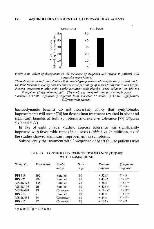
150 4-QUINOLONES AS POTENTIAL CARDIOVASCULAR AGENTS
Dyspnoea Fatigue
Figure 3.10. Ej’ect offlosequinan on the incidence of dyspnoea and fatigue in patients with congestive heart failure.
These data are taken from a double-blind parallel group sequential analysis study carried out by DI: Paul Nicholls in twenty patients and show the percentage of scores for dyspnoea and fatigue showing improvement after eight weeks treatment with placebo (open columns) or I00 mg
flosequinan (filled columns) daily. This study was analysed using a two-sample t-test. * denotes p < 0.05, significantly different from placebo; ** denotes p < 0.01, significantly
different.from placebo.
haemodynamic benefits do not necessarily imply that symptomatic improvements will occur [76] but flosequinan treatment resulted in clear and significant benefits in both symptoms and exercise tolerance [77] (Figures 3.10 and 3.11).
In five of eight clinical studies, exercise tolerance was significantly improved with favourable trends in all cases (Table 3.9). In addition, six of the studies showed significant improvement in symptoms.
Subsequently the treatment with flosequinan of heart failure patients who
Table 3.9. CONTROLLED EXERCISE TOLERANCE STUDIES WITH FLOSEQUINAN
Study No. Patient No. Study Dose Exercise Symptoms design ( m d response response
BPI 919 BPI 925 MS 86/122 MS 85/107 MS 84/099 BPI 916 MS 86/085 BPI 917
190 298 135 20 13 21 14 22
Parallel Parallel Parallel Parallel Crossover Parallel Crossover Crossover
100 100 125 100 125 I00 I00 100
+ 52 s* + 45 s* + 38 st + 328 s* + 165 s* +41 s + 54 s + 135s
F > P F > P* F > P* F > P * F > P* F > P* F > P* F > P
* p 5 0.05; p > 0.05 5 0.1
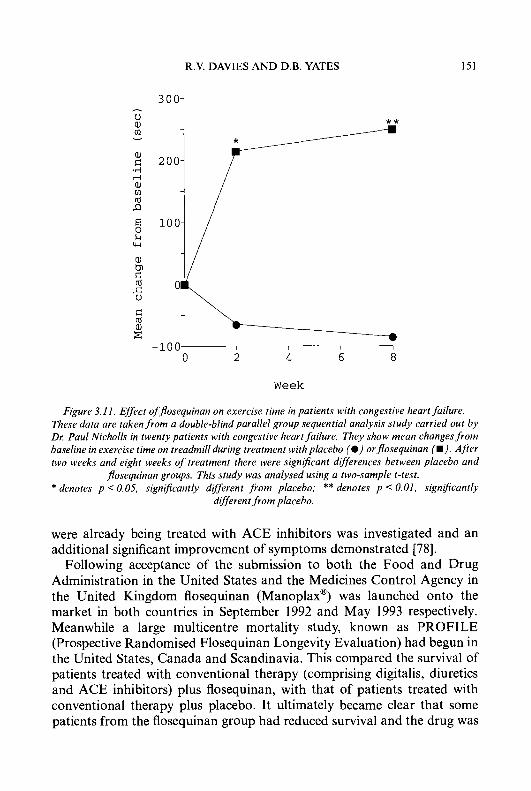
R.V. DAVIES A N D D.B. YATES
aJ
151
d rd aJ c
Week
Figure 3.11. Effect offlosequinan on exercise time in patients with congestive heart failure. These data are taken from a double-blind parallel group sequential analysis study carried out by Dr Paul Nicholls in twenty patients with congestive heart failure. They show mean changes from baseline in exercise time on treadmill during treatment with placebo (@) orflosequinan I.). Afier two weeks and eight weeks of treatment there were significant differences between placebo and
jlosequinan groups. This study was analysed using a two-sample t-test. * denotes p < 0.05, signiJicantly dij'erent from placebo; ** denotes p < 0.01, signlficantly
dfferent from placebo.
were already being treated with ACE inhibitors was investigated and an additional significant improvement of symptoms demonstrated [78].
Following acceptance of the submission to both the Food and Drug Administration in the United States and the Medicines Control Agency in the United Kingdom flosequinan (Manoplax@) was launched onto the market in both countries in September 1992 and May 1993 respectively. Meanwhile a large multicentre mortality study, known as PROFILE (Prospective Randomised Flosequinan Longevity Evaluation) had begun in the United States, Canada and Scandinavia. This compared the survival of patients treated with conventional therapy (comprising digitalis, diuretics and ACE inhibitors) plus flosequinan, with that of patients treated with conventional therapy plus placebo. It ultimately became clear that some patients from the flosequinan group had reduced survival and the drug was

152 4-QUINOLONES AS POTENTIAL CARDIOVASCULAR AGENTS
withdrawn from sale in both the United States and the United Kingdom in July 1993. The PROFILE study is still under detailed evaluation and the results will be published in due course.
CONCLUSION
Certain 4-quinolones have been shown to possess antihypertensive or hypotensive activity in SH rats, RH dogs and normotensive cats. The pharmacophore required for activity may be summarized by the structure (89). Several analogues were selected for further evaluation as potential antihypertensive agents but only compound (3) (flosequinan: Manoplax@) was developed for use in patients. Due to its particular haemodynamic profile, which combined both arterial and venous vasodilating activity, flosequinan was evaluated in heart failure patients and although subse- quently marketed for use in this condition in the UK and USA, was withdrawn due to adverse effects on survival in PROFILE.
0
Ri = Me, occasionally Et
R3 = Certain hydrophilic groups of S 4 atom chain length
R6 = Certain substituents in combination with 7-substituents
R7 = Variety of substituents
(89)
ACKNOWLEDGEMENTS
The authors are indebted to their co-workers and colleagues, too numerous to mention, to Prof. W.R. Buckett for critical reading of the manuscript and to Mrs. P. Johnson and Miss H. Sanders for their patience in the preparation of the typescript.
R.V. DAVIES AND D.B. YATES 153
REFERENCES
1 2
3 4
5
6
7 8
9 10 1 1 12 13 14 15 16 17 18
19
20
21
22
23 24 25 26 27 28 29
30 31 32 33
34
World Health Statistics Annual 1990. Published by World Health Organisation, Geneva. International Cardiomonitor Database Spring 1992 issue, copyright Taylor Nelson and Boots’ marketing research projections. Weidman, P., Velinger, D. and Gerber, A. (1985) J. Hypertens. 33,297-306. Martin, M.J., Hulley, S.B., Browner, W.S., Kuller, L.H. and Wentworth, D. (1986) Lancet ii, 933-936. Prichard, B.N.C., Owens, C.W.I. and Woolf, A.S. (1992) Eur. Heart J. 13 (suppl G), 96103. Bangdiwala, S.I., Bourassa, M.G., Friesinger, G., Weiner, D.H. and Yusuf, S. (1992) Am. J. Cardiol. 70, 347-353. Kannel, W.B. (1989) Cardiol. Clinics 7, 1-9. Pless, M., Juranek, D., Kozarsky, P., Steurer, F., Tapia, G. and Bermudez, H. (1992) J. Trop. Med. Hyg. 47, 539-546. Hansen, B.F. (1992) Acta Path. Microbiol. Immunol. Scand. 100,273-279. SOLVD Investigators. (1991) N. Engl. J. Med. 295,293-302. Furberg, C.D. and Yusuf, S. (1988) Am. J. Cardiol. 62,41A-45A. The CONSENSUS TRIAL STUDY GROUP. (1987) N. Engl. J. Med. 316,1429-1435. Abrams, M. and Sobin, S. (1947) Proc. SOC. Exp. Biol. Med., 64,412416. Parkinson, R. (1979) Br. J. Pharmacol. 64,437P. Yates, D.B. (1982) Br. J. Pharmacol. 77,303P. Van Leusen, A.M. and Taylor, E.C. (1968) J. Org. Chem. 33,6670. Adams, W.J. and Hey, D.H. (1951) J. Chem. SOC. 1521-1527. Hansch, C., Leo, S.H., Unger, K.H., Kim, K.H., Nikaitani, D. and Lien, E.J. (1973) J. Med. Chem. 16, 1207-1216. Connor, D.T., Young, P.A. and von Strandtman, M. (1978) J. Heterocycl. Chem. 15, 115-117. Davies, R.V., Fraser, J., Nichol, K.J., Parkinson, R., Sim. M.F. and Yates, D.B. (1980) G.B. Patent 2047691B; (1981) Chem. Abstr. 94, 83968; Davies, R.V. and Robinson, K. (1991) W.O. Patent 91/02724; (1991) Chem. Abstr. 115,92095. Birch, A.M., Davies, R.V., (the late) Maclean, L. and Robinson, K. (1994) J. Chem. SOC. Perkin I , 387-392. Bachman, G.B., Welton, D.G., Jenkins, G.L. and Christian, J.E. (1947) J. Am. Chem.
Schofield, K. and Swain, T. (1949) J. Chem. SOC. 1367-1371. Schofield, K. and Theobald, R.S. (1950) J. Chem. SOC. 395402. Price, J.R. (1949) Aust. J . Sci. Res. 2A, 272-280. Gould, R.G. and Jacobs, W.A. (1939) J. Am. Chem. SOC. 61,2890-2895. Schofield, K. and Simpson, J.C.E. (1946) J. Chem. SOC. 1033-1035. Price, C.C. and Roberts, R.M. (1 946) J. Am. Chem. SOC. 68, 1204-1 208. Renault, S., Renault, J., Cavier, R. and Delage, N. (1976) Eur. J. Med. Chem. 11,
Baker, B.R. and Bramhall, R.R. (1972) J. Med. Chem. 15,233-235. Albrecht, R. (1977) Prog. Drug Res., 21. 9-104. Yanagisawa, H., Nakao, H. and Ando, A. (1973) Chem. Pharm. Bull. 21, 1080-1089. Davies, R.V., Fraser, J. and Nichol, K.J. (1988) U.S. Patent 4772614; (1987) Chem. Abstr. 107, 58881. Davies, R.V. (1989) U.S. Patent 485529; (1986) Chem. Abstr. 105, 97340.
SOC. 67,365-371.
555-560.
1 54 4-QUINOLONES AS POTENTIAL CARDIOVASCULAR AGENTS
35 36
37
38 39
40
41
42 43
44 45 46 47
48
49
50
51
52 53
54
55
56 57
58 59 60 61 62 63 64 65
66 67
Vorbruggen, H. and Strehlke, P. (1973) Chem. Ber. 106, 305553061, Davies, R.V. and Green, R.D. (1984) G.B. Patent 2147581A; (1985) Chem. Abstr. 103, 104975. Davies, R.V. (1981) U.S. Patent 4447435; (1982) Chem. Abstr. 97, 23645; and Davies, R.V. and Robinson, K. (1991) W.O. Patent 91/02724; (1991) Chem. Abstr. 115,92095. Evans, D.J. and Eastwood, F.W. (1974) Aust. J. Chem. 27, 537-542. Davies, R.V., Brown, M.D. and Holman, N.J. (1991) U S . Patent 4997840; (1990) Chem. Abstr. 112, 76971. Sim, M.F., Yates, D.B., Parkinson, R. and Cooling, M.J. (1988) Br. J. Pharmacol. 94, 371-380. Yates, D.B., Parkinson, R., Casson, H.C.R. and Sim, M.F. (1988) Br. J. Pharmacol. 95, 8 18P. Yates, D.B. and Hicks, G.R. (1988) Br. J. Pharmacol. 95, 71 IP. Perreault, C.L., Hague, N.L., Loh, E., Hunneyball, I.M., Sim, M.F. and Morgan, J.P. (1992) Br. J. Pharmacol. 106, 511-516. Fry, C.H. and Sim, M.F. (1992) Br. J. Pharmacol. 107, 142P. Greenberg, G. and Touhey, B. (1990) J. Cardiovasc. Pharmacol. 15, 900-910. Falotico, R., Haertlein, B., Lakas-Weiss, C. and Tobia, A.J. (1988) FASEB J. 2,410. Yates, D.B., Parkinson, R., Casson, H. and Sim, M.F. (1990) J. Mol. Cell Cardiol. 22 (suppl HI), SI 10. Burstein, S., Semigran, M.J., Dec, G.W., Boucher, C.A. and Fifer, M.A. (1992) J. Am. Coil. Cardiol. 20, 822-829. Corin, W.J., Monrad, E.S., Strom, J.A., Giustino, S., Sonnenblick, E.S. and Lejemtel, T. (1991) Am. Heart J. 121,537-549. Riegger, G.A.J., Kahles, H., Wagner, A,, Kroman, E.P., Elsner, D. and Kochsiek, K. (1990) Cardiovasc. Drugs Ther. 4, 1395-1402. Haas, G.J., Binkley, P.F., Carpenter, J.A., Leier, R.N. and Leier, C.V. (1989) Am. J. Cardiol. 63, 13541359. Kessler, P. and Packer, M. (1987) Am. Heart J. 113, 137-143. Weishaar, R.E., Wallace, A.M., Kirker, M.L., Ferraris, V.A. and Sim, M.F. (1991) Br. J. Pharmacol. 104, 18P. Weishaar, R.E., Kiser, L.A., Wallace, A.M., Ferraris, V.A. and Sim, M.F. (1993) Eur. Heart J. 14, 258. Wynne, R.D., Crampton, E.L. and Hind, E.D. (1985) Eur. J. Clin. Pharmacol. 28, 659-664. Yates, D.B. and Holmes, J.R. (1993) Br. J. Pharmacol. 109, 137P. Sim, M.F., Yates, D.B., Allcock, A.A., Parkinson, R., Heal, D.J. and Needham, P.L. (1988) 12th Scientific Meeting of the International Society of Hypertension, Kyoto, Japan. Abstract No. 0747. Yates, D.B. (1988) Br. J. Pharmacol. 94,431P. Resnick, M.S., Maitland, L.A. and Morgan, K.G. (1991) Br. J. Pharmacol. 102,974978. Richards, N.T., Poston, L. and Hilton, P.J. (1989) Br. J. Pharmacol. 98, 734P Allcock, A.A., Frodsham, G. and Sim, M.F. (1988) Br. J. Pharmacol. 94,430P. Frodsham, G. and Jones, R.B. (1992) Eur. J. Pharmacol. 21 I , 383-391. Yates, D.B., Hicks, G. and Sim, M.F. (1989) Br. J. Pharmacol. 96, 112P. Berridge, M.J. and Irvine, R.F. (1984) Nature (London) 312, 315-321. Yamamoto, H. and van Breeman, C. (1985) Biochem. Biophys. Res. Commun. 130, 270-274. Bingham-Smith, J., Smith, L. and Higgins, B.L. (1985) J. Biol. Chem. 260, 14413-14416. Lang, D. and Lewis, M.J. (1992) Eur. J. Pharmacol. 226,259-264.

R.V. DAVIES AND D.B. YATES 155
68 69 70
71 72 73
74 75 76 77
78
Sternweiss, P.C. and Gilman, A.G. (1982) Proc. Natl. Acad. Sci. U.S.A. 99, 48884891. Hall, I.P., Donaldson, J. and Hill, S.J. (1990) Br. J. Pharmacol. 100, 646-650. Weishaar, R.E., Kirker, M.L., Wallace, A.M.. Britton, A.W., Ferraris, V.A. and Sim, M.F. (1992) Br. J. Pharmacol. 107, 141P. Miao, L., Hurrell, D.G., Perrault, C.L. and Morgan, J.P. (1992) Circulation 86, 2476. Miao, L., Perrault, C.L. and Morgan, J.P. (1 993) J. Am. Coll. Cardiol. 21, 949-950. Seigl, P.K.S., Cragoe Jr., E.J., Trimble, M.J. and Kacjorowski, G.J. (1984) Proc. Natl. Acad. Sci. U.S.A., 81, 3238-3242. Kimura, J., Migamae, S. and Norma, A. (1987) J. Physiol. (London) 384, 199-222. Elborn, J.S., Stanford, C.F. and Nicholls, D.P. (1989) Br. Heart J. 61, 331-335. Lipickey, R.J. and Packer, M. (1993) J. Am. Coll. Cardiol. 4 (suppl A), 179A-184A. Elborn, J.S., Riley, M., Stanford, C.F. and Nicholls, D.P. (1990) Br. J. Clin. Pharmacol. 29, 5 19-524. Massie, B.M. Berk, M.R., Brozena, S., Elkayam, U., Plehn, J., Kukin, M., Murphy, B., Neuberg, G., Steingart, R. and DeHaan, H. (1992) Circulation 86, 2565.
Progress in Medicinal Chemistry - Vol. 32, edited by G.P. Ellis and D.K. Luscombe 0 1995 Elsevier Science B.V. All rights reserved.
4 Biochemical Mechanisms of Resistance to Non-cell Wall Antibacterial Agents
NORRIS E. ALLEN, Ph.D
Infectious Disease Research, Eli Lilly and Company, Indianapolis, IN 46285, U. S. A.
INTRODUCTION
GENETIC BASIS OF RESISTANCE
BIOCHEMICAL BASIS OF RESISTANCE Inhibitors of membrane function
Polymyxins Cyclic depsipeptides, macrotetralides and other ionophores
Sulphonamides and diaminopyrimidines
Aminoglycosides Tetracyclines Chloramphenicol Fusidic acid Macrolides, Lincosamides and Streptogramins Thiostrepton
Ansamycins Quinolones Coumarins Nitrojurans and nitroimidazoles
Antifolates
Inhibitors of ribosome function
Nucleic acid inhibitors
CONCLUDING REMARKS
ACKNOWLEDGEMENTS
REFERENCES
INTRODUCTION
157
159
161 162 162 167 171 171 175 175 185 193 196 198 205 207 207 210 216 219
22 1
223
223
Alexander Fleming discovered penicillin in 1928 and reported his findings in the British Journal of Experimental Pathology the following year [l]. More than ten years elapsed before Abraham et al. [2] showed that small amounts
157

158 RESISTANCE TO NON-CELL WALL ANTIBACTERIALS
of this antibiotic, painstakingly isolated, successfully treated patients with severe infections. The profound life-saving benefits of penicillin were thus demonstrated, and the discovery inaugurated the present antibiotic era [3]. Even at this early date, the ability of bacteria to resist the inhibitory effects of antibacterial agents was observed and demonstrated. In 1940, Abraham and Chain [4] described an enzyme from bacteria able to destroy penicillin. They called the enzyme penicillinase. The variety and frequency of anti- bacterial resistance has seemingly been increasing ever since. To compound the problem, resistance in hospital isolates today is rarely limited to single agents. Some strains of Staphylococcus aureus are resistant to as many as 20 antibacterial compounds [5].
Neu [6] recently cited some 158 antibacterial agents used for the treatment of infectious diseases. Antibacterial agents have been introduced to treat most if not all bacterial infections in man, yet the record shows that emergence of resistance has invariably followed the clinical introduction of each one. Streptomycin was described in 1944 and resistance development was reported soon thereafter in 1946 [7]. On the other hand, emergence of resistance to vancomycin took thirty years to appear following its introduction [8, 91. Development of resistance has been a matter of when, rather than if. Now that resistance to vancomycin has appeared, no single class of clinically important antibacterial agent is without a known mechanism of resistance. Neu [6] has said ‘the need for new antibiotics will continue because bacteria have a remarkable ability to overcome each new agent synthesized’.
Today, the problem of antibiotic resistance is more acute than at any other time in the antibiotic era. In spite of the wealth of antibacterial agents available to clinicians, the frequency at which antibiotic resistance develops is increasing. There is also the particularly problematic emergence of multiple drug resistance in many different bacteria, and the pronounce- ments from some quarters that fewer new drugs are being developed as antibacterials. The dilemma led Cohen [lo] to suggest, ‘...we may be approaching the post-antimicrobial era’.
What are the peculiar characteristics of bacteria which have made this possible? Development of resistance to antibacterial agents is facilitated because of genetic and biochemical properties which, taken together, reflect the remarkable genetic versatility of bacteria. Specifically: (1) bacteria have relatively short generation times (about 20 minutes for Escherichiu coli). This leads to large numbers of generations in a short time period providing enormous opportunity for selection of chromosomal mutations. (2) Bacteria are capable of taking up and expressing large pieces of foreign DNA from other bacteria. (3) Bacteria have the abilty to transfer their own DNA as
N.E. ALLEN 159
well as foreign DNA to other bacteria. And, (4) bacteria can often control the expression and transmission of resistance genes by sophisticated mechanisms regulated by the very agent to which resistance is conferred. For example, derepression of transcription of the tetracycline efflux pump by tetracycline, and the induction of transposition of MLS resistance in Enterococcus by erythromycin. In addition to the biological properties of bacteria, development and spread of resistance occurs because usage of antibiotic agents to treat infectious disease provides the negative pressure needed to select resistant variants. Of equal importance are the societal demographics and technological changes occurring in the modern world which greatly influence the spread of resistance.
This review describes the biochemical mechanisms which provide the basis for resistance to antibiotics and other antibacterial agents*. A few comments regarding the genetic basis of resistance are included, but the reader is urged to consult other sources, including references [l l-151, for more detailed treatments of this important topic. Brief descriptions of specific biochemical mechanisms of action and relevant transport mecha- nisms will be given for each group of agents described. Those agents which act on the bacterial cell wall peptidoglycan have been excluded and will comprise the topic for a separate review.
GENETIC BASIS OF RESISTANCE
What are the origins of antibiotic resistance genes (where do they come from), and how are these genes disseminated (how are they spread among different bacteria)? Genetic determinants for resistance are intrinsic or are acquired as new information. Intrinsic resistance to antibiotics is determined by genetic and biochemical factors peculiar to a specific group of bacteria. It is usually genus specific (for example, resistance of Serratiu to the
* The following abbreviations are used in this review: acetyl-CoA, acetyl-coenzyme A; ATP, adenosine-5’-triphosphate; AAC, aminoglycoside acetyltransferase; AAD, aminoglycoside adenylyltransferase; ANT, aminoglycoside nucleotidyltransferase; APH, aminoglycoside phosphotransferase; CAMP, 3’,5’-cyclic adenosine monophosphate; CAP, catabolite gene activator protein; CAT, chloramphenicol acetyltransferase; CCCP, carbonylcyanide m- chlorophenylhydrazone; DCCD, dicyclohexylcarbodimide; DNA, deoxyribonucleic acid; EDTA, ethylenediaminetetraacetic acid; EFG, elongation factor G; fmet, N-formylmethionine; GDP, guanosine-5’-diphosphate; kDa, kilodalton; LPS, lipopolysaccharide; MLS, macrolide, lincosamide and streptogramin B-type antibiotics; mRNA, messenger ribonucleic acid; NADH, nicotinamide adenine dinucleotide, reduced form; NADPH, nicotinamide adenine dinucleotide phosphate, reduced form; PAB, p-aminobenzoic acid; RNA, ribonucleic acid; rRNA, ribosomal ribonucleic acid; tRNA, transfer ribonucleic acid.
160 RESISTANCE TO NON-CELL WALL ANTIBACTERIALS
aminoglycoside antibiotics) but can be specific to a larger grouping (for example, resistance of Gram-negative bacteria to macrolide antibiotics). Because intrinsic resistance does not require the acquisition of new genetic information, it may be less confusing if organisms having this form of resistance are referred to as ‘insensitive’.
Antibiotic resistance is understood to be acquired when a bacterial population, normally sensitive to an antibacterial agent, becomes resistant to that agent by virtue of obtaining new genetic information. This occurs either by mutation or through the acquisition of foreign DNA. In this review, those cases where a population becomes resistant due to selection of an existing rare individual in a population (for example, one producing a chromosomally determined antibiotic-inactivating enzyme), will be consid- ered as examples of acquired rather than intrinsic resistance.
Mutations conferring resistance to antibacterial agents generally occur in chromosomal DNA (for example, mutations in the DNA gyrase gene to confer quinolone resistance). Chromosomal mutations conferring resistance can be selected in the laboratory and occur at frequencies of to lo-* depending on the specific gene(s) involved and the antibacterial agent. In contrast to mutations, resistance resulting from the acquisition of foreign DNA containing resistance genes can only be demonstrated in the laboratory in controlled experiments where the donor DNA is known to contain a resistance gene.
Resistance determinants can be acquired as fragments of chromosomal DNA introduced and integrated into the new host by a variety of mechanisms (see below). However, many more resistance determinants seem to be carried on extrachromosomal pieces of DNA known as plasmids which replicate autonomously but which may also be integrated into the host chromosome. Transposons represent yet another element on which resistance genes can reside. Transposons are DNA sequences incapable of independent replication which must be maintained as part of a bacterio- phage, plasmid or host chromosome. The identifying characteristic of transposons is that they carry information to transfer themselves (by transposition) between DNA molecules.
What physical processes do bacteria use to acquire and exchange these pieces of foreign DNA? The extent to which these processes operate is a major determinant of how widespread resistance to a particular agent becomes. DNA is exchanged between bacteria by conjugation, transduction or transformation. Conjugation requires cell to cell contact between distinct mating types with subsequent exchange of genetic material (chromosomal and/or extrachromosomal) from donor to recipient. Conjugation is most often controlled by conjugative plasmids which carry the information

N.E. ALLEN 161
needed for mating and transfer. Many of these plasmids also carry resistance determinants. Some resistance determinants are carried on nonconjugative plasmids; transfer of nonconjugative plasmids requires mobilization which is possible when the plasmid co-resides in a cell with a conjugative plasmid.
Transduction is the process where genes are transferred by bacteriophage particles. Temperate bacteriophages can be maintained in a prophage state as an integral part of the host DNA. Transduction occurs when prophage DNA is excised and the viral DNA picks up and carries with it a fragment of host DNA. The fragment can then be transferred to other bacteria when the bacteriophage infects another cell. The third method for exchanging DNA, transformation, involves uptake of naked DNA by bacterial cells. Each of the transfer methods (conjugation, transduction and transforma- tion) has the potential to act as a mechanism for disseminating resistance genes; conjugation is thought to be the most common mechanism.
BIOCHEMICAL BASIS OF RESISTANCE
The occurrence of genetic mutations and the introduction of pieces of foreign DNA which lead to specific antibacterial resistance represent changes in the gene pool of bacterial populations. These changes get translated into new or altered gene products which ultimately change the biochemistry of cells in a manner that assures survival in the presence of specific antibacterial agents. A description of those biochemical mecha- nisms is the focus of this review.
The biochemical mechanisms bacteria employ to resist the action of specific antibacterial agents can be divided into three broadly defined categories: ( 1 ) target alterations, (2) transport alterations and (3) enzymatic modiJcation. The variety of mechanisms employed by bacteria to resist a given agent or class of agents reflects the genetic ingenuity and versatility of the organism as much as anything else. In principle, the mechanisms are the same regardless of whether they represent a mechanism of intrinsic resistance or an acquired property. In practice, certain mechanisms (for example, enzymatic modification) are rarely encountered intrinsically.
Target alterations. The action of antibacterial agents is directed against specific subcellular targets. Agents bind to, block, or in some way alter those targets in a manner resulting in inhibition of target function. Both mutations and acquired genes can encode alterations in a target conferring resistance to an agent (or class of agents) which inhibit the normal (wild type) target. In order to insure survival, however, the alteration cannot be so

162 RESISTANCE TO NON-CELL WALL ANTIBACTERIALS
severe that it compromises the biochemical function of the target. Target alterations can include changes in the structure of an enzyme or other protein and can involve modification of the active site or an allosteric site. Target alterations can also take the form of duplication of a target to give an inhibitor-resistant form.
Transport alterations. In order to exert an inhibitory effect, an antibacterial agent must reach the cellular site where its target is located. In some cases, especially for cytoplasmic targets, an agent may utilize a cellular transport system normally used to accumulate nutrients. Bacteria can sometimes modify these transport systems to prevent the agent from reaching its target. Again, the modification cannot be so severe that it destroys the normal function of an essential transport mechanism. Alternatively, bacteria may modify their envelope structure in such a way as to exclude an antibacterial agent. Transport alterations and exclusion mechanisms include loss of a carrier function, change in composition of the cytoplasmic (inner) membrane, appearance of an efflux mechanism to transport a toxic agent out of the cell, or changes in the structure of the outer membrane of Gram-negative bacteria.
Enzymatic mod$carion. The third general mechanism of resistance is chemical inactivation of the antibacterial agent. The capability of an agent to reach its target and to interact with and inhibit the target once it gets there, is very much determined by the chemical structure of the agent. A common mechanism of resistance in bacteria results from the acquisition of genetic determinants encoding specific enzymes which modify antibacterial agents and nullify their inhibitory properties. The modifications include hydrolyses, phosphorylations, nucleotidylations, acetylations and glycosyl- ations among others. These modifications can affect both transport properties and interactions at the target site. There are also cases where resistance is conferred by alteration of specific modifying enzymes which normally activate a given class of agents.
INHIBITORS OF MEMBRANE FUNCTION
Polymyxins
The polymyxins are cyclic lipopeptide antibiotics. Closely related members of this class differ with respect to amino acid and acyl side-chain composition. Polymyxins are polycationic, have a high content of 2,4-diaminobutyric acid, and are acylated with fatty acids having 8-14 carbons. Polymyxin B (l), which consists of 10 amino acids acylated at the N-terminus with 6-methyloctanoic acid, is the only polymyxin employed in

N.E. ALLEN 163
;""\
I Dab Dab I D-qhF Thr Dbb
'Dab'
Dab I
Thr I
Dab I NH
I
mechanism studies [16]. The octapeptins, including EM 49 [17], share similar structures but contain 8 amino acids (one amino acid in the stem portion instead of 3) and C-10 or C-1 1 P-hydroxy-fatty acid side-chains.
The polymyxins have much greater antimicrobial activity against Gram-negative than Gram-positive bacteria. Early studies on polymyxin B indicated that the antibiotic has a permeabilizing effect on susceptible bacteria [16, 181. Exposure to polymyxin effects changes in cell permeability resulting in release of intracellular constituents such as K'. The cationic nature of the antibiotic suggested that its mechanism of action could involve an interaction with negatively charged, anionic groups on the cell surface. Although early studies showed that polymyxin B had various effects on cell physiology, it is generally assumed that these effects are due to actions secondary to a direct effect on the cytoplasmic membrane.
LaPorte et al. [19] demonstrated that polymyxin B covalently linked to agarose beads permeabilized susceptible bacteria indicating that the antibiotic exerted its effects from the cell surface and did not need to penetrate to the cell cytoplasm. Several studies have shown that polymyxin permeabilizes the outer membrane of Gram-negative bacteria releasing components of the outer membrane by interacting specifically with the LPS layer [20-221. Whereas a short time exposure to polymyxin releases materials only from the periplasm, longer exposures lead to the release of cytoplasmic components [23]. Lack of activity against Gram-positive bacteria further supports an important role for the outer membrane in determining susceptibility to polymyxin.
Current understanding of the mechanism of action of polymyxin and octapeptins is that these antibiotics displace divalent cations (Mg2+ and

164 RESISTANCE TO NON-CELL WALL ANTIBACTERIALS
Ca”) from the surface layers of Gram-negative bacteria. The outer monolayer of the outer membrane consists of a rigid, highly charged, anionic LPS. The structural integrity of the LPS is thought to be maintained by divalent cation bridges between phosphate groups of phospholipids and/or the LPS [24, 251. The polycationic nature of polymyxin facilitates an initial interaction with the negatively charged LPS (core-lipid A region) resulting in disorganization of the outer membrane [26, 271. Disruption of the outer membrane exposes the inner, cytoplasmic membrane to poly- myxin. The net result is loss of selective permeability of the cytoplasmic membrane leading to cell death. Our understanding of the molecular basis of polymyxin’s interaction with the cytoplasmic membrane is limited, but there is no evidence for a specific receptor protein for polymyxin [18].
Mechanisms of resistance to polymyxins have been subdivided for this review into examples of intrinsic resistance, antagonism, adaptation and mutational resistance. The known mechanisms of resistance to polymyxins are more alike than different. Although all involve an effect or change in cellular membranes or envelope structure, and therefore appear to be target alterations, some of these mechanisms could be blocking polymyxin and preventing it from reaching a site of action in the membrane.
Intrinsic resistance. The lack of susceptibility of Gram-positive bacteria to polymyxin has been explained as being due to cell wall peptidoglycan acting as a barrier preventing the antibiotic from gaining access to the cytoplasmic membrane. In the experiments of LaPorte et al. [19], Bacillus subtilis was sensitized to polymyxin only after removing the cell wall. In other studies with B. subtilis [28], resistance to polymyxin was suppressed when the cell wall peptidoglycan was removed with lysozyme. Several Gram-negative bacteria (for example, Proteus mirabilis, Proteus vulgaris, Morgonella morganii, Providencia stuartii, Pseudomonas cepacia and Serratia marces- cens) tend to be intrinsically resistant to polymyxin [29]. Susceptibility of P mirabilis L forms (cells devoid of cell wall peptidoglycan) to polymyxin was increased significantly compared to the parental strain which had a normal cell wall peptidoglycan [30].
Intrinsic resistance to polymyxin in some Gram-negative bacteria may be due to the outer membrane structure [29]. Comparison of polymyxin susceptibility of smooth (normal LPS structure) and rough (LPS without the O-antigen polysaccharide) strains of P mirabilis revealed that resistance of rough strains could have resulted from alterations in LPS which suppressed the ability of this structure to bind the antibiotic [31]. However, intrinsic resistance in Ps. cepacia may involve more than LPS structure since purified LPS from this organism had roughly the same binding affinity for
N.E. ALLEN 165
polymyxin as LPS isolated from Pseudomonas aeruginosa which was highly susceptible to polymyxin [32]. The authors of this study suggest that the outer membrane of Ps. cepacia may be arranged in such a way as to conceal cation-binding sites on the LPS where polymyxin would normally bind.
Antagonism. As demonstrated in the early studies by Newton [33], the divalent cations Mg2+ and Ca2’ antagonize the lytic action of polymyxin conferring an apparent resistance to polymyxin in otherwise susceptible bacteria. This is consistent with polymyxin competing for divalent cation binding sites on the outer membrane of susceptible bacteria [22, 341 and interfering with the role of divalent cations in stabilizing this structure [35].
Adaptation. Ps. aeruginosa adapt to grow in the presence of high concentrations of polymyxin [3640]. The apparent resistance is unstable and the ability to grow in the presence of polymyxin is lost when the antibiotic is removed. Several alterations have been detected in these bacteria including ultrastructural changes [39], changes in lipid or fatty acid composition [41, 421, reduction in LPS [37], reduction in outer membrane Mg2+ and Ca” content [41], and reduction in outer membrane proteins [37, 381. It is clear that adaptation to polymyxin resistance involves changes in outer membrane structure [35], but the exact molecular basis for this mechanism of resistance remains to be determined.
Similar adaptation occurs in Sec marcescens [43]. Adaptation of this organism to grow in increasing concentrations of polymyxin, and the reversibility of resistance by addition of sodium deoxycholeate, points to a role for the cationic, surfactant-like nature of polymyxin in resistance. Adaptation could involve incorporation of polymyxin into the outer membrane where it would serve to repel additional antibiotic molecules [43, 441.
Adaptation of Ps. aeruginosa to grow in low Mgz+ can lead to resistance to polymyxin [22, 34, 451. Studies by Nicas and Hancock [46] show that growth in low M$+ leads to an increase in a specific outer membrane protein which may compensate for divalent cations in the outer membrane. This same alteration has been found in polymyxin-resistant mutants of Pseudomonas [40, 461 (see below). The investigators postulate that mechanisms involved in adaptive resistance differ from those which confer mutational resistance. Other workers [36] have identified changes in lipid composition in strains adapted to polymyxin resistance. These investigators propose that identical mechanisms of resistance can account for both adaptive and mutational resistance [36,47].
Growth of Pseudomonas in phosphate-limited media leads to adaptive
166 RESISTANCE TO NON-CELL WALL ANTIBACTERIALS
polymyxin resistance [22, 481. There is a reduction in major membrane phospholipids and the appearance of a positively charged ornithine a i d e lipid in these cells. Binding of polymyxin to membranes from the resistant cells was diminished. It was suggested that resistance could result from the elimination of negatively charged regions in the membrane [48].
Mutational alterations. Studies by Nicas and Hancock [34, 461 describe mutants of Ps. aeruginosa resistant to polymyxin. The mutants demonstrate resistance regardless of the Mg2' content of the growth medium. The mutants contain high levels of an outer membrane protein designated OprH (formerly HI). Increased levels of this protein correlate with decreased levels of divalent cations in the outer membrane and cross-resistance to EDTA and aminoglycosides. The authors propose that the OprH protein replaces divalent cation-binding sites in the outer membrane thereby protecting against binding by polymyxin. No major changes in phospho- lipid or fatty acid content were detected in the mutant in these studies; a separate report [36] contends that the mutant may contain other outer membrane alterations as well. Identification of laboratory conditions under which increases in OprH occur in non-limiting Mg" and the cells remain susceptible to polymyxin [49], plus results from other studies on the OprH protein [50], raise the possibility that increases in OprH alone may be insufficient to explain resistance to polymyxin.
In more recent studies, cloning and overexpression of oprH in wild-type Ps. aeruginosa, failed to confer polymyxin resistance suggesting that a second alteration may be involved in this phenotype [51]. However, an obligate role for OprH in polymyxin resistance in low Mg2+ was confirmed by insertional inactivation of the oprH gene [52]. Sequence analysis of oprH and purification studies on the protein indicate that OprH probably interacts with LPS in the outer membrane [53].
Polymyxin-resistant mutants of Salmonella typhimurium containing LPS with a reduced affinity for polymyxin have been described [26, 54, 551. An analysis of the LPS from these mutants indicated increased amounts of ethanolamine and arabinosamine esterified to lipid A phosphate [26]. These modifications could reduce Mg2+ crossbridging, and thus effectively remove polymyxin binding sites. Comparisons of the interactions of polymyxin with LPS isolated from polymyxin-susceptible and -resistant S. typhimurium and E. coli using electron spin resonance support the conclusion that alterations in LPS decrease polymyxin binding [27].
Closer examination of LPS from the polymyxin-resistant Escherichia coli mutants in the above study suggested esterification of multiple diphosphates on LPS. This could decrease the number of negative charges of the
N.E. ALLEN 167
core-lipid A and alter or reduce the number of binding sites for polymyxin and other cationic antibiotics. Although this is a different modification from that found in S. typhimurium [26], the studies implicate modifications in lipid A in both organisms.
A reduction of phospholipids in polymyxin-resistant Ps. aeruginosa, Klebsiella aerogenes and R vulgaris has been observed [56]. Champlin et al. [41], comparing readily extractable lipids, found decreases in phosphatidyl- ethanolamine and phosphatidylglycerol and increases in diphosphatidyl- glycerol in polymyxin-resistant Pseudomonas. A mixture of free fatty acids (termed ‘lipid X’) was also identified in resistant cells which consisted of the same fatty acids found esterified to readily extractable lipids from susceptible cells. The same alterations were observed when wild-type polymyxin-susceptible Pseudomonas was exposed to polymyxin [57]. The authors suggest that the initial response of Pseudomonas to polymyxin involves an alteration of cell envelope components via phospholipases and proteases in an attempt to resist the action of the antibiotic.
Cyclic depsipeptides, macrotetralides and other ionophores
In this section, several antibacterial agents have been grouped together on the basis of similar modes of inhibition rather than similarities in chemical structure. The structurally diverse group includes valinomycin (2) (a cyclic depsipeptide), monensin ( 3 ) and nigericin (examples of macrotetralides), and simpler molecules such as 2,4-dinitrophenol and CCCP (4). These compounds have in common the property of permeabilizing the hydropho-
NEC-C-CEN II N I
, D-Val - L-lac - L-Val . D-HOV D-HOV
I I L-Val D-Val
I I L-lac, ,~-Iac
D-Val - D-HOV - L-Val g CI
(2) (4)
OH Me Me Et
Me Me Me 0
HOOC
(3)
168 RESISTANCE TO NON-CELL WALL ANTIBACTERIALS
bic cytoplasmic membrane to protons and other cations; they are referred to collectively as ionophores [58, 591. Ionophores translocate cations across the membrane by inserting into the phospholipid bilayer and forming channels through which ions can selectively pass, or by acting as carriers that complex specific ions and shuttle them across the bilayer. Resistance to DCCD is also described here, although it is an inhibitor of ATPase and does not act as an ionophore.
Agents that disrupt cation gradients (especially the proton gradient) across the phospholipid bilayer of the cytoplasmic membrane, inhibit cellular processes which are coupled to and driven by these gradients. In chemiosmotic terms according to Mitchell [60], the electrochemical proton gradient, formed as a result of proton translocation from respiration or photorespiration, is the driving force for many energy-requiring cellular processes. The energy associated with this gradient, the proton-motive force (Ap) is defined as the sum of the electrical potential (AV and the pH gradient (ApH) [61]. The effects of ionophores tend to affect one or the other of the two components although observed experimental specificities can be easily influenced by conditions. Ionophores are said to act as ‘uncouplers’ when they destroy the proton gradient preventing the coupling of chemiosmotic energy to oxidative phosphorylation or active transport.
Dinitrophenol has been described as a classic uncoupler [16]. It acts as a proton conductor (a protonophore) and leads to proton equilibration across the membrane. CCCP acts in a similar fashion. The antibiotic valinomycin is a lipid-soluble carrier highly selective for K’ which dissipates AY. Monensin and nigericin are structurally similar macrotetralides that catalyze an electrically neutral exchange of alkali metals for protons. Monensin and nigericin demonstrate a preference for Na’ and K’, respectively, and affect the ApH component of Ap. Resistance to ionophores and uncouplers can occur by mutations resulting in exclusion. Uncoupler- resistant mutants have also been described which are sensitive to the uncoupling effects of the agent but which are capable of circumventing the loss of Ap. There are no reports of resistance due to enzymatic inactivation of uncouplers [62].
Target alterations. Studies by Decker and Lang [63, 641 described a protonophore-resistant mutant of Bacillus megaterium selected on CCCP. The mutant (designated C8) had a very high rate of respiration, reduced ATPase hydrolytic activity, and was cross-resistant to other uncoupling agents. The mutant was able to synthesize ATP at reduced Ap levels which, in the wild-type parent, were unable to support oxidative phosphorylation [65]. These observations were confirmed in membrane vesicle studies [66].

N.E. ALLEN 169
Analysis of the phospholipid content of the mutant and its parent indicated that the mutant had reduced levels of unsaturated fatty acids resulting in an elevated saturatedhnsaturated fatty acid ratio. Specifically, the nC,6:1 fatty acid content was 3% in the mutant and 7% in the parent [67]. That the change in fatty acid content could account for resistance was supported by the demonstration that increasing the saturated fatty acid content of the wild type (by supplementing the growth medium with a saturated fatty acid) caused an increase in resistance to CCCP. Supplementing the growth medium of the C8 mutant with an unsaturated fatty acid restored CCCP sensitivity.
Resistance to CCCP in the C8 mutant is due to reduced activity of the fatty acid desaturase enzyme [68]. CCCP-sensitive revertant strains had normal amounts of desaturase. Similar protonophore-resistant mutants have been described in B. subtilis [69]. The nC,,,, fatty acid content was 6 7 % in the mutants compared to 16% in the parent [70]. Unlike the C8 mutant of B. megaterium, the B. subtilis mutants had increased amounts of iso-branched relative to anteiso-branched chains, and increased ATPase hydrolytic activity. Saturated fatty acid-supplemented growth medium restored the saturatedhnsaturated fatty acid ratio to that of the parent and resistance to CCCP was lost [70]. As in B. megaterium, the B. subtilis mutants were altered in fatty acid desaturase activity [68]. Temperature- sensitive CCCP-sensitive revertants possessed temperature-sensitive desatu- rase activity. How changes in lipid composition may confer the ability to circumvent deleterious effects of protonophores on Ap has been discussed at some length [62]. Attempts to select B. subtilis mutants resistant to higher concentrations of protonophore resulted in strains producing a slime layer which excluded the agent [69].
Exclusion mechanisms. There are no reports of uncoupler-resistant mutants in Gram-negative bacteria having the ability to function energeti- cally in the face of decreased Ap. Rather, resistance in Gram-negatives is due to exclusion based on properties of the cell envelope, particularly the outer membrane [62, 7 11. Several studies [7 1-74] have described uncoupler- resistant mutants of E. coli. These mutants are often cross-resistant to other compounds which interfere with bioenergetics. In one mutant [75], electron spin resonance was used to demonstrate an elevated proteidlipid ratio resulting in reduced membrane fluidity.
Jones and Beechey [74] generated uncoupler-resistant mutants starting with an outer membrane-defective strain so that they could assume uncouplers would have equal access to the membrane in both parent and mutants. While this study found some evidence for alteration of bioener-

170 RESISTANCE TO NON-CELL WALL ANTIBACTERIALS
getic properties in the mutant, it also found evidence for the outer membrane acting as a barrier. The authors suggested that uncouplers were excluded by the outer membrane of the mutants which was controlled by the degree of energization of the inner membrane [76].
Monensin and other macrotetralide ionophores are noted for growth- promotant activities in ruminants and several have been developed for commercial use. Resistance to monensin was selected in a step-wise fashion in several rumen bacteria: Prevetolla (Bacteroides), Fibrobacter succino- genes, and Veillonella parvula [77, 781. Resistance to monensin in these organisms was accompanied by cross-resistance to other macrotetralide ionophores. A stable, ionophore-resistant strain of Bacteroides ruminicofa, selected on tetronasin, showed decreased binding of radiolabelled tetronasin to intact cells. This property was lost following EDTA treatment suggesting that permeability of the cell envelope accounted for resistance. Streptomyces 1ongisporoJuvus produces tetronasin and harbours two tetronasin resistance determinants, tnrA and tnrB [79]. The tnrB determinant has been se- quenced revealing a deduced hydrophobic protein with high similarity to ATP-dependent transporters suggestive of an active efflux resistance mechanism.
Resistance to DCCD. DCCD inhibits the proton-conducting bacterial F,F,-ATPase. This enzyme functions in the synthesis of ATP, and, in the reverse direction, uses ATP to energize active transport and other secondary processes. The F,F,enzyme complex consists of 8 different subunits in varying stoichiometries with a total molecular mass of over 500 kDa [go]. The action of DCCD on the F,F,-ATPase involves interaction with the Asp61 residue in the c subunit of the hydrophobic F, portion of the ATPase which blocks proton translocation [8 11.
Hoppe et al. [82] examined six mutants of E. cofi selected for resistance to DCCD. All carried an amino acid substituted at Ile28. Substitution with valine conferred moderate resistance to DCCD (designated Type I). Substitution with threonine (Type 11) gave high level resistance. Examina- tion of ATPase isolated from one of the mutants showed no changes in ATP hydrolysis rates or its proton-translocating activities (compared with wild type). High concentrations of DCCD still bound to Asp61 and inhibited the enzyme. These results suggest that residue 28 is somehow involved in the covalent interaction of DCCD with the Asp61 residue. Similar conclusions were made by Fillingame et al. [83] who examined four DCCD-resistant mutants of E. coli. These investigators determined the sequence of the uncE gene coding for subunit c of the F, domain of the ATPase enzyme. Each of the four mutants contained a serine substituted for Ala24 indicating that

N.E. ALLEN 171
amino acid regions 24-28 in the c subunit must fold in such as way as to bring them into close proximity with Asp61.
Braiies and Kay [84] isolated DCCD-resistant mutants from a deep rough mutant of S. typhimurium. Resistance in the mutants correlated with an alteration in the core region of LPS. The mutants contained a predominance of D-glycer-D-manno-heptose. The alteration in core LPS conferred cross-resistance to other hydrophobic membrane antagonists.
DCCD-resistance also occurs in Gram-positive bacteria. Comparison of F,F,-ATPase from a DCCD-sensitive and its DCCD-resistant mutant from Enterococcus (Steptococcus) faecalis revealed that DCCD reacted with the M6 complex of the wild-type enzyme but not with that of the mutant [MI. The study suggested that resistance in the mutant was due to modification of the M6 protein which hindered access of DCCD to its reactive site.
ANTIFOLATES
Sulphonam ides and diam inopy r im idines
The sulphonamides (sulpha drugs) are all derived from p-aminobenzene- sulphonic acid amide (sulphanilamide). The sulphonamide portion of these compounds closely resembles PAB, which is relevant to the mechanism of action of this class. The structure of sulphamethoxazole (sulfamethyl- isoxazole, 5) is shown. The diaminopyrimidines, including trimethoprim (6) and methotrexate, are derived from 2,4-diaminopyrimidine. Trimethoprim is included here with the sulphonamides, because the mechanisms of action of the two groups are closely related. Sulphamethoxazole (or another sulphonamide) and trimethoprim are used together since the combination of the two compounds (co-trimoxazole) is strongly synergistic. Both classes are

172 RESISTANCE TO NON-CELL WALL ANTIBACTERIALS
Dihydrofolate 4
Pteridine - H, + H,N 0 COOH
Diaminopyrimidines
Dihydropteroate sy"thase 1 4 Isultonamides]
Amino f i Pyrimidines acids Purines
Figure 4. I . Pathway for biosynthesis offolic acid from reduced pteridine and PA B. Steps where antijolate agents inhibit are indicated.
active against Gram-positive and Gram-negative bacteria. As an additional note, sulphonamides and diaminopyrimidines are not referred to as 'antibiotics' since they are totally synthetic. The same applies to the quinolone antibacterial agents. The term 'antibiotic' is limited to agents produced by microorganisms which inhibit growth of other microorganisms at low concentrations [3].
Sulphonamides and trimethoprim have antifolate activity, inhibiting important but distinct enzymes involved in the biosynthesis of folic acid (Figure 4.1) [86]. The observation that PAB antagonized inhibition of bacterial growth by sulpha drugs was an early indication that these agents interfere with a metabolic pathway involving this metabolite [87]. Sulpho- namides inhibit the biosynthesis of folic acid in growing bacteria [88, 891. Folic acid biosynthesis requires condensation between a pteridine and PAB to form dihydropteroate. Dihydropteroate is then condensed with glutamic acid to form dihydrofolic acid which can be further reduced to tetrahydrofo- lic acid by tetrahydrofolate reductase (Figure 4. Z).
The sulphas inhibit the enzymatic condensation of a pteridine derivative with PAB [90]. The enzyme is dihydropteroate synthetase. Since humans do not synthesize folic acid from low molecular weight precursors, the inhibitory activity of sulphonamides is highly selective for bacteria [ 161. Inhibiton of the condensation reaction is competitive. Antagonism by PAB requires that PAB be added coincidentally with the sulphonamide.
N.E. ALLEN 173
Preincubation of the enzyme with sulphonamide prior to addition of PAB precludes antagonism [90]. However, the overall explanation of how the sulphonamides inhibit bacterial growth may be more complex [16, 91, 921. Although PAB reverses growth inhibition competitively, other compounds (methionine, serine, glycine, adenine, guanine, thymine, vitamins) can reverse inhibition in a non-competitive manner [93]. An explanation for this observation lies in the fact that folic acid serves as a cofactor in one-carbon transfer reactions. Inhibition due to lack of folic acid in sulphonamide- treated bacteria can be overcome (non-competitively) by providing the end products of the one-carbon transfer reactions. The end products and the compounds that reverse inhibition are the same [86].
By comparison, trimethoprim and other diaminopyrimidines are inhib- itors of dihydrofolate reductase [16, 941. Whereas the target of the sulphonamides is absent in mammalian cells, dihydrofolate reductase is required for the synthesis of tetrahydrofolate by both bacteria and mammals. Nevertheless, the activity of trimethoprim against the bacterial enzyme is 60,000 fold greater than its activity against the mammalian enzyme making trimethoprim highly selective for bacteria [94]. The combination of trimethoprim and sulphamethoxazole serves as a ‘double blockade’ inhibiting two separate enzymes in the same biosynthetic pathway, and reducing, at least in theory, possibilities for resistance development [ 161. Useage of trimethoprim alone, without a sulphonamide, has led to an increasing number of strains resistant to trimethoprim but sensitive to sulphonamides [95].
Target alterations. Resistance to sulphonamides and diaminopyrimidines can be determined by chromosomal or plasmid genes. With few exceptions, mechanisms of resistance to the antifolate compounds involve a direct or indirect alteration of the target enzyme. These mechanisms include overproduction of the normal target enzyme, synthesis of a duplicate target enzyme with reduced sensitivity to the agent (bypass mechanism), and hyperproduction of the antagonist PAB.
Chromosomally-determined resistance to sulphonamides can be due to an altered dihydropteroate synthase having decreased susceptibility to sulphonamide inhibition [96]. Chromosomal mutations may also lead to the hyperproduction of PAB to antagonize the action of sulphonamides [97]. As with other types of sulphonamide resistance, resistance to one sulpho- namide confers cross-resistance to the others [98].
Sulphonamide-resistant E. coli carrying resistance plasmids contain duplicate dihydropteroate synthase enzymes [99, 1001. One enzyme is the normal, chromosomal enzyme sensitive to sulphonamides while the other is
174 RESISTANCE TO NON-CELL WALL ANTIBACTERIALS
plasmid-determined and sulphonamide resistant. The sulphonamide-insen- sitive form is slightly reduced in molecular weight and is less heat stable compared with the wild-type chromosomal enzyme [99]. Further examina- tion of plasmid-determined synthase revealed two variants of sulpho- namide-insensitive enzymes (types I and 11) [101, 1021. The type I enzyme is encoded by sull; the type I1 enzyme is encoded by sulll. The two enzymes share only 57% nucleotide sequence homology and 50% deduced peptide similarity. Whereas sull is often associated with a transposon, sulll is found on non-conjugative plasmids [103]. There is no nucleotide sequence homology between the enzymes from E. coli and the enzyme from Strept. pneurnoniae encoded by the chromosomal sulA gene [ 1041.
Resistance to trimethoprim can be determined by chromosomal or plasmid genes [97]. Trimethoprim may select for chromosomal mutants lacking a functional thymidylate synthetase [ 1051. These thymineless mutants require exogenous thymine or thymidine to grow and are less sensitive to trimethoprim since they are less dependent on dihydrofolate reductase, in other words, they are less dependent on one-carbon metabolism [ 1061. An alternative mechanism conferring resistance to trimethoprim is overproduction of the normal dihydrofolate reductase [98]. The mutations are presumably chromosomal and occur in both Gram- positive and Gram-negative bacteria. Overproduction of reductase enzyme may also account for resistance to methotrexate in Gram-positive bacteria [107].
The most common mechanism for resistance to trimethoprim is enzyme duplication [log]. Bacteria are able to survive with relatively low pool sizes of tetrahydrofolic acid. Presence of a second dihydrofolate reductase, less sensitive to trimethoprim, turns the bacterial cell into a functional diploid which bypasses dependence on the chromosomal enzyme. The genetic determinants for the duplicate reductases occur on plasmids and trans- posons which probably accounts for their widespread nature [92]. Amyes and Smith [106, 109, 1101 found that the plasmid-determined reductase, compared with the chromosomal enzyme, had a larger molecular mass (35 kDa versus 2 1 kDa), an approximately 20,000-fold decrease in sensitivity to trimethoprim, and inhibition kinetics suggestive of a different binding mechanism for trimethoprim. Unlike the chromosomal enzyme, the resistant enzyme was unable to use NADH as an H donor. In other strains, the plasmid-determined enzyme was found to be 10-fold more active than the chromosomal enzyme and much more heat sensitive [ 1 1 11. A similar duplication of the reductase enzyme has been shown in a chromosomal mutant of E. coli [112]. This enzyme had a 1000-fold reduced sensitivity to trimethoprim.

N.E. ALLEN 175
Although initially expected to be closely related [I lo], the plasmid- determined reductases from Gram-negative bacteria differ with respect to several biochemical properties. One study [113] described two enzyme types: one produced in large quantities and heat stable, the other in low quantities and heat resistant. Examination of other plasmid-determined reductases revealed differences in gel electrophoretic mobility, heat stability, sensitivity to trimethoprim, isoelectric points, and antigenic relatedness [ 1 14-1 171. Several studies [116, 118, 1 191 describe trimethoprim-resistant dihydrofolate reductases having a tetrameric structure.
As a result of continued investigations, seven major groups of plasmid-determined trimethoprim-insensitive dihydrofolate reductase en- zymes from Gram-negative bacteria have been identified [108]. Several of these groups have been further subdivided on the basis of genetic and biochemical data [108, 120-1221. For example, subgroup Ia and Ib share 7 1 % amino acid homology and 65% nucleic acid homology with each other but only 29% amino acid homology with the chromosomal reductase. In contrast, subgroup 111 is subdivided into IIIa, IIIb and IIIc. All show similar biochemical relatedness yet IIIa fails to hybridize with the other two subtypes suggesting they may have evolved separately [97]. Type IIIa shares 51% homology with the chromosomal enzyme. As summarized by Amyes and Towner [108], groups I, V, VI and VIII appear closely related. Groups IIIa and IV are similar to the chromosomally encoded enzyme. Group I1 is distinct from the other groups.
A trimethoprim-resistant reductase from Gram-positive bacteria, S 1, is unlike any of the enzymes from Gram-negative bacteria [97]. Nixon and Blakely [ 1231 described a chromosomally-determined bypass reductase in Enterococcus (Streptococcus) faecalis resistant to amethopterin. In the absence of drug, the normal enzyme functioned while the mutant enzyme was funtional when drug was present.
Drug exclusion. Guttmann et al. [ 1241 described impermeability-type mutants of Gram-negative bacteria which were resistant to several antibiotics including trimethoprim.
INHIBITORS OF RIBOSOME FUNCTION
Aminoglycosides
Aminoglycoside antibiotics are aminocyclitol (cyclic alcohol) structures glycosidically linked to aminosugars. The aminocyclitol in streptomycin (7) is streptamine; a large group of aminoglycosides (including neomycin B (8),

176 RESISTANCE TO NON-CELL WALL ANTIBACTERIALS
.NHz
kanamycin A (9), tobramycin and the gentamicins) contain the aminocy- clitol, deoxystreptamine. Aminoglycosides can be subdivided into three groups on the basis of their aminocyclitol moiety: the 4,5-disubstituted deoxystreptamines (neomycin group), the 4,6-disubstituted deoxystrept- amines (kanamycin group), and those having other aminocyclitols (for example, streptomycin). Spectinomycin (10) and kasugamycin (1 1) will be considered here also. Spectinomycin is an aminocyclitol with no amino- sugar moiety; kasugamycin is an aminosugar with no aminocyclitol.
Aminoglycoside antibiotics inhibit protein biosynthesis. A general diagram presenting the major steps in protein biosynthesis and sites of inhibition by various inhibitors, including the aminoglycosides, is presented in Figure 4.2. Much of the effort to understand the mechanism of action of these compounds has been expended on streptomycin, even though the specific action of this compound is not necessarily representative of the other aminoglycosides. Exposure of susceptible bacteria to streptomycin leads to polysome degradation and an accumulation of aberrant initiation complexes consisting of 70s ribosomes containing mRNA (1 6,125). The action of streptomycin is bactericidal and although an irreversible formation of aberrant initiation complexes could explain lethality, a

N.E. ALLEN
M e HN
Me ~ ~ ~ ~ - ~ - ~ ~ ~ d BH
OH
OH (11)
complete explanation for how streptomycin and other aminoglycosides kill bacteria may need to include effects on the cytoplasmic membrane [126, 1271. Since the transport of aminoglycosides is a disruptive process, changes
+GTP +rnRNA +Frnet-tRNA +IF1,2 and 3 +GTP
Translocation +EFG -1 +GTP
lStreptomycin1
A Chlorarnphenicol
Figure 4.2. Scheme for protein biosynthesis in bacteria. Formation of the 70s initiation complex starting with 30s and 50s ribosomal subunits is depicted followed by the steps involved in adding an additional amino acid to a growing polypeptide chain (elongation cycle). The approximate sites
where major inhibitors of protein synthesis exert their e f f c t s are indicated.
178 RESISTANCE TO NON-CELL WALL ANTIBACTERIALS
in selective permeability of bacterial membranes may figure in the lethal effects of these compounds.
In addition to inhibition of protein synthesis, streptomycin and other aminoglycosides can stimulate protein synthesis by a process known as misreading. Misreading occurs when amino acids are incorrectly incorpo- rated into polypeptides leading to the formation of ‘nonsense proteins’. One manifestation of this, phenotypic suppression, is manifested as the ability of streptomycin to facilitate growth of a mutant, auxotrophic for an amino acid, to grow in the absence of the required amino acid. The mechanism of misreading involves loss of accuracy of mRNA translation via binding of tRNA to the ribosome [ 161.
Streptomycin binds to a single site on the 30s ribosomal subunit, and all of the effects of streptomycin on protein synthesis can be accounted for by action at this site. Protein S12 plays a critical role in this interaction as evidenced by its alteration in streptomycin-resistant mutants. However, streptomycin does not necessarily bind directly to this protein. In contrast, aminoglycosides such as neomycin, gentamicin, and tobramycin appear to act at multiple sites [128, 1291 and have multiple effects on ribosomes binding to 30s and 50s subunits as well as 70s ribosomes (see Figure 4.1). The fact that no single-step, high-level resistant mutations to the deoxystreptamine-containing aminoglycosides have been detected supports a multi-site model for ribosome interactions. These compounds are thought to block the translocation step although interference with initiation and misreading may be a result of their interaction with the 30s subunit [130]. As in the case of other classes of ribosome inhibitors, there is increasing evidence that direct interaction of streptomycin and other aminoglycosides with rRNA plays an important role in the mechansim of inhibition by these compounds [ 13 11.
Spectinomycin binds to a single site on the 30s subunit (protein S5) and inhibits protein synthesis [ 1321, but does not cause misreading. Likewise, the aminosugar, kasugamycin, inhibits initiation of protein synthesis without causing misreading [16]. Kasugamycin binds to the 30s ribosomal subunit and inhibits formation of the initiation complex consisting of fmet-tRNA, mRNA and 30s subunits [133, 1341.
Resistance to aminoglycosides occurs by a variety of mechanisms including target alterations, transport alterations and antibiotic modifica- tion.
Target alterations. High level, single step resistance to streptomycin is readily induced in the laboratory. The mutants are altered in the rpsL (strA) gene resulting in a change in protein S12 of the 30s ribosomal subunit [135,
N.E. ALLEN 179
1361. Similar resistance to spectinomycin has been described wherein the mutation is in rpsE encoding the S5 protein of the 30s subunit. Strains having the same streptomycin or spectinomycin resistance phenotypes have been isolated clinically. Strains of Neisseria gonorrhoeae having ribosomal resistance to streptomycin and spectinomycin have been reported [137, 1381. Although the importance of protein S12 is recognized, the fact that mutations in 16s RNA render ribosomes resistant to streptomycin implicate RNA interactions in the mechanisms of action and resistance to strepto- mycin [ 13 1, 1391.
No single-step mutants resistant to high concentrations of deoxystrepta- mine-containing aminoglycosides have been isolated [ 1401. Low-level resistance to gentamicin in E. coli due to an altered L6 ribosomal protein has been described [141]. Ahmad et al. [142] described mutants of E. coli resistant to gentamicin which contained an alteration in the same protein plus a second mutation affecting membrane energization. The authors concluded that alteration in protein L6 afforded low-level resistance which was significantly increased by the second mutation (unc mutation). The net effect was decreased ribosome affinity for gentamicin resulting in depressed accumulation of antibiotic. A study on Haemophilus infuenzae resistant to deoxystreptamine-containing aminoglycosides, proposed that resistance was due to a ribosomal insensitivity of the antibiotics to cause misreading [143].
Several antibiotic-resistance determinants isolated from antibiotic-pro- ducing actinomycetes encode ribosomal RNA methylases which methylate a single site in 16s RNA [144]. Streptomyces kanamyceticus (kanamycin producer) [ 145, 1461, Streptomyces tenjimariensis (istamycin producer) [ 147, 1481 and Micromonospora purpurea (gentamicin producer) [ 145, 1471 have genes for the synthesis of rRNA methylases conferring resistance to aminoglycosides. Streptomyces kanamyceticus and M. purpurea each methylate guanine at postion 1405; the methylase from Streptomyces tenjimariensis methylates adenine at positon 1408. Examination of Streptomyces tenebrarius (tobramycin/apramycin producer) reveals that this organism makes two 16s RNA methylases; one methylates the guanine residue at position 1405, the other methylates adenine at residue 1408 [146, 1491. In addition to the two methylases, this organism possesses two aminoglycoside-modifying enzyme genes [ 1461. The 16s RNA methylases appear to methylate RNA in intact 30s ribosomal subunits. Structural changes in 16s RNA have been implicated in mutants of E. coli resistant to nine different aminoglycoside antibiotics [ 1501 and in streptomycin-resistant Mycobacterium tuberculosis [ 15 11. The fact that this region of 16s RNA has been shown to interact with ribosomal protein S12 [152] points to the
180 RESISTANCE TO NON-CELL WALL ANTIBACTERIALS
importance of rRNA in the mechanism of action of aminoglycosides. Mutation of cytosine at position 1192 in 16s RNA from E. coli confers resistance to spectinomycin [ 153, 1541.
Resistance to kasugamycin in E. coli and Bacillus stereothermophilus is determined by the ksgA gene and involves a ribosomal alteration [ 155-1 571. Resistant strains have normal ribosomal proteins but 16s RNA from 30s subunits was found to be undermethylated compared with the wild type. Wild-type, kasugamycin-susceptible strains contain a hexanucleotide with two residues of N6,N6-dimethyladenine; this hexanucleotide is unmethyl- ated in the resistant mutants. The methylating enzyme, which introduces four methyl groups, acts at the 3'-terminal portion of 16s RNA which is involved in mRNA recognition and decoding. Sequence comparison of the ksgA gene with three methyltransferase genes involved in MLS resistance revealed homologies which may reflect binding sites on these enzymes for common substrates [158]. Moreover, introduction of an MLS methylase gene from Streptococcus pyogenes into a kasugamycin-resistant E. coli modulates resistance to kasugamycin arguing for close proximity of the two methylation sites on 16s RNA [ 1591.
Transport alterations. Transport across the cytoplasmic membrane is an integral part of the bactericidal action of aminoglycoside antibiotics. Uptake is an energy-requiring process and effects on membrane energiza- tion have a profound effect on aminoglycoside susceptibility. Adaptive resistance to gentamicin and tobramycin in Ps. aeruginosa has been shown to result from down-regulation of energy-dependent aminoglycoside uptake [160].
As polycations, aminoglycosides interact with anionic sites on the cell surface. Although these interactions can be part of the overall uptake process, some of these interactions may account for resistance to aminoglycosides [140]. The divalent cations Mg2+ and Ca2+ are noted for antagonism of aminoglycoside susceptibility. In Ps. aeruginosa, a Mg2+ binding site in the outer membrane has been identified as being involved in cation antagonism [ 1611. Polyamines are known to antagonize uptake [I 621. Studies on Ps. cepacia, intrinsically resistant to many aminoglycosides, suggest that resistance is due to the structure of the outer membrane [163].
Since aminoglycoside uptake is dependent on a functioning electron transport system, obligate anaerobes and facultative bacteria with a fermentative metabolism are intrinsically resistant to aminoglycosides. Lack of uptake in these strains is due to either insufficient membrane potential or absence of appropriate membrane transporters [ 1401.
There are numerous reports of clinically isolated strains of Ps. aeruginosa
N.E. ALLEN 181
with impaired ability to accumulate aminoglycosides [164, 1651. These strains tend to be cross-resistant to numerous deoxystreptamine-containing aminoglycosides plus streptomycin. The resistant strains have normal electron transport chains, transmembrane electrical potential and outer membrane proteins, aminoglycoside-sensitive ribosomes and no evidence of inactivating enzymes. Resistance may involve changes in the structure of the lipopolysaccharide layer [ 1661. Yoneyama et al. [ 1671 propose that aminoglycoside resistance in the strain of Ps. aeruginosa they studied is due to modifications in the lipopolysaccharide which protects the outer membrane. A 42 kDa outer membrane protein was detected in aminoglyco- side-resistant strains of Ps. aeruginosa [168]. A monoclonal antibody to this protein was used to demonstrate that the 42 kDa protein was specific to resistant cells suggesting a role in exclusion.
Impaired aminoglycoside uptake also accounts for low-level aminoglyco- side resistance in ‘small-colony variants’ of clinically isolated strains of Ps. aeruginosa [ 1691, E. coli [ 1701 and Staphylococcus aureus [ 17 11. These strains have reduced susceptibility to a wide range of aminoglycosides and appear to have altered electron transport and/or reduced membrane potential. More detailed examination of small colony/respiratory-deficient mutants of E. coli revealed pleiotropic mutations in the haemin biosynthetic pathway with resulting deficiencies in active transport mechanisms [170]. These mutations point to the central role of membrane energetics and uptake in aminoglycoside susceptibility [ 1721.
cya mutants (lacking adenylate cyclase) are resistant to aminoglycosides [173, 1741. Resistance can be reversed by addition of exogenous CAMP. Again, resistance is likely to be a result of an effect on membrane energetics [126]. Examination of a strain of E. coli derived from a clinical isolate resistant to several aminoglycosides revealed a truncation of 39 amino acids in the gamma subunit of ATPase [175]. Aminoglycoside resistance was expressed in an ATP synthase-negative strain of E. coli when the mutant gene (uncG) was introduced.
Enzymatic modification. The most common mechanism of acquired resistance to aminoglycoside antibiotics in both Gram-negative and Gram-positive bacteria is enzymatic modification. Most if not all aminogly- cosides are subject to at least one mechanism of enzymatic modification. Although the vast majority are plasmid determined [176], there are examples of chromosomally encoded modifiying enzymes [ 1 771. Aminogly- coside-modifying enzymes fall into three general functional classes: N-acetylation, O-nucleotidylation and O-phosphorylation. Aminoglycoside acetyltransferases (AAC) acetylate amino groups using acetylCoA as
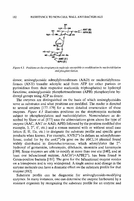
182 RESISTANCE TO NON-CELL WALL ANTIBACTERIALS
NH
bH b
OH
Figure 4.3. Positions on the streptomycin nzolecule susceptible to mod&ntion by nucleotidylation und phosphorylrtion.
donor; aminoglycoside adenylyltransferases (AAD) or nucleotidyltrans- ferases (ANT) transfer adenylic acid from ATP (or other purines or pyrimidines from their respective nucleoside triphosphates) to hydroxyl functions; aminoglycoside phosphotransferases (APH) phosphorylate hy- droxyl groups using ATP as donor.
The enzymes are distinguished on the basis of which aminoglycosides serve as substrates and what positions are modified. The reader is directed to several reviews [177-1791 for a more detailed enumeration of these enzymes. Figure 4.3 illustrates positions on the streptomycin molecule subject to phosphorylation and nucleotidylation. Nomenclature as de- scribed by Shaw et al. [177] uses the abbreviations given above for type of enzyme (AAC, ANT or AAD, APH) followed by the position modified (for example, 3, 2”, 6’, etc.) and a roman numeral with or without small case letters (I, 11, IIa, etc.) to designate the substrate profile and specific gene products when known. For example, ANT(2”)-Ia defines an adenylyltrans- ferase, coded for by the ant(2”)-Ia gene on the pSCL14 plasmid found widely distributed in Enterobacteriaceae, which adenylylates the 2”- hydroxyl of gentamicin, tobramycin, dibekacin, sisomicin and kanamycin [180]. Some enzymes are able to modify at more than one site [140], and at least one bifunctional enzyme, AAC(6‘)+APH(2’’), has been found in Gram-positive bacteria [ 18 I]. The gene for the bifunctional enzyme resides on a transposon and is very widespread. A single amino acid change in the enzyme molecule can have a dramatic effect on the substrate profile for that enzyme [182].
Substrate profile can be diagnostic for aminoglycoside-modifying enzymes. In many instances, one can determine the enzyme harboured by a resistant organism by recognizing the substrate profile for an enzyme and
N.E. ALLEN 183
the aminoglycoside-resistance profile [ 1771 for the organism. There are cases, however, where the aminoglycoside-resistance profile is insufficient to predict which enzyme is present. For example, some strains have more than one modifying enzyme. A recent study found as many as six enzymes in a single strain [ 1831. Several aminoglycoside-modifying enzyme activities have been detected in aminoglycoside-producing soil microorganisms [ 145, 1841.
Aminoglycoside modification results in inactivation. Although modified aminoglycosides are accumulated, in other words, cross the cytoplasmic membrane, they fail to inhibit ribosomal protein synthesis [ 1781. As expected, there are exceptions to the rule. Serratia marcescens, resistant to gentamicin, tobramycin and netilmicin, contains an adenylyltransferase which modifies netilmicin, but the enzyme fails to confer resistance to netilmicin. The reason is that netilmicin is a poor substrate for the enzyme having a K, higher than that for the other aminoglycosides [185]. Also, several 6’-N-acetylated aminoglycosides retain appreciable antibiotic activ- ity so that strains with enzymes for these modifications remain susceptible [186].
The cellular location of aminoglycoside-modifying enzymes plays a significant role in resistance to aminoglycoside antibiotics. The enzymes are membrane associated [187] and are located in the periplasm of Gram- negative bacteria. Examination of amino-terminal sequences of several modifying enzymes reveals putative signal sequences which may facilitate delivery to the periplasm [177]. It is generally accepted that uptake plays a critical role in this mechanism of resistance [188, 1891, and resistance is likely to be due to a combination of antibiotic modification and uptake. Modification occurs during uptake and there is competition between uptake and modification [187]. When an enzyme has sufficient affinity for an aminoglycoside, the drug will be modified as it is taken up and the cell will be resistant. However, if the aminoglycoside is a poor substrate, the susceptibility-resistance phenotype may depend on the amount of enzyme present in the cell, because if active (unmodified) antibiotic reaches the ribosome, protein synthesis will be inhibited [ 1871.
Amikacin is a poor substrate for APH(3’)-II in E. coli and the organism remains susceptible to amikacin [ 1901. However, when presence of the enzyme is combined with depressed transport of amikacin the bacteria become resistant, presumably because the slowed uptake of amikacin now permits modification by the phosphotransferase. In another study, inhibition of a modifying enzyme by a small molecular weight inhibitor led to increased uptake of substrate aminoglycosides as well as increased susceptibility [191].
In a recent study [192], a clinical isolate of E. coli showed high-level

184 RESISTANCE TO NON-CELL WALL ANTIBACTERIALS
resistance to kanamycin and low-level resistance to tobramycin, due to presence of APH(3’)-I enzyme. Kanamycin was phosphorylated at the 3’-position by this enzyme, but no phosphorylation of tobramycin, which has no 3’-hydroxyl, could be detected. The study went on to show that resistance to tobramycin was mediated by overproduction of the phosphotransferase which reversibly inactivated tobramycin, possibly via formation of an enzyme-antibiotic complex.
Most modifying enzymes are constitutively expressed. One notable exception is AAC(6’)-I11 in Ser. marcescens [193]. Expression of the chromosomal gene for this enzyme may be negatively regulated [194]. Exposure to increasing concentrations of netilmicin, a substrate for the enzyme, leads to increases in enzyme activity and increased levels of resistance. A variety of chromosomal mutations are responsible for the resistance profile. A similar situation has been seen in Prov. stuartii for a 2’-Wacetyltransferase [ 1771.
Circumvention of resistance. At pH 5 , membrane potential in Staph. aureus is minimal and cells are insensitive to gentamicin. Mates et al. [195] have shown that exposure of these cells to nigericin, an ionophore which depresses ApH but stimulates AY, results in an increase in uptake and susceptibility to gentamicin. Similar effects on aminoglycoside susceptibility were observed with nigericin when Staph. aureus was grown anaerobically [196]. The nigericin-induced effects are thought to be a result of stimulation of AY. Similarly, DCCD, an inhibitior of ATPase activity, stimulates uptake of gentamicin in Staph. aureus [197]. DCCD is known to cause increased membrane potential in this organism. However, the increase in antibiotic uptake is not associated with increased killing.
Exposure of streptomycin-susceptible and -resistant E. coli to puromycin enhances uptake of dihydrostreptomycin [ 1981. The increased uptake may be due to puromycin’s ability to stimulate polysome degradation which could result in increasing the number of ribosomal binding sites for the aminoglycoside. Although puromycin treatment increased killing due to the aminoglycoside in the susceptible cells, there was no effect on viability in resistant cells. The same effect was observed in Staph. aureus [197].
Numerous studies have shown that sensitivity of aminoglycosides to enzymatic modification can be modulated by a variety of approaches. Derivatives of spectinomycin having increased lipophilicity due to long side-chain substitutions have reduced affinity for an adenylyltransferase and increased activity against the organism harbouring the enzyme [199]. The more active derivatives in this series were also transported more effectively and were active against strains harbouring ribosomal resistance
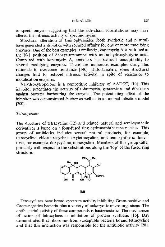
N.E. ALLEN 185
to spectinomycin suggesting that the side-chain substitutions may have altered the intrinsic activity of spectinomycin.
Structural alteration of aminoglycosides (both synthetic and natural) have generated antibiotics with reduced affinity for one or more modifying enzymes. One of the best examples is amikacin, kanamycin A substituted at the N- 1 position of deoxystreptamine with aminohydroxybutyric acid. Compared with kanamycin A, amikacin has reduced susceptibility to several modifying enzymes. There are numerous examples using this rationale to overcome resistance [ 1401. Unfortunately, some structural changes lead to reduced intrinsic activity, in spite of resistance to modification enzymes.
7-Hydroxytropolone is a competitive inhibitor of AAD(2”) [19]. This inhbitor potentiates the activity of tobramycin, gentamicin and dibekacin against bacteria harbouring the enzyme. The potentiating effect of the inhibitor was demonstrated in vitro as well as in an animal infection model [200].
Tetracyclines
The structure of tetracycline (12) and related natural and semi-synthetic derivatives is based on a four-fused ring hydronaphthacene nucleus. This group of antibiotics includes several natural products, for example, tetracycline, chlortetracycline, oxytetracycline, and semi-synthetic deriva- tives, for example, doxycycline, minocycline. Members of this group differ primarily with respect to the substitutions along the ‘top’ of the fused ring structure.
Me\/e
\ \ rn OH 0 OH OH 0 CQNH2
Tetracyclines have broad spectrum activity inhibiting Gram-positive and Gram-negative bacteria plus a variety of eukaryotic micro-organisms. The antibacterial activity of these compounds is bacteriostatic. The mechanism of action of tetracylines is inhibition of protein synthesis [16]. Day demonstrated that ribosomes from susceptible bacteria bound tetracycline and that this interaction was responsible for the antibiotic activity [20 1,
186 RESISTANCE TO NON-CELL WALL ANTIBACTERIALS
2021. Tetracyclines chelate metal ions and ribosome binding involves a Mg2+-tetracycline-chelate complex [203].
Numerous ribosome-binding studies have been used to elucidate the mechanism of action of tetracycline at the molecular level [204-2081. These studies, however, provide less than a complete story regarding the number and location of tetracycline-specific binding sites. In general, the collective data are consistent with a process where protein synthesis is inhibited when a single molecule of tetracycline binds to a 70s ribosomal particle. However, an interaction with the 30s subunit must be an important factor since the basis of tetracycline inhibition involves a function of this structure [209]. Goldman et al. [2 101 demonstrated that tetracycline preferentially binds to ribosomal protein S7 of the 30s subunit.
Tetracycline blocks polypeptide elongation by inhibiting the binding of aminoacyl-tRNA to the ribosome (see Figure 4.1) [211-2131. The antibiotic inhibits binding of aminoacyl-tRNA into the A (acceptor) site on the ribosome thereby preventing the addition of an incoming amino acid [211, 213-2151. The step sensitive to tetracycline is depicted in Figure 4.2. Although inhibition is not limited exclusively to the A site [209], binding of aminoacyl-tRNA into the P site is relatively unaffected under normal physiological conditions [216].
Tetracycline-resistance determinants can be plasmid or chromosomally determined. Resistance to tetracycline is extremely widespread and by the mid 1980s, it was claimed to be the most common resistance determinant encountered among all species of bacteria [217]. In spite of the action of tetracycline on the ribosome, mechanisms of resistance involving the target site appear to be due to effects on soluble factors rather than the ribosome itself. The vast majority of resistant strains, however, are modified in the manner in which they accumulate tetracycline.
Transport alterations. Tetracycline-susceptible E. coli accumulate tetracy- cline by an active process driven by the pH gradient (ApH) component of the proton electrochemical gradient [2 181. Uncouplers, such as dinitrophenol and CCCP, block the active accumulation of tetracycline by intact cells [219-2211, as well as the energy-dependent efflux from everted (inside-out) membrane vesicles [221]. Transport requires divalent cations. A study by McMurray et al. [222] revealed that under growth conditions where permeability of the outer membrane was reduced, a net efflux of tetracycline occurred. Thus, tetracycline-susceptible E. coli have both uptake and efflux mechanisms for regulating the accumulation of tetracycline. Interestingly, plasmid-determined resistance to tetracycline is based on active efflux (see below), but the magnitude of efflux in resistant cells is much higher than that

N.E. ALLEN 187
in susceptible cells, and this efflux is catalyzed by transporters different from those in susceptible cells [2 171.
Plasmid-determined resistance to tetracycline has been extensively studied in Enterobacteriaceae and Bacillus [217,223,224]. In the mid 1970s, plasmid-bearing strains of tetracycline-resistant E. coli were shown to encode inducible resistance to tetracycline. Exposure of these bacteria to tetracycline resulted in production of proteins which appeared to be associated with the inner membrane although an outer membrane location could not be completely ruled out at the tirie [225]. Levy and McMurray [226] demonstrated that a Tet determinant from E. coli inducibly coded for a new protein with a molecular weight of 50 kDa. A new protein from a tetracycline-resistant Staphylococcus, having a molecular weight of 32 kDa, was described by Wojdani et al. [227].
The thinking that prevailed in the late 1970s was that plasmid-determined resistance to tetracycline in E. coli and Staph. nureus was due to a membrane alteration which led to reduced accumulation of intracellular tetracycline [225]. Plasmid-determined proteins were thought to block tetracycline binding to a carrier molecule, or, alternatively, to associate with both membrane and ribosomes in order to protect protein synthesis. Franklin [228] raised the possibility that reduced accumulation could be explained by an active excretion (efflux) mechanism but general acceptance of this notion requires more investigation.
As a result of more detailed studies conducted in several laboratories over the last decade, we now know that bacteria harbouring plasmid-determined tetracycline resistance (tet genes) employ a carrier-mediated, active efflux system located in the cytoplasmic (inner) membrane to reduce intracellular accumulation and prevent tetracycline from reaching the ribosome [229-23 11. Efflux is mediated by integral membrane transporters (Tet proteins encoded by the tetA gene) [230, 232, 2331. These membrane proteins result in reduced binding of tetracycline and rapid loss of accumulated, intracellular tetracycline by an energy-dependent process [234]. An efflux mechanism was confirmed by demonstrating that tetracycline uptake by everted membrane vesicles prepared from resistant bacteria occurred in an energy-dependent manner [235, 2361
Inhibition of the electrochemical proton gradient by the uncoupler dinitrophenol increased uptake of tetracycline by intact resistant cells [220]. Studies with everted membrane vesicles prepared from four genetically distinct phenotypes, showed that, in each case, an energy-dependent efflux process was responsible for resistance [235]. Tet proteins are required for efflux and do not block the host active uptake system for tetracycline. Tetracycline is transported only when bound to divalent cations; the order
188 RESISTANCE TO NON-CELL WALL ANTIBACTERIALS
of effectiveness is as follows: Co2+ > Mn2+ > Mg2+ > Cd2+ > Ca” [237]. The true substrate is likely to be a monocationic tetracycline complex. Thus, the Tet protein is actually a transporter of cations when tetracycline is present. It functions as an antiporter exchanging H+ for the monocationic metal-tetracycline complex [235, 2381. The stoichiometry of antiport is 1 : 1 [239]. The affinity of the transporter for the antibiotic-cation complex is not affected by the cation species, but the turnover rate is affected, and may be related to cation size [237]. Energetically, transport by Tet is electrically neutral and is driven by the pH gradient (dpH) without dependence on membrane potential (dy3 [238, 2391.
Tet determinants (encoding efflux proteins as well as proteins not affecting transport) are differentiated and classified on the basis of DNA-DNA hybridization [2 17, 2401. In Enterobacteriaceae, five major classes (A to E) have been identified that encode Tet efflux proteins [241, 2421. Two classes (K and L) encoding Tet efflux proteins have been described in Gram-positive bacteria [243]. A summary of the different classes of Tet determinants, including determinants of non-efflux proteins, is available [230, 2311. ‘Tet X’ is used to denote a tetracycline resistance derminant from class X. The structural gene for the transporter and the transporter protein it encodes are designated as ‘tetA(X)’ and ‘TetA(X)’, respectively [240]. The ‘A’ is often omitted when it is understood that the gene encodes the transporter protein.
The different classes of Tet efflux proteins share highly conserved sequence motifs. For example, Classes A, B and C share 45-78% identity of nucleotide sequences [241]. Class D shares maximum homology with B (59%) but shows 4647% homology with the A and C classes [244]. Classes A and C and classes B and D are thought to represent two separate branches of a protein family. Class E shares 50-56% homology with classes A-D and may represent a third branch [242, 245, 2461. Classes K and L from Gram-positive bacteria are related to each other (65% homology), but not to the classes from Enterobacteriaceae [241, 2471. Classes M and 0 mediate non-efflux mechanisms and are described below. Other classes remain to be characterized [241].
Tet proteins share sequence similarities with transporters from diverse sources [233, 246, 2481. Tet transporters are most closely related (25-26%) to the seemingly diverse transporters Bmr (multi-drug resistance protein from B. subtilis) and NorA (quinolone-resistance protein from Staph. aureus). Tet(K) and Tet(L) from Gram-positive bacteria share lower levels of sequence similarity with all of these proteins [246]. The membrane topologies of all these proteins are similar; this indicates that they all function by similar mechanisms.
N.E. ALLEN 189
Tet determinants consist of two intracistronic genetically defined domains, designated tetAa and tetA/3 [249-25 13. The two domains define, respectively, the N-terminal and C-terminal halves of TetA which are separated by a central hydrophilic portion of approximately 30 amino acids in length [252, 2531. Mutations in either the N-terminal or C-terminal regions can abolish expression of resistance [249,25 1,2541. Rubin and Levy [253] have assessed the role of the two domains in tetracycline resistance by constructing hybrid Tet determinants where a and p domains were derived from classes A, B or C. Their experiments suggest that highly specific interactions between the two domains are required for tetracycline resistance. Tetracycline resistance was conferred when hybrids were derived from classes A and C (closely related) but not when derived from classes B and C (more distantly related).
The Tet(B) protein encoded by transposon TnlO has received considera- ble attention with respect to primary structure and membrane topology [255-2571. The protein contains 401 amino acid residues arranged into 12 membrane-spanning regions. Each of the 12 transmembrane regions or domains consists of approximately 20 amino acids in an a-helical conformation and each domain is separated by hydrophilic sequences extending out of the bilayer. There are 11 interhelix loops, 5 extend into the cytoplasm and 6 extend on the periplasmic side [255, 256, 258, 2591. The bulk of the protein lies in the hydrophobic bilayer with both N- and C-termini on the cytoplasmic side. Evidence favours a structure where the Tet(B) protein exists as a multimer in the membrane where the a and /I domains on different polypeptides interact [252].
The Tet(B) protein has 19 acidic amino acid residues and all but three of these (Aspl 5 , Asp84, Asp285) are in the non-hydrophobic regions. When the three aspartyl residues in the hydrophobic domain were replaced with basic amino acids so as to eliminate the negative charge at these positions, transport of tetracycline was severely affected [257]. Aspl 5 and Asp84 may define regions of the transporter essential for interaction with the tetracycline-cation complex [257, 2601. Asp285 may function in proton translocation [257,261]. Of the 16 acidic residues in the hydrophilic domain, only Asp66 appears to be essential and may serve a gating function [262, 2631. Chopra [264,265] proposed that Ser65-Asp66 may be a binding site for tetracycline. Yamaguchi et al. [262] claim that Asp66 is located in a region which may be more likely to facilitate a gating function.
Other, nonacidic residues of importance are Ser77 which may be part of the translocation channel for the cation-tetracycline complex [266], and His257 which may have a role along with Asp285 in proton trans- location [261]. All of these residues are highly conserved in each of the
190 RESISTANCE TO NON-CELL WALL ANTIBACTERIALS
Tet proteins from Enterobacteriaceae. A very recent report by Guay et al. [267] reveals that substitution of cysteine for tryptophan in position 231 or leucine for serine in position 308 (both positions are in the transmembrane regions) confer resistance in Tet B strains to the novel tetracycline derivative, 9-(dimethylglycylamido)minocycline. Wild-type Tet B does not transport this compound. The mutations also caused reduced resistance to tetracycline indicating that Trp23 1 and Ser308 function in substrate recognition.
Tetracycline resistance is highly regulated and is inducibly expressed in E. coli, Staph. aureus and B. subtilis [217]. The mechanism common to the Gram-negative bacteria, however, is different from that in Gram-positives [231]. In E. coli, the plasmid determinant for Tet B contains the Tet structural gene (tetA) and a gene (tetR) coding for a repressor protein. The two genes are organized in a manner where they flank a common regulatory region. A dimer of the repressor protein (TetR) binds the regulatory region negatively controlling transcription of both genes. When tetracycline is present, it acts as an inducer by binding to the repressor allowing divergent expression of both tetA and tetR [268-2701. Characterization of the repressor indicates a non-membrane associated protein having a molecular weight of 23-25 kDa [271, 2721. It has been suggested that in addition to its function to regulate expression of the Tet structural gene, it could also trap intracellular tetracycline thus protecting the ribosome [273].
Resistance determinants in Gram-positive bacteria (classes K and L) are regulated by an attenuation mechanism very similar to that used to regulate resistance to MLS antibiotics [274]. Rather than negative control at the level of transcription by a repressor, this mechanism involves formation of stem-loop structures in mRNA which control translation by regulating ribosome binding [23 11. The let genes from tetracycline-resistant Bacillus and Staphylococcus, include several inverted repeat sequences and two ribosome-binding sites preceding or upstream from the structural gene. The inverted repeats allow for two different mRNA conformations; only one of these conformations allows ribosome-binding to the second binding site and expression of tet. In the absence of tetracycline, only the first ribosome binding site is exposed and tet remains unexpressed. In the presence of sub-inhibitory concentrations, tetracycline binds to ribosomes and stalls translation. This facilitates the second stem-loop conformation exposing the second ribosome-binding site and tet messenger is translated [275, 2761.
In at least one case, tetracycline resistance was conferred by increasing the copy number of tet genes. Ives and Bott [277] showed that B. subtilis contained cryptic genes which conferred resistance to tetracycline when cloned onto a multicopy plasmid. In another case, multiple copies of the tet
N.E. ALLEN 191
determinant decreased resistance [278]. Increased sensitivity was not due to extra copies of repressor protein. Rather, Moyed et al. [279] showed that multicopies of tet were toxic and the best inducers conferred the most toxicity. Integration of the protein encoded by tetAa may be detrimental to the cell. There is no toxicity unless the determinant is induced indicating that membrane insertion plays an important role.
Another mechanism of resistance to tetracyline involving alterations in transport has been described [280, 2811. Growth of wild-type E. coli at subinhibitory levels of tetracycline or chloramphenicol results in ‘amplifia- ble’ resistance. The mutants (Mar) generated in this manner are not only resistant to tetracycline and chloramphenicol, but are cross-resistant to p-lactam antibiotics, quinolones, and rifampicin as well. Continued passage in tetracycline results in increasing levels of tetracycline resistance and resistance is lost unless the bacteria are maintained in the presence of antibiotic. The Mar phenotype is controlled by the n7urA locus [282]. Insertional mutagenesis by Tn.5 at this locus eliminates the resistance phenotype.
The basis of resistance in Mar mutants is decreased drug accumulation, which may involve efflux systems as well as changes in outer membrane proteins [283, 2841. Earlier studies had indicated that outer membrane alterations, specifically mutations in ompF, resulted in exclusion of tetracycline [285-2871.
Target alterations. Efflux mechanisms have not been demonstrated in all tetracycline-resistant organisms. A number of early studies proposed that tetracycline resistance could be due to ribosome alterations [288, 2891. A mutant of E. coli with temperature-sensitive tetracycline resistance but not demonstrating temperature-sensitive tetracycline efflux supported the notion that alternative mechanisms exist [217].
Tetracycline-resistance determinants of the M and 0 classes (Tet M and Tet 0) have been found in a variety of Gram-positive and Gram-negative bacteria. The more recently described Q class (Tet Q) has so far only been found in Bacteroides [23 11. Tet M and Tet 0 determinants are closely related (75% amino acid sequence homology) [290], but Tet Q shares only 40% homology with these two determinants [291].
The determinants encode cytoplasmic proteins having a molecular mass of 72 kDa which affect protein synthesis in some manner since amino acid polymerization in extracts from strains producing these proteins is resistant to tetracycline and there is no evidence for enzymatic modification [292-2941. The purified Tet(M) protein binds to ribosomes [294]. Tetracy- cline-sensitive ribosomes can be made resistant by incubation with soluble

192 RESISTANCE TO NON-CELL WALL ANTIBACTERIALS
extract from resistant bacteria [293]. The N-terminal regions of Tet M and Tet(0) determinants share significant homology with the N-terminal regions of the cytoplasmic elongation factors, EFTu and EFG [294, 2951. Greatest homology was seen in that portion of these proteins involved in GTP binding. The Tet(M) protein binds to ribosomes and has a ribo- some-dependent GTPase activity [294]. This plus the observation that the Tet(0) protein had no effect on tetracycline binding to ribosomes [293] suggests an indirect effect on the ability of tetracycline to block aminoacyl- tRNA to the ribosomal A site [293-2951. The mechanism appears to involve ribosome protection rather than ribosome alteration [296].
Streptomyces rimosus, an oxytetracycline producer, has two tet (or otr) resistance determinants flanking the biosynthetic genes for oxytetracycline [297]. Ohnuki et al. [298] have characterized tetA and tetB from this streptomycete. They found that tetB codes for a membrane-associated protein having weak but significant homology to the efflux determinants found in pathogenic bacteria, and was responsible for reduced intracellular accumulation of tetracycline [299]. However, tetA confers tetracycline resistance by modification of the protein synthetic apparatus [298]. Resistance is due to an alteration of a cytoplasmic factor or a factor associated with ribosomes rather than an effect on the ribosome itself. An alteration of a cytoplasmic factor may also explain tetracyline resistance in Streptomyces aureofaciens wherein protein synthesis was found to be resistant to tetracycline but ribosomes were sensitive [300].
Genetic determinants for ribosome protection are regulated by mecha- nisms sharing both similarities and differences with the efflux determinants. Su et al. [301] demonstrated that the Tet M determinant contains stem-loop structures upstream from the structural gene and suggested that expression of tet (M) is regulated by attenuation. In contrast, Stevens et al. [302] have shown that tet(Q) from Bacteroides was regulated at the level of transcription but no repressor gene nor stem-loop structures were identified. These investigators found sequences downstream from the tetA(Q) gene having amino acid homology with proteins from known two-component regulatory systems.
Enzymatic modiJication. A cryptic tetracycline resistance determinant from Bacteroides fragilis mediates two separate mechanisms of resistance to tetracycline when expressed in E. coli [303]. One mechanism is an active efflux transporter while the other is a chemical inactivation. The products of inactivation do not appear to be transported by the efflux system. Moreover, resistance is due to inactivation and is apparently unaffected by the presence or absence of the efflux pump [296]. A similar if not identical
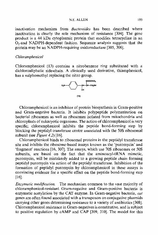
N.E. ALLEN 193
inactivation mechanism from Bucteroides has been described where inactivation is clearly the sole mechanism of resistance [304]. The gene product is a 44 kDa cytoplasmic protein that modifies tetracycline in an 0,-and NADPH-dependent fashion. Sequence analysis suggests that the protein may be an NADPH-requiring oxidoreductase [305, 3061.
Chloramphenicol
Chloramphenicol (1 3) contains a nitrobenzene ring substituted with a dichloroaliphatic side-chain. A clinically used derivative, thiamphenicol, has a sulphomethyl replacing the nitro group.
NHCOCHClr
4 i ~ ~ - L - c H 2 0 H
OH
Chloramphenicol is an inhibitor of protein biosynthesis in Gram-positive and Gram-negative bacteria. It inhibits polypeptide polymerization on bacterial ribosomes as well as ribosomes isolated from mitochondria and chloroplasts of eukaryotic organisms. The action of chloramphenicol is very specific; chloramphenicol inhibits the peptide bond-forming step by blocking the peptidyl transferase centre associated with the 50s ribosomal subunit (see Figure 4.2) [16].
Chloramphenicol binds to ribosomal proteins in the peptidyl transferase site and inhibits the ribosome-based assays known as the ‘puromycin’ and ‘fragment’ reactions [16, 3071. The assays, which use 70s ribosomes or 50s subunits, are based on the fact that the aminoacyl-tRNA mimetic, puromycin, will be mistakenly added to a growing peptide chain forming peptidyl puromycin via action of the peptidyl transferase. Inhibition of the formation of peptidyl puromycin by chloramphenicol in these assays is convincing evidence for a specific effect on the peptide bond-forming step
Enzymatic modijicution. The mechanism common to the vast majority of chloramphenicol-resistant Gram-negative and Gram-positive bacteria is enzymatic acetylation by the CAT enzyme. In Gram-negative bacteria, cut genes are often found associated with a transposon on conjugative plasmids carrying other genes determining resistance to a variety of antibiotics [308]. Chloramphenicol resistance in Gram-negatives is constitutive, and is subject to positive regulation by CAMP and CAP [309, 3101. The model for this
P61.
194 RESISTANCE TO NON-CELL WALL ANTIBACTERIALS
mechanism requires interaction of a CAMP-CAP complex and RNA polymerase at a regulatory region in DNA to initiate transcription of the cat structural gene. This explains why synthesis of CAT in Gram-negative bacteria is subject to catabolite repression where fermentation of glucose blocks formation of cAMP via adenylate cyclase [311, 3121. It also explains why high level CAT synthesis does not occur in adenylate cyclase-negative mutants unless cAMP is added exogenously [312].
CAT enzymes from a variety of Gram-negative bacteria have been described. The enzymes differ on the basis of their affinity for chloram- phenicol and acetylCoA substrates, electrophoretic mobility, immunologi- cal cross reactivity, and sensitivity to selected inhibitors [3 131. The enzymes all have in common a basic trimeric or tetrameric structure with identical subunits of 26 kDa [313, 3141. In E. coli there are three major types designated as types I, I1 and 111.
Type I enzyme, studied most extensively, consists of 219 amino acids containing a histidine at position 193 which is critical for acetylation at the active site. Two cysteines (Cys31 and Cysl96) may be near the active site since modification of the thiols inhibits enzyme activity [313, 3151. Type I enzyme is unusual in its ability to bind not only chloramphenicol but fusidic acid (an inhibitor of ribosome function) as well [316]. Fusidic acid acts as a competitive inhibitor of chloramphenicol acetylation. Triphenylamine dyes (for example, crystal violet) are competitive inhibitors of CAT activity [317]. Since CAT can represent as much as 1% of total cellular protein, colonies expressing cut genes on an agar-containing growth medium will be coloured by one of these dyes. This has been used as a diagnostic tool to distinguish CAT' from CAT- bacteria.
The substrate specificity of the type I enzyme is very similar to the substrate requirements for inhibition of peptidyl transferase [3 131. Both require the D,threo isomer of chloramphenicol, substitution at the C-2 amino group and no substitution at the C-1 or C-3 positions. The enzymatic mechanism is a 3-0-acetylation of chloramphenicol with acetylCoA as the acetyl donor [313, 3181. The general reaction is thought to proceed by 3-0-acetylation of chloramphenicol followed by an intramolecular migra- tion of the acetyl ester from the 3-0-position to the 1-0-position. Another molecule of acetylCoA provides acetate to make 1,3-0-diacetyl chloram- phenicol. It is somewhat surprising that the end-product of the reaction is the diacetyl derivative since both the mono- and di-acetoxy derivatives are inactive with respect to their ability to inhibit ribosome function.
In contrast to Gram-negative bacteria, CAT determinants are found on small, multicopy plasmids in staphylococci, or on the chromosome as in Bacillus pumilus or Strept. pneumoniue. The CAT enzyme is inducible (by
N.E. ALLEN 195
chloramphenicol and related congeners) in Gram-positive bacteria and is regulated by a translational attenuation mechanism [3 19, 3201. MLS resistance and some tetracycline resistance are regulated by similar mechanisms. The structural gene for CAT is preceded by an inverted complimentary repeat sequence allowing a stem-loop structure in mRNA which sequesters a ribosome binding site. In the absence of inducer, the ribosome binding site is masked and CAT mRNA is not translated. When inducing concentrations of chloramphenicol are present, the antibiotic binds to the ribosome causing it to stall. This leads to destabilization of the stem-loop structure, unmasking the ribosome binding site and allowing translation of the structural gene for CAT [320].
CAT enzymes from Staph. aureus and Staphylococcus epidermidis are distinct from the CAT enzymes produced by enteric bacteria [321]. There are no immunological cross reactivities with CATs from enteric bacteria. The enzymes are all inducible, less heat labile and less sensitive to some inhibitors of the enteric enzymes. But, as for the Gram-negative CATs, numerous variants have been described. CAT variants have been found in streptomycetes, a flavobacterium and Agrobacterium tumefaciens, all soil-inhabiting microorganisms. The cat genes in these organisms are chromosomal [322]. Interestingly, no CAT enzyme has been found in the chloramphenicol-producing organism, Streptomyces venezue- lae [323]. Instead, Streptorn. venezuelae produces a chloramphenicol hydrolase which it presumably uses to protect itself from the antibiotic it makes [324].
Transport alterations. Chloramphenicol-resistant, Gram-negative bacte- ria (both clinical and lab-derived) have been described having plasmid and chromosomally-determined alterations in transport mechanisms [321]. Studies on E. coli [325], Ps. aeruginosa [326, 3271, Ps. cepacia [328] and H. influenzae [329] all demonstrated resistance to chloramphenicol with no evidence for either enzymatic modification or ribosomal resistance to the antibiotic. The nonenzymatic mechanisms involve permeability barriers often resulting from a reduction in various outer membrane proteins. A strain of S. typhimurium, highly resistant to chloramphenicol, had a drastically reduced content of OmpF porin [330]. Studies on other strains demonstrated similar reductions in OmpF porin suggesting an important role for this protein [331, 3321. In another case, involving a resistant clinical isolate of Ps. cepacia, uptake of chloramphenicol was depressed 10-fold [328]. The fact that resistance to chloramphenicol was accompanied by cross-resistance to trimethoprim and ciprofloxacin, neither of which has structural relatedness to chloramphenicol, raised the possibility that

196 RESISTANCE TO NON-CELL WALL ANTIBACTERIALS
resistance in this organism was due to a nonspecific alteration in outer membrane permeability.
The cmlA gene in Ps. aeruginosa encodes a protein involved in chloramphenicol resistance which may be responsible for effecting altera- tions in both inner and outer membranes [327, 3331. The product of the cmlA gene is a hydrophobic, 44 kDa protein having structural and topological similarities to several antibiotic efflux proteins, in particular, TetA proteins from Gram-negative and Gram-positive tetracycline-resis- tant bacteria. This suggests a possible role for active efflux in CmlA- determined chloramphenicol resistance. Bacteria expressing CmlA show dramatic changes in protein content in both the inner and outer membranes. For example, both OmpA and OmpC porins are diminished in content and a third porin is overexpressed. These alterations could be secondary, compensatory effects resulting from an efflux mechanism and would be consistent with a global regulatory role for CmlA [333].
Target alterations. Although there exist early studies purporting to demonstrate ribosomal alterations conferring chloramphenicol resistance, no study has yet established a case where chloramphenicol resistance is due to alteration of a specific ribosomal protein [ 161.
Circumventing resistance. When fluorine replaces the hydroxyl at the C-3 position on chloramphenicol, the resulting derivative is not subject to acetylation by the CAT enzyme [334]. The nonenzymatic resistance to chloramphenicol encoded by the cmlA gene likewise fails to confer resistance to the flouro derivative [333].
Fusidic acid
Fusidic acid (14) is a steroid-like antibiotic having a cyclopentenophenan-
Me7-fMe C O O H
N.E. ALLEN 197
threne structure. Helvolic acid and cephalosporin P, (which is not related to the p-lactam cephalosporins) are structurally related.
In studies with isolated ribosomes, fusidic acid inhibits polypeptide elongation by stabilizing the EFG-GDP-ribosome complex (see Figure 4.2). Stabilization of this complex prevents peptidyl-tRNA moving from the A site into the P site and blocks translocation [16, 3351. Studies using protoplasts indicate that protein synthesis is blocked because aminoacyl- tRNA is prevented from binding in the A site as a result of sequestering EFG and GDP [336]. Fusidic acid may interact directly with EFG (or the EFG-GDP-ribosome complex) rather than directly with the ribosome [ 16, 3371. This idea is supported by the fact that mutants of E. coli, resistant to fusidic acid, have an altered EFG.
Target alterations. Cell-free protein-synthesizing systems from E. coli are sensitive to inhibition by fusidic acid, but whole cells are intrinsically insensitive to the antibiotic probably because of exclusion. Mutants CfusR) of E. coli isolated in the laboratory showing resistance to fusidic acid contain an altered EFG protein [338, 3391. Reconstitution experiments employing isolated ribosomes and purified EFG proteins demonstrated that an altered EFG protein and not an alteration in ribosomes in the mutant accounted for resistance to fusidic acid. The alteration is frequently associated with reduced affinity of EFG for GTP, but recent studies indicate that some mutations in EFG have an alternative effect on translocation [340].
In Staph. aureus, high frequency, chromosomally-determined resistance to fusidic acid can be readily selected in the laboratory. Protein synthesis in extracts from these mutants shows reduced sensitivity to fusidic acid [341]. The studies in E. coli mutants suggest the possibility of alterations in staphylococcal EFG.
Transport alterations. Plasmid-determined inducible fusidic acid resist- ance in Staph. aureus has been described [341, 3421. Resistance in these clinically isolated strains may be due to decreased permeability [341]. This conclusion is supported by the fact that changes were observed in the phospholipid composition in the resistant isolates. Cell-free protein synthesis in extracts from the resistant organisms was inhibited by fusidic acid, and there was no evidence for enzymatic modification. Studies on E. coli harbouring plasmid-determined resistance to fusidic acid likewise indicate changes in phospholipid composition of the cytoplasmic membrane as an explanation for resistance [342].
Enzymatic modiJication. Several streptomycetes, including Streptomyces
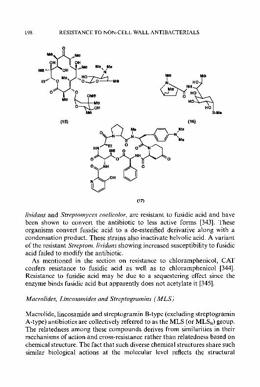
I98 RESISTANCE TO NON-CELL WALL ANTIBACTERIALS
nb I
lividans and Streptomyces coelicolor, are resistant to fusidic acid and have been shown to convert the antibiotic to less active forms [343]. These organisms convert fusidic acid to a de-esterified derivative along with a condensation product. These strains also inactivate helvolic acid. A variant of the resistant Streptom. lividans showing increased susceptibility to fusidic acid failed to modify the antibiotic.
As mentioned in the section on resistance to chloramphenicol, CAT confers resistance to fusidic acid as well as to chloramphenicol [344]. Resistance to fusidic acid may be due to a sequestering effect since the enzyme binds fusidic acid but apparently does not acetylate it [345].
Macrolides, Lincosamides and Streptogramins ( M L S )
Macrolide, lincosamide and streptogramin B-type (excluding streptogramin A-type) antibiotics are collectively referred to as the MLS (or MLS,) group. The relatedness among these compounds derives from similarities in their mechanisms of action and cross-resistance rather than relatedness based on chemical structure. The fact that such diverse chemical structures share such similar biological actions at the molecular level reflects the structural
N.E. ALLEN 199
complexity of the biochemical target, the bacterial ribosome. Streptogramin A-type agents are not included in the MLS group, but will be included in this section.
Macrolides consist of a macrocyclic lactone ring containing glycosidically linked neutral or basic sugar moieties. The lactone may be either 12-, 14- or 16-membered-ring structures. Examples include: methymycin (12-mem- bered ring); erythromycin ( 1 3 , oleandomycin, narbomycin (14-membered ring); and carbomycin, tylosin, rosamicin, spiramycin ( 16-membered ring). Azithromycin is a 15-membered, nitro-containing lactone ring derivative of erythromycin [346]. In contrast to the macrolides, lincosamides (lincomycin (1 6), clindamycin, celesticetin) are nonmacrocyclic and have a pyrrolidinyl structure. Streptogramin B-type compounds (pristinamycin IB (1 7), virgini- amycin S) consist of a group of closely related cyclic hexadepsipeptide lactone structures containing unusual amino acids. The streptogramin A-type compounds (pristinamycin IIA (1 8), virginiamycin M) are chemi- cally distinct from the B-types being polyunsaturated macrocyclic lactone structures.
0
In spite of their structural dissimilarities, these compounds all exert their antibacterial effects by inhibiting protein biosynthesis [16]. Macrolide, lincosamide and both streptogramin A- and B-types bind to the large (50s) subunit of prokaryotic ribosomes. MLS compounds bind to closely related or overlapping sites on the ribosome [347, 3481 and classic (constitutive) MLS resistance results in cross-resistance to members of all three classes. Early studies using erythromycin suggested that macrolides blocked protein biosynthesis by either inhibiting the peptide bond-forming step (pepti- dyltransferase) or the translocation step or both steps (see Figure 4.2) [16]. In more recent studies [349, 3501, it is proposed that inhibition by macrolides results from stimulating the dissociation of peptidyl-tRNA from ribosomes.
Although they share similar modes of action, members of the MLS group differ with respect to the details of how they interact with ribosomes and

200 RESISTANCE TO NON-CELL WALL ANTIBACTERIALS
influence the course of polypeptide chain elongation [16]. Nevertheless, as a group, the action of MLS antibiotics differs significantly from that of the A-type streptogramins [35 11. Of considerable interest is the fact that binding of streptogramin A-type compounds to the ribosome increases the affinity of the ribosome for binding B-type streptogramins. Combinations of A- and B-type streptogramins have synergistic activity and, unlike the many other protein synthesis inhibitors, the combinations are bactericidal. The differences between the two streptogramin classes is further illustrated by the fact that ribosomal based cross-resistance between members of the MLS group does not extend to the streptogramin A compounds.
Mechanisms of resistance to macrolide, lincosamide and streptogramin antibiotics include modification of the ribosomal binding site (alterations of ribosomal proteins and rRNA), enzymatic inactivation, as well as altered transport.
Target alterations. The most commonly encountered ribosomal alteration conferring resistance to MLS antibiotics is methylation of rRNA. This mechanism is often simply referred to as MLS or MLS, resistance since the ribosomal modification can confer resistance to all MLS antibiotics. MLS resistance is determined by several classes of erm genes which can have chromosomal or plasmid locations. Erm determinants have been identified in staphylococci, streptococci, enterococci, other Gram-positive species, Mycoplasma and even Gram-negative Enterobacteriaceae [273, 3521. Resistance results from dimethylation of adenine in 2 3 s rRNA of the 50s subunit [353]. The adenine residue at position 2058 of 2 3 s rRNA is converted to N6,N6-dimethyladenine [354]. In vitro ribosome reconstitution experiments using ribosomal proteins 5s RNA plus 2 3 s rRNA from wild-type and resistant bacteria confirmed a causal relationship between methylation of 2 3 s rRNA and resistance to MLS antibiotics [355]. Only 50s subunits containing dimethyladenine were resistant. Dimethylation of ade- nine is thought to confer a conformational change in the ribosome which re- sults in exclusion of MLS antibiotics from their overlapping binding sites [348].
Methylases from MLS-resistant staphylococci, streptococci, Bacteroides and soil bacteria all share similar amino acid compositions suggesting common ancestral origins [356]. Genes encoding enzymes to methylate 2 3 s rRNA have been identified in Saccharopolyspora erythraea (Streptomyces erythraeus; erythromycin producer) [357], Streptomyces thermotolerans (carbomycin producer) [358], Streptomyces fradiae (tylosin producer) [359] and Streptomyces lincolnensis (lincomycin producer) [360]. Calcutt and Cundliffe [361] describe a gene (clr) in Streptomyces caelestis (a celesticetin producer) which codes for a 2 3 s rRNA methylase which monomethylates
N.E. ALLEN 20 1
adenine 2058. Monomethylation is sufficient to confer high-level resistance to celesticetin and other lincosamides. MLS resistance in Bacillus anthracis is due to production of a rRNA methylase having unusual biochemical properties [362].
MLS resistance can be inducibly or constitutively expressed. Exposure of inducibly resistant strains to MLS antibiotics that act as inducers confers resistance to other MLS antibiotics. In the absence of an inducer, inducibly resistant strains are inhibited by noninducing MLS antibiotics. In Staph. aureus 1206, 14-membered-ring macrolides plus celesticetin induced resistance [363]. None of the 16-membered-ring macrolides, lincosamides or streptogramin B-type compounds induced MLS resistance, and induction resulted in cross-resistance to all MLS compounds. Inducer activity is limited to the 14-membered ring macrolides in most Staph. aureus; a mutant of this bacterium that responds to lincomycin and carbomycin (a 16- membered-ring macrolide) as inducers has been described [364]. A variety of MLS compounds act as inducers in streptococci [365]. Exposure of inducibly resistant strains to noninducing MLS drugs exerts a selective pressure for constitutive mutants expressing methylase continuously [353]. Susceptibility to A-type streptogramins is unaffected by the MLS resistance phenotype.
Methylation of rRNA may be the most common mechanism of resistance to MLS antibiotics, but reports of alternative mechanisms are increasing [366]. MLS-resistant strains with unique or unusual patterns of sensitivity/ resistance have been described. In some cases, the unusual pattern is due to modifications of the mechanism of resistance. Certain strains of Staphylo- coccus have been described as exhibiting ‘partial’ MLS resistance [367,368]. These strains are resistant to 14-membered ring macrolides and B-type streptogramins. They are susceptible to many but not all 16-membered ring macrolides (for example, they are resistant to spiramycin and rosamicin), and are susceptible to the lincosamides. It was originally thought that these strains might represent an unusual mechanism involving ribosome methyl- ation, but recent reports raise the possibility of alternative explanations [369, 3701.
Inducible expression of MLS resistance is regulated by a translational attenuation mechanism. Expression of the dimethylase gene (similar to expression of resistance to chloramphenicol and tetracycline), is controlled by a regulatory region upstream from the structural gene [353, 3711. The region contains inverted repeat sequences facilitating the formation of stem-loop structures in mRNA. When inducer is absent, a stem-loop structure masks the ribosome binding site for translation of mRNA encoding the methylase. When inducer is present, inducer binds to and stalls
202 RESISTANCE TO NON-CELL WALL ANTIBACTERIALS
unmethylated ribosomes. Stalling facilitates a conformational change leading to a new stem-loop structure unmasking the ribosome binding site and allowing methylase to be translated. Mutations in the inverted repeat sequences give rise to constitutive phenotypes as predicted from the translational attenuation model [353]. Once all the ribosomes become methylated, they are resistant to further binding by inducer and the methylase gene again becomes sequestered.
Expression of the CAT determinant, also subject to regulation by translational attenuation, can be induced by erythromycin in some strains [372]. Moreover, transposition of MLS-resistance in streptococci is inducible and is regulated by translational attenuation where erythromycin serves as an inducer [373]. It is interesting that the dissemination of MLS-resistance by transposition as well as the process of target modifica- tion are both regulated by a mechanism based on the inherent ability of the antibiotics to interact with ribosomes and inhibit polypeptide synthesis.
MLS-resistance in Bacillus lichenformis (ermk7) is regulated by transcrip- tional (as opposed to translational) attenuation [374]. In the absence of inducer, the methylase-containing mRNA is prematurely truncated. In the presence of an inducer, the complete transcript is transcribed for expression.
Another mechanism involving alteration of the ribosome target is mutation of ribosomal protein genes. High-level, single-step chromosomal resistance (ery) to erythromycin occurs in both Gram-positive and Gram-negative bacteria. Ribosomes isolated from eryA mutants of E. coli are unable to bind erythromycin due to altered ribosomal proteins L4 and L22 of the 50s subunit [375]. The protein alteration co-transduces with erythromcyin resistance establishing a causal relationship between resist- ance and the changed proteins. Reconstitution experiments showed that resistance to erythromycin binding was lost when two other proteins, L15 and L16, from erythromycin-sensitive ribosomes were substituted with the same two proteins from resistant ribosomes [376]. In B. subtilis, develop- ment of erythromycin resistance was accompanied by temperature-sensitive sporulation [377]. The 50s protein, L17, was altered in the resistant strain, and revertants that sporulated normally had ribosomes that could bind erythromycin.
Erythromycin and other macrolide antibiotics bind specifically to prokaryotic-type (70s) ribosomes but not to eukaryotic-type (80s) ribo- somes. The chloroplast of the alga Chlamydomonas reinhardtii contains prokaryotic-type ribosomes to which erythromycin binds and inhibits. Mutants of Ch. reinhardtii, resistant to erythromycin, have been isolated which carry mutations in chloroplast genes conferring erythromycin resistance to chloroplast ribosomes [378].

N.E. ALLEN 203
Enzymatic modfieation. Members of the family Enterobucteriuceue having high-level resistance to erythromycin have been described [379]. A clinical isolate of E. coli has been reported [352] that hydrolyzes the lactone ring of erythromycin via an esterase reaction [380]. Two types of esterase genes (ere) have been detected; one has been found in strains harbouring genes for rRNA methylation [38 11. Not unexpectedly, these strains are highly resistant to erythromycin. A macrolide, 2’-phosphotransferase which phosphorylates erythromycin and oleandomycin on the 2’-hydroxyl has been detected in another clinical isolate of E. coli highly resistant to erythromycin [382, 3831. A similar enzyme was reported in Streptom. coelicolor [384].
Streptomyces vendurgensis produces a glycosyltransferase capable of in- activating erythromycin by glycosylating the 2’-hydroxyl [385]. The mgt gene in Streptom. lividuns encodes a similar enzyme which uses UDP-glucose as cofactor to glycosylate selected 12-, 14- and 16-membered-ring macrolides [386]. The enzyme, which has a sequence resembling a eukaryotic gluco- syltransferase [387], is specific for the 2’-hydroxyl of sugars attached at the C-3 or C-5 of 12- and 14-membered-ring lactones or at the C-5 of 16-membered-ring lactones. Streptom. lividuns contains an additional gene encoding methylation of 23s rRNA [144]. Both genes are inducible [387].
The oleandomycin producer, Streptomyces untibioticus, produces an enzyme that inactivates oleandomycin in the presence of UDP-glucose [388]. The enzyme glycosylates oleandomycin and other macrolides having a free 2’-hydroxyl on the monosaccharide attached to the lactone ring. It does not glycosylate 16-membered-ring macrolides with disaccharides nor does it modify erythromycin. The same extracts were shown to contain a second enzymatic activity which seemed to reverse the glycosylation and regenerate active macrolide. More recent studies [389] have detected yet a third gene in this organism which encodes a membrane protein which may function in substrate translocation. Thus, the oleandomycin producer has at least three genes involved in intracellular glycosylation and secretion of the antibiotic it produces [390].
Enzymes that modify lincosamides by nucleotidylation have been detected in Stupli. uureus and Stuphylococcus huemolyticus [39 1, 3921. These strains produce O-nucleotidyltransferases which use nucleoside 5’-tri- phosphates as nucleotidyl donors to nucleotidylate the 3-position of lincomycin and the 4-position of clindamycin [39 11. The enzyme in these strains modifies both lincomycin and clindamycin yet the strains are resistant to lincomycin and susceptible to clindamycin when tested using standard procedures [392]. Barcs [393] has reported that clindamycin is a better substrate for the enzyme. However, an energy-dependent increase in
204 RESISTANCE TO NON-CELL WALL ANTIBACTERIALS
clindamycin accumulation by these strains suggests an active efflux system for clindamycin may be present.
Certain Staph. aureus bacteria are resistant to both A-type and B-type streptogramins due to the action of two inactivating enzymes [394, 3951. Streptogramin A-type antibiotics are subject to acetylation by an ace- tylCoA-dependent 0-acetyltransferase related to the CAT family of enzymes [396]. The streptogramin B-type antibiotics are inactivated by a nonspecific hydrolase in the same strains [394].
Transport alterations. Most Gram-negative bacteria are intrinsically resistant to MLS antibiotics. This is generally believed to be due to the inability of these relatively large, lipophilic compounds to cross the outer membrane barrier. Gram-negative bacteria contain ribosomes sensitive to erythromycin [397] and other MLS antibiotics, but the organisms grow in the presence of the antibiotics because the compounds are unable to reach their target in concentrations sufficient to inhibit. As mentioned earlier, strains of E. coli have been reported to have additional mechanisms conferring even higher levels of resistance to these antibiotics [379].
Staph. epidermidis strains having a ‘partial’ MLS resistance pattern (resistant to 14-membered ring macrolides and streptogramin B-type antibiotics) contain genes (msr) with sequences suggesting an ATP-binding protein which could function as an efflux pump [398]. In another Staph. epidermidis isolate, low-level, constitutive resistance to 14-membered-ring macrolides correlated with appearance of a 60 kDa membrane protein and active efflux of 14-membered-ring macrolides [399, 4001. Matsuoka et al. [370] detected a 63 kDa membrane protein in a Staph. aureus having a ‘partial’ MLS resistance phenotype and which showed reduced accumula- tion of erythromycin. Similarly, tlrC in Streptom. fraidiae, srrnB in Streptomyces arnbofaciens and carA in Streptomyces thermotolerans (all macrolide resistant) encode proteins similar to ATP-dependent transport proteins (401). lrnrA in Streptom. lincolnensis (lincomycin producer) encodes a similar protein [360]. This strain also methylates rRNA via a second gene (IrnrB) and is not cross-resistant to either celesticetin or clindamycin. The N-terminal and C-terminal sequences of these proteins are similar and resemble ATP-binding domains suggesting that resistance may be due to ATP-driven efflux. Streptom. ambofaciens, the spiramycin-producing cul- ture, has decreased permeability to spiramycin in older cultures [402]. Resistance in younger cells is inducible and may be due to a ribosomal alteration.
Circumvention of resistance. There are several reports of modifications of erythromycin which confer activity against MLS-resistant bacteria. Allen
N.E. ALLEN 205
[363] reported that modification of the 4”-position of cladinose on erythromycin- 1 1,12-cyclic carbonate destroyed ability to induce MLS resistance without affecting the ability to inhibit protein synthesis. Fernandez et al. [403] described several 1 1,12-cyclic carbamate derivatives of erythromycin (including modifications at the 4”-position) active against both inducible and constitutive MLS-resistant Strept. pyogenes. Goldman and Kadam [404] reported 6-O-methyl-l l , 12-cyclic carbamate derivatives of erythromycin able to inhibit in vitro protein synthesis on MLS-susceptible and -resistant ribosomes from B. subtilis and Staph. aureus. These compounds bound specifically to the 50s subunit of both resistant and sensitive ribosomes.
YM133, an analogue of the 16-membered ring macrolide tylosin having the structure 4”-O-(4-methoxyphenyl)acetyl-tylosin, has activity against MLS-resistant bacteria [405]. This compound also has significantly greater bactericidal activity than erythromycin. Yet another approach to develop antibiotics active against MLS-resistant bacteria is represented by RP59500, a combination of pristinamycin IIA (a streptogramin A-type) and pristinamycin IA (a streptogramin B-type) [350]. This combination is active against MLS-resistant bacteria since there is no cross-resistance to the streptogramin A-type compound (pristinamycin IIA). However, as men- tioned earlier, a strain of Staph. aureus has been found that produces streptogramin A- and B-modifying enzymes, and this strain is resistant to the combination [406].
TI1 ios t rep ton
Thiostrepton (1 9) is a sulphur-containing peptide antibiotic. Siomycin and thiopeptin are structurally and biologically related. Thiostrepton and its related antibiotics inhibit protein biosynthesis in bacteria (see Figure 4.2). Gram-negative bacteria show intrinsic resistance to thiostrepton, but their ribosomes are sensitive. Thiostrepton has no effect on eukaryotic ribo- somes.
Early studies [ 161 demonstrated that thiostrepton inhibits ribosome- EFG-dependent GTP hydrolysis and was labelled as an inhibitor of translocation. It apparently blocks at a site on the ribosome where EFG and GTP/GDP are bound. In fact, thiostrepton blocks the fusidic acid-induced formation of stabilized ribosome-EFG-GTP/GDP complexes [407]. Studies of polypeptide chain elongation showed that rather than preventing translocation, interference with the ribosome-EFG-GTP/GDP interactions by thiostrepton resulted in inhibition of binding of aminoacyl-tRNA into the ribosomal A site [408]. Thiostrepton binds to the 50s ribosomal subunit
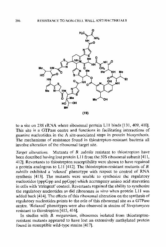
206 RESISTANCE TO NON-CELL WALL ANTIBACTERIALS
to a site on 23s rRNA where ribosomal protein L11 binds [131, 409, 4101. This site is a GTPase centre and functions in facilitating interactions of guanine nucleotides in the A site-associated steps in protein biosynthesis. The mechanisms of resistance found in thiostrepton-resistant bacteria all involve alteration of the ribosomal target site.
Target alterations. Mutants of B. subtilis resistant to thiostrepton have been described having lost protein L11 from the 50s ribosomal subunit [411, 4121. Revertants to thiostrepton susceptibility were shown to have regained a protein analogous to L11 [412]. The thiostrepton-resistant mutants of B. subtilis exhibited a 'relaxed' phenotype with respect to control of RNA synthesis [413]. The mutants were unable to synthesize the regulatory nucleotides (pppGpp and ppGpp) which accompany amino acid starvation in cells with 'stringent' control. Revertants regained the ability to synthesize the regulatory nucleotides as did ribosomes in vitro when protein L11 was added back [414]. The effects of this ribosomal alteration on the synthesis of regulatory nucleotides points to the role of this ribosomal site as a GTPase centre. 'Relaxed' phenotypes were also observed in strains of Streptomyces resistant to thiostrepton [415,416].
In studies with B. megaterium, ribosomes isolated from thiostrepton- resistant mutants appeared to have lost an extensively methylated protein found in susceptible wild-type strains [417].
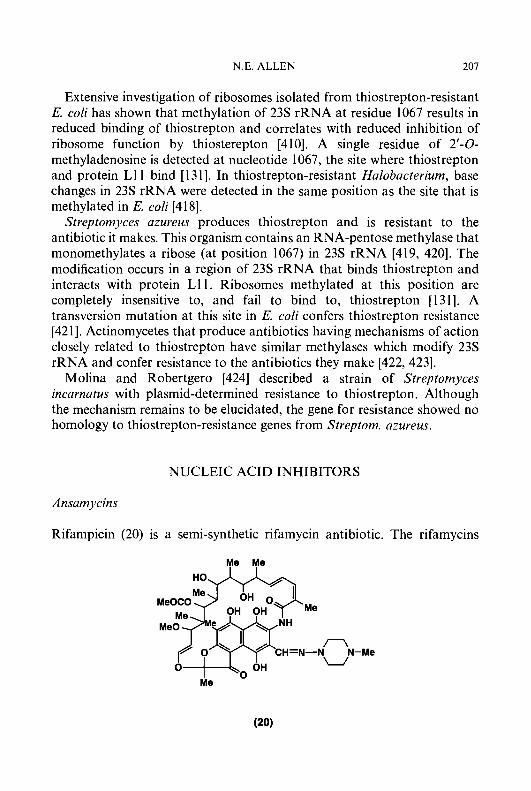
N.E. ALLEN 207
Extensive investigation of ribosomes isolated from thiostrepton-resistant E. coli has shown that methylation of 23s rRNA at residue 1067 results in reduced binding of thiostrepton and correlates with reduced inhibition of ribosome function by thiosterepton [410]. A single residue of 2’-0- methyladenosine is detected at nucleotide 1067, the site where thiostrepton and protein L11 bind [ 13 13. In thiostrepton-resistant Halobacterium, base changes in 23s rRNA were detected in the same position as the site that is methylated in E. coli [418].
Streptomyces azureus produces thiostrepton and is resistant to the antibiotic it makes. This organism contains an RNA-pentose methylase that monomethylates a ribose (at position 1067) in 23s rRNA [419, 4201. The modification occurs in a region of 23s rRNA that binds thiostrepton and interacts with protein L1 1. Ribosomes methylated at this position are completely insensitive to, and fail to bind to, thiostrepton [131]. A transversion mutation at this site in E. coli confers thiostrepton resistance [42 11. Actinomycetes that produce antibiotics having mechanisms of action closely related to thiostrepton have similar methylases which modify 23s rRNA and confer resistance to the antibiotics they make [422,423].
Molina and Robertgero [424] described a strain of Strepfomyces incarnatus with plasmid-determined resistance to thiostrepton. Although the mechanism remains to be elucidated, the gene for resistance showed no homology to thiostrepton-resistance genes from Streptom. azureus.
NUCLEIC ACID INHIBITORS
Ansamycins
Rifampicin (20) is a semi-synthetic rifamycin antibiotic. The rifamycins
Me Me
Me
0
Me

208 RESISTANCE TO NON-CELL WALL ANTIBACTERIALS
belong to the ansamycin class characterized as hydrophobic molecules containing an ansa ring (an aromatic ring system spanned by an aliphatic bridge). The structurally similar streptovaricins also belong in this class.
Rifampicin has potent, bactericidal activity against Gram-positive bacteria and the mycobacteria. Rifampicin has been widely used in the treatment of tuberculosis. The rifamycins and streptovaricins are potent inhibitors of bacterial DNA-dependent RNA polymerase, the enzyme that catalyzes transcription by polymerizing ribonucleoside triphosphates on a DNA template. Rifampicin activity is in the submicrogram range [16] and has no effect at these concentrations on bacterial DNA-dependent DNA polymerase I or on nuclear DNA-dependent RNA polymerase from eukaryotes [425]. RNA polymerase consists of four polypeptide subunits (2a, /?, /?'); the monomer can further associate with Q factors which confer promoter specificity to the polymerase. Studies on rifampicin-resistant mutants indicate that rifampicin acts by inhibiting the /? subunit of the enzyme [425]. The action of rifampicin on the /? subunit blocks initiation of polymerization [426] and is without effect on the elongation process. Nor does there appear to be any effect on binding of the enzyme to the DNA template. The streptovaricins probably share the same mechanism of action.
Target alterations. The spontaneous mutation frequency for rifampicin resistance in most susceptible bacteria is lo-' to lo-* and to lo-'' for mycobacteria. The mutation in the rpoB gene is chromosomal, single step, stable, and high level. Mutants are cross-resistant to streptovaricin. Mutation to rifampicin resistance is often accompanied by pleiotropic effects such as suppression of temperature-sensitive growth and phage sensitivity [427, 4281. The ease of mutation to rifampicin resistance has forced the use of this antibiotic in combination with other antimicrobials.
Core RNA polymerase from resistant mutants of E. coli does not bind rifampicin [429]. Reconstitution experiments involving hybrid enzyme molecules have established that the /? subunit determines susceptibility to rifampicin [430-432]. Only the hybrid enzyme containing a B subunit showed resistance to rifampicin. Resistance to rifampicin was conferred even on the Micrococcus luteus enzyme when reconstituted with a /? subunit from rifampicin-reistant E. coli [431]. In one study, the /? subunit from a rifampicin-resistant organism was shown to have an altered electrophoretic mobility [429]. Sensitivity-resistance of the enzyme to rifampicin may involve a conformational change in the /? subunit [433].
A comparison of the primary structures of RpoB proteins from several rifampicin-resistant mutants of E. coli identified highly conserved regions,

N.E. ALLEN 209
where resistance was due to missense mutations* [434-436]. Mutations occurred at Asp516, Ser531, Pro564 or His526. Substitution at Val146 in a rifampicin-resistant strain represents a second domain in the p subunit important for rifampicin interaction. A similar if not identical situation occurs in mycobacteria. Nine strains of rifampicin-resistant Mycobacterium leprae were all due to mutations in the rpoB gene encoding the p subunit of RNA polymerase [437]. Missense mutations affecting a serine residue (Ser425) were detected in eight of the mutants. The other strain carried an insertion close to this site (which is the same as the evolutionarily conserved region identified in E. coli). Similar mechanisms of resistance have been reported in M. smegmatis and may be found in Mycobacterium tuberculosis [438].
Streptolidigin is structurally unrelated to rifampicin but it too inhibits bacterial DNA-dependent RNA polymerase via action on the p subunit [ 161. However, unlike rifampicin, streptolidigin inhibits the elongation process rather than initiation [439]. RNA polymerase from streptolidigin- resistant mutants is insenstive to streptolidigin but sensitive to rifampicin [439]. Nevertheless, reconstitution experiments using isolated RNA poly- merase indicate that resistance is associated with the p subunit [432]. Resistance to these two antibiotics was, however, determined by different genes indicating that they act at different sites on the p subunit [440]. A recent study has identified parts of the RNA polymerase which specifically affect streptolidigin but not rifampicin binding [441].
Blanco et al. [442] found resistance to rifamycin, streptovaricin, and streptolidigin in an examination of the antibiotic-producing actinomycetes: Nocardia mediterranei, Streptomycces spectabilis and Streptomyces lydicus, respectively. Each was highly resistant to the antibiotic it produced and was not cross-resistant to the other two compounds. Resistance was due to target alteration as the isolated RNA polymerases were resistant to the corresponding antibiotic.
Transport alterations. Intrinsic resistance to rifampicin in S. typhimurium and possibly other Gram-negative bacteria may be due to structural peculiarities of the LPS of the outer membrane in these organisms [443]. Intrinsic resistance in two naturally occurring strains of Mycobacterium intracellulaire and M. smegmatis was due to impermeability since treatment of the strains with the surfactant, Tween 80, increased susceptibility to the
* In a missense mutation, a codon which is specific for one amino acid becomes specific for another.
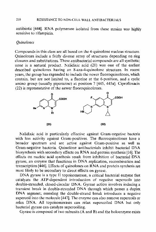
210 RESISTANCE TO NON-CELL WALL ANTIBACTERIALS
antibiotic [444]. RNA polymerase isolated from these strains was highly sensitive to rifampicin.
Quinolones
Compounds in this class are all based on the 4-quinolone nucleus structure. Quinolones include a fairly diverse array of structures depending on ring closures and substitutions. These antibacterial compounds are all synthetic; none is a natural product. Nalidixic acid (21) was one of the earliest described quinolones having an 8-aza-4-quinolone structure. In recent years, the group has expanded to include the newer fluoroquinolones, which contain, but are not limited to, a fluorine at the 6-position, and a cyclic amino group (usually piperazine) at position 7 1445, 445aI. Ciprofloxacin (22) is representative of the newer fluoroquinolones.
Me & I
Et
Nalidixic acid is particularly effective against Gram-negative bacteria with less activity against Gram-positives. The fluoroquinolones have a broader spectrum and are active against Gram-positive as well as Gram-negative bacteria. Quinolone antibacterials inhibit bacterial DNA biosynthesis with secondary effects on RNA and protein synthesis [ 161. The effects on nucleic acid synthesis result from inhibition of bacterial DNA gyrase, an enzyme that functions in DNA replication, recombination and transcription [446]. Effects of quinolones on RNA and protein synthesis are most likely to be secondary to direct effects on gyrase.
DNA gyrase is a type I1 topoisomerase, a critical bacterial enzyme that catalyzes the ATP-dependent introduction of negative supercoils into double-stranded, closed-circular DNA. Gyrase action involves inducing a transient break in double-stranded DNA through which passes a duplex DNA segment; resealing the double-strand break introduces a negative supercoil into the molecule [447]. The enzyme can also remove supercoils or relax DNA. All topoisomerases can relax supercoiled DNA but only bacterial gyrase can catalyze supercoiling.
Gyrase is composed of two subunits (A and B) and the holoenzyme exists
N.E. ALLEN 21 1
as a tetramer. Structural genes for the A and B subunits, gyrA and gyrB, have been distinguished by resistance to quinolones (gyrA mutations) and coumarins (gyrB mutations). The A subunit functions in the breakage and reunion of DNA [448], whereas the B subunit contains an ATPase activity used to drive the supercoiling reaction [449, 4501. Quinolones block gyrase-induced supercoiling of double-stranded DNA [448, 45 11, and they specifically block the gyrase reactions involving DNA strand breakage [451]. Quinolones are known to induce DNA cleavage by gyrase [452], but questions remain to be answered as to whether these inhibitors exert their action prior to or after cleavage. Quinolones have no effect on the DNA-dependent ATPase reaction associated with the B subunit [453].
Findings from studies of quinolone-resistance genes clearly indicate that these compounds act on the gyrase A subunit. However, several observa- tions raise questions as to the specific mechanism of inhibition. For example, in certain organisms such as E. coli, much higher concentrations of quinolones are required to inhibit in vitro DNA gyrase activity than are required to inhibit growth of the organism [454]. In Staph. aureus, however, concentrations needed to inhibit enzyme activity and bacterial growth are much closer [455]. Also, sensitivity to nalidixic acid is dominant to resistance [456] suggesting that the mode of action of quinolones may involve more than inhibition of DNA supercoiling. Some of these observations might be explained by a subtle interaction between gyrase and quinolones leading to the formation of a toxic complex (poison) which inhibits bacterial growth [457,458].
Much of our uaderstanding of the function of DNA gyrase and the role of the A subunit has derived from studies on mutants resistant to quinolones.
Target alterutions. High level resistance to nalidixic acid and other quinolones is chromosomal, not plasmid mediated, and most often is accompanied by cross-resistance to other quinolones. The vast majority of these mutants harbour point mutations in the gyrA gene. Purification of DNA gyrase subunits from quinolone-resistant and susceptible cells has facilitated reconstitution experiments to identify the determinant of quinolone resistance. Employing heterologous subunits from E. coli, Hallett and Maxwell [458] demonstrated that the A subunit was the determinant of quinolone resistance. Similar conclusions regarding the role of gyrA in quinolone resistance have come from subunit switching experiments in Staph. aureus [459], Canipylobacter jejuni [460], and Ps. ueruginosa [461].

212 RESISTANCE TO NON-CELL WALL ANTIBACTERIALS
In E. coli, spontaneous mutations to quinolone resistance occur between amino acids 67 and 106 in the N-terminal region of the 875 amino acid A subunit [462464]. Insertion of a tryptophan residue for serine at position 83 results in high-level quinolone resistance in a clinical isolate of E. coli [463]. Leucine can also substitute for serine at this position. Identical gyrA point mutations in quinolone-resistant bacteria have been found in a laboratory- induced mutant [462]. Amino acid position 83 of the A subunit is very close to the tyrosine at position 122, which is a critical amino acid involved in covalent attachment of single-stranded ends of the DNA substrate following the cleavage reaction [465].
Examination of quinolone-resistance in Stuph. uureus and Stuph. epiderrnidis [466, 4671 revealed amino acid substitutions in the same small region of the gyrA protein. Staph. nureus had a point mutation at Ser-84 which corresponds to Ser83 in E. coli. In Staph. epiderrnidis, Ser84 was replaced with a phenylalanine. Goswitz et al. [468] examined numerous ciprofloxacin-resistant Staph. aureus (which were also methicillin resistant) and found leucine and alanine substitutions at Ser84, lysine at Glu88 and a double substitution, leucine at Ser84 plus proline at Ser85. All quinolone strains in this study had at least one gyrA mutation between amino acid codons 84 and 88.
Although the evidence favours a significant role for the A subunit in the action of quinolones, mutations to quinolone resistance have also been mapped to the B subunit implying a role for this subunit in the action of quinolones [469,470]. A recent study [471] lends support to the notion that quinolones bind to a gyrase-DNA complex but wherein binding is determined by both subunits. Two single nucleotide changes in the midportion of the coding sequence in gyrB accounted for quinolone- resistance in E. coli [469]. The nucleotide changes resulted in an asparagine substitution for aspartate at position 426 and a glutamate substitution for lysine at position 447. This region is conserved in E. coli and B. subtilus. Yoshida et al. [472] examined thirteen spontaneous quinolone-resistant gyrB mutants and detected the same changes. The mutations fell into one of two classes: type 1, where asparagine substituted at Asp426 and mutants were cross-resistant to all quinolones; or type 2, where glutamate substituted at Lys447 and mutants were cross-resistant to acidic quinolones but hypersusceptible to amphoteric quinolones. The authors propose a quinolone-binding pocket which is bounded by the surfaces of quinolone- resistance-determining regions of the A and B subunits. A change in charge without a change in hydrophobicity when glutamate replaces Lys447 could explain resistance to acidic quinolones and hypersusceptibility to ampho- teric quinolones in the type 2 mutants.

N.E. ALLEN 213
gyrB alterations in quinolone-resistant Ps. aeruginosa [473] and N. gonorrhoeae [474] have been reported.
The question of whether quinolone resistance is determined by the gyrA protein alone or by both subunits is important as it relates to the mechanism of action of these agents. Shen and Pernet [475], based on studies employing equilibrium dialysis and membrane filtration techniques, claimed that the quinolone norfloxacin binds to DNA rather than to the holoenzyme. After a series of studies from the same group [476-478] a cooperative quinolone-DNA binding model was proposed to explain how a complex consisting of DNA-gyrase-quinolone can inhibit supercoiling.
The model assumes that a quinolone-binding pocket is induced in relaxed DNA when gyrase binds to the DNA substrate in the presence of ATP. The pocket could be bound by both subunits. Elucidation of the three- dimensional structure of DNA gyrase is likely to be needed to ensure a complete understanding of how quinolones bind and inhibit the action of this enzyme [479].
As further indication of our incomplete understanding of how quinolones act, several reports describe growth conditions or mutations which lead to quinolone tolerance where quinolones inhibit gyrase but do not kill. For example, anaerobiosis [480] and inhibition of protein synthesis [48 11 prevent killing by quinolones. A mutant of E. coli has been described [482] which is resistant to the killing effects of norfloxacin. Interestingly, the mutant is also tolerant to the action of coumarin antibiotics (gyrase B subunit antagonists) suggesting the possibility of gyrase alterations.
Transport alterations. Wild-type staphylococci contain a membrane- associated protein which appears to function as an active efilux pump for quinolones [483]. A comparison of sparfloxacin and norfloxacin uptake in quinolone-resistant clinical isolates of Staph. aureus revealed differences with respect to the two compounds [484]. Uptake of both quinolones was increased noticeably by the addition of CCCP, a membrane energy poison suggesting a possible role for an active efflux system in resistance. In a separate study [485], a quinolone-resistant Staph. aureus was shown to have a defect in accumulating hydrophilic quinolones which was relieved by CCCP. This strain harboured an alteration in gyrA and it was suggested that specific impairment of hydrophilic quinolone uptake could play an additional role in the mechanism of resistance.
Other studies in Staph. aureus show that the norA gene is involved in resistance to hydrophilic quinolones [486-488]. However, increased efflux does not confer the high levels of resistance typified by gyrA mutations. The presence of an endogenous norfloxacin efflux pump in the cytoplasmic

214 RESISTANCE TO NON-CELL WALL ANTIBACTERIALS
membrane of E. cofi suggests the possibility for a similar mechanism of resistance in this organism [489]. There is evidence for an active quinolone efflux system in Ps. aeruginosa [490].
Oshita et a f . [488] introduced norA genes cloned from quinolone- susceptible and -resistant Staph. aureus into E. cofi. Only norA from the resistant strain conferred resistance to hydrophilic quinolones which was accompanied by decreased cellular uptake of norfloxacin. Resistance to hydrophilic quinolones resulted from a single nucleotide substitution in the norA gene leading to an alanine substituted at Asp27. Kaatz et af . [491] demonstrated that norA from Staph. aureus mediates active efflux of quinolones. The nucleotide sequence of norA shows significant homology with the tetracycline efflux protein [487] and Bmr proteins from B. subtilis which mediate active efflux of quinolones and structurally unrelated compounds [492493]. The norA protein in Staph. aureus, which appears to function as a multidrug efflux transporter, is found in susceptible staphylococci indicating that resistance to quinolones may be due to overtranscription of the norA gene since gene dosage does not seem to be affected [491].
In Gram-negative bacteria, quinolones must cross the outer membrane barrier via water-filled porins or the LPS layer. This presents another target for resistance development. Examination of E. cofi mutants selected from serial passage on norfloxacin, revealed a mutation in gyrA plus a second mutation associated with decreased levels of the outer membrane protein OmpF [494]. The mutation was also associated with resistance to nonquinolone antibacterials (tetracycline, chloramphenicol and cefoxitin) indicating that each of these antibiotics could be transported across the outer membrane by the OmpF porin. Other quinolone-resistant E. coli show decreases in ompF protein [495].
Hooper et af . [496] characterized two genetic loci (nfxB and cjXB) for quinolone resistance in E. cofi. The two loci conferred pleiotropic resistance to quinolones, chloramphenicol and tetracycline. The strains had depressed amounts of OmpF protein in the outer membrane, and reduced accumula- tion of norfloxacin which could be overcome by an energy inhibitor. nJxB was distinct from ompF. cfxB was closely linked to marA which has a known involvement in pleiotropic resistance to antibiotics. The two loci are believed to exert their effects via a regulatory action. The study is consistent with a model for quinolone resistance where diffusion of the antibacterial agent through porin channels in the outer membrane is coupled to a saturable efflux system in the cytoplasmic membrane (489). A role for additional genes in regulating the expression of ompF has been described [479, 4851. Other reports [ 124, 4791 describe quinolone-resistant Entero-

N.E. ALLEN 215
bacteriaceae having depressed levels of various 35 to 41 kDa outer membrane proteins.
Hirai et al. [497] described norfloxacin-resistant mutants of E. coli having decreased LPS as well as reduced OmpF protein. These mutants had low- level resistance to hydrophilic quinolones and were susceptible to hydro- phobic quinolones possibly due to the alteration in LPS. Rough strains of E. coli have the highest susceptibility to hydrophobic quinolones [498].
In Ps. aeruginosa, quinolone resistance has been associated with both decreases and increases in outer membrane proteins. Daikos et al. [499] observed loss of a 31 to 32 kDa outer membrane protein in low-level ciprofloxacin-resistant strains. The mutation was unstable and revertants reacquired the ability to make the 31 to 32 kDa protein. Sparfloxacin- resistant Ps. aeruginosa have reduced amounts of the outer membrane protein D2, suggesting that D2 (a channel for the /?-lactam antibiotic imipenem) may transport sparfloxacin [500]. However, the resistant strain also showed increased amounts of 3-deoxy-~-manno-octulosonic acid in LPS which could affect sparfloxacin accumulation. Piddock et al. [501] described a clinically isolated enoxacin-resistant mutant totally lacking a major outer membrane protein (OprF) plus a substantially altered LPS structure. Although this mutant was susceptible to imipenem, it was resistant to other quinolones, /?-lactams, chloramphenicol and tetracycline. In another study, norfloxacin resistance was associated with the appearance of a new, 54 kDa outer membrane protein in Ps. aeruginosa [502]. No decrease in outer membrane proteins nor changes in lipopolysaccharide structure were detected. In yet another study, Fukuda et al. [503] described a norfloxacin-resistant strain characterized by an increase in a 50 kDa outer membrane protein and a decrease in a 46 kDa outer membrane protein.
Mutations in the outer membrane that confer resistance to the newer fluoroquinolones do not necessarily confer cross-resistance to all qui- nolones. This has been seen in E. coli and S. typhimuriuni [498] and in ps. aeruginosa [502]. This may relate, to some extent, to the relative hydrophobicity of different quinolones. Gram-negative bacteria tend to be more susceptible to hydrophilic quinolones [504, 5051 whereas Gram- positives are more susceptible to the hydrophobic compounds [484]. The hydrophilic fluoroquinolones (norfloxacin, ciprofloxacin) may readily cross the outer membrane via porin channels, but more hydrophobic quinolones (sparfloxacin) may depend on an alternative route involving the LPS [506]. Although outer membrane alterations can influence susceptibility to quinolones, properties such as hydrophobicity of the quinolone along with the presence or absence of other mutations (such as gyrA) are equally, if not more, critical [507].
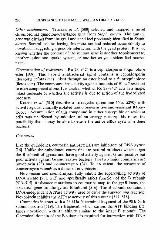
216 RESISTANCE TO NON-CELL WALL ANTIBACTERIALS
Other mechanisms. Trucksis et al. [508] selected and mapped a novel chromosomal quinolone-resistance gene from Staph. aureus. The mutant gene was distinct from the gyrA and norA loci previously identified in Staph. aureus. Several isolates having this mutation had reduced susceptibility to novobiocin suggesting a possible interaction with the gyrB protein. It is not known whether the product of the mutant gene is another topoisomerase, another quinolone uptake system, or another as yet unidentified mecha- nism.
Circumvention of resistance. Ro 23-9424 is a cephalosporin 3'-quinolone ester [509]. This hybrid antibacterial agent contains a cephalosporin (desacetyl cefotaxime) linked through an ester bond to a fluoroquinolone (fleroxacin). The compound has activity against mutants of E. coli resistant to each component alone. It is unclear whether Ro 23-9424 acts as a single, intact molecule or whether the activity is due to action of the hydrolyzed products.
Kotera et al. [510] describe a tetracyclic quinolone (No. 5290) with activity against clinically isolated quinolone-sensitive and -resistant staphy- lococci. Accumulation of this compound in either susceptible or resistant cells was unaffected by addition of an energy poison; this raises the possibility that it may be able to evade the active efflux system in these bacteria.
Coumarins
Like the quinolones, coumarin antibacterials are inhibitors of DNA gyrase [ 161. Unlike the quinolones, coumarins are natural products which target the B subunit of gyrase and have good activity against Gram-positive but poor activity against Gram-negative bacteria. The two major coumarins are novobiocin (23) and coumermycin (24). To an extent, the structure of coumermycin resembles a dimer of novobiocin.
Novobiocin and coumermycin fully inhibit the supercoiling activity of DNA gyrase [511, 5121 and specifically affect function of the B subunit [5 13-5 151. Resistance mutations to coumarins map to the gyrB locus, the structural gene for the gyrase B subunit [516]. The B subunit contains a DNA-independent ATPase activity used to drive the supercoiling reaction. Novobiocin inhibits the ATPase activity of this subunit [517, 5181.
Coumarins interact with a 43 kDa N-terminal fragment of the 90 kDa B subunit protein [519]. The fragment, which carries the ATP binding site, binds novobiocin with an affinity similar to the intact B subunit. The C-terminal domain of the B subunit is required for interaction with DNA
N.E. ALLEN 217
Me
M e O @ w Me
OH H 0 Me N-E
H2N-$-0 OH
Me I Me
and with the A subunit. Novobiocin blocks binding of the nucleotide to the B subunit [5 141, and inhibits ATPase activity and supercoiling. Inhibition appears competitive with respect to ATP [5 151. Since there is little structural relatedness between novobiocin and ATP, these compounds do not necessarily share a common binding site. Moreover, the kinetics of inhibition of ATPase have been described as non-Michaelian, further questioning the liklihood of classic competitive inhibition [446]. Novobiocin and coumermycin inhibit reactions involving participation of the B subunit and have no effect on those functions which depend solely on the A subunit, for example, DNA cleavage and relaxation of supercoiled DNA [519].
Target alterations. Coumarin resistance results from a chromosomal alteration in the gyrB locus in Gram-positive bacteria [516]. Resistance is conferred by the B subunit and the B subunit-catalyzed hydrolysis of ATP is resistant to novobiocin when the subunit originates from resistant bacteria. Supercoiling activity of DNA gyrase from quinolone-resistant organisms remains sensitive to novobiocin.
Holmes and Dyall-Smith [520] have sequenced a gyrB gene from a novobiocin-resistant mutant of Huloferux (a halophilic archaebacterium). The gene contains three amino acid changes, identified as Asp82 to glycine, Ser122 to threonine and Arg137 to histidine, in the N-terminal region of the B subunit. None of the equivalent amino acid positions in the GyrB protein from E. coli is thought to function in binding of ATP [521]; however, no
218 RESISTANCE TO NON-CELL WALL ANTIBACTERIALS
gyrB mutations in E. coli have been described at the amino acid level [446]. The sequence of gyrB from Haloferax predicts a protein having high homology with the GyrB protein in eubacteria [520].
An interesting story has developed around the novobiocin-producing culture Streptomyces sphaeroides. This organism makes two gyrase B proteins [522]. One (79 kDa) is resistant to novobiocin but the other is sensitive to the antibiotic. A portion of the N-terminal domain, consisting of amino acid residues 134-256, is thought to determine binding of novobiocin in the two enzymes [523]. The sensitive form predominates and is produced constitutively whereas the novobiocin-resistant species is produced only in response to the presence of novobiocin [524]. Transcription of the latter is regulated by a promoter sensitive to DNA topology. A reduction in DNA supercoiling activates the promoter triggering expression of the new B subunit.
Studies with Streptomyces niveus (a novobiocin producer) revealed 3 novobiocin-resistance determinants, two of which were specific to novobio- cin-producing strains. Mitchell et al. [525] described two distinct DNA sequences from this streptomycete conferring resistance to novobiocin. Neither sequence hybridized with the cloned sequence from Streptomyces sphaeroides. Kuo et al. [526] reported 0-carbamoylation of novobiocin by Streptomyces niveus. The end-product of the enzymatic modification, 2"-O-carbamoyl-novobiocin, had no activity against producing strains. If verified, this would be a new mechanism of resistance to coumarins.
Several novobiocin-resistant mutants of H. injluenzae contained novobi- ocin-resistant gyrase activity [527]. A closer examination revealed that several of the resistant mutants produced increased amounts of active gyrase. The investigators concluded that high-level resistance requires a structural change in gyrB, but low-level resistance to coumarins could result from multiple copies of wild type gyrB.
There are reports of alterations in other (non gyrB) genes associated with resistance to coumarins. For example, mutations in cysB and cysE genes of E. coli caused an increase in resistance to novobiocin but not to coumermycin [528]. The mutant alleles did not seem to affect drug transport or gyrase activity. Another study from the same laboratory [529] described another locus (nov) in E. coli affecting resistance to novobiocin and, to a lesser extent, coumermycin. Kranz et al. [530] described studies in Rhodobacter capsulatus where coumermycin-resistant strains had altera- tions in he1 genes involved in biosynthesis of cytochrome c. The authors suggest that effects on he1 genes could be indicative of a second target for coumarins. Pocklington et al. [53 13 report that novobiocin-resistant yeast cells are not altered in topoisomerase 11. The fact that novobiocin is toxic to
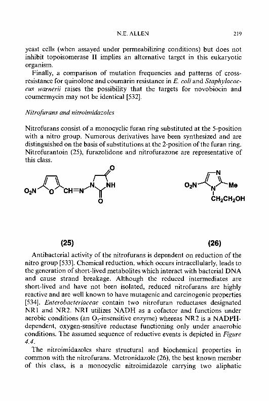
N.E. ALLEN 219
yeast cells (when assayed under permeabilizing conditions) but does not inhibit topoisomerase I1 implies an alternative target in this eukaryotic organism.
Finally, a comparison of mutation frequencies and patterns of cross- resistance for quinolone and coumarin resistance in E. coli and Staphylococ- cus warnerii raises the possibility that the targets for novobiocin and coumermycin may not be identical [532].
Nitrofurans and nitroimidazoles
Nitrofurans consist of a monocyclic furan ring substituted at the 5-position with a nitro group. Numerous derivatives have been synthesized and are distinguished on the basis of substitutions at the 2-position of the furan ring. Nitrofurantoin (25), furazolidone and nitrofurazone are representative of this class.
(25) (26) Antibacterial activity of the nitrofurans is dependent on reduction of the
nitro group [533]. Chemical reduction, which occurs intracellularly, leads to the generation of short-lived metabolites which interact with bacterial DNA and cause strand breakage. Although the reduced intermediates are short-lived and have not been isolated, reduced nitrofurans are highly reactive and are well known to have mutagenic and carcinogenic properties [534]. Enterobacteriaceae contain two nitrofuran reductases designated NRl and NR2. NRI utilizes NADH as a cofactor and functions under aerobic conditions (an 0,-insensitive enzyme) whereas NR2 is a NADPH- dependent, oxygen-sensitive reductase functioning only under anaerobic conditions. The assumed sequence of reductive events is depicted in Figure 4.4.
The nitroimidazoles share structural and biochemical properties in common with the nitrofurans. Metronidazole (26), the best known member of this class, is a monocyclic nitroimidazole carrying two aliphatic
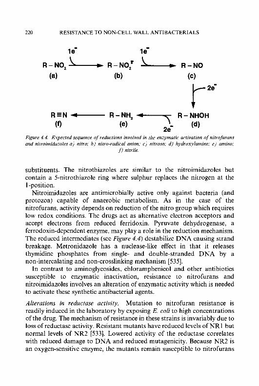
220 RESISTANCE TO NON-CELL WALL ANTIBACTERIALS
1 e' 1 e'
Figure 4.4. Expected sequence of' reductions involved in the enzymatic activation of nitrojurans and nitroimidazoles u) nitro; b ) nitro-radical anion; c) nitroso; d) iiydro.uylamine; e ) amino;
f) nitrile.
substituents. The nitrothiazoles are similar to the nitroimidazoles but contain a 5-nitrothiazole ring where sulphur replaces the nitrogen at the 1-position.
Nitroimidazoles are antimicrobially active only against bacteria (and protozoa) capable of anaerobic metabolism. As in the case of the nitrofurans, activity depends on reduction of the nitro group which requires low redox conditions. The drugs act as alternative electron acceptors and accept electrons from reduced ferridoxin. Pyruvate dehydrogenase, a ferrodoxin-dependent enzyme, may play a role in the reduction mechanism. The reduced intermediates (see Figure 4.4) destabilize DNA causing strand breakage. Metronidazole has a nuclease-like effect in that it releases thymidine phosphates from single- and double-stranded DNA by a non-intercalating and non-crosslinking mechanism [535].
In contrast to aminoglycosides, chloramphenicol and other antibiotics susceptible to enzymatic inactivation, resistance to nitrofurans and nitroimidazoles involves an alteration of enzymatic activity which is needed to activate these synthetic antibacterial agents.
Alterations in reductase activity. Mutation to nitrofuran resistance is readily induced in the laboratory by exposing E. coli to high concentrations of the drug. The mechanism of resistance in these strains is invariably due to loss of reductase activity. Resistant mutants have reduced levels of NR 1 but normal levels of NR2 [533]. Lowered activity of the reductase correlates with reduced damage to DNA and reduced mutagenicity. Because NR2 is an oxygen-sensitive enzyme, the mutants remain susceptible to nitrofurans
N.E. ALLEN 22 1
under anaerobic conditions. Mutants lacking both NR1 and NR2 have not been reported, perhaps indicating a critical role for reductase activity in normal cellular metabolism [534].
Sastry and Jayaraman [536] have isolated two proteins from E. coli which they claim have the ability to bind nitrofurantoin and inhibit nitroreductase activity. In nitrofurantoin-sensitive cells, the two proteins annul each other and have no effect on nitroreductase activity. However, in nitrofurantoin- resistant cells, alteration of one or the other protein results in depressed reductase activity. This study raises the possibility that alteration of reductase activity leading to nitrofuran resistance may be brought about indirectly by alteration of ‘target’ proteins OT: which reduced nitrofurans act.
Resistance to metronidazole likewise correlates with depressed levels of reductase activity. In one study, metronidazole-resistant mutants of Bact. fragilis had depressed levels of pyruvate dehydrogenase activity; the higher the level of resistance, the more depressed the dehydrogenase activity [537]. The resistant mutants were apparently unable to accumulate metronidazole which is consistent with the notion that passive uptake of the drug requires intracelluar reduction in order to maintain a concentration gradient [535]. The same phenotype was observed in laboratory-induced mutations and in a clinically isolated resistant strain of Bact. fragilis. Other studies describe metronidazole-resistant mutants having depressed nitroreductase activities but normal levels of pyruvate dehydrogenase [534]. The exact role of pyruvate dehydrogenase remains to be determined.
Other mechanisms. Breeze and Obaseiki-Ebor [538] described a nitrofu- rantoin-resistant mutant of E. coli having normal reductase activity but increased UV resistance suggesting that enhanced repair of damaged DNA could be associated with resistance to the antibiotic. These same investigators described aminoglycoside-resistant mutants of E. coli which were cross-resistant to nitrofurans [539]. The mutants were characterized as having gross membrane alterations which could lead to impaired accumula- tion of both aminoglycosides and nitrofurans.
CONCLUDING REMARKS
The enormous success derived from using antibacterial agents to treat infectious disease over the last forty-plus years is due, in part, to the extraordinary biochemical specificities of the agents. The most useful agents have been those with specific biochemical mechanisms of action at precise subcellular targets. The more selective an agent is for its bacterial target, the
222 RESISTANCE TO NON-CELL WALL ANTIBACTERIALS
greater the likelihood for safety with less chance for adverse effects on the patient. Ironically, this same specificity may have helped to facilitate the development of resistance to many of the agents. With few exceptions, bacteria can develop resistance to antibacterial agents by mutating or acquiring a single gene which blocks (directly or indirectly) action of the agent on the target. It should be more difficult for bacteria to develop resistance to agents acting on multitargets, but these agents would have increased potential for host toxicity.
The spread of antibacterial resistance is accelerating. At the same time, there appears to be a resurgence of infectious diseases once thought to be under control. For example, the popular press continually warns of increasing numbers of cases of tuberculosis and presents detailed descrip- tions of infections caused by tissue-necrotizing strains of group A streptococci. Additionally, new diseases are emerging, for example, the enteric infections caused by enterotoxigenic E. coli strain 0157:H7. By virtue of their genetic versatility, bacteria continue to devise novel mechanisms to wreak havoc on their human hosts, and, at the same time, develop resistance to existing antibacterial agents used to control them. Until recently, clinicians could make second or third choices of drugs in order to deal with antibiotic resistance. Today, there are fewer choices and clinicians are faced with the prospect of trying to treat infectious diseases for which there may be no effective drugs. There is an urgent need for new antibacterials to combat newly emergent and antibiotic-resistant pathogens.
In the pharmaceutical industry as well as in academic and hospital communities, research is being conducted to better understand mechanisms of resistance to antibacterial agents at the molecular level. Studies are concentrating on newly discovered and existing agents. In the pharmaceuti- cal industry, the search for novel natural products and synthetic organic compounds having the potential to circumvent mechanisms of resistance is being expedited. Empirical as well as rational structure-based design approaches are being used in this effort. To be effective, compounds could exert their action by either blocking the resistance mechanism itself, or by acting on a novel subcellular target. Additional approaches include the effort to improve our understanding of the mechanisms of pathogenesis (how pathogens cause disease) in order to develop drugs to nullify virulence. These kinds of studies are necessary to ensure a pipeline of new drugs into the future. The success or failure of these efforts will largely determine whether mankind is able to ultimately win its battle with the bacteria.
223 N.E. ALLEN
ACKNOWLEDGEMENTS
I wish to thank Dr. T.I. Nicas for helpul suggestions and comments on the manuscript and Ms. L.W. Crandall for assistance with the literature review.
REFERENCES
1 2
3
4 5 6 7
8 9
10 11
12
13 14 15 16
17
18
19 20 21 22
23 24 25 26
27
Fleming, A. (1929) Br. J. Exp. Pathol. 10, 226-236. Abraham, E.P., Chain, E., Fletcher, C.M., Gardner, A.D., Heatley, N.G., Jennings, M.A. and Florey, H.W. (1941) Lancet 240, ii, 177-189. Garrod, L.P., Lambert, H.P. and O’Grady, F.W. (1981) in Antibiotic and Chemother- apy, 5th Ed. pp.6-10, Churchill Livingston, Edinburgh. Abraham, E.P. and Chain, E. (1940) Nature (London) 146, 837. Lyon, B.R. and Skurray, R. (1987) Microbiol. Rev. 51, 88-134. Neu, H.C. (1992) Science (Washington, D.C.) 257, 10641073. Murray, R., Kilham, L., Wilcox, C. and Finland, M. (1946) Proc. SOC. Exp. Biol. Med. 63,470474. Uttley, A.H., Collins, C.H., Naidoo, J. and George, RC(1988) Lancet i, 57-58. Leclerq, R., Derlot, E., Duval, J.andCourvalin, P. (1988)N. Engl. J. Med. 319,157-161. Cohen, M.L. (1992) Science (Washington, D.C.) 257, 1050-1055. Watson, J.D., Hopkins, N.H., Roberts, J.W., Steitz, J.A. and Weiner, A.M. (1987) in Molecular Biology of the Gene, Vol. 1 General Principles, pp. 176-213, Benjamin/ Cummings, Menlo Park, California. Davis, B.D., Dulbecco, R., Eisen, H.N. and Ginsberg, H.S. (1990) in Microbiology, 4th ed. pp. 123-142, J.B. Lippincott, Philadelphia. Clewell, D.B. (1981) Microbiol. Rev. 45,409436. Saunders, J.R. (1984) Br. Med. Bull. 40,5460. Kleckner, N. (1981) Annu. Rev. Genet. 15, 341404. Gale, E.F., Cundliffe, E., Reynolds, P.E., Richmond, M.H. and Waring M.J. (1981) in The Molecular Basis of Antibiotic Action, 2nd Ed., pp. 189-195, John Wiley, New York. Meyers, E., Parker, W.L., Brown, W.E., Linnett, P. and Strominger, J.L. (1974) Ann. N.Y. Acad. Sci. 235,493-501. Storm, D.R., Rosenthal, K.S. and Swanson, P.E. (1977) Annu. Rev. Biochem. 46,
LaPorte, D., Rosenthal, K. and Storm, D.R. (1977) Biochemistry 16, 1642-1648. Rosenthal, K.S., Swanson, P.E. and Storm, D.R. (1976) Biochemistry 15,5783-5792. Cooperstock, M.S. (1974) Antimicrob. Agents Chemother. 6,422425. Brown, M.R.W. (1975) in Resistance of Pseudoinonus aeruginosu (Brown, M.R.W., ed.) pp.71-99, Wiley, London. Cerny, G. and Teuber, M (1971) Arch. Microbiol. 78, 166. Lieve, L. (1974) Ann. N.Y. Acad. Sci. 235, 109-129. Morrison, D.C. and Jacobs, D.M. (1976) Immunochemistry 13,813-818. Vaara, M., Vaara, T., Jensen, M., Helander, I., Nurminen, M., Rietschel, E.T. and Helena-Makela, P. (1981) FEBS Lett. 129, 145-149. Peterson, A.A., Fesik, S.W. and McGroaty, E.J. (1987) Antimicrob. Agents Chemother. 31,230-237.
723-763.
224 RESISTANCE TO NON-CELL WALL ANTIBACTERIALS
28 Galizzi, A,, Cacco, G., Siccardi, A.G. and Mazza, G. (1975) Antimicrob. Agents Chemother. 8,366-369.
29 Godfrey, A.J. and Bryan, L.E. (1984) in Antimicrobial Drug Resistance (Bryan, L.E., ed.) pp. 113-145, Academic Press, New York.
30 Teuber, M. (1969) J. Bacteriol. 98, 347-350. 31 Kaca, W., Radziejewska-Lebrecht, J. and Bhat, U.R. (1990) Microbios 61, 23-32. 32 Moore, R.A. and Hancock, R.E. (1986) Antimicrob. Agents Chemother. 30,923-926. 33 Newton, B.A. (1953) J. Gen. Microbiol. 9 ,5464. 34 Nicas, T.I. and Hancock, R.E.W. (1983) J. Gen. Microbiol. 129, 509-517. 35 Hancock, R.E.W. (1984) Annu. Rev. Microbiol. 38, 237-264. 36 Gilleland, H.E., Jr. and Conrad. R.S. (1982) Antimicrob. Agents Chemother. 22,
37 Gilleland Jr., H.E. and Farly, L.B. (1982) Can. J. Microbiol. 28, 830-840. 38 Gilleland Jr., H.E. and Lyle, R.D. (1979) J. Bacteriol. 138, 839-845. 39 Gilleland Jr., H.E. and Murray, R.G.E. (1976) J. Bacteriol. 125, 267-281. 40 Moore, R.A., Chan, L. and Hancock, R.E. (1984) Antimicrob. Agents Chemother. 36,
539-545. 41 Champlin, F.R., Gilleland Jr., H.E. and Conrad, R.S. (1983) Antimicrob. Agents
Chemother. 24,5-9. 42 Conrad, R.S. and Galanos, C. (1989) Antimicrob. Agents Chemother. 33, 17241728. 43 Traub, W.H. (1982) Chemotherapy (Basel) 28, 363-368. 44 Hancock, R.E.W. and Nicas, T.I. in Ref. 29, pp.147-171. 45 Brown, M.R.W. and Melling, J. (1969) J. Gen. Microbiol. 59, 263-274. 46 Nicas, T.I. and Hancock, R.E.W. (1980) J. Bacteriol. 143, 872-878. 47 Conrad, R.S. and Gilleland Jr., H.E. (1981) J. Bacteriol. 148, 487497. 48 Dorner, E. and Teuber, M. (1977) Arch. Microbiol. 114, 87-89. 49 Said, A A,, Livermore, D.M. and Williams, R.J. (1987) J . Med. Microbiol. 24, 267-274. 50 Shand, G.H., Anwar, H. and Brown, M.R. (1988) J . Antimicrob. Chemother. 22,
81 1-821. 51 Bell, A,, Bains, M. and Hancock, R.E.W. (1991) J. Bacteriol. 173, 6657-6664. 52 Young, M.L., Bains, M., Bell, A. and Hancock, R.E. (1992) Antimicrob. Agents
Chemother. 36,25662568. 53 Bell, A. and Hancock, R.E.W. (1989) J. Bacteriol. 171,321 1-3217. 54 Vaara, M., Vaara, T. and Sarvas, M. (1979) J. Bacteriol. 139, 664667. 55 Vaara, M. (1981) FEMS Microbiol. Lett. 11,321-326. 56 Brown, M.R.W. and Wood, S. (1972) J. Pharm. Pharmacol. 24,215-218. 57 Gilleland Jr., H.E. and Champlin, F.R. (1984) Can. J. Microbiol. 30, 869-873. 58 Harold, F.M. (1970) Adv. Microb. Physiol. 4,45-104. 59 Harold, F.M., Altendorf, K.H. and Hirata, H. Ann. (1974)N.Y. Acad. Sci. 235, 149-160. 60 Mitchell, P. (1966) Biol. Rev. Cambridge Philos. SOC. 41, 44-502. 61 Harold, F.M. (1986) in The Vital Force: A Study of Bioenergetics, W.H. Freeman, New
York. 62 Krulwich, T.A., Quirk, P.G. and Guffanti, A.A. (1990) Microbiol. Rev. 54, 52-65. 63 Decker, S.J. and Lang, D.R. (1977) J. Biol. Chem. 253, 5936-5938. 64 Decker, S.J. and Lang, D.R. (1978) J. Biol. Chem. 253,6738-6743. 65 Guffanti, A.A., Blumenfeld, H. and Krulwich, T. (1981) J. Biol. Chem. 256, 84168421. 66 Guffanti, A.A., Fuchs, R.T. and Krulwich, T.A. (1983) J. BioLChem. 258, 35-37. 67 Clejan, S., Guffanti, A.A., Falk, L.H. and Krulwich, T.A. (1988) Biochim. Biophys. Acta
932,43-51. 68 Dunkley Jr., E.A., Clejan, S. and Krulwich, T.A. (1991) J. Bacteriol. 173, 7750-7755.
10 12-10 16.

N.E. ALLEN 225
69 Guffanti, A.A., Clejan, S., Falk, L.H., Hicks, D.B. and Krulwich, T.A. (1987) J. Bacteriol. 169,4469478.
70 Krulwich, T.A., Clejan, S., Falk, L. and Guffanti, A.A. (1987) J. Bacteriol. 169, 4479485 .
71 Sedgwick, E.G., Hou, C. and Bragg, P.D. (1984) Biochim. Biophys. Actd 767,479492. 72 Date, T., Zwizinski, C., Ludmerer, S. and Wickner, W. (1980) Proc. Natl. Acad. Sci.
U.S.A. 77, 827-831. 73 Ito, M. and Ohnishi, Y. (1981) FEBS Lett. 136, 225-230. 74 Jones, M.R. and Beechey, R.B. (1987) J. Gen. Microbiol. 133,2759-2766. 75 Herring, F.G., Krisman, A,, Sedgwick, E.G. and Bragg, P.D. (1985) Biochim. Biophys.
Acta 819, 231-240. 76 Quirk, P.G., Jones, M.R., Haworth, R.S., Beechey, R.B. and Campbell, I.D. (1989) J.
Gen. Microbiol. 135, 2577-2587. 77 Morehead, M.C. and Dawson, K.A. (1992) Appl. Environ. Microbiol. 58, 1617-1623. 78 Newbold, C.J., Wallace, R.J. and Walker, N.D. (1993) J. Appl. Bacteriol. 75, 129-134. 79 Linton, K.J., Cooper, H.N., Hunter, I.S. and Leadlay, P.F. (1994) Mol. Microbiol. 11,
777-785. 80 Senior, A.E. (1990) Annu. Rev. Biophys. Chem. 19, 7-41, 81 Hoppe. J. and Sebald, W. (1984) Biochim. Biophys. Acta 768, 1-27. 82 Hoppe, J., Schairer, H.U. and Sebald, W. (1980) Eur. J. Biochem. 112, 17-24. 83 Fillingame, R.H., Oldenburg, M. and Fraga, D. (1991) J. Biol. Chem. 266,2093420939. 84 Brafies, L.S. and Kay, W.W. (1983) J. Bacteriol. 154, 1462-1466. 85 Leimgruber, R.M., Jensen, C. and Abrams, A. (1981) J. Bacteriol. 147, 363-372. 86 Anand, N. (1975) in Antibiotics, Vol. 111. Mechanisms of Action of Antimicrobial and
Antitumor Agents (Corcoran, J.W. and Hahn, F.E., eds.) pp. 668498, Springer-Verlag, New York.
87 Woods, D.D. (1940) Br. J. Exp. Pathol. 21, 7490. 88 Miller, A.K. (1944) Proc. SOC. Exp. Biol. Med. 57, 151-153. 89 Lascelles, J. and Woods, D.D. (1952) Br. J. Exp. Pathol. 33, 288. 90 Brown, G.M. (1962) J. Biol. Chem. 237,536540. 91 Seydel, J.K. (1968) J. Pharm. Sci. 57, 1455-1478. 92 Smith, J.T. and Amyes, S.G.B. (1984) Br. Med. Bull. 40, 4246. 93 Henry, R.J. (1943) Bacteriol. Rev. 7, 175-262. 94 Burchall, J.J. and Hitchings, G.H. (1965) Mol. Pharmacol. I , 126-136. 95 Towner, K.J. and Slack, R.C. (1986) Eur. J. Clin. Microbiol. 5, 502-506. 96 Pato, M.L. and Brown, G.M. (1963) Arch. Biochem. Biophys. 103,443448. 97 Towner, K.J. (1992) J. Med. Microbiol. 3 6 , 4 6 . 98 Hamilton-Miller, J.M.T. (1984) in Ref. 29, pp. 173-190. 99 Wise Jr., E.M. and Abou-Donia, M.M. (1975) Proc. Natl. Acad. Sci. U.S.A. 72,
262 1-2625. 100 Skold, 0. (1976) Antimicrob. Agents Chemother. 9,49-54. 101 Swedberg, G. and Skold, 0. (1983) J. Bacteriol. 153, 1228-1237. 102 Radstrom, P. and Swedberg, G. (1988) Antimicrob. Agents Chemother. 32, 16841692. 103 Sundstrom, L., Radstrom, P., Swedberg, G. and Skold, 0. (1988) Mol. Gen. Genet. 213.
191-201. 104 Pinney, R.J. and Smith, J.T. (1973) J. Med. Microbiol. 6, 13-19. 105 Stacey, K.A. and Simpson, E. (1965) J. Bacteriol. 90, 554555. 106 Amyes, S.G.B. and Smith, J.T. (1975) J. Antimicrob. Chemother. I , 85-89. 107 McCuen, R.W. and Sirotnak, F.M. (1974) Biochim. Biophys. Acta 338, 540-544. 108 Amyes, S.G.B. and Towner, K.J. (1990) J. Med. Microbiol. 31, 1-19.

226 RESISTANCE TO NON-CELL WALL ANTIBACTERIALS
109 Amyes, S.G.B. and Smith, J.T. (1976) Eur. J. Biochem. 61, 597-603. 110 Amyes, S.G.B. and Smith, J.T. (1978) J. Gen. Microbiol. 107, 263-271. 11 1 Skold, 0. and Widh, A. (1974) J. Biol. Chem. 249,43244325. 112 Grey, D., Hamilton-Miller, J.M.T. and Brumfitt, W. (1979) Chemotherapy (Basel) 25,
296302. 113 Pattishaw, K.H., Acar, J., Burchall, J.J., Goldstein, F.W. and Harvey, R.J. (1977) J. Biol.
Chem. 252,2319-2323. 114 Tennhamer-Ekman, B. and Skold, 0. (1979) Plasmid 2, 334346. 115 Broad, D.F. and Smith, J.T. (1982) Eur. J. Biochem. 125,617-622. 116 Fling, M.E. and Elwell, L.P. (1980) J . Bacteriol. 141, 779-785. 117 Fling, M.E., Walton, L. and Elwell, L.P. (1982) Antimicrob. Agents Chemother. 22,
882-888. 118 Zolg, J.W. and Hanggi, U.J. (1981) Nucleic Acids Res. 9, 697-710. 119 Smith, S.L., Stone, D., Novak, P., Baccanari, D.P. and Burchall, J.J. (1979) J. Biol.
Chem. 254,6222-6225. 120 Thomson, C.J., Young, H-K and Amyes, S.G.B. (1990) J. Med. Microbiol. 32, 153-
158. 121 Wylie, B.A. and Koornhof, H.J. (1991) J. Med. Microbiol. 35, 214218. 122 Thomas, C.J., Barg, N. and Amyes, S.G.B. (1 990) J. Gen. Microbiol. 136, 673-677. 123 Nixon, P.F. and Blakely, R.H. (1968) J. Biol. Chem. 243,47224731. 124 Gutmann, L., Williamson, R., Moreau, N., Kitzis, M.-D., Collatz, E., Acar, J.F. and
Goldstein, F.W (1985) J. Infect. Dis. 151, 501-507. 125 Luzatto, L., Apirion, D. and Schlessinger, D. (1969) J. Mol. Biol. 42, 315-335. 126 Hancock, R.E.W. (1981) J. Antimicrob. Chemother. 8, 249-276. 127 Davis, B.D. (1987) Microbiol. Rev. 51, 341-350. 128 Davies, J. and Davis, B.D. (1968) J. Biol. Chem. 243, 3312-3316. 129 Le Goffic, F., Capmau, M-L., Tangy, F. and Baillarge, M. (1979) Eur. J. Biochem. 102,
130 Dalhoff, A. (1987) Antimicrob. Chemother. 39, 182-204. 131 Cundliffe, E. (1990) in The Ribosome. Structure, Function & Evolution (Hill, W.E.,
Dahlberg, A,, Garrett, R.A., Moore, P.B., Schlessinger, D. and Warner, J.R., eds.) pp. 479490, American Society for Microbiology, Washington, D.C.
132 Bollen, A., Davies, J., Ozaki, M. and Mizushima, S. (1969) Science (Washington, D.C.) 165, 85-86.
133 Okuyama, A., Machiyama, N., Kinoshita, T. and Tanaka, N. (1971) Biochem. Biophys. Res. Commun. 43, 196199.
134 Okuyama, A,, Tanaka, N. and Komai, T. (1975) J. Antibiot. 28, 903-905. 135 Ozaki, M., Mizushima, S. and Nomura, M. (1969) Nature (London) 222, 333-339. 136 Funatsu, G. and Wittman, H.G. (1972) J. Mol. Biol. 68, 547-550. 137 Maness, M.J., Foster, G.C. and Sparling, P.F. (1974) J. Bacteriol. 120, 1293-1299. 138 Easmon, C.S., Ison, C.A., Bellinger, C.M. and Harris, J.W. (1982) Br. Med. J. 284,
160L1605. 139 Moazed, D. and Noller, H.F. (1987) Nature (London) 327, 389-394. 140 Bryan, L.E. in Ref. 29 pp. 241-273. 141 Buckel, P., Buchberger, A,, Bock, A. and Wittman, H.G. (1977) Mol. Gen. Genet. 158,
47-54. 142 Ahmad, M.H., Rechenbacher, A. and Bock, A. (1980) Antimicrob. Agents Chemother.
18,798-806. 143 Levy, J., Burns, J.L., Mendelman, P.M., Wong, K., Mack, K. and Smith, A.L. (1986)
Antimicrob. Agents Chemother. 29,474481.
73-8 1.

N.E. ALLEN 227
144 Cundliffe, E. (1992) Gene 115,75-84. 145 Thompson, J. , Skeggs, P.A. and Cundliffe, E. (1985) Mol. Gen. Genet. 201, 168-173. 146 Cundliffe, E. (1989) Annu. Rev. Microbiol. 43, 207-233. 147 Piendl, W. and Bock, A. (1982) Antimicrob. Agents Chemother. 22,231-236. 148 Skeggs, P.A., Thompson, J. and Cundliffe, E. (1985) Mol. Gen. Genet. 200,415421. 149 Beauclerk, A.A.D. and Cundliffe, E. (1987) J. Mol. Biol. 193,661-671. 150 De Stasio, E.A., Moazed, D., Noller, H.F. and Dahlberg, A.E. (1989) EMBO J. 8,
121 3-1 2 16. 151 Finken, M., Kirschner, P., Meier, A,, Woods, A. and Bottger, E.C. (1993) Mol.
Microbiol. 9, 1239-1246. 152 O’Connor, M., De Stasio, E.A. and Dahlberg, A.E. (1991) Biochemie 73, 1493-1500. 153 Sigmund, C.D., Ettayebi, M. and Morgan, E.A. (1984) Nucleic Acids Res. 12,46534663. 154 Makosky, P.C. and Dahlberg, A.E. (1987) Biochemie 69,885-889. 155 Helser, T.L., Davies, J.E. and Dahlberg, J.E. (1971) Nature (London) New Biol. 233,
12-14. 156 Helser, T.L., Davies, J.E. and Dahlberg, J.E. (1972) Nature (London) New Biol. 235,6-9. 157 van Bud, C.P., Damm, J.B. and van Knippenberg, P.H. (1983) Mol. Gen. Genet. 189,
475478. 158 van Buul, C.P.J.J. and van Knippenberg, P.H. (1985) Gene 38, 65-72. 159 Suvorov, A.N., van Gemen, B. and van Knippenberg, P.H. (1988) Mol. Gen. Genet. 215,
152-1 55. 160 Daikos, G.L., Jackson, G.G., Lolans, V.T. and Livermore, D.M. (1990) J. Infect. Dis.
162,414420. 161 Nicas, T.I. and Hancock, R.E.W. (1980) J. Bacteriol. 143, 872-878. 162 Abdel-Sayed, S., Gonzalez, M. and Eagon, R.G. (1982) J. Antimicrob. Chemother. 10,
173-182. 163 Moore, R.A. and Hancock, R.E. (1986) Antimicrob. Agents Chemother. 30,923-926. 164 Bryan, L.E., Haraphongse, R. and van den Elsen, H.M. (1976) J. Antibiot. 29,743-753. 165 Parr Jr., T.R. and Bayer, A.S. (1988) J. Infect. Dis. 158, 1003-1010. 166 Bryan, L.E., O’Hara, K. and Wong, S. (1984) Antimicrob. Agents Chemother. 26,
250-2 5 5. 167 Yoneyama, H., Sato, K. and Nakae, T. (1991) Chemotherapy (Basel) 37,239-245. 168 Norris, S.A. and Sciortino, C.V. (1988) J. Infect. Dis. 158, 13241328. 169 Gilleland, L.B., Gilleland, H.E., Gibson, J.A. and Champlin, F.R. (1989) J. Med.
Microbiol. 29,41-50. 170 Lewis, L.A., Li, K.B., Gousse, A., Periera, F., Pacheco, N., Pierre, S., Kodaman, P. and
Lawson, S. (1991) Microbiol. Immunol. 35, 289-301. 171 Miller, M.H., Edberg, S.C., Mandel, L.J., Behar, C.F. and Steigbeigel, N.H. (1980)
Antimicrob. Agents Chemother. 18,722-729. 172 Damper, P.D. and Epstein. W. (1981) Antimicrob. Agents Chemother. 20, 803-808. 173 Alper, M.D. and Ames, B.N. (1978) J. Bacteriol. 133, 149-157. 174 Holtje, J-V. (1978) Eur. J. Biochem. 86, 345-351. 175 Humbert, R. and Altendorf, K. (1989) J. Bacteriol. 171, 1435-1444. 176 Courvalin, P. and Carlier, P. (1981) J. Antimicrob. Chemother. 8, 57-69. 177 Shaw, K.J., Rather, P.N., Hare, R.S. and Miller, G.H. (1993) Microbiol. Rev. 57,
178 Benveniste, R. and Davies, J. (1973) Annu. Rev. Biochem. 42, 471-506. 179 Foster, T.J. (1983) Microbiol. Rev. 47, 361409. 180 Lee, S.C., Cleary, P.P. and Gerding, D.N. (1987) Antimicrob. Agents Chemother. 31,
138-163.
667-670.
228 RESISTANCE TO NON-CELL WALL ANTIBACTERIALS
181 182
183
Feretti, J.J., Gilmore, K.S. and Courvalin, P. (1986) J. Bacteriol. 167, 631-638. Rather, P.N., Munyyer, H., Mann, P.A., Hare, R.S., Miller, G.H. and Shaw, K.J. (1992) J. Bacteriol. 174, 3196-3203. Shaw, K.J., Hare, R.S., Sabatelli, F.J., Rizzo, M., Cramer, C.A., Naples, L., Kocsi, S., Munayyer, H., Mann, P., Miller, G.H., Verbist, L., van Landuyt, H., Glupczynski, Y., Catalano, M. and Woloj, M. (1991) Antimicrob. Agents Chemother. 35, 2253-2261.
184 Benveniste, R. and Davies, J. (1973) Proc. Natl. Acad. Sci. U.S.A. 70, 2276-2280. 185 DeHertogh, D.A. and Lerner, S.A. (1985) Antimicrob. Agents Chemother. 27,670-671. 186 Benveniste, R. and Davies, J. (1971) Biochemistry 10, 1787-1796. 187 Dickie, P., Bryan, L.E. and Pickard, M.A. (1978) Antimicrob. Agents Chemother. 14,
569-580. 188 Bryan, L.E. and Kwan, S. (1983) Antimicrob. Agents Chemother. 23, 835-845. 189 Tabor, H.W., Mueller, J.P., Miller, P.F. and Arrow, A.S. (1987) Microbiol. Rev. 51,
439457. 190 Perlin, M.H. and Lerner, S.A. (1986) Antimicrob. Agents Chemother. 29,216-224. 191 Allen, N.E., Alborn Jr., W.E., Hobbs Jr., J.N. and Kirst, H.A. (1982) Antimicrob. Agents
Chemother. 22,824831. 192 Menard, R., Molinas, C., Arthur, M., Duval, J., Courvalin, P. and Leclercq, R. (1993)
Antimicrob. Agents Chemother. 37, 78-83. 193 Shaw, K.J., Rather, P.N., Sabatelli, F.J., Mann, P., Munayyer, H., Mierzwa, R.,
Petrikkos, G.L., Hare, R.S. and Miller, G.H. (1992) Antimicrob. Agents Chemother. 36, 1447- 1455.
194 Hawkey, P.M. and Constable, H.K. (1988) J. Antimicrob. Chemother. 21, 535-544. 195 Mates, S.M., Esenberg, E.S., Mandel, L.J., Patel, L., Kaback, H.R. and Miller, M.H.
(1982) Proc. Natl. Acad. Sci. U.S.A. 79,6693-6697. 196 Mates, S.M., Patel, L., Kaback, H.R. and Miller, M.H. (1983) Antimicrob. Agents
Chemother. 23, 526-530. 197 Gilman, S. and Saunders, V.A. (1986) J. Antimicrob. Chemother. 18, 301-306. 198 Hurwitz, C., Braun, C.B. and Rosano, C.L. (1981) Biochim. Biophys. Acta 652, 168-176. 199 Werner, R.G., Lechner, U.L. and Goeth, H. (1982) Antimicrob. Agents Chemother. 21,
200 Kirst, H.A., Marconi, G.G., Counter, F.T., Ensminger, P.W., Jones, N.D., Chaney, M.O., Toth, J.E. and Allen, N.E. (1982) J. Antibiot. 35, 1651-1657.
201 Day, L.E. (1966) J. Bacteriol. 91, 1917-1923. 202 Day, L.E. (1966) J. Bacteriol. 92, 197-203. 203 White, J.P. and Cantor, C.R. (1971) J. Mol. Biol. 58, 397400. 204 Connamacher, R.H. and Mandel, H.G. (1968) Biochim. Biophys. Acta 166,475486. 205 Maxwell, I.H. (1968) Mol. Pharmacol. 4, 25-37. 206 Werner, R., Kollak, A,, Nierhaus, D., Shreiner, G. and Nierhaus, K.H. (1975) in Topics
in Infectious Diseases, Vol.1 (Drews, J. and Hahn, F.E., eds.) pp. 217, Springer-Verlag. Vienna.
207 Fey, G., Reiss, M. and Kersten, H. (1973) Biochemistry 12, 1160-1 164. 208 Tritton, T.R. (1977) Biochemistry 16,41334138. 209 Kaji, A. and Ryoji, M. (1979) in Antibiotics, Vol. V, Part 1 (Hahn, F.E., ed.) pp. 304328,
Springer-Verlag, Berlin. 210 Goldman, R.A., Hasan, T., Hall, C.C., Strycharz, W.A. and Cooperman, B.S. (1983)
Biochemistry 22,359-368. 21 1 Suarez, G. and Nathans, D. (1965) Biochem. Biophys. Res. Commun. 18,743-750. 212 Suzuka, I . , Kaji, H. and Kaji, A. (1966) Proc. Natl. Acad. Sci. U.S.A. 55, 1483-1490. 213 Sarkar, S. and Thach, R.E. (1968) Proc. Natl. Acad. Sci. U.S.A. 60, 1479-1486.
10 1-1 06.
N.E. ALLEN 229
214 Gordon, J. (1969) J. Biol. Chem. 244, 5680-5686. 215 Skoultchi, A,, Ono, Y., Waterson, J. and Lengyel, P. (1970) Biochemistry, 9, 508-514. 216 Igarashi, K. and Kaji, A. (1970) Eur. J. Biochem. 14,4146. 217 Levy, S.B. (1984) in Ref. 29, pp. 191-240. 218 Yamaguchi, A,, Ohmori, H., Kaneko-Ohdera, M.J., Nomura, T. and Sawai, T. (1991)
Antimicrob. Agents Chemother. 35, 53-56. 219 McMurray, L.M. and Levy, S.B. (1978) Antimicrob. Agents Chemother. 14,201-209. 220 Levy, S.B. and McMurray, L.M. (1978) Nature (London) 276,90-92. 221 McMurray, L.M., Cullinane, J., Petrucci, R.J. and Levy, S.B. (1981) Antimicrob. Agents
Chemother. 20, 307-313. 222 McMurray, L.M., Aronson, D. and Levy, S.B. (1983) Antimicrob. Agents Chemother.
24, 544551. 223 Chopra, I. (1984) Br. Med. Bull. 40, 11-17. 224 Foster, T.J. (1983) Microbiol. Rev. 47, 361409. 225 Chopra, I. and Howe, T.G.B. (1978) Microbiol. Rev. 42, 707-724. 226 Levy, S.B. and McMurray, L. (1974) Biochem. Biophys. Res. Commun. 56, 1060-1068. 227 Wojdani, A,, Avtalion, R.R. and Sompolinsky, D. (1976) Antimicrob. Agents
Chemother. 9, 526534. 228 Franklin, T.J. (1973) CRC Crit. Rev. Microbiol. 2, 253-272. 229 Levy, S.B. (1992) Antimicrob. Agents Chemother. 36, 695-703. 230 Chopra, I . , Hawkey, P.M. and Hinton, M. (1992) J. Antimicrob. Chemother. 29,
245-277. 231 Speer, B.S., Shoemaker, N.B. and Salyers, A.A. (1992) Clin. Microbiol. Rev. 5,387-399. 232 Mendez, B., Tachibana, C. and Levy, S.B. (1980) Plasmid 3,99-108. 233 Griffith, J.K., Baker, M.E., Rouch, D.A., Page, M.G.P., Skurray, R.A., Paulson, I.T.,
Chater, K.F., Baldwin, S.A. and Henderson, P.J.F. (1992) Curr. Opin. Cell Biol. 4, 68&695.
234 Ball, P.R., Shales, S.W. and Chopra, I. (1980) Biochem. Biophys. Res. Commun. 93, 74-8 1.
235 McMurray, L., Petrucci Jr., R.E. and Levy, S.B. (1980) Proc. Natl. Acad. Sci. U.S.A. 77, 39743977.
236 Hedstrom, R.C., Crider, B.P. and Eagon, R.G. (1982) J. Bacteriol. 152, 255-259. 237 Yamaguchi, A,, Udagawa, T. and Sawai, T. (1990) J. Biol. Chem. 265,48094813. 238 Kaneko, M., Yamaguchi, A. and Sawai, T. (1985) FEBS Lett. 193, 194198. 239 Yamaguchi, A,, Iwasaki-Ohba, Y., Ono, N., Kaneko-Ohdera, M. and Sawai, T. (1991)
FEBS Lett. 282,415418. 240 Levy, S.B., McMurray, L.M., Burdett, V., Courvalin, P., Hillen, W., Roberts, M.C. and
Taylor, D.E. (1989) Antimicrob. Agents Chemother. 33, 1373-1374. 241 Levy, S.B. (1989) J. Antimicrob. Chemother. 24, 1-3. 242 Marshall, B., Morrissey, S., Flynn, P. and Levy, S.B. (1986) Gene 50, 11 1-1 17. 243 Burdett, V., Inamine, J. and Rajagopalan, S. (1982) J. Bacteriol. 149, 995-1004. 244 Varela, M.F. and Griffith, J.K. (1993) Antimicrob. Agents Chemother. 37, 1253-1258. 245 Tovar, K., Ernst, A. and Hillen, W. (1988) Mol. Gen. Genet. 215, 7680. 246 Allard, J.D. and Bertrand, K.P. (1993) J . Bacteriol. 175,45544560. 247 Ishiwa, H. and Shibahara, H. (1985) Jpn. J. Genet. 60,485498. 248 Henderson, P.J.F. and Maiden, M.C.F. (1990) Philos. Trans. R. SOC. London B 326,
391410. 249 Curiale, M.S. and Levy, S.B. (1982) J. Bacteriol. 151,209-215. 250 Curiale, M.S., McMurray, L.M. and Levy, S.B. (1984) J. Bacteriol. 157, 21 1-217.
230 RESISTANCE TO NON-CELL WALL ANTIBACTERIALS
251 Coleman, D.C., Chopra, I., Shales, S.W., Howe, T.G.B. and Foster, T.J. (1983) J. Bacteriol. 153, 921-929.
252 Hickman, R. and Levy, S.B. (1988) J. Bacteriol. 170, 1715-1720. 253 Rubin, R.A. and Levy, S.B. (1990) J. Bacteriol. 172,2303-2312. 254 Hoffmann, G.R. and Fuchs, R.P.P. (1990) J. Mol. Biol. 213, 239-246. 255 Eckert, B. and Beck, C.F. (1989) J. Biol. Chem. 264, 11663-1 1670. 256 Yamaguchi, A,, Adachi, K. and Sawai, T. (1990) FEBS Lett. 265, 17-19. 257 Yamaguchi, A,, Akasaka, T., Ono, N., Someya, Y., Nakatani, M. and Sawai, T. (1992)
J. Biol. Chem. 267, 7490-7498. 258 Rubin, R.A., Levy, S.B., Henriksen, R.L. and Kezdy, F.S. (1990) Gene 87, 7-13. 259 Sheridan, R.P. and Chopra, I. (1991) Mol. Microbiol. 5, 895-900. 260 McMurray, L.M., Stephen, M. and Levy, S.B. (1992) J. Bacteriol. 174,6294-6297. 261 Yamaguchi, A., Adachi, K., Akasaka, N., Ono, N. and Sawai, T. (1991) J. Biol. Chem.
262 Yamaguchi, A,, Ono, N., Akasaka, T., Noumi, T. and Sawai, T. (1990) J. Biol. Chem. 265, 15525-15530.
263 Yamaguchi, A,, Nakatani, M. and Sawai, T. (1992) Biochemistry 31, 83448348. 264 Chopra, I. (1986) J. Antimicrob. Chemother. 18, Suppl. C, 51-56. 265 Chopra, I. (1988) J. Appl. Bacteriol. Symp. Suppl. (1988) 149S166S. 266 Yamaguchi, A,, Ono, N., Akasaka, T. and Sawai, T. (1992) FEBS Lett. 307,229-232. 267 Guay, G.D., Tuckman, M. and Rothstein, D.M. (1994) Antimicrob. Agents Chemother.
268 Hillen, W., Klock, G., Kaffenberger, I., Wray, L.V. and Reznikoff, W.S. (1982) J. Biol. Chem. 257, 660556613,
269 Hillen, W., Schollmeir, K. and Gatz, C. (1984) J. Mol. Biol. 172, 185-201. 270 Klock, G., Unger, B., Gatz, C., Hillen, W., Altenbuchner, J., Schmid, K. and Schmitt, R.
(1985) J. Bacteriol. 161, 326332. 271 Wray, L.V., Jorgensen, R.A. and Reznikoff, W.S. (1981) J. Bacteriol. 147, 297-304. 272 Beck, C.F., Metzel, R., Barbe, J. and Muller, W. (1982) J. Bacteriol. 150, 633-642. 273 Takahashi, M., Altschmied, L. and Hillen, W. (1986) J. Mol. Biol. 187, 341-348. 274 Leclerq, R. and Courvalin, P. (1991) Antimicrob. Agents Chemother. 35, 1267-1272. 275 Hoshino, T., Ikeda, T., Tomizuka, N. and Furukawa, K. (1985) Gene 37, 131-138. 276 Khan, S.A. and Novick, R.P. (1983) Plasmid 10, 225-229. 277 Ives, C.L. and Bott, K.F. (1989) J. Bacteriol. 171, 1801-1810. 278 Chopra, I., Shales, S.W., Ward, J.M. and Wallace, L.J. (1981) J. Gen. Microbiol. 126,
45-54. 279 Moyed, H.S., Nguyen, T.T. and Bertrand, K.P. (1983) J. Bacteriol. 155, 549-556. 280 George, A.M. and Levy, S.B. (1983) J. Bacteriol. 155, 531-540. 281 George, A.M. and Levy, S.B. (1983) J. Bacteriol. 155, 541-548. 282 Hachler, H., Cohen, S.P. and Levy, S.B. (1991) J. Bacteriol. 173, 5532-5538. 283 Cohen, S.P., McMurray, L.M., Hooper, D.C., Wolfson, J.S. and Levy, S.B. (1989)
Antimicrob. Agents Chemother. 33, 1318-1325. 284 Cohen, S.P., McMurray, L.M. and Levy, S.B. (1988) J. Bacteriol. 170, 54165422. 285 Chopra, I. and Eccles, S.J. (1978) Biochem. Biophys. Res. Commun. 83, 550-557. 286 McMurray, L.M., Cullinane, J. and Levy, S.B. (1982) Antimicrob. Agents Chemother.
22,791-799. 287 Leive, L., Telesetsky, S., Coleman Jr., W.G. and Carr, D. (1984) Antimicrob. Agents
Chemother. 25,539-544. 288 Williams, G. and Smith, I. (1979) Mol. Gen. Genet. 177, 23-29. 289 Shales, S.W., Chopra, I. and Ball, P.R. (1980) J. Gen. Microbiol. 121,221-229.
266,6045-605 1.
38,857-860.
N.E. ALLEN 23 1
290 LeBlanc, D.J., Lee, L.N., Titmas, B.M., Smith, C.J. and Tenover, F.C. (1988) J. Bacteriol. 170, 3618-3626.
291 Nickolich, M.P., Shoemaker, N.B. and Salyers, A.A. (1992) Antimicrob. Agents Chemother. 36, 1005-1012.
292 Burdett, V. (1986) J. Bacteriol. 165, 564569. 293 Manavathu, E.K., Fernandez, C.L., Cooperman, B.S. and Taylor, D.E. (1990)
Antimicrob. Agents Chemother. 34,71-77. 294 Burdett, V. (1991) J. Biol. Chem. 266,2872-2877. 295 Sanchez-Pescador, R., Brown, J.T., Roberts, M. and Urdea, M.S. (1988) Nucleic Acids
Res. 16, 1218. 296 Salyers, A.A., Speer, B.S. and Shoemaker, N.B. (1990) Mol. Microbiol. 4, 151-156. 297 Butler, M.J., Friend, E.J., Hunter, I.S., Kaczmarek, F.S., Sugden, D.A. and Warren, M.
(1989) Mol. Gen. Genet. 215,231-238. 298 Ohnuki, T., Katoh, T., Imanaka, T. and Aiba, S. (1985) J. Bacteriol. 161, 1010-1016. 299 Reynes, J.P., Calmels, T., Drocourt, D. and Tiraby, G. (1988) J. Gen. Microbiol. 134,
300 Mikulik, K., Jiranova, A,, Janda, I. and Weiser, J. (1983) FEBS Lett. 152, 125-130 301 Su, Y.A., He, P. and Clewell, D.B. (1992) Antimicrob. Agents Chemother. 36, 769-778. 302 Stevens, A.M., Sanders, J.M., Shoemaker, N.B. and Salyers, A.A. (1992) J. Bacteriol.
174,2935-2942. 303 Park, B.H. and Levy, S.B. (1988) Antimicrob. Agents Chemother. 32, 1797-1800. 304 Speer, B.S. and Salyers, A.A. (1988) J. Bacteriol. 170, 1423-1429. 305 Speer, B.S. and Salyers, A.A. (1989) J. Bacteriol. 171, 148-153. 306 Speer, B.S., Bedzyk, L. and Salyers, A.A. (1991) J. Bacteriol. 173, 176-183. 307 Noller, H.F. (1993) J. Bacteriol. 175, 529775300, 308 Iida, S. (1983) J. Gen. Microbiol. 129, 1217-1225. 309 LaGrice, S.F.J., Matzura, H., Marcoli, R., Iida, S. and Bickle, T.A. (1982) J. Bacteriol.
310 Shaw, W.V., Brenner, D.G. and Murray, I.A. (1985) Curr. Topics Cell. Regul. 26, 455468.
31 1 Harwood, J. and Smith, D.H. (1971) Biochem. Biophys. Res. Commun. 42, 57-62. 312 decrombrugghe, B., Pastan, I., Shaw, W.V. and Rosner, J.L. (1973) Nature (London)
313 Shaw, W.V. (1983) CRC Crit. Rev. Biochem. 14, 1 4 6 . 314 Harding, S.E., Rowe, A.J. and Shaw, W.V. (1987) Biochem. SOC. Trans. 15, 513. 315 Zaidenzaig, Y. and Shaw, W.V. (1978) Eur. J. Biochem. 83, 553. 316 Volker, T.A., Iida, S. and Bickle, T.A. (1982) J. Mol. Biol. 154,417425. 317 Proctor, G.N. and Rownd, R.H. (1982) J. Bacteriol. 150, 1375-1382. 318 Thibault, G., Guitard, M. and Daigneault, R. (1980) Biochim. Biophys. Acta 614,
339-349. 319 Horinouchi, S. and Weisblum, B. (1982) J. Bacteriol. 150, 815-825. 320 Lovett, P.S. (1990) J. Bacteriol. 172, 1-6. 321 Smith, A.L. and Burns, J.L. in Ref. 29, pp. 293-315. 322 Zaidenzaig, Y., Fitton, J.E., Packman, L.C. and Shaw, W.V. (1979) Eur. J. Biochem. 100,
609-6 18. 323 Shaw, W.V. (1984) Br. Med. Bull. 40,36-41.
585-598.
150,312-318.
241, 237-239.
324 Mosher, R.H., Renade, N.P., Shrepf, H. and Vining, L.-C. (1990) J. Gen. Microbiol. 136, 293-301.
325 Gaffney, D.F., Cundliffe, E. and Foster, T.J. (1981) J. Gen. Microbiol. 125, 113-121 326 Irwin, J.E. and Ingram, J.M. (1982) FEMS Microbiol. Lett. 13,6347.
232 RESISTANCE TO NON-CELL WALL ANTIBACTERIALS
327 Burns, J.L., Rubens, C.E., Mandelman, P.M. and Smith, A.L. (1986) Antimicrob. Agents Chemother. 29,445450.
328 Burns, J.L., Hedin, L.A. and Lien, D.M. (1989) Antimicrob. Agents Chemother. 33, 136141.
329 Burns, J.L., Mendelman, P.M., Levy, J., Stull, T.L. and Smith, A.L. (1985) Antimicrob. Agents Chemother. 27,4&54.
330 Toro,C.S., Lobos, S.R., Calderon, I., Rodriquez, M. andMora,G.C. (1990)Antimicrob. Agents Chemother. 34, 1715-1719.
331 Chopra, I. and Eccles, S.J. (1978) Biochem. Biophys. Res. Commun. 83, 550-557. 332 Chopra, I . and Ball, P.R. (1982) Adv. Microb. Physiol. 23, 183-240. 333 Bissonette, L., Champetier, S., Buisson, J-P. and Roy, P.H. (1991) Antimicrob. Agents
Chernother. 173,44934502. 334 Dorman, C.J. and Foster, T.J. (1982) Antimicrob. Agents Chemother. 22,912-914. 335 Bodley, J.W., Zieve, F.J. and Lin, L. (1970) J. Biol. Chem. 245, 5662-5667. 336 Cundliffe, E. (1972) Biochem. Biophys. Res. Commun. 46, 17961801. 337 Willie, G.R., Richman, N., Godtfredson, W.O. and Bodley, J.W. (1975) Biochemistry 14,
17 13-1 7 18. 338 Tanaka, N., Kawano, G. and Kinoshita, T. (1971) Biochem. Biophys. Res. Commun. 42,
564567. 339 Okura, A., Kinoshita, T. and Tanak, N. (1970) Biochem. Biophys. Res. Commun. 41,
1545-1550. 340 Dahlfors, A.A.R. and Kurland, G.G. (1990) J. Mol. Biol. 215, 549-557. 341 Chopra, I . (1976) J. Gen. Microbiol. 96, 229-238. 342 Smith, A.L. and Burns, J.L. in Ref. 29, pp. 306-315. 343 von der Haar, B., Rosenberg, D., Dittrich, W. and Schrempt, H. (1991) J. Antibiot. 44,
344 Volker, T.A., Iida, S. and Bickle, T.A. (1982) J. Mol. Biol. 154,417425. 345 Proctor, G.N., McKell, J. and Rownd, R.H. (1983) J. Bacteriol. 155,937-939. 346 Kirst, H.A. (1993) Prog. Med. Chem. 30,57-88. 347 Fernandez-Muiioz, R., Monro, R.E., Torres-Pinedo, R. and Vazquez, D. (1971) Eur. J.
Biochem. 23, 185-193. 348 Di Giambattista, M., Engelborghs, Y., Nyssen, E. and Cocito, C. (1987) J. Biol. Chem.
349 Menninger, J.R. and Otto, D.P. (1982) Antimicrob. Agents Chemother. 21, 81 1-818. 350 Menninger, J.R. (1985) J. Antimicrob. Chemother. 16 (Suppl.A), 23-34. 351 Di Giambattista, M., Chinali, G . and Cocito, C. (1989) J. Antimicrob. Chemother. 24,
485-507. 352 Arthur, M., Andremont, A. and Courvalin, P. (1987) Antimicrob. Agents Chemother. 31,
404409. 353 Weisblum, B. (1985) J. Antimicrob. Chemother. 16 (Suppl. A), 63-90. 354 Skinner, R., Cundliffe, E. and Schmidt, F.J. (1983) J. Biol. Chem. 258, 12702-12706. 355 Lai, C-J., Weisblum, B., Fahnestock, S.R. and Nomura, M. (1973) J. Mol. Biol. 74,
67-12. 356 Arthur, M., Brisson-Noel, A. and Courvalin, P. (1987) J. Antimicrob. Chemother. 20,
783-802. 357 Uchiyama, H. and Weisblum, B. (1985) Gene 38,103-1 10. 358 Zalacain, M. and Cundliffe, E. (1990) Eur. J. Biochem. 189,67-72. 359 Zalacain, M. and Cundliffe, E. (1989) J. Bacteriol. 171,42544260. 360 Zang, H.Z., Schmidt, H. and Piepersberg, W. (1992) Mol. Microbiol. 6, 2147-2157. 361 Calcutt, M.J. and Cundliffe, E. (1990) J. Bacteriol. 172, 47104714.
785-792.
262, 8591-8597.

N.E. ALLEN 233
362 Kim, H.S., Choi, E.C. and Kim, D.K. (1993) J. Gen. Microbiol., 139, 601-607. 363 Allen, N.E. (1975) Antimicrob. Agents Chemother. 11,669-674. 364 Tanaka, T. and Weisblum, B. (1974) Antimicrob. Agents Chemother. 5,538-540. 365 Horinouchi, S., Byeon, W.H. and Weisblum, B. (1983) J. Bacteriol. 154, 1252-1262. 366 Eady, E.A., Ross, J.I. and Cove, J.H. (1990) J. Antimicrob. Chemother. 26,461471. 367 Janosi, L., Nakajima, Y. and Hashimoto, H. (1990) Microbiol. Immunol. 34, 723-735. 368 Nakajima, Y., Janosi, L., Endou, K., Matsuoka, M. and Hashimoto, H. (1992) J.
Pharmacobiodyn. 15,319-324. 369 Endou, K., Endoh, S., Matsuoka, M. and Nakajima, Y. (1993) Biol. Pharm. Bull. 16,
370 Matsuoka, M., Janosi, L., Endou, K., Saitoh, S., Hashimoto, H. and Nakajuma, Y. (1993) Biol. Pharm. Bull. 16, 1288-1290.
371 Horinuchi, S. and Weisblum, B. (1980) Proc, Natl. Acad. Sci. U.S.A. 7, 7079-7083. 372 Rogers, E.J., Ambulos Jr., N.P. and Lovett, P.S. (1990) J. Bacteriol. 172, 6282-6290. 373 Tomich, P.K., An, F.Y. and Clewell, D.B. (1980) J. Bacteriol. 141, 1366-1374. 374 Kwak, J.H., Choi, E.C. and Weisblum, B. (1991) J. Bacteriol. 173,47254735. 375 Wittmann, H.G., Stofler, G., Apirion, D., Rosen, L., Tanaka, K., Tamaki, M., Takata,
R., Dekio, S., Otaka, E. and Osawa, S. (1973) Mol. Gen. Genet. 127, 175-189. 376 Teraoka, H. and Nierhaus, K.H. (1978) J. Mol. Biol. 126, 185-193. 377 Tipper, D.J., Johnson, C.W., Ginther, C.L., Leighton, T. and Wittmann, H.G. (1977)
Mol. Gen. Genet. 150, 147-159. 378 Conde, M.F., Boynton, J.E., Gillham, N.W., Harris, E.H., Tingle, C.L. and Wang, W.L.
(1975) Mol. Gen. Genet. 140, 183-220. 379 Leclercq, R. and Courvalin, P. (1991) Antimicrob. Agents Chemother. 35, 1273-1276. 380 BarthClkmy, P., Antissier, D., Gerbaud, G. and Courvalin, P. (1984) J. Antibiot. 37,
1692-1 696. 381 Arthur, hl. and Courvalin, P. (1986) Antimicrob. Agents Chemother. 30, 694-700. 382 O’Hara, K., Kanda, T., Ohmiya, K., Ebisu, T. and Kono, M. (1989) Antimicrob. Agents
Chemother. 33, 1354-1357. 383 Kono, M., OHara, K. and Ebisu, T. (1992) FEMS Microbiol. Lett. 76, 89-94. 384 Marshall, V.P., Ciadella, J.I., Baczynskyi, L., Liggett, W.F. and Johnson, R.A. (1989) J.
Antibiot. 42, 132-134. 385 Kuo, M-S., Chirby, D.G., Argoudelis, A.D., Cialdella, J.I., Coats, J.H. and Marshall,
V.P. (1989) Antimicrob. Agents Chemother. 33,2089-2091. 386 Cundliffe, E. (1992) Antimicrob. Agents Chemother. 36, 348-352. 387 Jenkins, G. and Cundliffe, E. (1991) Gene 108,55-62. 388 Vilches, C., Hernandez, C., Mtndez, C. and Salas, J.A. (1992) J. Bacteriol. 174, 161-165. 389 Hernkndez, C., Olano, C., Mendez, C. and Salas, J.A. (1993) Gene 134, 139-140. 390 Rodriquez, A.M., Olano, C., Vilches, C., Mendez, C. and Salas, J.A. (1993) Mol.
Microbiol. 8, 571-582. 391 Brisson-Noel, A,, Delrieu, P., Samain, D. and Courvalin, P. (1988) J. Biol. Chem. 263,
392 Leclerq, R., Brisson-Noel, A,, Duval, J. and Courvalin, P. (1987) Antimicrob. Agents Chemother. 31, 1887-1891.
393 Barcs, I. (1993) J. Chemother. 4, 215-222. 394 Le Goffic, F., Capmau, M.L., Abbe, J., Cerceau, C., Dublanchet, A. and Duval, J. (1977)
Ann. Inst. Pasteur (Paris) 128, 471474. 395 Le Goffic, F., Capmau, M.L., Bonnet, M.L., Cerceau, C., Soussy, C.J., Dublanchet, A.
and Duval, J. (1977) J. Antibiot. 30, 665-669.
328-330.
15880-1 5887.
234 RESISTANCE TO NON-CELL WALL ANTIBACTERIALS
396 Rende-Fournier, R., Leclercq, R., Galimand, M., Duval, J. and Courvalin, P. (1993) Antimicrob. Agents Chemother. 37,2119-2125.
397 Mao, J.C and Putterman, M. (1968) J. Bacteriol. 95, 11 11-1 117. 398 Ron, J.I., Eady, E.A., Cove, J.H., Cunliffe, W.J., Baumberg, S . and Wooten, J.C. (1990)
Mol. Microbiol. 4, 1207-1214. 399 Lampson, B.C., von David, W. and Parisi, J.T. (1986) Antimicrob. Agents Chemother.
30,653-658. 400 Goldman, R.C. and Capobianco, J.O. (1990) Antimicrob. Agents Chemother. 34,
1973-1 980. 401 Schoner, B., Geistlich, M., Rosteck Jr., P., Rao, R.N., Seno, E., Reynolds, P., Cox, K.,
Burgett, S. and Hershberger, C. (1992) Gene 115,93-96. 402 Pernodet, J.L., Alegre, M.T., Blondelet-Rouault, M.H. and Guerineau, M. (1993) J. Gen.
Microbiol. 139, 1003-101 1. 403 Fernandez, P.B., Baker, W.R., Freiberg, L.A., Hardy, D.J. and McDonald, E.J. (1989)
Antimicrob. Agents Chemother. 33,78-81. 404 Goldman, R.C. and Kadam, S.K. (1989) Antimicrob. Agents Chemother. 33, 1058-1066. 405 Terajawa, T., Watanabe, M., Okzubo, T. and Mitsuhashi, S. (1991) Antimicrob. Agents
Chemother. 35, 1370-1375. 406 Leclerq, R., Nantas, L., Soussy, C.J. and Duval, J. (1992) J. Antimicrob. Chemother. 30
407 Weisblum, B. and Demohn (1970) FEBS Lett. 11, 149. 408 Cundliffe, E. (1971) Biochem. Biophys. Res. Commun. 44,912-917. 409 Ryan, P.C., Lu, M. and Draper, D.E. (1991) J. Mol. Biol. 221, 1257-1268. 410 Thompson, J. and Cundliffe, E. (1991) Biochemie 73, 1131-1 135. 411 Cundliffe, E., Dixon, P., Stark, M., Stoffler, G., Ehrlich, R., Stoffler-Meilicke, M. and
Cannon, M. (1979) J. Mol. Biol. 132,235-252. 412 Wienen, B., Ehrlich, R., Stoffler-Meilicke, M., Stoffler, G., Smith, I., Weiss, D., Vince, R.
and Pestka, S. (1979) J. Biol. Chem. 254, 8031-8041. 413 Smith, I., Paress, P. and Pestka, S. (1978) Proc. Natl. Acad. Sci. U.S.A. 75, 5993-5997. 414 Stark, M., Cundliffe, E., Dijk, J. and Stoffler, G. (1980) Mol. Gen. Genet. 180, 11-15. 415 Bascaran, V., Sanchez, L., Hardisson, C. and Brana, A.F. (1991) J. Gen. Microbiol. 137,
416 Cundliffe, E. and Thompson, J. (1981) J. Gen. Microbiol. 126, 185-192. 417 Cannon, M. and Cundliffe, E. (1979) Eur. J. Biochem. 97,541-545. 41 8 Saarma, U. and Remme, J. (1992) Nucleic Acids Res. 20,3 147-3 152. 419 Cundliffe, E. (1978) Nature (London) 272,792-795. 420 Thompson, J., Schmidt, F. and Cundliffe, E. (1982) J. Biol. Chem. 257, 7915-7917. 421 Thompson, J., Cundliffe, E. and Dahlberg, A.E. (1988) J. Mol. Biol. 203,457465. 422 Thompson, J. and Cundliffe, E. (1980) J. Bacteriol. 142,455461. 423 Thompson, J., Cundliffe, E. and Stark, M.J. (1982) J. Gen. Microbiol. 128, 875-884. 424 Molina, H. and Robertgero, M. (1992) Appl. Environ. Microbiol. 58, 895-899. 425 Wehrli, W. and Staehlin, M. in Ref. 86, pp. 252-268. 426 Sippel, A. and Hartman, G.R. (1968) Biochim. Biophys. Acta 157,218-219. 427 Jin, D.J. and Gross, C.A. (1988) J. Mol. Biol. 202,45-58. 428 Jin, D.J. and Gross, C.A. (1989) J. Bacteriol. 171, 5229-5231. 429 Rabussay, D. and Zillig, W. (1969) FEBS Lett. 5, 104106. 430 Lill, U.I. and Hartman, G.R. (1973) Eur. J. Biochem. 38,336-345. 431 Lill, U.I., Behrendt, E.M. and Hartman, G.R. (1975) Eur. J. Biochem. 52,411420. 432 Heil, A. and Zillig, W. (1970) FEBS Lett. 1 I , 165-168. 433 Reynolds, P.E. (1984) Br. Med. Bull. 40, 3-10.
(SuppI.A), 67-75.
1625-1634.
N.E. ALLEN 235
434
435
436
437 438 439
440 44 1
442 443 444 445 445a 446 447
448
449
450
45 1
452
453
454 455 456 457 458 459
460 46 1 462
463
464
Lisitsyn, N.A., Sverdlov, E.D., Moiseyera, E.P., Danilevskaya, O.N. and Nikiforov, V.G. (1984) Mol. Gen. Genet. 196, 173-174. Ovchinnikov, Y.A., Monastyrskaya, G.S., Gubanov, V.V., Lipkin, E.D., Sverdlov, E.D., Kiver, I.F., Bass, I.A., Mindlui, S.Z., Danilevskaya, O.N. and Khesin, R.B. (1981) Mol. Gen. Genet. 184,536-538. Ovchinnikov, Y.A., Monastyrskaya, G.S., Guriev, N.F., Kalinina, N.F., Sverdlov, E.D., Gragerov, A.I., Bass, I.A., Kiver, I.F., Moiseyeva, E.P., Igumnov, V.N., Mindlin, S.Z., Nikiforov, V.G. and Khesin, R.B. (1983) Mol. Gen. Genet. 190,344348. Honore, N. and Cole, S.T. (1993) Antimicrob. Agents Chemother. 37,414418. Levin, M.E. and Hatfull, G.F. (1993) Mol. Microbiol. 8, 277-285. Cassani, G., Burgess, R.R., Goodman, H.M. and Gold, L. (1971) Nature (London) New Biol. 230, 197-200. Schlief, R. (1969) Nature (London) 223, 1068-1069. Severinov, K., Soushko, M., Godfarb, A. and Nikiforov, V. (1993) J. Biol. Chem. 268, 14820-14825. Blanco, M.G., Hardisson, C. and Salas, J.A. (1984) J. Gen. Microbiol. 130, 2883-2891. Nakaido, H. (1976) Biochim. Biophys. Acta 433, 118-132. Hui, J., Gordon, N. and Kojioka, R. (1977) Antimicrob. Agents Chemother. 1 I , 773-779. Radl, S. (1990) Pharmacol. Ther. 48, 1-17. Rosen, T. (1990) Prog. Med. Chem. 27, 235-295. Reece, R.J. and Maxwell, A. (1991) CRC Crit. Rev. Biochem. Mol. Biol. 26, 335-375. Sutcliffe, J.A., Gootz, T.D. and Barrett, J.F. (1989) Antimicrob. Agents Chemother. 33, 2027-2033. Sugino, A,, Peebles, C.L., Kreuzer, K.N. and Cozzarelli, N.R. (1977) Proc. Natl. Acad. Sci. U.S.A. 74,47674771. Sugino, A., Higgins, N.P., Brown, P.O., Peebles, C.L. and Cozzarelli, N.R. (1978) Proc. Natl. Acad. Sci. U.S.A. 75,4832-4838. Mizuuchi, K., O’Dea, M.H. and Gellert, M. (1978) Proc. Natl. Acad. Sci. U.S.A. 75,
Gellert, M., Mizuuchi, K., O’Dea, M.H., Itoh, T. and Tomizawa, J. (1977) Proc. Natl. Acad. Sci. U.S.A. 74,47724776. Barrett, J.F., Sutcliffe, J.A. and Gootz, T.D. (1990) Antimicrob. Agents Chemother. 34,
Mizuuchi, K., Fisher, L.M., O’Dea, M.H. and Gellert, M. (1980) Proc. Natl. Acad. Sci.
Zweerink, M.M. and Edison, A. (1986) Antimicrob. Agents Chemother. 29, 598-601. Takahata, M. and Nishino, T. (1988) Antimicrob. Agents Chemother. 32, 1192-1 195. Hane, M.W. and Wood, T.H. (1969) J . Bacteriol. 99,238-241. Kreuzer, K.N. and Cozzarelli, N.R. (1979) J. Bacteriol. 140,424435. Hallett, P. and Maxwell, A. (1991) Antimicrob. Agents Chemother. 35, 335-340. Okuda, J., Okamoto, S., Takahata, M. and Nishino, T. (1991) Antimicrob. Agents Chemother. 35,2288-2293. Gootz, T.D. and Martin, B.A. (1991) Antimicrob. Agents Chemother. 35, 84C-845. Robillard, N.J. and Scarpa, A.L. (1988) Antimicrob. Agents Chemother. 32, 535-539. Yoshida, H., Kojima, T., Yamkagishi, J-I. and Nakamura, S. (1988) Mol. Gen. Genet. 211, 1-7. Cullen, M.E., Wyke, A.W., Kuroda, R. and Fisher, L.M. (1989) Antimicrob. Agents Chemother. 33,886-894. Yoshida, H., Bogaki, M., Nakamura, M. and Nakamura, S. (1990) Antimicrob. Agents Chemother. 34. 1271-1272.
5960-5963.
1-7.
U.S.A. 77, 1847-1851.
236 RESISTANCE TO NON-CELL WALL ANTIBACTERIALS
465 Horowitz, D.S. and Wang, J.C. (1987) J. Biol. Chem. 262, 5339-5344. 466 Sreedharan, S., Oram, M., Jensen, B., Pererson, L.R. and Fisher, L.M. (1990) J.
Bacteriol. 172, 7260-7262. 467 Sreedharan, S., Peterson, L.R. and Fisher, L.M. (1991) Antimicrob. Agents Chemother.
35,2151-2154. 468 Goswitz, J.J., Willard, K.E., Fasching, C.E. and Peterson, L.E. (1992) Antimicrob.
Agents Chemother. 36, 1166-1 169. 469 Yamagishi, J-I., Yoshida, H., Yamayoshi, M. and Nakamura, S. (1986) Mol. Gen.
Genet. 204, 367-378. 470 Nakamura, S., Nakamura, M., Kojima, T. and Yoshida, H. (1989) Antimicrob. Agents
Chemother. 33,254-255. 471 Yoshida, H., Nakamura, M., Bogaki, M., Ito, H., Kojima, T., Hattori, H. and
Nakamura, S. (1993) Antimicrob. Agents Chemother. 37, 839-845. 472 Yoshida, H., Bogaki, M., Nakamura, M., Yamanaka, L.M. and Nakamura, S. (1991)
Antimicrob. Agents Chemother. 35, 1647-1650. 473 Yoshida, H., Nakamura, M., Bogaki, M. and Nakamura, S. (1990) Antimicrob. Agents
Chemother. 34, 1273-1275. 474 Stein, D.C., Danaher, R.J. and Cook, T.M. (1991) Antimicrob. Agents Chemother. 35,
622-626. 475 Shen, L.L. and Pernet, A.G. (1985) Proc. Natl. Acad. Sci. U.S.A. 82,307-31 1. 476 Shen, L.L., Kohlbrenner, W.E., Weigle, D. and Baronowski, J. (1989) J. Biol. Chem. 264,
2973-2978. 477 Shen, L.L., Mitscher, L.A., Sharma, P.N., O’Donnell, T.J., Chu, D.W.T., Cooper, C.S.,
Rosen, T. and Pernet, A.G. (1989) Biochemistry 28,38863894. 478 Shen, L.L., Baranowski, J. and Pernet, A.G. (1989) Biochemistry 28, 3879-3885. 479 Cambau, E. and Gutmann, L. (1993) Drugs 45 (Suppl.3), 15-23. 480 Hoopsr, D.C. and Wolfson, J.S. (1989) Rev. Infect. Dis. 2, S902-S911. 481 Smith, J.T. (1984) Pharm. J. 233,299-305. 482 Wolfson, J.S., Hooper, D.C., Shih, D.J., McHugh, G.L. and Swartz, M.N. (1989)
Antimicrob. Agents Chemother. 33,705-709. 483 Levy, S.B. (1992) Antimicrob. Agents Chemother. 36, 695-703. 484 Yoshida, S., Kojima, T., Inoue, M. and Mitsuhashi, S. (1991) Antimicrob. Agents
Chemother. 35,368-370. 485 Nakanishi, N., Yoshida, S., Wakebe, H., Inoue, M., Yamaguchi, T. and Mitsuhashi, S.
(1991) Antimicrob. Agents Chemother. 35,2562-2567. 486 Ubukata, K., Itoh-Yamashita, N. and Konno, M. (1989) Antimicrob. Agents
Chemother. 33, 1535-1539. 487 Yoshida, H., Bogaki, M., Nakamura, S., Ubukata, K. and Konno, M. (1990) J. Bacteriol.
172, 6942-6949. 488 Ohshita, Y., Hiaramatsu, K. and Yokota, T. (1990) Biochem. Biophys. Res. Commun.
172, 1028-1034. 489 Cohen, S.P., Hooper, D.C., Wolfson, J.S., Souza, K.S., McMurray, L.M. and Levy, S.B.
(1988) Antimicrob. Agents Chemother. 32, 1187-1 191. 490 Celesk, R.A. and Robillard, N.R. (1989) Antimicrob. Agents Chemother. 33, 1921-1926. 491 Kaatz, G.W., Seo, S.M. and Ruble, C.A. (1993) Antimicrob. Agents Chemother. 37,
10861094. 492 Neyfakh, A.A. (1992) Antimicrob. Agents Chemother. 36,484485. 493 Neyfakh, A.A., Borsch, C.M. and Kaatz, G.W. (1993) Antimicrob. Agents Chemother.
37, 128-129.
N.E. ALLEN 237
494 Hooper, D.C., Wolfson, J.S., Souza, K.S., Tung, C, McHugh, G.L. and Swartz, M.N. (1986) Antimicrob. Agents Chemother. 29,639-644.
495 Hooper, D.C., Wolfson, J.S., Bozza, M.A. and Ng, E.Y. (1992) Antimicrob. Agents Chemother. 36, 1151-1154.
496 Hooper, D.C., Wolfson, J.S., Souza, K.S., Ng, E.Y., McHugh, G.L. and Swartz, M.N. (1989) Antimicrob. Agents Chemother. 33,283-290.
497 Hirai, K., Aoyama, H., Suzuc, S., Irikura, T., Iyobe, S. and Mitsuhashi, S.(1986) Antimicrob. Agents Chemother. 30,248-253.
498 Hirai, K., Aoyama, H., Irikura, T., Iyobe, S. and Mitsuhashi, S. (1986) Antimicrob. Agents Chemother. 29, 535-538.
499 Daikos, G.L., Lolans, V.T. and Jackson, G.G. (1988) Antimicrob. Agents Chemother. 32, 785-787.
500 Michea-Hamzehpour, M., Furet, Y.X. and Pechkre, J.C. (1991) Antimicrob. Agents Chemother. 35,2091-2097.
501 Piddock, L.J.V., Hall, M.C., Belliso, F., Bains, M. and Hancock, R.E.W. (1992) Antimicrob. Agents Chemother. 36, 1057-1061.
502 Hirai, K., Suzue, S., Irikura, T., Iyobe, S. and Mitsuhashi, S. (1987) Antimicrob. Agents Chemother. 31,582-586.
503 Fukuda, H., Hosaka, M., Hirdi, K. and lyobe, S. (1990) Antimicrob. Agents Chemother. 34, 1757-1761.
504 Chapman, J.S. and Georgopapadakou, N.H. (1988) Antimicrob. Agents Chemother. 32, 438442.
505 Bazile, S., Moreau, N., Bouzdrd, D. and Essiz, M. (1992) Antimicrob. Agents Chemother. 36,2622-2627.
506 Bryan, L.E. and Bedard, J. (1991) Eur. J. Clin. Microbiol. Infect. Dis. 10,232-239. 507 Denis, A. and Moreau, N.J. (1993) J. Antimicrob. Chemother. 32, 379-392. 508 Trucksis, M., Wolfson, J.S. and Hooper, D.C. (1991) J. Bacteriol. 173, 58545860. 509 Pace, J., Bertasso, A. and Georgopapadakou, N.H. (1991) Antimicrob. Agents
Chemother. 35, 91&915. 510 Kotera, Y., Inoue, Y., Ohashi, M., Ito, K. andTsukamoto, G. (1991) Chem. Pharm. Bull.
39,26442646. 511 Gellert, M., Mizuuchi, K., O’Dea, M.H. and Nash, H.A. (1976) Proc. Natl. Acad. Sci.
U.S.A. 73,3872-3876. 512 Gellert, M., O’Dea, M.H., Itoh,T. andTomizawa, J. (1976) Proc. Natl. Acad. Sci. U.S.A.
73,44744478. 513 Higgins, N.P., Peebles, C.L., Sugino, A. and Cozzarelli, N.R. (1978) Proc. Natl. Acad.
Sci. U.S.A. 75, 1773-1777. 514 Mizuuchi, K., O’Dea, M.H. and Gellert, M. (1978) Proc. Natl. Acad. Sci. U.S.A. 75,
515 Sugino, A., Higgins, N.P., Brown, P.O., Peebles, C.L. and Cozzarelli, N.R. (1978) Proc. Natl. Acad. Sci. U.S.A.. 75,48384842.
516 Orr, L., Fairweather, N.F., Holland, 1.B. and Pritchard, R.H. (1979) Mol. Gen. Genet. 177, 103-1 12.
517 Staudenbauer, W.L. and Orr, E. (1981) Nucleic Acids Res. 9, 3589-3603. 518 Maxwell, A. and Gellert, M. (1984) J. Biol. Chem. 259, 14472-14480. 519 Cozzerelli, N.R. (1980) Science (Washington, D.C.) 207,953-960. 520 Holmes, M.L. and Dyall-Smith, M.L. (1991) J. Bacteriol. 173,642-648. 521 Wigley, D.B., Davies, G.J., Dodson, E.J., Maxwell, A. and Dodson, G. (1991) Nature
(London) 35 I , 624-629. 522 Thiara, A.S. and Cundliffe, E. (1988) EMBO J. 7, 2255-2259.
5960-5963.
238 RESISTANCE TO NON-CELL WALL ANTIBACTERIALS
523 524 525
526
527
528 529
530
531 532 533 534 535 536 537 538 539
Thiara, AS . and Cundliffe, E. (1993) Mol. Microbiol. 8,495-506. Thiara, AS . and Cundliffe, E. (1989) Gene 81,65-72. Mitchell, J.I., Logan, P.G., Cushing, K.E. and Ritchie, D.A. (1990) Mol. Microbiol. 4,
Kuo, M.S., Yurek, D.A., Chirby, D.G., Cialdella, J.I. and Marshall, V.P. (1991) J. Antibiot. 44, 1096-1 100. Setlow, J.K., Cabrera-Juarez, E., Albritton, W.L. and Mutschler, A. (1985) J. Bacteriol. 164, 525-534. Rakonjac, J., Milic, M. and Savic, D.J. (1991) Mol. Gen. Genet. 228, 307-31 1. Rakonjac, J., Milic, M., Ajdic-Predic, D., Santos, D., Ivanisevic, R. and Savic, D.J. (1992) Mol. Microbiol. 6, 1547-1553. Kranz, R.G., Beckman, D.L. and Foster-Hartnett, D. (1992) FEMS Microbiol. Lett. 72,
Pocklington, M.J., Jenkins, J.R. and Orr, E. (1990) Mol. Gen. Genet. 220,256-260. Howard, B.M., Pinney, R.J. and Smith, J.T. (1993) Microbios 75, 185-195. McCalla, D.R. in Ref. 209 pp. 176-213. Rabin, H.R. and Lockerby, D.L. in Ref. 29, pp. 317-344. Edwards, D.I. in Ref. 209, pp. 121-135. Sastry, S.S. and Jayaraman, R. (1985) Arch. Biochem. Biophys. 236,252-259. Britz, M.L. and Wilkinson, R.G. (1979) Antimicrob. Agents Chemother. 16, 19-27. Breeze, AS . and Obaseiki-Ebor, E.E. (1993) J. Gen. Microbiol. 129,99-103. Obaseiki-Ebor, E.E. and Breeze, AS . (1984) J. Antimicrob. Chemother. 13, 567-576.
845-849.
25-32.

Progress in Medicinal Chemistry - Vol. 32, edited by G.P. Ellis and D.K. Luscombe 0 1995 Elsevier Science B.V. All rights reserved.
5 Inhibitors of HIV Proteinase
J.A. MARTIN Ph.D., S. REDSHAW Ph.D. and G.J. THOMAS Ph.D.
Roche Products Ltd., Broadwater Road, Welwyn Garden City, H e m . AL73AX U.K.
INTRODUCTION
HIV PROTEINASE Identification of the proteinase as a therapeutic target Structure of HIV proteinase Catalytic mechanism Substrate specificity
ENZYME ASSAYS
ANTIVIRAL ASSAYS
STRATEGIES FOR INHIBITION Random screening Transition-state mimetics
Statine analogues Reduced amides Hydroxyethylene derivatives Dihydroxyethylene derivatives DiJuoroketone derivatives Phosphinic acid derivatives Hydroxyethylamine derivatives Hydroxymethylcarbonyl derivatives (Hydroxyethy1)urea derivatives
Symmetrical inhibitors Dimerisation inhibitors De novo design
Computer-aided design
ANTIVIRAL ACTlVITIES
240
240 240 24 1 243 243
245
245
246 246 248 25 1 25 1 252 256 251 251 260 263 264 266 266 21 1 212
215
SPECIFICITY 216
VIRAL RESISTANCE 216
SYNERGY 211
239
240 INHIBITORS OF HIV PROTEINASE
CLINICAL PROSPECTS
CONCLUSION
REFERENCES
278
219
28 1
INTRODUCTION
The World Health Organisation has recently estimated [l] that by mid-1993 more than thirteen million people had become infected by the human immunodeficiency virus (HIV), and that the number of infected individuals is likely to increase to more than forty million by the turn of the century. If these estimates of the transmission of HIV are correct, then the next decade will see a continued increase in the number of HIV-infected individuals, and in the number of resulting cases of acquired immune deficiency syndrome (AIDS). So far, more than two million people have developed AIDS and most of these have died.
The only currently-approved agents for the treatment of HIV-related disease, the reverse transcriptase inhibitors, have proved to be incapable of curing AIDS in the clinic, and new therapies are urgently needed. One of the main difficulties in the design of antiretroviral agents is that of achieving efficacy without attendant toxicity. During infection, the provirus becomes stably integrated into the host cell genome and many cellular enzymes, as well as virally encoded enzymes, are utilized in subsequent viral replication. Therapeutic agents that are designed to inhibit, or that unintentionally inhibit, any of the host-cell enzymes have the potential to cause toxicity, and processes that are limited to viral replication provide much more attractive targets for intervention. Elucidation of the replicative processes of HIV has allowed the identification of several virus-specific processes, each represent- ing a potential target for antiviral chemotherapy. As well as the reverse transcriptase, these include binding of the virus particle to the cell membrane [2-4], viral regulatory proteins such as tat [5], and also a virally-encoded proteinase which has been the subject of several excellent reviews [Gl l ] .
HIV PROTEINASE
IDENTIFICATION OF THE PROTEINASE AS A THERAPEUTIC TARGET
Much of the genetic information of HIV - that which encodes for virion
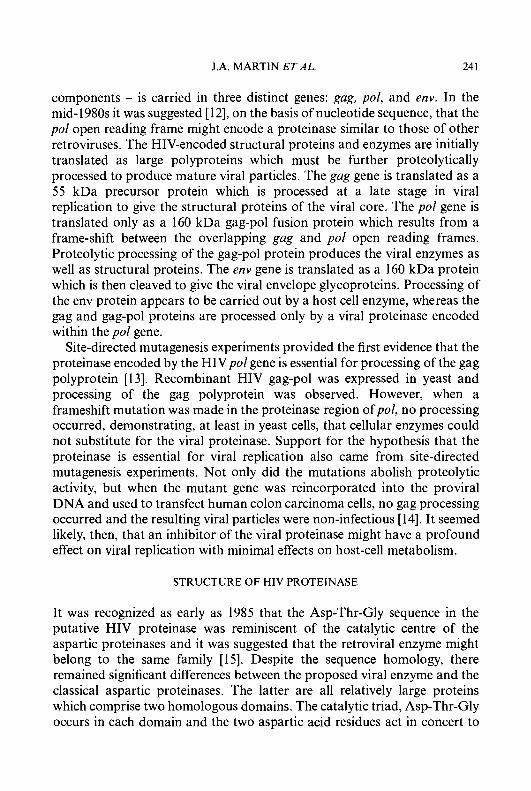
J.A. MARTIN ETAL. 24 1
components - is carried in three distinct genes: gag, pol, and env. In the mid-1980s it was suggested [12], on the basis of nucleotide sequence, that the pol open reading frame might encode a proteinase similar to those of other retroviruses. The HIV-encoded structural proteins and enzymes are initially translated as large polyproteins which must be further proteolytically processed to produce mature viral particles. The gag gene is translated as a 55 kDa precursor protein which is processed at a late stage in viral replication to give the structural proteins of the viral core. The pol gene is translated only as a 160 kDa gag-pol fusion protein which results from a frame-shift between the overlapping gag and pol open reading frames. Proteolytic processing of the gag-pol protein produces the viral enzymes as well as structural proteins. The env gene is translated as a 160 kDa protein which is then cleaved to give the viral envelope glycoproteins. Processing of the env protein appears to be carried out by a host cell enzyme, whereas the gag and gag-pol proteins are processed only by a viral proteinase encoded within the pol gene.
Site-directed mutagenesis experiments provided the first evidence that the proteinase encoded by the HIVpol gene is essential for processing of the gag polyprotein [13]. Recombinant HIV gag-pol was expressed in yeast and processing of the gag polyprotein was observed. However, when a frameshift mutation was made in the proteinase region ofpol, no processing occurred, demonstrating, at least in yeast cells, that cellular enzymes could not substitute for the viral proteinase. Support for the hypothesis that the proteinase is essential for viral replication also came from site-directed mutagenesis experiments. Not only did the mutations abolish proteolytic activity, but when the mutant gene was reincorporated into the proviral DNA and used to transfect human colon carcinoma cells, no gag processing occurred and the resulting viral particles were non-infectious [14]. It seemed likely, then, that an inhibitor of the viral proteinase might have a profound effect on viral replication with minimal effects on host-cell metabolism.
STRUCTURE OF HIV PROTEINASE
It was recognized as early as 1985 that the Asp-Thr-Gly sequence in the putative HIV proteinase was reminiscent of the catalytic centre of the aspartic proteinases and it was suggested that the retroviral enzyme might belong to the same family [15]. Despite the sequence homology, there remained significant differences between the proposed viral enzyme and the classical aspartic proteinases. The latter are all relatively large proteins which comprise two homologous domains. The catalytic triad, Asp-Thr-Gly occurs in each domain and the two aspartic acid residues act in concert to

242 INHIBITORS OF HIV PROTEINASE
form the active site [16]. The viral proteinase, on the other hand, is a small protein containing only around 100 amino acid residues and a single Asp-Thr-Gly motif. These observations led to the suggestion that the HIV proteinase must function as a homodimer, with each monomer contributing one aspartate residue to the active site [17].
The X-ray crystallographic structures of both synthetic [ 181 and recombinant [ 19, 201 HIV proteinases were solved almost simultaneously in a number of laboratories. The results validated early predictions about the enzyme structure [17, 21, 221, showing that the native enzyme is comprised of two monomers related by a crystallographic two-fold rotation, and also confirmed the expected close similarity with the two-domain fungal and mammalian aspartic proteinases. The viral enzyme was shown to display a typical P-sheet structure in which the amino- and carboxy-termini of the two subunits form an interdigitated four-stranded anti-parallel P-sheet which stabilizes the dimer. Glycine-rich @-hairpin loops projecting from each monomer form the flaps which project over the top of the catalytic cleft. The active site triad, A ~ p ~ ~ - T h r ~ ~ - G l y ~ ~ , is located within a loop which is stabilized by a network of hydrogen-bonds formed with the corresponding loop of the other monomer.
X-ray crystallographic structures of HIV proteinase complexed with a diverse range of inhibitors have now been published [23]. Despite large differences in the structures of the bound inhibitors, the structure of the enzyme is remarkably well conserved in all of the complexes. The body of the proteinase is similar, in each case, to that of the native enzyme but the two flap regions are seen to have ‘closed in’ on the bound inhibitor. The flaps thus form one side of a pocketed tunnel in which the pockets form the subsites and make extensive van der Waals’ contacts with the side-chains of inhibitors. All of the inhibitors bind in an extended conformation, forming an array of hydrogen-bonds with both the floor and flap regions of the enzyme. The hydroxyl group of the inhibitors, where present, is situated between the carboxyl groups of the two catalytic aspartate residues and forms at least one hydrogen bond with each carboxylate. The majority of HIV proteinase-inhibitor complexes that have been examined show a tightly bound water molecule which is buried deep within the active site. This water molecule, which is not found in the archetypal aspartic proteinase-inhibitor complexes, forms hydrogen-bonds with the amide carbonyl oxygen atoms of the P, and P,’ residues of the inhibitor and with the hydrogen atoms of the IleSO and Ile5” amides in the flap region of the proteinase. It has been suggested that by bridging the substrate and the flap of the enzyme, such a water molecule exerts extra strain on the scissile bond of the substrate and so facilitates hydrolysis [24].
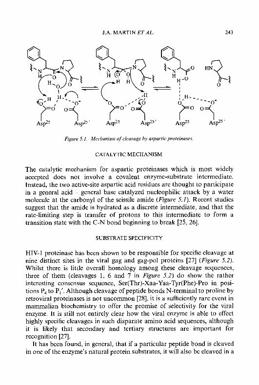
J.A. MARTIN ETAL. 243
. ---o-
Aspz5
Figure 5.1. Mechanism of cleavage by aspartic proteinases.
CATALYTIC MECHANISM
The catalytic mechanism for aspartic proteinases which is most widely accepted does not involve a covalent enzyme-substrate intermediate. Instead, the two active-site aspartic acid residues are thought to participate in a general acid - general base catalyzed nucleophilic attack by a water molecule at the carbonyl of the scissile amide (Figure 5.1). Recent studies suggest that the amide is hydrated as a discrete intermediate, and that the rate-limiting step is transfer of protons to this intermediate to form a transition state with the C-N bond beginning to break [25, 261.
SUBSTRATE SPECIFICITY
HIV-1 proteinase has been shown to be responsible for specific cleavage at nine distinct sites in the viral gag and gag-pol proteins [27] (Figure 5.2). Whilst there is little overall homology among these cleavage sequences, three of them (cleavages 1, 6 and 7 in Figure 5.2) do show the rather interesting consensus sequence, Ser(Thr)-Xaa-Yaa-Tyr(Phe)-Pro in posi- tions P, to P,'. Although cleavage of peptide bonds N-terminal to proline by retroviral proteinases is not uncommon [28], it is a sufficiently rare event in mammalian biochemistry to offer the promise of selectivity for the viral enzyme. It is still not entirely clear how the viral enzyme is able to effect highly specific cleavages in such disparate amino acid sequences, although it is likely that secondary and tertiary structures are important for recognition [27].
It has been found, in general, that if a particular peptide bond is cleaved in one of the enzyme's natural protein substrates, it will also be cleaved in a

244
p17 p24 p2 p7
INHIBITORS OF HIV PROTEINASE
POI pl p6
1 2 3 4 5
I I
P l l P51 : PI5 gag P34
t t 6 1
t t 8 9
1. Ser.Gln.Asn.Tyr * Pro.Ile.Val.Gln
2. Ala.Arg.Va1.Leu * Ala.Glu.Ala.Met
3. Ala.Thr.Ile.Met * Met.Gln.Arg.Gly
4. Arg.Gln.Ala.Asn * Phe.Leu.Gly.Lys
5. Pro.Gly.Asn.Phe * Leu.Gln.Ser.Arg
6 . Ser.Phe.Ser.Phe * Pro.Gln.Ile.Thr
I . Thr.Leu.Asn.Phe * Pro.Ile.Ser.Pro
8. Ala.Glu.Thr.Phe * Tyr.Va1.Asp.Gly
9. Arg.Lys.Val.Leu * Phe.Leu.Asp.Gly
Figure 5.2. Cleavage of gug und gug-pol by HIV proteinuse.
peptide substrate of sufficient length based on the same sequence. Using both synthetic and recombinant HIV proteinase, oligopeptides correspond- ing to all of the cleavage sites in the gag and gag-pol proteins have been shown to be cleaved by HIV proteinase [29], although the efficiency of the cleavage is very dependent on the precise sequence. The smallest peptides
J.A. MARTIN ET AL. 245
which can be processed efficiently and specifically have been shown to be heptapeptides spanning residues P,-P,’. As might be expected, the preferred sequences for peptide substrates correspond to those found around the cleavage sites in the natural protein substrates.
ENZYME ASSAYS
An essential requirement in any programme of inhibitor design is a suitable assay of enzyme activity. HIV proteinase activity has been measured using either continuous or discontinuous assay systems. Discontinuous assays are, in general, more sensitive since the conditions needed for detection can be optimized independently from the conditions needed for enzyme activity. Samples taken during the course of a reaction may be analyzed colorimetrically [30], by TLC or HPLC [29, 3 1-34] (for peptide substrates) or by gel electrophoresis [35] or immunoblot assay [36-40] (for protein substrates). Assays based on synthetic oligopeptide substrates give the best quantitative results, but even then, direct comparisons of IC,, values determined in different laboratories should be made with caution since these values can be affected by substrate concentration and also by factors such as mutual depletion.
ANTIVIRAL ASSAYS
The lack of a readily available, well substantiated animal model of HIV infection has meant that almost all antiviral testing has been carried out in cell culture systems. It is important to discriminate between cellular toxicity and antiviral activity when using in vitro assay systems [41], and ideally both values should be determined in order to obtain a therapeutic index for each compound. A wide range of protocols, designed to provide models of both acute [4247] and chronic [42,45] infection has been developed. These use a variety of cell-lines as well as different virus strains and detection methods. The extent of viral infection in cell cultures may be measured using fluorescent antibodies with specificity for viral proteins such as p24 [43], by determination of reverse transcriptase activity in the supernatant medium [45], or by estimation of the degree of syncitia formation [42, 47, 481. Alternatively, reduction of the cytopathic effects of HIV in infected cells may be used as an endpoint [43, 451. Again, comparison of activities which have not been determined in parallel experiments should be made with great caution.
246 INHIBITORS OF HIV PROTEINASE
STRATEGIES FOR INHIBITION
Successful approaches to the identification of inhibitors of HIV proteinase include random screening, the incorporation of transition-state mimetics into suitable peptide sequences and computer-assisted molecular modelling techniques. Examples of each of these strategies are described in the following sections.
RANDOM SCREENING
A number of groups have set up high throughput screening assays and tested large numbers of compounds, either chosen from existing compound libraries or produced by microbial fermentation. One of the attractions of this approach is the possibility of discovering inhibitors without the pharmacological problems traditionally perceived to be inherent in rationally designed peptide mimetics. Some of the inhibitors oi HIV proteinase which have been identified using this strategy are illustrated in Table 5.1.
Bioassay-guided fractionation of an extract of magenta ascidian Di- demnum Sp. allowed identification [49] of didemnaketal A (1) as an inhibitor of HIV proteinase with an IC,, of 2 pM, but unfortunately the known pharmacological lability of such compounds precluded further development of this inhibitor as a drug candidate. An examination of compounds with known pharmacological properties led to the discovery [50] that the tetrakiscarborane carboxylate ester of 2,4-bis-(a,/l-dihydroxyethyl)deutero- porphyrin IX (2) is a submicromolar inhibitor of HIV proteinase (IC,,, 185 nM). Removal of all four carborane moieties substantially reduced activity whereas removal of only two cages had little effect on potency suggesting that only two of the four closo-carborane cages are responsible for most of the binding interaction. Replacement of the carborane cages with similarly sized but less hydrophobic groups such as benzoyl, /?-naphthoyl or adamantoyl gave inhibitors with IC,, values in the low micromolar range, confirming the importance of supplying hydrophobic groups at these positions. Although the porphyrin derivative (2) was shown to inhibit viral polyprotein processing in an ex vivo experiment in the presence of a low concentration of foetal calf serum, its ability to inhibit HIV proteinase was severely attenuated by albumin which may have implications for the clinical effectiveness of this class of compounds.
A novel solid-phase immunoassay has been used [5 1,521 to screen around 12,000 fermentation broths derived mostly from Actinomycetes genera or fungal isolates. About 0.25% of them were found to show significant and
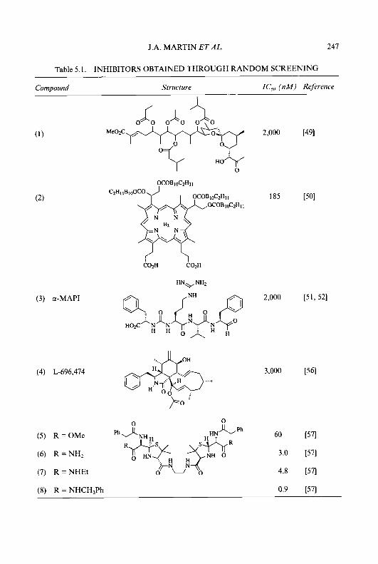
J.A. MARTIN ET AL. 247
Table 5.1. INHIBITORS OBTAINED THROUGH RANDOM SCREENING
Comoound Structure IC, ( n M ) Reference
( 3 ) a-MAP1
2,000 [49]
H O i f
0
3,000 [56]
60 [57]
(6) R = N H , 3.0 [57]
(7) R = NHEt 4.8 [57]
Ph 0
(5) R = O M e
(8) R = NHCH,Ph 0.9 [57]
248 INHIBITORS OF HIV PROTEINASE
reproducible activity against the viral enzyme. The proteinase inhibitor from one of the more stable broths was purified and identified as the known [53] a-microbial alkaline protease inhibitor, a-MAP1 (3). This was somewhat surprising since the compound had previously been characterized as an inhibitor of alkaline proteases with no activity against aspartic proteinases, and a-MAP1 was found to inhibit HIV proteinase with an IC,, of 2.0 pM, a value close to that obtained for the fungal aspartic proteinase inhibitor, Pepstatin A (4.5 pM).
Another novel inhibitor of HIV proteinase was purified from Hypoxylon frugiforme, a fungus isolated from the bark of the American beech tree [54]. L-696,474 (4) [55] a novel cytochalasin, was found to be a competitive inhibitor of HIV-1 proteinase with an IC,, of 3 pM [56]. L-696,474 did not inhibit stromelysin, papain or human leucocyte elastase although it did inhibit another aspartic proteinase, pepsin, with an IC,, of 52 pM.
A screening programme directed towards existing compound libraries [57] identified a crude sample of a penicillin dimer as having enzyme inhibitory activity. On purification, the penicillin dimer was found to be inactive, and the inhibitory activity was shown to be attributable to the C,-symmetric dimer (5) which was an artefact of the purification process. Further development of this lead gave the more potent inhibitors (6), (7) and (8). Although the lead diester (5) failed to block the cytopathic effects of HIV-1 in MT-4 cells, the diamides (7) and (8) were both highly effective (ED,, values of 5.4 pM and 0.29 pM, respectively). The more hydrophilic diamide (6) failed to show an antiviral effect, possibly because of poor cell penetration. An X-ray crystallographic structure of the diamide (7) complexed with recombinant HIV proteinase [58] showed that the interaction is indeed symmetrical, as had been speculated. One of the compounds derived from this study, GR- 1 16624X, was selected as a candidate for further development as a drug candidate, but was later dropped because of poor pharmacokinetics.
TRANSITION-STATE MIMETICS
The vast majority of inhibitors of HIV proteinase have been obtained by the incorporation of a stable transition-state mimetic into an appropriate peptide sequence. Based on the premise, originally proposed by Pauling [59], that an enzyme has a higher affinity for the transition-state than for either the substrate or the products [60], peptide analogues in which the PI-P,’ residues are replaced by a non-hydrolyzable isostere should prove to be potent inhibitors. This was a concept which had already proved to be very successful in the design of renin inhibitors, and many groups that had

J.A. MARTIN ET AL. 249
(9) (10)
Statine analogues Reduced amide
(13)
Difluoroketone hydrate
(15) (16)
Phosphinic acid Hydroxyethylamine
Hydroxyethylene Dihydroxyethylene
(14)
Difluorostatone analogue
(17) (18) Hydroxymethylcarbonyl Hydroxyethylurea
Figure 5.3. Transition-state mimetics
been involved in such projects were quick to appreciate that these compounds might also inhibit the related viral enzyme. Leads derived from such studies have been tailored to afford very potent inhibitors of HIV proteinase with little residual renin-inhibitory activity.
Some of the transition-state mimetics that have been incorporated into peptide analogues as potential inhibitors of aspartic proteinases are shown in Figure 5.3. Each mimetic incorporates some, but not all, of the proposed structural features of the transition-state. They all maintain tetrahedral geometry at the position which would have formed the carbonyl centre of the scissile amide bond of the substrate, but other characteristics are met with varying degrees of success. Several of the mimetics are 'out of register' with the substrate, i.e., they contain either more backbone atoms (as in the case of the hydroxyethylamine (1 6 ) and hydroxymethylcarbonyl (1 7) derivatives) or fewer backbone atoms (as in the case of the statine (9) and difluorostatone (14) derivatives) than the substrate. It might be supposed that such constructs, which seem to be either too long or too short, would not lead to very potent inhibitors, but this is not necessarily true as will be
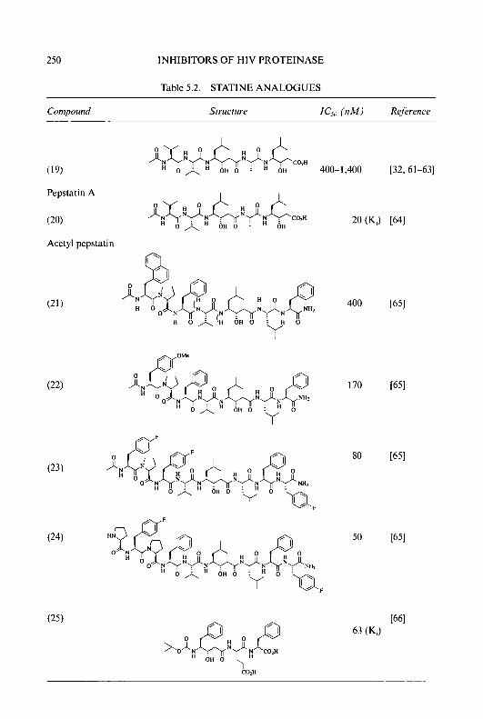
250 INHIBITORS OF HIV PROTEINASE
Table 5.2. STATINE ANALOGUES
Cornpound Structure IC,,, ( 1 1 M ) Reference
(19)
Pepstatin A
(20)
Acetyl pepstatin
OH 400- 1,400 [32. 61-63]
80
50
J.A. MARTIN ET ,415. 251
shown later. It would be an impossible task to describe all of the compounds that have been prepared and tested as inhibitors of HIV proteinase, but representative examples based on each transition-state mimetic are given below.
Statine analogues
The classical aspartic proteinase inhibitor, pepstatin A (19) (Table 5.2), has a relatively weak affinity for HIV-1 proteinase; IC,, values between 0.4 and 1.4pM have been reported [32,61-631. Acetyl pepstatin (20) was found [64] to be substantially more potent with a Ki value of 20 nM, as were the statine derivatives (2 1-24) [65], which were originally described as dog renin inhibitors. Although the most potent inhibitor of this series, (24), exhibited no antiviral activity, compounds (21) and (22) showed good antiviral efficacy, the activity of (21) being comparable to that of AZT (zidovudine) or ddC (dideoxycytidine) in MT-2 cells infected with HIV. The lack of efficacy of (24) is tentatively attributed to the presence of a free amino group which it was felt might prevent cell penetration or facilitate metabolism of the compound. More recently, a series of phenylnorstatine derivatives has been described [66]. The most potent member of this series was reported to be (25) with a Ki value of 63 nM.
Reduced amides
Compounds incorporating the reduced amide dipeptide isostere (1 0) based on the natural cleavage sequences of the viral enzyme such as (26) and (27) (Table 5.3) show only micromolar inhibitory potencies [33, 67, 681. The relatively weak activity of these compounds may be because such inhibitors are unable to form a direct interaction with the aspartic acid residues in the enzyme active site. More recently, a series of reduced amide tight-binding inhibitors of HIV-1 proteinase has been described [69-711. These com- pounds are based on synthetic substrates which bind to the enzyme with much higher affinities than the natural substrates. Incorporation of a glutamic acid residue at P,’ had an especially dramatic effect on binding (29 versus 28). This effect on potency of a glutamic acid residue in P,’ has also been observed in some phenylnorstatine-based inhibitors [72]. Oxidation of the secondary amine gave a fifty-fold increase in potency (30 versus 28) [71] suggesting that the hydroxylamine is able to form an interaction with the catalytic aspartates.

252 INHIBITORS OF HIV PROTEINASE
Table 5.3. REDUCED AMIDES
Compound Structure IC,, (nM) Rejerence
Hydroxyethylene derivatives
Of all the transition-state mimetics that have been considered in the search for inhibitors of HIV proteinase, the hydroxyethylene isostere (1 1) has received most attention, largely because of the availability of renin
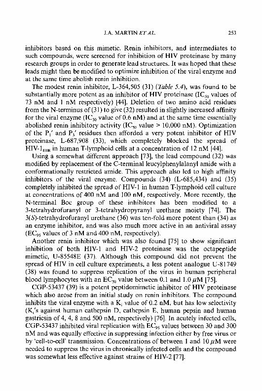
J.A. MARTIN ET AL. 253
inhibitors based on this mimetic. Renin inhibitors, and intermediates to such compounds, were screened for inhibition of HIV proteinase by many research groups in order to generate lead structures. It was hoped that these leads might then be modified to optimize inhibition of the viral enzyme and at the same time abolish renin inhibition.
The modest renin inhibitor, L-364,505 (31) (Table 5.4 , was found to be substantially more potent as an inhibitor of HIV proteinase (IC50 values of 73 nM and 1 nM respectively) [44]. Deletion of two amino acid residues from the N-terminus of (31) to give (32) resulted in slightly increased affinity for the viral enzyme (IC50 value of 0.6 nM) and at the same time essentially abolished renin inhibitory activity (ICs0 value > 10,000 nM). Optimization of the P,’ and P,’ residues then afforded a very potent inhibitor of HIV proteinase, L-687,908 (33), which completely blocked the spread of HIV-lIIIB in human T-lymphoid cells at a concentration of 12 nM [44].
Using a somewhat different approach [73], the lead compound (32) was modified by replacement of the C-terminal leucylphenylalanyl amide with a conformationally restricted amide. This approach also led to high affinity inhibitors of the viral enzyme. Compounds (34) (L-685,434) and (35) completely inhibited the spread of HIV-1 in human T-lymphoid cell culture at concentrations of 400 nM and 100 nM, respectively. More recently, the N-terminal Boc group of these inhibitors has been modified to a 3-tetrahydrofuranyl or 3-tetrahydropyranyl urethane moiety [74]. The 3(S)-tetrahydrofuranyl urethane (36) was ten-fold more potent than (34) as an enzyme inhibitor, and was also much more active in an antiviral assay (EC,, values of 3 nM and 400 nM, respectively).
Another renin inhibitor which was also found [75] to show significant inhibition of both HIV-1 and HIV-2 proteinase was the octapeptide mimetic, U-85548E (37). Although this compound did not prevent the spread of HIV in cell culture experiments, a less potent analogue U-81749 (38) was found to suppress replication of the virus in human peripheral blood lymphocytes with an EC50 value between 0.1 and 1 .O p M [75].
CGP-53437 (39) is a potent peptidomimetic inhibitor of HIV proteinase which also arose from an initial study on renin inhibitors. The compound inhibits the viral enzyme with a Ki value of 0.2 nM, but has low selectivity (K,’s against human cathepsin D, cathepsin E, human pepsin and human gastricsin of 4, 4, 8 and 500 nM, respectively) [76]. In acutely infected cells, CGP-53437 inhibited viral replication with EC,, values between 30 and 300 nM and was equally effective in suppressing infection either by free virus or by ‘cell-to-cell’ transmission. Concentrations of between 1 and 10 p M were needed to suppress the virus in chronically infected cells and the compound was somewhat less effective against strains of HIV-2 [77].
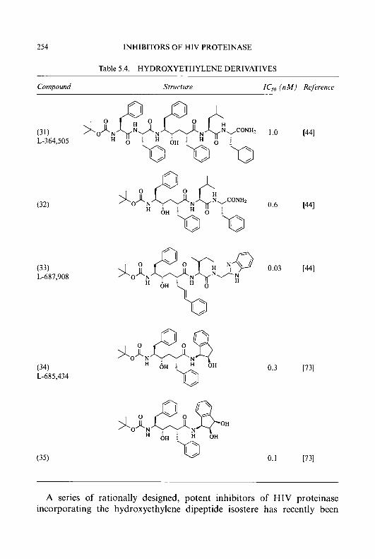
254 INHIBITORS OF HIV PROTEINASE
Table 5.4. HYDROXYETHYLENE DERIVATIVES
Compound Structure IC,,, ( n M ) Ref trence
(31) L-364,505
(33) L-687,908
(34) L-685,434
(35)
1 .o
0.3 [731
0.1
A series of rationally designed, potent inhibitors of HIV proteinase incorporating the hydroxyethylene dipeptide isostere has recently been
J.A. MARTIN ET AL. 255
Table 5.4. (contd.) HYDROXYETHYLENE DERIVATIVES
described [78]. One of these compounds, SK&F 108922 (40) inhibited HIV-1 proteinase with a K, value of 2.0 nM. The compound inhibited the spread of HIV-1 in acutely infected Molt 4 cells [79] with an ED,,, value of 54 nM as determined by reverse transcriptase activity, but was found to be somewhat less effective in a model of chronic infection, showing an EC,,, value of 3600 nM in H9,,,, cells. SK&F 108922 also inhibited the spread of HIV in
256 INHIBITORS OF HIV PROTEINASE
co-cultivation assays using either H9,,,,:Molt4 or CEM,,,,:Molt4 cells with EC,, values of 200 nM and 710 nM, respectively.
Dihydroxyethylene derivatives
A series of hexapeptide analogues containing the dihydroxyethylene isostere (12) which also arose from an earlier study on renin inhibitors has been described. The most potent of these compounds, U-75875 (41) (Table 5.5) inhibited HIV proteinase with a Ki of < 1 nM [80] and, at a concentration of 1 pM, also completely blocked the spread of infection in peripheral blood mononuclear cells infected with HIV,, [45]. A similar concentration of U-75875 also totally prevented the formation of mature viral particles in H9 cells chronically infected with HIV-lllIB. Modifications of the N- and C-termini of U-75875 led to the identification of compound (42) [81] as a potent inhibitor of HIV proteinase (Ki = 5 nM) which showed better antiviral efficacy than U-75875. In human peripheral mononuclear cells acutely infected with HIV-1, the EC,, values of U-75875 and (42) were < 1 nM and between 1 and 10 nM, respectively. Interestingly, in this series
Table 5.5. DIHYDROXYETHYLENE DERIVATIVES
Compound Structure K, ( n M ) Rejerence
J.A. MARTIN ETAL. 257
of peptide mimetics, substitution at the C-terminus by a l(S)-amino-2(R)- hydroxyindane moiety (43 versus 42) did not enhance potency as had been observed for the hydroxyethylene derivatives (34 versus 32), an observation which could not be explained by molecular modelling.
Dijluoroketone derivatives
a,a-Difluoro ketones exist largely as the hydrate in aqueous solution, and it is this hydrated species which interacts with the enzyme active site.
The difluoroketone transition-state mimetic (1 3) has been incorporated [82] into low-molecular-weight compounds which are relatively non- peptidic in nature. It was possible to enhance the aqueous solubility of such compounds without concomitant loss of enzyme inhibitory potency by the incorporation of a pyridyl moiety either at the C-terminus (45) (Table 5.6) or at the N-terminus (46). The lead compound of this series (44) did not show any antiviral activity, possibly because of its very low solubility in the aqueous culture medium. The more hydrophilic inhibitors, on the other hand, all showed good antiviral efficacy, the most potent (47) showing an EC,, value of 1.9 ,uM when tested in MT-4 cells infected with HIV-l,IIB.
Inhibitors incorporating an a,a-difluorostatone-type dipeptide mimetic (14) have also been shown to have high affinity for HIV-1 proteinase. Compound (48) was identified during the screening of a library of proteinase inhibitors, and found to inhibit the viral enzyme with a Ki value of 0.6pM [83]. Modification of this lead structure then led to the very potent inhibitor (49) with a Ki value of 1 nM. A /?-branched amino acid residue in position P, was found to significantly enhance activity, valine being better than leucine or t-butyl glycine. Both the difluoromethyl ketone moiety and the amide nitrogen atom were found to be important for activity (50 or 51 versus 49).
Phosph inic acid derivatives
Inhibitors incorporating a phosphinic acid dipeptide isostere (1 5) and based on scissile Phe-Pro, such as (52) [67] (Table 5.7) and (53) [84] show low affinity for HIV proteinase. The potency of compounds containing this transition state-mimetic appears to be especially sensitive to changes in the PI and PI’ residues [84], and, for a given sequence, compounds based on scissile Phe-Phe are more potent inhibitors than those based on scissile Phe-Pro (54 versus 53). It has been suggested [84] that the cyclopentane ring in the P,’ position may not be an optimal mimic of the conformation of a proline residue undergoing pyramidalization at nitrogen during proteolysis.

258 INHIBITORS OF HIV PROTEINASE
Table 5.6. DIFLUOROKETONE DERIVATIVES
Compound Structure IC,, ( n M ) Reference
(45)
(49)
14 [821
2.2 [82]
1
5000
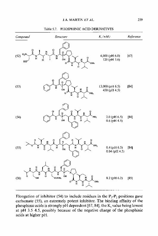
J.A. MARTIN E T A L . 259
Table 5.7. PHOSPHINIC ACID DERIVATIVES
Compound Structure K , ( n M ) Reference
4,000 (pH 6.0) [67] 120 (pH 3.6)
(52)
H O i
(53) 13,000 (pH 6.5) [84] 450 (pH 4.5)
(54) 2.0 (pH 6.5) [84] 0.6 (pH 4.5)
0.4 (pH 6.5) [84] 0.04 (pH 4.5) o n o n
( 5 5 )
8.2 (pH 6.2) [85]
O n
Elongation of inhibitor (54) to include residues in the P,-P, positions gave carbamate ( 5 9 , an extremely potent inhibitor. The binding affinity of the phosphinic acids is strongly pH dependent [67,84], the K, value being lowest at pH 3.54.5, possibly because of the negative charge of the phosphinic acids at higher pH.
260 INHIBITORS OF HIV PROTEINASE
A novel transition-state analogue mimicking scissile Phe-Pro [85] has been incorporated into inhibitor (56). The insertion of a methylene group between the nitrogen and phosphorus atoms produces a non-hydrolyzable moiety which is likely to be zwitterionic near physiological pH. It is suggested that such a construct might represent late transition-state/early product formation for the cleavage of an amide bond. Interestingly, in contrast to the phosphinates, the Ki of this compound at pH 4.7 was double that at pH 6.2.
Hydroxyethylarnine derivatives
In all of the inhibitors described so far, the more active diastereomer has the hydroxyl group, if present, in the S-configuration. In compounds incorpo- rating the hydroxyethylamine transition-state mimetic (1 6), however, the stereochemical requirement at the carbon centre bearing the hydroxyl group depends critically both upon the length of the inhibitor and also upon the nature of individual residues.
In heptapeptide analogues spanning P, to P,’ (57 versus 58) (Table 5.8) and in pentapeptide analogues spanning P, to P,’ (59 versus 60) the S-diastereomer shows significantly higher affinity for the enzyme [86]. Inhibitors which are truncated at the C-terminus, spanning only P, to PI’ show a weak preference for the R-diastereomer when the PI‘ residue is proline (61 versus 62) [42, 861. When the prolyl moiety at PI’ is replaced by the (4aS,8aS)-decahydroisoquinoline-3-(S)-carbonyl group, potency is substantially increased (63 versus 61) [87] and a strong preference for the R-diastereomer becomes apparent (63 versus 64 and 65 versus 66). The potency of inhibitors which incorporate the (4aS,8aS)-decahydroisoquino- line-3-(S)-carbonyl group at PI’ is significantly attenuated by extension to the C-terminus to include the P,’ and P,’ residues (67 and 68) [87]. It has been suggested [86, 871 that whilst binding in the S, to S, subsites is very similar for both types of inhibitor, binding in the S,’ and S,’ subsites is quite different. In compounds which have the (4aS,8aS)-decahydroisoquinoline- 3-(S)-carbonyl group at PI’, the t-butyl group occupies the S,’ subsite rather than following the path of the substrate backbone.
The most potent compound from this series of inhibitors, Ro 31-8959, (saquinavir) (65), [42] shows excellent selectivity for the viral enzyme over a range of other human aspartic proteinases [6] and also shows very good antiviral efficacy in a range of in vitro assays [MI. Ro 31-8959 has been found to be equally effective in models of acute [88,89], chronic [89,90], and latent inducible [91] infection with EC,, values typically less than 10 nM and
J.A. MARTIN ETAL. 261
a therapeutic index of greater than 3,000. Clinical (including zidovudine- resistant) isolates are almost as sensitive as laboratory strains [88, 891.
The long-term effects of treatment with Ro 31-8959 have also been studied. In one such experiment [92], MT-4 cells were infected at low multiplicity of infection to mimic levels of viral burden in vivo. The cells were treated with 100 nM Ro 31-8959 one hour post infection and the culture then maintained for up to 87 days with a daily change of inhibitor and medium. The culture was then maintained for a further 35 days in the absence of inhibitor. During the treatment phase, the treated cultures showed a marked reduction in the cytopathic effects of the virus compared with an infected but untreated control culture. At the end of the treatment phase, in the treated culture no evidence of reverse transcriptase activity or infectious virus could be detected. Moreover, the culture remained free of infection for the following 35 days as determined by either DNA-PCR or infectious virus yield. These results suggest that, in a long term experiment, Ro 31-8959 was able to prevent the production of infectious HIV and thus its spread to other cells. The infected cells were presumed to have died, allowing an outgrowth of uninfected cells. Thus after 80-87 days of dosing, treatment with Ro 31-8959 had ‘cured’ the culture of HIV infection.
Inhibitors based on the hydroxyethylamine transition-state mimetic which incorporate a non-cyclic secondary amine at PI’ have also been described [93]. These compounds also demonstate a clear preference for the R-configuration at the hydroxyl-bearing carbon atom. The dipeptide mimetic (69) is a very weak inhibitor of HIV proteinase, but replacement of the C-terminal t-butyl group by the 1 (S)-amino-2(R)-hydroxyindane moiety giving (70) led to a substantial increase in potency. Interestingly, replacement of the t-butyl group of the dipeptide mimetic, (71), which incorporates the (4aS,8aS)-decahydroisoquinoline-3-(S)-carbonyl group at PI’, by the l(S)-arnino-2(R)-hydroxyindane moiety does not similarly enhance potency (70 versus 69 and 72 versus 71). These results suggest that there must be distinct differences in the way in which the cyclic and non-cyclic inhibitors interact with the enzyme.
N-Terminal extension of (70) to include the P, and P, residues gave L-704,325 (73) with an IC,, value of 5.4 nM. Surprisingly, this enhancement of potency is much smaller than that achieved on analogous extension of (71) to give (65) (55-fold versus 8000-fold) despite the fact that in their truncated form the non-cyclic inhibitors are more potent. It has been proposed [93] that the discrepancy may arise because of the tighter binding of the non-cyclic compounds which either reduces the freedom of movement in the P,-P, region or induces changes in the overall conformation of the enzyme. Again, considerably different structure-activity relationships exist
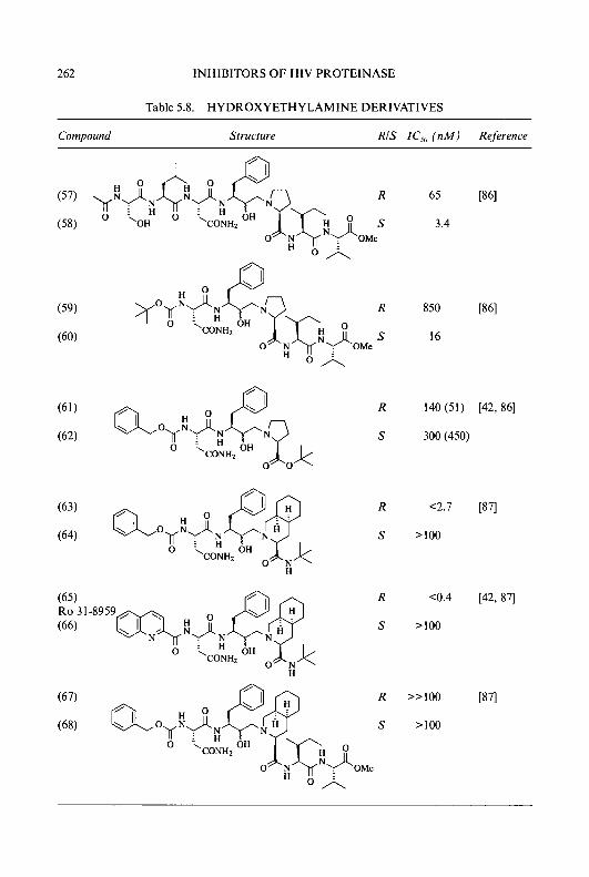
262 INHIBITORS OF HIV PROTEINASE
Table 5.8. HYDROXYETHYLAMINE DERIVATIVES
Compound Structure RIS IC,, ( n M ) Reference
65
3.4
850
16
R 140 (51) [42, 861
S 300 (450)
R C2.7 [87]
s >I00
R <0.4 [42, 871
s >I00
R >>I00
s >loo

J.A. MARTIN ET AL. 263
Table 5.8. (contd.) HYDROXYETHYLAMINE DERIVATIVES
R 252 [93]
R >3,000 [93] 'O'N ' OH
n
R >3,000 [93]
OH
H HO
between two closely related series of inhibitors. L-704,325 inhibited the spread of HIV-1 in human T-lymphoid cells with an EC,, value of 200 nM compared with a value of 12.5 nM for Ro 31-8959 in a parallel assay.
Hydroxymethylcarbonyl derivatives
In this series of compounds, the diastereomer with S-configuration (which corresponds to R-configuration in the hydroxyethylamine series because of

264 INHIBITORS OF HIV PROTEINASE
priority changes) appears to be the more potent, irrespective of whether the inhibitor incorporates the P,’ and P,’ residues or not (75 versus 74 [94, 951 and 77 versus 76 [95,96]). Incorporation of the P,’ and P,’ residues increases the potency of the less active R-diastereomer, but does not alter the activity of the more active S-diastereomer [96].
Interestingly, a decrease in activity for residues at PI’ is seen in the order proline > pipecolinic acid > (4aS,8aS)-decahydroisoquinoline-3-(S)-carbox- ylic acid (77, 78, and 79) [96, 971 which is the opposite to that observed for inhibitors incorporating the hydroxyethylamine transition-state mimetic where (4aS,8aS)-decahydroisoquinoline-3-(S)-carboxylic acid > pipecolinic acid > proline [42]. The need for specific interactions with the P, carbonyl group or intrinsic conformational effects may explain the intriguing differences in structure-activity relationships between these inhibitors and the seemingly closely related hydroxyethylamine derivatives.
Replacement of the pyrrolidine ring of proline by heterocyclic 5- membered rings, especially thiazolidine or dimethyl thiazolidine, however, led to significantly more potent inhibitors [97]. Suitable modification of the P, and P, residues in these compounds has resulted in the very potent and selective inhibitors of HIV proteinase (80, KNI-272), (8 1, KNI-227) [97,98], and (82) [96]. KNI-272 and XNI-227 are both active against a wide spectrum of HIV strains, including AZT-resistant isolates, with EC,, values of 20-100 nM [98]. KNI-227 also shows moderate bioavailability (4%) in the rat.
(Hydroxyethyl) urea derivatives
The (hydroxyethy1)urea dipeptide isostere (1 8) can be regarded as a modification of the hydroxyethylene isostere in which the P,’ a-carbon atom has been replaced by a trigonal nitrogen atom. In this series of inhibitors, the R-diastereomer shows higher affinity for the enzyme (83 versus 84 and 85 versus 86) 1991, as in the case of the hydroxyethylamine- and hydroxymethylcarbonyl-derivatives, but in contrast to the hydroxyethy- lene-containing inhibitors. The nature of the substituent on the terminal nitrogen atom was found to be critical for potency, with a t-butyl group conferring the best activity; a wide range of substituents was found to be tolerated at the other urea nitrogen atom. The conformation of one of the inhibitors (83) bound to the active site of recombinant HIV proteinase was determined by X-ray crystallography. Interestingly, the iso-butyl group is not bound in the S,’ subsite, but instead occupies the S,’ subsite. Similarly, the n-butyl group is not found in the S,’ subsite, but rather in the S,’ subsite.
Compound (85, SC-52121) prevented the spread of HTLV,,,, in CEM

J.A. MARTIN ET AL. 265
Table 5.9. HYDROXYMETHYLCARBONYL DERIVATIVES
Compound Structure RIS IC,, (nM) Reference
R 3100 ~961
S 7.4 (89) [96, 971 CONH:!
0
O \corn2 26 (450) [96,97l H
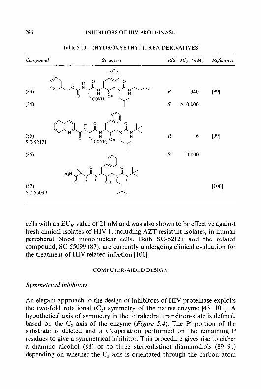
266 INHIBITORS OF HIV PROTEINASE
Table 5.10. (HYDR0XYETHYL)UREA DERIVATIVES
Compound Structure RIS IC,, ( n M ) Reference
SC-55099 A
cells with an EC50 value of 21 nM and was also shown to be effective against fresh clinical isolates of HIV- 1, including AZT-resistant isolates, in human peripheral blood mononuclear cells. Both SC-52121 and the related compound, SC-55099 (87), are currently undergoing clinical evaluation for the treatment of HIV-related infection [ 1001.
COMPUTER-AIDED DESIGN
Symmetrical inhibitors
An elegant approach to the design of inhibitors of HIV proteinase exploits the two-fold rotational (C,) symmetry of the native enzyme [43, 1011. A hypothetical axis of symmetry in the tetrahedral transition-state is defined, based on the C, axis of the enzyme (Figure 5.4). The P' portion of the substrate is deleted and a C,operation performed on the remaining P residues to give a symmetrical inhibitor. This procedure gives rise to either a diamino alcohol (88) or to three stereodistinct diaminodiols (89-9 1) depending on whether the C, axis is orientated through the carbon atom
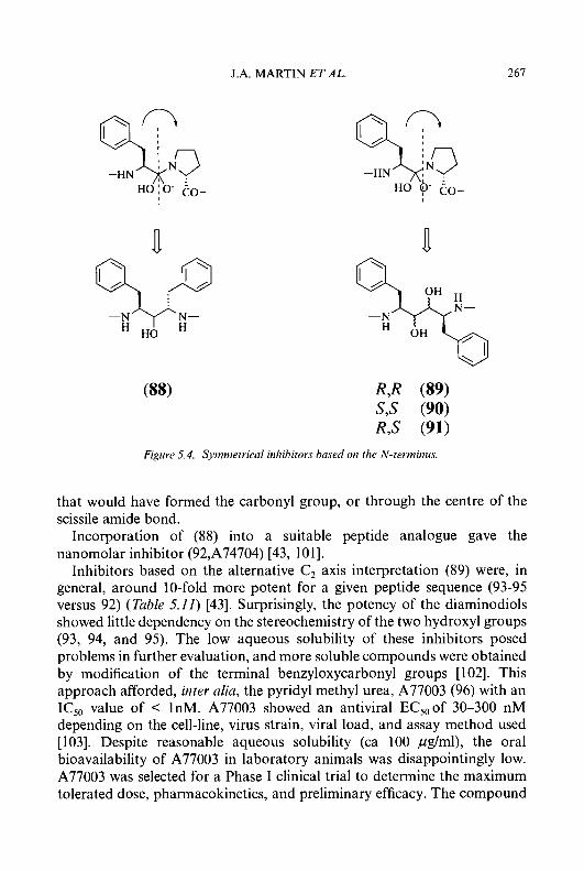
J.A. MARTIN E T A L . 267
n -N N-
H HO
n
Figure 5.4. Symmetrical inhibitors based on the N-terminus
that would have formed the carbonyl group, or through the centre of the scissile amide bond.
Incorporation of (88) into a suitable peptide analogue gave the nanomolar inhibitor (92,A74704) [43, 1011.
Inhibitors based on the alternative C, axis interpretation (89) were, in general, around 10-fold more potent for a given peptide sequence (93-95 versus 92) (Table 5.11) [43]. Surprisingly, the potency of the diaminodiols showed little dependency on the stereochemistry of the two hydroxyl groups (93, 94, and 95). The low aqueous solubility of these inhibitors posed problems in further evaluation, and more soluble compounds were obtained by modification of the terminal benzyloxycarbonyl groups [ 1021. This approach afforded, inter alia, the pyridyl methyl urea, A77003 (96) with an IC,, value of < 1nM. A77003 showed an antiviral EC,,of 30-300 nM depending on the cell-line, virus strain, viral load, and assay method used [103]. Despite reasonable aqueous solubility (ca 100 pglml), the oral bioavailability of A77003 in laboratory animals was disappointingly low. A77003 was selected for a Phase I clinical trial to determine the maximum tolerated dose, pharmacokinetics, and preliminary efficacy. The compound
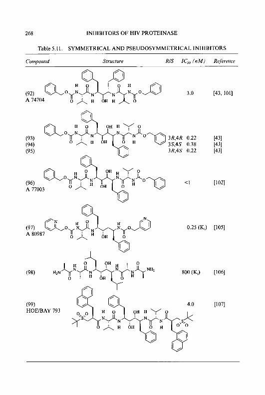
268 INHIBITORS OF HIV PROTEINASE
Table 5.1 1. SYMMETRICAL A N D PSEUDOSYMMETRICAL INHIBITORS
Compound Structure RIS IC,, (nM) Reference
[43, 1011
3R,4R 0.22 3S,4S 0.38
(95) 3R,4S 0.22
(93) (94)
(96) K i < I A 77003
0.25 (K,) [lo51
(99) HOE/BAY 793
PT I
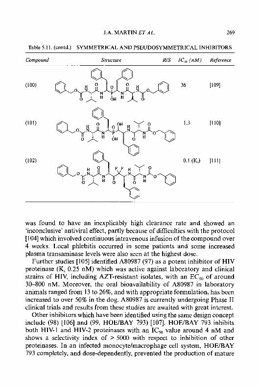
J.A. MARTIN ETAL. 269
Table 5.11. (contd.) SYMMETRICAL AND PSEUDOSYMMETRICAL INHIBITORS
Compound Structure RIS IC,, ( n M ) Reference
was found to have an inexplicably high clearance rate and showed an 'inconclusive' antiviral effect, partly because of difficulties with the protocol [ 1041 which involved continuous intravenous infusion of the compound over 4 weeks. Local phlebitis occurred in some patients and some increased plasma transaminase levels were also seen at the highest dose.
Further studies [lo51 identified A80987 (97) as a potent inhibitor of HIV proteinase (Ki 0.25 nM) which was active against laboratory and clinical strains of HIV, including AZT-resistant isolates, with an EC,, of around 30-800 nM. Moreover, the oral bioavailability of A80987 in laboratory animals ranged from 13 to 26%, and with appropriate formulation, has been increased to over 50% in the dog. A80987 is currently undergoing Phase I1 clinical trials and results from these studies are awaited with great interest.
Other inhibitors which have been identified using the same design concept include (98) [lo61 and (99, HOE/BAY 793) [107]. HOE/BAY 793 inhibits both HIV-1 and HIV-2 proteinases with an IC,, value around 4 nM and shows a selectivity index of > 5000 with respect to inhibition of other proteinases. In an infected monocyte/macrophage cell system, HOE/BAY 793 completely, and dose-dependently, prevented the production of mature

270 INHIBITORS OF HIV PROTEINASE
-OC w OH CO- -0C ‘e.TNT co-
Figure 5.5. Symmetrical inhibitors bused 011 the C-terniinus.
virus particles and the formation of giant cells [108]. It is hoped that clinical trials with this compound will begin in 1994.
Different transition-state mimetics have also been incorporated into such symmetrical inhibitors. Examples of such compounds include the phosphi- nates (100) [lo91 and (101) [110], and the difluoromethylketone (102) [ l l l ] . Interestingly, the phosphinate analogue (1 01) having an additional hydroxyl group, is some 10-fold more potent than the phosphinate (loo), as was also observed for the parent mono- and di-hydroxy compounds.
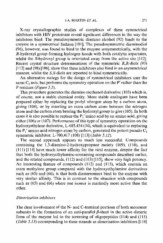
J.A. MARTIN E T A L . 27 1
X-ray crystallographic studies of complexes of these symmetrical inhibitors with HIV proteinase reveal significant differences in the way the inhibitors bind. The pseudosymmetric diamino alcohol (92) binds to the enzyme in a symmetrical fashion [ 1011. The pseudosymmetric diaminodiol (96), however, was found to bind to the enzyme unsymmetrically, with the R-hydroxyl group forming hydrogen bonds with both catalytic aspartates, whilst the S-hydroxyl group is orientated away from the active site [112]. Recent crystal structure determinations of the symmetric R,R-diols (93) [112] and (98) [106], show that these inhibitors also bind in an asymmetrical manner, whilst the S,S-diols are reported to bind symmetrically.
An alternative stategy for the design of symmetrical inhibitors uses the same C, axis, but performs the symmetry operation on the P’ rather than the P residues (Figure 5.5).
This procedure generates the diamino methanol derivative (103) which is, of course, not a stable chemical entity. More stable analogues have been prepared either by replacing the prolyl nitrogen atom by a carbon atom, giving (104), or by inserting an extra carbon atom between the nitrogen atom and the carbon atom bearing the hydroxyl group to give (105). In both cases it is also possible to replace the P,’ imino acid by an amino acid, giving either (106) or (107). Performance of this type of symmetry operation on the hydroxyethylene derivative, L-685,434 (34), which is equivalent to replacing the P,’ amino acid nitrogen atom by carbon, generated the potent pseudo C, symmetric inhibitor, L-700,4 17 (1 08) [ 1 131 (Table 5.12).
The second approach appears to much less successful. Compounds containing the 1,3-diamin0-2-hydroxypropane moiety (109), (1 lo), and (1 11) [114] have much lower affinity for the viral enzyme, despite the fact that both the hydroxyethylamine-containing compounds described earlier, and the related compounds, (1 12) and (1 13) [ 1 151, show very high potency. An interesting feature of compounds (1 12) and (1 13), which contain an extra methylene group compared with the hydroxyethylamine derivatives such as (65) and (66), is that both diastereomers bind to the enzyme with very similar affinity. This is in contrast to the situation with compounds such as (65) and (66) where one isomer is markedly more active than the other.
Dimerisation inhibitors
The clear involvement of the N- and C-terminal portions of both monomer subunits in the formation of an anti-parallel p-sheet in the active dimeric form of the enzyme led to the screening of oligopeptides (1 14) and (1 15) (Table 5.13) corresponding to these strands as dimerisation inhibitors [ 1 161

272 INHIBITORS OF HIV PROTEINASE
and as inhibitors of viral replication [I 171. Although these compounds do not themselves show particularly interesting potency, with IC,, values in the micromolar range, they do, at least, validate the concept of dimerization inhibitors, and may yet lead to more active inhibitors.
De novo design
The wealth of structural information about HIV proteinase, both as the
Table 5.12. SYMMETRICAL A N D PSEUDOSYMMETRICAL INHIBITORS
Compound Structure RIS IC,, ( n M ) Reference
(108) L-700,4 17
~
0.7 [I131
J.A. MARTIN ETAL. 273
native enzyme and in enzyme-inhibitor complexes, has allowed several approaches to de n o w inhibitor design. In one such approach, the program DOCK was used to search the Cambridge Crystallographic Database for steric, and to some extent functional group, complementarity with the active site of HIV proteinase. Bromoperidol was found to be a reasonable match, and the closely related antipsychotic drug, haloperidol (1 17) (Table 5.14), was found experimentally to inhibit both HIV-1 and HIV-2 proteinases with Ki values of around 100 p M [ 1 181.
In a similar exercise, three-dimensional substructures were defined on the basis of a known enzyme-inhibitor complex, and the program ALADDIN was then used to search several large databases for small non-peptidic molecules with the potential to inhibit HIV proteinase. One of the compounds identified by this search was the benzophenone (1 1 S), which was found to inhibit the viral enzyme with an IC,, value of 11 pM [119]. Perhaps the most bizarre inhibitor of HIV proteinase which has so far been described is the fullerene derivative (1 19) [120]. Modelling studies suggested that an icosahedral C,, fullerene molecule should have approximately the same radius as the cylindrical active site of HIV proteinase. Since the enzyme active site is lined mainly with hydrophobic residues, it was suggested that such a fullerene molecule should form strong hydrophobic interactions with the proteinase. The water soluble fullerene derivative (1 19) was subsequently shown to inhibit HIV proteinase with a Ki value of 5.3 pM. Whilst it is unlikely that such compounds will prove to be clinically useful, they do provide new structural leads with the potential for further development.
A combination of X-ray crystallographic data and computer-assisted modelling was used to design the very potent HIV proteinase inhibitor L-735,524 (120) [121]. The compound inhibits HIV-1 proteinase with an IC,, of 0.4 nM and the HIV-2 enzyme with an IC,, of 1.4 nM and does not
Table 5.13. DIMERISATION INHIBITORS
Compound Structure G,, (nM1 Rejhrence
Cy~~~.Thr~~.Leu~~.Asn~~.Phe~ (1 14) 150,000 11 161
(1 15) 40,000 [ I 161
H.Pro1.GLnZ.Ile3.Th~".Leu5.
Cy~~~.Thr~~.L.eu~~.Asn~~.Phe~~.OH
(1 16) A~.Gln~~.11e~~.Gly~~.Met~~.Thr~~.Leu~~.Asn~~.Phe~~.NH~ 27,000 (ED,,,) [ 1 171
214 INHIBITORS OF HIV PROTEINASE
Table 5.14. DE NOVO DESIGN OF INHIBITORS
Compound Srructure K, f n M I Rejerence
(121) XM-323
11,000 (IC5") [ I 191
vCozH 5,300 0
0.4 (ICjr,) [I211
0.27-3.54 [I261
1 .o
J.A. MARTIN ET AL. 275
interact with renin, pepsin, or cathepsin D. Antiviral activity was demonstrated in a variety of cell culture systems, with EC,, values in the range 50-100 nM [122]. L-735,524 shows good oral bioavailability, values of 22, 70, and 12% being observed in rats, dogs, and rhesus monkeys, respectively [121]. The compound entered clinical trials in February 1993 with initial safety studies reported to be complete.
A series of potent symmetrical non-peptidic inhibitors of HIV- 1 proteinase which also arose from a computer-assisted design study has recently been described [123-1251. One of these compounds, XM-323 (121), inhibits HIV proteinase with a Ki of 0.27-3.54 nM depending on the precise assay conditions [126] and is at least 1000-fold less active against either renin or pepsin. The compound inhibits both nucleoside-susceptible and -resistant clinical isolates of HIV-1 with EC,, values of 18-110 nM [127]. Viral replication, as measured by viral RNA content in MT-2 cells, was inhibited by 90% at a concentration of 45 nM [127]. Some activity was also reported against HIV-2. Perhaps more importantly, XM-323 shows significant oral bioavailability with values of 17-37% depending on dose and species [126]. The compound was selected for a Phase I clinical trial in the USA, but its development has since been discontinued.
A de now design study, based entirely on the structure of the HIV proteinase active site, was able to create an inhibitor with an IC,, value in the micromolar range. This lead structure was then modified, using X-ray crystallographic data as a guide, to give, ultimately, AG- 1284 (1 22) [ 1281 which, with a Ki value of 1 nM, is some 60,000-fold more potent than the lead compound. AG-1284 inhibited the spread of HIV in MT2 cells with an EC,, value of 0.5 mg/ml and showed substantial oral bioavailability (> 30%) in rats, dogs and monkeys.
ANTIVIRAL ACTIVITIES
Antiviral activities of individual compounds have been determined using a variety of in vitro assay systems. With few exceptions, the reported antiviral activities of the HIV proteinase inhibitors parallel their enzyme inhibitory activities remarkably well, although as discussed earlier, direct comparisons of results obtained in different laboratories are difficult to make. Deviations from this general observation occur mainly with polar or charged inhibitors whose antiviral activity may be compromised by their inability to cross cell membranes [57, 651. Unlike the reverse transcriptase inhibitors, which are active only in models of acute infection [ 1291, proteinase inhibitors are also effective in models of chronic infection [42, 46, 47, 89, 901.
276 INHIBITORS OF HIV PROTEINASE
SPECIFICITY
Highly specific inhibitors of HIV proteinase are thought to be desirable since significant inhibition of any of the human enzymes could potentially divert the inhibitor from its viral target. Many AIDS patients suffer from digestive disorders and exacerbation of this problem by inhibition of the gastric enzymes, pepsin or gastricsin, would also be a cause for concern. For many of the reported HIV proteinase inhibitors, scant or no selectivity data are given. Comparing the data which have been reported, there appear to be major differences in selectivity both between classes of inhibitor and even within the same class.
As had been hoped, compounds based on scissile Phe-Pro, such as Ro 31-8959 (65), show excellent specificity for the viral enzyme. Ro 31-8959 inhibits both HIV-1 and HIV-2 proteinases with IC,, values of < 1 nM, and yet does not inhibit the related human aspartic proteinases, renin, pepsin, gastricsin, or cathepsins D and E, even at a concentration of 10 p M [6]. Amongst other classes of proteinase inhibitors, there are significant differences in the degree of selectivity observed, and it is not readily apparent why this should be so. The hydroxyethylene-containing inhibitor, CGP-53437 (39) shows modest overall selectivity for the viral enzyme (Ki’s against HIV-1 proteinase, human cathepsin D, human cathepsin E, human pepsin, human gastricsin, and human renin of 0.2, 4, 4, 8, 500 and 190,000 nM respectively [76]), whereas L-735,524 (120) is reported [121] not to interact with renin, pepsin or cathepsin D, and HOE/BAY 793 (99) is reported [lo71 to be at least 1000-fold less active against renin or pepsin than against the viral enzyme.
VIRAL RESISTANCE
HIV is naturally highly prone to mutation and antiviral agents provide a selective pressure for mutations which confer resistance. Clinical use of the nucleoside derivative, zidovudine, has been accompanied by the rapid emergence of resistant virus strains [ 1301, while resistance to non-nucleoside reverse transcriptase inhibitors arises even more rapidly, and has caused the development of a number of such compounds to be abandoned [ 13 13. It has been suggested [l 11 that resistance to certain HIV proteinase inhibitors might emerge less readily, partly because of the similarity between the inhibitors and the natural substrates for the enzyme, and partly because the enzyme is inherently more selective and less error-prone than reverse transcriptase.
J.A. MARTIN ET AL. 211
In vitro experiments have been carried out to determine the potential of HIV proteinase inhibitors for selection of resistant virus [132-1351. Repeated passage of CEM cells infected with HIV-lGBs in the presence of increasing concentrations of Ro 31-8959 (65) did eventually lead to virus with reduced sensitivity, but this occurred much more slowly than in parallel studies with both zidovudine and the non-nucleoside reverse transcriptase inhibitor R82150 [ 1321. Cloning and sequence determination of the proteinase encoded by the virus with reduced sensitivity to Ro 31-8959 showed mutations at positions 48 (Gly to Val) and 90 (Leu to Met) [136], and the structural significance of these changes is currently under investigation. Similar experiments with other HIV proteinase inhibitors have also produced virus strains with reduced sensitivity [133-1351, resistance again being more difficult to establish than with reverse transcriptase inhibitors. The emergence and significance of resistance to HIV proteinase inhibitors in a clinical setting remains to be discovered.
SYNERGY
Many clinicians believe that combination therapy may prove to be the best way of treating AIDS and HIV-related disease. The clinical benefits might include dose-reduction of the individual components (with the potential for reduced toxicity, especially with the nucleoside analogues), broader antiviral surveillance and a reduced propensity for the development of resistance.
Synergy is a phenomenon whereby the total effect of two or more drugs used in combination is greater than would be predicted from the sum of their individual effects. In vitro studies of two-way and three-way combinations of Ro 31-8959 with other anti-HIV agents, including nucleoside analogues and interferon, have shown clear evidence of synergistic antiviral action [ 137-1 391. Compared with IC,, values for each compound alone, 2- to 30-fold reductions were achieved in combination. Cytotoxicity studies did not reveal any adverse interactions between the components of such combinations. Other HIV proteinase inhibitors also exhibit additive or synergistic interactions with reverse transcriptase inhibitors [79, 140-1431. An interesting exception is CGP-53437 (39), which is reported to show neither synergy nor antagonism with reverse transcriptase inhibitors [ 1441.
278 INHIBITORS OF HIV PROTEINASE
CLINICAL PROSPECTS
Immense progress has been made in the design of inhibitors of HIV proteinase since the enzyme was first suggested as a target for antiviral chemotherapy in 1986 [13]. Numerous highly potent and selective inhibitors have been identified, many of which exhibit potent in vitro anti-HIV activity. Electron micrographs of virions produced in chronically infected cells clearly show that HIV proteinase inhibitors act, as predicted, by preventing conversion of the immature, non-infectious ‘doughnut’ form of the virus into the mature, infectious form, which has a dense ‘bullet-shaped’ inner core [89, 901. Unlike reverse transcriptase inhibitors, which are effective only in models of acute infection, proteinase inhibitors show in vitro antiviral activity in both acute and chronic infection, and are able to prevent the spread of virus even when added many hours after infection.
The lack of suitable animal models of HIV infection, and the urgent need to explore new therapies, have necessitated progression to clinical trials in the absence of any in vivo efficacy data. Clinical dosing regimens have been formulated on the basis of in vitro antiviral efficacy and pharmacokinetic studies carried out in animals and healthy human volunteers.
Clinical trials of new treatments for HIV infections are complicated by the slow course of the disease, making it difficult to assess efficacy on the basis of delayed disease progression or increased survival times. To overcome these problems, surrogate markers of disease state such as numbers of CD4’ cells, levels of circulating infectious virus, or concentra- tions of viral proteins (p24 ELISA), or nucleic acids (DNA- or RNA-PCR) have been employed. Caution must be exercised in the interpretation of results obtained in this way, however, since it has not yet been established how well such surrogate markers correlate with clinical efficacy.
In addition to high potency and specificity, an antiviral agent (or indeed any other drug) should ideally exhibit metabolic stability, allowing a long duration of action, and good oral absorption, so that variability of exposure and patient compliance with dosing regimens do not cause problems. Most HIV proteinase inhibitors reported to date are peptide-like in structure, and display the rather short half-life and low oral bioavailability already encountered in earlier studies with renin inhibitors [ 1451. However, there are encouraging indications that potent enzyme inhibitors which have adequate oral bioavailability can be designed. It is also possible that effective prodrugs might be developed [146], and it has been shown that oral absorption can be improved using different formulations and through administration with food.
Although viral resistance to proteinase inhibitors has been demonstrated
J.A. MARTIN ET AL. 219
in vitro, the emergence of resistant strains occurs significantly more slowly than with reverse transcriptase inhibitors and cross resistance between different chemical series is not complete.
As noted earlier, the use of two or more antiviral agents in combination may offer considerable advantages over monotherapy, both in terms of lower incidence of side-effects and decreased potential for the development of viral resistance. It is therefore encouraging to note that several proteinase inhibitors have shown synergy with nucleoside derivatives. Although there are, as yet, few results from clinical trials of HIV proteinase inhibitors, preliminary data show some of these compounds to be very well tolerated, and to achieve immunological and antiviral effects based on laboratory markers of disease. Prevention of the spread of virus to uninfected cells through continued treatment with a proteinase inhibitor should allow the eradication of infected cells by natural processes, enabling the immune system to be reconstituted from non-infected stem cells. The production of viral antigen (in a non-infectious form) from chronically infected cells might further stimulate the patient’s immune system to combat the infection. Clearly it would be advantageous to begin treatment as soon as possible after infection. Since HIV proteinase inhibitors represent a novel class of antiviral agent with proven efficacy in vitro, as well as proven lack of toxicity, they should, for the first time, provide an opportunity for safe and effective early treatment. It is too soon to predict the eventual outcome of the treatment of AIDS with HIV proteinase inhibitors, but their high potency and selectivity, low potential for toxicity and for the induction of viral resistance, and clear synergism with other classes of antiviral agents all give rise to optimism that these compounds will play an important role in future therapies.
CONCLUSION
The clear demonstration through site-directed mutagenesis experiments that an HIV-encoded aspartic proteinase is essential for production of mature, infectious virus particles has stimulated considerable efforts to obtain potent inhibitors as potential therapeutic agents. Successful ap- proaches include random screening of compound libraries or microbial fermentation products, synthesis of suitable peptide sequences which incorporate transition-state mimetics, and computer-assisted design based on the numerous X-ray crystal structures of HIV proteinase which have recently become available. It is the transition-state mimetic approach which has led to the vast majority of HIV proteinase inhibitors published so far. A
280 INHIBITORS OF HIV PROTEINASE
variety of non-hydrolyzable transition-state mimetics (Figure 5.3) have been incorporated into peptide sequences based on substrate cleavage sequences, and many have provided highly potent and selective inhibitors of HIV proteinase. The hydroxyethylene transition-state mimetic (1 1) has received most attention, much of the methodology arising from earlier work on renin, and has been incorporated into some very active inhibitors such as (33) and (36) which also exhibit potent in vitro anti-HIV activity. The hydroxyethylamine transition-state mimetic (1 6) has also been extensively studied, and has led to a number of potent inhibitors, including Ro 31-8959 (saquinavir) (65), which was one of the first HIV proteinase inhibitors to enter Phase I clinical trials, and is currently in Phase 111. This highly potent inhibitor of both HIV-1 and HIV-2 proteinase shows a greater than 10,000-fold selectivity for the viral enzymes over human aspartic pro- teinases, and also shows very good efficacy in a range of in vitro antiviral assays. Unlike inhibitors of reverse transcriptase, which are effective only in models of acute infection, saquinavir is active in both acute and chronic infections, and can prevent the spread of virus even when added many hours after infection. In v i m studies have shown that emergence of viral resistance occurs much more slowly than with reverse transcriptase inhibitors, and that saquinavir exhibits synergy of antiviral activity with nucleoside derivatives and other classes of antiviral agents. Similar observations have been made for other potent inhibitors of HIV proteinase, and some have entered the early phases of clinical trials. More recently, the availability of numerous X-ray crystal structures of HIV proteinase has facilitated computer-aided design of inhibitors. Some, such as the symmetrical and pseudosymmetrical derivatives (Tables 5. ZZ and 5.12) are, like the transition-state mimetics, peptidic in nature, but others (Table 5.14) result from de novo design to give completely different structures, with consequent potential for improved pharmacokinetic properties. Such compounds may well provide the basis for a ‘second generation’ of HIV proteinase inhibitors. While it is too early to predict, with confidence, the eventual clinical outcome of treatment with inhibitors of HIV proteinase, their high potency and selectivity, low potential for toxicity and for induction of viral resistance, and their synergism with other classes of anti-HIV agents all give rise to optimism that they will play an important role in the therapy of AIDS.
J.A. MARTIN ETAL. 28 1
REFERENCES
1 2
3
4
Merson, M.H. (1993) Science (Washington, D.C.) 260, 1266-1268. Smith, D.H., Byrn, R.A., Marsters, S.A., Gregory, T., Groopman, J.E. and Capon, D.J. (1987) Science (Washington, D.C.) 238, 17041707. Deen, K.C., McDougal, J.S., hacker, R., Folena-Wasserman, G., Arthos, J., Rosenberg, J., Maddon, P.J., Axel, R. and Sweet, R.W. (1988) Nature (London) 331, 82-84. Fisher, R.A., Bertonis, J.M., Meier, W., Johnson, V.A., Costopoulos, D.S., Liu, T., Tizard, R., Walker, B.D., Hirsch, M.S., Schooley, R.T. and Flavell, R.A. (1988) Nature (London) 33 1, 76-78.
5 Hsu, M.-C., Schutt, A.D., Holly, M., Slice, L.W., Sherman, M.I., Richman, D.D., Potash, M.J. and Volsky, D.J. (1991) Science (Washington, D.C.) 254, 1799-1802.
6 Martin, J.A. (1992) Antiviral Res. 17, 265-278. 7 Huff, J.R. (1991) J. Med. Chem. 34,2305-2314. 8 Norbeck, D.W. and Kempf, D.J. (1991) Annu. Rep. Med. Chem. 26, 141-150. 9 Tomasselli, A.G., Howe, W.J., Sawyer, T.K., Wlodawer, A. and Heinrikson, R.L. (1991)
Chimica Oggi May, 6-27. 10 Meek, T.D. (1992) J. Enzyme Inhib. 6, 65-98. 11 Debouck, C. (1992) AIDS Res. Human Retroviruses 8, 153-164. 12 Ratner, L., Haseltine, W., Patarca, R., Livak, K.J., Starcich, B., Josephs, S.F., Doran,
E.R., Rafalski, J.A., Whitehorn, E.A., Baumeister, K., Ivanoff, L., Petteway Jr.. S.R., Pearson, M.L., Lautenberger, J.A., Papas, T.S., Ghrayeb, J., Chang, N.T., Gallo, R.C. and Wong-Staal, F. (1985) Nature (London) 313,277-284.
13 Kramer, R.A., Schaber, M.D., Skalka, A.M., Ganguly, K., Wong-Staal, F. and Reddy, E.P. (1986) Science (Washington, D.C.) 231, 1580-1584.
14 Kohl, N.E., Emini, E.A., Schleif, W.A., Davis, L.J., Heimbach, J.C., Dixon, R.A.F., Scolnick, E.M. and Sigal, I.S. (1988) Proc. Natl. Acad. Sci. U.S.A. 85,4686-4690.
15 Toh, H., Ono, M., Saigo, K. and Miyata, T. (1985) Nature (London) 315, 691-692. 16 Tang, J., James, M.N.G., Hsu, I.N., Jenkins, J.A. and Blundell, T.L. (1977) Nature
(London) 271,618-621. 17 Pearl, L.H. and Taylor, W.R. (1987) Nature (London) 329,351-354. 18 Wlodawer, A,, Miller, M., Jaskdski, M., Sathyanarayana, B.K., Baldwin, E., Weber,
I.T., Selk, L.M., Clawson, L., Schneider, J. and Kent, S.B.H. (1989) Science (Washington, D.C.) 245,616-621. Navia, M.A., Fitzgerald, P.M.D., McKeever, B.M., Leu, C.-T., Heimbach, J.C., Herber, W.K., Sigal, I.S., Darke, P.L. and Springer, J.P. (1989) Nature (London) 337, 615-620. Lapatto, P., Blundell, T., Hemmings, A,, Overington, J., Wilderspin, A,, Wood, S., Merson, J.R., Whittle, P.J., Danley, D.E., Geoghegan, K.F., Hawrylik, S.J., Lee, S.E., Scheld, K.G. and Hobart, P.M. (1989) Nature (London) 342,299-302.
21 Pechik, I.V., Gustchina, A.E., Andreeva, N.S. and Fedorov, A.A. (1989) FEBS Lett. 247, 1 1 8-1 22.
22 Weber, I.T., Miller, M., Jaskolski, M., Leis, J., Skalka, A.M. and Wlodawer, A. (1989) Science (Washington, D.C.) 243, 928-931.
23 Wlodawer, A. and Erickson, J.W. (1993) Annu. Rev. Biochem. 62,543-585. 24 Swain, A.L., Miller, M.M., Green, J., Rich, D.H., Schneider, J., Kent, S.B.H. and
Wlodawer, A. (1990) Proc. Natl. Acad. Sci. U.S.A. 87, 8805-8809. 25 Hyland, L.J., Tomaszek Jr., T.A., Roberts, G.D., Carr, S.A., Magaard, V.W., Bryan,
H.L., Fakhoury, S.A., Moore, M.L., Minnich, M.D., Culp, J.S., DesJarlais, R.L. and Meek, T.D. (1991) Biochemistry 30, 844-8453.
19
20
282 INHIBITORS O F HIV PROTEINASE
26 27
28 29
Hyland, L.J., Tomaszek, T.A., Jr. and Meek, T.D. (1991) Biochemistry 30, 8454-8463. Pettit, S.C., Michael, S.F. and Swanstrom, R. (1993) Perspect. Drug Discov. Des. I , 69-83. Pearl, L.H. and Taylor, W.R. (1987) Nature (London) 328,482. Darke, P.L., Nutt, R.F., Brady, S.F., Garsky, V.M., Ciccarone, T.M., Leu, C.-T., Lumma, P.K., Freidinger, R.M., Veber, D.F. and Sigal, I.S. (1988) Biochem. Biophys. Res. Commun. 156,297-303. Broadhurst, A.V., Roberts, N.A., Ritchie, A.J., Handa, B.K. and Kay, C. (1991) Anal. Biochem. 193,28&286. Danley, D.E., Geoghegan, K.F., Scheld, K.G., Lee, S.E., Merson, J.R., Hawrylik, S.J., Rickett, G.A., Ammirati, M.J. and Hobart, P.M. (1989) Biochem. Biophys. Res. Commun. 165, 1043-1050. Krausslich, H.G., Ingraham, R.H., Skoog, M.T., Wimmer, E., Pallai, P.V. and Carter, C.A. (1989) Proc. Natl. Acad. Sci. U.S.A. 86, 807-81 1. Tomasselli, A.G., Olsen, M.K., Hui, J.O., Staples, D.J., Sawyer, T.K., Henrikson, R.L. and Tomich, C.-S.C. (1990) Biochemistry 29,264-269. Meek, T.D., Dayton, B.D., Metcalf, B.W., Dreyer, G.B., Strickler, J.E., Gorniak, J.G., Rosenberg, M., Moore, M.L., Magaard, V.W. and Debouck, C. (1989) Proc. Natl. Acad. Sci. U.S.A. 86, 1841-1845.
35 Tomasselli, A.G., Hui, J.O., Sawyer, T.K., Staples, D.J., FitzGerald, D.J., Chaudhary, V.K., Pastan, I. and Heinrikson, R.L. (1990) J. Biol. Chem. 265,408413.
36 Schneider, J. and Kent, S.B.H. (1988) Cell 54, 363-368. 37 Graves, M.C., Lim, J.J., Heimer, E.P. and Kramer, R.A. (1988) Proc. Natl. Acad. Sci.
38 Debouck, C., Gorniak, J.G., Strickler, J.E., Meek, T.D., Metcalf, B.W. and Rosenberg, M. (1987) Proc. Natl. Acad. Sci. U.S.A . 84, 8903-8906.
39 Hansen, J., Billich, S., Schulze, T., Sukrow, S. and Moelling, K. (1988) EMBO J. 7, 1785-1791.
40 Giam, C.-Z. and Boros, I. (1988) J. Biol. Chem. 263, 14617-14620. 41 Hu, J.M. and Hsiung, G.D. (1989) Antiviral Res. 11,217-232. 42 Roberts, N.A., Martin, J.A., Kinchington, D., Broadhurst, A.V., Craig, J.C., Duncan,
I.B., Galpin, S.A., Handa, B.K., Kay, J., Krohn, A,, Lambert, R.W., Merrett, J.H., Mills, J.S., Parkes, K.E.B., Redshaw, S., Ritchie, A.J., Taylor, D.L., Thomas, G.J. and Machin, P.J. (1990) Science (Washington, D.C.) 248, 358-361. Kempf, D.J., Norbeck, D.W., Codacovi, L., Wang, X.C., Kohlbrenner, W.E., Wideburg, N.E., Paul, D.A., Knigge, M.F., Vasavanonda, S., Craig-Kennard, A,, Saldivar, A,, Rosenbrook Jr., W., Clement, J.J., Plattner, J.J. and Erickson, J. (1990) J. Med. Chem.
44 Vacca, J.P., Guare, J.P., deSolms, S.J., Sanders, W.M., Giuliani, E.A., Young, S.D., Darke, P.L., Zugay, J., Sigal, I.S., Schleif, W.A., Quintero, J.C., Emini, E.A., Anderson, P.S. and Huff, J.R. (1991) J. Med. Chem. 34, 1225-1228.
45 Ashorn, P., McQuade, T.J., Thaisrivongs, S., Tomasselli, A.G., Tarpley, W.G. and Moss, B. (1990) Proc. Natl. Acad.Sci. U.S.A. 87, 7472-7476.
46 McQuade, T.J., Tomasselli, A.G., Liu, L., Karacostas, V., Moss, B., Sawyer, T.K., Heinrikson, R.L. and Tarpley, W.G. (1990) Science (Washington, D.C.) 247,454456.
47 Meek,T.D., Lambert, D.M., Dreyer, G.B., Carr, T.J., Tomaszek Jr., T.A., Moore, M.L., Strickler, J.E., Debouck, C., Hyland, L.J., Matthews, T.J., Metcalf, B.W. and Petteway, S.R. (1990) Nature (London) 343,90-92.
48 Popovic, M., Sarngadharan, M.G., Read, E. and Gallo, R.C. (1984) Science (Washington, D.C.) 224,497-500.
30
31
32
33
34
U.S.A. 85,2449-2453.
43
33,2687-2689.

J.A. MARTIN ET AL. 283
49 Potts, B.C.M., Faulkner, D.J., Chan, J.A., Simolike, G.C., Offen, P., Hemling, M.E. and Francis, T.A. (1991) J. Am. Chem. SOC. 113,6321-6322.
50 Decamp, D.L., Babe, L.M., Salto, R., Lucich, J.L., Koo, M.-S., Kahl, S.B. and Craik, C.S. (1992) J. Med. Chem. 35, 34263428.
51 Sarrubi, E., Nolli, M.L., Andronico, F., Stella, S., Saddler, G., Selva, E., Siccardi, A. and Denaro, M. (1991) FEBS Lett. 279,265-269.
52 Stella, S., Saddler, G., Sarubbi, E., Colombo, L., Stefanelli, S., Denaro, M. and Selva, E. (1991) J. Antibiot. 44, 1019-1022.
53 Watanabe, T., Fukuhara, K. and Murao, S. (1979) Tetrahedron Lett. 625-628. 54 Dombrowski, A.W., Bills, G.F., Sabnis, G., Koupal, L.R., Meyer, R., Ondeyka, J.G.,
Giacobbe, R.A., Monaghan, R.L. and Lingham, R.B. (1992) J. Antibiot. 45,671-678. 55 Ondeyka, J., Hensens, O.D., Zink, D., Ball, R., Lingham, R.B., Bills, G., Dombrowski,
A. and Goetz, M. (1992) J. Antibiot. 45, 679-685. 56 Lingham, R.B., Hsu, A,, Silverman, K.C., Bills, G.F., Dombrowski, A., Goldman, M.E.,
Darke, P.L., Huang, L., Koch, G., Ondeyka, J.G. and Goetz, M.A. (1992) J. Antibiot. 45, 686-691.
57 Humber, D.C., Cammack, N., Coates, J.A.V., Cobley, K.N., Orr, D.C., Storer, R., Weingarten, G.G. and Weir, M.P. (1992) J. Med. Chem. 35,3080-3081.
58 Appelt, K. (1993) Perspect. Drug Discov. Des. 1 ,2348 . 59 Pauling, L. (1946) Chem. Eng. News 24, 1375-1377. 60 Wolfenden, R. (1972) Acc. Chem. Res. 5, 10-18. 61 Nutt, R.F., Brady, S.F., Darke, P.L., Ciccarone, T.M., Colton, C.D., Nutt, E.M.,
Rodkey, J.A., Bennett, C.D., Waxman, L.H., Sigal, I.S., Anderson, P.S. and Veber, D.F. (1988) Proc. Natl. Acad. Sci. U.S.A . 85, 7129-7133.
62 Darke, P.L., Leu, C.-T., Davis, L.J., Heimbach, J.C., Diehl, R.E., Hill, W.S., Dixon, R.A.F. and Sigal, I.S. (1989) J. Biol. Chem. 264, 2307-2312.
63 Tamburini, P.P., Dreyer, R.N., Hansen, J., Letsinger, J., Eking, J., Gore-Willse, A., Dally, R., Hanko, R., Osterman, D., Kamarck, M.E. and Yoo-Warren, H. (1990) Anal. Biochem. 186, 363-368.
64 Richards, A.D., Roberts, R., Dunn, B.M., Graves, M.C. and Kay, J. (1989) FEBS Lett. 247, 113-117.
65 Hui, K.Y., Manetta, J.V., Gygi, T., Bowdon, B.J., Keith, K.A., Shannon, W.M. and Lai,
66 Raju, B. and Deshpande, M.S. (1991) Biochem. Biophys. Res. Commun. 180, 187-190. 67 Dreyer, G.B., Metcalf, B.W., Tomaszek Jr., T.A., Carr, T.J., Chandler 111, A.C., Hyland,
L., Fakhoury, S.A., Magaard, V.W., Moore, M.L., Strickler, J.E., Debouck, C. and Meek, T.D. (1989) Proc. Natl. Acad. Sci. U.S.A. 85, 9752-9756.
68 Moore, M.L., Bryan, W.M., Fakhoury, S.A., Magaard, V.W., Huffman, W.F., Dayton, B.D., Meek, T.D., Hyland, L., Dreyer, G.B., Metcalf, B.W., Strickler, J.E., Gorniak, J.G. and Debouck, C. (1989) Biochem. Biophys. Res. Commun. 159,420425.
69 Miller, M., Schneider, J., Sathyanarayana, B.K., Toth, M.V., Marshall, G.R., Clawson, L., Selk, L., Kent, S.B.H. and Wlodawer, A. (1989) Science (Washington, D.C.) 246, 1149-1 152.
70 Cushman, M., Oh, Y.-I., Copeland, T.D., Oroszlan, S. and Snyder, S.W. (1991) J. Org. Chem. 56,41614167.
71 Urban, J., Konvalinka, J., Stehlikova, J., Gregorova, E., Majer, P., Soucek, M., Andreansky, M., Fabry, M. and Strop, P. (1992) FEBS Lett. 298,9-13.
72 Raju, B. and Deshpande, M.S. (1991) Biochem. Biophys. Res. Commun. 180, 181- 186.
73 Lyle, T.A., Wiscount, C.M., Guare, J.P.. Thompson, W.J., Anderson, P.S., Darke, P.L.,
M.-H.T. (1991) FASEB. J. 5, 2606-2610.

284 INHIBITORS OF HIV PROTEINASE
74
75
76
77
78
79
80
81
82
83
84
85
86
87
88
89
90
91
92
93
Zugay, J.A., Emini, E.A., Schleif, W.A., Quintero, J.C., Dixon, R.A.F., Sigal, I.S. and Huff, J.R. (1991) J. Med. Chem. 34,1228-1230. Ghosh, A.K., Thompson, W.J., McKee, S.P., Duong, T.T., Lyle, T.A., Chen, J.C., Darke, P.L., Zugay, J.A., Emini, E.A., Schleif, W.A., Huff, J.R. and Anderson, P.S. (1993) J. Med. Chem. 36,292-294. Tomasselli, A.G., Hui, J.O., Sawyer, T.K., Staples, D.J., Bannow, C., Reardon, I.M., Howe, W.J., Decamp, D.L., Craik, C.S. and Heinrikson, R.L. (1990) J. Biol. Chem. 265,
Alteri, E., Bold, G., Cozens, R., Farssler, A,, Klimkait, T., Lang, M., Lazdins, J., Poncioni, B., Roesel, J.L., Schneider, P., Walker, M. and Woods-Cook, K (1993) Antimicrob. Agents Chemother. 37,2087-2092. Lazdins, J.K., Alteri, E., Bold, G., Cozens, R., Faessler, A,, Klimkait, T., Lang, M., Schneider, P., Walker, M. and Woods-Cook, K. (1993) IXth International Conference on AIDS/IVth STD World Congress, Berlin, Abstr. PO-A25-0561. Black, P.L., Downs, M.B., Lewis, M.G., Ussery, M.A., Dreyer, G.B., Petteway Jr., S.R., and Lambert, D.M. (1993) Antimicrob. Agents Chemother. 37, 71-77. Lambert, D.M., Bartus, H., Fernandez, A.V., Bratby-Anders, C., Leary, J.J., Dreyer, G.B., Metcalf, B.W. and Petteway Jr., S.R. (1993) Antiviral Res. 21, 327-342. Thaisrivongs, S., Tomasselli, A.G., Moon, J.B., Hui, J., McQuade, T.J., Turner, S.R., Strohbach, J.W., Howe, W.J., Tarpley, W.G. and Heinrikson, R.L. (1991) J. Med. Chem. 34,2344-2356. Thaisrivongs, S., Turner, S.R., Strohbach, J.W., TenBrink, R.E., Tarpley, W.G., McQuade, T.J., Heinrikson, R.L., Tomasselli, A.G., Hui, J.O. and Howe, W.J. (1993) J. Med. Chem. 36,941-952. Sham, H.L., Betebenner, D.A., Wideburg, N.E., Saldivar, A.C., Kohlbrenner, W.E., Vasavanonda, S., Kempf, D.J., Norbeck, D.W., Zhao, C., Clement, J.J., Erickson, J.E. and Plattner, J.J. (1991) Biochem. Biophys. Res. Commun. 175, 914919. Schirlin, D., Baltzer, S., Van Dorsslaer, V., Weber, F., Weill, C., Altenburger, J.M., Neises, B., Flynn, G., Rimy, J.M. and Tarnus, C. (1993) Bioorg. Med. Chem. Lett. 3, 253-258. Grobelny, D., Wondrak, E.M., Galardy, R.E. and Oroszlan, S. (1990) Biochem. Biophys. Res. Commun. 169, 1 1 11-1 116. Ikeda, S., Ashley, J.A., Winching, P. and Janda, K.D. (1992) J. Am. Chem. SOC. 114, 76047606. Rich, D.H., Sun, C.-Q., Prasad, J.V.N.V., Pathiasseril, A,, Toth, M.V., Marshall, G.R.. Clare, M., Mueller, R.A. and Houseman, K. (1991) J. Med. Chem. 34, 1222-1225. Krohn, A,, Redshaw, S., Ritchie, J.C., Graves, B.J. and Hatada, M. (1991) J. Med. Chem. 34, 3340-3342. Holmes, H.C., Mahmood, N., Karpas, A,, Petrik, J., Kinchington, D., O’Connor, T., Jeffries, D.J., Desmyter, J., De Clercq, E., Pauwels, R. and Hay, A. (1991) Antiviral Chem. Chemother. 2,287-293. Craig, J.C., Duncan, I.B., Hockley, D., Grief, C., Roberts, N.A. and Mills, J.S. (1991) Antiviral Res. 16, 295-305. Craig, J.C., Grief, C., Mills, J.S., Hockley, D., Duncan, I.B. and Roberts, N.A. (1991) Antiviral Chem. Chemother. 2, 181-186. Feorino, P.M., Butera, S.T., Folks, T.M. and Schinazi, R.F. (1993) IXth International Conference on AIDSlIVth STD World Congress, Berlin, Abstr. PO-A25-0603. Nitschko, H., Deby, G., Eberle, J. and Von Der Helm, K. (1993) IXth International Conference on AIDS/IVth STD World Congress, Berlin, Abstr. PO-A25-0600. Tucker, T.J., Lumma Jr., W.E., Payne, L.S., Wai, J.M., de Solms, S.J., Giuliani, E.A.,
14675-14683.

J.A. MARTIN E T A L . 285
94
95
96
97
98
99
100
101
102
103
104
105
106
107
108
109
110
Darke, P.L., Heimbach, J.C., Zugay, J.A., Schleif, W.A., Quintero, J.C., Emini, E.A., Huff, J.R. and Anderson, P.S. (1992) J. Med. Chem. 35,2525-2533. Mimoto, T., Imai, J., Tanaka, S., Hattori, N., Takahashi, O., Kisanuki, S., Nagano, Y., Shintani, M., Hayashi, H., Sakikawa, H., Akaji, K. and Kiso, Y. (1991) Chem. Pharm. Bull. 39, 2465-2467. Mimoto, T., Imai, J., Tanaka, S., Hattori, N., Kisanuki, S., Akaji, K. and Kiso, Y. (1991) Chem. Pharm. Bull. 39,3088-3090. Tam, T.F., Carriere, J., MacDonald, I.D., Castelhano, A.L., Pliura, D.H., Dewdney, N.J., Thomas, E.M., Bach, C., Barnett, J., Chan, H. and Krantz, A. (1992) J. Med. Chem. 35, 1318-1320. Mimoto, T., Imai, J., Kisanuki, S., Enomoto, H., Hattori, N., Akaji, K. and Kiso, Y. (1992) Chem. Pharm. Bull. 40,2251-2253. Kageyama, S., Mimoto, T., Murakawa, Y., Nomizu, M., Ford Jr., H., Shirasaka, T., Gulnik, S., Erickson, J., Takada, K., Hayashi, H., Broder, S., Kiso, Y. and Mitsuya, H. (1993) Antimicrob. Agents Chemother. 37,810-817. Getman, D.P., DeCrescenzo, G.A., Heintz, R.M., Reed, K.L., Talley, J.J., Bryant, M.L., Clare, M., Houseman, K.A., Marr, J.J., Mueller, R.A., Vazquez, M.L., Shieh, H.-S., Stallings, W.C. and Stegeman, R.A. (1993) J. Med. Chem. 36,288-291. Getman, D.P., Bertenshaw, D.E., DeCrescenzo, G.A., Freskos, J.N., Heintz, R.M., Lin, K.C., Rogier, D.J., Talley, J.J., Clare, M., Houseman, K.A., Mueller, R.A., Bryant, M.L., Sheng-Shieh, H., Stallings, W.C. and Stegeman, R.A. (1993) 206th American Chemical Society National Meeting, Chicago, MEDl 137. Erickson, J., Niedhart, D.J., VanDrie, J., Kempf, D.J., Wang, X.C., Norbeck, D.W., Plattner, J.J., Rittenbouse, J.W., Turon, M., Wideburg, N., Kohlbrenner, W.E., Simmer, R., Helfrich, R., Paul, D.A. and Knigge, M. (1990) Science (Washington, D.C.) 249, 527-533. Kempf, D.J., Codacovi, L., Wang, X.C., Kohlbrenner, W.E., Wideburg, N.E., Saldivar, A,, Vasavanonda, S., Marsh, K.C., Bryant, P., Sham, H.L., Green, B.E., Betebenner, D.A., Erickson, J. and Norbeck, D.W. (1993) J. Med. Chem. 36, 320-330. Kempf, D.J., Marsh, K.C., Paul, D.A., Knigge, M.F., Norbeck, D.W., Kohlbrenner, W.E., Codacovi, L., Vasavanonda, S., Bryant, P., Wang, X.C., Wideburg, N.E., Clement, J.J., Plattner, J.J. and Erickson, J. (1991) Antimicrob. Agents Chemother. 35, 2209-2214. Danner, S.A., Reedijik, M., Boucher, C.A.B., Mayer, K.M., Leonard, J.M. and Tzeng, T.B. (1 993) IXth International Conference on AIDS/IVth STD World Congress, Berlin, Abstr. WS-B26-6. Kempf, D.J., Norbeck, D.W., Marsh, K.C. and Erickson. J. (1993) 206th American Chemical Society National Meeting, Chicago, ORGN 119. Dreyer, G.B., Boehm, J.C., Chenera, B., DesJarlais, R.L., Hassell, A.M., Meek, T.D., Tomaszek Jr., T.A. and Lewis, M. (1993) Biochemistry 32,937-947. Paessens, A,, Budt, K.-H.. Kleim, J.-P., Knolle, J., Meichsner, C., Rieb. G.. Stowasser, B. and Winkler, 1. (1993) IXth International Conference on AIDS/IVth STD World Congress, Berlin, Abstr. PO-A25-0591. Meichsner, C. and Budt, K.-H. (1993) IXth International Conference on AIDS/IVthSTD World Congress, Berlin, Abstr. PO-A25-0612. Peyman, A,, Budt, K.-H., Spanig, J., Stowasser, B. and Ruppert, D. (1992) Tetrahedron Lett. 45494552. Stowasser, B., Budt, K.-H., Jian-Qi. L., Peyman, A. and Ruppert, D. (1992) Tetrahedron Lett. 6625-6628.
111 Sham, H.L., Wideburg, N.E., Spanton, S.G., Kohlbrenner, W.E., Betebenner, D.A.,
286 INHIBITORS OF HIV PROTEINASE
Kempf, D.J., Norbeck, D.W., Plattner, J.J. and Erickson, J.W. (1991) J. Chem. SOC., Chem. Commun. 110-1 12.
112 Erickson, J.W. (1993) Perspect. Drug Discov. Des. I , 109-128. 113 Bone, R., Vacca, J.P., Anderson, P.S. and Holloway, M.K. (1991) J. Am. Chem. SOC. 113,
114 Yu, K.-L., Harte, W.E., Spinazze, P., Martin, J.C. and Mansuri, M.M. (1993) Bioorg. Med. Chem. Lett. 3, 535-538.
115 Trova, M.P., Babine, R.E., Byrn, R.A., Casscles Jr., W.T., Hastings, R.C., Hsu, G.C., Jirousek, M.R., Johnson, B.D., Kerwar, S.S., Schow, S.R., Wissner, A,, Zhang, N. and Wick, M.M. (1993) Bioorg. Med. Chem. Lett. 3,1595-1600.
116 Babe, L.M., Rose, J. and Craik, C.S. (1992) Protein Sci. 1, 12441253. 117 Schramm, H.J., Nakashima, H., Schramm, W., Wakayama, H. and Yamamoto, N.
(1991) Biochem. Biophys. Res. Commun. 179, 847-851. 118 DesJarlais, R.L., Seibel, G.L., Kuntz, I.D., Furth, P.S., Alvarez, J.C., Ortiz de
Montellano, P.R., Decamp, D.L., Babe, L.M. and Craik, C.S. (1990) Proc. Natl. Acad. Sci. U.S.A. 87,66446648.
119 Bures, M.G., Hutchins, C.W., Maus, M., Kohlbrenner, W., Kadam, S. and Erickson, J.W. (1992) Tetrahedron Comput. Methodol. 3, 673-680.
120 Friedman, S.H., Decamp, D.L., Sijbesma, R.P., Srdanov, G., Wudl, F. and Kenyon, G.L. (1993) J. Am. Chem. SOC. 11 5,65066509.
121 Dorsey, B.D., Levin, R.B., McDaniel, S.L., Vacca, J.P., Darke, P.L. and Zugay, J.A. (1993) 206th American Chemical Society National Meeting, Chicago, MEDI 6.
122 Vacca, J.P., Dorsey, B.D., Darke, P., Emini, E. and Lin, J. (1993) IXth International Conference on AIDS/IVth STD World Congress, Berlin, Abstr. PO-B26-2023.
123 Lam, P.Y.-S., Eyermann, C.J., Jadhav, P.K., Hodge, C.N., Ru, Y., DeLucca, G.V., Bacheler, L.T., Meek, J.L., Otto, M.J., Rayner, M.M., Wong, N.Y., Chang, C.-H., Weber, P.C., Jackson, D.A., Sharpe, T.R. and Ericksen-Viitanen, S. (1993) IXth International Conference on AIDS/IVth STD World Congress, Berlin, Abstr. PO-A25- 0584.
124 Jadhav, P.K., Lam, P.Y.-S., Eyermann C.J., Hodge, C.N. and Woerner, F.J. (1993) lXth International Conference on AIDS/IVth STD World Congress, Berlin, Abstr. PO-A25- 0585.
125 Lam, P.Y.S., Jadhav, P.K., Eyermann, C.J., Hodge, C.N., Ru, Y., Bacheler, L.T., Meek, J.L., Otto, M.J., Rayner, M.M., Wong, Y.N., Chang, C.-H., Weber, P.C., Jackson, D.A., Sharpe, T.R. and Erickson-Viitanen, S. (1994) Science (Washington, D.C.) 263,380-384.
9382-9384.
126
127
128
129
130
131 132
Ericksen-Viitanen, S., Meek, J., Wong, N., Grubb, M., O’Neal,-P. and Cawood, P. (1993) IXth International Conference on AIDS/IVth STD World Congress, Berlin, Abstr.
Winslow, D.L., Scarnati, H. and Otto, M.J. (1993) IXth International Conference on AIDSlIVth STD World Congress, Berlin, Abstr. PO-A25-0558. Appelt, K. (1993) 33rd Interscience Conference on Antimicrobial Agents and Chemo- therapy, New Orleans, Symp. 82. Poli, G., Orenstein, J.M., Kinter, A,, Folks, T.M. and Fauci, A.S. (1989) Science (Washington, D.C.) 244, 575-577. Larder, B.A., Darby, G. and Richman, D.D. (1989) Science (Washington, D.C.) 243,
Robins, T. and Plattner, J. (1993) J. Acq. Immune Deficiency Syndromes 6, 162-170. Duncan, I.B., Craig, J.C., Roberts, N.A. and Whittaker, L. (1993) IXth International Conference on AIDS/IVth STD World Congress, Berlin, Abstr. PO-A26-0469.
PO-A25-0583.
1731-1734.
J.A. MARTIN E T A L . 287
133 Otto, M.J., Garber, S., Winslow, D.L., Reid, C.D., Aldrich, P., Jadhav, P.K., Patterson, C.E., Hodge, C.N. and Cheng, Y.-S.E. (1993) Proc. Natl. Acad. Sci. U.S.A. 90, 7543-7547.
134 Blair, E.D., Wood, A.J., Blance, C., Bains, S., Darby, G., Wilderspin, A. and Sugrue, R. (1993) J. Cell Biochem. Supplement 17E, 74.
135 Singh, M.K., Toyoshima, T., Markowitz, M., Kempf, D.J., Norbeck, D., Erickson, J. and Ho, D.D. (1993) J. Cell Biochem. Supplement 17E, 86.
136 Jacobsen, H., Craig, J.C., Duncan, I.B., Krohn, A,, Mous, J., Yasargil, K., Whittaker, L. and Roberts, N.A. (1993) IXth International Conference on AIDS/IVth STD World Congress, Berlin, Abstr. WS-A19-1.
137 Craig, J.C., Duncan, I.B., Whittaker, L. and Roberts, N.A. (1990) Antiviral Chem. Chemother. 4, 161-166.
138 Johnson, V.A., Merrill, D.P., Chou, T.-C. and Hirsch, M.S. (1992) J. Infect. Dis. 166,
139 Craig, J.C., Duncan, I.B., Roberts, N.A. and Whittaker, L. (1993) IXth International Conference on AIDS/IVth STD World Congress, Berlin, Abstr. PO-A25-0602.
140 Kageydma, S., Weistein, J.N., Shirasaka, T., Kempf, D.J., Norbeck, D.W., Plantner, J.J., Erickson, J. and Mitsuya, H. (1992) Antimicrob. Agents Chemother. 36,926-933.
141 Lambert, D.M., Bartus, H., Fernandez, A.V., Bratby-Anders, C., Leary, J.J., Dreyer, G.B., Metcalf, B.W. and Petteway Jr., S.R. (1993) Antiviral Res.21, 327-342.
142 Bryant, M., Clare, M., DeCrescenzo, G., Dorn, C., Getman, D., Heintz, R., Houseman, K., Li, A. and Marshall, G. (1992) Antimicrob. Agents Chemother. 32, 361.
143 Chong, K.T., Pagano, P.J. and Hinshaw, R.R. (1993) IXth International Conference on AIDS/IVth STD World Congress, Berlin, Abstr. PO-A25-0557.
144 Roesel, J. (1993) IXth International Conference on AIDS/IVth STD World Congress, Berlin, Abstr. PO-A25-0560.
145 Greenlee, W.J. (1990) Med. Res. Rev. 10, 173-236. 146 Chong, K.-T., Ruwart, M.J., Hinshaw, R.R., Wilkinson, K.F., Rush, B.D., Yancey,
M.F., Strohbach, J.W. and Thaisrivongs, S. (1993) J. Med. Chem. 36, 2575-2577.
1143-1 146.
Progress in Medicinal Chemistry - Vol. 32, edited by G.P. Ellis and D.K. Luscombe 0 1995 Elsevier Science B.V. All rights reserved.
6 Paclitaxel: A Unique Tubulin Interacting Anticancer Agent
DOLATRAI M. VYAS, Ph.D. and JOHN F. KADOW, Ph.D.
Bristol-Myers Squibb Company, Pharmaceutical Research Institute, 5 Research Parkway, Wallingford, CT 06492-7660, U. S. A.
INTRODUCTION
PACLITAXEL
MECHANISM OF ACTION AND RESISTANCE
METABOLISM AND PHARMACOKINETICS
ANALOGUES AND STRUCTURE-ACTIVITY RELATIONSHIPS
Side-chain Analogues C-1 Modifications C-2 Modifications C-4 Modifications C-4, C-5 and Oxetane Modifications C-6 Modifications C-7 Modifications C-9 Modifications C-10 Modifications C-14 Modifications C-19 Modifications Photolysis Products
WATER-SOLUBLE PRODRUGS
CLINICAL SUMMARY
CONCLUSION
290
29 1
296
300
302
303 309 309 31 1 311 312 312 314 316 318 319 319
320
324
325
REFERENCES 325
ADDENDUM 334
289
290 PACLITAXEL: A UNIQUE TUBULIN-INTERACTING ANTICANCER AGENT
INTRODUCTION
In recent years, chemotherapy has come under close scrutiny and debate concerning its real efficacy in the successful treatment of the majority of common cancers [ 1-41. The perception that chemotherapy has had limited success, may perhaps be justified, since the more common (>70% of all) human cancers such as those of breast, non-small-cell lung, malignant melanoma, ovary, and colon have remained refractory or untreatable [4]. In contrast, treatment of several rapidly proliferating malignancies including leukaemias, non-Hodgkin's lymphomas, small-cell lung cancer, and testicular teratomas has been successful [5]. In the face of this major unmet medical need, drug discovery strategies involving either natural products screening for cytotoxics or designed DNA-targeted synthetic agents are being reformulated [6, 71. In the natural product-based screening programmes both at the National Cancer Institute (NCI) and in industry, novel mechanism-based and disease-oriented cell-based screens [8, 91 are being emphasized and implemented over the conventional cell cytotoxity (P388) screen [lo]. At the same time, drug discovery efforts involving synthetic analogues have shifted away from DNA as a cellular target in favour of targets related to processes (signal transduction pathways) [ 1 13 relevant to neoplastic growth. Also, in recent years molecular biology probes in cancer research have facilitated a better understanding of the many biochemical changes underlying the aetiology of cancer and thus providing opportunities to define more meaningful targets for cancer research [12, 131.
A class of non-DNA-targeted anticancer agents which have risen to prominence and research focus are tubulin interacting [14, 151 agents. The prototypes of this class that are currently in clinical use include the natural Vinca alkaloids such as vincristine (l), vinblastine (2) [14, 161, and the recently approved, semisynthetic vinca analogue, vinorelbine (Navalbine", 3) [14, 171. Several other experimental tubulin interacting drugs such as Rhizoxin, dolastatin 10 and comberstatin are also of great current clinical interest [15]. The most recent entry to this arsenal of chemotherapeutic agents is a novel and unique tubulin interacting agent, namely, the natural taxane diterpenoid paclitaxel (Taxol', 4)* [ 181. Paclitaxel and vinorelbine have both demonstrated significant clinical antitumour activity against several human solid tumours and are currently approved drugs for clinical use. Vinorelbine is currently in use in France for the treatment of non- small-cell lung cancer and breast cancer [19]. Paclitaxel is efficacious in the
* Throughout this review Bz = benzoyl.
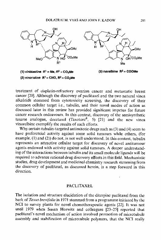
DOLATRAI M VYAS A N D JOHN F. KADOW 29 1
R’ I Me
(1) vinblastine R1 = Me, R2 = C02Me
(2) vincristine R1 = CHO, R2 = C02Me
(3) navalbine R2 = COOMe
treatment of cisplatin-refractory ovarian cancer and metastatic breast cancer [20]. Although the discovery of paclitaxel and the two natural vinca alkaloids stemmed from cytotoxicity screening, the discovery of their common cellular target i.e., tubulin, and their novel modes of action as discussed later in this review has provided significant impetus for future cancer research endeavours. In this context, discovery of the semisynthetic taxane analogue, docetaxel (Taxotere@, 5) [21] and the new vinca vinorelbine exemplify the results of such efforts.
Why certain tubulin-targeted antimitotic drugs such as (3) and (4) seem to have preferential activity against some solid tumours while others, (for example, (1) and (2)) do not, is not well understood. In this context, tubulin represents an attractive cellular target for discovery of novel antitumour agents endowed with activity against solid tumours. A deeper understand- ing of the interactions between tubulin and its small molecule ligands will be required to advance rational drug discovery efforts in this field. Mechanistic studies, drug development and medicinal chemistry research stemming from the discovery of paclitaxel, as discussed herein, is a step forward in this direction.
PACLITAXEL
The isolation and structure elucidation of the diterpine paclitaxel from the bark of Exus brevifolia in 1971 stemmed from a programme initiated by the NCI to survey plants for novel chemotherapeutic agents [22]. It was not until 1979 when Susan Horwitz and colleagues [23-251 reported that paclitaxel’s novel mechanism of action involved promotion of microtubule assembly and stabilization of microtubule polymers, that the NCI really
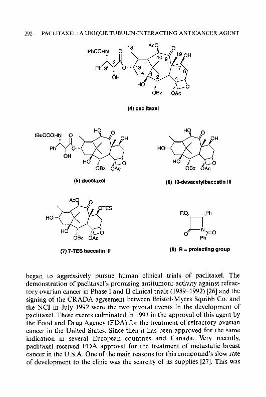
292 PACLITAXEL: A UNIQUE TUBULIN-INTERACTING ANTICANCER AGENT
OBz OAc
(4) paclitaxel
(5) docetaxel (6) 10-desacetylbaccatin 111
OBZ O i c -
(6) R = protecting group (7) 7-TES baccatin 111
began to aggressively pursue human clinical trials of paclitaxel. The demonstration of paclitaxel’s promising antitumour activity against refrac- tory ovarian cancer in Phase I and I1 clinical trials (1989-1992) [26] and the signing of the CRADA agreement between Bristol-Myers Squibb Co. and the NCI in July 1992 were the two pivotal events in the development of paclitaxel. These events culminated in 1993 in the approval of this agent by the Food and Drug Agency (FDA) for the treatment of refractory ovarian cancer in the United States. Since then it has been approved for the same indication in several European countries and Canada. Very recently, paclitaxel received FDA approval for the treatment of metastatic breast cancer in the U.S.A. One of the main reasons for this compound’s slow rate of development to the clinic was the scarcity of its supplies [27]. This was

DOLATRAI M. VYAS A N D JOHN F. KADOW 293
exacerbated by its extreme insolubility in water which necessitated extensive studies to develop a viable injectable formulation [28]. The current formulation of paclitaxel requiring Cremophor EL@ (a polyethoxylated castor oil) as a solubilizer has been a cause of considerable concern due to its alleged role in causing hypersensitivity reactions in patients [18, 291. The formulation issue has been at the centre of several investigations in both academic laboratories and industry, as is discussed later in this review in the section on prodrugs of paclitaxel.
Reliance on the yew tree as a source of paclitaxel for future clinical supplies raised strong concerns about the environmental impact of such an endeavour and led to massive efforts in both industry and the private sector to look for alternative sources of paclitaxel. In 1988, paclitaxel was prepared semisynthetically from 10-deacetyl baccatin I11 (6, 10-DAB) which is obtained from the needles of Taxus baccata L. of the Asian and European variety [30]. The major breakthrough in paclitaxel supply was realized when Bristol-Myers Squibb Co. (BMS) announced in 1993, that for future clinical use, semisynthetic paclitaxel from 10-DAB will be utilized. In the BMS process, the transformation of 10-DAB to paclitaxel was facilitated by the lactam acylation technology developed by Professor Holton at Florida State University (FSU) [31, 321. The key step in this process is the coupling of lactam (8) to a C-7 protected baccatin I11 derivative (7). The FSU acylation technology has been licensed by BMS for commercial synthesis of paclitaxel from 10-DAB. This process eliminates the reliance on the bark of the yew tree as a source of natural paclitaxel and thus constitutes a renewable source of future supplies for clinical use.
As an alternative to natural paclitaxel, a number of researchers in academia have embarked on the very ambitious task of the total synthesis of paclitaxel. Apparently, these efforts will not resolve the supply issue in the immediate future but will certainly pave the way for the synthesis of unique analogues for biological evaluation. The variety of synthetic approaches currently being pursued are lucidly summarized in several review articles; the two most recent in this context are by Boa et al. [33] and Nicolaou and co-workers [34]. An older review of synthetic strategies is also informative [35]. Only two academic groups have succeeded in achieving a total synthesis of paclitaxel. The Holton group at FSU was one of the earliest to become involved in this endeavour and reported the first synthetic route to paclitaxel after starting from a natural product, camphor [36, 371. The second synthesis emerged from the group at Scripps Research Institute, La Jolla, California and the University of California, San Diego, headed by Professor K. C. Nicolaou [38].
The Holton synthesis (outline depicted in Scheme 6.1 where p is a suitable
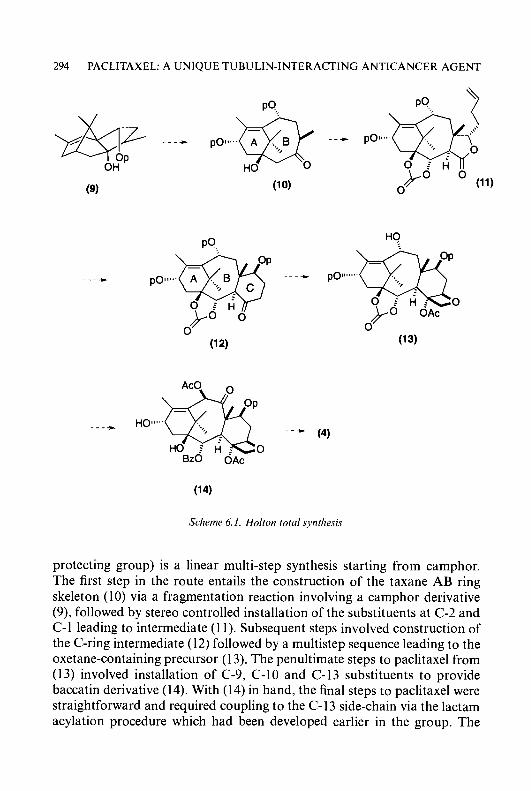
294 PACLITAXEL: A UNIQUE TUBULIN-INTERACTING ANTICANCER AGENT
Scherne 6. I . Holion toid sjwtliesis
protecting group) is a linear multi-step synthesis starting from camphor. The first step in the route entails the construction of the taxane AB ring skeleton (10) via a fragmentation reaction involving a camphor derivative (9), followed by stereo controlled installation of the substituents at C-2 and C-1 leading to intermediate (1 1). Subsequent steps involved construction of the C-ring intermediate (1 2) followed by a multistep sequence leading to the oxetane-containing precursor (1 3). The penultimate steps to paclitaxel from (13) involved installation of C-9, C-10 and C-13 substituents to provide baccatin derivative (14). With (14) in hand, the final steps to paclitaxel were straightforward and required coupling to the C-13 side-chain via the lactam acylation procedure which had been developed earlier in the group. The
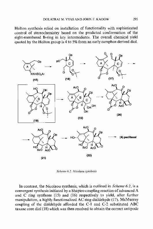
DOLATRAI M. VYAS AND JOHN F. KADOW 295
Holton synthesis relied on installation of functionality with sophisticated control of stereochemistry based on the predicted conformation of the eight-membered B-ring in key intermediates. The overall chemical yield quoted by the Holton group is 4 to 5% from an early camphor-derived diol.
*
Schenie 6.2. Nicoluou synthesis
In contrast, the Nicolaou synthesis, which is outlined in Scheme 6.2, is a convergent synthesis initiated by a Shapiro coupling reaction of advanced A and C ring synthons (15) and (16) respectively to yield, after further manipulation, a highly functionalized AC ring dialdehyde (1 7). McMurray coupling of the dialdehyde afforded the C-1 and C-2 substituted ABC taxane core diol(l8) which was then resolved to obtain the correct antipode
296 PACLITAXEL: A UNIQUE TUBULIN-INTERACTING ANTICANCER AGENT
for further transformations. Enantiopure diol (1 8) was transformed to intermediate (20), a compound which contains the intact oxetane ring via the precursor, trio1 (19). The final steps involved C-2 benzoylation with PhLi to afford intermediate (21), followed by oxygenation at C-13 to provide baccatin I11 derivative (22). Finally, installation of the C-13 side-chain completed the synthesis.
Other efforts have now assumed greater prominence than chemical synthesis in the quest to solve the supply issue. These include studies of plant genetics, tissue culture technology, analytical survey of needles of a variety of taxus species, and biosynthetic studies [27]. The exciting finding that taxol can be produced by Taxomyces andreanae, an endophytic fungus asociated with Taxus brevifolia was recently reported by Strobe1 and co-workers [39]. All of these approaches, though promising, are still in their infancy and are not of immediate utility for generating paclitaxel on commercial scale. The only viable approach that has received considerable attention to date is reliance on nursery cultivars of taxus species [40, 411.
The primary purpose of this review is to document and summarize some of the newer and recent advances in the medicinal chemistry of paclitaxel. In recent years chemical and biological interest in paclitaxel and taxoids has mushroomed in the light of their promising spectrum of activity against solid tumours. This is reflected by the numerous recent reviews published on various aspects of paclitaxel [20, 33, 34, 42-50]. Therefore, our attempt in this chapter is not to be exhaustive in our coverage but to provide a comprehensive overview highlighting critical areas which we feel may impact the discovery of second generation analogues having distinct biological, pharmaceutical and pharmacological advantages over pacli- taxel.
MECHANISM OF ACTION AND RESISTANCE
The mechanism of action of paclitaxel and related taxanes has been extensively, thoroughly, and recently reviewed [42, 451. It is beyond the scope of this article to completely reference the vast number of preclinical studies on the biological effects of paclitaxel. All of the available evidence suggests that the most important target of paclitaxel in the cell is tubulin [45, 511. The first mechanistic studies found that paclitaxel was a mitotic spindle poison [52]. Paclitaxel has been shown to block mitosis [53, 541 and was shown by flow cytometry to increase the proportion of cells with G,-M phase DNA content [24]. The detection of polyploid cells after long exposure to paclitaxel has been documented [55]. Mitotic spindle poisons

DOLATRAI M. VYAS AND JOHN F. KADOW 297
are an important class of antitumour agents and several are important clinically [ 141. The previously known agents such as colchicine, vincristine, podophyllotoxin, maytansine, rhizoxin, or the dolastatins were found to bind to tubulin monomers and prevent the assembly of tubulin into microtubules [14, 151. Paclitaxel is unique because it was found to promote the formation of unusually stable microtubules, both in cultured cells and in vitro with purified tubulin [23-251. Microtubules are long hollow cylinders formed from tubulin. Tubulin consists of a dimer of two acidic, distinct polypetides of molecular weight 50 kDa which are designated the a and B subunits [56]. The formation of microtubules is a dynamic process which is controlled by shifts in equilibrium [56]. Microtubules formed in the presence of paclitaxel, unlike those polymerized in vitro under standard conditions, are resistant to depolymerization upon exposure to cold temperatures or calcium and do not require the presence of GTP, a natural cofactor for initiation of polymerization [23, 25, 571. Paclitaxel has a binding site on the microtubule, which distinguishes it from the other spindle poisons which prevent polymerization of the monomer [58]. Paclitaxel binds reversibly to microtubules with a stoichiometry of one and an apparent binding constant of approximately 10-6M [58]. In contrast to other mitotic spindle poisons, paclitaxel is less active on rapidly growing, accessible leukaemias than on slower growing solid tumours. It has been suggested that this difference in mechanism may possibly be due to paclitaxel’s interactions in other parts of the cell [42]. Stable microtubule bundles, fibres, and asters can be seen throughout cultured cells after exposure to paclitaxel[24,59,60]. The ability to form bundles in leukaemic blasts in vitro has been directly linked to the response to paclitaxel in a Phase I trial against leukaemia [60]. Paclitaxel is not the sole requirement for the formation of bundles as their accumulation was completely suppressed in cells depleted of ATP, even though paclitaxel still binds to the microtubules in the cell and stabilizes them [56]. Microtubules play a critical role during cell division as they are a major component of the mitotic spindle. However, they also perform other essential functions in eukaryotic cells and are required for the maintenance of cell shape, motility, and intracellular transport between organelles of the cell [45]. The biological effects of these abnormal structures throughout the cell are likely to be both complex and considerable and no doubt contribute to the expanding literature describing alterations in other parts of the cellular machinery after exposure to paclitaxel.
The interaction of paclitaxel at the molecular level is only beginning to yield fruitful information and is not well understood. Pre-exposure of either cultured cells or tubulin in vitro to two microtubule poisons (colchicine, vinblastine) which act at two different binding sites greatly decreased
298 PACLITAXEL: A UNIQUE TUBULIN-INTERACTING ANTICANCER AGENT
paclitaxel concentrations in the cell and the level of microtubule formation respectively [5 11. However, paclitaxel in other studies was not competitively inhibited by vincristine, colchicine, or podophyllotoxin suggesting that the agents interact at different or conformationally altered binding sites [42,61, 621. Two different studies have suggested that paclitaxel or 7-acetylpacli- taxel do not bind to unassembled tubulin [58, 631. The authors of the latter study have suggested that the paclitaxel binding site must therefore be either unaccessible on free tubulin molecules or the site is generated during the assembly process. This is postulated to possibly involve a conformational change on individual subunits or the establishment of a site at the interface between adjacent subunits. Direct photolysis of microtubules formed in the presence of paclitaxel showed that the molecule was bound to the /?-subunit of tubulin [64]. Unfortunately the low efficiency of the direct photolysis reaction prevented further characterization of the binding site.
A number of analogues of paclitaxel have been synthesized for possible future use in photoaffinity studies intended to further elucidate the nature of the paclitaxel binding site. These analogues include several possessing a 4-azidophenyl moiety, the 3’-4-azidobenzamide (23) [65-671, and a C-7 4-azidobenzoyl ester [68] as well as the same corresponding analogues prepared with a 4-azido-2,3,5,6-tetrafluorobenzoyl moiety [69]. An ana- logue with a 5-azido-2-nitrobenzoate at C-7 has also been prepared [70] . A 3-azido C-2 benzoate [71] and a series of analogues possessing an aromatic trifluoromethyl azirine at C-7 [72, 731 have also been synthesized. Recently, results of a photolabelling study using the 3’-4-azidobenzamide analogue (23) have shown that paclitaxel binds to a site contained in the N-terminal 31 amino acids of the /?-subunit of tubulin [66]. This site places paclitaxel near Cys”, a residue which has been shown to be a component of the exchangeable GTP binding site. As mentioned earlier, GTP is the natural cofactor for shifting the equilibrium of tubulin from the depolymerized to the polymerized state. Future studies will no doubt shed further light on the paclitaxel binding site. Recently, Erlanger and co-workers have discovered an anti-idiotypic antibody to paclitaxel that polymerizes tubulin in a manner similar to the drug [74]. It is possible that further studies with these antibodies will shed further light on the paclitaxel-tubulin interaction.
Mechanisms explaining how human tumours develop resistance to paclitaxel in patients or why some tumours are innately insensitive to treatment in the clinical setting have not yet been identified. Paclitaxel has been shown to be an inducer and substrate of multidrug resistance [75] (MDR ) in cultured cells [76, 771. The overexpression of p-glycoprotein, a membrane protein that acts as an ATP-dependent drug efflux pump and which is the cause of MDR has been confirmed. Unfortunately, the
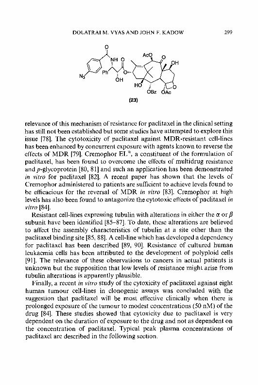
DOLATRAI M. VYAS A N D JOHN F. KADOW 299
0
relevance of this mechanism of resistance for paclitaxel in the clinical setting has still not been established but some studies have attempted to explore this issue [78]. The cytotoxicity of paclitaxel against MDR-resistant cell-lines has been enhanced by concurrent exposure with agents known to reverse the effects of MDR [79]. Cremophor EL@, a constituent of the formulation of paclitaxel, has been found to overcome the effects of multidrug resistance and p-glycoprotein [80, 8 11 and such an application has been demonstrated in vitro for paclitaxel [82]. A recent paper has shown that the levels of Cremophor administered to patients are sufficient to achieve levels found to be efficacious for the reversal of MDR in vitro [83]. Cremophor at high levels has also been found to antagonize the cytotoxic effects of paclitaxel in vitro [84].
Resistant cell-lines expressing tubulin with alterations in either the a or p subunit have been identified [85-871. To date, these alterations are believed to affect the assembly characteristics of tubulin at a site other than the paclitaxel binding site [85,88]. A cell-line which has developed a dependency for paclitaxel has been described [89, 901. Resistance of cultured human leukaemia cells has been attributed to the development of polyploid cells [91]. The relevance of these observations to cancers in actual patients is unknown but the supposition that low levels of resistance might arise from tubulin alterations is apparently plausible.
Finally, a recent in vitro study of the cytoxicity of paclitaxel against eight human tumour cell-lines in clonogenic assays was concluded with the suggestion that paclitaxel will be most effective clinically when there is prolonged exposure of the tumour to modest concentrations (50 nM) of the drug [84]. These studies showed that cytoxicity due to paclitaxel is very dependent on the duration of exposure to the drug and not as dependent on the concentration of paclitaxel. Typical peak plasma concentrations of paclitaxel are described in the following section.
300 PACLITAXEL: A UNIQUE TUBULIN-INTERACTING ANTICANCER AGENT
METABOLISM AND PHARMACOKINETICS
The pharmacokinetics of paclitaxel in humans [92] were first studied in phase I trials employing 6 or 24 h infusion times and demonstrated that peak plasma concentrations were higher than those required to induce tubulin polymerization in vitro [20, 441. Shorter 3-h infusion schedules are now becoming more common and a pharmacokinetic study using this protocol was recently reported [44, 93-95]. Higher peak plasma levels of paclitaxel result from the shorter 1 to 6 h infusion schedules and range from 1000 to 10,000 nM [18]. Paclitaxel peak plasma levels of from 600 to 3500 nM have been found after a 24 h infusion [ 181 while a 96 h infusion gave values of 53 to 77 nM [78]. The elimination of drug after a short infusion follows nonlinear kinetics which is not the case for the longer infusions [44, 94, 961 . The terminal half-life of paclitaxel in the plasma of patients ranges from five to eight hours [18]. Paclitaxel is highly bound to plasma proteins
The metabolism of paclitaxel has been studied in vivo in both humans and rats and also in vitro using both systems. The major site of metabolite formation in both rats and humans appears to be the liver [97]. Cytochrome P-450 enzymes have been shown to metabolize paclitaxel [98]. Monsarrat and co-workers detected up to nine metabolites in rat bile in a study using unlabelled paclitaxel [99]. The two major metabolites were isolated and identified as the products resulting from mono hydroxylation at either the para position of the 3’-C phenyl group (24) or at the meta position of the C-2 benzoate phenyl ring (25). These two metabolites were as active as the parent in a microtubule disassembly assay but were 9 and 39 times less potent in a cytotoxicity assay against L1210 leukaemia respectively. The only minor metabolite unambiguously identified was baccatin I11 (6) which arises from side-chain hydrolysis. Two different groups have reported that the major metabolite found in human bile is 6-a-hydroxypaclitaxe1(26) [97, 100, 1011. The group of Harris et al. isolated this compound in sufficient quantity to determine that it was thirty-fold less potent in cytotoxicity assays than the parent and therefore probably less potent in vivo [loo]. One group also detected 3’-C-p-hydroxyphenylpaclitaxel (24), a major metabo- lite in rat bile and in human bile [97, 1011. The three major hydroxylated metabolites described above were also reported in an abstract by Kumar et al. using isolated liver microsomes and rat hepatocytes [102]. No evidence of any glucuronide or sulphate formation has been detected in any study to date.
The major metabolites of docetaxel (9, a similar taxane in clinical development, have been reported to arise from oxidation of the methyl
(95-98%) [44].
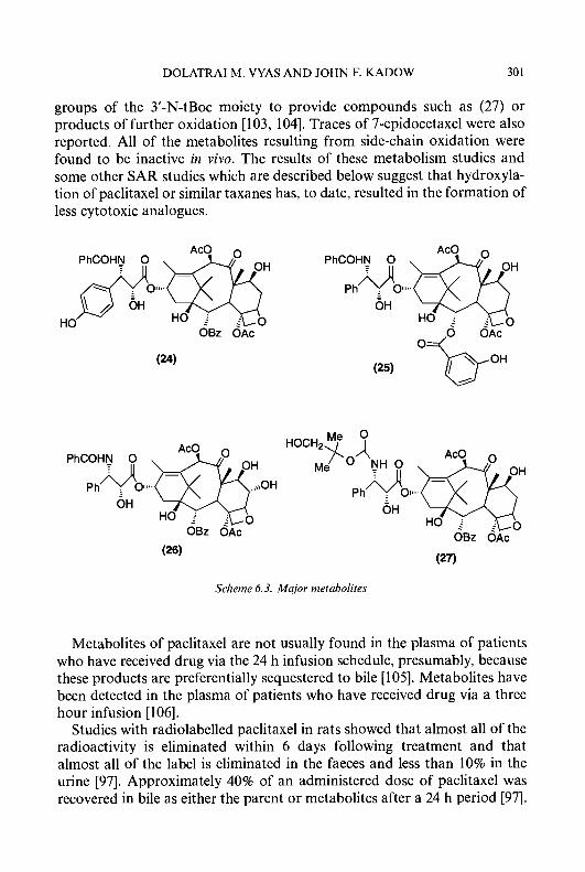
DOLATRAI M. VYAS AND JOHN F. KADOW 30 1
groups of the 3’-N-tBoc moiety to provide compounds such as (27) or products of further oxidation [103, 1041. Traces of 7-epidocetaxel were also reported. All of the metabolites resulting from side-chain oxidation were found to be inactive in vivo. The results of these metabolism studies and some other SAR studies which are described below suggest that hydroxyla- tion of paclitaxel or similar taxanes has, to date, resulted in the formation of less cytotoxic analogues.
PhCOHN 0
.,\OH
OH
Ph do,. OH
HO i .
OBz OAc OBz OAc
(27) (26)
Scheme 6.3. Major nietabolites
Metabolites of paclitaxel are not usually found in the plasma of patients who have received drug via the 24 h infusion schedule, presumably, because these products are preferentially sequestered to bile [ 1051. Metabolites have been detected in the plasma of patients who have received drug via a three hour infusion [106].
Studies with radiolabelled paclitaxel in rats showed that almost all of the radioactivity is eliminated within 6 days following treatment and that almost all of the label is eliminated in the faeces and less than 10% in the urine [97]. Approximately 40% of an administered dose of paclitaxel was recovered in bile as either the parent or metabolites after a 24 h period [97].
302 PACLITAXEL: A UNIQUE TUBULIN-INTERACTING ANTICANCER AGENT
Renal clearance contributes minimally, approximately 5-1 0%, to overall disposition [18, 1071. Thus, a significant pathway for disposition of paclitaxel in rats and humans appears to be hepatic metabolism and subsequent biliary clearance. Because other metabolites have not been identified in either plasma or urine, it is likely that the majority of an administered dose (i.e., 50-65%) is not metabolized [97]. More detailed studies which should account for the fate of all the administered dose are in progress.
ANALOGUES AND STRUCTURE-ACTIVITY RELATIONSHIPS
The biological activity of paclitaxel analogues has been evaluated using several methods. The first and most commonly used is an in vitro assay which provides a measure of the analogue’s ability to polymerize tubulin or to stabilize microtubules. Three methods have been used and all seem to give relatively similar results. The rate of initial tubulin polymerization (slope) [108, 1091, the extent of microtubule assembly [I 10, 11 I], or the measurement of microtubule disassembly [l 121 of analogues can be measured and compared with a standard which is usually paclitaxel. These assays measure the ability of the analogue to function at its biological target (tubulin) without regard for metabolism, pharmaceutics, or antitumour activity (therapeutic index). The most commonly used method for the assessment of the biological activity of taxane analogues is in vitro cytotoxicity assays against whole cultured cells. These assays measure cytotoxic potency (not antitumour activity) in different cell-lines of interest without providing any information about therapeutic index in the host. Initial in vivo assays may consist of models in which the tumour and drug are both implanted intraperitoneally, at the same site, in a mouse [113]. Distal tumour models in which the drug is given i.v. and the tumour is implanted at a different site, usually subcutaneously, are utilized for advanced studies with promising taxane analogues [21, 11 31. Relatively little distal tumour data have been reported in the literature for taxane analogues.
The ready availability of 10-DAB and other modified natural diterpene taxanes has facilitated the pace of analogues research in academia and industry. The main focus appears to be exploring structure-activity relationships (SAR) and identifying superior analogues with distinct advantages such as solubility over paclitaxel. In this vein, a plethora of analogues with core and side-chain modifications have been synthesized and reported [42, 43, 1141. In this review, we wish to discuss the newer developments in analogue research.
DOLATRAI M. VYAS AND JOHN F. KADOW 303
SIDE-CHAIN ANALOGUES
The C-13 side-chain is critical for the activity of paclitaxel. Baccatin 111, which lacks the side-chain, is 50 times less active than paclitaxel in a mammalian tubulin polymerization assay [ 1 151. Other baccatin analogues are also essentially inactive in this assay [112]. The importance of the side-chain to biological activity and its sensitivity to substitution are illustrated by comparing the antitumour activity of paclitaxel with the naturally and frequently co-occurring cephallomannine (28). These ana- logues have relatively similar polarities which makes their chromatographic separation difficult. The only structural difference between the two compounds is the substituent attached to the 3’ nitrogen of the side-chain. Cephallomannine (Figure 6.1, 28) has a tigloyl group while paclitaxel (4) possesses a benzoyl moiety. Both compounds display similar potency in tubulin and cytotoxicity assays when compared in the same laboratory [73, 1161, yet the antitumour activity of cephallomannine in in vivo tumour models is unimpressive and considerably poorer than that of paclitaxel [l 171. This result also illustrates the necessity of in vivo antitumour data in order to accurately determine the true effects of chemical modifications on antitumour activity. The importance of the side-chain for biological activity, its greater synthetic accessibility compared with the baccatin core, and the lack of knowledge of its conformation at the biological target have made this an area of substantial past and present activity.
Two significant early studies which examined the importance of each side-chain substituent and also substituent stereochemistry on in vitro biological activity were published [108, 1181 and have been summarized in detail in a previous review [42]. The first study [lo81 examined the effects of deleting either the 3’-benzamide, the 3’-phenyl group, or simultaneously both 3’-substituents while retaining the 2’-hydroxy group. These analogues were all synthesized independently with both possible stereoisomers of the 2‘-hydroxy group but all compounds were found to be substantially less active in a tubulin polymerization assay and less potent in cytoxicity assays. In the second study [118], investigators at Rhone-Poulenc Rorer studied the tubulin depolymerization activity of analogues in which the substituents and the stereochemistry at the C-2’ and C-3’ positions of the side-chain were modified while retaining the C-3’-phenyl group as a constant. In this series, interchanging the stereochemistry and/or substituting hydroxy groups for nitrogen amides or vice versa in all the various permutations resulted in analogues which ranged from similar to significantly inferior to paclitaxel in the tubulin depolymerization assay. However, some alterations of the moiety attached to the 3‘-nitrogen appeared promising when other changes
304 PACLITAXEL: A UNIQUE TUBULIN-INTERACTING ANTICANCER AGENT
0
Compound R’ R2
(28) E-MeCH=C(Me)OHN- Ph
(29) PhS02HN Ph
(30) tBuOC(0)HN Ph
(31 tBuC(0)HN Ph
(32) Me2CHCH2C(0)HN Ph
(33) tBuCHp Ph
(34) Me(CH&C(O)HN Ph
(35) Me(CH2)&(O)MeN Ph
(36) EtC(0)HN Ph
(37) MeOC(0)HN
(38) EtOC(0)HN
Ph
Ph
Figure 6.1. C-13 Side-chin analogues of’parlituxel
were absent. Replacement of the benzamide with a p-tolylsulphonamide (29) resulted in only a slight decrease in the compound’s ability to stabilize microtubules. Interestingly, analogues with a t-butoxy carbamate function- ality on the 3’-carbon position actually resulted in a compound with slightly better potency in this assay and this has proven to be a very important modification. The analogue which contains this side-chain and a free, unacetylated 10-hydroxy group, is docetaxel ( 5 ) and has been found to be more potent than paclitaxel and only partially cross-resistant in many whole cell in vitro cytotoxicity assays. Typically, docetaxel has been found to be
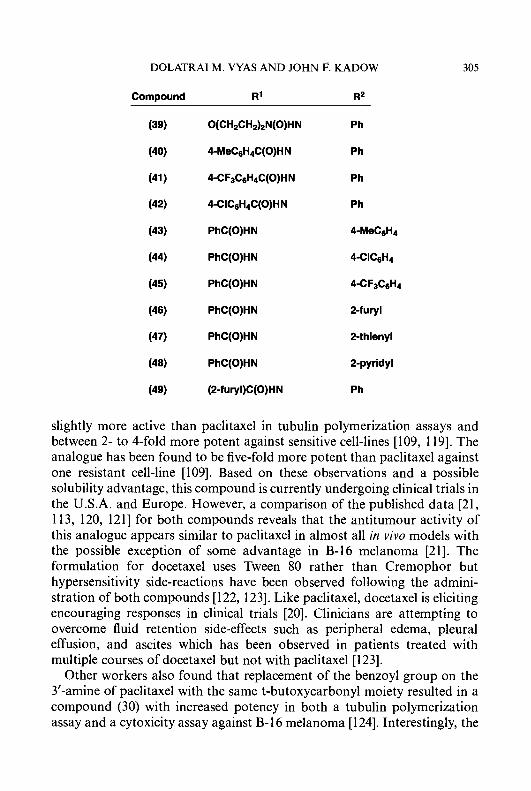
DOLATRAI M. VYAS AND JOHN F. KADOW 305
ComDound R' R*
(39) O(CH2CH2)2N(O)HN Ph
(46) PhC(0)HN 2-fury1
(47) PhC(0)HN 2-thienyl
(48) PhC(0)HN 2-pyridyl
(49) (P-furyl)C(O)HN Ph
slightly more active than paclitaxel in tubulin polymerization assays and between 2- to 4-fold more potent against sensitive cell-lines [109, 1191. The analogue has been found to be five-fold more potent than paclitaxel against one resistant cell-line [109]. Based on these observations and a possible solubility advantage, this compound is currently undergoing clinical trials in the U.S.A. and Europe. However, a comparison of the published data [21, 113, 120, 1211 for both compounds reveals that the antitumour activity of this analogue appears similar to paclitaxel in almost all in vivo models with the possible exception of some advantage in B-16 melanoma [21]. The formulation for docetaxel uses Tween 80 rather than Cremophor but hypersensitivity side-reactions have been observed following the admini- stration of both compounds [122, 1231. Like paclitaxel, docetaxel is eliciting encouraging responses in clinical trials [20]. Clinicians are attempting to overcome fluid retention side-effects such as peripheral edema, pleural effusion, and ascites which has been observed in patients treated with multiple courses of docetaxel but not with paclitaxel [123].
Other workers also found that replacement of the benzoyl group on the 3'-amine of paclitaxel with the same t-butoxycarbonyl moiety resulted in a compound (30) with increased potency in both a tubulin polymerization assay and a cytoxicity assay against B-16 melanoma [ 1241. Interestingly, the
306 PACLITAXEL: A UNIQUE TUBULIN-INTERACTING ANTICANCER AGENT
same workers found the N-trimethylacetyl analogue (3 1) to be less potent in both assays. The N-isovaleryl (32) and N-t-butyl acetyl (33) analogues both had activity similar to paclitaxel in the tubulin polymerization assay but were slightly less potent against B-16 melanoma in the cytoxicity assay [124]. The 3’-N-n-hexanoyl analogue of paclitaxel (34) was isolated from cell culture and found to possess activity similar to paclitaxel in a tiibulin polymerization assay and good potency in a number of cell-lines in the NCI cell panel [ 1251. The corresponding methylated 3’-N-methyl-3’-N-n-hex- anoyl analogue (35) and a 3’-N-propionyl congener (36) were also isolated and had slightly reduced but comparable activity to paclitaxel in the tubulin assay. The 3’-N-debenzoyl-3’-N-methoxy (37) and 3’-N-debenzoyl-3’-N- ethoxy (38) analogues of paclitaxel have been disclosed in an issued U.S. Patent [126]. Both analogues appeared slightly less potent in a tubulin polymerization assay. In a cytoxicity assay against HCT-116, the methoxy analogue (37) was 23-fold less potent while the ethoxy analogue (38) was slightly less potent but similar to paclitaxel. A 3’-N-debenzoyl-3’-N- morpholinocarbonyl analogue of docetaxel(39) was found to be considera- bly less potent than the parent in in vitro assays [127].
Several examples of paclitaxel analogues containing a 3’-N-benzoyl group with substituents on the aromatic ring have been reported. The analogues with a 3’-N-4-tolyl-(40), 4-trifluoromethylphenyl-(41), and 4-chorophenyl- (42) benzamide were found to have activity similar to paclitaxel in a microtubule assembly assay and a cytotoxicity assay against B- 16 melanoma [110, 128, 1291. Several groups have reported analogues of paclitaxel or docetaxel in which the phenyl group at the 3’-position of the side-chain has been modified or replaced. A recent report has demonstrated conclusively that replacement of the 3’-phenyl with a hydrogen provides analogues which are significantly less active in a tubulin assay regardless of other side-chain substitutions [130]. The analogues with a 4-tolyl (43) or 4-chlorophenyl(44) group at the 3’-position both had potency similar to the parent in a tubulin polymerization assay and a cytoxicity assay against B- 16 melanoma [110, 128, 1291. The same workers found the analogue (45) with a 4-trifluoromethylphenyl group at 3’ to be unstable. The 3’-4-hydroxy- phenyl analogue (24), a metabolite of paclitaxel, also appeared similar to the parent in a tubulin assay but was less potent in a cytoxicity assay [99]. A U.S. Patent containing tubulin polymerization data for 32 analogues of paclitaxel which contain modified phenyl or naphthyl substituents in place of either the 3’-N-or 3’-phenyl groups of the side-chain has been issued [131]. Several of the analogues discussed above are also contained in this patent and cytoxicity data for 15 of the analogues against a sensitive and resistant human colon cancer cell-line are included. All of the analogues appear to
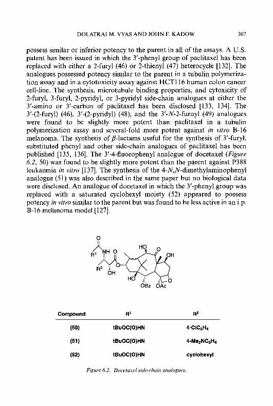
DOLATRAI M. VYAS AND JOHN F. KADOW 307
possess similar or inferior potency to the parent in all of the assays. A U.S. patent has been issued in which the 3’-phenyl group of paclitaxel has been replaced with either a 2-fury1 (46) or 2-thienyl (47) heterocycle [132]. The analogues possessed potency similar to the parent in a tubulin polymeriza- tion assay and in a cytotoxicity assay against HCTll6 human colon cancer cell-line. The synthesis, microtubule binding properties, and cytoxicity of 2-furyl, 3-fury], 2-pyridyl, or 3-pyridyl side-chain analogues at either the 3’-amino or 3’-carbon of paclitaxel has been disclosed [133, 1341. The 3’-(2-furyl) (46), 3’-(2-pyridyl) (48), and the 3’-N-2-furoyl (49) analogues were found to be slightly more potent than paclitaxel in a tubulin polymerization assay and several-fold more potent against in vitro B-16 melanoma. The synthesis of p-lactams useful for the synthesis of 3’-furyl, substituted phenyl and other side-chain analogues of paclitaxel has been published [135, 1361. The 3’-4-fluorophenyl analogue of docetaxel (Figure 6.2, 50) was found to be slightly more potent than the parent against P388 leukaemia in vitro [ 1371. The synthesis of the 4-N,N-dimethylaminophenyl analogue (5 1) was also described in the same paper but no biological data were disclosed. An analogue of docetaxel in which the 3’-phenyl group was replaced with a saturated cyclohexyl moiety (52) appeared to possess potency in vitro similar to the parent but was found to be less active in an i.p. B- 16 melanoma model [ 1271.
Compound R’
Figure 6.2. Docetasel side-chain analogues.

308 PACLITAXEL: A UNIQUE TUBULIN-INTERACTING ANTICANCER AGENT
Early work showed that esterification of the 2‘-hydroxy group of the side-chain results in compounds which display reduced activity in tubulin polymerization assays, suggesting that a free 2’-hydroxy group is required. Bulky or stable esters and ethers such as the 2’-t-butyldimethylsilyl ether of paclitaxel are inactive in vitro in cytoxicity assays. More labile esters such as acetates appear to be hydrolyzed in these assays as they exhibit potency similar to the parent in spite of their inactivity in tubulin assays [115, 138, 1391. A U.S. patent application covering 2‘-carbonate derivatives of paclitaxel has been published [140, 1411. A European patent application covering 2’-and 7-substituted benzoate analogues of paclitaxel has been published [142]. Replacement of the 2’-hydroxy group with a hydrogen (53) or either a 2’-fluoro (54) or methoxy ( 5 5 ) moiety with the same stereo- chemistry resulted in analogues which were inactive in a tubulin polymeriza- tion assay and much less potent in a cytotoxicity assay [143]. The authors suggest that the 2’-hydroxy group may be necessary to achieve a favourable ‘preferred conformation’ or to function as a hydrogen bond donor at the receptor site in tubulin. Synthesis of side-chain analogues of both paclitaxel (56) and docetaxel (57) in which a one carbon homologation between the 1’-carbonyl and the 2’-carbon has been inserted provided analogues which were at least 27 times less potent than the parents in a tubulin polymerization assay [144].
Compound R
(53) H
OBz O&‘
Compound R
(56) Ph
(54) F (57) tBuO
(55) OCH3
Interest in determining the biologically active side-chain conformation of the side-chain and core of paclitaxel or docetaxel is high. The relatively flexible nature of the side-chain as compared to the relatively rigid baccatin core has resulted in numerous modeling and spectroscopic studies designed to elucidate the more important or biologically important side-chain

DOLATRAI M. VYAS AND JOHN F. KADOW 309
conformers [145-1491. The accuracy of these studies and their importance to drug design cannot be accurately gauged at this time since so little is known about the interaction of the taxanes with tubulin at the molecular level.
C-1 MODIFICATIONS
Attempted C-1 deoxygenations of protected baccatins have so far been unsuccessful and therefore C-1 deoxy paclitaxel has not yet been reported [150]. Attempts to functionalize the C-1 hydroxy group or acid catalyzed ionizations at this position have resulted in a facile rearrangement to form A-ring contracted ‘nortaxane’ analogues [15 1, 1521. For example, reaction of 2’,7-bistriethylsilyl paclitaxel with methanesulphonyl chloride provided the A-ring contracted analogue (58) after removal of the protecting groups. This analogue retained comparable activity in a tubulin disassembly assay but was considerably less cytoxic than paclitaxel against KB cells in a cell culture assay [ 15 I]. Ojima and co-workers have synthesized ‘Nor-Seco’ analogues (59, 60) of paclitaxel and docetaxel respectively, in which the A-ring has been opened and the C-14 carbon excised [153]. These analogues possess considerably diminished but faint activity in in vitro assays.
Compound R
(59) Ph
C-2 MODIFICATIONS
Chen and co-workers have reported on the debenzoylation and subsequent deoxygenation of the 2 position to provide 2-deoxypaclitaxe1(61) [154]. This compound was found to be inactive in both a tubulin polymerization and a cytoxicity in vitro assay. A later report described the efficient Red-A1 mediated C-2 benzoate hydrolysis of 7,13-bistriethylsilyl baccatin 111 [ 1551. Rederivatization of the resulting 2-hydroxy-7,13-bistriethylsilyl baccatin
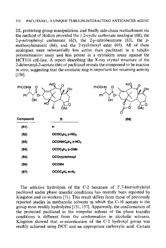
310 PACLITAXEL: A UNIQUE TUBULIN-INTERACTING ANTICANCER AGENT
111, protecting group manipulation, and finally side-chain reattachment via the method of Holton provided the 1,2-cyclic carbonate analogue (68), the 2-p-nitrophenyl carbamate (62), the 2-p-nitrobenzoate (63), the p- methoxybenzoate (64), and the 2-cyclohexyl ester (65). All of these analogues were substantially less active than paclitaxel in a tubulin polymerization assay and less potent in a cytoxicity assay against the HCT116 cell-line. A report describing the X-ray crystal structure of the 2-debenzoyl-2-acetate (66) of paclitaxel reveals the compound to be inactive in vitro, suggesting that the aromatic ring is important for retaining activity [156].
Compound R
The selective hydrolysis of the C-2 benzoate of 2’,7-bistriethylsilyl paclitaxel under phase transfer conditions has recently been reported by Kingston and co-workers [71]. This result differs from those of previously reported studies in methanolic solvents in which the C-10 acetate is the group most readily hydrolyzed [ 15 1, 1571. Apparently, the conformation of the protected paclitaxel in the nonpolar solvent of the phase transfer conditions is different from the conformation in alcoholic solvents. Kingston showed that re-esterification of the C-2 hydroxyl group was readily achieved using DCC and an appropriate carboxylic acid. Certain

DOLATRAI M. VYAS AND JOHN F. KADOW 31 1
C-2 meta-substituted benzoic acid analogues such as the azido analogue (67), which looked the most promising, were found to be more active in a tubulin polymerization assay and more potent in a cytoxicity assay against p388 leukaemia than paclitaxel. These analogues are currently being evaluated in in vivo models at the NCI. In contrast, the corresponding substitutions at the para position of the C-2 benzoate resulted in analogues which were less active than the parent in the same assays. As mentioned above, the C-2 rn-hydroxybenzoate (25) is a metabolite of paclitaxel and was found to be 39-fold less potent than the parent in a cytoxicity assay [99]. This preliminary SAR demonstrates that the benzoate at C-2 is critical for the function of paclitaxel. Substitution at the meta position by some substituents but not hydroxy, is well tolerated and may be beneficial while substitution at the para position results in a loss of activity. In vivo studies are required to determine the true value of these analogues.
C-4 MODIFICATIONS
Although 4-deacetylated analogues of baccatin I11 were reported in the literature several years ago [158], the synthesis and biology of the corresponding 4-deacetyl paclitaxel analogue (69) has just recently been presented [159]. The basic hydrolysis of a baccatin derivative with a free 13-hydroxy group led to a somewhat selective hydrolysis of the C-4 acetate. Subsequent side-chain attachment provided (69). This analogue was less active than the parent in in v i m assays indicating that the acetate at C-4 is necessary to retain full biological activity.
C-4, C-5 AND OXETANE MODIFICATIONS
A number of analogues of paclitaxel which are oxetane-ring opened analogues have been reported [73, 151, 1571. Many naturally occurring taxanes do not contain an intact oxetane ring [43]. All analogues with biology reported to date which do not have an intact oxetane ring are
312 PACLITAXEL: A UNIQUE TUBULIN-INTERACTING ANTICANCER AGENT
substantially less active than the parent in in vitro assays. This is most likely to be due to the fact that the oxetane ring determines the conformation of the C-ring and also determines the orientation of the C-4 substituent rather than a direct interaction of the oxetane with tubulin.
C-6 MODIFICATIONS
As mentioned previously, the metabolite 6a-hydroxy paclitaxel (26) was found to be 30-fold less potent against two different human cell-lines in vitro [loo].
C-7 MODIFICATIONS
Numerous studies of paclitaxel analogues modified at the 7-position have been reported. Early studies demonstrated that the 7-acetyl analogue (70) of paclitaxel was active in tubulin polymerization assays and was only slightly less cytotoxic than the parent, thus demonstrating that a free 7-hydroxy group was not necessary for tubulin polymerization [63, 115, 139, 1601. The 7-ethyl carbonate of paclitaxel possesses activity similar to the parent in in vitro assays and an i.p./i.p. M109 in vivo model [161, 1621. The overall in vitro profile for 7-epipaclitaxel(76) was also similar to the parent [109, 163, 1641 but recent results show that 7-epipaclitaxel is considerably less active than paclitaxel in an in vivo i.p./i.p. M109 lung carcinoma model [161,162]. 7-Xylosyl paclitaxel, l0-deacetyl-7-xylosy1 paclitaxel, and 7-glutaryl pacli- taxel were all found to possess similar activity in a tubulin depolymerization assay [112, 1 181. A number of paclitaxel analogues containing an amino acid substituent at the 7-position were prepared as potential water-soluble prodrugs of paclitaxel. For example, both 7-dimethylglycylpaclitaxel (7 1) and 7-~-alanyl paclitaxel (72) were about half as potent as the parent in a cytotoxicity assay against the B-16 cell-line but (71), dosed i.p., was found to be considerably less active than paclitaxel against the MX-1 mammary xenograph model [l 1 11. Reaction of paclitaxel with DAST (diethylamino- sulphur trifluoride) under controlled conditions has been reported to generate a mixture of 7-epifluoropaclitaxel (77) and the C19-C-7 cy- clopropane analogue (78) [ 1651. Interestingly, 7-epipaclitaxel (76) under the same conditions provides the cyclopropane analogue (78) exclusively, presumably due to the fact that the C-19 methyl group is more suitably positioned for SN, type participation [ 1661. Both these analogues were slightly less active than the parent in tubulin polymerization assays, cytoxicity assays against a sensitive HCTll6 human colon cell-line, and in the in vivo i.p./i.p. M109 model mentioned above [161, 165, 167, 1681. A

DOLATRAI M. VYAS AND JOHN F. KADOW 313
OBz OAc OBz OAc ~
Compound R Compound R
OCOMe (76) OH
OCOCH2NMe2
OCOCHMeNH2 (77) F
H
OCSSMe
OPO(0H)z
RCOHN 0
OH
OBz OAc
Compound R
(78)
(79)
Ph
tBuO
similar cympropane analogue has also been iscxted as a minor side-product after a complex rearrangement of a 9-dihydrobaccatin [ 1691. The docetaxel side-chain was subsequently attached to give (79), a compound which was more potent than paclitaxel against three cell-lines. The increased potency of (79) can be attributed to the 3’-N-Boc substituent on the side-chain. Two groups have reported the synthesis of 7- deoxypaclitaxel (73) via Barton radical deoxygenation of different sub- strates. Kingston and co-workers were able to prepare the 7-methylxanthate (74) of pacitaxel and subsequently successfully deoxygenated the 7-position under the influence of tributyl tin hydride [170]. The same paper also reported an efficient method of converting 7-epipaclitaxel to the parent
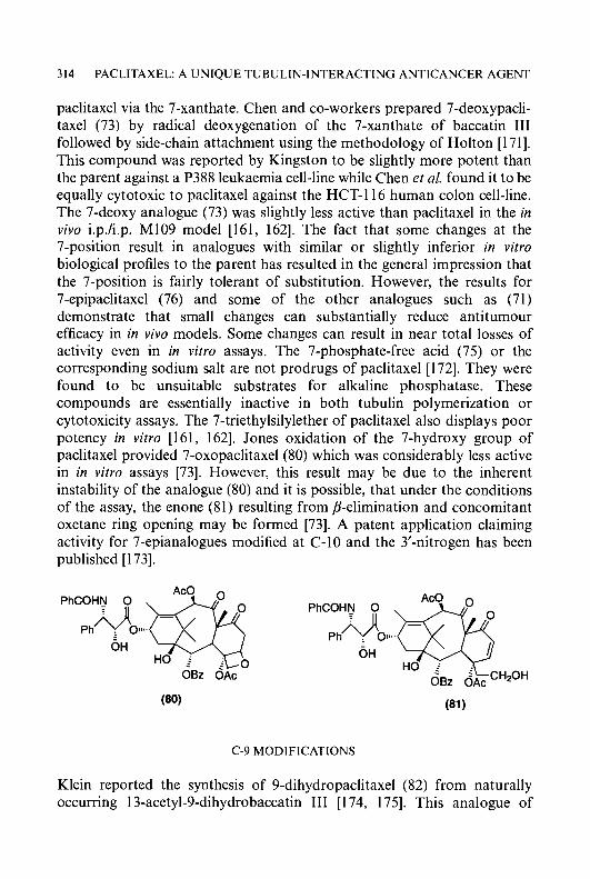
3 14 PACLITAXEL: A UNIQUE TUBULIN-INTERACTING ANTICANCER AGENT
paclitaxel via the 7-xanthate. Chen and co-workers prepared 7-deoxypacli- taxel (73) by radical deoxygenation of the 7-xanthate of baccatin I11 followed by side-chain attachment using the methodology of Holton [171]. This compound was reported by Kingston to be slightly more potent than the parent against a P388 leukaemia cell-line while Chen et al. found it to be equally cytotoxic to paclitaxel against the HCT-116 human colon cell-line. The 7-deoxy analogue (73) was slightly less active than paclitaxel in the in vivo i.p./i.p. M109 model [161, 1621. The fact that some changes at the 7-position result in analogues with similar or slightly inferior in vitro biological profiles to the parent has resulted in the general impression that the 7-position is fairly tolerant of substitution. However, the results for 7-epipaclitaxel (76) and some of the other analogues such as (71) demonstrate that small changes can substantially reduce antitumour efficacy in in vivo models. Some changes can result in near total losses of activity even in in vitro assays. The 7-phosphate-free acid (75) or the corresponding sodium salt are not prodrugs of paclitaxel [ 1721. They were found to be unsuitable substrates for alkaline phosphatase. These compounds are essentially inactive in both tubulin polymerization or cytotoxicity assays. The 7-triethylsilylether of paclitaxel also displays poor potency in vitro [161, 1621. Jones oxidation of the 7-hydroxy group of paclitaxel provided 7-oxopaclitaxel(80) which was considerably less active in in vitro assays [73]. However, this result may be due to the inherent instability of the analogue (80) and it is possible, that under the conditions of the assay, the enone (81) resulting from p-elimination and concomitant oxetane ring opening may be formed [73]. A patent application claiming activity for 7-epianalogues modified at C- 10 and the 3’-nitrogen has been published [173].
C-9 MODIFICATIONS
Klein reported the synthesis of 9-dihydropaclitaxel (82) from naturally occurring 13-acetyl-9-dihydrobaccatin I11 [ 174, 1751. This analogue of
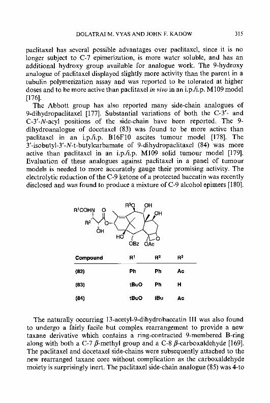
DOLATRAI M. VYAS A N D JOHN F. KADOW 315
paclitaxel has several possible advantages over paclitaxel, since it is no longer subject to C-7 epimerization, is more water soluble, and has an additional hydroxy group available for analogue work. The 9-hydroxy analogue of paclitaxel displayed slightly more activity than the parent in a tubulin polymerization assay and was reported to be tolerated at higher doses and to be more active than paclitaxel in vivo in an i.p./i.p. M109 model [176].
The Abbott group has also reported many side-chain analogues of 9-dihydropaclitaxel [ 1771. Substantial variations of both the C-3’- and C-3’-N-acyl positions of the side-chain have been reported. The 9- dihydroanalogue of docetaxel (83) was found to be more active than paclitaxel in an i.p./i.p. B16F10 ascites tumour model [178]. The 3’-isobutyl-3’-N-t-butylcarbamate of 9-dihydropaclitaxel (84) was more active than paclitaxel in an i.p./i.p. M109 solid tumour model [179]. Evaluation of these analogues against paclitaxel in a panel of tumour models is needed to more accurately gauge their promising activity. The electrolytic reduction of the C-9 ketone of a protected baccatin was recently disclosed and was found to produce a mixture of C-9 alcohol epimers [ 1801.
1330 OH R’COHN 0
6 H HO j
OBz OAc
Compound R’ R2 R3
(82) Ph Ph Ac
(83) tBuO Ph H
(84) tBuO iBu Ac
The naturally occurring 13-acetyl-9-dihydrobaccatin I11 was also found to undergo a fairly facile but complex rearrangement to provide a new taxane derivative which contains a ring-contracted 9-membered B-ring along with both a C-7 p-methyl group and a C-8 P-carboxaldehyde [169]. The paclitaxel and docetaxel side-chains were subsequently attached to the new rearranged taxane core without complication as the carboxaldehyde moiety is surprisingly inert. The paclitaxel side-chain analogue (85) was 4-to
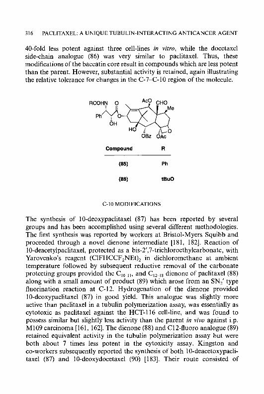
3 16 PACLITAXEL: A UNIQUE TUBULIN-INTERACTING ANTICANCER AGENT
40-fold less potent against three cell-lines in vitro, while the docetaxel side-chain analogue (86) was very similar to paclitaxel. Thus, these modifications of the baccatin core result in compounds which are less potent than the parent. However, substantial activity is retained, again illustrating the relative tolerance for changes in the C-7-C-10 region of the molecule.
RCOHN 0
Ph OH
OBz OAc
Compound R
C-10 MODIFICATIONS
The synthesis of 10-deoxypaclitaxel (87) has been reported by several groups and has been accomplished using several different methodologies. The first synthesis was reported by workers at Bristol-Myers Squibb and proceeded through a novel dienone intermediate [181, 1821. Reaction of 10-deacetylpaclitaxel, protected as a bis-2’,7-trichloroethylcarbonate, with Yarovenko’s reagent (ClFHCCF,NEt), in dichloromethane at ambient temperature followed by subsequent reductive removal of the carbonate protecting groups provided the C,,-,,, and CI2-,* dienone of paclitaxel (88) along with a small amount of product (89) which arose from an SN,‘ type fluorination reaction at C-12. Hydrogenation of the dienone provided 10-deoxypaclitaxel (87) in good yield. This analogue was slightly more active than paclitaxel in a tubulin polymerization assay, was essentially as cytotoxic as paclitaxel against the HCT-116 cell-line, and was found to possess similar but slightly less activity than the parent in vivo against i.p. M109 carcinoma [161, 1621. The dienone (88) and C12-fluoro analogue (89) retained equivalent activity in the tubulin polymerization assay but were both about 7 times less potent in the cytoxicity assay. Kingston and co-workers subsequently reported the synthesis of both 10-deacetoxypacli- taxel (87) and 10-deoxydocetaxel (90) [183]. Their route consisted of
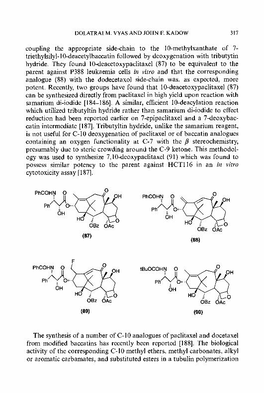
DOLATRAI M. VYAS AND JOHN F. KADOW 317
coupling the appropriate side-chain to the 10-methylxanthate of 7- triethylsilyl- 10-deacetylbaccatin followed by deoxygenation with tributytin hydride. They found 10-deacetoxypaclitaxel (87) to be equivalent to the parent against P388 leukaemia cells in vitro and that the corresponding analogue (88) with the dodecetaxol side-chain was, as expected, more potent. Recently, two groups have found that 1 0-deacetoxypaclitaxel (87) can be synthesized directly from paclitaxel in high yield upon reaction with samarium di-iodide [ 184-1 861. A similar, efficient 10-deacylation reaction which utilized tributyltin hydride rather than samarium di-iodide to effect reduction had been reported earlier on 7-epipaclitaxel and a 7-deoxybac- catin intermediate [ 1871. Tributyltin hydride, unlike the samarium reagent, is not useful for C-10 deoxygenation of paclitaxel or of baccatin analogues containing an oxygen functionality at C-7 with the p stereochemistry, presumably due to steric crowding around the C-9 ketone. This methodol- ogy was used to synthesize 7,lO-deoxypaclitaxel (91) which was found to possess similar potency to the parent against HCT116 in an in v i m cytotoxicity assay [ 1871.
The synthesis of a number of C-10 analogues of paclitaxel and docetaxel from modified baccatins has recently been reported [ 1881. The biological activity of the corresponding C- 10 methyl ethers, methyl carbonates, alkyl or aromatic carbamates, and substituted esters in a tubulin polymerization
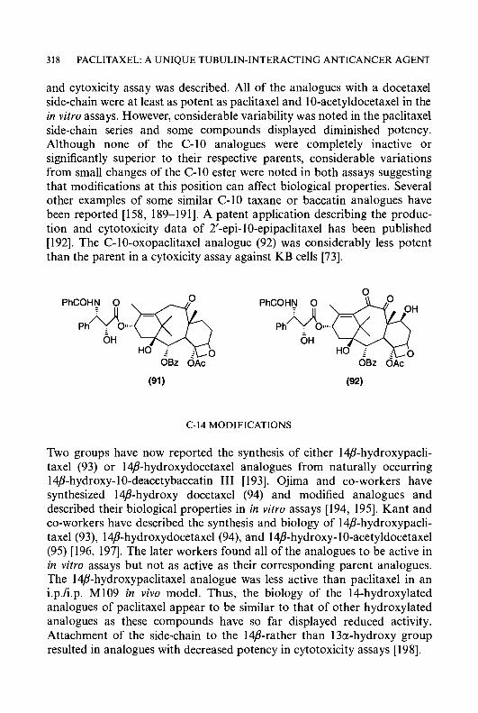
31 8 PACLITAXEL: A UNIQUE TUBULIN-INTERACTING ANTICANCER AGENT
and cytoxicity assay was described. All of the analogues with a docetaxel side-chain were at least as potent as paclitaxel and 10-acetyldocetaxel in the in vitro assays. However, considerable variability was noted in the paclitaxel side-chain series and some compounds displayed diminished potency. Although none of the C-10 analogues were completely inactive or significantly superior to their respective parents, considerable variations from small changes of the C-10 ester were noted in both assays suggesting that modifications at this position can affect biological properties. Several other examples of some similar C-10 taxane or baccatin analogues have been reported [158, 189-1911. A patent application describing the produc- tion and cytotoxicity data of 2’-epi- 10-epipaclitaxel has been published [ 1921. The C-10-oxopaclitaxel analogue (92) was considerably less potent than the parent in a cytoxicity assay against KB cells [73].
PhCOHN 0 i II
C-14 MODIFICATIONS
Two groups have now reported the synthesis of either 14B-hydroxypacli- taxel (93) or 14/?-hydroxydocetaxel analogues from naturally occurring 14B-hydroxy- 10-deacetybaccatin I11 [ 1931. Ojima and co-workers have synthesized 14B-hydroxy docetaxel (94) and modified analogues and described their biological properties in in vitro assays [194, 1951. Kant and co-workers have described the synthesis and biology of 14B-hydroxypacli- taxel (93), 14/3-hydroxydocetaxe1(94), and 14j?-hydroxy- 1 0-acetyldocetaxel (95) [196, 1971. The later workers found all of the analogues to be active in in vitro assays but not as active as their corresponding parent analogues. The 14~-hydroxypaclitaxel analogue was less active than paclitaxel in an i.p./i.p. M109 in vivo model. Thus, the biology of the 14-hydroxylated analogues of paclitaxel appear to be similar to that of other hydroxylated analogues as these compounds have so far displayed reduced activity. Attachment of the side-chain to the 14B-rather than 13a-hydroxy group resulted in analogues with decreased potency in cytotoxicity assays [ 1981.
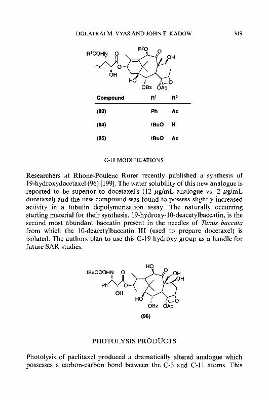
DOLATRAI M. VYAS A N D JOHN F. KADOW 319
Compound R1 R2
(93) Ph Ac
(94) tBuO H
(95) tBuO Ac
C-19 MODIFICATIONS
Researchers at Rhone-Poulenc Rorer recently published a synthesis of 19-hydroxydocetaxel(96) [199]. The water solubility of this new analogue is reported to be superior to docetaxel’s (12 pg/mL analogue vs. 2 pg/mL docetaxel) and the new compound was found to possess slightly increased activity in a tubulin depolymerization assay. The naturally occurring starting material for their synthesis, 19-hydroxy- 10-deacetylbaccatin, is the second most abundant baccatin present in the needles of Taxus baccata from which the 10-deacetylbaccatin I11 (used to prepare docetaxel) is isolated. The authors plan to use this C-19 hydroxy group as a handle for future SAR studies.
PHOTOLYSIS PRODUCTS
Photolysis of paclitaxel produced a dramatically altered analogue which possesses a carbon-carbon bond between the C-3 and C-11 atoms. This

320 PACLITAXEL: A UNIQUE TUBULIN-INTERACTING ANTICANCER AGENT
molecule was more than 100 times less potent than paclitaxel when evaluated in a tubulin polymerization assay [200].
In summary, the SAR studies illustrate the importance of functionalities in the ‘southern half (C-2, C-4, and the oxetane) for retaining the biological function of paclitaxel and its ability to polymerize tubulin. In contrast, minor modifications at other positions generally have small effects on the activity in tubulin assays but may result in modulation of cytoxicity and activity in in vivo models which is most likely due to other mechanistic and pharmacological reasons. Presently, there is a plethora of in vitro information on modified taxanes. A major task which remains is to ascertain the performance of these analogues in in vivo animal tumour models.
WATER-SOLUBLE PRODRUGS
In spite of paclitaxel’s promising antitumour profile in the clinic, the drug is not ideally suited for systemic delivery via intravenous infusion. Its extreme insolubility in water has necessitated extensive formulation studies [28] that have led to identification of an intravenous formulation containing 50% Cremophor EL@ (a polyethoxylated castor oil) and 50% anhydrous ethanol. This formulation is diluted with saline before administration to give a solution containing 0.03-0.60 mg/ml paclitaxel. This high dilution necessi- tates drug preparation at frequent intervals, and the administration of large volumes of fluids to administer therapeutic doses (1 50-200 mg/m2) of paclitaxel. Thus, the large amounts of Cremophor administered during infusion has exhibited vehicle-related toxicity; in particular, hypersensitivity reactions have been observed in patients in the form of dyspnoea, hypotension, bronchospasm, urticaria, and erythematous rash [29, 2011. These symptoms in the clinic have been largely controlled with prophylactic medication with histamine antagonists and corticosteroids. Schedule changes involving slower infusions of longer duration (24-36 h) were also tried in attempts to alleviate hypersensitivity reactions. The hypersensitivity reactions can be attributed to the known histamine-releasing properties of the Cremophor EL@ [202]. Efforts have been made to identify alternative formulations but with little success.
To circumvent the vehicle toxicity and water-solubility issues, several research groups in recent years have synthesized and evaluated water- soluble prodrugs of paclitaxel. The main theme of the earliest studies was the synthesis of esterase-cleavable prodrugs carrying a water-solubilizing functionality. Both the C-2 and the C-7 hydroxyl functionalities were
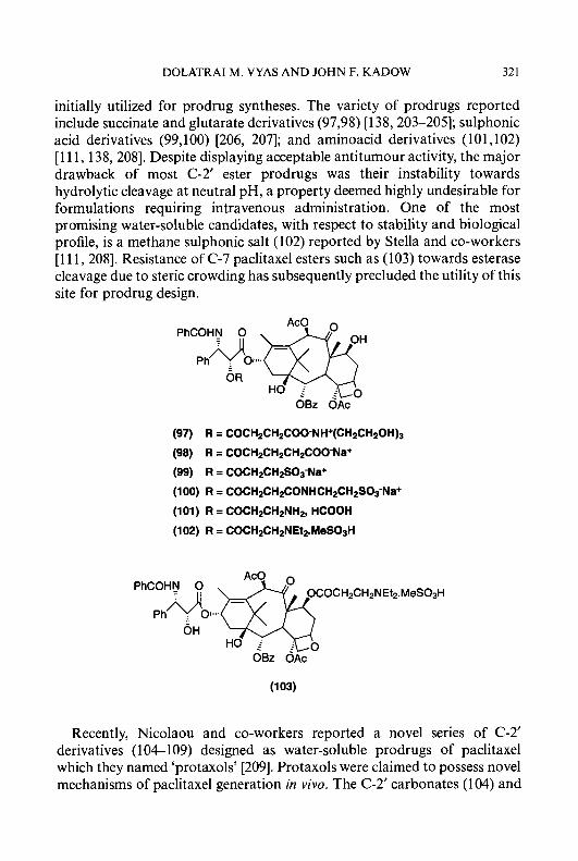
DOLATRAI M. VYAS AND JOHN F. KADOW 32 1
initially utilized for prodrug syntheses. The variety of prodrugs reported include succinate and glutarate derivatives (97,98) [ 138,203-2051; sulphonic acid derivatives (99,100) [206, 2071; and aminoacid derivatives (101,102) [l 1 1, 138,2081. Despite displaying acceptable antitumour activity, the major drawback of most C-2’ ester prodrugs was their instability towards hydrolytic cleavage at neutral pH, a property deemed highly undesirable for formulations requiring intravenous administration. One of the most promising water-soluble candidates, with respect to stability and biological profile, is a methane sulphonic salt (102) reported by Stella and co-workers [ l l 1, 2081. Resistance of C-7 paclitaxel esters such as (103) towards esterase cleavage due to steric crowding has subsequently precluded the utility of this site for prodrug design.
PhCOHN 0
O R
OBz OAc
(97) R = COCH2CH2COO-NH+(CH2CH2OH)3
(98) R = COCH2CH2CH2COO-Na+
(99) R = COCH2CH2S03-Na+
(100) R = COCH~CH~CONHCH~CH~SOS-N~+
(102) R = COCH2CH2NEt2.MeS03H
(101) R = COCH2CH2NH2, HCOOH
Recently, Nicolaou and co-workers reported a novel series of C-2’ derivatives (1 04-1 09) designed as water-soluble prodrugs of paclitaxel which they named ‘protaxols’ [209]. Protaxols were claimed to possess novel mechanisms of paclitaxel generation in vivo. The C-2’ carbonates (104) and

322 PACLITAXEL: A UNIQUE TUBULIN-INTERACTING ANTICANCER AGENT
(105) are postulated to generate paclitaxel via a base-induced beta- elimination in the basic microenvironment of certain tumours, whereas the C-2’ monoesters (106-109) are envisioned to generate paclitaxel through an intramolecular hydrolysis mechanism. The introduction of heteroatom was purposeful for imparting water-solubility and for controlling the mecha- nism of paclitaxel generation in vivo. For example the sulphone derivative (109) was reported to possess water-solubility of 1.5 mg/ml in contrast to ca. 0.25 pg/ml for paclitaxel. The report describing the above protaxols does not disclose any in vivo data on the efficacy of these analogues.
(104) R = COOCH2CH2S02Ph (105) R = COOCH2CH2S02C6H4-p-NO2 (106) R = COCH20CH2COOH (107) R = COCH2SCHzCOOH (108) R = COCHzSOCH2COOH (109) R = COCH2S02CH2COOH
The BMS prodrug strategy was focused on the design and synthesis of water-soluble phosphates of taxol as phosphatase-activated prodrugs, the rationale behind this approach being the ubiquitous distribution of nonspecific phosphatase in particular alkaline phosphatase (AP) in humans [210]. Also, there is considerable evidence that many tumours express high levels of alkaline phosphatase [211], thus raising the possibility of site-specific drug delivery. In support of the BMS strategy, the successful utility of phosphatase-cleavable prodrugs has precedents in other drugs such as etoposide [212], clindamycin [213] and mustards [214].
The prototype phosphatase-cleavable prodrug, namely the C-2’ and the C-7 phosphates ( 1 10) and (79), turned out to possess acceptable water- solubility (> 3 mg/ml) as their sodium salts but based on their in vitro and in vivo perfomance, were judged to be unsuitable prodrugs of paclitaxel [ 1721. These compounds were found to be extremely poor substrates for enzyme hydrolysis probably due to the proximity of the phosphate groups to the congested taxane core. To circumvent this steric problem, the BMS group initiated a double prodrug (pro-prodrug) programme aimed at derivatizing

DOLATRAI M. VYAS A N D JOHN F. KADOW 323
paclitaxel either at C-2’ or C-7 with phosphate functionality attached through a self-immolative linker or spacer. The success of this strategy relies upon unmasking of the sterically accessible phosphates by phosphatases in vivo, followed by generation of paclitaxel by self-immolation of the linker via a rapid lactonization [215] or a fragmentation [216] process.
PhCOHN 0
OBz OAc
The design of a functional, successful self-immolating linker for the pro-prodrug design was guided by the ‘trimethyl lock’ linker reported by Amsberry et al. [217,218]. The BMS version of the modified linker involved the use of a methylated 2’-phosphonooxyphenylpropionic acid as the ‘trimethyl lock’ spacer in the synthesis of phosphates (1 11) and (1 12) [140, 2191. The sodium salts of these phosphates were endowed with adequate water-solubility (> 10 mg/ml) to allow intravenous administration of their aqueous solutions. In in vitro experiments with isolated alkaline phosphat- ase, these derivatives, unlike their direct phosphate counterparts (1 10) and (79), were observed to generate paclitaxel. This confirmed the utility of the ‘trimethyl lock’ linker in pro-prodrug design. Furthermore, in the in vivo evaluation against Sc-implanted Madison 109 murine carcinoma, com- pound (111) was found to be marginally active, whereas (112) was equiactive to paclitaxel when compared head to head in the same experiment. In the general area of pro-prodrugs of paclitxel, the BMS group has actively explored utility of phosphates attached to paclitaxel via several potentially useful self-immolating linkers [ 1411.
(HO)2OPO
PhCOHN 0 (111) R2= ~ c o - ‘ R1 =
OR2
OBz OAc (H0)zOPO
(112) R1= pco-‘ R2=H
324 PACLITAXEL: A UNIQUE TUBULIN-INTERACTING ANTICANCER AGENT
In conclusion, the published biological data for new analogues or prodrugs of paclitaxel is, at this time, insufficient to allow the identification of a likely candidate for clinical development. It is probable that many more studies of analogues using in vivo tumour models and further efforts in analogue synthesis will be needed before a clinical candidate can be selected.
CLINICAL SUMMARY
The cumulative clinical experience with paclitaxel to date has been the subject of several recent reviews [20, 26, 47, 123, 220-2221. Herein, we briefly highlight the current overall clinical status on paclitaxel. Due to the earlier encouraging results for the efficacy of this drug on cisplatin-refactory ovarian cancer and doxorubicin-resistant breast cancer, paclitaxel is already approved in several countries as a second-line therapy for treatment of these neoplasms. Although, the earlier clinical trials of paclitaxel were hampered and threatened by the scarcity of its supply and the alleged Cremophor- related hypersensitivity reactions, these issues have been resolved as discussed earlier. Consequently, the major current effort in the oncology community is to establish the full clinical spectrum of this unique agent. In this vein, randomised clinical trials are ongoing to obtain indications in non-small-cell lung cancer [223, 2241, first-line breast [26, 222, 225, 2261, first-line ovarian [227-2291, and head and neck [230, 2311 cancers. Two positive studies have been published for small-cell lung cancer, germcell, lymphoma and melanoma [26].
The dose-limiting toxicity of paclitaxel [232] has been bone marrow suppression, specifically neutropenia which is reversible. In high dose trials (>250 mg/m2), peripheral neuropathy is of great concern since unlike neutropenia, it is cumulative. Until recently, to avert hypersensitivity episodes due to Cremophor, a 24 h infusion protocol was routinely employed and this required inpatient treatment. However, in recent reports it has been suggested that with proper monitoring and prophylactic medication, 3 h infusion may allow for administration of higher doses (than in 24 h infusion) of paclitaxel in an outpatient setting. Preliminary indications in a 3 h infusion protocol are that high plasma levels of paclitaxel are obtained and there is reduced neutropenia. The whole issue of schedule and dosage in paclitaxel therapy has been under debate [233] and needs to be resolved by well designed future clinical trials.
Paclitaxel treatment to date has been employed mainly for palliation purposes. However, maximization of its clinical potential in combination chemotherapy and multimodality therapy may eventually lead to curative
DOLATRAI M. VYAS AND JOHN F. KADOW 325
therapy. Already, combination therapy with cisplatin is promising in the first-line treatment of ovarian cancer [234]. Several such clinical trials are ongoing [235].
CONCLUSION
Review of the literature indicates that interest in clinical as well as preclinical research on paclitaxel and taxoids in general, for example, docetaxel, has reached peak levels. From a medical perspective, the mission appears fairly obvious; the immediate objective is to maximize the clinical potential of paclitaxel in treatment and eventually cure of cancer. However, prior to realizing these goals, several clinical issues on schedule and dosage remain to be resolved through well-designed trials. Preclinical research presently appears to lack a clear direction, presumably due to the lack of critical in vivo SAR data, mechanistic details, and supporting tumour biology. As more information on paclitaxel’s mechanisms of clinical resistance, interaction with tubulin, SAR and the role of Cremophor in MDR reversal are gathered, criteria for 2nd generation analogues with distinct pharmaceutical and pharmacological advantages may emerge. In this context, in vivo model development will face a major challenge to differentiate efficacy and toxicity profiles of analogues for future develop- ment. Another major factor which will impact the overall research and development picture, will be economic factors. In the present climate of cost containment of health care costs, unless cheap sources of precursors to analogues are identified, either through natural or synthetic means, development of future analogues will be in jeopardy.
REFERENCES
1 Double, J.A. and Bibby, M.C. (1990) Drug News Perspect. 3, 276-279. 2 Braverman, A.S. (1993) Ann. Intern. Med. 118,630-632. 3 Chabner, B.A. (1993) Ann. Intern. Med. 118,633436. 4 Grindey, G.B. (1990) Cancer Cells 2, 163-171. 5 Carter, S.K., Bakowski, N.T. and Hellman, K. (1987) Chemotherapy of Cancer, pp.
315-323, John Wiley and Sons, New York. 6 Johnson, R.K. and Hertzberg, R.P. (1990) Annu. Rep. Med. Chem. 25, 129. 7 Boyd, M.R. (1993) in Current Therapy in Oncology (Neiderhuber, J.E., ed.), pp. 11-22,
B.C. Decker, Philadelphia. 8 Driscoll, J.S. (1984) Cancer Treat. Rep. 68, 63-76. 9 Johnson, R.K. (1990) J. Natl. Cancer Inst. (U.S.) 82, 1082-1083.
10 Corbett, T.H., Valeriote, F.A. and Baker, L.H. (1987) Invest. New Drugs 5, 3-20.
326 PACLITAXEL: A UNIQUE TUBULIN-INTERACTING ANTICANCER AGENT
1 1 Powis, G. (1991) Trends Pharmacol. Sci. 12, 188-194. 12 Cohen, J. (1993) Science (Washington, D.C.) 262 ,86845 . 13 Hall, A. (1994) Science (Washington, D.C.) 264, 1413-1414. 14 Rowinsky, E.K. and Donehower, R.C. (1991) Pharmacol. Ther. 52,35-84. 15 Hamel, E. (1992) Pharmacol. Ther. 55, 31-51. 16 Gerzon, K. (1980) Anticancer Agents Based on Natural Product Models, pp. 271-317,
Academic Press, New York. 17 Mangeney, P., Andriamialisoa, R.Z., Lallemand, J.Y., Langlois, N., Langlois, Y. and
Potier, P. (1979) Tetrahedron 35, 2175-2179. 18 Rowinsky, E.K., Cazenave, L.A. and Donehower, R.C. (1990) J. Natl. Cancer Inst.
(U.S.) 82, 1247-1259. 19 Anon. (1993) Drugs Future 18, 681. 20 Donehower, R.C. and Rowinsky, E.K. (1993) Cancer Treat. Rev. 19,63-78. 21 Bissery, M.-C., Guenard, D., Gueritte-Voegelein, F. and Lavelle, F. (1991) Cancer Res.
5 1,48454852. 22 Wani, M.C., Taylor, H.L. and Wall, M.E. (1971) J. Am. Chem. SOC. 93,2325-2327. 23 Schiff, P.B., Fant, J. and Horwitz, S.B. (1979) Nature (London) 22,665-667. 24 Schiff, P.B. and Horwitz, S.B. (1980) Proc. Natl. Acad. Sci. U.S.A. 77, 1561-1565. 25 Schiff, P.B. and Horwitz, S.B. (1981) Biochemistry 20, 3247-3252. 26 Rowinsky, E.K. (1994) Ann. Pharmacother. 28, S18-S22. 27 Cragg, G.M., Schepartz, S.A., Suffness, M. and Grever, M.R. (1993) J. Nat. Prod. 56,
28 Adams, J.D., Flora, K.P., Goldspiel, B.R., Wilson, J.W., Arbuck, S.G. and Finley, R. (1993) J. Natl. Cancer Inst. Monogr. 15, 141-147.
29 Dorr, R.T. (1994) Ann. Pharmacother. 28, SI 1-S14. 30 Denis, J.-N., Greene, A.E., Gueritte-Voegelein, F., Mangatal, L. and Potier, P. (1988) J.
Am. Chem. SOC. 110,5917-5919. 31 Holton, R.A. (1992) U.S. Patent 5,175,315; (1993) Chem. Abstr. 114, 164568q. 32 Holton, R.A. (1993) U.S. Patent 5,229,526; (1993) Chem. Abstr. 119,49693t. 33 Boa, A.N., Jenkins, P.R. and Lawrence, N.J. (1994) Contemp. Org. Synth. I , 47-75. 34 Nicolaou, K.C., Dai, W.-M. and Guy, R.K. (1994) Angew. Chem., Int. Ed. Engl. 33,
15-44. 35 Swindell, C.S. (1991) Org. Prep. Proced. Int. 23,465-543. 36 Holton, R.A., Somoza, C., Kim, H.-B., Liang, F., Biediger, R.J., Boatman, P.D., Shindo,
M., Smith, C.C., Kim, S., Nadizadeh, H., Suzuki, Y., Tao, C., Vu, P., Tang, S., Zhang, P., Murthi, K.K., Gentile, L.N. and Liu, J.H. (1994) J. Am. Chem. SOC. 116, 1597-1598. Holton, R.A., Somoza, C., Kim, H.-B., Liang, F., Biediger, R.J., Boatman, P.D., Shindo, M., Smith, C.C., Kim, S., Nadizadeh, H., Suzuki, Y., Tao, C., Vu, P., Tang, S., Zhang, P., Murthi, K.K., Gentile, L.N. and Liu, J.H. (1994) J. Am. Chem. SOC. 116, 1599-1600. Nicolaou, K.C., Yang, Z., Liu, J.J., Ueno, H., Nantermet, P.G., Guy, R.K., Claiborne, C.F., Renaud, J., Couladouros, E.A., Paulvannan, K. and Sorensen, E.J. (1994) Nature (London) 367,630-634.
39 Stierle, A., Strobel, G. and Stierle, D. (1993) Science (Washington, D.C.) 260. 214216. 40 Witherup, K.M., Look, S.A., Stasko, M.W., Ghiorzi, T.J., Mushik, G.M. and Cragg,
G.M. (1990) J. Nat. Prod. 53, 1249-1255. 41 Vidensek, N., Lim, P., Campbell, A. and Carlson, C. (1990) J. Nat. Prod. 53, 1609-1610. 42 Suffness, M. (1993) Annu. Rep. Med. Chem. 28, 305-314. 43 Kingston, D.G.I., Molinero, A.A. and Rimoldi, J.M. (1993) Prog. Chem. Org. Nat. Prod.
61, 1-206. 44 Rowinsky, E.K. and Donehower, R.C. (1993) Semin. Oncol. 20, 16-25.
1657-1 668.
37
38
DOLATRAI M. VYAS AND JOHN F. KADOW 327
45 Horwitz, S.B., Cohen, D., Rao, S., Ringer, I., Shen, H.-J. and Yang, C.-P.H. (1993) Monogr. Natl. Cancer Inst. 15.55-61.
46 Rowinsky, E.K., Eisenhauer, E.A., Chaudhry, V., Arbuck, S.G. and Donehower, R.C. (1993) Semin. Oncol. 20, 1-15.
47 Canetta, R., Arbuck, S., Ondetto, N., Rozencweig, M. and Carter, S.K. (1993) in Cancer Chemotherapy: Challenges for the Future Volume 8 (Kimura, K. ed.), pp. 309-315, Excerpta Medica Ltd., Tokyo.
48 Lavelle, F. (1993) Curr. Opin. Invest. Drugs 2, 627-635. 49 Bissett, D. and Kaye, S.B. (1993) Eur. J. Cancer 29A, 1228-1231. 50 Cheson, B.D. and Arbuck, S.G. (1993) Oncology 7,63. 5 1 Manfredi, J.J., Parness, J. and Horwitz, S.B. (1982) J. Cell. Biol. 94, 688-696. 52 Fuchs, D.A. and Johnson, R.K. (1978) Cancer Treat. Rep. 62, 1219-1222. 53 Roberts, J.R. (1989) J. Histochem. Cytochem. 37, 1659-1665. 54 Baum, S.G., Wittner, M., Nadler, J.P., Horwitz, S.B., Dennis, J.E., Schiff, P.B. and
Tanowitz, H.B. (1981) Proc. Natl. Acad. Sci. U.S.A. 78,45714575. 55 Koponen, M., Greider, A. and Loor, F. (1982) Exp. Cell Res. 140,237-250. 56 Horwitz, S.B. and Manfredi, J.J. (1984) Pharmacol. Ther. 25, 83-125. 57 Diaz, J.F. and Andreu, J.M. (1993) Biochemistry 32, 2747-2755. 58 Horwitz, S.B. and Parness, J. (1981) J. Cell Biol. 91,479487. 59 Rowinsky, E.K., Donehower, R.C., Jones, R.J. and Tucker, R.W. (1988) Cancer Res. 48,
409341 00. 60 Rowinsky, E.K., Burke, P.J., Karp, J.E., Tucker, R.W., Ettinger, D.S. and Donehower,
R.C. (1989) Cancer Res. 49,4640-1647. 61 Correia, J.J. (1991) Pharmacol. Ther. 52, 127-147. 62 Ohta, S., Krishan, A,, Nishio, K., Ohmori, T., Kunikane, H., Inomata, M., Takahashi,
T. and Saijo, N. (1993) Anticancer Res. 13, 873-874. 63 Gueritte-Voegelein, F., Takoudju, M., Wright, M., Chenu, J. and Guenard, D. (1988)
FEBS Lett. 227, 96-98. 64 Honvitz, S.B., Rao, S. and Ringel, I. (1992) J. Natl. Cancer Inst. (U.S.) 84, 785-788. 65 Chatterjee, A,, Williamson, J.S., Zjawiony, J.K. and Peterson, J.R. (1992) Bioorg. Med.
Chem. Lett. 2, 91-94. 66 Rao, S., Krauss, N.E., Heerding, J.M., Swindell, C.S., Ringel, I., Orr, G.A. and Horwitz,
S.B. (1994) J.Biol. Chem. 269,3132-3134. 67 Swindell, C.S., Heerding, J.M., Kraus, N.E., Honvitz, S.B., Rao, S. and Ringel, I. (1994)
J. Med. Chem. 37, 14461449. 68 Georg, G.I. and Harriman, G.C.B. (1992) Bioorg. Med. Chem. Lett. 2, 735-738. 69 Georg, G.L., Harriman, G.C.B., Park, H. and Himes, R.H. (1994) Bioorg. Med. Chem.
Lett. 4,487490. 70 Carboni, J.M., Farina, V., Rao, S., Hauck, S.I., Horwitz, S.B. and Ringel, 1. (1993) J.
Med. Chem. 36, 513-515. 71 Chaudhary, A.G., Gharpure, M.M., Rimoldi, J.M., Chordia, M.D., Gunatilaka, A.A.L.
and Kingston, D.G.I. (1994) J. Am. Chem. SOC. 116,40974098. 72 Kingston, D.G.I., Rimoldi, J.M., Chaudhary, A.G. and Samaranayake, G. (1993) J. Nat.
Prod. 56, 1313-1330. 73 Kingston, D.G.I., Samaranayake, G. and Ivey, C.A. (1990) J. Nat. Prod. 53, 1-12. 74 Suffness, M. (1994) Abstracts of the 207th National Meeting of the American Chemical
Society San Diego, CA, MEDl 141. 75 Simon, S.M. and Schindler, M. (1994) Proc. Natl. Acad. Sci. U.S.A. 91, 3497-3504. 76 Roy, S.N. and Horwitz, S.B. (1985) Cancer Res. 45, 3856-3863.
328 PACLITAXEL: A UNIQUE TUBULIN-INTERACTING ANTICANCER AGENT
77 Greenberger, L.M., Williams, S.S. and Horwitz, S.B. (1987) J. Biol. Chem. 262,
78 Wilson, W.H., Berg, S. and Kang, Y.-K. (1993) Proc. Am. SOC. Clin. Oncol. 12, 335. 79 Jachez, B., Nordmann, R. and Loor, F. (1993) J. Natl. Cancer Inst. (U.S.) 85,478483. 80 Schuurhuis, G.J., Broxterman, H.J., Pinedo, H.M., v. Heijningen, T.H.M., v. Kalken,
C.K., Vermorken, J.B., Spoelstra, E.C. and Lankelma, J. (1990) Br. J. Cancer 62, 591-594.
81 Fjallskog, M.L., Frii, L. and Bergh, J. (1994) Eur. J. Cancer 30A, 687-690. 82 Chervinsky, D.S., Brecher, M.L. and Hoelcle, M.J. (1993) Cancer Res. 13, 93-96. 83 Woodcock, D., Webster, L., Linsenmeyer, M., Millward, M., Morton, C. and Bishop, J.
(1993) J. Natl. Cancer Inst. (U.S.) 85, 1685-1690. 84 Liebmann, J.E., Cook, J.A., Lipschultz, C., Teague, D., Fisher, J. and Mitchell, J.B.
(1993) Br. J. Cancer 68, 11041 109. 85 Cabral, F. and Barlow, S.B. (1991) Pharmacol. Ther. 52, 159-171. 86 Minotti, A.M., Barlow, S.B. and Cabral, F. (1991) J. Biol. Chem. 266, 3987-3994. 87 Cabral, F., Abraham, I. and Gottesman, M.M. (1981) Proc. Natl. Acad. Sci. U.S.A. 78,
43884391. 88 Cabral, F., Brady, R.C. and Schibler, M.J. (1986) Ann. N. Y. Acad. Sci. 466,745-756. 89 Cabral, F., Wible, L., Brenner, S. and Brinkley, B.R. (1983) J. Cell Biol. 97, 30-39. 90 Cabral, F.R. (1983) J. Cell Biol. 97, 22-29. 91 Roberts, J.R., Allison, D.C., Donehower, R.C. and Rowinsky, E. K. (1990) Cancer Res.
50, 710-716. 92 Kuhn, J.G. (1994) Ann. Pharmacother. 28, S15-Sl7. 93 Schiller, J.H., Storer, B., Tutsch, K., Arzoomanian, R., Alberti, D., Feierabend, C. and
Spriggs, D. (1994) J. Clin. Oncol. 12, 241-248. 94 Kearns, C., Gianni, L., Vigano, L., Giani, A,, Griggs, D., Tutsch, K., Schiller, J.,
Sridhara, R. and Egorin, M. (1993) Proc. Am. SOC. Clin. Oncol. 12,341. 95 Swenerton, K., Eisenhauer, E., t. BokkelHuinink, W., Myles, J., Magioni, C.,
vanderBerg, M., Kerr, I., Gianni, L., Vermorken, J., Buser, K., Sadura, A,, Bacon, M., Santabarbara, P., Onetto, N. and Canetta, R. (1993) Proc. Am. SOC. Clin. Oncol. 12,256.
96 Sannichsen, D.S., Hurwitz, C.A., Pratt, C.B., Shuster, J.J. and Relling, M.V. (1994) J. Clin. Oncol. 12, 532-538.
97 Monsarrat, B., Alvinerie, P., Wright, M., Dubois, J., Gueritte-Vogelein, F., Guenard, D., Donehower, R.C. and Rowinsky, E.K. (1993) J. Natl. Cancer Inst. Monogr. 15, 3946.
98 Cresteil, T., Monsarrat, B., Alvinerie, P., Treluyer, J.M., Vieira, I . and Wright, M. (1994) Cancer Res. 54,386-392.
99 Monsarrat, B., Mariel, E., Cros, S., Cares, M., Guenard, D., Gueritte-Voegelein, F. and Wright, M. (1990) Drug Metab. Dispos. 18, 895-901.
100 Harris, J.W., Katki, A,, Anderson, L.W., Chmurny, G.N., Paukstelis, J.V. and Collins, J.M. (1 994) J. Med. Chem. 37, 706-709.
101 Monsarrat, B., Wright, M., Dubois, J., Gueritte-Voegelein, F., Guenard, D., Rowinsky, E.K. and Donehower, R.C. (1992) Ann. Oncol. 3,258.
102 Kumar, G.N., Thornberg, K.R., Walle, U.K. and Walle, T. (1993) 5th North American ISSX Meeting, Tucson (USA), October 1993, ISSX Proceedings 3,24.
103 Bissery, M.C., Bourzat, J.D., Commercon, A,, Frydman, A,, Gaillard, C., Lavelle, F., Monegier, B., Sable, S. and Vuilhorgne, M. (1994) Abstracts of the 207th National Meeting of the American Chemical Society San Diego, CA, MEDI 144.
104 Monegier, B., Gaillard, C., Sable, S. and Vuilhorgne, M. (1994) Tetrahedron Lett. 35, 3715-371 8.
13685-13689.
DOLATRAI M. VYAS AND JOHN F. KADOW 329
105
106
107 108
109 110
111
112
1 I3 1 I4
115
116
1 I7
1 I8
1 I9
120
121
122
123
124
125
126
127
128
129
Jamis-Dow, C.A., Klecker, R.W., Sarosy, G., Reed, E. and Collins, J.M. (1993) Cancer Chemother. Pharmacol. 33,48-52. Huizing, M.T., Keung, A.C.F., Rosing, H., v.d. Kuij, V., Huinink, W.W.T.B., Mandjes, I.M., Dubbelman, A.C., Pineda, H.M. and Beijnen, J.H. (1993) J. Clin. Oncol. 1 I , 2127-2135. Rowinsky, E.K., Gilbert, M. and McGuire, W.P. (1991) J. Clin. Oncol. 9, 1692-1703. Swindell, C.S., Krauss, N.E., Horwitz, S.B. and Ringel, I. (1991) J. Med. Chem. 34, 1 I 7 6 1 184. Horwitz, S.B. and Ringel, I. (1991) J. Natl. Cancer Inst. (U.S.) 83,288-291. Georg, G.I., Cheruvallath, Z.S., Himes, R.H., Mejillano, M.R. and Burke, C.T. (1992) J. Med. Chem. 35,4230-4237. Stella, V.J., Mathew, A.E., Mejillan, M.R., Nath, J.P. and Himes, R.H. (1992) J. Med. Chem. 35, 145-151. Lataste, H., Senilh, V., Wright, M., Guenard, D. and Potier, P. (1984) Proc. Natl. Acad. Sci. U.S.A. 81,40904094. Rose, W.C. (1992) Anti-Cancer Drugs 3,311-321. Georg, G.I., Ali, S.M., Zygmunt, J. and Jayasinghe, L.R. (1994) Exp. Opin. Ther. Patents 4, 109-120. Parness, J., Kingston, D.G.I., Powell, R.G., Harracksingh, C. and Horwitz, S.B. (1982) Biochem. Biophys. Res. Commun. 105, 1082-1089. McLaughlin, J.L., Miller, R.W., Powell, R.G. and Smith Jr., C.R.S. (1981) J. Nat. Prod. 44, 312-319. Riondel, J., Jacrot, M., Nissou, M.-F., Picot, F., Beriel, H., Mouriquand, C. and Potier, P. (1988) Anticancer Res. 8, 387-390. Gueritte-Voegelein, F., Guenard, D., Lavelle, F., Goff, M.-T.L., Mangatal, L. and Potier, P. (1991) J. Med. Chem. 34, 992-998. Braakhuis, B.J.M., Hill, B.T., Dietel, M., Kelland, L.R., Aapro, M.S., Zoli, W. and Lelieveld, P. (1994) Anticancer Res. 14, 205-208. Nicoletti, M.I., Lucchini, V., D’lncalci, M. and Giavazzi, R. (1994) Eur. J. Cancer 30A, 691-696. Boven, E., Venema-Gaberseek, E., Erkelens, C.A.M., Bissery, M.C. and Pinedo, H.M. (1993) Ann. Oncol. 4, 321-324. Wanders, J., Schrijvers, D., Bruntsch, U., Gore, M., Venveij, J., Hanauske, A.R., Franklin, H., Roelvink, M., Bayssas, M. and Kaye, S.B. (1993) Proc. Am. SOC. Clin. Oncol. 12, 94. Pazdur, R., Kudelka, A.P., Kavanagh, J.J., Cohen, P.R. and Raber, M.N. (1993) Cancer Treat. Rev. 19, 351-386. Georg, G.L., Boge, T.C., Cheruvallath, Z.S. and Harriman, G.C.B. (1994) Bioorg. Med. Chem. Lett. 4, 335-338. Ma, W., Park, G.L., Gomez, G.A., Nieder, M.H., Adams, T.L., Aynsley, J.S., Sahai, O.P., Smith, R.J., Stahlhut, R.W. and Hylands, P.J. (1994) J. Nat. Prod. 57, 116122. Holton, R.A. and Beidiger, R.J. (1993) European Patent Application 534707-A1; (1993) Chem. Abstr. 119,49693t. Duclos, O., Zucco, M., Kuduk, S.D., Sun, C.M., Park, Y.H. and Ojima, I. (1993) Abstracts of the 206th National Meeting of the American Chemical Society Chicago, 111, MEDI 131. Georg, G.I., Cheruvallath, Z.S., Himes, R.H. and Mejillano, M.R. (1992) Bioorg. Med. Chem. Lett. 2, 1751-1754. Georg, G.I., Cheruvallath, Z.S., Himes, R. H. and Mejillano, M.R. (1992) Bioorg. Med. Chem. Lett. 2, 295-298.
330 PACLITAXEL: A UNIQUE TUBULIN-INTERACTING ANTICANCER AGENT
130 Swindell,C.S., Heerding, J.M., Krauss, N.E., Horwitz, S.B. and Ringer, 1. (1994) Bioorg. Med. Chem. Lett. 4, 1531-1536.
131 Holton, R.A., Nadizadeh, H., Kim, S. and Beidiger, R.J. (1993) U.S. Pat. 5,250,683; (1994) Chem. Abstr. 119,49694~.
132 Holton, R.A., Nadizadeh, H. and Beidiger, R.J. (1993) Eur. Patent Appl. 534708-AI; (1994) Chem. Abstr. 119,49693t.
133 Hepperle, M., Clowers, J.S., Harriman, G.C.B., Georg, G.L. and Himes, R.H. (1994) Abstracts of 207th National Meeting of the American Chemical Society San Diego, CA, MEDI 103.
134 Georg, G.I., Harriman, G.C.B., Hepperle, M. and Himes, R.H. (1994) Bioorg. Med. Chem. Lett. 4, 1381-1384.
135 Ojima, I . , Habus, I., Zhao, M., Zucco, M., Park, Y.-H., Sun, C.-M. and Brigaud, T. (1992) Tetrahedron 48,6985-7012.
136 Ojima, I . , Park, Y.H., Sun, C.M., Brigaud, T. and Zhao, M. (1992) Tetrahedron Lett. 33,
137 Bourzat, J.D. and Commercon, A. (1993) Tetrahedron Lett. 34,6049-6052. 138 Kingston, D.G.I. and Magri, N.F. (1988) J. Nat. Prod. 51,298-306. 139 Horwitz, S.B., Mellado, W., Magri, N.F., Kingston, D.G.I., Garcia-Arenas, R. and Orr,
G.A. (1984) Biochem. Biophys. Res. Commun. 124,329-336. 140 Ueda, Y. and Wong, H. (1993) U.S. Patent 5,272,171. 141 Ueda, Y. and Wong, H. (1993) Eur. Patent Appl. 0 558 959 Al . 142 Wong, H. and Doyle, T. (1993) Eur. Patent Appl. 0 569 281 Al . 143 Kant, J., Huang, S., Wong, H., Fairchild, C., Vyas, D. and Farina, V. (1993) Bioorg.
Med. Chem. Lett. 3,2471-2474. 144 Jayasinghe, L.R., Ah, S.M., Zygmunt, J., Georg, G.I. and Vandervelde, D. (1994)
Abstracts of the 207th National Meeting of the American Chemical Society, San Diego, CA, MEDI 98.
145 Swindell, C.S., Chirlian, L.E., Francl, M.M., Heerding, J.M., Krauss, N.E., Williams, H.J., Scott, A.I. and Dieden, R.A. (1993) Tetrahedron 49, 6545-6560.
146 Guenard, D., Dubois, J., Gueritte-Voegelein, F., Guedira, N., Potier, P., Gillet, B. and Beloeil, J.-C. (1993) Tetrahedron 49,6533-6544.
147 Williams, H.J., Scott, A.I., Deiden, R.A., Swindell, C.S., Chirlian, L.E., Francl, M.M., Heerding, J.M. and Krauss, N.M. (1994) Can. J. Chem. 72, 252-260.
148 Velde, D.G.V., Georg, G.I., Grunewald, G.L., Gunn, C.W. and Mitscher, L.A. (1993) J. Am. Chem. SOC. 115, 11650-1 1651.
149 Czaplinski, K.H. and Grunewald, G.L. (1994) 207th National Meeting of the American Chemical Society San Diego, CA, MEDI 105.
150 Chen, S.H., Huang, S., Gao, Q., Golik, J. and Farina, V. (1994) J. Org. Chem. 59, 1475-1484.
151 Kingston, D.G.I., Samaranayake, G., Magri, N.F. and Jitrangsri, C. (1991) J. Org. Chem. 56,51145119.
152 Kingston, D.G.I. and Samaranyake, G. (1992) Eur. Patent Appl. 0 505 047 Al; (1993) Chem. Abstr. 118, 59918~.
153 Ojima, I., Fenoglio, I., Park, Y.H., Sun, C.-M., Appendino, G., Pera, P. and Bernacki, R.J. (1994) J. Org. Chem. 59, 515-517.
154 Chen, S.-H., Wei, J.-M. and Farina, V. (1993) Tetrahedron Lett. 34, 3205-3206. 155 Chen, S.-H., Farina, V., Wei, J.-M., Long, B., Fairchild, C., Mamber, S.W., Kadow, J.F.,
Vyas, D. and Doyle, T.W. (1994) Bioorg. Med. Chem. Lett. 4, 479482. 156 Gao, Q., Wei, J.M. and Chen, S.H. (1994) Pharm. Res. Submitted for publication. 157 Chen, S.-H., Huang, S., Wei, J.-M. and Farina. V. (1993) Tetrahedron 49, 2805-2828.
5737-5740.
DOLATRAI M. VYAS AND JOHN F. KADOW 33 1
158 Wahl, A,, Gueritte-Voegelein, F., Guenard, D., Goff, M.-T.L. and Potier, P. (1992) Tetrahedron 48,6965-6974.
159 Georg, G.L. (1994) Abstracts of the 207th National Meeting of the American Chemical Society San Diego, CA, MEDI 146.
160 Chenu, J., Takoudju., M., Wright, M., Gueritte-Voegelein, F. and Guenard, D. (1988) FEBS Lett. 234, 177-180.
161 Chen, S.H., Kant, J., Mamber, S.W., Roth, G.P., Wei, J.M., Vyas, D.M., Doyle, T.W. and Farina, V. (1994) Bioorg. Med. Chern. Lett. 4, to be submitted.
162 Kadow, J.F., Casazza, A.M., Chen, S.H., Farina, V., Fairchild, C., Kant, J., Rose, W.C., Roth, G.P., Vyas, D.M. and Doyle, T.W. (1994) Abstracts of the 207th National Meeting of the American Chemical Society San Diego, CA, MEDI 96.
163 Kingston, D.G.I., Huang, C.H.O., Magri, N.F. and Samaranayake, G. (1986) J. Nat. Prod. 49,665-669.
164 Kingston, D.G.I. (1991) Pharmacol. Ther. 52, 1-34. 165 Chen, S.-H., Huang, S. and Farina, V. (1994) Tetrahedron Lett. 35, 4 1 4 . 166 Chen, S.-H., Huang, S., Wei, J.-M. and Farina, V. (1993) J. Org. Chem. 58, 45204521. 167 Chen, S.H., Farina, V., Kant, J. and Vyas, D.M. (1994) U.S. Patent 5,294,637 168 Chen, S.H. and Farina, V. (1993) U.S. Patent 5,254,580. 169 Klein, L.L., Maring, C.L., Li, L., Yeung, C.M., Thomas, S.A., Grampovnik, D.J.,
Plattner, J.J. and Henry, R.F. (1994) J. Org. Chem. 59, 2370-2373. 170 Kingston, D.G.I., Chaudhary, A.G. and Rimoldi, J.M. (1993) J. Org. Chem. 58,
171 Chen, S.-H., Huang, S., Kant, J., Fairchild, C., Wei, J.-M. and Farina, V. (1993) J. Org. Chem. 58,5028-5029.
172 Vyas, D.M., Wong, H., Crosswell, A.R., Casazza, A.M., Knipe, J.O., Mamber, S.W. and Doyle, T.W. (1993) Bioorg. Med. Chem. Lett. 3, 1357-1360.
173 Bourzat, J.D. and Commercon, A. (1993) PCT Patent Appl. WO 93/23389. 174 Klein, L.L. (1993) Tetrahedron Lett. 34, 2047-2050. 175 Gunawardana, G.P., Klein, L.L. and McAlpine, J.B. (1993) PCT Patent Appl. WO
93/21 173. 176 Klein, L.L., Maring, C.L., Li. L., Yeung, C.M., Thomas, S.A., Grampovnik, D.J. and
Plattner, J.J. (1994) Abstracts of the 207th National Meeting of the American Chemical Society San Diego, CA, MEDI 143. Maring, L.J., Grarnpovnik, D.J., Yeung, C.M., Klein, L.L., Li, L., Thomas, S.A. and Plattner, J.J. (1994) Bioorg. Med. Chem. Lett. 4, 1429-1432. Grarnpovnik, D.J., Maring, C.J., Klein, L.L., Li, L., Thomas, S.A., Yeung, C.M. and Plattner, J.J. (1994) Abstracts of the 207th National Meeting of the American Chemical Society San Diego, CA, MEDI 102. Li, L., Thomas, S.A., Klein, L.L., Yeung, C.M., Maring, C.J., Grampovnik, D.J. and Plattner, J.J. (1994) Abstracts of the 207th National Meeting of the American Chemical Society San Diego, CA, MEDI 103. Commercon, A,, Bourzat, J.-D., Didier, E., Fouque, E., Bissery, M.C., Combeau, C., Riou, J.-F., Vrignaud, P. and Lavelle, F. (1994) Abstracts of the 207th National Meeting of the American Chemical Society San Diego, CA, ORGN 344.
181 Chen, S.-H., Fairchild, C., Mamber, S.W. and Farina, V. (1993) J. Org. Chem. 58, 2927-2928.
182 Chen, S.H. and Farina, V. (1993) U.S. Patent 5,248,796. 183 Kingston, D.G.I. and Chaudhary, A.G. (1993) Tetrahedron Lett. 34,49214924. 184 Holton, R.A., Somoza, C. and Chai, K.-B. (1994) Tetrahedron Lett. 35, 1665-1668. 185 Holton, R.A. (1993) PCT Patent Appl. WO 9306093-Al.
3798-3799.
177
178
179
180
332 PACLITAXEL: A UNIQUE TUBULIN-INTERACTING ANTICANCER AGENT
186 Cheruvallath, Z.S., Falborg, L., Georg, G., Mitscher, L.A., Velde, D.V., Ye, Q. and Himes, R. (1994) Abstracts of the 207th National Meeting of the American Chemical Society San Diego, CA, MEDI 100.
187 Chen, S.-H., Wei, J.-M., Vyas, D.M., Doyle, T.W. and Farina, V. (1993) Tetrahedron Lett. 34, 6845-6848.
188 Kant, J., O’Keeffe, W.S., Chen, S.H., Farina, V., Fairchild, C., Johnston, K., Kadow, J.F., Long, B.H. and Vyas, D.M. (1994) Tetrahedron Lett. 35, in press.
189 Bourzat, J.-D., Commercon, A,, Cuenard, D., Gutritte-Voegelein, F. and Potier, P. (1993) Eur. Patent Appl. 524,093; (1993) Chem. Abstr. 119, 9005p.
190 Ettouati, L., Ahond, A,, Convert, O., Poupat, C. and Potier, P. (1989) Bull. SOC. Chim. Fr. 687-694.
191 Denis, J.-N., Greene, A.E., Guenard, D., Gueritte-Voegelein, F., Mangatal, L. and Potier, P. (1988) J. Am. Chem. SOC. 110, 5917-5919.
192 Saito, K., Ohashi, H., Tahara, M., Sakamoto, T. and Hibi, M. (1992) PCT Patent Appl. WO 93/02067; (1993) Chem. Abstr. 118, 232375j.
193 Appendino, G., Gariboldi, P., Gabette, B., Pace, R., Bombardelli, E. and Viterbo, D. (1992) J. Chem. SOC., Perkin Trans. I 2925-2929.
194 Fenoglio, I . , Park, Y., Sun, C.M., Ojima, I., Bernacki, R.J. and Pera, P. (1994) Abstracts of the 207th National Meeting of the American Chemical Society San Diego, CA, MEDI 85.
195 Ojima, I., Fenoglio, I., Park, Y.H., Pera, P. and Bernacki, R.J. (1994) Bioorg. Med. Chem. Lett. 4, in press.
196 Kant, J., Kadow, J.F., Farina, V., Fairchild, C., Long, B.H., Rose, W.C., Casazza, A.M., Vyas, D.M. and Doyle, T.W. (1994) Abstracts of the 207th National Meeting of the American Chemical Society San Diego, CA, MEDI 95.
197 Kant, J., Farina, V., Kadow, J.F., Langley, D.R., Fairchild, C., Long, B.H., Rose, W.C., Vyas, D.M. and Doyle, T.W. (1994) Bioorg. Med. Chem. Lett. 4, in press.
198 Ojima, I., Park, Y.H., Sun, C.-M., Fenoglio, I., Appendino, G., Pera, P. and Bernacki, R.J. (1994) J. Med. Chem. 37, 1408-1410.
199 Commercon, A., Margraff, R., Bezard, D. and Bourzat, J.D. (1994) Bioorg. Med. Chem. Lett. 4, 233-236.
200 Chen, S.-H., Combs, C.M., Hill, S.E., Farina, V. and Doyle, T.W. (1992) Tetrahedron Lett. 33, 7679-7680.
201 Slichenmyer, W.J. and Hoff, D.D.V. (1990) J. Clin. Pharmacol. 30, 770-778. 202 Lorenz, W., Riemann, H.J. and Schnial, A. (1977) Agents Actions 7, 63-67. 203 Zalkow, L.H., Deutsch, H.M., Glinski, J.A., Hernandez, M., Haugwitz, R.D.,
Narayanan, V.L. and Suffness, M. (1989) J. Med. Chem. 32, 788-792. 204 Haugwitz, R.D., Zalkow, L., Glinski, J., Suffness, M., Deutsch, H.M. and Narayanan, V.
(1990) U.S. Patent 4,942,184; (1989) Chem. Abstr. 110, 154617h. 205 Haugwitz,R.D., Zalkow, L., Glinski, J., Suffness, M., Deutsch, H.M. andNarayanan, V.
(1992) U.S. Patent 5,157,049; (1989) Chem. Abstr. 110, 354617h. 206 Kingston, D.G.I., Zhao, Z.Y. and Crosswel, A.R. (1991) J. Nat. Prod. 54, 1607-1611. 207 Kingston, D.G.I. and Zhao, Z. (1991) U.S. Patent 5, 059,699; (1992) Chem. Abstr. 116,
59670k. 208 Stella, V.J. and Mathew, A.E. (1990) U.S. Patent 4,960,790; (1991) Chem. Abstr. 114,
82 194a. 209 Nicolaou, K.C., Riemer, C., Kerr, M.A., Rideout, D. and Wrasidlo, W. (1993) Nature
(London) 364,464466. 210 Harris, H. (1989) Clin. Chim. Acta 186, 133-150.
DOLATRAI M. VYAS AND JOHN F. KADOW 333
211 Fishman, W.H., Inglis, N.R., Stolbach, L.L. and Krant, M.J. (1968) Cancer Res. 28, 150-154.
212 Rose, W.C., Basler, G.A., Trail, P.A., Saulnier, M.G., Crosswell, A.R. and Cassazza, A.M. (1990) Invest. New Drugs 8, S25-S32.
213 Tobkes, M., Diaz, S. and Krishnan, L. (1993) U.S. Patent 5,182,374; (1993) Chem. Abstr. 119, 72428~.
214 Workman, P., Ball, C.R. and Double, J. A. (1976) Biochem. Pharmacol. 25, 1139-1 144. 215 Johnson, C.D., Lane, S., Edwards, P.N. and Taylor, P.J. (1988) J. Org. Chem. 53,
5 130-5 139. 216 Stella, V.J., Safadi, M. and Oliyai, R. (1993) Pharm. Res. 10, 1350. 217 Amsberry, K.L., Gerstenberger, A.E. and Borchardt, R.T. (1991) Pharm. Res. 8,
455461. 218 Amsberry, K.L. and Borchardt, R.T. (1990) J. Org. Chem. 55,5867-5877. 219 Ueda, Y., Mikkilineni, A.B., Knipe, J.O., Rose, W.C., Casazza, A.M. and Vyas, D.M.
(1993) Bioorg. Med. Chem. Lett. 3, 1761-1766. 220 Rowinsky, E.K., McGuire, W.P. and Donehower, R.C. (1993) in Principles and Practice
of Gynecologic Oncology Updates (Hoskins, W.J., Perez, C.A. and Young, R.C., ed.), pp. 1-16, J. B. Lippincott Company, Philadelphia.
221 Spiridonidis, C.H. (1993) Drugs Today 29,295-300. 222 Diergarten, K. and Dreps, A. (1993) Onkologie 16, 329-337. 223 Chang, A.Y., Kim, K. and Glick, J. et al. (1993) J. Natl. Cancer Inst. (U.S.) 85, 388-394. 224 Ettinger, D. S. (1993) Semin. Oncol. 20,4649. 225 Holmes, F.A., Walters, R.S., Theriaul, R.L., Forman, A.D., Newton, L.K., Raber,
M.N., Buzdar, A.U., Frye, D.K. and Hortobag, G.N. (1991) J. Natl. Cancer Inst. (U.S.)
226 Seidman, A.D., Norton, L., Reichman, B.S., Crown, J.P.A., Yao, T.J., Heelan, R., Hakes, T.B., Lebwohl, D.E., Gilewski, T.A., Surbone, A,, Currie, V., Hudis, C.A., Klecker, R., Jamis-Dow, C., Collins, J., Quinlivan, S., Berkery, R., Toomasi, F., Canetta, R., Fishman, J. and Arbuck, S. (1993) Semin. Oncol. 20,40-45.
227 Runowicz, C.D., Wiernik, P.H., Einzig, A.I., Goldberg, G.L. and Horwitz, S.B. (1993) Cancer 71, 1591-1596.
228 Swenerton, K., Eisenhauer, E., t. BokkelHuinink, W., Myles, J., Magioni, C., vanderBerg, M., Kerr, I., Gianni, L., Vermorken, J., Buser, K., Sadura, A,, Bacon, M., Santabarbara, P., Onetto, N. and Canetta, R. (1993) Proc. Am. SOC. Clin. Oncol. 12,256.
229 Marty, M., Extra, J.M., Culine, S. and Rousseau, F. (1991) Pathol. Biol. 39, 834835. 230 Forastiere, A.A. (1993) Semin. Oncol. 20, 56-60. 231 Forastiere, A.A., Neuberg, D., Iv, S.G.T., DeConti, R. and Adams, G. (1993) Proc. Am.
SOC. Clin. Oncol. 12, 26. 232 Finley, R.S. and Rowinsky, E.K. (1994) Ann. Pharmacother. 28, S27-S30. 233 Arbuck, S.G. (1994) J. Clin. Oncol. 12,233-236. 234 McGuire, W.P., Hoskins, W.J. and Brady, M.F. (1993) Proc. Am. SOC. Clin. Oncol. 12,
255. 235 Klaasen, U., Diergarten, K., Dittrich, C.H., Kaufmann, J., Sauter, C.H. and Seeber, S.
(1994) Onkologie 17, 8690.
83, 1797-1805.
334 PACLITAXEL: A UNIQUE TUBULIN-INTERACTING ANTICANCER AGENT
ADDENDUM
The following citations are selected key references listed by section which appeared after this manuscript was written and which were added in proof. The large volume of activity prevented inclusion of recent patent literature.
1
2
3 4 5 6
7 8
9
10 11
12
13 14 15
16
17
18
19
20
21 22
PACLITAXEL (reviews) Georg, G. I., Chen, T. T., Ojima, I. and Vyas, D. M. (1995) Taxane Anticancer Agents Basic Science and Current Status, pp. 1-353, American Chemical Society, Washington DC. Suffness, M. and Wall, M. E. (1995) Taxol: Science and Applications, pp. in press, CRC Press, Boca Raton. Kingston, D. G. I. (1994) Trends Biotechnol. 12,222-227. Wall, M. E. and Wani, M. C. (1994) Econ. Med. Plant Res. 6, 299-322. Hepperle, M. and Georg, G. I. (1994) Drugs Future 19, 573-584. BurrisIII, H. A,, Dorr, R. T., Finley, R. S., Goldspiel, B. R., Koeller, J. M., Kuhn, J. G. and Rowinsky, E. K. (1994) Ann. Pharm. 28, SI-S34.
MECHANISM OF ACTION AND RESISTANCE Diaz, J. F. and Andreu, J. M. (1993) Biochemistry 32, 2747-2755. Swindell, C. S., Heerding, J. M., Kraus, N. E., Horwitz, S. B., Rao, S. and Ringel, 1. (1994) J. Med. Chem. 37, 14461449. Leu, J.-G., Chen, B.-X., Diamanduros, A. W. and Erlanger, B. F. (1994) Proc. Natl. Acad. Sci. U.S.A. 91, 10690-10694 Ringel, I. and Honvitz, S. B. (1994) J. Pharm. Exp. Ther. 259, 855-859. Dasgupta, D., Park, H., Harriman, G. C. B., Georg, G. I. and Himes, R. B. (1994) J. Med. Chem. 37,2976-2980. Balasubramanian, S. V., Alderfer, J. L. and Straubinger, R. M. (1994) J. Pharm. Sci. 83, 1470-1 476. Caplow, M., Shanks, J. and Ruhlen, R. (1994) J.Biol. Chem. 269,23399-23402. Long, B. H. and Fairchild, C. R. (1994) Cancer Res. 54,43554361. Combeau, C., Commercon, A., Mioskowski, C., Rousseau, B., Aubert, F. and Goeldner, M. (1994) Biochemistry 33,66766683. Fjallskog, M. L., Frii, L. and Bergh, J. (1994) Eur. J. Cancer 30A, 687-690.
METABOLISM AND PHARMACOKINETICS (Also see reference 6 of this addendum)
Monegier, B., Gaillard, C., Sable, S. and Vuilhorgne, M. (1994) Tetrahedron Lett. 35, 37 15-37 1 8. Walle, T., Bhalla, K. N., Walle, U. K., Kumar, G. N. and Stuart, R. K. (1994) Proc. ASCO 13, 152. Francis, P., Rowinsky, E., Hakes, T., Schneider, J . , Bakarat, R., Curtin, J., Jones, W., Jr., J. L. and al., e. (1993) Proc. ASCO 12, 257. Commercon, A,, Bourzat, J. D., Bezard, D. and Vuilhorgne, M. (1994) Tetrahedron 50, 10289-1 0298. Spencer, C. M. and Faulds, D. (1994) Drugs 48,794-847. Harris, J. W., Rahman, A,, Kim, B.-R., Guengerich, F. P. and Collins, J. M. (1994) Cancer Res. 54,402U035.
DOLATRAI M. VYAS AND JOHN F. KADOW 335
23 Walker, D. G., Swigor, J. E., Kant, J. and Schroeder, D. R. (1994) J. Labelled Compd. Radiopharm. 34,973-980.
24 Rahman, A,, Korzekwa, K. R., Grogan, J., Gonzalez, F. L. and Harris, J. W. (1994) Cancer Res. 54,5543-5546.
25 Sonnichsen, D. S. and Relling, M. V. (1994) Clin. Pharmacokinet. 27,256-269. 26 Eiseman, J. L., Eddington, N. D., Leslie, J., MacAuley, C., Sentz, D. L., Zuhowski, M.,
Kujawa, J. M., Young, D. and Egorin, M. J. (1994) Cancer Chemother. Pharmacol. 34, 46547 1.
ANALOGUES AND STRUCTURE-ACTIVITY RELATIONSHIPS, Side-chain Analogues
27 Georg, G. I . , Harriman, G. C. B., Hepperle, M. and Himes, R. H. (1994) Bioorg. Med. Chem. Lett. 4, 1381-1384.
28 Maring, L. J., Grdmpovnik, D. J., Yeung, C. M., Klein, L. L., Li, L., Thomas, S. A. and Plattner, J. J. (1994) Bioorg. Med. Chem. Lett. 4, 1429-1432.
29 Swindell, C. S., Heerding, J. M., Krauss, N. E., Horwitz, S. B. and Ringer, I. (1994) Bioorg. Med. Chem. Lett. 4, 1531-1536.
30 Kingston, D. G. I . , Chaudhary, A. G., Gunatilaka, A. A. and Middlton, M. L. (1994) Tetrahedron Lett. 35 ,4483484 .
31 Jayasinghe, L. R., Datta, A,, Ah, S. M., Zygmunt, J., VanderVelde, D. G. and Georg, G. 1. (1994) J. Med. Chem. 37, 2981-2984.
32 Li, L., Thomas, S. A,, Klein, L. L., Yeung, C. M., Maring, C. J., Grampovnik, D. J., Lartey, P. A. and Plattner, J. J. (1994) J. Med. Chem. 37, 2655-2663.
33 Ojima, I . , Duclos, O., Zucco, M., Bissery, M.-C., Combeau, C., Vrignaud, P., Riou, J. F. and Lavelle, F. (1994) J. Med. Chem. 37,2602-2608.
34 Ojima, I . , Duclos, O., Kuduk, S. D., Sun, C. M., Slater, J. C., Lavelle, F., Veith, J. M. and Bernacki, It. J. (1994) Bioorg. Med. Chem. Lett. 4, 2631-2634.
35 Boge, T. C., Himes, R. H., Velde, D. G. V. and Georg, G. I . (1994) J. Med. Chem. 37, 3337-3343.
36 Czaplinski, K. H. A. and Grunewald, G. L. (1994) Bioorg. Med. Chem. Lett. 4,221 1-2216. 37 Georg, G. I., Cheruvallath, Z. S., Harriman, G. C., Hepperle, M., Park, H. and Himes, R.
H. (1994) Bioorg. Med. Chem. Lett. 4,2331-2336. 38 Georg, G. I . , C.Boge, T., Cheruvallath, Z. S., Harriman, G. C. B., Hepperle, M., Park, H.
and Himes, R. H. (1994) Bioorg. Med. Chem. Lett. 4, 1825-1830.
C-2 and C-4 Modifications (Also see references 33 and 35 of this addendum)
39 Nicolaou, K. C., Couladouros, E. A,, Nantermet, P. G., Renaud, J., Guy, R. K. and Wrasidlo, W. (1994) Angew. Chem., Int. Ed. Engl. 33, 1581.
40 Georg, G. I . , Ali, S. M., Boge, T. C., Datta, A., Falborg, L. and Himes, R. H. (1994) Tetrahedron Lett. 35, 8931-8934.
41 Datta, A,, Jayasinghe, L. R. and Georg, G. I. (1994) J. Org. Chem. 59,46894690. 42 Pulicani, J.-P., Bezard, D., Bourzat, J.-D., Bouchard, H., Zucco, M., Deprez, D. and
Commercon, A. (1994) Tetrahedron Lett. 35,9717-9720. 43 Neidigh, K. A,, Gharpure, M. M., Rimoldi, J. M., Kingston, D. G. I . , Jiang, Y. Q. and
Hamel, E. (1994) Tetrahedron Lett. 35,6839-6842. 44 Chordia, M. D., Chaudhary, A. S., Kingston, D. G. I . , Jiang, Y. Q. and Hamel, E. (1994)
Tetrahedron Lett. 35,6843-6846. 45 Datta, A,, Jayasinghe, L. I . and Georg, G. 1. (1994) J. Med. Chem. 37,42584260.

336 PACLITAXEL: A UNIQUE TUBULIN-INTERACTING ANTICANCER AGENT
46
47
48
49
50
51
52
53
54
55
56
57
58
59
60
61
62
63
Chen, S. H., Kadow, J. F., Farina, V., Fairchild, C. R. and Johnston, K. A. (1994) J. Org. Chem. 59,6156-6158.
C-7 and C-19 Modifications Bouchard, H., Pulicani, J.-P., Vuilhorgne, M., Bourzat, J.-D. and Commercon, A. (1994) Tetrahedron Lett. 35,9713-9716. Johnson, R. A,, Nidy, E. G., Dobrowolski, P. J., Gebhard, I., Qualls, S. J., Wicnienski, N. A. and Kelly, R. C. (1994) Tetrahedron Lett. 35, 7893-7896. Chen, S. H., Kant, J., Mamber, S. W., Roth, G. P., Wei, J. M., Marshall, D., Vyas, D. M., Farina, V., Casazza, A., Long, B. H., Rose, W. C., Johnston, K. and Fairchild, C. (1994) Bioorg. Med. Chem. Lett. 4,2223-2228. Pulicani, J.-P., Bouchard, H., Bourzat, J.-D. and Commercon, A. (1994) Tetrahedron Lett. 35, 9709-9712.
C-9 Modifications (Also see references 28 and 32 of this addendum)
Klein, L. L., Leung, C. M., Li, L. and Plattner, J. J. (1994) Tetrahedron Lett. 35, 47074710. Pulicani, J.-P., Bourzat, J.-D., Bouchard, H. and Commercon, A. (1994) Tetrahedron Lett. 35, 4999-5002. Datta, A,, Aube, J., Georg, G. I., Mitscher, L. A. and Jayasinghe, L. R. (1994) Bioorg. Med. Chem. Lett. 4. 1831-1834.
C-10 Modifications (Also see reference 53 of this addendum)
Kant, J., OKeeffe, W. S., Chen, S. H., Farina, V., Fairchild, C., Johnston, K., Kadow, J. F., Long, B. H. and Vyas, D. M. (1994) Tetrahedron Lett. 35, 5543-5546'. Hanson, R. L., Wasylyk, J. M., Nanduri, V. B., Cazzulino, D. L., Patel, R. N. and Szarka, L. J. (1994) J.Biol. Chem. 269,22145-22149. Georg, G. I. and Cheruvallath, Z. S. (1994) J. Org. Chem. 59,40154018.
C-14 Modifications Kant, J., Fairchild, C., Farina, V., Kadow, J. F., Langley, D. R., Long, B. H., Rose, W. C., Vyas, D. M. and Doyle, T. W. (1994) Bioorg. Med. Chem. Lett. 4, 1565-1570. Ojima, I., Fenoglio, I., Park, Y. H., Pera, P. and Bernacki, R. J. (1994) Bioorg. Med. Chem. Lett. 4, 1571-1576. Ojima, I., Park, Y. H., Sun, C.-M., Fenoglio, I., Appendino, G., Pera, P. and Bernacki, R. J. (1994) J. Med. Chem. 37, 1408-1410.
Photolysis Products Chen, S. H., Farina, S. H., Huang, S., Gao, Q., Golik, J. and Doyle, T. W. (1994) Tetrahedron 50,8633-8650.
PRODRUGS Paloma, L. G., Guy, R. K., Wrasidlo, W. and Nicolaou, K. C. (1994) Chem. Biol. 1, 107-1 12. Greenwald, R. B., Pendri, A., Bolikal, D. and Gilbert, C. W. (1994) Bioorg. Med. Chem. Lett. 4, 2465-2470. Nicolaou, K. C., Guy, R. K., Pitsinos, E. N. and Wrasidlo, W. (1994) Angew. Chem., Int. Ed. Engl. 33, 1583-1587.
DOLATRAI M. VYAS AND JOHN F. KADOW 337
64 Ueda, Y., Wong, H., Matiskella, J. D., Mikkilineni, A. B., Farina, V., Fairchild, C., Rose, W. C., Mamber, S. W. and Long, 9 . H. (1994) Bioorg. Med. Chem. Lett. 4,1861-1864.
CLINICAL SUMMARY (Also see reference 6 of this addendum)
65
66
Yarbro, J. W., Bornstein, R. S. and Mastrangelo, M. J. (1994) Semin. Oncol. 21 ( 9 , Suppl
Holmes, F. A., Kudelka, A. P., Kavanagh, J. J., Huber, M. H., Ajani, J. A,, Valero, V. (1995) Taxane Anticancer Agents Basic Science and Current Status, pp. 31-57, American Chemical Society, Washington DC.
8, 1-62.

Subject Index
A-62198, renin inhibitor, 48 A-64662, renin inhibitor, 57 A-65317, renin inhibitor, 45,46, 68, 92, 95,
A-67993, renin inhibitor, 69,99 A-72517, renin inhibitor, 60 A-74273, renin inhibitor, 80, 81, 92,93,
A-74704, proteinase inhibitor, 267 A-77003, proteinase inhibitor, 267 A-80987, proteinase inhibitor, 269 2-Acetylpyridine thiosemicarbazone, 2 ACRIP, renin inhibitor, 95 Adenocarcinoma 755,2 a- and B-Adrenergic antagonists, 39 AG-1284, proteinase inhibitor, 275 Agrobucterium tumejaciens, 1 95 Aldehyde thiosemicarbazones, antitumour, 1 Amethopterin-resistant reductase, 175 Amikacin, resistance to, 183, 185 Aminoglycoside antibiotics, 176 Aminopyridine-2-carboxaldehyde
thiosemicarbazones, 8-16 Angiotensin I (AI), 40 Angiotensin I1 (AII), 40,41 Angiotensin Ill (AIII), 40, 41 Angiotensin-converting enzyme (ACE), 40,
I00
97- 100
51, 52, 59, 61, 151 inhibitors, 90, 94,96 selectivity, 41 side-effects, 41
modification of PI site, 55 modification of P2 site, 51, 77, 79, 81-83, 85, 88, 90 modification of P3 site, 75, 79, 8 1-85
Ansamycins, 207 Antibacterial agents, acquired resistance,
Angiotensinogen, reaction of renin, 40,43
159, 165 avoidance of resistance to, 184 enzymic action on, 182 genetic basis of, 159
intrinsic resistance, 159, 164 resistance to, 157, 158 role of chromosomal DNA, 160 role of plasmids, 160 role of tranposons, 160
Antibiotics with improved resistance, 204 Anticancer agents, I , 289 Antifolates and bacteria, 172 Antihypertensive action, screening for, 1 17 Antitumour thiosemicarbazones, 1 Antiviral activity of HIV proteinase
inhibitors, 275 Aspartic proteinases, 41, 241 Azithromycin, 199
B 16 melanoma, 2 Baccdtin 111, analogues, 303, 309-31 1, 314,
315, 319 anticancer activity, 303 10-deactetyl (10-DAB), 293, 302 metabolite of paclitaxel, 300
Bacillus unthrucis, resistance of, 20 I Bacillus lichenfurinis, resistance of, 202 Bacillus rneguteriuni, resistance of, 168, 169,
Bacillus pumilws, resistance of, 194 Bucillus stereuthermophilus, resistance of, 180 Bucillus subtilis, resistance of, 164, 169, 188,
Bucteroides frugilis, resistance of, 192 2,2’-Bipyridyl-6-carbothioamide (BPYTA),
206
190,202,206
21 antitumour action, 21, 26
Blood pressure and renin, 40 Bromoperidol, stereochemical map and
BW-175, renin inhibitor, 62, 91-93, 97, 99, I 0 0
Cumpylubuctrr. jejuni, resistance of, 21 1 Captopril, 41, 63, 95, 96 Carbomycin, 199, 200 Carcinostatic thiosemicarbazones, 1
protein inhibitor, 273
340 SUBJECT INDEX
Cathepsin, 41,91,92 Celesticetin 199, 200,204 Cephallomannine, 303 Cephalosporin P1, 197 CGP 29 287, renin inhibitor, 95 CGP 38 560, renin inhibitor, 52, 58, 92, 93,
CGP 44 099, renin inhibitor, 92-94 CGP 53 437, proteinase inhibitor, 253, 276 Chelation of metals by thiosemicarbazones,
Chlamydomonas reinhardtii, resistance of,
Chloramphenicol, 193
Chymosin, 41 CI-992, renin inhibitor, 61 Cilazapril, ACE inhibitor, 58, 94 Ciprofloxacin, 210,215
Cisplatin, 324, 325 Clindamycin, 199,203
Colchicine, 297, 298 Comberstatin, 290 Coumarins, antibacterial, 216 Coumermycin, DNA gyrase inhibitor, 21 6 CP-71,362, renin inhibitor, 93-95 CP-80,794, renin inhibitor, 56,94 Cremophor EL, 293,299,320,321,324,325 Cyclophosphamide, synergism with
Cyclosporin A, 85 Cyclothiazomycin as renin inhibitor, 89 Cytidine diphosphate (CDP), 16 Cytotoxic thiosemicarbazones, 1
99-101
21, 29
202
acetyltransferase (CAT), 193-195, 198,202
resistance to trimethoprim and, 195
effect of enzymes on, 207
thiosemicarbazones, 29
Hammett constants and activity, 27
2,4-Diaminopyrimidines and bacterial
Dicyclohexylcarbodi-imide, effect on
Dideoxycytidine, 252 Ditekiren and its analogues, 53, 54, 92,93,
Diterpenoid anticancer agents, 290 Dithiothreitol, effect on enzyme inhibition, 25 DNA biosynthesis, inhibition, 2, 6, 16 DNA gyrase, 210, 216,217 DNA polymerase, 2
resistance, 172, 173
bacterial growth, 170
98, 100, 101
inhibition of, 2, 6 synthase, 6
analogues, 306, 307, 3 13, 3 15, 3 17-3 19 formulation, 305 metabolism, 300
Docetaxel, anticancer agent, 291, 304
Dolastatin 10,290, 297 DuP753,41, 95
EM 49, antibiotic, 163 EMD 51921, 74, 100, 101 EMD 52297,95 Enalapril, 58, 59, 95 Enalkiren, 45, 46, 48, 57, 58, 91, 93, 95, 99,
101,102 effect on psoriasis, 96
Endothiapepsin, 41,91 Enterococcus faecalis, resistance to
amethopterin, 175 Enzymes, aminopeptidases, 40
angiotensin-converting (ACE), 40, 41, 51, 52, 59, 61, 92, 151 angiotensinases, 40 aspartic proteinases, 41 blood-pressure regulating, 39 EC 3.4.15.1, 40 EC 3.4.23.15, 39 genes encoding, 200 proteinases, 91, 92 proteolytic, 41
Erhlich carcinoma, 2, 17, 23 Erythromycin, 199
enzymic deactivation, 203 modified, 204, 205
ES-6864,49,58, 91-93, 97, 99 ES-8891, 50, 58, 93, 95, 99, 100 Escherichia coli, action of nitrofurans, 208, 218
action of quinolones, 21 1 effect of dicyclohexylcarbodi-imide on, 170 resistance of, 166, 169, 173, 181, 183, 186, 187, 190, 191, 195,202, 203,208,218
EXP132, A11 antagonis;, 94
FK744, renin inhibitor, 64 FK906, renin inhibitor, 63,64,88,95,99, 100 Fleroxacin, antibacterial action, 216 Flosequinan, 117, 139, 146 Flosequinoxan, 146 Frusemide, 49 Fullerene, hydrophobic reaction with

SUBJECT INDEX 34 1
proteinase, 273 Furazolidone, antibacterial action, 219 Furosemide, 49 Fusidic acid, resistance to, 194, 197
Gastricsin, 41, 91 Gentamicin, resistance to, 179, 180, 183, 184 Glycopeptide, 101 Glycoprotein, renin, 39 GR-l16624X, proteinase inhibitor, 248 GR 70982,55,64 Gram-negative bacteria, 163
effect of polymixins, 164 resistance of, 163
Guanazole, synergism with thiosemicarbazones, 29
Haemophilus influenzae, resistance to novobiocin, 21 8 resistant strain, 179, 195
Halobacterium, resistance of, 207 Hammett constants of cytotoxic compounds,
HCT-116 cell-line, 306, 307, 310, 312, 314 Helvolic acid, 197 Hepatoma 129 and Hepatoma 134,2 Herpes simplex virus, HCTs and, 2 Heterocyclic carboxaldehyde
thiosemicarbazones (HCTs), 1 biochemical mechanisms of action, 24 iron chelate, 26 mechanism of action, 2, 6, 16, 24, 25, 27 structure-activity relationships, 27 synergism with other agents, 29, 30
antiviral assays, 245 enzyme assays, 245 inhibitors, 246 proteins of, 241 structure, 241
276
HIV-1 proteinase, 243
aminoglycoside adenylyltransferase, 1 85
27
HIV proteinase, 241
HOEIBAY 793, proteinase inhibitor, 269,
Human immunodeficiency virus (HIV), 239
7-Hydroxytropolone, inhibitor of
Hypotensive action, screening for, 117
Ionophores, effect on antibiotics, 168 Iron metabolism, effect of chelating agents,
21-24 Isoquinoline-l -carboxaldehyde
thiosemicarbazone (IQ-I), 3-5, 17-19, 22 5-amino-4-methyl- (MAIQ-I), 17, 18, 29 chelation of metals, 21, 22, 25, 26 derivatives, 17-19, 25 4- and 5-hydroxy derivatives (4-HIQ-I and
synergism with other agents, 29
thiosemicarbazone, 4, 5
5-HIQ-1), 18
Isoquinoline-3-carboxaldehyde
Kanamycin, resistance to, 176, 178, 179, 184,
Kasugamycin, resistance to, 176, 178, 179,
Klebsiella aerogenes, resistance of, 167 KNI-102, proteinase inhibitor, 264 KNI-122, proteinase inhibitor, 264 KNI-227, proteinase inhibitor, 264 KNI-272, proteinase inhibitor, 264 KNI-93, proteinase inhibitor, 264
185
184, 185
KRI-1314, 56, 65,91-93
L-735,524, proteinase inhibitor, 273, 276 L-364,505, proteinase inhibitor, 253 L-685,434, proteinase inhibitor, 253 L-687,908, proteinase inhibitor, 253 Leukaemia L1210, chemotherapy of, 2, 6,9,
Leukaemia P388,20 Lewis lung carcinoma, 2 Lincomycin, 199,203 Lincosamides, resistance to, 198, 199, 203
enzymic deactivation of, 203 Losartan, angiotensin I1 antagonist, 41 Lyciumin B as renin inhibitor, 89 Lymphomas and HCTs, 2
13-16, 25, 28, 29
Macrolides, resistance to, 198-205 Manoplax, 139 Maytansine, 297 MDL 74147, renin inhibitor, 91-94 Metals, chelation by thiosemicarbazones, 21,
Methotrexate-resistant bacteria, 174 Methymycin, 199 Metronidazole, antibacterial action, 21 9 Micrococcus luteus, resistance of, 208 Micrornonospora purpurea, resistance of, 179
29
342 SUBJECT INDEX
Mitotic spindle poisons, 296 MK 521, ACE inhibitor, 95 Monensin, mode of action, 167 Morgonella rnorganii, resistance of, 164 Mycobacteriurn intracellulaire, intrinsic
Mycobacteriurn srnegrnatis, resistance of,
Mycobacteriurn tuberculosis, resistance of,
resistance of, 209
209
179.209
Nalidixic acid, 210,211 fluoro derivatives, 210, 215 hydrophobic and hydrophilic derivatives, 215
Neisseria gonorrlioeae, resistant strain, 179 Neomycin, resistance to, 176 Netilmicin, resistance to, 183 Nigericin, mode of action, 167 Nitrofurans, antibacterial action, 219 Nitrofurantoin, antibacterial action, 21 9 Nitrofurazone, antibacterial action, 219 Nitrothiazoles, antibacterial action, 220 Norfloxacin, 21 5 Norstatine-related renin inhibitors, 56, 86 Novalbine, 290, 291 Novikoff hepatoma, 17 Novobiocin. 216
Oleandomycin, 203 enzymic deactivation, 203
4-Oxoquinoline 3-sulphoxides, antihypertensive action, 119
Paclitaxel, 7-acety1, 298 analogues, 303 anticancer agent, 290 assays, 302 biological properties, 296 clinical observations, 324 cytotoxicity, 299, 300 derivatives, semisynthetic, 298, 302 development of resistance to, 298 formulation, 293, 320 isolation, 291 medicinal properties, 297 phamacokinetics and metabolism, 300 photolytic products, 319 prodrugs, 321 radiolabelled, 301
stereochemistry, 303 structure-activity relationships, 302 synthesis, 293-296
PD 132002, renin inhibitor, 61, 71,91,93 PD 134672, renin inhibitor, 61, 71,73,92,93,
100 isomer of, 98
Penicillopepsin, 41 Pentanoic acid, (3S,4S)-4-amino-5-
cyclohexyl-3-hydroxy- (ACHPA), 43,48 norACHPA, 86
Pepsin, 41, 91, 92 Pepstatin, 42,43 Phenazine antibiotics as renin inhibitors, 89 Plasma renin activity (PRA), 47,49-53,
Podophyllotoxin, 297, 298 Polymixins, mode of action, 162
Prazosin, 39 Pristinamycin IB and IIA, 199, 205 Proptanolol, 39 Prorenin, 39 Protaxols, 321 Proteinase inhibitors, Actinomycetes broths,
56-64, 67, 68, 78, 88, 96
Pseudornonas aeruginosa and, 165, 166
246 clinical aspects, 278 computer-aided design of, 266 Didernnurn sp. extract, 246 difluoroketones, 257, 270 dihydroxyhexapeptides, 256 hydroxyethylamines, 260 hydroxymethylcarbonyl derivatives, 27 1 Hypo.uylonfragijorme extract, 248 penicillin derivatives, 248 pepstatin A analogues, 25 1 phosphinic acids, 257 polypeptides, 255, 280 porphyrin-type tetraester, 246 reduced amides, 25 1 renin inhibitors, 253, 278 resistance to, 276 selectivity of, 260, 263, 264 synergy, 277 transition state mimetics, 248 urea derivatives, 264
Proteinases, 41 Proteus rnirabilis, resistance of, 164 Prnteus vulgaris, resistance of, 164 Providencia stuartii, resistance, of, 164, 184

SUBJECT INDEX 343
Pseudomonas aeruginosa, resistance of, 165,
Pseudomonas cepacia, resistance of, 164, 180,
6-Purinecarboxaldehyde thiosemicarbazone,
Puromycin, resistance to, 184 Pyrazine-2-carboxaldehyde
thiosemicarbazone, 4 Pyridazine-3-carboxaldehyde
thiosemicarbazone, 5 Pyridine-2-carboxaldehyde
thiosemicarbazone (PT), 3,4, 3- and 5-amino derivatives, 9, 13-1 7, 29 chelation of metals, 21-24 3-hydroxy (3-HP) and its derivatives, 6, 10, 14, 16, 27 5-hydroxy (5-HP) and its derivatives, 6, 7, 10, 16, 19, 21, 25, 27, 28 5-hydroxy (5-HP), clinical trial, 6, 28 Snitro-, 28 substituted, 5, 6 synergism with other agents, 29, 30
Pyrimidine-4-carboxaldehyde thiosemicarbazone, 5
Quinazoline-4-carboxaldehyde thiosemicarbazones, 4
Quinoline-2-carboxaldehyde thiosemicarbazone, 4, 5
Quinoline-8-carboxaldehyde thiosemicarbazide, 5,6-dihydro-, 20
4-Quinolinone and its derivatives, -see 4- Quinolones
4-Quinolones, against Gram-negative bacteria, 210, 214 antibacterial, 210 3-carboxamides, 131 clinical studies, 149 7-fluoro-3-methylsulphoxide, 1 17, 139 mechanism of action, 21 1-216 metabolism, 146 mode of action, 147 pharmacology, 139 physical properties, 123 resistance to, 212 structure-activity relationships, 118 I-substituted, 120, 124 3-sulphonamides, 119, 129 3-sulphoxides, 118, 127 synthesis, 126
180, 195, 196, 211
195
4
3-tetrazoles, 132 toxicity, 139 vasodilator, 1 16, 137
Remikiren, 58, 92-95, 99 analogues, 63
Renin, absorption of inhibitors, 97 angiotensin system (RAS), 39, 40 evaluation of inhibitors, 44 formulation of inhibitors, 97 in vitro assays of inhibitors, 44 in vivo assays of inhibitors, 47 inactive form (prorenin), 39 inhibitors, 37, 92, 95 isoforms of, 39 mode of action, 42 modes of delivery of inhibitors, 97, 102 occurrence in tissues, 48 role, 39,41 specificity of inhibitors, 90, 94 substrate for, 40.41
of, 184 Resistance to antibacterial agents, avoidance
Rhizopuspepsin, 41 Rhizoxin, 290, 297 Ribonucleoside diphosphate reductase, 2
inhibitors of, 2, 16, 17, 20-27 nucleosides as inhibitors, 2 structure of mammalian, 25
Ribosomes, role in bacterial resistance, 206 Rifampicin, 207
RNA polymerase, effect of antibiotics on,
Ro 23-9424, antibacterial agent, 216 Ro 31-8959, HIV inhibitor, 260, 276, 277,
Rosamicin, 199
resistance to, 208
208,209
280
S 2864,62,92,93 Saccliuropolyspora eryrhraea, resistance of,
Salmonella typliitnuriutn, resistance of, 166,
Saquinavir, 260, 280 Sarcoma 180, chemotherapy of, 2, 6, 18,23,
SC-46944, renin inhibitor, 102 SC-52121, inhibitor of HIV proteinase, 264 SC-55099, proteinase inhibitor, 266 SDZ 217-172, renin inhibitor, 74
200
171, 195,215
25
344 SUBJECT INDEX
Serratia marcescens, resistance of, 164, 183 SK&F 108922, proteinase inhibitor, 256 Spectinomycin, resistance to, 176, 178, 180,
Spiramycin, 199 SQ 30774, renin inhibitor, 65,92, 93 SQ 31844, renin inhibitor, 65,93 SR 43845, renin inhibitor, 92, 93, 102 Staphylococcus aureus, enzymic deactivation
by, 203,213,214,216 resistance of, 181, 187, 188, 190, 197, 201, 203
Staphylococcus epidermidis resistance of, 195,204,211
Staphylococcus haemolyticus, enzymic deactivation by, 203
Statine, 42,43 antiviral activity of derivatives, 42
Statine-related renin inhibitors, 48, 71, 82 Stereoisomers of dihydroquinolines, 20 Stereoselectivity in HIV inhibitors, 260,263,
Streptococcus ambofaciens, resistance of, 204 Streptococcusfaecalis, resistance of, 17 1
Streptogramins, 199, 200
Streptomyces antibioticus, enzyme from, 203 Streptomyces azureus, and thiostrepton, 207 Streptomyces caelesti.s, resistance of, 200 Streptomyces coelicolor, resistance of, 198,
Streptomyces erythraeus, resistance of, 200 Streptomyces fradiae, resistance of, 200, 204 Streptomyces kanamyceticus, resistant
Streptomyces lincolnensis, resistance of, 200,
Streptomyces lividans, resistance of, 198, 203 Streptomyces niveus, resistance of, 2 18 Streptomyces pneumoniae, resistance of, 174,
Streptomyces rimosus, resistance of, I92 Streptomyces spheroides, resistance of, 218 Streptomyces tenebrarius, resistance of, 179 Streptomyces tenjimariensis, resistance of,
Streptomyces thermotolerans, resistance of,
184, 185
264,271,273
resistance to amethopterin, 175
resistance to, 198
203
strains, 179
204
194
179
200,204 Streptomyces vendargensis, resistance of, 203 Streptomyces venezuelae, resistance of, 195 Streptomycin, resistance to, 158, 176-1 78,
Sulphonamides, resistance to, 171
Taxol, 290 Taxotere, 291 Terlakiren, 56, 92 Tetracyclines, mode of action, 185 Tetrazosin, 39 Thiamphenicol, 193 Thiosemicarbazones, antitumour, 1 Thiostrepton, mode of action, 205,206 Timolol, 39 Tobramycin and resistant strains, 179, 180,
183, 184 Topoisomerase and DNA coiling, 210 Trimethoprim, resistance to, 173-175
resistance to ciprofloxacin and, 195 Tubulin, properties, 297 Tubulin-interacting drugs, 290,291 Tylosin, 199, 200
184
U-71038, renin inhibitor, 52 U-75875, proteinase inhibitor, 256 U-77436, renin inhibitor, 54, 100-102 U-81749, proteinase inhibitor, 253 U-85548E, proteinase inhibitor, 253
Valinomycin, resistance to, 167, 168 Vancomycin, resistance to, 158 Vasoconstriction by angiotensin II,40 Vinblastine, 290 Vincristine, 290, 291, 297, 298 Vinorelbine, 290 Virginiamycin, 199
XM-323, proteinase inhibitor, 275
YM-21095, renin inhibitor, 65,92, 93, 98,99 YM133, antibacterial activity, 205
Zankiren and its metabolites, 60,81,91-93,
Zidovudine, 25 I , 276 97-101
Cumulative Index of Authors for Volumes 1-32 The volume number. (year of publication) and page number are given in that order
Adams, S.S., 5 (1967) 59 Agrawal, K.C., 15 (1978) 321 Albrecht, W.J., 18 (1981) 135 Allen, N.A., 32 (1995) 157 Andrews, P.R., 23 (1986) 91 Ankier, S.I., 23 (1986) 121
Bailey, E., 11 (1975) 193 Ballesta, J.P.G., 23 (1986) 219 Banting, L., 26 (1989) 253 Barker, G., 9 (1973) 65 Barnes, J.M., 4 (1965) 18 Barnett, M.I., 28 (1991) 175 Batt, D.G., 29 (1992) 1 Beaumont, D., 18 (1981) 45 Beckett, A.H., 2 (1962) 43; 4 (1965) 171 Beddell, C.R., 17 (1980) 1 Beedham, C., 24 (1987) 85 Beisler, J.A., 19 ( I 975) 247 Bell, J.A., 29 (1992) 239 Benfey, B.G., 12 (1975) 293 Bernstein, P R., 31 (1994) 59 Black, M.E., 1 1 (1975) 67 Blandina, P., 22 (1985) 267 Bond, P.A., 11 (1975) 193 Bonta, I.L., 17 (1980) 185 Booth, A.G., 26 (1989) 323 Boreham, P.F.I., 13 (1976) 159 Bowman, W.C., 2 (1962) 88 Bradner, W.T., 24 (1987) 129 Bragt, P.C., 17 (1980) 185 Branch, S.K., 26 (1989) 355 Braquet, P., 27 (1990) 325 Brezina, M., 12 (1975) 247 Brooks, B.A., 11 (1975) 193 Brown, J.R., 15 (1978) 125 Brunelleschi, S., 22 (1985) 267 Bruni, A,, 19 (1982) 111 Buckingham, J.C., 15 (1978) 165 Bulman, R.A., 20 (1983) 225
Carman-Krzan, M., 23 (1986) 41
Cassells, A.C., 20 (1983) 119 Casy,A.F.,2(1962)43;4(1965) 171; 7(1970)
Caton, M.P.L., 8 (1971) 217; 15 (1978) 357 Chang, J., 22 (1985) 293 Chappel, C.I., 3 (1963) 89 Chatterjee, S., 28 (1991) 1 Chawla, A.S., 17 (1980) 151; 22(1985) 243 Cheng, C.C., 6 (1969) 67; 7 (1970) 285; 8
229; 11 (1975) 1; 26 (1989) 355
(1971) 61; 13 (1976) 303; 19 (1982) 269; 20 (1983) 83; 25 (1988) 35
Clark, R.D., 23 (1986) 1 Cobb, R., 5 (1967) 59 Cochrane, D.E., 27 (1990) 143 Coulton, S., 31 (1994) 297 Crossland, J., 5 (1967) 251 Crowshaw, K.,15 (1978) 357 Cushman, D.W., 17 (1980) 41 Cuthbert, A.W., 14 (1977) 1
Dabrowiak, J.C., 24 (1987) 129 Daly, M.J., 20 (1983) 337 D’Arcy, P.F., 1 (1961) 220 Daves, G.D., 13 (1976) 303; 22 (1985) 1 Davies, G.E., 2 (1962) 176 Davies, R.V., 32 (1995) 115 De,A., 18(1981) 117 De Clercq, E., 23 (1986) 187 De Gregorio, M., 21 (1984) 11 1 Dimitrakoudi, M., 11 (1975) 193 Draffan, G.H., 12 (1975) 1 Duffield, J.R., 28 (1991) 175 Durant, G.J., 7 (1970) 124
Edwards, D.I., 18 (1981) 87 Edwards, P D., 31 (1994) 59 Ellis, G.P., 6 (1969) 266; 9 (1973) 65; 10
Evans, J.M., 31 (1994) 41 1
Falch, E., 22 (1985) 67 Fantozzi, R., 22 (1985) 267
(1 974) 245

346 CUMULATIVE AUTHOR INDEX
Feigenbaum, J.J., 24 (1987) 159 Feuer, G., 10 (1974) 85 Finberg, J.P.M., 21 (1984) 137 Frank, H., 27 (1990) 1 Francois, I ., 3 1 (1 994) 297
Gale, J.B., 30 (1993) 1 Garratt, C.J., 17 (1980) 105 Gill, E.W., 4 (1965) 39 Ginsburg, M., 1 (1961) 132 Goldberg, D.M., 13 (1976) 1 Gould, J., 24 ( I 987) 1 Graham, J.D.P., 2 (1962) 132 Green, A.L., 7 (1970) 124 Greenhill, J.V., 27 (1990) 51; 30 (1993) 206 Griffin, R.J., 31 (1994) 121 Griffiths, D., 24 (1987) 1 Griffiths, K., 26 (1989) 299 Groenewegen, W.A., 29 (1992) 217 Gunda, E.T., 12 (1975) 395; 14 (1977) 181 Gylys, J.A., 27 (1990) 297
Hacksell, U., 22 (1985) 1 Hall, A.D., 28 (1991) 41 Hall, S.B., 28 (1991) 175 Halliday, D., 15 (1978) 1 Hammond, S.M., 14 (1977) 105; 16 (1979)
Hamor, T.A., 20 (1983) 157 Hanson, P.J., 28 (1991) 201 Hargreaves, R.B., 31 (1994) 371 Harris, J.B., 21 (1984) 63 Hartley, A.J., 10 (1974) 1 Hartog, J., 15 (1978) 261 Heacock, R.A., 9 (1973) 275; 11 (1975) 91 Heinisch, G., 27 (1990) I ; 29 (1992) 141 Heller, H., 1 (1961) 132 Heptinstall, S., 29 (1992) 21 7 Herling, A.W., 31 (1994) 233 Hider, R.C., 28 (1991) 41 Hill, S.J., 24 (1987) 30 Hillen, F.C., 15 (1978) 261 Hino, K., 27 (1990) 123 Hjeds, H., 22 (1985) 67 Hooper, M., 20 ( I 983) 1 Hopwood, D., 13 (1976) 271 Hosford, D., 27 (1990) 325 Hubbard, R.E., 17 (1980) 105 Hughes, R.E., 14 (1977) 285 Hugo, W.B., 31 (1994) 351
223
H u h , B., 31 (1994) 1 Humber, L.G., 24 (1987) 299
Imam, S.H., 21 (1984) 169 Ireland, S.J., 29 (1992) 239
Jacques, L.B., 5 (1967) 139 James, K.C., 10 (1974) 203 Jaszberenyi, J.C., 12 (1975) 395; 14 (1977)
Jenner, F.D., 11 (1975) 193 Jewers, K., 9 ( I 973) I Jindal, D.P., 28 (1991) 233 Jones, D.W., 10 (1974) 159 Judd, A., 11 (1975) 193
181
Kadow, J.F., 32 (1995) 289 Kapoor, V.K., I6 (1979) 35; 17 (1 980) 151; 22
Kelly, M.J., 25 (1988) 249 Kendall, H.E., 24 (1987) 249 Khan, M.A., 9 (1973) 117 Kilpatrick, G.J., 29 (1992) 239 Kirst, H. A,, 30 (1993) 57; 31 (1994) 265 Kitteringham, G.R., 6 (1969) 1 Knight, D.W., 29 (1992) 217 Kobayashi, Y., 9 (1973) 133 Koch, H.P., 22 (1985) 165 Kopelent-Frank, H., 29 (1992) 141 Kramer, M.J., 18 (1981) 1 Krogsgaard-Larsen, P., 22 (1 985) 67 Kumar, M., 28 (1991) 233
Lambert, P.A., 15 (1978) 87 Launchbury, A.P., 7 (1970) 1 Law, H.D., 4 (1965) 86 Lawson, A.M, 12 (1975) 1 Lee, C.R., 11 (1975) 193 Lenton, E.A., 11 (1975) 193 Levin, R.H., 18 (1981) 135 Lewis, A.J., 19 (1982) I ; 22 (1985) 293 Lewis, D.A., 28 (1991) 201 Lien, E.L., 24 (1987) 209 Lin, T.-S., 32 (1995) 1 Liu, M.-C., 32 (1995) 1 Lloyd, E.J., 23 (1986) 91 Lockhart, I.M., 15 (1978) 1 Lord, J.M., 24 (1987) 1 Lowe, I.A., 17 (1980) 1 Lucas, R.A., 3 (1963) 146
(1985) 243
CUMULATIVE AUTHOR INDEX 347
Lue, P., 30 (1993) 206 Luscombe, D.K., 24 (1987) 249
Mackay, D., 5 (1967) 199 Main, B.G., 22 (1985) 121 Malhotra, R.K., 17 (1980) 151 Manchanda, A.H, 9 (1973) 1 Mannaioni, P.F., 22 (1985) 267 Martin, J.A., 32 (1995) 239 Martin, I.L., 20 (1983) 157 Masini, F., 22 (1985) 267 Matthews, R.S., 10 (1974) 159 Matsumoto, J., 27 (1990) 123 Maudsley, D.V., 9 (1973) 133 May, P.M., 20 (1983) 225 McLelland, M.A., 27 (1 990) 5 1 McNeil, S., 11 (1975) 193 Mechoulam, R., 24 (1987) 159 Megges, R., 30 (1993) 135 Michel, A.D., 23 (1986) 1 Miura, K., 5 (1967) 320 Moncada, S., 21 (1984) 237 Monkovic, I . , 27 (1990) 297 Montgomery, J.A., 7 (1970) 69 Moody, G.J., 14 (1977) 51 Morris, A., 8 (1971) 39; 12 (1975) 333 Murphy,F., 2 (1962) 1; 16 (1979) 1 Musallan, H.A., 28 (1991) 1 Musser, J.H., 22 (1985) 293
Natoff, I.L., 8 (1971) 1 Neidle, S., 16 (1979) 151 Nicholls, P.J., 26 (1989) 253 Nodiff, E.A., 28 (1991) 1 Nordlind, K., 27 (1990) 189
O’Hare, M., 24 (1987) 1 Ondetti, M.A., 17 (1980) 41 Ottenheijm, H.C.J., 23 (1986) 219 Oxford, A.W., 29 (1992) 239
Paget, G.E., 4 (1965) 18 Palatini, P., 19 (1982) 11 1 Palazzo, G.,21 (1984) 111 Palmer, D.C., 25 (1988) 85 Parkes, M.W., 1 (1961) 72 Parnham, M.J., 17(1980) 185 Parratt, J.R., 6 (1969) I 1 Patel, A., 30 (1993) 327 Paul, D., 16 (1979) 35; 17 (1980) 151
Pearce, EL., 19 (1982) 59 Peart, W.S., 7 (1970) 21 5 Petrow, V., 8 (1971) 171 Pinder, R.M., 8 (1971) 231; 9 (1973) 191 Ponnudurai, T.B., 17 (1980) 105 Powell, W.S., 9 (1973) 275 Price, B.J., 20 (1983) 337 Prior, B., 24 (1987) 1 Purohit, M.G., 20 (1983) 1
Ram, S., 25 (1988) 233 Reckendorf, H.K., 5 (1967) 320 Redshaw, S., 32 (1995) 239 Rees, D.C., 29 (1992) 109 Repke, K. R. H., 30 (1993) 135 Richards, W.G., 1 1 (1975) 67 Richardson, P.T., 24 (1987) 1 Roberts, L.M., 24 (1987) 1 Roe, A.M., 7 (1970) 124 Rose, H.M., 9 (1973) 1 Rosen, T., 27 (1990) 235 Rosenberg, S.H., 32 (1995) 37 Roth, B., 7 (1970) 285; 8 (1971) 61; 19 (1982)
269 Russell, A.D., 6 (1969) 135; 8 (1971) 39; 13
(1976) 271; 31 (1994) 351 Ruthven, C.R.J., 6 (1969) 200
Sadler, P.J., 12 (1975) 159 Sampson, G.A., 11 (1975) 193 Sandler, M., 6 (1969) 200 Sarges, R., 18 (1981) 191 Sartorelli, A.C., 15 (1978) 321; 32 (1995) 1 Schiller, P. W., 28 (1991) 301 Schon, R., 30 (1993) 135 Schwartz, M.A., 29 (1992) 271 Sewell, R.D.E., 14 (1977) 249; 30 (1993) 327 Shaw, M.A., 26 (1989) 253 Sheard, P., 21 (1984) 1 Shepherd, D.M., 5 (1967) 199 Silver, P.J., 22 (1985) 293 Silvestrini, B., 21 (1984) 111 Singh, H., 16 (1979) 35; 17 (1980) 151; 22
(1985) 243; 28 (1991) 233 Skotnicki, J.S., 25 (1988) 85 Slater, J.D.H., l(1961) 187 Smith, H.J., 26 (1989) 253; 30 (1993) 327 Smith, R.C., 12 (1975) 105 Smith, W.G., l(1961) I ; lO(1974) 11 Sorenson, J.R.J., 15 (1978) 21 I ; 26(1989)437

348 CUMULATIVE AUTHOR INDEX
Spencer, P.S.J., 4 (1965) 1; 14 (1977) 249 Spinks, A., 3 (1963) 261 StBhle, L., 25 (1988) 291 Steiner, K.E., 24 (1987) 209 Stenlake, J.B., 3 (1963) 1; 16 (1979) 257 Stevens, M.F.G., 13 (1976) 205 Stewart, G.A., 3 (1963) 187 Studer, R.O., 5 (1963) 1 Sullivan, M.E., 29 (1992) 65 Suschitzky, J.L., 21 (1984) 1 Swallow, D.L., 8 (1971) 119 Sykes, R.B., 12 (1975) 333
Taylor, E.C., 25 (1988) 85 Taylor, E.P., 1 (1961) 220 Taylor, S G., 31 (1994) 409 Tegntr, C., 3 (1963) 332 Thomas, G.J., 32 (1995) 239 Thomas, I.L., 10 (1974) 245 Thomas, J.D.R., 14 (1977) 51 Thompson, E.A., 11 (1975) 193 Tilley, J.W., 18 (1981) 1 Traber, R., 25 (1988) 1 Tucker, H., 22 (1985) 121 Tyers, M.B., 29 (1992) 239
Van den Broek, L.A.G.M., 23 (1986) 219 Van Dijk, J., 15 (1978) 261 Van Wart, H.E., 29 (1992) 271 Vincent, J.E., 17 (1980) 185 Volke, J., 12 (1975) 247 Von Seeman, C., 3 (1963) 89 Von Wartburg, A., 25 (1988) 1 Vyas, D.M., 32 (1995) 289
Waigh, R.D., 18 (1981) 45
Wajsbort, J., 21 (1984) 137 Walker, R.T., 23 (1986) 187 Walls, L.P., 3 (1963) 52 Walz, D.T., 19 (1982) 1 Waring, W.S., 3 (1963) 261 Wedler, F. C., 30 (1 993) 89 Weidmann, K., 31 (1994) 233 Weiland, J., 30 (1993) 135 West, G.B., 4 (1965) 1 Whiting, R.L., 23 (1986) 1 Whittle, B.J.R., 21 (1984) 237 Wiedling, S., 3 (1963) 332 Wien, R., 1 (1961) 34 Wikstrom, H., 29 (1992) 185 Wilkinson, S., 17 (1980) 1 Williams, D.R., 28 (1991) 175 Williams, J.C., 31 (1994) 59 Williams, K.W., 12 (1975) 105 Williams-Smith, D.L., 12 (1975) 191 Wilson, C., 31 (1994) 371 Wilson, H.K., 14 (1977) 285 Witte, E.C., 11 (1975) 119 Wold, S., 25 (1989) 291 Wood, E.J., 26 (1989) 323 Wright, I.G., 13 (1976) 159 Wyard, S.J., 12 (1975) 191
Yadav, M.R., 28 (1991) 233 Yates, D.B., 32 (1995) 115 Youdim, M.B.H., 21 (1984) 137 Young, P.A., 3 (1963) 187
Zee-Cheng, R.K.Y., 20 (1983) 83 Zon, G., 19 (1982) 205 Zylicz, Z., 23 (1986) 219
Cumulative Index of Subjects for Volumes 1-32 The volume number. (year ofpublication) and page number are given in that order.
Adamantane, amino derivatives, 18 (1981) 1 Adenosine triphosphate, 16 (1979) 223 Adenylate cyclase, 12 (1975) 293 Adipose tissue, 17 (1980) 105 Adrenergic blockers, a-, 23 (1986) 1
a,-Adrenoceptors, antagonists, 23 (1986) 1 Adrenochrome derivatives, 9 (1973) 275 Adriamycin, 15 (1978) 125; 21 (1984) 169 AIDS, drugs for, 31 (1994) 121 Aldehyde thiosemicarbazones as antitumour
Aldose reductase inhibitors, 24 (1987) 299 Allergy, chemotherapy of, 21 (1984) I ; 22
Amidines and guanidines, 30 (1993) 206 Aminoadamantane derivatives, 18 (1981) 1 Aminopterins as antitumour agents, 25
8-Aminoquinolines as antimalarial drugs, 28
Analgesicdrugs, 2(1962)43; 4(1965) 171; 7
Anaphylactic reactions, 2 (1962) 176 Angiotensin, 17 (1980) 41; 32 (1995) 37 Anthraquinones, antineoplastic, 20 (1983) 83 Antiallergicdrugs, 21 (1984) I ; 22 (1985) 293;
Antiarrhythmic drugs, 29 (1992) 65 Anti-arthriticagents, 15 (1978)211; 19 (1982)
1 Antibacterial agents, 6 (1969) 135; 12 (1975)
333; 19 (1982) 269; 27 (1990) 235; 30 ( 1993) 206; 3 1 (1 994) 349
resistance to, 32 (1995) 157
13-, 22 (1985) 121
agents, 15 (1978) 321; 32 (1995) 1
(1985) 293
(1988) 85
(1991) 1
(1970) 229; 14 (1977) 249
27 (1990) 34
Antibiotics, antitumour, 19 (1982) 247; 23 (1986) 219
R-lactam, 12 (1975) 395; 14 (1977) 181; 31
macrolide, 30 (1993) 57; 32 (1995) 157 polyene, 14 (1977) 105; 32 (1995) 157
(1 994) 297
Anticancer agents ~ see Antibiotics,
Anticonvulsant drugs, 3 (1963) 261 Antidepressant drugs, 15 (1978) 261; 23
Antiemetic drugs, 27 (1990) 297 Antiemetic action of 5-HT3 antagonists, 29
Antifilarial benzimidazoles, 25 (1988) 233 Antifolates as anticancer agents, 25 (1988)
Antifungal agents, I (1961) 220 Antihyperlipidaemic agents, 11 (1975) 119 Anti-inflammatory action of thalidomide, 22
of 5-lipoxygenase inhibitors, 29 (1992) 1 Antimalarial 8-aminoquinolines, 28 (1991) 1 Antineoplastic agents, a new approach, 25
Antitumour agents
(1986) 121
( 1992) 239
85; 26 (1989) 1
(1985) 165
( I 988) 35 anthraquinones as, 20 (1983) 83
Anti-rheumatic drugs, 17 (1980) 185; 19 (1 982) I
Antitumouragents, 9 (1973) I ; 19 (1982) 247; 20 (1983) 83; 23 (1986) 219; 24 (1987) I ; 24(1987) 129;25(1988)35; 25(1988)85; 26(1989)253;26(1989)299; 30(1993) I ; 32 (1995) 1; 32 (1995) 289
Antitussive drugs, 3 (1963) 89 Anti-ulcer drugs, of plant origin, 28 (1991)
20 1 ranitidine, 20 (1983) 337 synthetic, 30 (1993) 206
Antiviral agents, 8 (1971) 119; 23 (1986) 187 Aromatase inhibition and breast cancer, 26
Asthma, drugs for, 21 (1984) I ; 31 (1994)
ATPase inhibitors, gastric, H'/K'- 3 1 ( 1 994)
Azides, 3 1 (1994) 12 1
(1 989) 253
369; 31 (1994)409
233
resistance to, 31 (1994) 297; 32 (1995) 157 Bacterial and mammalian collagenases: their
350 CUMULATIVE SUBJECT INDEX
inhibition, 29 (1992) 271
(1990) I23
(1988) 233
1 -Benzazepines, medicinal chemistry of, 27
Benzimidazole carbamates, antifilarial, 25
Benzisothiazole derivatives, I8 ( I 98 I ) 1 I7 Benzodiazepines, 20 (1983) 157 British Pharmacopoeia Commission, 6
Bronchodilator and antiallergic therapy, 22 ( I 969) 1
(1985) 293
Calcium and histamine secretion from mast
Calcium channel blocking drugs, 24 (1987)
Cancer, aromatase inhibition and breast, 26
cells, 19 (1982) 59
249
(1989) 253 azides and, 3 I (1 994) I2 I endocrine treatment of prostate, 26 (1989)
retinoids in chemotherapy, 30 (1993) 1 Cannabinoid drugs, 24 (1987) 159 Carcinogenicity of polycyclic hydrocarbons,
Cardiotonic steroids, 30 (1993) 135 Cardiovascular system, effect of azides, 3 1
299
10 (1974) 159
(1994) 121 effect ofendothelin, 31 (1994) 369 4-quinolones as antihypertensives, 32
renin inhibitors as antihypertensive agents, (1995) 115
32 (1995) 37 Catecholamines, 6 (1969) 200 Cell membrane transfer, 14 (1977) 1 Central nervous system, drugs, transmitters
and peptides, 23 (1986) 91 Centrally acting dopamine D2 receptor
agonists, 29 (1 992) 185 Chartreusin, 19 (1982) 247 Chelating agents, 20 (1983) 225
Chemotherapy of herpes virus, 23 (1985) 67 Chemotopography of digitalis recognition
Cholinergic receptors, 16 (1976) 257 Chromatography, 12 (1975) I ; 12 (1975) 105 Chromone carboxylic acids, 9 ( 1 973) 65 Clinical enzymology, I3 ( I 976) I
tripositive elements as, 28 (1991) 41
matrix, 30 ( 1 993) I35
Collagenases, synthetic inhibitors, 29 (1992)
Column chromatography, 12 (1975) 105 Computers in biomedical education, 26
27 1
(1989) 323 Medlars information retrieval, 10 (1974)
Copper complexes, 15 (1978) 211; 26 (1989)
Coronary circulation, 6 (1969) 11 Coumarins, metabolism and biological
Cyclic AMP, 12 (1975) 293 Cyclophosphamide analogues, 19 (1982) 205 Cyclosporins as immunosuppressants, 25
1
437
actions, 10 (1974) 85
(1988) I
Data analysis in biomedical research, 25
Diaminopyrimidines, 19 (1982) 269 Digitalis recognition matrix, 30 (1993) 135 Diuretic drugs, 1 (1961) 132 DNA-binding drugs, 16 ( 1 979) 151 Dopamine D2 receptor agonists, 29 (1992)
Doxorubicin, 15 (1978) 125; 21 (1984) 169 Drug-receptor interactions, 4 (1965) 39 Drugs, transmitters and peptides, 23 (1986)
( I 988) 291
185
91
Elastase, inhibition, 3 I (1994) 59 Electron spin resonance, 12 (1975) 191 Electrophysiological (Class 111) agents for
Endorphins. 17 ( 1 980) 1 Endothelin inhibition, 31 (1994) 369 Enkephalin-degrading enzymes, 30 (1993)
Enkephalins, 17 (1980) 1 Enzymes, inhibitors of, 16 (1 979) 223; 26
arrhythmia, 29 (1992) 65
327
(1989) 253; 29 (1992) 271; 30 (1993) 327; 31 (1994) 59: 3 1 (1994) 297; 32 (1995) 37; 32 (1 995) 239
Enzymology, clinical use of, 10 (1976) 1 in pharmacology and toxicology, I0 ( 1 974)
11 Erythromycin and its derivatives, 30 (1993)
57: 31 (1994) 265
CUMULATIVE SUBJECT INDEX 35 1
Feverfew, medicinal chemistry of the herb,
Flavonoids, physiological and nutritional
Fluoroquinolone antibacterial agents, 27
29 (1992) 217
aspects, 14 (1977) 285
( 1990) 235 mechanism of resistance to, 32 (1995) 157
Folic acid and analogues, 25 (1988) 85; 26
Free energy, biological action and linear, 10 (1 989) 1
( 1974) 205
GABA, heterocyclic analogues, 22 (1985) 67 Gastric H'/K'-ATPase inhibitors, 31 (1994)
Gas-liquid chromatography and mass
Glutaraldehyde, biological uses, 13 (1976)
Gold, immunopharmacology of, 19 (1982) 1 Guanidines, 7 (1970) 124; 30 (1993) 206
Halogenoalkylamines, 2 (1962) 132 Heparin and heparinoids, 5 (1967) 139 Herpes virus, chemotherapy, 23 (1985) 67 Heterocyclic analogues of GABA, 22 (1985)
Heterocyclic carboxaldehyde
233
spectrometry, 12 (1975) 1
27 1
67
thiosemicdrbazones, 16 ( I 979) 35; 32 (1 995) 1
Heterosteroids, 16 (1979) 35; 28 (1991) 233 Histamine, H+ntagonists, 20 (1983) 337
receptors, 24 (1987) 30 release, 22 (1985) 26 secretion, calcium and, 19 (1982) 59
Histidine decarboxylases, 5 (1967) 199 HIV proteinase inhibitors, 32 (1995) 239 Hydrocarbons, carcinogenicity of, 10 (1974)
Hypersensitivity reactions, 4 (1965) 1 Hypoglycaemic drugs, 1 (1961) 187; 18
159
(1981) 191; 24(1987)209; 30(1993)206; 31 (1994) 1
206; 31 (1994) 409; 32 (1995) 37; 32 (1995) 115
Hypotensive agents, 1 (1961) 34; 30 (1993)
Immunopharmacology of gold, 19 (1 982) I Immunosuppressant cyclosporins, 25 (1 988)
1
India, medicinal research in, 22 (1985) 243 Information retrieval, 10 (1974) 1 Inotropic steroids, design of, 30 (1993) 135 Insulin, obesity and, 17 (1980) 105 Ion-selective membrane electrodes, 14 (1977)
Ion transfer, 14 (1977) 1 Isotopes, in drug metabolism, 9 (1973) 133
stable, 15 (1978) 1
51
Kappa opioid non-peptide ligands. 29 (1992) 109
Lactam antibiotics, 12 (1975) 395; 14 (1977)
R-Lactamase inhibitors, 31 (1994) 297 Leprosy, chemotherapy, 20 (1983) 1 Leukocyte elastase inhibition, 31 (1994) 59 Ligdnd-receptor binding, 23 (1986) 41 Linear free energy, 10 (1 974) 205 5-Lipoxygenase inhibitors and their anti-
inflammatory activities, 29 (1992) 1 Literature of medicinal chemistry, 6 (1969)
66 Lithium, medicinal use of, 1 1 (1975) 193 Local anaesthetics, 3 (1963) 332 Lonidamine and related compounds, 21
181
(1984) 1 I 1
Macrolide antibiotics, 30 (1993) 57; 31 (1994)
Malaria, drugs for, 8 (1971) 231; 19 (1982)
Manganese, biological significance, 30 (1 993)
Mass spectrometry and glc, 12 (1975) 1 Mast cells, calcium and histamine secretion,
cholinergic histamine release, 22 (1985)
peptide regulation of, 27 (1990) 143
265
269; 28 (1991) 1
89
19 (1982) 59
267
Medicinal chemistry, literature of. 6 (1969)
Medlars computer information retrieval, 10
Membrane receptors, 23 (1986) 41 Membranes, 14 (1977) I ; 15 (1978) 87; 16
Mercury (11) chloride, biological effects, 27
66
( I 974) 1
( 1 979) 223
(1990) 189
352 CUMULATIVE SUBJECT INDEX
Methotrexate analogues as anticancer drugs,
Microcomputers in biomedical education, 26
Molybdenum hydroxylases, 24 (1987) 85 Monoamine oxidase inhibitors, 21 (1984) 137 Multivariate data analysis and experimental
25 (1988) 85; 26 (1989) 1
(1989) 323
design, 25 (1988) 291
Neuromuscular blockade, 2 (1962) 88; 3 (1963) I ; 16 (1979) 257
Next decade [the 197O’s], drugs for, 7 (1970) 215
Nickel(I1) chloride and sulphate, biological effects, 27 (1990) 189
Nitriles, synthesis of, 10 (1974) 245 Nitrofurans, 5 (1967) 320 Nitroimidazoles, cytotoxicity of, 18 (1981) 87 NMR spectroscopy, 12 (1975) 159
Non-steroidal anti-inflammatory drugs, 5
Non-tricyclic antidepressants, 15 (1978) 39 C-Nucleosides, 13 (1976) 303; 22 (1985) 1 Nutrition, total parenteral, 28 (1991) 175
Obesity and insulin, 17 (1980) 105 Ondansetron and related 5-HT3 antagonists,
Opioid peptides, 17 (1 980) 1
Organophosphorus pesticides,
Oxopyranoazines and oxopyranoazoles, 9
high-field, 26 (1989) 355
(1 967) 59
29 (1 992) 239
receptor-specific analogues, 28 (1991) 301
pharmacology of, 8 (1971) 1
(1973) 117
Paclitaxel, anticancer agent, 32 (1995) 289 Parasitic infections, 13 (1976) 159; 30 (1993)
Parasympathomimetics, 11 (1975) 1 Parenteral nutrition, 28 (1991) 175 Parkinsonism, pharmacotherapy of, 9 (1973)
Patenting of drugs, 2 (1962) I ; 16 (1979) 1 Peptides, antibiotics, 5 (1967) 1
enzymic, 3 1 (1 994) 59 hypoglycaemic, 31 (1994) 1 mast cell regulators, 27 (1990) 143 opioid, 17 (1980) 1
206
191; 21 (1984) 137
Pharmacology of Vitamin E, 25 (1988) 249
Phospholipids, 19 (1982) 11 1 Photodecomposition of drugs, 27 (1990) 51 Platelet-aggregating factor, antagonists, 27
Platinum antitumour agents, 24 (1987) 129 Polarography, 12 (1975) 247 Polycyclic hydrocarbons, 10 (1974) 159 Polyene antibiotics, 14 (1977) 105 Polypeptide antibiotics, 5 (1967) 1 Polypeptides, 4 (1965) 86
from snake venom, 21 (1984) 63 Potassium channel activators 31 (1994) 41 1 Prostacyclins, 21 (1984) 237 Prostaglandins, 8 (1971) 317; 15 (1978) 357 Proteinases, inhibitors of, 32 (1995) 239 Pseudomonas aeruginosa, resistance of, 12
Psychotomimetics, 1 1 (1975) 91 Psychotropic drugs, 5 (1967) 251 Purines, 7 (1970) 69 Pyridazines, pharmacological actions of, 27
Pyrimidines, 6 (1969) 67; 7 (1970) 285; 8
(1990) 325
(1975) 333; 32 (1995) 157
(1990) 1; 29 (1992) 141
(1971) 61; 19 (1982) 269
Quantum chemistry, 1 I (1975) 67 Quinolines, 8-amino-, as antimalarial agents,
4-Quinolones as antibacterial agents, 27
as potential cardiovascular agents, 32
28 (1991) I .
(1990) 235
(1995) 115 Radioligand-receptor binding, 23 (1986) 41 7 Ranitidine and H,-antagonists, 20 (1983) 337 Rauwolfia alkaloids, 3 (1963) 146 Recent drugs, 7 (1970) 1 Receptors, adrenergic, 22 (1985) 121; 23
(1986) 1 histamine, 24 (1987) 29
Renin inhibitors, 32 (1995) 37 Ricin, 24 (1987) 1
Screening tests, 1 (1961) 1 Snake venoms, neuroactive, 21 (1984) 63 Sodium cromoglycate analogues, 21 (1984) 1 Sparsomycin, 23 (1986) 219 Spectroscopy in biology, 12 (1975) 159; 12
Statistics in biological screening, 3 (1963) (1975) 191; 26 (1989) 355
187; 25 (1988) 291
CUMULATIVE SUBJECT INDEX 353
Steroids, hetero-, 16 (1979) 35; 28 (1991) 233 design of inotropic, 30 (1993) 135
Tetrahydroisoquinolines, R-adrenomimetic activity, 18 (1981) 45
Tetrahydronaphthalenes, R-adrenomimetic activity, 18 (1981) 45
Tetrazoles, 17 (1 980) 15 1 Thalidomide as anti-inflammatory agent, 22
Thiosemicarbazones, biological action, 15
Thromboxanes, 15 (1978) 357 Tilorone and related compounds, 18 (1981)
(1985) 165
(1978) 321; 32 (1995) 1
135
Toxic actions, mechanisms of, 4 (1965) 18 Tranquillizers, 1 (1961) 72 1,2,3-Triazines, medicinal chemistry of, 13
Tripositive elements, chelation of, 28 (1991)
Trypanosomiasis, 3 (1963) 52
Venoms, neuroactive snake, 21 (1 984) 63 Virus diseases of plants, 20 (1983) 119 Viruses, chemotherapy of, 8 (1971) 119; 23
Vitamin E, pharmacology of, 25 (1988) 249
(1 976) 205
41
(1986) 187; 32 (1995) 239