Production of goniodomin A by the planktonic, chain-forming dinoflagellate Alexandrium monilatum...
10
Production of goniodomin A by the planktonic, chain-forming dinoflagellate Alexandrium monilatum (Howell) Balech isolated from the Gulf Coast of the United States Michelle H. Hsia a,b , Steve L. Morton c , Laurinda L. Smith c , Kevin R. Beauchesne b , Kevin M. Huncik b , Peter D.R. Moeller b, * a Marine Biomedicine and Environmental Sciences, Medical University of South Carolina, Charleston, SC 29425, USA b Toxin Chemistry, National Ocean Service, Hollings Marine Laboratory, 331 Fort Johnson Road, Charleston, SC 29412, USA c Marine Biotoxins Program, National Ocean Service, Hollings Marine Laboratory, 331 Fort Johnson Road, Charleston, SC 29412, USA Received 8 July 2005; received in revised form 14 August 2005; accepted 22 August 2005 Abstract The chain-forming dinoflagellate Alexandrium monilatum has been reported to be associated with widespread discolored water and increased fish mortality in the Mississippi Sound and off the eastern and western coasts of Florida. Previous studies over the last 60–70 years have determined that A. monilatum produces a harmful substance(s) that is predominantly contained in the cell mass as exhibited by apparent increased toxicity when the organism cytolyses. The current research in our lab corroborated earlier research demonstrating that A. monilatum produces a lipophilic toxin, unlike its Alexandrium relatives noted for their production of saxitoxin-like toxins. Using sophisticated chemical, chromatographic, and analytical techniques, we have successfully purified and identified the molecular structure of the toxin produced by A. monilatum. We utilized a 500 MHz NMR to carry out a number of experiments (i.e., 1 H, 13 C, COSY, HSQC, and HMBC) to unambiguously determine the molecular structure of the toxin. In addition, we report mass analysis of the toxin utilizing electrospray ionization-mass spectrometry (ESI-MS), matrix-assisted laser desorption ionization-time of flight-mass spectrometry (MALDI-TOF-MS), and Q-TOF mass spectral techniques. The toxin is representative of a polyether macrolide with an empirical formula of C 43 H 60 O 12 . This toxic compound is shown to be identical to a Japanese tidepool toxin identified as goniodomin A, which is produced by another Alexandrium species. Published by Elsevier B.V. Keywords: Alexandrium spp.; Dinoflagellate; Goniodomin A; Icthyotoxic; Macrolide toxin; Red-tide 1. Introduction The appearance of red, luminescent water and the mass mortality of fishes in the Offats Bayou near Galveston, TX, reported by Connell and Cross (1950) was found to be due to large numbers of the marine dinoflagellate, Alexandrium spp., formerly known as Gonylaux spp. (Balech, 1995). Local fisherman of that www.elsevier.com/locate/hal Harmful Algae 5 (2006) 290–299 * Corresponding author. Tel.: +1 843 762 8867; fax: +1 843 762 8737. E-mail address: [email protected] (Peter D.R. Moeller). 1568-9883/$ – see front matter. Published by Elsevier B.V. doi:10.1016/j.hal.2005.08.004
Transcript of Production of goniodomin A by the planktonic, chain-forming dinoflagellate Alexandrium monilatum...

Production of goniodomin A by the planktonic, chain-forming
dinoflagellate Alexandrium monilatum (Howell)
Balech isolated from the Gulf Coast
of the United States
Michelle H. Hsia a,b, Steve L. Morton c, Laurinda L. Smith c,Kevin R. Beauchesne b, Kevin M. Huncik b, Peter D.R. Moeller b,*
a Marine Biomedicine and Environmental Sciences, Medical University of South Carolina,
Charleston, SC 29425, USAb Toxin Chemistry, National Ocean Service, Hollings Marine Laboratory,
331 Fort Johnson Road, Charleston, SC 29412, USAc Marine Biotoxins Program, National Ocean Service, Hollings Marine Laboratory,
331 Fort Johnson Road, Charleston, SC 29412, USA
Received 8 July 2005; received in revised form 14 August 2005; accepted 22 August 2005
Abstract
The chain-forming dinoflagellate Alexandrium monilatum has been reported to be associated with widespread discolored water
and increased fish mortality in the Mississippi Sound and off the eastern and western coasts of Florida. Previous studies over the last
60–70 years have determined that A. monilatum produces a harmful substance(s) that is predominantly contained in the cell mass as
exhibited by apparent increased toxicity when the organism cytolyses. The current research in our lab corroborated earlier research
demonstrating that A. monilatum produces a lipophilic toxin, unlike its Alexandrium relatives noted for their production of
saxitoxin-like toxins. Using sophisticated chemical, chromatographic, and analytical techniques, we have successfully purified and
identified the molecular structure of the toxin produced by A. monilatum. We utilized a 500 MHz NMR to carry out a number of
experiments (i.e., 1H, 13C, COSY, HSQC, and HMBC) to unambiguously determine the molecular structure of the toxin. In addition,
we report mass analysis of the toxin utilizing electrospray ionization-mass spectrometry (ESI-MS), matrix-assisted laser desorption
ionization-time of flight-mass spectrometry (MALDI-TOF-MS), and Q-TOF mass spectral techniques. The toxin is representative
of a polyether macrolide with an empirical formula of C43H60O12. This toxic compound is shown to be identical to a Japanese
tidepool toxin identified as goniodomin A, which is produced by another Alexandrium species.
Published by Elsevier B.V.
Keywords: Alexandrium spp.; Dinoflagellate; Goniodomin A; Icthyotoxic; Macrolide toxin; Red-tide
www.elsevier.com/locate/hal
Harmful Algae 5 (2006) 290–299
* Corresponding author. Tel.: +1 843 762 8867;
fax: +1 843 762 8737.
E-mail address: [email protected] (Peter D.R. Moeller).
1568-9883/$ – see front matter. Published by Elsevier B.V.
doi:10.1016/j.hal.2005.08.004
1. Introduction
The appearance of red, luminescent water and the
mass mortality of fishes in the Offats Bayou near
Galveston, TX, reported by Connell and Cross (1950)
was found to be due to large numbers of the marine
dinoflagellate, Alexandrium spp., formerly known as
Gonylaux spp. (Balech, 1995). Local fisherman of that
M.H. Hsia et al. / Harmful Algae 5 (2006) 290–299 291
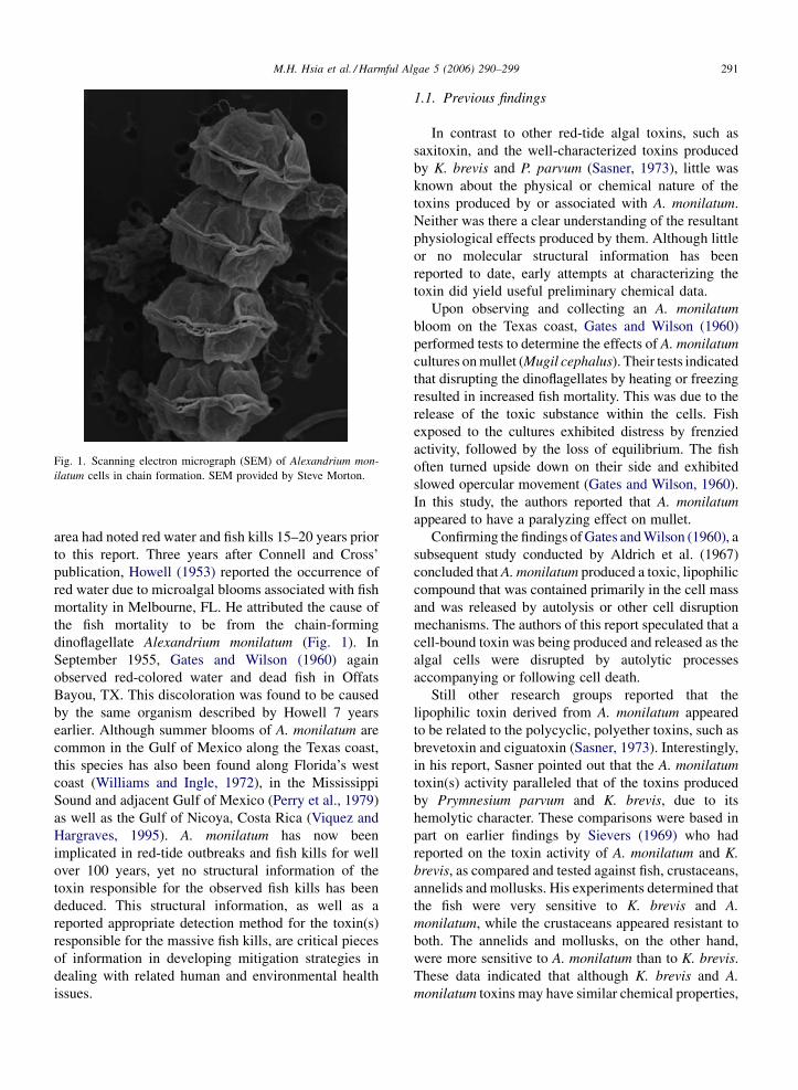
Fig. 1. Scanning electron micrograph (SEM) of Alexandrium mon-
ilatum cells in chain formation. SEM provided by Steve Morton.
area had noted red water and fish kills 15–20 years prior
to this report. Three years after Connell and Cross’
publication, Howell (1953) reported the occurrence of
red water due to microalgal blooms associated with fish
mortality in Melbourne, FL. He attributed the cause of
the fish mortality to be from the chain-forming
dinoflagellate Alexandrium monilatum (Fig. 1). In
September 1955, Gates and Wilson (1960) again
observed red-colored water and dead fish in Offats
Bayou, TX. This discoloration was found to be caused
by the same organism described by Howell 7 years
earlier. Although summer blooms of A. monilatum are
common in the Gulf of Mexico along the Texas coast,
this species has also been found along Florida’s west
coast (Williams and Ingle, 1972), in the Mississippi
Sound and adjacent Gulf of Mexico (Perry et al., 1979)
as well as the Gulf of Nicoya, Costa Rica (Viquez and
Hargraves, 1995). A. monilatum has now been
implicated in red-tide outbreaks and fish kills for well
over 100 years, yet no structural information of the
toxin responsible for the observed fish kills has been
deduced. This structural information, as well as a
reported appropriate detection method for the toxin(s)
responsible for the massive fish kills, are critical pieces
of information in developing mitigation strategies in
dealing with related human and environmental health
issues.
1.1. Previous findings
In contrast to other red-tide algal toxins, such as
saxitoxin, and the well-characterized toxins produced
by K. brevis and P. parvum (Sasner, 1973), little was
known about the physical or chemical nature of the
toxins produced by or associated with A. monilatum.
Neither was there a clear understanding of the resultant
physiological effects produced by them. Although little
or no molecular structural information has been
reported to date, early attempts at characterizing the
toxin did yield useful preliminary chemical data.
Upon observing and collecting an A. monilatum
bloom on the Texas coast, Gates and Wilson (1960)
performed tests to determine the effects of A. monilatum
cultures on mullet (Mugil cephalus). Their tests indicated
that disrupting the dinoflagellates by heating or freezing
resulted in increased fish mortality. This was due to the
release of the toxic substance within the cells. Fish
exposed to the cultures exhibited distress by frenzied
activity, followed by the loss of equilibrium. The fish
often turned upside down on their side and exhibited
slowed opercular movement (Gates and Wilson, 1960).
In this study, the authors reported that A. monilatum
appeared to have a paralyzing effect on mullet.
Confirming the findings of Gates and Wilson (1960), a
subsequent study conducted by Aldrich et al. (1967)
concluded that A. monilatum produced a toxic, lipophilic
compound that was contained primarily in the cell mass
and was released by autolysis or other cell disruption
mechanisms. The authors of this report speculated that a
cell-bound toxin was being produced and released as the
algal cells were disrupted by autolytic processes
accompanying or following cell death.
Still other research groups reported that the
lipophilic toxin derived from A. monilatum appeared
to be related to the polycyclic, polyether toxins, such as
brevetoxin and ciguatoxin (Sasner, 1973). Interestingly,
in his report, Sasner pointed out that the A. monilatum
toxin(s) activity paralleled that of the toxins produced
by Prymnesium parvum and K. brevis, due to its
hemolytic character. These comparisons were based in
part on earlier findings by Sievers (1969) who had
reported on the toxin activity of A. monilatum and K.
brevis, as compared and tested against fish, crustaceans,
annelids and mollusks. His experiments determined that
the fish were very sensitive to K. brevis and A.
monilatum, while the crustaceans appeared resistant to
both. The annelids and mollusks, on the other hand,
were more sensitive to A. monilatum than to K. brevis.
These data indicated that although K. brevis and A.
monilatum toxins may have similar chemical properties,

M.H. Hsia et al. / Harmful Algae 5 (2006) 290–299292
the mechanism of action by which these two toxins have
attributed, appears to be quite different (Sievers, 1969).
Earlier studies established that the main active toxic
compound(s) was not appreciably soluble in water
bearing a highly lipophilic nature. The toxin(s) appeared
to be primarily contained within healthy cells rather than
actively exuded into the environment. It became the goal
of this study to investigate and establish the molecular
nature of the toxic principles of A. monilatum. We report
the isolation and first unambiguous structural character-
ization of the toxin produced by A. monilatum.
2. Materials and methods
2.1. Isolation, identification and mass culturing of
A. monilatum
Samples from a ‘‘red-tide’’ of A. monilatum off
Gulfport, MS were collected by Dr. John Rogers (USEPA
Gulf Breeze, FL) and shipped to the NOAA Marine
Biotoxins Laboratory (Charleston, SC). Single cells were
isolated using the micro-pipette method of Guillard and
Morton (2003). Isolates were grown in modified L-1
medium (Guillard and Morton, 2003) at a salinity of
34 psu at 27 8C with 40 mE/(cm2 s) on a 16-h light:8-h
dark cycle. Seven isolates (AM01-07) were grown and
placed into the Marine Biotoxins Culture Collection.
All isolates were identified using both light micro-
scopy and scanning electron microscopy (SEM). For
light microscopy, an Olympus BX51 fitted with a
magnafire digital color camera was used. Plate
tabulation and visualization was achieved using 0.1%
Caulfour white fluorescent stain. For SEM, cells were
fixed with 1% gluteraldehyde and desalted using a
gradient of seawater to freshwater. Preparations were
dehydrated using a series of acetone and a series of
hexomethyldisilizane (HMDS). The samples were
coated with approximately 150 A of gold using a
Denton sputter-edge coater. SEM samples were imaged
on an environmental JEOL 5600LV.
Ten liter batch cultures of strain AM01 were grown
using 15 L Bellco m spinner flasks, fitted with a Teflon
impeller. Growth rates (Guillard, 1973) were calculated
by removing 1 mL of culture every 3 days. The number of
cells were counted using a Palmer–Maloney Counting
Chamber. All cultures were harvested at late log growth
phase, approximately 18 days after inoculation.
2.2. Cell harvest and extraction
A. monilatum cells were filtered from growth media
using a 25 mm sieve. Cells were lyophilized and horn
sonicated in 100 mL of ethyl acetate with a Branson
Sonifier 450 Horn Sonicator at full power for 15 min to
both disrupt the outer theca and ensure maximum
release of toxin from the cellular debris. An equal
volume of water was added after horn sonication for
solvent–solvent partitioning. Both layers were col-
lected, concentrated and tested for cytotoxicity using
GH4C1 rat pituitary cells and icthyotoxicity using
Sheepshead minnow. The layer with the greatest
toxicity was determined and taken on for further
purification.
2.3. GH4C1 rat pituitary cytotoxicity bioassay
(Mosmann, 1983)
GH4C1 rat pituitary cells were plated onto a 96-
well plate and allowed to incubate for 6 h to allow for
adherence to the plate. Dried fractions to be tested
were solubilized in 100 mL methanol of and sonicated
to ensure maximum solubility prior to carrying out
the bioassay. The assay was carried out by adding
4 mL (or smaller amount) of the methanol-solubilized
fractions to individual wells in the 96-well plate;
4 mL or less of pure methanol has been previously
tested to be safe to these cells (i.e., a negative
control). The plate was then allowed to incubate for a
minimum of 18 h; 15 mL of 3-(4,5-cimethylthiazol-2-
yl)-2,5-diphenyl tetrazolium bromide (MTT) dye was
added after incubation and allowed to incubate for
another 4 h before adding 100 mL of SDS to each
well. The plate was allowed to incubate for another
4 h prior to reading the assay.
2.4. Preparative thin layer chromatography (TLC)
The crude, concentrated ethyl acetate extract of A.
monilatum was loaded onto a Whatman Silica Gel
60A preparative TLC glass-backed plate and then
developed in a mixture of 7:3 ethyl acetate:benzene.
All colored and non-colored bands were scraped from
the plate, eluted with ethyl acetate, concentrated and
tested for cytotoxicity. These bioassays used metha-
nol as a carrier against the GH4C1 rat pituitary cells
and an MTT dye as determining the endpoint
(Mosmann, 1983). This procedure both established
the Rf value of the toxic compound and effectively
separated the toxin from contaminating pigments. A
modified molybidic acid stain (Stahl, 1965; H2SO4
added as modification-internal communication, Moel-
ler) was used in addition to cytotoxicity testing in
order to parallel activity of colored and non-colored
bands.

M.H. Hsia et al. / Harmful Algae 5 (2006) 290–299 293
Fig. 2. (a) ESI-MS of toxic extract; 791.2 indicates mass of extract in positive ion mode. (b) MALDI-TOF of toxic extract; 791.23 indicates mass of
extract in positive ion mode.

M.H. Hsia et al. / Harmful Algae 5 (2006) 290–299294
Fig. 3. (a) Proton NMR spectra of toxic extract in CDCl3. (b) Carbon NMR spectra (top) and APT NMR spectra (bottom) of toxic extract in CDCl3.
2.5. Mass spectrometry
Mass spectrometric detection of the A. monilatum
toxic extract was carried out using a number of mass
spectrometric techniques. Using a SL trap electrospray
ionization-mass spectrometry (ESI-MS) with an HPLC
interface, an ABI Voyager matrix-assisted laser
desorption ionization-time of flight-mass spectrometry
(MALDI-TOF-MS) with 2,5-dihydrobenzoic acid (2,5-
DHB) as a matrix and an ABI Voyager MALDI-TOF/

M.H. Hsia et al. / Harmful Algae 5 (2006) 290–299 295
TOF, purified toxic extract was tested in efforts to first,
fingerprint active fractions; secondly, to provide a non-
bioassay method to analytically detect and follow the
toxin derived from cellular extract(s); thirdly, to develop
an analytical technique to quantify the toxin where
necessary.
2.6. Liquid chromatography–mass spectrometry
(LC–MS)
Toxin identification and preparative toxin production
was augmented by using preparative LC–MS in the
reverse phase. Preparative toxin production was carried
out using an Agilent 1100 Purification Series LC/MSD
system equipped with a 9.4 cm � 50 mm, 5 mm, Zorbax
SBC18 column operating under APCI–MS conditions.
The following conditions were set for all LC–MS
experiments: drying gas rate at 5 L/min, gas temperature
at 300 8C, vaporizer at 325 8C, and corona voltage at
4000 V. Filtered A. monilatum toxic extract obtained
from preparative TLC was chromatographed using mass
based detection. Fractions of the appropriate mass were
collected using single ion monitoring in positive ion
mode. The Agilent system operated at a flow rate of
5 mL/min, using an injection volume of 500 mL. The first
purification procedure involved a gradient from 1:1
H2O:MeOH to 5:9.5 H2O:MeOH over 25 min. The
desired compound was collected, concentrated and dried
in preparation for the second LC–MS method. The
second purification LC–MS method followed the same
operations as above, but eluted the compound with an
isocratic method of 1:1 water:acetonitrile over 26 min.
The active compound was collected, concentrated and
dried in preparation for the third and final purification
step. This operation involved a step gradient using
acetonitrile and water over 30 min. The first 5 min
involved 9:1 water:acetonitrile, followed by 5 min of 3:1
water:acetonitrile, 5 min of 6.5:3.5 water:acetonitrile and
then 15 min at 1:1 water:acetonitrile.
2.7. Nuclear magnetic resonance (NMR)
Molecular structural analysis of the toxic compound
was carried out using a 500 MHz Bruker DMX NMR
equipped with a 5 mm triple nucleus gradient probe.
Purified sample (approximately 25 mg) was brought up in
CDCl3 as the lock solvent. The following NMR
experimental experiments were carried out to establish
molecular structure: 1H, 13C, APT, COSY (Neuhaus
et al., 1985), HSQC (Keeler and Neuhaus, 1985), HMBC
(Keeler and Neuhaus, 1985), and NOESY (Bax and
Davis, 1985a,b).
2.8. Accurate mass analysis
Accurate mass detection of the A. monilatum toxic
extract was performed using direct infusion of the
sample into a Waters Micromass Quadrupole Q-TOF
mass spectrometer operating with electrospray ioniza-
tion in positive ion mode. KBr and NH4 were added in
order to test for adduct formation. Chemical formulae
were calculated utilizing the elemental composition
tools within the Waters Masslynx software.
3. Results
3.1. Purification using preparative thin layer
chromatography
This method efficiently separated the toxic extract
from naturally occurring pigments also produced by
A. monilatum. This was an important step since co-
occurring peridinin and xanthophylls are cytotoxic in
high concentrations. Under this elution scheme, the
toxic component yielded an Rf value of 0.9, indicative of
its high lipophilicity. This upper-most band tested
positive against the GH4C1 rat pituitary cell line
cytotoxicity bioassay. A modified molybdic acid stain
was developed to highlight and detect both the active
and non-active non-colored/non-chromophoric bands in
efforts to develop an analytical technique to circumvent
the use of sample expensive bioassays. Non-colored
bands were clearly visible using this procedure. The
toxin itself was colorless and in pure state yielded no
UV chromophore under Agilent PDA detection
between 210 and 280 nm. It did, however, co-elute
with several green chlorophylls under the conditions
described.
The active compound(s) was eluted from the
preparative TLC plate and taken for further purification
and characterization on MS and LC–MS. The pre-
parative TLC method separated the active material from
most other organic soluble molecular components,
providing a large scale, time-efficient process for the
purification of the active toxin from the toxic pigments
produced by the dinoflagellate, A. monilatum.
3.2. Mass spectrometry of the toxic extract
The mass of the toxic extract (Fig. 2a and b) resulted in
a m/z for the toxic extract of 791.2 in positive ion mode on
both ESI-MS and MALDI-TOF-MS. The apparent mass
of 790 amu was used as an analytical tag for further mass
purification using LC–MS. With this MS method in hand,
it was possible to eliminate the need for bioassay

M.H. Hsia et al. / Harmful Algae 5 (2006) 290–299296
Table 1
Comparison of goniodomin A NMR data from Alexandrium monila-
tum and Alexandrium pseudogoniaulax
Alexandrium monilatum Alexandrium pseudogoniaulax
(Murukami et al., 1988)
Carbon # Carbon Hydrogen Carbon Hydrogen
1 168.5 168.1
2 76.6 4.53 76.2 4.309
3 140.3 139.7
4 112.7 4.82 112.1 4.779
4.98 5.002
5 41.1 2.26 40.7 2.331
2.76 2.82
6 70.6 3.83 70.2 4.133
7 80.7 3.55 80.2 3.683
8 73.5 4.81 73.1 5.133
9 149.3 148.6
10 110.8 4.96 110.4 4.933
5.05 5.177
11 34.1 2.63 33.6 2.537
12 20.4 1.35 20 1.412
1.35 1.412
1.35 1.412
13 44.6 1.70 44.1 1.73
2.19 2.095
14 100.6 101
15 150.6 150.1
16 107.8 4.95 107.5 4.675
5.03 4.933
17 27.92 1.82 27.5 1.948
1.92 1.948
18 25.7 1.47 25.4 1.17
2.04 1.33
19 76.9 3.68 76 3.686
20 76.5 3.86 76.5 3.818
21 27.95 2.34 27.5 1.55
2.34 1.55
22 123.6 5.81 123.2 5.659
23 129.6 5.97 129.1 6.277
24 76.8 4.14 76.3 4.39
25 81.8 3.86 81.4 4.03
26 29.9 1.29 29.9 2.226
1.29 2.226
27 31.8 1.78 31.4 1.55
2.34 2.14
28 79.7 5.04 79.1 5.203
29 147.2 147
30 114.3 5.16 113.4 5.039
5.16 5.039
31 81.8 3.86 81 4.073
32 73.2 3.68 72.8 3.93
Table 1 (Continued )
Alexandrium monilatum Alexandrium pseudogoniaulax
(Murukami et al., 1988)
Carbon # Carbon Hydrogen Carbon Hydrogen
33 32.1 1.94 31.8 2.14
2.75 2.976
34 135.4 6.16 135.1 6.453
35 123.8 5.59 123.3 5.866
36 74 5.71 73.5 5.962
37 98 97.5
38 41.3 1.25 40.9 1.329
39 13.2 0.93 12.7 0.98
0.93 0.98
0.93 0.98
40 31.3 1.70 30.8 1.664
41 20.4 0.93 20 0.74
0.93 0.74
0.93 0.74
42 34.3 1.35 34.2 1.17
1.53 1.17
43 61.1 3.70 60.6 3.56
3.95 3.902
Carbon count corresponds to Fig. 5.
cytotoxicity tests to monitor for the toxin. This technique
is amenable to all MS equipment in any lab.
3.3. Liquid chromatography–mass spectrometry of
the toxic extract
Three sequential purification experiments carried out
on the LC/MS preparative system resulted in highly
purified material suitable for structural analysis. In the
first purification method, the desired compound eluted
at 20–21 min. The second purification method eluted
the active compound in 9.5–10.5 min. The third step
eluted the active compound(s) at 13, 20–21, 26–27 min.
These three compounds, all exhibiting the same
nominal mass, were collected separately based on their
respective elution times. The 26–27 min fraction was
the major compound of interest and was taken on for
total structural analysis using NMR.
3.4. Nuclear magnetic resonance of the toxic
extract
The LC–MS purified sample resulted in a colorless
amorphous solid. Structural elucidation of this highly
toxic substance was accomplished by detailed data
analyses of a number of NMR experiments. The 1H,

M.H. Hsia et al. / Harmful Algae 5 (2006) 290–299 297
Fig. 4. (a) Accurate mass analysis of toxic extract; 807.3716 indicates the potassium adduct in positive ion mode. (b) Accurate mass analysis of toxic
extract; 786.4421 represents the ammonium adduct in positive ion mode.

M.H. Hsia et al. / Harmful Algae 5 (2006) 290–299298
13C, and APT (Fig. 3a and b) gave valuable information
regarding preliminary structural analysis and compound
class. These experiments clearly defined a polycyclic/
polyether type of molecule. Subsequent two-dimen-
sional NMR experiments provided the necessary
molecular connectivity information to establish the
molecular structure of the targeted toxin. 13C and HSQC
spectra revealed 43 signals representing 3 methyls, 14
methylenes, 19 methines, and 7 quaternary carbons, and
the existence of 7 double bonds in the molecule; 13C
chemical shift data are listed in Table 1.
This carbon and associated hydrogen count resulted
in some early confusion as the NMR data did not appear
to fit the observed mass at 790 amu. This issue was
resolved through the use of accurate mass analysis,
which provided information regarding the toxic
extract’s mass and empirical formula (see below).
3.5. Accurate mass analysis of analytically pure
compound
Addition of KBr to the purified sample resulted in a
potassium adduct with a m/z of 807.3716 in positive ion
mode (Fig. 4a). Confirmation of the potassium adduct
was established via the addition of an NH4 to the
purified compound. With the addition of ammonium, a
m/z of 786.4421 in positive ion mode was obtained,
clearly demonstrating adduct formation (Fig. 4b).
Purified samples subjected to accurate mass analysis
Fig. 5. Toxic compound isolated from Alexandrium monilatum:
goniodomin A.
demonstrated that the mass of 791.0 found from
previous ESI-MS and MALDI-TOF-MS experiments
was a sodium adduct with an accurate mass of 791.3974
in positive ion mode. The actual non-adduct molecular
ion was also observed at m/z 768. The best-fit formula
for the accurate mass that fit the NMR data were
C43H60O12. This formula defines 14 degrees of unsa-
turation, as corroborated in the NMR spectra. These
14 degrees of unsaturation are defined as 4 exocyclic
methylenes, 2 double bonds, 7 rings and 1 ester ca-
rbonyl (Fig. 5).
4. Discussion
By utilizing the combined NMR experiments, it was
possible to fully assign and structurally characterize a
polyether macrolide compound from A. monilatum. This
compound had a rare characteristic boasting of four
exocyclic methylenes. The structure is somewhat re-
miniscent of the spirolides, toxic compounds produced
by the dinoflagellate, Alexandrium ostenfeldii (Paulsen)
(Balech and Tangen, 1985). Established unambiguously
using high-field NMR and mass spectrometry, we now
report that A. monilatum produces the toxin goniodomin
A (Fig. 5), a toxin reportedly produced by the Japanese
rock pool dinoflagellate, Alexandrium pseudogoniaulax.
The NMR data obtained from the cultures are identical to
the macrolide toxin reported for this organism (Supple-
mentary material, Murukami et al., 1988). A comparison
of the 13C NMR signals is listed in Table 1. Pharma-
cological research carried out on this substance indicated
that goniodomin A has extreme effects on the reorganiza-
tion of the cytoskeleton. Abe et al. (2002) found that this
compound inhibited angiogenic properties of endothelial
cells partly by inhibition of actin reorganization. The
findings from Furukawa et al. (1993) suggested that
goniodomin A causes a conformational change resulting
in an enhancement of actomyosin ATPase activity at
concentration < 10�6 M, but conformational changes at
concentrations > 10�6 M may lead to a loss of ATPase
activity in rabbit skeletal muscle. This modulation
differed according to the type of cardiac myosin (Yasuda
et al., 1998), and the activation of skeletal actomyosin
ATPase was sensitive to the troponin/tropomyosin co-
mplex (Matsunaga et al., 1999). Other findings also
indicated that goniodomin A inhibited cell division in the
fertilized sea urchin (Murukami et al., 1988). In addition,
this compound demonstrates antifungal activity against
Candida albicans and Mortierella ramannianus (Mur-
ukami et al., 1988).
Previous reports by Clemons et al. (1980) and Bass
et al. (1983) have found evidence of a hemolytic
M.H. Hsia et al. / Harmful Algae 5 (2006) 290–299 299
principle associated with A. monilatum, but no toxin
was identified at the time. We did not test goniodomin A
against a hemolytic assay for this report. However, with
pure compound in hand, we plan on subsequent research
to establish whether or not this toxin exhibits hemolytic
activity.
5. Conclusions
We have found that A. monilatum produces the toxic
compound, goniodomin A. This toxin is identical to the
active compound produced by the Japanese rock pool
organism Alexandrium pseudogoniaulax. NMR data
chemical shifts match identically with those found by
Murukami et al. (1988). In the process of this research,
we have also developed highly sensitive mass spectro-
metric methods that are currently being optimized to
detect goniodomin A in marine matrices, such as
shellfish and mammal tissue. Other ongoing research
will focus on the algal–bacteria relationships as related
to toxin production.
Acknowledgement
This work was supported by NOAA/NOS. [SES]
Appendix A. Supplementary data
Supplementary data associated with this article can
be found, in the online version, at 10.1016/j.hal.2005.
08.004.
References
Abe, M., Inoue, D., Matsunaga, K., Ohizumi, Y., Ueda, H., Asano, T.,
Murakami, M., Sato, Y., 2002. Goniodomin A, an antifungal
polyether macrolide, exhibits antiangiogenic activities via inhibi-
tion of actin reorganization in endothelial cells. J. Cell. Physiol.
192, 109–116.
Balech, E., 1995. The Genus Alexandrium Halim (Dinoflagellata).
Sherkin Island Marine Station, Irealnd.
Balech, E., Tangen, K., 1985. Morphological and taxonomy of toxic
species in the tamarensis group (Dinophyceae): Alexandrium
evcavatum (Braarud) comb. nov. and Alexandrium ostenfeldii
(Paulsen) comb. nov. Sarsia 70, 333–343.
Bass, E.L., Pinion, J.P., Sharif, M.E., 1983. Charachteristics of a
hemolysin from Gonyaulax monilata Howell. Aquat. Toxicol. 3,
15–22.
Bax, A., Davis, D.G., 1985a. Practical aspects of two-dimensional
transverse NOE spectroscopy. J. Magn. Res. 63, 207–213.
Bax, A., Davis, D.G., 1985b. Mev-17 based two-dimensional homo-
nulcear magnetization transfer. J. Magn. Res. 65, 355–360.
Clemons, G.P., Pinion, J.P., Bass, E., Pham, D.V., Sharif, M., Wutoh,
J.G., 1980. A hemolytic principle associated with the red-tide
dinoflagellate Gonyaulax monilata. Toxicon 18, 323–326.
Connell, C.H., Cross, J.B., 1950. Mass mortality of fish associated
with the protozoan Gonyaulax in the Gulf of Mexico. Science 112,
359–363.
Furukawa, K., Sakai, K., Watanbe, S., Maruyama, K., Murakami, M.,
Yamaguchi, K., Ohizumi, Y., 1993. Goniodomin A induces mod-
ulation of actomyosin ATPase mediated through conformational
change of actin. J. Biol. Chem. 268, 26026–26031.
Gates, J.A., Wilson, W.B., 1960. The toxicity of Gonyaulax monilata
Howell to Mugil cephalus. Limnol. Oceanogr. 5, 171–174.
Guillard, R.R.L., Morton, S.L., 2003. Culture methods. In: Halle-
graeff, G.M., Anderson, D.M., Cembella, A.D. (Eds.), Manual of
Harmful Marine Microalgae. UNESCO Publisihing, France, pp.
77–97.
Howell, J.F., 1953. Gonyaulax monilata, sp. nov., the causative
dinoflagellate of a red tide on the east coast of Florida in
August–September, 1951. Trans. Am. Micro. Soc. 72, 153–156.
Keeler, J., Neuhaus, D., 1985. Comparison and evaluation of methods
for two-dimensional NMR spectra with absorption-mode line-
shapes. J. Magn. Res. 63 (3), 454–472.
Matsunaga, K., Nakatani, K., Murakami, M., Yamaguchi, K., Ohi-
zumi, Y., 1999. Powerful activation of skeletal muscle actomyosin
ATPase by goniodomin A is highly sensitive to troponin/tropo-
myosin. J. Pharm. Exp. Ther. 291 (3), 1121–1126.
Mosmann, T., 1983. Rapid colorimetric assay for cellular growth and
survival: application to proliferation and cytotoxicity assays. J.
Immunol. Methods 65, 55–63.
Murukami, M., Makabe, K., Yamaguchi, K., Konosu, S., 1988.
Goniodomin A: A polyether macrolide from the dinoflagellate
Goniodoma pseudogoniaulax. Tetrahedron Lett. 29 (10), 1149–
1152.
Neuhaus, D., Keeler, J., Freeman, R., 1985. Investigation of individual
proton spin multiplets by C-/H correlation spectroscopy. J. Magn.
Res. 61, 553–558.
Perry, H.M., Stuck, K.C., Howse, H.D., 1979. First record of a bloom
of Gonyaulax monilata in coastal waters of Mississippi. Gulf Res.
Rep. 6 (3), 313–316.
Sasner, J.J., 1973. Comparative studies on algal toxins. In: Martin,
D.F., Padilla, G.M. (Eds.), Marine Pharmacognosy: Action of
Marine Biotoxins at the Cellular Level. Academic Press, NY, p.
127.
Sievers, A.M., 1969. Comparative toxicity of Gonyaulax monilata and
Gymnodinium breve to annelids, crustaceans, mollusks, and a fish.
J. Protozol. 16 (3), 401–404.
Stahl, E., 1965. In: Stahl, E. (Ed.), Thin-Layer Chromatography: A
Laboratory Handbook. second ed. Academic Press Inc. Publishers,
NY.
Viquez, R., Hargraves, P.E., 1995. Annual cycles of potentially
harmful dinoflagellates in the Golfo de Nocoya, Costa Rica. Bull.
Mar. Sci. 57 (2), 467–475.
Williams, J., Ingle, R.M., 1972. Ecological notes on Gonyaulax
monilata (Dinophyceae): blooms along the west coast of Florida.
Florida Dep. Nat. Resour. 1 (5), 1–12.
Yasuda, M., Nakatani, K., Matsunaga, K., Murakami, M., Momose,
K., Ohizumi, Y., 1998. Modulation of actomyosin ATPase by
goniodomin A differs in types of cardiac myosin. Eur. J. Pharma-
col. 346, 119–123.