Proceeding Book - RP2U Unsyiah
198
Proceeding Book th Proceeding book of the 49 Pokjanas TOI International Seminar
-
Upload
khangminh22 -
Category
Documents
-
view
3 -
download
0
Transcript of Proceeding Book - RP2U Unsyiah

Proceeding Book
thP
roce
edin
g b
ook o
f the 4
9 P
okja
nas T
OI In
tern
atio
nal S
em
inar

Proceeding Book:
The 49th Pokjanas TOI International Seminar
ISBN: 978-602-72418-2-4
Published : 2016
Advisory Team
Rector of Pancasila University
Prof. Dr. rer. nat. Wahono Sumaryono, Apt.
Dean of Pharmacy Faculty Pancasila University
Prof. Dr. Shirly Kumala, M.Biomed., Apt.
Editor Chief
Yesi Desmiaty, S.Si., M.Si., Apt.
Editorial Board Member
Prof. Dr. rer. nat. Wahono Sumaryono, Apt.
Prof. Dr. Shirly Kumala, M.Biomed., Apt.
Prof (ris). Swasono R. Tamat, M.Sc., Ph.D., Apt.
Prof (ris). Dr. Partomuan Simanjutak, M.Sc., APU
Prof. Dr. Syamsudin, M.Biomed., Apt.
Redactional Board Member
Sesilia Andriani Keban, MSi., Apt.
Mita Restinia, M.Farm., Apt
Retno Ayu Pratiwi, S.Si.
Publisher
Faculty of Pharmacy, Pancasila University
Srengseng Sawah, Jagakarsa, Jakarta 12640
Phone/ Fax (021)7864727-28/ 23

i
PREFACE
The 49th Pokjanas TOI International seminar has been scheduled organized by the Faculty of Pharmacy University Pancasila in collaboration with Pokjanas TOI Organization at Jakarta, Indonesia, 21-22 October 2015. Which is aimed to share information, findings and collaboration between researches, pharmacists, institution and natural product industries. Finally we were able to publish the proceedings and it is now ready for circulation among the researchers, industries, and scientists. This proceeding is consisted of 43 titles manuscripts which were presented as oral and poster in seminar. The topic of manuscript contain many fields including natural product chemistry, analytical technique in phytochemistry, biological activity, pharmacological study, herbal drugs and formulation.. The Organizing Committe gratefully acknowledges the Rector of University Pancasila, Pokjanas TOI Organization, as well us all sponsors in bringing forth this seminar. Furthermore, personally, I would like to express my deep apreciation to the members of the Organizing Committee, for the good teamwork and their great effort to bring success to the seminar.
Jakarta, November 2015
Chairman of Committee Dr. Ratna Djamil M.Si., Apt

ISBN : 978-602-72418-2-4
International Seminar Pokjanas TOIFaculty of Pharmacy Pancasila University
ii
DAFTAR ISI
Halaman
Citotoxicity and Radical Scavenging Activity Test of Gambir (Uncaria gambir (HUNTER)ROXB.) In VitroSri Ningsih, Churiyah, Fahri Fahrudin, Rini Damayanti, Eriawan Rismana …………………... 1
The Biological Activity of Eurycomanone Derivatives On T47d, MCF-7, HELA, and WIDRCancer CellsHanifah Yusuf, Darma Satria, Zulkarnain ……………………………………………….......... 6Antibacterial Activities of Dayak Paser Medicinal Plants Against Escherichia ColiSeptina Asih Widuri, Noorcahyati ………………………………………………………........... 11
Isolation of Anticancer Active Compound From Trigonella Foenumgraecum Leading by MCF7CytotoxicityKurnia Agustin, Sriningsih, Julham Effendi …………………………………………………… 16
Issues of Halal Standardization of Food, Drug and Cosmetic for the Implementation theMandatory of Halal Certification According to Halal Product GuaranteeM. Yanis Musdja ……………………………………………………………………………….. 21Virtual Screening Compounds in Fabaceae Plants as Ligands on Alfa Estrogen Receptor (ER-α)Esti Mumpuni, LH Gulo ……………………………………………………………………….... 26
Preparation of Standardized Aqueous Extract of Annona Muricata Linn. Leaf, Its Potency asAntioxidant, and Total Flavonoid Content AssayYesi Desmiaty, Deni Rahmat, Nilam Sari Maulidina …………………………………….............. 30Phytochemical Screening and Toxicity Test BSLT of 70 % Ethanol Extract of Gaharu Leaves(Aquilaria beccariana Tiegh.)Ahmad Musir, Wiwi Winarti, Siti Hasnah P. Siregar …………………………………………...... 34Optimization of Production of Β-Carotene and Astaxanthin from Microalgae Chlorellapyrenoidosa and Its Potential as an AntioxidantNi Wayan Sri Agustini …………………………………………………………………………... 40Antioxidant Compound Isolated from Bioproduction of Endophytic Fungi of Turmeric(Curcuma longa L.)Hindra Rahmawati, Partomuan Simanjuntak …………………………………………………....... 45Analysis of Beta-Carotene In Green Melon and Orange Melon (Cucumis melo L. var. sky rockand var. cantaloupe) by TLC-DensitometryRifa Rizkiyah, Zuhelmi Aziz …………………………………………………………………....... 49Spectrophotometric Method Precision to Assay of Lycopene in Tomatoes Fruit (Solanumlycopersicum Lam.)Liliek Nurhidayati, Wening Ariwanty …………………………………………………....………. 54Optimization and Validation of High Performance Liquid Chromatography for Determinationof Coffein In White TeaZuhelmi Aziz, Dhiah Resti …………………………………………………………………....…... 58The Effect of Extraction Method on Total Alkaloid Levels of Jembirit Leaves(Tabernaemontana sphaerocarpa BL) with Spectrofotometric MethodNina Salamah, Miftahul Rozak ………………………………………………………………....… 62Integration of Herbal or Traditional Medicine through Evidence Based PracticeAnny Lumban Toruan, Galih Ajeng Kencana Ayu ……………………………………................. 69

ISBN : 978-602-72418-2-4
International Seminar Pokjanas TOIFaculty of Pharmacy Pancasila University
iii
Identification of Soursop Seeds (Annona muricata L.) Extract as a Candidate Against the Aedesaegypti L. Musquito Vector Control DBDSarah Zaidan, Ratna Djamil, Siti Nuraini ……………………………………………………...... 74Antimicrobial and Biology Activity from Parasite Soursop (Dendropthoe pentandra L.)Extract HerbsErlindha G, Lia Kartika Sari …………………………………………………………..………...... 81Antioxidant, Cytotoxic and Apoptotic Induction Activity of Ethanolic Extract of Andrographispaniculata on MCF-7 Cancer Cell LineChuriyah, Kurnia Agustini, Siska Andrina Kusumastuti ................................................................. 85In Vitro α-Glucosidase Inhibition Activities Test from Standardized Sambung Nyawa (Gynuraprocumbens (Lour.) Merr.) Leaves ExtractWiwi Winarti, Ratna Djamil, Sarah Zaidan, Raymond …………………………………….....….. 90Identification of Sugar-Apple Seeds (Annona squamosa L.) Extract as a Candidate Against theAedes aegypti L. Musquito Vector Control DBDRatna Djamil, Sarah Zaidan,Siti Nuraini ……………………………………………………......... 94Anticancer Activity of Jatropha SP. on Breast Cancer Cells and Cervix CellsSiti Rofida, Nailis Syifa, Nurkhasanah, Laela Hayu Nurani …………………………………........ 102
Antihyperlipidemia Effect of Red Cabbage Juice (Brassica Oleracea VAR CAPITATA L.FORMA RUBRA) in MiceLestari Rahayu, Yati Sumiyati, Desti Dwi Nandini ……………………………………………..... 108Hepatoprotective Study of Cosolvent Solution from Mangosteen (Garcinia mangostana L.)Rind in RatsRos Sumarny, Liliek Nurhayati, Yati Sumiyati, Astri Yuliastri Permana ………………............... 112Immunomodulatory Activitiy of Lutein Extract from Sweet Corn Seeds (Zea mays L.) ThroughIn Vivo Measurement of Activity and Phagocytic Capacity of Peritoneal Macrophage Cells ofMiceKusmiati, Yudha Prasetya, Erlindha Gangga …………………………………………………....... 115Effect of Ethanolic Extract of Bawang Tiwai (Eleutherine bulbosa (Mill.) Urb.) in MonosodiumUrate-Induced Inflammation in Rat Hind PawDian R. Laksmitawati, Siti R.Rani ……………………………………………………................... 120Acute Toxicity of Ethanolic Extract of Fenugreek Seeds (Trigonella foenum-graecum L.) onWhite RatsKurnia Agustini, Sriningsih, Julham Effendi ................................................................................... 124Antihypertensive and Diuretic Effects of the Ethanol Extract of Colocasia esculenta (L.) Schott.LeavesRini Prastiwi, Siska, Ervina Bhakti Utami, Gigih Pangestu Witji ……………………………....... 128The Antioxidant Activity of Ethanol Extract of White-Oyster Mushroom in Decrease MDA andIncrease the Activity of Catalase in Mice HypercholesterolemiaVera Ladeska, Priyanto,Juju Jumiati …………………………………………………………........ 133Soursop Leaf (Annona muricata L.) Infusion in Lipid Profile of Hyperlipidemic MiceNi Made Dwi Sandhiutami, Neni Anggraini ……………………………………………….…....... 141Xanthine Oxidase Inhibitory Activity of Secang (Caesalpiniasappan L.), Tempuyung(Sonchusarvensis L.), and Kepel (Stelechocarpusburahol) ExtractsPertamawati, Mutia Hardhiyuna, Shelvi Listiana, Rian Triana ………………………………....... 146Potency of Curcuma Mangga Val Rhizome Extract as a Selective Anti-Proliferative Agent onBreast Cancer Cell Line MCF-7Siska Andrina, Churiyah, Nuralih ……………………………………………………………….... 151Assessment of Antibacterial Activity of Herbal Toothpastes to the Bacteria Causing HalitosisSyarmalina, Syahdu A. Ekowati dan Dwi A. Maulana ………………………………………...... 155

ISBN : 978-602-72418-2-4
International Seminar Pokjanas TOIFaculty of Pharmacy Pancasila University
iv
Formulation and the Antioxidant Activity of Green Cincau Leaves (Cyclea barbata L.MIERS)from the Ethanol Extract 70%Yunahara Farida, Erlindha Gangga, Kartiningsih, Arsila …………………………….................... 158Accelerated Stability Test and Antioxidant Activity of Ethanol Extract Green Cincau Leaves(Cyclea barbata L.Miers) with Gelling Agent HPCM AND HPMCKartiningsih, Erlindha Gangga, Yunahara Farida, Maria Ulfah …….....………………….……… 162Capsule Formulation of Standardized 70% Ethanol Extract Johar Leaves (Senna siamea (Lam.)Irwin and Barneby) as α-Glucosidase InhibitorRisma Marisi Tambunan, Kartiningsih, Everly Hendra ………………..................………….…… 166Formulation and Evaluation of Herbal Tablets Containing Voacanga foetida (Bl.) K.SchumExtractFahleni, Yandi Syukri, Novelta Femmy Rischa, Adriani Susanty ………………….................…..
172
Optimization of Patchouli Oil and Tea Tree Oil Emulgel FormulationYuslia Noviani, Teti Indrawati, Shelly Taurhesia ……………………………………....................
176
Formulation of Liquorice Extract (Glycyrrhiza glabra L) as Skin Whitening CreamSiti Umrah Noor, Faridah, Michico …………………………………………….………................
180
Accelerated Stability Test of Liquorice Extract (Glycyrrhiza glabra L) CreamFaridah, Siti Umrah Noor, Sulih Probo Sindi ………………………………….................……….
187
The Variation of Tofu’s Wastewater Concentrations as Culture Medium to the Protein, Lipidand Chlorophyl Contents from Microalgae Nannochloropsis sp.Rian Nurul Hidayat, Sudjaswadi Wiryowidagdo, Ni Wayan Sri Agustini ……….…….................
193
Utilization of Corn Husk Waste to Produce Cellulase Enzymes by Trichoderma viride FNCC6013Mira Andam Dewi, Ririn Puspadewi, Sylvia Heryanti ………………………………..................
199
Clinical Trials Efficacy Of Hyperglycemia Herbs FormulaAgus Triyono, Zuraida Zulkarnain ……………………………………………….…….................
207
Antihiperkolesterolemia Jamu Formula Effect on Plasma Cholesterol Levels in Patients withMild Hypercholesterolemiam in Rumah Riset Jamu 'Hortus Medicus' TawangmanguZuraida Zulkarnain, Agus Triyono ……………………………………………….……................
211
ISBN : 978-602-72418-2-4
International Seminar Pokjanas TOIFaculty of Pharmacy Pancasila University
1
ISSUES OF HALAL STANDARDIZATION OF FOOD, DRUG ANDCOSMETIC FOR THE IMPLEMENTATION THE MANDATORY OF
HALAL CERTIFICATION ACCORDING TO HALAL PRODUCTGUARANTEE NUMBER 33 YEAR 2014
Dr. Muhammad Yanis Musdja, M.ScEmail : [email protected]
Former of Head of Study Program of Pharmacy, Islamic State University, JakartaFormer Dean of School of Pharmacy Muhammadiyah, Tangerang
Chairman of Indonesian Halal Products Foundation
ABSTRACT
INTRODUCTION: Indonesian parliament has ratified law of the Halal Products Guarantee (LHPG)number 33 year 2014 (Undang-undang Jaminan Produk Halal No. 33 Tahun 2014) on September 25,2014 ago. The basic principles on LHPG No. 33 Year 2014 is the change of halal certificate fromvoluntary becomes mandatory for food, drug and cosmetic, which will begin in 2017 and will beimplemented gradually in Indonesia. For the implementation of halal certification mandatory, theIndonesian government does not yet have halal standardization, especially for drugs and cosmetics, andalready there are some standardization of halal for food made by Majelis Ulama Indonesia (MUI).OBJECTIVES: How to prepare Halal Standardization for the implementation of LHPG No. 33 Year2014, so that mandatory halal certification for food, drug and cosmetic can be implemented inIndonesia. ANALYSIS ISSUES AND CHALLENGES: Halal standardization must exist in order toimplement of LHPG No. 33 Year 2014. To be able to make halal Standardization, necessary Muslimpharmacists and other experts that so much know halal Standardization. Based on the SWOT theory, itcan be said briefly about: STRENGTHS; Islamic concept for consuming Halalanthoyyiban food is thebest concept not only for Muslims but for all mankind. WEAKNESS: Until now, Indonesia does nothave halal standardization of food, drug and cosmetic. OPPORTUNITIES: Make halal standardizationis a good opportunity to make the food, drug and cosmetic that qualified in accordance withhalalanthoyyiban concept in Islam. For make halal standardization can do verification food ingredientsthat exist on the Codex Alimentarius, Pharmacopoeia and standards books for food, drugs and cosmetics.THREATHENS: On Article 56 and 57 of this LHPG, there are severe legal sanctions for IndustryPlayer and everyone that involved in the implementation of process Halal Products Guarantee shall bepunished with imprisonment for a maximum of 5 (five) years or a fine of Rp 2,000,000,000.00, (twobillion rupiah), if there is no halal standardization, the article No. 56 and 57 of LHPG No. 33, year 2014can not be executed. CONCLUSION: Halal standardization should be made as soon as possible, if it isnot exist. LHPG No. 33, year 2014 can not be implemented. The easiest way to create a standardizationof halal with a faster time, low cost and easily understood and accepted by the international community iswith do clarification about halal, mubaah, makruh or haram of material content in the book CodexAlimentarius for food and the book Martindale & Indonesia Pharmacopoeia for drugs and cosmetic.
Key Words: Halal food standardization; Halal drugs standardization; Halal cosmeticsstandardization; LHPG No. 33 year 2014; Muslim pharmacists law experts.
ISBN : 978-602-72418-2-4
International Seminar Pokjanas TOIFaculty of Pharmacy Pancasila University
2
INTRODUCTION
As it is said in the Quran on surah Al Baqarah verse 168, “O mankind, eat from whatever is on earththat is lawful and good (Halalan thayyiban) and do not follow the footsteps of Satan. Indeed, he is toyou a clear enemy”. Verse in the Qur'an mentioned above, emphasize, that consuming of halal food andgood (Halalanthoyyiban) is an obligation not just for Islamic people but for all mankind. As has beenshown, according to evidence based medicine, that due to consuming of Haram foods, such as, alcohol,pork, beast animal, blood, disgusting animal and animals were slaughtered in a way not true, all of thisharam food will have an adverse effect to the people who had consumed it. On the other hand, in Quran,Surah Al A’raaf Verse 31, “O Children of Adam, take your adornment at every place of worship, andeat, and drink, but do not overdoses. Certainly, He (Allah) loves not the extravagant” The meaning ofeat and drink do not overdoses in this verse is Standardization. Therefore, eat food, drugs and cosmeticsare not standard is dangerous and may disturb of health.Because of standardization of food, drugs and cosmetics are mandatory in Islam, then this provision is setin law of the Halal Products Guarantee (LHPG) number 33 year 2014 (Undang-undang Jaminan ProdukHalal No. 33 Tahun 2014)
On the other hand at Article 56 of LHPG No. 33 Year 2014: Industry Player that does not keep halalproducts have gained the Halal Certificate referred to in Article 25 letter b shall be punished withimprisonment for a maximum of 5 (five) years or a fine of Rp 2,000,000,000.00 (two billion rupiah ). Atarticle 57 of LHPG No. 33 Year 2014: Everyone involved in the implementation of process HalalProducts Guarantee that is not maintain the confidentiality of formulas that contained in information issubmitted by industry player as referred to in Article 43 shall be punished with imprisonment for amaximum 2 (two) years or a maximum fine Rp. 2,000,000,000.00 (two billion Rupiah).
Due to the fairly heavy sanctions to industry players and Everyone involved in the implementation ofprocess Halal Products Guarantee, then, there will be many disputes in the implementation of mandatoryHalal in the future. To be able to resolve the dispute with justice certainly needed Halal Standardizationfor food, drugs and cosmetics. If standardization of halal for food, drugs and cosmetics can not bedetermined, the implementation of the LHPG number 33 year 2014 will fail.
On one side, standards are essential tools for local and international businesses, which shape thecontribution of economic progress through industry development and trade, as well as, a guideline in theassurance of consumer protection. On the other hand, standards are able to be eliminators of tradebarriers, which means that, they play a critical role to facilitate goods and services exchange acrossborders. Orriss and Whitehead (2000),
On the other hand, Halal logistics is a new phenomenon, driven by the halal industry to extend halalfrom source to the point of consumer purchase, to ensure the integrity of the halal product for the end-consumer and export markets. The large discussion group shows that the conventional logisticshandling of halal products does not provide sufficient assurance for the Muslim consumer in bothMuslim and non-Muslim countries. Tieman, M. (2011).

ISBN : 978-602-72418-2-4
International Seminar Pokjanas TOIFaculty of Pharmacy Pancasila University
3
METHODS
Make a compilations or attachment to the standard book for food, drugs and cosmetics by doing theclarification of these materials are halal, mubaah, makruh or haram with make the selection of a fewbooks, to obtain book which more appropriate for clarification.Clarification of these materials carried out by Majelis Ulama Indonesia (MUI) in cooperation withexperts of Pharmacy and other experts who are familiar with standardization of halal.
RESULTS
Results of the main choice of books to make a compilation or an appendix on halal standards are:1. Codex Alimentarius new edition for food2. Martindale new edition and Indonesian Pharmacopoeia new edition for drugs and cosmetics
DISCUSSION
Make books for the standardization of halal food, drugs and cosmetics are a very difficult job, need along time and need very expensive cost.If we take a lesson from the history of the making of books Codex Alimentarius, the present, it takesabout 52 years to become a book as it is now. The making of this book are result of synergy of severalcountries. Which is regulated by the Food Administraion Organization (FAO)The Codex Alimentarius (Latin for "Book of Food") is a collection of internationally recognizedstandards, codes of practice, guidelines and other recommendations relating to foods, food productionand food safety.The Codex Alimentarius covers all foods, whether processed, semi-processed or raw. In addition tostandards for specific foods, the Codex Alimentarius contains general standards covering matters such asfood labeling, food hygiene, food additives and pesticide residues, and procedures for assessing the safetyof foods derived from modern biotechnology. It also contains guidelines for the management of officiali.e. governmental import and export inspection and certification systems for foods.
Advantages the using of result clarification (halal, mubaah, makruh and haram) ingredients ofCodex Alimentarius book as Halal Standart for food: Indonesia can make a book that integration Halal Standart with Thayyiban Standart becomeHalalanthayyiban Standart Readily accepted by every country and the international community, because the CodexAlimentarius is the main book used as a standard book for food by countries that are members of FAOand WHO. Provide substantial economic benefits, because Indonesia does not need to spend a huge cost toconduct research by spending big to establish halal Standart Indonesia only needs to perform the clarification of ingredients which foods are halal, mubaah,makruh and haram in the Codex Alimentarius and add it as a compilations or attachment in the book.ofcodex alimentarius. Halal standart that exist on compilations or attachment on the codex alimentarius will easily beused as a manuals halal standards by any countries in the world, therefore the book codex alimentarius isthe worldwide for food standard thayyiban

ISBN : 978-602-72418-2-4
International Seminar Pokjanas TOIFaculty of Pharmacy Pancasila University
4
Advantages the using of result clarification (halal, mubaah, makruh and haram) ingredients ofMartindale book as Halal Standart for drugs and cosmetics, because of Martindale bookcontains : Monographs on drugs and ancillary substances, listing over 6,000 monographs arranged in 49chapters based on clinical use with the corresponding disease treatment reviews. Monographs summarizethe nomenclature, properties, and actions of each substance. A chapter on supplementary drugs and othersubstances covers some 1190 monographs on new drugs, those not easily classified, herbals, and drugsno longer clinically used but still of interest. Monographs of some toxic substances are also included. Preparations - including over 180,000 items from 43 countries and regions, including China. Directory of Manufacturers listing some 20,000 entries. Pharmaceutical Terms in Various Languages: this index lists nearly 5,600 pharmaceuticalterms and routes of administration in 13 major European languages as an aid to the non-native speaker ininterpreting packaging, product information, or prescriptions written in another language. General index: prepared from 175,000 entries it includes approved names, synonyms andchemical names; a separate Cyrillic section lists nonproprietary and proprietary names in Russian andUkrainian.
Advantages the using of result clarification (halal, mubaah, makruh and haram) ingredients ofIndonesia Pharmacopoeia as Halal Standart for drugs and cosmetics, because of IndonesiaPharmacopoeia book :
Indonesia Pharmacopoeia is book standard used by the Indonesian people for drugs andcosmetics. Indonesia Pharmacopoeia using Indonesian language. therefore it is easily understood by thepeople of Indonesia.
On the other hand, at this time. There is one institution that made under Organization for IslamicCooperation (OIC). The name of institution is Standardization and Metrology of Industry for IslamicCountry (SMIIC). Headquarters of SMIIC is located in Istanbul turkey. Because of this new institutionwas founded in 2011 ago. then they also have not been able to make halal standardization manual thatcan be used as a standard for food, drugs and cosmetics. Then, Indonesia has not been able to use thehalal standard from the SMIIC.
To be able to do the work above, should be started immediately. Therefore clarification halal, mubaah,makruh and haram for thousands of food drugs and cosmetics is not easy. Therefore, there are someingredients of food, drugs and cosmetics that are difficult to set their status.
5. CONCLUSION1. To be successful implementation of law of the Halal Products Guarantee (LHPG) number 33 year2014, where its implementation will begin in early 2017. Then, Halal standardization should be made assoon as possible, if it is not exist. LHPG Number 33, year 2014 can not be implemented.
2. The easiest way to create a standardization of halal with a faster time, low cost and easily understoodand accepted by the international community is with do clarification about halal, mubaah, makruh orharam of material content in the book Codex Alimentarius for food and the book Martindale &Indonesia Pharmacopoeia for drugs and cosmetic.
ISBN : 978-602-72418-2-4
International Seminar Pokjanas TOIFaculty of Pharmacy Pancasila University
5
REFERENCES
1. Yanis Muhammad, Musdja, Majalah SWARA FBN, Edisi 1, Juni – Juli 2015, ISSN: 2443-1982,Bahaya Mengkonsumsi Makanan Haram Ditinjau dari Aspek Ilmiah Medis (Harmful ofConsuming Food Based on Evidence Based Medicine)
2. Law of the Halal Products Guarantee (LHPG) number 33 year 2014 (Undang-undang JaminanProduk Halal No. 33 Tahun 2014).
3. Al Quran, Terjemahan Kementerian Agama RI 2003.4. Orriss, D. G. and J.A. Whitehead, 2000. Hazard Analysis and Critical Control Point (HACCP) as a
Part of an Overall Quality Assurance System in InternationalFood Trade. Food Control, 11: 345-351
5. Tieman, M. (2011). The application of Halal in supply chain management: in -depthinterviews. Journal of Islamic Marketing, 2(2), 186 – 195.
6. CODEX Alimentarius: Understanding Codex". FAO and WHO. 1999. Retrieved 6 September 20127. Martindale : The Complete Drug Reference Thirty-eighth edition.8. Farmakope Indonesia edisi V, Tahun 2014.9. OIC/SMIIC 2:2011, “Guidelines for Bodies Providing Halal Certification” (with the references of
ISO/IEC 17020, ISO/IEC 17021, ISO/IEC 17025, ISO/TS 22003 + Islamic Fiqh Rules),

ISBN : 978-602-72418-2-4
International Seminar Pokjanas TOI
Faculty of Pharmacy Pancasila University
6
THE BIOLOGICAL ACTIVITY OF EURYCOMANONE DERIVATIVES
ON T47D, MCF-7, HELA, AND WIDR CANCER CELLS
1Hanifah Yusuf,
2Darma Satria,
3Zulkarnain
1Departement of Pharmacology and Therapeutic Faculty of Medicine University of Syiah Kuala,
Tanoh Abee Street, Darussalam, Banda Aceh, Indonesia 2Departement of Pathology Anatomi Faculty of Medicine University of Syiah Kuala, Tanoh
Abee Street, Darussalam, Banda Aceh, Indonesia 3Departement of Physiology Faculty of Medicine University of Syiah Kuala, Tanoh Abee Street,
Darussalam, Banda Aceh, Indonesia Email: [email protected]
ABSTRACT
Eurycomanone from the roots of Eurycoma longifolia Jack, has been reported to exhibit anticancer
activity. Four of ester eurycomanone derivatives: eurycomanone dibutyrate, eurycomanone
monovalerate, eurycomanone dimethoxybenzoate and eurycomanone disuccinate were synthesized for
knowing their potencies on cancer cell lines T47D, MCF-7, Hela, WIDR and normal cells (Vero cells).
The activity of eurycomanone derivatives as anticancer were evaluated by MTT colorimetric assay
method. The results showed that eurycomanone has anticancer activity on T47D, MCF-7, Hela, WIDR
cancer cells with IC50 values (1.17 ± 0.09; 3.96 ± 0.02; 2.95 ± 0.08;1.45 ± 0.01 µg/mL), respectively and
no toxic to Vero cells (IC50 609.89± 29.77 µg/mL). Eurycomanone derivatives namely: eurycomanone
dibutyrate have anticancer activity on T47D, MCF-7, Hela, WIDR cancer cells with IC50 values (25.16±
2.25; 21.56 ± 4.55; 29.32 ± 1.25; 149.42 ± 12.50 µg/mL), eurycomanone monovalerate (25.59 ± 1.31;
22.48 ± 1.25; 30.14 ± 1.89; 91.88 ± 8.90 µg/mL), eurycomanone dimethoxybenzoate ( 102.77 ± 2.56;
38.83 ± 2.55; 66.65 ± 1.90; 51.61 ± 2.37 µg/mL), eurycomanone disuccinate (218.94 ± 9.30; 198.87±
5.50; 166.67 ± 12.34; 145.39 ± 6.67 µg/mL)respectively. Eurycomanone dibutyrate, eurycomanone
monovalerate and eurycomanone dimethoxybenzoate are safe to Vero cells with selectivity index (IS)
more than 3, besides that eurycomanone disuccinate toxic to Vero cells.
Keywords: Anticancer, Eurycomanone, Eurycoma longifolia Jack, Synthesis
INTRODUCTION
Natural compounds from plants have been proven as a source of lead compounds for developing of new
drugs (1,2
). Eurycomanone is a natural pentacyclic quassinoid obtained from the roots of Eurycoma
longifolia Jack (3) in the family Simaroubaceae. Eurycoma longifolia Jack (ElJ) or commercially known
as Tongkat Ali in Malaysia, Pasak Bumi in Indonesia, Tung Saw in Thailand and Cay Ba Binh in
Vietnam (4) is very popular plant as aphrodisiac and usually taken after 4 years of cultivation for
preparing pharmaceutical products(5). Various natural compounds have been isolated and characterized
from ElJ, mostly from the roots. Natural compounds from ElJ have been reported to have a wide
pharmacological activities such as antimalarial (6,7,8
), anticancer (6,7,8,9,10
), antipyretic (10
) and anti ulcer
(11
). Eurycomanone is one of the major natural quassinoids isolated from the roots of ElJ and has
exhibited cytotoxic activities against selected cancer cell lines (12,13
). Its pharmacological potency has
been proven in numerous in vitro and in vivo experimental laboratory. But until now very limited efforts
to develope this compound for obtaining its derivatives. Therefore this investigation was conducted to
examine the anticancer activity of synthesized eurycomanone derivatives on selected cancer cells line.
In this study, design and synthesis of eurycomanone derivatives were done by using eurycomanone
natural compound and esterified without using protecting agent by using pharmacophore agents such as
butirylchloride, valeroylchloride, para methoxy benzoyl chloride and succinate anhydride.

ISBN : 978-602-72418-2-4
International Seminar Pokjanas TOI
Faculty of Pharmacy Pancasila University
7
METHODS
Materials: The plant and roots of E. longifolia Jack were taken after 4 years of cultivation and identified
by a specialist. Pharmacophore agents such as butirylchloride, valeroylchloride, para methoxy benzoyl
chloride and succinate anhydride (p.a, Merck). Chloroform, ethyl acetate, methanol, pyridine, all
chemical substances and selective cancer cell lines for study of anticancer activity
Extraction:
The roots (10kg) of E. longifolia were cleaned with tap water and then dried in the oven at 400C. After
cutting in small pieces, the dried roots were ground into crude powder and stored in the desiccators. Then
the crude powder was soaked in 30L methanol at room temperature and stirred regularly. The liquid
extract was filtered and concentrated in rotary evaporator at 400C to produce methanolic extract.
Isolation of eurycomanone
Before isolation the methanolic extract of ElJ was subjected to vacum liquid chromatography by using
stationary phase silica gel and the mixed mobile phase chloroform: methanol: water in ratio (5:5:1; 3:7:1:
1:9:1). Fractions with similar Rf values on thin layer chromatography (TLC) which were monitored at
UV lamp at 254 nm, then pooled and used for isolation of eurycomanone as starting material for
synthesizing its derivatives. Isolation of eurycomanone was done by preparative thin layer
chromatography (PTLC) using silica gel PF254 as stationary phase and the mixed mobile phase
ethylacetate: methanol : water in ratio 80: 20: 1. Repeated isolation, purification and crystallization were
done to get the pure compound. The structure and its purity were confirmed by comparison with detailed
spectroscopic data in published reports (UV, IR, NMR, LCMS-ESI positive ion, DEPT, COSY, HMQC
and HMBC).
Synthesis of eurycomanone derivatives
Synthesis of eurycomanone derivatives can be performed by simple esterification without using
protecting agent. Eurycomanone which is isolated from the E. longifolia roots (50 mg, 0,1225 mmol) was
dissolved in cold pyridine (1 mL) and pharmacophore agent (butiryl chloride, valeroyl chloride, para
methoxy benzoyl chloride and succinate anhydride 0,49 mmol, respectively) dissolved in cold
chloroform. The solution of pharmacophore agent was added slowly to the eurycomanone solution at 00C
and the reaction mixture was stirred for 1 hour in ice bath. After that, the reaction mixture was refluxed
and stirred using magnetic heat stirrer for 6-8 hour and every 2 hours checked the product by TLC. After
esterification process ended, the mixture was extracted three times with 10 ml of cold ethyl acetate. The
ethyl acetate layer is washed three times with 10 mL cold water and then dried with sodium sulfate
anhydrate. After filtration and drying, the cooled precipitate is poured in methanol and preparing for
detailed spectroscopic analysis.
Testing of Anticancer activity
The anticancer activity test of eurycomanone and its derivatives on Vero cells and cancer cell lines
(T47D, MCF-7, Hela, WIDR) were carried out by MTT Colorimetric Assay Methods (Mosman, 1983).
The tested compounds were used at concentration 25; 12,5; 6,25; 3,125; 1,57625, 0,78125 µg/mL and
prepared from the substock solutions by serial dilution of media to give a volume of 100 µL in each
microtitre plate well. The concentration of tested compounds were prepared in triplicate. As standard
drug used doxorubicine and 5 fluorouracil in same concentration. Then each well was added with 100 µL
of 104/
mL of cells in complete growth media, respectively. As controls were used the cells and media that
were placed into 96 well microplate then incubated for 24 hours at 37
0C, 5% CO2 and 90% humidity.
After incubation, the media was removed and 100 µL of new medium and 10 µL MTT was added. Then,
it was incubated again for 4 hours and next the media was aspirated and 100 µL SDS 10% in 0,001N HCl
added. The microplate was re-incubated for 24 hours in room temperature and its absorbance was read at
λ 405 nm (Vero cells) and 595 nm (cancer cell lines) by ELISA reader. The IC50 value on Vero cells and
cancer cell lines were determined by probit or linear regression analysis.

ISBN : 978-602-72418-2-4
International Seminar Pokjanas TOI
Faculty of Pharmacy Pancasila University
8
RESULTS
The result of eurycomanone isolation
Investigating of new anticancer compound was originated from pasak bumi roots (E. longifolia, Jack.),
we macerated the powdered of pasak bumi roots with methanol and after evaporation the liquid extract in
vacum condition gave ± 6% solid extract. Fractionation were done to the extract by vacum liqiud
chromatography (VLC) for obtaining the concentrated eurycomanone and yielded ± 2,5%. Isolation of
eurycomanone from this fraction was performed by preparative thin layer chromatography (TLC) and
yielded ± 0,04%.
The result of eurycomanone derivatives synthesis
Eurycomanone is the potential quasinoid anticancer was structurally esterified by using acyl chloride and
carboxyclic anhydride to influence their activity and cytotoxicity to cancer cell lines. Synthesis of its
derivatives by esterification is attempted with the aim of increasing activity, decreasing toxicity, or
improving other pharmacological profiles. In finding new anticancer with better activity than previous
compound, it was esterificated OH group in eurycomanone structure by butiryl chloride, valeryl chloride,
para-methoxybenzoyl chloride and succinic anhydride. The result of esterification gave, eurycomanone
dibutyrate (60,35%), eurycomanone monovalerate (55,10%), eurycomanone dimethoxybenzoate
(60,10%) and eurycomanone disuccinate (65,25%).
The result of identifying eurycomanone and its derivatives by spectroscopic analysis
The chemical structure of all tested compounds had been analyzed by spectroscopic analysis.
Eurycomanone as starting material has formula C20H24O9 (MW 408.02; m.p 2540-257
0C) and its
derivatives eurycomanone dibutyrate C28H36O11 (MW 548,94; m.p 241-2430C), eurycomanone
monovalerate C25H32O10 (MW492,8; m.p 235-2370C), eurycomanone dimethoxybenzoate C36H36O13
(MW 676,13; m.p 225-2280C) and eurycomanone disuccinate C28H30O15 (MW 606,86, m.p 251-254
0C).
The result of anticancer activity test
The evaluation of tested compounds on Vero cell is aimed for knowing the safety of these compounds on
normal cells. In addition, these compounds are also used for examining their potencies on growth
inhibition of cancer cell lines. The test was performed by MTT colorimetric assay method which was
modified (14,15
). The IC50 and selectivity index values of these compounds on cancer cell lines and Vero
cells are showed in Table 1 and 2.
DISCUSSION
In vitro screening of anticancer activity of eurycomanone and its derivatives is based on the ability of the
compounds to inhibit the growth of cancerous cel lines in medium culture. Several derivatives of
eurycomanone: eurycomanone dibutyrate, eurycomanone monovalerate, eurycomanone
dimethoxybenzoate and eurycomanone disuccinate were synthesized. Previous studies showed the
anticancer activity of eurycomanone on various cancer cell lines has IC50 value on MCF-7 is 4.40 ± 0.42
µg/mL (16
); 3.63 ± 0.11 µg/mL(17
); and less than 2.5 µg/mL (9); 1,1µg/mL (
7). The anticancer activity of
eurycomanone on Hela cells has IC50 value 2.13 ± 0.09 µg/mL (17
).
The result of the test toward four semisynthesized compounds showed that eurycomanone more potent
than monoacetylated and diacetylated eurycomanone to selected cancer cell lines above. Some structural
requirements, like an α, β-unsaturated ketone in the A ring, an oxymethylene bridge in the C ring and an
ester function in C-15 in the D ring are considered very important for the anticancer activity and
antimalarial activity presented by quassinoids (18,19
).

ISBN : 978-602-72418-2-4
International Seminar Pokjanas TOI
Faculty of Pharmacy Pancasila University
9
Table 1. The IC50 of eurycomanone and its derivatives on selected cancer cell lines and Vero cells
Tested compounds IC50 Values (µg/mL)
Vero T47D MCF-7 Hela
WIDR
Eurycomanone 609.89 ± 29.77 1.17 ± 0.09 3.96 ± 0.02 2.95 ± 0.08
1.45 ± 0.01
E. dibutyrate 219.29 ± 11.38 25.16 ± 2.25 21.56 ± 4.55 29.32 ± 1.25
149.42 ± 12.50
E. monovalerate 92.40 ± 7.51 25.5 ± 1.31 22.48 ± 1.25 30.14 ± 1.89
91.88 ± 8.90
E dimethoxy benzoate 132.22± 6.98 102.77± 2.56 38.83 ± 2.55 66.65 ± 1.90 51.61 ±
2.37
E. disuccinate 12.75 ± 2.88 218.94 ± 9.30 198.87 ± 5.50 166.67 ± 12.3 4
145.39 ± 6.67
Doxorubicine 3.54 ± 0.64 1.97 ± 0.05 4.6 8 ± 0.10 3.51 ± 0.05
41.81 ± 2.25
5-Fluouracil 280.54 ± 10.11 4.03 ± 0.23 3.16 ± 0.11 1.99 ± 0.0 1
5.41 ± 1.33
Table 2. The selectivity index of eurycomanone and its derivetives on selected cancer cell lines
Tested compounds
Selectivity Indexs
T47D MCF-7 Hela
WIDR
Eurycomanone 521.27 154.00 206.54
420.20
E. dibutyrate 8.72 10.17 7.48
1.47
E. monovalerate 3.61 3.41 3.07
1.00
E dimethoxy benzoate 128.70 340.61 198.44 256.27
E. disuccinate 0.05 0.06 0.08
0.09
Doxorubicine 1.80 0.76 1.01
0.08
5-Fluouracil 69.61 88.78 140.97
52.86
CONCLUSION
The data suggest that eurycomanone has anticancer activity more potential than its derivatives on selected
cancer cell lines (T47D, MCF-7, Hela and WIDR) and safe to normal Vero cells. Eurycomanone
dibutyrate, eurycomanone monovalerate and eurycomanone dimethoxybenzoate are safe to Vero cells
with selectivity index (IS) more than 3, besides that eurycomanone disuccinate toxic to Vero cells.
ACKNOWLEDGMENTS
This study was supported by Ministry of Education and Culture of Indonesia. The authors would like to
thank University of Syiah Kuala for this grant. The authors are grateful to Indonesian Institute of
Sciences (LIPI) for analyzing our synthesis compounds (Mrs.Sofa Fajriah for NMR analysis and Mrs.
Puspa Dewi for helping in LC-MS analysis).
ISBN : 978-602-72418-2-4
International Seminar Pokjanas TOI
Faculty of Pharmacy Pancasila University
10
REFERENCES
1. Newman, D.J., Cragg, G.M., Snader, K.M. 2003. Natural products as sources of new drugs
over the period 1981–2002. J. Nat Prod. 66(7):1022-1037.
2. Kinghorn, A.D., Farnsworth, N.R., Soejarto, D.D., Cordell, G.A., Pezzuto, J.M., Udeani, G.O.,
Wani, M.C., Wall. M.E., Navarro, H.A., Kramer, H.A., Menendez, A.T., Fairchild, C.R., Lane,
K.E., Forenza, S., Vyas, D.M., Lam, K.S., Shu, Y.Z. 1999. Novel strategies for the discovery of
plant-derived anticancer agents. Pure Appl Chem, 71:1611-1618.)
3. Darise, M., Kohda, H., Mizutani, K., Tanaka, O. 1982. Eurycomanone and eurycomanol,
quassinoids from the roots of Eurycoma longifolia. Phytochemistry. 21:20912093
4. Kuo, P.C., Shi, L.S., Damu, A.G., Su, C.R., Huang, C.H., Ke, C.H. 2003. Cytotoxic and
antimalarial betacarboline alkaloids from the roots of Eurycoma longifolia. J Nat Prod.
66:13241327.
5. Chan, K.L., Lee, S.P.,Yuen, K.H. 1995. Antipyretic activity of quassinoids from Eurycoma
longifolia Jack. Planta Medica : 219 .
6. Chan, K.L., O’Neill, M.J., Phillipson, J.D., and Warhurst, D.C. 1986. Plants as Source of Antimalarial Drugs. Part 3. Eurycoma longifolia. Planta Medica 52(2): 105 – 107.
7. Kardono, L.B.S., Angerhofer, C.K., Tsauri, S., Padmawinata, K., Pezzuto, J.M., and
Kinghorn, A.D., 1991. Cytotoxic and Antimalarial Constituents of The Roots of Eurycoma
longifolia. Journal of Natural Product. 54: 1360-1367
8. Jiwajinda, S., Santisopasri, V., Murakami, A., Kawanaka, M., Kawanaka, H., Gasquet, M.
2002. In Vitro Anti-tumor promoting and Anti-parasitic Activities of the Quassinoids from
Eurycoma longifolia, a Medicinal Plant in Southeast Asia. J. Ethnopharmacol. 82: 55–58.
9. Kuo, P.C., Damu, A.G., Lee, K.K., and Wu, T.S. 2004. Cytotoxic and Antimalarial
Constituent from The Roots of Eurycoma longifolia. Journal of Bioorganic Medicinal
Chemistry. 12: 537 -544.
10. Tee, T.T., Cheah, Y.H., Hawariah, L.P. 2007. F16, a fraction from Eurycoma longifolia Jack
extract, induces apoptosis via a caspase9independent manner in MCF7 cells. Anticancer Res.
27:34253430.
11. Tada, H., Yasuda, F., Otani, K., Dotenchi, M., Ishihara, Y., and Shiro, W. 1991. New
Antiulcer Quassinoids from Eurycoma longifolia. European Journal of Medicinal Chemistry.
26: 345 – 349.
12. Wong, P., Cheong, W., Shu, M., Teh, C., Chan, K. and Bakar, S. A. 2012. Eurycomanone
Suppresses Expression of Lung Cancer Cell Tumor Markers, Prohibitin, Annexin 1 and
Endoplasmic Reticulum Protein 28. Phytomedicine. 19: 138–144
13. Zakaria Y, Rahmat A, Pihie AH, Abdullah NR, Houghton PJ (2009) Eurycomanone induce
apoptosis in HepG2 cells via upregulation of p53. Cancer Cell Int 9:16
14. Mosman, T. 1983. Rapid Colorimetric Assay for Celluler Growth and Survival Application to
Proliferation and Cytotoxicity Assays. Journal of Immunology Method. 65: 55 – 63.
15. Tada, H., Shiho, O., Kuroshima, K., Koyama, M., and Tsukamoto. 1986. An Improved
Colorometric Assay for Interleukin-2. Journal of Immunology Method. 93 (2): 157-165.
16. Tee, T.T., and Hawariah, L.P. 2005. Induces of Apoptosiss by Eurycoma longifolia Jack Extract.
Anticancer Res. 25 : 2205 -2214. 17. Nurhasanah, M., Hawariah L.P. 2007. Eurycomanone Induces Apoptosis Through The Up
Regulation of p53 in Human Cervical Carcinoma Cells. Paper Research International
Coference on Chemical Sciences (ICCS-2007), Yogyakarta, Indonesia.
18. Kupchan, S.M., Britton, R.W., Lacadie, J.A., Ziegler, M.F., and Siegel, C.W.J. 1975. The
Isolation and Structural Elucidation of Bruceantin and Bruceantinol, New Potent Antileukemic
Quassinoids from Bruceae antidysenterica. Journal Organic Chemistry 40: 648 – 654.
19. Kupchan, S.M., Fessler, D.C., Eakin, M.A., and Giacobbe, T.J. 1970. Reaction of Alpha
Methylen Lactone Tumor Promoter Inhibitors with Model Biological Nucleophiles. Science
168, 376 – 377.

ISBN : 978-602-72418-2-4
International Seminar Pokjanas TOI
Faculty of Pharmacy Pancasila University
11
ANTIBACTERIAL ACTIVITIES OF DAYAK PASER MEDICINAL
PLANTS AGAINST Escherichia coli
Septina Asih Widuri* and Noorcahyati*
* Research Institute for Natural Resources Conservation and Technology, Ministry of
Environment and Forestry, Republic of Indonesia
Jl. Soekarno Hatta Km 38 Samboja PO Box 578 Balikpapan 76112,
East Kalimantan, Indonesia [email protected]
ABSTRACT
Increasing resistance of infectious microorganisms to antibiotics leads to a challenge to develop new and
more effective antibacterial agents. Traditional medicine is a potential source of antibacterial agents
derived from screening ethnomedicinal plants. This study examined the antibacterial activities of five
Dayak Paser medicinal plants from Paser, East Kalimantan namely Spatholobus ferrugineus, Melicope
glabra, Ardisia serrata, Gonocaryum calleryanum, and Neonauclea gigantea against Escherichia coli by
a disc diffusion method. All the ethanol extracts of these plants exhibited antibacterial performance at
concentrations of 5000 ppm and 10000 ppm. The diameter of inhibition zone ranged between 7.17-10.65
mm at a concentration of 5000 ppm and 8.00-15.05 mm at a concentration of 10000 ppm. Melicope
glabra showed the highest activity at both of concentration.
Keywords: Antibacterial, Spatholobus ferrugineus, Melicope glabra, Ardisia serrata, Gonocaryum
calleryanum, Neonauclea gigantea, Escherichia coli
INTRODUCTION
Infectious diseases caused by pathogenic bacteria have been considered as a major cause of
morbidity and mortality in humans not only in Indonesia but also worldwide(1)
. Consequently, a number
of new antibiotics have been produced but resistance of infectious microorganisms to antibiotics has also
increased(2)
. This circumstance eventually leads to a challenge to develop new and more effective
antibacterial agents.
Natural products have been used in traditional medicine all over the world for thousands of years.
Plants have been investigated as sources of many bioactive compounds. Tropical rain forests of
Kalimantan have a vast diversity of bioactive potential plants. Tribes living around the forests have
depended on forests for their needs especially for food and medicine. The local tribes’ knowledge about traditional medicine is a potential source of antibacterial agents derived from screening the plants.
Dayak Paser is one of local tribe in East Kalimantan. Traditionally, they use plants from forest
around them to treat some diseases such as diarrhea, dysentery, cold, toothache, wound, even stamina
booster, high blood pressure and diabetic (3)
. The objective of this study was to examine antibacterial
activities from selected Dayak Paser medicinal plants used for treatment of diseases that mostly caused by
microorganisms. The phytochemicals and antibacterial properties from the plants which were focused in
this study have not been widely reported.
METHODS
Plants material and extraction
The plants material was collected from Petangis village, Batu Engau district, Paser, East Kalimantan in
September 2014. The information about the plants and their traditional use were obtained by interviewing
traditional healer and people at Petangis village. Spatholobus ferrugineus, Melicope glabra, Gonocaryum
calleryanum and Neonauclea gigantea were selected in this study due to their properties in Dayak Paser

ISBN : 978-602-72418-2-4
International Seminar Pokjanas TOI
Faculty of Pharmacy Pancasila University
12
traditional medicine to treat bacterial infection symptoms while Ardisia serrata was subjected to represent
traditional medicine to treat non bacterial infection symptoms. They were further identified at the
Herbarium of Research Institute for Natural Resources Conservation and Technology at Samboja, East
Kalimantan. Roots, stem, stem barks, and leaves (Table 1) were dried at room temperature (27oC) and
sent to LPPM Biofarmaka Laboratory, Bogor for further analysis. The dried samples were extracted using
maceration with 70% ethanol. The concentration of extracts used in all assays was 5000 ppm and 10000
ppm.
Table 1. Summary of plants, part of plants and medicinal properties based on Dayak Paser traditional medicine.
Species Family Local name Traditional usea
Part of
plantsb
Spatholobus ferrugineus Benth. Leguminosae Rayak akar mouth ulcer roots
Melicope glabra (Blume) T.G.
Hartley
Rutaceae Kotep stomachache leaves
Ardisia serrata (Cov.) Pers. Primulaceae Tetung ngrengat
bulu
arthritis stem
Gonocaryum calleryanum
(Baill.) Becc.
Stemonuraceae Kembayan
bintang
wound leaves
Neonauclea gigantea (Valeton)
Merr.
Rubiaceae Memberatan wound Stem bark
a based on interview with traditional healers at Petangis village
b Part of plant used in antibacterial activity determinations
Antibacterial activity determinations
Extracts were screened against Escherichia coli (Biofarmaka IPB collection). It was maintained on Liquid
Broth (LB) slant cultures and kept at 37oC. The turbidity of liquid culture for use in the assay was
adjusted to 1x10⁸ CFU/mL
(4). The antibacterial activity of plant extracts was measured using a standard
disc diffusion assay(5)
. 100µL of liquid E coli culture was spread onto plates using a sterile technique and
10 µL of each concentration of extract was pipetted onto a 6 mm sterile filter paper disc. Ampicillin
10000 ppm served as a positive control and DMSO served as a negative control. Each extract was tested
on two replicate plates. The plates were incubated at 37oC for 24 hours before zone of inhibition
calculated.
RESULTS
All the ethanol extracts of the selected plants exhibited antibacterial performance at concentrations of
5000 ppm and 10000 ppm. The zone of inhibition ranged between 7.17-10.65 mm at a concentration of
5000 ppm and 8.00-15.05 mm at a concentration of 10000 ppm (Figure 1).
ISBN : 978-602-72418-2-4
International Seminar Pokjanas TOI
Faculty of Pharmacy Pancasila University
13
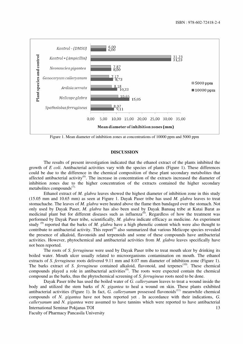
Figure 1. Mean diameter of inhibition zones at concentrations of 10000 ppm and 5000 ppm
DISCUSSION
The results of present investigation indicated that the ethanol extract of the plants inhibited the
growth of E coli. Antibacterial activities vary with the species of plants (Figure 1). These differences
could be due to the difference in the chemical composition of these plant secondary metabolites that
affected antibacterial activity(6)
. The increase in concentration of the extracts increased the diameter of
inhibition zones due to the higher concentration of the extracts contained the higher secondary
metabolites compounds(7).
Ethanol extract of M. glabra leaves showed the highest diameter of inhibition zone in this study
(15.05 mm and 10.65 mm) as seen at Figure 1. Dayak Paser tribe has used M. glabra leaves to treat
stomachache. The leaves of M. glabra were heated above the flame then bandaged over the stomach. Not
only used by Dayak Paser, M. glabra has also been used by Dayak Banuaq tribe at Kutai Barat as
medicinal plant but for different diseases such as influenza(8)
. Regardless of how the treatment was
performed by Dayak Paser tribe, scientifically, M. glabra indicate efficacy as medicine. An experiment
study (9)
reported that the barks of M. glabra have a high phenolic content which were also thought to
contribute to antibacterial activity. This report(9)
also summarized that various Melicope species revealed
the presence of alkaloid, flavonoids and terpenoids and some of these compounds have antibacterial
activities. However, phytochemical and antibacterial activities from M. glabra leaves specifically have
not been reported.
The roots of S. ferrugineus were used by Dayak Paser tribe to treat mouth ulcer by drinking its
boiled water. Mouth ulcer usually related to microorganisms contamination on mouth. The ethanol
extracts of S. ferrugineus roots delivered 9.11 mm and 8.07 mm diameter of inhibition zone (Figure 1).
The barks extract of S. ferrugineus contained alkaloid, flavonoid, and terpenes(10)
. These chemical
compounds played a role in antibacterial activities(9)
. The roots were expected contain the chemical
compound as the barks, thus the phytochemical screening of S. ferrugineus roots need to be done.
Dayak Paser tribe has used the boiled water of G. calleryanum leaves to treat a wound inside the
body and utilized the stem barks of N. gigantea to heal a wound on skin. These plants exhibited
antibacterial activities (Figure 1). In fact, G. calleryanum possessed flavonoids(11)
meanwhile chemical
compounds of N. gigantea have not been reported yet . In accordance with their indications, G.
calleryanum and N. gigantea were assumed to have tannins which were reported to have antibacterial
ISBN : 978-602-72418-2-4
International Seminar Pokjanas TOI
Faculty of Pharmacy Pancasila University
14
activities against E. coli (12)
. Tannins also could accelerate the wound healing through several cellular
mechanism(13)
.
A. serrata was used by Dayak Paser tribe to cure arthritis. They boil the stem and drink the water.
Although it was non-bacterial symptom, A. serrata exhibited antibacterial activities against E coli. This
plant was expected to have bioactive compounds that have a role in either antiinflammatory, analgesic or
antibacterial activities. Therefore, it becomes important to investigate the phytochemical and bioactivities
of this plant since there are no reports about it.
The result obtained from this study showed that medicinal plants used by Dayak Paser to treat
bacterial and non bacterial infection symptoms exhibited antibacterial properties. This in vitro
antibacterial determination was the first step towards to development of new antibacterial agents from
ethnomedicinal plants. Although these selected plants produced less activity than positive control against
E coli, however they were still potential for further investigation. Phythochemical screening,
identification for the specific bioactive compounds and antibacterial activities determination of these
plants against various pathogen bacteria are needed.
CONCLUSION
Ethanol extracts of S. ferrugineus, M. glabra, G. calleryanum, N. gigantea and A. serrata exhibited
antibacterial performance against E. coli with zone of inhibition ranged between 7.17-10.65 mm at a
concentration of 5000 ppm and 8.00-15.05 mm at a concentration of 10000 ppm. M. glabra showed the
highest activity. They are potential as source of antibacterial agents.
ACKNOWLEDGMENTS
This work was part of the research program of Research Institute for Natural Resources Conservation and
Technology, Ministry of Environment and Forestry, Republic of Indonesia.
REFERENCES
1. Nathan C. Antibiotics at the crossroads. Nature 2004, 431:899-902.
2. Adwan G, Mhanna M. Synergistic effects of plant extracts and antibiotics on Staphylococcus
aureus strains isolated from clinical specimens. Middle East J Sci Res [Internet]. 2008 [cited
2015 Sep 28]; 3:134-139. Available from: www.idosi.org/mejsr/mejsr3(3)/5.pdf.
3. Noorcahyati, Widuri SA, Sitepu BS, Mediawati I, Arifin Z, Wibisono Y. Kajian etnobotani dan
uji fitokimia jenis bahan baku obat kurang dikenal. Laporan Hasil Penelitian. Balai Penelitian
Teknologi Konservasi Sumberdaya Alam. Tidak diterbitkan. 2014.
4. Frey FM, Meyers R. Antibacterial activity of traditional medicinal plants used by Haudenosaunee
peoples of New York State. BMC Complement Altern Med [Internet]. 2010 Nov [cited 2015 Jan
25]; 10(64):1-10. Available from: http://www.biomedcentral.com/1472-6882/14/122.
5. Bauer AW, Kirby WMM, Sherris JC, Turck M. Antibiotic susceptibility testing by a standardized
single disk method. Amer J Clin Pathol [Internet]. 1966 Aug [cited 2014 Des 12]; 45:493-496.
Available from: garfield.library.upenn.edu/classics1985/A1985ANC2900001.pdf.
6. Noumedem J, Mihasan M, Lacmata S, Stefan M, Kuiate J, Kuete V: Antibacterial activities of the
methanol extracts of ten Cameroonian vegetables against Gram-negative multidrug resistant
bacteria. BMC Complement Altern Med [Internet]. 2013 Jan [cited 2015 Sep 28];13(26):[about
5pp]. Available from: http://www.biomedcentral.com/1472-6882/13/26.
7. Setha B, Laga A, Mahendradatta M, Firdaus. Antibacterial activity of leaves extracts of Jatropha
curcas, Linn against Enterobacter aerogenes. International Journal of Scientific & Technology
Research [Internet]. 2014 Jan [cited 2015 Oct 1];3(1):129-131. Available from:
http://www.ijstr.org/final-print/jan2014/Antibacterial-Activity-Of-Leaves-Extracts-Of-Jatropha-
Curcas-Linn-Against-Enterobacter-Aerogenes.pdf.

ISBN : 978-602-72418-2-4
International Seminar Pokjanas TOI
Faculty of Pharmacy Pancasila University
15
8. Falah F, Sayektiningsih T, Noorcahyati. Keanekaragaaman jenis dan pemanfaatan tumbuhan
berkhasiat obat oleh masyarakat sekitar Hutan Lindung Gunung Beratus, Kalimantan Timur.
Jurnal Penelitian Hutan dan Konservasi Alam. 2013;10(1): 1-18.
9. Kassim NK, Rahmani M, Ismail A, Sukari MA, Ee GC, Nasir NM, Awang K. Antioxidant
activity-guided separation of coumarins and lignan from Melicope glabra (Rutaceae). Food Chem
[Internet]. 2013 Aug [cited 2015 Sep 28];139(1-4):87-92. Available from:
www.ncbi.nlm.nih.gov/pubmed/23561082.
10. Marliana E. Analisis senyawa metabolit sekunder dari batang Sptaholobus ferrugineus
(Zoll&Moritzi) Benth yang berfungsi sebagai antioksidan. Jurnal Penelitian MIPA [Internet].
2007 Dec [cited 2015 Sep 27];1(1):23-29. Available from:
http://repository.usu.ac.id/bitstream/123456789/21203/1/kpm-des2007-1%20(2).pdf.
11. Kaneko T, Sakamoto M, Ohtani K, Ito A, Kasai, R, Yamasaki K. Secoiridoid and flavonoid
glycosides from Gonocaryum calleryanum. Phytochemistry [Internet]. 1995 May [cited 2015 Oct
6];39(1):115-120. Available from:
http://www.researchgate.net/publication/222585377_Secoiridoid_and_flavonoid_glycosides_fro
m_Gonocaryum_calleryanum
12. Oboh G. Antioxidant and antimicrobial properties of ethanolic extract of Ocimum gratissimum
leaves. J Pharmacol Toxicol [Internet]. 2010 [cited 2015 Sep 18]; 5(7):396-402. Available from:
www.doi:10.3923/jpt.2006.47-53.
13. Sheikh AA, Sayyed Z, Siddiqui AR, Pratapwar, Sheakh SS. Wound healing activity of Sesbania
grandiflora Linn flower ethanolic extract using excision and incision wound model in wistar rats.
International Journal of PharmTech Research [Internet]. 2011 April-June [cited 2015 Oct
6];3(2):895-898. Available from: sphinxsai.com/vol3.no2/pharm/pharmpdf/PT=43(895-
898)AJ11.pdf.

ISBN : 978-602-72418-2-4
International Seminar Pokjanas TOI
Faculty of Pharmacy Pancasila University
16
CITOTOXICITY AND RADICAL SCAVENGING ACTIVITY TEST OF
GAMBIR (Uncaria gambir (HUNTER) ROXB.) IN VITRO
Sri Ningsih1*, Churiyah
1, Fahri Fahrudin
1, Rini Damayanti
2, Eriawan Rismana
3
1Center of Phamaceutical and Medical Technology – Agency for the Assessment and
Application of Technology - LAPTIAB 610-611 Bld. Kawasan Puspiptek Serpong , Tangerang
Selatan, Banten, Indonesia 2Indonesian Research Center for Veterinary Science - Jl. RE Martadinata 30 Bogor, West Java,
Indonesia 3BPPT
*Corresponding author : [email protected]
ABSTRACT
Gambir (Uncariagambir(Hunter) Roxb.) is a native Indonesian medical shrub and used as traditional
medicine for treating various diseases because of its highly polyphenol content. The aim of this studies
were to evaluate the toxic effect of some gambir extracts on normal cell line in vitro and to determine
their antioxidant activity. The samples were consist of 5 kinds of extracts, namely, gambir, ethanolic
50%extract of gambir, ethanolic 96% extract of gambir, ethanolic 50% extract of gambir leaves and
ethanolic 96% extract of gambir leaves. Cytotoxicity assay was conducted on Vero normal cell line by
4,5-dimethylthiazol-2yl (MTT) assay, and showed that the Inhibitory Concentration 50(IC50) values of
the 3samples of gambir extract were more than 1000 ppm resulted in the absent of proliferative effect, as
well as both gambir leaves extracts were less than 1000 ppm. The 1,1-diphenyl-2-picrylhydrazyl (DPPH)
radical scavenging activities of all samples measured at 4 ppm final concentration had range of 29.5%-
49.7% with standard Vitamin C value of 53.1%. The activities decreased in the following order:
ethanolic 96% extract of gambir>ethanolic 96% extract of gambir leaves>gambir >ethanolic 50% extract
of gambir>ethanolic 50% extract of gambir leaves, respectively. These results showed that all gambir
extract were categorized as non-cytotoxic with highly antioxidant activities.
Keywords: Gambir (Uncariagambir(Hunter) Roxb.), cytotoxicity assay, radical scavenging, DPPH,
antioxidant.
INTRODUCTION
Gambir or Uncaria gambir (Hunter) Roxb. belonging to Rubiaceae family, is a native plant
especially found in Sumatera inland and Malaysia peninsula (Hussin MH. et al, 2011). Indonesia produce
very high gambir that fulfill almost 80% of the worldwide need (Dhalimi A., 2006). The polyphenol
compound of gambir is sufficiently high with flavonoid (+)-catechin content as the major compound.
(+)Catechin content is almost 40-80% of dried water extract depending on the preparation process
(Hayani E. et al, 2003). According to the natural compounds, gambir demonstrated some
pharmacological benefits such as antiflatulence, antibacterial, skin tanning, remedies for diarrhea and
sore throat and pesticides properties (Kassim MJ. et al., 2011).These polyphenol also exhibit antioxidant
activity in some in vitro test (Widiyarti G. et al., 2011; Amir M., 2012, Anggraini T. et al., 2011).
According to The National Agency of Drugs and Food Control (NA-DFC) regulation, the
development of new medicines, including traditional medicines, cosmetics, and products complement as
well as food and hazardous materials, beside of the efficacy assessment, it is also necessary to perform a
set of toxicity evaluation (Anonim, 2014). Cell culture technique is frequently used as the first tool for
estimating toxic effect of new material (Anussavice KJ., 2003).This methods is conducted in normal cells
such as fibroblast (Graidist P. et al., 2015) and Vero(Pour BM, et al., 2011)cells. Toxicity levels is related
to the cell viability as exposure to material tested in which the number of living cells is measured with
MTT colorimetric (Graidist P. dkk., 2015). The studies were conducted to demonstrate the viability of
Vero cells and the radical scavenging activity after exposing of some gambir extracts.
ISBN : 978-602-72418-2-4
International Seminar Pokjanas TOI
Faculty of Pharmacy Pancasila University
17
METHODS
Preparation of gambir extracts
Fresh gambir simplisia (leaves and twigs) were collected from Limapuluh Kota – West Sumatera
Province on February 2013. The shrubs was identified in Bogoriense Herbarium Research Center for
Biology Indonesian Institute of Science (LIPI) Bogor, before being processed.
The samples tested were gambir, ethanolic 50% extract of gambir, ethanolic 96% extract of
gambir, ethanolic 50% extract of gambir leaves and ethanolic 96% extract of gambir leaves. Gambir was
prepared based on Farmakope Herbal Indonesia with modification. Briefly, 1 kg of fresh gambir was
steam for 60minutes and then pressed until gambir gum collected. Gum was separated from water with
decantation for 10-12 hours followed by drying the obtained semisolid sediment in oven at temperature of
45-500C for overnight.Then, it was powdered by electric blender. Both ethanolic extracts of gambir were
prepared with the agitating maceration technique at room temperature for 16-18 hours using ethanolic
96% and ethanolic 50%, respectively. Each collected filtrate was then separately evaporated under
vacuum at 450C to get dried mass. The similar method was applied to obtain both extracts of gambir
leaves in which the final extracts were semisolid mass.
4,5-dimethylthiazol-2yl (MTT) assay
Cytotoxicity effect of gambir extract was estimated through the MTT assay. It is a colorimetric
assay for assessing cell metabolic activities, conducted based on protocol test developed by Laboratory of
Center of Phamaceutical and Medical Technology – Agency for the Assessment and Application of
Technology. Vero cells was maintained in RPMI-1640 medium supplemented with 10% FBV, 1%
penicillin-streptomicin, and 0.2% NaHCO3. The cells were cultured at 300C in humidified 5% CO2
incubator.
Vero cells were diluted with Roswell Park Memorial Institute (RPMI) medium to 5x10⁵ cells/mL
and aliquots (5x10⁴ cells/0,1mL) were placed in individual wells in 96-well micro plate. After attaching
into the wells with incubating at CO2 5%, 370C for overnight, cells were treated with a diluted 2-fold
series of concentration of each sample ranged from 62.5 to 1000 ppm in final solution. Samples were
dissolved in medium with maximal dimethyl sulphoxide (DMSO) 0.1% each well. Next, the cells were
incubated at the above conditions for 24 h and then their viability was determined by MTT color. The
MTT solution (0.5 mg/mL in medium, 100 uL each well) was added to each well and incubated for 4 h.
The 10% Sodium dodecyl sulphate (SDS) solution in 0.1 N HCl was added to each well to dissolve the
formed formazan crystals, followed by incubating of the plate for 24 hours at room temperature for
completing dissolution process. The absorbance was read at 570 nm on a microplate reader after shaking
at 120 rpm for 15 minutes. Blank and control absorbance were prepared similar to samples treatment by
using reagent only and reagent plus cells without samples, respectively. The tests were performed in
triplicate. Percent viability was calculated with this equation
a: sample absorbance, b: blank absorbance, c: control absorbance
1,1-diphenyl-2picrylhydrazyl (DPPH) radical scavenging assay
The antioxidant activity that is measured from free radical scavenging activity of the extracts was
measured by using the stable DPPH free radical based on previous paperwith modifications (Hanani E. et
al., 2005). Briefly, into 5 mL glass tube, it put in 3000 uL samples solution, 150 uL DPPH solution,
consecutively. The concentration of sample solution was prepared by dissolving each extract with DMSO
and methanol to get 4 ppm of final solution and DPPH solution was 0,004% in methanol as well. The
absorbance was measured at 517 nm after being incubated at room temperature for 30 minutes. Ascorbic
% viability = [ a – b ] x 100%
[ c – b ]

ISBN : 978-602-72418-2-4
International Seminar Pokjanas TOI
Faculty of Pharmacy Pancasila University
18
acid was used as standard. The control absorbance was prepared from similar to the above reaction
without sample, and blank absorbance was used methanol. The experiments were done in triplicates. The
activity of radical scavenging was calculated with this equation.
a: sample/standart absorbance, b: blank absorbance, c: control absorbance
Data Analysis
Microsoft excel program was applied to calculate IC50 value of percent proliferation from
proliferative percentage figure (concentration caused 50% Vero cells proliferate). T-test for two
independent samples by SPSS 14.00 software was to analyze intergroup difference of free radical
scavenging activity each extract against to standard vitamin C.
RESULTS
Cytotoxicity test
The toxicity tests of all samples were conducted on kidney pig normal cell line, Vero cells, followed by
MTT assay was depicted at Figure 1. The cytotoxicity level stated as IC50 value of percent viability was
performed at Table 1. The IC50 value was indicated as concentration of tested sample that caused 50% of
cell population survived.
Figure 1. Percent viability of Vero cells after extract treatment
Table 1. The IC50 value of percent viability Vero cells after treated with gambir extracts
Samples IC50 (ppm)
Gambir > 1000
Ethanolic 96% extract of gambir leaves 692
Ethanolic 50% extract of gambir leaves 716
Ethanolic 96% extract of gambir > 1000
Ethanolic 50% extract of gambir > 1000
1,1-diphenyl-2picrylhydrazyl (DPPH) radical scavenging activity
The antioxidant activity of all samples was tested using DPPH free radical scavenging method.
The level of antioxidant activity of each sample was compared to standard vitamin C, which the results
were as shown in Table 2.
% radical scavenging activity = [ a – b ] x 100%
[ c – b ]
ISBN : 978-602-72418-2-4
International Seminar Pokjanas TOI
Faculty of Pharmacy Pancasila University
19
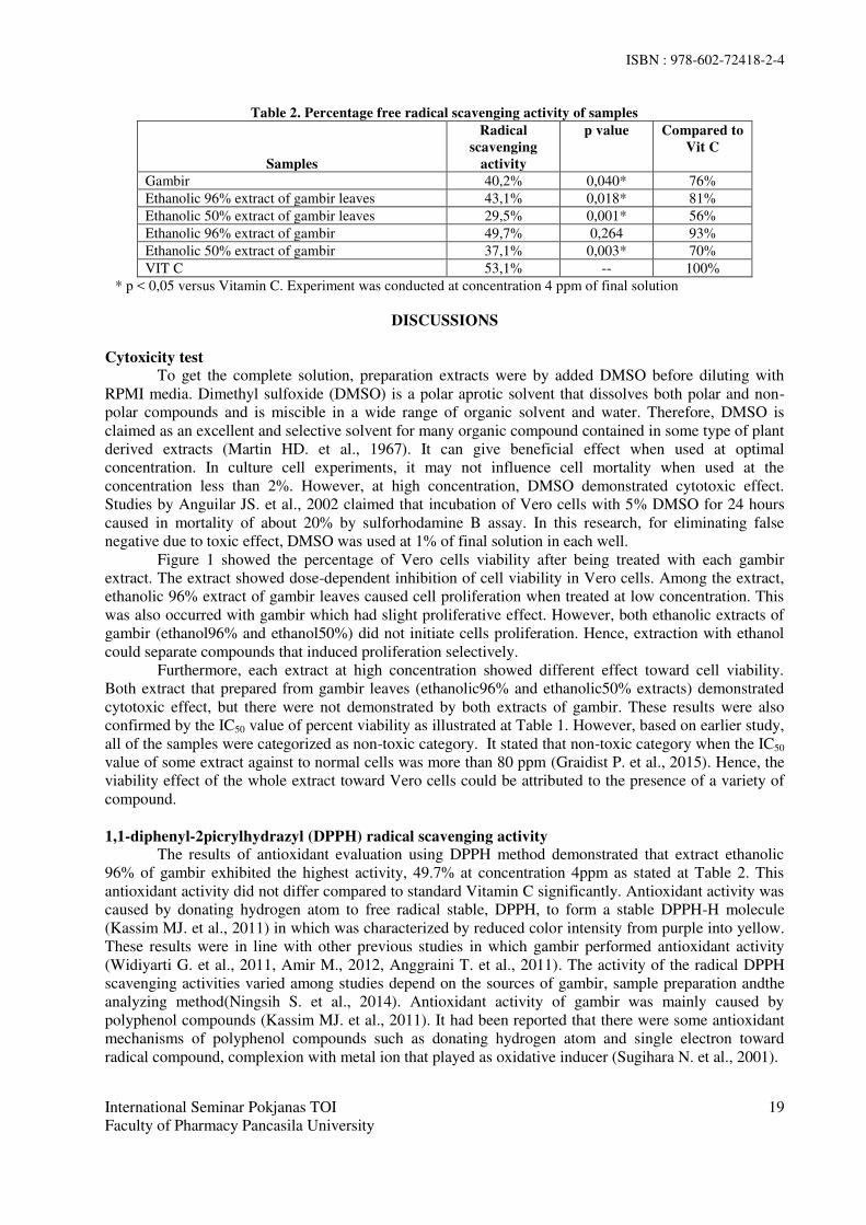
Table 2. Percentage free radical scavenging activity of samples
Samples
Radical
scavenging
activity
p value Compared to
Vit C
Gambir 40,2% 0,040* 76%
Ethanolic 96% extract of gambir leaves 43,1% 0,018* 81%
Ethanolic 50% extract of gambir leaves 29,5% 0,001* 56%
Ethanolic 96% extract of gambir 49,7% 0,264 93%
Ethanolic 50% extract of gambir 37,1% 0,003* 70%
VIT C 53,1% -- 100%
* p < 0,05 versus Vitamin C. Experiment was conducted at concentration 4 ppm of final solution
DISCUSSIONS
Cytoxicity test
To get the complete solution, preparation extracts were by added DMSO before diluting with
RPMI media. Dimethyl sulfoxide (DMSO) is a polar aprotic solvent that dissolves both polar and non-
polar compounds and is miscible in a wide range of organic solvent and water. Therefore, DMSO is
claimed as an excellent and selective solvent for many organic compound contained in some type of plant
derived extracts (Martin HD. et al., 1967). It can give beneficial effect when used at optimal
concentration. In culture cell experiments, it may not influence cell mortality when used at the
concentration less than 2%. However, at high concentration, DMSO demonstrated cytotoxic effect.
Studies by Anguilar JS. et al., 2002 claimed that incubation of Vero cells with 5% DMSO for 24 hours
caused in mortality of about 20% by sulforhodamine B assay. In this research, for eliminating false
negative due to toxic effect, DMSO was used at 1% of final solution in each well.
Figure 1 showed the percentage of Vero cells viability after being treated with each gambir
extract. The extract showed dose-dependent inhibition of cell viability in Vero cells. Among the extract,
ethanolic 96% extract of gambir leaves caused cell proliferation when treated at low concentration. This
was also occurred with gambir which had slight proliferative effect. However, both ethanolic extracts of
gambir (ethanol96% and ethanol50%) did not initiate cells proliferation. Hence, extraction with ethanol
could separate compounds that induced proliferation selectively.
Furthermore, each extract at high concentration showed different effect toward cell viability.
Both extract that prepared from gambir leaves (ethanolic96% and ethanolic50% extracts) demonstrated
cytotoxic effect, but there were not demonstrated by both extracts of gambir. These results were also
confirmed by the IC50 value of percent viability as illustrated at Table 1. However, based on earlier study,
all of the samples were categorized as non-toxic category. It stated that non-toxic category when the IC50
value of some extract against to normal cells was more than 80 ppm (Graidist P. et al., 2015). Hence, the
viability effect of the whole extract toward Vero cells could be attributed to the presence of a variety of
compound.
1,1-diphenyl-2picrylhydrazyl (DPPH) radical scavenging activity
The results of antioxidant evaluation using DPPH method demonstrated that extract ethanolic
96% of gambir exhibited the highest activity, 49.7% at concentration 4ppm as stated at Table 2. This
antioxidant activity did not differ compared to standard Vitamin C significantly. Antioxidant activity was
caused by donating hydrogen atom to free radical stable, DPPH, to form a stable DPPH-H molecule
(Kassim MJ. et al., 2011) in which was characterized by reduced color intensity from purple into yellow.
These results were in line with other previous studies in which gambir performed antioxidant activity
(Widiyarti G. et al., 2011, Amir M., 2012, Anggraini T. et al., 2011). The activity of the radical DPPH
scavenging activities varied among studies depend on the sources of gambir, sample preparation andthe
analyzing method(Ningsih S. et al., 2014). Antioxidant activity of gambir was mainly caused by
polyphenol compounds (Kassim MJ. et al., 2011). It had been reported that there were some antioxidant
mechanisms of polyphenol compounds such as donating hydrogen atom and single electron toward
radical compound, complexion with metal ion that played as oxidative inducer (Sugihara N. et al., 2001).

ISBN : 978-602-72418-2-4
International Seminar Pokjanas TOI
Faculty of Pharmacy Pancasila University
20
It was concluded that all of gambir extracts prepared in the study demonstrated radical
scavenging activity and non-toxic properties to normal Vero cells, therefore this advantages can be
thoroughly studied more deeply as potential traditional herbs.
ACKNOWLEDGMENT
Authors would like to deeply thank toward Insentif Sinas Ristek 2014 Program for providing this research
funding.
REFERENCES
1. Aguilar JS, Roy D, Ghazal P, Wagner EK. Dimethyl sulfoxide blocks herpes simplex virus-1
productiveinfection in vitro acting at different stages with positivecooperativity. Application of
micro-array analysis. BMC Infectious Diseases2002;2(9):1-10.
2. Amir M,Mujeeb M, Khan A, Ashraf K, Sharma D, Aqil M, Phytochemical analysis andin
vitroantioxidant activity ofUncaria gambir. International Journal of Green Pharmacy 2012;6(1):67-
72.
3. Anggraini T, Tai A, Yoshino T, Itani T. Antioxidative activity and catechin content of four kinds of
Uncaria gambir extracts from West Sumatra Indonesia. African Journal of Biochemistry Research
2011;5(1):33-8.
4. Anonim, Peraturan Kepala Badan Pengawas Obat Dan Makanan Republik Indonesia Nomor 7 Tahun
2014 tentang Pedoman Uji Toksisitas Nonklinik secara in vivo, Kementrerian Kesehatan Republik
Indonesia 2014, Jakarta.
5. Anussavice KJ. Phillips' science of dental materials. 11st ed. Elsevier Science (USA) Saunders;
2003:172–94.
6. Dhalimi A. Permasalahan gambir (Uncaria gambir L.) di Sumatera Barat dan alternatif
pemecahannya. Perspektif 2006;5(1):46-59.
7. Graidist P, Martla M, Sukpondma Y. Cytotoxic activity of Piper cubebaextract in breast cancer cell
lines. Nutrients 2015;7:2707-18.
8. Hanani E, Mun’im A dan Sekarini R. Identifikasi senyawa antioksidan dalam spons Callyspongia sp
dari kepulauan seribu. Majalah Ilmu Kefarmasian. 2005;2(3):127 – 133Sugihara N, Ohnishi M,
Imamura M, Furuno K. Differences in Antioxidative Efficiency ofCatechins in Various Metal-
Induced Lipid Peroxidations in Cultured Hepatocytes. Journal of HealthScience 2001;47(2):99-106.
9. Hussin MH, Kassim MJ. The corrosion inhibition and adsorption behavior ofUncaria gambir extract
on mild steel in 1 M HCl. Materials Chemistry and Physics 2011;125(3):461–8.
10. Kassim MJ, Hussin MH, Achmad A, Dahon NH, Suan TK, Hamdan HS. Determination of total
phenol, condensed tannin and flavonoid contents and antioxidant activity of Uncaria gambir
extracts. Majalah Farmasi Indonesia 2011;22(1):50–9.
11. Martin HD, Weise A, Niclas HJ. The solvent dimethyl sulfoxide. Angewande Chemie
1967;6(4):318-334 (abstract).
12. Ningsih S, Fachrudin F, Rismana E, Purwaningsih EH,Sumaryono W, Jusman SWA. Evaluation of
antilipid peroxidationactivity of gambir extract on liver homogenat in vitro. International Journal of
PharmTech Research 2014;6(3):982-9.
13. Pour BM, Latha LY, Sasidharan S. Cytotoxicity and oral acute toxicity studies of Lantana
camaraleaf extract. Molecules 2011;16:3663-74.
14. Widiyarti G, Sundowo A, Hanafi M. The free radical scavenging and anti-hyperglycemic activities
of various gambiers available in Indonesian market. Makara Sains 2011;15(2):129-34.

ISBN : 978-602-72418-2-4
International Seminar Pokjanas TOIFaculty of Pharmacy Pancasila University
21
Acute Toxicity of Ethanolic Extractof Fenugreek Seeds (Trigonella foenum-graecum L.)
on White Rats
KURNIA AGUSTINI*, SRININGSIH, JULHAM EFFENDI
Center for Pharmaceutical and Medical Technology,Agency for the Asessment and Application of Technology, BPPT, Jakarta.
correspondence author: [email protected]
Abstract: Fenugreek seed or biji klabet (Trigonella foenum-graecum L.) was known having activity tohandle some of degenerative diseases such as diabetes mellitus, hypercholesterolemia and alsopostmenopausal symptoms. This study was conducted to investigate the safety of ethanolic extract of bijiklabet on white rat, especially to count the value of Lethal Concentration 50 (LC50). This in-vivo assayreferred to WHO protocols for toxicity assay of natural medicines. We used Spraque dawley white rats,female and male, 6 weeks age, which divided into one group normal and five treatment groups(1g/kgBW, 4g/kgBW, 8g/kgBW, 12g/kgBW, 16g/kgBW). Sample was given once orally then animalwere monitored for two weeks. Observation of toxic effect e.g physical symptom of central nervesystem, autonom nerve system and digestive system. All lethality animal were observed and LC50 werecounted. Result showed that there was no toxic effect and no lethal animal until 16g/kgBW dose. Wecan conclude that ethanolic extract of Fenugreek is practically non toxic.
Keywords : Fenugreek seeds, Trigonella foenum-graecum L., acute toxicity.
INTRODUCTION
Fenugreek seed or Foenigraeci semen is dried seed from Trigonella foenum-graecum L., Leguminosae(1)
(WHO, 2007). In Indonesia, it calls Biji Klabet. Empirically, biji klabet was used for hemorrhoids,asthma, ulcers, muscle pain and often used as a preventative hair loss and skin softener. Many studiesshowed its activity as antidiabetic, anticancer and for hypercholesterolemia handling(2) (Mills, 2000). BijiKlabet has antiandrogen activities, due to its active compounds as beta-sitosterol, palmitic-acid andstearic-acid, and also has the ability to decrease of total cholesterol, LDL, VLDL cholesterol andtriglycerides significantly. The anti-hyperglycemic and anti-inflammatory properties investigated infenugreek are additional benefit. Agustini’s study (2007) showed that ethanolic extract of Biji Klabethave estrogenic effect on ovariectomized and immature female Wistar rats(3).
Biji Klabet contains some sapogenin steroid ingredients, e.g. diosgenin, precursor for sexualhormone(4) (Evans, 2002), its isomer Yamogenin, gitogenin, tigogenin, and trigoneoside(5) (Dewick,1997). Biji Klabet contains diosgenin in free base form 0.8 – 2.2 %(6) (Wiryowidagdo, 2000). BijiKlabet also contains fatty oil 20-30%, alkaloids (trigonelline, an alkaloid pyridine, gentianin andkarpain), flavonoids e.g. vitexin in glycoside or ester form, isovitexin, orientin, vicenin, quercetin andluteolin, essential oil, saponine, nicotinamide, choline, bitter compound and mucilage(4).
This study was carried out to investigate safety effect of ethanolic extract of Fenugreek in white rat.Acute toxicity assay also known as short term toxicity assay. Sample are given once in various grade ofdoses and observation are carried out for two weeks. All physical symptoms and lethal animal wereanalysed then compare to normal group. This acute toxicity study was meant to count the value of Lethal

ISBN : 978-602-72418-2-4
International Seminar Pokjanas TOIFaculty of Pharmacy Pancasila University
22
Dose 50% (LD50) from sample. LD50 is a dose that can cause 50% lethality of animal test. Sample cancategorized as safe material when they have LD50 value bigger than 15g/kgBW(7) (Lu, 1995).
MATERIAL AND METHODS
Sampel preparation. Biji klabet were obtained from Tawangmangu, Central Java, Indonesia. Seed weredried and grind, then were extracted with ethanol 96% food grade. Crude extracts were suspense withCarboxy Methyl Celulose (CMC) 0.5%.
Animal preparation. Experimental animals used in this study were 30 males and 30 femalesSpraque Dawley (SD) white rats, 5-6 weeks age, obtained from Indonesian Food and DrugAdministration (FDA/BPOM). The animal were kept in the animal room (25 + 20C) under 12 hlight/dark cycle and fed with standard diet of pellet rat diet and free access to distilled water prior to thestart of the study. Animal were kept for acclimatization for one week. Animal were maintained andhandled according to ethical committee which approved the design of the animal experiment. Ethicalclearance was approve by Ethical Committee of Indonesian Agency for Health Research andDevelopment (Badan Penelitian dan Pengembangan Kesehatan/Balitbangkes).
Animal treatment. Animals were divided into one group normal and five treatment groups(1g/kgBW, 4g/kgBW, 8g/kgBW, 12g/kgBW, 16g/kgBW), each 5 males and 5 females. Sample was givenonce orally then animal were monitored for two weeks. Observation of toxic effects were done, e.g.physical symptom of central nerve system, autonomy nerve system and digestive system. Body weightswere weighing at day 1, 6,9,12,14. After two weeks all animal were autopsied.
Table 1. Group of animal treatment.No. Groups Treatment N
1. N Normal Diet + CMC Na 0,5% suspense 5 Male + 5 Female
2. D1 Normal Diet + Sample 1 g/kgBW 5 Male + 5 Female
3. D2 Normal Diet + Sample 4 g/kgBW 5 Male + 5 Female
4. D3 Normal Diet + Sample 8 g/kgBW 5 Male + 5 Female
5. D4 Normal Diet + Sample 12g/kgBW 5 Male + 5 Female
6. D5 Normal Diet + Sample 16 g/kgBW 5 Male + 5 Female
RESULT AND DISCUSSION
All animal treated with dose 1 (1g/kgBW) until dose 5 (16g/kgBW) showed no toxic effect significantly.Normally, after orally given sample treatment, all animal showed decrease of motoric activity for someminutes. But after 30 minutes, all activity back to normal. This spontaneous effect is normal after orallygavage treatment. The results of observation of physical symptom of central nerve system, autonomnerve system and digestive system observation, can be seen on Table 2.
Table 2. Physical toxic effect observation.
ObservationGroups
N D1 D2 D3 D4 D5Central Nerve System
1. Sedasi - - - - - -2. Motoric Activity +/- +/- +/- +/- +/- +/-

ISBN : 978-602-72418-2-4
International Seminar Pokjanas TOIFaculty of Pharmacy Pancasila University
23
ObservationGroups
N D1 D2 D3 D4 D53. Convulsion - - - - - -4. Tremor - - - - - -
Autonom Nerve System1. Open eye +/- +/- +/- +/- +/- +/-2. Salivation - - - - - -3. Urination - - - - - -
Breath Rate +/- +/- +/- +/- +/- +/-Heart Rate +/- +/- +/- +/- +/- +/-Digestive System
1. Diarrhea - - - - - -2. Constipation - - - - - -3. Bloody Fesses - - - - - -
Stress hair - - - - - -Note:- : No symptom
+ : There are symptom+/- : Normal
Body weight analysis from all animal, both male and female, with treatment dose 1g/kgBW until16g/kgBW gives no significant difference compare to normal control. Both male and female rats in allgroups gives increasing of body weight almost similar to normal control for 14 days.
Figure 1. Body weight of male rats from all group for 14 days observation.
ISBN : 978-602-72418-2-4
International Seminar Pokjanas TOIFaculty of Pharmacy Pancasila University
24
Figure 2. Body weight of female rats from all group for 14 days observation.
There was no lethality case of animal found in all group after 14 days observation. Therefor this assayshould be repeat using higher level doses than 16g/kgBW. But technically it’s so difficult to treat orallyto the animal. Beside that, regulation from Indonesian FDA says that if until dose 15g/kgBW no lethalitycase occur, then its not necessary to repeat the assay. The sample can be categorized having LD50 higherthan 15g/kgBW or categorized practically non toxic. The data about lethality case can be seen on Table3.
Table 3. Lethality case of animal from all groups.
GroupsMale Female Total
Lethal% Lethality
n Lethal n Lethal
Normal 5 0 5 0 0 0%
D1 5 0 5 0 0 0%
D2 5 0 5 0 0 0%D3 5 0 5 0 0 0%
D4 5 0 5 0 0 0%
D5 5 0 5 0 0 0%
Table 4. Catagorization of Toxic Effect(8). (Lu,FC, 1996)Toxic Category LD50 ValueSuper toxic < 5 mg/kgBBVery hard toxic 5-50 mg/kgBBVery toxic 50-500 mg/kgBBMedium Toxic 0,5-5 g/kgBBMild Toxic 5-15 g/kgBBPractically Non Toxic > 15 g/kgBB
CONCLUSION

ISBN : 978-602-72418-2-4
International Seminar Pokjanas TOIFaculty of Pharmacy Pancasila University
25
No delayed toxic effect and lethality was observed in all rats during fourteen days of recovery period.Orally treatment of Fenugreek within this range and treatment duration would not cause any severe toxiceffects and organ damages in rats. In conclusion, ethanolic extract of Fenugreek have pseudo LD50 andcategorized as practically non toxic.
REFERENCES
1. Agustini, K, Sumali W., Dadang K. 2007. Estrogenic Effect of Fenugreek (Trigonellafoenum-graecum L.) on White Female Rats. Conference Proceedings "Women's Health and TraditionalMedicine", International Medicine and Medicinal Plants, Surabaya.
2. Agustini, Kurnia, Sumali W., Dadang K. 2005. Pengaruh Pemberian Biji Klabet (Trigonellafoenum-graecum L.) terhadap Kadar Hormon Estradiol dan FSH Plasma Tikus Putih BetinaGalur Wistar yang Diovariektomi. Prosiding Seminar Nasional Penggalian Potensi SembilanTanaman Obat Unggulan Indonesia, Purwokerto.
3. Agustini, Kurnia, Sumali W., Dadang K. 2005. Efek Estrogenik Biji Klabet (Trigonella foenum-graecum L.) Terhadap Perkembangan Uterus Tikus Putih Betina. Jurnal Bahan Alam Indonesia,Perhimpunan Peneliti Bahan Obat Alami (PERHIPBA), Vol.4, No.2, Juli
4. Anonim. 2000. Pedoman Pelaksanaan Uji Klinik Obat Tradisional. BPOM DepartemenKesehatan RI, Jakarta: vi + 47p
5. Anonim. 2000. Parameter Standar Umum Ekstrak Tumbuhan Obat. BPOM DepartemenKesehatan RI, Jakarta: viii + 68p
6. Dewick PM. Medicinal Natural Products. A Biosynthetic Approach. New York: John Wiley &Sons; 1997.
7. Evans CW. Pharmacognosy. 15th edition. London: W.B. Saunders; 2002.8. General guideline for methodologies on research and evaluation of traditional medicine. 2000.
Geneva: World Health Organization.9. Guyton, C.Arthur. 1995. Fisiologi manusia dan mekanisme penyakit. Translate from Human
Physiology and mechanism of disease, by Petrus Andrianto. EGC, Jakarta: xii + 821p.10. Lu F.C., Toksikologi dasar, asas, organ sasaran dan penilaian resiko. [Translate by Edi
Nugroho]. Ed. 2, Jakarta:UI Press; 1995.11. Mills, Simon & K. Bone. 2000. Principles and Practice of Phytoterapy. Modern Herbal
Medicine. Churcill Livingstone, Edinburgh: xx + 643p.12. Wiryowidagdo S. Kimia dan Farmakologi Bahan Alam. Jakarta: Universitas Indonesia. 2001;
318-328

ISBN : 978-602-72418-2-4
International Seminar Pokjanas TOI
Faculty of Pharmacy Pancasila University
26
VIRTUAL SCREENING COMPOUNDS IN FABACEAE PLANTS AS LIGANDS ON ALPHA
ESTROGEN RECEPTOR (ER-α)
Gulo LH1, Mumpuni E
1*
Faculty of Pharmacy Pancasila University Jakarta
1
[email protected]; [email protected]
ABSTRACT
Family Fabaceae has about 730 genera and 19.400 species. Some plant of family Fabaceae are known
to have activity as an anti-breast cancer. This study does a series of computational chemistry method
in virtual or in silico screening to compounds in the plant of family Fabaceae that are Abrus
schimperi, Caesalpinia bonduc, Dalbergia vacciniifolia, Eriosema robustum, Erythrina
falcata,Flemingia macrophylla, Genista saharae, Trifolium pratense L., Pachyrhizus erosus, Pissum
sativum and dan DNP (Dictionary of Natural Products). The aim of this study is to find candidates of
compounds as active ligands on estrogen receptor alpha (ER-α) by in silico and elucidating the amino acids contained in the binding site of compounds by using virtual screening validated Anita et al
(2012) protocol. This protocol uses operating system LINUX Ubuntu LTS 14.04 with integrated
applications such as SPORES, PLANTS 1.2, BKChem, Open Babel, R Computational Statistics and
PyMOL, ZINC 01914469 as comparator compound, 4-[4-hydroxy-3-(prop-2-en-1-yl) fenil]-2-(prop-
2-en-1-yl) (dimer compound number 11) as reference compound and 4-hidroksitamoksifen as positive
control. The results of virtual screening conducted on 60 compounds from ten plant of family
Fabaceae obtained 24 compounds be active and 38 inactive compounds in the binding pocket of ER-α. The important amino acids to affinity compounds with estrogen receptor alpha (ER-α) that is GLU353, ARG394, ASP351 and THR347.
Key words: Fabaceae, Virtual Screening, Estrogen Receptor Alpha (ER-α).
INTRODUCTION
In an era new drug design, drug plants were interested as materials of new drug design. One
of strategy to developing molecule design a new drug is utilization of computational chemistry
methods by virtual screening or in silico screening. Virtual screening can be reduced, cost and time
can be more efficient. This study does a series of virtual screening to compounds in the plant of
family Fabaceae. Isoflavon was contained in family Fabaceae has known as anti breast cancer. The
most of breast cancers are selective on estrogen receptor alpha (ER-α). The important of breast cancer treatment is to inhibit the activity of estrogen on estrogen receptor alpha (ER-α). In this study using virtual screening validated Anita et al (2012) protocol. The protocol used to identify ligands can be
active on estrogen receptor.
MATERIALS
Tools: Hardwere: A computer TOSHIBA with processor Intel(R) Core(TM) i5-4200M CPU @ 2.50GHZ,
NVIDIA 2GB, RAM 4GB, HDD 750GB.
Software: LINUX Ubuntu operating system (version 14.04), screening virtual applications (SPORES,
PLANTS, BKChem, Open Babel, PyMOL), Statistical analysis application R i386 3.1.3.

ISBN : 978-602-72418-2-4
International Seminar Pokjanas TOI
Faculty of Pharmacy Pancasila University
27
Materials:
The 2D structure of compounds: Abrus schimperi, Caesalpinia bonduc, Dalbergia vacciniifolia,
Eriosema robustum, Erythrina falcata,Flemingia macrophylla, Genista saharae, Trifolium pratense
L., Pachyrhizus erosus, Pissum sativum and DNP (Dictionary of Natural Products)
The 2D structure of comparison ligand: ZINC 01914469
The 2D structure of reference compound: 4-[4-hidroksi-3-(prop-2-en-1-y1)fenil]-2-(prop-2-en-1-y1)
(senyawa dimer no.11)
The 2D structure of positive control: 4-hidroksitamoksifen
METHODS
This study using a protocol of screening virtual validated Anita et al., for the screening virtual in
silico compounds in the plant of family Fabaceae that areAbrus schimperi, Caesalpinia bonduc,
Dalbergia vacciniifolia, Eriosema robustum, Erythrina falcata, Flemingia macrophylla, Genista
saharae, Trifolium pratense L., Pachyrhizus erosus, Pissum sativum and DNP (Dictionary of Natural
Products), which is used as a test compounds. BKChem application for drawing the structure of the
test compound in the form of 2-dimensional (2D), and then type your converted files by using
applications Open Babel. Ligands and receptors were prepared using Spores application. PLANTS
application is used to simulate the docking of test compound to ER-α at least 3 times replication. Virtual screening results were analyzed using a statistical test one-tailed paired t-test (to see the match
data between pairs of samples tested). PyMOL application used to display the active compounds
representative of the test compound in the form of three-dimensional (3D) and the elucidation of
amino acids in the binding pocket of ER-α.
RESULTS
Compound
Score
Chem PLP ±
SD
p-value
Ligand activity as
estrogen receptor alfa
(ER-α) (in silico)
Caesalpinia bonduc
Asam asetat 5-hidroksi-4,4,7,11b-tetrametil-
9-okso-1,2,3,4,4a,5,6,7,9,11,11a,11b-
dodekahidro-6aH-10-oksa-
siklopenta[b]fenantren-10a-il ester
-87,6484 ±
0,3821 0,9989 Active
Dalbergia vacciniifolia
7-[6-(3,4-Dihidroksimetil-tetrahidro-furan-2-
iloksimetil)-3,4,5,-trihidroksi-tetrahidro-
pyran-2-iloksi]-5-hidroksi-3-(5-hidroksi-2,4-
dimetoksi-fenil)
-99,2499 ±
1,8347 0,9949 Active
7-[6-(3,4-Dihidroksimetil-tetrahidro-furan-2-
iloksimetil)-3,4,5,-trihidroksi-tetrahidro-
piran-2-iloksi]-6-metoksi-3-(5-hidroksi-2,4-
dimetoksi-fenill)
-92,4449 ±
0,5732 0,9953 Active
Eriosema robustum
5,7-Dihidroksi-6-(3-metil-but-2-enil)-2-fenil
-85,8764 ±
0,6143 0.9991 Active
Erythrina falcata
Table 1. Compounds Score and In Silico Activity

ISBN : 978-602-72418-2-4
International Seminar Pokjanas TOI
Faculty of Pharmacy Pancasila University
28
5,7-Dihidroksi-2-(4-hidroksi-fenil)-8-(3,4,5-
trihidroksi-6-hidroksimetil-tetrahidro-piran-2-
il)-6-(3,4,5-trihidroksi-6-hidroksimetil-
tetrahidro-piran-2-il)
-84,5962 ±
0,1870 0,9997 Active
5,7-Dihidroksi-2-(4-hidroksi-fenil)-8-(3,4,5-
trihidroksi-6-hidroksimetil-tetrahidro-piran-2-
il)
-84,7865 ±
0,5025 0,9984 Active
5,7-Dihidroksi-2-(4-hidroksi-fenil)-6-(3,4,5-
trihidroksi-6-hidroksimetil-tetrahidro-piran-2-
il)
-85,1969 ±
0,2934 0,9991 Active
5,7-Dihidroksi-2-(3-hidroksi-4-metoksi-
fenil)-6-(3,4,5-trihidroksi-6-hidroksimetil-
tetrahidro-piran-2-il)
-84,5379 ±
0,2995 0,998 Active
Flemingia macrophylla
2-(3,7-3-Dihidroksi-2,2-dimetil-6-il)-5-
hidroksi-8,8,dimetil-10(3-metil-but-2-enil)-
2,3-dihidro-8H-piranol[3,2-g]
-99,4763 ±
0,1407 0,9966 Active
5,7-Dihidroksi-3-(4-hidroksi-fenil) -86,6118 ±
0,0303 0,9991 Active
5,7-Dihidroksi-3-[4-hidroksi-2-(3-metil-but-
enil)-fenil]
-88,4177 ±
0,5095 0,9975 Active
4-(4-Metoksi-7,7-dimetil-7H-furo[3,2-g] -2-
il)-benzen-1,3-diol
-93,6756 ±
0,6518 0,9941 Active
5-Hidroksi-2-(2-hidroksi-4-metil-fenil)-8,8-
dimetil-10-(3-metil-but-2-enil)-2,3-dihidro-
8H-pirano[3,2-g]-2-il)
-111,2990 ±
0,6015 0,9743 Active
5-Hidroksi-2-[2-hidroksi-5-(3-metil-but-2-
enil)-fenil]-8,8-dimetil-10-(3-metil-but-2-
enil)-2,3-dihidro-8H-pirano[3,2-g]
-103,3447 ±
0,4218 0,997 Active
5-Hidroksi-2-[2-hidroksi-5-(3-metil-but-2-
enil)-fenil]-8,8-dimetil-10-(3-metil-but-2-
enil)-2,3-dihidro-8H-pirano[3,2-g]
-90,3197 ±
0,2126 0,9998 Active
2-(2,4-Dihidroksi-fenil)-5,7-dihidroksi-6,8-
bis-(3-metil-but-2-enil)
-90,5774 ±
0,1828 0,9998 Active
2-(2,4-Dihidroksi-fenil)-5,7-dihidroksi-6,8-
bis-(3-metil-but-2-enil)
-87,6588 ±
0,2543 0,997 Active
Genista saharae
3-(3,4-Dihidroksi-fenil)-5,7-dihidroksi -84,7146 ±
0,0203 1 Active
Dihidroalpinumisoflavon -84,5062 ±
0,1480 0,9996 Active
Trifolium pratense L.
5,7-Dihidroksi-3-(4-hidroksi-fenil) -95,0540 ±
0,0032 0,9978 Active
5,7-Dihidroksi-3-(4-metoksi-fenil) -85,5595 ±
0,0503 0,9971 Active
Pachyrhizus erosus
2-(3,4,5-Trihidroksi-fenil) -3,5,7-triol -85,7861 ±
0,0055 0,9999 Active
Pissum sativum
2-(4-Hidroksi-fenil)- 3,5,7-triol -87,3650 ±
0,0174 0,9997 Active
2-(3,4-Dihidroksi-fenil)-,5,7-triol -84,7334 ±
0,0543 0,9989 Active

ISBN : 978-602-72418-2-4
International Seminar Pokjanas TOI
Faculty of Pharmacy Pancasila University
29
DISCUSSION
Based on the score Chem PLP from the docking simulation of 60 compounds in the plants of
family Fabaceae with using reference ligand 4-[4-hidroksi-3-(prop-2-en-1-yl) fenil]-2-(prop-2-en-1-
yl) (dimer compound number 11) obtained 24 active compounds (Table.1) on binding pocket ER-α by
in silico.
Figure A and B is a 3D visualized from active and inactive representative compounds using
PyMOL application. The compounds were elucidated using PyMOL to explore some active amino
acid residues.
CONCLUSION
1. The results of virtual screening conducted on 60 compounds from ten plant of family Fabaceae
obtained 24 compounds be active and 38 inactive compounds in the binding pocket of ER-α. 2. The important amino acids to affinity compounds with the protein that is GLU353, ARG394,
ASP351 and THR347.
REFERENCE
[1 Anita Y, Radifar M, Kardono LBS, Hanafi M, Enade P. Structure-Based Design Of Eugenol
Analogs As Potential Estrogen Receptor Antagonists. 2012. p. 8(19).
[2] Huang, Niu. Schoichet BKIJ. Benchmarking Sets for Molecular Docking. JMed Chem: 2006. p.
801.
[3] Pranowo Dwi Harno HA. Pengantar Kimia Komputasi. Bandung: Lubuk Agung. 2011. 4-5:118.
[4] Molares S, Ladio A. The Usefulness of Edible and Medicinal Fabaceae in Argentine and Chilean
Patagonia : Environmental Availability and Other Sources of Supply. 2012.
Figure A. 5-Hydroxy-2-[2-hydroxy-5-(3-methyl-but-2-enyl)-phenyl]-8,8-
dimetyhl-10-(3-methyl-but-2-enyl)-phenyl)-2,3-dihydro-8H-pyrano[3,2-g]
chromen-2-yl)-chromen-4-one. A. representative active compound from
Fleminga macrophylla plantss visualized 3D and showed the compound
position in binding pocket ER-α.
Figure B. Pentanoic acid. A. representative inaktive compound
from Eriosema robustum plantss visualized 3D and showed the
compound position in binding pocket ER-α.
ISBN : 978-602-72418-2-4
International Seminar Pokjanas TOI
Faculty of Pharmacy Pancasila University
30
Preparation of Standardized Aqueous Extract of
Annona Muricata Linn. Leaf and Its Potency as Antioxidant
Yesi Desmiaty, Deni Rahmat, Nilam Sari Maulidina
Faculty of Pharmacy, University of Pancasila,
Srengseng Sawah, Jagakarsa, South Jakarta, 12460
ABSTRACT
Extracts from various parts of Soursop leaves (Annona muricata Linn.) are widely used medicinally in all
over the world for the management, control and/or treatment of cancer. Based on in vitro and animal
studies, the presence of exogenous antioxidant has been shown to prevent free radicals activities
associated with cancer development. Soursop leaves is one of medicinal herbals containing flavonoid
compounds and has a potency as an antioxidant. Flavonoids are polyphenolic compounds and generally
present as constituents of flowering plants, particularly dietary plants. The role of dietary flavonoids in
cancer prevention has been widely discussed. The present study was carried out to investigate the
intensity of free-radical scavenging activity, pharmacognosy characteristics and phytochemical screening
of soursop leaves extract. The results of extract quality showed that water soluble extract of 45.30%,
ethanol soluble extract of 39.99%, water content of 8.99%, loss on drying of 9.69%, total ash content of
5.53%, acid insoluble ash content of 1.14%, total plate count of 0.389 x 10⁴ colony/g, yeast plate count
of 0.9970 x 10³ colony/g, and total flavonoid of 3.40%. From the study of antioxidant activity in vitro the
extract has a powerful antioxidant with IC50 value of 76.91 ppm.
Keywords: Soursop leaf, Annona muricata Linn., DPPH, radical scavenging activity, flavonoid.
INTRODUCTIONS
Various degenerative diseases such as diabetes, cancer, tissue inflammation, immune disorders,
cardiac infarction and premature aging are caused by high levels of free radicals in the body. Free
radicals that damage the body can be neutralized by antioxidants. Antioxidants are compounds that can
inhibit reactive oxygen and free radicals in the body. These antioxidant compounds will share one or
more electrons to free radicals to make a normal form of the molecule back and prevent the harmful
reactions. One of the compounds containing a potential as antioxidants is flavonoid (1).
Based on research Ni Putu, et al, n-butanol extract of soursop leaves has a value of DPPH free
radical reduction of 91.10% (2). While Kunthi, et al, demonstrated the results of the phytochemical
screening indicated that the leaves contain alkaloids, flavonoids, saponins, essential oils, quinones,
coumarins, steroids/triterpenoids and tannins. The antioxidant activity of 70% ethanolic extract of the
leaves displays the IC50 value of 22.25 ppm (3). According to the study, the leaves have an antioxidant
activity and the potential as a standardized herbal medicine.
Accordingly, the leaves can be developed as a preparation containing standardized herbal
medicine which has been proven its safety and efficacy scientifically with preclinical trials and
standardization of raw material (4). Therefore, in this research, the determination of extract quality
(specific and non-specific parameters) was carried out to guarantee their consistency. To prove its
efficacy in vitro, the antioxidant activity of the extract was performed using the method of scavenging of
DPPH (1,1-diphenyl-2-picrylhydrazyl) free radical.
ISBN : 978-602-72418-2-4
International Seminar Pokjanas TOI
Faculty of Pharmacy Pancasila University
31
MATERIALS AND INSTRUMENTS
Materials
Dry simplicia of soursop leaves (Annona muricata L.), DPPH (1,1-difenil-2-pikrilhidrazil), and standard
reference of quercetin..
INSTRUMENTS
Spectrophotometer (Shimadzu UV-Visible Spectrophotometer), furnace (Nabertherm-Germany), oven
(Memmert), and air Karl-Fischer (Metrohm 870 KF Titrino plus).
METHODS
1. Collecting of dry soursop leaves
Soursop leaves used in this study was obtained from Balitro, Cimanggu, Bogor and the determination was
conducted in the Laboratory of Biological Research Center, Institute of Sciences Research Indonesia,
Cibinong.
2. Preparation of soursop leaves infusion
A total of 80 g of soursop leaves simplicia was poured into an infusion pot and added 1.6 L of water.
Afterwards, the pot was heated in water bath for 15 minutes at the temperature of 90 °C, while stirring
occasionally. The infusion was filtered through a flannel. The whole process was repeated for 7 times.
3. Phytochemical screening
Phytochemical screening of the extract included alkaloids, falvonoid, saponins, tannins, essential oils,
quinones, coumarin, and steroid/triterpenoid.
4. Determination of extract parameter
The study included the determination of specific parameters (organoleptic, water soluble compounds,
ethanol soluble compounds) and non-specific parameters (loss on drying, moisture content, total ash
content, acid-insoluble ash content and microbial contamination).
5. Determination of total flavonoid
1) The test solution unless otherwise stated the amount of the concentrated extract equivalent to 200 mg
of simplicia was carefully weighed and poured into a round-bottom flask. 1 ml HMT solution, 20 ml
acetone and 2 ml of hydrochloric acid were added successively and refluxed for 30 minutes. The resulting
extract was filtered through the cotton. The filtrate was collected into a 100 ml flask. The residue was
refluxed with 20 ml acetone for 30 minutes and filtered. The filtrate was also collected into the same
flask. Acetone was added up to the mark. 20 ml of the mixture was put into a separating funnel, added 20
ml of water and extracted three times, each time using 15 ml of ethyl acetate. Ethyl acetate phase was put
into 50 ml flask, added ethyl acetate up to the mark.
2) Dilution of test solution
Pipette 10 ml of test solution into a 25 ml flask; add 5% v/v of glacial-acetic acid solution in methanol up
to the mark.
3) Measurement of total flavonoid
Pipette 10 ml of the test solution into a 25 ml flask; add 1 ml aluminum chloride solution in 5% v/v
glacial-acetic acid solution in methanol up to the mark. In different flask, 10 ml of 0.001% quercetin
solution was added 1 ml aluminum chloride solution. The measurements were performed after incubation
time of 30 minutes using a spectrophotometer at the wavelength of 429.5 nm. The total flavonoid content
is calculated as indicated in the monograph by the formula :
F (%) = X 1.25 X 100 ml/ W (g)
Where,
F = Total flavonoid
Cp = Concentration of standard solution (ppm)
Au = Absorbance of test solution with aluminum chloride
ISBN : 978-602-72418-2-4
International Seminar Pokjanas TOI
Faculty of Pharmacy Pancasila University
32
Abu = Absorbance of test solution without aluminum chloride
Ap = Absorbance of standard solution with aluminum chloride
Abp = Absorbance of standard solution without aluminum chloride
DPPH-Radical Scavenging Activity
The free radical scavenging activity of the fractions was measured in vitro by 2,20- diphenyl-1-
picrylhydrazyl (DPPH) assay. The stock solution was prepared by dissolving 8 mg DPPH with 50 ml
methanol and stored at 20°C in the dark. A 1 ml aliquot of this solution was mixed with the sample
solution in methanol and added methanol to get various final concentrations (5 - 100 μg/ml) in 5 ml flask. The reaction mixture was shaken well and incubated in the dark for 15 min at room temperature. Vitamin
C served as standard and was treated in the same manner with the final concentration in the range of 2 –
10 μg/mL. Both the sample and the standard solution were incubated at temperature of 37 oC for 30
minutes. Afterwards, the absorbance was measured at 517 nm. The control was prepared in the same
manner excluding any sample. The scavenging activity was estimated based on the percentage of DPPH
radical scavenged as the following equation:
Inhibition (%) = (Blank absorbance – Sample absorbance)/ Blank absorbance x 100%
RESULTS
1. Phytochemical Screening Table 1. Phytochemical Screening
Secondary metabolite Result
Alkaloid +
Flavonoid +
Saponin +
Polyphenol +
Quinone +
Steroid/triterpenoid +/-
Volatile oil +
Qoumarin +
2. Quality Parameter and Total Flavonoid
Table 2. Quality Parameter and Total Flavonoid
Parameter Soursop leaves extract Requirement (Materi Medika
Indonesia)
Water soluble content 45.30% ≥ 18%
Ethanol soluble content 39.99% ≥ 12.5%
Water content 8.99% ≤ 10%
Loss on drying 9.69%
Total ash content 5.53% ≤ 6%
Acid-insoluble ash content 1.14% ≤ 1.5%
Total plate count 0.3888x10⁴ colony/g ≤ 104
colony/g
Yeast plate count 0.9970x10³ colony/g ≤ 103
colony/g
Total flavonoid 3.40%
3. Antioxidant Activity Study
The antioxidant activity was expressed in the relationship between concentration (x) versus % inhibition
(y) by soursop leaves extract.
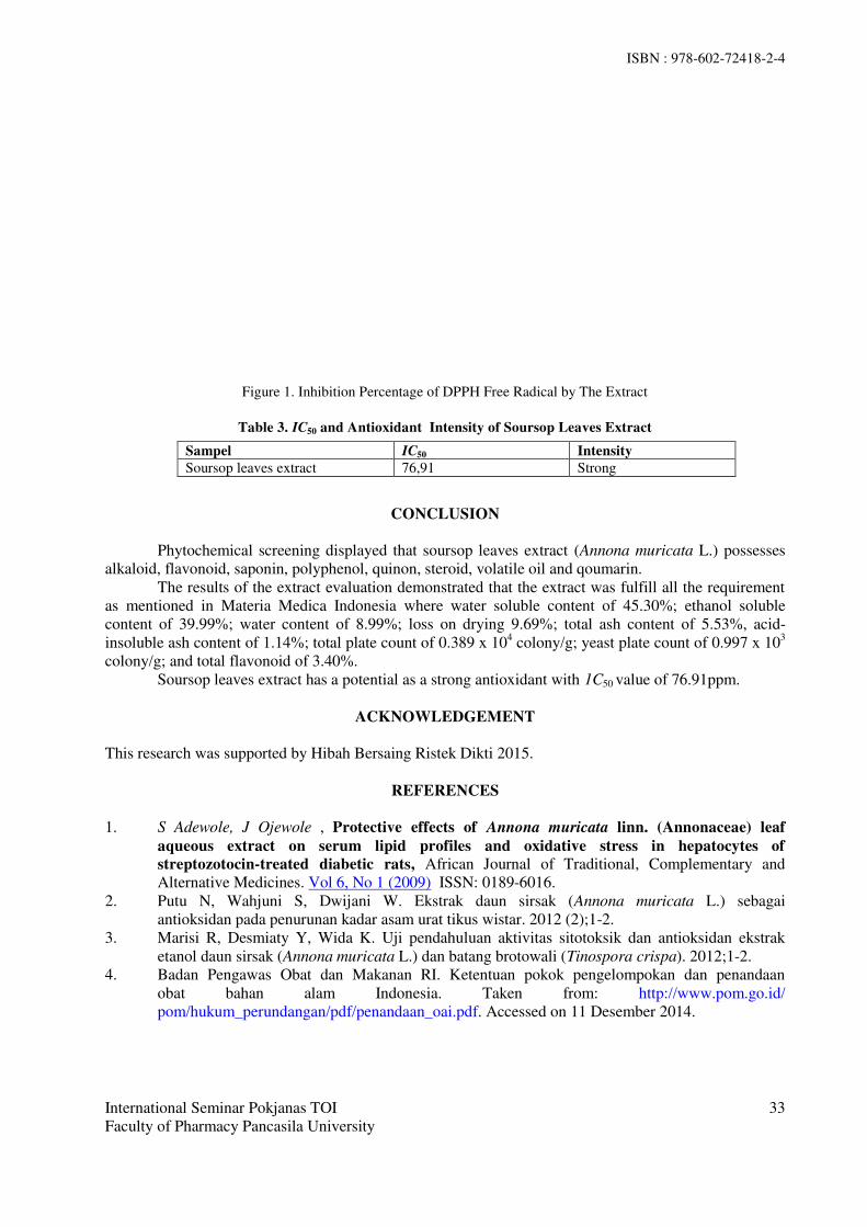
ISBN : 978-602-72418-2-4
International Seminar Pokjanas TOI
Faculty of Pharmacy Pancasila University
33
Figure 1. Inhibition Percentage of DPPH Free Radical by The Extract
Table 3. IC50 and Antioxidant Intensity of Soursop Leaves Extract
CONCLUSION
Phytochemical screening displayed that soursop leaves extract (Annona muricata L.) possesses
alkaloid, flavonoid, saponin, polyphenol, quinon, steroid, volatile oil and qoumarin.
The results of the extract evaluation demonstrated that the extract was fulfill all the requirement
as mentioned in Materia Medica Indonesia where water soluble content of 45.30%; ethanol soluble
content of 39.99%; water content of 8.99%; loss on drying 9.69%; total ash content of 5.53%, acid-
insoluble ash content of 1.14%; total plate count of 0.389 x 10⁴ colony/g; yeast plate count of 0.997 x 10
3
colony/g; and total flavonoid of 3.40%.
Soursop leaves extract has a potential as a strong antioxidant with 1C50 value of 76.91ppm.
ACKNOWLEDGEMENT
This research was supported by Hibah Bersaing Ristek Dikti 2015.
REFERENCES
1. S Adewole, J Ojewole , Protective effects of Annona muricata linn. (Annonaceae) leaf
aqueous extract on serum lipid profiles and oxidative stress in hepatocytes of
streptozotocin-treated diabetic rats, African Journal of Traditional, Complementary and
Alternative Medicines. Vol 6, No 1 (2009) ISSN: 0189-6016.
2. Putu N, Wahjuni S, Dwijani W. Ekstrak daun sirsak (Annona muricata L.) sebagai
antioksidan pada penurunan kadar asam urat tikus wistar. 2012 (2);1-2.
3. Marisi R, Desmiaty Y, Wida K. Uji pendahuluan aktivitas sitotoksik dan antioksidan ekstrak
etanol daun sirsak (Annona muricata L.) dan batang brotowali (Tinospora crispa). 2012;1-2.
4. Badan Pengawas Obat dan Makanan RI. Ketentuan pokok pengelompokan dan penandaan
obat bahan alam Indonesia. Taken from: http://www.pom.go.id/
pom/hukum_perundangan/pdf/penandaan_oai.pdf. Accessed on 11 Desember 2014.
Sampel IC50 Intensity
Soursop leaves extract 76,91 Strong

ISBN : 978-602-72418-2-4
International Seminar Pokjanas TOI Faculty of Pharmacy Pancasila University
34
PHYTOCHEMICAL SCREENING AND TOXICITY TEST BSLT
OF 70 % ETHANOL EXTRACT OF GAHARU LEAVES
(Aquilaria beccariana Tiegh.)
Ahmad Musir1, Wiwi Winarti
1, Siti Hasnah P. Siregar
1
1 Faculty of Pharmacy, University of Pancasila, Jakarta 12640 Jagakarsa
E-mail: [email protected]
ABSTRACT
Gaharu or agarwood (Aquilaria beccariana Tiegh.) is a forest plant in Indonesia that has been
developed with the cultivation by the public and can be used as anti-tumor drugs, anti-cancer, diarrhea
and others This research aimed to determine the toxicity of 70% ethanol extract of gaharu leaves using
Brine Shrimp Lethality Test (BSLT). The study was conducted on the phytochemical screening and
toxicity testing of ethanol extract of gaharu leaves is not inoculated and inoculated. Results of
phytochemical screening of the powder and condensed extract of gaharu leaves to non-inoculated and
inoculated showed the presence of compounds flavonoids, saponins, catechuic and gallic tannins ,
quinons, coumarins, steroids/ triterpenoids, but on the inoculated also showed the presence of volatile
oil compounds. The toxicity test of 70% ethanol extract of non inoculated gaharu leaves that does
have a LC50 value of 113.73 ppm and 60.60 ppm LC50 inoculated.
Key words: Agarwood, Aquilaria beccariana Tiegh, BSLT
INTRODUCTION
Plants gaharu (Aquilaria beccariana Tiegh ) is a forest plant that has been developed with the
cultivation by the public and can be used as a diarrhea medication, anti-tumor, anti-cancer, and others.
Agarwood is also commonly used as fragrances, body by burning (fumigation) at religious events.
Currently no less than 46 (forty six) types of plants that can produce agarwood, plants most frequently
used is (Aquilaria beccariana Tiegh ) from thymeleaceae family, where one of the benefits as
anticancer. Broadly speaking, the process of formation of aloes consists of 2 kinds: natural or artificial
and not at inoculation or inoculation. The process of formation of gaharu inoculation include: hurt / trunk
section, include the injection of a microorganism of the genus Fusarium sp that play a role in the
formation of agarwood. Thus aloes contain secondary metabolites are thought to have anticancer
activity.
Based on this crude drug powder in this study were extracted by the method of kinetic maceration with
70% ethanol, and the extracts obtained, further toxicity tests with the method BSLT (Brine Shrimp
Lethality Test).
MATERIALS AND METHODS
MATERIALS
Gaharu leavest powder (Aquilaria beccariana Tiegh ) that is not in the inoculation and inoculation used
were obtained from the Center for Conservation and Rehabilitation Research Development and the
Ministry of Forestry in the Ministry of Forestry determined Forestry Research and Development Agency
Bogor, after the wood is dried and pulverized.
Chemicals: ethanol 96%, Dragendorf LP, LP Mayer, p HCl, amylalcohol, ether, anhydrous acetic acid,
FeCl3 1% , H2SO4 cons., chloroform, ammonia 10% LP, water, salt NaCl, shrimp larvae (Artemisia
salina Leach).
Instruments: vacuum rotary evaporator (Buchi 205), analytical balance (Sartorius) ,macerator kinetic
(Heidolph), glass tools. 18 watt fluorescent lamp, a magnifying glass, bottles vials and micro pipette.
ISBN : 978-602-72418-2-4
International Seminar Pokjanas TOI Faculty of Pharmacy Pancasila University
35
METHODS Preparation of extracts. A total of approximately 250 grams of gaharu leaves is pulverized dried
macerated using 70 % ethanol (by means of kinetic maserator) and filtered, then filtered. Remaceration
until all secondary metabolites extracted perfectly. The filtrate obtained was collected, further
concentrated by vacuum rotary evaporator, then evaporated on a water bath at a temperature of 40 ° C to
obtain a viscous extract.
Phytochemical screening : Performed by identifying classes of secondary metabolites, compounds
contained in the leaves powder and extracts
Biological activity test using larval shrimp Artemia salina Leach
a. Hatching eggs Artemia salina Leach. Prepare by dissolving synthetic sea water (38 g Sodium Chloride (NaCl) in
1000 mL of water) and filtered with Whatman paper. Brooders sealed vessel that has two sides of the
room, which is open and closed sides. Then enter the egg Artemia salina Leach. into the incubator vessel
which already contains the synthetic sea water and irradiated with 18 watt fluorescent lamp. After 24
hours the eggs that have hatched into nauplii were transferred to another place, 24 hours after the nauplii
is ready to be used as test animals.
b. Preparation of test solutions Extract solution of each sample were made in 9 vials for three concentration is 10 ppm, 100 ppm, 1000
ppm and one vial for control. The mother liquor is made by weighing 20 mg of extract, dissolved in 2 ml
of sea water that has been filtered if it is poorly soluble samples added dimethylsulfoxide (DMSO) 1% as
much as 0.1 to 50.0 mL to increase the solubility.
c. Toxicity tests Pipetted mother liquor of 500, 50 and 5 mL in a row is inserted into the vial and then evaporated to
dryness. Each concentration was made with three repetitions, then into each vial inserted + 3 ml of sea
water, if samples of poorly soluble in sea water, then add dimethyl sulfoxide (DMSO) 1% as much as 0.1
to 50.0 mL. The solution was stirred until homogeneous and enter 10 nauplii tail, sea water is then added
to 5 mL. For each concentration performed three repetitions. Determination of LC50 in mg/ mL
performed using probit analysis.
RESULTS AND DISCUSSION Gaharu wood extraction . Extraction by maceration kinetic gaharu leaves pulvered was not
inoculated and inoculated each with 70 % ethanol, producing a viscous ethanol extract of gaharu leaves
54,40 grams not inoculated with a yield of 21.76 % and condensed ethanol extract of gaharu leaves
were inoculated 59.00 grams with a yield of 23.58 %.
The content of chemical powder and extract of gaharu leaves
The study of the content of the extract of gaharu leaves (Aquilaria beccariana Tiegh ) both powder and
ethanol extracts of gaharu leaves that was not inoculated and inoculated showed a class of
compounds flavonoids, saponins,catechuic and gallic tannins, steroids / terpenoids, quinons and
coumarins, but on the inoculated also showed the presence of volatile oil compounds. Content
of the test results can be seen in Table 1.

ISBN : 978-602-72418-2-4
International Seminar Pokjanas TOI Faculty of Pharmacy Pancasila University
36
Table 1. Result of phytochemical screening the leaves powder and extract
No. Class of
Compounds
Result
Powder of
Gaharu leaves
non inoculation
Extract of
Gaharu leaves
non
inoculation
Powder of
Gaharu leaves
in inoculation
Extract of
Gaharu
leaves in
inoculation
1 Alkaloids - - - -
2 Flavonoids + + + +
3 Saponins + + + +
4
Catechuic Tannins + + + +
Gallic tannins + + + +
5 Quinons + + + +
6 Steroids/
triterpenoids +/+ +/+ +/+ +/+
7 Coumarins + + + +
8 Essential oils - - + +
Description : + = positive results
- = negative results
BSLT biological activity test (Brine Shrimp LethalityTest) Biological activity test with metod (BSLT), 70 % ethanol extract of gaharu leaves turns inoculation
showed the toxic with LC50 values of 60.60 ppm, while the ethanol extract of gaharu leaves not
inoculated at 113.73 ppm LC50, Based on this it can be said that the ethanol extract of 70 % ethanol
of inoculated gaharu leaves are toxic and ethanol extracts were not inoculated gaharu leaves is less
toxic. The data can be seen in Table 2 and Table 3.
Table 2. Ethanol extract toxicity test results were inoculated gaharu
Concentration
(ppm)
Log C
(X)
Dead Life MR NL MR/T % of
Mortality
(Y)
LC50
(ppm)
1000 3 27 3 53 3 53/56 94.64
60.60 100 2 20 10 25 15 25/40 64.50
10 1 7 23 7 37 7/44 15.91
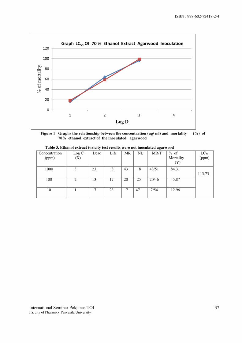
ISBN : 978-602-72418-2-4
International Seminar Pokjanas TOI Faculty of Pharmacy Pancasila University
37
0
20
40
60
80
100
120
1 2 3 4
% o
f m
ort
alit
y
Log D
Graph LC50 Of 70 % Ethanol Extract Agarwood Inoculation
Figure 1 Graphs the relationship between the concentration (ug/ ml) and mortality (%) of
70% ethanol extract of the inoculated agarwood
Table 3. Ethanol extract toxicity test results were not inoculated agarwood
Concentration
(ppm)
Log C
(X)
Dead Life MR NL MR/T % of
Mortality
(Y)
LC50
(ppm)
1000 3 23 8 43 8 43/51 84.31
113.73
100 2 13 17 20 25 20/46 45.87
10 1 7 23 7 47 7/54 12.96
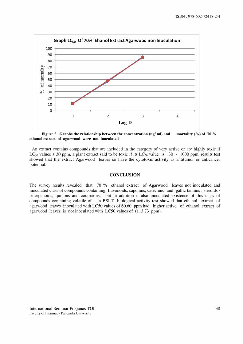
ISBN : 978-602-72418-2-4
International Seminar Pokjanas TOI Faculty of Pharmacy Pancasila University
38
0
10
20
30
40
50
60
70
80
90
100
1 2 3 4
%
of
mo
rtal
ity
Log D
Graph LC50 Of 70% Ehanol Extract Agarwood non Inoculation
Figure 2. Graphs the relationship between the concentration (ug/ ml) and mortality (%) of 70 %
ethanol extract of agarwood were not inoculated
An extract contains compounds that are included in the category of very active or are highly toxic if
LC50 values ≤ 30 ppm, a plant extract said to be toxic if its LC50 value is 30 - 1000 ppm. results test
showed that the extract Agarwood leaves so have the cytotoxic activity as antitumor or anticancer
potential.
CONCLUSION
The survey results revealed that 70 % ethanol extract of Agarwood leaves not inoculated and
inoculated class of compounds containing flavonoids, saponins, catechuic and gallic tannins , steroids /
triterpenoids, quinons and coumarins, but in addition it also inoculated existence of this class of
compounds containing volatile oil. In BSLT biological activity test showed that ethanol extract of
agarwood leaves inoculated with LC50 values of 60.60 ppm had higher active of ethanol extract of
agarwood leaves is not inoculated with LC50 values of (113.73 ppm).

ISBN : 978-602-72418-2-4
International Seminar Pokjanas TOI Faculty of Pharmacy Pancasila University
39
REFERENCES 1. Kartasubrata J. 2010. Successful cultivation of medicinal plants. Bogor: IPB Press.
Department of Forestry in 2004, Utilization profile (cultivation) Agarwood. Jakarta.
Forestry Extension Development Center. Jakarta; p. 41-45
2. Farnsworth NR, 1966. Biological and phytocemycal of plant. J. Pharm, Sci; . p. 224 -
264.
3. Novriyanti Eka 2008. Role of Extractive Substances In Formation Agarwood on
Aquilaria crassna Pierre ex Lecomte and Aquilaria microcarpa Baill. Bogor
Agricultural University.
4. Meyer BN, et al. 1982 . Brine shrimp a convenient general bioassay for active plant
constituent, Planta Medika 45; things. page 31-34
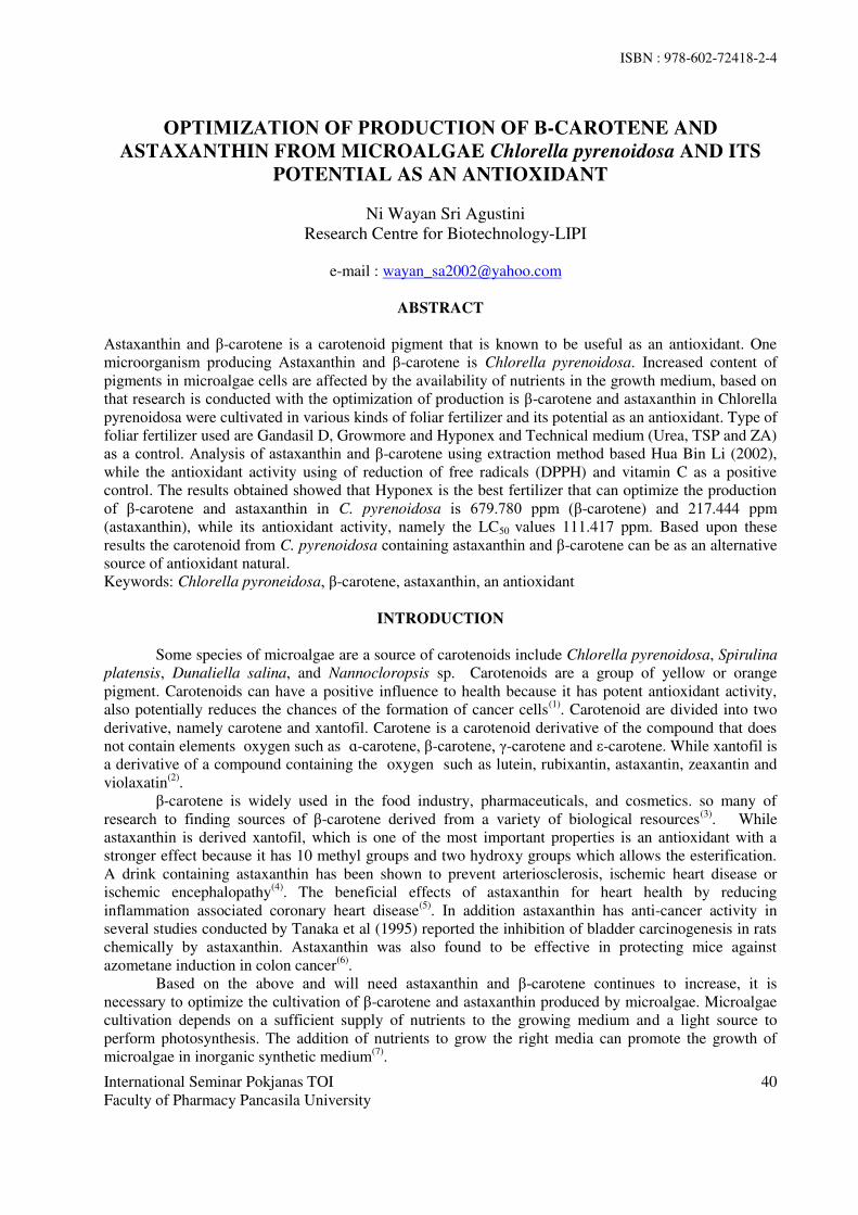
ISBN : 978-602-72418-2-4
International Seminar Pokjanas TOI
Faculty of Pharmacy Pancasila University
40
OPTIMIZATION OF PRODUCTION OF Β-CAROTENE AND
ASTAXANTHIN FROM MICROALGAE Chlorella pyrenoidosa AND ITS
POTENTIAL AS AN ANTIOXIDANT
Ni Wayan Sri Agustini
Research Centre for Biotechnology-LIPI
e-mail : [email protected]
ABSTRACT
Astaxanthin and -carotene is a carotenoid pigment that is known to be useful as an antioxidant. One
microorganism producing Astaxanthin and -carotene is Chlorella pyrenoidosa. Increased content of
pigments in microalgae cells are affected by the availability of nutrients in the growth medium, based on
that research is conducted with the optimization of production is -carotene and astaxanthin in Chlorella
pyrenoidosa were cultivated in various kinds of foliar fertilizer and its potential as an antioxidant. Type of
foliar fertilizer used are Gandasil D, Growmore and Hyponex and Technical medium (Urea, TSP and ZA)
as a control. Analysis of astaxanthin and -carotene using extraction method based Hua Bin Li (2002),
while the antioxidant activity using of reduction of free radicals (DPPH) and vitamin C as a positive
control. The results obtained showed that Hyponex is the best fertilizer that can optimize the production
of -carotene and astaxanthin in C. pyrenoidosa is 679.780 ppm ( -carotene) and 217.444 ppm
(astaxanthin), while its antioxidant activity, namely the LC50 values 111.417 ppm. Based upon these
results the carotenoid from C. pyrenoidosa containing astaxanthin and -carotene can be as an alternative
source of antioxidant natural.
Keywords: Chlorella pyroneidosa, -carotene, astaxanthin, an antioxidant
INTRODUCTION
Some species of microalgae are a source of carotenoids include Chlorella pyrenoidosa, Spirulina
platensis, Dunaliella salina, and Nannocloropsis sp. Carotenoids are a group of yellow or orange
pigment. Carotenoids can have a positive influence to health because it has potent antioxidant activity,
also potentially reduces the chances of the formation of cancer cells(1)
. Carotenoid are divided into two
derivative, namely carotene and xantofil. Carotene is a carotenoid derivative of the compound that does
not contain elements oxygen such as ɑ-carotene, -carotene, -carotene and ε-carotene. While xantofil is
a derivative of a compound containing the oxygen such as lutein, rubixantin, astaxantin, zeaxantin and
violaxatin(2)
.
-carotene is widely used in the food industry, pharmaceuticals, and cosmetics. so many of
research to finding sources of -carotene derived from a variety of biological resources(3)
. While
astaxanthin is derived xantofil, which is one of the most important properties is an antioxidant with a
stronger effect because it has 10 methyl groups and two hydroxy groups which allows the esterification.
A drink containing astaxanthin has been shown to prevent arteriosclerosis, ischemic heart disease or
ischemic encephalopathy(4)
. The beneficial effects of astaxanthin for heart health by reducing
inflammation associated coronary heart disease(5)
. In addition astaxanthin has anti-cancer activity in
several studies conducted by Tanaka et al (1995) reported the inhibition of bladder carcinogenesis in rats
chemically by astaxanthin. Astaxanthin was also found to be effective in protecting mice against
azometane induction in colon cancer(6)
.
Based on the above and will need astaxanthin and -carotene continues to increase, it is
necessary to optimize the cultivation of -carotene and astaxanthin produced by microalgae. Microalgae
cultivation depends on a sufficient supply of nutrients to the growing medium and a light source to
perform photosynthesis. The addition of nutrients to grow the right media can promote the growth of
microalgae in inorganic synthetic medium(7)
.

ISBN : 978-602-72418-2-4
International Seminar Pokjanas TOI
Faculty of Pharmacy Pancasila University
41
Cultivation of microalgae in laboratory scale, the type of fertilizer frequently used type of
fertilizer such as Conwy and Knops. Economically, this type of fertilizer has a price that is quite
expensive, so it is necessary to find a replacement fertilizer price is relatively cheaper, but it is expected
the fertilizer is capable of producing -carotene and astaxanthin maximum. Fertilizers such as Gandasil
D, Hyponex and Growmore fertilizer which is technical and has some elements of nutrients such as
nitrogen, phosphorus, sulfur is needed in culture and are cheaper and easily available in the market.
Based on the above, then do research on the effect of several types of synthetic fertilizers on the
production of -carotene and astaxanthin in microalgae Chlorella pyrenoidosa, and its potential as an
antioxidant
METHODOLOGY
a. Cultivation of Microalgae
Stock cultures of microalgae Chlorella pyrenoidosa cultivation in a 2-liter bottle size. The composition of
media of stock culture consists of urea (1 g / L), TSP (0.3 g / L) and ZA (0.8 g / L) (technical media ).
After reaching the logarithmic phase of stock cultures are harvested by means of centrifuge. Biomass of
C. pyrenoidosa cultivated again in 4 bottles that have added 4 different kinds of fertilizers are Gandasil
D, Hyponex, Growmore and Medium Control (Urea 1 g/L, TSP 0,3 g/, and ZA 0,8 g/L). The
concentration of fertilizer used is 1 g / L. Cultivasi performed in a 2-liter bottle size.C. pyrenoidosa
cultivated again to 4 bottles in a 2 -litre bottle size. . Cultivation is carried out with the addition of
continuous aeration using a blower, the light intensity of 2500 lux and a neutral pH 7. Sampling was done
when the culture reaches logarithmic and stationary phases
b. Analysis β-carotene and astaxanthin(8)
Extraction use of Hua Bin Li (2002). 0.5 gram of wet biomass is suspended with 4 ml of 10 M KOH
then heated above the bath at 60°C for 10 minutes, then cooled at room temperature. Once cool, plus 2.6
ml dichloromethane were homogenized with a homogenizer for 5 min and centrifuged at a speed of 3500
rpm for 10 minutes. The resulting filtrate is collected and re-extracted sludge treatment as above to obtain
a pale yellow filtrate. once collected, centrifuged and the filtrate evaporated at 40 ° C. Rest of evaporation
added 10 ml of ethanol and 10 ml of n-hexane Furthermore, inserted into the separating funnel, shaken
and allowed to separate and form a second layer of the phase of ethanol and n-hexane phase, the
measured absorption at a wavelength of 455 nm (beta carotene) and 479 nm (astaxanthin) using a UV-Vis
spectrophotometer. Uptake value obtained plotted on a standard that was created regression equation
c. Antioxidant Activity of Free Radicals Reduction Method Using 1,1-Diphenyl-2-
Picrylhydrazyl (DPPH).
Free radical scavenging activity of extracts carotenois from C. pyrenoidosa were measured by 1, 1-
diphenyl-2-picryl hydrazyl (DPPH). In brief, 0,4 mM solution of DPPH in methanol was prepared.
Pipette stok solution as much as 25, 50, 125, 250 and 500 mL. DPPH solution was added 1.0 mL into
each tube and added with methanol pro analysis up to add 5 mL, to obtain the concentration of the sample
5, 10, 25, 50 and 100 mg / mL. The mixture was shaken vigorously and allowed to stand at room
temp for 30 min. then, absorbance was measured at 517 nm. by using spectrophotometer (UV-VIS
Shimadzu). Reference standard compound being used was ascorbic acid and experiment was done in
triplicate. The IC50 value of the sample was calculated using following equation: DPPH scavenging
effect (%) or Percent inhibition = A0 - A1/ A0× 100. Where A0 was the Absorbance of control reaction
and A1 was the Absorbance in presence of test or standard sample (Taylor
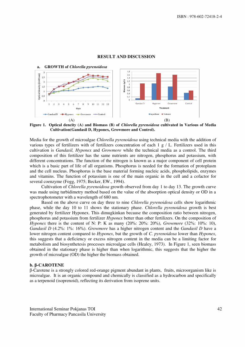
ISBN : 978-602-72418-2-4
International Seminar Pokjanas TOI
Faculty of Pharmacy Pancasila University
42
RESULT AND DISCUSSION
a. GROWTH of Chlorella pyrenoidosa
(A) (B)
Figure 1. Optical dencity (A) and Biomass (B) of Chlorella pyrenoidosa cultivated in Various of Media
Cultivation(Gandasil D, Hyponex, Growmore and Control).
Media for the growth of microalgae Chlorella pyrenoidosa using technical media with the addition of
various types of fertilizers with of fertilizers concentration of each 1 g / L. Fertilizers used in this
cultivation is Gandasil, Hyponex and Growmore while the technical media as a control. The third
composition of this fertilizer has the same nutrients are nitrogen, phosphorus and potassium, with
different concentrations. The function of the nitrogen is known as a major component of cell protein
which is a basic part of life of all organisms. Phosphorus is needed for the formation of protoplasm
and the cell nucleus. Phosphorus is the base material forming nucleic acids, phospholipids, enzymes
and vitamins. The function of potassium is one of the main organic in the cell and a cofactor for
several coenzyme (Fogg, 1975; Becker, EW., 1994).
Cultivation of Chlorella pyrenoidosa growth observed from day 1 to day 13. The growth curve
was made using turbidimetry method based on the value of the absorption optical density or OD in a
spectrophotometer with a wavelength of 680 nm.
Based on the above curve on day three to nine Chlorella pyrenoidosa cells show logarithmic
phase, while the day 10 to 11 shows the stationary phase. Chlorella pyrenoidosa growth is best
generated by fertilizer Hyponex. This dimugkinkan because the composition ratio between nitrogen,
phosphorus and potassium from fertilizer Hyponex better than other fertilizers. On the composition of
Hyponex there is the content of N: P: K as many (20%: 20%: 20%), Growmore (32%: 10%: 10),
Gandasil D (4.2%: 1%: 16%). Growmore has a higher nitrogen content and the Gandasil D have a
lower nitrogen content compared to Hyponex, but the growth of C. pyrenoidosa lower than Hyponex,
this suggests that a deficiency or excess nitrogen content in the media can be a limiting factor for
metabolism and biosynthetesis processes microalgae cells (Healey, 1973). In Figure 1, seen biomass
obtained in the stationary phase is higher than when logarithmic, this suggests that the higher the
growth of microalgae (OD) the higher the biomass obtained.
b. β-CAROTENE
-Carotene is a strongly colored red-orange pigment abundant in plants, fruits, microorganism like is
microalgae. It is an organic compound and chemically is classified as a hydrocarbon and specifically
as a terpenoid (isoprenoid), reflecting its derivation from isoprene units.

ISBN : 978-602-72418-2-4
International Seminar Pokjanas TOI
Faculty of Pharmacy Pancasila University
43
Figure 2. β-carotene of Chlorella pyrenoidosa cultivated in Various of Media Cultivation (Gandasil D,
Hyponex, Growmore and Control).
Β-carotene in microalgae cells to function to absorb light energy for use in photosynthesis and also serve
to protect chlorophyll from damage caused by light(9)
.In this study the content of -carotene logarithmic
phase current is smaller than the stationary phase. The highest beta carotene obtained in C. pyrenoidosa
were cultivated on Hyponex that is equal to 697.716 ppm, followed Growmore in the amount of 389.945
ppm and the lowest in the media Gandasil D is 194.065 ppm (Figure 2). It is also consistent with the
results obtained in this study that the maximum achievable beta carotene when the cells undergo
stationary phase. According Fogg, 1975 when the stationary phase cell density reaches a maximum so
that frequent self-shading among cells, chlorophyll will suffer destruction was replaced by secondary
carotenoid pigments as photosynthetic pigments. And it is known that the Beta carotene is a carotenoid
part of the maximum will accumulate in the cell when the stationary phase(10)
.
b. ASTAXANTHIN
Astaxanthin, unlike some carotenoids and the other carotenoids are known, are not converted to
vitamin A (retinol) in the human body. As with beta-carotene, astaxanthin in microalgae cells to function
as a secondary photosynthetic pigments so that the accumulation of the pigment astaxanthin in the cells
associated with the growth and cell density. In this study, the maximum content of astaxanthin obtained
during the stationary phase is 47.722 ppm on Gandasil D, 217.444 ppm in Hyponex, 119.018 ppm on
Growmore and 80.870 ppm on Control. Visually visible as yellowish stationary phase cells and showed
that chlorophyll suffered destruction and was replaced by carotenoids to perform photosynthesis
Figure 3. Astaxanthin of Chlorella pyrenoidosa cultivated in Various of Media Cultivation (Gandasil D,
Hyponex, Growmore and Control).
C. ANTIOXIDANT ACTIVITY
DPPH method measures the ability of an antioxidant compound in capturing free radicals. Radical
scavenging ability relates to the ability of the compound components in donating electrons or hydrogen.
Each molecule that can donate electrons or hydrogen will react and will dilute DPPH. DPPH color
intensity will change from purple to yellow by electrons derived from antioxidant compounds. DPPH
concentration at the end of the reaction depends on the initial concentration and structural components
catcher compound radical(11)
. In this study was obtained, with a concentration of 150 ppm carotenoid
extract containing beta carotene and astaxanthin have a value of inhibition of 55.66%, whereas vitamin C
(positive control) has a value of 49.83% inhibition at a concentration of 8 ppm
IC50 is a number that indicates the extract concentration (ppm), which is able to inhibit the oxidation
process by 50%. The smaller the IC50 value means the higher the antioxidant activity. Specifically a

ISBN : 978-602-72418-2-4
International Seminar Pokjanas TOI
Faculty of Pharmacy Pancasila University
44
compound said to be a very powerful antioxidant if the IC50 value of less than 50 ppm, strong for the IC50
value-50-100 ppm, while if it is worth 100-150 ppm, and weak if the IC50 value worth 151-200 ppm
(Anonymous, 2005). Based on the results obtained carotenoid extract of C. pyrenoidosa including weak
category because it had LC50 values of 111.42 ppm . Nevertheless these carotenoid can be used as an
alternative as natural antioxidant.
CONCLUSION
C. pyrenoidosa can grow well in growth media used Hyponex and resulted in -carotene and Astaxanthin
respectively 697.716 ppm and 217.444 ppm on the stationary phase. The antioxidant activity of the
carotenoid extract of C. pyrenoidosa at 111.417 ppm so that it can be used as asource of natural
antioxidant
REFERENCES
1. Leach G, G Oliveira, and R Morais. Production of a caretonoid-rich product by Alginate en modern
trapment and fluid-bed drying of Dunaliella salina. J Sci Food Agric. 1998. 76:298-302.
2. Becker EW. Biotechnology and Microbiology, 1st edition. New York: Cambridge University Press;
1994. p. 9-12,51-58,253.
3. Vega PJ, MO Balaban, CA Sims, SF O’Keefe, and JA Cornell. 1996. Supercritical carbon dioxide
extraction efficiency for carotene
4. Miki, W., K Hosoda, K Kondo, and Itakura, H. Astaxanthin containing drink. 1998. Patent abstract
JP10155459
5. Tracy, R. P. Inflamantion Markers And Coronary Heart Disease. Current Opinion In Lipidologi .
1999. 10, 435-441
6. Tanaka, T., T Kawamori., M Ohnishi., H Makita., H Mori,. K Satoh, and A Ha. Suppression of
azomethan-induced rat colon carcinogenesis by dietary administration of naturally occurring
xanthophylls astaxanthin and canthaxanthin during the postinitiation phase. Carcinogenesis 1995.
16,2957-2963
7. Teresa M, Mata., Antonio A, Martins., Nidia, S, Caetano. Microalga for Biodiesel Production And
Other Applications. 2010. Portugal.
8. Li Hua-Bin, Jiang You and Cheng Feng. Isolation and purification of lutei from the microlaga
Chlorella vulgaris by extraction after saponifonification. Journal of agricultural and food chemistry.
2002, p. 1070-1072.
9. Armstrong GA, Hearst JE. Carotenoids 2: Genetics and molecular biology of carotenoid pigment
biosynthesis. FASEB J. 1996. 10 (2): 228–37. PMID 8641556
10 Fogg GE. Algal Culture and Phytoplankton Ecologi. London: The University of Wisconsin Press
from carrot by RSM. J. Food Sci. 1975. 61(4):757
11 Naik, G.H., Priyadarsini, K.I., Satav, J.G., Banavalikar, M.M., Sohoni, D.P., Biyani, M.K., and
Mohan H. Comparative antioxidant activity of individual herbal components used in ayurvedic
medicine, Phytochemistry. 2003. 63(1): 97-104
ISBN : 978-602-72418-2-4
International Seminar Pokjanas TOI
Faculty of Pharmacy Pancasila University
45
ANTIOXIDANT COMPOUND ISOLATED FROM BIOPRODUCTION OF
ENDOPHYTIC FUNGI OF TURMERIC (Curcuma longa L.)
Hindra Rahmawati1, Partomuan Simanjuntak
1,2
1Faculty of Pharmacy, Pancasila University,
Jl. Srengseng Sawah, Jagakarsa, Jakarta 12640, Indonesia 2Research Centre for Biotechnology - Indonesian Institute of Sciences (LIPI),
Jl. Raya Bogor Km 46 Cibinong 16911, Indonesia
Email: [email protected]
ABSTRACT
Endophytic fungi are microorganisms that live in the plants and do mutualism symbiosis with the hosts in
order to produce chemical compounds which are similar to what the hosts do. Five endophytic fungi
isolates were successfully obtained from various parts of turmeric (Curcuma longa L.) from Cibinong.
Bioproduction of those five isolates were tested for their antioxidant activity by free radical scavenging
method using 1,1-diphenyl-2-picrylhydrazyl (DPPH). One of the isolates, K.CI.Cb.U.1, produced
compound(s) with highest antioxidant activity. Bioproduction of K.CI.Cb.U.1 in large amount was
obtained by fermentation method using Potato Sucrose Broth (PSB) medium. Fermentation result showed
that the ethyl acetate extract of the biomass had higher antioxidant activity than that of the filtrate. The
biomass extract was fractionated by gradient column chromatography (silica gel 60; n-hexane-ethyl
acetate 10:1 to 2:1). Three combined fractions were obtained and their IC50 values were determined by
DPPH. Fraction no. 2 had the highest IC50 value (67.18 ppm), and was selected for further purification
using preparative TLC method to produce isolate X. Based on FTIR and GC-MS spectra of isolate X, it
could be estimated that the bioproduction compound of K.CI.Cb.U.1 endophytic fungi isolate was a
glycoside with one saccharide molecule and mass weight of 578, and its aglycone was supposed to
be 3,7-Dimethyl-7-(4-methyl-3-pentenyl)-8-(2,6,10-trimethyl-1,5,9-undecartrienyl)bicyclo[4.2.0]oct-2-
ene.
Keywords: antioxidant, bioproduction, endophytic fungi, turmeric (Curcuma longa L.)
INTRODUCTION
The level of pollution in the world becomes higher from day to day. Indonesia has become one
of the countries that have pretty high levels of pollution. Pollution is a source of free radicals that can
cause dangerous diseases, therefore we need antioxidants that can counteract and prevent the effect of
pollution. Antioxidants are substances which can prevent oxidation process by scavenging free radicals
that attack our body cells. Biodiversity of medicinal plants in Indonesia reveals high potency to discover
new antioxidant compound. One of the plants that can be a source of antioxidants is turmeric (Curcuma
longa L.).
Endophytic fungi are microorganisms that live in the plants and do mutualism symbiosis with the
hosts in order to produce chemical compounds which are similar to what the hosts do. The plants provide
the nutrition for the endophytes and this microorganism convert the nutrition into secondary metabolite
compounds (1,2)
. In this study, endophytic fungi of turmeric was fermented to produce antioxidant
compound(s). The bioproduction obtained was then isolated and identified.

ISBN : 978-602-72418-2-4
International Seminar Pokjanas TOI
Faculty of Pharmacy Pancasila University
46
METHODS
Material
Five endophytic fungi isolates, with codes K.Cl.Cb.Bu.1, K.Cl.Cb.Bu.2, K.Cl.Cb.U.1, K.Cl.Cb.U.2 and
K.Cl.Cb.B.1, were successfully obtained from various parts of turmeric from Cibinong. These isolates
were collection of Natural Product Laboratory, Research Centre for Biotechnology - Indonesian Institute
of Sciences (LIPI), Cibinong.
The filtrates and biomasses extracts of their bioproductions were examined their antioxidant activities and
revealed that the biomass extract of the isolate K.Cl.Cb.U.1 had the highest antioxidant activity with
Inhibition Concentration value of 86.41%. This isolate was selected to be examined further to isolate its
bioproduction antioxidant compound.
Bioproduction The endophytic fungi isolate K.Cl.Cb.U.1 was inoculated in Potato Dextrose Agar (PDA) medium in
Petri dish and incubated for seven days at room temperature. The morphology of the fungi was performed
at Figure 1. The culture was then transferred into Potato Sucrose Broth (PSB) medium and fermented for
14 days while being shaken using the rotary shaker at room temperature.
(a) (b)
Figure 1. Endophytic fungi of K.Cl.Cb.U.1; top side (a); bottom side (b)
Extraction Procedure The fermentation yield was filtered to seperate the biomass from the filtrate. The biomass, which had
higher antioxidant activity than the filtrate, was dried and extracted using ethyl acetate as solvent and
evaporated under reduced pressure using rotary evaporator.
Antioxidant Activity Test
The extracts of the filtrates and the biomasses of the five fungi isolates, the biomass extract of isolate
K.Cl.Cb.U.1 and its fractions were tested for their antioxidant activity by free radical scavenging method
using 1,1-diphenyl-2-picrylhydrazyl (DPPH)(3)
. Series of concentration of the extracts and fractions were
made and each solution was added with one mL of 1,1-diphenyl-2-picrylhydrazyl (DPPH) solution (0.4
mM in methanol) and diluted until 5 mL with methanol. After being homogenized, the solutions were
incubated for 30 minutes at 37oC. The absorptions were measured spectrometrically at 517 nm. The IC50
value of each extract or fraction was calculated using its linear regression curve (4)
. The ascorbic acid was
used as standard.
Fractionation and Purification
Fractionation and purification procedure was carried out using column chromatography, followed by
preparative TLC to get pure substance(s). The column chromatography procedure was performed using
silica gel 60 with gradient solvent system of n-hexane-ethyl acetate 10:1 to 2:1. TLC monitoring was
carried out during the fractionation using silica gel GF254 plate developed in n-hexane-ethyl acetate 2:1.
The spots were examined visually after being sprayed with cerium sulphate solution (1% in 10%
sulphuric acid) and heated electrically until the spots were appeared.

ISBN : 978-602-72418-2-4
International Seminar Pokjanas TOI
Faculty of Pharmacy Pancasila University
47
Purification was performed by preparative TLC using silica gel GF254 plate and n-hexane-ethyl acetate 2:1
as mobile phase. The band was extracted with ethyl acetate solvent, and after filtering the extract was
evaporated until the pure substance was obtained.
RESULT
Three combined fractions obtained from column chromatography were tested their antioxidant
activity and indicating that the fraction 2 was the most active fraction with IC50 value of 67.18 ppm
compared to vitamine C as positive standard with IC50 value of 4.07 ppm. Further purification by
preparative TLC revealed single band of substance. After being extracted, the compound of the isolate
(isolate X) was identified using FTIR Spectrophotometer and Gas Chromatography-Mass Spectrometer.
Figure 2. FTIR Spectrum of the isolate X
Figure 3. GC-MS spectrum of the isolate X
The peak with Retention Time (tR) 25.346 was the highest peak
Figure 4. The mass spectrum of the isolate X
DISCUSSION
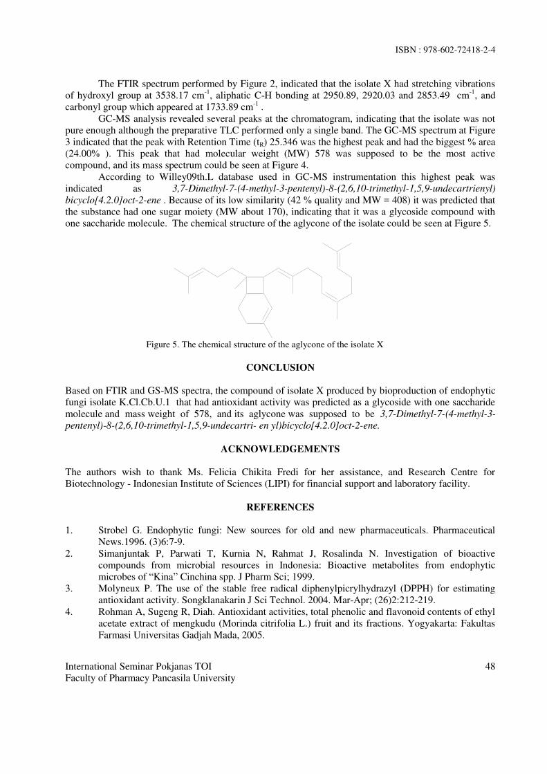
ISBN : 978-602-72418-2-4
International Seminar Pokjanas TOI
Faculty of Pharmacy Pancasila University
48
The FTIR spectrum performed by Figure 2, indicated that the isolate X had stretching vibrations
of hydroxyl group at 3538.17 cm-1
, aliphatic C-H bonding at 2950.89, 2920.03 and 2853.49 cm-1
, and
carbonyl group which appeared at 1733.89 cm-1
.
GC-MS analysis revealed several peaks at the chromatogram, indicating that the isolate was not
pure enough although the preparative TLC performed only a single band. The GC-MS spectrum at Figure
3 indicated that the peak with Retention Time (tR) 25.346 was the highest peak and had the biggest % area
(24.00% ). This peak that had molecular weight (MW) 578 was supposed to be the most active
compound, and its mass spectrum could be seen at Figure 4.
According to Willey09th.L database used in GC-MS instrumentation this highest peak was
indicated as 3,7-Dimethyl-7-(4-methyl-3-pentenyl)-8-(2,6,10-trimethyl-1,5,9-undecartrienyl)
bicyclo[4.2.0]oct-2-ene . Because of its low similarity (42 % quality and MW = 408) it was predicted that
the substance had one sugar moiety (MW about 170), indicating that it was a glycoside compound with
one saccharide molecule. The chemical structure of the aglycone of the isolate could be seen at Figure 5.
Figure 5. The chemical structure of the aglycone of the isolate X
CONCLUSION
Based on FTIR and GS-MS spectra, the compound of isolate X produced by bioproduction of endophytic
fungi isolate K.Cl.Cb.U.1 that had antioxidant activity was predicted as a glycoside with one saccharide
molecule and mass weight of 578, and its aglycone was supposed to be 3,7-Dimethyl-7-(4-methyl-3-
pentenyl)-8-(2,6,10-trimethyl-1,5,9-undecartri- en yl)bicyclo[4.2.0]oct-2-ene.
ACKNOWLEDGEMENTS
The authors wish to thank Ms. Felicia Chikita Fredi for her assistance, and Research Centre for
Biotechnology - Indonesian Institute of Sciences (LIPI) for financial support and laboratory facility.
REFERENCES
1. Strobel G. Endophytic fungi: New sources for old and new pharmaceuticals. Pharmaceutical
News.1996. (3)6:7-9.
2. Simanjuntak P, Parwati T, Kurnia N, Rahmat J, Rosalinda N. Investigation of bioactive
compounds from microbial resources in Indonesia: Bioactive metabolites from endophytic
microbes of “Kina” Cinchina spp. J Pharm Sci; 1999.
3. Molyneux P. The use of the stable free radical diphenylpicrylhydrazyl (DPPH) for estimating
antioxidant activity. Songklanakarin J Sci Technol. 2004. Mar-Apr; (26)2:212-219.
4. Rohman A, Sugeng R, Diah. Antioxidant activities, total phenolic and flavonoid contents of ethyl
acetate extract of mengkudu (Morinda citrifolia L.) fruit and its fractions. Yogyakarta: Fakultas
Farmasi Universitas Gadjah Mada, 2005.
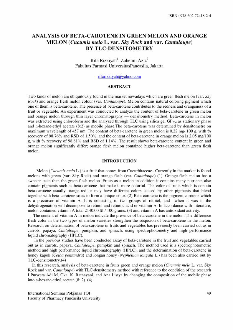
ISBN : 978-602-72418-2-4
International Seminar Pokjanas TOI
Faculty of Pharmacy Pancasila University
49
ANALYSIS OF BETA-CAROTENE IN GREEN MELON AND ORANGE
MELON (Cucumis melo L. var. Sky Rock and var. Cantaloupe)
BY TLC-DENSITOMETRY
Rifa Rizkiyah
1, Zuhelmi Aziz
2
Fakultas Farmasi UniversitasPancasila, Jakarta
ABSTRACT
Two kinds of melon are ubiquitously found in the market nowadays which are green flesh melon (var. Sky
Rock) and orange flesh melon colour (var. Cantaloupe). Melon contains natural coloring pigment which
one of them is beta-carotene. The presence of beta-carotene contributes to the redness and orangeness of a
fruit or vegetable. An experiment was conducted to analyze the content of beta-carotene in green melon
and orange melon through thin layer chromatography — densitometry method. Beta-carotene in melon
was extracted using chloroform and the analyzed through TLC using silica gel GF254 as stationary phase
and n-hexane-ethyl acetate (8:2) as mobile phase.The beta-carotene was determined by densitometre on
maximum wavelength of 457 nm. The content of beta-carotene in green melon is 0.22 mg/ 100 g, with %
recovery of 98.76% and RSD of 1.50%, and the content of beta-carotene in orange melon is 2.05 mg/100
g, with % recovery of 98.81% and RSD of 1.14%. The result shows beta-carotene content in green and
orange melon significantly differ; orange flesh melon contained higher beta-carotene than green flesh
melon.
INTRODUCTION
Melon (Cucumis melo L.) is a fruit that comes from Cucurbitaceae . Currently in the market is found
melons with green (var. Sky Rock) and orange flesh (var. Cantaloupe) (1). Orange-flesh melon has a
sweeter taste than the green-flesh melon. Fruits as a melon in addition it contains many nutrients also
contain pigments such as beta-carotene that make it more colorful. The color of fruits which is contain
beta-carotene usually orange-red or may have different colors caused by other pigments that blend
together with beta-carotene so as to form a unique color. (2) Beta-carotene is the pigment carotene which
is a precursor of vitamin A. It is consisting of two groups of retinol, and when it was in the
dehydrogenation will decompose to retinol and retinoic acid or vitamin A. In accordance with literature,
melon contained vitamin A total 2140.00 SI / 100 grams. (3) and vitamin A has antioxidant activity.
The content of vitamin A in melon indicate the presence of beta-carotene in the melon. The difference
flesh color in the two types of melon varieties strengthen the suspicion of beta-carotene in the melon.
Research on determination of beta-carotene in fruits and vegetables has previously been carried out as in
carrots, papaya, Cantaloupe, pumpkin, and spinach, using spectrophotometry and high performance
liquid chromatography (HPLC).
In the previous studies have been conducted assay of beta-carotene in the fruit and vegetables carried
out as in carrots, papaya, Cantaloupe, pumpkin and spinach. The method used is a spectrophotometric
method and high performance liquid chromatography (HPLC), and the determination of beta-carotene in
honey kapok (Ceiba pentandra) and longan honey (Nephelium longata L.) has been also carried out by
TLC-densitometry.(4)
In this research, analysis of beta-carotene in fruits green and orange melon (Cucumis melo L. var. Sky
Rock and var. Cantaloupe) with TLC-densitometry method with reference to the condition of the research
I Purwata Adi M. Oka, K. Ratnayani, and Ana Listya by changing the composition of the mobile phase
into n-hexane-ethyl acetate (8: 2). (4)

ISBN : 978-602-72418-2-4
International Seminar Pokjanas TOI
Faculty of Pharmacy Pancasila University
50
METHODOLOGY
Green and orange melon (Cucumis melo L. var. Sky Rock and var. Cantaloupe) from
supermarkets in Depok area were used as sample. Chloroform (pa), n-hexane (distilled) , ethyl acetate
(distilled), reference standard of beta-carotene (Sigma Aldrich).
Analysis was performed by Densitometer Camag-TLC scanner 3, Digistore Camag-Reprostar 3,
chamber, separating funnel, rotary evaporator, analytical balance, silica gel GF254 TLC plate, vial,
linomat, capillary tube, filter paper, water bath, set of glassware used in laboratory.
Sample Processing and Extraction: green and orange flesh melon (Cucumis melo L. var. Sky
Rock and var. Cantaloupe) cleaned and peeled as generally for consumption, and extracted with
chloroform 20 ml disposed cloroformnya phase. The extraction process be repeated 4 times with each 20
mL of chloroform, disposed of all phases of chloroform in a 100-mL volumetric flask and diluted with
chloroform until the line mark. Chloroform phase concentrated by rotary evaporator to obtain a
concentrated extract.
Preparation reference standard of beta-carotene solution at concentration 200 ppm, and sample
solution of green melon extract 10mg/2.0 mL of chloroform and sample solution of orange melon extract
10mg/5.0 mL of chloroform
Optimization of TLC-Densitometry performed with selection of the mobile phase based on the
mobile phase used by previous studies (4), which is a mixture of chloroform - ethyl acetate (7: 3) with a
various composition, besides that it also performed the mobile phase composition of n-hexane - ethyl
acetate with a various composition. It also carried out optimization the distance of migration mobile phase
were used 8, 10 and 15 cm using silica gel GF254 plate, and determining the maximum wavelength by
densitometer at specific wavelengths (200-700 nm)
Qualitative Analysis was performed usde TLC by comparing Rf spot in sample with Rf reference
standard of beta-carotene. It were observed visually, and a 254 nm UV lamp.
Linearity test was performed to determine whether there is an influence of the sample matrix on
the relationship between the concentration with peak area using a series solution consisting a minimum 5
samples of different concentrations and then the data is processed by using linear regression. Spotted a
certain number of samples with 5 different concentration levels side by side, then eluated using the best
mobile phase, then spotting gained broad peak measured using a densitometer at maximum wavelength of
beta-carotene.
Quantitative Analysis of Beta-Carotene was performed by spotted reference standard of beta-
carotene and samples solution each 20 µL on the plates side by side chromatographs, then eluated using
the best mobile phase in a chamber . The plates were dried at room temperature and then measured peak
area of beta-carotene with a densitometre at maximum wavelength 457 nm.
Precision and accuracy test were performed by standard addition method, the determination of
precision accuracy test performed 3 times.
RESULT AND DISCUSSION
Optimization Mobile Phase and Distance of Migration
The selection starts with a mobile phase of chloroform-ethyl acetate in various compositions with the
distance of migration mobile phase 8 cm, obtained Rf is too high. Then try to reduce the polarity of the
mobile phase, replacing the mobile phase with a mixture of n-hexane -ethyl acetate in various
compositions. Best separation was obtained from the mobile phase n-heksan- ethyl acetate (8: 2) with
successive Rf value for reference standards of beta-carotene and analyte in orange melon extract: 0.77
and 0.75.
Analysis of Beta-Carotene In Green And Orange Melon (Cucumis melo L. Var. Sky Rock And Var.
Cantaloupe) By Thin Layer Chromatography
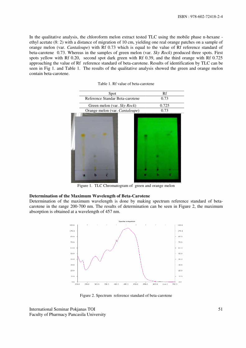
ISBN : 978-602-72418-2-4
International Seminar Pokjanas TOI
Faculty of Pharmacy Pancasila University
51
In the qualitative analysis, the chloroform melon extract tested TLC using the mobile phase n-hexane -
ethyl acetate (8: 2) with a distance of migration of 10 cm, yielding one real orange patches on a sample of
orange melon (var. Cantaloupe) with Rf 0.73 which is equal to the value of Rf reference standard of
beta-carotene 0.73. Whereas in the samples of green melon (var. Sky Rock) produced three spots. First
spots yellow with Rf 0.20, second spot dark green with Rf 0.39, and the third orange with Rf 0.725
approaching the value of Rf reference standard of beta-carotene. Results of identification by TLC can be
seen in Fig 1. and Table 1. The results of the qualitative analysis showed the green and orange melon
contain beta-carotene.
Table 1. Rf value of beta-carotene
Spot Rf
Reference Standar Beta-carotene 0.73
Green melon (var. Sky Rock) 0.725
Orange melon (var. Cantaloupe) 0.73
Figure 1. TLC Chromatogram of green and orange melon
Determination of the Maximum Wavelength of Beta-Carotene
Determination of the maximum wavelength is done by making spectrum reference standard of beta-
carotene in the range 200-700 nm. The results of determination can be seen in Figure 2, the maximum
absorption is obtained at a wavelength of 457 nm.
Figure 2. Spectrum reference standard of beta-carotene

ISBN : 978-602-72418-2-4
International Seminar Pokjanas TOI
Faculty of Pharmacy Pancasila University
52
Linearity test
The linearity test showed a linear relationship between spot area with the concentration, linearity test
results can be seen in Figures 3 and 4.
Figure 3. Linearity test curve of green melon (var. Sky Rock)
Figure 4. Linearity test curve of orange melon (var. Cantaloupe)
Data from linearity test is used to calculate the limit of quantitation (LOQ) of green melon and orange,
respectively for 89.83 (mg / mL); 32.81 (mg / mL)
Determination of Beta-carotene on green dan orange melon (Cucumis melo L. Var. Sky Rock Dan
Var. Cantaloupe) by TLC-Densitometry
The result of determination of beta-carotene in green and orange melon can be seen in Table 2.
Table 2. Quantitative Analyze Result of Beta-carotene on Green and Orange Melons
Material Content (mg/100g) Average content
(mg/100g)
Green Melon (var. Sky Rock)
0.22
0.22 0.22
0.23
Orange Melon (var. Cantaloupe)
2.04
2.05 2.11
2.00
The average content of beta-carotene in green melon is 0.22 mg/ 100 g and orange melon is 2.05 mg/
100 g. Meanwhile according to literature, content of beta-carotene in the melon is 1.284 mg/100 g. Based
on these results showed that there is a correlation between the colour of the fruit with higher levels of

ISBN : 978-602-72418-2-4
International Seminar Pokjanas TOI
Faculty of Pharmacy Pancasila University
53
beta-carotene contained in it. The more orange-red color of the fruit, the higher the content of beta-
carotene.
Accuracy Precision Test
The result of recovery test obtained by comparing the difference in the total weight of beta-carotene
contained in the sample after the addition of reference standard of beta-carotene solution with the weight
of beta-carotene contained sample to the reference standard weight of beta-carotene which is actually
added. Precision test results obtained by calculating the relative standard deviation (RSD) from third 3%
recovery of the sample solution is added a solution of reference standard beta-carotene. The test results
reacquisition and precision green melon (var. Sky Rock) and orange melon (var. Cantaloupe) can be seen
in Table 3.
Table 3. The Results of Accuracy Precision Test in Green and Orange Melons
Material Recovery (%) RSD (%)
Melon hijau (var. Sky Rock) 98.76 1.50
Melon jingga (var. Cantaloupe) 98.81 1.14
The method used to meet the requirements of linearity with R2 = 0.9955 for green melon and 0.9854
for orange melon . The precision of the method is shown by the coefficient of variation of less than 2%,
while accuracy indicated by the value recovery of 98.76% - 98.81% and limit of quantitation of green
melon and orange, respectively for 89.83 (mg / mL); 32.81 (mg / mL). The method has been successfully
to determine of beta-carotene in green melon is 0.22 mg/ 100 g and orange melon is 2.05 mg/ 100 g.
CONCLUSION
1. The content of beta-carotene in green melon is 0.22 mg/ 100 g, with % recovery of 98.76% and
RSD of 1.50%, and the content of beta-carotene in orange melon is 2.05 mg/100 g, with % recovery of
98.81% and RSD of 1.14%.
2. The result shows beta-carotene content in green and orange melon significantly differ; orange
flesh melon contained higher beta-carotene than green flesh melon.
REFERENCES
1. Tim Redaksi Agromedia. Budi Daya Melon. Jakarta: Agromedia Pustaka; 2007. p. 1-14
2. Beta-karoten [Internet]. [cited 2015 May 5]. Available from: http://www.
bbppbinuang.info/news17-beta-karoten.html.
3. Rukmana R. Melon Hibrida. Yogyakarta: Kanisius; 1995. p 14
4. Parwata IM; OA, K. Ratnayani, Ana L. Aktivitas Antiradikal Bebas Serta Kadar Betakaroten
Pada Madu Randu (Ceiba pentandra) dan Madu Kelengkeng (Nephelium longata L.). Bukit
Jimbaran, Bali : Jurnal Fakultas Kimia FMIPA Universitas Udayana; 2010
5. International Conference On Harmonization (ICH). (2005). Validation of analytical
procedure of: text and methodology.

ISBN : 978-602-72418-2-4
International Seminar Pokjanas TOI Faculty of Pharmacy Pancasila University
54
SPECTROPHOTOMETRIC METHOD PRECISION TO ASSAY OF
LYCOPENE IN TOMATOES FRUIT (Solanum lycopersicum Lam.)
Liliek Nurhidayati, Wening Ariwanty
Faculty of Pharmacy, Pancasila University, Jakarta
ABSTRACT
The main active substance in tomatoes fruit (Solanum lycopersicum Lam.) which found in large quantities
was lycopene. Lycopene was one of the natural carotenoid antioxidants, as well as a pigment which give
the red color in tomatoes. In this study, lycopene was extracted using Low Volume Hexane Method by
addition of butylated hydroxytoluene. The mixture was filtered, and the absorbtion of n-hexane phase was
measured at ±503 nm. Lycopene content in ripe fruits was 6.15 mg/100g , This method met the precision
requirements with the relative standard deviation of 1.03%.
Keywords : lycopene, tomatoes, visible spectrophotometry, precision
INTRODUCTION
Tomatoes (Solanum lycopersicum Lam) is one of the Solanaceae plant. The main active
compound in tomatoes fruit was lycopene. Lycopene was good for health because of its antioxidant
activity(1)
. Lycopene was one of potential antioxidant with the ability to reduce singlet oxygen twofold
better than beta-carotene and tenfold better than the tocopherol (2)
. Lycopene content in tomatoes were
interesting to be studied. It could be extracted using Fish et al, method. Lycopene was cool macerated
using aceton, ethanol of 95%, and n-hexane and added with butylated hydroxytoluene (BHT) as
antioxidant in aceton (3)
. Since it was a color pigment so the absorbance of lycopene could be measured in
visible region . It has 11 conjugated double bond and the absobance could be measured at the longer
wavelength (λmax 444, 470, 502 nm in n-hexane) (4)
. Before applied to assay the lycopene in tomatoes fruit
this methods must be validated. In this research, the precision of visible spectrophotometry was tested.
METHODS
Ripened tomatoes (± 3.5 months) fruit was obtained from a local fruit farm at Gunung Cibiuk, Sukabumi.
Taxonomic determination was performed by Herbarium Bogoriense LIPI, Bogor. Lycopene working
standard was obtained from PT. Soho Industri Farmasi. Spectroscopic analysis was carried out on
Shimadzu 1700 double beam UV/Visible spectrophotometer.
Determination of maximum wavelength of lycopene
Absorbtion spectrum of working standard lycopene solution (2.5 ppm in n-hexane) were recorded over
the wavelength range of 300 to 700 nm against solvent blank, in quartz cuvetts with 1 cm diameters.
Absorbance stability
Absorption of working standar 2.5 ppm lycopene solution were recorded on 502.5 nm during 60 minute
against solvent blank, in quartz cuvetts with 1 cm diameters.
Calibration curve
Preparation of calibration curve was performed by measuring the absorption of lycopene working
standard solution of 1.0; 1.5; 2.0; 2.5; 3.0; 3.5; 4.0 and 4.5 ppm in 502.5 nm.

ISBN : 978-602-72418-2-4
International Seminar Pokjanas TOI Faculty of Pharmacy Pancasila University
55
Precision test of spectrophotometry methods for determination of lycopene in tomatoes.
Fresh tomatoes was cut into small pieces, blended. About 0.2 g of tomatoes juice was poured into
Erlenmeyer, then added of 0.05% (w/v) BHT in 5 ml of acetone, 5 ml of 95% ethanol, 10.0 ml of n-
hexane. Put the Erlenmeyer inside the beaker glass with ice and placed on orbital shaker, rotated 180 rpm
for 15 minutes. After 15 minutes of agitation, 3 ml aquademineralisata neutral was added, and shake
again for 5 minutes. Filtered, the filtrate was transferred to a separating funnel and then allowed to stand 5
minutes at room temperature. Take n-hexane phase. The absorption of sample solution was measured in
502.5 nm against n-hexane as blank . Lycopene content was calculated using calibration curve equation.
It was conducted six times to test the precision method. To identify of lycopene in tomatoes, absorption
spectrum of sample solution were recorded over the wavelength range of 300 to 700 nm against solvent
blank, in quartz cuvetts with 1 cm diameters.
RESULTS
Report from research Center for Biology, Indonesian Institute of Sciences mentioned that tomatoes has
species name that is Solanum lycopersicum Lam. The spectrum of lycopene standard solution was showed
in Fig.1.
Fig. 1. Absorption spectra of lycopene working standard solution in n-hexane
Absorbance stability
Fig 2. Showed the result of absorbance stability test.
Fig. 2. The absorbance stability of lycopene solution in 502.5 nm

ISBN : 978-602-72418-2-4
International Seminar Pokjanas TOI Faculty of Pharmacy Pancasila University
56
Qualitative analysis
The spectrum of solution extracted from tomatoes was shown in Fig 3.
Fig 3. Spectrum of sample solution I n-hexane
Determination of lycopene content in tomatoes fruit
Determination of lycopene content was conducted five times to assay the precision of this methods. The
results was showed in Table 1.
Table 1. The results of lycopene content determination
Sample
weight (g)
Absorption Content of lycopene (mg/100g)
0.2783 0.3545 215.1
=6.15
SD= 0.0636
RSD = 1.03
0.2746 0.3534 225.5
0.2871 0.3585 210.4
0.2850 0.3573 211.2
0.2823 0.3567 214.2
DISCUSSION
Determination of maximum wavelength was performed to choose the wavelength where the absorbance
of both standard solution and samples were measured. Fig. 1 shows that the maximum wavelength of
lycopene were 444.0 nm; 470.5 nm; and 502,5 nm. The highest absorption was in 502.5 nm. This
wavelength was choosen to conducted the lycopene analysis. In this wavelength there was no disturbance
of the other carotenoid(4)
.
Absorbance stability test was performed to determine the correct time to measure the absorbance
solution. The lycopene solution had stable absorbance during minute of 15 until 40. Calibration curve of

ISBN : 978-602-72418-2-4
International Seminar Pokjanas TOI Faculty of Pharmacy Pancasila University
57
lycopene that performed using seven different consentration of lycopene showed that the equation of
regression line was y = 0.0866 + 0.1557x with correlation coeffisient of 0.9913. This equation was used
to calculate lycopene content.
The solution spectrum of samples (Fig 3) was similar with the spectrum of lycopene working standard
(Fig 1). It indicated that there were lycopene in ripe tomatoes.
Detemination results with five replication showed that the tomatoes fruit has lycopene content of 6.15
mg/100 g with RSD of 1.03%. RSD is not allowed more than 2.0% (5)
or based on the analyte content
not more than 11.31% (6)
. The precision of this methods fulfill both of them.
CONCLUSION
The visible spectrophotometry using n-hexane as solvent in 502.5 nm fulfill the precision requirement to
determination of lycopene in tomatoes fruit.
REFERENCES
1. Kailaku SI, Dewandari KT, Sunarmani. Potensi likopen dalam tomat untuk kesehatan. Buletin
Teknologi Pascapanen Pertanian Volume 3 2007. pp .50-8
2. Sanjiv A. Rao AV. Tomato lycopene and its role in human health and chronic disease. Canadian
Medical Association Journal. Vol. 163 (6). 2000. p.734-44
3. Fish WW, Veazie PP, Collins JK. A quantitative assay for lycopene that utilizes reduced volumes
of organic solvents. J. Food Comp. and Anal. 15; 2002. p.309-17
4. Delia B, Amaya R, Mieko K. Harvestplus handbook for carotenoid analysis second ed.
Washington DC and California: Harvestplus; 2004. p.4, 13-20
5. United States Pharmacopeial Convention. The United States Pharmacopeia 35-The National
Formulary 30. Rockville: United States Pharmacopeial Convention. 2012. pp. 877-81.
6. Harmita. Petunjuk Pelaksanaan Validasi Metode dengan Cara Perhitungannya. Majalah Ilmu
Kefarmasian. Vol 1No.3 Desember 2004.h.119
ISBN : 978-602-72418-2-4
International Seminar Pokjanas TOI
Faculty of Pharmacy Pancasila University
58
OPTIMIZATION AND VALIDATION OF
HIGH PERFORMANCE LIQUID CHROMATOGRAPHY FOR
DETERMINATION OF CAFFEINE IN WHITE TEA
Zuhelmi Aziz*, Dhiah Resti
Fakultas Farmasi Universitas Pancasila
Srengseng Sawah, Jagakarsa, Jakarta 12640
email: [email protected]
ABSTRACT
White tea comes from the terminal buds and leaves of Camellia sinensis, L which are still unopened and
covered with a fine silvery-white hairs. Thence, white tea has more caffeine than green and black tea
because active of biosynthesis of caffeine occurs in young tea leaves. In accordance with United State
Pharmacopeae (USP) 35th, reverse phase high performance liquid chromatography (HPLC) is used to
determination of caffeine. HPLC method has been optimized and validated for determination of caffeine
in white tea. Optimum conditions for the HPLC in the experiment are as follows: stationary phase;
Waters C18 (150 mmx 3.9 mm), column temperature 40oC, mobile phase; methanol - water (30:70) with
a flow rate of 1.0 mL / min and UV detector with the maximum wavelength 273 nm. The result of
validation as follow: the linearity test showed a linear relationship with a correlation coefficient; r =
0.9989, the accuracy and precision test appropriate ICH requirements. The experiment found that the
contain of caffeine in white tea which is determine by HPLC in the optimum condition is 3.84%, with
precision indicated by RSD value ≤ 2%, and accuracy indicated by recovery value 98.97% -99.72% with
t-score which is less than the corrresponding value on the t-table (DoF=4, p=0.05)
Keywords: Caffeine, High Performance Liquid Chromatography, White tea, Validation
INTRODUCTION
White tea comes from the terminal buds and leaves of Camellia sinensis, L which are still
unopened and covered with a fine silvery-white hairs. White tea has a higher polyphenol and has more
caffeine than green and black tea, because active of biosynthesis of caffeine occurs in young tea leaves.
Therefore, it is necessary to the determination of caffeine in white tea. In accordance with United State
Pharmacopeae (USP) 35th, reverse phase high performance liqiud chromatography (HPLC) is used to
determination of caffeine . In this study, HPLC method has been optimized and validated for
determination of caffeine in white tea.
Optimization of HPLC performed with determining the maximum wavelength and composition
of the mobile phase. Validation methode of HPLC performed on several parameters such as: linearity
test, accuracy and precision test and sensitivity according to the International Conference On
Harmonization (ICH). (8,9)
METHODOLOGY
Commercial Products white tea A and B were used as sample, reference standard of caffeine, methanol
pro-HPLC, distilled water, aquabidest, chloroform, ammonium 30%.
Analysis was performed by HPLC (Shimadzu Autosampler LC-20AD), Spectrophotometer Shimadzu
1800, analytical balance (Mettler AB 204), thermometer (Jena), ultrasonic cleaner (Elma LC 30 H), filter
Millipore, syringe (special HPLC), rotary evaporator (IKA RV-10), and a glass tools that are commonly
used in laboratory analysis (Pyrex Iwaki Glass).
Optimization of HPLC performed with determining the maximum wavelength and composition of the
mobile phase.
The linearity test were used reference standard of caffeine solution at concentrations of 40, 60, 80, 100,
120, 140, and 160 ppm and each of which are added of the sample solution. Limit of detection and

ISBN : 978-602-72418-2-4
International Seminar Pokjanas TOI
Faculty of Pharmacy Pancasila University
59
quantitation were determined based on linear line equation. Precision and accuracy test were performed
by standard addition method.
RESULTS AND DISCUSSION
Optimum conditions for the HPLC in the experiment are as follows: stationary phase; Waters C18 (150
mmx 3.9 mm), column temperature 40oC, mobile phase; methanol - water (30:70) with a flow rate of 1.0
mL / min and UV detector with the maximum wavelength 273 nm.
The standard curve equation is y = -8239.90 + 56507.23 x, with coefficient correlation (r) of 0.9986.
While the result of the linearity test showed that the equation is y = 3306287.62 + 27932.56 x with
coefficient correlation (r) of 0.9989 and limit of detection and quantitation of the method were 7.4 and
22.3 ppm.
Figure 1: The Curve of Linearity Test ;
relationship between concentration(X) and peak area (Y).
Accuracy of the method can be showed by the value of recoveries of with the addition of reference
standards caffeine 400 ug, 600 ug and 800 ug in white tea. The experiments were performed three times
repetitions for each analyte concentration. Method is precise and accurate with the relative standard
deviation (RSD) of ≤ 2% and precent recoveries of 98.97 – 99.72% are included in the range of
requirements 97-103% with t-score which is less than the corrresponding value on the t-table. (DOF = 4,
p = 0 05. The result of precision and accuracy test of caffeine in white tea can be seen in Table 1. Table 1: Results of precision and accuracy test of caffeine in white tea.
The method used to meet the requirements of linearity with r = 0.9989. The precision of the method is
shown by the coefficient of variation of less than 2%, while accuracy indicated by the value recovery of
98.97% -99.72% and the t value smaller than t table in the degrees of freedom (df) = 4 and p = 0 05. The
value of the limit of detection of 7.4 ppm and 22.3 ppm limit of quantitation.

ISBN : 978-602-72418-2-4
International Seminar Pokjanas TOI
Faculty of Pharmacy Pancasila University
60
The method has been successfully to determine caffeine in white tea by HPLC in the optimum condition
with the result is 3.84%.
CONCLUSION
Reverse phase HPLC method with in condition of: a stationary phase; Waters C18 (150 mmx 3.9 mm),
column temperature 40oC, mobile phase; methanol - water (30:70), flow rate of 1.0 mL / min and UV
detector with the maximum wavelength 273 nm met the requirements of ICH for determination of
caffein in white tea, with limit of detection and quantitation of the method were 7.4 and 22.3 ppm. The
content of caffeine in white tea is 3.84%.
ISBN : 978-602-72418-2-4
International Seminar Pokjanas TOI
Faculty of Pharmacy Pancasila University
61
REFERENCES
1. Dalimartha, Setiawan. Atlas Tumbuhan Obat Indonesia. Jilid I. Jakarta: Trubus Agriwidya;
1999. h. 150-3.
2. Kubota, H. Patterns of Adenine Metabolism and Caffeine Biosynthesis in Different Parts of Tea
Seedlings. Plant Physiology; 1986. h. 275-281.
3. Tiurmaida, Yuliana. Penetapan Kadar Kofein dalam Minuman Teh Kemasan secara
Kromatografi Cair Kinerja Tinggi (Essay). Jakarta: Fakultas Farmasi Universitas Pancasila;
2012.
4. Komes, D. et. al. Determination of Caffeine Content in Tea and Mat Tea by using Different
Methods. Czech J. Food Sci (journal) 2009; vol. 27:p.S213-6.
5. Skoog, D. A. Principles of Instrumental Analysis. Sixth Edition. Canada: Standford University;
2007. h. 818-48.
6. Harmita. Petunjuk Pelaksanaan Validasi Metode dan Cara Perhitungannya. Majalah Ilmu
Kefarmasian. 2004; 1(3).h. 117-34.
7. Is White Tea Better Than Other Teas as a Potential Anticarcinogen?. Diambil dari:
http://lpi.oregonstate.edu/news/whitetea.html. Diakes: 1 Februari 2014.
8. International Conference On Harmonization (ICH). (2005). Validation of analytical procedure
of: text and methodology.
9. United States Pharmacopeial Convention (USP) 35th. (2012). The United States Pharmacopeia
The National Formulary 32 27. Rockville: The United States Pharmacopeial Convention Inc., pp.
734-6.

ISBN : 978-602-72418-2-4
International Seminar Pokjanas TOI
Faculty of Pharmacy Pancasila University
62
THE EFFECT OF EXTRACTION METHOD
ON TOTAL ALKALOID LEVELS OF JEMBIRIT LEAVES
(Tabernaemontana sphaerocarpa BL)
WITH SPECTROFOTOMETRIC METHOD
Nina Salamah, Miftahul Rozak
Pharmacy Faculty, Universitas of Ahmad Dahlan Yogyakarta
Jl. Prof. Dr. Soepomo, Janturan, Yogyakarta, phone 0274 – 385123
Email: [email protected]
ABSTRACT
One of the plants which contain the alkaloid is a plant jembirit (Tabernaemontana sphaerocarpa BL. ).
Sap and leaves from this plants have been used to treat skin diseases and sprain. Alkaloid from jembirit
plants showed potent cytotoxicity against various human cancer cell. The goal of this research is to find
out the influence of the extraction method against the level of total alkaloid jembirit leaves. Jembirit
leaves were extracted by maceration method and extraction with Soxhlet apparatus used ethanol 70 % as
a solvent. Standardization of extracts conducted by test of ash content, test of moisture content, and
extract yield. Qualitative analysis conducted by alkaloid test. Determination of total alkaloid was
analyzed with visible spectrophotometry method using Bromocresol green as complexing agent. The
results showed that jembirit leaves contained alkaloid compounds. The determination resulted the levels
of total alkaloid of maceration was 0.727% ± 0.0032, levels of total alkaloid extraction with Soxhlet
apparatus was 0.666% ± 0.0022. The stastitical analysis showed significance differences of total alkaloid
levels between maceration method and extraction with Soxhlet apparatus viewed from siginificancy value
(0.001<0.005).
Key Words : Jembirit leaves, Tabernaemontana sphaerocarpa BL, Levels of total alkaloid,
extraction method, maseration, extraction with Soxhlet apparatus
INTRODUCTION
Many alkaloid used as an organic compounds that is alkaline is obtained from plants. The
chemical structure having heterocyclic alkaloid arrangement with nitrogen as hetero atomic. In the
treatment, a compound alkaloid have an effect analgetic (morphine and kodein), antitusif (kodein),
antimalaria (quinin), spasmolitic (papaverin), antiamuba, and antiemetic (emetin) (Damin sumardjo,
2008).
To research against a stem plants jembirit have been found two compounds alkaloid bisindol
namely biscarpamontamine A and B. Compound biscarpamontamine B effective as cytotoxic cancer cells
in humans (Zaima et al , 2009). It is therefore necessary to optimize extraction process leaves and stems
plant jembirit (Anonym, 2001).
Technique to get extract leaves and stems jembirit, can be done by several methods, of them are
maceration and extraction with a soxhlet. Maceration is the process of extraction by means of soaking the
in water or organic solvents until percolate to soften the arrangement of cells, so that substance is
contained in it will be dissolved (Ansel, 2005). Extraction with a soxhlet is by means of heat that is
generally used a soxhlet, so there the process of extracting sustainable by the number of a solvent is
relatively constant with the cooling turning .
As many alkaloid benefits for human life, but research on influence extraction method of the total
alkaloid contained in the jembirit leaves necessary. The use of jembirit plants optimally and directed at
the best extraction method used in medicine .
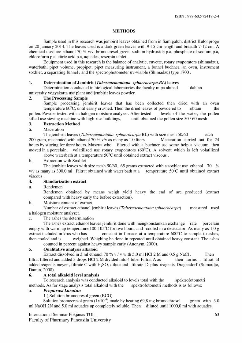
ISBN : 978-602-72418-2-4
International Seminar Pokjanas TOI
Faculty of Pharmacy Pancasila University
63
METHODS
Sample used in this research was jembirit leaves obtained from in Samigaluh, district Kulonprogo
on 20 january 2014. The leaves used is a dark green leaves with 9-15 cm length and breadth 7-12 cm. A
chemical used are ethanol 70 % v/v, bromocresol green, sodium hydroxide p.a, phosphate of sodium p.a,
chloroform p.a, citric acid p.a, aquades, reserpin tablet .
Equipment used in this research is the balance of analytic, cuvette, rotary evaporators (shimadzu),
waterbath, pipet volume, propipet, pipet measuring instrument, a funnel buchner, an oven, instrument
soxhlet, a separating funnel , and the spectrophotometer uv-visible (Shimadzu) type 1700 .
1. Determination of Jembirit (Tabernaemontana sphaerocarpa.BL) leaves
Determination conducted in biological laboratories the faculty mipa ahmad dahlan
university yogyakarta use plant and jembirit leaves powder.
2. The Processing Sample
Sample processing jembirit leaves that has been collected then dried with an oven
temperature 600C, until easily crushed. Then the dried leaves of powdered to obtain the
pollen. Powder tested with a halogen moisture analyzer. After tested levels of the water, the pollen
sifted use sieving machine with high-rise buildings, until obtained the pollen size 50 / 60 mesh .
3. Extraction Method
a. Maceration
The jembirit leaves (Tabernaemontana sphaerocarpa.BL) with size mesh 50/60 each
200 gram, macerated with ethanol 70 % v/v as many as 1.0 liters. Maceration carried out for 24
hours by stirring for three hours. Maserat who filtered with a buchner use some help a vacuum, then
moved in a porcelain, volatilized use rotary evaporators (600C). A solvent which is left volatilized
above waterbath at a temperature 500C until obtained extract viscous .
b. Extraction with Soxhlet
The jembirit leaves with size mesh 50/60, 65 grams extracted with a soxhlet use ethanol 70 %
v/v as many as 300,0 ml . Filtrat obtained with water bath at a temperature 500C until obtained extract
viscous .
4. Standarization extract
a. Rendemen
Rendemen obtained by means weigh yield heavy the end of are produced (extract
compared with heavy early the before extraction).
b. Moisture content of extract
Number of extract ethanol jembirit leaves (Tabernaemontana sphaerocarpa) measured used
a halogen moisture analyzer.
c. The ashes the determination
The ashes extract ethanol leaves jembirit done with mengkonstankan exchange rate porcelain
empty with warm up temperature 100-105oC for two hours, and cooled in a desiccator. As many as 1.0 g
extract included in krus who has constant in furnace at a temperature 600oC to sample to ashes,
then cooled and is weighed. Weighing be done in repeated until obtained heavy constant. The ashes
counted in percent against heavy sample early (Anonym, 2000).
5. Qualitative analysis alkaloid
Extract dissolved in 3 ml ethanol 70 % v / v with 5,0 ml HCl 2 M and 0.5 g NaCl . Then
filtrat filtered and added 3 drops HCl 2 M divided into 4 tube. Filtrat A as their forms , filtrat B
added reagents meyer , filtrate C with H2SO4 dilute and filtrate D plus reagents Dragendorf (Sumardjo,
Damin, 2008).
6. A total alkaloid level analysis
To research analysis was conducted alkaloid to levels total with the spektrofotometri
methods. As for stage analysis total alkaloid with the spektrofotometri methods is as follows:
a. Preparasi Larutan 1 ) Solution bromocresol green (BCG)
Solution bromocresol green (1x10⁻⁴
) made by heating 69,8 mg bromochresol green with 3.0
ml NaOH 2N and 5.0 ml aquades up completely soluble. Then diluted until 1000,0 ml with aquades
ISBN : 978-602-72418-2-4
International Seminar Pokjanas TOI
Faculty of Pharmacy Pancasila University
64
2) Dapar phosphate-citric pH 2.2
For solution sodium phosphate 2M (71.6 g Na2HPO4 in 1000,0 ml aquades) then adjust pH 2.2
with a solution of citric acid 0.2M (42.02 g citric acid in 1000.0 ml aquades)
3) Standard solutions of reserpin
Standard solutions of reserpin have weigh equivalent 25 µg/ml reserpin in 10.0 ml acetic acid
glacial, and add until 100.0 ml aquades.
b. The determination of operating time Standard solutions of reserpin by concentration of the 25 ug / ml with 5.0 ml dapar phosphate pH
2,2 and 5.0 ml solution bcg , then extracted by 5.0 ml chloroform (2 times) and extracted phase
chloroform. The extraction was collected in squash measures 10.0 ml, then add by chloroform to
sign. Then examined time absorption a stable at wavelengths 420.8 nm. The data collected used
to make a curve the relationship between absorbansi to the time .
c. The determination of maximum Wavelenghth Standard solutions of reserpin with low 25 µg/ml included in a separating funnel coupled with 5
ml dapar phosphate pH 2.2 and 5.0 ml solution BCG, then extracted by 5.0 ml chloroform (2
times) and extracted phase chloroform. The extraction brought in squash 10.0 ml measures, then
add with chloroform to sign. Checked absorbansinya at wavelengths 350-700 nm using the
operating time has received. The data used to make a curve the relationship between absorbansi
and wavelength.
d. The Determination of Standart Curve Standard solutions of reserpin by concentration of the 25 µg/ml taken 4,0; 5.0; at 6; 7,0; 8.0; 9;
10.0 ml then put in a funnel break coupled with 5.0 ml dapar phosphate ph 2.2 and 5.0 ml
solution bcg, then extracted by 5.0 ml chloroform (2 times) and extracted phase chloroform. The
extraction was collected in squash measures 10.0 ml, then add by chloroform to sign. Then
examined absorbansinya at wavelengths maximum.
e. Sample Preparation Preparasi samples from 50,0 mg extract ethanol leaves jembirit dissolved in 3 ml HCl 2 N. Then
1.0 ml filtrat extracted use 5.0 ml chloroform ( 3 times). After extracted, pH solution neutralized
use 0.1 N NaOH. Then added 5.0 ml dapar phosphate pH 2.2 and 5.0 ml solution BCG. A
mixture of the extracted back with 5.0 ml chloroform (2 times). Phase chloroform was collected
in squash measures 10.0 ml, then in addition to with chloroform to sign. Read absorbansi at
wavelengths maximum (Amrizal, 2010).
f. Data Analysis An alkaloid levels total with the maceration methods and extraction with a Soxhlet tested
normality and homogeneity use application SPSS. Test normality done with kolmogorov-smirnov
analysis . Test of homogeneity done with levene analysis . When data normally distributed and
homogeneous , then the analysis continued with the methods t-test. But if results showing that the
data not normally distributed and not homogeneous or one of them , the continued with the
methods non parametric .
RESULT AND DISCUSSION
a. The Determinasi of Jembirit (Tabernaemontana sphaerocarpa BL) From the determination be seen that plant used in this research was leaves jembirit ( Tabernaemontana
sphaerocarpa BL ). Leaves a deep green, and the powder of leaves green brownish .
Materials Pollinated collection and Jembirit Leaves then dried in an oven with a temperature of 600C to
reduce the water content of simplicia. Dried leaves diserbuk and sifted using a sieve mesh 50/60, until
retrieved the powder with size 50/60. The purpose of the pollination is done to minimize the size of the
particle surface area so that particle simplisia becomes large so that the liquid penyari that will easily
dissolve the active compounds of simplicia.

ISBN : 978-602-72418-2-4
International Seminar Pokjanas TOI
Faculty of Pharmacy Pancasila University
65
b. The determination of the moisture content of jembirit leaves powder Drying shrinkage Measurements-dust powder is done by using Halogen Moisture Analyzer. Shrinkage of
drying powders can be measured as a reference of the moisture contained in simplisia, because it does not
contain jembirit leaf essential oil so that experience evaporation in drying shrinkage is the determination
of the moisture content contained in the extract. Table I. Moisture content of jembirit leaves powder
Based on
the data
moisture
content can
be
concluded
that the simplisia leaves jembirit meet the requirements because the water level contained less than 10 % (
Anonym, 2000 ).
c. Extraction Making extract leaves jembirit done variation method that is by using a method of maceration and with a
soxhlet. Maceration methode are liquids would pass through the cell walls, an active substance will
dissolved because of differences in concentration between solution an active substance in in the cells and
outside a cell, so that solution of high concentration will out from cells (Anonym, 1986 ).
Maserat obtained concentrated with rotary vaporator and water bath until obtained extract viscous the
process extraction with the methods of using tools soxhlet done by heating a solvent at a temperature
700C .When a solvent ethanol 70 % v /v heated, a solvent would evaporate and will form fluid back when
about cooling turning. A solvent fluid drop on the simplisia, and will dissolving back an active substance
from the powder. The warming can affect levels of an active substance from the powder. To compound
active that do not bear warming cannot be done with this method (Dayanti and suyatno, 2012). An extract
obtained concentrated by water bath until obtained extract viscous.
d. Standarisation extract 1. Rendemen
The size of value of a rendemen show effectiveness of the process extraction .
Table II. Rendemen based on extraction method
Extraction Method Mass
Simplisia Mass Ekstrak Rendemen
Maceration 200,250 g 89,290 g 44,59 %
Soxhlet 64,990 g 32,226 g 49,58 %
Efectivitas extraction process influenced by the types of solvent, size of particles, a method of the
extraction and long process of extraction .
2. Moisture content On jembirit leaves does not contain volatile oil, so as to the result of moisture content can as the
reference are contained in extract .
Table III. Moisture content of extract
Extraction Methode Moisture Content X ± LE CV
Maceration
5,69 %
5,67 % ± 0,03 2,30 % 5,80%
5,54 %
Soxhlet
9,30 %
9,27 % ±0,02 0,90 % 9,35 %
8,89 %
Replication Weight Moisture
content X ± LE CV
1 1,224 g 5,51 %
(5,48% ± 0,015)
1,09 % 2 1,145 g 5,41 %
3 1,312 g 5,54 %

ISBN : 978-602-72418-2-4
International Seminar Pokjanas TOI
Faculty of Pharmacy Pancasila University
66
From the results of the determination of moisture content extract ethanol leaves jembirit with
variations method penyarian above can be concluded that extract produced meet the requirements
at less than 10 %. Moisture content to the extraction method with a soxhlet higher than with the
maceration.
3. The determination of ashes The determination of ashes total can be used for various purposes , among others to determine
good or failure a processing , the knowledge of the material used (Apriantono, 1989) . Tabel IV. The Determination of ashes ethanolic extract of jembirit leaves
Extraction Methode The determination of
ashes X ± LE CV
Maceration
9,61 %
9,80% ± 0,186 1,898 % 9,98 %
9,82 %
Soxhlet
5,75 %
5,79% ± 0,061 1,050 % 5,76 %
5,86 %
e. Qualitative of Alkaloid Qualitative analysis alkaloid identification extract to compound the alkaloid was conducted
qualitativ by using test tube with reagent mayer, dragendorf, and H2SO4 dilute. Extract ethanol
leaves jembirit dissolved in HCl 2 N tested positive contain the alkaloid if when sample with
reagent mayer will form the sediment yellowish white, reagent dragendorf produce precipitate
brown orange, and add H2SO4 are not formed the sediment (Dayanti and suyatno, 2012).
f. The levels of Alkaloid The levels of total alkaloid done with the complex bromocresol green (BCG) in
spectrophotometri visible. The principle of this method is based on the levels of alkaloid the
formation of complex reagents between alkaloid with BCG to form a compound yellow.
Picture 1. Complex BCG and Alcaloid
g. The determination of operating time (OT) The research obtained for operating time standard reserpin is from 43-50 minutes .While to
extract the results of maceration is in the to 53-57 , and to extract the results with a soxhlet is in
the to 10-12.
h. Determination of a wavelength on absorbansi maximum In this research obtained a wavelength on absorbansi maximum solution reserpin is 420,8 nm,
extract the results of maceration 415,5 nm and extract the with a soxhlet 417,5 nm.
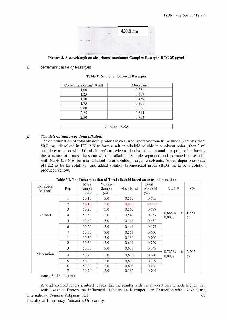
ISBN : 978-602-72418-2-4
International Seminar Pokjanas TOI
Faculty of Pharmacy Pancasila University
67
Picture 2. A wavelength on absorbansi maximum Complex Reserpin-BCG 25 µg/ml
i. Standart Curve of Reserpin
Table V. Standart Curve of Reserpin
Consentration (µg/10 ml) Absorbansi
1,00 0,231
1,25 0,307
1,50 0,459
1,75 0,501
2,00 0,556
2,25 0,614
2,50 0,703
y = 0,3x - 0,05
j. The determination of total alkaloid The determination of total alkaloid jembirit leaves used spektrofotometri methods. Samples from
50,0 mg , dissolved in HCl 2 N to form a salt an alkaloid soluble in a solvent polar , then 3 ml
sample extraction with 5.0 ml chloroform twice to deprive of compound non polar other having
the structure of almost the same with the alkaloid. Sample separated and extracted phase acid,
with NaoH 0.1 N to form an alkaloid bases soluble in organic solvents. Added dapar phosphate
pH 2.2 as buffer solution , and added solution bromocresol green (BCG) as to be a solution
produced yellow.
Table VI. The Determination of Total alkaloid based on extraction method
Extraction
Method Rep
Mass
sample
(mg)
Volume
Sample
(mL)
Absorbansi
Total
Alkaloid
(%)
X ± LE CV
Soxhlet
1 50,10 3,0 0,559 0,675
0,666% ±
0,0022
1,651
%
2 50,10 3,0 0,432 0,536*
3 50,20 3,0 0,562 0,677
4 50,50 3,0 0,547 0,657
5 50,60 3,0 0,545 0,652
6 50,20 3,0 0,461 0,677
7 50,50 3,0 0,551 0,660
Maceration
1 50,30 3,0 0,589 0,706
0,727% ±
0,0032
2,202
%
2 50,30 3,0 0,611 0,729
3 50,50 3,0 0,627 0,743
4 50,20 3,0 0,620 0,740
5 50,30 3,0 0,618 0,739
6 50,30 3,0 0,608 0,726
7 50,20 3,0 0,585 0,704
note : * : Data delete
A total alkaloid levels jembirit leaves that the results with the maceration methods higher than
with a soxhlet. Factors that influential of the results is temperature. Extraction with a soxhlet use
420.8 nm
ISBN : 978-602-72418-2-4
International Seminar Pokjanas TOI
Faculty of Pharmacy Pancasila University
68
some help temperatures of roughly 700C so that there is alkaloid compound that do not bear warming
damaged a consequence of the warming. In addition , in extract the results with a soxhlet the water level
contained is greater than the maceration, so that influential in the alkaloid total obtained .
Based on the t-test obtained significance 0,001 (<0,005 ) where HO rejected which means there is a
difference meaningful so that can be concluded that extraction method impact on the alkaloid total leaves
jembirit (Tabernaemontana sphaerocarpa ) .
CONCLUSION
Jembirit Leaves (Tabernaemontana sphaerocarpa Bl) compound containing alkaloid. A total alkaloid
levels jembirit leaves with the maceration methods is 0.727 % ± 0.0032 , a total alkaloid levels jembirit
leaves with a soxhlet is 0.666 % ± 0.0022. Different calculation extraction methods impact on the total
alkaloid jembirit leaves. The stastitical analysis showed significance differences of total alkaloid levels
between maceration method and extraction with Soxhlet apparatus viewed from siginificancy value
(0.001<0.005).
REFERENCE
1. Amrizal. Isolasi Senyawa Alkaloid Dari Ekstrak Metanol Daun Tumbuhan
Tabernaemontana sphaerocarpa BI (Apocynaceae). Pekanbaru. Fakultas F-MIPA
Universitas Riau. 2010
2. Anonim. Tabernaemontana sphaerocarpa BL, Jakarta : Departemen Kesehatan
Indonesia.Jakarta. 2001
3. Anonim. Sediaan Galenik. Departemen Kesehatan Republik Indonesi. Jakarta.Hal 19-20. 1986
4. Anonim. Paremeter Standar Umum Ekstrak Tumbuhan Obat, EDISI I. Departemen
Kesehatan Republik Indonesia. Jakarta. Hal 10-12. 2000.
5. Ansel. H.C. Pengantar Bentuk Sediaan Farmasi. Edisi keempat. Jakarta.UI Press. Hal 103-
105. 2005.
6. Apriyantono, A., D. Fardiaz, N. L. Puspitasari, Sedamawati dan S. Budiyanto., Analisis Pangan.
PAU Pangan dan Gizi. IPB Press. 1989.
7. Dayanti, R., dan Suyatno.. Aktifitas Anti Oksidan Ekstrak Metanol Bagian Batang
Tumbuhan Paku Nephrolepis radicans (Burm) Kuhn. UNESA Journal of Chemistry. 1(1) ; 86-92.
2012.
8. Sumardjo, Damin. Pengantar Kimia. Jakarta: EGC. Hal 26. 2008.
9. Zaima K, Hirata T, Hosoya T, Hirasawa Y, Koyama K, Rahman A, Kusumawati I, Zaini NC,
Shiro M, Morita H.. Biscarpamontamines A and B, an Aspidosperma-iboga Bisindole
Alkaloid and an Aspidosperma-aspidosperma Bisindole Alkaloid,From Tabernaemontana
sphaerocarpa. Faculty of Pharmaceutical Sciences, Hoshi University, Ebara 2-4-41
Shinagawa, Tokyo, Japan. 2009.
ISBN : 978-602-72418-2-4
International Seminar Pokjanas TOIFaculty of Pharmacy Pancasila University
69
INTEGRATION OF HERBAL OR TRADITIONAL MEDICINETHROUGH EVIDENCE BASED PRACTICE
Anny Lumban Toruan*, Galih Ajeng Kencana Ayu ***Graduate Pharmacy Program, University of Pancasila, Indonesia
** Public Health Technology Intervention Center, National Institute Health Research andDevelopment, Ministry of Health of Indonesia.
Corresponding: [email protected]
ABSTRACT
Traditional medicines are used by some 60% of the world’s population and in some countries areextensively incorporated into the public health system. Herbal medicines are plant extracts that have beenused for medicinal purposes for thousands of years. The integration of herbal and other forms oftraditional medicine (TM) can be done as follows: incorporation of traditional and modern evidence-based medicine (EBM) as integral parts of a country’s formal health care system. It is most likely to beachieved and has been demonstrated to be practicable in many countries, particularly in Asian countriessuch as China, Japan, Korea, and India, among others it can be practice integrated with modern medicineby individual health care practitioners and also it can be integrated as two branches of medical science,with the ultimate incorporation of elements of both to form a new branch. A range of interrelated quality,safety and efficacy issues could contribute to the rational and successful integration of herbal medicineinto modern medical practices. The issues related to the integration such as herb quality, qualityassurance/quality in processing and manufacturing/preparation of herbal medicines (good Manufacturingpractical) issues, herbal mechanisms of action, bioavailability and herbs’ chemical constituents, herb druginteractions, herb-herb interactions, efficacy measurements objective quantiviable versus subjectivequality of life and other safety issues has to be done.Key words: traditional medicines, herbal medicines, evidence based practice, formal health care
INTRODUCTION
In the last few decades biomedicine has been revolutionized by; the application of the principles ofEvidence Based Medicine (EBM) as a way of establishing the effectiveness and safety of modernmedical intervention.1
In Western countries, such as the United States, Australia, Canada, and members of the European Union,the popular use of herbal medicine in the form of complementary and alternative medicine (CAM) orphytomedicine in the last two to three decades has led to a multinational, multibillion dollar industry,professional and trade organizations, national and international practice and research conferences,establishment of specialized integrated medicine practices and clinics in pain management and adjunctivecancer therapy, incorporation of CAM courses in conventional medical colleges, introduction of CAMdegree-level education programs, and establishment of research funding agencies such as the U.S.National Institutes of Health (NIH) National Center for Complementary and Alternative Medicine. 1
Indonesian herbal medicines, called JAMU, have been widely used by Indonesian to maintain their healthand to cure the diseases since many centuries ago. In the future, the development and the use ofIndonesian herbal medicines must be based on the stronger scientific evidence, especially through R&Dand standardization, so that they can be integrated into national health care system.2
The integration of herbal and other forms of traditional medicine (TM) can be done in one of thefollowing three ways: First, it can be incorporated as an integral part of a country’s formal health caresystem, with each being separately recognized as legitimate forms of health care within the sameframework. Second, it can be practice integrated with modern medicine by individual health care

ISBN : 978-602-72418-2-4
International Seminar Pokjanas TOIFaculty of Pharmacy Pancasila University
70
practitioners. Third, traditional and modern practices can be integrated as two branches of medicalscience, with the ultimate incorporation of elements of both to form a new branch. 3
METHODS
A. FACTORS INFLUENCING THE INTEGRATION:Several factors influencing the integration of herbal medicine in evidence based medical therapy werediscussed below:
a. Current status and major issues of integration of herbal medicine in evidence-based medicaltherapy
Currently, thousands of TM and other CAM herbal products are available as therapeutic agentsworldwide. Yet few of these products have been subjected to randomized clinical trials (RCTs) under theInternational Conference on Harmonization (ICH) Good Clinical Practice Guidelines to determine theirefficacy and/or safety.4. Of the nearly 2000 herbal medicine clinical studies listed on the CochraneControlled Trials Register as of June 2009, most concern single-plant herbal or phytomedicine. In recentyears, in the case of multicomponent herbal medicines, an increased number of RCTs on traditionalherbal medicine has been reported in the literature. Concluded that the majority of these studies sufferedfrom methodological defects. For example, only 15% of these studies used blinding, the sample size wasmostly less than 300 patients, the controls were inadequate, few studies used quantitative outcomemeasures, and the studies were short term.5
b. Factors Relevant Affecting Integration of Herbal Medicine Into Modern MedicalPractices.
A range of interrelated quality, safety and efficacy issues could contribute to the rational and successfulintegration of herbal medicine into modern medical practices.
1. Herb Quality IssuesFundamental to assuring the efficacy and reproducibility of any medicinal agent, be it a single chemicalor a complex herbal mixture, is the assured quality of the product. In the case of single chemical drugs,the quality and properties are well defined and documented in pharmacopoeias or on file with regulatoryagencies or marketing authorities. On the other hand, herbal medicines, be they single herbs orpolyherbal products, suffer from a lack of uniformity in their chemical and physical qualities due tovarious factors as mentioned above.6 Unintentional in-process adulteration with heavy metals, microbialand chemical agents (pesticides, herbicides, and heavy metals), as well as with foreign matter such asinsects, animals, animal parts, and animal excreta during any of the stages of source plant materialproduction or procurement can result in unsafe source materials. Multicomponent Chinese or Ayurvedicherbal medicines have long been documented to be adulterated with synthetic anti-inflammatory drugssuch as phenylbutazone, Indomethacin, and/or corticoid steroids in arthritis remedies.7
2. Good Manufacturing Practices Issues.The most important extrinsic factor affecting the quality of herbal medicines is the lack of effectivepolicies on quality assurance (QA)/QC in the processing and manufacturing of herbal products undergood manufacturing practices (GMP). The majority of herbal medicines marketed in the United States aresold as dietary supplements under the provisions of the Dietary Supplement Health and Education Act(DSHEA) of 1994, and only recently been mandated by law to be produced under cGMP.8
3. Herbal Mechanisms of Action, Bioavailability, and Herbs′ Chemical ConstituentsThe underlying mechanisms of action of herbal medicine, whether single herbal or multiple herbalformulations, have generally not been elucidated due to the lack of knowledge of identifying theircontained active and/or adjuvant phytochemical constituents. The same problem applies to the study ofpharmacokinetics and bioavailability.9
4. Herb–Drug Interactions
ISBN : 978-602-72418-2-4
International Seminar Pokjanas TOIFaculty of Pharmacy Pancasila University
71
The potential of interactions of herbal medicine with prescribed drugs or OTC drugs has been a majorsafety concern for clinicians as such interactions are difficult to predict and the general lack of availableinformation on the herbs’ composition and pharmacological actions. If the definition of the herbalmedicine extended to botanicals including fungi, algae, and other component matters, nearly 80 herbalmedicines would be identified that had clinically significant interactions with drugs. Garlic, ginger, andginkgo are among the herbs most commonly involved in herb–drug interactions.10
5. Herb-Herb InteractionsHerb–herb interactions, sometimes referred to as contraindications in the application of herbs orprescription incompatibility, were documented in ancient textbooks on TCM medicinal formulae (i.e., amixture of herbs). Herbal medicine practice routinely uses combination herbal formulations, termedpolypharmacy, designed to enhance the effectiveness and minimize any potential side effects oftreatment. Synergy between active ingredients is a characteristic aspect of herbal treatment and occurs atboth a pharmacodynamic level and at a pharmacokinetic level. In both cases, active ingredientsdemonstrate potentiated effects—in other words, the therapeutic effect of herbal medicines is greater thenthe sum of its constituents parts.1
6. Efficacy Measurements: Objective Quantifiable versus Subjective Quality of LifeThe evidence needs to be verified legitimately and scientifically according to the conventional EBMframework. If possible, evidence generated for herbal medicine should be derived from the mostpowerful method of testing the effect of treatment intervention, the RCT. With a plausible biologicalbasis, herbal products can be evaluated through double-blinded, placebo-controlled, multicenter trials.Reflecting this, the World Health Organization (WHO) has published a number of guidelines for clinicalevaluation of the herbal and TMs.10 Within the EBM paradigm, RCTs are suggested to be reported inaccordance with the 22-item Consolidated Standards of Reporting Trials (CONSORT) checklist, such asa detailed description on patient eligibility criteria, sample size calculation, specific objectives andhypotheses, implementation of the trial, and statistical methods, regardless of whether the intervention isconventional or herbal.11
7. Other Safety IssuesOther safety issues include cultural and behavioral contexts as well as efficient communication on its useamong patients, conventional medical practitioners, and herbal medicine practitioners. Over a fewdecades of development and with more scientific research data being published, although not allconvincing, at least some promising evidence has met the EBM standard. Nevertheless, it is of criticalconcern to clinicians that many herbal medicine users take herbal remedies and conventional therapiesconcurrently without informing their medical doctors. Such communication gaps can lead to herb–druginteractions that may be otherwise avoided.12
c. Quality Assurance, Quality Control and Standardization of Herbal MedicinesQA of herbal medicine for integrative medical use is a process spanning from the acquisition of thesource material to the production of the clinical formulation. Therefore, QA/ QC research on sourcematerials should begin from the point at which a specific plant part to be used is acquired by cultivationor field collection through Good agricultural practice GACP. GACP Guidelines have been established bya number of countries, and the WHO has also published a guideline on GACP to assist member states inthe production of quality herbs. A most essential part of botanical QA is that plant materials should beidentified by their scientific names (Latin binomial) rather than by common names and should beauthenticated botanically according to pharmacopoeial standards employing macroscopic/organolepticand microscopic methods. Each herb should be subjected to purity as well as contaminant tests for thepresence of foreign matters, toxic metals, pesticide residues, mycotoxins, and microorganisms.13
d. Preclinical Pharmacological Assessments and Action MechanismsIn current practice, acute and chronic toxicities are usually determined by experimental studies usinganimal models. Suitable methods for testing toxicity need to be established so that herbal ingredients andtheir derived products can be reliably assessed. However, the biological response to a drug product may

ISBN : 978-602-72418-2-4
International Seminar Pokjanas TOIFaculty of Pharmacy Pancasila University
72
not be species transferable, and an active substance in animals may be entirely inactive in humans. On theother hand, acute and/or chronic toxicity manifestations in animal models are reliable indicators of drugsafety. 7
e. Clinical Efficacy and Safety AssessmentsFundamentally, conducting and reporting clinical studies on the efficacy and safety of herbal medicineshould follow the context-specific elaborations of the CONSORT statement.11 In addition to thedocumentation of all general aspects of RCTs (e.g., randomization, blinding, and analysis) that are knownto influence the estimation of treatment effects, specific considerations are needed to attend to the uniqueobstacles of implementing herbal medicine trials.
B. RESEARCH NEEDSFor effective integration of herbal medicine into modern therapeutic practices, the level of research onthe preclinical and clinical efficacy of these products and their complementation of or interaction withmodern pharmaceuticals must be elevated to much higher levels than is presently the case. Preliminary tothese studies, clinical products must be produced by GMP from source materials acquired through goodagriculture and collection practices (GACP),14,16 be botanically validated, be chemically and/orbiologically standardized, and their stability be established. Hence, the research on herbal medicines forthe integrated medical use must begin with the acquisition and QC of source materials and processedstarting materials. The three most important major areas of research can be defined as (1) herbal medicinequality and standardization, (2) preclinical pharmacological assessments and action mechanisms, and (3)clinical efficacy and safety assessments.
CONCLUSIONS
WHO member states and other stakeholders were called upon to take steps to integrate TM into nationalhealth systems. But integration of herbal medicine into modern clinical practice must be based on anEBM approach. Prior to the clinical evaluation of herbal medicine, be it a single compound, a mixture ofherbal ingredients, or a complex herbal formula based on historic evidence of use, the QA/QC in sourcematerial acquisition and processing and manufacturing of the products under GMP must be addressed toassure efficacy and reproducibility. In addition to the use of scientifically irrefutable efficacymeasurements, clinical studies should monitor and report adverse events, including potential drug–herbinteractions. When the safety and efficacy are established in accordance with conventional scientificprinciples, the integration of herbal medicine into evidence-based clinical practice will likely occur.
REFERENCES
1. McClure L, Flowler A, and Price S., Scoping the evidence for the effectiveness of HerbalMedicines. January 2014:1
2. WHO traditional medicine strategy 2014-2023. World Health Organization 2013:383. WHO. 2008. http://www.who.int/dg /speeches/2008/ 20081107/en/index.html WHO Congress on
traditional medicine. (Accessed August 2, 2009).4. International Conference on Harmonization. 2010. http.//www.ich.org/cache/compo/276-254-1.html
ICH guidelines. (accessed September 2014 .5. Cochrane Collaboration. 2009. www.thecochranelibrary.com The Cochrane Library. (accessed May
22, 2011).4. Gagnier J. J, DeMelo J, Boon H, Rochon P, Bombardier C. Quality of reporting of randomized
controlled trials of herbal medicine interventions. Am J Med. 2006;119(9):800.e1–11.. [PubMed]5. Hu Z, Yang X, Ho P. C, Chan S. Y, Heng P. W, Chan E, Duan W, Koh H. L, Zhou S. Herb-
drug interactions. A literature review. Drugs. 2005;65(9):1239–82. [PubMed]6. Ulbricht C, Chao W, Costa D, Rusie-Seamon E, Weissner W, Woods J. Clinical evidence
of herb-drug interactions. A systematic review by the natural standardresearch collaboration. Curr Drug Metab. 2008;9(10):1063–120. [PubMed]

ISBN : 978-602-72418-2-4
International Seminar Pokjanas TOIFaculty of Pharmacy Pancasila University
73
7. Fong H. H. S, Pauli G. F, Bolton J. L, van Breemen R. N, Banuvar S, Shulman L, Geller S. E,Farnsworth N. R. Evidence-based herbal medicine: challenges in efficacy and safety assessments.In: Leung P. C, Fong H. H. S, Xue C. C, editors. Annals of Traditional Chinese
8. World Health Organization. WHO Guidelines on Good Manufacturing Practices (GMP)for Herbal Medicines. Geneva: World Health Organization; 2007.
9. Fong H. H. S, Pauli G. F, Bolton J. L, van Breemen R. N, Banuvar S, Shulman L, Geller S. E,Farnsworth N. R. Evidence-based herbal medicine: challenges in efficacy and safety assessments.In: Leung P. C, Fong H. H. S, Xue C. C, editors. Annals of Traditional Chinese Medicine Vol 2:Current Review of Chinese Medicine. Singapore: World Scientific; 2006. pp. 11–26.
10. WHO, 2000. General Guidelines for Methodologies on Research and Evaluation of TraditionalMedicine, World Health Organization, Geneva 2000:9-17.
11. CONSORT 2010 Statement, Published online March 24, 2010 Webappendix (availableat www.consort-statement.org), access Jan 11, 2012.
12. Chadwick L, Fong H. H. S. Herb quality assurance and standardization in herb-druginteraction evaluation and documentation. In: Lam Y. W. F, Huang S. M, Hall S. D,editors. Herbal Supplement-Drug Interactions. New York: Taylor & Francis; 2006. pp.191–203.
13. World Health Organization. WHO Guidelines on Good Agricultural and CollectionPractices (GACP) for Medicinal Plants. Geneva: World Health Organization; 2003.

ISBN : 978-602-72418-2-4
International Seminar Pokjanas TOI
Faculty of Pharmacy Pancasila University 74
Identification of Soursop Seeds (Annona Muricata L.)Extract as A Candidate
Against The Aedes Aegypti L. Musquito
Vector Control DBD
SARAH ZAIDAN, RATNA DJAMIL, SITI NURAINI
Faculty of Pharmacy, Pancasila University
Jln. Srengseng Sawah Jagakarsa, Pasar Minggu, Jakarta Selatan 12640.
Abstract: Aedes aegypti L. mosquitos are the disease vectors of dangue hemorrhagie fever (DHF),
caused by dengue virus which is transmitted by Ae. Aegypti mosquito. The effort to control Ae. aegypti
vector have been done in so many times, including chemical, physical, and biological control method.
Multilevel extraction by kinetic maceration have been done with soursop seeds (Annona muricata L.)
with the solvent of n-hexane, ethyl acetate and 70% of ethanol. Subsequently, the obtained extract is
tested phytochemical screening along with the powder and larvicidal activity against Ae. aegypti. The
results of phytochemical screening of the powder and 70% ethanol extract of soursop seeds have
obtained the compound of saponin, triterpenoid and coumarin. In the n-hexane extract have obtained
triterpenoids and in ethyl acetate extract which is found triterpenoids and coumarin. Based on the activity
test against the larva of Ae. aegypti from n-hexane extract, ethyl acetate and ethanol 70% of soursop seeds
sequentially, show LC50 values were about 198,610 ppm, 74,798 ppm and 67,042 ppm. Soursop seeds
extract that has the highest activity is 70% ethanol extract. These are indicate that the chemical
compounds which is found in soursop seeds have a potential as a larvicides.
Keywords:Aedes aegyti L., Annona muricata L., soursop seeds, larvasida.
INTRODUCTION
Mosquitoes are insects that are often found in tropical countries such as Indonesia. Besides
disturbing human life, the presence of mosquitoes act as vectors of some diseases. In Indonesia, a disease
transmitted by mosquitoes is still a health problem because of the high mortality rate caused. Some
diseases transmitted by mosquito vectors such as filariasis, malaria and dengue fever (DBD)(1,2)
. Aedes
aegypti L. mosquitoes are diurnal, or active during the morning and afternoon. Ae. aegypti mosquitoes
carrying dengue virus causes dengue which is obtained from infected individuals and multiply in the body
and the salivary glands of mosquitoes (2)
.
Dengue disease not only in children but in all ages. DBD becoming known in Indonesia in 1968 in
Surabaya and Jakarta, and then continue to expand as the spread of dengue endemic area. The number of
cases of dengue and widely spread is increasing along with the increasing mobility and population
density. There are 150,000 cases of dengue in 2007 and continued to increase until 2010. In addition,
WHO reported more than 35% of the population living in urban areas affected by the disease. Until now
there is no specific vaccine to treat dengue fever, and the only control vector for controlling the spread of
the disease(2, 3, 4, 5)
.
Vector control mosquitoes until today, still put emphasis on the use of insecticides, for example, is a
synthetic larviciding. Larvicides in general have a higher efficacy and the results can be seen quickly. The
use of continuous and repetitive can cause environmental pollution and resistance against the target
organism. This encourages biological larviciding as controlling mosquito vectors (Biolarvasida). These
biological larvicides are safer for humans, readily available and environmentally friendly(2, 6)
.
Biological larvicides useful for the improvement of local natural resources. Local plant as a potential
biological larvicides generally from families Annonaceae, including soursop (Annona muricata L.).
Empirically, it has been much research done on the soursop as larvicides. The plant parts are potentially
as larvicides are seed (semen)(7)
. Soursop seed (with shell beans) have larvicidal activity against Ae.
aegypti with LC50 value of 244.27 ppm for the ethanol extract of the seeds of soursop6, 8
. In addition,
Ward et.all, reported that seeds of soursop and sugar apple seed (without seed coat) effect on mortality
Chrysomya bezziana fly larvae(9, 10)
.

ISBN : 978-602-72418-2-4
International Seminar Pokjanas TOI
Faculty of Pharmacy Pancasila University 75
The main active compounds from the seeds of the soursop is annonacin and squamocin belonging
asetogenin compound. Squamocin annonacin compound of the family Annonaceae and is reported to
have toxicity properties are quite effective against insects of the order Diptera (Ae.aegypti L.) which are
cytotoxic, and neurotoxic. Asetogenin compounds can inhibit the action of the enzyme NADH in the
mitochondria, causing the death of larvae, as well as toxic contact and stomach poison to insects(6, 9, 10)
.
This research is continuing efforts to obtain biological larvicides. In this study, conducted Qualitative
identification through screening phytochemicals and seen the activity of seed extracts of soursop (Annona
muricata L.) (without seed coat) against Ae. aegypti. Extract obtained from extraction method using a
multi-storey penyari with different polarities ie n-hexane, ethyl acetate and ethanol 70% and seen the
highest larvicidal activity of the three fractions of the extract. Extract larvicidal activity test using
Standard Methods of Pesticide Efficacy Testing Households and Vector Control11
. The data seen LC50
values were obtained by probit analysis using Probit Analysis Program Epa Used For Calculating LC /
EC Values Version 1.5.
MATERIAL AND METHODS
MATERIAL. Soursop seeds (Annona muricata L.) were obtained from Balitro (Research Institute for
Spices and Medicinal Plants) Cimanggis, Bogor. Determination at Bogoriense Herbarium, Research
Center, LIPI Cibinong , Bogor.
Extraction. Seed Soursop (Annona muricata L.) which have been dried in the sun was directly
crushed and blended into a fine powder. Powdered crude drug was extracted by maceration kinetic in
stages using different solvent polarity is n-hexane, ethyl acetate, and ethanol 70% at room temperature
until the extracted perfectly, then filtered with cotton and proceed with filter paper, pulp, and each extract
n -heksan, ethyl acetate, and ethanol is 70% separated. Each extract was concentrated by vacuum rotary
evaporator at a temperature of 450 C to obtain a viscous extract n-hexane, ethyl acetate and ethanol 70%.
Identification with phytochemical screening. Phytochemical screening performed on pollen and
seed extract of soursop with Farnsworth method in Biological and phytochemical screening of Plant seed
sirsakdilakukan to identify the qualitative content of secondary metabolites in seed soursop.
Flavonoids. 2 grams of powder simplisia or 0,67 g of n-hexane extract and ethyl acetate extract;
0.15g of extract ethanol 70% boil with 100 ml of hot water for 5 minutes, then filtered with filter paper, 5
mL filtrate of extract solution coupled with a bit of powdered zinc or magnesium and 1 mL of 2 N HCl
and 5 mL amyl alcohol . Flavonoids compounds would pose orange to red(12)
.
Saponins. Entering 10 ml sample into a test tube and shake for 30 seconds and observe what
happens. If the foam is formed solid (not lost for 30 seconds) the identification showed the presence of
saponins(12)
.
Coumarin. 2.12 grams of powder simplisia or 0.67 g of n-hexane extract and ethyl acetate extract;
0.15g of extract ethanol 70% included in the test tube and add 10 ml of chloroform, heated 20 minutes on
waterbath is then cooled. After it is filtered with filter paper, the filtrate waterbath until dry. The residue
was added 10 mL of hot water, then cooled and put into a test tube, add 0.5 mL of 10 % ammonia
solution and then observed under UV light at a wavelength of 365 nm (blue or green fluorescence showed
the presence of cumarin(12)
.
Volatile oil. 2 of powder simplicia and 0.67 g extract put into a test tube, then added 10 mL of
petroleum ether, at the mouth of the tube fitted with a mouthpiece that has given cotton that has been
moistened with water, then heated above waterbath10 minutes after the cold water and filtered with a
filter paper. The Obtained filtrate is evaporated in the vaporizer cup, the residue is dissolved in 5 mL
ethanol and then filtered with filter paper. If residues smelling aromatic indicate a of compounds volatile
oils(12)
.
Kuinon. 5 ml of solution experiments inserted into a test tube, add a few drops of 1 N sodium
hydroxide solution, Occurs in red indicate a compounds of quinine(12)
.
Steroids/Triterpenes. 1.10 grams of powder or soursop seed extract: 0.33 g extract of n-hexane;
0.34 g of ethyl acetate extract; 0.67 g of ethanol extract 70% extract, macerated with 20 mL eter for 2
hours, then filtrated the solution, and A total of 5 mL of the extract solution evaporated to dryness, then
added with a reagent Lieberman- Burchard. green - red color arising indicates compounds terpenoids or
steroids(12)
.

ISBN : 978-602-72418-2-4
International Seminar Pokjanas TOI
Faculty of Pharmacy Pancasila University 76
Tannin. 2 grams of powder simplisia or 0.67 g of n-hexane extract and ethyl acetate extract; 0.15g
of extract ethanol 70% added 100 mL of water, boil for 15 Minutes, cooled and filtered. divided to each 5
mL filtrate (reaction tubes) : Added a few drops of solution of iron (III) chloride 1 %, Changes blue or
blackish green and Added a few drops of 1 % solution of gelatin to form white precipitate indicates the
compounds of tannins . To 5 mL Second filtrate was added 15 mL reagent Stiasny (formaldehyde 30% -
hydrochloric acid = 2:1), the precipitate formed pink color indicates the presence of tannins katekuat.
Subsequently the precipitate is filtered, the filtrate who saturated with sodium acetate powder, add a few
drops of solution of iron (III) chloride 1 %, occurred in blue ink Showed the presence of tannins galat(12)
.
Alkaloids. 2.12 grams of powder simplisia or 0.67 g of n-hexane extract and ethyl acetate extract;
0.15g of extract ethanol 70% is inserted in a porcelain bowl and then add 5 mL of ammonia 30% crushed
and then added 20 mL chloroform and crushed again, then filtered. The filtrate obtained was added HCl 1
N as much as 5 ml and then separated into 2 sections namely A and B. The filtrate A coupled with Mayer
reagent, filtrate B coupled with Dragrendroff reagent. With reagent Meyer gives a white precipitate, and
Dragendorff reagent give an red brick precipitate(12)
.
Larvasidal activity test. Larvae Maintenance. Mosquito eggs incubated in a plastic container
(tray) measuring 20 x 15 x 10 cm3yang containing distilled water. The eggs will hatch within 24 hours of
becoming the first instar larvae, then the 2nd day will have become instar II stage of development, at this
stage larvae fed chicken liver, then after 1-2 days will be changed again to the third instar.
Implementation of Experimental Test larvicidal activity. Larvicidal activity test was conducted
using ”Pesticide Efficacy Testing Standards Household and Vector Control”. Carefully weigh
approximately 100 mg extract and then dissolved in 100 mL of of solvent. This solution is a mother liquor
(1000 ppm). The mother liquor 18.750 ml pipette; 12.500 mL; 6.250 mL; 3.125 mL; 1.250 mL
respectively inserted into plastic cups that have ditara 25 mL to obtain a concentration of 750 ppm, 500
ppm, 250 ppm, 125 ppm, 50 ppm, then evaporated completely. Each concentration was made in 3 plastic
cups (triplo), then into individual plastic cups partially added to 25 mL of distilled water homogenkan,
and included 20 third instar larvae of Ae. aegypti. Observations were made after 24 hours of exposure to
the test solution and counted the number of larvae were dead and stated in the presentation of death.
Negative controls only solvent without the extract, in the same way. Positive controls carried out on
Temephos 1 ppm.
Data Processing Methods. Test data processing is done systematically using probit analysis
method. Probit analysis is used to determine the percentage of larval mortality LC50 of Ae. aegypti L. uses
Epa Probit Analysis Program Used For Calculating LC/EC Values Version 1.5. In Epa Probit
Analysis Program Used For Calculating LC/EC Values Version 1.5. the data entered is the relationship
with the concentration of the value of the average percentage mortality of larvae of Ae. aegypti.
RESULT AND DISCUSSION
Phytochemical Screening. The phytochemical screening via Farnsworth method was conducted
using powder of simplicia and Soursop seeds and sugar-apple seeds extract. In powder and extract
having metabolite compound such as saponin, triterpenoid, and cumarin. The result of phytochemical
test is shown in Table 1. Table 1. Result of phytochemical screening of Soursop seeds (Annona muricata L.) and sugar-
apple seeds (Annona squamosa L.) powder and extract.
Notes : + = giving positive reaction − = giving negative reaction
No Secondary
Metabolites
Simplicia
powder
Ethanol 70%
Extract
Ethyl acetate
Extract
N-heksan
Extract
1. Alkaloids - - - -
2. Flavonoids - - - -
3. Saponins + + - -
4. Kuinon - - - -
5. Tannin - - - -
6. Steroids /
triterpenoids
- /+ - / + - / + - / +
7. Volatile oil - - - -
8. Coumarin + + + -

ISBN : 978-602-72418-2-4
International Seminar Pokjanas TOI
Faculty of Pharmacy Pancasila University 77
In Table 1 it can be seen that the results of the qualitative identification of secondary metabolite content
of the seed powder soursop (Annona muricata L.) by means of screening phytochemical compounds derived
class of saponins, triterpenoids, and coumarin. In the n-hexane extract obtained compound class of
triterpenoids. At the ethyl acetate extract obtained triterpenoids groups and coumarin compounds. While the
70% ethanol extract derived class compound saponin, triterpenoids, and coumarin.
Larvicidal Activity Test. Larvicidal activity test extract n-hexane, ethyl acetate, and ethanol 70% seed
soursop done by the method of “Pesticide Efficacy Testing Standards Household and Vector Control” for
mosquito larvae Ae.aegypti L. larvae used test is the third instar larvae of mosquitoes Ae.aegypti because has
a fairly good resistance against external environment and durability stronger mechanically when the transfer
of the larvae, and have a long time to turn into adult mosquitoes. Test solution at a concentration of 50, 125,
250, 500 and 750 ppm generated triplo, then put 20 third instar larvae of Ae. aegypti L. and counted the
number of larvae mortality after 24 hours of observation. Negative controls only the solvent used and
Temephos (larvicidal commonly used) as a positive control.
Table 2. The average percentage mortality of larvae of Ae. aegyptiL. extract after exposure to n-hexane, ethyl
acetate and ethanol 70% soursop seeds on a 24-hour observation.
The results showed that the larvae of Ae. aegypti exposed seed extract of soursop (without skin) for 24
hours to come LC50 ie, n-hexane extract of soursop seeds of 198.610 ppm, ethyl acetate extract had LC50
values of 74.798 ppm and 70% ethanol extract of the seeds of the soursop has a value of 67.042 ppm LC50.
This shows that the 70% ethanol extract of soursop seed having the highest activity as larvicides. This study
can be interpreted that the seed extract of soursop (without skin) also has larvicidal activity. This can be
caused by secondary metabolites contained in the soursop seed saponin, coumarin suspected triterepenoid and
potentially as larvasida(13)
.
Saponins allegedly able to diffuse into the cuticle layer of larvae that can damage cell membranes and
toxic compounds can be entered and off the larvae. Saponins have a bitter taste and sharp and can cause
irritation of the stomach. Larvae digestive tract, particularly the midgut (midgut) is the major site of
absorption of nutrients and digestive enzymes seksresi. Saponin absorption into the intestine larvae can
inhibit the action of digestive enzymes and cause damage to the cells in the channel pencernaan(14)
.
Triterpenoids also thought to be as antifeedant on the larvae so that the larvae loss of appetite, this led to
the loss of energy and development of larvae will be hampered even can cause mortality15
. In addition,
coumarin is also reported as larvicides because potentially able to change the detoxification ability to
reversibly or irreversibly inhibit the enzyme cytochrome P45016
.Dari third ability of secondary metabolites in
seed soursop concluded that sugar apple seeds potentially sebagaii larvicides against mosquito larvae Ae.
aegypti L.
Mortality of larvae on seed extract of soursop seeds (Annona muricata L.) allegedly also because of the
effects of the component compounds acetogenin toxic squamosin contact. Where after the larvae exposed to
Concentration
(ppm)
% Mortality
Type Solvent Control
n-hexane Ethyl acetate Ethanol 70% Negative
(Solvent)
Positive
(Temephos 1 ppm)
750 93.35 95 100 0 100
500 78.35 88.35 100 0 100
250 50 73.35 98,35 0 100
125 20 65 58,35 0 100
50 15 40 45 0 100
LC50 (ppm) 198.610 74.798 67,042 - -
Linear
regression
a = -115.7371
b = 70.9888
r = 0.9654
a = -34.9054
b = 45.5671
r = 0.9926
a = -43.2775
b = 52.5234
r = 0.9302
-
-
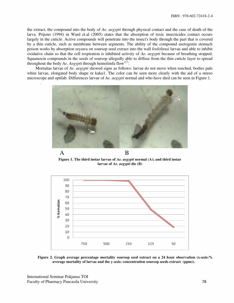
ISBN : 978-602-72418-2-4
International Seminar Pokjanas TOI
Faculty of Pharmacy Pancasila University 78
the extract, the compound into the body of Ae. aegypti through physical contact and the case of death of the
larva. Prijono (1994) in Ward et.al (2005) states that the absorption of toxic insecticides contact occurs
largely in the cuticle. Active compounds will penetrate into the insect's body through the part that is covered
by a thin cuticle, such as membrane between segments. The ability of the compound asetogenin stomach
poison works by absorption seyawa on soursop seed extract into the wall fosfolirasi larvae and able to inhibit
oxidative chain so that the cell respiration is inhibited activity of Ae. aegypti because of breathing stopped.
Squamocin compounds in the seeds of soursop allegedly able to diffuse from the thin cuticle layer to spread
throughout the body Ae. Aegypti through hemolimfa flow(17)
.
Mortaitas larvae of Ae. aegypti showed signs as follows: larvae do not move when touched, bodies pale
white larvae, elongated body shape or kaku1. The color can be seen more clearly with the aid of a stereo
microscope and optilab. Differences larvae of Ae. aegypti normal and who have died can be seen in Figure 1.
.
Figure 1. The third instar larvae of Ae. aegypti normal (A); and third instar
larvae of Ae. aegypti die (B)
Figure 2. Graph average percentage mortality soursop seed extract on a 24 hour observation (x-axis:%
average mortality of larvae and the y-axis: concentration soursop seeds extract (ppm)).
A B

ISBN : 978-602-72418-2-4
International Seminar Pokjanas TOI
Faculty of Pharmacy Pancasila University 79
From Figure 2 shows that the higher the concentration of soursop seed extract, the higher the death rate
of Ae. aegypti L. The solvent n-hexane, ethyl acetate and 70% ethanol and distilled water as a negative
control test the same activity against larvae of Ae. aegypti, and the results obtained all the larvae do not occur
death. This indicates that the solvent does not affect the mortality of larvae. Temephos as a positive control, in
which the larvicidal activity at a concentration of 1 ppm trials have demonstrated 100% mortality against
larvae of Ae. aegypti L.
CONCLUSION
Based on the results of phytochemical screening of the seeds of soursop (Annona muricata L.) obtained by
the content of secondary metabolites. The test results with the larvicidal activity and data analysis has been
done, it can be concluded that the 70% ethanol extract of the seeds of the soursop has the highest activity as
larvicides against Ae. aegypti L. with LC50 values of 97, 462 ppm.Ethanol 70% extract of the seeds of
soursop (Annona muricata L.) has a good chance to be used as biological insecticides to control mosquito
larvae that are environmentally friendly.
REFERENCES
1. Kaihena M, Vika L, Maria N. Efektivitas ekstrak etanol daun sirih (Piper betle L) terhadap mortalitas
larva nyamuk Anopheles sp dan Culex . Molluca medica. ISSN: 1979-6358
2. Susanti PD, Danang B, Dini S, Susilawati. Penggunaan ekstrak kulit kayu gemor (Nothaphoebe
coriacea K.) sebagai larvasida hayati terhadap tingkat mortalitas jentik nyamuk Aedes aegypti serta
dampaknya pada kualitas air hujan. ISSN 1978-8096. 2013;9:117–22.
3. Hadi, Upik Kesumawati. Penyakit tular vektor: demam berdarah dengue. Bogor: Fakultas
Kedokteran Hewan IPB. 2005
4. World Health Organization. Dengue: guidelines for diagnosis, trearment, prevention and control, new
edition. Swiss. 2010. h.5.
5. Palgunadi BU, Asih Rahayu. Aedes aegypti sebagai vektor penyakit demam berdarah dengue. Surabaya:
Universitas Wijaya Kusuma. 2011.
6. Rosmayanti, Kiki. Uji efektivitas ekstrak biji sirsak (Annona muricata L ) sebagai larvasida pada larva
Aedes segypti instar III/IV (skripsi). Jakarta: Fakultas Kedokteran Dan Ilmu Kesehatan Universitas Islam
Negeri Syarif Hidayatullah. 2014. 1-3, 16-17, 37-40.
7. Mulyawati AP, Hayati EK, Nashihuddin A, Tukimin. uji efektivitas dan identifikassi senyawa
ekstrak biji sirsak (Annona muricata Linn.) yang besifat bioaktif insektisida nabati terhadap hama
thrips. Alchemy 2010;2(1): 104-1575.
8. Taslimah. Uji efikasi biji srikaya (Annona squamosa L.) sebagai bioinsektisida dalam upaya integrated
vector management terhadap Aedes aegypti (skripsi). Jakarta: Fakultas Kedokteran Dan Ilmu Kesehatan
Universitas Islam Negeri Syarif Hidayatullah. 2014. h.1-6, 23-28,76-81.
9. Wardhana A.H, Amir H, Muharsini S, dan Yuningsih, Veteriner BP. Uji Keefektifan Biji Sirsak (Annona
muricata) dan Akar tuba (Derris edliptica) terhadap Larva Chrysomya bezziana secara In Vitro. 2006;
1013-1017.
10. Wardhana AH, Amir H, dan J Manurung, Veteriner BP. Uji efikasi ekstrak heksan daging biji
srikaya ( Annona squamosa L ) terhadap pertumbuhan larva lalat Chrysomya bezziana secara in
vitro. 2004. 134-142. 11. Departemen Pertanian Indonesia. Metode standar pengujian efikasi pestisida rumah tangga dan
pengendalian vektor. Direktorat Pupuk dan Pestisida dan Direktorat Jenderal Prasarana dan Sarana
Pertanian Kementrian Pertanian. 2012.h.20-23

ISBN : 978-602-72418-2-4
International Seminar Pokjanas TOI
Faculty of Pharmacy Pancasila University 80
12. Farnsworth NR. Biological and phytochemical screening of plants. Journal of pharmaceutical, Sci.
1966. (55). No 3.
13. Riswanto S. Uji Efektivitas Pestisida Nabati terhadap Hama Spodoptera litura (Lepidoptera: Noctuidae)
pada Tanaman Tembakau (Nicotiaana tabaccum L.). Medan: Universitas Sumatera Utara. 2009.20-23.
14. Susilowati D, Rahayu MP, Prastiwi R. Efek Penolak Serangga (Insecct Repellent) dan Larvasida Ekstrak
Daun Jeruk Purut (Citrus hystrix D. C.) terhadap Aedes aegypti. Surakarta:Fakultas Farmasi, Universitas
Setia Budi.
15. Nopitasari. Uji Aktivitas Ekstrak Biji Langsat (Lansium domesticum Cor.) sebagai Larvasida Aedes
aegypti. Universitas Tanjungpura. 2013. 12-14.
16. Venugopala KN, Raquel MG, Kabange K, Bandar EA, Mahesh VA, Bharti O. Evaluation of
halogenated coumarins for antimosquito properties. Hindawi Publishing Corporation The Scientific
World Journal, Vol. 2014, 4.
17. Wardhana A.H, Amir H, dan J.Manurung, Veteriner BP. Efektifitas ekstrak biji srikaya
(Annona squamosa L ) dengan pelarut air , metanol dan heksan terhadap mortalitas larva caplak
boophilus microplus secara In Vitro. 2005.134–42.

ISBN : 978-602-72418-2-4
International Seminar Pokjanas TOI
Faculty of Pharmacy Pancasila University
81
Antimicrobial and Biology Activity from
Extract Herbs Parasite Soursop (Dendropthoe pentandra L.)
ERLINDHA GANGGA*, LIA KARTIKA SARI
Faculty of Pharmacy Pancasila University, Jl Srengseng Sawah, Jakarta 12640.
e-mail: [email protected]
Abstract: Parasite of Soursop (Dendropthoe pentandra L.) is one of many plants that grow in
Indonesia.Indonesia is a country who has a many plants of drugs, where the plant has been used since
the time of our ancestors to cure various diseases. The parts of the plants used leaves, rods, roots or
whole plant. Besides that parts, the people also used Loranthus for medicine. Loranthus is a plant
parasite and a half parasite or substances that still have green leaves (chlorophyll) used for the
assimilation process. In addition parasite also absorb the food from its host plant, so the loranthus
has the same activity with host plants. The species of loranthus usually used is the parasite of the
species Dendropthoe pentandra which grows on a soursop. hosts. Parasite of soursop plants has
been cleaned from dust and then dried in powder. The powder and the extract herbal parasite
soursop conducted phytochemical screening.After that, did the antimicrobial activity by diffusion
methode and the biological activity test (LC50) used the BSLT method. In testing the
antimicrobial activity of Escherichia coli and Staphylococcus aureus from ethyl acetate and
methanol extracts retrived power resistor from concentration 2,43.10-3
%. The activity results of
Candida albicans from n-hexane, ethyl acetate and methanol extract had retrived inhibitory
from concentration 28.14%. The biological result used the BSLT method in soursop loranthus
showed that methanol extract has a high (LC50) activity is 21.22 ppm; 510.07 ppm from n-hexane
extract and ethyl acetate extract of 87.78 ppm.
Keyword : Herbs parasite of soursop (Dendropthie pentandra L.), biology activity methode, diffusion
methode Escherichia coli, Staphylococcus aureus ,Candida albicans
INTRODUCTION
Indonesia is a rich country of biological resources and known as one of the larger mega biodiversity in the
world. Indonesia has about 17% species from all around the world. Large tropical forest with biological
diversity is natural resources which is priceless. And known as a warehouse of herb, Indonesia has a
nickname as live laboratory. Parasite of Soursop ( Dendropthoe pentandra L.) is one of many plants
that grow in Indonesia has been used empirically as medicinal plant which is a herbaceous that can be
grown easilyon tropical land. According to literature this plant containing highly Flavonoid, saponine,
alcaloid, tanine, amino acid, lorantilalkohol, kholin, and fat.
To get the biological activity test (LC50) and antimicrobe Parasite of Soursop ( Dendropthoe
pentandra L.) using extraction was done by maceration with ethanol 70 % , and partitioned gradient
started from n – hexan, ethyl acetat, and n- buthanol . after that the extracts were done phytochemical
screening method biological activity (cytotoxic ) with BSLT method and antioxidant activity with DPPH
free radical scavenging.

ISBN : 978-602-72418-2-4
International Seminar Pokjanas TOI
Faculty of Pharmacy Pancasila University
82
MATERIAL AND METHODS
MATERIAL. Parasite of Soursop ( Dendropthoe pentandra L.), n–hexan, ethyl acetat, Methanol,
shrimp larvae (Artemia salina Leach), NaCl. Escherichia coli, Staphylococcus aureus, dan Candida.
METHODS. Extraction was done by maceration with started from n–hexan, ethyl acetat, and n-
methanol . after that the extracts were done phytochemical screening method , biological activity with
BSLT method and antimicrobial activity with.
Research Stages. 1.Sample preparation 2. Extract forming and fractionation 3. Phytochemical
screening 4. Toxicity Test 5. Antimicrobial Activity Test
Extract Forming and Fractionation. As much as 300.1 g kelor leaf ( Moringa oleifera Lamk L.)
leaf powder was placed in a jar, then maceration with ethanol 70 % and partitioned gradient started from
n-hexane, saturated ethyl acetate, and n- butanol. Rotavapor was used to saturate the filtrate until thick
extract formed. The waste was then thrown.
Phytochemical Screening. Content identification of kelor leaf’s secondary metabolite was done using fresh leaf and thick extract of n-hexane, ethyl acetate, and methanol, which include: 1. Alkaloids
identification, 2. Flavonoid identification, 3. Saponin identification, 4. Tannin identification 5. Kuinon
identification 6. Steroid / triterpenoid identification 7. Aetheric oil identification 8. Coumarin
identification.
Cytotoxic Test. Writer chose BSLT (Brine Shrimp Lethality Test) method for cytotoxic test using
natural ingredients of shrimp larvae (Artemia salina Leach) and sea water as the media. All substances
were tested three times in a vial of 5mL sea water and 10 larvae. Observation was done after 24 hours, the
data was analyzed to get LC50 using probit analysis. The next step was placing ± 20mg of Artemia salina
Leach.’s egg inside the hatching container filled with synthetic sea water. The sea water was prepared by weighing 38 g salt without iodine and dissolve it with 1L of water, then filter it with Whatman paper and
radiate it with 18 watt lamp. After 24 hours, hatched egg became nauplii and relocated to other place.
After the next 24 hours, nauplii is ready to be used as a test animal.
Antimicrobial Test. Anti-microbial assay was done by diffusion using paper disc with 6 mm
diameter. The paper disc contains anti-bacterial substance and then placed in the jelly’s surface which has been inoculated and incubated at 37°C for 18-24 hours. Activities decided by the blocked zone which
formed by the clear zone around the anti-bacterial substance. Other than diffusion, this research also used
positive control chloramphenicol and amphotericin B. Chloramphenicol has anti-microbes spectrum
activities for both Gram Positive Bacteria and Gram Negative Bacteria, and has bacteriostatic
characteristic where it blocked microbes’ protein synthesis. Amphotericin B also has similar characteristics where it served as wide spectrum anti-fungus.
RESULT AND DISCUSSION
Phytochemical Screening Result. Results from the phytochemical screening powder of Parasite of
Soursop (Dendropthoe pentandra L.) and the extract n- hexan,aetyl acetat, methanol showed that the
herbs powder contained, flavonoid, saponine, tannin, quinon and steroid, where as n-hexane extract
contained steroide, and ethyl acetate extract contained, flavonoide, tannin, steroide,. Furthermore,
methanol extract contained flavonoide, saponine, tannin and quinon

ISBN : 978-602-72418-2-4
International Seminar Pokjanas TOI
Faculty of Pharmacy Pancasila University
83
Table 1. Phytochemical screening result.
Table 2. Cytotoxic test by BSLT method.
Ekstrak Nilai IC50 ( bpj )
Table 3. Antimicrobial activity.
CONCLUSION
1. Flavonoid, saponin, tannine, steroid and coumarin compound were found from phytochemical
screening of herbs powder and Parasite of Soursop (Dendropthoe pentandra L.).
2. Cytotoxic test by BSLT method found that thick methanol extract has the highest toxicity level of
LC50 21.22 ppm, ethyl acetate extract of 87.78 ppm and n-hexane extract of 501, 07 ppm,
3. Antimicrobe activity test found that methanol extract has, ethyl acetate extract has activities to
Escherichia coli, Staphylococcus aureus, and Candida albicans and has anti-microbes spectrum
activities for both Gram Positive Bacteria and Gram Negative Bacteria, and anti fungus (broad
spectrum ), n-hexane extract has activities to Candida albicans and has anti microbes/ anti-fungus
only.
No Compound Herbs Powder n- Hexane Ethyl acetate Methanol
1. Alkaloid - - = -
2 Flavanoid + - + +
3 Saponine + - - +
4 Tannine +/- - +/- +/-
5 Quinon + - - +
6 Steroide/
triterpenoide +/- +/- +/- +/-
7 Volatile oil - - - -
8 Coumarine - - - -
E
Extract
LC 50
Value( ppm)
n- Heksan
510.07
Ethyl Acetat 87.78
Methanol 21.22
NO
Extract
Microbe
Escherichia colli Staphylococcus aureus Candida albicans
1 n- Hexane Negative Negative Positive
2 Ethyl acetate Positive Positive Positive
3 Methanol Positive Positive Positive
ISBN : 978-602-72418-2-4
International Seminar Pokjanas TOI
Faculty of Pharmacy Pancasila University
84
REFERENCES
1. Meyer BN, et. al. Brine shrimp: a convenient general bioassay for active plant constituens. Planta
medica. Volume 45. 1982. h. 31-4.
2. Famsworth, NR. Biological and phytochemical screening of plant 55(3). J Pharm. Sci;
Chicago. 1985. h. 28-58.
3. Jawetz E, Melnick JL, Adelberg EA. Mikrobiolog kedokteran. Edisi 20. Alih bahasa Nugroho
E, Maulany RF. Jakarta: Penerbit Buku Kedokteran. 1996. h. 153-64, 177-78, 238-9, 245.

ISBN : 978-602-72418-2-4
International Seminar Pokjanas TOI
Faculty of Pharmacy Pancasila University
85
Antioxidant, Cytotoxic and Apoptotic Induction Activity of Ethanolic Extract of
Andrographis paniculata on Mcf-7 Cancer Cell Line
CHURIYAH*, KURNIA AGUSTINI AND SISKA ANDRINA KUSUMASTUTI
Center for Pharmaceutical and Medical Technology
Agency for the Assessment and Application of Technology (BPPT)
LAPTIAB Building 611, Puspiptek area Serpong, Tangerang – Selatan. Telp/Fax: +62-21-7560536,
email: [email protected]
Abstract: Andrographis paniculata is usually used as a traditional medicine to treat various diseases
including cancer . The research purposes was to evaluate the antioxidant activity of the ethanolic extract of
A. paniculata and also to evaluate the cytotoxic and apoptotic induction activity of this extract against breast
cancer cell line MCF-7. Cytotoxic activity of the extract was performed using enzymatic reaction of 3-(4,5-
dimethylthiazoyl-2-yl) 2,5 diphenyltetrazolium bromide (MTT) method and the anti oxidant-activity was
determine relatively to 1,1-diphenyl-2-picrylhydrazyl (DPPH) acivity, and further apoptotic cell was
determined by flowcytometry using Propidium Iodide (PI). The radical scavenging activity resulted from the
antioxidant assay using DPPH methods showed that ethanolic extract of A. paniculata gave very low activity
with IC50 value was 201.95 µg/ml compared to the positive control of α-tocopherol with its IC50 value was
41.58 ppm. The cyototoxic analysis using MTT method indicated that the its extract exhibited toxic effect on
MCF-7 cell line with IC50 value was 111.37 µg/ml and in its concentration induced apoptotic cell about 64%
by flow cytometry analysis. A. paniculata extract can be developed as a potential anticancer agent for breast
cancer deseases.
Keyword : A. paniculata, antioxidant, cytotoxic, apoptotic, MCF-7 cell.
INTRODUCTION
Apoptosis is a mechanism of cell death for the purpose of maintaining a stable cell population and plays an
important role in tumorigenesis. Inhibition of apoptosis results in uncontrolled growth, such as occurs in the
development of malignancy. The induction of apoptosis, which is not accompanied by an inflammatory
reaction, is one of the strategies has been developed for cancer therapy(1)
.
There are various studies emphasizing that free radicals contribute to the development of many diseases,
including tumor promotion and carcinogenesis. Antioxidants are substances that play an important role in
delaying or preventing degenerative diseases caused by oxidative damage of living cell components or by
free radicals(2)
.
Andrographis paniculata (Burm. F) Nees, is an herbaceous plant belonging to the Acanthaceae and is
found throughout tropical and subtropical Asia, Southeast Asia, Indonesia and India. Extracts of this plant
exhibit pharmacological activities such as immunostimulatory, antiviral and antibacterial. As major active
constituent, andrographolide exhibits biological activities, including anti-inflammatory, antidiabetic and
antitumor(3)
. Hydroalcoholic extract of A. paniculata increased the activities of antioxidant enzymes such as
super oxide dismutase, catalase, glutathione peroxidase in rats(4)
, then ethanolic extrac was promising as
antioxidant(5)
.
ISBN : 978-602-72418-2-4
International Seminar Pokjanas TOI
Faculty of Pharmacy Pancasila University
86
Andrographolide isolated from A. paniculata induced apoptosis of prostate cancer (PC-3) cells(6)
, also
exhibited cytotoxic effect and induced apoptosis in HeLa cells(7)
and breast cancer cell line T-47D(8)
. The A.
paniculata extracts using different solvent, namely water, ethanol and acetone was reported that those extract
indicated excellent cytotoxic effect against neuroblastima IMR-32 and human colon HT-29 cancer cell line(9)
.
This research was purposed to determine antioxidant activity of the ethanolic extract of A. paniculata
using DPPH scavenging method and also to evaluate the cytotoxic activity against breast cancer cell line
MCF-7 by MTT method, followed with apoptotic cell analysis by flow cytometry
METERIALS AND METHODS
Extract preparation. The dried powdered of A. paniculata simplicia was extracted by macerating in ethanol
solvent. Afterward the maserate was filtered, followed by evaporation using rotary evaporator until excess
solvent was evaporated and obtained the viscuous extract.
Antioxidant Assay Using DPPH Radical Scavenging Method. Free radical scavenging activity of
extracts was measured by 1,1-diphenyl-2-picryl hydrazyl (DPPH). In brief, 500 µl of 0.1 mM solution of
DPPH in ethanol was added to 1 ml of extracts in ethanol at different concentration (5, 10, 20, 50 and 100
μg/ml), it was shaken vigorously and allowed to stand at room temperature for 30 minutes, then the
absorbance was measured at 517 nm using spectrophotometer. The positive control being used was α-
tocopherol and the experiment was done in triplicate. The percent DPPH scavenging activity was calculated
by following equation:
DPPH scavenging activity (%) = (Abs DPPH-Abs sample)/Abs DPPH × 100,
The IC50 value of the sample, which is the concentration of sample required to inhibit 50% of the DPPH free
radical, was calculated using dose inhibition curve.
Cytotoxic Activity Assay Using MTT Method. MCF-7 cells were cultured in 96-wells plates at a
density of 5x10⁴ cells/well. After 24 hours, the medium culture was discarded, cell then was treated with
various concentrations (10, 20, 50, 100, 250 and 500 µg/ml) of the A. paniculata extract, and incubated for
24 hours. Next, the medium cultured were aspirated and 100 µl culture medium contain 0,5 mg/mL MTT
was added, then the cell was incubated for 4 hours. Further 100 µl of 10% SDS 10% was added to stop the
MTT reaction and dissolved formazan crystals, incubated overnight, than was measured at 570 nm using a
multiwell plate reader. The IC 50 value of the sample, which is the concentration of sample required to inhibit
50% of cell viability was calculated using dose inhibition curve.
Apoptotic Cell Assay Using Flow Cytometry. MCF-7 cells was plated in 24 well plates with density
about 3 x 10⁴ cells / well, incubated in 5% CO2 incubator at 37°C. After 24 hours, cell was treated with A.
paniculata extract at a concentration equal to the IC50 value and incubated for 24 hours. Next, cell was
harvested and centrifuged at 1500rpm for 5 minutes. After supernatant discarded, the pellet cell was fixed by
70% of EtOH for 15 minutes in the 4°C. Then cell was centrifuged at 1500 rpm for 5 min, and the pellet cell
was stained by 50 µl Propidium Iodide (1mg of PI/ml of deionized water) in 450 µl PBS and incubated at
room temperature in dark for at least 30 minutes, then was observed using flow cytometry.
RESULTS AND DISCUSSION
The result of free radical scavenging activity of A. paniculata extract and positive control of α–tocopherol by
1,1-diphenyl-2-picryl hydrazyl (DPPH)method (Figure 1A) indicated that the extract gave lower DPPH
scavenging activity compared with positive control of α–tocopherol in the all of concentration treatments (5,
10, 20, 50 and 100 μg/ml). So as described in the IC50 value between extract and positive control, the IC50
value of A. paniculata extract (201.95 µg/ml) was much higher then IC50 value α-tocopherol (41.58 µg/ml ).
Cytotoxic activity assay using MTT method showed that the effect of A. paniculata extract treatment in
the cell viability was dose dependent, where the increasing extract concentration resulted in the lower cell
viability (Figure 1B). The IC 50 value of the sample, which is the concentration of sample required to inhibit

ISBN : 978-602-72418-2-4
International Seminar Pokjanas TOI
Faculty of Pharmacy Pancasila University
87
50% of cell viability was calculated using dose inhibition curve, indicated that the A. paniculata extract
exhibited toxic effect on MCF-7 cell line with IC50 value was 111.37 µg/ml. Whereas the IC50 value of
control positive Doxorubicin was very low only 0.094 µg/ml (Table 1).
Results of the Flow cytometry analysis of apoptotic cell induced by A. paniculata extract described in
the cytogram (Figure2) indicated that in the IC50 value concentration of its extract (111.37 ug/ml) induced
cell apoptotic about 64% (Table 1). Whereas, the control negative without any extract treatment nearly did
not exhibit cell apoptotic only found about 5%, in the contrary the positive control of doxorubicin treatment
gaved very hight percentage of apoptotic cell it was about 94% of total cell analyzed.
A B C
Figure 1. A = DPPH radical scavenging activity of A. paniculata extract and positive control α-tocopherol; B =
cytotoxic activity of A. paniculata extract against MCF-7 cancer cell line. C = cytotoxic activity of doxorubicin
against MCF-7 cancer cell line
Figure 2. Cytogram of MCF-7 cell treated and untreated with A. paniculata extract and positive control of
doxorubicin A = the normal cell untreated with extract, B = the treated cell with A. paniculata extract and C =
cell treated with positive control of doxorubicin.
Table1. The IC50 value of DPPH radical scavenging activity and cytotoxic activity by
MTT assay of A. paniculata extract. No Antioxidant activity by
DPPH radical scavecging
Cytotoxic activity
by MTT methode
Extract IC50 (µg/ml) Exctract IC50 (µg/ml)
1 A. paniculata 201.95 A. paniculata 111.37
2 α-tocopherol 41.58 Doxorubicin 0.094
0
20
40
60
80
100
120
5 10 25 50 100
% r
ad
ica
l sc
av
en
gin
g a
ctiv
ity
Extract Concentration (µg/ml)
DPPH Scavenging Activity
A. paniculata α-Tocopherol
0
20
40
60
80
100
120
10 20 50 100 250 500
% C
ell
via
bil
iy
Extract concentration (µg/ml)
A. paniculata vs MCF-7
0
20
40
60
80
0,03 0,06 0,13 0,25 0,50 1,00
% C
ell
via
bil
ity
Doxorubicin concentration (µg/ml)
Doxorubicin vs MCF-7
A
B
B C

ISBN : 978-602-72418-2-4
International Seminar Pokjanas TOI
Faculty of Pharmacy Pancasila University
88
Table 2. Apoptotic cell of MCF-7 cancer cell line induced by A. paniculata extract and positive control
Doxorubicin compared with normal cell without any treatment.
No. Cell Treatments Total of cell
analyzed
% of
apoptotic cell
Total of
apoptotic cell
1 Normal cell (negative control) without any
treatment
250
5
4843
2 Cell treated with A. paniculata extract 4109
64
2522
3 Cell treated with positive control
Doxorubicin
2869
94
3039
The DPPH radical scavenging activity of A. paniculata extract were resulted in IC50 value of A.
paniculata extract (201.95 µg/ml) much higher then positive control α-tocopherol (41.58 µg/ml ). Previous
research also reported similarly, that 200 µg/ml of its extract inhibited DPPH radical scavenging activity only
about 28.74%(10)
.
Cytotoxic activity assay using MTT method showed that the IC50 value of the A. paniculata extract
(111.37 µg/ml) was lower than positive control doxorubicin (0.094 µg/ml). Then other research reported that
this extract showed IC50 value for neuroblastima IMR-32 and human colon cancer HT-29 cell lines was 200
μg/ml(9). Interestingly, previous research reported that its extract did not exhibit any toxic in human
normal lung fibroblast cell line (Hs888Lu) with highly IC50 value about 500 µg/ml(2)
.
Fluorescent labelling is the suitable method for apoptotic analysis, through observe the morphological
and biochemical change of apoptosis for differentiate of normal, apoptotic, and dead cells, or cell cycles. Cell
Apoptosis assay using PI provides a rapid and convenient method for apoptosis analysis observed by flow
cytometry, cell DNA is degraded by endogenous nuclease activated and diffuse out of cells with the process
of apoptosis. A highly definable sub-G1 peak occurred is easily quantified by PI(11)
. Using this method, A.
paniculata extract could induced apoptotic cell relatively high about 64%, whereas the percentage of
apoptotic cell in the negative control without any extract treatment only 5% of the total cell analyzed.
This research concluded that A.paniculata extract exhibit lower antioxidant activity compared with α-
tocopherol, but this extract showed excellent anticancer activity. So, it could be develop as potential
anticancer through apoptotic induction pathway. Further research was needed to observe the molecular
mechanism of apoptotic induction and the cytotoxic effect of its extract on normal cell to obtaine the
selectivity of its extract against cancer cell.
REFERENCES
1. Nugrahaningsih, Sarjadi, Dharmana E, Subagio HW. Andrographis paniculata extract induced apoptosis
of adenocarcinoma mammae in C3H mice. Univ Med. 2013. Vol.32 - No.2: 99-107
2. Qader SW, Abdulla MA, Chua LS, Najim N, Zain MM, Hamdan S. Antioxidant, total phenolic content
and cytotoxicity evaluation of selected malaysian plants. Molecules. 2011. 16: 3433-3443;
Doi:10.3390/molecules16043433
3. Jayakumar T, Hsieh CY, Lee JJ, Sheu JR. Experimental and clinical pharmacology of Andrographis
paniculata and its major bioactive phytoconstituent andrographolide. Evidence-based complementary
and alternative medicine. Volume 2013. Article ID 846740, 16 pages.

ISBN : 978-602-72418-2-4
International Seminar Pokjanas TOI
Faculty of Pharmacy Pancasila University
89
4. Ojha SK, Nandave M, Kumari S, Arya IDS. Antioxidant activity of Andrographis paniculata in
ischemic myocardium of rats. Global Journal of Pharmacology. 2009. 3 (3): 154-157.
5. Vijayakumar AD, Kalaichelvan, PT. In vitro antimicrobial and antioxidant activity screening of
andrographis paniculata leaf ethanolic extract in Tamil Nadu. Int J Pharm Pharm Sci. 2012. 4(1): 227-
229.
6. Zhao F, He EQ, Wang L, Liu K. Anti-tumor activities of andrographolide, a diterpene from
Andrographis paniculata, by inducing apoptosis and inhibiting VEGF level. J Asian Nat Prod Res. 2008.
10(5-6):467-73.
7. Sukardiman, Rahman A, Ekasari W, Sismindari. Induction of apoptosis by Androgra-pholide compound
from sambiloto (Andrographis paniculata Nees) in Cancer Cells Culture. 2005. Media Kedokteran
Hewan 21(3):105-110.
8. Harjotaruno S, Widyawaruyanti A, Sismindari, Zaini NC. Apoptosis inducing effect of andrographolide
on T-47D human breast cancer cell line. Afr. J. Trad. CAM .2007. 4 (3): 345 – 351.
9. Rajeshkumar S, Nagalingam M, Ponnanikajamideen M, Vanaja M, Malarkodi C. Anticancer activity
of Andrographis paniculata leaves extract against neuroblastima (Imr-32) and human colon (Ht-29)
cancer cell line. World J Pharm Pharm Sci. 2015. 4(6): 1667-1675.
10. Doss VA, Kalaichelvan PT. In vitro antimicrobial and antioxidant activity screening of Andrographis
paniculata leaf ethanolic extract in Tamil Nadu. Int J Pharm Pharm Sci. 2012. 4(1):227-229.
11. Liu K, Liu PC, Liu R, Wu X. Dual AO/EB staining to detect apoptosis in osteosarcoma cells compared
with flow cytometry. Med Sci Monit Basic Res. 2015. 21: 15-20

ISBN : 978-602-72418-2-4
International Seminar Pokjanas TOI Faculty of Pharmacy Pancasila University 90
In-Vitro α-Glucosidase Inhibition Activities Test from Standardized sambung nyawa (Gynura
procumbens (Lour.) Merr.) Leaves Extract
WIWI WINARTI*, RATNA DJAMIL, SARAH ZAIDAN, RAYMOND
Faculty of Pharmacy Pancasila University, Jakarta 12640, Indonesia.
email : [email protected].
Abstract: Diabetes is a disease that is not curable, but the risk of diabetes can be reduced. Diabetes therapy
can be done by taking traditional medicine or modern medicine. Based on literature searches, Sambung
Nyawa leaves can be used in the treatment of diabetes mellitus because Sambung Nyawa compound contains
flavonoids, saponins, tannins, triterpenoids, and essential oils, caffeic acids, vanilic acid, chlorogenic acid,
and p-cumaric acid. This study aims to determine the activity of the enzyme α-glucosidase inhibition to 70%
ethanol extract of the leaf powder propped Sambung Nyawa. In this study we test the enzyme α-glucosidase
inhibition by in- vitro by using p-nitrophenyl-α-D-glucopiranosida as a substrate to 70% ethanol extract of
Sambung Nyawa leaves. Results of in- vitro study by inhibition activity against α-glucosidase showed that
70% ethanol extract of Sambung Nyawa leaves acquired 72.37% inhibition at concentrations of 200 ppm
with IC50 value of 56.75 ppm.
Keyword: 70% ethanol extract of Sambung Nyawa leaves (Gynura procumbens (Lour.) Merr.).
α-glucosidase.
INTRODUCTION
Diabetes mellitus is characterized by excessive glucose levels in the blood are often called hyperglycemia.
Hyperglycemia that lasts for a long time would cause serious damage to the body's systems, especially the
nerves and blood vessels. Sambung Nyawa leaves (Gynura procumbens (Lour.) Merr.) , this one of the plants
that have potential as antidiabetic agent. Based on the research of June et.al (2012), the fraction of n-
hexane, ethyl acetate and n-butanol from Sambung Nyawa can reduce blood glucose levels respectively in
rats induced by streptozotocin as much as 29.7%, 60.1%, and 33.5% in the provision for 14 days in rats
induced by streptozotocin. Liver glycogen content in diabetic rats were given three fractions increased (p
<0.05) in the provision for 14 days (1). Agariri K et al (2013) conducted a study regarding the
antihyperglycemic effect of the ethanol extract 95, 75 , 50 , and 25% of the Sambung Nyawa leaves of the
rats induced by Streptozotocin. Khalid research results Agariri showed that 25% ethanol extract of the leaves
continued life can lower blood level of glucose levels in a rat model of diabetes by 47% within 2 hours of the
acute dose of 1 g/ kg(2)
. Mean while, Hassan Z (2010) conducted a study on the effect of Sambung Nyawa
leaves extract to blood level sugar in mice model of diabetes induced by streptozotocin. The results showed
that the water extract of Sambung Nyawaleaves with dose of 500 and 1000 mg / kg body weight can lower
blood glucose levels significantly different (p <0.05) to the controls after administration of the extract for 14
days(3)
.
MATERIALS AND METHODS
MATERIAL. Sambung Nyawa leaves (Gynura procumbens (Lour.) Merr.); Ethanol 70%; P dilute
hydrochloric acid; distilled water; chloroform; ethanol 96%; ammonia 30%; 1:10 hydrochloric acid;
concentrated hydrochloric acid; amyl alcohol; iron (III) chloride 1%; sodium hydroxide 1N; eter P; acetic

ISBN : 978-602-72418-2-4
International Seminar Pokjanas TOI Faculty of Pharmacy Pancasila University 91
acid anhydride; concentrated sulfuric acid; ammonia 10%; Mg pulv: Mayer reagent; Dragendorff reagent;
Stiasny reagent: potassium phosphate monobase; phosphate buffer pH 6.8; 7.0; 7.2; p-nitrophenyl-α-D-
glucopiranoside; α-glucosidase enzyme; bovine serum albumin; dimethyl sulfoxide (DMSO); 0.2 M sodium
carbonate; acarbose.
METHODS. In this study used a standardized 70% ethanol extract of the Sambung Nyawa leaves powder,
then carried out phytochemical screening and the inhibition of the enzyme α-glucosidase test. Stages include
phytochemical screening study, a preliminary test to determine the concentration, pH, incubation time and the
optimum substrate concentration. Test of inhibition of the enzyme α-glucosidase, acarbose used as positive
control. Measurements were made using Absorbance Mikroplate Reader EL x 800 at a wavelength of 405
nm.
RESULTS AND DISCUSSION
Test Activities α-Glucosidase Enzyme Inhibition. 1. Preliminary Test. Before testing the activity of the
enzyme α-glucosidase inhibitory done, a preliminary test which aims to find the optimum conditions for
activity test. The principle of preliminary test and the enzyme inhibitory activity of α-glucosidase is α-
glucosidase enzyme will hydrolyze p-nitrophenyl-α-D-glukopiranosida into p-nitrophenol which is yellow
and glucose. The enzyme activity was measured based on the absorbance of the yellow p-nitrophenol.
Optimization is done to optimize the enzyme concentration, pH, incubation time and the concentration of the
substrate. Enzymes are used as much as 1.2 to 29.4 mg solid enzyme specification contains 25% protein and
there are 200 units per mg protein.
The solution was tested consisting of DMSO, phosphate buffer, substrate p-nitrophenyl-α-D-
glukopiranosida (pNPG), α-glucosidase, and sodium carbonate. The addition of sodium carbonate was to stop
the enzyme reaction, sodium carbonate chosen as a stopper because the reaction is able to increase the pH of
the test solution becomes alkaline, so the enzyme will be denatured. Uptake is measured is the absorban of
the test solution, the control solution test, blank solution and blank control solution. Test solution is a solution
of the extract while the blank solution ie the extract solution without control solution for correcting the results
of uptake of the test solution and blank solution, observations were made on the activity of the enzyme by
swapping positions between the addition of the enzyme α-glucosidase and sodium carbonate. The results of
the test control and blank control can be used to see whether the enzyme activity was stopped when the
condition first mixture was basified with sodium carbonate so that no product that is formed. The incubation
process consists of two stages. The first stage, aimed pre-incubated for 5 minutes to allow time for the test
solutions to achieve a temperature of 37˚C. The second stage, which is a 15-min incubation time for
enzymatic reaction. Results of α-glucosidase enzyme optimization can be seen in Table 1.
Table 1. Results of α-glucosidase enzyme optimization.
No. Optimization Optimum Results
1. The enzyme concentration 0.025 U / mL
2. pH 7.0
3. Incubation time 15 minutes
4. The substrate concentration 10 mM
a. Determination of the optimum enzyme concentration. The determination of the optimum concentration
of the enzyme is done by units of α-glucosidase 0.015 U/ml, 0.020 U/ml, 0.025 U/ml, and 0.030 U/ml,
the concentration of the substrate p-nitrophenyl-α-D-glukopiranosida 10 mM, pH 7.0, 37˚C and
incubation time of 15 min. The results obtained showed the greatest enzyme activity are at a
concentration of 0.030 U/ml is 49.9139 U/mg. But uptake obtained at concentrations of more than 1.0,
the selected optimum enzyme concentration was 0.025 U/ml with enzyme activity of 20.8889 U/mg
which has an absorption of 0.4. Enzyme activity is eligible activeness on the label enzyme > 10 U/mg.

ISBN : 978-602-72418-2-4
International Seminar Pokjanas TOI Faculty of Pharmacy Pancasila University 92
b. Determination of the optimum pH. pH determination is done using phosphate buffer at pH 6.8, 7.0, and
7.2, enzyme concentration of 0.025 U/ml, concentration substrat p-nitrophenyl-α-D-glucopiranoside 10
mM, at 37˚C and incubation time of 15 min. The results showed that at pH 7.0 the enzyme to work
optimally with the enzyme activity 21.3194 U/mg.
c. Determination of the optimum incubation time. Variations incubation time used 15, 20 and 30 min.
Determination of the optimum incubation time is done with α-glucosidase units of 0,025 U/ml, the
concentration of the substrate p-nitrophenyl-α-D-glukopiranosida 10 mM, pH 7.0, 37˚C. The results
showed the greatest enzyme activity present in the incubation time of 20 min is 33.3139 U/mg, but
uptake obtained at the incubation time is more than 0.8. Therefore, selected the optimum incubation time
15 min with the enzyme activity 20.8861 U/mg.
d. Determination of the optimum substrate concentration. Variation of substrate concentration used 2.5
mM, 5 mM, 10 mM, and 20 mM. At a concentration of 20 mM substrate enzyme activity decreased.
Decline in activity is expected due to the formation of the reaction product of the enzyme inhibitors such
as α-D-glucose and p-nitrophenyl which have similar structures to the substrate p-nitrophenyl-α-D-
glucopiranoside, so it can compete with the substrate to occupy the active site of the enzyme. Based on
the results obtained, the concentration of substrate which is used to test the inhibitory activity of α-
glucosidase enzyme was 10 mM with enzyme activity 18.0278 U/mg.
The inhibition of α-glucosidase Test. Testing was conducted with 0.025 units of α-glucosidase U/ml,
the concentration of the substrate p-nitrophenyl-α-D-glucopiranoside 10 mM, pH 7.0, at 37˚C and incubation
time of 15 min. Test α-glucosidase inhibition of the enzyme is done by measuring the uptake of the product at
a wavelength of 405 nm using Absorbance Mikroplate Reader EL x 800. Uptake is measured absorbance of
the sample solution, sample control solution, a solution of acarbose, acarbose control solution, a blank
solution and blank control solution , The sample solution is it self a viscous extract solution with 5 variations
of concentration of 12.5 ppm, 25 ppm, 50 ppm, 100 ppm , and 200 ppm. Concentration variation is made so
that it can be used to create a regression equation to calculate the IC50.
Acarbose standardized testing done first. It is intended so that the IC50 and IC50 acarbose samples
(extracts) can be compared. Acarbose chosen for comparison because acarbose is an antidiabetic drug that
works to inhibit the enzyme α-glucosidase derived from nature. In the blank solution instead of the sample
solution is used dimethyl sulfoxide (DMSO). Blank made as a comparison of data difference between the
absorbance of the extract solution suspected of having α-glucosidase inhibitor agent. Activity test results are
shown in Table 2.
Table 2. The activity of the enzyme α-glucosidase inhibitory No. Inhibitor IC50 (ppm) % Inhibition
1. Acarbose 50.37 78.11
2. Sambung Nyawa leaves
extract
56.75 72.37
IC50 values indicate that the concentrations of extracts that can inhibit 50% of enzyme activity of α-
glucosidase. On the results showed that % inhibition at the highest extract is at a concentration 0f 200 ug/ ml
with 72.37 % as a result, it is equivalent to % inhibition at acarbose with the same concentration of 200 ug /
ml which is 78.11 %. IC50 result of Sambung Nyawa leaves extract 56.75 ppm and acarbose as a positive
control (50.37 ppm).
CONCLUSION
The result of the research activities of leaves 70% ethanol extract from Sambung Nyawa leaves (Gynura
procumbens (Lour.) Merr.) has idicated as antidiabetic activity (56.75 ppm )
ISBN : 978-602-72418-2-4
International Seminar Pokjanas TOI Faculty of Pharmacy Pancasila University 93
ACKNOWLEDGEMENT
The authors are grateful to Ditlitabmas Ditjen Dikti for Hibah Bersaing Research Funding as Dipa Kopertis
region III, Year 14/15.
REFERENCES
1. June CC, Wen LH, Chin LP, Embi N. Hypoglycemic effect of gynura procumbens fraction on
streptozotocin-induced diabetic rats involved phosphorylation pf GSK3β (Ser-9) in Liver. Malaysia:
Universiti Kebangsaan Malaysia. 2012.
2. Algariri K, Meng YK, Atangwho JI, Asmawi ZM. Hypoglicemic and anti-hyperglicemic study of
Gynura procumbens leaf extracts. Malaysia : School of Pharmaceutical Science, Universiti Sains
Malaysia. 2013.
3. Hassan Z, Yam FM, Ahmad M. Antidiabetic properties and mechanism of action of Gynuraprocumbens
water extract in streptozotocin induced diabetic rats. Malaysia: School of Pharmaceutical Science,
Universiti Sains Malaysia. 2010.
4. Kumar GS, Naidu RS. Hypoglicemic effects of aqueous extract of Gynura procumbens. Malaysia:
School of Pharmaceutical Science, Universiti Sains Malaysia; 2008
5. Farnsworth NR. Phytochemical screening. Chicago: Department of Pharmacognosy and Pharmacology
College of Pharmacy. 1966. P. 26-62.
6. Acarbose. Diambil dari :http://dinkes.tasikmalayakota.go.id/index.php/informasi-obat/176-
acarbose.html diakses tanggal 20 April 2014; pukul 19:40 WIB.

ISBN : 978-602-72418-2-4
International Seminar Pokjanas TOI
Faculty of Pharmacy Pancasila University
94
Identification of Sugar-Apple Seeds (Annona squamosa L.) Extract as A
Candidate Against The Aedes aegypti L. Musquito Vector Control DBD
RATNA DJAMIL, SARAH ZAIDAN,SITI NURAINI
Fakultas Farmasi Universitas Pancasila.
Abstract: Aedes aegypti L. mosquitos are the disease vectors of dangue hemorrhagie fever (DHF).
This diseases is caused by dengue virus which is transmitted by Ae. Aegypti mosquito. The effort to
control Ae. aegypti vector have been done in so many times, including chemical, physical, and
biological control method. Extraction by kinetic maceration have been done with sugar-apple seeds
(Annona squamosa L.) with the solvent of 70% of ethanol. Subsequently, the obtained extract is tested
phytochemical screening along with the powder and larvicidal activity against Ae. aegypti. The results
of phytochemical screening of the powder and 70% ethanol extract of sugar apple seeds have obtained
the compound of saponin, triterpenoid and coumarin. Based on the activity test against the larva of Ae.
aegypti from ethanol 70% extract of of sugar-apple seeds, show LC50 values is 97,462 ppm. These are
indicate that the compound which is found in sugar-apple seeds have a potential as a larvicides.
Keyword : Aedes aegypti L., Annona squamosa L., sugar-apple seeds, larvasida.
INTRODUCTION
The existence of a mosquito that is close to human life pose a serious health problem, because
mosquitoes act as vectors of some diseases with high rates of morbidity and mortality caused. Ae. L.
aegypti mosquito is getting attention because it is one of the vectors penyakit(1)
. Ae. aegypti is diurnal,
or active during the morning and afternoon. This mosquito-colored striped black and white, would
rather be in a protected area as home. Transmission of the disease carried by female mosquitoes,
because only the female mosquito sucks blood. This was done to obtain the protein to produce eggs.
The virus is transmitted by Ae. aegypti is the dengue virus, the virus that causes dengue hemorrhagic
fever (DHF). Ae. aegypti mosquitoes carrying dengue virus obtained from infected individuals and
multiply in the body and salivary glands of female mosquitoes.
Dengue disease not only in children but in all ages. DBD becoming known in Indonesia in 1968
in Surabaya and Jakarta, and then continue to expand as the spread of dengue endemic area. The
number of cases of dengue and widely spread is increasing along with the increasing mobility and
population density. There are 150,000 cases of dengue in 2007 and continued to increase until 2010. In
addition, WHO reported more than 35% of the population living in urban areas affected by the disease.
Until now there is no specific vaccine to treat dengue fever, and the only control vector disease
control(2,3,4,5)
.
Controlling the mosquito vector can be done with the use of biological larvicides to control
mosquito larvae stage. Biological larvicides are safer for humans and the environment and poses no
resistance penyemaran target organisms. One of the plants that can be used as larvicides are sugar
apple (Annona squamosa L.) of the family Annonaceae. The plant parts are potentially as larvicides
are seed (semen)(6)
.
Previous research reported the main active compound of sugar apple seed is annonain and
squamocin belonging asetogenin compound. Squamocin annonain compound of the family
Annonaceae and is reported to have toxicity properties cukupefektif against Chrysomya bezziana fly
larvae and insects of the order Diptera (Ae.aegypti L.) which are cytotoxic, and neurotoxic. Asetogenin

ISBN : 978-602-72418-2-4
International Seminar Pokjanas TOI
Faculty of Pharmacy Pancasila University
95
compounds can inhibit the action of the enzyme NADH in the mitochondria, causing the death of
larvae, as well as toxic contact and stomach poison to insects(7,8,9)
.
Based on these results, the authors want to identify the qualitative content of secondary
metabolites and activity test seed extract sugar apple (Annona squamosa L.) against Ae. aegypti.
Larvicidal activity test seed extract srikaya with some variation of the concentration method Efficacy
Testing Standards Pestisda Household and vector control to determine the optimal concentration of
sugar apple seed extract used 20 third instar larvae of Ae. aegypti. The data seen LC50 values were
obtained by probit analysis.
The research objective is to identify chemical compounds / secondary metabolites qualitatively
and larvicidal activity of seed extract sugar apple (Annona squamosa L.) against larvae of Aedes
aegypti L. Results from this study are expected seed extract sugar apple (Annona squamosa L.) can be
used as a biological larvicides dengue vector control with chemical compounds / secondary
metabolites contained in sugar-apple seeds.
MATERIAL AND METHODS
MATERIAL. Sugar-Apple seeds (Annona squamosa L.) were obtained from Balitro (Research
Institute for Spices and Medicinal Plants) Cimanggis, Bogor. Determination at Bogoriense Herbarium,
Research Center, LIPI Cibinong , Bogor.
Extraction. Sugar-Appleseeds (Annona muricata L.) which have been dried in the sun was
directly crushed and blended into a fine powder. Powdered crude drug was extracted by maceration
kinetic in stages using different solvent polarity is n-hexane, ethyl acetate, and ethanol 70% at room
temperature until the extracted perfectly, then filtered with cotton and proceed with filter paper, pulp,
and each extract n -heksan, ethyl acetate, and ethanol is 70% separated. Each extract was concentrated
by vacuum rotary evaporator at a temperature of 450 C to obtain a viscous extract n-hexane, ethyl
acetate and ethanol 70%.
Identification with phytochemical screening. Phytochemical screening performed on pollen
and seed extract of soursop with Farnsworth method in Biological and phytochemical screening of
Plant seed sirsakdilakukan to identify the qualitative content of secondary metabolites in seed soursop.
Flavonoids. 2 grams of powder simplisia or 0.15g of extract ethanol 70% boil with 100 ml of
hot water for 5 minutes, then filtered with filter paper, 5 mL filtrate of extract solution coupled with a
bit of powdered zinc or magnesium and 1 mL of 2 N HCl and 5 mL amyl alcohol. Flavonoids
compounds would pose orange to red(12)
.
Saponins. Entering 10 ml sample into a test tube and shake for 30 seconds and observe what
happens. If the foam is formed solid (not lost for 30 seconds) the identification showed the presence of
saponins(12)
.
Coumarin. 2.12 grams of powder simplisia or 0,15g of extract ethanol 70% included in the test
tube and add 10 ml of chloroform, heated 20 minutes on waterbath is then cooled. After it is filtered
with filter paper, the filtrate waterbath until dry. The residue was added 10 mL of hot water, then
cooled and put into a test tube, add 0.5 mL of 10 % ammonia solution and then observed under UV
light at a wavelength of 365 nm (blue or green fluorescence showed the presence of cumarin(12)
.
Volatile oil. 2 of powder simplicia and 0.67 g extract put into a test tube, then added 10 mL of
petroleum ether, at the mouth of the tube fitted with a mouthpiece that has given cotton that has been
moistened with water, then heated above waterbath10 minutes after the cold water and filtered with a
filter paper. The Obtained filtrate is evaporated in the vaporizer cup, the residue is dissolved in 5 mL
ethanol and then filtered with filter paper. If residues smelling aromatic indicate a of compounds
volatile oils(12)
.
Kuinon. 5 ml of solution experiments inserted into a test tube, add a few drops of 1 N sodium
hydroxide solution, Occurs in red indicate a compounds of quinine(12)
.
Steroids/Triterpenes. 1.10 grams of powder or sugar-apple seed extract: 0.67 g of ethanol
extract 70% extract, macerated with 20 mL eter for 2 hours, then filtrated the solution, and A total of 5
mL of the extract solution evaporated to dryness, then added with a reagent Lieberman- Burchard.
green - red color arising indicates compounds terpenoids or steroids(12)
.
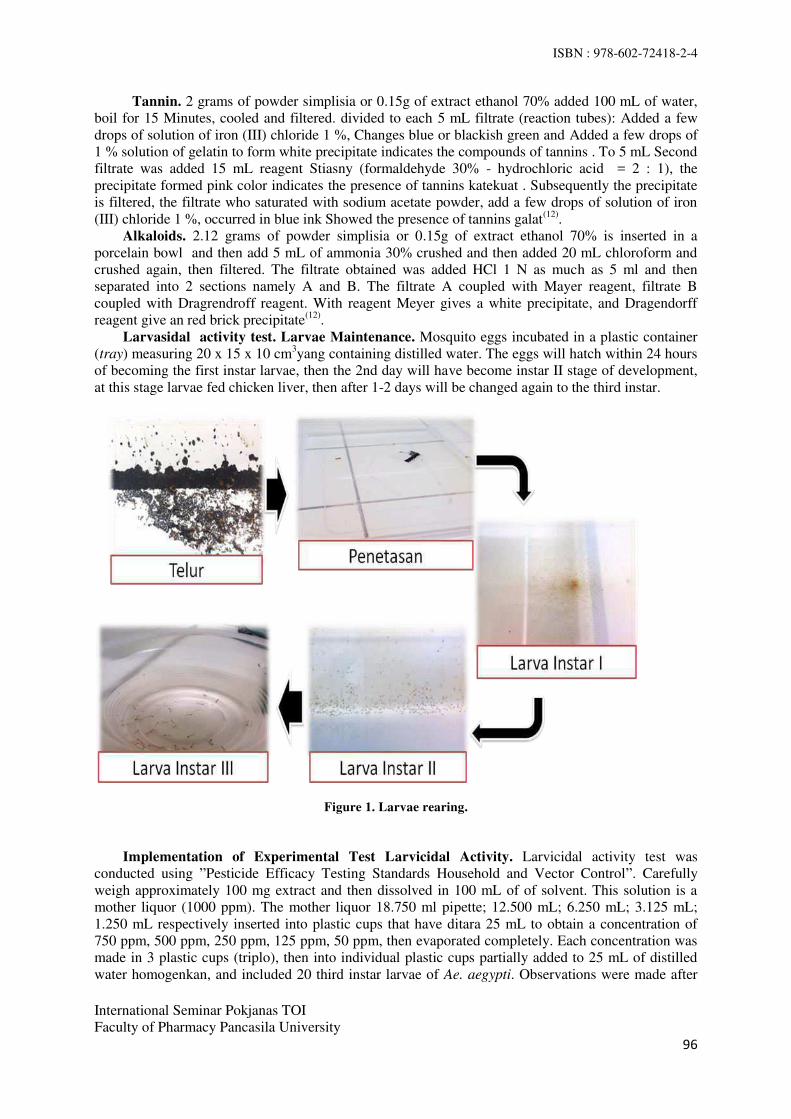
ISBN : 978-602-72418-2-4
International Seminar Pokjanas TOI
Faculty of Pharmacy Pancasila University
96
Tannin. 2 grams of powder simplisia or 0.15g of extract ethanol 70% added 100 mL of water,
boil for 15 Minutes, cooled and filtered. divided to each 5 mL filtrate (reaction tubes): Added a few
drops of solution of iron (III) chloride 1 %, Changes blue or blackish green and Added a few drops of
1 % solution of gelatin to form white precipitate indicates the compounds of tannins . To 5 mL Second
filtrate was added 15 mL reagent Stiasny (formaldehyde 30% - hydrochloric acid = 2 : 1), the
precipitate formed pink color indicates the presence of tannins katekuat . Subsequently the precipitate
is filtered, the filtrate who saturated with sodium acetate powder, add a few drops of solution of iron
(III) chloride 1 %, occurred in blue ink Showed the presence of tannins galat(12)
.
Alkaloids. 2.12 grams of powder simplisia or 0.15g of extract ethanol 70% is inserted in a
porcelain bowl and then add 5 mL of ammonia 30% crushed and then added 20 mL chloroform and
crushed again, then filtered. The filtrate obtained was added HCl 1 N as much as 5 ml and then
separated into 2 sections namely A and B. The filtrate A coupled with Mayer reagent, filtrate B
coupled with Dragrendroff reagent. With reagent Meyer gives a white precipitate, and Dragendorff
reagent give an red brick precipitate(12)
.
Larvasidal activity test. Larvae Maintenance. Mosquito eggs incubated in a plastic container
(tray) measuring 20 x 15 x 10 cm3yang containing distilled water. The eggs will hatch within 24 hours
of becoming the first instar larvae, then the 2nd day will have become instar II stage of development,
at this stage larvae fed chicken liver, then after 1-2 days will be changed again to the third instar.
Figure 1. Larvae rearing.
Implementation of Experimental Test Larvicidal Activity. Larvicidal activity test was
conducted using ”Pesticide Efficacy Testing Standards Household and Vector Control”. Carefully
weigh approximately 100 mg extract and then dissolved in 100 mL of of solvent. This solution is a
mother liquor (1000 ppm). The mother liquor 18.750 ml pipette; 12.500 mL; 6.250 mL; 3.125 mL;
1.250 mL respectively inserted into plastic cups that have ditara 25 mL to obtain a concentration of
750 ppm, 500 ppm, 250 ppm, 125 ppm, 50 ppm, then evaporated completely. Each concentration was
made in 3 plastic cups (triplo), then into individual plastic cups partially added to 25 mL of distilled
water homogenkan, and included 20 third instar larvae of Ae. aegypti. Observations were made after

ISBN : 978-602-72418-2-4
International Seminar Pokjanas TOI
Faculty of Pharmacy Pancasila University
97
24 hours of exposure to the test solution and counted the number of larvae were dead and stated in the
presentation of death.
Negative controls only solvent without the extract, in the same way. Positive controls carried out on
Temephos 1 ppm.
Data Processing Methods. Test data processing is done systematically using probit analysis
method. Probit analysis is used to determine the percentage of larval mortality LC50 of Ae. aegypti L.
uses Epa Probit Analysis Program Used For Calculating LC/EC Values Version 1.5.In Epa Probit
Analysis Program Used For Calculating LC/EC Values Version 1.5. the data entered is the
relationship with the concentration of the value of the average percentage mortality of larvae of Ae.
aegypti.
RESULT AND DISCUSSION
Making the Ethanol 70% Extract. Sugar apple seed is extracted by cold by maceration kinetic (use
maserator). The solvent used for extraction is 70% ethanol. Extracts were obtained in the form of
extracts viscous blackish brown. 36.9 g of 506.2 g of crude drug powder sugar apple seeds with 7.29%
yield.
Phytochemical screening. The phytochemical screening via Farnsworth method was conducted
using powder of simplicia and Soursop seeds and sugar-apple seeds extract. In powder and extract
having metabolite compound such as saponin, triterpenoid, and cumarin. The result of phytochemical
test is shown in Table 1.
Table 1. Result of phytochemical screening of soursop seeds (Annona muricata L.) and sugar-
apple seeds (Annona squamosa L.) powder and extract. No Secondary
Metabolites
Simplicia
powder
Ethanol
70% Extract
1 Alkaloids - -
2 Flavonoids - -
3 Saponins + +
4 Kuinon - -
5 Tannin - -
6 Steroids /
triterpenoids
- /+ - / +
7 Volatile oil - -
8 Coumarin + +
Notes : + = giving positive reaction
− = giving negative reaction
In Table 1 it can be seen that the results of the qualitative identification of secondary metabolite
content of the seed powder soursop (Annona muricata L.) by means of screening phytochemical
compounds derived class of saponins, triterpenoids, and coumarin. While the 70% ethanol extract
derived class compound saponin, triterpenoids, and coumarin.
Larvicidal Activity Test. Larvicidal activity test 70% ethanol extract sugar apple seeds is done
by the method of Pesticide Efficacy Testing Standards Household and Vector Control for mosquito
larvae Ae.aegypti L. larvae used test is the third instar larvae of mosquitoes Ae.aegypti because it has a
fairly good resistance against external environment and durability stronger mechanically when the
transfer of the larvae, and have a long time to turn into adult mosquitoes. Test solution at a
concentration of 50, 125, 250, 500 and 750 ppm generated triplo, then put 20 third instar larvae of Ae.
aegypti L. and counted the number of larvae mortality after 24 hours of observation. Negative controls
only the solvent used and Temephos (larvicidal commonly used) as a positive control.

ISBN : 978-602-72418-2-4
International Seminar Pokjanas TOI
Faculty of Pharmacy Pancasila University
98
Table 2.The average percentage mortality of larvae of Ae.aegypti L., after exposure to 70% ethanol extract
sugar-apple seeds in the 24-hour observation.
Concentration
(ppm)
% Kematian
Sugar-apple Seeds
Ethanol 70% Extract
Control
Negative
(Solvent)
Positive
(Temephos 1 ppm)
750 100 0 100
500 100 0 100
250 98,35 0 100
125 48,35 0 100
50 18,35 0 100
LC50 (ppm) 97,462 - -
Linear regression a = -103,6709
b = 75,0693
r = 0,9372
-
-
The test results larvicidal activity against sugar-apple seed LC50 values obtained from the ethanol
70%extract is 97,462 ppm. From the LC50 values of 97.462 ppm can be concluded that the sugar apple
seed extract has larvicidal activity against Ae. aegypti. This can be caused by chemical compounds /
secondary metabolites contained in the sugar-apple seed saponin, coumarin suspected triterepenoid
and potentially as larvicidal(23)
.
Saponins allegedly able to diffuse into the cuticle layer of larvae that can damage cell membranes
and toxic compounds can be entered and off the larvae. Saponins have a bitter taste and sharp and can
cause irritation of the stomach. Larvae digestive tract, particularly the midgut (midgut) is the major
site of absorption of nutrients and digestive enzymes seksresi. Saponin absorption into the intestine
larvae can inhibit the action of digestive enzymes and cause damage to the cells in the channel
pencernaan(1)
.
Triterpenoids also thought to be as antifeedant (antimakan) on the larvae so that the larvae loss of
appetite, this led to the loss of energy and development of larvae will be hampered even can cause
death 24. In addition, coumarin is also reported as larvicides because potentially able to change the
ability of detoxification with reversible and irreversible inhibits the enzyme cytochrome P450(25)
.Dari
third ability of secondary metabolites in seed srikaya concluded that sugar apple seeds potentially
sebagaii larvicides against mosquito larvae Ae. aegypti L.
Mortality of larvae on seed extract sugar apple (Annonasquamosa L.) allegedly also because of
the effects of the component compounds acetogenindansquamosin toxic contact. Where after the
larvae exposed to the extract, the compound into the body of Ae. aegypti through physical contact and
the case of death of the larva. Prijono (1994) in Wardhanaet.al (2005) states that the absorption of
toxic insecticides contact occurs largely in the cuticle. Active compounds will penetrate into the
insect's body through the part that is covered by a thin cuticle, such as membrane between segments.
Stomach poison ability of the compound to absorb seyawaasetogenin work on sugar apple seed extract
into the wall fosfolirasi larvae and able to inhibit oxidative chain so that the cell respiration is inhibited
activity of Ae.aegypti because of breathing stopped. Squamocin compounds in seeds srikaya allegedly
able to diffuse from the thin cuticle layer to spread throughout the body Ae. aegypti through
hemolimfa flow(15)
.
Mortaitas larvae of Ae.aegypti showed signs as follows: larvae do not move when touched, bodies
pale white larvae, elongated body shape or rigid(1)
. The color can be seen more clearly with the aid of a
stereo microscope and optilab. Differences larvae of Ae.aegypti normal and who have died can be seen
in Figure 2.

ISBN : 978-602-72418-2-4
International Seminar Pokjanas TOI
Faculty of Pharmacy Pancasila University
99
Figure 2. Third instar larvae of Ae. aegypti normal (A);
and third instar larvae of Ae. aegypti die (B).
Figure 3. Graph average percentage mortality soursop seed extract on a 24 hour observation (x-axis:%
average mortality of larvae and the y-axis: concentration sugar apple seeds extract (ppm)).
From Figure 3 shows that the higher the concentration of sugar apple seed extract, the higher the
death rate of Ae. aegypti L. Ethanol 70% and distilled water as a negative control test the same activity
against larvae of Ae. aegypti, and the results obtained all the larvae do not occur death. This indicates
that the solvent does not affect the mortality of larvae. Temephos as a positive control, in which the
larvicidal activity at a concentration of 1 ppm trials have demonstrated 100% mortality against larvae
of Ae. aegypti L.
CONCLUSION
Based on the results of phytochemical screening of the seeds sugar apple (Annonasquamosa L.)
obtained secondary metabolites content of saponins, triterpenoids, and coumarin., Test results with the
larvicidal activity and data analysis has been done, it can be concluded that the 70% ethanol extract of
the seeds have activity sugar apple seeds against larvae of Ae. aegypti L. with LC50 values of 97, 462
ppm.
Suggestion. Ethanol 70% extract of the seeds sugar apple (Annonasquamosa L.) has a good chance to
be used as biological insecticides to control mosquito larvae that are environmentally friendly.
A B

ISBN : 978-602-72418-2-4
International Seminar Pokjanas TOI
Faculty of Pharmacy Pancasila University
100
REFERENCES
1. Kaihena M, Vika L, Maria N. Efektivitas ekstrak etanol daun sirih (Piper betle L) terhadap
mortalitas larva nyamuk Anopheles sp dan Culex. Molluca medica. ISSN: 1979-6358.
2. Susanti PD, Danang B, Dini S, Susilawati. Penggunaan ekstrak kulit kayu gemor (Nothaphoebe
coriacea K.) sebagai larvasida hayati terhadap tingkat mortalitas jentik nyamuk Aedes aegypti
serta dampaknya pada kualitas air hujan. ISSN 1978-8096. 2013.9:117–22.
3. Hadi, Upik Kesumawati. Penyakit tular vektor: demam berdarah dengue. Bogor: Fakultas
Kedokteran Hewan IPB. 2005.
4. World Health Organization. Dengue: guidelines for diagnosis, trearment, prevention and control,
new edition. Swiss. 2010. h.5.
5. Palgunadi BU, Asih Rahayu. Aedes aegypti sebagai vektor penyakit demam berdarah dengue.
Surabaya: Universitas Wijaya Kusuma. 2011.
6. Mulyawati AP, Hayati EK, Nashihuddin A, Tukimin. uji efektivitas dan identifikassi senyawa
ekstrak biji sirsak (Annona muricata Linn.) yang besifat bioaktif insektisida nabati terhadap hama
thrips. Alchemy 2010;2(1): 104-1575.
7. Rosmayanti, Kiki. Uji efektivitas ekstrak biji sirsak (Annona muricata L ) sebagai larvasida pada
larva Aedes segypti instar III/IV (skripsi). Jakarta: Fakultas Kedokteran Dan Ilmu Kesehatan
Universitas Islam Negeri Syarif Hidayatullah. 2014. 1-3, 16-17, 37-40.
8. Wardhana AH, Amir H, Muharsini S, dan Yuningsih, Veteriner BP. Uji keefektifan biji sirsak
(Annona muricata) dan aar tuba (Derris edliptica) terhadap larva Chrysomya bezziana secara in
vitro. 2006. 1013-1017.
9. Wardhana AH, Amir H, dan J Manurung, Veteriner BP. Uji efikasi ekstrak heksan daging biji
srikaya ( Annona squamosa L ) terhadap pertumbuhan larva lalat Chrysomya bezziana secara in
vitro. 2004. 134-142.
10. Syamsuhidayat Ss, Hutapea JR. Inventaris tanaman obat indonesia. Jilid I. Jakarta: Departemen
Kesehatan Indonesia. 1991. h. 50,56-57.
11. Standard of Asean herbal medicine. jilid 1. Asean Countries. 1993. h.51.
12. Dalimartha S. Atlas tumbuhan obat Indonesia. Jilid III. Jakarta: Trubus Agriwidya. 2003. 145-
147.
13. Asyiyah, Nur. Perluasan hama sasaran formulasi insektisida nabati pada tiga spesies serangga
hama sayuran (skripsi). Bogor: Fakultas Pertanian Institut Pertanian Bogor. 2010. h.17.
14. Taslimah. Uji efikasi biji srikaya (Annona squamosa L.) sebagai bioinsektisida dalam upaya
integrated vector management terhadap Aedes aegypti (skripsi). Jakarta: Fakultas Kedokteran Dan
Ilmu Kesehatan Universitas Islam Negeri Syarif Hidayatullah. 2014. h.1-6, 23-28,76-81.
15. Wardhana AH, Amir H, dan J.Manurung, Veteriner BP. Efektifitas ekstrak biji srikaya (Annona
squamosa L ) dengan pelarut air, metanol dan heksan terhadap mortalitas larva caplak boophilus
microplus secara in vitro. 2005. 134–42.
16. Supartha, I wayan. Pengendalian terpadu vektor virus demam berdarah dengue, Aedes aegypti
(Linn) dan Aedes albopictus (Skuse) (Diptera: Culicidae). Denpasar: Fakultas Pertanian
Universitas Udayana. 2008.
17. Suyanto, Darnoto S, Astuti D. Hubungan pengetahuan dan sikap pengendalian nyamuk Aedes
aegypti di kelurahan sangkrah kecamatan pasar kliwon kota Surakarta. 2002. 705:1-13.
18. Sigit SH, Hadi UK. Hama Pemukiman Indonesia. Bogor: Unit kajian pengendalian hama
pemukiman. Fakultas Kedokteran Hewan IPB. 2006. h.34,37-45.
19. Gandahusada S, Llahude HD, Pribadi W. Parasitologi kedokteran, edisi ketiga. Jakarta: Fakultas
Kedokteran Universitas Indonesia. 1998. h.219.
20. Soeharto. Beberapa sifat kehidupan Aedes aegypti L. Jakarta: Universitas Nasional; 1974. h.55.
21. Antonius, P.Rumengan. Uji larvasida nyamuk (Aedes aegypti) dari Ascidian (Didemnum molle).
2010. VI-2.h.83.
22. Soedarto. Entomologi kedokteran. Jakarta: Penerbit Buku Kedokteran EGC. 1989. h.104.
ISBN : 978-602-72418-2-4
International Seminar Pokjanas TOI
Faculty of Pharmacy Pancasila University
101
23. Riswanto S. Uji efektivitas pestisida nabati terhadap hama Spodoptera litura (Lepidoptera:
Noctuidae) pada tanaman tembakau (Nicotiaana tabaccum L.). Medan: Universitas Sumatera
Utara. 2009. 20-23.
24. Nopitasari. Uji aktivitas ekstrak biji langsat (Lansium domesticum Cor.) sebagai larvasida Aedes
aegypti. Universitas Tanjungpura. 2013. 12-14.
25. Venugopala KN, Raquel MG, Kabange K, Bandar EA, Mahesh VA, Bharti O. Evaluation of
halogenated coumarins for antimosquito properties. Hindawi Publishing Corporation The
Scientific World Journal. 2014. Vol.4.

ISBN : 978-602-72418-2-4
International Seminar Pokjanas TOI
Faculty of Pharmacy Pancasila University
102
Anticancer Activity of Jatropha sp. on Breast
and Cervix Cancer Cells Lines
SITI ROFIDA1*, NAILIS SYIFA’1
, NURKHASANAH2, LAELA HAYU NURANI
2
1Pharmacy Departement, Health Science Faculty, University of Muhammadiyah
Malang, Indonesia. 2Faculty of Pharmacy, University of Ahmad Dahlan Yogyakarta, Indonesia.
Corresponding: [email protected]
Abstract: Jatropha sp. is a plant that comes from Euphorbiaceae which has 70 species and some of them
reported have activity as a drug. Secondary metabolites contained in Jatropha sp. are alkaloids, tannins,
flavonoids, steroid, saponins, and phenol. Based on research, Jatropha sp. has the potential to be
developed as the raw materials of anticancer drug. This study aims to obtain raw materials traditional
medicines standardized from Jatropha sp. which have anticancer activity against breast and cervical
cancer. This study has been testing the anticancer activity of J.curcas L. and J.gossypifolia L. using MTT
assay cytotoxicity method. Thin Layer Chromatography technique applied to identified the secondary
metabolites on parts of plants which have anticancer activity. The results showed roots extract of
J.gossypifolia L. have activity as breast and cervix anticancer. Cytotoxicity on T47D breast cancer cell
lines and HeLa cervical cancer cell lines with IC50 values respectively 43.57 and 4.32 µg/mL. The
identification using TLC showing chemical compounds such as terpenoids, flavonoids, polyphenols and
anthraquinone.
Keyword: Anticancer activity, J.curcas L., J.gossypifolia L., MTT assay method.
INTRODUCTION
In Indonesia, breast cancer and cervical cancer are the highest cancer incidence rates. According to data
from the Hospital Information System (SIRS) in 2008, breast cancer patients as much as 18.4%, ranked
first in-patients in all hospitals in Indonesia, followed by cervical cancer at 10.3% (1)
. Treatment of cancer
through surgery, radiation, chemotherapy and immunotherapy give adverse effects such as pain,
wounding and causing the death of normal cells, bone marrow depression, anorexia and nausea (2)
. The
use of several natural products especially plant has been recommended by doctors in the treatment of
cancer, such as vinca alkaloids (Catharanthus), epipodophyllotoxins, taxanes and camptothecins (3,4)
.
Jatropha sp. is a plant that comes from family Euphorbiaceae which has 70 species and some of them
have been reported to have activity as a drug. Its use as a popular drug that is as a laxative. Parts of plants
which had this activity is part of the seeds and leaves. Besides oil seeds, latex, bark, leaves and roots are
used to treat skin diseases, stomach aches and teeth, inflammatory, antioxidant, dysentery, vertigo,
malaria, anemia, diabetes, bronchitis, asthma and aphrodisiac (5)
. Classes of compounds in Jatropha sp. ie
alkaloids, tannins, flavonoids, steroid, saponins, and phenol (6,7,8)
. Several studies have reported the
activity of Jatropha sp. as anticancer colon (9)
, liver (10)
, cervix (11)
and breast cancer (12)
. Based on research
that has been done, jatropa sp. has the potential to be developed as an anticancer drug raw materials.
The aim of this study is to get information anticancer activity of extracts of Jatropha sp specifically J.
curcas L. and J.gossypifolia L. which selectively in T47D breast cancer cells lines and Hela cervical
cancer cells lines, so its use is not only based on empirical, but based on scientific data.

ISBN : 978-602-72418-2-4
International Seminar Pokjanas TOI
Faculty of Pharmacy Pancasila University
103
J.curcas L leaves and stems contain chemical compounds saponins, flavonoids and polyphenols, while
the leaves also contain tannins. Empirically, the leaves efficacious as an anthelmintic, flatulence drugs
and drug injuries. J.gossypifolia L. leaves, contained chemical compounds alkaloids, saponins, flavonoids
and polyphenols. Empirically, the leaves efficacious as a laxative and anti-inflammatory agent ear
children (13)
.
MATERIAL AND METHODS
MATERIAL and equipment. Ethanol 96%, n-hexan, ethyl acetat, TLC Plate silica gel F254, T47D breast
cancer cells lines and Hela cervical cancer cells lines collected from parasitology laboratory, Medicine
Faculty, University of Gajahmada, Yogyakarta, medium RPMI, fetal bovine serum 10%, penicillin-
streptomicyn 1%, fungison 0,5 %, Phosphat Buffer Saline 20 %; Dimethyl sulfoxide; 3-(4,5-
Dimetiltiazol-2-il)-2,5-difeniltetrazolium bromide; Sodium Dodecyl Sulphate 10 %.
Plant Material. The leaves, stembark, fruits and roots of J.curcas L. and J. gossypifolia L. were
obtained from Pasuruan in January 2015 and identified by UPT Materia Medica, Batu, Malang. The
leaves, bark, fruits and roots cleaned with running tap water, and dried in an oven at 45 ° for 3 days for
the leaves, stembark and root for 4 days and fruits for 5 days. Then milled to obtain fine powder for
extraction.
Preparation of Extract. One hundred grams of leaves, stembark, fruits and roots powder J.curcas
L. and J. gossypifolia L., macerated using 1 L ethanol 96% for 24 hours (3 times). The filtrate was
collected and concentrated by evaporation with a vacuum rotary evaporator at 50°C to obtain a viscous
extract, the extract then dried in an oven at 40°C. The crude ethanolic extract was also stored in the
refrigerator at 5°C until required for use. The sample for cytotoxicity assay was dissolved into DMSO.
MTT assay. Cancer cells are distributed into 96 well plate and then incubated in 5% CO2 conditions
at 37 °C for overnight. Test solution with a certain concentration is inserted into cancer cells cultured and
incubated under conditions of 5% CO2 at 37 °C. After 24 hours, the liquid was pipetted and discarded,
then washed with PBS solution with a rocked then discarded. Added 100 mL of culture medium per well
and 10 mL solution of MTT in PBS at a concentration of 5 mg / ml. Then incubated under conditions 5%
CO2 of 37 °C. After 4-6 hours stopper added reagents (SDS 10%) and incubated overnight at room
temperature and stored in a dark place. Absorptions is read by ELISA reader at a wavelength of 550-600
nm. The percentage of life cells was calculated to obtain the IC 50 value. The data were analyzed with
probit analysis.
Phytochemical identification. To identify the components of chemical compounds in the crude
ethanolic extract from leaves, stembark, fruits and root J.curcas L. and J.gossypifolia L., TLC technique
were applied. Derivatization for the compound alkaloids, flavonoids, terpenoida, polyphenols and
anthraquinone, used reagent dragendorf, ammonia fumes, anisaldehid-sulfuric acid, FeCl3 and KOH
respectively.
RESULTS AND DISCUSSION
Powder simplicia from leaves and stem bark were obtained have a fine degree from rather crude to
fine. While powder simplicia of fruits and roots have a fine degree rather crude to crude. The water
content that contained in powder simplicia ranged between 2.76 to 8.78%. Ethanolic extract that obtained
from maceration process of simplicia powder of J.curcas L and J.gossypifolia L presented in Table 1.
Table 1. Results of extraction the simplicia powder of J.curcas L and J. gossypifolia L No Simplicia Organoleptic Yield
1 J.curcas L leaves Dark green 9.86 %
2 J.curcas L fruits Brown greenish 7.50 %
3 J.curcas L stem bark Brown blackish 4.76 %
4 J.curcas L roots Dark brown 9.10 %
5 J.gossypifolia L leaves Green brownish 9.20 %
6 J. gossypifolia fruits Brown blackish 7.40%
7 J.gossypifolia L stem bark Dark brown 12.17 %

ISBN : 978-602-72418-2-4
International Seminar Pokjanas TOI
Faculty of Pharmacy Pancasila University
104
8 J.gossypifolia L roots Dark brown 9.03 %
Based on cytotoxicity assay on J.curcas L. and J.gossypifolia L.by MTT assay, obtained the
percentage of life cells and then was calculated to obtain the IC 50 value are presented in Table 2.
Table 2. The potential for cytotoxicity of J.curcas L. and J.gossypifolia L.
No Samples IC50 (µg/mL)
T47D Cell lines Hela Cell lines Vero Cell lines
1 J.curcas L leaves 1030.15 1031.84 545.22
2 J.curcas L fruits 1627.66 1280.33 1155.05
3 J.curcas L stem bark 1283.76 260.30 472.78
4 J.curcas L roots 131.12 191.47 52.50
5 J.gossypifolia L leaves 189.55 191.09 62.55
6 J. gossypifolia fruits 25687.36 1902.92 2460.96
7 J.gossypifolia L stem bark 430.53 401.39 83.12
8 J.gossypifolia L roots 43.57 4.32 9.46
According to the National Cancer Institute (NCI), the extracts that had IC50 values less than 20 µg/mL
against cancer cell cultures could be considered effective as anticancer (14)
. The samples that had IC50
values < 30 µg/mL, identified secondary metabolites with chromatographic techniques and derivatized
with dragendorf, ammonia fumes, anisaldehid-sulfuric acid, FeCl3 and KOH reagents for indicating the
presence of compounds alkaloids, flavonoids, terpenoida, polyphenols and anthraquinone were is
presented in figure 1-5.
A B C D
1 2 1 2 1 2 1 2
A B C D
1 2 1 2 1 2 1 2
Figure 2. TLC profile roots extract of
J.gossypifolia L., stationary phase :
silica gel F254; mobile phase:
hexan:chloroform:aethyl acetat=5:3:3;
(A. UV 254; B. UV 366; C. derivatized:
ammonia fumes, UV 366; D.
derivatized: ammonia fumes, visual).
Figure 1. TLC profile roots extract of
J. Gossypifolia L., stationary phase:
silica gel F254; mobile phase:
hexan:choloroform: aethyl acetat =
5:3:3
(A. UV 254; B. UV 366; C. derivatized:
reagent dragendorf, UV 366; D.
derivatized: reagent dragendorf,
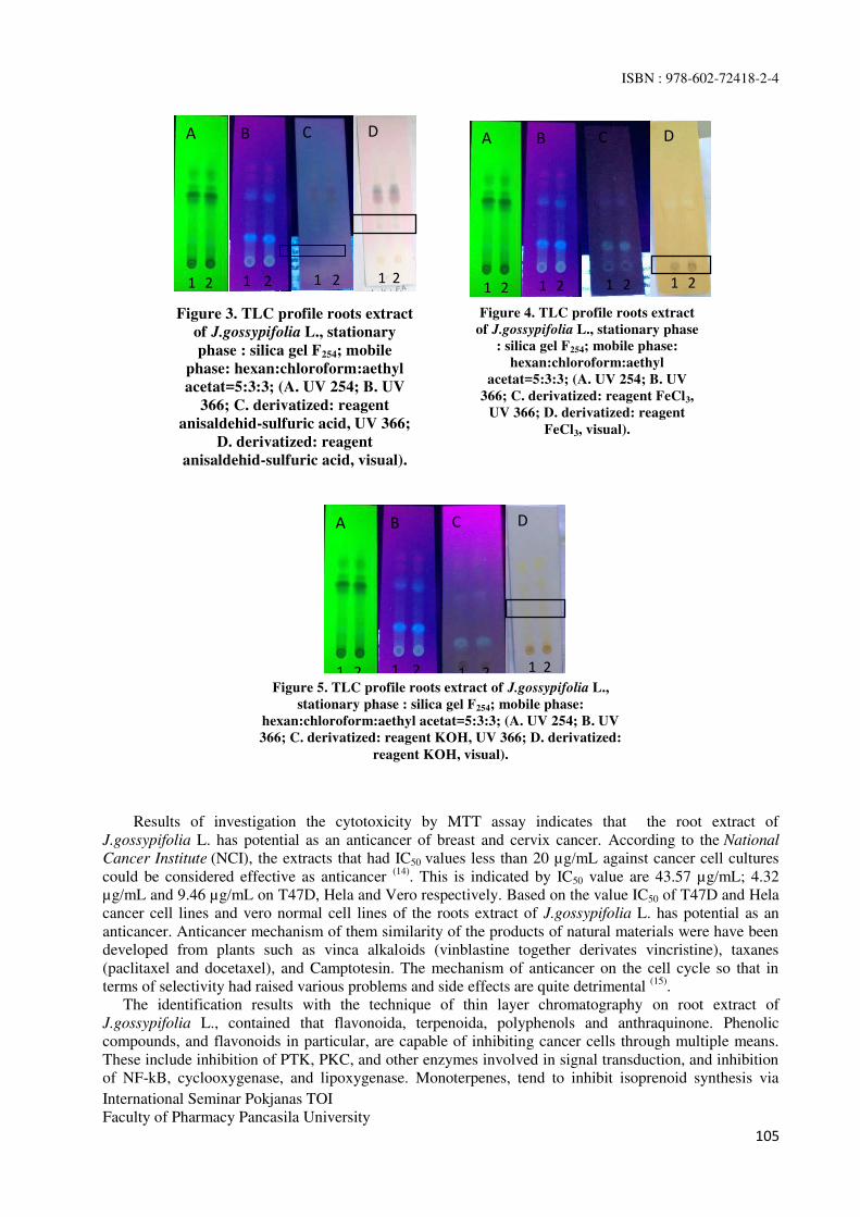
ISBN : 978-602-72418-2-4
International Seminar Pokjanas TOI
Faculty of Pharmacy Pancasila University
105
Results of investigation the cytotoxicity by MTT assay indicates that the root extract of
J.gossypifolia L. has potential as an anticancer of breast and cervix cancer. According to the National
Cancer Institute (NCI), the extracts that had IC50 values less than 20 µg/mL against cancer cell cultures
could be considered effective as anticancer (14)
. This is indicated by IC50 value are 43.57 µg/mL; 4.32
µg/mL and 9.46 µg/mL on T47D, Hela and Vero respectively. Based on the value IC50 of T47D and Hela
cancer cell lines and vero normal cell lines of the roots extract of J.gossypifolia L. has potential as an
anticancer. Anticancer mechanism of them similarity of the products of natural materials were have been
developed from plants such as vinca alkaloids (vinblastine together derivates vincristine), taxanes
(paclitaxel and docetaxel), and Camptotesin. The mechanism of anticancer on the cell cycle so that in
terms of selectivity had raised various problems and side effects are quite detrimental (15)
.
The identification results with the technique of thin layer chromatography on root extract of
J.gossypifolia L., contained that flavonoida, terpenoida, polyphenols and anthraquinone. Phenolic
compounds, and flavonoids in particular, are capable of inhibiting cancer cells through multiple means.
These include inhibition of PTK, PKC, and other enzymes involved in signal transduction, and inhibition
of NF-kB, cyclooxygenase, and lipoxygenase. Monoterpenes, tend to inhibit isoprenoid synthesis via
A B C D
1 2 1 2 1 2 1 2
A B C D
1 2 1 2 1 2 1 2
A B C D
1 2 1 2 1 2 1 2
Figure 4. TLC profile roots extract
of J.gossypifolia L., stationary phase
: silica gel F254; mobile phase:
hexan:chloroform:aethyl
acetat=5:3:3; (A. UV 254; B. UV
366; C. derivatized: reagent FeCl3,
UV 366; D. derivatized: reagent
FeCl3, visual).
Figure 5. TLC profile roots extract of J.gossypifolia L.,
stationary phase : silica gel F254; mobile phase:
hexan:chloroform:aethyl acetat=5:3:3; (A. UV 254; B. UV
366; C. derivatized: reagent KOH, UV 366; D. derivatized:
reagent KOH, visual).
Figure 3. TLC profile roots extract
of J.gossypifolia L., stationary
phase : silica gel F254; mobile
phase: hexan:chloroform:aethyl
acetat=5:3:3; (A. UV 254; B. UV
366; C. derivatized: reagent
anisaldehid-sulfuric acid, UV 366;
D. derivatized: reagent
anisaldehid-sulfuric acid, visual).

ISBN : 978-602-72418-2-4
International Seminar Pokjanas TOI
Faculty of Pharmacy Pancasila University
106
feedback inhibition of HMGR, and others, such as boswellic acid, tend to produce anti-inflammatory
effects. Others may cause anticancer effects through other direct or indirect actions. Because they are
lipid-soluble, they are more easily absorbed, and therefore lower doses can be used to reach the plasma
target concentration (16)
.
CONCLUSION
Anticancer activity of root extract of J.gossypifolia L. with MTT assay had cytotoxicity in T47D breast
cancer cell lines and HeLa cervical cancer cell lines with IC50 values respectively 43.57 and 4.32 µg/mL.
The identification using TLC showing chemical compounds such as terpenoids, flavonoids, polyphenols
and anthraquinone.
ACKNOWLEDGMENT
Thanks to Deputy Minister for Science and Technology Relevance and Productivity, State Ministry of
Research and Technology to founded this reseach at 2015 period.
REFERENCE
1. Depkes RI. 143 milyar dana jamkesmas untuk biaya rawat inap pengobatan kanker.
http://www.depkes.go.id/index.php/berita/press-release/1831-143-milyar-dana-jamkesmas-untuk-
biaya-rawat-inap-pengobatan-kanker.html. 2012. [cited 2014 Mar 15].
2. Corwin JE. Buku saku patofisiologi (terjemahan Nike B.S.).Ed.3, Jakarta: EGC, 2009. p. 66-94.
3. Nobili S, Lippi D, Witort E, Donninic M, Bausi L, Mini E, Capacciolic S. Natural compounds for
cancer treatment and prevention. Pharmacological Research. 2009, 59: 365-378. Available from:
(http://citeseerx.ist.psu.edu/viewdoc/download? doi=10.1.1.468.6501&rep=rep1&type=pdf).
4. Balunas MJ, Kinghorn AD, Drug discovery from medicinal plants. Life sciences. 2005. 78: 431-441.
Available from: (http://webspace.pugetsound.edu/facultypages/bdasher
/Chem361/Review_Articles_files/Drugs%20from%20Plants.pdf).
5. Sabandar CW, Ahmat N, Jaafar FM, Sahidi I. Medicinal property, phytochemistry and pharmacology
of several Jatropha species (Euphorbiaceae): A review. Phytochemistry. 2013., 59:7-29. (doi:
10.1016/j.phytochem.2012.10.009.).
6. Kafagy SM, Mohamed YA, Abdel Salam NA, Mahmoud ZF, Phytochemical study of Jatropha
curcas. Planta Medica Med, 1977, 31(3):274-277. (Doi: 10.1055/s-0028-1097529).
7. Nwokocha A., Blessing IO, Agbagwa Okoli BE. Comparative phytochemical screening of Jatropha
L. Spesies in the Niger Delta. Research Journal of Phytochemistry. 2011,
(DOI: 10.3923/rjphyto.2011.107.114 ).
8. Gupta DD, Haque MD, Islam NM, Rahman S, Hasan AKMM, Shibib BA. Alkaloid and steroid
from the stem bark of Jatropha curcas (Euphorbiaceae). Journal Pharmaceutical Science. 2011,
10(1):9-11. (DOI: http://dx.doi.org/10.3329/dujps.v10i1.10009).
9. Prakash E. Gupta DK, Cytotoxic activities of extracts of medicinal plants of euphorbiaceae family
studied on seven human cancer cell lines. Universal Journal of Plant Science, 1(4):113-117. (DOI:
10.13189/ujps.2013.010401).
10. Lin J, Yan F, Tang L, Chen F. 2003. Antitumor effects of curcin from seeds of Jatropha curcas.
Acta Pharmacol Sin. 2013, 24(3):241-246. (http://www.chinaphar.com/1671-4083/24/241.pdf)
11. Ee GCL, Lim CK., Taufiq-Yap YH, Go R. Ferulic ester from Jatropha podagrica (Euphorbiaceae).
Malaysian Journal of Chemistry. 2005. 1(7):045-048. Available from: (:
http://www.researchgate.net/publication/229429819)
12. Oskoueian E, Abdullah N, Ahmad S. Phorbol esters from jatropha meal triggered apoptosis,
activated PKC-δ, caspase-3 proteins and down-regulated the proto-oncogenes in MCF-7 and hela
cancer cell lines. Molecules. 2012, 17:10816-10830. (doi:10.3390/molecules170910816)
13. Hutpea JR. Inventaris tanaman obat Indonesia (III), Jakarta: Badan Penelitian dan Pengembangan
Kesehatan Departemen Kesehatan Republik Indonesia. 1994. hal.113.

ISBN : 978-602-72418-2-4
International Seminar Pokjanas TOI
Faculty of Pharmacy Pancasila University
107
14. Ampasavate C, Okonogi S, Anuchapreeda S, Cytotoxic of extracts from fruit plants against leukemic
cell lines. African Journal of Pharmacy and Pharmacology. 2010, 4(1):13-21. Available from:
(http://www.researchgate.net/publication/228663822_Cytotoxicity_of_extracts_from_fruit_plants_a
gainst_leukemic_cell_lines).
15. Fajarningsih ND, Nursid M, WikantaT, Marraskuranto E. Bioaktivitas ekstrak Turbinaria decurrens
Sebagai antitumor (Hela dan T47D) serta efeknya terhadap proliferasi limfosit. Jurnal Pascapanen
dan Bioteknologi Kelautan dan Perikanan. 2008. 3(1):21-27.
16. Boik J. Natural compounds in cancer theraphy. Ed. 1st, Minnesota: Oregon Medical Press. 2001. P:
251-266; 297-311.
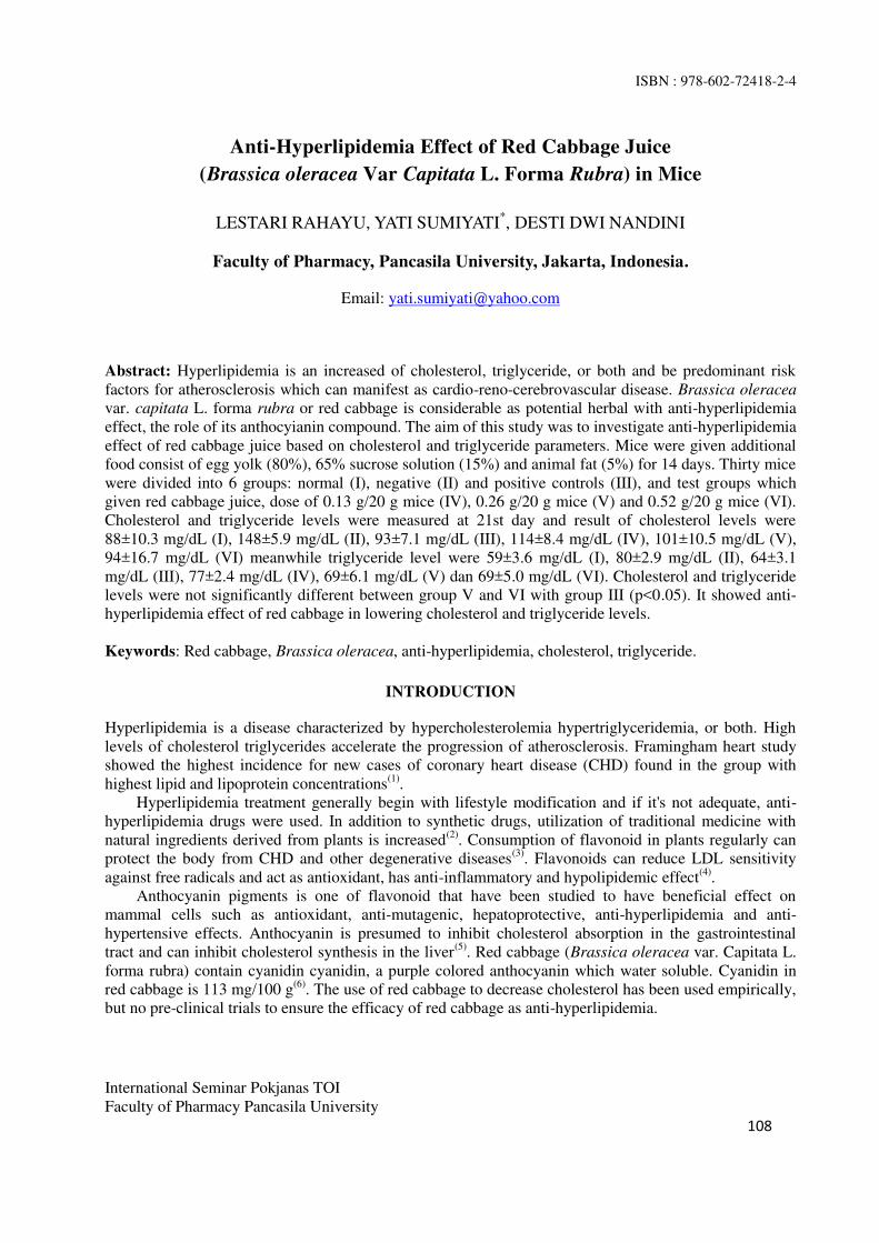
ISBN : 978-602-72418-2-4
International Seminar Pokjanas TOI
Faculty of Pharmacy Pancasila University
108
Anti-Hyperlipidemia Effect of Red Cabbage Juice
(Brassica oleracea Var Capitata L. Forma Rubra) in Mice
LESTARI RAHAYU, YATI SUMIYATI*, DESTI DWI NANDINI
Faculty of Pharmacy, Pancasila University, Jakarta, Indonesia.
Email: [email protected]
Abstract: Hyperlipidemia is an increased of cholesterol, triglyceride, or both and be predominant risk
factors for atherosclerosis which can manifest as cardio-reno-cerebrovascular disease. Brassica oleracea
var. capitata L. forma rubra or red cabbage is considerable as potential herbal with anti-hyperlipidemia
effect, the role of its anthocyianin compound. The aim of this study was to investigate anti-hyperlipidemia
effect of red cabbage juice based on cholesterol and triglyceride parameters. Mice were given additional
food consist of egg yolk (80%), 65% sucrose solution (15%) and animal fat (5%) for 14 days. Thirty mice
were divided into 6 groups: normal (I), negative (II) and positive controls (III), and test groups which
given red cabbage juice, dose of 0.13 g/20 g mice (IV), 0.26 g/20 g mice (V) and 0.52 g/20 g mice (VI).
Cholesterol and triglyceride levels were measured at 21st day and result of cholesterol levels were
88±10.3 mg/dL (I), 148±5.9 mg/dL (II), 93±7.1 mg/dL (III), 114±8.4 mg/dL (IV), 101±10.5 mg/dL (V),
94±16.7 mg/dL (VI) meanwhile triglyceride level were 59±3.6 mg/dL (I), 80±2.9 mg/dL (II), 64±3.1
mg/dL (III), 77±2.4 mg/dL (IV), 69±6.1 mg/dL (V) dan 69±5.0 mg/dL (VI). Cholesterol and triglyceride
levels were not significantly different between group V and VI with group III (p<0.05). It showed anti-
hyperlipidemia effect of red cabbage in lowering cholesterol and triglyceride levels.
Keywords: Red cabbage, Brassica oleracea, anti-hyperlipidemia, cholesterol, triglyceride.
INTRODUCTION
Hyperlipidemia is a disease characterized by hypercholesterolemia hypertriglyceridemia, or both. High
levels of cholesterol triglycerides accelerate the progression of atherosclerosis. Framingham heart study
showed the highest incidence for new cases of coronary heart disease (CHD) found in the group with
highest lipid and lipoprotein concentrations(1)
.
Hyperlipidemia treatment generally begin with lifestyle modification and if it's not adequate, anti-
hyperlipidemia drugs were used. In addition to synthetic drugs, utilization of traditional medicine with
natural ingredients derived from plants is increased(2)
. Consumption of flavonoid in plants regularly can
protect the body from CHD and other degenerative diseases(3)
. Flavonoids can reduce LDL sensitivity
against free radicals and act as antioxidant, has anti-inflammatory and hypolipidemic effect(4)
.
Anthocyanin pigments is one of flavonoid that have been studied to have beneficial effect on
mammal cells such as antioxidant, anti-mutagenic, hepatoprotective, anti-hyperlipidemia and anti-
hypertensive effects. Anthocyanin is presumed to inhibit cholesterol absorption in the gastrointestinal
tract and can inhibit cholesterol synthesis in the liver(5)
. Red cabbage (Brassica oleracea var. Capitata L.
forma rubra) contain cyanidin cyanidin, a purple colored anthocyanin which water soluble. Cyanidin in
red cabbage is 113 mg/100 g(6)
. The use of red cabbage to decrease cholesterol has been used empirically,
but no pre-clinical trials to ensure the efficacy of red cabbage as anti-hyperlipidemia.
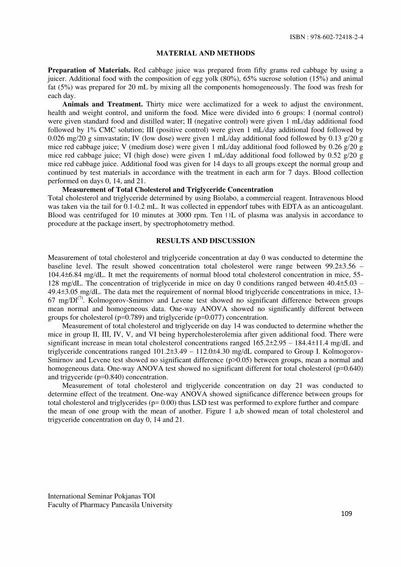
ISBN : 978-602-72418-2-4
International Seminar Pokjanas TOI
Faculty of Pharmacy Pancasila University
109
MATERIAL AND METHODS
Preparation of Materials. Red cabbage juice was prepared from fifty grams red cabbage by using a
juicer. Additional food with the composition of egg yolk (80%), 65% sucrose solution (15%) and animal
fat (5%) was prepared for 20 mL by mixing all the components homogeneously. The food was fresh for
each day.
Animals and Treatment. Thirty mice were acclimatized for a week to adjust the environment,
health and weight control, and uniform the food. Mice were divided into 6 groups: I (normal control)
were given standard food and distilled water; II (negative control) were given 1 mL/day additional food
followed by 1% CMC solution; III (positive control) were given 1 mL/day additional food followed by
0.026 mg/20 g simvastatin; IV (low dose) were given 1 mL/day additional food followed by 0.13 g/20 g
mice red cabbage juice; V (medium dose) were given 1 mL/day additional food followed by 0.26 g/20 g
mice red cabbage juice; VI (high dose) were given 1 mL/day additional food followed by 0.52 g/20 g
mice red cabbage juice. Additional food was given for 14 days to all groups except the normal group and
continued by test materials in accordance with the treatment in each arm for 7 days. Blood collection
performed on days 0, 14, and 21.
Measurement of Total Cholesterol and Triglyceride Concentration
Total cholesterol and triglyceride determined by using Biolabo, a commercial reagent. Intravenous blood
was taken via the tail for 0.1-0.2 mL. It was collected in eppendorf tubes with EDTA as an anticoagulant.
Blood was centrifuged for 10 minutes at 3000 rpm. Ten L of plasma was analysis in accordance to
procedure at the package insert, by spectrophotometry method.
RESULTS AND DISCUSSION
Measurement of total cholesterol and triglyceride concentration at day 0 was conducted to determine the
baseline level. The result showed concentration total cholesterol were range between 99.2±3.56 – 104.4±6.84 mg/dL. It met the requirements of normal blood total cholesterol concentration in mice, 55-
128 mg/dL. The concentration of triglyceride in mice on day 0 conditions ranged between 40.4±5.03 – 49.4±3.05 mg/dL. The data met the requirement of normal blood triglyceride concentrations in mice, 13-
67 mg/Dl(7)
. Kolmogorov-Smirnov and Levene test showed no significant difference between groups
mean normal and homogeneous data. One-way ANOVA showed no significantly different between
groups for cholesterol (p=0.789) and triglyceride (p=0.077) concentration.
Measurement of total cholesterol and triglyceride on day 14 was conducted to determine whether the
mice in group II, III, IV, V, and VI being hypercholesterolemia after given additional food. There were
significant increase in mean total cholesterol concentrations ranged 165.2±2.95 – 184.4±11.4 mg/dL and
triglyceride concentrations ranged 101.2±3.49 – 112.0±4.30 mg/dL compared to Group I. Kolmogorov-
Smirnov and Levene test showed no significant difference (p>0.05) between groups, mean a normal and
homogeneous data. One-way ANOVA test showed no significant different for total cholesterol (p=0.640)
and trigyceride (p=0.840) concentration.
Measurement of total cholesterol and triglyceride concentration on day 21 was conducted to
determine effect of the treatment. One-way ANOVA showed significance difference between groups for
total cholesterol and triglycerides (p= 0.00) thus LSD test was performed to explore further and compare
the mean of one group with the mean of another. Figure 1 a,b showed mean of total cholesterol and
trigyceride concentration on day 0, 14 and 21.
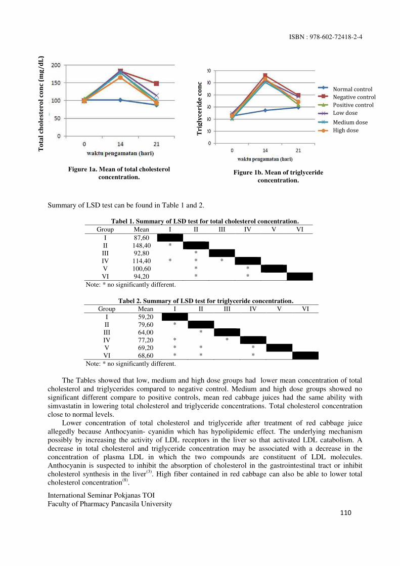
ISBN : 978-602-72418-2-4
International Seminar Pokjanas TOI
Faculty of Pharmacy Pancasila University
110
Summary of LSD test can be found in Table 1 and 2.
Tabel 1. Summary of LSD test for total cholesterol concentration.
Group Mean I II III IV V VI
I 87,60
II 148,40 *
III 92,80 *
IV 114,40 * * *
V 100,60 * *
VI 94,20 * *
Note: * no significantly different.
Tabel 2. Summary of LSD test for triglyceride concentration.
Group Mean I II III IV V VI
I 59,20
II 79,60 *
III 64,00 *
IV 77,20 * *
V 69,20 * * *
VI 68,60 * * *
Note: * no significantly different.
The Tables showed that low, medium and high dose groups had lower mean concentration of total
cholesterol and triglycerides compared to negative control. Medium and high dose groups showed no
significant different compare to positive controls, mean red cabbage juices had the same ability with
simvastatin in lowering total cholesterol and triglyceride concentrations. Total cholesterol concentration
close to normal levels.
Lower concentration of total cholesterol and triglyceride after treatment of red cabbage juice
allegedly because Anthocyanin- cyanidin which has hypolipidemic effect. The underlying mechanism
possibly by increasing the activity of LDL receptors in the liver so that activated LDL catabolism. A
decrease in total cholesterol and triglyceride concentration may be associated with a decrease in the
concentration of plasma LDL in which the two compounds are constituent of LDL molecules.
Anthocyanin is suspected to inhibit the absorption of cholesterol in the gastrointestinal tract or inhibit
cholesterol synthesis in the liver(3)
. High fiber contained in red cabbage can also be able to lower total
cholesterol concentration(8)
.
da
Normal control
Negative control
Positive control
Low dose
Medium dose
High dose
To
tal
ch
ole
ste
ro
l c
on
c (
mg
/d
L)
Tri
gly
ce
rid
e c
on
c
Figure 1a. Mean of total cholesterol
concentration. Figure 1b. Mean of triglyceride
concentration.

ISBN : 978-602-72418-2-4
International Seminar Pokjanas TOI
Faculty of Pharmacy Pancasila University
111
CONCLUSION
Red cabbage juice can decrease concentration of total cholesterol and triglyceride in hyperlipidemia mice;
Reduction level of medium and high dise as effective as simvastatin, close to normal levels for cholesterol
where dosage II is the most effective for lowering the concentration of total cholesterol and triglycerides.
ACKNOWLEDGMENTS
The Authors are Grateful to Pharmacy Faculty of Pancasila University for Incentive Research Funding.
REFERENCES
1. Murray RK. Biochemistry harper. XXII Edition. Translated by A Hartono. EGC. Jakarta: 1995. p.
252-93, 308-10.
2. Kelompok kerja ilmiah. Penapisan farmakologi, pengujian fitokimia dan pengujian klinik. Jakarta:
Yayasan Pengembangan Obat Bahan Alam Phyto Medica; 1993. h.191.
3. Davalos A, Fernandez-Hernando C, Cerrato F, Martinez-Botas J, et al. Red grape juice and
polyphenols alter cholesterol homeostasis and increase LDL-receptor activity in human cells in vitro.
J Nutr, 2006. 136. 1766-1773.
4. Middleton E, Kandaswami C, Theoharides CT. The effect of plant flavonoids on mammalian cells:
implication for inflammation, heart disease, and cancer. Pharmacological Reviews, 52 (4).2000.673-
751.
5. Huang D J, Lin C D, Chen H J, Lin Y H. Antioxidant and antiproliferative activities of sweet potato
(Ipomoea batatas L. Lam Tainong 57) constituents. Bot Bull Acad. Sin, 45: 179-186.
6. Kwon SH, et al. Anti-obesity and hypolipidemic effects of black soybean anthocyanins. JM Food.
2007. 10 (3).552-556.
7. Fox GJ, Barthold WS, Davisson TM, Newcomer EC, Quimby WF, Smith LA. The mouse in
biomedical research: normative biology, husbandry, and model. 2nd edition. UK: Academy Press;
2007. P. 188.
8. El mowafy, A. Maha. Treatment effect of red cabbage and cystein against paracetamol induced
hepatotoxicity in experimental rats. Journal of Applied Science Research. 2012; 8(12): 5852-5859.

ISBN : 978-602-72418-2-4
International Seminar Pokjanas TOI
Faculty of Pharmacy Pancasila University
112
Hepatoprotective Study of Cosolvent Solution
From Mangosteen (GARCINIA MANGOSTANA L.) Rind in Rats
ROS SUMARNY*, LILIEK NURHAYATI, YATI SUMIYATI, ASTRI YULIASTRI PERMANA
Faculty of Pharmacy, Pancasila University, Jakarta, Indonesia.
Email: [email protected]
Abstract: Garcinia mangostana L. rind consist of -mangostin, an active compound that considerably
can protect the liver -mangostin which
prepared as cosolvent solution and ethanolic extract suspension. This was experimental study where
hepatoprotective effect was assessed using Aspartate transminase (AST) and Alanine transminase (ALT).
Twenty five rats were divided into 5 groups: negative (I) and positive controls (II); cosolvent (III), extract
(IV) and commercial extract® suspensions (V) of 33,1 mg/kg BW -mangostin. Rats were treated for 14
days, and then induced by carbon tetrachloride to produce trichlorometylperoxyl, a free radical which
cause lipid peroxidation. AST and ALT levels were analyzed by using kinetic UV method. Kruskall
Wallis and Mann Whitney test were used in statistical analysis. AST levels were 207.08±9.17;
111.98±29.90; 134.54±30.00; 128.10±21.84 and 138.84± 16.62 UI/L meanwhile ALT levels were
79.18±17.29; 28.02±5.17; 37.1±5.25; 46.34±13.19 and 34.5±4.09 UI/L for group I, II, III, IV, V
respectively. There were no significant differences between groups II with group III, IV and V (p<0.05)
for AST, meaning comparable ability to inhibit AST and ALT increment between positive control with
cosolvent and extracts solution. There were no significant differences between group III with group IV
and V (p>0.05) for ALT and AST, meaning cosolvent, extract and extract® has comparable ability to
inhibit enzymes increment.
Keywords: α-mangostin, Garcinia mangostana L, AST, ALT, hepatoprotective.
INTRODUCTION
Mangosteen (Garcinia mangostana L.) is a tropical fruit where mangosteen rind mostly thrown away as a
waste. The rinds previously has been utilized only for tanning leather, anti-rust and textile dyes. Research
showed that mangosteen rind contains many compounds that are beneficial to health such as
anthocyanins, tannins, polyphenols, epicatechin and xanthone, including α-, -, -mangostin(1)
.
Mangosteen rind extract were marketed in capsule dosage form, meanwhile it is known that
mangostin has very low solubility in water (1:>10.000). Enhancement of α-mangostin solubility has been
studied by Fatimah who formulates oral solution of mangosteen rind extract by using cosolvency
technique(2)
.
Liver has complex role of metabolism in the body. Hepatotoxic compounds such as carbon
tetrachloride, is metabolized by liver which produce free radicals that bind unsaturated fatty acids cause
lipid peroxidation and lead to liver damage(3)
. Antioxidant activity of mangosteen rind cosolvent solution
has similarities mechanism in preventing free radicals that cause liver damage, presumed mangosteen rind
has an activity as hepatoprotector.
Thus, to investigate in vivo hepatoprotective activity of cosolvent mangosteen rind solution, a
comparative study against mangosteen rind extracts suspension was conducted. There were two types of
extracts: the same extracts of cosolvent solution (hereinafter referred as extract) and marketed extract
from capsule dosage form (hereinafter referred as extract®). Levels of α-mangostin in cosolvent solution
and extracts is equated, 33.1 mg/kg.

ISBN : 978-602-72418-2-4
International Seminar Pokjanas TOI
Faculty of Pharmacy Pancasila University
113
METHODS
Materials. Mangosteen rind extract solution with 8,94% α-mangostin; mangosteen rind extract capsule
with 15,32% α-mangostin; curcumin; PEG 400, glycerin, aqua demineralization, Diagnostic kit for AST
and ALT, alcohol 70, 80, 90 and 100%; formalin 40%; glacial acetic acid; solid paraffin; xylol;
hematoxylin 2%; hydrochloric acid.
Equipment. Analytical balance (Kern KB 3600-2N), laboratory glassware, rotary evaporator (Buchi
R-205), rat cage, animal scales, syringes (Terumo), oral sonde, surgical tools, eppendorf tube,
microcentrifuge (Kubota m6800), micropipette (Transferpette), MicroLab 300, microscope (Olympus
BH2), object and cover glass.
Cosolvent Solution and Extracts Suspension Preparation. Determination of mangosteen rind was
performed at “Herbarium Bogoriense”, Indonesian Institute of Sciences - Cibinong. One kg of
mangosteen rind powder was extracted by maceration using 70% ethanol (1:4) for 3x24 hours with
stirring. The filtrate is evaporated at 400C and 120 mBar, and evaporated on a water bath to obtain a
viscous extract. Solution is made from the extract by cosolvency techniques. Extract and extract®
suspension were suspended into CMC.
Animals. Twenty five of Sprague-Dawley strain rats were adapted to the laboratory environment for
a week and divided into 5 groups: group I (negative control) were given cosolvent, group II (positive
control) were given curcumin dose of 75 mg/kg bw, group III (cosolvent solution), group IV (extract
suspension), group V (extract® suspension). Treatments were given for 14 days. CCl4 (dose of 1 mL/kg
bw) were induced on day 14, two hours after the last administration of preparation. Blood was collected
on day 16.
AST/ALT measurement. 50 uL of serum was added to 500 uL mixture of R1-R2 AST/ALT (1:4);
After Incubation for a minute, measurement was performed by using Microlab 300.
RESULTS
Average of AST and ALT concentration is shown at Table 1 and 2.
Table 1. Summary of Mann Whitney Test for AST (IU/L).
Note:
Different sign at the same column showed significant different (p<0.05).
The analysis showed that there were significant difference between group I with groups II, III, IV,
and V for levels of AST on day 16.The averages of AST levels was increasing compare to baseline. There
were no significant difference between group II with group III, IV and V, showed comparable activities
between curcumin dose of 75 mg/kg with cosolvent and extracts solution. No significant difference
between test groups showed same activities performed by all dosage forms in lowering AST level.
Table 2. Summary of Mann Whitney Test for ALT (IU/L).
Group Average of AST(UI/L)
I (negative ) 207.08 ± 9.17a
II (positive) 111.98 ± 29.90bc
III (cosolvent) 134.54 ± 30.00bcd
IV (extract solution) 128.10 ± 21.84bcde
V ( extract® solution) 138.84 ± 16.62
bcde
Group Average of AST (IU/L)
I (negative ) 79.18 ± 17.29a
II (positive) 28.02± 5.17bc
III (cosolvent) 37.1 ± 5.25 bdc
ISBN : 978-602-72418-2-4
International Seminar Pokjanas TOI
Faculty of Pharmacy Pancasila University
114
Note: Different sign at the same column showed significant different (p<0.05).
The result showed significant differences between group II with group III, IV, and V which mean
lower activity performed by test groups compared to curcumin in reducing ALT levels. No significant
difference between group III, IV and V showed the same activity in all dosage forms.
DISCUSSION
No significant difference between group I with other groups for AST and ALT parameters showed the
production of trichloromethyl peroxyl, a free radicals from CCl4 damage the hepatocytes by altering cell
membrane permeability cause increasing AST and ALT level in the blood, while group I gave no
protection treatment(4)
. There were no significant difference between group II with test groups (III, IV, V)
showed the test formulation have comparable activity to curcumin (dose of 75 mg/kg) as positive control
in lowering levels of AST. Lower level of AST and ALT possibly caused by reduction activity of
trichloromethyl peroxyl by a-mangostin so that hepatocyte damage can be prevented(5.6)
.
CONCLUSION
Mangosteen cosolvent, extract and extract® solutions have comparable ability in lowering levels of AST
and ALT while the test formulation have similarities in reducing AST level with curcumin.
ACKNOWLEDGMENTS
The Authors are Grateful to Ditlitabmas Ditjend Dikti For Hibah Bersaing Research Funding As Dipa
Kopertis Wilayah III Number 023.04.189705/2014
REFERENCES
1. Aisha AFA, Abu-Salah KM, Ismail Z, Majid AMSA. Determination of total xanthones in Garcinia
mangostana fruit rind exstracts by ultraviolet (UV) spectrophotometri. J Med Plants Res 2013.
7(1):29.
2. Fatimah. Formulasi larutan oral ekstrak kulit buah manggis (Garcinia mangostana L.) sebagai
antioksidan dengan teknik kosolvensi (script). Jakarta: Fakultas Farmasi Universitas Pancasila; 2013.
24-32.
3. Panjaitan, RGP, et al. Pengaruh pemberian karbon tetraklorida terhadap fungsi hati dan ginjal tikus.
Makara Kesehatan. 2007. 11: 11-16.
4. Sun F, Hamagawa E, Tsutsui C, Ono Y, Ogini Y, Kojo S. Evaluating of oxidative stress during
apoptosis and necrosis caused by carbon tetrachloride in rat liver. Biochem Biophys Acta. 2001.
1535: 186-91.
5. Eidi A, Mortazavi P, Bazargan M, Zaringhalam J. Hepatoprotective activity of cinnamon ethanolic
extract against CCl4 induced liver injury in rats. EXCLI journal. 2012.
6. Natsume M, Tsuji H, Harada A, Akiyama M, Yano T, Ishikura H, et al. 1999. Attenuated liver
fibrosis and depressed serum albumin level in carbon tetrachloride treated IL-6-deficient mice. J
Leukoc Biol. 1992.66:601-8.
IV (extract solution) 46.34 ± 13.19bdef
V ( extract® solution) 34.50 ± 4.09
bdef

ISBN : 978-602-72418-2-4
International Seminar Pokjanas TOI
Faculty of Pharmacy Pancasila University
115
Immunomodulatory Activitiy of Lutein Extract From Sweet Corn Seeds
(Zea mays L.) through In Vivo Measurement of Activity and Phagocytic
Capacity of Peritoneal Macrophage Cells of Mice
KUSMIATI1*, YUDHA PRASETYA
2, ERLINDHA GANGGA
2
1 Research Center for Biotechnology–LIPI, Jl Raya Bogor Km 46, Cibinong 16911-Indonesia.
2Fac of Pharmacy–Univ. of Pancasila, Srengseng Sawah Jagakarsa, Jakarta 12640,Indonesia.
E-mail: [email protected]
Abstract: Sweet corn (Zea mays L.) is a plant that contains lutein as known to have potential as an
immunomodulatory which may improve immune system function. This study was aimed to determine the
potential of sweet corn extract lutein as an immunomodulatory. Identification of lutein by TLC on silica
gel GF254 plate showed that Rf of extract sweet corn seed lutein was the same as Rf of lutein standard, i.e.
0.61. HPLC results showed that there was chromatogram peak at a retention time approaching the lutein
standard in 3.6 minutes. Identification of the functional group of lutein extract using FTIR showed similar
results with the lutein standard, i.e. there was a group C=C (Alkenyl), C-C (alkyl), -C=C (aromatic), and -
OH (Hydroxyl). Immunomodulatory activity test was performed in vivo against peritoneal macrophage
cells of mice induced by Staphylococcus aureus. The experiments were divided into 7 treatment groups,
each group consisted of 4 mice. The lutein extract with consecutive doses of 0.15, 0.3, 0.6, and 0.9
mg/day. The results showed the highest activity and phagocytic capacity and approaching the positive
control (84.5% and 682).
Keywords: Sweet corn (Zea mays L), lutein, immunomodulatory, phagocytosis, macrophage.
INTRODUCTION
Immunity is such a resistance to diseases, especially infectious diseases. The activity of the immune
system can be decreased due to various factors, such as age or illness. The presence of compounds that
can increase the activity of the immune system is very helpful to overcome the decline in immune system
activity. One example of immunostimulatory is lutein contained in sweet corn kernels(1)
. Lutein is a
natural pigmen belonging to the carotenoid that is easily found naturally in fruits, vegetables, and also in
the seeds of sweet corn (Zea mays L.) which content of lutein (0.41 mg/100 g fresh weight)(5)
.
Carotenoids act as antioxidants that can increase immunity and become immunomodulation in the human
body. Its other benefits are to prevent eye-macular degeneration (cataracts), to protect skin from UV
radiation, and to prevent of degenerative diseases due to aging (2)
. Results of previous studies reported that
lutein has immune regulatory activity. Test in vivo in mice given lutein intake has increased proliferation
of lymphocytes response stimulated by phytohemaglutinin (PHA) and has increased the production of
antibodies that respond to T dependent antigen cells. Lutein and zeaxanthin compounds have
physiological functions, i.e. protecting cells and tissues from oxidative damage and stimulating the
immune system. Further research showed that 10 mg of lutein consumed per day by cats for 12 weeks led
to an increase of the percentage of CD4 + and CD21 + lymphocytes, plasma concentrations of IgG, and
NK cell activities. The results showed that lutein could stimulate cell-mediated immunity and humoral
immunity in cats conducted in vivo (3,4)
.
This research was focused to test the compounds lutein from the seeds of sweet corn (Zea mays L.)
as an immunomodulation in vivo in macrophage cells obtained from mice peritoneal fluid. Macrophage

ISBN : 978-602-72418-2-4
International Seminar Pokjanas TOI
Faculty of Pharmacy Pancasila University
116
cells are the main mononuclear phagocytic cells in tissues in the process of phagocytosis of
microorganisms or other foreign molecules. Based on a research on cats, result of conversion of lutein
dose was obtained at 0.3 mg for the mice (4)
.
MATERIAL AND METHODS
This study used sweet corn (Zea mays L.) obtained from Bogor, West Java. Animals used in this study
was the strain DDY mice aged 4 months, weight 22-24 g. Before treatment mice were adapted for one
week. Bacteria Staphylococcus aureus with a density of 108 cells/mL were obtained from the Faculty of
Veterinary of Bogor Agricultural Institute, West Java.
Phytochemical Screening. Preliminary phyto chemical screening was performed to identify the
various classes of active chemical constituents i.e alcaloid, flavonoids, saponins, tannin, quinone, volatile
oil, steroids / triterpenoids, and coumarin.
Extraction Lutein From Sweet Corn Seeds (Zea Mays L.). One hundred gram of sweet corn seed
powdered were maseraced with 1L of n-heksan for 48 hours. The extracts were collected and evaporated
to dryness by rotavapor. The oleoresin was mixed with isopropanol and heated 60°C. An aqueous 50%
NaOH was added slowly at 60°C with stirring for 90 min. The saponifed mixture was allowed to cool and
then diluted with Deionized Water. The mixture was allowed to stand for approximately 60 min followed
by addition of 4 times (v/v) deionized water. The lutein precipitate was collected using a centrifuge and it
was dried under incubator vacuum at 40°C(6)
.
Analysis by Thin Layer Chromatography. Sample of lutein extract from Sweet corn seeds (Zea
mays L.) was spotted by 20 μL on the plates of silica gel 60 GF254, then eluated with the mobile phase n-
hexane-chloroform-acetone (6:2:2).
Analysis of Lutein from Sweet Corn (Zea mays L.) by High Performance Liquid
Chromatography (HPLC). Lutein extract solution injected into HPLC. instrument. Instrument
conditions were used Column SunFireTM
C18 5 µm (4,6 x 150 mm), Mobile phase Methanol; Acetonitrile
(70:30), Detector PDA@450 nm and Flow rate 1,0 ml/min.
Identification of Lutein by FTIR. FT-IR analysis of lutein extracts from Sweet corn (Zea mays L.)
was done for the purpose of functional groups associated was determined.
Mice Oral Treatment. The experiments were carried out towards 7 treatment groups, each group
consisted of 4 mice. Each group was given treatment 0.5 mL orally every day for 14 days. Group I was
given Stimuno (positive control), group II was given non-cholesterol oil (negative control), group III was
given distilled water (normal control), group IV-VII consecutively were given extract lutein doses of
0.15, 0.3, 0.6, and 0,9mg/day.
Phagocytosis Test. Mice were infected with bacteria S. aureus suspension intraperitoneally, then
were incubated for one hour. Mice were euthanated, dislocated, then their stomachs were dissected.
Peritoneal fluid containing macrophages was pippeted and preparate was made. The liquid was fixed
with methanol and stained with Giemsa. The preparate rinsed, dried and observation under a microscope.
Phagocytosis macrophage activity and capacity was calculated (8)
. The data of effect of various doses of
lutein extract from the seeds of sweet corn on the activity and phagocytic capacity of macrophage cells
were analyzed using one-way ANOVA, followed by Duncan test.
RESULTS AND DISCUSSION
Determination of Water Content by Karl Fischer. The water content average of sweet corn seed
powder obtained was 9.31%. The result qualifies for simplicia powder according Herbal Pharmacopoeia,
i.e. below 10%.
Phytochemical Screening. Pollen grains of sweet corn (Zea mays L.) contains compounds of
flavonoids, saponins, steroids/triterpenoids, and coumarin, while the hexane extract contains
steroids/triterpenoids and coumarin.
Lutein Extraction of Sweet Corn Seed. Yield of sweet corn lutein extract obtained was at 1.035%
with a value of DER-native was at 96.62.
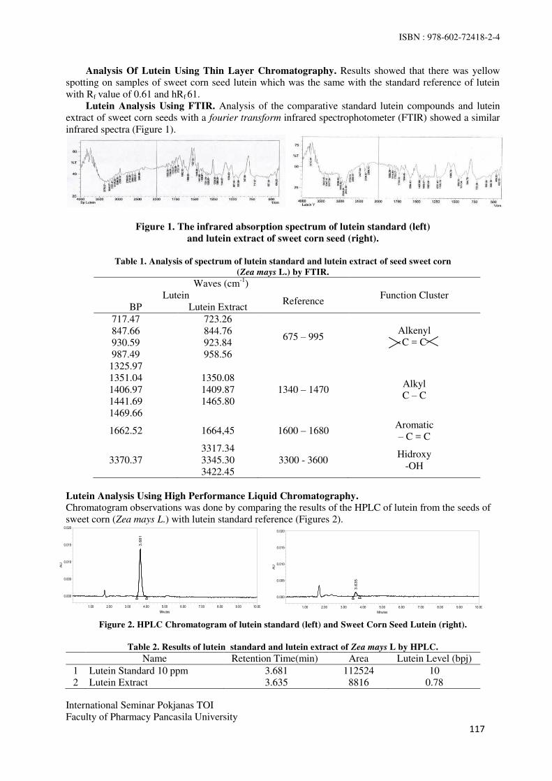
ISBN : 978-602-72418-2-4
International Seminar Pokjanas TOI
Faculty of Pharmacy Pancasila University
117
AU
0.000
0.005
0.010
0.015
0.020
Minutes
1.00 2.00 3.00 4.00 5.00 6.00 7.00 8.00 9.00 10.00
3.681
AU
0.000
0.005
0.010
0.015
0.020
Minutes
1.00 2.00 3.00 4.00 5.00 6.00 7.00 8.00 9.00 10.00
3.6
35
Analysis Of Lutein Using Thin Layer Chromatography. Results showed that there was yellow
spotting on samples of sweet corn seed lutein which was the same with the standard reference of lutein
with Rf value of 0.61 and hRf 61.
Lutein Analysis Using FTIR. Analysis of the comparative standard lutein compounds and lutein
extract of sweet corn seeds with a fourier transform infrared spectrophotometer (FTIR) showed a similar
infrared spectra (Figure 1).
Figure 1. The infrared absorption spectrum of lutein standard (left)
and lutein extract of sweet corn seed (right).
Table 1. Analysis of spectrum of lutein standard and lutein extract of seed sweet corn
(Zea mays L.) by FTIR.
Waves (cm-1
)
Function Cluster Lutein Reference
BP Lutein Extract
717.47
847.66
930.59
987.49
723.26
844.76
923.84
958.56
675 – 995 Alkenyl
C = C
1325.97
1351.04
1406.97
1441.69
1469.66
1350.08
1409.87
1465.80
1340 – 1470 Alkyl
C – C
1662.52 1664,45 1600 – 1680 Aromatic
– C = C
3370.37
3317.34
3345.30
3422.45
3300 - 3600 Hidroxy
-OH
Lutein Analysis Using High Performance Liquid Chromatography.
Chromatogram observations was done by comparing the results of the HPLC of lutein from the seeds of
sweet corn (Zea mays L.) with lutein standard reference (Figures 2).
Figure 2. HPLC Chromatogram of lutein standard (left) and Sweet Corn Seed Lutein (right).
Table 2. Results of lutein standard and lutein extract of Zea mays L by HPLC.
Name Retention Time(min) Area Lutein Level (bpj)
1 Lutein Standard 10 ppm 3.681 112524 10
2 Lutein Extract 3.635 8816 0.78

ISBN : 978-602-72418-2-4
International Seminar Pokjanas TOI
Faculty of Pharmacy Pancasila University
118
Phagocytic activity and capacity of macrophages.
Phagocytic activity of macrophages of each group listed in Figure 3.
Figure 3. Percentage macrophage phagocytic activity of peritoneal fluid of mice against the use of lutein from
seeds Sweet Corn (Zea mays L.).
An increase in phagocytic activity after the administration of lutein extract of seeds Sweet Corn (Zea
mays L.) for 14 days with a dose of 0.15mg/mice, 0.3mg/mice, 0,6mg/mouse, and 0.9 mg/mouse per day
in a row was 16.51%, 30.66%, 42.45% and 52.36%. The increase in value is calculated from the
difference between the phagocytic activity of each group IV, group V, group VI, and VII group reduced
phagocytic activity of negative group. Figure 4 shows a fairly good linear relationship between the dose
of extract lutein of Sweet Corn seeds (Zea mays L.) with the capacity of macrophages. The higher the
dose of lutein, the greater the increase in phagocytic activity, up to the observation dosage 0,9mg/
mouse/day.
Figure 4. The increase in phagocytic activity in the treatment of various concentrations of lutein extract from
seeds sweet corn (Zea mays L.) for 14 days.
Figure 5. Phagocytic capacity of macrophage cells in the peritoneal fluid of mice in the treatment of
lutein extract from sweet corn seeds (Zea mays L.).
Figure 5 shows the phagocytic capacity of macrophages of each group. DMRT analysis shows a fairly
good linear relationship between the dose of lutein extract of Sweet Corn seeds (Zea mays L.) with the
capacity of macrophages. The higher the daily dose of lutein extract, the greater the increase in
phagocytic capacity, up to the observation of a dose of 0,9mg/ mouse/day.
Sweet corn ( Zea mays L. ) were extracted to obtain lutein extract. The FT-IR analysis of the lutein seed
corn was done and the functional groups associated were determined. The FT-IR spectrum of the sample
was obtained effective peaks. The FTIR spectra were obtained functional group of –OH, -CH2,-C-O- and
–C=O- (Table 1). Table 2 shows the results of HPLC analysis of lutein standard reference producing
e
a a b c
c d
0
50
100
I II III IV V VI VIIPh
ag
osi
tic A
cti
vit
y (
%)
Treatment Groups
0
20
40
60
0,15 0,3 0,6 0,9
Ph
ag
oc
yti
c
Ac
tiv
ity
(%
)
dose of lutein (mg /mouse)
f
a a b c d e
0
200
400
600
800
I II III IV V VI VII
Ph
ag
ocy
tic
Ca
pa
cit
y
Treatment Group

ISBN : 978-602-72418-2-4
International Seminar Pokjanas TOI
Faculty of Pharmacy Pancasila University
119
chromatogram with peak in the a retention time of 3.681 and an area of 112524. Analysis of lutein
samples from sweet corn seeds shown in Figure 2 and Table 2 which shows the peak area at the retention
time of 3.635 minutes and an area of 8816. It approaches the standard compound lutein that has a peak
area at the retention time of 3.681.In the chromatogram, it is shown that there are two peaks in the
standard samples of lutein and lutein sweet corn seeds. It is because the sample was not pure so it
produced more than one peak.
Lutein extract was tested the capacity of phagocytosis and activity using in vivo methods .
Macrophages cells activated were given extract lutein doses of 0.15, 0.3, 0.6, and 0.9mg/day. Stimuno
used as a positive control. Cells that have been stained purplish red Giemsa will consist of vacuoles and
the cell nucleus. The cell nucleus color is more concentrated than the vacuole (7)
. The activity of the
macrophages of the group I was (84% ± 2.08); Group II (53% ± 0.82); group III (51% ± 1.41); group IV
(61.5% ± 1.91); V group (69% ± 1.25) and group VI (75.5% ± 2.08), and Group VII (80.75% ± 1.89).
Duncan test shows that there was no difference between the distilled water control group and vegetable
oil control group. Each dose of lutein extract from the seeds of sweet corn had a different activity. The
higher the dose of lutein extract from the seeds of sweet corn, the higher the activity of the macrophages.
The capacity of macrophages group I was (682 ± 20); group II was (427.74 ± 6.70); group III was (410 ±
12.08); group IV was (497.25 ± 13.62); group V was (563.5 ± 15.35); group VI was (608 ± 8.79); and
Group VII was (647.75 ± 16.26). Duncan test shows that there is no difference between thee control
group with distilled water and the group vegetable oil is no different. Each dose lutein extract from the
seeds of sweet corn has a different activity. The higher the dose of lutein extract from the seeds of sweet
corn, it would increase the activity of the macrophages.
CONCLUSIONS
Screening of phytochemical seed powder Sweet Corn (Zea mays L.) contained flavonoids, saponins,
steroids/ triterpenoids, and coumarin, while the n-hexane extract contains compounds steroid/triterpenoid
and coumarin.TLC analysis shows that lutein extract of sweet corn seeds has the same Rf with lutein
standard. HPLC results of lutein extract of corn seeds showed chromatogram peak at a retention time of
3.6 minutes, the same with the lutein standard. Functional group analysis with FTIR showed the same
group with a lutein standard.The administration of lutein extract from Sweet Corn seeds with different
doses to mice showed the various phagocytic activity and capacity of macrophages cells. The higher the
dose of lutein extract, the higher the activity and phagocytic capacity macropag cells. The lutein extract
from the Sweet Corn seeds can be used as an immunomodulator that works as an immunostimulant.
REFERENCES
1. "Immunology". School of Medicine, University of South Carolina. From
http://pathmicro.med.sc.edu/book/immunol-sta.html. November 2014.
2. Kusmiati, Ni Wayan SA, Swasono RT, and Mellia I. Extraction and purification of lutein from
microalgal C. pyrenoidosa local strains INK. 2010. Vol. 5(1):30-34.
3. Jung I. Effect of natural oxycarotenoid on the imune function of Japanese Quails. Hungary:
Department of Animal Physiology & Health, St. István University; 2009. p. 21.
4. Kim, HW, et al. Dietary lutein stimulates cell-mediated and humoral immunity in cats. 1999.
Experimental Biology 99.
5. Hamulka J, Koczara J, Gronek M. Lutein content of selected polish foods and estimation of its intake.
Warsaw: Warsaw Agricultural Unversity; 2005.Vol. 14/55(2): 201-206.
6. Madhavi DL, dan Kagan DI. Process for the isolation of mixed carotenoids from Plants. United States
Pantent Documents, United States. 2002. 6,380,442.
7. Ellis R. Giemsa’s staining protocol for tissue sections. IMVS Division of Pathology Queen Elizabeth
Hospital. 2007.

ISBN : 978-602-72418-2-4
International Seminar Pokjanas TOI
Faculty of Pharmacy Pancasila University
120
Effect of Bawang Tiwai (Eleutherine bulbosa (Mill.) Urb.) Ethanol Extract on
Monosodium Urate-Induced Rat Hind Paw Inflammation
DIAN R. LAKSMITAWATI*, SITI R.RANI
Faculty of Pharmacy, Pancasila University
Srengseng Sawah, Jagakarsa, Jakarta.
Corresponding email : [email protected]
Abstract: Bawang tiwai ((Eleutherine bulbosa (Mill.) Urb.) also named bawang berlian grown in
Kalimantan. Local people in Kalimantan have used the bulbous of bawang tiwai for their health. Our
previous research showed that the bulbous of bawang tiwai have decreased plasma uric acid in
hyperuricemic rat. The aim of this study was to investigate the antiinflamation effect of bawang tiwai on
Mono Sodium Urate (MSU)-induced rat hind paw inflammation. Male Sprague-Dawley rat were divided
into 6 groups as follows: normal group, negative group received Sodium CMC suspension, positive
group received diclofenac sodium (9mg/kg, p.o) and tests groups received different doses of bawang tiwai
extract (140 mg/kgBB, 280 mg/kgBB, 560mg/kgBB, p.o). Edema was induced on the left hind paw of the
rat by a subplantar injection of Mono Sodium Urate (MSU) suspension (0,4 mL 20mg/mL). At 1, 2, 4, 24,
48 and 72 hours after injection, the edema volume was measured using pletismometer and inhibitory
percentage of edema was calculated. The percentage inhibition of rat hind paw edema at the doses 140
mg/kgBW, 280 mg/kgBW dan 560 mg/kgBW of bawang tiwai were 7,67%, 2,11%, 52,31% respectively
and diclofenac sodium inhibited 57,44% of edema. In conclusion, this research demonstrated
antiinflammatory effect of extract ethanol bawang tiwai in MSU-induced rat hind paw edema. In line with
previous research of antihyperuricemic effect, bawang tiwai could be proposed further as herbal antigout.
Keywords : Eleutherine bulbosa (Mill.) Urb., monosodium urate, antiinflammation.
INTRODUCTION
Disorder of purine metabolism in human could result in gouty arthritis detected from higher uric acid
level in blood. This increased uric acid level may progress to saturated uric crystal in the synovial joints
that cause an inflammation.
The treatment of gouty arthritis entails several approches those are by lowering blood uric acid level,
increasing the urate excretion and relieving the symptoms of pain and swelling associated with
inflammation at the joints. To date, the most frequently used urate lowering drug is alopurinol and
antialgetics and antiinflamation drugs are groups of non steroid antiinflammation drug. The greatest
disadvantage in presently available potent synthetic drugs for the treatment of inflammation lies in their
toxicity and reappearance of symptoms after discontinuation especially for antiinflammation drug. The
side effect of non steroid antiinflammation drug was ulcers, bleeding, kidney failure. Therefore the
screening and development of drugs for their anti-inflammatory activity is still in progress and there is
much hope for finding anti-inflammatory drugs from indigenous medicinal plants.
Bawang tiwai ((Eleutherine bulbosa (Mill.) Urb.) also named bawang berlian grown in Kalimantan.
Local people in Kalimantan have used the bulbous of bawang tiwai for their health. Our previous
research showed that the bulbous of bawang tiwai have decreased plasma uric acid in hyperuricemic rat(1)
.
The aim of this study was to investigate the antiinflamation effect of bawang tiwai in MSU-induced
inflammation in rat hind paw.

ISBN : 978-602-72418-2-4
International Seminar Pokjanas TOI
Faculty of Pharmacy Pancasila University
121
MATERIAL AND METHODS
Plant Material. The plants were collected from Borneo (Kalimantan) and authenticated by Herbarium of
Indonesian Institute of Science (LIPI) Bogor as Eleutherine palmifolia (L.) Merr. Their bulbous were
sorted from its skin and other contaminant materials, washed by running water then cut into small pieces
and dried at 50°C. The dried bulbous were grinded. Dry powder of bawang tiwai was macerated with
ethanol 70% 24 hours with occasional shaking. The maceration process was repeated 4 times. The filtrat
was concentrated under reduced pressure 50oC.
Chemical. MSU crystal preparation. MSU crystal preparation was modified from Murakami’s method
(2). Four g of uric acid (Merck) was dissolved in 800 mL of boiling water containing 24.5 mL 1N
NaOH. The pH value was adjusted to 7.4, cooled gradually at room temperature, stayed overnight at 4° C,
washed, and dried. Needle-like crystals were recovered and suspended in sterile saline (20mg/mL).
Monosodium Urate (MSU) crystals, was used to induce the inflammation.
Animal.Thirty male Sprague Dawley rats of age 2-3 months weighing 100-200 g were obtained from
Laboratorium Non Ruminansia dan Satwa Harapan Fakultas Peternakan Institut Pertanian Bogor. They
were fed with standard diet fed and allowed food and water ad libitum for 1 week acclimatization. They
were handled according to the recomendation of local ethics commitee. They randomly divided into 6
groups as follows: normal group, negative group received CMC Na suspension, positive group received
diclofenac sodium (9mg/kg, p.o) and three tests groups received different doses of Bawang Tiwai Extract
140 mg/kgBW, 280 mg/kgBW, 560mg/kgBW, p.o, respectively. The doses of Bawang tiwai extract were
prepared by suspending extract with 1% of Na.CMC.
Antiinflammation Evaluation. MSU crystal-induced rats paw edema and assessment of
inflammation. Rats were divided into six groups each contained of five rats. Group I was normal group.
The left hind paw of rat in All groups unless normal group was induced inflammation by injected
intracutaneous on subplantar injection of 0.4 mL suspense MSU crystals. 9 mg/kg/BW sodium diclofenac
, bawang tiwai extract (140 mg/kgBB, 280 mg/kgBB, 560mg/kgBW, p.o) were given an hour before the
injection of suspense MSU crystals and is repeated for the next 3 days (at 23, 47, 71 hr). Edema on left
hind paw of each rat was measured by plethysmometer at 0, 1, 2, 4, 24, 48,72 hr. Percentage of edema
volume was calculated with Xn formula. The edema inhibitory activity was calculated with AUC value,
according to the following formula :
Xn = (V)n – (V)o x 100%
(V)o
AUC0-72 = (Xn-1 + Xn) (tn – tn-1)
2
Edema inhibition percentage = (AUC0-72)0 - (AUC0-72)n
(AUC0-72)0
(V) n = Volume of rat’s paw at n hr ; (V) o = Volume of rat’s paw at 0 hr. X n-1 = % of Rat’s paw edema in (n-1) hours
X n = % of Rat’s paw edema in (n) hours
t n = n- hours (hr)
t n-1 = (n-1) hours (hr)
(AUC0-72)0 = mean of AUC0-72 of negative control goup (%.hr)
(AUC0-72)n = mean of AUC0-72 of test group at a dose of n (%.hr)
X 100

ISBN : 978-602-72418-2-4
International Seminar Pokjanas TOI
Faculty of Pharmacy Pancasila University
122
RESULTS AND DISCUSSION
Data of edema volume that resulted from this study was treated as a percentage of edema. The graph and
percentage values can be seen in Figure 1. Figure 1 showed that each group has different line profile. The
group without treatment (negative) has higher line profile than extract group and diclofenac sodium
(positive) group, whereas diclofenac sodium group showed lower line profile than negative group and
extract group. These result showed that edema volume on the negative group was higher than bawang
tiwai extract group. It can be said that bawang tiwai extract reduced edema volume but not as potent as
sodium diclofenac.
0
5
10
15
20
25
30
0 1 2 4 24 48 72
Me
an
pe
rce
nta
ge
of
ed
em
a
vo
lum
e (
%)
Normal Negative control
Na.diclofenac 9 mg/kg EEBT 140 mg/kg
EEBT 280 mg/kg EEBT 560 mg/kg
Figure 1. Mean of edema volume percentage Vs time.
Due to the different patterns of line profil between groups on the chart, we compared the strength of
antiinflammatory effect by calculating area under the curve (AUC). The calculation results is shown in
Table 1.
Area under the curve (AUC) is a value which represents total edema volume for 72 hours
observation. Table 1 showed that the biggest of AUC values was negative group and this value was
significantly different from the normal group. This result indicated that MSU inflammation induction has
significantly succeeded.
Table 1. Mean of AUC and percentage of edema inhibition.
Statistical analysis showed that the AUC value of sodium diclofenac group was not significantly
different with the dose of 140 mg/kg and 560 mg/kg of bawang tiwai extract groups. Calculation of
Groups Mean of AUC
(%edema.hr) Inhibition of edema (%)
I. Normal 0,00a 0,00
II. Negative Control 1147,043b 0,00
III. Na diclofenac 9 mg/kg 488,23c 57,44
IV. EEBT 140 mg/kg 1059,028bc
7,67
V. EEBT 280 mg/kg 1122,87b 2,11
VI. EEBT 560 mg/kgBB 891,67bc
52,31
ISBN : 978-602-72418-2-4
International Seminar Pokjanas TOI
Faculty of Pharmacy Pancasila University
123
edema inhibitory percentage of bawang tiwai extract showed that 560 mg/kg of bawang tiwai extract has
52.31% inhibit paw edema. This value statistically similar with 9 mg/kg sodium diclofenac (57.44%).
Induction using monosodium urate is intended to be similiar with pathophysiology of inflammation
due to accumulation of uric acid. From this study it appears that bawang tiwai has potential as an anti-
inflammatory. From the results of phytochemical screening that has been done by Yulinda, there are
quinones, flavonoids, tannins and alkaloids in bawang tiwai(1)
. Based on the supporting literature,
isoeleutherine (naphthoquinone derivative) compound which isolated from the bulbs of bawang tiwai can
inhibit nitric oxidase activity by in vitro with IC50 value is 30 μM(3), where in nitric oxidase is the one of
inflammatory mediator that released while inflammation mechanisms happen. It causes vasodilatation
that increase capillary permeability, therefore plasma fluid move from capillaries to the tissue
(extravasation) and generate an edema response.
CONCLUSION
This research demonstrated the anti-inflammatory effect of extract ethanol of bawang tiwai in MSU
induced rat hind paw edema. At high doses of bawang tiwai 560 mg/kg BW, its antiinflammatory effect
was significantly as same as sodium diclofenac 9 mg/kgBW, p.o.
REFERENCES
1. Yulinda. Pengaruh ekstrak etanol bawang tiwai (Eleutherine palmifolia L. Merr) terhadap kadar asam
urat darah mencit yang diberi suspensi ragi dan kalium oksonat (skripsi). Jakarta: Fakultas Farmasi
Universitas Pancasila; 2013: h.27.
2. Murakami Y, Akahoshi T, Kawai S, Inoue M, Kitasato H. Antiinflammatory effect of retrovirally
transfected interleukin-10 on monosodium urate monohydrate crystal-induced acute inflammation in
murine air pouches. Arthritis Rheum. 2002.46:2504–2513. doi: 10.1002/art.10468.
3. Kelly GS. Monograph quercetin. Alternative Medicine Review. 2011.16(2):172-194.

ISBN : 978-602-72418-2-4
International Seminar Pokjanas TOI
Faculty of Pharmacy Pancasila University
124
Acute Toxicity of Ethanolic Extract
of Fenugreek Seeds (Trigonella foenum-graecum L.)
on White Rats
KURNIA AGUSTINI*, SRININGSIH, JULHAM EFFENDI
Center for Pharmaceutical and Medical Technology,
Agency for the Asessment and Application of Technology, BPPT, Jakarta.
correspondence author: [email protected]
Abstract: Fenugreek seed or biji klabet (Trigonella foenum-graecum L.) was known having activity to
handle some of degenerative diseases such as diabetes mellitus, hypercholesterolemia and also
postmenopausal symptoms. This study was conducted to investigate the safety of ethanolic extract of biji
klabet on white rat, especially to count the value of Lethal Concentration 50 (LC50). This in-vivo assay
referred to WHO protocols for toxicity assay of natural medicines. We used Spraque dawley white rats,
female and male, 6 weeks age, which divided into one group normal and five treatment groups
(1g/kgBW, 4g/kgBW, 8g/kgBW, 12g/kgBW, 16g/kgBW). Sample was given once orally then animal
were monitored for two weeks. Observation of toxic effect e.g physical symptom of central nerve system,
autonom nerve system and digestive system. All lethality animal were observed and LC50 were counted.
Result showed that there was no toxic effect and no lethal animal until 16g/kgBW dose. We can conclude
that ethanolic extract of Fenugreek is practically non toxic.
Keywords : Fenugreek seeds, Trigonella foenum-graecum L., acute toxicity.
INTRODUCTION
Fenugreek seed or Foenigraeci semen is dried seed from Trigonella foenum-graecum L., Leguminosae(1)
.
In Indonesia, it calls Biji Klabet. Empirically, biji klabet was used for hemorrhoids, asthma, ulcers,
muscle pain and often used as a preventative hair loss and skin softener. Many studies showed its activity
as antidiabetic, anticancer and for hypercholesterolemia handling(2)
. Biji Klabet has antiandrogen
activities, due to its active compounds as beta-sitosterol, palmitic-acid and stearic-acid, and also has the
ability to decrease of total cholesterol, LDL, VLDL cholesterol and triglycerides significantly. The anti-
hyperglycemic and anti-inflammatory properties investigated in fenugreek are additional benefit.
Agustini’s study (2007) showed that ethanolic extract of Biji Klabet have estrogenic effect on
ovariectomized and immature female Wistar rats(3)
.
Biji Klabet contains some sapogenin steroid ingredients, e.g. diosgenin, precursor for sexual
hormone(4)
, its isomer Yamogenin, gitogenin, tigogenin, and trigoneoside(5)
. Biji Klabet contains
diosgenin in free base form 0.8 – 2.2 %(6)
. Biji Klabet also contains fatty oil 20-30%, alkaloids
(trigonelline, an alkaloid pyridine, gentianin and karpain), flavonoids e.g. vitexin in glycoside or ester
form, isovitexin, orientin, vicenin, quercetin and luteolin, essential oil, saponine, nicotinamide, choline,
bitter compound and mucilage(4)
.
This study was carried out to investigate safety effect of ethanolic extract of Fenugreek in white rat.
Acute toxicity assay also known as short term toxicity assay. Sample are given once in various grade of
doses and observation are carried out for two weeks. All physical symptoms and lethal animal were
analysed then compare to normal group. This acute toxicity study was meant to count the value of Lethal
Dose 50% (LD50) from sample. LD50 is a dose that can cause 50% lethality of animal test. Sample can
categorized as safe material when they have LD50 value bigger than 15g/kgBW(7)
.

ISBN : 978-602-72418-2-4
International Seminar Pokjanas TOI
Faculty of Pharmacy Pancasila University
125
MATERIAL AND METHODS
Sampel preparation. Biji klabet were obtained from Tawangmangu, Central Java, Indonesia. Seed were
dried and grind, then were extracted with ethanol 96% food grade. Crude extracts were suspense with
Carboxy Methyl Celulose (CMC) 0.5%.
Animal preparation. Experimental animals used in this study were 30 males and 30 females
Spraque Dawley (SD) white rats, 5-6 weeks age, obtained from Indonesian Food and Drug
Administration (FDA/BPOM). The animal were kept in the animal room (25 + 20C) under 12 h light/dark
cycle and fed with standard diet of pellet rat diet and free access to distilled water prior to the start of the
study. Animal were kept for acclimatization for one week. Animal were maintained and handled
according to ethical committee which approved the design of the animal experiment. Ethical clearance
was approve by Ethical Committee of Indonesian Agency for Health Research and Development (Badan
Penelitian dan Pengembangan Kesehatan/Balitbangkes).
Animal treatment. Animals were divided into one group normal and five treatment groups
(1g/kgBW, 4g/kgBW, 8g/kgBW, 12g/kgBW, 16g/kgBW), each 5 males and 5 females. Sample was given
once orally then animal were monitored for two weeks. Observation of toxic effects were done, e.g.
physical symptom of central nerve system, autonomy nerve system and digestive system. Body weights
were weighing at day 1, 6,9,12,14. After two weeks all animal were autopsied.
Table 1. Group of animal treatment.
No. Groups Treatment N
1. N Normal Diet + CMC Na 0,5% suspense 5 Male + 5 Female
2. D1 Normal Diet + Sample 1 g/kgBW 5 Male + 5 Female
3. D2 Normal Diet + Sample 4 g/kgBW 5 Male + 5 Female
4. D3 Normal Diet + Sample 8 g/kgBW 5 Male + 5 Female
5. D4 Normal Diet + Sample 12g/kgBW 5 Male + 5 Female
6. D5 Normal Diet + Sample 16 g/kgBW 5 Male + 5 Female
RESULT AND DISCUSSION
All animal treated with dose 1 (1g/kgBW) until dose 5 (16g/kgBW) showed no toxic effect significantly.
Normally, after orally given sample treatment, all animal showed decrease of motoric activity for some
minutes. But after 30 minutes, all activity back to normal. This spontaneous effect is normal after orally
gavage treatment. The results of observation of physical symptom of central nerve system, autonom
nerve system and digestive system observation, can be seen on Table 2.
Table 2. Physical toxic effect observation.
Observation Groups
N D1 D2 D3 D4 D5
Central Nerve System
1. Sedasi - - - - - -
2. Motoric Activity +/- +/- +/- +/- +/- +/-
3. Convulsion - - - - - -
4. Tremor - - - - - -
Autonom Nerve System
1. Open eye +/- +/- +/- +/- +/- +/-
2. Salivation - - - - - -
3. Urination - - - - - -
Breath Rate +/- +/- +/- +/- +/- +/-
Heart Rate +/- +/- +/- +/- +/- +/-

ISBN : 978-602-72418-2-4
International Seminar Pokjanas TOI
Faculty of Pharmacy Pancasila University
126
Observation Groups
N D1 D2 D3 D4 D5
Digestive System
1. Diarrhea - - - - - -
2. Constipation - - - - - -
3. Bloody Fesses - - - - - -
Stress hair - - - - - -
Note:
- : No symptom
+ : There are symptom
+/- : Normal
Body weight analysis from all animal, both male and female, with treatment dose 1g/kgBW until
16g/kgBW gives no significant difference compare to normal control. Both male and female rats in all
groups gives increasing of body weight almost similar to normal control for 14 days.
Figure 1. Body weight of male rats from all group for 14 days observation.
Figure 2. Body weight of female rats from all group for 14 days observation.
0
20
40
60
80
100
120
140
160
180
Day1 Day 6 Day 9 Day 12 Day 14
BW
(g
r)
Time Weighing-
Males Body Weight
NORMA
L
0
20
40
60
80
100
120
140
160
Day 1 Day 6 Day 9 Day 12 Day 14
BW
(g
r)
Time weighing
Females Body Weight
NORMAL
DOSIS1
DOSIS2
DOSIS3
DOSIS4
DOSIS5

ISBN : 978-602-72418-2-4
International Seminar Pokjanas TOI
Faculty of Pharmacy Pancasila University
127
There was no lethality case of animal found in all group after 14 days observation. Therefor this assay
should be repeat using higher level doses than 16g/kgBW. But technically it’s so difficult to treat orally to the animal. Beside that, regulation from Indonesian FDA says that if until dose 15g/kgBW no lethality
case occur, then its not necessary to repeat the assay. The sample can be categorized having LD50 higher
than 15g/kgBW or categorized practically non toxic. The data about lethality case can be seen on Table
3.
Table 3. Lethality case of animal from all groups.
Groups Male Female Total
Lethal % Lethality
n Lethal n Lethal
Normal 5 0 5 0 0 0%
D1 5 0 5 0 0 0%
D2 5 0 5 0 0 0%
D3 5 0 5 0 0 0%
D4 5 0 5 0 0 0%
D5 5 0 5 0 0 0%
Table 4. Catagorization of Toxic Effect
(8). (Lu,FC, 1996)
Toxic Category LD50 Value
Super toxic < 5 mg/kgBB
Very hard toxic 5-50 mg/kgBB
Very toxic 50-500 mg/kgBB
Medium Toxic 0,5-5 g/kgBB
Mild Toxic 5-15 g/kgBB
Practically Non Toxic > 15 g/kgBB
CONCLUSION
No delayed toxic effect and lethality was observed in all rats during fourteen days of recovery period.
Orally treatment of Fenugreek within this range and treatment duration would not cause any severe toxic
effects and organ damages in rats. In conclusion, ethanolic extract of Fenugreek have pseudo LD50 and
categorized as practically non toxic.
REFERENCES
1. General guideline for methodologies on research and evaluation of traditional medicine. Geneva:
World Health Organization. 2000.
2. Mills, Simon & K Bone. Principles and practice of phytoterapy. Modern Herbal Medicine. Churcill
Livingstone, Edinburgh. 2000. xx + 643p.
3. Agustini K, Sumali W, Dadang K. Estrogenic effect of Fenugreek (Trigonellafoenum-graecum L.)
on white female rats. Conference Proceedings "Women's Health and Traditional Medicine",
International Medicine and Medicinal Plants, Surabaya. 2007.
4. Evans CW. Pharmacognosy 15th edition. London: W.B. Saunders. 2002.
5. Dewick PM. Medicinal natural products. A biosynthetic approach. New York: John Wiley &
Sons.1997.
6. Wiryowidagdo S. Kimia dan farmakologi bahan alam. Jakarta: Universitas Indonesia; 2001.318-
328.
7. Lu FC. Toksikologi dasar, asas, organ sasaran dan penilaian resiko. [Translate by Edi Nugroho]. Ed.
2, Jakarta:UI Press. 1995.

ISBN : 978-602-72418-2-4
International Seminar Pokjanas TOI
Faculty of Pharmacy Pancasila University
128
Antihypertensive and Diuretic Effects of the Ethanol Extract of
Colocasia esculenta (L.) Schott. Leaves
RINI PRASTIWI*, SISKA, ERVINA BHAKTI UTAMI, GIGIH PANGESTU WITJI
Department of Pharmacology, Department of Phytochemical, Department of Clinical
Pathology: Pharmacy and Science Faculty
Muhammadiyah Prof. Dr. Hamka University, Jakarta.
Abstract: Colocasia esculenta (L.) Schott (CE) is traditionally used for the treatment of various ailments
such as high blood pressure, diarrhea, rheumatic pain, pulmonary congestion, etc. Hence in present study, the
effect of Ethanol extract of CE leaves (EECE) was evaluated for antihypertensive and diuretic activity in rats.
Male Sprague dawley rats were randomly divided into five groups (n=5), and treated as follow: Positive
control group (hydrochlortiazide 0.2569 mg / 200g), negative control (NaCl 8%) and EECE (20, 40 and 80
mg / 200g) was given 14 days. The parameters systole blood pressure (SBP) and diastole blood pressure
(DBP) was estimate by Kent Scientific's CODA Non-invasive Blood Pressure on the days 0, 15 and 29.
Diuretic activity of EECE was studied based on the volume of urine for 6 hours and measuring the levels of
sodium in urine 24 hours. The result of the study showed that EECE 40 mg/200 g/day significant (p<0.05)
decreased in SBP 16.07% and in DBP 13.67%. EECE 40 mg/200 g/day showed positive diuretic activity and
significantly (p<0.05) increased sodium levels in urine. Preliminary phytochemical evaluation revealed the
presence of saponins, tannin, triterpenoid and flavonoids in EECE.
Keywords : Colocasia esculenta, antihypertensive, diuretic activity, NaCl induced, flavonoids.
INTRODUCTION
Hypertension is an increase blood pressure (BP) above normal and permanent, or when systole blood
pressure (SBP) is above 140 mmHg and diastole blood pressure (DBP) above 90 mmHg(1)
. Pharmacological
therapy for hypertension is using synthetic drugs. Hypertension drugs are use in long term lead to increase
cost and side effects.
Medicinal plant which is owned by Indonesia has enough potential to be utilized and developed as raw
materials and herbal medicines. Herbal medicines for therapy is also no longer something new to the
community. In line with the trend of 'back to nature' that developed among the public at this time, the use of
herbal as alternative medicine continues to grow bigger. One of the them is used as an alternative medicine
are the leaves of taro (Colocasia esculenta (L.) Schott.) (CE)(2)
.
Taro is known as the tuber which can be used as food substitute. All parts of this plant can be used for
treatment, including the petiole and leaf. The content of the active compounds in CE is polyphenols. Taro
leaves have medicinal properties as diarrhea, arthritis, pulmonary edema(3)
. And based on previous research
was showed that taro leaf aqueous extract at a dose of 400 mg/Kg has efficacy as an antihypertensive and
diuretic activity(4)
.
ISBN : 978-602-72418-2-4
International Seminar Pokjanas TOI
Faculty of Pharmacy Pancasila University
129
MATERIAL AND METHODS
Collection and identification of plant material. The leaves of Colocasia esculenta (CE) were collected
from Badan Penelitian Tanaman Rempah dan Obat (BALITRO), Bogor, Indonesia. The plant specimen was
authenticated and herbarium was deposited at Indonesian Institute of Science, Cibinong, Indonesia.
Preparation of Ethanol extract of CE leaves (EECE). The leaves were dried under the shade and
powdered using a grinder mixer. The powdered material (25 g) was filled in soxhlet apparatus containing 250
mL of ethanol 70%. The obtain filtrate was concentrated and stored in a desiccators till use(5)
.
Drug and Chemical. Hydrochlorthiazide (HCT) and sodium chloride were obtained from PT. Kimia
Farma (Bandung, Indonesia), sodium estimation kit (Research Lab, Indonesia), polysorbat 80, ethanol 70%
and other reagents used were purchased from local vendor from Jakarta, Indonesia.
Preparation of Drug Solution. EECE and HCT were powdered and suspended in 1% polysorbat 80 in
distillled water. Sodium Chloride (NaCl) was powdered and dissolved in distillated water. All solutions were
prepared freshly and stored in glass bottles.
Preliminary phytochemical evaluation of EECE. EECE was subjected for the qualitative analysis by
using the standard phytochemical test to evaluate the presence of various phytoconstituens.
Effect of EECE on NaCl 8% induced hypertension in rats. Male Sprague dawley rats (3-4 month old,
weight between 150 and 200 g) were randomly divide into five groups (n=5) and treated as follows: negative
control (NaCl 8% induced); positive control (HCT 0.2569 mg/ 200 gBW); EECE 20, 40, 80 mg/ 200 gBW.
NaCl 8% induced given orally 3 ml/ day in rats every day for 28 days to obtain the condition of hypertension.
EECE and HCT provided during the last 14 days orally once daily according to the group. Systolic blood
pressure (SBP) and diastolic blood pressure (DBP) was estimated for each animal on day 0 (zero), 15 and 29.
Blood pressure measurements made by the indirect method using a Kent Scientific's CODA Non-invasive
Blood Pressure.
Diuretic activity of EECE in rats. Diuretic activity was determinate by following methods of Depkes
RI(6)
, with minor modification. Male Sprague dawley rats (2-4 month old, weight between 150 and 250 g)
were randomly divide into five groups (n=5) and treated as follows: negative control (NaCl 4.5% and tween
80 1%); positive control (HCT 0.514 mg/ 200 gBW); EECE 20, 40, 80 mg/ 200 gBW.
The rats were fasted overnight (18 hr) prior to the test. After that, the rats were given an oral loading
NaCl 4.5% of 2 mL/ 200 gBW and the treatment according to each group. Immediately after administration,
the rats were placed in metabolism cages. Urine volume was collected and calculated at 6 hr and sodium level
was estimated using urine 24 hr.
Statistical analysis. The results were expressed as mean ± S.E.M (n=5). The statistical comparison was
carried out by one way ANOVA followed by LSD test. The result were considered statistically significant
when p < 0.05.
RESULTS AND DISCUSSION
Preliminary phytochemical evaluation of EECE. Preliminary phytochemical evaluation revealed the
presence of saponins, tannin, triterpenoid and flavonoids in EECE.
Effect of EECE on NaCl 8% induced hypertension in rats. The administration of NaCl 8% in rats for
28 days showed increase in SBP and DBP. The treatment with EECE and HCT showed significantly (p <
0.05) decrease in SBP and DBP as compared with negative control. EECE (40 mg/ 200g) showed the greatest
reduction in SBP 16.07% and DBP 13.65% but the effect is still smaller as compared with HCT.
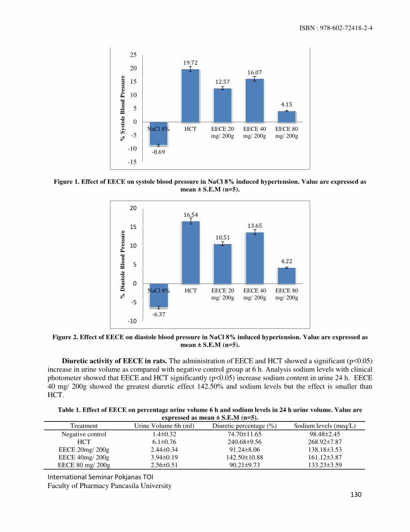
ISBN : 978-602-72418-2-4
International Seminar Pokjanas TOI
Faculty of Pharmacy Pancasila University
130
Figure 1. Effect of EECE on systole blood pressure in NaCl 8% induced hypertension. Value are expressed as
mean ± S.E.M (n=5).
Figure 2. Effect of EECE on diastole blood pressure in NaCl 8% induced hypertension. Value are expressed as
mean ± S.E.M (n=5).
Diuretic activity of EECE in rats. The administration of EECE and HCT showed a significant (p<0.05)
increase in urine volume as compared with negative control group at 6 h. Analysis sodium levels with clinical
photometer showed that EECE and HCT significantly (p<0.05) increase sodium content in urine 24 h. EECE
40 mg/ 200g showed the greatest diuretic effect 142.50% and sodium levels but the effect is smaller than
HCT.
Table 1. Effect of EECE on percentage urine volume 6 h and sodium levels in 24 h urine volume. Value are
expressed as mean ± S.E.M (n=5).
Treatment Urine Volume 6h (ml) Diuretic percentage (%) Sodium levels (meq/L)
Negative control 1.4±0.32 74.70±11.65 98.48±2.45
HCT 6.1±0.76 240.68±9.56 268.92±7.87
EECE 20mg/ 200g 2.44±0.34 91.24±8.06 138.18±3.53
EECE 40mg/ 200g 3.94±0.19 142.50±10.88 161.12±3.87
EECE 80 mg/ 200g 2.56±0.51 90.21±9.73 133.23±3.59
-8.69
19.72
12.57
16.07
4.15
-15
-10
-5
0
5
10
15
20
25
NaCl 8% HCT EECE 20
mg/ 200g
EECE 40
mg/ 200g
EECE 80
mg/ 200g
% S
yst
ole
Blo
od
Pres
sure
-6.37
16.54
10.51
13.65
4.22
-10
-5
0
5
10
15
20
NaCl 8% HCT EECE 20
mg/ 200g
EECE 40
mg/ 200g
EECE 80
mg/ 200g% D
iast
ole
Blo
od
Pres
sure

ISBN : 978-602-72418-2-4
International Seminar Pokjanas TOI
Faculty of Pharmacy Pancasila University
131
Sodium Chloride (NaCl) as an inducer experimental hypertension was studied. The administration NaCl
8% for 28 days has been managed to increase blood pressure(7)
. NaCl shows hypertensive action through
increasing plasma volume, cardiac output and ultimately increase in BP(8)
. BP measurements by the indirect
method using a Kent Scientific's CODA Non-invasive Blood Pressure. This device is works by recording
systolic and diastolic blood pressure simultaneously through a transducer that is in the tail-cuff(9)
. In the
present study, the administration of NaCl 8% for 28 day showed increase in SBP and DBP. BP was
significantly decreased after the treatment with EECE 20 and 40 mg/ 200g.
Herbal plants used as diuretic in traditional medicinal system might be useful in the treatment of
hypertension. In the present study, EECE at a dose of 40 mg/ 200g showed positive diuretic activity at 6 h,
as evident from the diuretic percentage. Furthermore, EECE showed significant increase in sodium content
of urine at 24 h but the result revealed the weak diuretic activity of EECE.
The results showed that there was an increase in the activity of the first dose to the second dose. But at
the third dose of the extract decreased the activity of diuretics when compared with the second dose. This is
possible because the levels of the compounds that are too high, causing a decrease in affinity so that the
effects produced are not in accordance with the increase in dose (10)
.
The preliminary phytochemical investigations in the present study revealed the presence of flavonoid,
saponins, tannins and triterpenoid. The flavonoids isoquercitrin showed inhibition of ACE activity (11)
.
Flavonoids suspected to have efficacy as a diuretic to stimulate blood flow to the kidneys and lead to the
inhibition of tubular reabsorption of water and ions that cause diuretic effect (12)
. The result of the present
study were suggested that the flavonoids presence in EECE may be responsible for the antihypertensive and
weak diuretic effect.
CONCLUSION
Our result show that the greatest effect of antihypertensive and diuretic is EECE 40 mg/ 200g but the effect
still lower than HCT. Futher studies are necessary to be performed for the purification, isolation and
characterization of the pyhtoconstituens responsible for the anthypertensive and diuretic effect and to explore
the exact mechanism of the action.
REFERENCES
1. Priyanto. Farmakoterapi & terminologi medis. LESKONFI. Depok. 2008; 183.
2. Wasito H. Obat kekayaan Indonesia. Graha Ilmu. Yogyakarta. 2011; 5-7.
3. Departemen Kesehatan RI. Inventaris tanaman obat Indonesia (II). Departemen Kesehatan Republik
Indonesia, Jakarta. 1993: 145.
4. Vasant, Otari Kishor et al.. Antihypertensive and diuretic effect of the aqueous extract of Colocasia
esculenta Linn. Leaves in experimental paradigms. Iranian Journal of Pharmacutical Research. 2012;
11(2): 621-634.
5. Voight R. Buku pelajaran teknologi farmasi. Terjemahan : Soendani NS. Gadjah Mada University Press.
Yogyakarta. 1995: 561-577.
6. Depkes RI. Penapisan farmakologi, pengujian fitokimia dan pengujian klinik. Depkes RI Pusat
Pemeriksaan Obat dan Makanan. Jakarta. 1993: 49-51.
7. Lailani Mutia, Zulkarnain Edward, Rahmatina BH. Gambaran tekanan darah tikus wistar jantan dan
betina setelah pemberian diet tinggi garam. Jurnal Kesehatan Andalas. Fakultas Kedokteran Universitas
Andalas. Padang. 2013: 146-149.
8. Dipiro JT. et al. Pharmacotherapy principles & practice. McGraw-Hill. United Stated of America. 2008:
143, 148-154.
9. Kent Scientific Corporation. Buku panduan CODA™ non-invasive blood pressure. Kent Scientific
Corporation. 2011: 6.

ISBN : 978-602-72418-2-4
International Seminar Pokjanas TOI
Faculty of Pharmacy Pancasila University
132
10. Bourne, Henry R, & Mark von Zastrow. Basic and clinical pharmacology. Editor: Bertram G. Katzung.
Department of Pharmacology University of California. San Francisco. 2012: 12-16
11. Junior, Arquimedes Gasparotto et al. Antihypertensive effect of isoquercitrin and extract from
Tropaeolum majus L.: Evidence for the Inhibition of Angiotensin Converting Enzym. Journal of
Ethnopharmacology. 2011; 134: 363-372.
12. Patel Umang, Mukul Kulkarni et al. Evaluation of diuretic activity of aqueos and methanol extracts of
Lepidium sativum garden cress in rats. Tropical Journal of Pharmaceutical Research. 2009; 8 (3): 215-
219.

ISBN : 978-602-72418-2-4
International Seminar Pokjanas TOI
Faculty of Pharmacy Pancasila University
133
The Antioxidant Activity of Ethanol Extract of White-Oyster Mushroom
(Pleurotus ostreatus ( Jacq) P Kumm) in Decrease MDA and Increase the
Activity of Catalase in Mice Hypercholesterolemia
VERA LADESKA,PRIYANTO,JUJU JUMIATI
Faculty of Pharmacy and Sciences, Prof.Dr.HAMKA Muhammadiyah University.
Abstract: This study aimed to determine the effect of the 70% ethanol extract of white oyster mushroom in
decrease the levels of MDA and increase the activity of catalase mice hypercholesterolemia.This study uses 6
groups: normal group,positive group which was given atorvastatin 5.2 mg/kg BW, negative group and 3
treatment groups were each given ekstract. Treatment animals fed cholesterol for 28 days to undergo
hypercholesterolemia and then given treatment for 14 days. On day 44 of MDA and catalase were measured
using a spectrophotometer UV-Vis. Test one way ANOVA statistical analysis showed white oyster
mushroom extract dose of 168 mg/kg BW comparable to the positive control group in decreasing the MDA
and increase the activity of catalase. White-Oyster mushroom extract dose of 168 mg/kg BW can lower the
levels of MDA and increase catalase activity comparable to atorvastatin 5.2 mg/kg BW.
Keyword : White-oyster mushroom, antioxidant, MDA, activity of catalase.
INTRODUCTION
Coronary heart disease is the leading cause of morbidity and mortality in industrialized countries and resulted
in approximately 30% of deaths in the United States, heart disease is also in Indonesia tends to increase as the
primary cause of death. One of the causes of coronary heart disease is atherosclerosis. Atherosclerosis is the
hardening of the arteries due to a change, loss of elasticity and narrowing of blood vessels, although the cause
is uncertain but one of the main risk factors are the increased levels of blood cholesterol
(hypercholesterolemia). Prevention of atherosclerosis can be done by inhibiting LDL oxidation using
antioxidants found in many foodstuffs(1)
. Malonildialdehide (MDA) is one of the end products manufactured
for lipid peroxidation and membrane damage by free radicals. MDA spread and increase udema cells and
affect the permeability of blood vessels, causing platelet aggregation, causing inflammation and reduce the
activity of the enzyme, whereas catalase is an enzyme that converts H2O2 into water and oxygen, the role of
catalase is very important because H2O2 is very dangerous for the life of cells, both in shape or after changing
into hydroxyl radicals (OH*)(2)
.
Terpenoids are the most active antioxidant compounds in white-oyster mushroom, test results of
antioxidant activity with DPPH that the ethyl acetate fraction oyster mushroom produces IC 50
73.24 ug / ml(3)
. The test results of antioxidant activity of 70% ethanol extract of white oyster mushroom with
DPPH methode IC50 79.03 ug / ml and water extract of oyster mushroom 94.47 ug / ml(4)
. Feeding raw oyster
mushroom, oyster mushroom stew, white oyster mushrooms grilled and fried oyster mushroom can decrease
levels of plasma MDA by 66.92%, 57.21%, 63.59% and 36.81%(5)
. In the previous study 96% ethanol extract
of white oyster mushroom with a dose of 30 mg / kg BW and 60 mg / kg BW can lower the concentration of
cholesterol in the blood of mice by 53.89% and 66.43%.

ISBN : 978-602-72418-2-4
International Seminar Pokjanas TOI
Faculty of Pharmacy Pancasila University
134
MATERIALS AND METHODS
Drug Test. White-oyster mushroom determined in Bogoriense Herbarium, Research Center for Biology
LIPI-Botany-Bogor. Mushroom weighed as much as 13 kg were cleaned, then dried under direct sunlight
then were crushed by a blender.Dried powder obtained is then extracted using 70% ethanol and suspended
in Na-CMC 5% before tested on experimental animals. Induction hypercholesterolemia performed by
administering food ingredients consisting of 12% egg yolk.
Experimental Protocol. Mice were divided into 6 groups each consisting of 4 mice .The weight of all
mice was taken before starting the research. All mice were fed with atherogenic to induce
hypercholesterolemic by feeding a high cholesterol (except the normal group) for 28 days. On day 29 mice
were treated for 14 days as follows: Group 1 nornal mice were given standard food, group 2 mice
hypercholesterolemia as a negative control, group III: Mice hypercholesterolemia + Atorvastatin 5.2 mg / kg
BW as positive control. Group IV: Mice hypercholesterolemia + extract of white oyster mushroom dose of 42
mg / kg BW, group V: Mice hypercholesterolemia + extract of white oyster mushroom dose of 84 mg / kg
BW and Group VI: Mice hypercholesterolemia + extract of white oyster mushroom dose of 168 mg / kg BW.
On day 29 and on day 43 taking blood through the venous sinus orbital.
Biochemical Analysis in Blood. Mice were anesthetized with ketamine and taking blood through the
venous sinus orbital using capillary tube. Blood sample of 1 ml volume was taken.Blood plasma and serum
were separated by centrifuge at 4000 rpm rotation for 15 minutes at a temperature of 40 °C. Serum is used for
the measurement of MDA while the blood plasma are used for the measurement of catalase.
1. Measurement of levels cholesterol Total
Serum taken 10 mL, and then mixed with the enzyme reagent (kit) of 1000 mL, and then mixed using a
homogenizer and incubated for 10 minutes at a temperature of 20-25°C or 5 minutes at a temperature of
37°C, and measured with a clinical photometer(6)
.
2. Determination of Levels of MDA and activity of catalase
a. Determination of Levels of MDA. The sample being measured is blood plasma with the method TBA test
consisting of 0.375% TBA and 15% TCA in HCl 0.01 N. Blood plasma taken 250 µl, then add 3.0 ml TBA
heated in boiling water for 15 minutes, then cool the tube at room temperature, centrifugation at 1000 rpm for
15 minutes. Absorbent supernatant was measured at a wavelength of 532 nm. MDA calculated based on the
molar extinction MDA (1,56.105/ μmolar cm)(7)
.
b. Determination of catalase activity. Determination of catalase activity by measuring levels of peroxide
based on the molar extinction coefficient of 43.6 M/cm. Then work peroxide solution made 19 mM in
phosphate buffer.Catalase activity at 25 ° C was defined as micromoles of peroxide consumed per minute per
ml of sample(8)
.
Catalase activity (unit / ml) = Δ abs / min X 1000
43.6 x ml sample
ml vol Reaction
Statistical Analysis. The data obtained is of MDA and catalase activity analyzed statistically using one
way analysis of variance (ANOVA) followed by the multiple comparison test tuckey.
RESULT AND DISCUSSION
Blood sample was taken after induced hypercholesterolemia, then that is the end of the blood sampling after
treatment for 14 days. Before blood sampling, the mice were fasted beforehand for ± 16 hours in order to
avoid the effect of increasing cholesterol levels resulting from food. Retrieval of the data obtained by high

ISBN : 978-602-72418-2-4
International Seminar Pokjanas TOI
Faculty of Pharmacy Pancasila University
135
initial blood total cholesterol level is different in each group, the results can be seen in Table 3. Taking the
blood of mice obtained from orbital venous sinus, because more blood than in other regions. Mice were
anesthetized until unconscious, then the corner of the eye pierced by a capillary tube. Blood obtained
microtube accommodated in the tube, then centrifuged at 4000 rpm for 15 minutes, resulting in a separation
of the serum blood cells. Serum obtained is then separated into the tube microtube. Samples were tested with
clinical photometer to look at total cholesterol levels. In the second blood sampling, microtube added Na
EDTA as an anticoagulant to obtain plasma and blood plasma and centrifuged at a speed of 4000 rpm for 15
minutes at a temperature of 40 °C and then obtained for the measurement of MDA plasma and blood plasma
for measurement of catalase activity.
Malonildialdehide levels (MDA) Blood Mice. This study using TBA test method as a parameter of
lipid peroxidation that occurs .The principle of measurement MDA is the reaction of one molecule of MDA
with two molecules form a complex compound TBA, MDA-TBA pink that can be read with a
spectrophotometer. Cholesterol can be eliminated from the body after first converted into bile acids. This
process takes place with 7α-hydroxylation reaction as a key reaction that is catalyzed by 7α-hydroxylase and
require oxygen, this stage causes the oxygen molecules is reduced to superoxide anion(9)
. Thus, the higher the
concentration of cholesterol in hyperlipidemic cases can improve 7α-hydroxylase activity so that more
superoxide radicals are formed and attacked the unsaturated fatty acid chains. Disconnection chain
unsaturated fatty acids will produce various compounds, among others, malonilaldehyde (MDA). Results of
the analysis of MDA levels from each experimental group can be seen in Table 4.
Based on Table 1, the measurement results showed decreased levels of MDA. The highest levels of
3.777 ± 0.233 g / ml is the negative control group mice. High levels of MDA are influenced by feeding
hypercholesterolemia causes the mice became hypercholesterolemia. This condition can increase free radicals
in the body. High levels of free radicals can lead to increased levels of lipid peroxidation which the MDA as a
final product. At dose levels of MDA III produces low and comparable to the positive control group. This can
happen because in the white oyster mushrooms are antioxidant compounds.
In the study(3)
terpenoids are the most active antioxidant compounds in white-oyster mushrooms. Terpenoids
white-oyster mushroom capable of donating one electron sole deciding a chain reaction with the way it reacts
with radicals, and turn it into a stable product.
Normality test results obtained significance value 0.261 >0.05 and homogenity test results obtained
significance value 0.196 > 0.05 so that the data normally distributed and homogeneous, followed by analysis
using one-way ANOVA. From the results of the ANOVA table for MDA significance value 0.000 < 0.05.
This shows the significant difference between treatments. It can be concluded that the effect of the test
preparation to decreased levels of MDA. Later analysis followed by Tukey's test. MDA to Tukey test showed
that the positive control significantly different with all groups, except group III dose.
Levels of catalase activity Mice. This test is called calorimetric method using color as an indicator. One
unit of catalase activity is expressed as the amount of H2O2 (in micromoles) were destroyed by catalase per
minute in each 10 µL sample. Catalase function as a catalyst to accelerate the decomposition of H2O2 into
H2O and water. Catalase activity will be decreased when oxidative stress occurs. A decrease in the catalase
activity associated with damage to the lipid membranes due to increased free radicals in the body. According
to research3 white- oyster mushroom capable of donating one electron sole deciding a chain reaction with the
way it reacts with radicals, and turn it into a product that is stable, so that the free radicals in the body
decreases and increased catalase activity. Results of the analysis of the catalase enzyme levels from each
experimental group can be seen in Table 5.

ISBN : 978-602-72418-2-4
International Seminar Pokjanas TOI
Faculty of Pharmacy Pancasila University
136
Table 1. Results of measurement of MDA levels. Group Average (µg/ml) ± SD
Normal 1.396 ± 0.098 a
Negatif 3.777 ± 0.233 b
Positif 1.713 ± 0.085 c
Dosis I 2.464 ± 0.173 d
Dosis II 2.057 ± 0.054 e
Dosis III 1.743 ± 0.110 c
Based on the Table 2, that the analysis of the activity of catalase in mice using a UV-VIS
spectrophotometer showed the normal group had the highest catalase activity is 119.835 ± 7.46 unit / mL and
a negative group showed the lowest catalase activity is 54.067 ± 2.97 unit / mL. Catalase activity decrease
due to the condition of hypercholesterolemia causes the production of free radicals produced by high
resulting in lower activity of catalase. Catalase activity increased significantly demonstrated by the
third dose is 88.567 ± 4.80 units / mL.Statistically there was no significant difference between the dose III
with positive control group.
Normality test results obtained significance value 0.760 > α 0.05 and homogeneity test results
obtained significance value 0.676 > α 0.05. ANOVA table of the results of the activity of catalase
significance value 0,000 < α 0.05. This shows the significant difference between treatments. It can be
concluded that the effect of the test preparation to the decrease of catalase activity. Catalase activities for
Tukey test showed that the first dose group there was no significant difference in dose group II and III dose
there was no significant difference in the positive control group.
Table 2. Results of measurement of catalase activity.
Group Average (Unit/mL) ± SD
Normal 119,835 ± 7,46 a
Negatif 22,415 ± 2,97 b
Positif 94,615 ± 5,61 c
Dosis I 54,067 ± 5,67 d
Dosis II 65,902 ± 4,09 d
Dosis III 88,567 ± 4,80 c,e

ISBN : 978-602-72418-2-4
International Seminar Pokjanas TOI
Faculty of Pharmacy Pancasila University
137
Group Measurement of Total Cholesterol Levels
Average (mg/dl) ± SD
Repeat Total cholesterol
Normal
1 73.22
76.445 ± 7.427 2 67.9
3 84.86
4 79.8
Negatif
1 249.23
249.34 ± 16.317 2 229.75
3 248.68
4 269.7
Positif
1 232.14
247.867 ± 17,428 2 270.93
3 251.45
4 236.95
Dosis I
1 260.33
242. 703 ± 13.940 2 237.14
3 227.49
4 245.85
Dosis II
1 244.03
238.582 ± 13.466 2 241.91
3 209.44
4 218.95
Dosis III
1 247.05
247.723 ±13.270 2 229.19
3 257.23
4 217.42
Table 3. Results of measurement of total cholesterol levels.

ISBN : 978-602-72418-2-4
International Seminar Pokjanas TOI
Faculty of Pharmacy Pancasila University
138
Table 4. Results measurement MDA levels.
Group Measurement MDA levels
Average (µg/ml) ± SD Repeat Absorbansi MDA levels
Normal
1 0.227 1.255
1.396 ± 0.098 2 0.263 1.454
3 0.266 1.471
4 0.254 1.405
Negatif
1 0.642 3.550
3.777 ± 0.233 2 0.675 3.733
3 0.742 4.103
4 0.673 3.722
Positif
1 0.328 1.814
1.713 ± 0.085 2 0.293 1.620
3 0.316 1.747
4 0.302 1.670
Dosis I
1 0.457 2.527
2.464 ± 0.173 2 0.427 2.361
3 0.484 2.677
4 0.414 2.289
Dosis II
1 0.361 1.996
2.057 ± 0.054 2 0.382 2.112
3 0.367 2.030
4 0.378 2.090
Dosis III
1 0.327 1.808
1.743 ± 0.110 2 0.289 1.598
3 0.311 1.720
4 0.334 1.847

ISBN : 978-602-72418-2-4
International Seminar Pokjanas TOI
Faculty of Pharmacy Pancasila University
139
Table 5. Results of measurement of catalase activity.
Group
Measurement of Catalase Activity
Average (Unit/mL) ± SD
Repeat Activity Catalase
Normal
1 123.89
119.835 ± 7.46 2 118.2
3 127.14
4 110.11
Negatif
1 23.88
22.415 ± 2.97 2 20.12
3 19.77
4 25.89
Positif
1 102.34
94.615 ± 5.61 2 94.76
3 89.32
4 92.04
Dosis I
1 51.97
54.067 ± 5.67 2 48.03
3 54.78
4 61.49
Dosis II
1 63.14
65.902 ± 4.09 2 68.05
3 61.87
4 70.55
Dosis III
1 89.19
88.567 ± 4.80 2 81.72
3 92.82
4 90.54
CONCLUSION
Based on the results of this study concluded that 70% ethanol extract of white-oyster mushroom (Pleurotus
ostreatus (Jacq.) P. Kumm at a dose of 42 mg/ kg BW, 84 mg / kg BW, and 168 mg / kg BW was able to
reduce levels of MDA and increased activity catalase. Dose of 168 mg / kg BW was able to reduce levels of
MDA and increase catalase activity comparable to atorvastatin dose of 5.2 mg / kg BW.

ISBN : 978-602-72418-2-4
International Seminar Pokjanas TOI
Faculty of Pharmacy Pancasila University
140
REFERENCES
1. Trilaksani W. 2003. Antioksidan: jenis, sumber, mekanisme kerja dan peran terhadap kesehatan. Tesis.
Institut Pertanian Bogor, Bogor. Hlm.12.
2. Priyanto. Toksikologi, mekanisme, terapi antioksidan dan penilaian resiko. Editor Hadi Sunaryo.
Leskonfi, Depok. 2009. Hlm.73-86.
3. Mutiasari IR. Uji Aktifitas antioksidan jamur (pleurotus ostreatus) dengan metode DPPH dan identifikasi
golongan senyawa kimia dari fraksi teraktif. Skripsi. Fakultas FMIPA Universitas Indonesia, Depok.
2012 .Hlm.41.
4. Nawawi RH. Uji aktivitas, stabilitas fisik dan keamanan sediaan gel pencerah kulit yang mengandung
ekstrak jamur tiram. Tesis. FMIPA Universitas Indonesia. Depok. 2012. Hlm. 94
5. Suniarti FRT. Aktifitas antioksidan jamur tiram putih (Pleurotus ostreatus) rebus, panggang dan goreng
pada tikus sparague dawley hiperkolesterolemia. Tesis. UGM, Yogyakarta. 2014. Abstrak.
6. Asri DS. Aktivitas antihiperkolesterolemia fraksi etanol ekstrak daun kelor (Moringa Oleifera Lam.)
Berdasarkan kadar kolesterol total dan LDL kolesterol. Skripsi. Fakultas Farmasi dan Sains. UHAMKA,
Jakarta. 2014. Hlm. 17.
7. Soewoto, H. dkk. Biokimia eksperimen laboratorium. Widya Medika. Jakarta. 2001. Hlm. 153.
8. Priyanto. Status oksidan dan antioksidan serta pengaruh pemberian kombinasi vitamin (E + C) pada
Polantas di kota besar dan polisi di pedesaan. Tesis. Program Pasca Sarjana Bidang Ilmu Kesehatan.
Universitas Indonesia. Depok. 1999. Hlm. 36.
9. Mayes PA, Botham PA. Cholesterol synthesis, transport, and excretion. McGraw-Hill. New York. 1996.
Hlm 26: 219-230.

141
International Seminar Pokjanas TOI
Faculty of Pharmacy Pancasila University
141
Soursop Leaf (Annona muricata L.) Infusion in Lipid Profile
of Hyperlipidemic Mice
NI MADE DWI SANDHIUTAMI*, NENI ANGGRAINI
Faculty of Pharmacy, Pancasila University.
Email : [email protected]
Abstract: The increase of blood lipid levels can be leaded into atherosclerosis. Soursop leaf (Annona
muricata L.) contains flavonoid, that can be used as anti hyperlipidemia. This study aims to determine the
effect of soursop leaf (Annona muricata L.) infusion for the reduction of Total Cholesterol, Triglyceride,
LDL, and also the elevation of HDL. In this experiment, male mice (DDY strain) were induced by 80%
yolk, 65% sucrose solution 15%, and 5% animal-fats two times/day, for 14 days. 30 mice were divided
into 6 groups of 5 mice each: a normal group (I), a negative control group (II), a positive control group
treated with simvastatin (III), and a final group had been treated with soursop leaf infusion (1.4 g/kg BW,
2.1 g/kg BW, and 2.8 g/kg BW) (IV). The preparation given at 15th – 21
st day and were observed on 0,
14th, and 21
st days with a photometer 4010. On the 21
st day, the measurement of Total Cholesterol,
Triglyceride and LDL decreased to 33.12%, 35.95%, and 46.05%, while HDL increased to 36.36% in
mice that had been given soursop leaf infusion dose 2.8 g/kg BW. The results showed that a soursop leaf
infusion dose 2.8 g/kg BW is equal to simvastatin dose 1.3 mg/kg BW.
Keywords: Soursop leaf, Annona muricata L., flavonoid, hyperlipidemic.
INTRODUCTION
Nutritious food is needed as an additional health factor. One of the effects potentially experienced if a lot
of food high in saturated fat is consumed is hyperlipidemia. Hyperlipidemia is an increase in the total
level of cholesterol, LDL, triglyceride, or several of them, or a decrease in HDL(1)
. Hyperlipidemia can
lead to atherosclerosis, which in turn can lead to coronary heart disease(2)
.
The blood lipid level can be reduced with hypolipidemic drugs, one of which is klofibrat. Klofibrat
was withdrawn from circulation because increased mortality had been documented in users of the drug(2)
.
The potential of natural material for treatment is being investigated because it is cheap and has a smaller
risk than chemical treatment.
Soursop (Annona muricata L.) has long been known as a medicinal plant; its leaves, flowers, fruits,
seeds, roots and bark can be used for medical purposes(3)
. The results showed that the ethanol extract from
the soursop bark (Annona muricata L.) has antihyperglycemic and hypolipidemic activity, especially in
lowering the state of hypertriglyceridemia and hypercholesterolemia induced by alloxan in rats(4)
. Soursop
leaves contain flavonoid compounds that can be used as agents for antihiperlipidemia. Commonly,
soursop leaves are used to lower blood cholesterol levels by boiling them in water. Given the
hypolipidemic activity found in the bark of the soursop, the public assumption that the leaves of the
soursop lower cholesterol levels requires further testing.

ISBN : 978-602-72418-2-4
International Seminar Pokjanas TOI
Faculty of Pharmacy Pancasila University
142
MATERIAL AND METHODS
MATERIAL : Soursop leaf (Annona muricata L.), simvastatin, inducer of hyperlipidemia, and reagents
kits (Biolabo).
Tools. Oral probes, syringes, animal scales, analytical scales, glasses, surgical instruments, cottons,
eppendorf tubes, centrifuge, and the photometer 4010.
Animal preparation : Mice, DDY strain, male, 2-3 months old, weight 25-30 grams.
Preparation of the soursop leaf infusion (Annona muricata L.)
Fresh leaves were collected and washed in tap water to remove feces, and then drained, also dried,
however do not dry it in direct sunlight. Soursop leaf infusion was made by boil 11.2 grams soursop leaf
simplicia (equivalent to 35 sheets soursop leaf simplicia) with 125 mL water. Heating begins at 900 C for
15 minutes. Infusaion was filtered while hot with flannel. Infusion volume was obtained 100 mL, So that,
the concentration of soursop leaf is 11.2%. Administration volume was given according to dose 1.4 g /
kg, 2.1 g / kg, and 2.8 g / kg.
Preparation of Hyperlipidemia Inducers. Animal-fat was obtained from chicken skin. It weighed
in 100 grams, then heated it in a non-stick frying pan until all the oil come out. 65% sucrose solution was
prepared by dissolve 65 grams of sugar in 100 ml hot water. Yolk were used as an emulsifier. Each
ingredients is were mixed according to its weight and volume. inducers was made every day and given
it orally two times/day.
Preaparation of simvastatin suspense dose 1.3 mg/kg. 0.5% CMC Na solution was made by
weigh in 0.5 gram CMC Na in hot water with volume 20 times of weight of CMC Na, that is 10 mL and
let it stand for approximately 30 minutes until its expands. 20 tablets simvastatin which has been
weighed, and crushed into powder and converted into the dose used in mice. 0.1 grams simvastatin
powder is mixed with CMC Na which has expanded and crushed until homogeneous, then added to 100
mL distilled water.
Soursop leaf (Annona muricata) Infusion in Lipid Profile of Hyperlipidemic Mice. Animals were
divided into 6 groups of 5 mice each: a normal group, a negative control group, a positive control group
treated with simvastatin dose 1.3 mg/kg, and a final group had been treated with soursop leaf infusion
(1.4 g/kg BW, 2.1 g/kg BW, and 2.8 g/kg BW) The lipid levels was measured in all groups on day 0 and
furthermore given a hyperlipidemia inducers dose 17.5 g/kg BW two time/day for 14 days. Re-
measurement of lipid levels and given the preparation test for 7 days.
RESULTS AND DISCUSSION
Soursop leaf infuse (Annona muricata L.) positively contains flavonoids in the form of yellow color in
amyl alcohol layers. Flavonoid compounds can be used as anti hiperlipidemia which affect total
cholesterol, triglycerides, LDL, and HDL blood levels.
0
50
100
150
200
0 14 21
Averag
e o
f to
tal
ch
ole
sterol
levels
(m
g/d
L)
Experimental time (days)
The average total cholesterol levels (mg/dL)
Normal
ControlNegative
ControlPositive
ControlLow Dose
Average total cholesterol levels in mice (mg/dL).
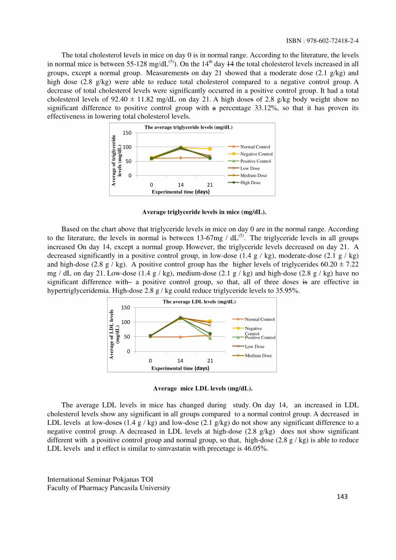
ISBN : 978-602-72418-2-4
International Seminar Pokjanas TOI
Faculty of Pharmacy Pancasila University
143
The total cholesterol levels in mice on day 0 is in normal range. According to the literature, the levels
in normal mice is between 55-128 mg/dL(5)
). On the 14th day 14 the total cholesterol levels increased in all
groups, except a normal group. Measurements on day 21 showed that a moderate dose (2.1 g/kg) and
high dose (2.8 g/kg) were able to reduce total cholesterol compared to a negative control group. A
decrease of total cholesterol levels were significantly occurred in a positive control group. It had a total
cholesterol levels of 92.40 ± 11.82 mg/dL on day 21. A high doses of 2.8 g/kg body weight show no
significant difference to positive control group with a percentage 33.12%, so that it has proven its
effectiveness in lowering total cholesterol levels.
0
50
100
150
0 14 21Av
era
ge
of
trig
lyce
rid
e
lev
els
(mg
/dL
)
Experimental time (days)
The average triglyceride levels (mg/dL)
Normal Control
Negative Control
Positive Control
Low Dose
Medium Dose
High Dose
Average triglyceride levels in mice (mg/dL).
Based on the chart above that triglyceride levels in mice on day 0 are in the normal range. According
to the literature, the levels in normal is between 13-67mg / dL(5)
. The triglyceride levels in all groups
increased On day 14, except a normal group. However, the triglyceride levels decreased on day 21. A
decreased significantly in a positive control group, in low-dose (1.4 g / kg), moderate-dose (2.1 g / kg)
and high-dose (2.8 g / kg). A positive control group has the higher levels of triglycerides 60.20 ± 7.22
mg / dL on day 21. Low-dose (1.4 g / kg), medium-dose (2.1 g / kg) and high-dose (2.8 g / kg) have no
significant difference with a positive control group, so that, all of three doses is are effective in
hypertriglyceridemia. High-dose 2.8 g / kg could reduce triglyceride levels to 35.95%.
0
50
100
150
0 14 21
Av
era
ge
of
LD
L l
evel
s
(mg
/dL
)
Experimental time (days)
The average LDL levels (mg/dL)
Normal Control
Negative
ControlPositive Control
Low Dose
Medium Dose
Average mice LDL levels (mg/dL).
The average LDL levels in mice has changed during study. On day 14, an increased in LDL
cholesterol levels show any significant in all groups compared to a normal control group. A decreased in
LDL levels at low-doses (1.4 g / kg) and low-dose (2.1 g/kg) do not show any significant difference to a
negative control group. A decreased in LDL levels at high-dose (2.8 g/kg) does not show significant
different with a positive control group and normal group, so that, high-dose (2.8 g / kg) is able to reduce
LDL levels and it effect is similar to simvastatin with precetage is 46.05%.

ISBN : 978-602-72418-2-4
International Seminar Pokjanas TOI
Faculty of Pharmacy Pancasila University
144
Average HDL levels in mice (mg / dL).
The chart above shows the changes in HDL levels in mice during study, especially on days 0, 14, and
21. On Day 0, the range of HDL levels in all groups is between 27.60 ± 3.58 mg/dL ─ 30 60 ± 2.97 mg/d.
On day 14, a decreased HDL levels in mice occurred in the group II, III and IV. On day 21, an increased
HDL levels in low-dose (1.4 g / kg) and moderate-dose (2.1 g / kg) do not show any significant difference
compared to a normal group and a negative control group. High-dose (2.8 g / kg) could elevate the HDL
levels which is has no significant difference to a positive control groups and normal controls group. So
that, high-dose (2.8 g / kg) could increase the HDL levels with percentation is 36.36%, and this effect is
similar to simvastatin.
Results of in vitro studies indicate that flavonoids work as inhibitors of HMG-CoA reductase enzyme that
decreases cholesterol synthesis(6)
. Flavonoids also increases the activity of lipoprotein lipase, and
therefore contributes to a decrease in blood triglyceride levels(7)
. In addition, flavonoids can affect the
metabolism of LDL cholesterol by increasing LDL ability to bind to its receptor. LDL bound to the
receptor to be metabolized into cholesterol ester in the network. HDL binds cholesterol esters contained in
the network and then excreted into the small intestine(8)
.
CONCLUSION
Soursop leaf infuse a dose of 2.8 g/kg BW can decreases total cholesterol, triglycerides, and LDL, also
increase HDL levels.
REFERENCES
1. Wells BG, Dipiro JT, Schwinghammer TL, Dipiro CV. Pharmacotherapy handbook seventh
edition. The McGraw-Hill Companies; 2009. p. 98, 102-103.
2. Gunawan SG, Setiabudy R, Nafrialdi, Elysabeth. Pharmacology and therapeutics 5th edition
(reprinted with improvements). Jakarta: Department of Pharmacology and Therapeutics Faculty of
Medicine, University of Indonesia; 2008. p. 373-77, 379-88.
3. Mardiana L, Ratnasari J. Herb and efficacy of soursop. Jakarta: Penebar Organization; 2011. p. 17,
38-40.
4. Ahalya B, Shankar KR, Kiranmayi GVN. Exploration of anti-hyperglycemic and hypolipidemic
activities of Ethanolic extract of Annona muricata bark in alloxan induced diabetic rats. Int J Pharm
Sci Rev Res. 2014; 25 (2): 21.26.
5. Fox JG, Barthold SW, MT Davisson, Newcomer CE, Quimby FW, Smith AL. The mouse in
biomedical research: normative biology, husbandry, and models, second edition.UK: Academy
Press; 2007. p. 188.

ISBN : 978-602-72418-2-4
International Seminar Pokjanas TOI
Faculty of Pharmacy Pancasila University
145
6. Prahastuti S, Tjahjani S, Hartini E. Effect bay leaf infusion (Syzygium polyanthum (Wight) Walp)
against a decrease in total blood cholesterol levels of rat models of dyslipidemia wistar
strain. Medical journal Planta. 2011; 1 (4): 27-9.
7. Arauna Y, Aulanni 'am, Oktavianie DA. Study triglyceride levels and histopathological picture liver
animal model rat (Rattus norvegicus) hypercholesterolemia treated with aqueous extracts of mango
parasites (Dendrophthoe petandra). Malang: Study Program of the University of Brawijaya
Veterinarian; 2012. p. 2.
8. Rofida S, Firdiansyah A, Fitriyastuti E. Activities antihiperlipidemia ethanol extract of leaves
of Annona squamosa L. J Pharm Sci Pharm Pract. 2015; 2 (1): 1-3.
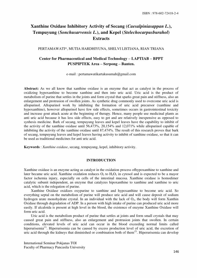
ISBN : 978-602-72418-2-4
International Seminar Pokjanas TOI
Faculty of Pharmacy Pancasila University
146
Xanthine Oxidase Inhibitory Activity of Secang (Caesalpiniasappan L.),
Tempuyung (Sonchusarvensis L.), and Kepel (Stelechocarpusburahol)
Extracts
PERTAMAWATI*, MUTIA HARDHIYUNA, SHELVI LISTIANA, RIAN TRIANA
Center for Pharmaceutical and Medical Technology – LAPTIAB – BPPT
PUSPIPTEK Area – Serpong – Banten.
e-mail : [email protected]
Abstract: As we all know that xanthine oxidase is an enzyme that act as catalyst in the process of
oxidizing hypoxanthine to become xanthine and then into uric acid. Uric acid is the product of
metabolism of purine that settles in the joints and form crystal that sparks great pain and stiffness, also an
enlargement and protrusion of swollen joints. As synthetic drug commonly used to overcome uric acid is
allopurinol. Allopurinol work by inhibiting the formation of uric acid precursor (xanthine and
hypoxanthine), however allopurinol have few side effects, sometimes occurs in gastrointestinal toxicity
and increase gout attack acute at the beginning of therapy. Hence, many people use medicinal plants as
anti uric acid because it has less side effects, easy to get and are relatively inexpensive as opposed to
synhesis medicine. Bark of secang, tempuyung leaves and kepel leaves have the capability to inhibit of
the activity of the xanthine oxidase until 56,473%, 20,154% and 12,071% while allopurinol capable of
inhibiting the activity of the xanthine oxidase until 87,474%. The result of this research proves that bark
of secang, tempuyung leaves and kepel leaves having activity to inhibit of xanthine oxidase, so that it can
be used as traditional medicines for anti uric acid.
Keywords : Xanthine oxidase, secang, tempuyung, kepel, inhibitory activity.
INTRODUCTION
Xanthine oxidase is an enzyme acting as catalyst in the oxidation process ofhypoxanthine to xanthine and
later became uric acid. Xanthine oxidation reduces O2 to H2O2 in cytosol and is expected to be a mayor
factor ischemia injury, especially on cells of the intestinal mucosa. Xanthine oxidase is homodimer
catalytic subunit independent, an enzyme that catalyzes hipoxanthine to xanthine and xanthine to uric
acid, which is the relegation of purine.
Xanthine Oxidase oxidizes oxypurine to xanthine and hypoxanthine to become uric acid. So
everything septal on the metabolism of purine will produce uric acid and will cause deposit of sodium
hydrogen urate monohydrate crystal. In an individual with the lack of O2, the body will form Xanthin
Oxidase through degradation of ADP. In a person with high intake of purine can produced uric acid more
easily. If alcaloida is present at high level in the blood, the existence of enzyme Xanthine Oxidase will
form uric acid.
Uric acid is the metabolism product of purine that settles at joints and form small crystals that may
caused great pain and stiffness, also an enlargement and protrusion joints that swollen. In certain
conditions, elevated levels of uric acid can occur in the blood exceeding normal limits called
hiperurisemia(1)
. Hiperurisemia can be caused by excess production level of uric acid, the excretion of
uric acid through the kidneys that diminished or combination both of them(2)
. Hiperurisemia can develop

ISBN : 978-602-72418-2-4
International Seminar Pokjanas TOI
Faculty of Pharmacy Pancasila University
147
into gout. Gout is a type of metabolic disease. Its existence is quite popular among the community as
pirai(3)
.
A synthetic drug commonly used to overcome uric acid is allopurinol. Allopurinol is an analogous of
uric acid, works by inhibiting the formation of uric acid from their precursor (Xanthine and
Hipoxanthine) by inhibiting the activity of the Xanthine Oxidase(3)
. However allopurinol have few side
effects such as redness of the skin, leukopenia, sometimes toxic in gastrointestinal and increase acute
attack in early therapy(4)
. Therefore, nowaday people use medicinal plant as a traditional remedy because
it has relatively small side effects, easy to get and are relatively inexpensive as opposed to synthesis
medicine.
Bark of secang (Caesalpinia sappan L.), has been empirical used as material for the treatment of
uric acid disease. Various substance contained in the bark secang include brazilin, an alkaloid, flavonoid,
saponin, tannin, phenyl propane and terpenoid . It also contain gallic acid , brasilein, delta-a phellandrene,
oscimene, resins and resorin(5)
. Perceived research is weak in terms of effect anti-hiperurisemia. Because
of the great biology potential of secang needs to be surveyed as agent of anti hiperurisemia.
Tempuyung (Sonchus arvesnsis) is a herb found in the wild that grows in the mois areas that have
high enough intensity of rainfall. The chemical content contained in this plant are mineral ions include
silica , potassium , magnesium , sodium and compound organic kind of flavonoid (kaemferol, luteolin-7-
o-glukosida and apigenin-7-o-glukosida), coumarin (scepoletin) , taracsasterol , inositol , and acid fenolat
(cinnamic , cumarat and vanilat). In a report, the total flavonoid content in the tempuyung leaves is
0,1044 %. Whereas tempuyung root contain a total alkaloid of more or less 0,5 % and the highest
flavonoid is apigenin-7-0-glukosida. Apigenin-7-0-glukosida is one of the flavonoid that have the
potential to hinder the activity of Xanthine Oxidase and superoxidase, so that the formation of uric acid
can be constrained or reduced(6)
. Kepel or burahol (Stelechocarpus burahol)l is a tree producing the fruit
Kepel fruit is popular with women because is believed to make fragant scented sweat and make water
odorless. For medicine, meat this fruit serves as deciduous urine, prevent inflamation of the kidney and
cause sterility (temporary) in women. Its timber is suitable to wrench households and reported to last 50
years.
The purpose of this experiment is to find out the Xanthine Oxidase inhibitory effect of three extracts
of secang bark (Caesalpinia sappan L.), tempuyung (Sonchus arvesnsis) and kepel (Stelechocarpus
burahol). Data processing acquired is carried out a statistical analysis of the variants and methods to test
the difference between the average values of the two treatment used the methods of Multiple Region Test
Duncan. The results of the experiment are expected to provide information to improve health by
developing Obat Herbal Terstandar/ Standardisea Herbal Medicine (OHT). This research was carried out
in April until June 2014 in the laboratory of Center of Pharmaceutical and Medical Technology –
LAPTIAB - TAB - BPPT.
MATERIAL AND METHODS
MATERIAL. Bark of secang, leaves of tempuyung and leaves of kepel are mashed into small pieces
(approximately 1 mm).
Chemicals. DMSO (Sigma), K2HPO4 (Sigma P-0662), NaOH (Sigma 221465), HCl (Merck),
Xanthine (X 0626), Xanthine Oxidase (Sigma X 4375), ethanol 96%, aquadest free CO2, Allopurinol.
Equipment. Percolator (BuchiPump Controller C-610 and Buchi Pump Module C-610: UV-
VIS1700 Spectrophotometer Shimadzu PharmaspecGlassware.
METHODS. Plant Identification. The plants were identified in Research Center for Biology,
Indonesian Institute for Science (LIPI). Preparation of a solvent extraction simplisia.The solvent for
simplisia are ethanol 96% as much as 30%, 50% and 70%. and for the comparison of simplisia are 1:10;
1:15 and 1:20.
Preparation of Extract. The percolation used as usual, with material secang herbs, tempuyung
herbs and kepel herbs. Percolation run for 2 hour until the expected extract is obtained.

ISBN : 978-602-72418-2-4
International Seminar Pokjanas TOI
Faculty of Pharmacy Pancasila University
148
Preparation of Solutions. Potasium phosphat dapar solution pH 7.5 (50 mM). Substrat solution
0.75 mM (Xanthine) make it fresh. Enzyme solution. Enzyme solution stock. Solution of enzyme 0.3
unit/ml. Sampel solution : - Stock solution (LS); Working solution (LK)
Preparation of Comparison Solution. Allopurinol tablets crushed to smooth and put into squash
measure. Add phosphat buffer and sonification for 5 minutes at room temperature. Move it into small
Ependorf flask and centrifuge to get supernatan. Nex, the addition of phosphat buffer to achive 1000 ppm
concentration.
Implementation of Xanthine Oxidase Inhibitory Activity. The process of inhibition Xanthine
Oxidase testing is performed using spectrophotometer UV-VIS with 3ml quatz cuvette at 290 nm
wavelength.
Table 1.Composition reagent on measuring inhibition xanthine oxidase.
Control
Enzyme
Blanco
Enzyme
Sampel/
Allopurinol
Blanco
Sampel
Sampel/allopurinol
-- 100 100
bp 800 840 700 740
xantin 160 160 160 160
-----incubation at 370C for 5 minutes
XO 40 -- 40 --
-----incubation at 370C andmeasuring absorbance every minute
of the 4 minutes in λ mak 290 nM
Data analysis. The data collected of the measurement result absorption in UV spectrophotometri
namely ΔA/minutes is used to calculate activity Xanthine Oxidase with the formula(7)
(Anonymous,
1994). The data is used to calculate the percentage of Xanthine Oxidase inhibitory with the formula. The
value of IC50 inhibitor (concentration that produces inhibitory Xanthine Oxidase avtivity worth (50%) can
be determine with linear regression analysis between compound concentration test against the percentage
of inhibitory Xanthine Oxidase activity, then continue with statistical test by test with confidence level of
95%.
RESULT AND DISCUSSION
The Xanthine Oxidase inhibitory activity of secang, tempuyung and kepel extracts conducted
triplo using equipment UV-VIS spectrophotometry. The results obtained written in Table 2 as follows :
Table 2. The xanthine oxidase inhibitory activity of secang, tempuyung and kepel extracts.
From the table 2, it can be showed that percentage of inhibition of secang extract is the highest
(52.922%), followed by percentage of inhibition of tempuyung extract (20.154%) and percentage of
inhibition of kepel extract (12.072%). Meanwhile, the percentage of inhibition of Allopurinol is 87.474%.
No. Extract % Inhibition
1 Secang (1) 56.473
2 Secang (2) 55.808
3 Secang (3) 58.922
4 Tempuyung (1) 20.154
5 Tempuyung (2) 16.760
6 Tempuyung (3) 16.200
7 Kepel (1) 7.138
8 Kepel (2) 12.071
9 Kepel (3) 4.059
10 Allopurinol (positive control) 1000 ppm 87.474
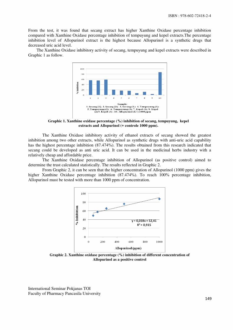
ISBN : 978-602-72418-2-4
International Seminar Pokjanas TOI
Faculty of Pharmacy Pancasila University
149
From the test, it was found that secang extract has higher Xanthine Oxidase percentage inhibition
compared with Xanthine Oxidase percentage inhibition of tempuyung and kepel extracts.The percentage
inhibition level of Allopurinol extract is the highest because Allopurinol is a synthetic drugs that
decreased uric acid level.
The Xanthine Oxidase inhibitory activity of secang, tempuyung and kepel extracts were described in
Graphic 1 as follow.
Graphic 1. Xanthine oxidase percentage (%) inhibition of secang, tempuyung, kepel
extracts and Allopurinol (+ controle 1000 ppm).
The Xanthine Oxidase inhibitory activity of ethanol extracts of secang showed the greatest
inhibition among two other extracts, while Allopurinol as synthetic drugs with anti-uric acid capability
has the highest percentage inhibition (87.474%). The results obtained from this research indicated that
secang could be developed as anti uric acid. It can be used in the medicinal herbs industry with a
relatively cheap and affordable price.
The Xanthine Oxidase percentage inhibition of Allopurinol (as positive control) aimed to
determine the trust calculated statistically. The results reflected in Graphic 2.
From Graphic 2, it can be seen that the higher concentration of Allopurinol (1000 ppm) gives the
higher Xanthine Oxidase percentage inhibition (87.474%). To reach 100% percentage inhibition,
Allopurinol must be tested with more than 1000 ppm of concentration.
Graphic 2. Xanthine oxidase percentage (%) inhibition of different concentration of
Allopurinol as a positive control

ISBN : 978-602-72418-2-4
International Seminar Pokjanas TOI
Faculty of Pharmacy Pancasila University
150
CONCLUSION
1. Secang, tempuyung and kepel extracts are potential as anti uric acid with the inhibitory percentage
of each extract are 58.922%, 20.154% and 12.071% respectively compared with the Allopurinol as a
synthetic drugs with 87.474%.
2. This study was restricted to the preliminary screening of Xanthine Oxidase inhibitory activity from
secang, tempuyung and kepel extracts. Further structural elucidation and characterization
methodologies have to be carried out in order to identify the bioactive metabolites in the extracts.
REFERENCES
1. Walker R, &Edward C. Clinical pharmacy and therapeutics. Edisi3. USA: Churchill Livingstone.
2003.
2. Wibowo S. Asam Urat, http://suryo_wibowo.blogspot.com. 2006. Diakses pada tanggal 9
September 2015
3. Price S & Wilson L. Patofisiologi: Konsep klinis proses-proses penyakit. Edisi 6. EGC, Jakarta.
2005.
4. Joseph T DiPiro, Robert L, Talbert Gary C, Yee Gary R, Matzke Barbara G, Wells L, Michael
Posey. Pharmacotherapy: A pathophysiological approach (Eds). 7th edition. 2005.
5. Xu H, Zhou Z, Yang J. Chemical constituents of caesalpiniasappan L.
ZhongguoZhongyaoZazhi. 1994. 19, (8) 485-486.
6. Chairul. Tempuyung untuk menghadang asam urat. http://digilib.itb.ac.id/
files/disk1/615/jbptitbpp-gdl-drchairula-30748-1-tempuyun-t.pdf Accessed on October 12th 2015.
Chapter 100. Acne Vulgaris : Treatment : Acne Vulgaris Accesspharmacy.
http://www.accesspharmacy.com/content.aspx?aID=3212123
http://www.agrobisnisinfo.com/2015/07/daun-tempuyung-tanaman-obat-herbal-Accessed on 9th
September 2015.
https://id.wikipedia.org/wiki/Kepel. Accessed on 10th September 2015..
http://tyasistiqomah.blogspot.co.id/2011_02_01_archive.html xanthine oxidase Accessed on
September 10th 2015.
.

ISBN : 978-602-72418-2-4
International Seminar Pokjanas TOI
Faculty of Pharmacy Pancasila University
151
Potency of Curcuma Mangga Val Rhizome Extract as a Selective Anti-
Proliferative Agent on Breast Cancer Cell Line MCF-7
SISKA ANDRINA*, CHURIYAH, NURALIH
Center of Pharmaceutical and Medical Technology,
Agency for the Assessment and Application of Technology.
Corresponding Author Email: [email protected]
Abstract: Curcuma Mangga Val is original plant from Indo-malesian which is distributed in Indonesia,
Taiwan, Thailand, China, Pacific and North Australia. In Indonesia, Curcuma Mangga have been used for
traditional medicine as an antioxidant, anti-diarrhea, and anti-pyretic. In this study, we investigate potency of
ethanolic extract of Curcuma Mangga Rhizome as an anti-proliferative agent against Breast Cancer cell line
MCF-7. We also measured the cytotoxicity effect of extract on Human Dermal Fibroblast as a normal cell to
determine its selectivity. Rhizome of Curcuma Mangga Val was macerated by 70% ethanolic and tested for
cytotoxicity using MTT cell viability assay on MCf-7 and Human Dermal Fibroblast. The result showed
selectively inhibited of ethanolic extract of Curcuma Mangga Rhizome on MCF-7 Cell with IC50 6.05 ppm
and less sensitivity on Human Dermal Fibroblas with IC50 31,9 ppm. We also observed apoptotic mechanism
of Curcuma Mangga Val extract on MCF-7 by double staining using etidium bromide and acridin orange.
The figure showed that half of cell was dead because of apoptotic and necrotic mechanism either in 7 ppm.
This study demonstrate the potential of Curcuma Mangga Val Ethanolic Extract as a selective anti-
proliferative agent on breast cancer cell line MCF-7.
Keyword: Curcuma Mangga Val, anti-proliferative, MCf-7, Human Dermal Fibroblast, apoptotic
INTRODUCTION
Cancer is a major public health problem in the worldwide. Cancer is one of non-communicable disease which
is the first leading causes of death all over the world. In 2012, WHO reported 8.2 billion of people in the
world having cancer and it would be increasing up to 22 billion of cases in next two decades. In Indonesia,
Ministry of Health has reported that breast cancer is the top leading cause of cancer deaths among Indonesian
women in 2014. Chemotherapy is one of the commonly-used strategies in breast cancer treatment. This
therapy is usually associated with adverse side effects, ranging from nausea to bone marrow failure(2)
and
development of multidrug resistance (MDR)(3)
.
In a few last decades, herbal plants have been widely used for diseases treatment and immunological
enhancement. The increasing trend of herbal application in traditional herbal industry is mainly due to
numerous beneficial effects of natural sources compared to single synthetic drug. Natural herbal medicines
usually offer less undesirable side effect, more efficiency and less toxic to patient. Therefore, finding natural
compounds from plants may provide an alternative cancer treatment. Curcuma Mangga Val is original plant
from Indo-malesian which is distributed in Indonesia, Taiwan, Thailand, China, Pacific and North Australia.
Curcuma manga Val is a member of the family Zingiberacae. It is also known by its common Indonesian
name Temu Mangga, because of its mango-like smell when fresh rhizomes are cut. In Indonesia, Curcuma
Mangga Val have been used as traditional medicine for relieving stomachic complaint, gastric ulcer, chest

ISBN : 978-602-72418-2-4
International Seminar Pokjanas TOI
Faculty of Pharmacy Pancasila University
152
pain, fever, hepato-protective and anti-allergic. Rhizome of Curcuma Mangga has been known containing
phenolic, alkaloid, saponin and also natural antioxidant curcuminoid. As we know that those of substance
have potential activity as a cancer killer from herbal plants
In this study, we investigated the potential activity of ethanolic extract of Curcuma manga val as a
cytotoxicity agent on breast cancer cell line MCF-7 and its apoptotic mechanism using staining assay. We
also evaluated extract selectivity against human dermal fibroblast as a normal cell. Selectivity index between
IC50 on breast cancer cell line and normal cell will inform its selectivity as an anti-proliferative agent against
breast cancer cell.
MATERIAL AND METHODS
MATERIAL. Plant Material and Chemicals. Rhizome of Curcuma mangga Val, Ethanol 70% Analytical
Grade, Cell line MCF.7, Human Dermal Fibroblas Cell, Roswell Park Memorial Institute medium (RPMI),
Dulbecco’s Modified Eagle Medium Low Glucose (DMEM-LG), Fetal Bovine Serum (FBS) 10% v/v
(Gibco), penisilin-streptomisin 1% v/v (Gibco), Dimethyl sulfoxide (DMSO), and 3-(4,5dimethylthiazol-2-
yl)-2,5-diphenyltetrazoliu bromide (MTT) , Sodium Deodecyl Sulfate (SDS), Phosphate Buffer Saline (PBS).
Sample Preparation. Rhizome of Curcuma manga Val cut and dried in the oven at 60o C, macerated all
of dried sample in ethanol 70% for a night. Evaporated filtrate of ethanol using high pressure evaporation
until viscous extract was obtained.
Cell Line Culture. MCF-7 was obtained from Gadjah Mada University Faculty of Pharmacy
Yogyakarta and Human Dermal Fibroblast was original cells from Center of Pharmacy and Medical
Technology BPPT. MCF-Cells were cultured in RPMI 1640 and Human Dermal Fibroblas were cultured in
DMEM-LG and both of medium were supplemented with 10% foetal bovine serum, glutamine (2mM) and
1% penicillin-streptomycin in static 75 cm T-Flask (Nunc, Denmark). The cells were incubated in a
humidified atmosphere with 5% CO at 37 oC.
Cell Cytotoxicity Assay. Cells were plated in a 96-well-plate with 50.000 cells/well of concentration.
The cells were left to adhere for 24 hours before exposed to the plant extracts (1-50 µg/ml) administered in
media containing 10% of FBS and returned to the incubator for 24 hrs. Subsequently, MTT reagent (0.5
mg/mL in sterile PBS) was added directly to the wells. Cells were returned to the incubator for 4 hrs. The
formation of insoluble purple formazan from yellowish MTT by enzymatic reduction was dissolved in SDS.
Incubate for a night and the optical density of solution was measured at 570 nm using a microplate reader.
Analyze the absorbance to obtain percentage of inhibition
Apoptotic Observation on MCF-7. Cells were plated in a 24 well plate with 100.000 cells/well
growing in RPMI medium supplemented 10% of FBS and 1% Penicillin-Streptomycin. Incubate the cells for
a night to adhere then add the extract with IC50 concentration. Incubating for 24 hours and observe the
morphology and staining of the cell after acridin orange and etidium bromide (1:1) was added under
fluorescence microscope 100x.
RESULT
The Cytotoxicity Effect of Ethanolic Extract of Curcuma Manga Val Rhizome on Breast Cancer Cell
Line MCF-7. MTT assay is a rapid and high accuracy colorimetric approach that widely used to determine
cell growth and cell cytotoxicity, particularly in the development of new drug. It measures cell membrane
integrity by determining mitochondrial activity through enzymatic reaction on the reduction of MTT to
insoluble formazan salt. Ethanolic Extract of Curcuma mangga Val Rhizome was measured its potency to
inhibit MCF-7 cell growth using MTT assay. This method will provide the percentage of cell proliferation
inhibition and also Proliferation inhibition concentration 50 (IC50). The result showed that all concentration
of extract inhibit the proliferation of cell with dose dependent manner. It means that increasing of
concentration will give higher inhibition of proliferation cells (Figure 1). That graph represented IC50 of
extract with values 6.05 ppm

ISBN : 978-602-72418-2-4
International Seminar Pokjanas TOI
Faculty of Pharmacy Pancasila University
153
Fig 1. Cell proliferation inhibition of ethanolic etract of curcuma manga val rhizome on breast cancer cell line
MCF-7 in 5 concentration of extract.
The Cytotoxicity Effect of Ethanolic Extract of Curcuma manga Val Rhizome on Human Dermal
Fibroblast Cell. Human Dermal Fibroblast cells as a normal cell were used to determine extract selectivity.
Index selectivity between IC50 on HDF and MCF-7 more than 2 means that the extract has selectivity
inhibiting cancer cells and does not has impact on normal cell. The result showed that Ethanolic Extract of
Curcuma manga Val Rhizome has higher inhibition of percentage compared than cytotoxicity on MCF-7
(Figure 2). Extract has IC50 31.93 ppm and index selectivity 5 (Table 1). It means that Ethanolic Extract of
Curcuma manga Val Rhizome has selectivity inhibiting MCF-7 breast cancer cell proliferation.
Figure 2. Comparison of cell proliferation inhibition on MCF-7 and human dermal fibroblast cell.
Table 1. IC50 of ethanolic extract of curcuma manga val rhizome on MCF-7; human dermal fibroblast and index
selectivity of extract.
Extract IC50 value (ug/mL) Index Selectivity
MCF-7 HDF
Ethanolic Extract of Curcuma
manga Val Rhizome
6.05 31.93 5
Apoptotic Observation of Ethanolic Extract of Curcuma manga Val Rhizome on MCF-7. The
apoptotic mechanism of the treated cells has been observed under an inverted microscope using ethidium
bromide and acridine orange (EB/AO) as staining agent. The apoptotic cells are stained in orange with lighter
in nucleus or green, the live cells are stained in green and the necrotic cells are stained in orange. The figure
showed that early apoptotic, late apoptotic and necrotic was found in all treated cell as presented in figure 3.
It means that in IC50 concentration of extract, cells were died because of apoptotic mechanism.
0.000
20.000
40.000
60.000
80.000
100.000
120.000
0 0.2 0.4 0.6 0.8 1 1.2 1.4 1.6 1.8
% C
ell
pro
life
rati
on
Inhib
itio
n
Log Concentration (ug/mL)
Rhizome of Curcuma Mangga Val. Ethanolict Extract
-20.000
0.000
20.000
40.000
60.000
80.000
100.000
120.000
0.194 0.796 1.398 1.699% P
roli
fera
tio
n I
nhib
itio
n
Log Concentration
Comparison of inhibition proliferation on MCF-7 and HDF
MCF-7
HDF

ISBN : 978-602-72418-2-4
International Seminar Pokjanas TOI
Faculty of Pharmacy Pancasila University
154
(a) (b) Fig 3. Double staining of ethanolic extract of curcuma manga val rhizome on MCF.7 in (a) control cell; (b)
extract at 7 ppm.
CONCLUSION
Ethanolic Extract of Curcuma Mangga Val has potency as cytotoxic agent on breast cancer cell line MCF-7.
Ethanolic Extract of Curcuma Mangga Val Rhizome has selective proliferation inhibition due to value of
index selectivity on HDF.Ethanolic Extract of Curcuma Mangga Val Rhizome has cytotoxicity effect through
apoptotic mechanism.
REFERENCE
1. Castell J V, and Lechon MG. In vitro methods in pharmaceutical research. Academic Press, San Diego,
California. 1997.
2. Da’i M. Uji aktivitas antiproliferatif pentagamavunon-o terhadap sel raji, sel hela, dan sel myeloma.
Tesis. Fakultas Farmasi UGM. Yogyakarta. 2003.
3. Doyle A, and Griffiths JB. Cell and tissue culture for medical research. John Willey and Sons Ltd, New
York.2000.
4. Fox M. Cancer deaths declining in U.S. Reuters Health, United States. 2007.
5. Franks LM, and Teich NM. Introduction to the cellular and molecular of cancer. Third Ed.,Oxford
University Press, New York.1997.
6. Freimoser, F.M., Jakob, C.A., Aebi, M, and Tuor, U, 1999, The MTT [3-(4,5-Dimethylthiazol-2-yl)-
2,5-Diphenyltetrazolium Bromide] Assay Is a Fast and Reliable Method for Colorimetric Determination
of Fungal Cell Densities, Applied and environmental microbiology, 65(8), 3727-3729.
7. Hanahan D, and Weinberg RA. The hallmark of cancer. Cell, 100, 57-70. 2000.
8. Harborne J B. Metode fitokimia, penuntun cara modern menganalisis tumbuhan. Penerbit ITB, Bandung.
1987.
9. Haridas V, Mujoo K, Hoffmann JJ, Wachter GA, Hutter LK, Lu Y, et.al. Triterpenoid saponins from
Acacia victoriae (Bentham) decrease tumor cell proliferation and induce apoptosis.Cancer Research, 61,
5486–5490. 2001.
10. Hartwell JL. Plant used against cancer. a Survey, Lioydia. 1971. 30-34.
11. Jacobz. Tumbuhan berkhasiat obat sebagai obat. KTO Karyasari, Jakarta. 2003.
12. King RJB. Cancer biology, 2nd ed., Pearson Education Limited, London.2000.
13. Mosmann T. Rapid colorimetric assay for cellular growth and survival: application to proliferation and
cytotoxicity assays. Journal of Immunological Methods,1983. 65 (1-2): 55-63 cit.
14. Itagaki, H et.al. Validation study on five cytotoxicity assay. JSAAE-VII details of the MTT. 1997.1-12.

ISBN : 978-602-72418-2-4
International Seminar Pokjanas TOI
Faculty of Pharmacy Pancasila University
155
Assessment of Antibacterial Activity of
Herbal Toothpastes to The Bacteria Causing Halitosis
SYARMALINA*, SYAHDU A. EKOWATI DAN DWI A. MAULANA
Faculty of Pharmacy,Pancasila University
Jalan Srengseng Sawah, Jagakarsa,Jakarta Selatan 12640, Indonesia.
Email:[email protected]
Abstract: The use of toothpaste is now not only as a cosmetic, but also as a treatment of halitosis. The
content of the herbal in the toothpaste can help halitosis therapy because of its antibacterial activity. The
purpose of the study is toobtain pure isolates from saliva of halitosis patients and to test antibacterial activity
of toothpaste to microbes causing halitosis. Microbial limit test of toothpaste, isolation of microbial testing
from saliva of halitosis patients and antibacterial activity test of herbal toothpaste to microbes were
accomplished. Microbial limit test toothpaste MYC (mold yeast count) and TPC (total plate count) values
obtained toothpaste A, B and D are eligible not more than 103
colonies/g TPC is between 3.6x10²-5.48x10
2
colonies/g; MYC between <10-2.76 x10² colonies/g while the value of TPC and MYC toothpaste C is
4.63x10⁴ colonies/g and 9.84 x10
3 colonies/g as well as the entire sample there is no growth of microbial
pathogens. The isolation of pure isolates produced 19 and then selection test is performed macroscopic,
microscopic and biochemical reactions produced four pure isolates representative used as test microbes.
Antibacterial activity test using agar diffusion method with paper discs. The results of the inhibition by
effectiveness sample: toothpaste C with clove between 23.5-28 mm; toothpaste B with betel between 21-28
mm; toothpaste A with miswak between 18-23 mm; toothpaste D with sodium fluoride between 15.5-22.5
mm. Concluded toothpaste A, B, C and D are potential as antibacterial against four pure isolates from saliva
of halitosis patients and bacteria Streptococcus mutans as comparison.
Keyword: Halitosis, herbal toothpaste, pure bacterial isolates, Streptococcus mutans, microbial
contamination test, antibacterial activity test.
INTRODUCTION
The oral cavity is a reflection of a healthy body because the mouth is an integral part of the body. In a healthy
state, the mouth is occupied by more than 6x10⁷ microorganisms that can be recognized by the aroma of our
mouth when wake up in the morning(1)
. If the growth of microorganisms in the mouth exceeded the maximum
number coupled with the presence of infection in the oral cavity are likely to cause halitosis.Halitosis is the
smell of bad breath that comes out of the oral cavity.
Halitosis is one of oral health problems that many people complain after caries and periodontal
disease(2)
. Gases containing sulfur that is released through breathing air. VSCs consists of hydrogen sulphide
(H2S), methyl mercaptan (CH3SH) and dimethyl sulfide (CH3SCH3). These gases are the result of the
production activity of bacteria in the mouth which form malodorous compounds and volatile(3)
.
One of efforts to prevent and treat halitosis is to use toothpaste. The use of toothpaste is now not only as
a cosmetic, but also as a treatment of halitosis. The content of the herbal in the toothpaste can help halitosis
therapy because of its antibacterial activity.

ISBN : 978-602-72418-2-4
International Seminar Pokjanas TOI
Faculty of Pharmacy Pancasila University
156
MATERIAL AND METHODS
MATERIAL. Toothpaste contain siwak, betel, clove and Natrium fluoride, Microbes testing (pure isolate of
bactery causing halitosis isolated from saliva halitosis patients and Streptococcus mutans ATCC 70061 as
comparison), peptone water, Nutrient Agar (NA), Potato Dextrose Agar (PDA), glucosa, lactosa, sacarosa,
maltosa, manitol, indol solution, MR-VP solution, Simmons Citrate Agar, Triple Sugar Iron Agar (TSIA),
Lysine Iron Agar (LIA), pereaksi kovac’s, methyl red reagent, a-naftol reagent, KOH solution, phosphate
buffer solution, Trypticase Soy Broth (TSB), Vogel Johnson Agar (VJA), Cetrimide Agar (CetA),
Chromogenic Agar (CrA), NaCl 0,9% solution, crystal violet solution, lugol solution, carbol fuchsin solution,
etanol 95% and aquadest.
METHODS.
RESULT AND DISCUSSION
1. The Data of Toothpaste Samples Used in This Study.

ISBN : 978-602-72418-2-4
International Seminar Pokjanas TOI
Faculty of Pharmacy Pancasila University
157
2. Microbiology Tests of Toothpaste Sample.
3. Inhibition Zone Diameter Data of Pure Isolates to Toothpaste Sample and Sodium Fluoride.
CONCLUSION
Tothpaste A, B, C and D are potential as antibacterial to four pure isolates of bacteria causing halitosis which
were isolated from saliva of halitosis patients and Streptococcus mutans .
REFERENCES
1. Tandelilin R. Oral health care: present oral biology approach and indigenous wisdom (disertation).
Yogyakarta: Gadjah Mada University. 2009.h.107-115.2.
2. Pintauli S. Halitosis and management issues. DentJ. 2008. 5(3):74-79.
3. Tonzetich J. Production and origin of oral malodor: A review of mechanism and methods of analysis.
JPeriodontal. 1974.48:13-20.
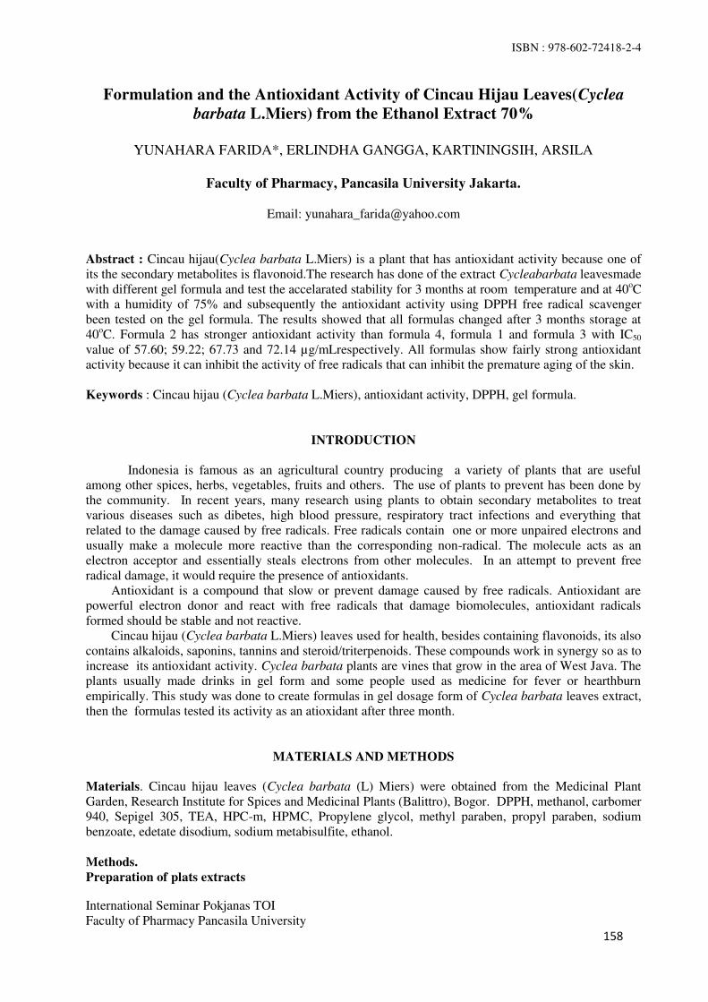
ISBN : 978-602-72418-2-4
International Seminar Pokjanas TOI
Faculty of Pharmacy Pancasila University
158
Formulation and the Antioxidant Activity of Cincau Hijau Leaves(Cyclea
barbata L.Miers) from the Ethanol Extract 70%
YUNAHARA FARIDA*, ERLINDHA GANGGA, KARTININGSIH, ARSILA
Faculty of Pharmacy, Pancasila University Jakarta.
Email: [email protected]
Abstract : Cincau hijau(Cyclea barbata L.Miers) is a plant that has antioxidant activity because one of
its the secondary metabolites is flavonoid.The research has done of the extract Cycleabarbata leavesmade
with different gel formula and test the accelarated stability for 3 months at room temperature and at 40oC
with a humidity of 75% and subsequently the antioxidant activity using DPPH free radical scavenger
been tested on the gel formula. The results showed that all formulas changed after 3 months storage at
40oC. Formula 2 has stronger antioxidant activity than formula 4, formula 1 and formula 3 with IC50
value of 57.60; 59.22; 67.73 and 72.14 µg/mLrespectively. All formulas show fairly strong antioxidant
activity because it can inhibit the activity of free radicals that can inhibit the premature aging of the skin.
Keywords : Cincau hijau (Cyclea barbata L.Miers), antioxidant activity, DPPH, gel formula.
INTRODUCTION
Indonesia is famous as an agricultural country producing a variety of plants that are useful
among other spices, herbs, vegetables, fruits and others. The use of plants to prevent has been done by
the community. In recent years, many research using plants to obtain secondary metabolites to treat
various diseases such as dibetes, high blood pressure, respiratory tract infections and everything that
related to the damage caused by free radicals. Free radicals contain one or more unpaired electrons and
usually make a molecule more reactive than the corresponding non-radical. The molecule acts as an
electron acceptor and essentially steals electrons from other molecules. In an attempt to prevent free
radical damage, it would require the presence of antioxidants.
Antioxidant is a compound that slow or prevent damage caused by free radicals. Antioxidant are
powerful electron donor and react with free radicals that damage biomolecules, antioxidant radicals
formed should be stable and not reactive.
Cincau hijau (Cyclea barbata L.Miers) leaves used for health, besides containing flavonoids, its also
contains alkaloids, saponins, tannins and steroid/triterpenoids. These compounds work in synergy so as to
increase its antioxidant activity. Cyclea barbata plants are vines that grow in the area of West Java. The
plants usually made drinks in gel form and some people used as medicine for fever or hearthburn
empirically. This study was done to create formulas in gel dosage form of Cyclea barbata leaves extract,
then the formulas tested its activity as an atioxidant after three month.
MATERIALS AND METHODS
Materials. Cincau hijau leaves (Cyclea barbata (L) Miers) were obtained from the Medicinal Plant
Garden, Research Institute for Spices and Medicinal Plants (Balittro), Bogor. DPPH, methanol, carbomer
940, Sepigel 305, TEA, HPC-m, HPMC, Propylene glycol, methyl paraben, propyl paraben, sodium
benzoate, edetate disodium, sodium metabisulfite, ethanol.
Methods.
Preparation of plats extracts

ISBN : 978-602-72418-2-4
International Seminar Pokjanas TOI
Faculty of Pharmacy Pancasila University
159
The dried powdered leaves of Cyclea barbata (L) Miers was macerated with ethanol 70% at room
temperature for 24 hours (three times), and the crude ethanolic solution was subsequently concentrated
using rotary vacuum evaporator to obtain the respective extracts and stored in a freeze condition until
used for further analysis.
Preparation of gel formula containing extract The ingredient on formula 1 – 4
No. Ingredient Formula % (w/w)
F1 F2 F3 F4
1 Cincau hijau extract (Cyclea
barbata L.Miers)
0.0522 0,0522 0,0522 0,0522
2 Carbomer 940 0,5 - - -
3 Sepigel 305 - 3 - -
4 Triethanolamine (TEA) 0,5 - - -
5 HPC-m - - 5 -
6 HPMC - - - 4
7 Propylene glycol 15 15 15 15
8 Methylparaben 0,03 0,03 - -
9 Propylparaben 0,01 0,01 - -
10 Sodium benzoate - - 0,1 0,1
11 Edetate disodium 0,05 0,05 0,05 0,05
12 Sodium metabisulfite 0,1 0,1 0,1 0,1
13 Ethanol - - 40 -
14 Distilled water qs 100 100 100 100
Formula 1 and 2 : Dispersed carbomer 940(gelling agent formula 1) and sepigel 305 (gelling agent
formula 2) in distilled water at room temperature for 24 hours with continuous stirring at 2000 rpm in
speed. Methyl paraben and propyl paraben were dissolved in propylene glycol. sodium metabisulfite and
edetate disodium were dissolved indistilled water and mixed it with Cyclea barbataextract. Finally full
mixed ingredients were mixed properly to the carbomer 940 gel with continuous stirring 10-15 min. with
2000 rpm speeduntil homogen and triethanolamine was added drop wise to the formulation to obtain the
gel at required consistency.The same method was followed for preparation with basis gel formula in
Table 1. Test the accelarated stability for 3 months at room temperature and at 40oc± 2°Cand 75% ± 5%
RH in climatic chamber.
Formula 3 and 4: Dispersed carbomer 940 with aq.dest at room temperature for 24 hours and then
homogenized with 2000 rpm speed (as gelling agent). Triethanolamine (TEA) was dissolved in distilled
water, then mix with carbomer 940. Sodium benzoate, sodium metabisulfite and edetate disodium were
dissolved in distilled water and the mixed with extract of Cyclea barbata. Formula 3 was used HPC-m
and formula 4 was used HPMC as basis gel. HPC-m and HPMC were dispersed with ethanol (as gelling
agent).
Antioxidant activity Test
The antioxidant activity of Cyclea barbata extract and formulas were tested using DPPH scavenging.
Briefly, samples (extract dan four formulas) of various concentrations were prepared 5, 10, 25, 50, and
100 µg/mL. Each sample concentration was mixed with 1.0 mL of 1mM methanolic DPPH solution. All
the solution were prepared with methanol to 5.0 mL. Experiment was done in triplicate. The test sample
were incubated for 30 min. at room temperature and the absorbance measured at 517 nm. Ascorbic acid
was used as a standard and DPPH in methanol was used as a control. The different in absorbance between
the test and the control was calculated and the expressed as % scavenging of DPPH radical. Percent
scavenging of DPPH free radical was measured using following equation:
Absorbance of Control - Absorbance of Sample
% DPPH radical scavenging = - x 100

ISBN : 978-602-72418-2-4
International Seminar Pokjanas TOI
Faculty of Pharmacy Pancasila University
160
Absorbance of Control
Then % inhibitions were plotted against respective concentration used and from the graph IC50 was
calculated.
RESULTS AND DISCUSION
The result of antioxidant activity test using DPPH free radical scavenging of ascorbic acid as standard,
the ethanol extract of cyclea barbata leaves was done in triplicate. The IC50 value of Ascorbic acid of
4.08µg/mL with r 0,9799 (Table 1). The results of extracts showed that IC50 value of 47.94 µg/mL
(Table 2.). The IC50 value was obtained then its used as dosage of gel formula.
The results showed that all formulas changed after 3 months ofstorage at 40oC, which is caused by
the effect of temperature and time. However, green grass jelly extract still have high antioxidant activity
with IC50 value is between 50-100µg/mL. Formula 2 is stronger antioxidant activity than the formula 1,
because the formula 2 using a base gel modification (sepigel 305), so its more stable than the others. The
IC50 value of formula 2,4,1 and formula 3 with IC50 value of 57.60; 59.22; 67.73 and 72.14
µg/mLrespectively (Table 3). The smaller the IC50 value, the higher the antioxidant activity. Figure 1
shows The graph of antioxidant activity of cincau hijau , the Formulas and Vit.C as a control.
The gel formulation can be used as an antioxidant because it can inhibit the activity of free radicals
that can inhibit premature aging of skin.
Table 1. IC50 value of vitamin C.
C
(ppm)
Acontrol Absorbance of vit.C % Inhibition IC50
(µg/mL) I II III I II III
1
0,7615
0,5615 0,6004 0,5671 26,2640 21,1556 25,5286
4,08 2 0,5353 0,5563 0,5449 29,7045 26,9468 28,4439
3 0,4236 0,4240 0,4209 44,3729 44,3204 44,7275
4 0,4008 0,4182 0,4164 47,3670 45,0821 45,3185
5 0,3180 0,2219 0,3212 58,2403 70,8601 57,8201
Table 2. IC50 value of cincau hijau (Cyclea barbataL Miers) extract.
Table 3. IC50 value of cincau hijau (Cyclea barbataL.Miers) formulas after 3 months at room temperature
and at 40oC ± 2°Cand 75% ± 5% RH.
IC50 value (µg/mL)
Temp.
Month
Formula 1 Formula 2 Formula 3 Formula 4
Batch I Batch
II
Batch I Batch II Batch I Batch II Batch I Batch II
Room temp.
(15-30oC)
0 61,763
0
60,974
4
51,782
1
59,4050 70,6755 68,0105 58,3833 62,8872
3 65,514
1
61,279
2
52,946
5
61,7707 71,9485 74,5973 62,9523 67,2856
40oC ± 2°C 0 - - - - - - - -
3 67,682
1
67,725
1
57,603
7
63,2377 72,1391 92,2392 59,2221 67,2856
Blank/control 228,2685
C
(ppm)
Ablank Absorbance of extract % Inhibition IC50
(µg/mL) I II III I II III
5
0,8223
0,7363 0,7484 0,7316 10,4585 8,9870 11,0300
47,94 10 0,6504 0,6407 0,6383 20,9048 22,0844 22,3763
25 0,6060 0,5673 0,5619 26,3043 31,0106 31,6673
50 0,4716 0,4807 0,4571 42,6487 41,5420 44,4120
100 0,3671 0,3954 0,3935 55,3569 51,9154 52,1464
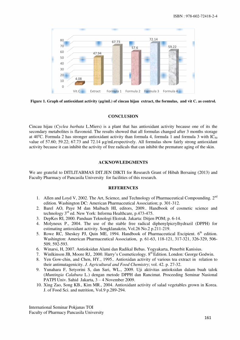
ISBN : 978-602-72418-2-4
International Seminar Pokjanas TOI
Faculty of Pharmacy Pancasila University
161
Figure 1. Graph of antioxidant activity (µg/mL) of cincau hijau extract, the formulas, and vit C. as control.
CONCLUSION
Cincau hijau (Cyclea barbata L.Miers) is a plant that has antioxidant activity because one of its the
secondary metabolites is flavonoid. The results showed that all formulas changed after 3 months storage
at 40oC. Formula 2 has stronger antioxidant activity than formula 4, formula 1 and formula 3 with IC50
value of 57.60; 59.22; 67.73 and 72.14 µg/mLrespectively. All formulas show fairly strong antioxidant
activity because it can inhibit the activity of free radicals that can inhibit the premature aging of the skin.
ACKNOWLEDGMENTS
We are grateful to DITLITABMAS DIT.JEN DIKTI for Research Grant of Hibah Bersaing (2013) and
Faculty Pharmacy of Pancasila University for facilities of this research.
REFERENCES
1. Allen and Loyd V, 2002. The Art, Science, and Technology of Pharmaceutical Compounding. 2nd
edition. Washington DC: American Pharmaceutical Association; p. 301-312.
2. Barel AO, Paye M dan Maibach HI, editors, 2009.. Handbook of cosmetic science and
technology 3rd
ed. New York: Informa Healthcare, p.473-475.
3. DepKes RI, 2000. Panduan Teknologi Ekstrak. Jakarta: Ditjen POM; p. 6-14.
4. Molyneux P., 2004. The use of the stable free radical diphenylpicrylhydrazil (DPPH) for
estimating antioxidant activity. Songklanakrin, Vol.26 No.2 p.211-219.
5. Rowe RC, Sheskey PJ, Quin ME, 1994. Handbook of Pharmaceutical Excipient. 6th edition.
Washington: American Pharmaceutical Association, p. 61-63, 118-121, 317-321, 326-329, 506-
509, 592-593.
6. Winarsi, H, 2007. Antioksidan Alami dan Radikal Bebas. Yogyakarta, Penerbit Kanisius.
7. Winlkinson JB, Moore RJ,. 2000. Harry’s Cosmeticology. 8th Edition. London: George Godwin.
8. Yen Gow-chin, and Chen, HY., 1995.. Antioxidan activity of various tea extract in relation to
their antimutagenicity. J. Agricultural and Food Chemistry; vol. 42. p. 27-32.
9. Yunahara F, Setyorini S, dan Sari, WL., 2009. Uji aktivitas antioksidan dalam buah talok
(Muntingia Calabura L.) dengan metode DPPH dan Rancimat. Proceeding Seminar Nasional
PATPI Univ. Sahid Jakarta, 3 – 4 November 2009.
10. Xing Zao, Song KB., Kim MR., 2004. Antioxidant activity of salad vegetables grown in Korea.
J. of Food Sci. and nutrition, Vol.9 p.289-294.
0
10
20
30
40
50
60
70
80
Vit.C Extract Formula 1 Formula 2 Formula 3 Formula 4
4.08
47.94
67.73
57.6
72.14
59.22

ISBN : 978-602-72418-2-4
International Seminar Pokjanas TOI
Faculty of Pharmacy Pancasila University
162
Accelerated Stability Test of Green Grass Jelly (Cyclea barbata L.Miers) Leaves
Extract Gel Preparation as Antioxidant With Gelling Agent HPCM and HPMC
KARTININGSIH, ERLINDHA GANGGA, YUNAHARA FARIDA, MARIA ULFAH
Faculty of Pharmacy, Pancasila University.
Jl. Srengseng Sawah, Jagakarsa, Jakarta Selatan 12460.
Email: [email protected]
Abstract : Green grass jelly leaves (Cyclea barbata L.Miers) which its upper and lower surfaces are hairy
contain flavonoids as antioxidants, have a very strong activity for free radical scavenging in the body. Green
grass jelly leaves were maserated by 70% ethanol and then the crude extract were formulated into 2 gel
formulas using HPC-m 5% and HPMC 4% that have been dispersed and then added to crude extract of
green grass jelly leaves. Green grass jelly leaves extract gel formed were accelerated stability testing for 3
months in room temperature 30oC ± 2
oC and 40°C or 75% RH. The gels were evaluated physically and
chemically, such as organoleptic test, homogenity, viscosity and rheology, spreadability, pH test, antioxidant
activity test, and analysis of microbial contamination tests then were compared with gel in the market. The
results showed the best formula was the first formula (F1) with greenish yellow gel, specific odor,
homogeneous, viscosity of 16000 - 60800 cps, spreadability of 1962,5 - 3576.66 mm2, pH 5.73 - 6.37, and
analysis of microbial contamination has a colonies not more than 103 colonies/g or colonies/mL. Statistical
analysis using two ways ANOVA to see time and temperature effects toward gel preparations stability. There
is no effect on the viscosity, spreadability, and pH tests, on the first formula with P = 0.00 > 0.05.
Keywords: accelerated stability test, cincau hijau leaves extract, gel, HPC-m, HPMC, antioxidant.
INTRODUCTION
Stability is ability of a product or cosmetic to be able to last or stay in standard specification range during
storage time and use to ensure the identity, strength, quality dan purity of the product. Medicine product
of each preparation has different stability quality, therefore, each preparation or product has to undergo
stability test to ensure the medicine product manufucturing and storaging. Stability test proceed as
instructed in Good Manufacturing Practice for medicine products (CPOB) such as testing method and
storaging condition that must qualify the standard.
In this paper, stability test will be held for three months in month 0, 1, 2, and 3 in room temperature
27 ± 20C dan temperature of 45
o-50
oC/RH 75% in climatic chamber. Green grass jelly leaf extracts have
the most active chemical content of alkaloids and flavonoids. Flavonoids acts as natural antioxidant to
decrease the number of free radical in body, make it able to reduce /prevent aging process, therefore, not
only the stability test is held, but also the antioxidant activity test. There are two formulas with 4% HPCm
gel base and 5% HPMC base under stability test in room temperature 27 ± 20C dan temperature of 40
o RH
75% in climatic chamber for three months and then then the gel be chemically and physically evaluated,
inculding organoleptic, homogenity, viscosity and rheology, spreadability, pH and antioxidant activity
test.
MATERIALS AND METHODS
Materials. Fresh green grass jelly leaf extract (Cyclea barbata L.Miers), Hydroxypropyl cellulose (HPC-
m), Hydroxypropylmethyl cellulose (HPMC), Propylene glycol, sodium benzoate, sodium metabisulfite,
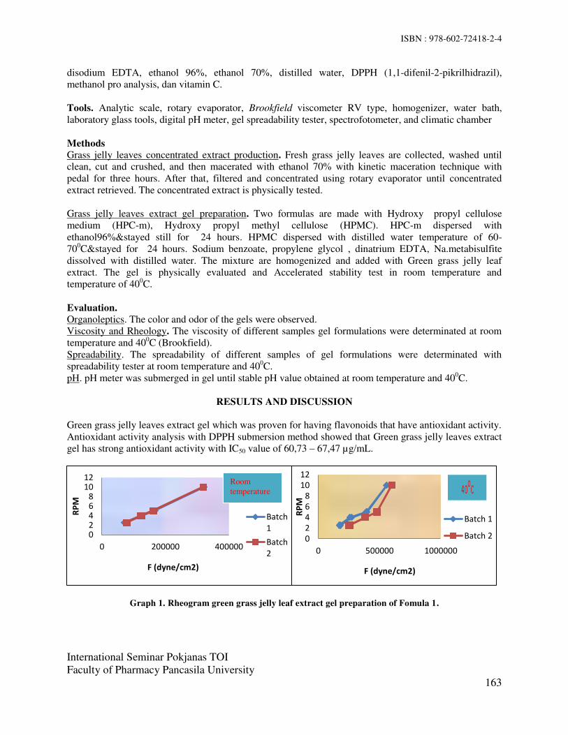
ISBN : 978-602-72418-2-4
International Seminar Pokjanas TOI
Faculty of Pharmacy Pancasila University
163
disodium EDTA, ethanol 96%, ethanol 70%, distilled water, DPPH (1,1-difenil-2-pikrilhidrazil),
methanol pro analysis, dan vitamin C.
Tools. Analytic scale, rotary evaporator, Brookfield viscometer RV type, homogenizer, water bath,
laboratory glass tools, digital pH meter, gel spreadability tester, spectrofotometer, and climatic chamber
Methods
Grass jelly leaves concentrated extract production. Fresh grass jelly leaves are collected, washed until
clean, cut and crushed, and then macerated with ethanol 70% with kinetic maceration technique with
pedal for three hours. After that, filtered and concentrated using rotary evaporator until concentrated
extract retrieved. The concentrated extract is physically tested.
Grass jelly leaves extract gel preparation. Two formulas are made with Hydroxy propyl cellulose
medium (HPC-m), Hydroxy propyl methyl cellulose (HPMC). HPC-m dispersed with
ethanol96%&stayed still for 24 hours. HPMC dispersed with distilled water temperature of 60-
700C&stayed for 24 hours. Sodium benzoate, propylene glycol , dinatrium EDTA, Na.metabisulfite
dissolved with distilled water. The mixture are homogenized and added with Green grass jelly leaf
extract. The gel is physically evaluated and Accelerated stability test in room temperature and
temperature of 400C.
Evaluation. Organoleptics. The color and odor of the gels were observed.
Viscosity and Rheology. The viscosity of different samples gel formulations were determinated at room
temperature and 400C (Brookfield).
Spreadability. The spreadability of different samples of gel formulations were determinated with
spreadability tester at room temperature and 400C.
pH. pH meter was submerged in gel until stable pH value obtained at room temperature and 400C.
RESULTS AND DISCUSSION
Green grass jelly leaves extract gel which was proven for having flavonoids that have antioxidant activity.
Antioxidant activity analysis with DPPH submersion method showed that Green grass jelly leaves extract
gel has strong antioxidant activity with IC50 value of 60,73 – 67,47 µg/mL.
Graph 1. Rheogram green grass jelly leaf extract gel preparation of Fomula 1.
0
2
4
6
8
10
12
0 200000 400000
RP
M
F (dyne/cm2)
Batch
1
Batch
2
0
2
4
6
8
10
12
0 500000 1000000
RP
M
F (dyne/cm2)
Batch 1
Batch 2
Room
temperature

ISBN : 978-602-72418-2-4
International Seminar Pokjanas TOI
Faculty of Pharmacy Pancasila University
164
Viscosity stability evaluation. Rheogram green grass jelly leaf extract gel preparation of Fomula 1 was
shown in graph 1. Gel preparation has stable rheogram for three months, which is plastic. Plastic flow of
the gel preparation did not change after 3 months storage.
Spreability Stability evaluation. Spreability Stability results of formula 1 was shown in table 1.
Spreadability of the gel decreased after 3 months storage due to the increase of viscosity which increased
each month.
pH stability evaluation. pH stability evaluation results were shown in table 2. pH value of room
temperature and 400C of the gels were decreased each month. pH is affected by time. In room temperature
3 months storage, the pH decreased from 6,31 ± 0,02 to 6,04 ± 0 and in 400C 3 months storage, the pH
decreased from 6,02 ± 0,01 to 5,76 ± 0,03.
Table 1. Spreadability Stability results of green grass jelly leaves extract gel preparation Formula 1.
Table 2. pH stability test results of green grass jelly leaves extract gel preparation.
CONCLUSION
Different gelling agent in Formula 1 HPC-m and Formula 2 HPMC have different physical quality. In
Formula 1, green grass jelly leaf extract gel preparation has more stable physical quality after 3 months
storage time in room temperature 30oC ± 2
oC and temperature of 40
0C / RH 75% which is a gel with
greenish yellow gel, specific odor, homogeneous.
Statistic Analysis with two way ANVA on viscosity test, spreadability, and pH test. Gel preparation
Formula 1 Batch 1 dan 2 in room temperature 30oC ± 2
oC and 40
oC for 3 months storage time have P
value =0,00 > 0,05, Means that there is no effect on viscosity, spreadability and pH Formula 1 and 2
during storage time of 3 months.
ACKNOWLEDGEMENT
Special thanks are devote for Directorate General of Higher Education of Indonesia which has given
grants in this research.
Temperature Time
(months)
1 2 3
D (mm) F (mm2) D (mm) F (mm
2) D (mm) F (mm
2)
Room
0 66,80 3502,86 66,50 3471,47 67,10 3534,39
1 67,00 3523,87 66,50 3471,47 67,10 3534,39
2 67,10 3534,39 66,70 3492,38 67,30 3555,49
3 67,30 3555,49 66,90 3513,35 67,50 3576,66
40oC 1 65,20 3337,07 65,60 3378,14 65,00 3316,63
2 62,80 3095,91 61,00 2920,99 61,70 2988,41
3 55,00 2374,63 53,30 2230,10 53,00 2205,07
Temperature Time (Months) Batch 1
1 2 3 Mean ± SD
Room
0 6,29 6,33 6,30 6,31 ± 0,02
1 6,24 6,25 6,26 6,25 ± 0,01
2 6,10 6,12 6,12 6,11 ± 0,01
3 6,04 6,04 6,04 6,04 ± 0
40oC
1 6,01 6,02 6,03 6,02 ± 0,01
2 5,90 5,76 5,80 5,82 ± 0,07
3 5,78 5,73 5,77 5,76 ± 0,03

ISBN : 978-602-72418-2-4
International Seminar Pokjanas TOI
Faculty of Pharmacy Pancasila University
165
REFERENCES
1. Lieberman HA, Rieger, Martin M, Banker, Gilbert S. Pharmaceutical Dosage Forms Dieperse System.
Vol 2. USA. Marcel Dekker, Inc. 1996. P. 403
2. Carstensen JT. Drug Stability Principles and Practices. Third Edition. New York. Marcel Dekker, Inc.
2000. P. 516
3. Ansel HC. Pharmaceutical Dosage Forms and Drug Delivery System. Eight Edition. Lippincott
Williams and Wilkins, 2010.
4. Aulton, ME. Pharmaceutical The Science of Dosage Form Design. Second Edition. New York:
Churchill Livingstone. 2007.
ISBN : 978-602-72418-2-4
International Seminar Pokjanas TOI Faculty of Pharmacy Pancasila University 166
Capsule Formulation of Standardized 70% Ethanol Extract Johar Leaves (Senna siamea (Lam.) Irwin and Barneby)
as Α-Glucosidase Inhibitor
RISMA MARISI TAMBUNAN, KARTININGSIH, EVERLY HENDRA
Faculty of Pharmacy, Pancasila University.
Srengseng sawah, Jagakarsa, South Jakarta 12640.
Email: [email protected] Abstract : Standardized 70% ethanol extract of johar leaves showed that the extract has the consistency viscous, fulfill the requirements parameters of quality extract and has inhibitory activity α-glucosidase at 225 ppm is 90,88% equivalent to acarbose 95,21%. In this study, dried extract of johar leaves was formulated into capsule dosage form for practical use as an oral antidiabetic. The extract was dried with freeze drying using maltodextrin 10%. Dried extract was formulated into capsule dosage form using three variety diluents, namely dibasic calcium phosphate, lactose and avicel pH 102. Based on the evaluation results obtained that avicel pH 102 as the optimal formula that showed weight uniformity 0,4234 g and disintegration time 7 minutes 2 second with inhibitory activity α-glucosidase 85,88%. Keywords: Johar leaves, capsule, diluent
INTRODUCTION
Johar leaves (Senna siamea (Lam.) Irwin and Barneby) is a plant that people using empirically as a antidiabetic drug (1). 70% ethanol extract of johar leaves known to inhibits α-glucosidase at 225 ppm is 85,88% equivalent to acarbose 77,49%.
In this study, 70% ethanol extract of johar leaves was made into capsule dosage form for practically use and cover the bitter taste of johar leaves. 70% ethanol extract of johar leaves was dried by freeze drying using maltodextrin 10%. Dried extract was made into capsule dosage form using variation diluent to obtain formula of capsule dosage form with good characteristics. Capsule dosage form was evaluated physical quality, ie weight uniformity and disintegration time (2, 3, 4).
MATERIALS AND METHODS
Materials Crude drug of johar leaves (Senna siamea (Lam.) Irwin dan Barneby), ethanol 70%, maltodextrin, pvp, dibasic calcium phosphate, lactose and avicel pH 102, aquadest, hard gelatin capsule no. 0, α-glucosidase (G5003-100UN, Sigma), p-nitrophenyl-α-D-glucopyranoside (N1377-1G, Sigma), bovine serum albumin (Sigma), acarbose (Bayer), phosphate buffer pH 7,0, dimethyl sulfoxide, sodium carbonate, sodium hydroxide. Tools Analytical scales, glass tools, vacuum rotary evaporator, desiccators, mortar and stamfer, water bath, freeze dryer (LABCONCO), moisturemeter karl fischer, flow rate tools (LES-AUTONICS), stopwatch, sieve shaker (shive shakers of BBS product, BCL 601), bulk density Omron H3CR Tester, oven (Memmert), capsule filler, disintegration time tools (Guoming BJ-2), micropipette, absorbance microplate reader Elx 800.

ISBN : 978-602-72418-2-4
International Seminar Pokjanas TOI Faculty of Pharmacy Pancasila University 167
Methods Determination of Extract Quality Parameters. Performed of determination of parameters quality extract includes determination of the ash content, the determination of ash content acid insoluble, water soluble extract assay, content of ethanol soluble extract, loss on drying, water content, residual solvent, contamination of metals (Pb and Cd), Microbial contaminations, and total flavonoid content From the determination of parameter quality extract johar leaves by methods “Parameter standart mutu ekstrak tumbuhan obat” results can be seen in Table 1.
Table 1. Result of determination of parameters quality extract. Pharameters extract Result
Water soluble extract (%) 65,27 Content of ethanol soluble extract (%) 72,60 Loss on drying (%) 13,27 Water content (%) 10,45 Ash content (%) 0,48 Ash content acid insoluble (%) 0,05 Residual solvent (%) 0,10 Contamination of metals (mg/kg) Pb Cd
0,0301 0,003
Microbial contaminations (colonies/g) ALT AKK
9,1x10³ 3,98x10²
Total Flavonoids content (%) 8,44 Preparation of Capsules Dried extract of johar leaves is the active ingredients. Additional materials (excipients) ie avicel pH 102, lactose, dibasic calcium phosphate as diluent. Three formula was made into capsule dosage form using hard gelatin capsule no. 0 (table 2). Dried extract of johar leaves added diluent and mixed until homogeneous. Homogeneous mass inserted into the capsule shell using capsule filler tools.
Table 2. Capsule formulation. Formulation
Materials Weight (g)
I II III Dried extract 35,08 35,08 35,08
CaHPO4 8,06337 - - Lactose - 8,9357 -
Avicel pH 102 - - 4,4736 Evaluation Of Capsule Weight Uniformity A number of 20 capsules were weighed. Then one by one capsule weighed and removed its contents. Empty capsule shells then weighed. Calculated weight of the contents of each capsule and the average weight of the content. Capsule weight uniformity requirement is there should not be any deviation capsules larger percentage of column A (±7,5%) and there should be no more than 2 capsules a greater percentage deviation from column B (±15%).

ISBN : 978-602-72418-2-4
International Seminar Pokjanas TOI Faculty of Pharmacy Pancasila University 168
Disintegration time Six capsules are put into the basket on the medium water-temperature 37⁰C ± 2⁰C as much as 1000 mL, then run the tool. Automatically, the basket will be up and down on a regular basis. When the basket fell, the basket into the medium of water. For each capsule, which was disintegrated, note the time. The capsule was disintegrated if there is no part of the capsule remains, except parts of the outer shell of the capsule. When 2 capsules not disintegrated completely, repeat the test with 12 other capsules with terms no less 16 of 18 capsules tested must be crushed completely. Terms of the time disintegrated is not more than 15 minutes (4). α-Glucosidase Inhibitory Activity of Johar Leaves Capsule Dosage Form The same concentration of each capsule formula and acarbose as positive control were conducted to α-glucosidase inhibitory activity using p-nitrophenyl-α-D-glucopyranoside as substrate. α-glucosidase activity was determined by kawanishi method at 405 nm wave length. 200 mg bovine serum albumin and 1,0 mg α-glucosidase were diluted in buffer phosphate pH (7,0). The mixing solution contains 250 µl solution of 2 mM p-nitrophenyl-α-D-glucopiranoside, 400 µl buffer phosphate (pH 7,0), and series of 10 µl, 30 µl, 50 µl, 70 µl dan 90 µl sample. Then, it was pre-incubated at 37⁰C for 5 minutes. Reaction was started by adding 250 µl α-glucosidase, then incubated it at 37⁰C for 15 minutes. The reaction was stopped by adding 1000 µl Na2CO3 solution. The amount of p-nitrophenol obtained was measured at 405 nm wave length. The percentage of the inhibitory activity was counted by using this formula :
Where C = absorbance of enzyme activity without inhibitor (absorbance of DMSO), and S = absorbance of enzyme activity with sample examined.
RESULTS AND DISCUSSION
Determination of extract quality parameters The result determination of parameters quality specific extract of (Senna siamea (Lam.) Irwin and Barneby) leaves obtained shows that the extract has the consistency viscous, brown, and distinctive smell. Assay of compounds that are dissolved in two solvents was conducted to compare the number of secondary metabolites compound who extracted in a solvent of water and ethanol . Assay of compounds dissolved in a solvent of water shows the amount of inorganic compounds contained in extracts . While compounds assay dissolved in ethanol shows the amount of organic compounds present in the extract. From the results of the determination showed that the concentration of substances dissolved in ethanol (72,60%) and compounds that are dissolved in water (65,27%).
Determination of total ash and ash insoluble in acid the conducted to determine the presence of trace elements in extracts who known as inorganic matter or ash. In the process of combustion in furnaces with a temperature of 4500C , organic materials in the extract can be burned but not for inorganic substances, namely ash. Ash total is the ash produced from a number of extracts which incandescent in the furnace. Ash total can be use to determine the mineral content of both physiological compounds such as K, Mg, Ca, or non-physiological such as pollutants, dust, soil contained in the extract. Results of the determination of total ash content obtained by 0,48%. While the levels of acid insoluble ash conducted to determine heavy metals compounds that do not dissolve in acid such as Hg, Pb, Cd and silicates. Acid insoluble ash content of 0,05% were obtained. These results meet the requirements of acid insoluble ash content of the extract is generally less than 1%.
Assay of compounds that are dissolved in two solvents was conducted to compare the number of secondary metabolites compound who extracted in a solvent of water and ethanol . Assay of compounds dissolved in a solvent of water shows the amount of inorganic compounds contained in extracts. While compounds assay dissolved in ethanol shows the amount of organic compounds present in the extract . From
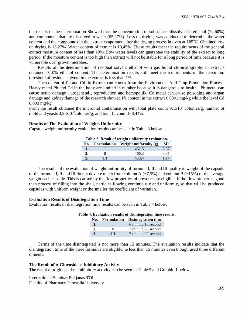
ISBN : 978-602-72418-2-4
International Seminar Pokjanas TOI Faculty of Pharmacy Pancasila University 169
the results of the determination Showed that the concentration of substances dissolved in ethanol (72,60%) and compounds that are dissolved in water (65,27%). Loss on drying was conducted to determine the water content and the compounds in the extract evaporated after the drying process in oven at 1050C. Obtained loss on drying is 13,27%. Water content of extract is 10,45%. These results meet the requirements of the general extract moisture content of less than 10%. Low water levels can guarantee the stability of the extract to long period. If the moisture content is too high then extract will not be stable for a long period of time because it is vulnerable over grown microbes.
Results of the determination of residual solvent ethanol with gas liquid chromatography in extracts obtained 0,10% ethanol content. The determination results still meet the requirements of the maximum threshold of residual solvent in the extract is less than 1%.
The content of Pb and Cd in Extract can comes from the Environment And Crop Production Process. Heavy metal Pb and Cd in the body are limited in number because it is dangerous to health . Pb metal can cause nerve damage , urogenetal , reproduction and hemopoitik. Cd metal can cause poisoning and organ damage and kidney damage of the research showed Pb content in the extract 0,0301 mg/kg while the level Cd 0,003 mg/kg. From the result obtained the microbial contamination with total plate count 9,1x10³ colonies/g, number of mold and yeasts 3,98x10²colonies/g, and total flavonoids 8,44%. Results of The Evaluation of Weights Uniformity Capsule weight uniformity evaluation results can be seen in Table 3 below.
Table 3. Result of weight uniformity evaluation. No. Formulation Weight uniformity (g) SD 1. I 462,3 3,57 2. II 466,3 3,31 3. III 423,4 1,14
The results of the evaluation of weight uniformity of formula I, II and III quality ie weight of the capsule
of the formula I, II and III do not deviate much from column A (±7,5%) and column B (±15%) of the average weight each capsule. This is caused by the flow properties of powders are eligible. If the flow properties good then process of filling into the shell, particles flowing continuously and uniformly, so that will be produced capsules with uniform weight or the smaller the coefficient of variation. Evaluation Results of Disintegration Time Evaluation results of disintegration time results can be seen in Table 4 below.
Table 4. Evaluation results of disintegration time results. No. Formulation Disintegration time 1. I 6 minute 10 second 2. II 7 minute 28 second 3. III 7 minute 02 second
Terms of the time disintegrated is not more than 15 minutes. The evaluation results indicate that the
disintegration time of the three formulas are eligible, ie less than 15 minutes even though used three different diluents. The Result of α-Glucosidase Inhibitory Activity The result of α-glucosidase inhibitory activity can be seen in Table 5 and Graphic 1 below.
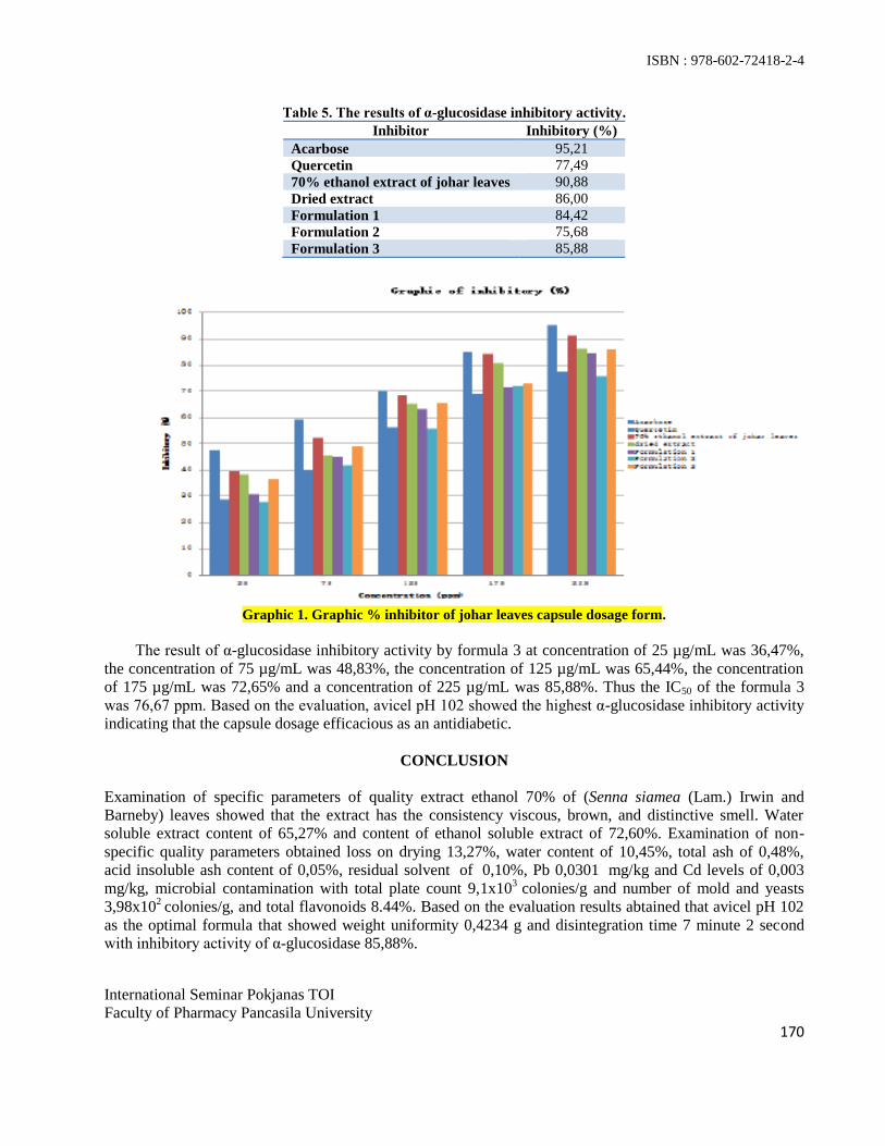
ISBN : 978-602-72418-2-4
International Seminar Pokjanas TOI Faculty of Pharmacy Pancasila University 170
Table 5. The results of α-glucosidase inhibitory activity. Inhibitor Inhibitory (%)
Acarbose 95,21 Quercetin 77,49 70% ethanol extract of johar leaves 90,88 Dried extract 86,00 Formulation 1 84,42 Formulation 2 75,68 Formulation 3 85,88
Graphic 1. Graphic % inhibitor of johar leaves capsule dosage form.
The result of α-glucosidase inhibitory activity by formula 3 at concentration of 25 µg/mL was 36,47%, the concentration of 75 µg/mL was 48,83%, the concentration of 125 µg/mL was 65,44%, the concentration of 175 µg/mL was 72,65% and a concentration of 225 µg/mL was 85,88%. Thus the IC50 of the formula 3 was 76,67 ppm. Based on the evaluation, avicel pH 102 showed the highest α-glucosidase inhibitory activity indicating that the capsule dosage efficacious as an antidiabetic.
CONCLUSION Examination of specific parameters of quality extract ethanol 70% of (Senna siamea (Lam.) Irwin and Barneby) leaves showed that the extract has the consistency viscous, brown, and distinctive smell. Water soluble extract content of 65,27% and content of ethanol soluble extract of 72,60%. Examination of non-specific quality parameters obtained loss on drying 13,27%, water content of 10,45%, total ash of 0,48%, acid insoluble ash content of 0,05%, residual solvent of 0,10%, Pb 0,0301 mg/kg and Cd levels of 0,003 mg/kg, microbial contamination with total plate count 9,1x10³ colonies/g and number of mold and yeasts 3,98x10² colonies/g, and total flavonoids 8.44%. Based on the evaluation results abtained that avicel pH 102 as the optimal formula that showed weight uniformity 0,4234 g and disintegration time 7 minute 2 second with inhibitory activity of α-glucosidase 85,88%.
ISBN : 978-602-72418-2-4
International Seminar Pokjanas TOI Faculty of Pharmacy Pancasila University 171
REFERENCES 1. Syamsuhidayat SS, Hutapea JR. Inventaris tanaman obat Indonesia. Edisi I. Jakarta: departemen
Kesehatan Republik Indonesia Badan Penelitian dan Pengembangan Kesehatan; 1991. 2. Aulton M. E., 2007, Pharmaceutics; the science of dosage form design, New York; Churchill
Livingstone, p. 517-523. 3. Lachman L, Lieberman HA, Kanig JL. Teori dan Praktek Farmasi Industri Edisi II Vol 1 & II.
Diterjemahkan oleh Suyatmi S. Jakarta: UI Press; 1989, p. 795-822. 4. Swarbrick J, Boylan JC. Encyclopedia of Pharmaceutical Technology vol.4 Newyork: Marcel Dekker
Inc; 1991. p. 37-76.
ISBN : 978-602-72418-2-4
International Seminar Pokjanas TOI
Faculty of Pharmacy Pancasila University
172
Formulation and Evaluation of Herbal Tablets Containing Voacanga foetida
(Bl.) K.Schum Extract
FAHLENI
1*, YANDI SYUKRI
2, NOVELTA FEMMY RISCHA
3, ADRIANI SUSANTY
3
1)
Faculty of Pharmacy Pancasila University, Jakarta. 2)
Islamic University of Indonesia, Yogyakarta. 3)
Sekolah Tinggi Ilmu Farmasi Riau.
Email : [email protected]
Abstract : Ethanolic extract of leaves of Voacanga foetida (Bl.) K.Schum has been formulated using wet
granulation method. The aim of this study was to obtain the optimal concentration of starch as binder with
varying concentrations; F1 (1%), F2 (2%) and F3 (3%). Evaluation of granules including true specific
density, tapped density, apparent density, compressibility, Hausner factor, porosity, water content and
angle of repose. Tablets were evaluated for weight variation, thickness, hardness, friability, in vitro
desintegrating time. The results of angle of repose, Carr’s Index and Hausner ratio of all formulas indicated that the powder mixtures possess good flow properties and good packing ability. The physical
properties tablets met the requirements according to Indonesian Pharmacopoeia 3th
edition. It can be
concluded that F1 was the best formula with the hardness 6,51 ± 0,62 kg/cm2, friability 0,99 ± 0,16%, and
disintegration time 9,38 ± 0,28 minutes. The hardness and disintegration time increased with increasing
concentration of cassava starch as binder employed.
Keywords : Voacanga foetida (Bl.) K.Schum, cassava starch, herbal tablets, binder
INTRODUCTION
Tampa Badak (Voacanga foetida (Bl.) K.Schum), belongs to the family called Apocynaceae, have
traditionally been used by people in Indonesia to cure skin infection, headache and abdominal pain(1)
.
This plant also has hypotensive and sedative effect(2)
. Its leaves contain alkaloids vobtusin, vobtusina
lactone, desoxy vobtusinlakton, voafolin, voafolidin, isovoafolin and isovoafolidin. In traditional
medicine, the leaves of Voacanga foetida is usually soaked in water and unspecified quantities of the
decoction are ingested. The ethanolic extract of the leaves of plants Voacanga foetida (Bl.) K.Schum at a
dose of 300 mg / kg in mice have potential and specifically inhibit cancer cell growth and safe to use with
IC50 < 50 µg/mL(3)
.
In spite of their efficacy, herbal medicinal products have been widely criticized due to lack of
standardization and poor-quality presentation. However, to improve patient compliance and acceptance,
there is need to formulate the extract of leaves of Voacanga foetida (Bl.) K.Schum into tablet dosage
form.
Based on the description above, the authors were interested in doing the research to make a tablet of
the ethanol extract of the leaves using wet granulation method with various concentrations of cassava
starch paste as binder. Binder intended to provide compatibility and durability(4)
. Starch paste has good
binding properties, especially for active ingredients that are insoluble and are in significant amounts.
MATERIALS AND METHODS
The leaves of Voacanga foetida (Bl.) K.Schum were collected from Anai Valley, Padang Panjang, West
Sumatra.
Extraction of the Powdered leaves
The leaves were washed thoroughly in water, chopped and airdried at 35 to 40°C. The dried leaves were
milled severally in an electric grinder. 4.6 Kg of the powdered sample was exhaustively extracted three

ISBN : 978-602-72418-2-4
International Seminar Pokjanas TOI
Faculty of Pharmacy Pancasila University
173
times with absolute ethanol for 5 days by maceration. The solvent was removed at 30°C under reduced
pressure and then evaporated.
Preparation of Granules
V.foetida extract was dried with dibasic calsium phospate (1:4) in mixer kneader. Primojel and dibasic
calsium phospate were added into the dry-mixed of extract for 15 minutes then moistened with 1, 2 and
3% w/w concentration of binder solution (cassava starch mucilage). The wet masses were granulated by
passing them manually through a No. 12 mesh sieve, dried in hot air oven for 18 hours at 500C, and then
resieved through a No. 16 mesh size. Evaluation of mixed blends was carried out for all the formulations
for angle of repose, bulk density, tapped density, % compresibility and flowability.
Table 1. Formulations of the tablets.
Ingredients Formula I Formula II Formula III
Extract of Voacanga foetida
(Bl.) K.Schum leaves 200 mg 200 mg 200 mg
Dibasic calcium phospate 800 mg 800 mg 800 mg
Cassava starch (mucilage amyli
10% b/v) 1 % 2 % 3 %
Primojel 112,5 mg 112,5 mg 112,5 mg
Magnesium stearat 15 mg 15 mg 15 mg
Dibasic calcium phospate 357,5 mg 342,5 mg 327,5 mg
Preparation of Tablets
Tablets (final weight - 1500 mg) were prepared from the granules by compressing the materials using
single punch tablet machine (Korsch type EKO)
Friability
Tablet friability was measured as the percentage of weight loss of 20 tablets randomly selected from each
batch tumbled in friability apparatus (Erweka type T-200). After 4 minutes of rotation at 25 rpm, the dust
of tablet was removed and the percentage of weight loss calculated.
Tablet Hardness
Twenty tablets randomly selected from each batch were used for the test. Erweka automatic hardness
tester was employed.
Disintegration Test The disintegration times (DT) of the tablets were determined in distilled water at 37±0.5
0C using
an Erweka type ZT-502 disintegration testing apparatus. Six tablets randomly selected from each
batch were used for the test.
RESULTS AND DISCUSSION
The recommended dose of dry extract of Voacanga foetida (Bl.) K.Schum leaves according to Susanty
(2008) was 300 mg/kg for producing required therapeutic response in mice. When converting this dose
into a tablet form, this dosage regimen should not be altered. Thus, a tablet containing as high as 200 mg
of the dry extract is formulated, because in order to make dry powder there were large portion of dibasic
calcium phospate needed.

ISBN : 978-602-72418-2-4
International Seminar Pokjanas TOI
Faculty of Pharmacy Pancasila University
174
Table 2. Pre-compression parameters of formulations.
Physical properties Formula I Formula II Formula III
Tapped Density (g/mL) 0,7431 0,6749 0,6845
Bulk Density (g/mL) 0,6192 0,5647 0,5659
Flow rate (second) 6,17 ± 0,15 5,97 ± 0,06 5,63 ± 0,25
Angle of repose (º) 28,62 ± 0,55 29,41 ± 0,29 28,88 ± 1,32
Compresibility index (%) 16,67 ± 0,58 16,33 ± 0,58 17,33 ± 2,31
Moisture content (%) 2,91 ± 0,40 2,90 ± 0,09 2,64 ± 0,30
Hausner ratio 1,20 1,19 1,21
Figure 1. Pictures of tablets Formula I, II and III (respectively)
Table 3. Tablets properties.
Physical properties Formula I Formula II Formula III
Weight variations (%) 0,82 1,52 2,39
Thickness (mm) 6,07±0,07 5,90±0,22 6,15±0,03
Diameter (mm) 15,06±0,13 15,15±0,04 15,11±0,03
Hardness (kg/cm2) 6,51 ± 0,62 6,42±0,91 7,26±1,01
Friability(%) 0,99 ± 0,16 0,41 ± 0,01 0,34 ± 0,01
Disintegration time (min) 9,38 ± 0,28 13,31 ± 0,47 14,17 ± 0,17
Results obtained from the micromeritic studies are presented in Table 3. The results showed that
granules exhibited good flowability and the values obtained fell within the acceptable range for good
powder flow. Values of angle of repose below 35°, which showed that the granules had low interparticle
cohesion and hence good flowability. Hausner’s ratio less than or equal to 1.25 indicates good flow, while Hausner’s ration greater than 1.25 indicates poor flow. Therefore, the granules were within the specified limits for good flow. Also, Carr’s index of 5 to 16 indicates good flow, while 18 to 23 shows fair flow
(5,6).
The results of compressibility index indicate that the prepared granules had good flowability and
consolidation properties. When the CI and HR are adequate, the powder flows at minimum bulk density.
A high bulk density, that is, a low porosity, will result in a low deformation potential, a lack of space for
deformation during compression will cause less intimate contact between the particles within the tablets,
resulting in weaker tablets(6)
. The results showed that the granules had low bulk and tapped densities and
hence, exhibited good properties required for the production of good quality tablets.
The maximum weight variation of the tablets was ± 2.39%, which fulfilled the acceptable weight
variation range of ± 5%, hence the tablets of all batch passed the weight variation test. Hardness for
tablets of all batches was in the range of 6.42 to 7.26 kg/cm², which falls above the limit of not less than
3.0 kg/cm². Friability value for parameters observed are given in table 2. None of the tablets of the
batches has friability more than 1%. Therefore, the tablets can comfortably withstand handling, packaging
and transportation without compromisingthe properties of the tablets. The thickness of the tablets of all
the batches was found in the range of 5.90- 6.15 mm2
and 15.06 – 15.15 mm in diameter indicating fairly
acceptable tablets. Disintegration time is an important parameter of tablet. An ideal tablet should
disintegrate within 15min. The tablets of all the batches disintegrated less than 15 minutes.

ISBN : 978-602-72418-2-4
International Seminar Pokjanas TOI
Faculty of Pharmacy Pancasila University
175
CONCLUSION
Tablets made from ethanolic extract of Voacanga foetida (Bl.) K.Schum leaves were produced by wet
granulation method using cassava starch mucilage as binder. Increase in binder concentration caused an
decrease in friablity and increase disintegration time of tablets.
ACKNOWLEDGMENT
The authors thank to Faculty of Pharmacy Islamic University of Indonesia, Yogyakarta for research
fasilities.
REFERENCES
1. Valkenburg, V.J.L and Bunyapraphatsara. Medicinal and Poisonous Plants, 2008:584.
2. Rahmanudin. Uji Efek Hipotensif Ekstrak Akar Voacanga foetida (Bl.) K.Schum pada Tikus Putih
Jantan.1988. Universitas Andalas.Padang.
3. Susanty, A. Uji Toksisitas dan Daya Antikanker Ekstrak Etanol Daun Tampa Badak (Voacanga
foetida (Bl.) K.Schum).2008. Andalas University. Padang.
4. Voigt, R. Buku Pelajaran Teknologi Farmasi, Edisi V, diterjemahkan oleh Dr.Soedani Noerono.
Penerbit Gadjah Mada University Press: Yogyakarta, 1994.
5. Aulton ME. Pharmaceutics. The Science of dosage form design, 3rd
Ed. Churchill Living Stone,
Edinburgh. 2007:197-210.
6. Yüksel N, Türkmen B, Kurdoğlu AH, Basaran B, Erkin J, Baykara T. Lubricant efficiency of
magnesium stearate in direct compressible powder mixtures comprising cellactose® 80 and
pyridoxine hydrochloride. FABAD. J. Pharm. , 2007, Sci. 32:173-183.

ISBN : 978-602-72418-2-4
International Seminar Pokjanas TOI
Faculty of Pharmacy Pancasila University
176
Optimization of Patchouli Oil and Tea Tree Oil
Emulgel Formulation
YUSLIA NOVIANI
1*, TETI INDRAWATI, SHELLY TAURHESIA
1,2
1)
Faculty of Pharmacy University of Pancasila, Srengseng Sawah Jagakarsa Jakarta. 2)
Amway Indonesia, Jakarta.
Abstract : Emulgel preparations is created in order to be easier and convenient used. The objective of the
study to obtain an emulgel preparation which has containing natural active ingredients as anti-bacteria
that cause acne. There are 3 factors which influence this study, therefore 23 factorial design. The factor
are concentration of carbopol 940 (0.5% - 1%), oils (7.5% - 10%), and emulsifying agent (1.5% - 2.5%).
Evaluation of physical, chemical, and effectiveness in vitro are conducted to emulgel preparation. Data
were analyzed using Minitab 16 to determine the effects of these factors and their interactions to the
response and determine the optimum formula.
Keywords : optimization, emulgel, patchouli oil, tea tree oil, patchouli oil and tea tree oil emulgel
INTRODUCTION
Emulgels are emulsion, either of the oil-in-water or water-in-oil type, which are galled by mixing with a
gelling agent. They have a high patient acceptability since they possess the previously mentioned
advantages of both emulsions and gels. Therefore, they have been recently used ad vehicles to deliver
various drugs to the skin(1).
The aim of this work was to develop an emulgel formulation of patchouli oil
and Tea Tree Oil (TTO) with the oil-in-water type emulsion, washable and has a good spread on the skin.
The design of the formula in this work is using a factorial design 23 with 3 factors thought to play an
important role on the physical, chemical, and effectiveness of the emulgel preparations, there are
carbopol 940 as gelling agent, olive oil as the oil component, and the combination of tween 20 and Span
20 as emulsifying agent. Each material used in high and low concentrations that can be known optimum
concentrations of each factor to result the good, effective, and stable emulgel preparations as anti bacteria
that cause acne.
MATERIALS AND METHODS
Experimental Design. Eight emulgel formulations were prepared according to a 23 factorial design
employing the qualitative factors and levels shown in Table 1.
Table 1. Factors and levels for the 2
3 factorial design.
Factors Low Levels (%) High Levels (%)
A = gelling agent concentration 0,5 1
B = oils concentration 7,5 10
C = emulsifying agent concentration 1,5 2,5
Preparation of emulgel formulations. The composition of emulgel formulations is shown in Table 2.
The gel formulations was prepared by dispersing carbopol 940 in purified water with constant stirring at a
moderate speed, then the pH was adjusted to 6 – 6,5 using TEA. The oil phase of the emulsion was
prepared by dissolving span 20 and BHT in olive oil, pathchouli oil and TTO while the aqueous phase

ISBN : 978-602-72418-2-4
International Seminar Pokjanas TOI
Faculty of Pharmacy Pancasila University
177
was prepared by dissolving tween 20 in purified water. Methyl and propyl parabens were dissolved in
propylene glycol, and both solutions were mixed with the aqueous phase. Both the oily and aqueous
phases were separately heated to 70°C, then the oily phase was added to the aqueous phase with
continous stirring until cooled to room temperature.
Table 2. Quantitative composition of emulgel formulations.
Ingredient % (w/w)
F1 F2 F3 F4 F5 F6 F7 F8
Patchouli oil 2,5 2,5 2,5 2,5 2,5 2,5 2,5 2,5
TTO 2,5 2,5 2,5 2,5 2,5 2,5 2,5 2,5
Olive oil 2,5 2,5 5 2,5 5 2,5 5 5
Carbopol 940 0,5 1 0,5 0,5 1 1 0,5 1
Tween 20 0,6 0,6 0,6 1 0,6 1 1 1
Span 20 0,9 0,9 0,9 1,5 0,9 1,5 1,5 1,5
Propylene glycol 5 5 5 5 5 5 5 5
Methyl paraben 0,01 0,01 0,01 0,01 0,01 0,01 0,01 0,01
Prophyl paraben 0.03 0.03 0.03 0.03 0.03 0.03 0.03 0.03
BHT 0,01 0,01 0,01 0,01 0,01 0,01 0,01 0,01
Purified water 100 100 100 100 100 100 100 100
Physical Examination. The prepared emulgel formulation were inspected visually for their color,
homogeneity, emulsion type and spreadability. The pH values of 1% aqueous solutions of the prepared
emulgels were measured by a pH meter.
Rheological Studies. The viscosity of the different emulgel formulations was determined using
Viscometer Brookfield RV with spindel 6.
Microbiological Assay. Ditch plate technique was used. Previously prepared Nutrient Agar dried plates
were used. Three grams of the emulgel were placed in a ditch cut in the plate. Freshly prepared culture
loops were streaked accros the agar at a right angel from the ditch to the edge of the plate. The
commercial gel was used for comparison. Control plates containing plain emulgel bases were also
prepared. After incubation at 35 - 37°C for 18 – 24 hours (Staphylococcus aureus) and for 24 – 48 hours
(Propionibacterium acnes), the bacterial growth was observed and the percentage inhibition was
measured as follows:
% inhibition = L1/L2 x 100
where L1 = total length of the streaked culture, and L2 = length of inhibition(2)
.
RESULTS AND DISCUSSION
The result of the emulgel formulations are shown in Table 3.

ISBN : 978-602-72418-2-4
International Seminar Pokjanas TOI
Faculty of Pharmacy Pancasila University
178
Table 3. Result of the examination of emulgel formulations.
Examination F1 F2 F3 F4 F5 F6 F7 F8 C1 C2
Physical
- Color
- Homogeneity
- Emulsion type
- Spreadability
white
good
O/W
good
white
good
O/W
good
white
good
O/W
good
white
good
O/W
good
white
good
O/W
good
white
good
O/W
good
white
good
O/W
good
white
good
O/W
good
white
good
O/W
good
white
good
O/W
good
pH 5,49 5,27 5,84 5,74 5,04 5,06 5,69 5,30 5,82 5,39
Rheological
Studies
plasti
s
plasti
s
plasti
s
plasti
s
plasti
s
plasti
s
plasti
s
plasti
s
plasti
s
plasti
s
Microbiological
assay
- S. aureus (%)
- P. acne (%)
100
78,67
100
78,14
100
78,38
100
78,62
100
77,90
100
78,33
100
79,77
100
78,52
100
100
89,29
61,67
Where F1 – F8 = formula 1 to 8, C1 = commercial gel 1 that containing clindamycin 1%, C2 =
commercial emulgel that containing TTO.
Physical Examination. The prepared emulgel formulation were white viscous creamy preparation with a
smooth and homogeneous appearance. There were easily spreadable with acceptable bioadhesion and fair
mechanical properties. The pH values of all the prepared formulations range from 5,04 to 5,84, which is
considered acceptable to avoid the risk of irritation upon application to the skin.3,4
Rheological Studies. Figure 1 show the entire rheograms (shear stress vs shear rate) of emulgel
formulations. As seen in the figures, the emulgel formulations has thixotropy rheological properties and
has the yield value of 276,568.6443 dyne/cm2. Thixotropy is a desirable characteristic in pharmaceutical
preparations. Thixotropy, or time dependent flow, occurs because the gel requires a finite time to rebuild
its original structure that breaks down during continuous shear measurements.5
Figure 1. Rheograms of emulgel formulation.
Microbiological Assay. The use of control plates showed that the plain emulgel bases were
microbiologically innert toward the tested S. aureus and P. acne. The antibacteria activity of in its
different emulgel formulations as well as in its commercially available (C1) for S. aureus. Percentage
inhibition was taken as a measure of the antibacteria activity. The greatest activity was observed with
formula F7, where the percentage inhibition reached up 79,77%, while the lowest activity was found with
F5, where the percentage inhibition was 77,90%.
CONCLUSION
The composition of optimum formula for the emulgel formulation are 0,635% of karbopol 940
concentration, 8,75% oils, and 1,5% of emulsifying agent.
F

ISBN : 978-602-72418-2-4
International Seminar Pokjanas TOI
Faculty of Pharmacy Pancasila University
179
ACKNOWLEDGMENT
Faculty of Pharmacy University of Pancasila, Srengseng Sawah Jagakarsa Jakarta.
REFERENCES
1. Magdy IM. Optimization of chlorphenesin emulgel formulation. The AAPS journal. 2004;6(3).
2. Hugo WB, Russell AD. Pharmaceutical Microbiology. Oxford, UK: Blackwell Scientific
Publications; 1977:190.
3. Clearly GW. Transdermal controlled release systems. In: Larger RS, Wise DS, eds. Medical
Applications of Controlled Release. Vol 1. Boca Raton, FL: CRC Press; 1984:204-251.
4. Lucero MJ, Vigo J, Leon MJ. A Study of shear and compression deformarions on hydrophilic gels of
tretionin. Int J Pharm. 1994; 106:125-133.
5. Klich CM. Jels and Jellies. In: Swarbrick J, Boylan JC, eds. Encyclopedia of Pharmaceutical
Technology. Vol 6. New York, NY: Marcel Dekker Inc; 1992:415-439.

ISBN : 978-602-72418-2-4
International Seminar Pokjanas TOI
Faculty of Pharmacy Pancasila University
180
Formulation of Liquorice Root Extract (Glycyrrhiza glabra L.)
as Skin Whitening Cream
SITI UMRAH NOOR*, FARIDAH, MICHICO
Faculty of Pharmacy Pancasila University, Jakarta 12640.
Abstract : Liquorice root extract (Glycyrrhiza glabra L.) contains glabridin isoflavane as a tyrosinase
inhibitors to inhibit melanin synthesis under the skin so that potentially as a skin whitener. The aim of this
research are to determine the tyrosinase inhibition activity in liquorice root extract as skin whitening and
formulated into a cream with a variety of 0.1%, 0.5% and 0.9% of emulsifier glyceryl monostearate to
determine the effect of emulsifier on the physical quality and effective as a skin whitener. Liquorice root
extract made by kinetic maceration using ethanol 96%, invitro tyrosinase inhibition assay was then conducted
with kojic acid as positive control using 96-well microtiter plate and microplate reader, sample incubated at
temperature 37°C for 20 minutes, and its absorbance measured at 490 nm wavelength. The physical quality
parameters of cream were evaluated includes organoleptic, homogeneity, type of emulsion, viscosity, flow
properties, spreadability, droplet size, centrifugation, pH and the inhibition activity of the cream. The research
results that tyrosinase inhibition activity of liquorice root extract (IC50) was 126.75 µg/mL. Creams of 1.01 %
liquorice root extract were yellowish white, aromatics odours, soft textures, homogeneous and segregation did
not occur with O/W emulsion type and plastic thixotropic rheological properties, viscosity of (2800±0.00) –
(4000±0.00) Ps, spreadability of (3029.72±0.81) – (3531.79±6.15)mm2, droplet size of (60.00±0.00) –
(65.12±0.01)μm, pH of (4.55±0.03)–(4.63±0.04) with (10.14 -19.30)% tyrosinase inhibition value of cream. It
can be concluded that the formula with concentration of 0.1% of glyceryl monostearate was the best formula
that conforms physical quality test and potentially as a skin whitening cream.
Keywords: liquorice root extract, glyceryl monostearate, tyrosinase inhibitor, skin whitening cream
INTRODUCTION
Increased production and accumulation of Melanin locally and unadequately can cause local pigmentation or
black spot on certain parts of the face. The production of melanin occurs with the help of biocatalis tyrosinase
enzymes and was further accelerated by the UV light. One of the ways to prevent or inhibit the production
of melanin is to inhibit the activity of tyrosinase.
The concept of "from nature to cosmetic" concept will produce natural cosmetics. Besides more
secure, cosmetics made from natural ingredients have been proven to have a better effectiveness which is
good for health, beauty and eco-friendly.
One of the plants that can inhibit the activity of tyrosinase is liquorice (Glycyrrhiza glabra L.).
Licorice contains glycirrhizin (10-25%), liquiritin, liquiritigenin, isoliquiritigenin, isoliquiretin, glizirhizat,
glabrene acid and glabridin. Glabridin as a phenolic compounds (isoflavan) contained in the root of liquorice
and can act as an antioxidant, neuroprotective agent, anti inflamation agent, cure eczema, pruiritis,
other dermatitis symptoms, as well as an effective whitening agent.
According to Yokota (1998), inhibitory effects of glabridin against inflammation and melanogenesis in
B-16 melanoma cell culture of and guinea pig skin showed that glabridin inhibited the enzyme tyrosinase
activity at 0.1 -1,0 μg/ml concentration without affect DNA synthesis. The research of Zheng (2011) also
stated that Glabridin has the stronger activity than kojic acid. Depigmentation effect of Glabridin known 15
times better than kojic acid and the wider activity than arbutin which is a natural compound that is widely
used in skin whitening cosmetic.
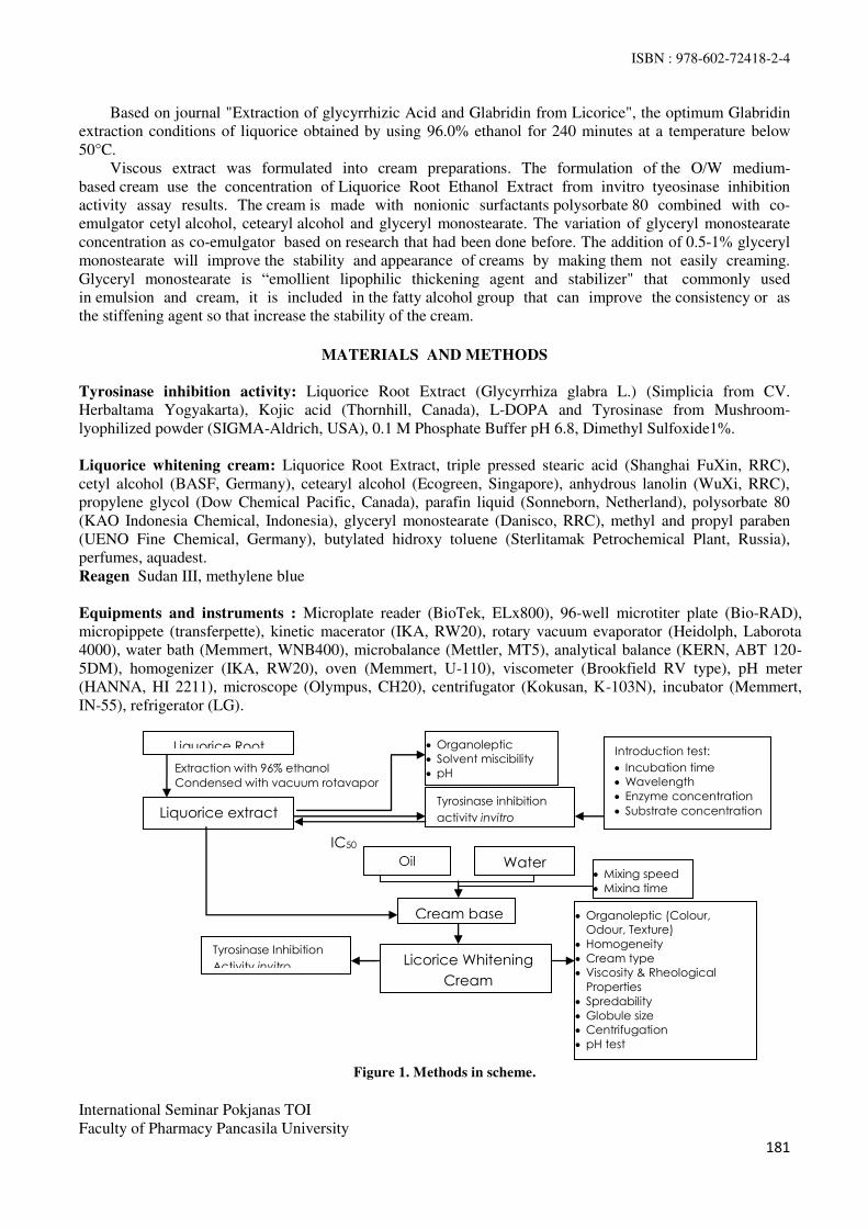
ISBN : 978-602-72418-2-4
International Seminar Pokjanas TOI
Faculty of Pharmacy Pancasila University
181
Based on journal "Extraction of glycyrrhizic Acid and Glabridin from Licorice", the optimum Glabridin
extraction conditions of liquorice obtained by using 96.0% ethanol for 240 minutes at a temperature below
50°C.
Viscous extract was formulated into cream preparations. The formulation of the O/W medium-
based cream use the concentration of Liquorice Root Ethanol Extract from invitro tyeosinase inhibition
activity assay results. The cream is made with nonionic surfactants polysorbate 80 combined with co-
emulgator cetyl alcohol, cetearyl alcohol and glyceryl monostearate. The variation of glyceryl monostearate
concentration as co-emulgator based on research that had been done before. The addition of 0.5-1% glyceryl
monostearate will improve the stability and appearance of creams by making them not easily creaming.
Glyceryl monostearate is “emollient lipophilic thickening agent and stabilizer" that commonly used
in emulsion and cream, it is included in the fatty alcohol group that can improve the consistency or as
the stiffening agent so that increase the stability of the cream.
MATERIALS AND METHODS
Tyrosinase inhibition activity: Liquorice Root Extract (Glycyrrhiza glabra L.) (Simplicia from CV.
Herbaltama Yogyakarta), Kojic acid (Thornhill, Canada), L-DOPA and Tyrosinase from Mushroom-
lyophilized powder (SIGMA-Aldrich, USA), 0.1 M Phosphate Buffer pH 6.8, Dimethyl Sulfoxide1%.
Liquorice whitening cream: Liquorice Root Extract, triple pressed stearic acid (Shanghai FuXin, RRC),
cetyl alcohol (BASF, Germany), cetearyl alcohol (Ecogreen, Singapore), anhydrous lanolin (WuXi, RRC),
propylene glycol (Dow Chemical Pacific, Canada), parafin liquid (Sonneborn, Netherland), polysorbate 80
(KAO Indonesia Chemical, Indonesia), glyceryl monostearate (Danisco, RRC), methyl and propyl paraben
(UENO Fine Chemical, Germany), butylated hidroxy toluene (Sterlitamak Petrochemical Plant, Russia),
perfumes, aquadest.
Reagen Sudan III, methylene blue
Equipments and instruments : Microplate reader (BioTek, ELx800), 96-well microtiter plate (Bio-RAD),
micropippete (transferpette), kinetic macerator (IKA, RW20), rotary vacuum evaporator (Heidolph, Laborota
4000), water bath (Memmert, WNB400), microbalance (Mettler, MT5), analytical balance (KERN, ABT 120-
5DM), homogenizer (IKA, RW20), oven (Memmert, U-110), viscometer (Brookfield RV type), pH meter
(HANNA, HI 2211), microscope (Olympus, CH20), centrifugator (Kokusan, K-103N), incubator (Memmert,
IN-55), refrigerator (LG).
Figure 1. Methods in scheme.
Cream base
Mixing speed
Mixing time
Licorice Whitening
Cream
Oil Water
Organoleptic (Colour,
Odour, Texture)
Homogeneity
Cream type
Viscosity & Rheological
Properties
Spredability
Globule size
Centrifugation
pH test
Tyrosinase Inhibition
Activity invitro
Organoleptic
Solvent miscibility
pH
Introduction test:
Incubation time
Wavelength
Enzyme concentration
Substrate concentration Tyrosinase inhibition
activity invitro
IC50
Extraction with 96% ethanol
Condensed with vacuum rotavapor
Liquorice Root
Liquorice extract

ISBN : 978-602-72418-2-4
International Seminar Pokjanas TOI
Faculty of Pharmacy Pancasila University
182
Simplicia manufacture and extraction Part of the root of the liquorice plant is harvested in the form
of cylindrical rods with a length of up to 1 m and diameter in 0.5 cm to 3 cm, yellowish brown to dark
brown, and wrinkle. All the cleaned simplicia then dried by sunlight indirectly for 1-3
days. Then the simplicia mashed using a blender and sifted with no. 4/18 mesh. 500 g
of liquorice powder were macerated kinetically using 5 L of ethanol (96%) for 4 hours. In 10 repeated
process, each process using 4 L of solvents. Then the collected filtrate was evaporated in the rotary vacuum
evaporator to obtain Liquorice Root Ethanol Extract at a temperature of ± 40°C, 180 mmHg pressure, and
speed of 60 rpm up to gained a thick extract of ethanol. It was packed in a seal opaque container and stored in
the refrigerator.
Inhibition of tyrosinase activity invitro This assay was performed using methods as described earlier with
modification (Zhang, (2011); Batubara, et.al., (2010) ; Juwita, N.K. (2011). Extract were dissolved in DMSO
to a final concentration of 20 mg mL-1. This extract stock solution was then diluted to 25-4000µg mL-1 in
100 mM phosphate buffer (pH 6.8). Kojic acid, whick was used as positive control was also tested at
concentrations 2,5-50 µg mL-1. In a 96-well plate 80 μL phospate buffer (0,1 M, pH 6.8) was combined with 40 μL L-DOPA (5mM phospate buffer) in triplicate, 40 μL of each sample dilution. After 5 minutes, add 40 μL tyrosinase ( 310 Units mL-1 in phospate buffer) to each well. Incubation commenced for 20 minutes at
37°C. Optical densities of the wells were then determined at 490 nm. Each samples done with blank samples
which tyrosinase solution not added. The tyrosinase inhibition activity was calculated according to the
following formula:
B : control absorbance – control blank absorbance (B1-B0)
S : sample absorbance – sample blank absorbance (S1-S0)
Inhibitory activity of the sample can be determined by calculating the IC50, namely the concentration in
which the sample inhibit tyrosinase activity by 50% by using the linear regression equation. Concentration of
the sample (in logarithms) as the x-axis and the percent inhibition (% inhibition) as the y-axis.
Liquorice cream formulation Cream made by mixing the oil phase (Stearic acid, Cetyl alcohol, Cetearyl
Alcohol, Lanolyn anhydrous, Paraffin Liquid, Glyceryl monostearate, BHT) to the aqueous phase (Aquadest
70°C added Propylene Glycol, Polysorbate 80, Methyl paraben, Propyl paraben) which has been heated at a
temperature of 70°-75°C. Liquorice extract certain concentration test results inhibitory activity mixed into a
cream base in warm temperature (50°C).
Table.1 Formula of cream.
Ingridients FORMULA (%) b/v
Blank I Blank II Blank III I II III
Liquoorice Root Ethanol Extract - - - 1.01 1.01 1.01
Sodium metabisulfite (to extract) - - - 0,1 0,1 0,1
Stearic acid 2,0 2,0 2,0 2,0 2,0 2,0
Cetyl alcohol 3,0 3,0 3,0 3,0 3,0 3,0
Cetearyl alcohol 6,0 6,0 6,0 6,0 6,0 6,0
Lanolin anhydrous 4,0 4,0 4,0 4,0 4,0 4,0
Paraffin liquid 10,0 10,0 10,0 10,0 10,0 10,0
Glyceryl monostearate 0,1 0,5 0,9 0,1 0,5 0,9
Polysorbate 80 6,0 6,0 6,0 6,0 6,0 6,0
Propylene glycol 15,0 15,0 15,0 15,0 15,0 15,0
Methyl paraben 0,15 0,15 0,15 0,15 0,15 0,15
Propyl paraben 0,05 0,05 0,05 0,05 0,05 0,05
Butyl hydroxy toluene 0,05 0,05 0,05 0,05 0,05 0,05
Perfumes qs qs qs qs qs qs
Aquadest ad 100 100 100 100 100 100

ISBN : 978-602-72418-2-4
International Seminar Pokjanas TOI
Faculty of Pharmacy Pancasila University
183
Physical quality parameter Evaluation of the physical quality of cream were done, include: organoleptic
(colour, odour, and texture); homogeneity by using a glass object; cream emulsion type with microscopic
methods; viscosity and rheological properties by using a Brookfield viscometer type RV; spreadability by
using a teflon ring; globule size is measured using an optical microscope; centrifugation for 5 hours at a speed
of 3800 rpm; cream pH using pHmeter.
Liquorice cream Inhibition of tyrosinase activity invitro Liquorice cream inhibition of Tyrosinase Activity
were done with a microplate reader. This assay was performed using methods as described earlier after the
cream extracted with a solvent and centrifuged for 15 minutes.
Statistical analysis Data of physical parameter assay were expressed with ANOVA one ways without
replication, such as viscocity, spreadability, globule size and pH with p = 0.05 significance level
RESULTS AND DISCUSSION
Liquorice Extract and Kojic Acid Tyrosinase Inhibition Activity Assay
Figure 2. Liquorice extract tyrosinase inhibition activity assay results.
Figure 3. Kojic acid tyrosinase inhibition activity assay results.
Inhibition of tyrosinase enzyme activity in vitro carried out on 96% ethanol extract of the Liquorice root
of and kojic acid as a positive control using enzyme concentration of 310 U / mL, pH 6.8, incubation time of
20 minutes, 5 mM substrate concentration and temperature of 37ºC in accordance with preliminary test done.
Kojic acid chosen as a positive control because of kojic acid is one of the active substances that are commonly
used in skin whitening preparations with a mechanism of tyrosinase inhibitory activity as non-competitive in
the oxidation of L-DOPA into the pigment melanin. IC50 value of kojic acid obtained in this study was 20.88
mgmL-1. When compared with the positive control kojic acid, inhibitory activity shown by the liquorice root
ethanolic extract lower at 126.76 mgmL-1
. In the previous study revealed that Glabridin activity 15 times more
potent than kojic acid, but in this study the activity of the liquorice root ethanolic extract showed a lower
activity. It caused by the crude extract of liquorice ethanol extract, so there was still many other component
which disturb the inhibition activity assay.
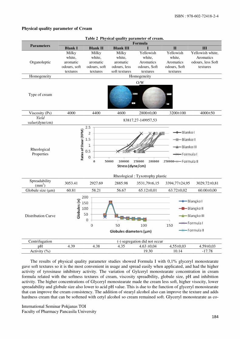
ISBN : 978-602-72418-2-4
International Seminar Pokjanas TOI
Faculty of Pharmacy Pancasila University
184
Physical quality parameter of Cream
Table 2 Physical quality parameter of cream.
Parameters Formula
Blank I Blank II Blank III I II III
Organoleptic
Milky
white,
aromatic
odours, soft
textures
Milky
white,
aromatic
odours, soft
textures
Milky
white,
aromatic
odours, less
soft textures
Yellowish
white,
Aromatics
odours, Soft
textures
Yellowish
white,
Aromatics
odours, Soft
textures
Yellowish white,
Aromatics
odours, less Soft
textures
Homogeneity Homogeneity
Type of cream
O/W
Viscosity (Ps) 4000 4400 4600 2800±0,00 3200±100 4000±50
Yield
value(dyne/cm) 83817,27-149957,53
Rheological
Properties
Rheological : Tyxotrophy plastic
Spreadability
(mm2)
3053.41 2927.69 2885.98 3531,79±6,15 3394,77±24,95 3029,72±0,81
Globule size (μm) 60.81 58.21 56.67 65.12±0,01 63.72±0,02 60.00±0,00
Distribution Curve
Centrifugation (-) segregation did not occur
pH 4.39 4.38 4.35 4.63 ±0,04 4,55±0,03 4.59±0,03
Activity (%) 19.30 10.14 -17.78
The results of physical quality parameter studies showed Formula I with 0,1% glyceryl monostearate
gave soft textures so it is the most convenient in usage and spread easily when applicated, and had the higher
activity of tyrosinase inhibitory activity. The variation of Gylceryl monostearate concentration in cream
formula related with the softness textures of cream, viscosity spreadbility, globule size, pH and inhibition
activity. The higher concentrations of Glyceryl monostearate made the cream less soft, higher visocity, lower
spreadability and globule size also lower to acid pH value. This is due to the function of glyceryl monostearate
that can improve the cream consistency. The addition of stearyl alcohol also can improve the texture and adds
hardness cream that can be softened with cetyl alcohol so cream remained soft. Glyceryl monostearate as co-
ISBN : 978-602-72418-2-4
International Seminar Pokjanas TOI
Faculty of Pharmacy Pancasila University
185
emulsifier can reduce the size of the oil globules and can reinforce the coating film that is formed thus
increasing the cream consistency. O/W cream emulsion type can be determined by the addition of sudan III,
which gained colorless outer phase and inner phase in the form of red globules, while the addition of
methylene blue obtained blue outer phase and colorless inner phase in the.
The formula viscocity had a lower value than blank formula. This is due to the addition of liquorice
extract which tends to be acidic in formula, the interaction between the extract containing polyphenols and
saponins compounds with a base of cream, causing a decrease viscosity. The ability to spread decreases with
increasing concentration of glyceryl monostearate is used, which is an oil phase of a solid with a large
molecular weight, the greater the molecular weight of a substance will be smaller then spreadability, so that
the higher the concentration, the lower spreadability. Results of the evaluation showed that the increased
concentration of glyceryl monostearate may reduce the ability to spread the cream. Most globules obtained in
the range of 31-41 μm and decreases with increasing diameter. Globule size distribution showed normal
distribution, if the curve did not meet the normal distribution law can lead to distability of an emulsion system
according to Stokes law, which is expected to sedimentation. Globule size can also be influenced by various
factors that occur during the manufacturing process such as stirring or mixing. It can be concluded that the
concentration of glyceryl monostearate used can affect the liquorice cream globule size distribution.
All formulas showed no sedimetation occured after centrifugation for 5 hours at a speed of 3800 rpm, so
it can be said cream can be stable during storage year. Cream to be unstable by observing the separation of the
dispersed phase that occurs as a result of centrifugation. The principle is that if globules meet the same liquid
then tends to form a globule with a larger size. From the evaluation it can be concluded that the concentration
of emulsifier in cream enough to form a monomolecular layer on the surface of the oil globules of water so as
to prevent coalescence.
Liquorice extract containing polyphenols and saponins compounds that big amount of OH- causing the
pH of the dosage decreased to acid. The results of the pH approached liquorice extract and also get into the
skin's normal pH range is 4.5-6.5. With this pH range, we expected to remain stable and do not irritate the skin
because too acidic or alkaline dossage forms can irritate the skin or damage the acid mantle skin can cause
skin unprotected against invading microorganisms.
However, when compared, % inhibition decreased with the increasing glyceryl monostearate
concentrations used in cream. More concentrations of glyceryl monostearate used, then the closer film formed
and the closer matrix were available, made it more difficult to extarct the compound dispersed in there. High
consistency of cream also produced a complex matrix that currently extracted by the solvent and put in a plate
in a certain amount to make the conditions in the plate becomes complex and made the inhibition reaction
more difficult for the compound. Based on previous research, liquorice extract contained glycosides, saponins,
flavonoids and tannins that had a different stability. The total content of these compounds, especially total
polyphenols and total flavonoids affect the activity of the extract in inhibiting the enzyme tyrosinase. Cream
formulation using crude extract led to many constituents who might be able to disrupt the activities. Inhibitory
activity of formula III result was negative, this is due to the amount of substrate and enzyme were less in favor
of the course of the enzymatic reaction. In addition, the compounds Glabridin identified as a whitening agent
is a flavonoid. Flavonoids in the plant as a sugar bound to aglycone glycosides and flavonoids that may be in
some form of glycosides combination. In order that, the flavonoids are usually better examine the aglycone
contained in extracts of plants through the process of hydrolysis before to be its glycon and aglycon. In
general, groups of a flavonoid aglycone more active than as a single form of flavonoids.
CONCLUSSION
Liquorice (Glycyrrhiza glabra L.) ethanol extract which contains glabridin is potential to be whitening agent
through invitro tyrosinase inhibition activity assay with IC50 value of 126, 75 ppm. Liquorice Ethanol extract
1.01% can be formulated into creams which pass physical quality test and pH as well as effective in inhibiting
tyrosinase enzyme. Formula with 0.1% gliceryl monostearate concentration (Formula I) as the best formula.
ACKNOWLEDGEMENT
Special thanks are devote for Directorate General of Higher Education of Indonesia which has given grants in
this research.
ISBN : 978-602-72418-2-4
International Seminar Pokjanas TOI
Faculty of Pharmacy Pancasila University
186
REFERENCES
1. Aulton”s Pharmaceutics; The Design and Manufacture of Medicines; 3thed; Edinburgh; Churchill
Livingstone Elsevier. 2007; p. 42-58; 70-97; 384-405.
2. Lloyd HW, Jenna N, Kammer BA. Treatment of Hyperpigmentation. Semin Cutan Med.
Surg.2011;30:171-175.
3. Fais A, Corda M, Era B, et al. Tyrosinase Inhibitor Activity of Coumarin-Resveratrol Hybrids.
Molecules.2009;14:2514-2520.
4. Parvez S, Kang M, Chung HS, et al. Survey and Mechanism of Skin Depigmenting and Lightening
Agent. Phytother. Res.2006;20(11):921-934.
5. Draelos ZD, Thaman LA. Cosmetic Formulation of Skin Care Products. London: Taylor&Francis;
2006. p.203, 206, 214.
6. Smith N, Vicanova J, Pavel S. The hunt for natural skin whitening agents. Int. J. Mol. Sci.
2009;10:2440-2475
7. Vinayak BR, Mary SR. Natural ingredients for creating food textured cosmetics. USA: Cosmetic
Science Technology Journal. 2007. p.33.
8. Qiushi C. Evaluate the effectiveness of the natural cosmetic compared to chemical-based product.
International Journal of Chemistry.2009;1:57-59.
9. Yokota T, Nishio H, Kubota, et al. The inhibitory effect of Glabridin from Licorice Extracts on
Melanogenesis. Pigment Cell Res. 1998; 11:955-961.
10. Ramsden CA, Patrick AR. Mechanistic Studies of Tyrosinase Inhibitors in Cultures of Pityrosporum.
The Journal of Investigative Dermatology. 2010;71:205-208.
11. Jennifer C, Stephie CM. A review on skin whitening property of plant extracts. Bangalore. Int J Pharm
Bio Sci. 2012; 3(4):332 – 347.
12. Schrader K, Domsch A. Cosmetology Theory and practice Vol. III. Germany: Cassler Druck and
Medien;2005.p.17,28,32,44-53.
13. Ansels HC. Pharmaceutical Dosage Forms and Drug Delivery Systems 8th, Philadelphia; Lippincott
Williams & Wilkins; 2005.p.276-297.
14. Wilkinson JB, Moore RJ. Harry’s Cosmeticology. 8th ed. London: George Godwin;2005.p. 226, 707-
726.
15. Batubara I, Darusman LK, Mitsunaga T, et al. Potency of Indonesian medicinal plants as tyrosinase
inhibitor and antioxidant agent. J. Bio. Sci., 2010;10(2): 138-144.

ISBN : 978-602-72418-2-4
International Seminar Pokjanas TOI
Faculty of Pharmacy Pancasila University
187
Accelerated Stability Test of Liquorice (Glycyrrhiza glabra L.) Extract Cream
FARIDAH, SITI UMRAH NOOR, SULIH PROBO SINDI
Faculty of Pharmacy, Pancasila University.
Abstract : The accelerated stability test of Liquorice extract as a whitening agent has been done. The test
was conducted for three months at a room temperature and at a temperature of 40°C with 75% RH in
climatic chamber for 2 formulas with different concentrations of the co-emulsifier glyceryl monostearate
0.1% and 0.5%. Cream was evaluated include organoleptic test, homogenity, cream type, viscosity and flow
properties, the ability to spread, the size of the globules, centrifugation, and the pH test. The results was
obtained by these two formulas have cream-colored, flavored green tea, soft textured, homogeneous, O/W
type, thixotropy-pseudoplastis,no undergo separation after centrifugation test (3800 rpm for 5 hours), as well
as pH which decreased at both room temperature storage at pH formula I (4,56 to 4,30) and formula II (4,54
to 4,33) and at 40˚C / 75% RH storage both of formula I (4,49 to 4,31) and formula II (4,49 to 4,31). The spreadability cream at room temperature storage was reduced in formula I (4416,7 mm
2-2237,3 mm
2) and the
formula II (2734 mm2-1666,9 mm
2). In contrast, at 40˚C / 75% RH storage was increased in formula I
(4217,6 mm2-5360,9 mm
2) and formula II (2886,2 mm
2-4287,8 mm
2). Viscosity was measured by the speed
of 0.5 rpm at room temperature storage has increased in formula I (172000 cPs to 228667 cPs) and formula II
(195333 to-269333 cPs), but at 40˚C / 75% RH storage, the viscosity has decreased in formula I (172667 cPs to131333 cPs) and formula II (185333 cPs-158 000 cPs) .The globule sizes of cream at room temperature
storage was decreased in formula I (62.26 µm -55.64 µm) and formula II (56,66 µm to 47.81 µm). While at
40˚C / 75% RH storage, was increased in formula I (63,75 µm to 69,03 µm) and formula II (61,55 µm -64,90
µm). It can be concluded that both liquorice cream formula with various glyceryl monostearate concentration
was stable for 1 year storage at room temperature.
Key words : accelerated stability test, liquorice, Glycyrrhiza glabra l.
INTRODUCTION
Stability is defined as the ability of a product to keep the specification limits set throughout the period of
storage and use to ensure the identity, strength, quality and purity of the product and keep the characteristics
product that has been made. There are five types of stability that is generally known are the stability of the
chemical, physical, microbiological, therapeutic and toxicology(1)
.
Cream stability test is designed to assess the stability characteristics, appropriate storage conditions
and the expired date(1)
. the cream that has been made must be qualified in general criteria, which is
physically and chemically stable. as well as effective and safe when it used. Stability cream is a period in
which the cream is stored in a certain period of time will have a constant level, no changes in shape, color,
and other changes that can be determined by physical or chemical(2)
.
To obtain the value of the stability of the cream in a short time, can be carried out by accelerated
stability test. This test is intended to obtain the desired information in the shortest possible time by keep the
sample in designed conditions to accelerate the changes that normally occur in normal conditions. If the test
results cream in an accelerated test for 3 months obtained stable results, it shows that the cream is stable at
room temperature storage for one year(3)
. Based on the recommendations of WHO documents, for products

ISBN : 978-602-72418-2-4
International Seminar Pokjanas TOI
Faculty of Pharmacy Pancasila University
188
that are marketed globally tested under conditions of climatic zone IV (hot and humid), in which Indonesia is
zone IV claim. According to ICH and FDA, a country located in the climatic zones III and IV with the
temperature and humidity, is 400 C ± 2
0C and 75% ± 5% RH.
In nature, there are various plantswhich has efficacy as bleaching or whitening agent, such as yam bean,
chamomile and liquorice. In the previous studies, the efficacy of liquorice (Glycyrrhiza glabra L.) as an
inhibitor of the enzyme tyrosinase has been proved and formulated into a cream dosage forms with varying
concentrations of glyceryl monostearate as a co-emulsifier and stiffening agent(4.5)
.
Liquorice extract cream should be required the criteria of stability, so that the formulation is designed
to consider some related issues, among physico chemical properties of substances forming such as glabridin
which contained in liquorice extracts. . Glabridin is stable at storage temperatures of 2-10˚C in a dry place
and protected from light(6)
.
The selected cream base in the formula is a medium cream. This base has a pH of 4.40 to 4.50 so that
it has a similarity with the active substance Glabridin, is 4.54. Using the non ionic surfactant as emulgator,
such as polysorbat 80 and some co-emulgator as a stiffening agent such as cetyl alcohol, stearyl alcohol,
monostearic glyceryl that will enhance the stability of cream.
MATERIALS AND METHODS
Ingredients
Ethanol extract of liquorice (Glycyrrhiza glabra L.), stearic acid (Wilfarin, China), stearyl alcohol, lanolin
anhydrous (Wuxi, China), propylene glycol (Dow Chemical Co., Japan), paraffin Liquidum (Sonneborn,
Japan), cetyl alcohol , glyceryl monostearate, polysorbate 80, methyl paraben, propyl paraben, BHT, distilled
aqua and green tea perfume.
Tools
Cup vaporizer, analytical balance, spatula, stir bar, mortar, pestle, stirrer, water bath, viscometer, glass
objects, glass cover, pH meter, optical microscope, spreadebility tester, centrifuges and climatic chamber
(Memmert).
Procedure
1. Extraction Process
Liquorice powder macerated with ethanol 96% until perfectly extracted. The filtrate was evaporated with
a rotary evaporator to become thick extract.
2. Identification of Flavonoids
Extract was boiled with hot water for 5 minutes, then filtered (solution A), to 5 ml of filtrate was added
magnesium powder and 1 ml of concentrated HCl, add 2 ml of amyl alcohol, shaked vigorously and allow
to separate. The presence of the flavonoid was showed by the formation of red, yellow, or orange in the
lining of amyl alcohol.
3. Cream Basic Formulation
The cream consist of liquorice extract 0,2% with the medium base :
Stearyl alcohol, stearic acid, lanolin, squalene, paraffin liquidum,glyceryl monostearic, polysorbat 80,
propylenglycol, methylparaben, propylparaben, BHT, green tea parfume and aquadest.
4. Evaluation of the Cream
a. Organoleptic
Observed the cream appearance is the color and odor visually.
b. Homogenity
The cream is applied on the objects glass then clenched with another object glass, and then observed
the homogenity, look at the surface. It should be smooth and homogen
c. Examination of cream type

ISBN : 978-602-72418-2-4
International Seminar Pokjanas TOI
Faculty of Pharmacy Pancasila University
189
Tests carried out using the color method by mixing the cream base with a few drops of a methylene
blue solution on the glass object, and then observed with a microscope, if the external phase is blue
and the internal phase is transparant drops, so the type of emulsion is oil in water emulsion. On the
addition of sudan III, the external phase is red and the internal phase is transparant drops. So, the
emulsion type is an oil in water.
d. Viscosity and flow properties
Determination of the viscosity is carried out by using a Brookfield viscometer RV with a capacity 1-
8000000 Cps. Cream was placed in a container and the appropriate spindle is inserted to the prescribed
limit, then rotated at a certain speed until the needle viscometer showed a constant scale.
e. Spreadability test
The cream is filled on the 15 mm inner diameter of Teflon ring and spread the cream with spatula
until there is no bubble. Remove the ring carefully, then covered with a glass plate, then press with a
200 grams load, allow for 3 minutes, then measured the diameter of cream surface then calculated by
the following formula :
F = π x r2 (mm
2)
Description: F = Spreadability; π = 3,14; r = radius (mm)
f. Globule size
Analysis of globul size was done by measuring the average diameter of globule using microscope.
Globul diameter on the object glass was measured using a calibrated micrometer. The amount of
calculated globule is about 300 – 500.
g. Mechanical tests (centrifugal test)
Samples was put into a test tube and then inserted into sentrifugator at a speed of 3800 rpm for 5 hours,
then observed the separation of oil and water.
h. pH cream
pH cream measurement was obtained using a calibrated pH meter.
Data analysis
Stability test results data were analyzed by 2-way ANOVA to find out the effect of temperature and self life
to the response.
RESULTS AND DISCUSSION
A. Extract Characteristics
Test Result
Extract Characteristic - A thick , blackish brown color , aromatic odor
and a sweet taste
- Yield = 8,99 %
- DER – Native = 11,13 %
- Flavonoids = +
- pH = 4,54
B. Stability Test Result
Organoleptic Test
Organoleptic observation until the first month either at room temperature or at 40 ° C / 75% RH, do not
change the color, odor and texture cream. Yellowish white to cream color due to the addition of liquorice
extract. The Color changes can occur due to high temperature might accelerate chemical reactions,
because each 10˚C rise could accelerate chemical reactions 2 to 3 times. The cream uses green tea
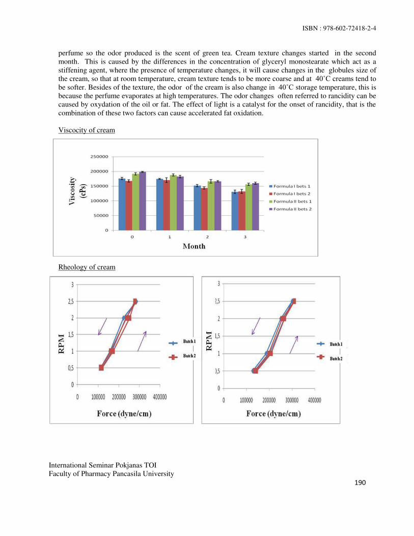
ISBN : 978-602-72418-2-4
International Seminar Pokjanas TOI
Faculty of Pharmacy Pancasila University
190
perfume so the odor produced is the scent of green tea. Cream texture changes started in the second
month. This is caused by the differences in the concentration of glyceryl monostearate which act as a
stiffening agent, where the presence of temperature changes, it will cause changes in the globules size of
the cream, so that at room temperature, cream texture tends to be more coarse and at 40˚C creams tend to be softer. Besides of the texture, the odor of the cream is also change in 40˚C storage temperature, this is
because the perfume evaporates at high temperatures. The odor changes often referred to rancidity can be
caused by oxydation of the oil or fat. The effect of light is a catalyst for the onset of rancidity, that is the
combination of these two factors can cause accelerated fat oxidation.
Viscocity of cream
Rheology of cream
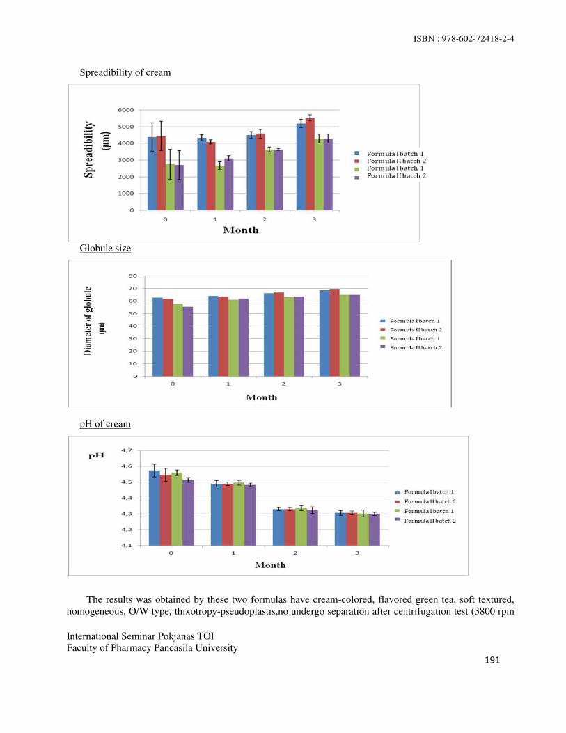
ISBN : 978-602-72418-2-4
International Seminar Pokjanas TOI
Faculty of Pharmacy Pancasila University
191
Spreadibility of cream
Globule size
pH of cream
The results was obtained by these two formulas have cream-colored, flavored green tea, soft textured,
homogeneous, O/W type, thixotropy-pseudoplastis,no undergo separation after centrifugation test (3800 rpm

ISBN : 978-602-72418-2-4
International Seminar Pokjanas TOI
Faculty of Pharmacy Pancasila University
192
for 5 hours), as well as pH which decreased at both room temperature storage at pH formula I (4,56 to 4,30)
and formula II (4,54 to 4,33) and at 40˚C / 75% RH storage both of formula I (4,49 to 4,31) and formula II (4,49 to 4,31). The spreadability cream at room temperature storage was reduced in formula I (4416,7 mm
2-
2237,3 mm2) and the formula II (2734 mm
2-1666,9 mm
2). In contrast, at 40˚C / 75% RH storage was increased in formula I (4217,6 mm
2-5360,9 mm
2) and formula II (2886,2 mm
2-4287,8 mm
2). Viscosity was
measured by the speed of 0.5 rpm at room temperature storage has increased in formula I (172000 cPs to
228667 cPs) and formula II (195333 to-269333 cPs), but at 40˚C / 75% RH storage, the viscosity has decreased in formula I (172667 cPs to131333 cPs) and formula II (185333 cPs-158 000 cPs) .The globule
sizes of cream at room temperature storage was decreased in formula I (62.26 µm -55.64 µm) and formula II
(56,66 µm to 47.81 µm). While at 40˚C / 75% RH storage, was increased in formula I (63,75 µm to 69,03
µm) and formula II (61,55 µm -64,90 µm).
Cream-type examination is performed to determine the suitability of the composition of the proportion
of each phase, the dispersed phase and the dispersing phase associated with comfort during use. Results of
the evaluation of the type of cream made from moon-0 until the 3rd month, either at room temperature or a
temperature of 40 C showed that the cream has type O/W.. Cream Type O/W more consumer demand for
more convenient to use and have better penetration capability compared to type W/O.
CONCLUSION
Both formulations liquorice cream type O/W with the variation of glyceryl monostearate stable in storage at
temperatures of 400C / 75% RH for 3 months or at room temperature (20˚C-25˚C) for a year.
REFERENCES
1. Joshita D. Cosmetic stability. Departemen Farmasi Fakultas Matematika dan Ilmu Pengetahuan Alam
Universitas Indonesia. Depok: 2004
2. United State Pharmacopea. Volume 2. United State: 2009
3. Departemen Kesehatan Republik Indonesia. Farmakope Indonesia. Edisi IV. Jakarta: 1995
4. Jens T. Cartensen, C.T. Rhodes. Drug Stability principles and practices, 3rd ed. New York: 1990
5. Connors K.A, Gordon LA, Valentino JS, Stabilitas kimiawi sediaan farmasi. Edisi II, New York: 1986
6. Tim Padfield. A climate chamber for simulating a temperature and humidity gradient across a wall of
roof. Technical University of Denmark: 2000
7. Mitsui T. New cosmetic science Edisi I. Amsterdam: 1997
8. B. Eckmann. Prediction of emulsion properties from binder/ emulsifier characteristic. Barcelona: 2000
9. Chang TS, An update review of tyrosinase inhibitors. Taiwan: 2009
10. Yokota T, Nishio N, Kubata Y, Mizoguchi M. The inhibitory effect of glabridin from licorice extract on
melanogenesis and inflamatory [abstract]. Pigmen cell research, 2008
11. Raymon CR, Paul JS, Marian EQ. Handbook of pharmaceutical excipients. 7th edition. London: 2009
Powered by TCPDF (www.tcpdf.org)Powered by TCPDF (www.tcpdf.org)Powered by TCPDF (www.tcpdf.org)Powered by TCPDF (www.tcpdf.org)Powered by TCPDF (www.tcpdf.org)Powered by TCPDF (www.tcpdf.org)