Principles of Animal Biology Lancelot Hogben.pdf
433
TEXT FLY WITHIN THE BOOK ONLY
-
Upload
khangminh22 -
Category
Documents
-
view
3 -
download
0
Transcript of Principles of Animal Biology Lancelot Hogben.pdf

TEXT FLY WITHINTHE BOOK ONLY

w > CD
m< OU_1 60527 >m


PRINCIPLES OF
ANIMAL BIOLOGY
Professor Hogben's textbook was originally
published in 1930. He has completely re-
vised and rewritten the later chapters. Mostof the illustrations have been re-drawn byMr. J. F. Horrabin. The unique combination
which was so successful in Science for the
Citizen and Mathematicsfor the Million is there-
fore repeated here; the combination of Pro-
fessor Hogben's extreme care and brilliant
teaching technique in devising diagrams, and
dissections with Mr. Horrabin's beautiful
lettering and line in executing the drawings.
The book has been brought up to date to
cover the latest advances in research. Thenew edition also shows how much one of our
greatest teachers has developed his teaching
technique since the work first appeared. The
experience gained, not only in the lecture-hall
and laboratory but in the colossal teaching
experiments Science for the Citizen and Mathe-
matics for the Million, is applied in this newedition.
The book is for the upper forms of schools
and for universities, polytechnics, and privatestudents. A textbook by an outstanding re-
search worker who is also a brilliant teacher
is rare.
by LANCELOT HOGBEN
MATHEMATICS FOR THE MILLION
Demy Svo Illustrated by J. F. Horrabin 1 2s. 6d. net
1 6th Printing
"One of the indispensable works of popalanzationour generation has produced." H. J. Laskl
SCIENCE FOR THE CITIZENSmall Royal Svo. Illustrated by J. F. Horrabin 1 6s. net
7th Printing
"The civilized world is indebted to him." The
Spectator
DANGEROUS THOUGHTSDemy Svo. 8*. 6d. net
"Of instant importance for the present and future
of Western civilization." Storm Jameson
NATURE AND NURTUREDemy Svo. 6s. 6d. net
GENETIC PRINCIPLES IN MEDICINEAND SOCIAL SERVICE
EDITED AND ARRANGED BY
LANCELOT HOGBEN
THE LOOM OF LANGUAGEA GUIDE TO FOREIGN LANGUAGES FOR
THE HOME STUDENT
by FREDERICK BODMERDemy Svo. i Ss. net
EDITED BY LANCELOT HOGBEN
POLITICAL ARITHMETICRoyal Svo. 30$. net

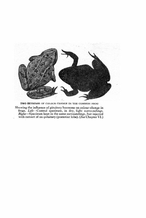
TWO EXTREMES OF COLOUR CHANGE IN THE COMMON FROG
Showing the influence of pituitary hormone on colour-change in
frogs. Left Control specimen, in dry, light surroundings.Right Specimen kept in the same surroundings ,
but injectedwith extract of ox-pituitary (posterior lobe). (See Chapter VI.)

PRINCIPLES OF
ANIMAL BIOLOGY
BY
LANCELOT HOGBEN, F.R.S.
MASON PROFESSOR OF ZOOLOGY IN THE UNIVERSITYOF BIRMINGHAM, FORMERLY RFGIUS PROFESSOR OFNATURAL HISTORY IN THE UNIVERSITY OF ABERDEEN,RESEARCH PROFESSOR OF SOCIAL BIOLOGY IN THEUNIVERSITY OF LONDON, AND PROFESSOR OF
ZOOLOGY IN THE UNIVERSITY OF CAPE TOWN
SECOND EDITION
revised and with new illustrations
throughout by
J. F. HORRABIN
LONDONGEORGE ALLEN & UNWIN LTD
MUSEUM STREET
First published in 1930 by Christophers, Ltdi
COMPLETELY REVISED SECOND EDITION IQ4O
REPRINTED IQ42
REPRINTED IQ45
COMPLETE CONFORMITY WITH THEAUTHORIZED ECONOMY STANDARDS
ALL RIGHTS RESERVED
PRINTED IN GREAT BRITAIN
in n-Point Imprint TypeBY UNWIN BROTHERS LIMITED
WOKING
PREFACE TO SECOND EDITION
PRINCIPLES OF ANIMAL BIOLOGY was pub--* lished first in 1930. It was based on the plan of elemen-
tary instruction adopted in the University of Cape Town
during the author's tenure of the chair of zoology. Its
object was not then, and is not now, to supplant the manyexcellent dissecting manuals available for use in the labora-
tory. It aims at supplementing laboratory work with a
general introduction based on evolutionary principles with
emphasis on function throughout.
While the original plan remains, each chapter has been
extensively revised or, where necessary, entirely rewritten.
The chapter in which the Vertebrate skeleton is used to
illustrate the principles of geological succession by reference
to the fossil record has been recast to take advantage of the
many new discoveries which have been made during the
past ten years. The author is greatly indebted to Dr. Westoll,
Lecturer in Palaeontology in the University of Aberdeen, for
advice and information, especially with reference to the
design of new illustrations. All the illustrations in the
previous edition have been redrawn by Mr. Horrabin, and
many new ones have been added or substituted. To help
students to practise methodical methods of memorizingessential facts new tabular matter has been introduced in
various places.
Dr. H. Waring who saw the new edition through the
press is responsible for many valuable suggestions.
THE UNIVERSITY LANCELOT HOGBENABERDEEN
Nov. 1939

CONTENTS
PART I
The Vertebrate Body as a Going ConcernCHAPTER PAGE
I. LIVING MATTER AND REPRODUCTION 15
II. THE MACHINERY OF RESPONSE 45
III. THE MACHINERY OF CO-ORDINATION 73
IV. THE SOURCES OF ENERGY OF THE ANIMAL BODY 98
V. DIGESTION, RESPIRATION AND EXCRETION 114
VI. THE TRANSPORT OF MATERIALS IN THE ANIMAL
BODY 129
VII. THE DEVELOPMENT OF A NEW ORGANISM 155
VIII. THE MACHINERY OF INHERITANCE 186
PART II
How Animals Differ
IX. THE DIVERSITY OF ANIMAL LIFE 345
X. THE PRINCIPLE OF UNITY OF TYPE 371
XI. UNITY OF TYPE AMONG INVERTEBRATES 291
XII. THE PRINCIPLE OF SUCCESSION WITH SPECIAL
REFERENCE TO THE VERTEBRATE SKELETON 331
XIII. EVOLUTIONARY THEORY TO-DAY 367
APPENDIX I. CLASSIFICATION OF CRUSTACEA 389
APPENDIX II. KEYS TO THE ORDERS OF INSECTS 394
APPENDIX III. SERIAL HOMOLOGY 400
APPENDIX IV. PARTS OF THE BRAIN 403
INDEX 405

ILLUSTRATIONS
Two Extremes of Colour Change in the Common Frog
Frontispiece
FIG. PAGE
1. Set-up of Experiment to show the Dependence of the
Heart-beat on Metallic Ions 16
2. Paramcecium Dividing 23
3. Reproductive and Associated Organs of the Frog 24
4. Fertilization in the Starfish 25
5. Human Egg and Sperm 26
6. Spermatozoa of Different Animals 27
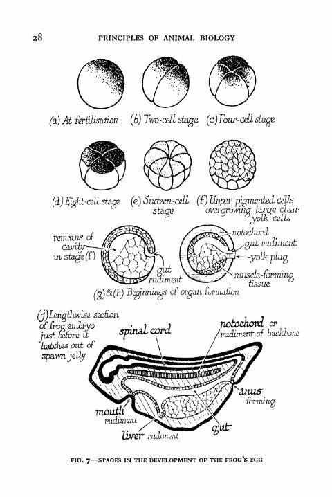
7. Stages in the Development of the Frog's Egg 28
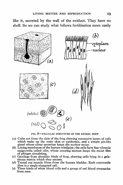
8. Cellular Structure of the Animal Body 29
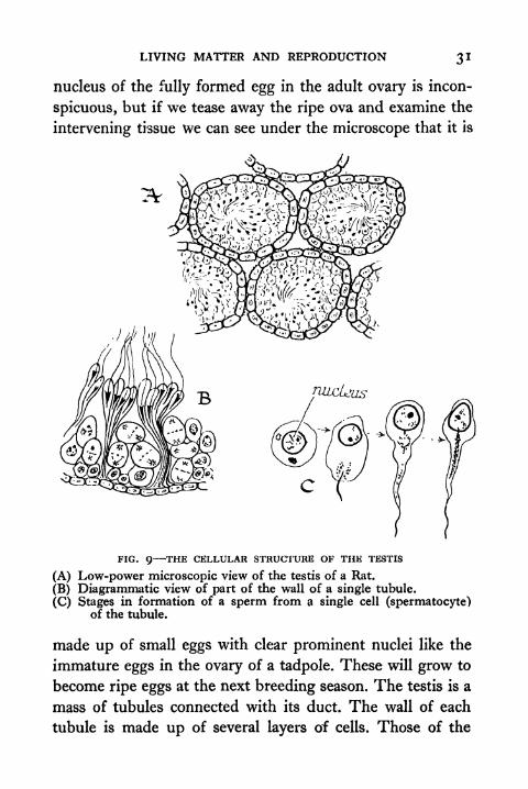
9. The Cellular Structure of the Testis 31
10. Immature Ova of Different Vertebrate Types 33
11. Early Stages in the Segmentation of the Egg of the
Sea Urchin, Sturgeon and Fowl 36
12. Genital Organs of Man with Associated Structures 37
13. Black Pigment Cells (Melanophores) from Web of
Foot of Frog 46
14. Stages in "Expansion" of Melanophores 48
15. Microscopic Structure of Muscle 50
1 6. Relation of Muscle and Skeletal Parts 51
17. The Skeleton of a Frog 53
1 8. Comparison of the Microscopic Appearance of Car-
tilage and Bone 54
19. Muscle Contraction 56
20. Glands 58
21. Ciliated Columnar Epithelium 59
22. Diagram of Ciliary Motion 60
23 . Diagram to show Ciliary Currents in the Gill Chamberof the Mussel 61
24. Gill of the Mussel 62
25. Vertebrate Eye 64
8 PRINCIPLES OF ANIMAL BIOLOGY
FIG. PAGE26. The Vertebrate Eye in situ 65
27. Diagrammatic View of the Eye of Xenopus to show
Regions stimulated by Overhead Illumination in
Different Situations 67
28. The Human Ear 69
29. Diagrammatic Half-section through Head of Frog 70
30. Diagram to show Displacement of Statolith in the
Statocyst of a Shrimp or Squid 7 1
3 1 . Apparatus for determining the Speed of the Nervous
Impulse 76
32. The Speed of the Nervous Impulse 77
33. Motor (efferent) and Sensory (afferent) Nerve Endingsin Striped Muscle 80
34. Diagram to illustrate Miiller's Experiment 81
35. Diagram to illustrate Effect of Degenerative Section
of Nerve Roots 83
36. Diagram of the Reflex Arc 84
37. Effect of Faradic Stimulation of the Roof of the
Mouth in the Cape Chameleon when the SpinalCord is severed 85
38. Brain of Frog 86
39. Diagrammatic Representation of the Central Nervous
System of Man 89
40. Diagrammatic Representation of the Nerve Paths in-
volved in the Control of the Pigmentary Effector
System of the Chameleon 90
41. Diagram of the Human Retina 91
42. Diagram to illustrate Movement of Insect (Hover Fly)towards the Light 93
43 . Alimentary Canal and Associated Structures in the Frog 115
44. The Alimentary Canal, its Associated Structures, andthe Lungs in Man 116
45. Respiratory Organs of Frog and Man 123
46. Diagrammatic Representation of the Kidney of the
Frog 125

ILLUSTRATIONS Q
FIG. PAGE
47. Apparatus for Measuring the Oxygen Consumption of
a Worm or other Small Organism 127
48. Diagram of Vein showing Valves 130
49. Diagram of the Main Paths in the Human Blood
System 132
50. Principal Arteries of the Frog 133
51. Main Veins of Frog and Man 135
52. The Heart of the Frog 136
53. Physical Model to illustrate Resistance of the Capil-
laries to Arterial Pressure 139
54. The Relation of the Heart to the Lungs and Tissues 141
54A. Blood Supply of Small Intestine 143
55. Diagram of Gill Arches and Heart of a Typical Fish,
and Tadpole or Lung Fish 144
56. Blood System of a Fish 145
57. Diagrammatic Representation of the Oxygcnation of
the Blood in Fish and Man 146
58. Embryos of Shark, Bird and Man at the Stage whenthe Gill Clefts are formed 146
59. Arterial Arches of Reptile, Bird and Mammal 147
60. Venous System of Vertebrates 150
61. Microscopic Section of the Thyroid Gland 153
62. Four Embryos of Frog showing Excavitation of
Primitive Gut 156
63. Posterior, Anterior and Dorsal Aspect of the Frog
Embryo at the Stage when the Neural Grooveis closing up 157
64. Transverse and Longitudinal Views of Frog Embryoshortly before hatching 158
65. Early Stages in the Development of the Egg of the
Domestic Fowl 159
66. Formation of the Foetal Membranes of the Chick 160
67. Three Stages in the Development of the Mammal 161
68. Human Foetus in the Uterus about Two Months after
Conception 163
69. Effect of Temperature on the Development of the Frog
Tadpole 165
1O PRINCIPLES OF ANIMAL BIOLOGY
FIG. PAGE
70. One-eyed Tadpole 166
71. Metamorphosis of Axolotl Larva by Treatment with
Thyroid 168
72. Cell Division by Mitosis 176
73. Reduction Division in Male of a Species with Four
similar Chromosomes 178
74. Reduction Division in the Female of a Species with
Four Similar Chromosomes 179
75. Microphotograph of a Dividing Cell showing Four
U-shaped Chromosomes 180
76. Chromosomes of the Stone-fly 181
77. Chromosomes of the Plant Bug Protenor 183
78. Chromosomes of the Plant Bug Euchistus 183
79. Male and Female of Drosophila and their Chromo-somes 183
80. The First Studbook in History 187
81. Mendel's Hypothesis 199
82. Independent Assortment of Two Pairs of Factors 201
83A. Representation of the First Generation of a Cross
between Red-eyed Drosophila and the Purple-
eyed Mutant 202
836. Continuation of Fig. 83A 203
84. Cross between the "Ebony" and "Vestigial" Mutants
of Drosophila 207
85. Continuation of Fig. 84 208
86. Four Different Types of Comb in the Domestic Fowl 209
87. Sex-linked Inheritance in Drosophila 211
88. Continuation of Fig. 87 213
89. Crossing over between two Sex-linked Mutant Factors 218
90. Diagram to explain crossing over between Two Loci
on a Chromosome 219
91. Linkage in a Non-sex-linked Cross 221
92. Crossing over in the Black-vestigial Cross 223
93. Linear Arrangement of the Genes 224
94. Map of Some of the Mutant Genes of Drosophila 226
ILLUSTRATIONS 1 1
FIG. PAGE
95. Normal Frequency Curve illustrated by Variability
in Weight of Beans 23 1
96. Curves to illustrate Maximum Variability in the
Second Generation 233
97. Figure of the Barnacle and Goose in Gerard's
Herball (1594) 249
98. Shells of Radiolaria and Foraminifera 253
99. Types of Micro-organisms 255
100. Vertebrate Wings 267
101. The Duck-billed Platypus 268
102. Anatomy of the Crayfish, etc. 272
103. The Cape Scorpion (Uroplectcs insignis) 275
104. The Anatomy of the Cockroach 276
105. The Anatomy of the Cockroach (continued) 277
106. Cyclops and Daphnia, Two Water Fleas, Members of
the Class Crustacea 279
107. Trilobite 281
108. Diagrammatic Representation of an Ideal Arthropod 283
109. The Barnacle 288
no. Diagrammatic Representation of an Ideal Polychsete 294
in. Peripatus capensis 295
112. Chiton 301
113. Generalized View of the Molluscan Visceral Con-stellation 302
1 14. Unity of Type in Mollusca 303
115. Hypothetical Common Ancestor of Cephalopodsand Gastropods and Structure of a ModernGastropod 304
1 1 6. Cephalopod 305
117. Ciliated Larvae of Molluscs Trochophore and
Veliger 3071 1 8. Diagrammatic Representation of an Ideal Echinoderm 311
119. Hydra ;A Fresh-water Polyp 313
120. Diagrammatic Representation of an Ideal Flat worm 319121. Diagrammatic Representation of a Polyzoan and a
Rotifer 323
12 PRINCIPLES OF ANIMAL BIOLOGY
FIG. PAGE122. Amphioxus 326
123. Amphioxus Transverse Section through Gill Region 327
124. Tunicata 329
125. The Remains of a Giant Salamander unearthed in 1726 332
126. The Succession of Vertebrates 333
127. Remains of Archaeoptcryx preserved in Shale from
Solenhoven, Bavaria 335
128. Trilobite 336
129. Tail Vertebrae of the Ganoid Fish (Amia) 338
130. Vertebrae of an Elasmobranch (Acanthias) 339
131. Trunk Vertebra of a Stegocaphalian 340
132. Fins of Selachians 343
133. Relation of Fins to the Pentadactyl Limb 345
134. The Pentadactyl Limb and its Girdle 347
135. Shoulder Girdle of a Monotreme and Typical Mammal 348
136. Limb Skeleton of a Bird 349
137. Digits of Fore Limb of Archaeopteryx, Chick and Fowl 350
138. Cartilaginous Skulls of Dogfish and the Frog Tadpole 352
139. The Chondrial Bones of the Mammalian Skull 353
140. Heads of Two Fossil Fishes from Devonian Rocks
Osteolepis, Dipterus 354
141. The Skull of a Primitive Amphibian (Stegocephalian)or Reptile (Cotylosaur) 355
142. The Compound Lower Jaw of a Reptile 356
143. Comparison of Skulls of Reptiles and Mammal 357
144. Palatal and Upper Jawbones of Stegocephalian and
Therapsid Reptile 358
145. Head of Human Foetus 359
146. Skulls of Rhynchocephalian, Dinosaur, and ModernBird 361
147. Evolution of the Jaw Suspension of Mammals 363
APPENDIX II. Biting Mouthparts 397
Licking Mouthparts, Sucking Mouthparts 398
Piercing Sucking Mouthparts 399
CHART. Habitat, Habit, and Sexual Reproduction
PART I
THE VERTEBRATE BODY AS AGOING CONCERN

CHAPTER I
LIVING MATTER AND REPRODUCTION
"T X 7E can classify the things of which we have any know-V V ledge in two groups as living and non-living matter. A
piece of chalk or a motor bicycle are examples of non-living
matter. A frog or a university professor are examples of what
is called living matter. Biology is the study of things which
belong to the latter class. It is sometimes defined as the
science of Life. This is misleading. Science is not the studyof abstract nouns. The practice of dealing with facts instead
of playing with words is what distinguishes science from
metaphysics.
Biologists do not use the word living in the same sense as
lawyers or metaphysicians. When the heart of a frog has been
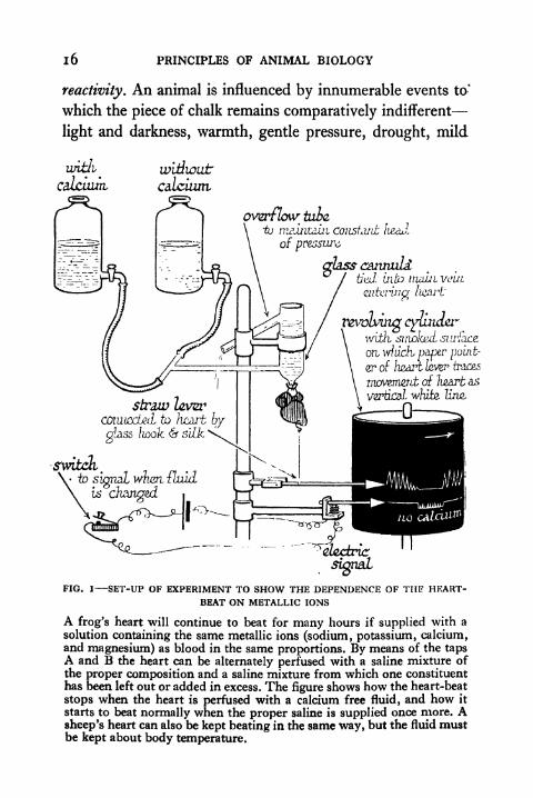
removed, it is legally dead. In the laboratory it will continue
to beat for some weeks, if perfused with a suitable fluid at
the right temperature (Fig. i). Biologists continue to speakof it as a living heart, because it still continues to show the
characteristics of a heart in the living animal. The charac-
teristics which distinguish living matter from non-living
matter are not easy to put in legal language.If we take a piece of chalk as an example of dead matter,
no elaborate experiment is necessary to show that it does not
display any discernible changes in shape or colour or texture
over long periods of time. Unless it is subjected to great heat,
the application of chemical reagents, or mechanical pressure,
it does not increase in bulk or give rise to new pieces of chalk
if left to itself. An animal is different. It is constantly changingin shape, position, colour, texture, etc. In short, it has greater
i6 PRINCIPLES OF ANIMAL BIOLOGY
reactivity. An animal is influenced by innumerable events to*
which the piece of chalk remains comparatively indifferent
light and darkness, warmth, gentle pressure, drought, mild
overflow talcto rmintdhi constxit i
of pressure
afass czawuLdI ti^d mto nuiLti vain
calcium
straw levzrto /icarfc by
glass/Loolc 61 silk "^^
.
tosignal wwn ilmdis
4
changed
r&vlvutg <yunc/erivith, knotted, surface
on \viudi paper point-
er of heart ic-ve?' traces
movemertt of Imrb as
vertical white, line,
FIG. 1 SET-UP OF EXPERIMENT TO SHOW THE DEPENDENCE OF THE HEART-BEAT ON METALLIC IONS
A frog's heart will continue to beat for many hours if supplied with a
solution containing the same metallic ions (sodium, potassium, calcium,and magnesium) as blood in the same proportions. By means of the tapsA and B the heart can be alternately perfused with a saline mixture ofthe proper composition and a saline mixture from which one constituent
has been left out or added in excess. The figure shows how the heart-beat
stops when the heart is perfused with a calcium free fluid, and how it
starts to beat normally when the proper saline is supplied once more. Asheep's heart can also be kept beating in the same way, but the fluid mustbe kept about body temperature.

LIVING MATTER AND REPRODUCTION 17
electrical stimuli. More briefly, it has greater receptivity. Then
again, living animals grow and produce other animals like
themselves. They have the power of reproduction.
i. REACTIVITY
Reactivity is the most characteristic feature of the kind of
living matter which we call animals. Artificial machines
may have great capacity for effecting changes and very
powerful activity which a piece of chalk does not display.
But, in general, animals show a greater variety of response
than the machines which we call inanimate. The difference
is one of degree rather than of kind.
When we study the reactions of an animal we find that they
are confined for the most part to some definite portion of it.
They are localized. Different organs carry out different types
of reaction movement, emission of fluids, light production,
etc. Below is a list of some characteristic reactions of animals
and the organs which carry out these reactions. Such organs
as the muscles and glands which carry out characteristic
changes of behaviour are called collectively Effector organs.
These organs will be studied in detail later, when the terms
will be explained.
TYPE OF RESPONSE EFFECTOR ORGAN
Movement
(a) limb, etc. Striped
(b) gut (peristalsis) 1
uterus (parturition) > Plain ^Musclebladder (micturition) J
(c) heart beat Cardiac
Fluid MotionRoof of frog's mouth Ciliated EpitheliumGills of clam, etc.
1 8 PRINCIPLES OF ANIMAL BIOLOGY
TYPE OF RESPONSE EFFECTOR ORGAN
Secretion
Flow of saliva
Sweating Glands
Venoms, etc.
Colour ChangeChameleon's skin, etc. Pigmentary Effectors
Retinal pigment cells of Man (Chromatophores)
Bioluminescence
Firefly Photogenic OrgansGlow-worm
ii. RECEPTIVITY
When an animal responds in any of the characteristic
ways which have been enumerated above, careful observation
usually reveals that the response is related to some definite
happening in its surroundings, to some physical event in
the external world. The reader perhaps yawns once more as
he reads this. The yawn (if it occurs) is definitely related to
the words he is reading. In similar circumstances he (or she)
would not yawn if the written matter announced a legacy
of $5,000 to himself or herself.
As a more manageable illustration of the dependence of
an animal's behaviour on its immediate surroundings we
may take the power of colour change in the chameleon
or the frog. Like the chameleon the frog exhibits changesof hue, but they are not so rapid. Indeed, they require
periods of several hours rather than minutes to reach com-
pletion, though they are just as striking. Colour response
in the frog is influenced by three principal external influences
Jight, moisture, and temperature. If the field of vision is
occupied by a light-scattering surface ("white background")the animal will be paler than if the field of vision is occupied
LIVING MATTER AND REPRODUCTION 19
by a light-absorbing surface ("black background"). If kept
dry the animal will be paler than if it is kept in water; and
at 20 C. it will be paler than at 5 C., other factors being
constant. So the optimum conditions for pallor are white
background, dryness, and warmth. For darkening of the skin,
black background, moisture, and cold are best.
The chameleon, like the frog, responds to light and
temperature, and it will become completely pale after suit-
able electrical or mechanical stimulation. A toy shocking-coil
serves well for the former purpose. If we apply these stimuli
to different regions of the body, we find that the same stimulus
does not everywhere call forth the same colour changes. If
the skin of the surface of the body, roof of the mouth, and
excretory orifice (cloaca) are respectively stimulated by a
series of electric shocks, or by hard rubbing with a glass rod,
the results obtained may be tabulated thus :
COLOUR CHANGE IN THE CHAMELEON
Stimulus Area ResponseElectrical Skin of surface Local pallor
Roof of mouth Generalized pallor
Cloaca Generalized pallor
Mechanical Skin of surface No effect
Roof of mouth No effect
Cloaca Generalized pallor
Light acts on any portion of the skin of the chameleon,
making dark (not pale) the area on which the light falls.
The direct action of light on the skin of the frog is too small
to detect with the naked eye. The same is true of fishes,
many of which respond in a very striking manner to light
and darkness. Blinded or blindfolded fish do not respondlike fish in which the eye is perfectly intact and accessible
to the light. Fish respond to background only, when the
eyes are intact. This is also true of the frog, but because
2O PRINCIPLES OF ANIMAL BIOLOGY
colour response in the common frog is influenced powerfully
by temperature and moisture, the part played by the eyes
can be better illustrated in one of its near cousins, the South
African clawed toad (Xenopus Icevis) which lives in water
and from this standpoint is much less susceptible to the influ-
ence of temperature. When the skin colour of clawed toads
is compared under different conditions of illumination, the
contrast of normal and eyeless or blindfolded individuals is
indicated in the following table :
COLOUR CHANGE IN XENOPUS L^EVIS
White background Black backgroundNormal animal Very pale Very dark
Eyeless animal Intermediate Intermediate
We see from this that just as different portions of the body,
the effector organs (muscles, glands, the "pigment cells"
which are responsible for colour changes like the above, etc.),
are specially concerned with carrying out responses, so special
parts of the body (receptor organs) are specially affected bysuch happenings in the animal's surroundings as call forth
response of one kind or another.
In contemporary biological nomenclature the term receptor
organ replaces the older one sense organ. It is greatly to be
preferred, because it involves no departure from our initial
resolve to treat the animal as a tangible object in the world
around us. For the same reason the term purpose should
never be used in biology. An organ fulfils a certain role in
the economy of the organism. Whether it discharges a pur-
pose we cannot know by any methods whose validity has been
vindicated so far. Thus the eye is the main receptor organ
through which light influences the behaviour of an animal.
The ear of our bodies is the organ by which sound vibrations
exercise an effect upon us. The ear is a complex organ, and
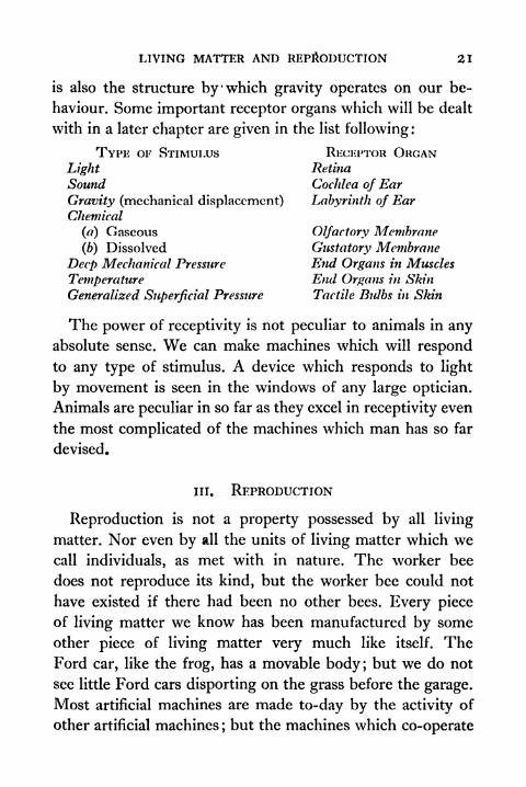
LIVING MATTER AND REPRODUCTION 21
is also the structure by 'which gravity operates on our be-
haviour. Some important receptor organs which will be dealt
with in a later chapter are given in the list following :
TYPE OF STIMULUS RECEPTOR ORGANLight Retina
Sound Cochlea of Ear
Gravity (mechanical displacement) Labyrinth of EarChemical
(a) Gaseous Olfactory Membrane
(b) Dissolved Gustatory Membrane
Deep Mechanical Pressure End Organs in Muscles
Temperature End Organs in Skin
Generalized Superficial Pressure Tactile Bulbs in Skin
The power of receptivity is not peculiar to animals in anyabsolute sense. We can make machines which will respondto any type of stimulus. A device which responds to light
by movement is seen in the windows of any large optician.
Animals are peculiar in so far as they excel in receptivity even
the most complicated of the machines which man has so far
devised.
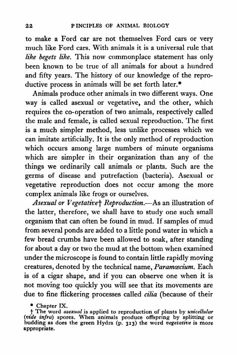
in. REPRODUCTION
Reproduction is not a property possessed by all living
matter. Nor even by all the units of living matter which wecall individuals, as met with in nature. The worker bee
does not reproduce its kind, but the worker bee could not
have existed if there had been no other bees. Every piece
of living matter we know has been manufactured by some
other piece of living matter very much like itself. TheFord car, like the frog, has a movable body; but we do not
see little Ford cars disporting on the grass before the garage.
Most artificial machines are made to-day by the activity of
other artificial machines ;but the machines which co-operate
22 PINCIPLES OF ANIMAL BIOLOGY
to make a Ford car are not themselves Ford cars or very
much like Ford cars. With animals it is a universal rule that
like begets like. This now commonplace statement has only
been known to be true of all animals for about a hundred
and fifty years. The history of our knowledge of the repro-
ductive process in animals will be set forth later.*
Animals produce other animals in two different ways. One
way is called asexual or vegetative, and the other, which
requires the co-operation of two animals, respectively called
the male and female, is called sexual reproduction. The first
is a much simpler method, less unlike processes which we
can imitate artificially. It is the only method of reproduction
which occurs among large numbers of minute organisms
which are simpler in their organization than any of the
things we ordinarily call animals or plants. Such are the
germs of disease and putrefaction (bacteria). Asexual or
vegetative reproduction does not occur among the more
complex animals like frogs or ourselves.
Asexual or Vegetative^ Reproduction. As an illustration of
the latter, therefore, we shall have to study one such small
organism that can often be found in mud. If samples of mudfrom several ponds are added to a little pond water in which a
few bread crumbs have been allowed to soak, after standing
for about a day or two the mud at the bottom when examined
under the microscope is found to contain little rapidly movingcreatures, denoted by the technical name, Paramoecium. Each
is of a cigar shape, and if you can observe one when it is
not moving too quickly you will see that its movements are
due to fine flickering processes called cilia (because of their
*Chapter IX.
f The word asexual is applied to reproduction of plants by unicellular
(vide infra) spores. When animals produce offspring by splitting or
budding as does the green Hydra (p. 313) the word vegetative is moreappropriate.

LIVING MATTER AND REPRODUCTION 23
resemblance to eyelashes the Latin word). These cover the
whole body. Somewhat similar organisms (Opalina, Balanti-
dium, etc.) can always be found living parasitically in the
muddy contents of the hindermost portion of the bowel of
the frog. When such organisms as these have gone on feeding
for a certain time, they become constricted about the middle,
and gradually divide into two, just as a drop of a fluid will
divide into separate drops when it has reached a certain size
FIG. 2 Paramcecium dividing
(Fig. 2). Each half becomes a new individual and swims off
on its own. Thus every Paramcecium arises from another
Paramcecium.
Like begets like is a rule that applies equally to Paramcecium,the common frog, and ourselves. But in all those organismswhich are properly called animals in every-day languageeach sort or species has two kinds of individual, males and
females; and reproduction involves the co-operation of a
male and a female. Both are equally essential to the pro-duction of a new being, though the precise part which the
male plays in the process was not clearly understood till
aboutfifty years ago.

PRINCIPLES OF ANIMAL BIOLOGY
Sexual Reproduction. The female frog is externally differ-
ent from the male. It is somewhat larger and its forelimb has
no horny thumb-pad. At the breeding season, in Spring,
frogs may be found in ponds and streams in pairs. A male
tightly clasps a female about the body with its forelimb s,
his ventral surface in contact with her back. Thus they
inbernaL orifice
of oviduct'fatiy bodies
9 rf
FIG. 3 REPRODUCTIVE AND ASSOCIATED ORGANS OF THE FROG
remain, till the eggs are shed in great numbers embedded
in masses of clear jelly, which may be seen floating on the
surface of the water. Each egg is almost completely spherical,
black in colour towards the pole which floats uppermost, but
lighter beneath. As the eggs are laid the male squirts a viscid
stream, the seminal fluid, over the surface of the egg.
If the seminal fluid is examined with a microscope, as
was first done by a Dutch draper, Leeuwenhoek, in 1668, it
is seen to swarm with myriads of minute moving objects,
called spermatozoa, or more shortly sperms. With a very high
magnification each sperm is seen to be somewhat like a tad-
pole in shape. It has a short thick body with a thin whip-like
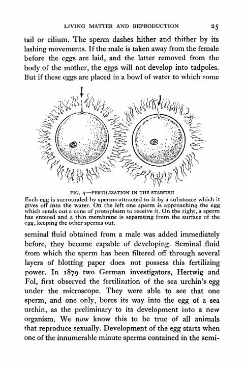
LIVING MATTER AND REPRODUCTION 25
tail or cilium. The sperm dashes hither and thither by its
lashing movements. If the male is taken away from the female
before the eggs are laid, and the latter removed from the
body of the mother, the eggs will not develop into tadpoles.
But if these eggs are placed in a bowl of water to which some
FIG. 4 FERTILIZATION IN THE STARFISH
Each egg is surrounded by sperms attracted to it by a substance which it
gives off into the water. On the left one sperm is approaching the eggwhich sends out a cone of protoplasm to receive it. On the right, a spermhas entered and a thin membrane is separating from the surface of the
egg, keeping the other sperms out.
seminal fluid obtained from a male was added immediately
before, they become capable of developing. Seminal fluid
from which the sperm has been filtered off through several
layers of blotting paper does not possess this fertilizing
power. In 1879 two German investigators, Hertwig and
Fol, first observed the fertilization of the sea urchin's eggunder the microscope. They were able to see that one
sperm, and one only, bores its way into the egg of a sea
urchin, as the preliminary to its development into a new
organism. We now know this to be true of all animals
that reproduce sexually. Development of the egg starts whenone of the innumerable minute sperms contained in the semi-
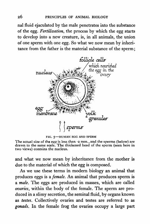
26 PRINCIPLES OF ANIMAL BIOLOGY
nal fluid ejaculated by the male penetrates into the substance
of the egg. Fertilization, the process by which the egg starts
to develop into a new creature, is, in all animals, the union
of one sperm with one egg. So what we now mean by inheri-
tance from the father is the material substance of the sperm ;
wtiudi nourished
tiic oner ui the,
o
granzzZcs"
FIG. 5 HUMAN EGG AND SPERM
The actual size of the egg is less than -2 mm., and the sperms (below) are
drawn to the same scale. The thickened head of the sperm (seen here in
two views) contains the nucleus.
and what we now mean by inheritance from the mother is
due to the material of which the egg is composed.As we use these terms in modern biology an animal that
produces eggs is a. female. An animal that produces sperm is
a male. The eggs are produced in masses, which are called
ovaries, within the body of the female. The sperm are pro-
duced in a slimy secretion, the seminal fluid, by organs known
as testes. Collectively ovaries and testes are referred to as
gonads. In the female frog the ovaries occupy a large part

LIVING MATTER AND REPRODUCTION 27
of the body cavity in the trunk region. They are masses of
eggs in different stages of growth. Two coiled white tubes
(oviducts) on either side convey them to the exterior at the
breeding season. With the excretory orifices the oviducts
discharge into a short tube, the cloaca, between the legs. In
the male there are two yellow or white bodies of ellipsoidal
shape the testes lying over the kidneys and communi-
(
f
FIG. 6 SPERMATOZOA OF DIFFERENT ANIMALS
(a) Bat, (b) Frog, (c) Bird (finch), (d) Sheep, (e) Pig, (/) Jelly fish,(g) Monkey.The Spermatozoa of threadworms (h) and of crabs and lobsters (j)
are unlike those of all other animals.
eating with the exterior by the same duct or passage which
conveys the urine to the common excretory orifice (Fig. 3).
At the breeding season, when the eggs in the ovaries are ripe,
they can be fertilized after removal from the body. If keptin clean water they will not develop, unless we add to it a
drop of seminal fluid, which can be prepared by crushing upthe testes of a male in tap water.
The female frog lays her eggs embedded in masses of a
clear jelly, analogous to the albumen of the fowl's egg, and,
PRINCIPLES OF ANIMAL BIOLOGY
(a) At fertifatfwri (ty Two-cellstage, (c) Four-cell stage
fdj Sgfci-cell sfage (e) Sixteen-coll (f) llppw pjgnumbd defac
5ta/7u overraivi/z' /a/^e cZ&i
at^s^-*:<"-<'/cA&(h) IScQiiu-iinQS of
organfui
notochord or
of
forming
FIG. 7 STAGES IN THE DEVELOPMENT OF THE FROG'S EGG
LIVING MATTER AND REPRODUCTION
like it, secreted by the wall of the oviduct. They have no
shell. So we can study what follows fertilization more easily
''' *'
FIG. 8 CELLULAR STRUCTURE OF THE ANIMAL BODY
() Cube cut from the skin of the frog shewing successive layers of cells
which make up the outer skin or epidermis, and a simple pit-like
gland whose slimy secretion keeps the surface moist.
(b) Lining membrane of the human windpipe ; the cells have fine vibratile
outgrowths called cilia, whose swaying motion keeps the moist filmof phlegm circulating.
(c) Cartilage from shoulder blade of frog, shewing cells lying in a gela-tinous matrix which they secrete.
(d) Teased out muscle fibres from the human bladder. Each contractilefibre is a single elongated cell.
(e) Three kinds of white blood cells and a group of red blood corpusclesfrom man.
30 PRINCIPLES OF ANIMAL BIOLOGY
in the frog than in an animal whose eggs are protected by an
opaque envelope or develop to a late stage in the body of the
mother. Once the sperm has made its way into the egg a
change occurs in its outermost layer. No other sperm can
now penetrate it. Soon after a furrow appears on the surface
of the egg, which divides into two in much the same way as a
Paramcecium divides in asexual reproduction. But the two
halves, which are called cells, do not separate, like two newlyformed Paramcecia, to start separate lives of their own. Theyremain connected and divide again. The process is repeated
again and again. Within twenty-four hours after fertiliza-
tion, the egg can be distinctly seen with a simple lens as a
hollow ball of many such cells. Thenceforth changes take placein the rate of multiplication of the cells in different regions.
From different groups of cells the characteristic structures
or organs of the body begin to take shape. Cells are the
microscopic bricks from which the whole edifice of the
body is built up (Figs. 7 and 8).
The Cellular Structure of the living body. The cells of the
frog embryo are very much alike at first. Each resembles
a very small egg from the ovary. The cells of different parts of
the body of a tadpole or of an adult frog are not all alike. In
different regions they have different shapes and sizes. What is
common to all of them is that they possess a structure called
the nucleus. This essential part of every cell is recognized
partly by the fact that it takes up certain dyes and partly bythe peculiar changes which it undergoes during cell division.
These will be described later (p. 177). The ovaries of a tadpole
are simply masses of more or less spherical cells with large
spherical nuclei. Each of these cells enlarges as it deposits fatty
storage material for the use of the embryo, and eventually
becomes one of the ripe eggs in the ovary of an adult. The
LIVING MATTER AND REPRODUCTION
nucleus of the fully formed egg in the adult ovary is incon-
spicuous, but if we tease away the ripe ova and examine the
intervening tissue we can see under the microscope that it is
FIG. 9 THE CELLULAR STRUCTURE OF THE TESTIS
(A) Low-power microscopic view of the testis of a Rat.
(B) Diagrammatic view of part of the wall of a single tubule.
(C) Stages in formation of a sperm from a single cell (spermatocyte)of the tubule.
made up of small eggs with clear prominent nuclei like the
immature eggs in the ovary of a tadpole. These will grow to
become ripe eggs at the next breeding season. The testis is a
mass of tubules connected with its duct. The wall of each
tubule is made up of several layers of cells. Those of the
33 PRINCIPLES OF ANIMAL BIOLOGY
outermost layer are rather like the immature eggs in the ovary
of a young tadpole. They undergo repeated division to
replace the innermost layers from which the sperms are shed.
Each sperm is formed from a single cell of the innermost
layer. The bulk of the body of the sperm is its nucleus.
The rest of the cell body is drawn out to form the tail.
The parts of the body of an adult animal may be classified
as organs according to the sort of work they do, and as tissues
according to their texture or visible appearance. A single
organ may be mainly made up of one sort of tissue, or it maybe a combination of many. Each tissue has its characteristic
microscopic structure. Like the testis or ovary, the substance
of all the organs of the animal body is also built up of micro-
scopic bricks called cells (Fig. 8). In some tissues like bone
and cartilage the bricks are separated by a good deal of plaster,
or to use the technical term, matrix. Others, such as the
epithelia or lining membranes of all surfaces, internal or
external, including the tubular cavities of the glands, consist
simply of cells packed closely together. Although the cells
of different tissues acquire different shapes and sizes as the
development of the frog's egg proceeds, they all arise bydivision of the undifferentiated cells in the hollow ball stage.
The process of cell division involves the partition of the
nucleus in the same characteristic manner, described under
the term mitosis (vide infra). Its detailed features were not
described till high-power microscopes began to be used.
The distinction between tissues and organs in the preceding
paragraph runs parallel to the way in which the two terms,
homology and analogy, are commonly used by biologists.
Structures are said to be analogous when they do the same
work. In contradistinction to functional similarity or analogy,
homology is structural resemblance. Organs are said to be
LIVING MATTKR AND REPRODUCTION 33
homologous when they arc built up in the same way or have
the same structural relation to adjacent and corresponding
ovary of
aiadpok
"B
>>--*'
nucleus
COVUm from
ovary of a:̂ ^x
voungpart of ovary
of animiaadborc mouse
FIG. 10 IMMATURE OVA OF DIFFERENT VERTEBRATE TYPES
Note that relatively few cells of the mammalian ovary eventually becomeripe ova. These grow at the expense of others which form spherical
capsules, called Graafian follicles, around them.
parts. Homologous structures of the adult may be recognized
as such because they develop from corresponding parts of the
embryo, even if their resemblance is not apparent when
c
34 PRINCIPLES OF ANIMAL BIOLOGY
animals are full grown. The homology of organs may also be
recognized, because of their common structural resemblance
to an intermediate type.
General Characteristics of Sexual Reproduction. Certain
features of sexual reproduction are common to nearly all
animals. Others are characteristic of particular types. A com-
parison between the reproductive processes of the fowl and
the frog draws attention to some of the latter. In birds the
two sexes are recognizable by secondary sexual differences
analogous to those of the frog. For instance, the spurs, neck
hackles, tail sickles, and comb distinguish the male White
Leghorn from the hen. The cock has two testes which look
like those of a frog, and have a similar microscopic structure.
The seminal fluid produced by them contains spermatozoaof the same general appearance and dimensions. It is not
squirted over the eggs as they are laid, but is introduced
into the oviduct of the female, when the excretory orifice of
the male is closely applied to that of the female during the
act of coitus or copulation. The immature eggs in the ovary
of a newly born chick are much like the immature eggs of a
frog, and like them have large, clear, spherical nuclei. At
sexual maturity they grow till they attain the size of what wecall the yolk of an egg. They are then liberated into the
oviduct. After the yolk or ovum sensu stricto has been fer-
tilized, it acquires an albuminous coat comparable to the
mucilaginous jelly around the egg of the frog. In the lower
part of the oviduct other envelopes are secreted around it,
first the shell membrane and then the calcareous shell itself.
After the sperm nucleus has united with the egg nucleus,
which lies in the clear circular area on the side of the yolk
of an infertile fowl's egg, the combined nucleus divides in
the usual way, but only a small part of the fowl's yolk is
LIVING MATTER AND REPRODUCTION 35
partitioned off round each cell. Division (segmentation} of the
ovum is incomplete. The embryo is at first a small plate of
cells lying on the surface of the remaining yolk and growingat its expense (Fig. n). The domestic fowl lays eggs at
definite intervals in high-laying strains like the White Leg-
horn about twenty-six hours. The eggs are deposited whether
they are fertilized or not.
The salient similarities and differences may be summarized
thus:
Frog Fowl
A new individual is produced as the result of the union of
a motile cell (sperm) of microscopic dimensions containedin the seminal fluid of the male, with an immobile cell (ovum).

36 PRINCIPLES OF ANIMAL BIOLOGY
Our table brings out five similarities and five differences.
Of the similarities the first two are based on microscopic
examination and are shared by the overwhelming majority
sturgeon fowl
FIG. Hr-FIRST CLEAVAGE DIVISIONS
(Relative size of Fowl's cleavage furrows and blastoderm exaggerated
owing to space limits, cf. Fig. 65).
of animals. There are very many hermaphrodite species to
which the third does not apply. Animals are said to be
hermaphrodite if testes and ovaries are present in one and
the same individual. Hemaphrodite species include most
sedentary animals (polyps, barnacles, sea mats, sea squirts), a
LIVING MATTER AND REPRODUCTION 37
high proportion of parasitic animals, and many familiar types
such as snails and slugs, earth-worms and leeches, which are
neither sedentary nor completely parasitic. Many clams and
some other animals are consecutively hermaphrodite, i.e.
male at one stage in their lives and female at another. The
male of the common toad has a small ovary called Bidder's
organ attached to each testis. Occasionally ripe eggs develop
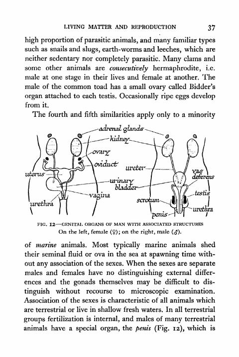
from it.
The fourth and fifth similarities apply only to a minority
uterus'
'penis-
-urethra.
FIG. 12 GENITAL ORGANS OF MAN WITH ASSOCIATED STRUCTURES
On the left, female (?); on the right, male (<J).
of marine animals. Most typically marine animals shed
their seminal fluid or ova in the sea at spawning time with-
out any association of the sexes. When the sexes are separate
males and females have no distinguishing external differ-
ences and the gonads themselves may be difficult to dis-
tinguish without recourse to microscopic examination.
Association of the sexes is characteristic of all animals which
are terrestrial or live in shallow fresh waters. In all terrestrial
groups fertilization is internal, and males of many terrestrial
animals have a special organ, the penis (Fig. 12), which is
38 PRINCIPLES OF ANIMAL BIOLOGY
inserted into the female orifice during coitus. There is one
striking exception to the general rule that fertilization is
always internal among land animals. Oligochaetes (earth-
worms) are hermaphrodite and copulate reciprocally. In
copulation seminal fluid from the male genital orifice of
each worm is introduced into special sacs (spermathecae) of
the other. From the spermathecae it is subsequently extruded
over the eggs which are liberated from the neighbouring
genital orifice.
Evolution of Sex. There are therefore differences of sexual
reproduction associated with the habit and habitat of animals
as the last item of our Table suggests. Such differences are
summarized in the next Table. The animals placed in the
larger (phyla) and smaller (classes) groups referred to in this
Table will be found in the Addendum to this chapter
on p. 43.
The record of life in the rocks tells us that life began in
the sea. The features of sexual reproduction associated with
habitat and habit as displayed in the accompanying Table
therefore suggest that all animals alive at one time repro-
duced as sea urchins, starfishes, and bristle worms reproduce
to-day. The sexes were not externally different and did not
associate. They shed innumerable sperm or innumerable
minute naked eggs into the surrounding water, where fertili-
zation occurred without sexual union of the parents them-
selves. The naked embryos quickly embarked on a free living
aquatic existence, and required no generous supply of food
stored in the egg for their needs. This state of primitive inno-
cence could not last when the exposure of great continental
land shelves began. Females of littoral species could now lay
their eggs in gullies where males might not be available, or on
rocks left dry by the receding tide. So the survival of species
LIVING MATTER AND REPRODUCTION 39
which frequented the water edge imposed two new conditions
which entailed other changes. One was for some loose associa-
tion of the sexes such as exists in the salmon to-day. The other
was for some form of protection against desiccation, such as
the water-absorbing jelly around the egg of the frog. It is
possible that association of the sexes began as the result of
chemical stimulation due to substances extruded at spawningtime. In any case, visible differences would provide an
additional safeguard. A survey of existing animal types shows
that visible differences which have no connexion with the
sex organs (so-called secondary sexual characters) only occur
in animals which associate at spawning time or perform the
more intimate act of coitus.
When the appearance of land floras made it possible for
some littoral species to make their way into estuaries or creep
under banks of seaweed beyond the intertidal zone, the
dangers of exposure to drought were necessarily greater.
The secretion of a calcareous (reptiles, birds, some whelks),
horny (many molluscs), chitinous (insects), or gelatinous
(snail) envelope around the egg could provide the necessary
protection. The production of a cleidoic or shelled egg would
in general presuppose a parallel development of sexual asso-
ciation to ensure internal fertilization before the egg was
enclosed in its protective layer. An early free-living aquatic
existence as a larval form had now become impossible and
a more protracted embryonic existence was only possible
if there were sufficient reserves of food material. The pro-
duction of a smaller number of more heavily yolked eggswas therefore a necessary condition of the rise of a land
fauna. Since eggs which contain relatively little yolk do
not undergo partial cleavage, incomplete segmentation
appears to be a mechanical necessity of development

40 PRINCIPLES OF ANIMAL BIOLOGY
when the amount of reserve material is relatively large,
Fig. n.
A few animals have carried this process of protecting the
developing embryo against drought or food shortage still
further. Cleavage has already begun in the oviduct of the
fowl. In some reptiles (e.g. the Cape chameleon) and fishes
the embryo completes its development within the oviduct.
With the exception of the duck-billed Platypus and the spiny
anteater Echidna, which lay eggs with chalky shells like those
of a bird or a lizard, all mammals are viviparous, as opposedto oviparous. The embryo is nourished by a connexion with
the blood supply of the mother in the part of the oviduct
called the uterus or womb. Thus it has no need for a large
reserve of food. The ova of mammals are minute and undergo
complete cleavage. The egg of an ostrich weighs several
pounds. The egg of the human female is about half the size of
a full stop as printed on this page. It is liberated into the
oviduct once a month, after sexual maturity has been reached
(about the age of thirteen). The process occurs about half-
way between a monthly renewing of the mucous membrane
of the internal generative passages preceded by a slight
haemorrhage (menstruation).
Parthenogenesis. Broadly speaking, what we mean bysexual reproduction to-day is contained in the first two items
of the Table on page 35 ;and implies nothing more than that.
Sexual reproduction is not absolutely universal amonganimals in this sense. Here and there in the animal kingdomwe come across species which have dispensed with the male
contiibution to the process. Among Ilymenoptera (bees, ants,
wasps, gall flies) and among the freshwater wheel animalculae
(Rotiferd) females are formed from eggs fertilized in the
usual way, and males are produced from eggs which develop
LIVING MATTER AND REPRODUCTION 41
without contact with the seminal fluid. Parthenogenesis or
virgin birth is carried further in a few species. Most gall
flies and Aphids have a regular alternation of parthenogenetic
generations composed entirely of females and bisexual gene-
rations composed of both sexes. A few gall flies (e.g. Cynipsand Rhodites), some stick insects, and a few freshwater fleas
reproduce exclusively by virgin birth. The male of the
species either does not exist in nature or, if it does, occurs
with great rarity.
Thus it is not a physical impossibility for animal species to
dispense with the male, and it is even conceivable that
biologists may help to bring about this sex reform in the
human race. The entry of the sperm is a physical event
associated with material changes in the egg, and these changesare capable of being imitated. One of them is an increase of
permeability to dissolved substances. Physical agents which
change the semi-permeable property of the egg periphery
are capable of initiating the process of cleavage without the
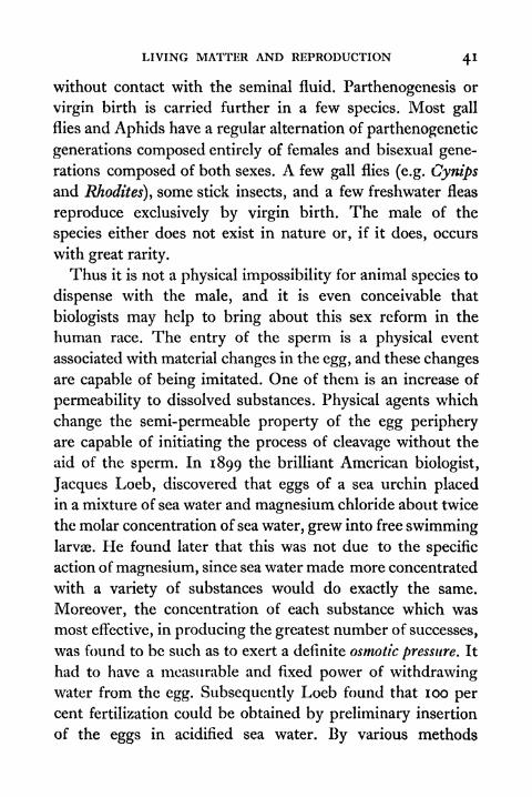
aid of the sperm. In 1899 the brilliant American biologist,
Jacques Loeb, discovered that eggs of a sea urchin placed
in a mixture of sea water and magnesium chloride about twice
the molar concentration of sea water, grew into free swimminglarvae. He found later that this was not due to the specific
action of magnesium, since sea water made more concentrated
with a variety of substances would do exactly the same.
Moreover, the concentration of each substance which was
most effective, in producing the greatest number of successes,
was found to be such as to exert a definite osmotic pressure. It
had to have a measurable and fixed power of withdrawingwater from the egg. Subsequently Loeb found that 100 percent fertilization could be obtained by preliminary insertion
of the eggs in acidified sea water. By various methods
42 PRINCIPLES OF ANIMAL BIOLOGY
artificial parthenogenesis has now been carried out with a
variety of species. The frog's egg will develop without the
aid of the sperm if pricked with a fine glass fibre. Fatherless
tadpoles have actually been raised through metamorphosisto the adult condition.
Immediately after fertilization another important changeoccurs in the egg. All the while it is, in popular language,
"alive," that is all the while it retains its capacity for deve-
loping further, the egg can be shown by an electrical
thermometer, the thermopile, to be giving off heat. It
can also be shown, by chemical analysis, to be steadily
taking up oxygen and giving off carbon dioxide gas. Imme-
diately after the penetration of the sperm or artificial acti-
vation, the heat production and oxygen intake suddenlyincrease, sometimes a thousand-fold in a few minutes. Wecall this activity respiration, and have hitherto regarded it
as inseparably bound up with the other properties we refer
to, when we say the egg-cell is alive. We can now separate it
from other characteristics of which we imply the existence
when we speak of a living cell. Warburg (1912) showed that,
if sea-urchin eggs are suddenly dried by plunging them in
acetone, and subsequently powdered, the moistened powdercan continue to take up oxygen for some time, like the egg-cell. Poisons which act by stopping respiration in the tissues
of an animal, such as cyanides (prussic acid salts), affect it in
just the same way. Warburg also found that the action of
prussic acid was definitely related to the amount of iron in
the egg cell, and that suspensions of charcoal containingtraces of iron will induce fatty substances, sugars, and the
products of substances like egg white to undergo slow com-bustion in air. These iron-containing charcoal suspensionsare also affected by cyanides in the same way as living cells.

LIVING MATTER AND REPRODUCTION 43
That is to say, minute 1
traces of prussic acid salts will bring
their power to facilitate oxidation of organic substances to a
standstill.
More than a century ago the Abbe Spallanzani showed
that when luminous jellyfishes are desiccated and ground to
powder the powder will at once emit light when moistened.
Nobody recognized at the time that a property hitherto associ-
ated with "living matter" had been taken over into the domain
of chemistry and physics by that simple experiment. We are
far from being able to manufacture a system that would be
called by any ordinary person "living," but every year shows
more striking advances in manufacturing systems which are
similar in some respect to what we call living things.
ADDENDUM TO CHAPTER I
When comparisons are made between other animals and types studiedin the earlier chapters of this book, reference may be made to the follow-
ing classification which will be explained more fully in ChaptersXand XI.
1. PORIFERA (sponges)
2. CCELENTERATA
(a) Hydrozoa (polyps)
(6) Scyphozoa (jellyfishes)
(c) Actinozoa (sea anemones and corals)
3. PLATYHELMINTHES (flat-worms, flukes, tape-worm)
4. NEMATHELMINTHES (thread-worms)
5. ROTIFERA (wheel animalcule)
6. ECHINODERMATA (starfishes, sea urchins)
7. MOLLUSCOIDA
(a) Brachiopoda (lamp shells)
(6) Polyzoa (sea mats)
8. ANNELIDA
(a) Polychteta (marine bristle worms)(b) Oligochaeta (earth-worms)(c) Hirudinea (leeches)

44 PRINCIPLES OF ANIMAL BIOLOGY
9. MOLLUSCA
(a) Pelecypoda (oysters, mussels)(b) Gastropoda (limpets, whelks, snails, and slugs)
(c) Cephalopods (cuttlefish, octopus)
10. AKTHROPODA
(a) Crustacea (shrimps, slaters, water fleas, crabs)
(b) Arachnida (kinj* crabs, scorpions, spiders, ticks)
(c) Myriapoda (millipedes, centipedes)
(d) Insects (all winged invertebrates, fleas, lice)
11. VERTEBRATA
(fi) Pisces (fishes)
(b) Amphibia (frogs, newts)(r) Reptilia (lizards, snakes, crocodiles, tortoises)
(d) Aves (birds)
(e) Mammalia (man, whale, bat)
TOPICS FOR HOME STUDY
1 . Make a table to show the association of habit, habitat, and sexual
reproductive phenomena in the following:
Jellyfish; starfish; lamprey; dogfish; whale; toad.
State what conclusions arc suggested thereby. Place them in
their appropriate classes by reference to the scheme on page 43.Discuss the relation between sexual reproduction, habit and
habitat.
2. What characteristics of sexual reproduction are common to
most animals, and how do the characteristics of sexual repro-duction vary in animals which pursue different modes of life ?
3. What is meant by (a) parthenogenesis, (b) hermaphroditism.Give examples of their occurrence.
4. For students who are studying botany. Compare the influence
of habitat on the evolution of sex in animals and plants, (cf.
Bovver's Origin of a Land Flora, and Church's Thallasiophyta).
CHAPTER II
THE MACHINERY OF RESPONSE
T If TE have already enumerated some of the characteristic
V V differences between animals and non-living objects
under the three headings reactivity y receptivity, and repro-
duction. In the preceding chapter we have made a preliminary
study of the last named. Before we can learn more about
the process of development it is necessary to have more
information about the architecture of the living machine.
So we shall now retrace our steps to what has been said
already about the variety of response which animals exhibit
and examine in greater detail the organs responsible for
carrying them out.
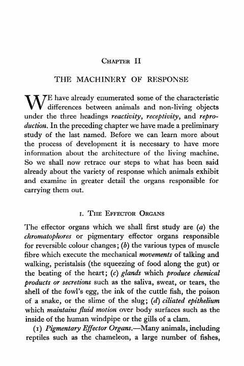
i. THE EFFECTOR ORGANS
The effector organs which we shall first study are (a) the
chromatophores or pigmentary effector organs responsible
for reversible colour changes ; (b) the various types of muscle
fibre which execute the mechanical movements of talking and
walking, peristalsis (the squeezing of food along the gut) or
the beating of the heart; (c) glands which produce chemical
products or secretions such as the saliva, sweat, or tears, the
shell of the fowl's egg, the ink of the cuttle fish, the poison
of a snake, or the slime of the slug; (d) ciliated epithelium
which maintains fluid motion over body surfaces such as the
inside of the human windpipe or the gills of a clam.
(i) Pigmentary Effector Organs. Many animals, including
reptiles such as the chameleon, a large number of fishes,
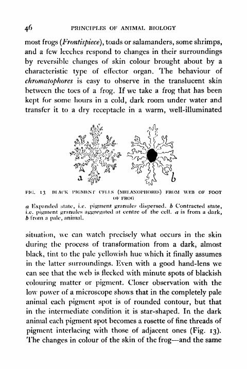
46 PRINCIPLES OF ANIMAL BIOLOGY
most frogs (Frontispiece) ,toads or salamanders, some shrimps,
and a few leeches respond to changes in their surroundings
by reversible changes of skin colour brought about by a
characteristic type of effector organ. The behaviour of
chromatophores is easy to observe in the translucent skin
between the toes of a frog. If we take a frog that has been
kept for some hours in a cold, dark room under water and
transfer it to a dry receptacle in a warm, well-illuminated
& ,*?
FI<;. 13 IH ACK riCJMF.Nr CFI.1.S (MKLANOrtlORKS) FROM \VEB OF FOOTOF FROG
a Kxpanded state, i.e. pigment granules dispersed, b Contracted state,i.e. pigment granules aggregated at centre of the cell, a is from a dark,b from a pale, animal.
situation, we can watch precisely what occurs in the skin
during the process of transformation from a dark, almost
black, tint to the pale yellowish hue which it finally assumes
in the latter surroundings. Even with a good hand-lens wecan see that the web is flecked with minute spots of blackish
colouring matter or pigment. Closer observation with the
low power of a microscope shows that in the completely pale
animal each pigment spot is of rounded contour, but that
in the intermediate condition it is star-shaped. In the dark
animal each pigment spot becomes a rosette of fine threads of
pigment interlacing with those of adjacent ones (Fig. 13).
The changes in colour of the skin of the frog and the same
THE MACHINERY OF RESPONSE 47
is true of the skin of the chameleon are due to the way in
which the pigment is distributed in the skin. Each pigment
spot in the pale animal is the centre of a profusely branchingcell with a nucleus of its own. When the skin is dark, the
pigment is found to be distributed through the whole length
of the ramifying processes of the pigment cell, 01% to use its
technical name, melanophore. As the animal becomes paler,
the pigment is gradually withdrawn along the cell processes
until it is finally concentrated in a single compact speck at
the cell centre. As was first shown by Milne-Edwardes (1848),
the melanophores are therefore the effector organs of colour
response. Colour response in the frog, in the chameleon,
and in fishes depends on the presence in the skin of effector
organs which are single branching cells. These cells possess
the peculiarity of containing a mobile colouring matter or
pigment that migrates under appropriate stimulation along
their cell processes.
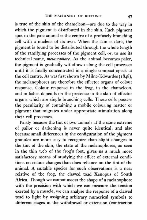
Partly because the tint of two animals at the same extreme
of pallor or darkening is never quite identical, and also
because small differences in the configuration of the pigment
granules are more easy to recognize than slight changes in
the tint of the skin, the state of the melanophores, as seen
in the thin web of the frog's foot, gives us a much more
satisfactory means of studying the effect of external condi-
tions on colour changes than does reliance on the tint of the
animal. A suitable species for such observations is a near
relative of the frog, the clawed toad Xenopus of South
Africa. Though we cannot assess the shape of a melanophorewith the precision with which we can measure the tension
exerted by a muscle, we can analyse the response of a clawed
toad to light by assigning arbitrary numerical symbols to
different stages in the withdrawal or extension (contraction
48 PRINCIPLES OF ANIMAL BIOLOGY
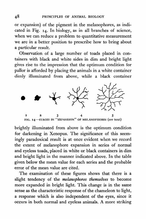
or expansion) of the pigment in the melanophores, as indi-
cated in Fig. 14. In biology, as in all branches of science,
when we can reduce a problem to quantitative measurement
we are in a better position to prescribe how to bring about
a particular result.
Observation of a large number of toads placed in con-
tainers with black and white sides in dim and bright light
gives rise to the impression that the optimum condition for
pallor is afforded by placing the animals in a white container
dimly illuminated from above, while a black container
123 4 5
FIG. 14 STAGES IN*
'EXPANSION*' OF MELANOPHORES (see text)
brightly illuminated from above is the optimum condition
for darkening in Xenopus. The significance of this seem-
ingly paradoxical result is at once evident when we record
the extent of melanophore expansion in series of normal
and eyeless toads, placed in white or black containers in dim
and bright light in the manner indicated above. In the table
given below the mean value for each series and the probableerror of the mean value are cited.
The examination of these figures shows that there is a
slight tendency of the melanophores themselves to become
more expanded in bright light. This change is in the same
sense as the characteristic response of the chameleon to light,
a response which is also independent of the eyes, since it
occurs in both normal and eyeless animals. A more striking
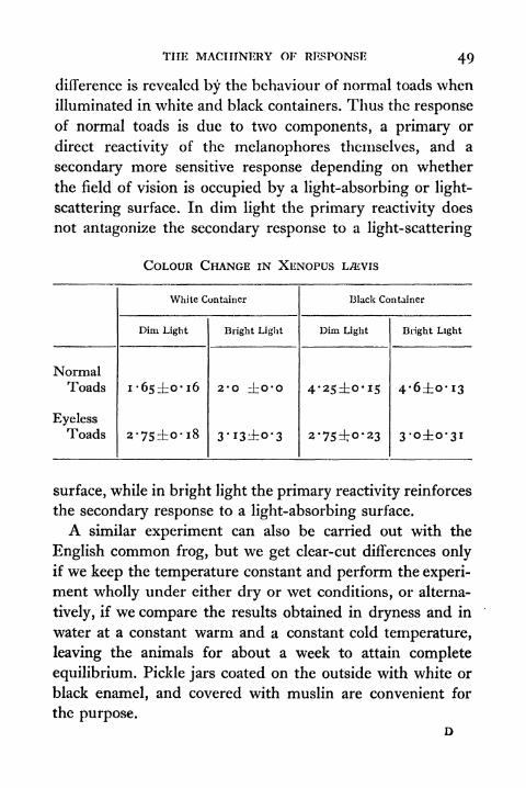
THE MACHINERY OF RESPONSE 49
difference is revealed by the behaviour of normal toads when
illuminated in white and black containers. Thus the response
of normal toads is due to two components, a primary or
direct reactivity of the melanophores themselves, and a
secondary more sensitive response depending on whether
the field of vision is occupied by a light-absorbing or light-
scattering surface. In dim light the primary reactivity does
not antagonize the secondary response to a light-scattering
COLOUR CHANGE IN XENOPUS
surface, while in bright light the primary reactivity reinforces
the secondary response to a light-absorbing surface.
A similar experiment can also be carried out with the
English common frog, but we get clear-cut differences onlyif we keep the temperature constant and perform the experi-
ment wholly under either dry or wet conditions, or alterna-
tively, if we compare the results obtained in dryness and in
water at a constant warm and a constant cold temperature,
leaving the animals for about a week to attain complete
equilibrium. Pickle jars coated on the outside with white or
black enamel, and covered with muslin are convenient for
the purpose.D
50 PRINCIPLES OF ANIMAL BIOLOGY
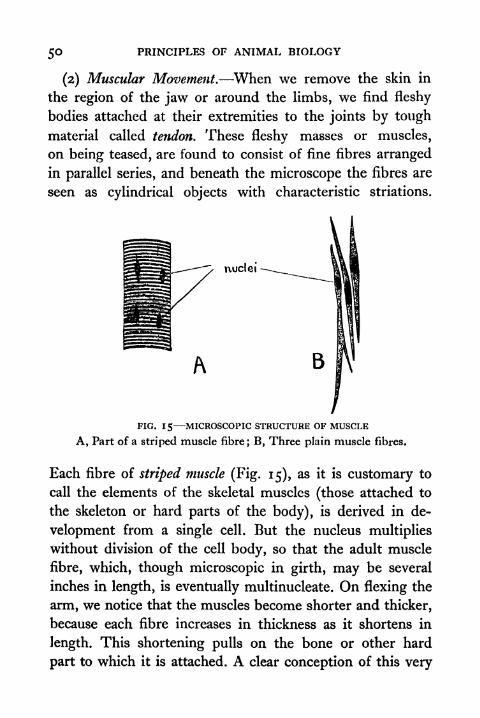
(2) Muscular Movement. When we remove the skin in
the region of the jaw or around the limbs, we find fleshy
bodies attached at their extremities to the joints by tough
material called tendon. These fleshy masses or muscles,
on being teased, are found to consist of fine fibres arranged
in parallel series, and beneath the microscope the fibres are
seen as cylindrical objects with characteristic striations.
nuclei -
B
FIG. 15 MICROSCOPIC STRUCTURE OF MUSCLE
A, Part of a striped muscle fibre; B, Three plain muscle fibres.
Each fibre of striped muscle (Fig. 15), as it is customary to
call the elements of the skeletal muscles (those attached to
the skeleton or hard parts of the body), is derived in de-
velopment from a single cell. But the nucleus multiplies
without division of the cell body, so that the adult muscle
fibre, which, though microscopic in girth, may be several
inches in length, is eventually multinucleate. On flexing the
arm, we notice that the muscles become shorter and thicker,
because each fibre increases in thickness as it shortens in
length. This shortening pulls on the bone or other hard
part to which it is attached. A clear conception of this very

THE MACHINERY OF RESPONSE 51
elementary fact is a comparatively modern discovery.
Vesalius, the pioneer physiologist of the Renaissance, first
clearly enunciated the idea that movement of the limbs was
specifically due to a change in the fleshy portion or muscles
in his epoch-making Fabrica Humani Corporis (1543). It
FIG. 1 6 RELATION OF MUSCLES AND SKELETAL PARTS (shaded)
a, Human forearm; b, Leg of an insect.
was not until the invention of the microscope that the nature
of the muscle fibres could be studied. As late as 1662 Glisson
first showed that when muscle contracts a change in shape,
not, as previously believed, a change of bulk, occurs in each
fibre (Fig. 19).
Muscles of the kind that consist of cylindrical fibres
showing well-marked striations are concerned with the
movements of the outside of the body, and are generally
attached to some hard material which is called skeletal tissue.
52 PRINCIPLES OF ANIMAL BIOLOGY
In an insect they are attached to the horny outer covering
of the body, which is divided up into separate segments or
joints with thinner more flexible tissue between them serving
as hinges (Fig. 16). In the frog and in ourselves, the skeletal
tissues are internal and form a number of separate bones
hinged by a tough fibrous material or tissue of whitish
colour known as tendon, which also serves to bind the ends
of the muscles to them.
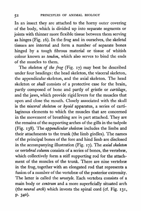
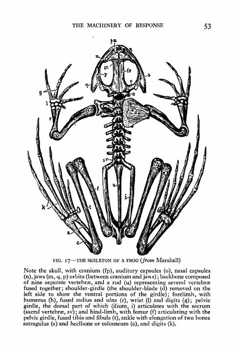
The skeleton of the frog (Fig. 17) may best be described
under four headings: the head skeleton, the visceral skeleton,
the appendicular skeleton, and the axial skeleton. The head
skeleton or skull consists of a protective case for the brain,
partly composed of bone and partly of gristle or cartilage,
and the jaws, which provide rigid levers for the muscles that
open and close the mouth. Closely associated with the skull
is the visceral skeleton or hyoid apparatus, a series of carti-
laginous elements to which the muscles that are concerned
in the movement of breathing are in part attached. They are
the remains of the supporting arches of the gills in the tadpole
(Fig. 138). The appendicular skeleton includes the limbs and
their attachments to the trunk (the limb girdles). The names
of the principal bones of the fore and hind limb are disclosed
in the accompanying illustration (Fig. 17). The axial skeleton
or vertebral column consists of a series of bones, the vertebrae,
which collectively form a stiff supporting rod for the attach-
ment of the muscles of the trunk. There are nine vertebrae
in the frog, together with an elongated rod that represents a
fusion of a number of the vertebrae of the posterior extremity.
The latter is called the urostyle. Each vertebra consists of a
main body or centrum and a more superficially situated arch
(the neural arch} which invests the spinal cord (cf. Fig. 131,
P- 34)-
THE MACHINERY OF RESPONSE 53
FIG. 17 THE SKELETON OF A FROG (from Marshall)
Note the skull, with cranium (fp), auditory capsules (o), nasal capsules
(n), jaws (m, q, p) orbits (between cranium and jaws) ;backbone composed
of nine separate vertebra, and a rod (u) representing several vertebrae
fused together; shoulder-girdle (the shoulder-blade (cl) removed on the
left side to show the ventral portions of the girdle); forelimb, withhumerus (h), fused radius and ulna (r), wrist (1) and digits (g); pelvic
girdle, the dorsal part of which (ilium, i) articulates with the sacrum
(sacral vertebrae, sv); and hind-limb, with femur (f) articulating with the
pelvic girdle, fused tibia and fibula (t), ankle with elongation of two bones
astragulus (a) and heelbone or calcaneum (c), and digits (k).
54 PRINCIPLES OF ANIMAL BIOLOGY
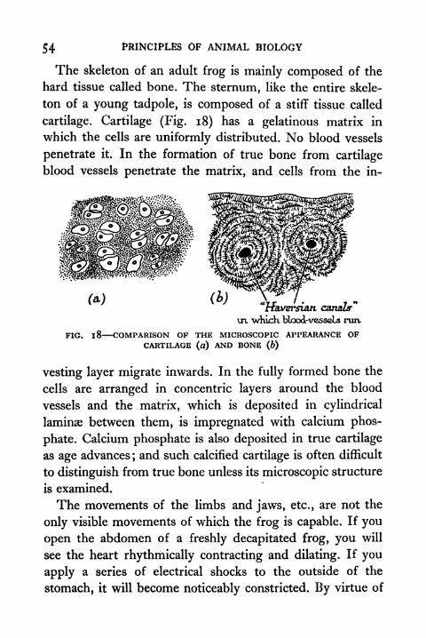
The skeleton of an adult frog is mainly composed of the
hard tissue called bone. The sternum, like the entire skele-
ton of a young tadpole, is composed of a stiff tissue called
cartilage. Cartilage (Fig. 18) has a gelatinous matrix in
which the cells are uniformly distributed. No blood vessels
penetrate it. In the formation of true bone from cartilage
blood vessels penetrate the matrix, and cells from the in-
"tfaversian canals"
\n "whicK blood-vesscLs nut
FIG. 1 8 COMPARISON OF THE MICROSCOPIC APPEARANCE OFCARTILAGE (a) AND BONE (b)
vesting layer migrate inwards. In the fully formed bone the
cells are arranged in concentric layers around the blood
vessels and the matrix, which is deposited in cylindrical
laminae between them, is impregnated with calcium phos-
phate. Calcium phosphate is also deposited in true cartilage
as age advances;and such calcified cartilage is often difficult
to distinguish from true bone unless its microscopic structure
is examined.
The movements of the limbs and jaws, etc., are not the
only visible movements of which the frog is capable. If you
open the abdomen of a freshly decapitated frog, you will
see the heart rhythmically contracting and dilating. If you
apply a series of electrical shocks to the outside of the
stomach, it will become noticeably constricted. By virtue of

THE MACHINERY OF RESPONSE 55
this power which the walls of the gut possess, the food is
squeezed from one end of the digestive canal to the other
in the normal course of events. The actual process of
squeezing is not easily perceived in the frog, because its gut
does not always contain much food material. But if we openthe abdomen of a decapitated rabbit before it is cold, we can
see waves of writhing movement along the intestine. Galen
(A.D. 170) understood that these movements of the gut and
those of the uterus or womb, which assist in expelling the
child from the body of the mother, are due to the action of
the fibrous walls of the alimentary and generative tracts, when
he wrote :
the fact is that the stomach possesses two coats which certainly
exist for some purpose; they extend as far as the mouth . . .
simple observation will show that these coats have their fibres
in opposite directions. And, although Erasistratus did not attemptto say for what reason they are like this, I am going to do so.
The outer coat has its fibres straight for traction. The inner coat
has its fibres transverse for the purpose of peristalsis. . . .
The individual fibres of the muscular wall of the gut are
single cells (Fig. 15, B) without the well-defined transverse
markings of striped muscle. They are spindle-shaped and
each possesses a single nucleus. Heart muscle in the frog
is somewhat intermediate in microscopic appearance between
the smooth or plain muscle of the gut, and the striped muscles
of the limbs. The individual fibres are single cells with
single nuclei, but they have distinct transverse striations.
The properties of these three types of muscle in an animal
such as Man or the frog, while alike in their capacity to
produce visible movements by shortening and thickening
about their long and short axes respectively, are in other waysdifferent. The power of contraction is an essential property
56 PRINCIPLES OF ANIMAL BIOLOGY
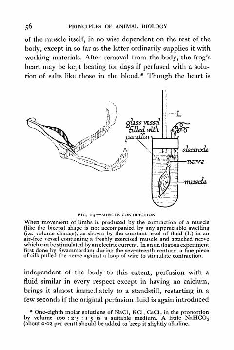
of the muscle itself, in no wise dependent on the rest of the
body, except in so far as the latter ordinarily supplies it with
working materials. After removal from the body, the frog's
heart may be kept beating for days if perfused with a solu-
tion of salts like those in the blood.5*Though the heart is
lectrode,
musda
FIG. 19 MUSCLE CONTRACTION
When movement of limbs is produced by the contraction of a muscle
(like the biceps) shape is not accompanied by any appreciable swelling(i.e. volume change), as shown by the constant level of fluid (L) in anair-free vessel containing a freshly exercised muscle and attached nervewhich can be stimulated by an electric current. In an an Uo.gous experimentfirst done by Swammerdam during the seventeenth century, a fine pieceof silk pulled the nerve against a loop of wire to stimulate contraction.
independent of the body to this extent, pcrfusion with a
fluid similar in every respect except in having no calcium,
brings it almost immediately to a standstill, restarting in a
few seconds if the original perfusion fluid is again introduced
*One-eighth molar solutions of NaCl, KC1, CaCI 2 in the proportion
by volume 100 : 2-5 : 1-5 is a suitable medium. A little NaHCO 3
(about 0-02 per cent) should be added to keep it slightly alkaline.
THE MACHINERY OF RESPONSE 57
without too long delay (Fig. i). This is a material fact about
the machinery of the heart which is as controllable and as
clear-cut as any experiment that we can carry out in the
chemical laboratory. Isolated striped muscle such as the calf
muscle (gastrocnemius) of the leg of the frog will also con-
tract. If kept moist, it will do so for hours after removal
from the body, when stimulated with electric shocks. Themain difference between plain and striped muscle lies in the
fact that striped muscle does not generally contract unless a
special stimulus is applied to it, whereas plain muscle, like
cardiac muscle, has a spontaneous rhythmical activity. When
striped muscle is taken from the body, it is completely
relaxed, whereas plain muscle is in a state of sustained
partial contraction or tone.
(3) Secretion. One very characteristic feature of the
behaviour of amphibians (a group to which frogs and toads
belong) is the readiness with which their skins emit a slimy
material, especially if they are roughly handled or placed in
water containing a little ether. The emission of this slime
from the skin of the body is an illustration of the kind of
response known as secretion. Glands are the parts of an
organism which are concerned with the manufacture of what
we call secretions. Spittle, phlegm, bile, sweat, etc., are
examples of secretions. The simplest sort of glands are
single cells, such as the mucous or slime cells scattered over
the inside surface of the frog's intestine. Most glands are
many-celled tubes or pockets of cubical cells with large
nuclei and often with conspicuous granules (Fig. 20). Theterm was originally applied only to compact masses of such
tubes packed together to form bulky organs of visible
dimensions, such as the liver, pancreas (sweet-bread), etc.
The skin glands of a frog or a toad are simple pits of large

5 PRINCIPLES OF ANIMAL BIOLOGY
cells (Fig. 8), and the venomous secretion of the skin glandsof some toads contains, in addition to mucous compoundswhich give to it a slimy character, a number of physiologically
active substances. One of these is allied to the substance
Lobule
oF gland
FIG. 20 GLANDS
A, Diagrammatic microscopic section through a lobule of a complexgland (Huxley). B and C, Microscopic sections through terminal secretoryunit of the salivary gland of mammal, showing nerve (n) and blood-vessels (b.v.).
called adrenalin, secreted by the adrenal glands (Chapter VI).The sweat glands of the human skin are relatively longercoiled tubes, but likewise unbranched. Their secretory
product is a watery solution of salts. The hygroscopicmucous secretion of the skin of an amphibian protects it
against loss of water through surface evaporation. Increased
skin secretion of a toad is also a response to mechanical
Stimulation of the surface of the body: possibly it aids the

THE MACHINERY OF RESPONSE 59
animal to escape from its- enemies by making it more slippery
to grasp. Sweat secretion on the other hand is a response
to increased temperature, and its evaporation on the surface
of the skin has the effect of cooling the animal. It is part of
an elaborate arrangement which preserves the tissues of
mammals at a remarkably constant temperature well above
that of their surroundings in most circumstances. Only birds
and mammals maintain a constant body temperature above
that of the medium in which they live.
(4) Ciliary Motion. A form of response, which can easily
FIG. 21 CILIATED COLUMNAR EPITHELIUM
be observed in the common frog, is known as ciliary motion.
We have already encountered it in describing the locomotion
of Paramcecium;but the reader may not have expected to meet
with itagain in a more complex animal. If we cut out a strip of
the skin of the roof of the frog's mouth, and examine it under
the microscope, we are able to see at the edges that the whole
surface is covered with a layer of cells (Fig. 21) providedwith fine vibratile filaments or cilia, like those which cover the
body of Paramcecium or Opalina. These cilia keep up move-
ment of the slimy secretion of the mouth in the direction
of the gullet. A few grains of charcoal sprinkled over the
anterior end of the roof of the mouth of a frog killed by
pithing* moves at a quite perceptible rate towards the throat.
*i.e. insertion of a needle into the skull cavity to destroy the brain.
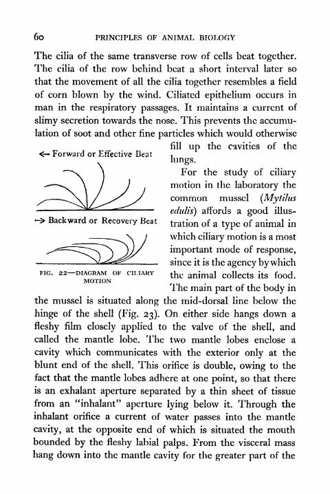
6o PRINCIPLES OF ANIMAL BIOLOGY
< Forward or Effective Beat
Backward or Recovery Beat
The cilia of the same transverse row of cells beat together.
The cilia of the row behind beat a short interval later so
that the movement of all the cilia together resembles a field
of corn blown by the wind. Ciliated epithelium occurs in
man in the respiratory passages. It maintains a current of
slimy secretion towards the nose. This prevents the accumu-
lation of soot and other fine particles which would otherwise
fill up the cavities of the
lungs.
For the study of ciliary
motion in the laboratory the
common mussel (Mytihis
edulis) affords a good illus-
tration of a type of animal in
which ciliary motion is a most
important mode of response,
since it is the agency bywhich
the animal collects its food.
The main part of the body in
the mussel is situated along the mid-dorsal line below the
hinge of the shell (Fig. 23). On either side hangs down a
fleshy film closely applied to the valve of the shell, and
called the mantle lobe. The two mantle lobes enclose a
cavity which communicates with the exterior only at the
blunt end of the shell. This orifice is double, owing to the
fact that the mantle lobes adhere at one point, so that there
is an exhalant aperture separated by a thin sheet of tissue
from an "inhalant" aperture lying below it. Through the
inhalant orifice a current of water passes into the mantle
cavity, at the opposite end of which is situated the mouthbounded by the fleshy labial palps. From the visceral mass
hang down into the mantle cavity for the greater part of the
FIG. 22- -DIAGRAM OF CILIARYMOTION

THE MACHINERY OF RESPONSE 6l
moyth,
long axis of the body a- pair of so-called "gills" (Fig. 23).
Each consists of a pair of flat plates formed by the lateral
approximation of a longitudinal row of filaments like the
fringe of a lamp shade. This
fringe of filaments is bent back
to meet the body wall, so that
each plate, or lamella, encloses
a chamber which communi-
cates behind with the exterior
by way of the exhalant aper-
ture. A slow, continuous
current of water enters throughthe inhalant orifice, strains
through the filaments which
make up the lamellae of the
gills, passes backwards between
the two halves of each lamella,
and thence flows outwards bythe exhalant aperture. It is
maintained by the activity of
cilia which line the sides of the
filaments (Fig. 34). The whole
apparatus is an animated filter;
it strains minute suspendednartir1e<* out of the water- Ab ve the animal as seen whenparticles out Ot me water-
one valve of the shell is removed ;
Stream, sorts them, and COn- below transverse section through1t t whole animal.
veys smaller ones to the
mouth. The gill-filaments secrete mucus. Entrapped in this
slime, food particles are driven by the action of the cilia,
which are also present on the exposed face of the gills,
downwards to the lower margin of the lamella, which is
grooved at its distal extremity. By ciliary^motionof the cells
CHAMBER OF THE MUSSEL
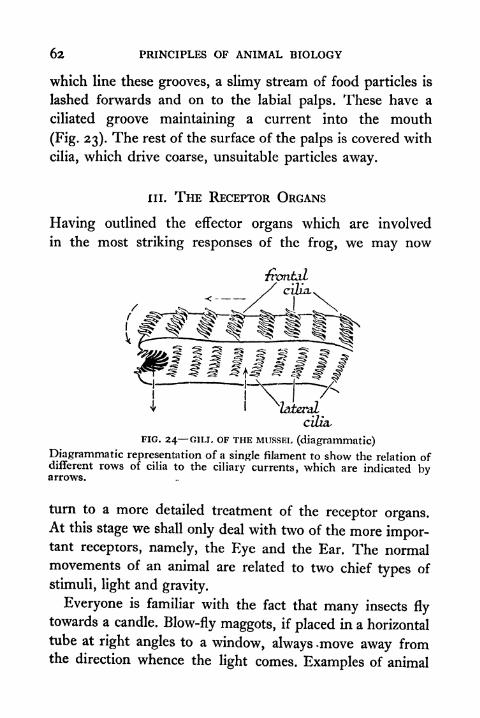
62 PRINCIPLES OF ANIMAL BIOLOGY
which line these grooves, a slimy stream of food particles is
lashed forwards and on to the labial palps. These have a
ciliated groove maintaining a current into the mouth
(Fig. 23). The rest of the surface of the palps is covered with
cilia, which drive coarse, unsuitable particles away.
in. THE RECEPTOR ORGANS
Having outlined the effector organs which are involved
in the most striking responses of the frog, we may now
"frontal
cilia. ^
^
cilia,
FIG. 24 GILT. OF THE MUSSEL (diagrammatic)
Diagrammatic representation of a single filament to show the relation ofdifferent rows of cilia to the ciliary currents, which are indicated byarrows.
turn to a more detailed treatment of the receptor organs.At this stage we shall only deal with two of the more impor-tant receptors, namely, the Eye and the Ear. The normalmovements of an animal are related to two chief types of
stimuli, light and gravity.
Everyone is familiar with the fact that many insects fly
towards a candle. Blow-fly maggots, if placed in a horizontal
tube at right angles to a window, always .move away fromthe direction whence the light comes. Examples of animal
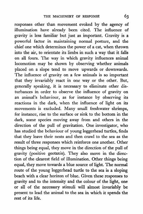
THE MACHINERY OF RESPONSE 63
responses other than movement evoked by the agency of
illumination have already been cited. The influence of
gravity is less familiar but just as important. Gravity is a
powerful factor in maintaining normal posture, and the
chief one which determines the power of a cat, when thrown
into the air, to orientate its limbs in such a way that it falls
on all fours. The way in which gravity influences animal
locomotion may be shown by observing whether animals
placed on a slope tend to move upwards or downwards.
The influence of gravity on a few animals is so importantthat they invariably react in one way or the other. But,
generally speaking, it is necessary to eliminate other dis-
turbances in order to observe the influence of gravity on
an animal's behaviour, as for instance by observing its
reactions in the dark, when the influence of light on its
movements is excluded. Many small freshwater shrimps,
for instance, rise to the surface or sink to the bottom in the
dark, some species moving away from and others in the
direction of the pull of gravitation. One investigator, whohas studied the behaviour of young loggerhead turtles, finds
that they leave their nests and then crawl to the sea as the
result of three responses which reinforce one another. Other
things being equal, they move in the direction of the pull of
gravity (positive geotaxis). They also move in the direc-
tion of the dearest field of illumination. Other things being
equal, they move towards a blue source of light. The normal
route of the young loggerhead turtle to the sea is a sloping
beach with a clear horizon of blue. Given these responses to
gravity and to the intensity and the colour of the light, one
or all of the necessary stimuli will almost invariably be
present to lead the animal to the sea in which it spends the
rest of its life.
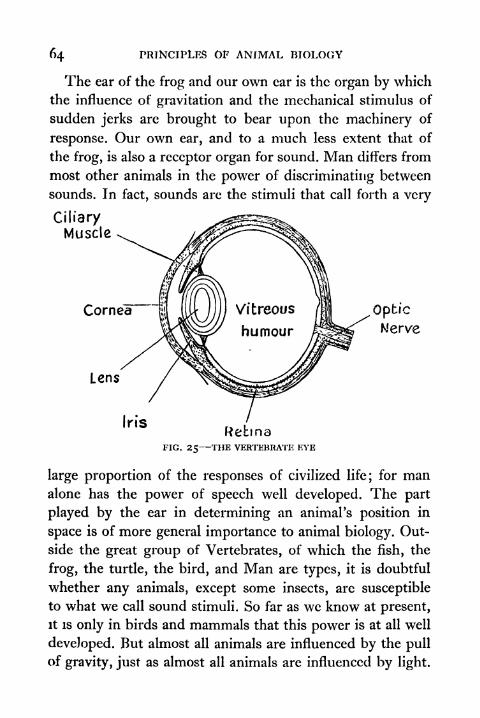
64 PRINCIPLES OF ANIMAL BIOLOGY
The ear of the frog and our own ear is the organ by which
the influence of gravitation and the mechanical stimulus of
sudden jerks are brought to bear upon the machinery of
response. Our own ear, and to a much less extent that of
the frog, is also a receptor organ for sound. Man differs from
most other animals in the power of discriminating between
sounds. In fact, sounds arc the stimuli that call forth a very
CiliaryMuscle
Cornea 1 J^ ))}}Vitreous /| Coptic
K'erve
Uns
RehmaFIG. 25 THE VERTEBRATE EYE
large proportion of the responses of civilized life; for manalone has the power of speech well developed. The part
played by the ear in determining an animal's position in
space is of more general importance to animal biology. Out-
side the great group of Vertebrates, of which the fish, the
frog, the turtle, the bird, and Man are types, it is doubtful
whether any animals, except some insects, are susceptible
to what we call sound stimuli. So far as we know at present,
it is only in birds and mammals that this power is at all well
developed. But almost all animals are influenced by the pull
of gravity, just as almost all animals are influenced by light.

THE MACHINERY OF RESPONSE
(i) The Eye. In its gross construction the eye is verymuch like an ordinary camera. It consists of a box or capsule,
a lens which focuses an image on the back of the box, and
a layer of tissue, the retina, whose cells contain a substance
chemically sensitive to light like the gelatin film of silver salt
on the photographic plate or film. In front there is a trans-
parent region of the tough and elsewhere opaque capsule.
It is called the cornea. Behind, it is connected with the brain
Inferior Oblique
Superior Oblique
Internal Rectus _ _SuperiorRectus
Inferior Rectus
External Rectus
FIG. 26 THE VERTEBRATE EYE IN SITU
Showing associated muscles and their nerves from above.
by the optic nerve. Among the receptive cells of the retina is
a layer of pigment cells somewhat like those which occur
in the skin of the frog, for they are contractile in the samesense. These cells also occur in the eye of Man, which is
essentially like that of the frog, and they are the only remains
of the pigmentary effector system of the frog or the chame-
leon which the writer or reader of this book possesses. The
layer of black pigment cells may be compared to the coat of
black paint with which the inside of the box of the camera is
covered.
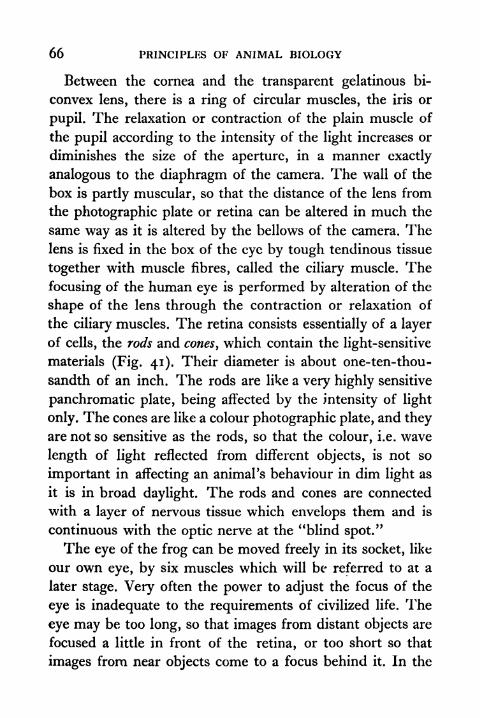
66 PRINCIPLES OF ANIMAL BIOLOGY
Between the cornea and the transparent gelatinous bi-
convex lens, there is a ring of circular muscles, the iris or
pupil. The relaxation or contraction of the plain muscle of
the pupil according to the intensity of the light increases or
diminishes the size of the aperture, in a manner exactly
analogous to the diaphragm of the camera. The wall of the
box is partly muscular, so that the distance of the lens from
the photographic plate or retina can be altered in much the
same way as it is altered by the bellows of the camera. Thelens is fixed in the box of the eye by tough tendinous tissue
together with muscle fibres, called the ciliary muscle. The
focusing of the human eye is performed by alteration of the
shape of the lens through the contraction or relaxation of
the ciliary muscles. The retina consists essentially of a layer
of cells, the rods and cones, which contain the light-sensitive
materials (Fig. 41). Their diameter is about one-ten-thou-
sandth of an inch. The rods are like a very highly sensitive
panchromatic plate, being affected by the intensity of light
only. The cones are like a colour photographic plate, and they
are not so sensitive as the rods, so that the colour, i.e. wave
length of light reflected from different objects, is not so
important in affecting an animal's behaviour in dim light as
it is in broad daylight. The rods and cones are connected
with a layer of nervous tissue which envelops them and is
continuous with the optic nerve at the "blind spot."
The eye of the frog can be moved freely in its socket, like
our own eye, by six muscles which will be referred to at a
later stage. Very often the power to adjust the focus of the
eye is inadequate to the requirements of civilized life. The
eye may be too long, so that images from distant objects are
focused a little in front of the retina, or too short so that
images from near objects come to a focus behind it. In the
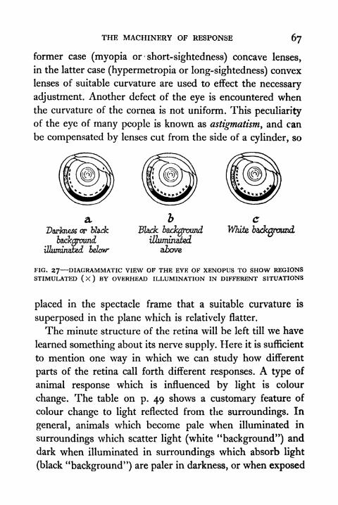
THE MACHINERY OF RESPONSE 67
former case (myopia or short-sightedness) concave lenses,
in the latter case (hypermetropia or long-sightedness) convex
lenses of suitable curvature are used to effect the necessary
adjustment. Another defect of the eye is encountered whenthe curvature of the cornea is not uniform. This peculiarity
of the eye of many people is known as astigmatism, and can
be compensated by lenses cut from the side of a cylinder, so
a 2> cor blade Blade background
White1
illuminated
above,
FIG. 27 DIAGRAMMATIC VIEW OF THE EYE OF XENOPUS TO SHOW REGIONSSTIMULATED ( X ) BY OVERHEAD ILLUMINATION IN DIFFERENT SITUATIONS
placed in the spectacle frame that a suitable curvature is
superposed in the plane which is relatively flatter.
The minute structure of the retina will be left till we have
learned something about its nerve supply. Here it is sufficient
to mention one way in which we can study how different
parts of the retina call forth different responses. A type of
animal response which is influenced by light is colour
change. The table on p. 49 shows a customary feature of
colour change to light reflected from the surroundings. In
general, animals which become pale when illuminated in
surroundings which scatter light (white "background") and
dark when illuminated in surroundings which absorb light
(black "background") are paler in darkness, or when exposed
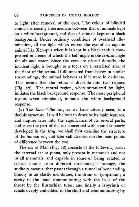
68 PRINCIPLES OF ANIMAL BIOLOGY
to light after removal of the eyes. The colour of blinded
animals is usually intermediate between that of animals kept
on a white background, and that of animals kept on a black
background. Under ordinary conditions of overhead illu-
mination, all the light which enters the eye of an aquatic
animal like Xenopus when it is kept in a black tank is com-
pressed in a cone of which the half angle is the critical angle
for air and water. Since the eyes are placed dorsally, the
incident light is brought to a focus on a restricted area of
the floor of the retina. If illuminated from below in similar
surroundings, the animal behaves as if it were in darkness.
This means that the retina is divisible into two regions
(Fig. 27). The central region, when stimulated by light,
initiates the black background response. The more peripheral
region, when stimulated, initiates the white background
response.
(2) The Ear. The ear, as we have already seen, is a
double structure. It will be best to describe its main features,
and inquire later into the significance of its several parts,
and since the part of the ear concerned with sound is poorly
developed in the frog, we shall first examine the structure
of the human ear, and later call attention to the main points
of difference between the two.
The ear of Man (Fig. 28) consists of the following parts :
the external ear or pinna, only present in mammals and not
in all mammals, and capable in some of being rotated to
collect sounds from different directions; a passage, the
auditory meatus, that passes through a tunnel of bone ending
blindly in an elastic membrane, the drum or tympanum; a
cavity in the bone communicating with the back of the
throat by the Eustachian tube; and finally a labyrinth of
canals deeply embedded in the skull and communicating by
THE MACHINERY OF RESPONSE 69
a small window with the cavity behind the tympanum.This labyrinth encloses a complicated sac, which is the most
essential part of the ear, the sac and the space between it
and its bony case being filled with a clear fluid. Across the
space between the drum and the window of the bony laby-
rinth is a chain of three small bones, the auditory ossicles.
3 s&nidrcularcanals
eat
cavity-
FIG. 28 THE HUMAN EAR
tube.
These transmit vibrations from the former to the latter.
The innermost sac consists of three portions. The upper-most or utriculus is continuous with three semi-circular
canals at right angles to one another. Below it communicates
with an elongated chamber, the sacculus, which leads into
the third spirally coiled portion known as the cochlea. In
the latter, which is specially concerned with sound, fine
microscopic fibres joined by a membrane are stretched
across its walls. The internal surfaces of the utriculus, semi-
circular canals, and sacculus bear patches of fine hairs in
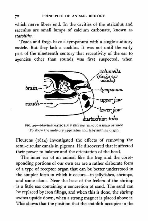
70 PRINCIPLES OF ANIMAL BIOLOGY
which nerve fibres end. In the cavities of the utriculus and
sacculus are small lumps of calcium carbonate, known as
ttatotiths.
Toads and frogs have a tympanum with a single auditory
ossicle. But they lack a cochlea. It was not until the early
part of the nineteenth century that receptivity of the ear to
agencies other than sounds was first suspected, when
coZumeZ/a,
/'(single ear
ossicle)
eu&tacluati tube
FIG. 29 DIAGRAMMATIC HALF SECTION THROUGH HEAD OF FROG
To show the auditory apparatus and labyrinthine organ.
Flourens (1824) investigated the effects of removing the
semi-circular canals in pigeons. He discovered that it affected
their power to balance and the orientation of the head.
The inner ear of an animal like the frog and the corre-
sponding portions of our own ear are a rather elaborate form
of a type of receptor organ that can be better understood in
the simpler form in which it occurs in jellyfishes, shrimps,
and some clams. Near the base of the feelers of the shrimpis a little sac containing a concretion of sand. The sand can
be replaced by iron filings, and when this is done, the shrimpswims upside down, when a strong magnet is placed above it.
This shows that the position that the statolith occupies in the

THE MACHINERY OF RESPONSE Jl
sac which contains it determines whether certain muscles are
brought into play (Fig. 30). A shrimp which has had both its
feelers removed will swim in a normal manner in daylight,
but if illuminated from below in a dark room, it will swim on
its back, whereas a normal shrimp will always swim so that
FIG. 30 DIAGRAM TO SHOW DISPLACEMENT OF STATOLITII IN THE STATO,CYST OF A SHRIMP OR SQUID
1 . Before the beginning of rotation.
2. Immediately after displacement in the direction of the arrow.
3. Immediately after cessation of movement in same direction.
its ventral surface is directed towards the pull of gravity. Ashrimp with one feeler removed will swim normally in
daytime. In the dark it will swim on one side. If both eyes
and feelers are removed, it loses all power to balance itself
in water. If one eye and one feeler are removed, it swims
perpetually in a spiral path. These experiments show clearly
that the normal posture of swimming depends partly on
light and partly on gravity. So long as both the receptors for
72 PRINCIPLES OF ANIMAL BIOLOGY
either light or gravity are intact the influence of that stimulus
is adequate to ensure the correct muscular responses. Weshall go more thoroughly into this question when we have
seen how it is that the effect of the external stimulus on the
receptor organ travels to the effector organ, which carries out
the appropriate response.
TOPICS FOR HOME STUDY1. What is meant by ciliary motion, and what part does it play in
the life of different types of animals.
2. Describe the microscopic changes which occur during colour
change in frog and chameleon and how these changes are
related to the external world.
3. Summarize the chief characteristics of the following tissues:
Blood, cartilage, bone, muscle, epithelium, nerve.
4. What receptor organs are involved in response of animals to
rotation and disorientation in relation to gravity.
5. Explain how the eyes are accommodated to near and distant
objects and how they move in the orbits.
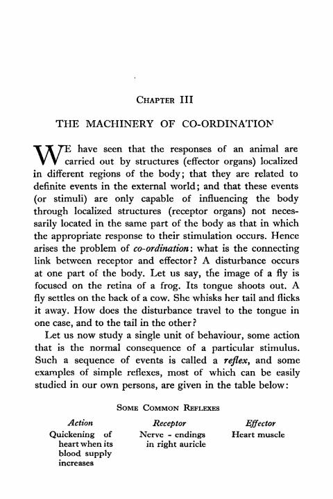
CHAPTER III
THE MACHINERY OF CO-ORDINATION
T T TE have seen that the responses of an animal are
V V carried out by structures (effector organs) localized
in different regions of the body; that they are related to
definite events in the external world; and that these events
(or stimuli) are only capable of influencing the body
through localized structures (receptor organs) not neces-
sarily located in the same part of the body as that in which
the appropriate response to their stimulation occurs. Hence
arises the problem of co-ordination: what is the connectinglink between receptor and effector? A disturbance occurs
at one part of the body. Let us say, the image of a fly is
focused on the retina of a frog. Its tongue shoots out. Afly settles on the back of a cow. She whisks her tail and flicks
it away. How does the disturbance travel to the tongue in
one case, and to the tail in the other?
Let us now study a single unit of behaviour, some action
that is the normal consequence of a particular stimulus.
Such a sequence of events is called a reflex, and some
examples of simple reflexes, most of which can be easily
studied in our own persons, are given in the table below :
SOME COMMON REFLEXES
Action Receptor Effector
Quickening of Nerve - endings Heart muscleheart when its in right auricle
blood supplyincreases
74 PRINCIPLES OF ANIMAL BIOLOGY
SOME COMMON REFLEXES continued
Action Receptor Effector
Contraction of Retina Plain muscle
p u p i 1 i n of iris
strong light
Secretion of sal- Olfactory organ Salivaryiva on smell- glands
ing food
Sneezing after Olfactory organ Muscles of
pepper chest and
diaphragm
Knee jerk "End organs" in Extensor
tendon muscles of
thigh
A reflex that is very easy to demonstrate in the frog is the
withdrawal of the foot, when the toes are allowed to dip
into water warmed to about the temperature of the human
body. If we suspend a frog which has been pithed (i.e.
the brain destroyed) with the legs hanging downwards and
bring a beaker of water gently into position, so that the toes
just dip into the water, we find that nothing happens if the
water is at the temperature of the room;but the leg is pulled
up if the water has been previously warmed to about 40 C.
If we time the interval between the immersion of the toes
and the withdrawal of the limb, we find that it is a matter of
seconds. Something has been happening during those seconds.
What happens and where it happens is the aspect of behaviour
implied by the term co-ordination.
The chief means of transmission of the disturbances set
up in the receptor organs to the effector organs that carry
out the ensuing responses is the nervous system. An instructive
experiment can be carried out if we hang up five decapitated

THE MACHINERY OF CO-ORDINATION 75
frogs in the manner indicated above after snipping awaythe bone known as the urostyle (at the end of the vertebral
column). This exposes two groups of shining white cylin-
drical trunks which are then seen to pass from the backbone
downwards to the muscles of the legs. We can now learn
what is the path by which the disturbance set up in the tem-
perature receptors of the skin of the toes travels to the muscles
of the thigh.
In one frog the white trunks (sciatic nerves) of both sides
are left intact. In a second the sciatic nerve of the right side
is cut. In a third frog the sciatic nerve of the left side is cut.
In a fourth frog the sciatic nerves of both sides are cut. Andin a fifth frog neither are cut, but a wire is passed down the
backbone to destroy the central nervous system or spinal
cord. Having done this we may try the effect of letting the
toes of both feet dip into warm water. Both legs of the first
frog are withdrawn. Only the left leg of the second and only
the right leg of the third frog is withdrawn. Neither leg of
the other two frogs is withdrawn. From this we conclude
that the something which is happening between the immersion
of the toes and the withdrawal of the foot travels by a definite
road, namely along the sciatic nerve; and further that it
travels first up to the spinal cord and then down to the leg
muscles. This something we call the nervous impulse.
The Nervous Impulse. Propagation of a disturbance from
the receptor to the effector is a physical property of the kind
of matter we call nerve. The nervous impulse is a physical
event. It has space, time, and mass relations. The rate at
which the nerve impulse travels can be determined exactly.
This was first done by Helmholtz, in 1851, six years after
Miiller had stated that it was theoretically impossible to
measure its velocity of propagation. The method employed
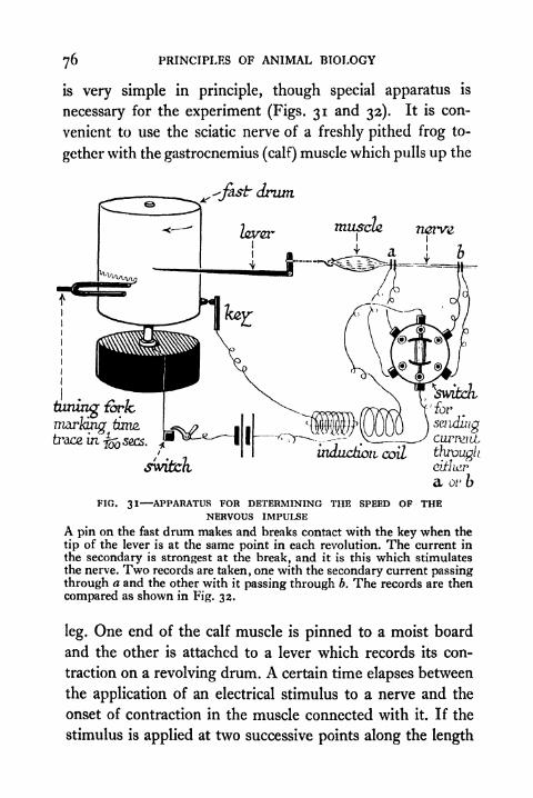
76 PRINCIPLES OF ANIMAL BIOLOGY
is very simple in principle, though special apparatus is
necessary for the experiment (Figs. 31 and 33). It is con-
venient to use the sciatic nerve of a freshly pithed frog to-
gether with the gastrocnemius (calf) muscle which pulls up the
^J^str drum
nervz
timing fork,
marking turuz
trace, uij sees.
switch
switchfor
sendingcurrent,
throughctthtr
a or b
FIG. 31 APPARATUS FOR DETERMINING THE SPEED OF THENERVOUS IMPULSE
A pin on the fast drum makes and breaks contact with the key when the
tip of the lever is at the same point in each revolution. The current in
the secondary is strongest at the break, and it is this which stimulates
the nerve. Two records are taken, one with the secondary current passingthrough a and the other with it passing through b. The records are then
compared as shown in Fig. 32.
leg. One end of the calf muscle is pinned to a moist board
and the other is attached to a lever which records its con-
traction on a revolving drum. A certain time elapses between
the application of an electrical stimulus to a nerve and the
onset of contraction in the muscle connected with it. If the
stimulus is applied at two successive points along the length
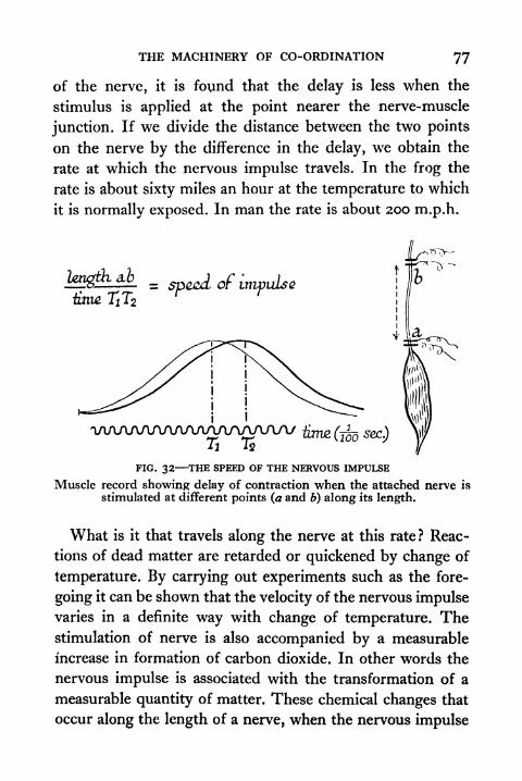
THE MACHINERY OF CO-ORDINATION 77
of the nerve, it is found that the delay is less when the
stimulus is applied at the point nearer the nerve-muscle
junction. If we divide the distance between the two points
on the nerve by the difference in the delay, we obtain the
rate at which the nervous impulse travels. In the frog the
rate is about sixty miles an hour at the temperature to which
it is normally exposed. In man the rate is about 200 m.p.h.
FIG. 32 THE SPEED OF THE NERVOUS IMPULSE
Muscle record showing delay of contraction when the attached nerve is
stimulated at different points (a and b) along its length.
What is it that travels along the nerve at this rate ? Reac-
tions of dead matter are retarded or quickened by change of
temperature. By carrying out experiments such as the fore-
going it can be shown that the velocity of the nervous impulsevaries in a definite way with change of temperature. Thestimulation of nerve is also accompanied by a measurable
increase in formation of carbon dioxide. In other words the
nervous impulse is associated with the transformation of a
measurable quantity of matter. These chemical changes that
occur along the length of a nerve, when the nervous impulse
78 PRINCIPLES OF ANIMAL BIOLOGY
is said to traverse it, give rise to measurable electrical
phenomena which can be measured by a sufficiently sensitive
galvanometer.
A clear appreciation of the significance of the nervous
system in the economy of the animal body came late in
history. Galen, the foremost physician of antiquity, had no
clear idea of the way in which disturbances are transmitted.
A new understanding began in the year 1543, when Vesalius
published his celebrated Fabrica Humani Corporis. Vesalius
"dared not only to show how often Galen was wrong, but
to insist that when Galen was right he was to be followed,
not because he said it, but because what he said was in
accordance with what everyone who took the pains to inquire
could assure himself to be the real state of things."* In the
chapter on vivisections in his great book, Vesalius showed
that the action of this or that muscle can be abolished by
cutting or ligaturing one nerve or another, and that the
action of the muscle is restored by removing the ligature.
This proved that the propagation of disturbances through
the body is effected through the agency of the nerves. Healso showed that transection of the spinal cord prevents the
propagation of stimuli from the region above it to the muscles
supplied by nerves emerging from the central nervous
system below the section.
It is customary to distinguish-the brain and spinal cord of
the frog as the central nervous system (commonly written
C.N.S.) in contradistinction to the other nerve cords which
connect it with the receptor and effector organs of the body.
The central nervous system was not clearly recognized to
be the turning point in the road which the nervous impulse
travels until 1751, when Whytt showed by experiments on
*Foster, History of Physiology.
THE MACHINERY OF CO-ORDTNATION 79
frogs that destruction of the spinal cord abolishes reflex
actions. Before we can get a deeper insight into the adven-
tures of the nervous impulse as it travels first towards the
central nervous system, and thence outwards to the muscles,
glands, etc., we must examine the microscopic structure of
nerves.
Structure of the Nervous System. If we tease a nerve like
the sciatic nerve of the frog with needles we can separate it
into a large number of cylindrical fibres, each of microscopic
thickness and each surrounded by a minute sheath of fatty
material. By tracing these fibres back to the spinal cord, we
eventually find that they are simply attenuated processes of
branching cells. Some of them originate from branchingcells in the grey matter or core of the spinal cord, where their
finer branches are closely connected with the terminal
branches of other nerve cells or neurones. Branching cells of
this type also occur in the central nervous system itself.
Their fibres (propriospinal) run up and down the outer rind
or white matter of the spinal cord, carrying nervous impulses
from one level to another. Others of the fibres of a mixed
nerve, i.e. a nerve carrying impulses both to and from the
cord, like the sciatic nerve of the frog, do not have their
cell bodies within the spinal cord. Their cell bodies are
in swellings known as the dorsal root ganglia, situated near
where the nerve is joined to the cord. We shall have more
to say about these ganglia later; it suffices for the present
to note that the term ganglion is used for any part of the
nervous system in which the cell bodies of nerve fibres
are congregated. The cells of the dorsal ganglia send
branches into the spinal cord, where they arborize around
the cell bodies found in the grey matter. A single nerve
fibre, with its sheath, in a Vertebrate animal, is not more
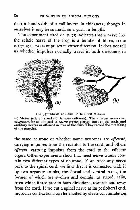
8o PRINCIPLES OF ANIMAL BIOLOGY
than a hundredth of a millimetre in thickness, though in
ourselves it may be as much as a yard in length.
The experiment cited on p. 75 indicates that a nerve like
the sciatic nerve of the frog is a bundle of fibres, some
carrying nervous impulses in either direction. It does not tell
us whether impulses normally travel in both directions in
(*>)
FIG. 33 NERVE ENDINGS IN STRIPED MUSCLE
(a) Motor (efferent) and (b) Sensory (afferent). The afferent nerves are
proprioceptive as opposed to extero-ceptive nerves such as the optic and
auditory nerves or afferent nerves of the skin. They record the stretchingof the muscles.
the same neurone or whether some neurones are afferent,
carrying impulses from the receptor to the cord, and others
efferent, carrying impulses from the cord to the effector
organ. Other experiments show that most nerve trunks con-
tain two different types of neurone. If we trace any nerve
back to the spinal cord, we find that it is connected with it
by two separate trunks, the dorsal and ventral roots, the
former of which are swollen and contain, as stated, cells,
from which fibres pass in both directions, towards and awayfrom the cord. If we cut a spinal nerve at its peripheral end,
muscular contractions can be elicited by electrical stimulation

THE MACHINERY OF CO-ORDINATION 8l
of the nerve on the central side of the cut. After cutting the
dorsal root these reflexes will not be obtained. This shows
that all the afferent impulses pass into the cord by the fibres
whose cells are located in the dorsal root ganglion. If the
dorsal root of the cut nerve is left intact, section of all other
dorsal roots of the remaining spinal nerves does not interfere
stimulated Kcrci
no response |
receptorsurface
stimulated, licrc
responseon left-
side
efferent-
fibre,
,
muscle;'
fibrematter! .
^
matterJPm core
FIG. 34 DIAGRAM TO ILLUSTRATE MULLER*S EXPERIMENT
Dorsal root cut at A ; ventral root cut at B.
with the reflex response obtained by stimulating its central
end. This shows that all the efferent fibres leave the cord bythe ventral root. The separate connexions of the afferent
and efferent neurones to the C.N.S. were finally established
by Johannes Miiller (1834) who severed all the dorsal roots
on one side of the body of a frog, and all the ventral or motor
roots on the other side (Fig. 34). Such an animal shows
complete indifference to stimuli on the side on which the
dorsal roots are cut and complete inability to respond byactive movement on the opposite side,, but stimuli applied
F
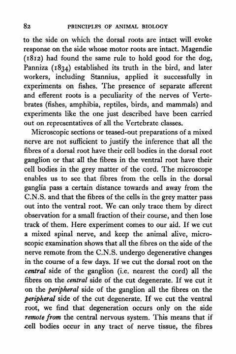
82 PRINCIPLES OF ANIMAL BIOLOGY
to the side on which the dorsal roots are intact will evoke
response on the side whose motor roots are intact. Magendie
(1812) had found the same rule to hold good for the dog,
Panniza (1834) established its truth in the bird, and later
workers, including Stannius, applied it successfully in
experiments on fishes. The presence of separate afferent
and efferent roots is a peculiarity of the nerves of Verte-
brates (fishes, amphibia, reptiles, birds, and mammals) and
experiments like the one just described have been carried
out on representatives of all the Vertebrate classes.
Microscopic sections or teased-out preparations of a mixed
nerve are not sufficient to justify the inference that all the
fibres of a dorsal root have their cell bodies in the dorsal root
ganglion or that all the fibres in the ventral root have their
cell bodies in the grey matter of the cord. The microscopeenables us to see that fibres from the cells in the dorsal
ganglia pass a certain distance towards and away from the
C.N.S. and that the fibres of the cells in the grey matter pass
out into the ventral root. We can only trace them by direct
observation for a small fraction of their course, and then lose
track of them. Here experiment comes to our aid. If we cut
a mixed spinal nerve, and keep the animal alive, micro-
scopic examination shows that all the fibres on the side of the
nerve remote from the C.N.S. undergo degenerative changesin the course of a few days. If we cut the dorsal root on the
central side of the ganglion (i.e. nearest the cord) all the
fibres on the central side of the cut degenerate. If we cut it
on the peripheral side of the ganglion all the fibres on the
peripheral side of the cut degenerate. If we cut the ventral
root, we find that degeneration occurs only on the side
remote from the central nervous system. This means that if
.cell bodies occur in any tract of nerve tissue, the fibres
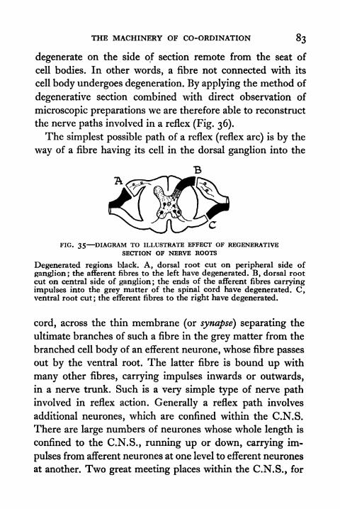
THE MACHINERY OF CO-ORDINATION 83
degenerate on the side of section remote from the seat of
cell bodies. In other words, a fibre not connected with its
cell body undergoes degeneration. By applying the method of
degenerative section combined with direct observation of
microscopic preparations we are therefore able to reconstruct
the nerve paths involved in a reflex (Fig. 36).
The simplest possible path of a reflex (reflex arc) is by the
way of a fibre having its cell in the dorsal ganglion into the
FIG. 35 DIAGRAM TO ILLUSTRATE EFFECT OF REGENERATIVESECTION OF NERVE ROOTS
Degenerated regions black. A, dorsal root cut on peripheral side of
ganglion; the afferent fibres to the left have degenerated. B, dorsal rootcut on central side of ganglion ; the ends of the afferent fibres carryingimpulses into the grey matter of the spinal cord have degenerated. C,ventral root cut; the efferent fibres to the right have degenerated.
cord, across the thin membrane (or synapse) separating the
ultimate branches of such a fibre in the grey matter from the
branched cell body of an efferent neurone, whose fibre passes
out by the ventral root. The latter fibre is bound up with
many other fibres, carrying impulses inwards or outwards,
in a nerve trunk. Such is a very simple type of nerve path
involved in reflex action. Generally a reflex path involves
additional neurones, which are confined within the C.N.S.
There are large numbers of neurones whose whole length is
confined to the C.N.S. , running up or down, carrying im-
pulses from afferent neurones at one level to efferent neurones
at another. Two great meeting places within the C.N.S., for
84 PRINCIPLES OF ANIMAL BIOLOGY
neurones which run forwards to the head and backwards to
the posterior extremity of a Vertebrate, are the two parts of
the brain known as the cerebrum and cerebellum. When the
brain is destroyed these paths are eliminated : and partly for
propiiospuvaL
.dorsal^
gang/ion,
plainmuscle
stripedrrazscfe
nerve
FIG. 36 DIAGRAM OF THE REFLEX ARC
this reason, partly for others which will be referred to later,
the behaviour of the pithed frog is much simpler, and there-
fore more suitable for the study of reflex action than that of
the normal animal.
The existence of nerve tracts running up and down the
spinal cord can be demonstrated in a very simple way.When a dark chameleon, i.e. a chameleon which has been
kept a few minutes in bright light in a cool room, is stimu-
lated by an alternating current from a shocking coil by

THE MACHINERY OF CO-ORDINATION 85
electrodes applied either to the roof of the mouth or to the
anal orifice, the animal becomes extremely pale after about a
minute. If the spinal cord has been cut with a pair of dis-
secting scissors about the level of the eighth vertehra
FIG. 37 EFFECT OF FARADIC STIMULATION OF THE ROOF OF THEMOUTH IN THE CAPE CHAMELEON WHEN THE SPINAL CORDIS SEVERED AT C
generalized pallor does not result from stimulating the mouth.
The skin becomes pale only in the half of the body in front
of the cut. After stimulating the cloaca the body becomes
pale only on the posterior side of the cut.
The nerve fibres that supply the striped muscles of the
limbs, etc., have their cell bodies in the grey matter of the
cord.
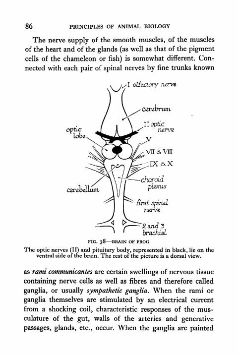
86 PRINCIPLES OF ANIMAL BIOLOGY
The nerve supply of the smooth muscles, of the muscles
of the heart and of the glands (as well as that of the pigment
cells of the chameleon or fish) is somewhat different. Con-
nected with each pair of spinal nerves by fine trunks known
vjs\> olfactory nerve
\ .x cerebrum.
opticlobe
II opticnerve
cgreoeuuiri
first spinalnerve
2 and 3brachiaL
FIG. 38 BRAIN OF FROG
The optic nerves (II) and pituitary body, represented in black, lie on theventral side of the brain. The rest of the picture is a dorsal view.
as rami communicantes are certain swellings of nervous tissue
containing nerve cells as well as fibres and therefore called
ganglia, or usually sympathetic ganglia. When the rami or
ganglia themselves are stimulated by an electrical current
from a shocking coil, characteristic responses of the mus-
culature of the gut, walls of the arteries and generative
passages, glands, etc., occur. When the ganglia are painted

THE MACHINERY OF CO-ORDINATION 87
with the drug nicotine, stimulation of the rami does not
evoke these responses. Since painting the nerves themselves
does not prevent their ability to transmit impulses, nicotine
appears to act primarily on nerve cells which occur in the
ganglia. These nerve cells belong to neurones which supply
the gut, glands, etc. They can be distinguished from efferent
neurones which supply striped muscle via the spinal nerves
and from the nerve fibres which pass out of the cord to the
ganglia via the rami communicantes, because no fatty sheath
(medulla) surrounds the fibre itself. The nerve fibres that
originate in the ganglia are therefore called non-medullated
fibres. The fact that nicotine prevents the conduction of
nervous impulses beyond the ganglia by paralysing their
nerve cells shows that plain muscle, glands, etc., of the bodyare not directly innervated by the fibres with cells in the
grey matter of the cord. Impulses leaving the C.N.S. for
effector organs other than striped muscle pass in the ganglia
to a second relay of neurones. Thus the sympathetic ganglia
are distributive centres which permit impulses arriving from
the cord along a single fibre to set up relay impulses in large
numbers of non-medullated fibres going to different
destinations.
In the region of the spinal cord the part of the C.N.S.
of a Vertebrate enclosed by the spinal column a pair of
nerves with motor and ganglionated roots arises between
each pair of vertebrae. The main nerves to the limbs, such
as the sciatic nerve, are formed by the union of several of
these. The anterior end of the C.N.S. is considerably swollen
and enclosed within the skull. From this region, the brain
(Fig. 38), ten pairs of nerves called cranial nerves* come off in
* Two additional nerves of Man (and other mammals) in realitytypical spinal nerves are incorporated in the skull cavity.

88 PRINCIPLES OF ANIMAL BIOLOGY
the frogs and ourselves. These nerves especiallythose of the
anterior end of the brain are not built on the same plan as
the spinal nerves. The same nerve does not always contain
both afferent neurones and efferentneurones supplying striped
muscle. They have technical names, but are conventionally
denoted more briefly by roman numerals. Starting from the
most anterior pair they are: I. The Olfactory nerves from the
nasal receptor surface : purely afferent. II. The Optic nerves
from the retina: purely afferent. Ill, IV, and VI are efferent
nerves supplying the stripedmuscleswhich move the eye in its
socket. In the frog, in man, and in other Vertebrate animals
there are six of these in each eye socket or orbit. They are
called the anterior, posterior, dorsal, and ventral rectus
muscles, and the dorsal and ventral oblique muscles (Fig. 26).
The anterior (internal) rectus, the dorsal (superior) rectus,
and the ventral (inferior) rectus and oblique muscles are
innervated by III. The posterior or external rectus is
innervated by VI and the superior or dorsal oblique by IV;
VIII is a purely afferent nerve with two divisions from the
two separate portions (utricular and cochlear) of the ear.
The remaining cranial nerves, V, VII, IX, and X contain
afferent neurones V from the skin of the face, and in the
mammals the pulp cavities of the teeth,VII fromthe skin of the
face, and in the mammals the receptive surface of the tongue,
IX and X from the respiratory and digestive tracts. These
nerves (IXandX) containsome efferent medullated fibres with
cells in the brain. Someofthem supply muscles of the face and
throat. Others end in connexion with short non-medullated
neurones in the musculature of the gut or heart. Stimulation of
the Xth or Vagus nerve of the frog, as in our nearer allies (e.g.
rabbit or dog), always results in slowing and enfeebling of the
heart beat. This nerve exerts a restraining influence on the
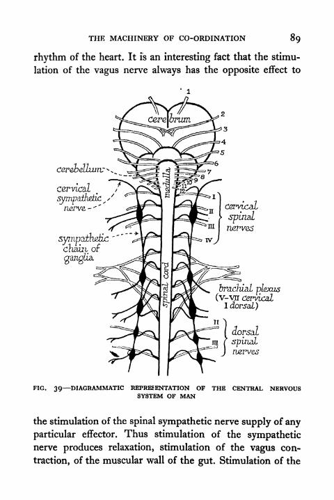
THE MACHINERY OF CO-ORDINATION 89
rhythm of the heart. It is an interesting fact that the stimu-
lation of the vagus nerve always has the opposite effect to
cerebellum; -.
Cervical
ympdfazrtTV-
br&dual plexus(v-Vff cervical
I dorsal)
ruerves
FIG. 39 DIAGRAMMATIC REPRESENTATION OF THE CENTRAL NERVOUSSYSTEM OF MAN
the stimulation of the spinal sympathetic nerve supply of any
particular effector. Thus stimulation of the sympatheticnerve produces relaxation, stimulation of the vagus con-
traction, of the muscular wall of the gut. Stimulation of the
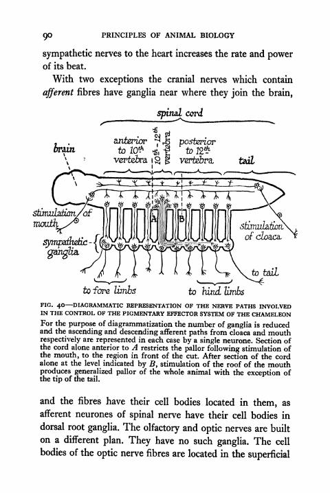
PRINCIPLES OF ANIMAL BIOLOGY
sympathetic nerves to the heart increases the rate and powerof its beat.
With two exceptions the cranial nerves which contain
afferent fibres have ganglia near where they join the brain,
spinal cord
brain
to fore limbs to hixuL limbs
FIG. 40 DIAGRAMMATIC REPRESENTATION OF THE NERVE PATHS INVOLVEDIN THE CONTROL OF THE PIGMENTARY EFFECTOR SYSTEM OF THE CHAMELEONFor the purpose of diagrammatization the number of ganglia is reducedand the ascending and descending afferent paths from cloaca and mouthrespectively are represented in each case by a single neurone. Section ofthe cord alone anterior to A restricts the pallor following stimulation ofthe mouth, to the region in front of the cut. After section of the cordalone at the level indicated by B, stimulation of the roof of the mouthproduces generalized pallor of the whole animal with the exception ofthe tip of the tail.
and the fibres have their cell bodies located in them, as
afferent neurones of spinal nerve have their cell bodies in
dorsal root ganglia. The olfactory and optic nerves are built
on a different plan. They have no such ganglia. The cell
bodies of the optic nerve fibres are located in the superficial
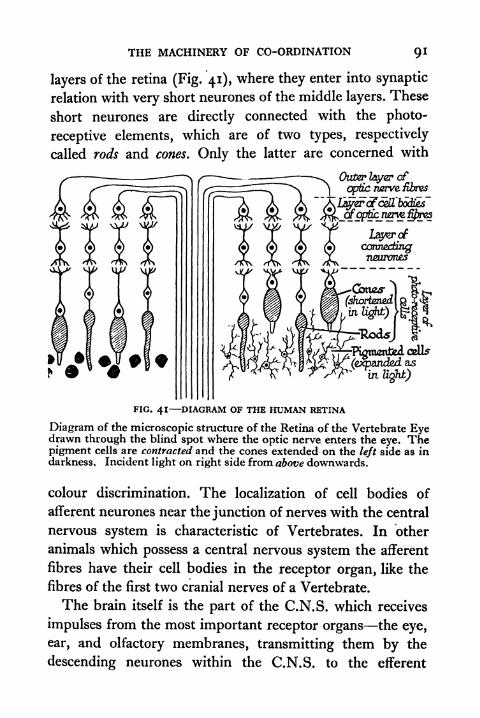
THE MACHINERY OF CO-ORDINATION QI
layers of the retina (Fig. 41), where they enter into synaptic
relation with very short neurones of the middle layers. These
short neurones are directly connected with the photo-
receptive elements, which are of two types, respectively
called rods and cones. Only the latter are concerned with
Outer layer of
optic nerve fibres
layer rf
connectingneuron&s
(expanctedasx
intighJb)
FIG. 41 DIAGRAM OF THE HUMAN RETINA
Diagram of the microscopic structure of the Retina of the Vertebrate Eyedrawn through the blind spot where the optic nerve enters the eye. Thepigment cells are contracted and the cones extended on the left side as indarkness. Incident light on right side from above downwards.
colour discrimination. The localization of cell bodies of
afferent neurones near the junction of nerves with the central
nervous system is characteristic of Vertebrates. In other
animals which possess a central nervous system the afferent
fibres have their cell bodies in the receptor organ, like the
fibres of the first two cranial nerves of a Vertebrate.
The brain itself is the part of the C.N.S. which receives
impulses from the most important receptor organs the eye,
ear, and olfactory membranes, transmitting them by the
descending neurones within the C.N.S. to the efferent
92 PRINCIPLES OF ANIMAL BIOLOGY
neurones that arise from the cord at different levels. For
anatomical description it is somewhat arbitrarily divided
into three regions: the fore-, mid- and Am</-brain.* The
fore-brain consists of two parts, a more anterior portion, the
dorsal aspect of which is called the cerebral hemispheres, and a
more posterior portion which is narrower (the thalamence-
phalori) and bears two interesting glandular structures on its
roof and floor. The former is the pineal body which in some
lizards has an eye-like structure. Whether it is of any im-
portance to the frog we do not know probably it is not. Thesecond is called the pituitary gland. Of this organ we shall
have more to say later. The roof of the mid-brain is defined
by two globular lobes, the so-called optic lobes. The hind-
brain is poorly developed in the frog. Its roof in the human
being is called the cerebellum. The first two pairs of cranial
nerves arise from the fore-brain, III arises from the floor of
the mid-brain, IV emerges dorsally between the mid- and
the hind-brain, and V-X arise from the floor of the hind-
brain. A frog in which the brain has been destroyed maybe kept alive indefinitely; and its behaviour is very much
simpler and therefore more easy to predict after careful
study. It is no longer subject to the very varied stimuli of
ever-changing images from the retina, and of smells or noises
in its vicinity. The interplay of all these helps to make the
behaviour of the intact frog a much more complicated affair.
The Study of Behaviour. When the influence of one
group of stimuli, e.g. light, predominates over all others,
it is possible to see how simple reflexes like the ones we can
study in the pithed frog determine the normal behaviour of
an intact animal. Thus it is proverbial that certain insects
move towards the light. A generation ago we should have
* See Appendix IV, p. 403.

THE MACHINERY OF CO-ORDINATION 93
said they move towards, the light because they like the light.
This tells us nothing. It is not really very different from the
habit of mind which made the alchemist speak of "spirits"
of nitre, "spirits" of salt, etc., attributing to personal agencies
all the properties of dead matter as the savage peopled his
universe with unseen beings. In general the phototactic
movements of insects depend on two facts : (a) light acting
FIG. 42 DIAGRAM TO ILLUSTRATE MOVEMENT OF INSECT (HOVER FLY)TOWARDS THE LIGHT (see text, p. 93).
on the same region of the same eye reflexly increases the
tension of muscles on one side of the body and reflexly relaxes
the same ones on the other; (b) light acting on one part of
the eye produces contraction of a particular group of muscles
and light acting on another part of the same eye producesreflex relaxation of the same group of muscles. Thus if one
eye of the hover fly, Eristalis, is blackened, a beam of light
focused on the anterior margin (A, Fig. 42) of the other
eye makes the animal bend the legs of the same side forwards
and the legs of the other side backwards, so that the bodytilts away from the side illuminated. If a beam is directed
on the posterior edge (P, Fig. 42), the legs of the opposite

94 PRINCIPLES OF ANIMAL BIOLOGY
side are bent forwards and the legs of the same side back-
wards so that the body tilts towards the side illuminated. Acomparatively feeble beam acting on the posterior margin
will compensate the effect of an intense illumination of the
region in front. When a fly is moving along a beam of light
the posterior edge of neither eye gets much light and one gets
as little as the other. When it is deflected to the right, the
posterior margin of the left eye gets more light (Fig. 42)
and this brings about the tilting of the body to the left. Its
direction of movement is at once restored, so that the eyes
are illuminated symmetrically. This is only realized when
the axis of the body is in line with the source of light.
With the knowledge that we now have, we can form a
clearer picture of the way in which such a receptor as the
statocyst of a shrimp, or its more complicated counterpart
the membranous labyrinth or inner ear of the frog, performsits role in the maintenance of normal posture. The inner
wall of the statocyst of the shrimp is covered with cells
provided with fine hairs projecting into its cavity (Fig. 30).
These cells are connected with the ends of separate nerve
fibres. The effect of a jerky movement is that the statolith
hits the wall of the statocyst as our bodies hit the wall of a
train if it starts suddenly when we are standing up. Hence it
strikes a new set of sensitive hairs. If different nerve fibres
of the statocyst belong to reflex arcs involving different sets
of muscles, one or another set of muscles will be broughtinto play by the mechanical stimulus of the statolith resting
on one or another set of hairs (Fig. 30). This, roughly speak-
ing, is the mechanism underlying the fact that a cat falls on
all fours.
In the spinal cord or brain of an animal like the frog (or
the writer of this book) the neurones do not lie in isolated
THE MACHINERY OF CO-ORDINATION Q5
reflex arcs, but each is connected by its ramifying branches
to a number of others. The occurrence of any reflex implies
that impulses pass across the junction between two particular
neurones more readily than across the junction of the same
neurones and all others. That is to say, reflex action is a
physical property of the junctions between nerve endings in
the C.N.S. Seeing that the animal is subjected to innumerable
stimuli simultaneously in normal life, its behaviour would
be quite chaotic were it not for an important feature of reflex
activity. Suppose the stimulation of a receptor A producesrelaxation and of B produces contraction of a certain muscle,
which in ordinary conditions is in the state of steady incom-
plete relaxation known as tone. Both reflexes have commonefferent connections, called by Professor Sherrington a
"final common path." When A and B are stimulated simul-
taneously, either A holds the field alone or B;that is to say,
the response is exclusively characteristic of stimulating
either A or B alone. Now in the C.N.S. the number of fibres
running up and down are by no means infinite in number,and enormous numbers of reflexes share common paths in
the cord. The fact that one group of receptors can use a
common path to the exclusion of any antagonistic reflex
system makes the behaviour of the animal more stable and
definite than it would otherwise be.
The Nature of Conscious Behaviour. The relatively simplebehaviour of the pithed frog as compared with that of the
intact animal is not exclusively due to the fact that the
influence of the great sense organs of the head is eliminated,
nor to the fact that many of the possible ascending nerve
paths, which converge on descending ones at a higher level,
are cut across. To some extent far less than the reader or
writer of this book the frog can, in the language of every-
96 PRINCIPLES OF ANIMAL BIOLOGY
day life, learn. The word learning is given to several ways in
which the behaviour of an animal can be modified by past
experience. One way is that a stimulus which has been
repeated a large number of times can become the signal for
a response which it did not formerly call forth. The appro-
priate conditions can now be studied by experiment and
described in the language of science.
The ability to build up new reflexes under definitely
ascertainable conditions is a physical property of the fore
brain or cerebrum. Noteworthy progress in defining the con-
ditions under which new reflex systems are built up has been
made during the present century by the Russian physio-
logists of Pavlov's school. This is a very revolutionary deve-
lopment of modern science, because it brings within the realm
of scientific treatment a body of experiences which have
hitherto been the province of metaphysics. The fact that it
is now possible to study the machinery by which the effect
of previous stimuli modify the behaviour of an animal at
any given moment, brings within the scope of scientific
treatment animal characteristics which have hitherto been
ascribed to non-measurable agencies such as "memory,""consciousness," "sensation," and the like.
The capacity to learn is one of a number of characteristics
of behaviour which distinguish the intact frog from the
brainless frog. These characteristics correspond to what are
called in traditional language "conscious behaviour." At one
time it was customary to assume that the behaviour of an
animal can be treated under two headings, "reflex" activity
which we have referred to briefly above and "voluntary
activity" or "conscious behaviour." The former alone was
regarded as the proper domain of the biologist, the latter
as the province of moral philosophy. The success of Pavlov's

THE MACHINERY OF CO-ORDINATION 97
researches show that conscious behaviour can be studied
scientifically and therefore controlled.
TOPICS FOR HOME STUDY1. Indicate the evidence on which our knowledge of the reflex arc
is based.
2. What is meant by (a) the nervous impulse; (6) a simple reflex
and (c) a conditioned reflex?
3. What are meant by photomechanical processes in the Vertebrate
eye?
4. What is meant by the autonomic nervous system, and how canits connexions be established.
5. What are the characteristics of the Vertebrate head?
6. What is meant by geotaxis and phototaxis ? Explain how photo-tactic movements are regulated in an insect.
CHAPTER IV
THE SOURCES OF ENERGY OFTHE ANIMAL BODY
WHEN any change occurs in the world around us, we
say that work has been done or that energy has been
transformed. A living creature is always changing in one
way or another. We have studied some of the characteristic
forms of change which it displays and the sequence of events
which constitute that extraordinary pattern of change which
we call an animal's behaviour. So now we have to ask, what
is the mechanism of all this transformation of energy which
is called life ?
Energy of the Living Machine. The fundamental Law of
Energy is the Principle of Conservation. A fixed quantity of
heat is always equivalent to a given amount of mechanical
work in any machine. When a machine is generating both
heat and movement we can express the total energy it is
giving out either in purely mechanical units or in heat
units (calories). When a motor bicycle is running the total
energy output (heat and movement) corresponding to a fixed
petrol consumption is always the same. The difference
between economical and uneconomical running of an enginelies in whether the ratio of mechanical to thermal produc-tion is higher or lower. If we run with the brakes partly on,
the same amount of energy is liberated by the explosion.
The difference in speed is made up by the heating of the
brake linings, brake drums, etc. When the petrol of a motor
bicycle gives out, the machine stops. Living matter, like
a motor bicycle, is constantly doing work, often like the

THE SOURCES OF ENERGY OF THE ANIMAL BODY 99
motor bicycle in the form of movement from one place to
another or of the emission of startling noises. From time
to time the living machine has to be fed, like the petrol
tank of the motor bicycle. Actually no living machines
feed on petrol like motor bicycles. But the energy of the
living machine depends on the chemical materials with which
it is supplied as truly as the energy of the motor bicycle
depends upon the petrol, benzene, paraffin, or other hydro-carbon with which it is supplied.
Living creatures, like a frog or a human being, are all
subject to the same inexorable restriction. Their principal
activities belong to the same two categories as those of the
motor bicycle. While the motor bicycle is working, petrol
is being burnt up. Motor bicycles breathe in oxygen and
breathe out carbon dioxide, just as they feed on petrol. If
we give a motor bicycle a full meal of petrol, let us say one
gallon, and run it until it stops, in general we find that it
has lost weight roughly equivalent to the weight of one
gallon of petrol at the end of the run. Of course, if allowed
to run inexpertly, it may become tired before the petrol has
given out. Insufficient lubrication results in over-heating,
or expansion of some essential fitting results in immobility.
In the meantime the air around has also suffered a change,
sometimes more impressive to the pedestrian than to the
cyclist. If the engine is running efficiently this change con-
sists in the disappearance of a certain quantity of oxygenand the production of a certain quantity of water vapourand carbon dioxide. If the combustion is incomplete the
motor bicycle excretes soot. The rate at which a motor
bicycle breathes is regulated by the air control. In all these
operations nothing is lost or gained in the net result. Whatthe bicycle loses in weight, the air gains.

100 PRINCIPLES OF ANIMAL BIOLOGY
The material balance sheet will show on the credit side the
weight of petrol consumed and the weight of oxygen lost by the
air. The debit side will include the weight of carbon dioxide
and moisture gained by the air together with the weight of
soot excreted. If it is difficult to empty the petrol tank, we
can get the weight of the petrol consumed by weighing the
machine before and after the trial period. If all the petrol
put in the tank is placed on the credit side, we must add the
increased weight of the machine to the debit side. The total
of one side is the same as the total of the other, and the same
is true of the analogous balance sheet which we can draw
up for human nutrition, respiration, and excretion. On the
credit side we have to reckon with food, drink, and oxygen.
On the debit side we put the carbon dioxide and water
excreted, together with any gain of body weight resulting
from the fact that some of the food has been retained for
growth. A typical balance sheet of an ordinary man weighingten stone, when supplied with food for one day in an air-
tight chamber, is given by Haldane and Huxley.* On the
credit side it shows :
Food . . . . . . i i kilos (60 per cent water)Drink . . . . . . i 5 kilos
Oxygen, 500 litres or . . 0-7 kilo
This makes a total of 3 30 kilograms. On the debit side
we have to reckon with the fact that he excretes during the
same time i 3 kilos of water and 70 grams of solids in his
urine. He loses i i kilos of water by evaporation from the
skin as sweat. He loses 425 litres of carbon dioxide or 0-82
kilo, making in all a net loss of 3 29 kilos. The difference of
10 grams represents exactly what he has gained in body-
weight at the end of the experiment. The carbon dioxide
* Animal Biology.
THE SOURCES OF ENERGY OF THE ANIMAL BODY IOI
that appears is equivalent to that which would be formed
by the same amount of food (allowing for what is retained for
growth) if burnt in a flask.
If a human being or a frog is placed in a heat-proof
box or calorimeter, the effect of any movements he mayexecute is to generate additional heat by friction with the
surrounding atmosphere, and all the energy he releases will
appear on the balance sheet of energy as an increase of heat
in the calorimeter. When this experiment is performed,
two things are found. The amount of energy expended is
definitely related to the oxygen taken in and the carbon
dioxide given off. In addition, the amount of energy
generated during the same period is equivalent to the heat
which would be generated by combustion of the quantity
of food consumed if it were burnt in another calorimeter.
In one experiment a corresponding balance sheet for the
energy expenditure of Haldane's ten-stone man over the
same period was drawn up somewhat as follows. Burnt out-
side the body, the same amount of food as he takes would
yield 2,400 kilocalories. The total heat lost by the body
during the period of experiment being 2,190 kilocalories, the
difference includes a deficiency, due to incomplete oxidation
of food. It can be proved from analysis of the excreta to be
equivalent to 150 kilocalories, leaving only 60 kilocalories to
account for. This represents the increased potential energy
of the body due to stored materials which appeared in the
previous balance sheet as increased weight.
Thus the two great mechanical principles called the con-
servation of energy and the conservation of matter both
apply to all man-made contrivances and all physical pheno-
mena. That is why it is right and proper to speak of the living
machine. We call a dynamo, a water-mill, a steam-engine.
102 PRINCIPLES OF ANIMAL BIOLOGY
and a motor bicycle machines because they transform energy
in accordance with certain definite rules, which are known
as the laws of Energetics. We call a frog or man a living
machine because it transforms one form of energy into other
forms of energy in accordance with the same rules. We shall
see later (p. 124) that the oxidation of food-stuffs is not the
direct source of the energy of bodily movement. From a
chemical point of view the muscle is more like a rocket than
an internal combustion engine.
Chemistry of the Body. To understand their food require-
ments we must bear in mind that frogs grow and reproduce
their kind, and it is hence necessary to inquire into the
chemical constituents of the animal body. The principal
constituents of the animal body are, first, water even manis at least 60 per cent water; secondly, dissolved in the
water, certain mineral salts notably those of sodium, cal-
cium, potassium, and magnesium in the form of chlorides,
bicarbonates, and phosphates ; finally, organic matter which
can be classified under three principal headings, carbo-
hydrates, fats, and proteins. The proteins are the most
important. They all contain carbon, hydrogen, oxygen, and
nitrogen: often sulphur and phosphorus. The molecule of
a simple protein may be more than a thousand times heavier
than the molecule of a compound like common salt.
The protein molecule is usually regarded as built up of
large numbers of amino acids. An amino acid is an organic
acid in which a hydrogen atom is replaced by an NH2 group,
e.g. amino-acetic acid is CH2NH2COOH. Fischer has syn-
thesized a compound containing eighteen amino acid group-
ings. Such a compound is vastly simpler than the simplest
protein, but we have moved a long way from the age when,in the very same vear that received Wohler's announcement

THE SOURCES OF ENERGY OF THE ANIMAL BODY 103
of the successful synthesis of urea, the great chemist Henrywrote (1827) concerning organic compounds:
It is not probable that we shall ever attain the power of imi-
tating nature in these operations. For in the functions of a living
plant a directing principle appears to be concerned peculiar to
animated bodies and superior to and differing from the cause whichhas been termed Chemical affinity.
The white of a fowl's egg is almost exclusively composedof the protein, egg albumen. Gelatin is another example of
a pure protein. Yet another is haemoglobin, the red colouring
matter of our blood. Carbohydrates contain carbon, hydrogen,
and oxygen only, the last two in the same proportion as in
water. The simplest carbohydrates are the simple sugars,
glucose and grape sugar. More complex sugars are formed
by the union of two molecules of simple sugars. Such are
cane sugar (the sugar we eat), malt sugar, and the sugar
contained in milk. The union of several molecules of simple
sugar gives rise to much more complicated compounds of
the same class, known as starch, cellulose, dextrins, etc.
Potato flour is largely composed of a starch. Rice paper is
largely composed of a dextrin. Cellulose, which forms a large
part of the dry substance of any plant, since all plant cells are
enclosed in a wall of cellulose, and lignin, the substance of
woody fibres, are also highly complex members of the
carbohydrate class. The fats also contain the same three
elements alone at least the simpler ones do; but their
chemical structure is quite different. A simple fat like lard
or olive oil is an ester of the trihydric alcohol glycerine and
one or more of the higher fatty acids (usually acids with at
least sixteen carbon atoms).
With the exception of the sugars, all these essential con-
stituents of the organic matter of the animal or plant body
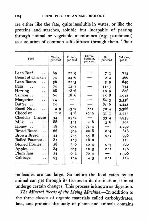
104 PRINCIPLES OF ANIMAL BIOLOGY
are either like the fats, quite insoluble in water, or like the
proteins and starches, soluble but incapable of passing
through animal or vegetable membranes (e.g. parchment)
as a solution of common salt diffuses through them. Their
molecules are too large. So before the food eaten by an
animal can get through its tissues to its destination, it must
undergo certain changes. This process is known as digestion.
The Mineral Needs of the Living Machine. In addition to
the three classes of organic materials called carbohydrates,
fats, and proteins the body of plants and animals contains
THE SOURCES OF ENERGY OF THE ANIMAL BODY 105
various mineral salts dissolved in the tissue fluids, such as
the cell sap of plants, the blood, and the lymph or clear
fluid in the body cavities of animals. The mineral needs of
the body may be divided under two headings. First come
elements which enter into the composition of the organic
materials. Thus most proteins contain sulphur. Many con-
tain phosphorus. Haemoglobin contains iron. Haemocyanin,the corresponding blue respiratory pigment of lobsters and
snails, contains copper. The plant gets sulphur and phos-
phorus as it gets the nitrogen necessary to build up the
protein molecule from mineral salts (sulphates or phos-
phates). Animals get their proteins ready made. So the supply
of sulphur and phosphorus is usually adequate if the nitro-
genous content of the diet is sufficient.
Magnesium is specifically necessary for the green plant
because chlorophyll is an organic magnesium compound. In
Vertebrate animals (fishes, reptiles, birds, and mammals) the
thyroid gland in the neck manufactures an organic com-
pound of which the molecule contains iodine (see p. 165). Onthis account iodine is essential to healthy growth. Iodine is
not present in appreciable quantities in other animal tissues.
For plants and Invertebrates it does not seem to be an essen-
tial element, though it is present in seaweeds. Near the
sea the quantities present in salt carried by the breeze
ensures a supply adequate to animal needs. Some inland
and mountainous districts of Europe and America have a
very low soil iodine content. Thyroid deficiency diseases
(goitre, cretinism, etc.) of human beings and livestock are
therefore endemic. This is remediable by a very small
addition of iodine to the diet. In Switzerland the State
provides iodized table salt, the necessary iodine content of
which need not be greater than one-millionth of an ounce
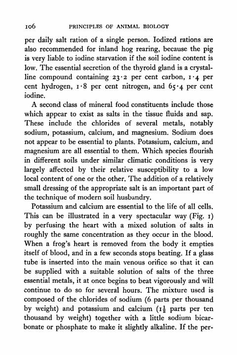
106 PRINCIPLES OF ANIMAL BIOLOGY
per daily salt ration of a single person. Iodized rations are
also recommended for inland hog rearing, because the pig
is very liable to iodine starvation if the soil iodine content is
low. The essential secretion of the thyroid gland is a crystal-
line compound containing 23-2 per cent carbon, 1-4 per
cent hydrogen, 1-8 per cent nitrogen, and 65-4 per cent
iodine.
A second class of mineral food constituents include those
which appear to exist as salts in the tissue fluids and sap.
These include the chlorides of several metals, notably
sodium, potassium, calcium, and magnesium. Sodium does
not appear to be essential to plants. Potassium, calcium, and
magnesium are all essential to them. Which species flourish
in different soils under similar climatic conditions is very
largely affected by their relative susceptibility to a low
local content of one or the other. The addition of a relatively
small dressing of the appropriate salt is an important part of
the technique of modern soil husbandry.
Potassium and calcium are essential to the life of all cells.
This can be illustrated in a very spectacular way (Fig. i)
by perfusing the heart with a mixed solution of salts in
roughly the same concentration as they occur in the blood.
When a frog's heart is removed from the body it emptiesitself of blood, and in a few seconds stops beating. If a glass
tube is inserted into the main venous orifice so that it can
be supplied with a suitable solution of salts of the three
essential metals, it at once begins to beat vigorously and will
continue to do so for several hours. The mixture used is
composed of the chlorides of sodium (6 parts per thousand
by weight) and potassium and calcium (i| parts per ten
thousand by weight) together with a little sodium bicar-
bonate or phosphate to make it slightly alkaline. If the per-
THE SOURCES OF ENERGY OF THE ANIMAL BODY 107
fusion fluid is replaced by a mixture from which either the
calcium or potassium is omitted, the heart-beat comes to a
standstill. This happens almost instantaneously if no calcium
is supplied. After being stopped in this way, the beat resumes
almost at once when the heart is supplied with the original
mixture. If the solution contains a little sugar the isolated
heart can be made to beat for several weeks. Otherwise it
gradually uses up its food reserves, and then ceases to be
able to work. The same results can be obtained with the
sheep's heart, which can also be made to go on workingoutside the body for at least three weeks, provided the tem-
perature of the fluid is kept near blood heat, i.e. about 36 C.
For prolonged experiments of this kind a trace of magnesiumis necessary. Lately it has been shown that small traces of
manganese may be essential to fertility in mammals. It is
not yet known how it is used. Manganese is also necessary
to many plants.
Animals are not liable to magnesium deficiency since all
green plants contain an abundance of magnesium for their
needs. On the other hand, plants which grow on soils with
a low lime content may contain a relatively small amount of
calcium. Hence herbivorous animals like the pig, which is
highly susceptible to calcium deficiency, are liable to various
diseases (especially rickets) unless lime fertilizers are applied
to the soil or an extra calcium ration is given. The figure
already cited with reference to iodine illustrates the general
principle that a soil deficiency which might completely
exclude the possibility of successful livestock husbandry can
often be remedied at a negligible cost. The provision of suit-
able mineral rations or soil dressings is one way in which
advancing biological knowledge is making it possible to
break down the natural limitations of locality.
108 PRINCIPLES OF ANIMAL BIOLOGY
Vitamins. In the first decade of the present century the
chemistry of foodstuffs had progressed so far that it was
possible to carry out more exact observations by feeding
animals on diets of chemically pure protein, fat, and carbo-
hydrate. The animals reared on such diets refused to grow,
though they thrived if comparatively small traces of natural
foods were added. Further research showed that different
symptoms of disorders produced by exclusive feeding on
such purified rations could be eliminated by the addition of
small quantities of ordinary articles of food. So it has been
possible to distinguish different "accessory food factors," the
absence of any one of which is associated with a particular
defect.
Generically these substances have been called vitamins.
The name does not mean that they have anything in commonbesides the fact that they do not belong to the classes of diet
constituents previously believed to be all-inclusive, the fact
that the requisite quantities are very small, that they all
appear to be organic compounds, and that their chemical
constitution was unknown when the word was first intro-
duced. The chemical constitution of several of them is now
known, some can be synthesized, and others will be syn-
thesized in the near future. Research along these lines was
encouraged by the conditions of food shortage during the
European war of 1914-18 when "nutritional" diseases like
scurvy and rickets came into prominence.
Scurvy is due to the absence of a comparatively simple
substance in the diet. It is ascorbic acid. Before its chemical
nature had been determined it was called "vitamin C."
Ascorbic acid has the formula C6H
8OG . It has alcoholic and
ketone characteristics on account of four OH and one COradicle in the molecule. It is present in most fresh fruit and

THE SOURCES OF ENERGY OF THE ANIMAL BODY 109
in many vegetables, especially in oranges, lemons, black
currants, green leaves, and potatoes. Since its molecular
constitution has been discovered, its commercial manu-
facture from glucose has been started.
Calciferol ,with the formula C28II43OH, is one of the
known forms of another vitamin, "D." It is produced bythe action of ultra-violet light on another alcohol called
ergosteroL Ergosterol has the same formula C28H43OH with
a different internal arrangement of the atoms in its molecule.
The exact chemical constitution of both substances is now
known. Various fish liver oils contain another form of vita-
min D whose formula is C27H
41OH. So also does butter fat.
The D content of the husks of ripe cocoa beans is high, and
the latter can be used in cattle feeding to raise the D content
of the milk. The absence ofD is responsible for the deficiency
disease called rickets, still common among the poorer section
of the population. Rickets can be cured and, of course, pre-
vented by administering any form of vitamin D, or by
exposure to ultra-violet radiation from intense sunlight or a
mercury vapour lamp. The exposure leads to the productionofD in the skin from pro-vitamins such as ergosterol. Rickets
is a deficiency disease affecting especially bone and tooth
development in children. They are especially prone to it if
brought up in dingy dwellings and sunless cities, unless the
lack of ultra-violet light in their normal environment is
counterbalanced by a diet containing excess of D, that is to
say a diet with plenty of fresh eggs, summer milk, butter
with, preferably to make sure, some fish liver oil or calciferol
preparation. Pigs are also liable to get rickets. Modern pig-
breeders take special precautions to avoid this. In Scandi-
navian countries glass which transmits ultra-violet light is
used for the windows of pig-styes. There has been more
110 PRINCIPLES OF ANIMAL BIOLOGY
progress in the education of Danish pig-breeders than in
the education of British politicians.
Scurvy and rickets are the chief diet "deficiency" diseases
which have attracted attention in the West. There are several
others. Apparently the animal body needs a substance which
is abundant in the husk but not in the grain of plant seeds
like cereals. Its absence produces severe muscular weakness,
especially in birds. A similar condition in human beings is
the disease called "beri-beri" to which Japanese peasants
who live largely on a diet of polished rice are liable. This is
because removal of the husk removes "vitamin B r"
Beri-
beri can be cured by the administration of suitable extracts
made from these husks, from wheat germ, or from yeast.
The active chemical constituent is apparently an unusual
type of organic compound with the provisional formula
C12H17O N4S . Cl. To keep animals healthy on diets of pure
proteins, fats, carbohydrates, and mineral salts it is essential
to add various other substances like the above in minute
quantities. One of these, called vitamin A, is present along
with D in butter fat, egg yolk, and cod-liver oil, especially in
the first two. It is also an alcohol with the formula C20H29OH,
chemically related to the plant pigment carotin. The animal
body can convert the latter into A. If the diet is deficient in
A, or in carotin, its pro-vitamin, animals show general
liability to bacterial infections, and an adequate minimum
seems to help resistance to diseases like colds and influenza.
Another, called vitamin B2 ,has a rather similar distribution
to B1 , and was at first confused with it. Deficiency of one
of the B vitamins is responsible for pellagra, a disease of the
digestive system and skin. The composition of what is now
called vitamin E, present in cereal oils and green leaves,
especially lettuce, is not yet known. If it is absent complete
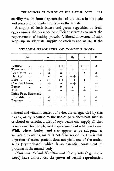
THE SOURCES OF ENERGY OF THE ANIMAL BODY HI
sterility results from degeneration of the testes in the male
and resorption of early embryos in the female.
A supply of fresh butter and green vegetables or fresh
eggs ensures the presence of sufficient vitamins to meet the
requirements of healthy growth. A liberal allowance of milk
keeps up an adequate supply of calcium and of B 2 . If the
VITAMIN RESOURCES OF COMMON FOOD
mineral and vitamin content of a diet are safeguarded by this
means, or by recourse to the use of pure chemicals such as
calciferol or carotin, a diet of soya beans can supply all that
is necessary for the physical requirements of a human being.
While wheat, barley, and rice appear to be adequate as
sources of proteins, maize is not. The reason for this is that
digestion of maize protein does not yield one of the amino
acids (tryptophane), which is an essential constituent of
proteins in the animal body.
Plant and Animal Nutrition. A few plants (e.g. duck-
weed) have almost lost the power of sexual reproduction
112 PRINCIPLES OF ANIMAL BIOLOGY
and propagate mainly by the vegetative method, and a few
plants and animals (e.g. the parasitic maggot of the horse-
fly) do not use oxygen to burn up waste materials. Apart
from such isolated exceptions it is correct to say that two
processes are common to animals and plants. One is sexual re-
production. The other is respiration or the oxidation of organic
material with the production of carbon dioxide and water.
The most obvious differences between animals and plants
are characteristics of behaviour. Animals have a greater
range and scope of reversible responses than plants; and
more specialized sensitivity to changes in their surroundings.
Plants have greater power of rapid and sustained growth than
animals. A striking structural difference between the tissues
of animals and plants is that animal cells are naked and plant
cells have an inert cell wall nearly always composed of
cellulose. These differences are associated with others which
are less obvious but equally significant. All animals are
dependent on food containing complex organic substances.
The animal cannot synthesize amino-acids, fats, or carbo-
hydrates from simpler organic compounds. It has to get
them by eating other living things or by living saprophyti-
cally, i.e. on decaying organic matter. The green plant
needs only carbon dioxide, water, and simple mineral consti-
tuents of the soil such as nitrates, phosphates, and sulphatesto build all the complex organic compounds which are
essential constituents of living matter. Energy is necessary
to make complex organic compounds which give up energywhen they are broken down into simpler ones, and the
green plant absorbs this energy from sunlight by means of
the assimilatory pigment chlorophyll. This leads to the
formation of sugar from carbon dioxide and water with the
liberation of oxygen.
THE SOURCES OF ENERGY OF THE ANIMAL BODY 113
Even plants, such as the mushroom or the dodder, which
have taken to a saprophytic or parasitic habit and have lost
their chlorophyll, are able to synthesize proteins if they are
supplied with sugar and nitrates. The ability of plant cells to
synthesize any essential amino-acid molecule and the inability
of the animal body to do so is therefore a fundamental differ-
ence between animals and plants. There are certain other
differences between the chemical processes which occur in
plant and animal cells, as, for instance, the fact that althoughsome animals can synthesize vitamins A, C, and D, no animals
are yet known to be able to synthesize Bj,, B2 ,or E, as plants
can.
TOPICS FOR HOME STUDY1 . Discuss what is meant by the living machine.
2. Tabulate the differences between animals and plants from the
standpoint of structure, mechanism, and reproduction.
ii

CHAPTER V
DIGESTION, RESPIRATION AND EXCRETION
Tyl7E have now seen from what sources the animal body
V V replenishes its stores of potential energy and obtains
the raw materials essential for growth and repair of tissue
waste. Our next task will be to examine how the food is made
available to the cellular engines of the body, and how the
products of combustion are removed.
Digestion. Frogs feed on worms, insects, and the like.
These, like our own food and that of all animals, contain all
three classes of essential organic compounds the body re-
quires for growth and activity. It may be asked, where do
these come from in the first place? Those animals that do
not feed on other animals are vegetarians; and plants pos-
sess a peculiarity of nutrition that animals do not. They can
build up carbohydrates from the carbon dioxide and water
of their environment and build up proteins as well. Since
these are the things which supply chemical energy to the
living machine, it may be asked, whence comes all the energy
required to build up these stores of chemical energy? Theanswer is that plant tissues absorb light by means of the green
colouring matter, chlorophyll, of the stem and leaves: only
in the day-time can they build up starch. The ultimate source
of the energy of life on this planet is therefore the radiant
energy of the sun.
From the mouth the food of man or of any other Verte-
brate animal passes into a winding tube which terminates
in a second opening to the exterior, the anus, situated
dorsally where the legs are joined to the body. This tube,

DIGESTION, RESPIRATION AND EXCRETION 115
known as the alimentary canal, or gut (Fig. 43), has every-
where muscular walls, an inner layer of muscle fibres
arranged circle-wise, and an outer coat with a lengthwise
arrangement. The slow, rhythmical movements of these
muscles help to churn the food, and squeeze it along the
alimentary canal to the anus. The inner wall of the gut is
xutfcernaZ
(fH
glottis- *j
~~~ -liver
!*pancreas*
*-j Zazge intestine,
FIG. 43 ALIMENTARY CANAL AND ASSOCIATED STRUCTURES IN THE FROG
lined with a layer of cells which for a great part of its course
consists of secreting cells and is folded in pits. The first
part of the tube, which in our own case is called the
cssphagus, or gullet, opens into a capacious bag. This bag,
the stomach, leads into a narrower tube, the intestine. The
intestine of Vertebrates has two distinct portions, one
narrower, called the small intestine, and the other wider,
called the large intestine. Its final part, the rectum, com-
municates with the exterior by the anus (Fig. 44).
In its passage along the alimentary canal human food
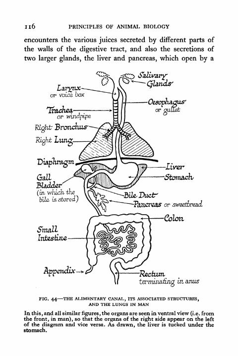
n6 PRINCIPLES OF ANIMAL BIOLOGY
encounters the various juices secreted by different parts of
the walls of the digestive tract, and also the secretions of
two larger glands, the liver and pancreas, which open by a
Larynxor voice.
or wijiopips
Right Bronchus
Right Lung.
^jji$\mm^-Liver
StomachGallBUdj&zr(izi which thz
bile is stored)or sweetbread
Colon
zi anus
FIG. 44 THE ALIMENTARY CANAL, ITS ASSOCIATED STRUCTURES,AND THE LUNGS IN MAN
In this, and all similar figures, the organs are seen in ventral view (i.e. fromthe front, in man), so that the organs of the right side appear on the left
of the diagram and vice versa. As drawn, the liver is tucked under thestomach.
DIGESTION, RESPIRATION AND EXCRETION 1 17
common duct into the small intestine near its anterior end.
These digestive juices contain substances known as enzymes
which act on the complex organic constituents of the food,
turning them into compounds sufficiently simple to diffuse
through the walls of the intestine or sufficiently simple for the
cells of the body to build them up into other substances as
these may be required.
Enzyme Action. Chemical reactions may be divided into
two categories. One includes all such as are involved in
ordinary methods of volumetric analysis, where the com-
bination of reacting substances is practically instantaneous,
as, for instance, the neutralization of hydrochloric acid with
soda or the precipitation of barium sulphate from a soluble
barium compound in the presence of free SO4 ions. The
other includes most organic reactions, such as the saponifica-
tion of esters or the inversion of cane sugar, in which an
appreciable period of time must elapse before the point of
equilibrium is reached. The reactions with which the
biologist is concerned belong pre-eminently to the latter
class. Many reactions such as the combination of pure
oxygen and hydrogen ordinarily proceed at an immeasurablyslow rate, but can be induced to take place within ordinary
time limits in the presence of certain substances (in this case,
e.g., finely divided platinum), which do not appear to have
undergone any change whatsoever at the conclusion of the
experiment. For such bodies as are capable of influencing
the velocity of a chemical reaction without entering into the
composition of the end products, or, in general, shifting
the point of equilibrium for a given set of conditions, the
term catalyst is commonly employed. The facility and
rapidity with which the organism is enabled to disintegrate
highly stable compounds, such as proteins, is due to the

1 1 8 PRINCIPLES OF ANIMAL BIOLOGY
agency of a special type of catalyst known as enzymes, of which
a culinary example familiar to most of us is the rennin of the
gastric juice.
The term enzyme was introduced by Kuhne (1878) to
remove the confusion resulting from the use of the older
term "ferment," a term originally used to include micro-
organisms like the yeast fungus. This confusion was finally
dissipated when Buchner (1903) extracted from crushed
yeast cells an "unorganized" ferment capable of effecting
the identical transformation hitherto associated with the
activities of the living organism.
Apart from the fact that they are all derived from living
organisms, it is difficult to fix on any single characteristic
which will differentiate enzymes from all other types of
catalysts. At present their chemical constitution is unknown.
What may be described as the general catalytic properties
of enzymes may be classified under three headings, the
reversibility of their action, their efficacy in very small
quantities, and the independence of the amount present to
the final result. Of these the first is of special importance to
the understanding of the chemical balance of the body, or,
to use the term usually employed for the chemical processes
of the organism as a whole, metabolism.
A straightforward illustration of what is implied by the
term reversibility of reaction is provided by the preparation
of ethyl acetate, which was the subject of the historic re-
searches of Berthellot and Pean de Saint Gilles (1862) on
mass action. If equal amounts of molar solutions of acetic acid
and ethyl alcohol are brought into contact an equilibrium
point is reached when the molecular concentration of the
reacting compounds is reduced by two-thirds owing to the
formation of ethyl acetate. Precisely the same point of equili-
DIGESTION, RESPIRATION AND EXCRETION 1 19
brium is reached when a molar solution of ethyl acetate is
subjected to acid hydrolysis, i.e. broken down by the catalytic
action of a mineral acid. It is usual to think of both reactions
as taking place continuously and simultaneously with
different velocities except at the point of equilibrium con-
centration for the reacting substances. The catalyst, in this
case an acid, acts by accelerating the reaction in either
direction, and the equilibrium point for the same conditions
of concentration etc., is unchanged, so that the total energy
of the system is unaltered.
Theoretically any reaction may be regarded as reversible.
In their efficacy to accelerate the velocity of reaction in either
direction, enzymes conform to the behaviour of other
catalysts. The first demonstration of this was due to Croft
Hill (1898) who studied the hydrolysis of the disaccharide
malt sugar under the influence of the enzyme maltase. Malt
sugar is split by this enzyme into the simpler sugar d-glucose
(a monosaccharide). Hill showed that when maltase is added
to glucose, malt sugar is formed, and the equilibrium point
is reached at the same relative concentration of the two
sugars as in the reverse reaction for identical conditions.
Needless to say, any reversible reaction may proceed to com-
pletion either way by the continuous removal of one of the
end products by means either mechanical (e.g. diffusion),
or chemical (e.g. precipitation). Hill's discovery makes it
possible to envisage how, for example, the balance of sugar in
the bodyand glycogen in the liver and muscles is continuously
maintained throughout life, or how, to take a second illustra-
tion, the tissues store up fat when the diet is plentiful, while
in times of shortage fat disappears from them.
In true catalysis the final result of a reaction is not deter-
mined by the quantity of the catalyst present. Like in-
120 PRINCIPLES OF ANIMAL BIOLOGY
organic catalysts an enzyme can often be reclaimed at the
end of a reaction and made to react on a fresh supply of
material. This is not always so. Enzymes are highly unstable
bodies, being readily destroyed, like many other colloidal
systems, by heat. Surprisingly small quantities of enzymeare efficacious. Rennet, the gastric juice extract employed
domestically for the preparation of junkets and commer-
cially on a large scale in the manufacture of cheese, is able
to clot 400,000 times its own weight of the milk protein
caseinogen. Even this figure gives an imperfect picture of the
prodigious activity of the enzyme. The active constituent
is very small in proportion to the bulk of the extract.
Digestive Enzymes. The secretion of the stomach or
gastric juice contains free hydrochloric acid. It also contains
an enzyme pepsin which breaks down complex proteins, like
albumen, into very simple proteins that are called peptones.
It has no action on the starchy or fatty constituents of the
food. The bile or secretion of the liver does not contain a
digestive enzyme. It contains excretory matter in the form
of certain pigments together with alkaline salts which
neutralize the acidity of the gastric juice, and greatly facilitate
the emulsification of fats.
The pancreas or sweetbread is the chief digestive gland.
Its secretion contains three important enzymes. One called
amylase breaks down starch or dextrin into malt sugar. Asecond called trypsin can break down proteins into the
organic "amino" acids of which the protein molecule is built
up. It thus carries protein digestion a stage farther than that
reached in the stomach. A third called lipase breaks up fat
into fatty acid and glycerol. The intestinal juice secreted bythe glandular walls of the small intestine also contains
enzymes, one callederepsin,
which acts on the final stages of
DIGESTION, RESPIRATION AND EXCRETION 121
protein decomposition, -completing the work of the pan-creatic juice, and a series of others which act on the complex
sugars like malt sugar, converting them into simple sugars.
Man possesses three pairs of salivary glands whose ducts
open into the mouth. Our saliva contains an enzyme which
in comparatively alkaline and neutral solution acts like the
pancreatic amylase on starch. It cannot act in the acid
medium of the stomach contents, and vigorous mastication,
while probably an admirable means of preserving the teeth,
has no justification on the grounds of facilitating starch
digestion to any considerable extent. The digestive tract of
man does not contain any enzyme that breaks down cellulose.
Some cellulose is broken down by bacteria in the large in-
testine, and is wasted. This is a very strong argument against
an exclusively vegetarian diet. Another objection to vege-
tarianism is that the cellulose wall of plant cells makes them
much less accessible to the action of the digestive juices than
animal tissues. The chief value of vegetable food lies in the
fact that it can be eaten raw as a supply of certain constituents
of a normal diet, the so-called vitamins, which are requisite
in extremely minute quantities and have not as yet been
identified chemically. The vitamin content of food is
diminished by prolonged cooking.
Any undigested or indigestible matter eventually passes
out of the body, along with the bile pigments that confer
its characteristic colour, as faeces. In the small intestine the
starches have all been absorbed as sugar by the fine blood
vessels beneath its lining. The proteins have been broken
down into the diffusible substances known as amino-acids
and travel to the tissues by the same route. All the blood
returning from the alimentary tract passes through the
capillary network of the liver en route for the heart. The
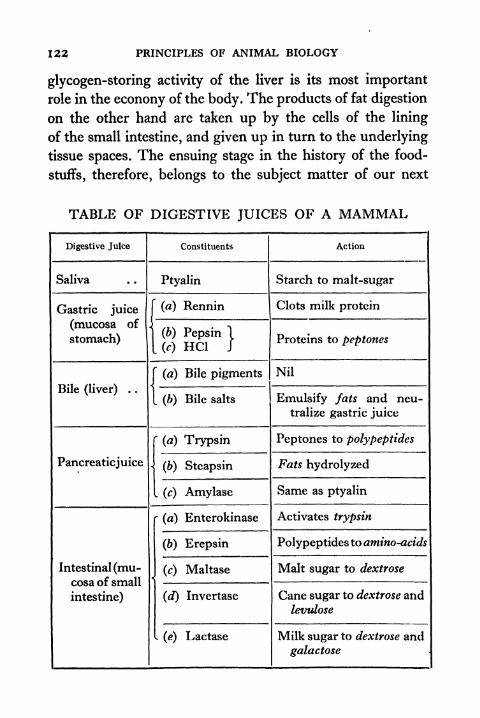
122 PRINCIPLES OF ANIMAL BIOLOGY
glycogen-storing activity of the liver is its most important
role in the econony of the body. The products of fat digestion
on the other hand are taken up by the cells of the lining
of the small intestine, and given up in turn to the underlying
tissue spaces. The ensuing stage in the history of the food-
stuffs, therefore, belongs to the subject matter of our next
TABLE OF DIGESTIVE JUICES OF A MAMMAL
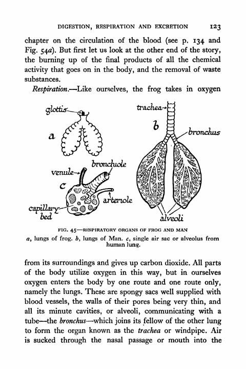
DIGESTION, RESPIRATION AND EXCRETION I23
chapter on the circulation of the blood (see p. 134 and
Fig. 540). But first let us look at the other end of the story,
the burning up of the final products of all the chemical
activity that goes on in the body, and the removal of waste
substances.
Respiration. Like ourselves, the frog takes in oxygen
3 ^-bronchus
alveoli
FIG. 45 RESPIRATORY ORGANS OF FROG AND MAN
a t lungs of frog, b, lungs of Man. c, single air sac or alveolus fromhuman lung.
from its surroundings and gives up carbon dioxide. All parts
of the body utilize oxygen in this way, but in ourselves
oxygen enters the body by one route and one route only,
namely the lungs. These are spongy sacs well supplied with
blood vessels, the walls of their pores being very thin, and
all its minute cavities, or alveoli, communicating with a
tube the bronchus which joins its fellow of the other lung
to form the organ known as the trachea or windpipe. Air
is sucked through the nasal passage or mouth into the
124 PRINCIPLES OF ANIMAL BIOLOGY
windpipe at the back of the throat by the suction of a broad
sheet of muscle, the diaphragm, beneath the lungs and above
the stomach. In the frog absorption of oxygen and elimina-
tion of carbon dioxide also takes place all over the surface of
the body through the moist skin. The lungs are less spongythan ours are and have relatively fewer compartments. The
two lungs open together by a longitudinal slit, the glottis,
at the back of the throat. The frog breathes by swallowing
air. It has no diaphragm.In the frog the main use of food is to supply chemical
materials for the activities of the organs, e.g. glycogen or
animal starch for the muscles, and materials for growingtissues. The frog is much less capable of surviving cold than
we are. Man is a warm-blooded animal and maintains a
temperature higher than that of his surroundings by the
oxidation of foodstuffs. Man requires relatively more food
than the frog for this reason. Though oxygen is essential for
sustained muscular activity, the energy of muscular activity
does not come from the oxidation of materials in the muscles.
Muscles will contract in the absence of oxygen. The energyof muscular movement comes from the explosive breakdown
of an organic phosphorus compound. This reaction is accom-
panied by the formation of lactic acid. If lactic acid accumu-
lates, fatigue ensues. By burning up the lactic acid into
carbon dioxide and water vapour, a constant supply of
oxygen makes possible the removal of the fatigue products.
In the efficient activity of an animal, non-volatile products
of muscular work are burnt up as soon as they are formed.
For this reason muscular exercise is quantitatively related to
oxygen consumption in the normal course of events, and it
has been customary in the past to regard the oxidation of the
foodstuffs as the source of muscular energy. Strictly speak-
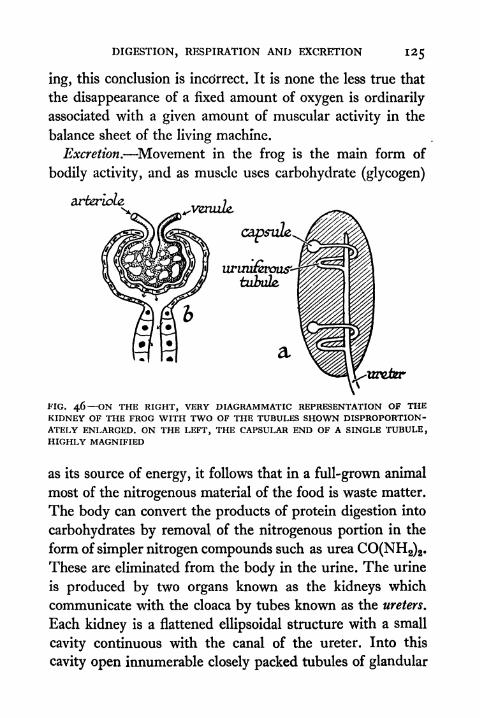
DIGESTION, RESPIRATION AND EXCRETION 125
ing, this conclusion is incclrrect. It is none the less true that
the disappearance of a fixed amount of oxygen is ordinarily
associated with a given amount of muscular activity in the
balance sheet of the living machine.
Excretion. Movement in the frog is the main form of
bodily activity, and as muscle uses carbohydrate (glycogen)
arteriolzxVenuZe
FIG. 46 ON THE RIGHT, VERY DIAGRAMMATIC REPRESENTATION OF THEKIDNEY OF THE FROG WITH TWO OF THE TUBULES SHOWN DISPROPORTION-ATELY ENLARGED. ON THE LEFT, THE CAPSULAR END OF A SINGLE TUBULE,HIGHLY MAGNIFIED
as its source of energy, it follows that in a full-grown animal
most of the nitrogenous material of the food is waste matter.
The body can convert the products of protein digestion into
carbohydrates by removal of the nitrogenous portion in the
form of simpler nitrogen compounds such as urea CO(NH2)2 .
These are eliminated from the body in the urine. The urine
is produced by two organs known as the kidneys which
communicate with the cloaca by tubes known as the ureters.
Each kidney is a flattened ellipsoidal structure with a small
cavity continuous with the canal of the ureter. Into this
cavity open innumerable closely packed tubules of glandular

126 PRINCIPLES OF ANIMAL BIOLOGY
structure which make up the substance of the kidney. These
tubules terminate in a flask-shaped swelling whose walls are
invaginated to receive a tuft of small blood vessels, this por-
tion being called the Malpighian body, or capsule, after
Malpighi (1628-94), one of the earliest microscopists. The
structure of our own kidney is essentially like that of the
frog in this respect, but in man the ureters open into a
capacious receptacle with muscular walls, the bladder. This
communicates with the exterior by a duct called the urethra,
separately in the female and along with the male generative
ducts in the male.
History of Digestion and Respiration. -We have said
nothing so far about the history of metabolism. The chemis-
try of the animal body could not be studied at all until com-
paratively recent times, and in point of fact the applicability
of the two great scientific generalizations known as the
indestructibility of matter and the conservation of energy
were applied to animals almost as soon as they were recog-
nized to hold good in the world of dead matter. Towards
the end of the eighteenth century the ingenious Abbot
Spallanzani made observations on the digestion of meat in
the stomach by making dogs swallow pieces of meat attached
to string, and withdrawing them at intervals. He also showed
that the gastric juice would perform the same action outside
the body. Under the influence of Liebig and others in the
first half of the nineteenth century, the foundations ofmodern
knowledge about digestion had been laid down.
The experiments of Robert Hooke with his air pump first
showed the necessity of air to animal life. Hooke also showed
that only a part of the air was so essential. The history of
the study of respiration is closely bound up with the dis-
covery of oxygen, nitrogen, hydrogen, and carbonic acid gas.
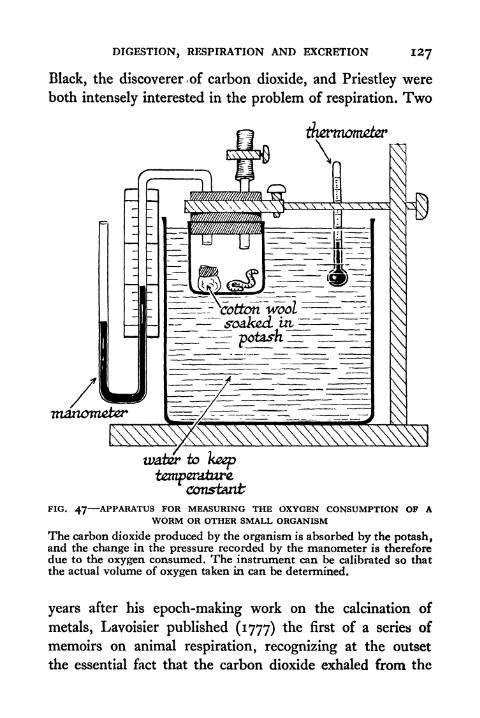
DIGESTION, RESPIRATION AND EXCRETION I27
Black, the discoverer -of carbon dioxide, and Priestley were
both intensely interested in the problem of respiration. Two
jfamnonuzter
\
manometer
_^ soaked, ui
potash jz: m_
water to
temperature,constant
FIG. 47- -APPARATUS FOR MEASURING THE OXYGEN CONSUMPTION OF AWORM OR OTHER SMALL ORGANISM
The carbon dioxide produced by the organism is absorbed by the potash,and the change in the pressure recorded by the manometer is thereforedue to the oxygen consumed. The instrument can be calibrated so that
the actual volume of oxygen taken in can be determined.
years after his epoch-making work on the calcination of
metals, Lavoisier published (1777) the first of a series of
memoirs on animal respiration, recognizing at the outset
the essential fact that the carbon dioxide exhaled from the

128 PRINCIPLES OF ANIMAL BIOLOGY
lungs was the product of an oxidation process, in which the
source of oxygen was the atmosphere. In 1780, in collabora-
tion with the great mathematician Laplace, Lavoisier pub-lished a memoir on animal heat, showing by direct physical
measurement with the balance and the thermometer that
the warmth of our bodies is a process of combustion of a
kind essentially like that of the candle flame. After the dis-
covery of hydrogen by Cavendish, Lavoisier discovered that
water vapour as well as carbon dioxide arises from the
oxidation processes of the body. In 1803 Spallanzani pub-lished a memoir recording large numbers of experiments on
a variety of animals (Vertebrate and Invertebrate) showingthat the phenomena of respiration are universal properties
of the animal economy, and that the seat of the respiratory
processes is the tissues themselves.
TOPICS FOR HOME STUDY1. Compare the method of breathing in Man, the Frog, and a bird
(refer to Rogers (1938), Textbook of Comparative Physiology
(McGraw-Hill); Barcroft (1928), Respiratory Function of the
Blood (Cambridge) ; Krogh, Respiratory Exchange of Animalsand Man: Monographs on Biochemistry).
2. Describe the structure of the Vertebrate Kidney and the func-tions of its parts.
3. Describe the digestion of fats, carbohydrates, and proteins in the
animal body.
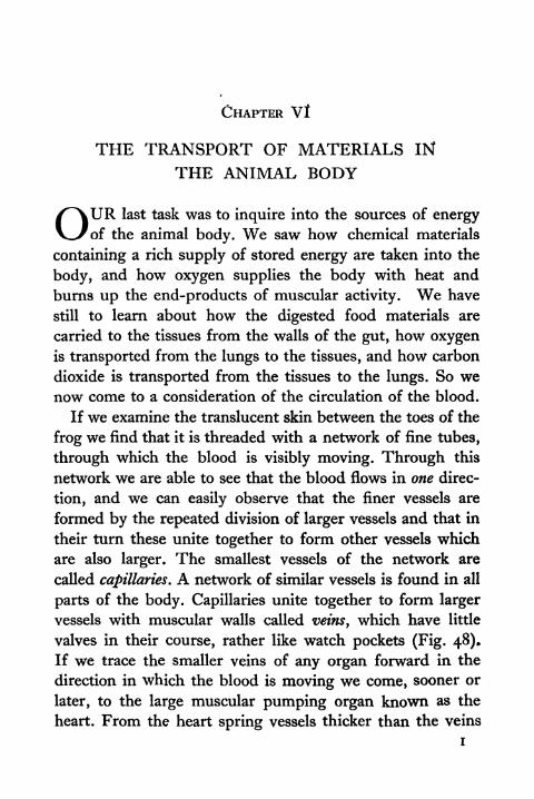
CHAPTER Vl
THE TRANSPORT OF MATERIALS IN
THE ANIMAL BODY
OURlast task was to inquire into the sources of energy
of the animal body. We saw how chemical materials
containing a rich supply of stored energy are taken into the
body, and how oxygen supplies the body with heat and
burns up the end-products of muscular activity. We have
still to learn about how the digested food materials are
carried to the tissues from the walls of the gut, how oxygenis transported from the lungs to the tissues, and how carbon
dioxide is transported from the tissues to the lungs. So wenow come to a consideration of the circulation of the blood.
If we examine the translucent skin between the toes of the
frog we find that it is threaded with a network of fine tubes,
through which the blood is visibly moving. Through this
network we are able to see that the blood flows in one direc-
tion, and we can easily observe that the finer vessels are
formed by the repeated division of larger vessels and that in
their turn these unite together to form other vessels which
are also larger. The smallest vessels of the network are
called capillaries. A network of similar vessels is found in all
parts of the body. Capillaries unite together to form larger
vessels with muscular walls called veins, which have little
valves in their course, rather like watch pockets (Fig. 48).
If we trace the smaller veins of any organ forward in the
direction in which the blood is moving we come, sooner or
later, to the large muscular pumping organ known as the
heart. From the heart spring vessels thicker than the veins
i
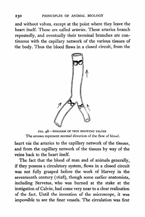
130 PRINCIPLES OF ANIMAL BIOLOGY
and without valves, except at the point where they leave the
heart itself. These are called arteries. These arteries branch
repeatedly, and eventually their terminal branches are con-
tinuous with the capillary network of the various tissues of
the body. Thus the blood flows in a closed circuit, from the
FIG. 48 DIAGRAM OF VEIN SHOWING VALVES
The arrows represent normal direction of the flow of blood.
heart via the arteries to the capillary network of the tissues,
and from the capillary network of the tissues by way of the
veins back to the heart itself.
The fact that the blood of man and of animals generally,
if they possess a circulatory system, flows in a closed circuit
was not fully grasped before the work of Harvey in the
seventeenth century (1628), though some earlier anatomists,
including Servetus, who was burned at the stake at the
instigation of Calvin, had come very near to a clear realization
of the fact. Until the invention of the microscope, it was
impossible to see the finer vessels. The circulation was first
THE TRANSPORT OF MATERIALS IN THE ANIMAL BODY 131
observed directly in the manner indicated above by the
pioneer microscopists Malpighi (1661) and Leeuwenhoek
(1668).
The Blood. Blood is not a homogeneous fluid, but a
suspension of densely packed cells. Some of these are ovoid
discs charged with a colouring material which in thin layers
is yellowish, but seen in greater concentration appears red.
This colouring matter which gives to blood its characteristic
hue is known as haemoglobin. A solution of haemoglobin is
normally of a bright scarlet hue, like that of the blood in our
arteries. When shaken in a vacuum or in pure nitrogen or
hydrogen it changes colour, as was first shown by Priestley
(1774). It assumes the purple hue of the blood flowing in
the veins. Priestley also succeeded in showing that oxygen,
or, as he called it, dephlogisticated air, was the constituent
of the atmosphere with which purple, or reduced haemoglobin
combines. The presence of haemoglobin is thus a fact of
immense importance. Owing to its power to combine loosely
with oxygen, the amount of oxygen that blood can take upis considerably higher than the amount which dissolves in a
corresponding quantity of water. Thus 100 c.c. of our own
blood will take up 18-5 c.c. of oxygen in air at atmospheric
pressure, whereas 100 c.c. of water would absorb only o 7 c.c.
The movement of the blood in the vessels ensures a con-
stant supply of food materials and oxygen to the tissues,
and a constant draining away of waste products which diffuse
through the thin walls of the capillaries. The oxygen-
carrying property of the blood is specially important. All
tissues store food materials, just as the muscle stores glycogen
or animal starch. But the storage capacity of the cells of the
body for oxygen is very small, and oxygen is constantly
being used. Insects which have a peculiar system of minute
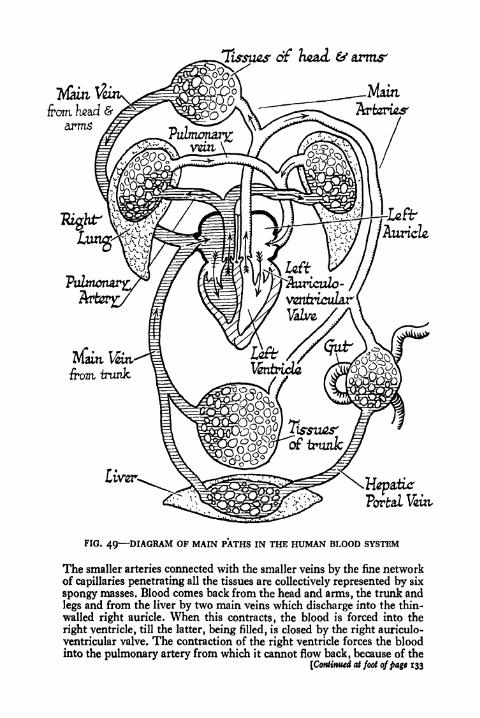
2-fepatixr
Portal %ttt
FIG. 49 DIAGRAM OF MAIN PATHS IN THE HUMAN BLOOD SYSTEM
The smaller arteries connected with the smaller veins by the fine networkof capillaries penetrating all the tissues are collectively represented by six
spongy masses. Blood comes back from the head and arms, the trunk and
legs and from the liver by two main veins which discharge into the thin-
walled right auricle. When this contracts, the blood is forced into the
right ventricle, till the latter, being filled, is closed by the right auriculo-
yentricularvalve. The contraction of the right ventricle forces the blood
into the pulmonary artery from which it cannot flow back, because of the
[Continued at foot ofpage 133
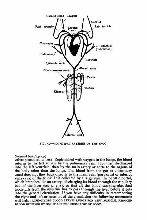
Carotid gland Lingual
Right Auricle/ \\ Carotid'
arch ..
Cutaneous..
NPulmonary
Systemic arch
Coeliaco-mesenteric
Carotid
Left Auricle
Brachial
(Subclavian)
FIG. 50 PRINCIPAL ARTERIES OF THE FROG
Continued from page 132]
valves placed at its base. Replenished with oxygen in the lungs, the bloodreturns to the left auricle by the pulmonary vein. It is then dischargedinto the left ventricle, then by the main artery or aorta to the organs ofthe body other than the lungs. The blood from the gut or alimentarycanal does not flow back directly to the main vein (post-caval or inferior
vena cava) of the trunk. It is collected by a large vein, the hepatic portal,which branches like an artery, discharging its blood through the capillarybed of the liver (see p. 134), so that all the blood carrying absorbedfoodstuffs from the intestine has to pass through the liver before it getsinto the general circulation. If you have any difficulty in rememberingthe right and left orientation of the circulation the following mnemonicwill help: LIFE-GIVING BLOOD LEAVES LUNGS FOR LEFT AURICLE. REDUCEDBLOOD RECEIVED BY RIGHT AURICLE FROM REST OF BODY.

134 PRINCIPLES OF ANIMAL BIOLOGY
tubes (trachece) bringing air directly into contact with the
tissues can get on perfectly well without a well-developed
circulatory system, though many insects surpass all animals
except birds in their incessant muscular activity. Haemoglobinoccurs in the blood of all the great groups known as Verte-
brates (fishes, frogs, lizards, birds, man, etc.) and in most
of the worms. The blood of snails, lobsters, squids, and a few
other animals contains another respiratory pigment, known
as hcemocyanin, which is blue in the presence of oxygen, and
becomes colourless when shaken in a vacuum. Haemocyanin,like haemoglobin, is a protein combined with a metal. The
metallic constituent of haemoglobin is iron, that of haemo-
cyanin is copper. Hence the importance of traces of iron in
our own food.
The Circulatory System of the Frog. A convenient starting
place to take in considering the circulation is the capillary
network of the digestive tract. The small veins of the intestine
unite into larger ones which eventually merge into the vessel
known as the hepatic portal vein. This vein does not unite
with other veins directly, but branches in the substance of
the liver into smaller veins which are continuous with the
capillary network of that organ. The liver is a great store-
house of carbohydrate in the animal body. It stores carbo-
hydrate in the form of the animal starch known as glycogen.
The blood coming from the capillaries of the intestine after
a meal is ordinarily rich in sugar. In the liver the excess of
sugar is built up into glycogen. The glycogen of the liver
is transformed into sugar, as the supply of glycogen in the
muscles is used up. The work of Claude Bernard in 1857
first showed that the blood in the hepatic portal vein after a
meal is richer in sugar than the rest of the circulation, and
that the store of glycogen in the liver goes down during

THE TRANSPORT OF MATERIALS IN THE ANIMAL BODY 135
starvation and increases immediately after the assimilation
of food.
The blood in the capillary network of the liver is collected
by small veins which join to form the two hepatic veins.
These flow into a large vein, known as the postcaval or
liapahc
\VS<zr
kidney
FIG. 51 MAIN VEINS OF MAN AND FROG
inferior vena cava, that picks up blood from the kidneys and
discharges it into the thin-walled chamber of the heart
known as the sinus venosus. Into the sinus venosus flow two
large veins from the head and fore limbs, known as the
precavals (right and left). The principal tributaries of each
precaval are the jugular veins, bringing back blood from the
head, the subdavian veins bringing back blood from the
capillary network of the arms, and the musculo-cutaneous,
which bring back most of the blood from the skin of the
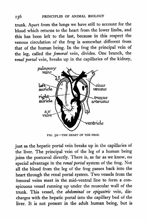
136 PRINCIPLES OF ANIMAL BIOLOGY
trunk. Apart from the lungs we have still to account for the
blood which returns to the heart from the lower limbs, and
this has been left to the last, because in this respect the
venous circulation of the frog is somewhat different from
that of the human being. In the frog the principal vein of
the leg, called the femoral vein, divides. One branch, the
renal portal vein, breaks up in the capillaries of the kidney,
FIG. 52 THE HEART OF THE FROG
just as the hepatic portal vein breaks up in the capillaries of
the liver. The principal vein of the leg of a human being
joins the postcaval directly. There is, as far as we know, no
special advantage in the renal portal system of the frog. Not
all the blood from the leg of the frog passes back into the
heart through the renal portal system. Two vessels from the
femoral veins meet in the mid-ventral line to form a con-
spicuous vessel running up under the muscular wall of the
trunk. This vessel, the abdominal or epigastric vein, dis-
charges with the hepatic portal into the capillary bed of the
liver. It is not present in the adult human being, but is
THE TRANSPORT OF MATERIALS IN THE ANIMAL BODY 137
represented in the human embryo. The most important fact
about the venous circulation of both the frog and man, and
we might add of fish or fowl, is the hepatic portal system,
that is to say, the fact that all the blood returning from the
alimentary tract passes through the capillary network of the
liver en route for the heart. The glycogen-storing activity of
the liver is its most important role in the economy of the
body. In human beings the liver stores a good deal of fat.
The frog possesses special masses of fatty tissues, the fat-
bodies attached to the kidneys. These diminish greatly in
size during hibernation (winter sleep), when no food is
available.
We have now described the larger veins, which bring back
blood from the capillaries of all parts of the body, except the
lungs. We have next to describe the structure of the frog's
heart. The heart of the frog consists of three portions, the
sinus venosus, a thin-walled chamber into which the pre-
and post-caval veins pour blood; a portion, the atrium, with
somewhat thicker walls, divided completely into two
chambers, the right and left auricles ;and a very thick-walled
chamber, the ventricle, which pumps blood out into the
arteries. The sinus venosus opens into the right auricle. Into
the left auricle open two small veins, the pulmonary veins
from the right and left lungs. Both auricles discharge into
the ventricle. Between the auricles and the ventricle is a
flap of fibrous tissue connected by fine cords of tendon to the
inner wall of the latter. This is called the auriculo-ventricular
valve. The whole of the heart is composed of muscular tissue,
and it will continue to beat when it is removed from the
body, devoid of all connexion with the nervous system.
The chambers of the heart contract and relax in orderly
succession, the sinus venosus first, the auricles next, and the

138 PRINCIPLES OF ANIMAL BIOLOGY
ventricle last. The contraction of the sinus venosus forces
blood into the right auricle. When the auricles contract blood
is forced into the ventricle. As the latter fills up, the auriculo-
ventricular valve floats upward, so that, when the ventricle
contracts in its turn no blood can pass backwards into the
auricles (Fig. 52).
From the ventricle emerges a single arterial trunk, the
truncus arteriosus. On either side it divides into three large
arteries (Fig. 50). The most anterior of these is the carotid,
whose ultimate branches end in the capillaries of the head
region. The most posterior is the pulmo-cutaneons supplying
the lung, and sending a small artery to the skin of the trunk.
The middle one, known as the systemic, supplies a larger
area, and requires more detailed description. Each systemic
gives rise to a snbclavian or brachial artery, which runs out-
ward to the forelimbs, where its ultimate branches terminate
in the capillary network of that region. The two systemics
meet in the middle line of the body, dorsal to the postcaval
vein, to form a single median artery, the aorta or dorsal
aorta. From the dorsal aorta springs one large median artery,
the cceKaco-mesenteric, whose terminal branches end in the
capillary network of the gut and liver, etc., paired renal
arteries, whose final ramifications end in the capillary net-
work of the kidneys ;and a pair of iliac arteries which supply
the hind limbs.
The arteries have thicker walls than the veins, and except
at the point where the carotid, systemic, and pulmonaryarteries leave the truncus arteriosus, and at the origin of the
latter from the ventricle itself, they have no valves like those
which are found in the veins. The blood in the arteries is
at a much higher pressure than the blood in the veins.
Blood spurts from a cut artery and trickles from a vein. The

THE TRANSPORT OF MATERIALS IN THE ANIMAL BODY 139
reason for this is that the fine tubes of the capillary network
offer a considerable frictional resistance to the flow of the
blood; as is easily appreciated when we remember how hard
it is to blow air or water through a very fine jet. In con-
sequence the energy of the blood-flow in the veins (Fig. 53)
is much less than in the arteries, and the presence of valves
in their course ensures that the blood cannot flow back, when
FIG. 53 PHYSICAL MODEL
To illustrate resistance of the capillaries to arterial pressure. An addi-
tional resistance is introduced by the small bore of the tap in the courseof the horizontal outlet.
the arterial pressure falls while the ventricle is relaxing in
the act of filling.
Regulation of blood-flow. The amount of blood flowing
through an organ varies. To some extent it is adjusted to
the oxygen requirements of the moment. Other things being
equal, the rate at which a tissue can take up oxygen depends
upon the amount of blood which flows through it in unit
time. Now the flow of a liquid through a tube of uniform bore
depends upon the force propelling it, the length traversed,
and the sectional area of the tube. Only the first and last of
these need be considered in connection with the circulation,
because the length of tube traversed is approximately con-
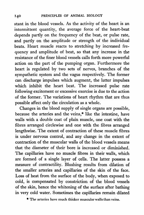
140 PRINCIPLES OF ANIMAL BIOLOGY
stant in the blood vessels. As the activity of the heart is an
intermittent quantity, the average force of the heart-beat
depends partly on the frequency of the beat, or pulse rate,
and partly on the amplitude or strength of the individual
beats. Heart muscle reacts to stretching by increased fre-
quency and amplitude of beat, so that any increase in the
resistance of the finer blood vessels calls forth more powerful
action on the part of the pumping organ. Furthermore the
heart is regulated by two sets of nerves, branches of the
sympathetic system and the vagus respectively. The former
can discharge impulses which augment, the latter impulses
which inhibit the heart beat. The increased pulse rate
following excitement or excessive exercise is due to the action
of the former. The variations of heart rhythm that are thus
possible affect only the circulation as a whole.
Changes in the blood supply of single organs are possible,
because the arteries and the veins,* like the intestine, have
walls with a double coat of plain muscle, one coat with the
fibres arranged circlewise and one with the fibres arranged
lengthwise. The extent of contraction of these muscle fibres
is under nervous control, and any change in the extent of
contraction of the muscular walls of the blood vessels means
that the diameter of their bore is increased or diminished.
The capillaries have no muscle fibres in their walls, which
are formed of a single layer of cells. The latter possess a
measure of contractility. Blushing results from dilation of
the smaller arteries and capillaries of the skin of the face.
Loss of heat from the surface of the body, when exposed to
cold, is compensated by constriction of the blood vessels
of the skin, hence the whitening of the surface after bathingin very cold water. Sometimes the capillaries remain dilated
* The arteries have much thicker muscular walls than veins.

THE TRANSPORT OF MATERIALS IN THE ANIMAL BODY 141
while the smaller arteries are almost completely closed up.
When this occurs, stagnant pools of blood occur in the
capillary network and as their oxygen is used up the blood
itself becomes completely reduced. This is what happens
when the tips of the fingers become blue on a cold day.
to head,
to trunk
FIG. 54 THE RELATION OF THE HEART TO THE LUNGS AND TISSUES
Above in the frog. Below in Man.
The blood of the frog is never warmer than the atmo-
sphere, and the animal cannot withstand either great cold
or high temperatures at which we can easily live. Our blood
is kept~aty constant temperature (about 37 C.) in ordinary
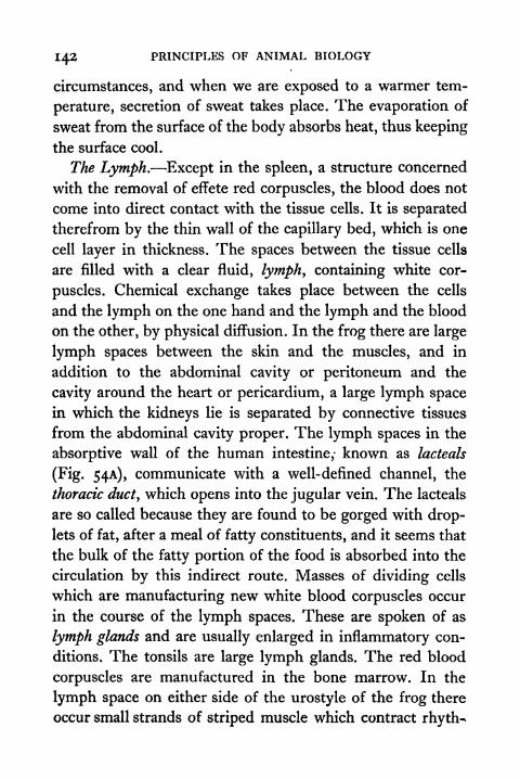
142 PRINCIPLES OF ANIMAL BIOLOGY
circumstances, and when we are exposed to a warmer tem-
perature, secretion of sweat takes place. The evaporation of
sweat from the surface of the body absorbs heat, thus keeping
the surface cool.
The Lymph. Except in the spleen, a structure concerned
with the removal of effete red corpuscles, the blood does not
come into direct contact with the tissue cells. It is separated
therefrom by the thin wall of the capillary bed, which is one
cell layer in thickness. The spaces between the tissue cells
are filled with a clear fluid, lymph, containing white cor-
puscles. Chemical exchange takes place between the cells
and the lymph on the one hand and the lymph and the blood
on the other, by physical diffusion. In the frog there are large
lymph spaces between the skin and the muscles, and in
addition to the abdominal cavity or peritoneum and the
cavity around the heart or pericardium, a large lymph space
in which the kidneys lie is separated by connective tissues
from the abdominal cavity proper. The lymph spaces in the
absorptive wall of the human intestine,- known as lacteals
(Fig. 54A), communicate with a well-defined channel, the
thoracic duct, which opens into the jugular vein. The lacteals
are so called because they are found to be gorged with drop-
lets of fat, after a meal of fatty constituents, and it seems that
the bulk of the fatty portion of the food is absorbed into the
circulation by this indirect route. Masses of dividing cells
which are manufacturing new white blood corpuscles occur
in the course of the lymph spaces. These are spoken of as
lymph glands and are usually enlarged in inflammatory con-
ditions. The tonsils are large lymph glands. The red blood
corpuscles are manufactured in the bone marrow. In the
lymph space on either side of the urostyle of the frog there
occur small strands of striped muscle which contract rhyth^

THE TRANSPORT OF MATERIALS IN THE ANIMAL BODY 143
mically, and are called the lymph hearts. Their pulsation can
be easily detected through the skin in the intact animal, but
their precise significance is not clearly understood.
The Human Circulation. The circulation of the frog
ZacteaZ
layer oFcircularin miiscLa fibres
___ _
== plain iiuiscK fibnes
FIG. 54A BLOOD SUPPLY OF SMALL INTESTINE
differs in several respects from that of the human being,
and of all warm-blooded forms. It is much less efficient as a
means of discharging the principal office of the circulatory
system, the transport of the respiratory gases. Both the
deoxygenated purple blood coming from the tissues of the
body as a whole via the right auricle, and the oxygenatedblood returning from the lungs via the left auricle, have
plenty of opportunity for mixing in the single ventricle
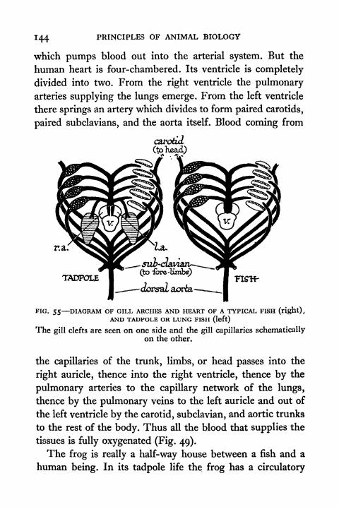
144 PRINCIPLES OF ANIMAL BIOLOGY
which pumps blood out into the arterial system. But the
human heart is four-chambered. Its ventricle is completely
divided into two. From the right ventricle the pulmonaryarteries supplying the lungs emerge. From the left ventricle
there springs an artery which divides to form paired carotids,
paired subclavians, and the aorta itself. Blood coming from
c&rokui(to head).A Jfc _X
r.a.
TADPOLE
FIG. 55 DIAGRAM OF GILL ARCHES AND HEART OF A TYPICAL FISH (right),AND TADPOLE OR LUNG FISH (left)
The gill clefts are seen on one side and the gill capillaries schematicallyon the other.
the capillaries of the trunk, limbs, or head passes into the
right auricle, thence into the right ventricle, thence by the
pulmonary arteries to the capillary network of the lungs,
thence by the pulmonary veins to the left auricle and out of
the left ventricle by the carotid, subclavian, and aortic trunks
to the rest of the body. Thus all the blood that supplies the
tissues is fully oxygenated (Fig. 49).
The frog is really a half-way house between a fish and a
human being. In its tadpole life the frog has a circulatory
THE TRANSPORT OF MATERIALS IN' THE ANIMAL BODY 145
system essentially like that of a fish. At a certain stage of
our embryonic life, we, too, have a circulation of this type,
succeeded by one that is very much like that of the adult
frog. The fish is an animal that breathes oxygen dissolved
in water by passing a current of the latter through slits in
the sides of the throat. These are provided with filaments
aorfca
-first, f
gttdeft?
gu*
sng
auricle
FIG. 56 BLOOD SYSTEM OF A FISH
The front end only of the fish is shown. The veins are in black and the
arteries, which carry deoxygenated blood from the heart to the gills, are
lightly shaded. The portal vein, which carries blood from the gut to the
liver, is also shown.
richly supplied with capillaries and known as gills. The fish
heart is single throughout; the atrium is not divided into
two separate auricles. All the blood from the body entering
the heart is deoxygenated, and passes from the ventricle into
a median artery which pumps blood by a paired series of
arteries to the capillaries of the gills. Thence they are col-
lected by arterieswhich join to form the aorta (Figs. 55 and 56).
Many fishes have a structure, known as the swim-bladder
by which they adjust their specific gravity. In a few fishes,

146 PRINCIPLES OF ANIMAL BIOLOGY
such as the lung-fishes, it opens into the throat and is used
as a lung. The lung of these fishes is supplied by an artery
from the last of the gill arches. This precisely corresponds to
tisjrues
FISH
TIC. 57 DIAGRAMMATIC REPRESENTATION OF THE OXYGENATION OF THEBLOOD IN FISH (left) AND MAN (tight)
Gill Clefts
A B CFIG. 58 EMBRYOS OF (A) SHARK, (B) BIRD, (c) MAN AT THE STAGE WHEN
THE GILL CLEFTS ARE FORMEDThe gill clefts never become completely perforated in birds and mammals,but skeletal and circulatory arrangements of the fish type are temporarilyassociated with them.
the arrangement in the frog just before it develops its limbs
and sheds its tail, and it corresponds with the stage in our
own development, when we possess gill-clefts like a fislu
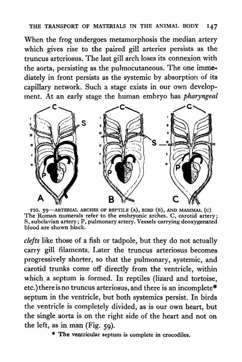
THE TRANSPORT OF MATERIALS IN THE ANIMAL BODY 147
When the frog undergoes metamorphosis the median artery
which gives rise to the paired gill arteries persists as the
truncus arteriosus. The last gill arch loses its connexion with
the aorta, persisting as the pulmocutaneous. The one imme-
diately in front persists as the systemic by absorption of its
capillary network. Such a stage exists in our own develop-
ment. At an early stage the human embryo has pharyngeal
BFIG. 59 ARTERIAL ARCHES OF REPTILE (A), BIRD (fi), AND MAMMAL (c)
The Roman numerals refer to the embryonic arches. C, carotid artery;S, subclavian artery ; P, pulmonary artery. Vessels carrying deoxygenatedblood are shown black.
clefts like those of a fish or tadpole, but they do not actually
carry gill filaments. Later the truncus arteriosus becomes
progressively shorter, so that the pulmonary, systemic, and
carotid trunks come off directly from the ventricle, within
which a septum is formed. In reptiles (lizard and tortoise,
etc.) there is no truncus arteriosus, and there is an incomplete*
septum in the ventricle, but both systemics persist. In birds
the ventricle is completely divided, as is our own heart, but
the single aorta is on the right side of the heart and not on
the left, as in man (Fig. 59).* The ventricular septum is complete in crocodiles.
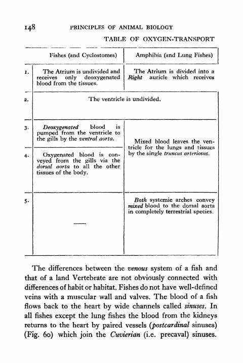
148 PRINCIPLES OF ANIMAL BIOLOGY
TABLE OF OXYGEN-TRANSPORT
Fishes (and Cyclostomes) Amphibia (and Lung Fishes)
I.
2.
The Atrium is undivided andreceives only deoxygenatedblood from the tissues.
The Atrium is divided into a
Right auricle which receives
The ventricle is undivided.
Deoxygenated blood is
pumped from the ventricle to
the gills by the ventral aorta.
Oxygenated blood is con-
veyed from the gills via the
dorsal aorta to all the other
tissues of the body.
Mixed blood leaves the ven-tricle for the lungs and tissues
by the single trnncus arteriosus.
Both systemic arches conveymixed blood to the dorsal aorta
in completely terrestrial species.
The differences between the venous system of a fish and
that of a land Vertebrate are not obviously connected with
differences of habit or habitat. Fishes do not have well-defined
veins with a muscular wall and valves. The blood of a fish
flows back to the heart by wide channels called sinuses. In
all fishes except the lung fishes the blood from the kidneys
returns to the heart by paired vessels (postcardinal sinuses)
(Fig. 60) which join the Cuvierian (i.e. precaval) sinuses.

THE TRANSPORT OF MATERIALS IN THE ANIMAL BODY 149
IN THE VERTEBRATE BODY
Left auricle which receives oxygenated blood from the Lungs and a
deoxygenated blood from the Rest of the Body.
The ventricle is
more or less com-pletely divided.
The ventricle is completely divided.
The pulmonary artery arising from the right side of the ventricular
septum takes only deoxygenated blood to the lungs.
The Right systemic arch from the left side
of the ventricular septum conveys oxygenatedblood to the head, to the fore limbs, and to
the aorta.
The Left systemicarch from the left side
of the ventricular sep-tum takes oxygenatedblood to all the organs(except lungs).
The Left systemicarch from the rightside of the ventri-
cular septum conveysdeoxygenated bloodto the dorsal aorta,so that the hind endof the body getsmixed blood.
The Left systemicarch has disappearedand the whole body(except lungs) gets
oxygenated bloodfrom the right sys-temic arch.
The Right systemicarch has disappeared.
So it does not mix with the blood from the liver before it
enters the sinus venosus, and there is no sinus which corre-
sponds to the postcaval vein. Postcardinal vessels are present
in the embryos of land Vertebrates before a postcaval vein
develops. In salamanders they persist intact throughout adult
life. The axygos veins which bring back blood from the
inside of the thorax are the remains of the embryonic post-
cardinals of Man and other mammals-. Fishes, like land

PRINCIPLES OF ANIMAL BIOLOGY
Vertebrates, have a hepatic portal circulation. The hepatic
portal vessel of fishes is more like a true vein than the
sinuses which join the heart.
JuguZar
(a.)
Cuvisnon sums-pre-cavaL win*>
post-cavaLvain,
vctrt
FIG. 60 VENOUS SYSTEM OF VERTEBRATES
(a) Adult Fish (Early Embryo of Frog or Man).(b) Adult Newt or Lung Fish (Tadpole or Human embryo).(c) Adult Land Vertebrate.
The ductless glands. In the activities of the body of Manor the frog the blood plays a part to which we have not
referred so far. We have already seen what an important
part the nervous system plays in co-ordinating the responses
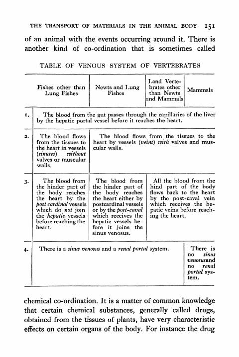
THE TRANSPORT OF MATERIALS IN THE ANIMAL BODY 151
of an animal with the events occurring around it. There is
another kind of co-ordination that is sometimes called
TABLE OF VENOUS SYSTEM OF VERTEBRATES
Fishes other than
Lung Fishes
Newts and LungFishes
Land Verte-brates otherthan Newts
and Mammals
Mammals
i.
2.
The blood from the gut passes through the capillaries of the liver
by the hepatic portal vessel before it reaches the heart.
The blood flows
from the tissues to
the heart in vessels
(sinuses) withoutvalves or muscularwalls.
The blood flows from the tissues to the
heart by vessels (veins) tvith valves and mus-cular walls.
The blood fromthe hinder part ofthe body reachesthe heart by the
post cardinal vessels
which do not jointhe hepatic vessels
before reaching the
heart.
The blood fromthe hinder part of
the body reachesthe heart either bypostcardinal vessels
or by the post-cavalwhich receives the
hepatic vessels be-fore it joins the
sinus venosus.
All the blood from the
hind part of the bodyflows back to the heart
by the post-caval veinwhich receives the he-
patic veins before reach-
ing the heart.
There is a sinus venosus and a renal portal system. There is
no sinus
venosusandno renal
portal sys-tem.
chemical co-ordination. It is a matter of common knowledgethat certain chemical substances, generally called drugs,
obtained from the tissues of plants, have very characteristic
effects on certain organs of the body. For instance the drug
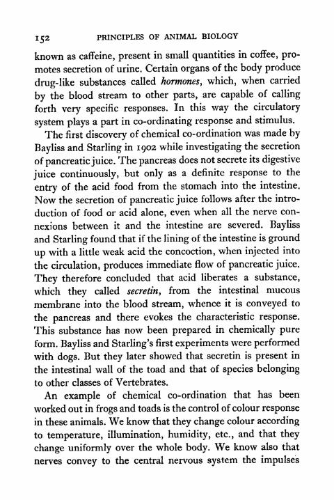
PRINCIPLES OF ANIMAL BIOLOGY
known as caffeine, present in small quantities in coffee, pro-
motes secretion of urine. Certain organs of the body produce
drug-like substances called hormones, which, when carried
by the blood stream to other parts, are capable of calling
forth very specific responses. In this way the circulatory
system plays a part in co-ordinating response and stimulus.
The first discovery of chemical co-ordination was made by
Bayliss and Starling in 1902 while investigating the secretion
of pancreatic juice. The pancreas does not secrete its digestive
juice continuously, but only as a definite response to the
entry of the acid food from the stomach into the intestine.
Now the secretion of pancreatic juice follows after the intro-
duction of food or acid alone, even when all the nerve con-
nexions between it and the intestine are severed. Bayliss
and Starling found that if the lining of the intestine is ground
up with a little weak acid the concoction, when injected into
the circulation, produces immediate flow of pancreatic juice.
They therefore concluded that acid liberates a substance,
which they called secretin, from the intestinal mucous
membrane into the blood stream, whence it is conveyed to
the pancreas and there evokes the characteristic response.
This substance has now been prepared in chemically pure
form. Bayliss and Starling's first experiments were performed
with dogs. But they later showed that secretin is present in
the intestinal wall of the toad and that of species belonging
to other classes of Vertebrates.
An example of chemical co-ordination that has been
worked out in frogs and toads is the control of colour response
in these animals. We know that they change colour according
to temperature, illumination, humidity, etc., and that they
change uniformly over the whole body. We know also that
nerves convey to the central nervous system the impulses

THE TRANSPORT OF MATERIALS IN THE ANIMAL BODY 153
set up by appropriate stimuli in their surroundings. It was
at one time thought that the nervous system transmitted
these impulses directly to the melanophores or black pig-
ment cells. This is not so. In fact it is doubtful whether the
melanophores of the frog have any nerve supply. Within the
skull beneath the brain of the frog and of man himself lies
a little gland which has no duct
conveying its secretion to the ex-
terior. Extracts made by grinding upthis organ, the pituitary gland, in salt
solution have some very remarkable
properties. In particular, when in-
jected into a pale frog, they bring
about within half an hour a complete
darkening of the skin resulting from
expansion of the melanophores
(Frontispiece). There is enough of the
active constituent in one gland to
bring about darkening in a hundred
individuals. When the pituitary gland
is removed, the frog recovers and sur-
vives, but in whatever circumstances it is kept, it will always
remain pale. Darkening of the skin in a frog from which
the pituitary gland has been extirpated can still occur, if a
small quantity of the extract of the pituitary gland (about
equivalent to one-millionth of a gram of the gland substance)
is introduced into the circulation by injection. It thus seems
that the nervous system controls the behaviour of the pig-
ment cells by increasing or diminishing the rate at which
the pituitary body pours its secretion into the blood.
Over the surface of each kidney there is an orange-coloured
mass of tissue, the adrenal body. It contains a 'drug-like
FIG. 6l MICROSCOPICSECTION OF THE THYROID
GLAND
Showing follicular cystslined by cubical epithe-lium and filled with a
jellyish material.
154 PRINCIPLES OF ANIMAL BIOLOGY
substance that acts on plain muscle, in some cases relaxing
and in others inducing contraction or increased rapidity of
an inherent rhythm. Thus it accelerates the heart of the frog
and relaxes the muscular wall of the intestine. This sub-
stance, adrenalin, can now be manufactured in pure form
in the laboratory. It is liberated into the circulation in small
quantities, but we are not as yet certain under what con-
ditions. The adrenal and pituitary bodies are sometimes
spoken of as ductless glands. A third ductless gland, the
thyroid (Fig. 61), is dealt with in the next chapter.
TOPICS FOR HOME STUDY1. Name the respiratory pigments. Describe their chemical nature
and properties and the part they play in the living body.
2. Contrast the oxygenation of the blood in dogfish, frog, tortoise,
fowl and rabbit.
3. Describe the Hepatic and Renal portal circulation in fish, frog,
bird and mammal.
4. Tabulate the uses of the blood.
5. Describe the microscopic structure of blood vessels giving the
differences between arteries, capillaries and veins. What are
the functions of the following : (a) valves ; (b) muscular walls
of arteries ; (c) capillaries.
6. Describe the role of blood in (a) heat regulation, (6) protection
against diseases, (c) chemical co-ordination.
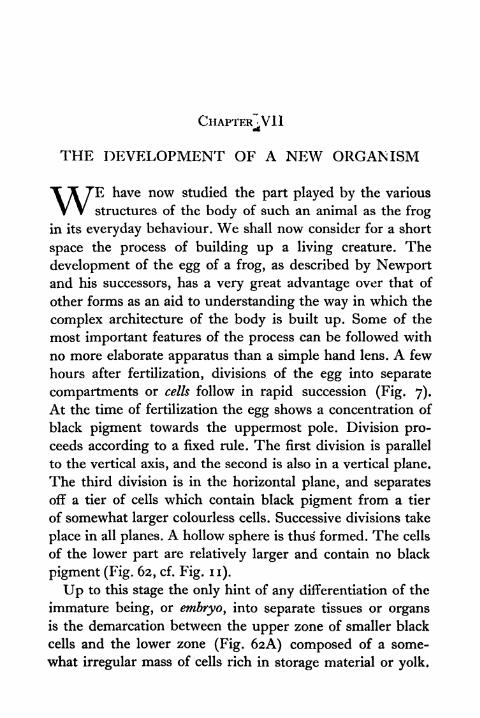
CHAPTER^VII
THE DEVELOPMENT OF A NEW ORGANISM
WE have now studied the part played by the various
structures of the body of such an animal as the frog
in its everyday behaviour. We shall now consider for a short
space the process of building up a living creature. The
development of the egg of a frog, as described by Newportand his successors, has a very great advantage over that of
other forms as an aid to understanding the way in which the
complex architecture of the body is built up. Some of the
most important features of the process can be followed with
no more elaborate apparatus than a simple hand lens. A few
hours after fertilization, divisions of the egg into separate
compartments or cells follow in rapid succession (Fig. 7).
At the time of fertilization the egg shows a concentration of
black pigment towards the uppermost pole. Division pro-
ceeds according to a fixed rule. The first division is parallel
to the vertical axis, and the second is also in a vertical plane.
The third division is in the horizontal plane, and separates
off a tier of cells which contain black pigment from a tier
of somewhat larger colourless cells. Successive divisions take
place in all planes. A hollow sphere is thus formed. The cells
of the lower part are relatively larger and contain no black
pigment (Fig. 62, cf. Fig. n).
Up to this stage the only hint of any differentiation of the
immature being, or embryo, into separate tissues or organsis the demarcation between the upper zone of smaller black
cells and the lower zone (Fig. 6aA) composed of a some-
what irregular mass of cells rich in storage material or yolk.
156 PRINCIPLES OF ANIMAL BIOLOGY
Now begins the stage of organ building. Soon almost the
entire surface of the embryo is covered with a layer of small
pigmented cells, the large unpigmented cells being evident
mesod&m
FIG. 62 FOUR EMBRYOS OF FROG
Showing stages in excavitation of the primitive gut.
only at one end, the yolk plug. This is destined to be the
posterior end of the tadpole (Fig. 62).
Now begins the differentiation of two systems of organs
characteristic of the adult animal the gut or alimentary
canal and the central nervous system. The black outer
covering of cells forms a distinct lip over the yolk plug on
one side. A small chink beneath this lip grows inwards,
THE DEVELOPMENT OF A NEW ORGANISM 157
enlarging as it grows. This is the rudiment of the alimentary
cavity (Fig. 62B-D) of the tadpole. It is the embryonic gut.
Meanwhile a groove appears on the surface of the embryofrom the region (Fig. 63) just above the lip already referred
to. The groove is wider in front, and as it deepens, its edges
grow up, and coalesce, so that a tube is formed beneath a
thin covering of cells. This tube, broadest at the future head
end, is the rudiment of the spinal cord and brain. While
this has been going on, cavitation of the mass of internal
FIG. 63 (l) POSTERIOR, (2) ANTERIOR, AND (3) DORSAL ASPECT OF THEFROG EMBRYO AT THE STAGE WHEN THE NEURAL GROOVE IS CLOSING UP
unpigmented cells has progressed inwards from the yolk
plug.
The original cavity of the sphere has been obliterated. The
new cavity excavated inwards from the yolk plug is destined
ultimately to become the cavity of the alimentary canal. As this
cavitation proceeds, a belt of cells is separated off from the
mass of colourless cells which form the innermost part of the
embryo. This belt of tissue (mesoderm) is derived from cells
which migrated inwards from the lips surrounding the yolk
plug and is destined to give rise to the body musculature,
including the walls of the blood vessels which arise as canals
in it. Beneath the dorsal rudiment of the central nervous
system a rod of tissue is detached from the roof of the
primitive gut in the mid-dorsal line. The cells of this rod
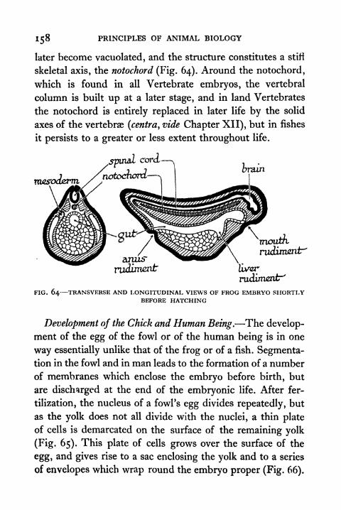
158 PRINCIPLES OF ANIMAL BIOLOGY
later become vacuolated, and the structure constitutes a stifi
skeletal axis, the notochord (Fig. 64). Around the notochord,
which is found in all Vertebrate embryos, the vertebral
column is built up at a later stage, and in land Vertebrates
the notochord is entirely replaced in later life by the solid
axes of the vertebrae (centra, vide Chapter XII), but in fishes
it persists to a greater or less extent throughout life.
spinalcordL
notocfaoni
liver
rudiment"'
FIG. 64 TRANSVERSE AND LONGITUDINAL VIEWS OF FROG EMBRYO SHORTLYBEFORE HATCHING
Development of the Chick and Human Being. The develop-
ment of the egg of the fowl or of the human being is in one
way essentially unlike that of the frog or of a fish. Segmenta-tion in the fowl and in man leads to the formation of a number
of membranes which enclose the embryo before birth, but
are discharged at the end of the embryonic life. After fer-
tilization, the nucleus of a fowl's egg divides repeatedly, but
as the yolk does not all divide with the nuclei, a thin plate
of cells is demarcated on the surface of the remaining yolk
(Fig. 65). This plate of cells grows over the surface of the
egg> and gives rise to a sac enclosing the yolk and to a series
of envelopes which wrap round the embryo proper (Fig. 66).
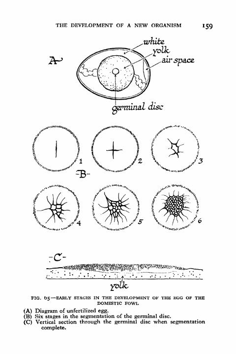
THE DEVELOPMENT OF A NEW ORGANISM 159
white.
FIG. 05 EARLY STACKS IN THE DEVELOPMENT OF THE EGG OF THEDOMESTIC FOWL
(A) Diagram of unfertilized egg.
(B) Six stages in the segmentation of the germinal disc.
(C) Vertical section through the germinal disc when segmentationcomplete.
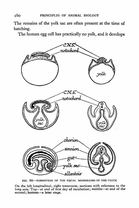
i6o PRINCIPLES OF ANIMAL BIOLOGY
The remains of the yolk sac are often present at the time of
hatching.
The human egg cell has practically no yolk, and it develops
FIG. 66 FORMATION OF THE FCETAL MEMBRANES OF THE CHICK
On the left longitudinal, right transverse, sections with reference to the
long axis. Top at end of first day of incubation; middle at end of the
second; bottom a later stage.
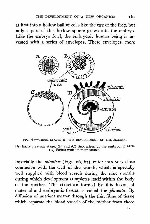
THE DEVELOPMENT OF A NEW ORGANISM 161
at first into a hollow ball of cells like the egg of the frog, but
only a part of this hollow sphere grows into the embryo.Like the embryo fowl, the embryonic human being is in-
vested with a series of envelopes. These envelopes, more
yolksac
FIG. 67 THREE STAGES IN THE DEVELOPMENT OF THE MAMMAL
(A) Early cleavage stage. (B) and (C) Separation of the embryonic area.
(D) Foetus with its membranes.
especially the allantois (Figs. 66, 67), enter into very close
connexion with the wall of the womb, which is specially
well supplied with blood vessels during the nine months
during which development completes itself within the bodyof the mother. The structure formed by this fusion of
maternal and embryonic tissues is called the placenta. Bydiffusion of nutrient matter through the thin films of tissue
which separate the blood vessels of the mother from those
L
162 PRINCIPLES OF ANIMAL BIOLOGY
of the embryo, the latter is able to grow in spite of the fact
that the egg itself has no storage. A feature common to the
development of the fowl and the mammalian egg is the fact
that, though it will develop into a land animal, the embryoat a certain stage has clefts on the side of the throat like those
through which water passes over the gills of the tadpole or
adult fish (Fig. 58).
Metamorphosis. After the embryo of the frog has reached
the stage when the gut rudiment and the beginnings of the
nervous system have been differentiated, it grows more
rapidly in the axis parallel to the length of the spinal cord.
After about a fortnight the head and tail ends are distin-
guishable. The muscles of the tail are sufficiently active to
enable the embryo to wriggle out of the mass of jelly in which
the eggs are laid. Independent existence starts. Unlike our-
selves, the frog does not begin its separate existence with an
organization very much like the sexually mature form. Thehuman baby at birth is essentially an immature adult. Thechief difference is that it has no teeth, and even after it
acquires teeth it has to shed one set of them before the
permanent set appear. The sexual organs are immature and
remain so until about the age of fourteen, when certain
minor distinctions between the sexes (secondary sexual
characters) such as the pitch of voice, presence of hair on
the upper lip and chin of the male, and enlargement of the
milk glands in the female, become evident.
Otherwise a human adult is in most respects, anatomically
speaking, a grown-up baby. But the embryo of the frog,
which has hitherto grown at the expense of food or "yolk"stored in the egg, is very different from the adult when it
first starts independent feeding. It breathes like a fish by
gills and not by lungs. The arterial system is essentially fish-

THE DEVELOPMENT OF A NEW ORGANISM i63
like. It has no fore limbs and only the tiniest rudiments of
hind limbs. It possesses a tail.
On each side of the throat in the newly hatched tadpole
of the frog, shortly after the time of hatching out, there are
plaaznia,
cordwvth, blood-vessels
etrdxyo
placenta.
a;
sutrcnviding embryo
FIG. 68 HUMAN FGETUS IN THE UTERUS ABOUT TWO MONTHS AFTERCONCEPTION
The black lines in the placenta represent diagrammatically the capillaries
containing the blood of the foetus, and the dotted spaces connect withthe blood system of the mother. The arrows show the circulation of the
blood in both.
four clefts which communicate with the exterior and with
the throat, thus letting water pass from the mouth
outwards. The walls between these clefts are known as the
gill arches. From the first three gill arches there grow out
on either side tufts of filaments well supplied with blood
vessels. These "external" gills disappear within a few days
after the tadpole begins to swim, and the sides of the gill

164 PRINCIPLES OF ANIMAL BIOLOGY
arches become coveredwith epithelial foldsvery richlysupplied
with blood vessels like the gills of a fish. A fold of skin, the
operculum, also similar to that of a fish, grows back over the
gill clefts, as in many fishes, and conceals the gill from out-
side. The tadpole continues to breathe partly by its skin and
partly by passing water over the gill clefts, which absorb the
oxygen contained in it; and at first it has no true lungs. This
state of affairs is not permanent. The tadpole stage of the
British common frog lasts only about three months. An
abrupt change supervenes ; and in a very short time the adult
characteristics are assumed. This change is called the meta-
morphosis. The preceding phase or tadpole stage is spokenof as the larval period. At metamorphosis the hind limbs,
which in the half-grown tadpole are tiny rudiments, begin
to grow rapidly ;then the rudiments of the fore limbs break
through the skin. Finally the gill clefts begin to close up and
the tail shrinks. A four-legged, air-breathing, tailless adult
emerges on to land.
The way in which an animal develops is a very difficult
problem to understand, and we are only just beginning to
do so. It is possible to experiment with the development of
a frog or toad just as it is possible to experiment with the
heart or the stomach (Fig. 69). We can make the eggs of a
frog or toad grow up into individuals with two heads or two
tails or with one eye on the top of the head instead of two
(Fig. 70). One question about the development of the toad or
frog which we are beginning to understand is of considerable
importance to medicine and agriculture. This is, what brings
about the sudden transformation from the tadpole into the
adult form?
In the neck of human beings there is a flat lump of glan-
dular tissue known as the thyroid gland. It is represented in

THE DEVELOPMENT OF A NEW ORGANISM
the frog by a pair of ovoid bodies on either side of two large
veins of the neck region. In both man and the frog the
thyroid gland consists of small capsules of a jellyish material
and is very richly supplied with blood vessels (Fig. 61). Like
FIG. 69 EFFECT OF TEMPERATURE ON THE DEVELOPMENT OF THE FROGTADPOLE. (A) YOLK PLUG FORMED. (B) NEURAL FOLDS FORMED, (c) MAINORGAN-SYSTEMS FORMED. (D) READY TO HATCH, NOTE EXTERNAL GILLS.
ALL THE EMBRYOS ARE THREE DAYS OLD, BUT THEY WERE KEPT AT 11, 15.20 AND 24 RESPECTIVELY.
the adrenal and pituitary glands, two other structures which
are somewhat like the digestive glands in texture, it has no
duct. Such organs are for that reason sometimes called duct-
less glands. The jellyish material of the thyroid gland is
peculiar in that it contains a compound of iodine, an element
not found in other tissues of the animal body. When the

1 66 PRINCIPLES OF ANIMAL BIOLOGY
thyroid gland of any animal is given as food to young tad-
poles they start to develop limbs and to lose their tails very
rapidly.
A large American frog, which normally lives for two years
in water as a tadpole, will undergo metamorphosis within six
weeks from hatching if fed on thyroid gland instead of
ordinary meat. The thyroid gland of the tadpole can be
removed by a comparatively simple surgical operation about
the time of hatching, and tadpoles deprived of their thyroid
FIG. 70 ONE-EYED TADPOLE
Produced by exposing the yolk plug stage to the action of lithium chloride.
glands, though otherwise normal, will go on growing for
years without undergoing metamorphosis. They never
assume the adult characteristics. Among human beings there
is a disease of infancy known as cretinism. Its characteristics
include permanent undergrowth and persistence of childish
characteristics. It can now be cured, as these cretin tadpoles
can be cured, by giving thyroid gland (dried and in the
form of a pill) as medicine. Metamorphosis occurs when the
thyroid gland begins to pour its secretion into the blood.
Tadpoles can also be prevented from developing into the
adult form if kept in water free of any trace of iodine com-
pounds. The thyroid is then unable to build up its secretion,
which is an iodine compound. This compound can now be
manufactured from pure chemicals in the laboratory. Allied
THE DEVELOPMENT OF A NEW ORGANISM 167
to the frogs and the toads are the salamanders, in appearance
somewhat like a lizard without scales, but having a larval
stage spent in the water like the tadpole.
Heredity and Environment. There is a local variety of the
Mexican salamander, which never grows into the land form
in nature. It remains a sort of tadpole called the axolotl,
and breeds in the immature state. Though a single meal of
ox-thyroid (Fig. 71) suffices to turn it into a land salamander
in six weeks, supplying it with iodine does not make it develop
into a land form like that of similar salamander larvae in
other localities. Its own thyroid does not have the power to
use iodine. The Mexican salamander does not develop into
the land form in nature, because it inherits from its ancestors
an inefficient thyroid gland. Tadpoles in mountain lakes
sometimes fail to develop because there is insufficient iodine
in their environment.
This gives us an opportunity for examining two words used
very loosely by people who have no biological training. Byinheritance we simply mean the stuff which the sperm and
the egg contribute to the new being. How that stuff will
shape depends partly on what sort of stuff it is, that is to say,
on what sort of parents it came from; and partly on the
surroundings in which it finds itself. Before the nature of
reproduction was understood as we understand it to-day, it
was generally believed that any bodily change which results
from a change in our surroundings can be transmitted to our
children. Experiment does not support this view, which was
commonly believed in the past and was stated in a very
explicit form by Lamarck in his Philosophie Zoologique. The
hereditary stuff does occasionally undergo great changes
(mutations). Sports (mutants) appear, and breed true to their
new characteristics. Otherwise the same materials are trans-
i68 PRINCIPLES OF ANIMAL BIOLOGY
FIG. 71 ON THE RIGHT, AXOLOTL LARVA OF MEXICAN SALAMANDER. ONTHE LEFT, SAME SIX WEEKS AFTER FEEDING WITH THYROID GLAND IT
HAS NOW ASSUMED THE ADULT FORM.
THE DEVELOPMENT OF A NEW ORGANISM 169
mitted from generation to generation unchanged through the
sperm and the egg. Heredity and environment are different
aspects of development. What we inherit is a capacity to
develop in a particular way in a particular environment.
Two authors have recently expressed this distinction very
clearly in the following passage :
Many plants grow both high up among mountains and down in
the plains and valleys. Those which grow higher up must exist in
lower temperature, longer winter, more violent weather, poorersoil. As a result, they grow into a shape quite unlike that of the
lowland variety. The Dandelion, for instance, when growing in
the Alps is a dense rosette of small leaves, a long root, and a short
flower stalk; in the lowlands, as we all know, its stalk may be long,its leaves large and spreading, its root (though too long for the
gardener) much shorter. If dandelions from the Alps are taken
down and grown in the plain, all their new growth is of lowland
type, and in a short time they became indistinguishable from low-
land dandelions, while exactly the reverse is true of lowland plants
transplanted to the Alps. The same is true when seeds of the Alpine
type are sown in the plains, and vice versa. The time spent in an
Alpine environment has not in the least fixed the Alpine habit of
growth.This can be readily understood if we suppose that the Dandelion
has a fixed constitution which, however, reacts differently to
different external circumstances; it has, that is to say, a fixed
capacity of being modified in special ways. This is obviously true
for simple chemical substances. One particular sort of paraffin, for
instance, melts at, say, 61, another at 62 C. Keep the former in
one environment below 61 and it stays solid; keep it above 62,and it stays liquid. But you will not make it raise its melting-pointhowever long you keep it melted. The two will continue to showtheir characteristic modifications in relation to heat their original
melting-points as long as you like to keep them. To take another
example, this time from the animal kingdom, attempts have beenmade to explain the black colour of the negro as the accumulatedeffect of generations of sun-burn. It is of course true that most whitemen in a tropical climate become sun-burnt; the strong sunlighthas a direct effect on causing more pigment to form in the skin.
But anybody can verify for themselves the fact that the children of
170 PRINCIPLES OF ANIMAL BIOLOGY
men tanned in this way are not noticeably less white than those of
parents who have never left England.What does this imply? Surely, that European and negro have
different constitutions as regards skin-colour. The European stays
pale in temperate countries, darkens in the tropics. But the negrois black wherever he lives. We can go back to our previous com-
parison; we can think of two grades of paraffin, one melting at
65 C., and the other at 40 C. The first, if kept in the shade, will
remain solid throughout the summer in any region of the earth;
but the other, though it would stay solid in an English summer,would be liquid in the tropics.
This interpretation would clearly leave no room for the
Lamarckian theory, and, as time has gone on, more and more of
the examples that Lamarckians claimed as proving their pointhave been shown to be explicable in some such way as this.*
When the Lamarckian principle was first challenged, even
prominent scientists were willing to believe such fables as
the story that a cock deprived of one eye transmits eye-
defects to all his offspring. When it was conclusively provedthat mutilations effected through several generations left no
impress on the hereditable characters of the stock, the
Lamarckians fell back on the gratuitous defence that only
"adaptive" changes could be transmitted. The precise
meaning of this adjective was never defined. No reason was
forthcoming to suggest how the gonads could discriminate
between mutilations and bodily changes that are "adaptive."
The "Neo-Lamarckian" view, that acquired characters
gradually become impressed on the hereditary constitution
only after countless generations, transfers the issue from the
plane of verifiable experience to one of pure surmise, ren-
dering further discussion meaningless. On one point we can
be quite definite. If the Lamarckian idea is true there must
be some mechanism by which the tissues of the body can
influence the germ cells in a specific fashion. Experiments*
J. B. S. Haldane and J. S. Huxley, Animal Biology.
THE DEVELOPMENT OF A NEW ORGANISM 17 1
exist which throw light on the question, such as the work of
Castle and Phillips (1909-11). If the ovaries of an albino
guinea-pig are removed from the body and replaced byovarian tissue from a black female of pure stock, the offspring
produced when the ingrafted female is mated to an albino
male are all black, without white hairs, as they would be
in the case of a black female crossed to a white male. Theyare in fact indistinguishable from the offspring of a normal
cross between a black and albino guinea pig.
To understand the tenacity with which the belief in the
Lamarckian principle has clung to biological thought, we
have to remember that the microscopic study of develop-
ment is the most recently developed branch of anatomical
science. The prevailing idea about development until the
classical researches of von Baer and Meckel in the first half
of the nineteenth century was that each organism was from
the very first complete in all its parts and only needed growthto make its minute structure manifest to the eye. Caspar
Wolff, in 1759, made observations on the hen's egg that led
him to enunciate the "epigenetic" as opposed to the pre-
vailing "evolutionary" view. That is to say, he sought to
show that the hen's egg is at the beginning without any
gross anatomical organization and that structural organiza-
tion within the egg is a gradual development. His work was
disregarded entirely at the time. Von Baer's researches on
the same subject were published synchronously with the
formulation of the Cell doctrine (1832). We may thus say
that until the middle of the nineteenth century the current
conception of inheritance in biology was closely analogous
to the legal notion. The parent was supposed to hand on its
characters to the offspring in the same sense as the well-to-
do hand on their belongings. With so erroneous a concep-
172 PRINCIPLES OF ANIMAL BIOLOGY
tion of the nature of development to guide them, it is little
wonder that the idea of the inheritance of acquired characters
seemed a perfectly reasonable one. Nor is it any cause for
surprise that the doctrine of Lamarck should have been first
called in question during the decade that first saw a clear
recognition of the nature of fertilization and the process of
maturation in the germ cells.
The following passage from Goodrich (1924), also ex-
presses the conclusion, elsewhere cited from Haldane and
Huxley, with admirable lucidity :
Owing to that universal property of irritability or the power of
response to stimuli already described, all organisms are the result
of the interaction of the factors of inheritance, and the conditions
of the environment. By the latter we mean all those conditions or
stimuli which are capable of influencing the differentiation, growth,behaviour or, in other words, the metabolism of the organism. Bythe factors of inheritance, on the contrary, we mean that complexassociation (transmitted from the parents) of substances with pro-
perties or capacities which make up the specific inheritance charac-
teristic of each organism, and which may be called its assemblageof germinal factors of inheritance, or its germinal constitution.
They are contained in the germ-plasm, and constitute the physicalbasis of inheritance. Not only are organisms as we see them before
us necessarily the results of the combined action of these factors
and conditions, but so also is every part of them, every structure,
every activity, every organ, every habit. Therefore the characters
observed and measured, dealt with in statistics and experiments,are likewise their products. The inheritance might be compared to
a musical instrument, the stimuli to the player, and the organismand its characters to the music produced. What particular tune is
called forth will depend on the player, but the range of possiblesound will be limited by the structure of the instrument, by the
factors which make up its capacity to respond to the touch of the
player.
Every character of whatever kind is, in a sense, partly acquired,since it is called forth by stimuli, and partly inherited, being a
product of the activities of the germ-plasm; and no character is
more acquired or more inherited than any other. Hence the popular

THE DEVELOPMENT OF A NEW ORGANISM 173
distinction between acquired and not acquired characters, betweenthose which have been developed in the course of the individual's
lifetime and those which are inborn, is misleading. The very terms
involve a fallacy. We cannot point to this or that bodily or mental
structure and say this is acquired and that is not. Characters beingof the nature of a response must necessarily be produced anew at
every generation. The characters of an organism are but the sumof its past responses.
Sir Ray Lankester (1894) drew attention to the important fact
that characters are responses, and pointed out that Lamarck's first
law, that a new stimulus alters the characters of an organism,contradicts his second law that the effects of previous stimuli are
"fixed" by inheritance. The Lamarckian position is illogical, andto support it its advocates are driven to the performance of
"illogical" experiments. They usually begin by taking an organismand submitting it to a new stimulus. If a change is thus induced,this very fact shows that the previous stimulus (under whoseinfluence the organism has perhaps been living for centuries) has
not "fixed" the character in inheritance. To prove that characters
are rendered permanent, evidence is constantly brought forward
that they are changeable.
Only those characters reappear regularly in successive genera-tions which depend for their development on stimuli always presentin the normal environment. Others, depending on a new or
occasional stimulus, do not reappear in the next generation unless
the stimulus is present. But both being equally due to factors of
inheritance and to conditions of environment, the popular dis-
tinction between acquired and not acquired characters is illusory.
In every case it is the material basis with the capacity to acquire,to become modified, or to respond, which is really transmitted;the direction and extent of the response depends on the stimulus
encountered. The presence of a given hereditary factor cannot be
determined by mere inspection of the characters of an organism;the factor may be present, but the corresponding character fails
to show itself owing to the absence of the necessary stimulus. Onthe other hand, dissimilar stimuli acting on different factors maygive apparently similar results. "Heredity" must be admitted to
include two different processes the transmission of the factors of
inheritance, and the reappearance of characters in successive
generations. To avoid confusion we speak of the factors as trans-
mitted and the characters as inherited. It would be well to drop

174 PRINCIPLES OF ANIMAL BIOLOGY
altogether the use of the expression "acquired character." . . .
Modifications are not transmitted as such. In the absence of the
stimuli which originally gave rise to them they could only reappearif the new environment produced such a change in the factors of
inheritance themselves that, when replaced in the old environment,
they continued to respond as if the new stimuli were still present.
This is what the Lamarckian theory of evolution demands. We will
not say that such a thing is impossible, but it is in the highest degree
improbable, and it is very difficult to conceive how such a result
could be reached. At all events, no case of the supposed transmission
of modifications has yet been brought forward which could not be
better explained in other ways.*
The Sexual Metamorphosis. One question of special
interest in connexion with development arises from one of
the earliest biotechnical inventions of mankind. Between the
ages of twelve to seventeen there is more or less abrupt
development of what we have already called the"secondary
sexual characters" in girls and boys. The age of puberty maybe compared to the period of metamorphosis in the frog.
At puberty, so called because of the appearance of hair on
the pubic region (i.e. the region where the legs join the trunk),
the ovaries begin to liberate egg cells and the testes begin to
manufacture seminal fluid. The development of the secondarysexual characters, e.g. bass voice and beard of the male, can
be prevented by removal of the gonads, just as the meta-
morphosis of the frog can be prevented by removal of the
thyroid glands. One result of removal of the gonads (castra-
tion) is that the individual tends to deposit fat in the tissues,
for which reason castration is practised in agriculture (wherea single normal male suffices to serve a large number of
females). From ancient times castration was practised in
cattle rearing. It was also carried out on the male of the human
species. Such individuals are called eunuchs.
*Living Organisms, Oxford University Press.
THE DEVELOPMENT OF A NEW ORGANISM 175
The assumption of the most obvious differences between
the sexes is correlated with the activity of the gonads in
many animals, just as the loss of the larval characters is
dependent on the activity of the thyroid glands in frogs,
toads, and salamanders. In fowls the hen will assume the
plumage and spurs of the cock, if the ovary is removed at
any time in the life cycle. Male crabs attacked by the parasite
Sacculina, develop the characteristic female configuration
of the abdomen and pincers, when their testes are destroyed
by the parasite. We are thus led to inquire into the factors
which, in the course of development, determine whether a
male or female type of gonad will be formed.
A number of facts about animals suggest the intervention
of some agencies tending to predetermine the sex of the
developing organism at a very early stage in development,in fact at the time of fertilization. One which was known to
practical beekeepers in ancient times indeed Aristotle
attacked the belief is that the male bee or wasp is producedfrom unfertilized eggs. The females (queens and workers)
are produced in the usual way. This is true of the entire
order of insects (Hymenoptera) in which bees and waspsare placed, and also of the minute freshwater wheel
animalcules (Rotifera). In some animals the products of
the first segmentation divisions separate, and several em-
bryos are produced from one egg. When this happens, as
in the production of "identical twins'* in the human species,
the individuals produced from a single egg are always of the
same sex. The nine-banded armadillo produces litters of four
or five young from a single egg. Individuals of the same brood
are always of the same sex. In related species of armadillos,
which produce litters from several eggs shed at once, indi-
viduals of the same litter may be either male or female.

i76 PRINCIPLES OF ANIMAL BIOLOGY
nucleus
Spindle
FIG. 72 CELL DIVISION BY MITOSIS
This figure shows rather schematically the chief stages of mitotic cell
division in an organism with four chromosomes in each of the ordinarycells of the body, (i) Resting nucleus just before commencement ofdivision. (2) Spindle forming and the four chromosomes distinct,each withthe beginnings of a longitudinal split. (3) The spindle extends across thecentre of the cell, the membrane enclosing the nucleus has disappeared 9
and the chromosomes are becoming short and thick. (4) The compactchromosomes lie along the equator of the cell. (5) Chromosomes separating,cell elongating. (6) Cell beginning to divide, daughter chromosomesbeginning to become diffuse and to run together to form two new nuclei.
(7) Cell division complete and daughter nuclei entering a new restingphase.
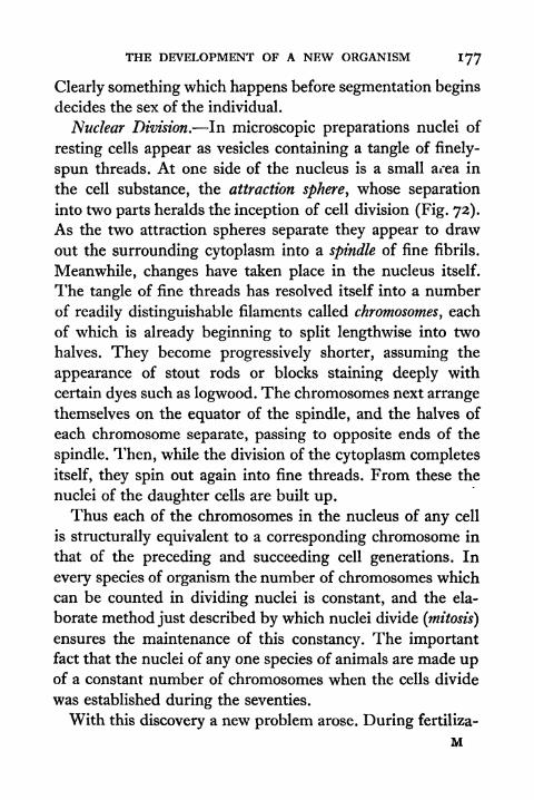
THE DEVELOPMENT OF A NEW ORGANISM 177
Clearly something which happens before segmentation begins
decides the sex of the individual.
Nuclear Division. In microscopic preparations nuclei of
resting cells appear as vesicles containing a tangle of finely-
spun threads. At one side of the nucleus is a small area in
the cell substance, the attraction sphere, whose separation
into two parts heralds the inception of cell division (Fig. 72).
As the two attraction spheres separate they appear to draw
out the surrounding cytoplasm into a spindle of fine fibrils.
Meanwhile, changes have taken place in the nucleus itself.
The tangle of fine threads has resolved itself into a number
of readily distinguishable filaments called chromosomes, each
of which is already beginning to split lengthwise into two
halves. They become progressively shorter, assuming the
appearance of stout rods or blocks staining deeply with
certain dyes such as logwood. The chromosomes next arrange
themselves on the equator of the spindle, and the halves of
each chromosome separate, passing to opposite ends of the
spindle. Then, while the division of the cytoplasm completes
itself, they spin out again into fine threads. From these the
nuclei of the daughter cells are built up.Thus each of the chromosomes in the nucleus of any cell
is structurally equivalent to a corresponding chromosome in
that of the preceding and succeeding cell generations. In
every species of organism the number of chromosomes which
can be counted in dividing nuclei is constant, and the ela-
borate method just described by which nuclei divide (mitosis)
ensures the maintenance of this constancy. The importantfact that the nuclei of any one species of animals are made upof a constant number of chromosomes when the cells divide
was established during the seventies.
With this discovery a new problem arose. During fertiliza-
M
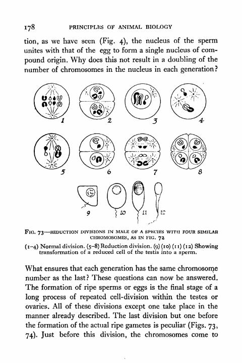
178 PRINCIPLES OF ANIMAL BIOLOGY
tion, as we have seen (Fig. 4), the nucleus of the spermunites with that of the egg to form a single nucleus of com-
pound origin. Why does this not result in a doubling of the
number of chromosomes in the nucleus in each generation?
FlG. 73 REDUCTION DIVISIONS IN MALE OF A SPJiClES WITH FOUR SIMILARCHROMOSOMES, AS IN FIG. 72
(1-4) Normal division. (5-8) Reduction division. (9) (10) (i i) (12) Showingtransformation of a reduced cell of the testis into a sperm.
What ensures that each generation has the same chromosome
number as the last? These questions can now be answered.
The formation of ripe sperms or eggs is the final stage of a
long process of repeated cell-division within the testes or
ovaries. All of these divisions except one take place in the
manner already described. The last division but one before
the formation of the actual ripe gametes is peculiar (Figs. 73,
74). Just before this division, the chromosomes come to
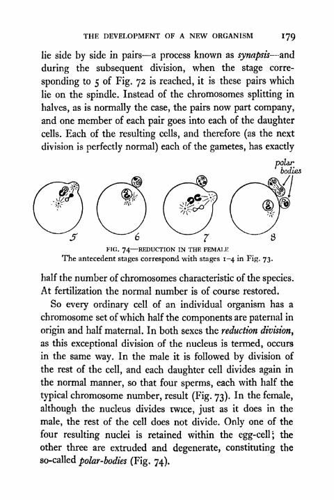
THE DEVELOPMENT OF A NEW ORGANISM 179
lie side by side in pairs a process known as synapsis and
during the subsequent division, when the stage corre-
sponding to 5 of Fig. 72 is reached, it is these pairs which
lie on the spindle. Instead of the chromosomes splitting in
halves, as is normally the case, the pairs now part company,
and one member of each pair goes into each of the daughter
cells. Each of the resulting cells, and therefore (as the next
division is perfectly normal) each of the gametes, has exactly
polarbodies
FIG. 74 REDUCTION IN THE FEMALE
The antecedent stages correspond with stages 1-4 in Fig. 73.
half the number of chromosomes characteristic of the species.
At fertilization the normal number is of course restored.
So every ordinary cell of an individual organism has a
chromosome set of which half the components are paternal in
origin and half maternal. In both sexes the reduction division,
as this exceptional division of the nucleus is termed, occurs
in the same way. In the male it is followed by division of
the rest of the cell, and each daughter cell divides again in
the normal manner, so that four sperms, each with half the
typical chromosome number, result (Fig. 73). In the female,
although the nucleus divides twice, just as it does in the
male, the rest of the cell does not divide. Only one of the
four resulting nuclei is retained within the egg-cell; the
other three are extruded and degenerate, constituting the
so-called polar-bodies (Fig, 74),
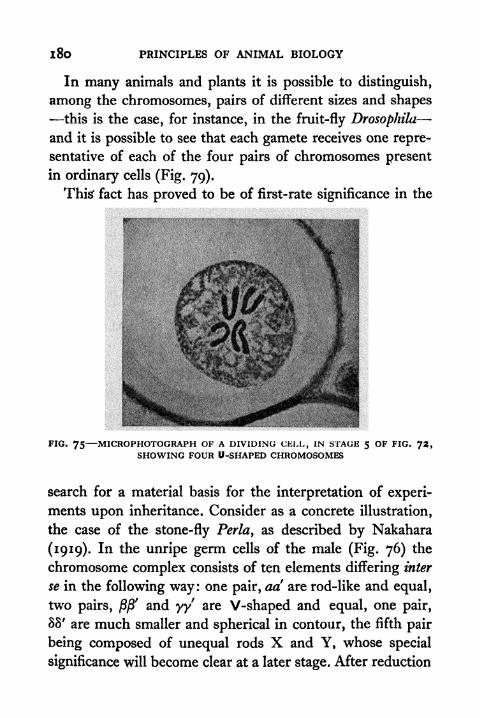
l8o PRINCIPLES OF ANIMAL BIOLOGY
In many animals and plants it is possible to distinguish,
among the chromosomes, pairs of different sizes and shapes
this is the case, for instance, in the fruit-fly Drosophila
and it is possible to see that each gamete receives one repre-
sentative of each of the four pairs of chromosomes present
in ordinary cells (Fig. 79).
Thisf fact has proved to be of first-rate significance in the
FIG. 75 MICROPHOTOGRAPH OF A DIVIDING CELL, IN STAGE 5 OF FIG. 72,SHOWING FOUR U-SHAPED CHROMOSOMES
search for a material basis for the interpretation of experi-
ments upon inheritance. Consider as a concrete illustration,
the case of the stone-fly Perla, as described by Nakahara
(1919). In the unripe germ cells of the male (Fig. 76) the
chromosome complex consists of ten elements differing inter
se in the following way: one pair, ad are rod-like and equal,
two pairs, j8j8' and yy are V-shaped and equal, one pair,
88' are much smaller and spherical in contour, the fifth pair
being composed of unequal rods X and Y, whose special
significance will become clear at a later stage. After reduction
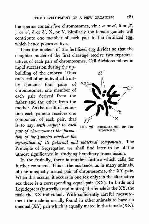
THE DEVELOPMENT OF A NEW ORGANISM l8l
the sperms contain five chromosomes, viz.: a or a', /? or /}',
y or y', S or S', X, or Y. Similarly the female gamete will
contribute one member of each pair to the fertilized egg,
which hence possesses five.
Thus the nucleus of the fertilized egg divides so that the
daughter nuclei of the first cleavage receive two represen-
tatives of each pair of chromosomes. Cell divisions follow in
rapid succession during the up-
building of the embryo. Thus
each cell of an individual fruit-
fly contains four pairs of
chromosomes, one member of
each pair derived from the
father and the other from the
mother. As the result of reduc-
tion each gamete receives one
component of each pair, that
is to say, with respect to eachFIO/ 7fr
_CHRoMosoMi OF THE
pair of chromosomes the forma- STONE-FLY
tion of the gametes involves the
segregation of its paternal and maternal components. The
Principle of Segregation we shall find later to be of the
utmost significance in studying hereditary transmission.
In the fruit-fly, there is another feature which calls for
further comment. This is the existence, as in many animals,
of one unequally mated pair of chromosomes, the XY pair.
When this occurs, it occurs in one sex only ;in the alternative
sex there is a corresponding equal pair (XX). In birds and
Lepidoptera (butterflies and moths), the female is the XY, the
male the XX individual. With sufficiently careful measure-
ment the male is usually found in other animals to have an
unequal (XY) pair which is equally mated in the female (XX).
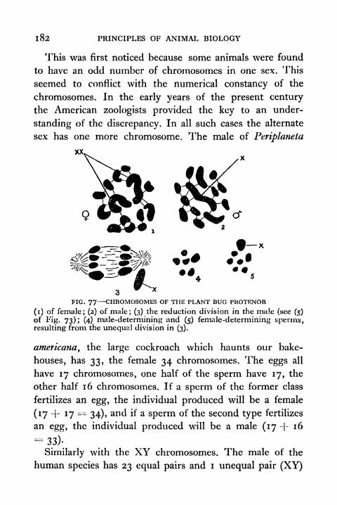
1 82 PRINCIPLES OF ANIMAL BIOLOGY
This was first noticed because some animals were found
to have an odd number of chromosomes in one sex. This
seemed to conflict with the numerical constancy of the
chromosomes. In the early years of the present century
the American zoologists provided the key to an under-
standing of the discrepancy. In all such cases the alternate
sex has one more chromosome. The male of Periplaneta
3FIG. 77 CHROMOSOMES OF THE PLANT BUG PUOTENOR
(i) of female; (2) of male; (3) the reduction division in the male (see (5)
of Fig. 73); (4) male-determining and (5) female-determining sperms,resulting from the unequal division in (3).
americana, the large cockroach which haunts our bake-
houses, has 33, the female 34 chromosomes. The eggs all
have 17 chromosomes, one half of the sperm have 17, the
other half 16 chromosomes. If a sperm of the former class
fertilizes an egg, the individual produced will be a female
(17 -|- 17 = 34), and if a sperm of the second type fertilizes
an egg, the individual produced will be a male (17 -|- 16
=33)-
Similarly with the XY chromosomes. The male of the
human species has 23 equal pairs and i unequal pair (XY)
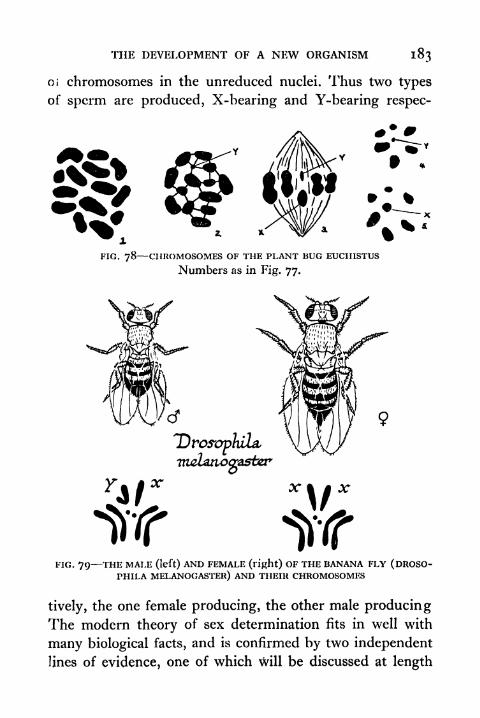
THE DEVELOPMENT OF A NEW ORGANISM 183
oi chromosomes in the unreduced nuclei. Thus two types
of sperm are produced, X-bearing and Y-bearing respec-
*FIG. 78 CHROMOSOMES OF THE PLANT BUG EUCIIISTUS
Numbers as in Fig. 77.
FIG. 79 THE MALE (left) AND FEMALE (right) OF THE BANANA FLY (DROSO-PHILA MELANOGASTER) AND THEIR CHROMOSOMES
tively, the one female producing, the other male producingThe modern theory of sex determination fits in well with
many biological facts, and is confirmed by two independent
lines of evidence, one of which will be discussed at length

184 PRINCIPLES OF ANIMAL BIOLOGY
later. The other may be mentioned here. In species having
an XY pair in the male, measurement of the sperm heads
shows that the sperms are of two different sizes. This sug-
gests that it may be possible eventually to separate seminal
fluid into portions containing predominantly one or other
type of sperm, the X-bearing or Y-bearing. If this could be
done the control of the sex ratio would be experimentally
realizable.
Many medical men still adhere to the superstition that the
sex of human offspring depends upon whether the egg fer-
tilized is derived from the right or left ovary. Apart from the
very conclusive evidence we have from other sources that
this view is wrong, it is demonstrably false in other mammals,where removal of the ovary of one side does not affect the
sex ratio. In this connexion, one may pertinently recall the
words of Sir Thomas Browne :
And therefore what admission we owe unto many conceptions
concerning right and left requireth circumspection. That is howfar we ought to rely on the remedy in Kiranides, that is the left eyeof a hedgehog fried in oil to procure sleep, and the right foot of a
frog in a deer's skin for gout ; or that to dream of the loss of right
or left tooth presageth the death of male or female kindred, accord-
ing to the doctrine of Artemidorus. What verity there is in that
numeral conceit in the lateral division of man by even and odd,
ascribing the odd unto the right side and even unto the left; and
so, by parity or imparity of letters in men's names to determine
misfortunes on either side of their bodies, by which account in
Greek numeration Hephaestus or Vulcan was lame in the right
foot, and Annibal lost his right eye. And lastly what substance there
is in that auspicial principe and fundamental doctrine of ariolation,
that the left hand is ominous and that good things do pass sinis-
trously upon us, because the left hand of man respected the right
hand of the gods, which handed their favours unto us. (Vulgar
Errors.)
When the sex chromosomes were first discovered the con-

THE DEVELOPMENT OF A NEW ORGANISM 185
elusions we have outlined seemed to conflict with the well-
known fact that many familiar animals change their sex
under certain conditions. But we may presume that whatever
influence the X chromosome exerts requires the proper
co-operation of external agencies; indeed the facts of sex
transformation fit in very well with the hypothesis, now they
have been studied more carefully. Crew found (1921) that,
whereas the offspring of a normal mating in frogs producesthe customary i : i ratio of male and female offspring, a
quite different result occurs when we mate with a normal
female a male that started its life as a female. He reared a
generation of seven hundred offspring of such a cross, all
females. If the transformed male was, as its former life
would suggest, an XX individual in disguise, it could produceno Y-bearing sperm and therefore no male offspring.
TOPICS FOR HOME STUDY1. Explain the difference between holoblastic and meroblastic
segmentation by reference to transitional types.
2. How are the following structures formed:
(a) notochord.
(b) C.N.S.
(c) gut.
(d) placenta.
3. Illustrate how external circumstances can modify the process of
development.
4. What part do ductless glands play in Amphibian metamorphosis.
5. On the assumption that the maternal and paternal componentsof each chromosome pair are distributed independently, show
by diagrams how many different classes of gametes can beformed by reduction when there are T, 2, 3, 4 pairs of chromo-somes. In this context gametes- are regarded as identical if for
every pair both have two chromosomes of maternal origin,
both have two chromosomes of paternal origin, or both have
one maternal and one paternal component.
CHAPTER VIII
THE MACHINERY OF INHERITANCE
INthe last chapter we have studied the characteristic
features of the development of a new being, and in so
doing we have dwelt on the fact that the external conditions
under which the fertilized egg develops have a very importantinfluence. Thus we have been led to make a distinction
between two aspects of the process. One is the nature of the
conditions which affect the segmenting egg and growing
embryo. The other is the kind of materials which the spermand egg respectively contribute to the constitution of the
new organism at the time of fertilization. Now it is a matter
of common knowledge that if it is allowed to develop under
the same conditions as the fertilized egg of one species,
the fertilized egg of another species of animal will not lead
to the production of the same type of adult. It is a universal
experience that any dog resembles its father and mother
more closely than it resembles any cat or any fish; any cat
resembles its father and mother more closely than it resem-
bles any dog or any fish; any fish resembles its father and
mother more closely than it resembles any cat or any dog.We may express this by saying that cats, dogs, and fish have
certain specific hereditable properties. If we examine these
hereditable properties we find that a cat has more hereditable
properties in common with any dog than those which it
shares with any fish. Thus organisms can be arranged or
classified in groups expressing the extent of resemblance in
their hereditable properties.
The Problem of Inheritance. Within the limits of a single
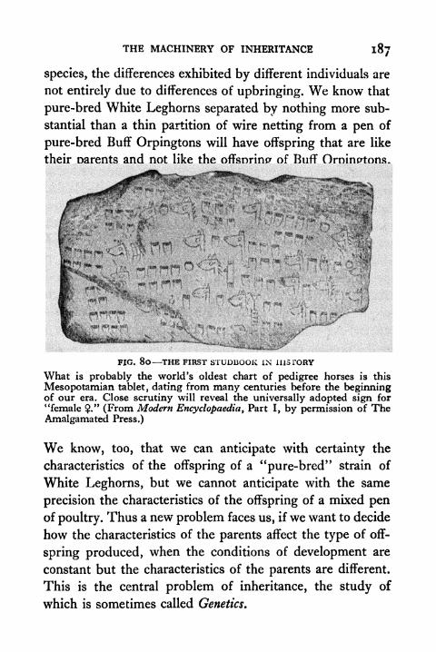
THE MACHINERY OF INHERITANCE 187
species, the differences exhibited by different individuals are
not entirely due to differences of upbringing. We know that
pure-bred White Leghorns separated by nothing more sub-
stantial than a thin partition of wire netting from a pen of
pure-bred Buff Orpingtons will have offspring that are like
their oarents and not like the offsnrinp of Buff Oroinptons.
FJG. 80 THE FIRST STUDBOOK IN HISTORY
What is probably the world's oldest chart of pedigree horses is this
Mesopotamia!! tablet, dating from many centuries before the beginningof our era. Close scrutiny will reveal the universally adopted sign for
"female ." (From Modern Encyclopaedia, Part I, by permission of TheAmalgamated Press.)
We know, too, that we can anticipate with certainty the
characteristics of the offspring of a "pure-bred" strain of
White Leghorns, but we cannot anticipate with the same
precision the characteristics of the offspring of a mixed penof poultry. Thus a new problem faces us, if we want to decide
how the characteristics of the parents affect the type of off-
spring produced, when the conditions of development are
constant but the characteristics of the parents are different.
This is the central problem of inheritance, the study of
which is sometimes called Genetics.

1 88 PRINCIPLES OF ANIMAL BIOLOGY
The issue we have just stated is very much like another
problem in science that has a somewhat similar history. It
is like the problem of how two chemical reagents react
together to form a new compound, and how the original
constituents can be recovered when the new compound is
in turn decomposed. Modern chemical theory begins
with the recognition of the Principle of Conservation of
matter and the discovery by Richter (1792) of the law of
constant proportions. Richter*s discovery led Dalton (1802)
to the atomic theory of matter. We are still far from under-
standing the physical chemistry of inheritance, but we are
to-day in a position to form a corresponding picture of
the process of combination of hereditable properties, as it
takes place when a new unit of living matter is brought
into being. Since the study of inheritance is to-day an exact
science, we shall devote some detailed consideration to it
in the present chapter. Our first task will be to show that
there is also a law of the conservation of genetic materials
and a law of constant proportions of hereditable properties.
These two laws can be generalized in a Principle of Segrega-
tion which is closely analogous to the Atomic Principle.
As in the study of chemistry, genetic experiments must be
done with pure materials, that is stocks whose offspring wecan predict, if the conditions are defined. For instance, we
may mate a cock of the white variety of the Andalusian
breed of poultry and a hen of the black variety both indi-
viduals being selected from stocks which have been inbred
for their characteristic plumage colour for a large number
of generations, and are therefore, genetically speaking, purematerials. Their progeny will be found to be neither white
nor black but intermediate between them, being slate-blue
birds with feathers shading into blue-black upon the neck and
THE MACHINERY OF INHERITANCE 189
back. If these blue hybrids are mated with one another their
offspring will be of three kinds white, black, and blue
and if sufficiently large numbers of eggs are hatched the
proportions will approximate to the ratio 1:1:2. Exactlysimilar results would follow if the original father had been
a black Andalusian cock and the original mother a white
hen. One thing which this shows us is that the sperm and
egg, though so different in size, contribute equally to the
constitution of the fertilized egg. We also learn that thoughthe hereditable properties of both the parents seemingly
disappear in the individual resulting from the combination
of a particular sperm and a particular egg, they are really
there but somehow hidden. In addition we find that when
the parental contributions reappear they do so in definite
numerical proportions.
Mendel's Hypothesis. These conclusions were proved true
of animals at the beginning of the present century, but they
were discovered to hold good in plants nearly half a century
earlier by an Austrian Abbot, Mendel (1858-65), contem-
porary with Darwin. Mendel first formulated the Principle
of Segregation which stands in the field of genetics for the
Atomic Theory in chemistry. The experimental fact which
Mendel and several of his contemporaries had discovered
that, when plants of different pure stocks are crossed and the
offspring mated inter se, the pure parental types reappear in
definite numerical proportions in the second crossbred
generation. In seeking for a more general rule, Mendel
started with two assumptions which are in keeping with
observations of hereditary transmission and of the nature of
fertilization :
(a) the two parents contribute equally to the hereditary
constitution of the offspring; and

190 PRINCIPLES OF ANIMAL BIOLOGY
(b) there is no reason to suppose that the chance of any
kind of gamete fertilizing any other kind (of the
opposite sex) is influenced by anything but the
numerical proportions in which they are produced.
If we take these two conclusions as granted, all the quantita-
tive results follow from the laws of chance on the hypothesis
that there are particles of hereditary combination, contributed
by both parents, and so distributed among the gametesthat any particular gamete contains one member either the
paternal or the maternal of a corresponding pair. Mendel
called these particles or atoms of heredity.
In the last chapter we have acquainted ourselves with
material entities in the sperm and egg which do behave like
Mendel's units, and the student will find it more easy to
envisage what is implied by the Principle of Segregation,
when we have seen how the behaviour of the chromosomes
bears on the interpretation of such experiments as we have
last described. But for the sake of historical perspective
we shall insert a brief account of Mendel's own researches.
The student may find it helpful to return to this after gaining
the clearer insight which the study of the chromosomes
affords us.
Mendel's hypothesis was greatly in advance of his time.
His doctrine assumed the principle that fertilization is the
combination of two gametes. This had not then been shown
to be true of animals. He also postulated the existence of
material entities for which at that time no microscopicevidence was available, because he worked long before the
facts of mitosis and reduction, set forth in Chapter VII,
began to be understood. When three Continental workers,de Vries, Correns and Tschermak independently redis-
THE MACHINERY OF INHERITANCE
covered his principles in the opening years of the twentieth
century and unearthed his papers in an obscure horticultural
journal, knowledge of the microscopic bodies known as the
chromosomes was already sufficient to confirm the essential
truth of his conclusions.
In his original work Mendel chiefly employed the common
pea. This possesses two advantages which recommend it for
such experimentation, namely, that its flowers are capable
of self-fertilization (i.e.the pistil can be pollinated from
the stamens of the same flower) and that it has a number
of well-marked varieties distinguished by tangible charac-
teristics, such as the shape (round or wrinkled) and colour
(green or yellow) of the seeds, or the stature (tall or dwarf)
of the shoot, etc. A single example will be easier to see howMendel was led to the fundamental genetic principle which
he discovered.
As a type of Mendelian experiment we shall consider the
results of crossing a variety of pea having a dwarf shoot,
from a stock breeding true to this feature, with an individual
from another variety characterized by a tall shoot, likewise of
pure pedigree. When pollen from flowers of the one is trans-
ferred to the stigma of the other, the same result always
occurs. Every seed produced in consequence of such a union
gives rise to a tall plant, whichever way the cross is made
with respect to the sex of the parents. But if these seeds
are allowed to germinate and grow into plants, the results
of self-fertilizing the flowers of the first generation (tech-
nically the F.I or first filial generation of the cross), or,
alternatively, crossing them with other F.I plants, is entirely
different from the effect of crossing two of the parental
pedigree tall plants. Instead of obtaining only tall plants
true to type, it is found that three-quarters of the seed
PRINCIPLES OF ANIMAL BIOLOGY
produced (F.2 generation) give rise to tall plants, and one-
quarter to dwarfs like one of the original parents. Further
breeding shows that two-thirds of the F.2 tall individuals
breed in the same way, throwing dwarfs in the same ratio.
The remainder breed true to type like the original tall parent
when self-fertilized, as do the dwarfs of the F.2 generation,
which, when self-fertilized, or crossed inter se> have only
dwarf offspring.
The first clue to Mendel's line of reasoning is that the
hereditary constitution of the tall individual in the F.I
generation differs from that of the tall parents in so far as it
is capable of giving rise to dwarf offspring. It differs presum-
ably in producing gametes characterized by the possession of
some material (Mendel called it, without discussing its nature,
a factor) responsible for the production of the dwarf condi-
tion. We notice also that in the F.2 generation the propor-
tions of pure tall and pure dwarf plants are identical, that
is to say, one-quarter have the factor for tallness only and
one-quarter the factor for dwarfness only, so that neither
of the gametes from which an individual of either type
originates contains the alternate factor. In addition we must
bear in mind that the F.I tall plants behave in a similar
manner whether the factor for dwarfness is borne by the
maternal or paternal gametes (ovules and pollen grains).
Let us therefore suppose that each F.I tall plant producesin equal quantities gametes bearing the tall and dwarf
factors respectively but never both and further that there
is an equal chance of any pollen grain fertilizing an ovule
with the same or an alternative factor to that which it bears.
It follows that the number of individuals produced in the
F.2 generation containing both factors for the tall and dwarf
conditions will be twice the number containing only the tall
THE MACHINERY OF INHERITANCE 1 93
or only the dwarf factor. For T may fertilize T or D, giving
TT or DT; and D may fertilize D or T, giving DD or DT.
This, of course, satisfies the conditions, and is an adequate
account of the facts so far. It is the essence of Mendel's
hypothesis, which states that characters distinguishing
different hereditary strains depend upon factors which are
inherited from both parents and segregate in the formation
of the gametes, so that one-half contain the paternal and
one-half the maternal factor.
The assumptions advanced may not involve every con-
ceivable possibility, and the truth of the factorial hypothesis
must stand or fall, like all other hypotheses, with its capacity
to yield verifiable consequences. Consequences of a veri-
fiable nature which can at once be subjected to test are
exemplified by crossing back the F.I impure tall plants with
(a) the pure tall parents and (b) the dwarf plants -which are
all pure. Using as before letters to denote the material
factors at work, the pure breeding tall plants and dwarfs have
the constitution TT, DD on the hypothesis outlined, since
their character depends on factors inherited from both
parents: the impure plants F.I have the constitution TDor DT. By crossing TD with TT we should get two types
of offspring TT and TD, equal numbers of pure and impuretall plants. Also by crossing TD with DD, we get two types
of offspring TD and DD, equal numbers of impure tall and
pure dwarf. Mendel tested and verified these and other
implications; and all subsequent inquiry has confirmed the
anticipations raised.
He was thus led to this conclusion. Hereditable differences
are dependent upon separate particles derived from both
parents, remaining distinct throughout the entire life-cycle,
and finally separating in the formation of the gametes, so
that with respect to any single pair of them one-half of the
N
194 PRINCIPLES OF ANIMAL BIOLOGY
gametes contain the particle derived from one parent and
the other half contain the particle contributed by the alternate
parent. This is Mendel's law of genetic segregation.
For the sake of convenience we may here introduce some
necessary technical terms. An individual which like the
"impure" F.I tall plants receives dissimilar factors from its
parents is said to be heterozygous in respect of those factors
in contradistinction to the homozygous (e.g. pure dwarf or
pure tall) type. The character which appears to predominate,
if its material forerunner is present in the fertilized egg, is
said to be dominant in contradistinction to the recessive
character, which is only manifest when both the gametescontribute its material forerunner.
Dominance is not a universal phenomenon. For instance,
if we cross individuals from pure stocks of the red and white
varieties of the flowering plant popularly known as four
o'clock or botanically as Mirabilis jalapa, the F.I generation
bear only pink flowers. If these F.I heterozygotes are selfed
or crossed inter se, the F.z generation is composed of reds,
pinks, and whites, in the proportion 1:2:1. This would
correspond to the 3 : i ratio if the heterozygotes were similar
to one or the other type of homozygote. The blue hybridAndalusian fowl produced by crossing black and splashedwhite birds is analogous. Mating of Blue Andalusian fowls
results in producing black and white offspring. Hence, if wewish to obtain blue individuals it is more profitable to mate
blacks with whites. This gives us a heterozygous generationof blues only. The effect of crossing the blue heterozygotes
among themselves is to raise a progeny of blacks, blues, and
whites in the ratio of i : 2 : i . In these examples the hetero-
zygote is intermediate between the parental homozygoustypes. In the inheritance of the white colour of Leghornfowls, we encounter an instance of what may be called in.

THE MACHINERY OF INHERITANCE 195
complete dominance; for, if mated with an individual of a
coloured strain, the heterozygous individuals are white with
a few coloured tail feathers. Statistical analysis in fact shows
that dominance is never absolutely complete. Whatever be
the degree of resemblance between the F.I generation of a
cross between pure stocks and the parental types, the factors
remain distinct and recombine to produce pure parental types
again in the F.2 generation.
An analogy will help the student to appreciate the question
of dominance in its true perspective. Sodium and potassium
yield colourless salts with most common acids, but the per-
manganates of both are purple in solution. The salts of
copper are generally of a bluish or greenish tint in solution.
In the one case the anion, in the other case the kation is the
dominant factor in determining the physical property of
colour. In neither case does the other component behave in
any reaction with less characteristic efficacy on account of
its presence being seemingly masked. So in the process of
hereditary transmission the recessive factor is just as impor-
tant as the dominant factor. It is only in its bodily expression
that the two are differentiated.
It must not be imagined that every clear-cut characteristic
which distinguishes two related forms need depend on only
one factor difference. Mendel was particularly fortunate in
hitting upon a form in which there exist a number of strains
differing with respect to single pairs of unit factors. Hadhe studied, for example, the inheritance of the "walnut"
type of comb which occurs in the Malay breeds of domestic
fowl (Fig. 86), he would have had a more difficult problem.
The "rose" comb of Wyandottes and the "pea" comb of
the Indian Game breeds are each dominant to the single
comb of the Leghorn, Sussex, and Rhode Island breeds.
The ''walnut" comb can be produced by crossing individuals
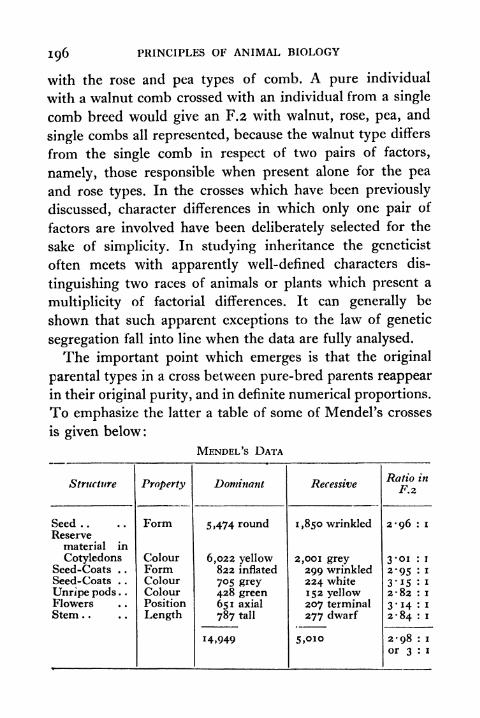
196 PRINCIPLES OF ANIMAL BIOLOGY
with the rose and pea types of comb. A pure individual
with a walnut comb crossed with an individual from a single
comb breed would give an F.2 with walnut, rose, pea, and
single combs all represented, because the walnut type differs
from the single comb in respect of two pairs of factors,
namely, those responsible when present alone for the pea
and rose types. In the crosses which have been previously
discussed, character differences in which only one pair of
factors are involved have been deliberately selected for the
sake of simplicity. In studying inheritance the geneticist
often meets with apparently well-defined characters dis-
tinguishing two races of animals or plants which present a
multiplicity of factorial differences. It can generally be
shown that such apparent exceptions to the law of genetic
segregation fall into line when the data are fully analysed.
The important point which emerges is that the original
parental types in a cross between pure-bred parents reappear
in their original purity, and in definite numerical proportions.
To emphasize the latter a table of some of Mendel's crosses
is given below :
MENDEL'S DATA
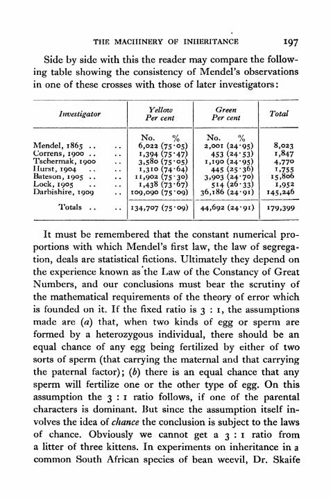
THE MACHINERY OF INHERITANCE 197
Side by side with this the reader may compare the follow-
ing table showing the consistency of Mendel's observations
in one of these crosses with those of later investigators :
It must be remembered that the constant numerical pro-
portions with which Mendel's first law, the law of segrega-
tion, deals are statistical fictions. Ultimately they depend on
the experience known as the Law of the Constancy of Great
Numbers, and our conclusions must bear the scrutiny of
the mathematical requirements of the theory of error which
is founded on it. If the fixed ratio is 3 : i, the assumptionsmade are (a) that, when two kinds of egg or sperm are
formed by a heterozygous individual, there should be an
equal chance of any egg being fertilized by either of two
sorts of sperm (that carrying the maternal and that carrying
the paternal factor); (b) there is an equal chance that any
sperm will fertilize one or the other type of egg. On this
assumption the 3 : i ratio follows, if one of the parental
characters is dominant. But since the assumption itself in-
volves the idea of chance the conclusion is subject to the laws
of chance. Obviously we cannot get a 3 : i ratio from
a litter of three kittens. In experiments on inheritance in a
common South African species of bean weevil, Dr. Skaife

198 PRINCIPLES OF ANIMAL BIOLOGY
crossed the dominant black form with a red recessive
mutant, and obtained in the second crossbred generation 347
black and 119 red individuals. For this number of indi-
viduals the 3 : i ratio requires two classes differing from
349-5 and 116-5 by a "probable error" of approximately 6.
That is to say, it is as likely as not that the actual figures
will exceed or fall short of these numbers by 6. The observed
figures differed by much less than the probable error. An
exposition of the methods used in determining the probable
error of Mendelian ratios lies outside the scope of this book.
Mendel's investigations followed up what happens when
two different pairs of characters, each dependent on a separate
pair of factors, are involved in a cross between pure-bred
parents. The actual results of a cross between a strain of
peas with green wrinkled seeds and yellow round seeds or of
peas with green round and yellow wrinkled seed respec-
tively will be understood by referring to Fig. 82. Theratios of the double dominant, the two classes of single
dominants and the double recessives which Mendel obtained
in all his crosses was 9 : 3 : 3 : T, as should occur if the
yellow-green factors and the round-wrinkled factors behave
quite independently of one another. Mendel formulated this
result as a generalization sometimes known as Mendel's
Second Law. It is not, however, a law in the same sense as
Mendel's First Law, of segregation, which we have deduced
above, for it is only applicable to certain cases, and as we
shall see later, the exceptions are of more theoretical interest
than the rule. Here again an analogy from chemistry will
help. The fundamental law of chemical combination is the
law of constant proportions. The law of multiple proportionsis no more than a statement of certain experiences that the
pioneers of chemistry encountered in dealing with some of
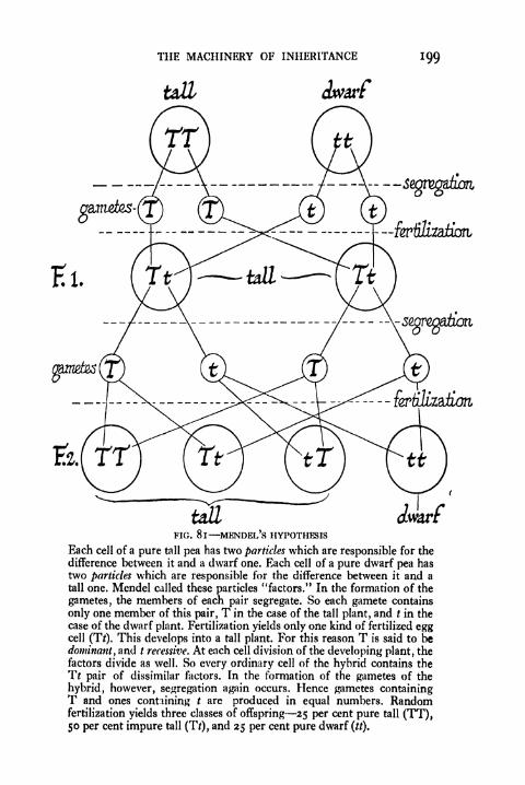
THE MACHINERY OF INHERITANCE 199
dwarf
dwarfFIG. 8 1 MENDEL'S HYPOTHESIS
Each cell of a pure tall pea has two particles which are responsible for the
difference between it and a dwarf one. Each cell of a pure dwarf pea has
two particles which are responsible for the difference between it and a
tall one. Mendel called these particles "factors." In the formation of the
gametes, the members of each pair segregate. So each gamete contains
only one member of this pair, T in the case of the tall plant, and t in the
case of the dwarf plant. Fertilization yields only one kind of fertilized eggcell (TV). This develops into a tall plant. For this reason T is said to be
dominant, and t recessive. At each cell division of the developing plant, the
factors divide as well. So every ordinary cell of the hybrid contains the
Tt pair of dissimilar factors. In the formation of the gametes of the
hybrid, however, segregation again occurs. Hence gametes containingT and ones containing t are produced in equal numbers. Randomfertilization yields three classes of offspring 25 per cent pure tall (TT),50 per cent impure tall (T/), and 25 per cent pure dwarf (tt).
200 PRINCIPLES OF ANIMAL BIOLOGY
the compounds that were first investigated. Had they started
with the higher hydrocarbons, they would have encountered
no such simple relations. Both the law of multiple propor-
tions and the second law of Mendel are historically impor-
tant, because they helped to suggest hypotheses which, once
stated, were broad enough to take in other possibilities. Weshall leave a consideration of the 9 13:3:1 ratio of Mendel's
original experiments till a later stage, and now approach the
problem of hybridization from a different point of view.
The Chromosome Hypothesis. Mendel spoke of factors
which determine the characters which organisms display
under appropriate environmental influences. All that is
meant by heredity must refer to the contributions which
the sperm and the egg make to the new individual. Can we
go farther and identify within the sperm or egg the material
particles which enter the gametes after innumerable cell
divisions? Can we detect the existence of anything which
behaves as our"factors'* have been seen to behave?
Hybridization experiments lead us to conclude that the
particles of heredity are present in the fertilized egg in
duplicate, and that they segregate before the formation of
the gametes into maternal and paternal components, one
member of each pair and one only being present in each
gamete. We now know that the number of chromosomes of
any species of animals or plants is twice the number present
in the gametes. In many animals (and plants) from the most
diverse phyla, the chromosome complex of a species has a
definite configuration as well as a definite number. Amongthe chromosomes it is possible to distinguish pairs of
different sizes and shapes (e.g. Fig. 76), and the main-
tenance of this constant configuration implies that whenreduction takes place one member of each pair passes into
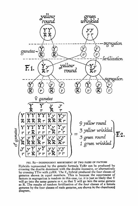
9 yellow round
green
2 ar^rt winkled
FIG. 82 INDEPENDENT ASSORTMENT OF TWO PAIRS OF FACTORS
Hybrids represented by the genetic formula Y^Rr can be produced by
crossing the double dominant with the double recessive, or alternatively
by crossing YYrr with j'yRR. The Fj hybrid produced the four classes of
gametes shown in equal numbers, This is because the assortment of
factors in segregation is random in this case, i.e. it is just as likely that Ywill go into the same gamete as r, as that Y will go into the same gameteas R. The results of random fertilization of the four classes of x female
gametes by the four classes of male garnetes, are shown in the chessboard
diagram.

202 PRINCIPLES OF ANIMAL BIOLOGY
each gamete (p. 178). In other words, the chromosomes are
present in the fertilized egg in pairs, and they segregate in
x
purple-
FIG. 83^
Diagrammatic representation of the first generation of a cross betweenthe red-eyed wild type of the fruit fly and the purple-eyed mutant. TheY chromosome is shaded, and the chromosome bearing the mutant geneis shown in black.
the formation of the gametes into paternal and maternal
components, only one member of each pair being represented
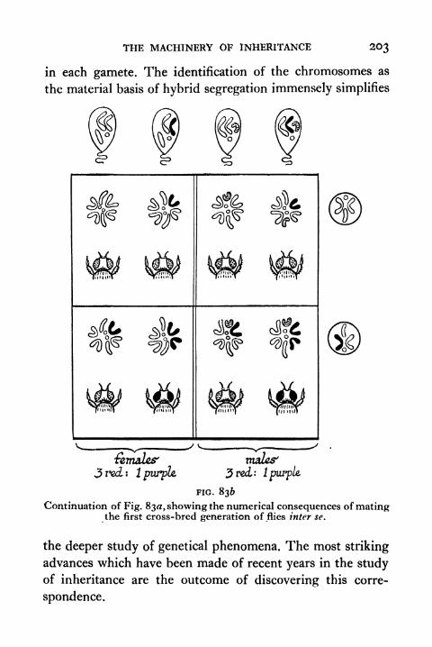
THE MACHINERY OF INHERITANCE 203
in each gamete. The identification of the chromosomes as
the material basis of hybrid segregation immensely simplifies
: IpurpU
malts'
3 red: 1purple.
FIG. 836
Continuation of Fig. 83^, showing the numerical consequences of matingthe first cross-bred generation of flies inter se.
the deeper study of genetical phenomena. The most striking
advances which have been made of recent years in the studyof inheritance are the outcome of discovering this corre-
spondence.
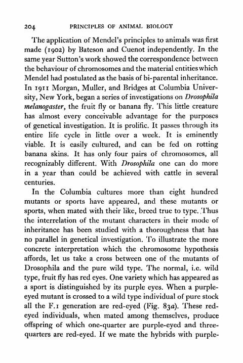
204 PRINCIPLES OF ANIMAL BIOLOGY
The application of Mendel's principles to animals was first
made (1902) by Bateson and Cuenot independently. In the
same year Sutton's work showed the correspondence between
the behaviour of chromosomes and the material entitieswhich
Mendel had postulated as the basis of bi-parental inheritance.
In 1911 Morgan, Muller, and Bridges at Columbia Univer-
sity, New York, began a series of investigations on Drosophila
melanogaster, the fruit fly or banana fly. This little creature
has almost every conceivable advantage for the purposes
of genetical investigation. It is prolific. It passes through its
entire life cycle in little over a week. It is eminently
viable. It is easily cultured, and can be fed on rotting
banana skins. It has only four pairs of chromosomes, all
recognizably different. With Drosophila one can do more
in a year than could be achieved with cattle in several
centuries.
In the Columbia cultures more than eight hundred
mutants or sports have appeared, and these mutants or
sports, when mated with their like, breed true to type. ,Thus
the interrelation of the mutant characters in their mode of
inheritance has been studied with a thoroughness that has
no parallel in genetical investigation. To illustrate the more
concrete interpretation which the chromosome hypothesis
affords, let us take a cross between one of the mutants of
Drosophila and the pure wild type. The normal, i.e. wild
type, fruit fly has red eyes. One variety which has appeared as
a sport is distinguished by its purple eyes. When a purple-
eyed mutant is crossed to a wild type individual of pure stock
all the F.I generation are red-eyed (Fig. 830). These red-
eyed individuals, when mated among themselves, produce
offspring of which one-quarter are purple-eyed and three-
quarters are red-eyed. If we mate the hybrids with purple-
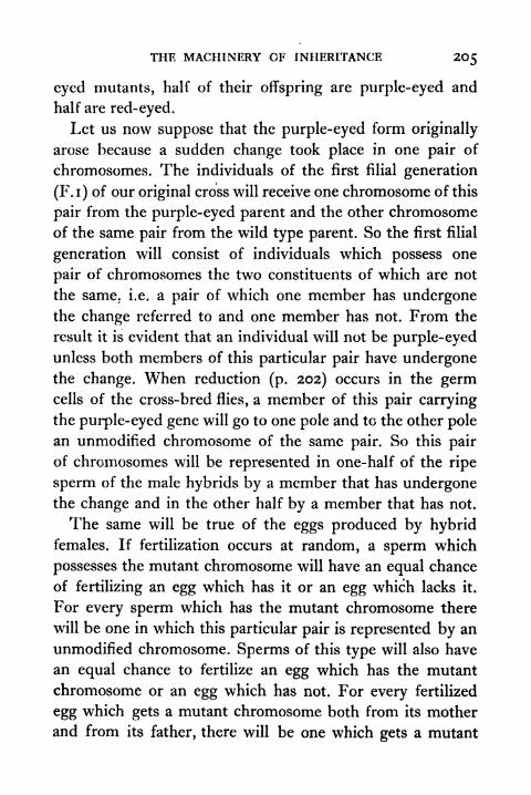
THE MACHINERY OF INHERITANCE 205
eyed mutants, half of their offspring are purple-eyed and
half are red-eyed.
Let us now suppose that the purple-eyed form originally
arose because a sudden change took place in one pair of
chromosomes. The individuals of the first filial generation
(F.i) of our original cross will receive one chromosome of this
pair from the purple-eyed parent and the other chromosome
of the same pair from the wild type parent. So the first filial
generation will consist of individuals which possess one
pair of chromosomes the two constituents of which are not
the same. i.e. a pair of which one member has undergonethe change referred to and one member has not. From the
result it is evident that an individual will not be purple-eyed
unless both members of this particular pair have undergonethe change. When reduction (p. 202) occurs in the germcells of the cross-bred flies, a member of this pair carrying
the purple-eyed gene will go to one pole and to the other pole
an unmodified chromosome of the same pair. So this pair
of chromosomes will be represented in one-half of the ripe
sperm of the male hybrids by a member that has undergonethe change and in the other half by a member that has not.
The same will be true of the eggs produced by hybridfemales. If fertilization occurs at random, a sperm which
possesses the mutant chromosome will have an equal chance
of fertilizing an egg which has it or an egg which lacks it.
For every sperm which has the mutant chromosome there
will be one in which this particular pair is represented by an
unmodified chromosome. Sperms of this type will also have
an equal chance to fertilize an egg which has the mutant
chromosome or an egg which has not. For every fertilized
egg which gets a mutant chromosome both from its mother
and from its father, there will be one which gets a mutant
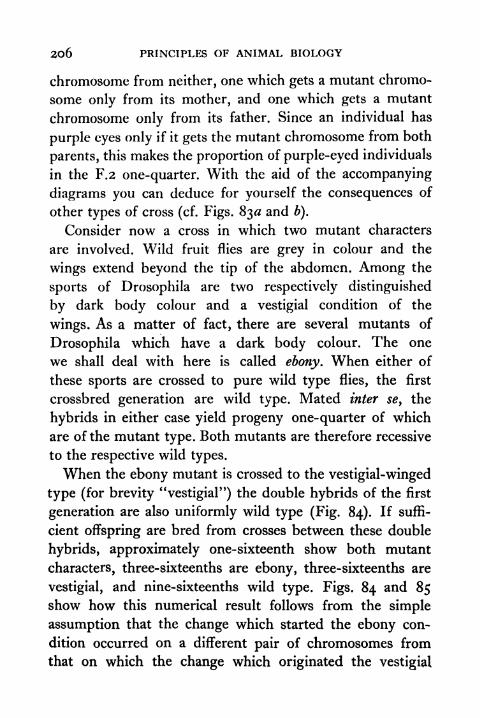
206 PRINCIPLES OF ANIMAL BIOLOGY
chromosome from neither, one which gets a mutant chromo-
some only from its mother, and one which gets a mutant
chromosome only from its father. Since an individual has
purple eyes only if it gets the mutant chromosome from both
parents, this makes the proportion of purple-eyed individuals
in the F.2 one-quarter. With the aid of the accompanying
diagrams you can deduce for yourself the consequences of
other types of cross (cf. Figs. 830 and b).
Consider now a cross in which two mutant characters
are involved. Wild fruit flies are grey in colour and the
wings extend beyond the tip of the abdomen. Among the
sports of Drosophila are two respectively distinguished
by dark body colour and a vestigial condition of the
wings. As a matter of fact, there are several mutants of
Drosophila which have a dark body colour. The one
we shall deal with here is called ebony. When either of
these sports are crossed to pure wild type flies, the first
crossbred generation are wild type. Mated inter sey the
hybrids in either case yield progeny one-quarter of which
are of the mutant type. Both mutants are therefore recessive
to the respective wild types.
When the ebony mutant is crossed to the vestigial-winged
type (for brevity "vestigial") the double hybrids of the first
generation are also uniformly wild type (Fig. 84). If suffi-
cient offspring are bred from crosses between these double
hybrids, approximately one-sixteenth show both mutant
characters, three-sixteenths are ebony, three-sixteenths are
vestigial, and nine-sixteenths wild type. Figs. 84 and 85show how this numerical result follows from the simple
assumption that the change which started the ebony con-
dition occurred on a different pair of chromosomes from
that on which the change which originated the vestigial
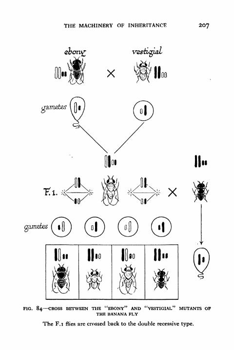
THE MACHINERY OF INHERITANCE 207
x
gametes
II
V/\
II
II"
FIG. 84 CROSS BETWEEN THE "EBONY" AND "VESTIGIAL" MUTANTS OFTHE BANANA FLY
The F.I flies are crossed back to the double recessive type.

208 PRINCIPLES OF ANfMAL BIOLOGY
type occurred. In the reduction division of the germ cells
of the F. i generation there will be two different configura-
tions according as the mutant members of each pair go to
the same or opposite poles, and the numerical recombina-
II .
II.
II"
FIG. 85 RESULT OF MATING FIRST HYBRID GENERATION OF THE EBONY-VESTIGIAL CROSS OF FIG. 84 inter se (cf. text)
tions can be simply deduced by using a chessboard diagramto show that for every set of four different types of eggsfertilized by one of the four different types of sperm there
will be a corresponding quartette fertilized by each of the
remaining three types of sperm. If the double hybrid flies
are bred with the new type having both mutant characters,
the four possible combinations will appear in the progenyin equal proportions (Fig. 85).
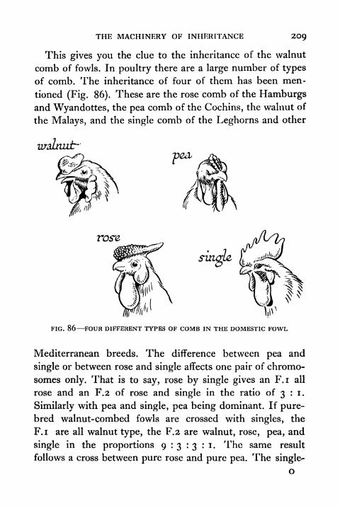
THE MACHINERY OF INHERITANCE 2O9
This gives you the clue to the inheritance of the walnut
comb of fowls. In poultry there are a large number of types
of comb. The inheritance of four of them has been men-
tioned (Fig. 86). These are the rose comb of the Hamburgsand Wyandottes, the pea comb of the Cochins, the walnut of
the Malays, and the single comb of the Leghorns and other
rose
FIG. 86 FOUR DIFFERENT TYPES OF COMB IN THE DOMESTIC FOWL
Mediterranean breeds. The difference between pea and
single or between rose and single affects one pair of chromo-
somes only. That is to say, rose by single gives an F.I all
rose and an F.2 of rose and single in the ratio of 3 : i.
Similarly with pea and single, pea being dominant. If pure-
bred walnut-combed fowls are crossed with singles, the
F.I are all walnut type, the F.2 are walnut, rose, pea, and
single in the proportions 9:3:3:1. The same result
follows a cross between pure rose and pure pea. The single-
o
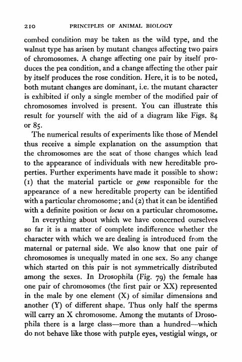
210 PRINCIPLES OF ANIMAL BIOLOGY
combed condition may be taken as the wild type, and the
walnut type has arisen by mutant changes affecting two pairs
of chromosomes. A change affecting one pair by itself pro-
duces the pea condition, and a change affecting the other pair
by itself produces the rose condition. Here, it is to be noted,
both mutant changes are dominant, i.e. the mutant character
is exhibited if only a single member of the modified pair of
chromosomes involved is present. You can illustrate this
result for yourself with the aid of a diagram like Figs. 84
or 85.
The numerical results of experiments like those of Mendel
thus receive a simple explanation on the assumption that
the chromosomes are the seat of those changes which lead
to the appearance of individuals with new hereditable pro-
perties. Further experiments have made it possible to show:
(i) that the material particle or gene responsible for the
appearance of a new hereditable property can be identified
with a particular chromosome ;and (2) that it can be identified
with a definite position or locus on a particular chromosome.
In everything about which we have concerned ourselves
so far it is a matter of complete indifference whether the
character with which we are dealing is introduced from the
maternal or paternal side. We also know that one pair of
chromosomes is unequally mated in one sex. So any changewhich started on this pair is not symmetrically distributed
among the sexes. In Drosophila (Fig. 79) the female has
one pair of chromosomes (the first pair or XX) represented
in the male by one element (X) of similar dimensions and
another (Y) of different shape. Thus only half the spermswill carry an X chromosome. Among the mutants of Droso-
phila there is a large class more than a hundred which
do not behave like those with putple eyes, vestigial wings, or
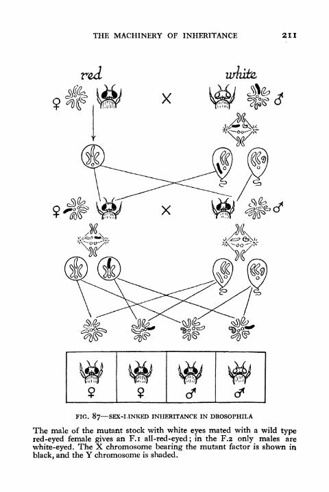
THE MACHINERY OF INHERITANCE 211
FIG. 87 SEX-LINKED INHERITANCE IN DROSOPHILA
The male of the mutant stock with white eyes mated with a wild typered-eyed female gives an F.I all-red-eyed; in the F.2 only males are
white-eyed. The X chromosome bearing the mutant factor is shown in
black, and the Y chromosome is shaded.

212 PRINCIPLES OF ANIMAL BIOLOGY
ebony body colour. Crosses with wild type give different
results according as the mutant character is introduced from
the maternal or paternal side.
One example of this class has white instead of red eyes.
Crossed to pure-bred wild type (red-eyed) females, white-
eyed males produce offspring all of which are wild type, and
when the hybrids are mated inter se one-quarter of their
progeny are white-eyed (Fig 88). The only anomaly is that
all the F.2 white-eyed flies are males. When white-eyed
females are crossed to pure stock wild type males, the result
is more remarkable. Only the female offspring of the first
generation are red-eyed. The males are white-eyed. Whenthese crossbred flies are mated inter se one-quarter of
their progeny are white-eyed males, one-quarter white-
eyed females, one-quarter red-eyed males, and one-quarter
red-eyed females. We can interpret this result if we
assume that the mutant gene which is responsible for white
eyes is given on the X chromosome, and that it can override
the effect of all the other chromosomes unless it is paired
off with anotherX chromosome from wild stock. If a mutant
X chromosome is present in the male there can be no Xchromosome to pair with it. So the mutant condition will
always be exhibited. The mutant condition will not be
exhibited by the female unless both members of the Xpair carry the mutant gene.
The phenomenon of "sex-linked inheritance," as this
asymmetrical type of transmission is sometimes called, was
first discovered by Doncaster (1906) in moths. In Lepidop-tera the male is the XX type and the female XY or XO. In
the currant or magpie moth Abraxas, there is a pale-winged
(lacticolor) mutant in contradistinction to the dark-winged
(grossulariata) type. Doncaster set out to find why females
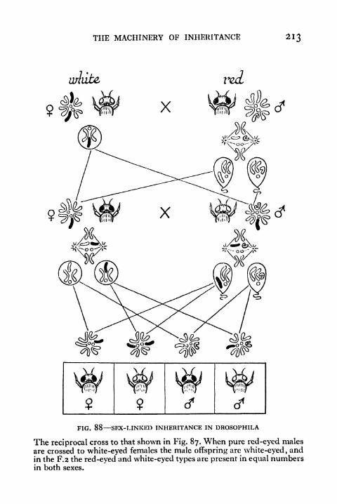
THE MACHINERY OF INHERITANCE 213
FIG. 88 SEX-LINKED INHERITANCE IN DROSOPHILA
The reciprocal cross to that shown in Fig. 87. When pure red-eyed males
are crossed to white-eyed females the male offspring are white-eyed ,and
in the F.2 the red-eyed and white-eyed types are present in equal numbersin both sexes.
214 PRINCIPLES OF ANIMAL BIOLOGY
of the lacticolor variety are much commoner than males. On
breeding he found that dark-winged males crossed to pale
females have only dark-winged offspring, which give dark-
winged and pale moths in the ratio 3 : i when mated inter se.
All the pale moths of this cross are females. In the reciprocal
cross only the males are of the dark type, and the F.2 is
composed of dark and pale, males and females, in equal
proportions. Bearing in mind the difference (p. 181) between
the chromosomes of moths and Drosophila you will have no
difficulty in applying the methods of Figs. 87-88 to inter-
pret this result. An analogous type of sex-linked inheritance
occurs in birds (canaries, poultry, etc.), as, for instance, in
the dominant X-borne mutant gene responsible for the
barring of the plumage in the Plymouth Rock breed.
Agreement between genetical experiment and microscopic
observation is also illustrated by the phenomenon of non-
disjunction described by Bridges in connexion with several
sex-linked mutant characters of which our original mutant
character (white-eye colour) will serve as an example. There
appeared among the white-eyed mutant stock of Drosophila
certain strains of which the females, when crossed to normal
red-eyed males, gave a certain proportion of red-eyed males
and white-eyed females, in addition to the usual red-eyed
females and white-eyed males alone. When the white-eyed
female offspring of such abnormal matings were crossed
back to red-eyed males they, in their turn, gave all four
classes red-eyed males and females, white-eyed males and
females. The white-eyed females behaved like their mothers,
giving abnormal results in all cases. Certain of the red-eyed
females gave normal and others abnormal results in crossing.
Of the male progeny the red-eyed individuals were normal,
whereas only half the white-eyed individuals were normal,

THE MACHINERY OF INHERITANCE 215
the remainder begetting daughters whose progeny was ex-
ceptional. Bridges found that in the abnormal white-eyed
F.I females the chromosome complex of the dividing body-
cells was also abnormal it showed a Y element in addition
to the XX pair.
Microscopic observation shows that in rare circumstances
the X chromosomes fail to separate when reduction occurs.
So the ripe egg may contain either two X chromosomes or
none at all. If we represent the sperms of a red male as X'
of Y, two additional types of individuals will result from
fertilization by a Y or X' sperm respectively, an XXY or
white female, and X'O or red male. This accounts for the
exceptional individuals in the F. i,and accords with the facts
observed. According to whether the X elements segregate
with respect to one another or the Y chromosome, the F.I
white females will have four types of eggs XX, Y, XY, X.
If these are fertilized by a Y sperm (which does not influence
red-eye colour) we get four types (a) XXY white females,
which will obviously behave in the same way, thus agreeing
with breeding experience; (b) YY individuals with this
constitution cannot live; (c) XYY white males which should
produce XY sperms so that in crossing with normal white
females daughters of the XXY type, producing exceptional
progeny, would result; (d) XY normal white males. When
the same four classes of eggs are fertilized by an X' sperm
carrying the red factor, four red types of offspring would
result, as follows : (a) X'XX triploid (X) female which usually
dies; (b) X'Y normal red males; (c) X'YX red females
with abnormal offspring; (d) X'X normal red females.
Thus the non-disjunction of the X chromosome in the
formation of the eggs of some of the females of the
parental white-eyed stock accounts for the entire series
2l6 PRINCIPLES OF ANIMAL BIOLOGY
of exceptional genetic phenomena which occur in these
strains.
The Chromosome Map. Such evidence can leave very
little doubt about the conclusion that the behaviour of the
chromosomes provides the material basis of the numerical
proportions found in breeding experiments. There is now
abundant evidence of the same kind derived from the study
of inheritance in widely different types of animals and plants
pointing to the same conclusion. Other experiments have
made it possible to locate the changes which are responsible
for particular mutant characters on a definite region of a
particular chromosome.
The experimental results which lead to this conclusion
may first be illustrated by what happens in crosses which
involve more than one sex-linked mutant character. There
is a yellow-bodied mutant of the fruit fly which behaves,
when crossed to wild stock, in a manner analogous to the
white-eyed form. That is to say, the yellow females crossed
to pure wild stock males give yellow males and grey females,
and yellow males mated to pure wild type females give all
grey offspring. If we cross a yellow female with white eyes
with a pure stock wild type male, the female offspring
are wild type, but the males are yellow with white eyes,
as we should expect (Fig. 89). When these are interbred
an unexpected result occurs. A small but definite propor-tion of yellow individuals with red eyes and grey individuals
with white eyes occurs. Since the two properties are separable
we can only conclude that the same part of the chromosomeis not involved in whatever is responsible for the yellowmutant on the one hand and the white-eyed mutant on the
other. The numerical proportions in this case are approxi-
mately as follows;
THE MACHINERY OF INHERITANCE 217
Per cent
FEMALES: Wild type . . . . . . 24-725Yellow-white .. .. 24*725
Grey-white . . . . . . o 275Yellow-red
.. . . . o 275
MALES: Wild type 24-725Yellow-white . . . . 24-725
Grey-white . . . . . . 0-275Yellow-red . . . . o 275
We can also make a cross involving these two characters
in a different way, as when we mate a yellow male and a
white-eyed female. The first generation consists of white-
eyed males and wild type females. When bred inter se these
give offspring of all four types :
Per cent
FEMALES : Wild type . . . . . . 25
White-eyed . . . . 25MALES: Yellow .. .. .. 24-725
White . . . . . . 24 725Yellow-white . . . . 0-275Wild type . . . . . . o 275
Inspection of the figures for the males in these two crosses
shows that the extent (i i per cent) to which the yellow and
white mutant genes get detached, when they are introduced
from the same parent, is numerically equivalent to the extent
(i-i per cent) to which they tend to come together, when
they are introduced in the first place from different parents.
In a fixed proportion of reduction divisions the two points
or loci, A and B, where the mutant genes are respectively
located, become interchanged in the two members of the
X pair of chromosomes (Fig. 90). This is in agreement with
microscopic observation. In the pairing (synapsis) before the
reduction division, corresponding chromosomes become
twisted, and since the split takes place longitudinally, appear-
2l8 PRINCIPLES OF ANIMAL BIOLOGY
: white
FIG. 89 CROSSING OVER BETWERN TWO SEX-LINKED MUTANT FACTORS
The part of the X chromosome containing the gene for yellow body is
shown in black and the part containing the gene for white eyes is shaded.The normal X chromosome is left white and the Y chromosome is
hooked and stippled.
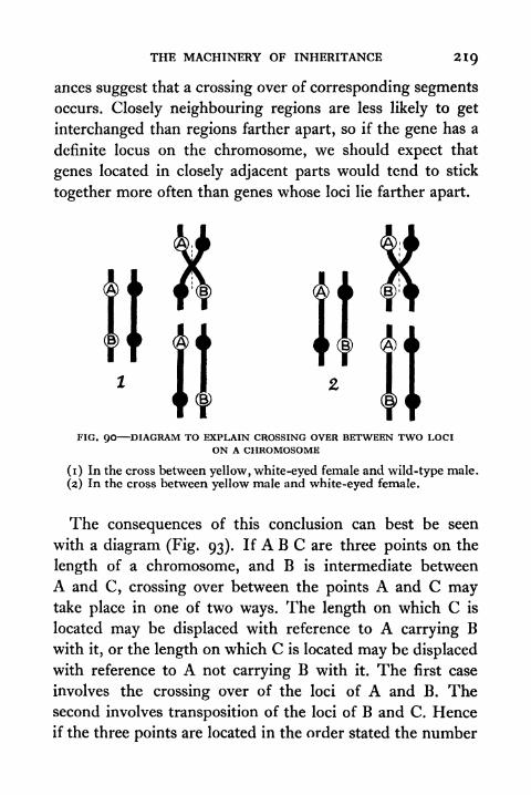
THE MACHINERY OF INHERITANCE 219
ances suggest that a crossing over of corresponding segmentsoccurs. Closely neighbouring regions are less likely to get
interchanged than regions farther apart, so if the gene has a
definite locus on the chromosome, we should expect that
genes located in closely adjacent parts would tend to stick
together more often than genes whose loci lie farther apart.
FIG. QO DIAGRAM TO EXPLAIN CROSSING OVER BETWEEN TWO LOCION A CHROMOSOME
(1) In the cross between yellow, white-eyed female and wild-type male.
(2) In the cross between yellow male and white-eyed female.
The consequences of this conclusion can best be seen
with a diagram (Fig* 93). If A B C are three points on the
length of a chromosome, and B is intermediate between
A and C, crossing over between the points A and C maytake place in one of two ways. The length on which C is
located may be displaced with reference to A carrying Bwith it, or the length on which C is located may be displaced
with reference to A not carrying B with it. The first case
involves the crossing over of the loci of A and B. Thesecond involves transposition of the loci of B and C. Hence
if the three points are located in the order stated the number

220 PRINCIPLES OF ANIMAL BIOLOGY
of cases in which the loci of A and C are transposed will be
the mm of the number of cases in which A and B cross over
and B and C cross over. If the order of A B C is not given,
the amount of crossing over between A and C may be the
sum or difference of the cross over A-B and B-C.
Let us now see how this applies to the example just given.
Crossing over takes place between the locus of yellow and
the locus of white in i i per cent of the reduction divisions
in the formation of the eggs. There is another sex-linked
mutant characterized by possession of wings which do not
extend beyond the tip of the abdomen. It is referred to as
miniature. In yellow miniature matings which produce a
very large generation of flies, the cross-over percentage (cross-
over value or C.O.V.) is found to be 34-3, and in the white
miniature cross the C.O.V. is 33-2. The difference i-i
corresponds to the cross-over value for yellow and white.
Thus the locus of white lies between the locus of yellow
and miniature i i units of length from the former and 33*2units from the latter. This relation holds good for all the
sex-linked mutant genes of Drosophila. So it is possible to
construct, as Morgan and his collaborators have done, a
map (Fig. 94) of the X-chromosome.
Thus far we have confined our attention to one groupof mutant characters which have their origin in changes
which occur at definite loci on the X or first pair of chromo-
somes in Drosophila. Now Drosophila has four pairs of
chromosomes, all of which have been mapped out on the
same principle. All that we have learned about the other
chromosomes so far is that the genes for vestigial, ebony,
and purple eye are not on the first (X) pair, and that ebonyand vestigial are not on the same pair. Besides ebony there
is another dark-coloured mutant which is referred to as
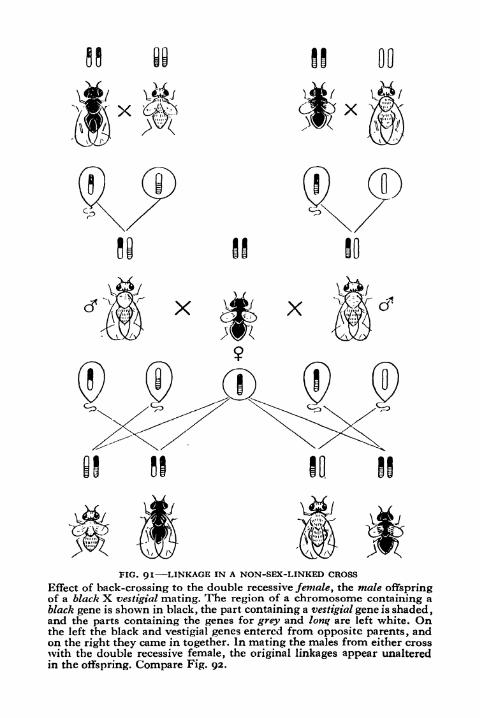
00
FIG. 91 LINKAGE IN A NON-SEX-LINKED CROSS
Effect of back-crossing to the double recessive female, the male offspringof a black X vestigial mating. The region of a chromosome containing a
black gene is shown in black, the part containing a vestigial gene is shaded,and the parts containing the genes for grey and long are left white. Onthe left the black and vestigial genes entered from opposite parents, andon the right they came in together. In mating the males from either crosswith the double recessive female, the original linkages appear unalteredin the offspring. Compare Fig. 92.
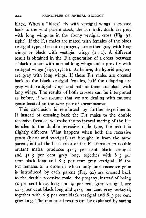
222 PRINCIPLES OF ANIMAL BIOLOGY
black. When a "black" fly with vestigial wings is crossed
back to the wild parent stock, the F.I individuals are grey
with long wings as in the ebony vestigial cross (Fig. 91,
right). If the F.I males are mated with females of the black
vestigial type, the entire progeny are either grey with long
wings or black with vestigial wings (i : i). A different
result is obtained in the F.2 generation of a cross between
a black mutant with normal long wings and a grey fly with
vestigial wings (Fig. 91, left). As before, the hybrid progenyare grey with long wings. If these F.I males are crossed
back to the black vestigial females, half the offspring are
grey with vestigial wings and half of them are black with
long wings. The results of both crosses can be interpreted
as before, if we assume that we are dealing with mutant
genes located on the same pair of chromosomes.
This conclusion is reinforced by further experiments.
If instead of crossing back the F.I males to the double
recessive females, we make the reciprocal mating of the F.I
females to the double recessive male type, the result is
slightly different. What happens when both the recessive
genes (black and vestigial) are brought in from the same
parent, is that the back cross of the F.I females to double
mutant males produces 41 5 per cent black vestigial
and 41-5 per cent grey long, together with 8-5 percent black long and 8-5 per cent grey vestigial. If the
F.I females of a cross in which only one recessive geneis introduced by each parent (Fig. 92) are crossed back
to the double recessive male, the progeny, instead of being
50 per cent black long and 50 per cent grey vestigial, are
41 -5 per cent black long and 41 -5 per cent grey vestigial,
together with 8-5 per cent black vestigial and 8-5 per cent
grey long. The numerical results can be explained by saying
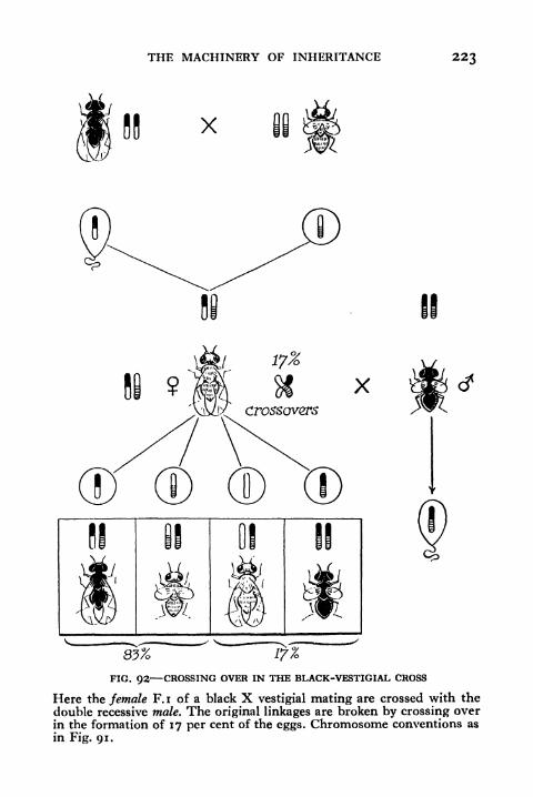
THE MACHINERY OF INHERITANCE 223
X
X
33%
FIG. 92 CROSSING OVER IN THE BLACK-VESTIGIAL CROSS
Here the female F.I of a black X vestigial mating are crossed with the
double recessive male. The original linkages are broken by crossing overin the formation of 17 per cent of the eggs. Chromosome conventions as
in Fig. 91.
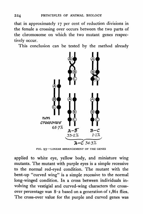
224 PRINCIPLES OF ANIMAL BIOLOGY
that in approximately 17 per cent of reduction divisions in
the female a crossing over occurs between the two parts of
the chromosome on which the two mutant genes respec-
tively occur.
This conclusion can be tested by the method already
noncrossovers'
65-7%
33-2%
a-C 34-3%
FIG. 93 LINEAR ARRANGEMENT OF THE GENES
applied to white eye, yellow body, and miniature wingmutants. The mutant with purple eyes is a simple recessive
to the normal red-eyed condition. The mutant with the
bent-up "curved wing" is a simple recessive to the normal
long-winged condition. In a cross between individuals in-
volving the vestigial and curved-wing characters the cross-
over percentage was 8-2 based on a generation of 1,861 flies.
The cross-over value for the purple and curved genes was
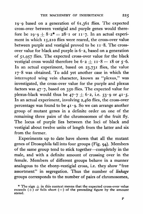
THE MACHINERY OF INHERITANCE 225
19 '9 based on a generation of 61,361 flies. The expected
cross-over between vestigial and purple genes would there-
fore be 19-9 :t 8-2* = 28-1 or 11-7. In an actual experi-
ment in which 15,210 flies were reared, the cross-over value
between purple and vestigial proved to be 1 1 8. The cross-
over value for black and purple is 6-2, based on a generation
of 51,957 flies. The expected cross-over value for the black
vestigial cross would therefore be 6'2 ^t: ii'8 1 8 or 5-6.
In an actual experiment, based on 23,731 flies, the value
17*8 was obtained. To add yet another case in which the
interrupted wing vein character, known as "plexus," was
investigated, the cross-over value for the purple and plexus
factors was 47*7, based on 350 flies. The expected value for
plexus-black would thus be 47-7 ^ 6-2, i.e. 53-9 or 41-5.
In an actual experiment, involving 2,460 flies, the cross-over
percentage was found to be 41 9. So we can arrange another
group of mutant genes in a definite order on one of the
remaining three pairs of the chromosomes of the fruit fly.
The locus of purple lies between the loci of black and
vestigial about twelve units of length from the latter and six
from the former.
Experiments up to date have shown that all the mutant
genes of Drosophila fall into four groups (Fig. 94). Membersof the same group tend to stick together completely in the
male, and with a definite amount of crossing over in the
female. Members of different groups behave in a manner
analogous to the ebony-vestigial cross, i.e. they show "free
assortment"
in segregation. Thus the number of linkage
groups corresponds to the number of pairs of chromosomes,
* The sign i in this context means that the expected cross-over valueexceeds (-f ) or falls short ( ) of the preceding figure by the amountstated.

nr
25-3. _ Sepia
38-5-
42-0-
45-5-
Dichaete
-Scarlet
-Pink
54-0- -4
59*0 -GZas^
63-565-5-
67-5-
-Delta.
Hairl&ss
Ebony
72-0 -
86-5 -Rough.
5-4.95-7
ClaretMinnie
101-0 --Minnie-^
IV
FIG. 94 MAP OF SOME OF THE MUTANT GENES OF DROSOPHILA
THE MACHINERY OF INHERITANCE
and of these four groups one, the smallest, can be identified
with the small "fourth" pair of chromosomes in Drosophila
on account of abnormal genetical results obtained in cross-
ing flies in which one member of this pair of chromosomes
was found to be absent. Recently Punnett has shown the
existence of seven linkage groups in the sweet pea which has
seven pairs of chromosomes. So the arrangement of the
genes in linear series has been shown to hold good for the
sweet pea. We may conclude that the results obtained with
experiments on the fruit fly are of general applicability.
The Universal Validity of the Principle of Segregation.
Wehave now toturn to the consideration of a question of some
importance in connexion with the problems which we shall
study in the second part of this book, namely whether the
Principle of Segregation applies to all the hereditable pro-
perties of the organism. When Mendel's principles were
first rescued from obscurity, few, if any, biologists would
have been so bold as to assert that they apply to all types
of heredity, and the majority were only willing to concede
grudgingly a restricted validity to the Law of Segregation.
Many continued to speak as if there were two sorts of in-
heritance, Mendelian and non-Mendelian. Every year since
the beginning of this century has witnessed a wider exten-
sion of the principle which Mendel first announced.
At one time a great deal of confusion arose through the
use of the misleading term "unit character." A single clear-
cut anatomical difference may involve one or many genes.
There is no relation between the magnitude of the one and
the extent of the other. The inheritance of the walnut type
of comb in the domestic fowl illustrates a clear-cut character
difference which involves two mutant genes. A character
difference which involves two mutant genes on different
228 PRINCIPLES OF ANIMAL BIOLOGY
pairs of chromosomes leads to nine instead of four distinct
types in the F.2 generation, and the analysis of this result
would not be at all obvious at first sight. If three dominant
or recessive genes on different pairs of chromosomes deter-
mine a single character difference, the F.2 individuals would
fall into one class with all three dominant characters, three
classes with two dominant characters, three classes with one
dominant character, and one class with no dominant charac-
ters in the ratio 27 : 9 : 9 : 9 : 3 : 3 : 3 : i. If neither
member of any of the three pairs of characters show domi-
nance the number of classes will be twenty-seven instead of
eight.
So extremely complicated cases may arise, when only a
few genes are involved in the inheritance of some discrete
anatomical or physiological difference distinguishing two
strains. If only four genes on different pairs of chromosomes
come into play, and none is dominant to its alternative, the
number of classes in the F.2 is 81. Even so, we have only
exhausted a small fraction of problems that present difficulty
at first sight. The successful analysis of such difficult cases
justifies the confidence which Mendel's principle now enjoys.
Numerical proportions which arise through crossing
hybrids depend partly on dominance, partly on the number
of genes involved, and partly on the "interaction" of genes.
Two simple cases of the interaction of genes may be cited.
On crossing a black and a red guinea pig, the F.I indi-
viduals are of the mouse grey type known to breeders as
agouti. The F.2 individuals are agoutis, blacks, and reds in
the ratio 9:3:4. This at once suggests that the red strain
carries a dominant gene which modifies black, but does not
modify red, so that the F.2 reds are composed of two classes,
one which mated to black gives agouti and one which mated

THE MACHINERY OF INHERITANCE 22Q
to black gives black. An example which specially bears on
the species problem is the case of two strains of sweet peas,
both white and pure for white when inbred, but giving an
F.I of coloured individuals and an F.2 of 9 : 7 coloured to
white when crossed. If strain A carries a gene absent in
strain B, and strain B carries a gene absent in strain A, if
both are located on different pairs of chromosomes, both
having arisen in a coloured stock, both tending to make
the colour white, and both recessive, the result necessarily
follows from the fact that out of every sixteen three will have
A in duplicate, three will have B in duplicate, and one will
have both A and B in duplicate, making seven whites.
An entirely different type of complication arises when a
gene affects the viability of the offspring. Such genes are
classed together under the general term lethals. An immense
number of cases of this kind have been thoroughly in-
vestigated with the most rigorous quantitative treatment.
We need only take a single example, that of a sex-linked
lethal in Drosophila. Some females of Drosophila have an
excessive proportion of female offspring, in fact, a 2 : i,
instead of a i : i sex ratio. This is explicable on the assump-tion that such females bear on one of their sex chromosomes
a gene which, if not compensated by a normal sex chromo-
some, prevents the individual from surviving to maturity.
As the male has only one sex chromosome, every male which
inherits a lethal-bearing X chromosome from its mother
dies. The hypothesis is tested rigorously by mating such
females to white-eyed males, when the disturbances in the
ratios of white-eyed and red-eyed individuals conform to
the assumption that one maternal chromosome carries some-
thing which prevents the survival of her male offspring
arising from eggs possessing it.

330 PRINCIPLES OF ANIMAL BIOLOGY
The most difficult, and for long the most insoluble
problem which arises in the study of inheritance was the
transmission of characters which normally show a good deal
of variability with reference to environment. In discussing
hereditable properties in what has gone before we have
assumed that the bodily expression of the gene has been
clear-cut, and the environmental condition necessary to its
appearance a normal concomitant of the conditions of life.
When this is so, our problem is greatly simplified, and it is
by the scrutiny of such simple cases that we gain knowledgewhich enables us to investigate the more intricate. To clarify
this distinction, let us revert to the case of Mendel's experi-
ments with tall and dwarf peas. The F.2 tall individuals are
not all of the same size, nor are the dwarfs. Each has a certain
range of variability with respect to the local conditions in
which the individual seed germinates. Nevertheless, they are
distinct groups. Given proper conditions, the smallest tall is
recognizably larger than the largest dwarf. In dealing with
size characteristics, however, hereditary differences may not
be so nicely defined. Often they can only be described in
statistical terms. Two inbred stocks may differ by some
average that is characteristic of the progeny of individuals
reared under identical conditions, but it may be quite im-
possible to distinguish the individuals themselves.
This makes ordinary experimental procedure impossible.
But the problem is simplified if the organism is hermaphro-dite and self- fertilizing, as are many plants. If we take beans
from a mixed collection of the same species but not of the
same parentage, and weigh them, we can arrange them in
groups according as they fall within certain limits. On
counting the number of beans in each group or class, we can
plot a curve of which the ordinates represent the numbers
THE MACHINERY OF INHERITANCE 231
in a class (class frequency) and the abscissae correspond to
the mean value of each class. In general the curve will have
the shape indicated in Fig. 95. If we select individual beans
of different sizes, and allow them to germinate and seed byself-fertilization, we find, as was first shown by Johannsen
o
80 -85 -90to to to85 -90 -95
95 1-0 1-05 UO 1-15 1-20 1-25 1-3O 1-35to w to to to to to to to1-0 W5 HO 1-15 i-20 1-25 1-3O 1-35 HO
FIG. 95 NORMAL FREQUENCY CURVE ILLUSTRATED BY VARIABILITY INWEIGHT OF BEANS
(1909), that the frequency curves for the progeny of someare well nigh identical in form with those of the original
mixed population. Others, however, will have curves whosemost fashionable value or mode corresponds to a higher or
lower class than that of the mixed population. If we take
any of the offspring of a bean whose progeny show a highor low modal value and allow it in turn to germinate and
seed, we find that the modal class of its grandchildren will
be the same as that of their parent. Thus we can have twostocks of beans distinguished only by the fact that they regu-
232 PRINCIPLES OF ANIMAL BIOLOGY
larly produce offspring whose frequency curves for the size
character in question display a constant difference of the
modal class value. When equal numbers of individuals from
both stocks are mixed the two series might so completely
overlap that the mixed population would yield a frequency
curve with only one peak.
Let us now see how we might test whether the theory
of the gene applies to hereditable properties of this type.
Unless sex-linked factors are involved in a cross between
parents of two different pure-bred strains, all the individuals
of the F.I must be genetically alike. Any differences that
exist among them inter se will be due to external agencies
alone. The differences between individuals of the F.2 will
always be the result, on the other hand, of both genetic
differences and bodily variability, and there can never be
more genetic types in any succeeding generation than appearin the F.2. Hence, if the Principle of Segregation applies to
cases like height, weight, girth, growth-rate, etc., cases, that
is, where the character in question is a very variable one.
any cross between individuals of pure strains differing with
respect to the modal value of the character must always
conform to a simple rule: maximum variability in the F.2
and minimum variability in the F.I. The test is one which
has been satisfactorily applied to many cases.
The mathematical treatment of this problem when one
factor difference is concerned can be set forth without
recourse to elaborate statistical treatment. Let us take the
case of two parents of stocks whose progeny give the curves
P.I and P.2, which are supposed to be based on the same
number of individuals, let us say 1,000. The two series
overlap but their modes are different. For the sake of sim-
plicity let the modal value for the F,i curve be taken as
THE MACHINERY OF INHERITANCE 233
intermediate, and the F.I curve in Fig. 96 be plotted for the
same number of individuals as the P.I and P.2. Now the F.2
generation will consist of three genetic types, the two
parental types and the F.I type, and for each individual of
Pi made, El niuJji P.2 modz
FIG. 96 CURVES TO ILLUSTRATE MAXIMUM VARIABILITY IN THE SECONDGENERATION (see text)
the parental types there will be two of the F.I on the
Principle of Segregation. Let us, therefore, add each ordinate
of the parental curves to twice the value of the correspondingordinates of the F.I. The new dotted curve will be based
on 4,000 individuals. To reduce this to comparable dimen-
sions with the P.I and P.2 curves we must divide each
ordinate by 4. This gives us the flat curve of the F.2. Theflatness of this curve shows the greater variability of the
234 PRINCIPLES OF ANIMAL BIOLOGY
F.2, and affords a mathematical test of the validity of the
Principle of Segregation as applied to highly variable
characters. A few applications of this method of analysis to
animals (number of tail feathers in pigeons, e.g.) have been
made, and it has been extensively tested in plant breeding.
Application of Genetics to Human Ecology. In the
absence of definite knowledge about hereditary transmission
the improvement of stock and seed-producing crops was
accomplished by inbreeding and by artificial selection.
Individuals with the selected characteristic were propagated
by repeated mating of close relatives having the same charac-
teristic. If this is carried on for many generations pure lines
of homozygous individuals can be established. What could
only be achieved by a long process of selection and inbreed-
ing can now be brought about by systematic testing in two
or three generations. The difference between the two methods
can be illustrated by taking an example analogous to combinheritance in poultry (p. 194 and Fig. 86).
In rabbits two varieties, Blue Bevran and Chocolate
Havana, are distinguished by the slate and reddish brown
colour of the fur. If pure individuals of both types are crossed,
the hybrid is black. When the black hybrids are mated with
one another four colour types of offspring are produced,
namely black, blue, chocolate, and a fourth whose pale
silvery coat is called "lilac." The fact that there are four
types shows that we are dealing with two mutant genes.
One recessive gene (b) makes black fur slate-blue, the other
(c) makes black fur reddish brown, both together make black
fur lilac. The F.I hybrids receive b from their blue and c
from their chocolate parents respectively. They have neither
in duplicate. So if b and c are assumed to be recessive, the
F,i hybrids should be black. All the possible genetic types
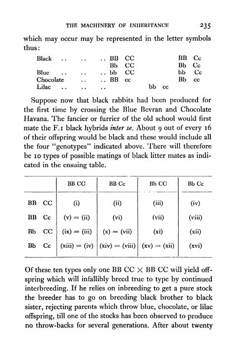
THE MACHINERY OF INHERITANCE 235
which may occur may be represented in the letter symbols
thus:
Black
Blue
Chocolate
Lilac
BB CCBb CCbb CCBB cc
BB CcBb Ccbb CcBb cc
bb cc
Suppose now that black rabbits had been produced for
the first time by crossing the Blue Bevran and Chocolate
Havana. The fancier or furrier of the old school would first
mate the F.I black hybrids inter se. About 9 out of every 16
of their offspring would be black and these would include all
the four "genotypes" indicated above. There will therefore
be 10 types of possible matings of black litter mates as indi-
cated in the ensuing table.
Of these ten types only one BB CC X BB CC will yield off-
spring which will infallibly breed true to type by continued
interbreeding. If he relies on inbreeding to get a pure stock
the breeder has to go on breeding black brother to black
sister, rejecting parents which throw blue, chocolate, or lilac
offspring, till one of the stocks has been observed to produceno throw-backs for several generations. After about twenty

236 PRINCIPLES OF ANIMAL BIOLOGY
generations this result will usually have been achieved.
Selective inbreeding is an exceedingly wasteful way of
getting it, because there is a simple way of deciding to which
genotype an F.2 black hybrid belongs. By making a chess
board diagram (see Fig. 82) you will see that when mated
to lilac (bb cc),
BB CC gives progeny all black
BB Cc gives progeny 50 per cent black and 50 per cent
chocolate
Bb CC gives progeny 50 per cent black and 50 per cent blue
Bb Cc gives progeny 25 per cent black, 25 per cent chocolate,
25 per cent blue, and 25 per cent lilac.
So if each F.2 hybrid black is mated once to a lilac indi-
vidual the production of an all-black litter makes it almost
certain that it is a pure black (BB CC). The odds are at
least 127 to i in a single litter of seven and 4,095 to i if two
successive all black litters of seven are reared. At one step
we can therefore determine which of the F.2 hybrids are
pure stock, and so achieve the result required in the second
generation from the original cross.
You will also see that there is no need to inbreed twenty
generations of lilac rabbits to get a pure stock. All lilac
rabbits must be pure, since they are double recessives. If
we had started, knowing only the black and lilac colour
varieties, we should, of course, get the same result in the
F.I and F.2 of a cross between lilac and pure black. Some
of our blue and chocolates would be impure. If we wanted
to build up pure stocks of either, we could test the blues and
chocolates for further use by mating to lilac, when
BB cc would give all chocolate
Bb cc would give 50 per cent chocolate and 60 per cent lilac
bb CC would give all blue
bb Cc would give 50 per cent blue and 50 per cent lilac.
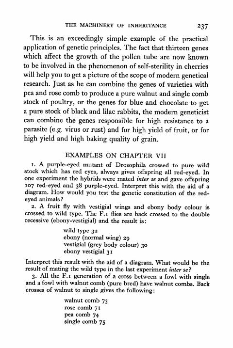
THE MACHINERY OF INHERITANCE 237
This is an exceedingly simple example of the practical
application of genetic principles. The fact that thirteen geneswhich affect the growth of the pollen tube are now knownto be involved in the phenomenon of self-sterility in cherries
will help you to get a picture of the scope of modern genetical
research. Just as he can combine the genes of varieties with
pea and rose comb to produce a pure walnut and single combstock of poultry, or the genes for blue and chocolate to get
a pure stock of black and lilac rabbits, the modern geneticist
can combine the genes responsible for high resistance to a
parasite (e.g. virus or rust) and for high yield of fruit, or for
high yield and high baking quality of grain.
EXAMPLES ON CHAPTER VII
1. A purple-eyed mutant of Drosophila crossed to pure wildstock which has red eyes, always gives offspring all red-eyed. Inone experiment the hybrids were mated inter se and gave offspring107 red-eyed and 38 purple-eyed. Interpret this with the aid of a
diagram. How would you test the genetic constitution of the red-
eyed animals ?
2. A fruit fly with vestigial wings and ebony body colour is
crossed to wild type. The F.I flies are back crossed to the doublerecessive (ebony-vestigial) and the result is :
wild type 32
ebony (normal wing) 29vestigial (grey body colour) 30ebony vestigial 31
Interpret this result with the aid of a diagram. What would be theresult of mating the wild type in the last experiment inter sel
3. All the F.I generation of a cross between a fowl with singleand a fowl with walnut comb (pure bred) have walnut combs. Backcrosses of walnut to single gives the following :
walnut comb 73rose comb 71
pea comb 74
single comb 75
238 PRINCIPLES OF ANIMAL BIOLOGY
Interpret this result by means of a diagram. What would be the
result of mating those with walnut combs inter se?
4. The white plumage of leghorn poultry is dominant to coloured,
feathered shanks is dominant to clean, and pea comb is dominant
to single. What would be the result of back crossing the F.I of a
pure white, feathered, bird with a pea comb and a black, clean,
bird with a single comb to the triple recessive ?
5. A race of black rabbits mated to lilac gives black hybrids
which, when mated to their lilac parents, yield black, blue, choco-
late, and lilac offspring in approximately equal numbers. Four black
offspring of black hybrid parents behaved as follows. Buck Acrossed with a lilac female had one surviving offspring which waslilac. When crossed with Doe 13, the same buck sired three suc-
cessive litters, including in all 13 black and 3 blue offspring. WithDoe C it also sired three litters including in all 6 chocolate and
13 black offspring. With Doe D it sired only black offspring in four
successive matings. Interpret these data, giving the genetic con-
stitution of the four individuals.
6. A yellow male mutant of Drosophila crossed to wild type
(grey) females always gives grey offspring. A yellow female mutantcrossed to pure-bred wild type male has offspring half of whichare grey females and half yellow males. Interpret this result with
the aid of a diagram. What will result if flies from each of the F.I
are mated inter set
7. Barred plumage is a sex-linked character of the PlymouthRock breed dominant to the black of the Ancona breed. A black
Ancona hen is crossed with a Plymouth Rock cock and the F.I
males are crossed back to the mother, the F. i females being crossed
back to the father. What are the results obtained ? What would be
the result of an analogous experiment in which the reciprocal cross
was the starting point in the parental generation.
8. White eye is a sex-linked recessive in Drosophila. White-eyedmales are crossed to wild type females and the F.I flies are matedinter se. What is the result in the F.2? How would you test the
constitution of each of F.2 females without the use of white-eyedmales ?
9. Interpret the following experiment on Drosophila hybridswith the aid of a chromosome diagram :
Pure stock sable male by pure stock wild type female gave onlywild type offspring. These hybrids when mated inter se gaveoffspring as follows:

THE MACHINERY OF INHERITANCE 239
wild type males 146 sable males 155wild type females 305 sable females o
What results would you expect in the reciprocal cross ?
10. Interpret the following experiments with the aid of a
diagram : A pure wild stock of male Drosophila was crossed to the
mutant white-eyed stock, and its progeny when mated inter se
yielded a generation composed of:
white-eyed females 76 white-eyed males 73wild type females 69 wild type males 68
With another white-eyed female the same type of experiment wascarried out and the second generation was composed of:
white-eyed females 97 white-eyed males 46wild type females 91 wild type males 93
1 1 . Construct a diagrammatic solution of the following problem :
In Drosophila vermilion eyes are referred to a sex-linked reces-
sive mutant from the red-eyed wild stock. Certain vermilion-eyedfemales have vermilion-eyed male and female and red-eyed male
and female offspring when crossed to wild type. These females
have a Y chromosome in addition to the normal XX group. Howwould you test their offspring in a cross with red-eyed males to
detect differences in genetic constitution of outwardly similar
individuals ?
12. Yellow (body colour), vermilion (eye colour) and sable (bodycolour) are sex-linked mutants of Drosophila. A yellow-vermilionmale is crossed to a wild type female, and the female offspring
crossed to a wild type male. The result is :
wild type females 120
yellow vermilion males 38wild type males 42
yellow males 21
vermilion males 23
A vermilion male is crossed to a sable female and the female
offspring crossed to wild type males. The result is :
wild type females 144vermilion males 64sable males 62
vermilion sable males 8
wild type males 6

240 PRINCIPLES OF ANIMAL BIOLOGY
What would be the result of crossing the female offspring of a sable-
yellow female and a wild type male to wild type males ?
13. Two dark-bodied strains of Drosophila were homozygousfor vestigial (i.e. both strains were vestigial). When crossed together
these strains gave vestigial wing and grey body (wild type). Each
of these strains was then crossed to wild type and the F.I males
back-crossed to their own double recessive parent. The results were
as follows:
Strain A.
dark body 23
vestigial 21
dark body vestigial 20
wild type 25
Strain B.
dark body o
vestigial o
wild type 75dark body vestigial 81
Interpret these results with the aid of a diagram.
14. A laboratory worker forgot to label new culture bottles of
dark-body and vestigial used in the previous examples. In order
to rectify this, males from each were crossed to wild type females
and the F.I males were then crossed back to respective double
recessives. The temperature regulation of the incubator wentout of order, and the temperature rose to a very high level, killing
the parent flies. One yielded 12 W.T. and 10 others all double
recessive. The second yielded two flies only, both of which weredark-bodied with normal wings.Use this information to identify your unlabelled cultures, giving
reasons. Supposing these flies had been dark-bodied with vestigial
wings could he have labelled his bottles ? Give reasons.
15. Illustrate the principle of the linear alignment of the genes
by means of the following experiments on Drosophila mutants.
(a) Female offspring of vestigial by black back-crossed to the double
recessive male gave :
vestigial 416 wild type 87black 421 black vestigial 91
(b) Female offspring of vestigial-purple back-crossed to the doublerecessive male gave:

THE MACHINERY OF INHERITANCE 241
vestigial 336 wild type 44purple 325 purple vestigial 42
(c) Female offspring of black-purple back-crossed to the double
mutant male offspring gave:
black 464 wild type 28
purple 488 black purple 32
1 6. In a cross between the yellow-bodied mutant and a male of
the vermilion-eyed mutant stock of Drosophila, the F.2 generation
produced by mating the hybrids inter se was as follows :
yellow females 103 wild type males 36wild type females 98 vermilion-eyed yellow-
bodied males 33vermilion males 66
yellow males 64
Explain this result with a diagram, and determine the probablepercentage of the following types :
wild type male and female ;
vermilion, yellow male and female:
vermilion male and female ;
in the F.2 generation of the cross between a wild type female anda vermilion-eyed yellow-bodied male.
17. A black purple vestigial female was crossed to a wild type
male, and the F.I females were crossed back to black purple
vestigial males. The following results were obtained :
black purple vestigial 105wild type 104black 6
purple vestigial 8
black purple 15
vestigial T7
black vestigial i
j (doub,e crossover)purple i J
Interpret this by means of a diagram. Note the small number of
double cross-overs.
1 8. In a series of linkage crossovers of the X chromosome yellowwas found to be 36 units (% crossover between yellow and minia-
Q

242 PRINCIPLES OF ANIMAL BIOLOGY
ture) from miniature, white was found to be 36 units from minia-
ture and yellow was found to be i unit from white. The numberson which this was based were small. The experiment as it stands
yields no information as to the order of yellow, white, and minia-
ture. Female offspring of yellow males and white miniature
females were crossed to wild type males. The following was the
result :
wild type females 105white miniature males 37
yellow males 34wild type males i
yellow white miniature males i
yellow miniature males 20white males 19white yellow males ominiature males o
What is the order of genes concerned ? What conclusions could youdraw if there had been no yellow white miniature, no wild type,no miniature, and two yellow whites ?
19. A purple vestigial strain in Drosophila was crossed to wild
type, and the offspring were found to be all wild type. The F.I
females were mated singly with single purple vestigial males andtwo cultures gave the following result:
Culture I Culture II
purple vestigial 80 purple vestigial 57wild type 82 wild type 104
purple ii purple nvestigial 9 vestigial 25
Interpret these results with the aid of a diagram, and state what
experiments you would set up to prove your hypothesis. You mayassume that animals suitable for mating were retained in bothcultures.

PART II
HOW ANIMALS DIFFER


CHAPTER IX
THE DIVERSITY OF ANIMAL LIFE
BROADLYspeaking, we can study animals from two
points of view. We may concentrate our attention on
the living machine as a going concern, and investigate the
relation of its parts to one another and the relation of its
processes to the properties of non-living systems. Wemay also be interested in the differences between different
kinds of animals, and investigate how these differences
have arisen. So far we have been chiefly concerned with
the first. We shall now devote ourselves to the second.
Though no two animals of the same sex are exactly alike,
it is a matter of common observation that some are muchmore alike than others, and that individuals which are verymuch alike inter-breed and preserve their common charac-
teristics. Barriers to inter-breeding may be natural or arti-
ficial. Assemblages of individuals which preserve commoncharacteristics which distinguish them from other animals
by breeding among their own kind are called species, whennature supplies the barriers. When they are segregated byhuman interference they are called breeds, strains, races,
domestic varieties, or pure lines. From what we have learned
in the previous chapter we know that pure lines or domestic
varieties exist because of the occasional occurrence of muta-
tions;and because of the combination of mutant characters
which breed true to type by hybridization and by subsequentselection of homozygous individuals.
A laboratory or a flower garden is a part of nature. So if
mutations can occur in a laboratory or in a flower garden, they
246 PRINCIPLES OF ANIMAL BIOLOGY
presumably occur when there are no human beings in their
vicinity. On the whole those that live longer or produce more
offspring will survive. As species spread over a larger area
natural conditions in different regions will favour the survival
of some mutant combinations rather than of others. In the
course of time what was once a single species will have broken
up into several, which will do the same again. Intermediate
types will die out. The differences among the progeny of
ancestors which were once recognized as a single species will
become more sharply defined, and the gaps will obscure their
common family resemblance. Thus climate and the changing
contours of the earth's surface during prolonged periods of
time continually impose on living organisms a process com-
parable to the selection of new domestic strains.
Though it has been accepted generally by scientific
workers for less than three-quarters of a century, the doctrine
of evolution^ which is the belief that species change in this
way and that the immense variety of living animals have a
common ancestry, is not essentially new. It was foreshadowed
in the writings of the Greek materialists. Before its credentials
could be established, obstacles due to ignorance about the
elementary facts of birth and parenthood, the grades of
structural similarity among different types of animals, the
history of past life on earth and the present distribution of
living creatures in different parts of the world had to be
got out of the way by laborious observations of countless
investigators. That all animals reproduce sexually, that
animals can be arranged in groups which exhibit greater
or less degrees of family likeness, that animals which are
totally unlike any living ones existed on earth millions of
years before Man, and that different geographical regions
have their own characteristic faunas, are facts known to
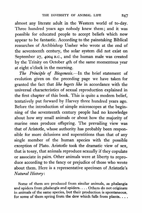
THE DIVERSITY OF ANIMAL LIFE 247
almost any literate adult in the Western world of to-day.
Three hundred years ago nobody knew them; and it was
possible for educated people to accept beliefs which now
appear to be fantastic. According to the painstaking Biblical
researches of Archbishop Ussher who wrote at the end of
the seventeenth century, the solar system did not exist on
September 27, 40046.0., and the human male was created
by the Trinity on October 4th of the same momentous year
at eight o'clock in the morning.
The Principle of Biogenesis. In the brief statement of
evolution given on the preceding page we have taken for
granted the fact that like begets like in accordance with the
universal characteristics of sexual reproduction explained in
the first chapter of this book. This is quite a modern belief,
tentatively put forward by Harvey three hundred years ago.
Before the introduction of simple microscopes at the begin-
ning of the seventeenth century people had no knowledgeabout how any small animals or about how the majority of
marine ones produce offspring. The prevailing view was
that of Aristotle, whose authority has probably been respon-
sible for more delusions and superstitions than that of any
single member of the human species with the possible
exception of Plato. Aristotle took the dramatic view of sex,
that is to say, that animals reproduce sexually if they copulate
or associate in pairs. Other animals were at liberty to repro-
duce according to the fancy or prejudice of those who wrote
about them. Here is a representative specimen of Aristotle's
Natural History :
Some of them arc produced from similar animals, as phalangiaand spiders from phalangia and spiders. . . . Others do not originate
in animals of the same species, but their production is spontaneous,
for some of them spring from the dew which falls from plants, . , ,
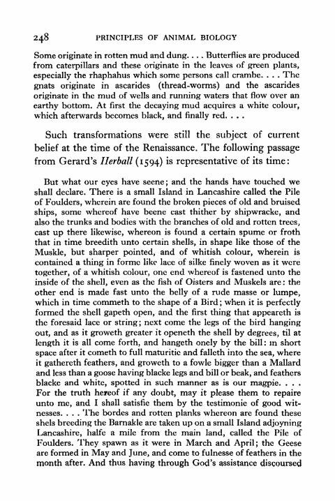
248 PRINCIPLES OF ANIMAL BIOLOGY
Some originate in rotten mud and dung Butterflies are producedfrom caterpillars and these originate in the leaves of green plants,
especially the rhaphahus which some persons call crambe. . . . The
gnats originate in ascarides (thread-worms) and the ascarides
originate in the mud of wells and running waters that flow over an
earthy bottom. At first the decaying mud acquires a white colour,
which afterwards becomes black, and finally red. . . .
Such transformations were still the subject of current
belief at the time of the Renaissance. The following passage
from Gerard's Herball (1594) is representative of its time:
But what our eyes have scene;and the hands have touched we
shall declare. There is a small Island in Lancashire called the Pile
of Foulders, wherein are found the broken pieces of old and bruised
ships, some whereof have beene cast thither by shipwracke, and
also the trunks and bodies with the branches of old and rotten trees,
cast up there likewise, whereon is found a certain spume or froth
that in time breedith unto certain shells, in shape like those of the
Muskle, but sharper pointed, and of whitish colour, wherein is
contained a thing in forme like lace of silke finely woven as it were
together, of a whitish colour, one end whereof is fastened unto the
inside of the shell, even as the fish of Oisters and Muskels are : the
other end is made fast unto the belly of a rude masse or lumpe,which in time commeth to the shape of a Bird ; when it is perfectly
formed the shell gapeth open, and the first thing that appeareth is
the foresaid lace or string ;next come the legs of the bird hanging
out, and as it groweth greater it openeth the shell by degrees, til at
length it is all come forth, and hangeth onely by the bill: in short
space after it cometh to full maturitie and falleth into the sea, whereit gathereth feathers, and groweth to a fowle bigger than a Mallard
and less than a goose having blacke legs and bill or beak, and feathers
blacke and white, spotted in such manner as is our magpie. . . .
For the truth hereof if any doubt, may it please them to repaire
unto me, and I shall satisfie them by the testimonie of good wit-
nesses. . . . The hordes and rotten planks whereon are found these
shels breeding the Barnakle are taken up on a small Island adjoyning
Lancashire, halfe a mile from the main land, called the Pile of
Foulders. They spawn as it were in March and April; the Geeseare formed in May and June, and come to fulnesse of feathers in the
month after. And thus having through God's assistance discoursed

THE DIVERSITY OF ANIMAL LIFE 249
somewhat at large of Grasses, Herbs, Shrubs, trees and Mosses,and certain Excrescences of the earth, with other things moe,incident to the historic thereof, we conclude and end our present
Volume, with this Wonder of England. For the which God's
Name be ever honoured and praised.
According to the Aristotleian view which lingered on till
the beginning of the eighteenth century, there was no one
process of natural generation such as we take for granted
when we discuss the common ancestry of animals to-day.
FIG. 97 FIGURE OF THE BARNACLE AND GOOSE IN GERARD 's Herbdll (l594)
Hence the first step towards the modern theory of evolution
was the recognition of biogenesis, i.e. the rule that like begets
like. The discovery of spermatozoa with the first microscopesat the end of the seventeenth century brought about a com-
plete revolution in the history of man's knowledge of the
universe. A practical result of this was the recognition of
sexuality in plants, and hence the production of new hybridvarieties by artificial pollination. The successes of com-
mercial horticulturalists and seed merchants stimulated stock-
breeders to new efforts, which resulted in great improve-ments of cattle and sheep.
Micro-organisms. The invention of the microscope also
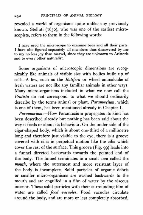
250 PRINCIPLES OF ANIMAL BIOLOGY
revealed a world of organisms quite unlike any previously
known. Stelluti (1630), who was one of the earliest micro-
scopists, refers to them in the following words :
I have used the microscope to examine bees and all their parts.
I have also figured separately all members thus discovered by meto my no less joy than marvel, since they are unknown to Aristotle
and to every other naturalist.
Some organisms of microscopic dimensions are recog-
nizably like animals of visible size with bodies built up of
cells. A few, such as the Rotifera or wheel animalculae of
fresh waters are not like any familiar animals in other ways.
Many micro-organisms included in what we now call the
Protista do not correspond to what we should ordinarily
describe by the terms animal or plant. Paramoecium, which
is one of them, has been mentioned already in Chapter I.
Paramcecium. How Paramoecium propagates its kind has
been described already but nothing has been said about the
way it feeds or about its behaviour. On the under side of the
cigar-shaped body, which is about one-third of a millimetre
long and therefore just visible to the eye, there is a groove
covered with cilia in perpetual motion like the cilia which
cover the rest of the surface. This groove (Fig. 99) leads into
a funnel directed backwards towards the pointed end of
the body. The funnel terminates in a small area called the
mouth, where the outermost and more resistant layer of
the body is incomplete. Solid particles of organic debris
or smaller micro-organisms are washed backwards to the
mouth and are engulfed in a film of water by the viscous
interior. These solid particles with their surrounding film of
water are called food vacuohs. Food vacuoles circulate
around the body, and are more or less completely absorbed.

THK DIVERSITY OF ANIMAL LIFE 251
Eventually any non-assimilable matter is ejected from a
temporary opening at a fixed spot behind the mouth on the
undermost surface.
Paramoecium also exhibits a process which may be called
excretion. Its chemical nature is not understood. So it is not
certain that it is at all comparable to the nitrogenous excretory
function of our own kidneys. In the viscous interior near
the extremities of the upper surface there are two pulsating
reservoirs of clear fluid called contractile vacuoles. The con-
tractile vacuoles are connected with radiating canals of clear
fluid in the surrounding substance or endoplasm. The
radiating canals and the central vacuole pulsate alternately,
and the latter seems to discharge the fluid accumulated
therein through the peripheral pellicle of more resistant
ectoplasm.
Paramoecium is constantly on the move, swimming by the
lashing motion of its cilia with its blunt end foremost. Thecilia do not move simultaneously. A wave of motion pro-
gresses along the axis of the body, as in the motion of cilia
on the gill of the mussel. The appearance is like stalks of
wheat when a breeze sweeps over a field. In response to
certain changes in its surroundings Paramoecium mayabandon this indiscriminate and continuous locomotion.The
principal types of response which it shows may be called
browsing and avoidance reactions.
The browsing reaction is a feeding response. The favourite
food of Paramoecium is bacteria which are apt to stick
together in clumps. When it comes up against such a clump,it becomes still. All the cilia cease to beat except those
inside the food groove. The cilia around the mouth create a
vigorous food current which sucks in bacteria. When the
meal is finished it gets on the move. The effective stimulus is
252 PRINCIPLES OF ANIMAL BIOLOGY
contact, Paramoccium will browse without effect, if it comes
up against a piece of cotton.
The avoidance reaction takes place when Paramoecium
swims into strong light, collides with a surface, or gets into
the neighbourhood of a hot object or a harmful chemical
reagent. Its first reaction is to stop dead. It then turns
sharply through an angle of indefinite dimensions and goes
off again in a straight line. It will repeat this several times, if
necessary, till its line of motion takes it out of danger, i.e.
out of the field of the effective stimulus.
Inside the body of Paramcecium there are two permanent
objects which take up nuclear stains. The smaller or micro-
nucleus divides by mitosis during reproduction and is com-
parable to the nucleus of animal or plant cells. Sometimes
two Paramoecia come together side by side. When this
process of conjugation occurs each micronucleus divides three
times. The process involves reduction of the chromosomes
to half the normal number. Six of the eight products de-
generate. Of the two persisting one remains behind and the
other migrates. In each conjugant the migratory nucleus
fuses with the one which stays at home. The two Paramoecia
then separate, each with a new micronucleus of which only
half the chromosome content originally belonged to. This
process of conjugation reproduces two essential features
of Sexuality, i.e. nuclear reduction and restitution of the
diploid number of chromosomes by nuclear fusion. It is not
correct to call it sexual reproduction, because it does not
of itself lead to the production of a new individual, and is
equally comparable to the reciprocal copulation of two
earthworms.
Amoeba. The term Protista proposed by Haeckel (1834-
1919) includes a various assemblage of minute forms of
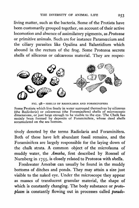
THE DIVERSITY OF ANIMAL LIFE 253
living matter, such as the bacteria. Some of the Protista have
been customarily grouped together, on account of their active
locomotion and absence of assimilatory pigments, as Protozoa
or primitive animals. Such are for instance Paramceciuni and
the ciliary parasites like Opalina and Balantidium which
abound in the rectum of the frog. Some Protozoa secrete
shells of siliceous or calcareous material. They are respec-
FIG. 98 SHELLS OF RADIOLARIA AND FORAMINIFERA
Some Protista which live freely in water surround themselves by siliceous
(the Radiolaria) or calcareous (the Foraminifera) shells of microscopicdimensions, or just large enough to be visible to the eye. The Chalk has
mainly been formed by deposits of Foraminifera, whose dead shells
accumulated on the sea bottom.
tively denoted by the terms Radiolaria and Foraminifera.
Both of these have left abundant fossil remains, and the
Foraminifera are largely responsible for the laying down of
the chalk strata. A common object of the microfauna of
muddy water, the Amoeba, first described by Rosesel of
Nurnberg in 1755, is closely related to Protozoa with shells.
Freshwater Amoebae can usually be found in the muddybottoms of ditches and ponds. They may attain a size just
visible to the naked eye. Under the microscope they appearas masses of translucent granular material, the shape of
which is constantly changing. The body substance or proto-
plasm is constantly flowing out in processes called pseudo-
254 PRINCIPLES OF ANIMAL BIOLOGY
podia. As new pseudopodia are formed old ones are with-
drawn, and by this streaming out of the cell substance
Amccba both creeps over the surface to which it is attached
and engulfs the food which it is constantly taking in. This
food consists of smaller organisms and organic debris. What
is called the contractile vacuole of Amoeba is not a fixture
like those of Paramcecium. It is a clear spherical fluid vesicle,
which enlarges and bursts, discharging its contents to the
exterior. So soon as it has burst another begins to form.
There are several reasons for keeping a separate niche
for microorganisms such as Paramcecium, Amoeba or
Euglena. One is that the body of what we ordinarily call an
animal is divided up into cells, and these cells arc not all
alike. They form different systems of tissues and organs
performing different work. So when we speak of reproduc-tion we mean the net result of a process of repeated cell
division. Where no such differentiation exists the clear out-
lines of the division between animals and plants breaks
down. Before there was a separate assemblage for organismswhich are not divided into cells, botanists and zoologists
competed for the privilege of classifying many of them as
primitive plants or primitive animals. The same creature
was classified as a flagellate by a zoologist and as an alga bya botanist. Another would be catalogued among animals
under the heading Mycetozoa in one text-book and as a
fungus under the heading Myxomycetes in another.
Amoeba reproduces like Paramoecium by binary fission.
This process involves the division of a structure within the
body substance which stains like the nucleus of the cells of
animal bodies, and behaves in division like a nucleus. In
short, Amoeba is essentially like one of the colourless cells in
the blood of the frog or Man. The white blood corpuscle is

THE DIVERSITY OF ANIMAL LIFE
however, part of a whole animal, whereas Amoeba is a com-
plete independent organism. Many writers speak of the
Protozoa as unicellular organisms on this account. As
ciZia
FIG. 99 TYPES OF MICRO-ORGANISMS
(A) A green form, called Chlamydomonas, which progresses by the aid of
two long cilia called flagella : four stages in the conjugation of similar
gametes.
(B) Paramcecium. Food particles in clear droplets are seen travellinground the body from the "gullet" to the place where faeces are
discharged at a temporary opening in the surface. The "contractile
vacuoles" get rid of surplus water which has entered the organism byosmosis.
(C) Amoeba, which progresses by a flowing motion in which temporarylobes of the body are pushed out. This specimen is in the act of
enclosing another .Protist to form a food vacuole in which digestionwill take place.
(D) Yeast. Successive stages of budding are seen, leading to the formationof chains of cells.
256 PRINCIPLES OF ANIMAL BIOLOGY
Dobcll has pointed out, it is more consistent to refer to
them as non-cellular, since the term cell was first given to
the parts of a whole plant or animal.
Euglena. Many motile Protista do not move by pseudo-
podia like Amoeba and are not uniformly covered with fine
cilia like Paramcecium. They have a tuft of one or more very
long cilia (flagella) at one or both ends of a more or less
spindle-shaped or pea-shaped body. A large proportion of
these flagellate types have chlorophyll and can build up sugar
from carbon dioxide and water in sunlight. They move about
as animals do and feed like green plants.
A representative type common in stagnant water is
Euglena viridis, a pear-shaped micro-organism generally
about a third as long as Paramcecium. It has a single long
cilium attached to the blunt end near the short gullet and
mouth. There is an excretory vacuole in the neighbourhoodof the latter. The rest of the interior of the body is largely
taken up with the nucleus and the chloroplasts rod-like
tracts of protoplasm containing chlorophyll. The only known
method of reproduction is fission.
Euglena can take solid food like an animal or manufacture
carbohydrate in sunlight like a plant. It can also stretch
itself like an animal because its body is not bounded by a
cellulose wall like that of a plant cell. Another green flagellate
called Chlamydomonas has more definite plant characteristics.
The ellipsoidal body is only slightly pointed at one end,
where its two flagella are attached and its two contractile
vacuoles are located. It has no gullet and cannot take solid
food. It cannot stretch itself, because the whole body is
enclosed in a cellulose capsule.
Reproduction occurs by repeated fusion inside the
cellulose capsule. Normally four individuals are produced.

THE DIVERSITY OF ANIMAL LIFE 257
Each secretes a coat of cellulose and develops a pair of
flagella. Theythen escape from the ruptured parental capsule.
Sometimes more numerous (8-64) individuals are formed.
These small ones then proceed to associate in pairs bycontact at the end where the cilia come off. The details of
conjugation vary among species assigned to the family in
which Chlamydomonas is placed. The cilia are shed, and
the cellulose capsule breaks down at the point of fusion.
The naked bodies of the conjugants may fuse before or after
escaping from the common ruptured capsule. In some
species the conjugants are of equal size. In others smaller
conjugants derived by more repeated divisions of a single
individual always unite with larger ones. The process of
fusion is complete, and is followed by the secretion of a thick
cellulose capsule. No cilia are produced. After a short rest
four ciliated individuals are produced inside it by the
ordinary process of internal fusion.
This is not strictly comparable to the exchange of nuclei by
conjugation in Paramoecium, and is more like the fusion of
true gametes in ordinary animals and plants. Gametes pro-duced by flagellates and by certain parasitic Protozoa show
every gradation between complete equality (isogamy) at one
extreme and differentiation of the type characteristic of
sexual production in animals or flowerless plants. Thus in
some species the larger of the two conjugants has no flagellum
and is non-motile like an egg.
Naturally a clear appreciation of their structure did not
emerge until after the Cell Theory of Schleiden and Schwann
(1834-9) had been established. The comparison between the
body of a Protist and a cell of the body of a typical animal
or plant was first made by Barry (1843) and von Siebold
(1845). Up till that time Protista had been classified under
258 PRINCIPLES OF ANIMAL BIOLOGY
the term Infusoria (now retained for the ciliate Protozoa
only) along with a number of minute organisms, amongwhich may be mentioned the Rotifera, or wheel animalculae,
which abound in fresh water. The Rotifers, first seen byLeeuwenhoek (1677), who also described numerous Pro-
tozoa, are, in spite of their small size, animals of fairly com-
plex organization, with tissues and organs formed of separate
cells. Till the advent of the Cell doctrine there were numerous
biologists who attributed to the Protozoa an elaboration of
internal organs analogous to those of a typical animal.
Controversy concerning the propagation of the Protista
continued from their discovery by Leeuwenhoek until nearly
the end of the nineteenth century. In 1718 Joblot showed
that hay infusions can be sterilized by boiling against infec-
tion with Infusoria. From 1765 onwards, in a series of re-
searches, Spallanzani continued this work, improving the
technique of Joblot by boiling his cultures in hermetically
sealed flasks, so that the surrounding air was sterilized. Onthe basis of his experiments Spallanzani asserted the doctrine
of Biogenesis as applicable to the micro-organisms. But the
study of minuter forms of life the bacteria after the im-
provements of microscopic apparatus in the first half of the
nineteenth century reopened the question. For while better
microscopic equipment permitted more detailed study of the
life histories of the Protozoa and thus firmly established the
principle that like begets like for the Infusoria, it led to the
recognition of smaller organisms which are far .more resistant
to heat.
Pasteur's name has pre-eminence in connection with the
identification of the bacteria as the agents of putrefactive
processes, and it was his work which first placed on a firm
basis the universal truth of the doctrine of Biogenesis. The
THE DIVERSITY OF ANIMAL LIFE 259
reality of spontaneous generation was reaffirmed by a French
zoologist Pouchet, in 1858, in a paper communicated to the
Academic des Sciences under the title "Note on Vegetable
and Animal proto-organisms spontaneously generated in
artificial air and oxygen gas." In a series of researches
published in the three succeeding years Pasteur took upthe challenge. Pasteur began with the microscopic study of
atmospheric air. He passed air continuously through plugs
of cotton wool, and showed that by adding the deposited dust
to sterile media, cultures of bacteria could be obtained. He
then showed that the most putrescible fluids remained un-
infected if kept out of range of atmospheric dusts. After
Pasteur's work the question was again reopened by Bastian
during the seventies. Bastian in particular dwelt upon the
fact that mere boiling for short periods does not suffice to
destroy all bacterial organisms, but the later investigations of
Tyndall and of Dallinger and Drysdale led to the recogni-
tion of stages in the life cycle of bacteria that are more
resistant to heat than is the motile form. Since that time
the issue has not been raised. The possibility that ultra-
microscopic organisms are spontaneously generated is not
absolutely disproved, but there is no reason to believe
in it.
The Principle of Unity of Type. When the common
features of reproduction had been elucidated, there were
still many obstacles to be overcome before it was plausible
to speculate with profit about the diversity of animal types
in nature. Any citizen of a large city in America or Europecan now visit public museums and zoological gardens, where
he can get a world view of animal life. It is a commonplacethat cats, tigers, leopards, and lions, or that dogs, wolves,
and jackals have a family likeness, and it is also a common-
260 PRINCIPLES OF ANIMAL BIOLOGY
place that cats and dogs resemble each other more than theyresemble rabbits and guinea pigs. Hence it is not difficult
for anybody nowadays to grasp the possibility of arranging
the animal kingdom in grades of family resemblance. Indeed
it is hard to understand that any civilized people could have
failed to recognize it. The perspective of the mediterranean
world of Aristotle was quite different. Very few people had
any knowledge of animals other than those which inhabit
a limited geographical region. The physicians of antiquity
pigeon-holed plants with alleged medicinal properties, but
there was no social impetus for systematic classification of
animal life in ancient times, and even if there had been the
means of transport available it would have led to a very limited
view of the ways in which animals resemble or differ from
one another. Four hundred years ago the faunas of America
were still unknown. Two hundred years ago little was knownabout the faunas of tropical Asia, and nothing was knownabout the faunas of tropical Africa or of the Australian region.
Systematic classification of animals, and with it correct beliefs
about the degrees of family likeness which animals display,
has been the result of expanding communications and more
rapid transport during the last century.The first attempt to make a complete survey of available
information concerning animal life is included in the
Systema Naturce of Linnaeus. This was published in the
middle of the eighteenth century. From the practice of
contemporary botanists Linnaeus adopted the convention of
arranging animals in different grades of resemblance. Speciesare grouped in genera, genera in families, families in order,
families in classes. Each species has a Christian name and a
surname. The surname is the name of the genus in whichit is placed and is put first. Families of genera which resemble
THE DIVERSITY OF ANIMAL LIFE 26 1
one another most closely, like the Patrician families of
Rome, take the suffix -idee. Thus human beings of whatever
nationality, "race," or creed are representatives of the species
Homo sapiens, of the family Hominidce, of the genus Homo.
In modern classifications of animals the Hominidae include
various fossil types such as Pithecanthropus, the Ape-manof Java, and are placed with monkeys in the order Primates
of the class Mammalia. To-day it is customary to assemble
classes in larger groups called phyla. Mammals are placed
with birds and fishes in the phylum Vertebrata.
The system of Linnaeus divided the animal kingdom into
six classes, Mammalia, Aves, Amphibia, Pisces, Insecta,
and Vermes. The first, second, and fourth correspond closely
with the modern groups of the same name. In addition to
the Amphibia of present-day zoologists, the Amphibia of
Linnaeus includes reptiles and several fishes such as the
sturgeon and shark. Linnaeus' class"Insecta'
'
correspondsto the phylum which we call the Arthropoda. Linnaeus
divided his Insecta into seven orders, in one of which, the
"Aptera," he placed along with true wingless insects, in the
modern usage of the term, all other Arthropods, namely,
Crustacea, Arachnida, and Myriapoda (see p. 273). Thenames of the remaining orders of Linnaeus' "Insecta" are
retained in classifications of insects still in use (Coleoptera
beetles; Lepidoptera butterflies; Diptera houseflies and
gnats; Hymenoptera bees, ants, etc.). In the sixth class,
Vermes, Linnaeus placed all animals other than Vertebrates
and Arthropods.The classification of Linnaeus (from Syst. Nat. y
I2th ed.,
1766) including a complete list of genera was as follows:*
* The student will find no difficulty in identifying the names frommuseum specimens.
262 PRINCIPLES OF ANIMAL BIOLOGY
Class I. MAMMALIAOrder i. Primates
Genera : Homo, Simla, Lemur, Vespertilio.
Order 2. BrutaGenera: Elephas, Trichccus, Bradypus, Myrmeco-
phaga, Manis, Dasypus.
Order 3. Ferce
Genera: Phoca, Canis, Felis, Viverra, Mustela,
Ursus, Didelphys, Talpa, Sorex, Erinaceus.
Order 4. Glires
Genera: Hystn'x, Lepus, Castor, Mus, Sciurus,
Noctilio.
Order 5. Pecora
Genera: Camelus, Moschus, Cervus, Capra, Ovis,
Bos.
Order 6. Belluce
Genera : Equus, Hippopotamus, Sus, Rhinoceros.
Order 7. Cete
Genera : Monodon, Ealcena, Physeter, Delphinus.
Class II. AVES
Order i. Accipitres
Genera: Vultur, Falco, Strioc, Lanius.
Order 2. Piece
Genera: (a) Trochilus, Certhia, Upupa, Buphaga,
Sitta, Oriolus, Coracias, Gracula, Corvus, Para-
disea; (b) Ramphastos, Trogon, Psittacus, Croto-
phaga, Picus, Yunx, Cuculus, Bucco; (c) Buceros,
Alcedo, Merops, Todos.
Order 3. Anseres
Genera: (a) Anas, Mergus, Phcethon, Phtus;
(b) Rhyncops, Diomedea, Alca, Proccttaria, Pe/e-
canus, Larus, Sterna, Colymbus.
Order 4. Grallte
Genera: (a) Phoenicopterus, Platalea, Palamedea,
Mycteria, Tantalus, Ardea, Recurvirostra, Scolo-
pax, Tringa, Fulica, Parra, Rallus, Psophia,
Cancronm; (b) Hematopus, Charadrius, Otis,
Struthio.
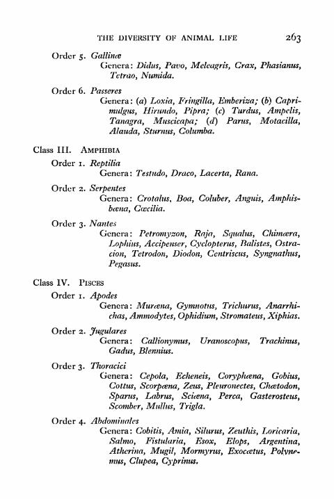
THE DIVERSITY OF ANIMAL LIFE 263
Order 5. Gallinte
Genera: Didus, Pavo, Meleagris, Craxy Phasianus,
Tetrao, Numida.
Order 6. Passeres
Genera: (a) Loxia, Fnngilla, Emberiza; (b) Capri-
mulgiis, Himndo, Pipra; (c) Turdus, Ampclis,
Tanagra, Muscicapa; (d) Pants, Motacilla,
Alauda, Starmis, Columba.
Class III. AMPHIBIA
Order i. Reptilia
Genera: Testudo, Draco, Lacerta, Rana.
Order 2. SerpentesGenera: Crotalus, Boa, Coluber, Anguis, Amphis-
bcena, Gwcilia.
Order 3. Nantes
Genera : Petromyzon, Raja, Squalus, Chimeera,
Lophius, Acxipemer, Cyclopterus, Batistes, Ostra-
cion, Tetrodon, Diodon, Ccntriscus, Syngnathus,
Pegasus.
Class IV. PISCES
Order i. ApodesGenera: Mureetw, Gymnotus, Trichurus, Anarrhi-
chas, Ammodytes, Ophidium, Stromatens, Xiphias.
Order 2. Jugulares
Genera: Gallionymus, Uranoscopus, Trachinus,
Gadus, Blennius.
Order 3. Thoracici
Genera: Gepola, Echeneis, Gorypluena, Gobius,
Gottus, Scorpcena, Zeus, Pleuronectes, Chcetodon,
Spams, Labrus, Scuena, Perca, Gasterosteus,
Scomber, Mulhis, Trigla.
Order 4. Abdominales
Genera: Gobitis, Anna, Silurus, Zeuthis, Loricaria,
Salmo, Fistularia, Esox, Elops, Argentina,
Athcrina, MugU, Mormyrus, Exoccetus, Polyne*
mus, Glupeat Gyprinus,
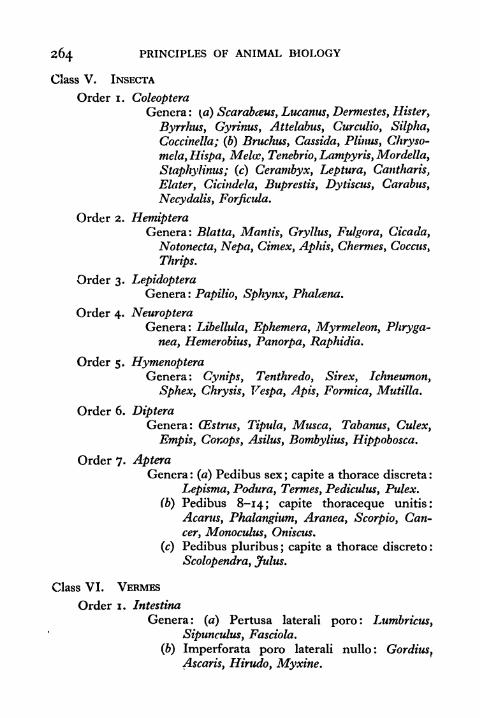
264 PRINCIPLES OF ANIMAL BIOLOGY
Class V. INSECTA
Order i. ColeopteraGenera : (a) Scarabcsus, Lucanus, Dermestes, Ulster,
Byrrhus, Gyrinus, Attelabus, Curculio, Silpha,
Cocdnella; (b) Bruchus, Cassida, Plinus, Chryso-
mela, Hispa, Melee, Tenebrio, Lampyris, Mordella,
Staphylinus; (c) Cerambyx, Leptura, Cantharis,
Elater, Cicindela, Buprestis, Dytiscus, Carabus,
Necydalis, Forficula.
Order 2. HemipteraGenera: Blatta, Mantis, Gryllus, Fulgora, Cicada,
Notonecta, Nepa, Cimex, Aphis, Chermes, Coccus,
Thrips.
Order 3. LepidopteraGenera: Papilio, Sphynx, Phalcena.
Order 4. NeuropteraGenera: Libellula, Ephemera, Myrmeleon, Phryga-
nea, Hemerobius, Panorpa, Raphidia.
Order 5. HymenopteraGenera: Cynips, Tenthredo, Sirex, Ichneumon,
Sphex, Chrysis, Vespa, Apis, Formica, Mutilla.
Order 6. DipteraGenera: CEstrus, Tipula, Musca, Tabanus, Culex,
Empis, Conops, Asilus, Bombylius, Hippobosca.
Order?. ApteraGenera : (a) Pedibus sex ; capite a thorace discreta :
Lepisma, Podura, Termes, Pediculus, Pulex.
(b) Pedibus 8-14; capite thoraceque unitis:
Acarus, Phalangium, Aranea, Scorpio, Can-
cer, Monoculus, Oniscus.
(c) Pedibus pluribus ; capite a thorace discrete :
Scolopendra, Julus.
Class VI. VERMES
Order i. Intestina
Genera: (a) Pertusa lateral! poro: Lumbricus,
Sipunculus, Fasciola.
(b) Imperforata poro laterali nullo: Gordius,Ascaris, Hirudo, Myxine.

THE DIVERSITY OF ANIMAL LIFE 265
Order 2. Mollusca
Genera: (a) Ore supero; basi se affigens: Actinia,
Ascidia.
(b) Ore antico; corpore pertuso laterali fora-
minulo : Limax, Aplysia, Doris, Tethis.
(c) Ore antico; corpore tentaculis antice cincto :
Holothuria, Terebella.
(d) Ore antico; corpore brachiato : Triton, Sepia,
Clio, Lerncea, Scyllcea.
(e) Ore antico; corpore pedato: Aphrodita,Nereis.
(/) Ore infero centrali: Medusa, Asterias,
Echinus.
Order 3. Testacea
Genera: (a) Multivalvia : Chiton, Lepas, Pholas.
(b) BivaJvia (= Conchce): Mya, Solon, Tellina,
Cardium, Mactra, Donax, Venus, Spondylus,
Chama, Area, Ostrea, Anomia, Mytilus,Pinna.
(c) Univalvia spira regulari (== Cochleee) : Argo-
nauta, Nautilus, Conus, Cyprace, Bulla,
Voluta, Buccinum, Strombus, Murex, Trochus,
Turbo, Helix, Nerita, Haliotis.
(d) Univalvia absque spira regulari: Patella,
Dentalium, Serpula, Teredo, Sabella.
Order 4. LithophytaGenera: Tubipora, Madrepora, Millepora, Celle-
pora.
Order 5. ZoophytaGenera: (d) Fixata: Isis, Gorgonia, Alcyonium,
Spongia, Flustra, Tubularia, Corallina, Sertu-
laria, Vorticella.
(b) Locomotiva: Hydra, Pennatula, Tcenia,
Volvox, Furia, Chaos.
The characters of the six classes are thus given byLinnaeus :
Cor biloculare, biauritum ; sanguine calido, rubro :
viviparis, Mammalibus
pviparis,
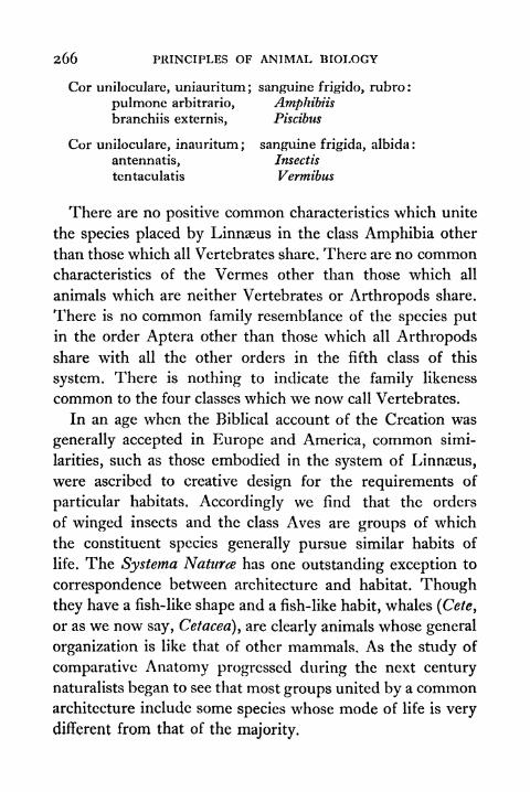
266 PRINCIPLES OF ANIMAL BIOLOGY
Cor unilocularc, uniauritum ; sanguine frigido, rubro :
pvilmone arbitrario, Amphibiisbranchiis externis, Piscibus
Cor uniloculare, inauritum ; sanguine frigida, albida :
antennatis, Insectis
tentaculatis Vermibus
There are no positive common characteristics which unite
the species placed by Linnaeus in the class Amphibia other
than those which all Vertebrates share. There are no commoncharacteristics of the Vermes other than those which all
animals which are neither Vertebrates or Arthropods share.
There is no common family resemblance of the species putin the order Aptera other than those which all Arthropods
share with all the other orders in the fifth class of this
system. There is nothing to indicate the family likeness
common to the four classes which we now call Vertebrates.
In an age when the Biblical account of the Creation was
generally accepted in Europe and America, common simi-
larities, such as those embodied in the system of Linnaeus,
were ascribed to creative design for the requirements of
particular habitats. Accordingly we find that the orders
of winged insects and the class Aves are groups of which
the constituent species generally pursue similar habits of
life. The Systerna Naturce has one outstanding exception to
correspondence between architecture and habitat. Though
they have a fish-like shape and a fish-like habit, whales (Cete,
or as we now say, Cetacea), are clearly animals whose general
organization is like that of other mammals. As the study of
comparative Anatomy progressed during the next century
naturalists began to see that most groups united by a commonarchitecture include some species whose mode of life is very
different from that of the majority.

foixiJi
-e,v
FIG. 100 VERTEBRATE WINGSThe extinct flying reptiles called Pterodactyls had a wing membranestretched between the elongated fourth finger and the hind limb. In bats,the second to fifth fingers are long and the hind limb again helps to keepthe membrane taut. Birds have feathers, which stand out stiffly on theirown account, and the awkward tying up of the hind limbs with the organsof flight is thus avoided.

268 PRINCIPLES OF ANIMAL BIOLOGY
Another fact became clear. Though most members of
many groups may share many features which distinguish
them sharply from species included in others, some species
occupy an intermediate position and bridge what would
otherwise be wide gaps in the scheme of classification. An
impressive example of this turned up when the world
became acquainted with the fauna of Australia after Captain
Cook's voyages. Two mammals, the Duckbill (Platypus)
FIG. 1 01 THE DUCK-BILLED PLATYPUS
and the Spiny Anteater (Echidna) of the Australasian region
lay eggs with shells like those of the birds and reptiles, and
have many reptilian characteristics which no other mammals
possess. Their habits are quite different, and there is nothing
about their situation or the kind of life they lead to suggest
that their reptilian characteristics were designed by Provi-
dence for the rigours of life in Australia. The study of such
intermediate types drew attention to the fact that the animal
body has vestiges of organs which, if useful at any time, have
now ceased to be so. The recognition of Unity of Type among
species which do not live the same kind of life destroyed
the complacent belief that all structural differences are
THE DIVERSITY OF ANIMAL LIFE 269
the expression of creative prescience, and thus encouraged
naturalists to speculate about how such differences might
be produced by natural agencies.
The Principle of Succession. The belief that different
species have come into being by natural agencies pre-
supposes that the age of the habitable earth is immensely
great in comparison with the record of human history. Till
the middle of the nineteenth century the Biblical account
of the age of the earth was still widely accepted in countries
where biological studies were advancing. Towards the end
of the eighteenth century Hutton put forward the view that
the stratified rocks have been laid down by the continual
wearing away of the earth's surface and the deposition of
sediment on the ocean bed. The progress of geology en-
couraged the study of fossils about which little was known
before the beginning of the nineteenth century. A third
obstacle to the recognition of evolution was removed when
modern views about the age of the earth were tested and
finally accepted. The study of geology displayed vast vistas
of time with an orderly succession of organic types, and
showed that the earliest members of a group are more
generalized than their modern descendants.
The Principle of Geographical Distribution. While this
belief gained ground the use of new communications and
encouragement given to exploration made it possible to see
the relation between structure and habitat from a new per-
spective. In the course of their travels naturalists began to
notice that similarities associated with habitat are not exclu-
sively associated with differences of climate or soil. Theyare also associated with barriers to dispersal. Areas that have
been separated for long periods of time by high ranges or
deep oceans are more likely to have species peculiar to them.
2JO PRINCIPLES OF ANIMAL BIOLOGY
In other words, the differences between species are con-
nected with the time during which they have been separated
from one another.
In the next four chapters we shall examine the Principle
of Unity of Type, the fossil record, and the phenomena of
geographical distribution in greater detail.
TOPICS FOR HOME STUDY
1 . What is meant by The Principle of Biogenesis ?
2. Compare the structure, behaviour and reproduction of Protists
with the higher animals.
3. In what respects does the system of Linnaeus (a) recognise
(b) neglect the principle of Unity of Type.

CHAPTER X
THE PRINCIPLE OF UNITY OF TYPE
THE Principle of Unity of Type, which impressed itself
on students of comparative anatomy during the latter
half of the eighteenth and the first half of the nineteenth
century, has been defined in general terms on p. 259. Ourfirst impressions of the diversity of animal life suggestthat species fall into clearly defined groups, such as insects,
fishes, birds, or sea urchins, which share a common patternof organization and a common habit of life. As we pursueour studies further we find that we can assemble them in
larger units which possess common characteristics of bodily
structure, and do not necessarily share the same mode of
existence. We also discover species wrhich bridge the gapbetween the compact classes into which most animals can be
put; and so force us to notice a pattern common to classes
of animals which live in different situations, get their food
from different sources, and move in different ways.*
Comparison of Vertebrate and Arthropod. To illustrate
structural uniformities which are associated with difference
of habit and habitat three types of Vertebrates and three
representatives of the Arthropod phylum will serve our
purpose. In these two groups the greatest activity and com-
plexity of behaviour is attained. The types we shall select
are respectively characterized by an aerial, an aquatic, and a
* The subject matter treated in this chapter is intended to be supple-mented by laboratory dissections of Invertebrates, as directed in a
practical manual, and by field work and museum study with the use ofsome standard works such as those of Parker and Haswell, Shipley andMacBride or Sedgwick for further reference.
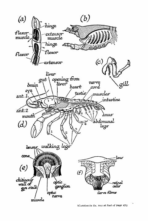
waiting legs'
tens'
HCTVJ2- fibres
\C,ihtinn to fie. 102 at foot of t>a&e 273
THE PRINCIPLE OF UNITY OF TYPE 273
terrestrial method of existence. From the phylum Vertebrata
it will be convenient to select the pigeon (Columba livid)
as an aerial type, the dogfish (Scyllium caniculd) as an
aquatic type, and the frog or the rabbit (Lepus cuniculus) as
a terrestrial type. As an aerial Arthropod, an insect, the
cockroach (Blatta orientalis, the "black beetle," or Periplaneta
americana, the large cockroach of bakehouses) is suitable
because of its size and general availability, although immature
individuals and the females of B. orientalis are wingless. Asan aquatic Arthropod we shall take one of the larger crusta-
ceans, such as the crayfish (Astacus fluviatilis) or the lobster
(Homarus vulgaris). As a terrestrial flightless Arthropod we
may select an Arachnid, the scorpion, because of its size.
The species shown in Fig. 103 is the South African Uroplectes
insignis, but any of the true scorpions, of which preserved
specimens are easily obtainable for dissection, will serve.
Let us start with the characteristic responses of these two
sets of types. The most obvious form of response is muscular
activity. If we compare the wing of the pigeon, the pectoral
fin of the dogfish, and the fore leg of the frog or rabbit,
we notice that the innermost core of the limb of all these
animals is a hard, jointed structure made of bone or cartilage.
To these jointed skeletal elements the muscles are attached.
The muscles are superficial to the joints; and the limb is
not externally divided into clearly marked-off segments.
FIG. 102 ANATOMY OF THE CRAYFISH, ETC.
(a) Part of a walking leg with windows cut in the joints to show themuscles. Note the directions (indicated by arrows) in which each jointis able to move, (b) The gill chamber opened from the side, (c) Walkingleg with gill attached, (d) Right half of a crayfish. In the heart, three ofthe openings which connect the heart with blood spaces can be seen.
(e) Longitudinal section through the compound eye. (/) Longitudinalsection through a simple Arthropodan eye.
S

274 PRINCIPLES OF ANIMAL BIOLOGY
The same remarks apply to the hind limbs of all three types
and to the tail of the dogfish, which is its chief organ of
propulsion. The relation of the muscles to the hard parts in
our three Arthropod types is quite different. If we examine
the walking legs* of the cockroach, the crayfish, or the
scorpion, we find that a limb of each is externally hard and
is divided into very distinct segments. On cutting through
the hard tubular wall of a limb, we find the muscles within
the tube attached at one end to inward projections of one
segment and to the internal wall of another segment at the
opposite extremity (Fig. 102). The wing of the cockroach is
attached at its base to muscles which pass in an analogous
manner to the inner ventral surface of the hard wall of the
thorax.
The trunk, like the limbs, of the three Arthropod types
we are considering, is also segmented, and powerful extensor
and flexor muscles attached to the hard outer shell of the
body control its movements in a manner analogous to
the action of muscles in the jointed limbs. In addition
to the walking legs, the latter include other "appendages"used for various activities. The crayfish has nineteen pairs
of appendages, thirteen being attached to the anterior un-
segmented part of the body the cephalothorax and six
to the six segments of the abdomen. The two most anterior
pairs are feelers or antenna*. Then follow a series just behind
the mouth, and these are employed for seizing and masti-
cating the food. The most anterior pair, the mandibles, have
powerful cutting bases. Behind these, but still on the cephalo-
thorax, come five pairs of walking legs, of which the foremost
end in the conspicuous pincers. The abdominal appendagesare mostly two-branched or biramous, and are used in swim-
ming or serve for the attachment and aeration of the eggs in
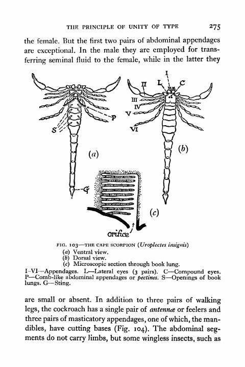
THE PRINCIPLE OF UNITY OF TYPE 275
the female. But the first two pairs of abdominal appendages
are exceptional. In the male they are employed for trans-
ferring seminal fluid to the female, while in the latter they
FIG. 103 THE CAPE SCORPION (Uroplectes tnsigtils)
(a) Ventral view.
(b) Dorsal view.
(c) Microscopic section through book lung.
I-VI Appendages. L Lateral eyes (3 pairs). C Compound eyes.P Comb-like abdominal appendages or pectines. S Openings of booklungs. G Sting.
are small or absent. In addition to three pairs of walking
legs, the cockroach has a single pair of antennae or feelers and
three pairs of masticatory appendages, one of which, the man-
dibles, have cutting bases (Fig. 104). The abdominal seg-ments do not carry limbs, but some wingless insects, such as
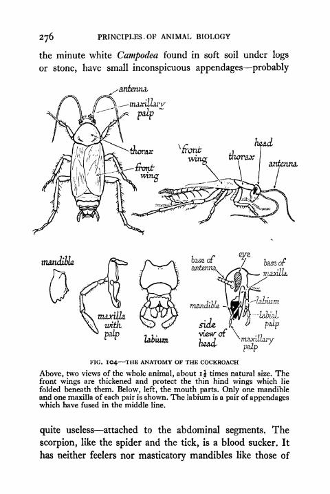
PRINCIPLES. OF ANIMAL BIOLOGY
the minute white Campodea found in soft soil under logs
or stone, have small inconspicuous appendages probably
antenna
FIG. IO4 THE ANATOMY OF THE COCKROACH
Above, two views of the whole animal, about i J times natural size. Thefront wings are thickened and protect the thin hind wings which lie
folded beneath them. Below, left, the mouth parts. Only one mandibleand one maxilla of each pair is shown. The labium is a pair of appendageswhich have fused in the middle line.
quite useless attached to the abdominal segments. The
scorpion, like the spider and the tick, is a blood sucker. It
has neither feelers nor masticatory mandibles like those of

THE PRINCIPLE OF UNITY OF TYPE
nudel
trackeal
system,
or
FIG. 105 THE ANATOMY OF THE COCKROACH COfJtinned
278 PRINCIPLES OF ANIMAL BIOLOGY
the cockroach or crayfish. There are two pairs of appendagesin front of the walking legs, the large pincers or pedipalps,
used to seize the prey. In front of the mouth there is a three-
jointed pair, the chelicerce, by which the animal attaches
itself to the part from which it sucks the juices of its victim.
In contradistinction to the crayfish, which has five, and the
insect, which has three, the scorpion has four pairs of walking
legs. Behind the last is a pair of comb-like appendages
(pectines) whose precise significance, if any, is not known.
Bodily movement in the insect is notoriously influenced
by light, and it is the light receptor which usually shows the
greatest structural complexity of any of the receptor organs.
To each of our Vertebrate types, the description of the eye
with its cornea, iris, lens, aqueous and vitreous humour,retina (p. 64), is equally applicable. In all three the eye is
movable within its orbit by the action of six very charac-
teristic eye muscles. In all three the retinal part of the eye
arises in the embryo as an outgrowth from the brain. The
scorpion has two sets of eyes. There are three pairs of
small eyes on either side at the anterior extremity, called
the lateral eyes, and there is a single larger pair on the centre
of the carapace. The lateral (simple) eyes consist of a biconvex
thickening of the transparent cuticle lying over a group of
photosensitive cells (cf. Fig. I02/) which are the specialized
endings of afferent fibres running back to the brain. In the
central eyes these photosensitive cells are arranged in groupsof about six around a central vitreous rod secreted by them,and are separated from adjacent groups by a thin layer of
pigmented connective tissue cells. This subdivision of the
retinal part of the eye into separate "retinulae" is carried
still further in the large compound eyes (Fig 102 e) of the
crayfish or of the cockroach, in which the surface of the
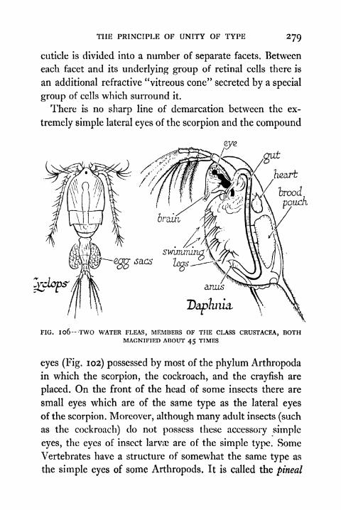
THE PRINCIPLE OF UNITY OF TYPE 279
cuticle is divided into a number of separate facets. Between
each facet and its underlying group of retinal cells there is
an additional refractive "vitreous cone" secreted by a special
group of cells which surround it.
There is no sharp line of demarcation between the ex-
tremely simple lateral eyes of the scorpion and the compound
,gut
.Heart
ferooJ
powch
FIG. 1 06-TWO WATER FLEAS, MEMBERS OF THE CLASS CRUSTACEA, BOTHMAGNIFIED ABOUT 45 TIMES
eyes (Fig. 102) possessed by most of the phylum Arthropodain which the scorpion, the cockroach, and the crayfish are
placed. On the front of the head of some insects there are
small eyes which are of the same type as the lateral eyes
of the scorpion. Moreover, although many adult insects (such
as the cockroach) do not possess these accessory simple
eyes, the eyes of insect larva; are of the simple type. Some
Vertebrates have a structure of somewhat the same type as
the simple eyes of some Arthropods, It is called the pineal
280 PRINCIPLES OF ANIMAL BIOLOGY
eye, and is best developed in the New Zealand lizard and in
the lamprey larva. In all Vertebrates there is a median
glandular body, the pineal gland, attached by a stalk to the
posterior part of the dorsal surface of the fore brain. It is
known to control the colour change of lampreys. In some
reptiles it lies outside a small window in the top of the skull
immediately beneath the epidermis, and such foramina are
common in the skull of fossil reptiles and amphibia. In a
few forms, such as those mentioned above, it has a structure
very much like the eye of many invertebrates. From the
embryological standpoint the paired eyes of Vertebrates
differ from any Arthropod eye, in that the retina is developed
as a cup-like outgrowth of the brain, which meets the cellular
lens formed from the outer skin. The retinal elements of
the Arthropod eye grow inwards in development to join the
brain.
Let us next consider the linking up of the receptor and the
effector, that is to say the apparatus of reflex response. In
all the animals we are dealing with this involves the trans-
mission of a nervous impulse through at least two fibres, one
conveying it to a part of the concentrated mass of nervous
tissue called the central nervous system, and one passing
out from the C.N.S. by way of a nerve fibre whose cell bodyis present in the C.N.S. itself. The central nervous systemof all our types is itself clearly demarcated into regions where
the nerve cells are congregated, and regions composed exclu-
sively of fibres running from one segment to another. In all
there is a special concentration of nervous tissue at the
anterior extremity of the body, where the mouth and eyes
are located. Here the similarity ends. In the three Vertebrate
types we have taken, the afferent fibre has its cell body
(exceptin the case of the olfactory and optic nerves) in a

THE PRINCIPLE OF UNITY OF TYPE 28l
ganglion close to the junction of the nerve with the C.N.S.
In the three Arthropod types the afferent nerve has its cell
uppersurface, wider
FIG. 107 TRILOBITE
The earliest representatives of the phylum Arthropoda found in theoldest sedimentary rocks are the Trilobites, a group which partake ofthe characteristics of all the more specialized classes which evolved later.
Thus they have a single pair of feelers like insects, bifid swimming limbslike Crustacea, and no specialized mandibles or biting jaws such as are
present in insects and Crustacea but absent in the arachnids.
body on the receptive surface. In the vertebrate types most
of the large nerves are connected with the C.N.S. by separate
afferent and efferent roots. In the Arthropod types this is
not so. In the Vertebrate types the C.N.S, arises as a tubular
282 PRINCIPLES OF ANIMAL BIOLOGY
infolding of the outer skin of the embryo along the mid-
dorsal line (Fig. 63), and is wholly dorsal to the alimentary
canal. In the three Arthropod types the central nervous
system (Fig. 108) lies above the alimentary canal at the
anterior extremity only (brain or supracesophageal ganglia).
This part is connected by fine strands with a double cord com-
posed of alternate thick portions (ganglia) and thin portions
below the alimentary canal. In this type of C.N.S. the
regions in which nerve cells and fibres occur alternate with
one another, and a ganglion, or more strictly a pair of ganglia,
is typically present in each distinct segment. In the Verte-
brate C.N.S. the connecting fibres lie outside (white matter)
the regions where nerve cells (grey matter) occur in the
spinal cord.
If we now turn to those structures wrhich are specially con-
cerned with the distribution of the materials from which
the energy of reflex response is derived, we see equally
striking differences between the three Vertebrate and the
three Arthropod types. In all the Vertebrate types we find
three very characteristic sets of digestive glands manu-
facturing the same type of secretions the gastric glands,
the liver with its gall bladder, and the pancreas. No such
close similarity is shown by the digestive system of the three
Arthropod types, but we may note in all of them a feature
which distinguishes them sharply from the Vertebrate types.
For some length inwards both the hind gut and the fore gut
are lined by a thin coat of chitin, the material of which the
hard outer wall of the body is formed. The Vertebrates have
in their blood a red, iron-containing pigment, haemoglobin,
which is carried by special blood cells and by combining
reversibly with oxygen acts as the vehicle by which the latter
is transported. Among Arthropods this function is generally

THE PRINCIPLE OF UNITY OF TYPE 283
performed by the quite different pigment, hsemocyanin,
which is pale blue when oxidized, and contains copper
instead of iron. Reduced haemocyanin is colourless.
Compoundbrain(dorsal
ganglion,)
heartr
FIG. 108 DIAGRAMMATIC REPRESENTATION OF AN IDEAL ARTHROPOD
The circulatory organs of the Arthropod types exhibit an
entirely unique arrangement. There is a large muscular heart
lying above the alimentary canal. This heart (Fig. 108), has
a paired series of valvular apertures known as ostia on either
side of it. The heart pumps blood out to the tissues byarteries which are chiefly derived from two main trunks
from the anterior and posterior end of the heart respectively.

284 PRINCIPLES OF ANIMAL BIOLOGY
Thence the blood percolates into the tissue spaces which
communicate ultimately with the body cavity in which the
heart lies. The body cavity in which the organs lie is a blood
space, and the heart fills itself at each beat from the sur-
rounding fluid which enters it via the ostia. The Vertebrate
circulation, on the other hand, is a closed system of vessels.
It displays certain universal features within the group even
in the adult, notably the important fact that the blood from
the capillary bed of the alimentary canal passes via the
hepatic portal vein to the liver, through whose capillaries
it must pass on its way back to the heart.
Though there are striking differences between the circu-
latory systems of land and water vertebrates, the main plan
of the arterial system is essentially similar at an early stage of
development. This we have studied already (p. 145) and have
noted its relation to the methods of respiration that exist in
different types of Vertebrates. No such extreme differences
are correlated with the different methods of breathing which
occur in different Arthropods (Figs. 102 and 105). In insects
the heart generally is very thin and the arterial system is
well-nigh non-existent. The insect has a system of fine
tubes lined by thin cuticle with annular thickenings. These
tubes are known as tracheae. These tracheae ramify through-
out the entire body, ending blindly in the deepest tissues.
They communicate by paired apertures (spiracles or stig-
mata) on either side of the body, and air is kept in circulation
by rhythmical movements of the abdomen that can be easily
observed in a cockroach or locust. The oxygen content of
the tracheae goes down during activity, and the carbon
dioxide production goes up. If we block the spiracles the
oxygen intake of an insect falls to a small fraction of its
normal dimensions. Thus oxygen is brought to the tissues

THE PRINCIPLE OF UNITY OF TYPE 285
directly in insects, which therefore have no need for a rapid
efficient circulation.
Tracheae are also found in centipedes and millipedes, and
in spiders and ticks. They do not occur in scorpions or in the
crayfish. The third, fourth, fifth, and sixth abdominal seg-
ments of the scorpion each have a pair of slit-like apertures
on the ventral surface. These slits lead into sacs called book
lungs, on account of the fact that the internal wall (Fig. 103)
is thrown into thin plate-like projections overlapping like the
leaves of a book. They offer a large surface for the diffusion
of respiratory gases. Experiment shows that if the apertures
of the book lungs are sealed, the oxygen consumption of the
scorpion is very greatly reduced. So they are the main
seat of oxygen intake, like our own lungs.
The crayfish has respiratory organs of another type.
Superficial examination reveals a large space between the
bases of the legs and a downward projection of the dorsal
wall of the cephalothorax on either side. These lateral gill
chambers, as they may be called, are open to the exterior
by fairly wide apertures at the sides of the mouth. The
cavity of the chamber is largely taken up by tufts of fine
filaments springing from the bases of the legs and resemblinga test tube brush in arrangement. These are the "gills.
"
Water is kept constantly circulating over them by the in-
cessant rhythmical motion of a plate-like extension of the
base of the third masticatory appendage (second maxilla)
which constitutes a baler, sometimes referred to by the
technical term scaphognathite. If the baler is removed the
oxygen consumption of the animal immediately sinks to
negligible proportions. So we may conclude that the main
surface for diffusion of oxygen from the water into the bodyis provided by the gill filaments.

286 PRINCIPLES OF ANIMAL BIOLOGY
Our survey of the physiological anatomy of terrestrial,
aquatic, and aerial Vertebrates, and terrestrial, aquatic, and
aerial Arthropods may be summed up as follows. In every
aspect of structure the terrestrial Arthropod shares certain
features of clear similarity with the aerial or aquatic Arthro-
pod, and does not share them with the terrestrial Vertebrate.
In every aspect of structure the aerial Vertebrate shares
with an aquatic Vertebrate or with a terrestrial Vertebrate,
certain features it does not share with an aerial Arthropod.
On this basis we are justified in grouping the three Verte-
brate types and the three Arthropod types in two separate
assemblages based on underlying similarity independent of
the mode of life they pursue, and we are thus forced to seek
some non-teleological interpretation of the similarities which
animals display,
Classifications of Lamarck and Cuvier. A very funda-
mental similarity thus exists between the representatives of
three great classes of Vertebrates on the one hand, and
three representatives of what we now call Arthropods on
the other. The recognition of this unity is foreshadowed in
the classification of animals given by Lamarck (1808) fifty
years after the publication of the Systema Naturte. Lamarck
departs so far from the anthropocentric point of view of
Linnaeus as to create a major assemblage, the Vertebrata,
to include the four first classes of Linnaeus' six divisions of
the animal kingdom. Lamarck also split up Linnaeus' order
Insecta and adopted the two classes Arachnida and Crus-
tacea, from which we have selected two of the types discussed
above, but he included in the Arachnida a certain number
of what are to-day regarded as wingless insects and the
centipedes and millipedes. Below is given the classification
of Lamarck:
THE PRINCIPLE OF UNITY OF TYPE 287
A. INVERTEBRATA1. "Apathetic" animals
Class I. Infusoria
Class II. Polypi (this includes Hydroids, Rotifera,
Polyzoa, some Echinoderms)
Class III. Radiaria (jellyfishes, sea anemones, star-
fishes, and sea urchins)
Class IV. Tunicata (present group)Class V. Vermes (non-segmented worms)
2. "Sensitive*' animals
Class VI. Insecla (Linnaean orders maintained)
Class VII. Arachnida
Class VIII. Crustacea
Class IX. Annelida (segmented worms)Class X. Cirripedia (barnacles)
Class XI. Conchifera (clams)
Class XII. Mollusca
Orders: Pteropoda; Gastropoda, Cephalopoda, Hetero-
poday Trachelipoda
B. VERTEBRATA. "Intelligent" animals
Class XIII. Pisces
Class XIV. Reptilia (= Linnaeus1
"Amphiba")Class XV. AvesClass XVI. Mammalia
In the system of Lamarck we see the introduction of
many terms that have been incorporated in modern schemes
of classification. The outstanding positive feature is the
recognition of unity of type within the Vertebrate series.
While correctly recognizing the heterogeneity of Linnaeus'
"Insecta," Lamarck fails to grasp the fundamental similarity
of the three great classes of Arthropods which he now
denotes by the terms "Insecta," "Arachnida"
and
"Crustacea." The modern term Arthropoda was introduced
by von Suboid in 1845. As the phylum is now defined
arthropods are segmented animals in which the nervous

288 PRINCIPLES OF ANIMAL BIOLOGY
system has the same relations as in segmented worms (vide
infra), i.e. a large pair of ganglia situated dorsally in the
preoral segment, and connected on either side with a double
chain of ganglia more or less approximated ventrally beneath
the alimentary canal. The body cavity, however, is a blood
space which communicates with the dorsal median heart by
paired valvular ostia. With the single exception of the
FIG. 109 THE BARNACLE
(i) The Nauplius or first larval phase with median eye and three pairs ofbiramous appendages, characteristic of the larvce of many orders of
Crustacea; (2) Vertical section through whole adult showing shell, plates,and six pairs of biramous jointed thoracic limbs (t.l.) ; (3) The Cyprisor second larval phase which has in addition to the antenna (A) and
masticatory appendages, six pairs of biramous swimming limbs (t.l.)
aberrant genus, Peripatus, they have jointed limbs with
separate muscles. The group may be divided into the follow-
ing classes :
1. Trilobita (primitive and exclusively fossil) Segments are alike
hearing biramous (two-branched) appendages with masticatory
bases, except the preoral pair which are antenniform.
2. Crustacea These resemble trilobites most closely. Somesegments bear biramous appendages. They have two pairs of
nntennse and mandibles and are mostly marine, e.g. shrimps,
lobsters, crabs, wood lice, pill bugs, water fleas.
3. Arachnida No antennae, no mandibles. Six or seven pairs of
appendages in front of the genital orifice. The first pair are typically
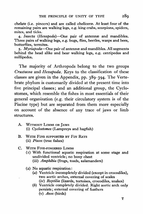
THE PRINCIPLE OF UNITY OF TYPE 289
chelate (i.e. pincers) and are called chelicerae. At least four of the
remaining pairs are walking legs, e.g. king crabs, scorpions, spiders,
mites, and ticks.
4. Insecta (Hexapodd) One pair of antennae and mandibles.
Three pairs of walking legs, e.g. bugs, flies, beetles, wasps and bees,
butterflies, termites.
5. Myriapoda One pair of antennae and mandibles. All segmentsbehind the head alike and bear walking legs, e.g. centipedes and
millipedes.
The majority of Arthropods belong to the two groupsCrustacea and Hexapoda. Keys to the classification of these
classes are given in the Appendix, pp. 389-394. The Verte-
brate phylum is customarily divided at the present time into
five principal classes; and an additional group, the Cyclo-
stomes, which resemble the fishes in most essentials of their
general organization (e.g. their circulatory system is of the
Piscine type) but are separated from them more especially
on account of the absence of any trace of jaws or limb
structures.
A. WITHOUT LIMBS OR JAWS(i) Cyclostomes (Lampreys and hagfish)
B. WITH FINS SUPPORTED BY FIN RAYS
(ii) Pisces (true fishes)
C. WITH FIVE-FINGERED LIMBS
(1) With functional aquatic respiration at some stage andundivided ventricle ; no bony chest
(iii) Amphibia (frogs, toads, salamanders)
(2) No aquatic respiration :
(a) Ventricle incompletely divided (except in crocodiles),two aortic arches, external covering of scales
(iv) Reptilia (lizards, tortoises, crocodiles, snakes)
(b) Ventricle completely divided. Right aortic arch onlypersists ; external covering of feathers
(v) Aves (birds)
T
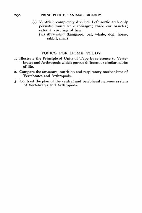
2QO PRINCIPLES OF ANIMAL BIOLOGY
(c) Ventricle completely divided. Left aortic arch only
persists; muscular diaphragm; three ear ossicles;
external covering of hair
(vi) Mammalia (kangaroo, bat, whale, dog, horse,
rabbit, man)
TOPICS FOR HOME STUDY1. Illustrate the Principle of Unity of Type by reference to Verte-
brates and Arthropods which pursue different or similar habits
of life.
2. Compare the structure, nutrition and respiratory mechanisms of
Vertebrates and Arthropods.
3. Contrast the plan of the central and peripheral nervous systemof Vertebrates and Arthropods.
CHAPTER XI
UNITY OF TYPE AMONG INVERTEBRATES
THE Principle of Unity of Type has been illustrated
in the foregoing chapter by comparison of Arthropodsand Vertebrates which pursue different or similar habits of
life. We shall now examine it in relation to the systematic
study of other groups of animals.
In Cuvicr's Le Regne Animal (1829) tne Arthropods are
associated with another group, the Annelida, or segmented
worms, in one of the major divisions of the animal kingdom,and several new groups based on Unity of Type make their
appearance for the first time. Cuvier's classification was as
follows :
FIRST BRANCH: ANIMALIA VERTEBRATAClass I. MAMMALIA
Orders: Bimana, Quadrumana, Carnivora, Marsupialia, Ro-
dentia, Edentata, Pachydermata, Ruminantia, Cetacea
Class II. BIRDS
Orders: Accipitres, Passeres, Scansores, Gallince, Grallee,
Palmipedes
Class III. REPTILIA
Orders : Chelonia, Sauria, Ophidia, Batrachia
Class IV. FISHES
Orders: (a) Acanthopterygii, Abdominales, Subbrachi, Apodes,
Lophobranchii, Plectognatha; (b) Sturiones, Selachii,
Cydostomi
SECOND BRANCH: ANIMALIA MOLLUSCAClass I. CEPHALOPODA
Class II, PTEROPODA
292 PRINCIPLES OF ANIMAL BIOLOGY
Class III. GASTROPODAOrders: Pulmonatay Nudibranchia, Inferobranchia, Heteropoda,
Pectinibranchia, Tubulibranthia, Scuti branchia, Cyclo-branchia
Class IV. ACEPHALAOrders: Testacea, Tunicata
Class V. BRACHIOPODA
Class VI. CIRRHOPODA
THIRD BRANCH: ANIMALIA ARTICULATAClass I. ANNELIDES
Orders: Tubicolce, Dorsibranchice, Abranchia
Class II. CRUSTACEAOrders: (a) Malp.costraca : Decapoda, Stomapuda, Amphipoda,
Lcemodipoda, Isopoda; (b) Entomostraca : Branchiopoda,Pcecilopoda, Trilobitce
Class III. ARACHNIDESOrders : Pulmonarice
y Trachearice
Class IV. INSECTA
Orders : Myriapoda, Thysanura, Parasita, Suctoria, Coleoptera,Orthoptera, Hemiptera, Neuroptera, Hymenoptera, Lepi-doptera, Rhipiptera, Diptera
FOURTH BRANCH: ANIMALIA RADIATAClass I. ECHINODERMS
Orders : Pedicellata, Apoda
Class II. INTESTINAL WORMSOrders : Nematoideay Parenchymatosa
Class III. ACALEPH^Orders : Simplices, Hydrostaticce
Class IV. POLYPI (including the Ccelenterata of modern authoritiesand the Polyzod)
Orders : Carnosi, Gelatinosi, Polypiarii
Class V. INFUSORIA
Orders: Rotifera, Homogenea (this includes the Protozoa ofrecent writers and Protophyta)

UNITY OF TYPE AMONG INVERTEBRATES 293
Though most modern authors prefer to retain the term
Arthropoda because all the classes mentioned on p. 288 share
many structural characteristics which distinguish them from
all Annelida, many subsequent zoologists have followed
Cuvier in making a super-phylum to include both. One
reason for this is the existence of a small group of closely
related genera (Peripatus, Peripatopsis, etc.) which occupy
a position midway between the two groups.
The Segmented Worms. The Annelida are externally seg-
mented animals without limbs, but typically provided on
either side in each segment with a tuft of bristles or setae
which serve for progression. The integument, which is
covered externally with a thin layer of chitin, consists of an
epidermis of one cell-layer together with an outer circular
and inner longitudinal layer of muscles, and encloses a
capacious cavity which is lined by secretory epithelium like
the peritoneum of the Vertebrate. The alimentary canal runs
from one end of the body to the other. The central nervous
system is of the same type as that of arthropods. A large
pair of ganglia, situated dorsally in the preoral segment,
connect on either side with a double chain of ganglia in
each segment more or less approximated ventrally beneath
the alimentary canal (Fig. no). The main trunks of the
blood system are longitudinal vessels running above and
below the gut and connected by annular vessels in each
segment. The blood generally contains haemoglobin. The
gonads are present in several, sometimes in all, segments, in
pairs, and discharge their products into the body cavity,
whence they are conveyed by segmental ducts to the exterior.
A paired series of segmental tubules, the nephridia, thought
to be of an excretory character, open to the exterior and end
either blindly in flame-cells like those of flat-worms (vide

294 PRINCIPLES OF ANIMAL BIOLOGY
infra) or by ciliated apertures into the body cavity. Annelids
provide the most perfect examples of metameric segmenta-
brain
maui'blood-
vessel,
excretory* 1 G**'tuba
ventralnerve,
chain.
FIG. 1 10 DIAGRAMMATIC REPRESENTATION OF AN IDEAL POLYCH^ETE
tion, i.e. the repetition of structure along the main axis of the
body, in the animal kingdom. The phylum Annelida include
the marine bristle worms, the earthworms, and leeches, and
are customarily classified as follows :

UNITY OF TYPE AMONG INVERTEBRATES 295
1. Polycheeta Marine worms with numerous setae, usually pro-
jecting from cushion-like prominences on the side of the body,
called parapodia. Often the preoral segment bears eyes and
tentacles. The gonads are not localized, e.g. Nereis, Aphrodite,
Arenicola, Bispira, Sabellaria.
2. Oligochceta Earthworms and a few freshwater worms. Few
bristles, no parapodia, eyes and tentacles never present. Herma-
phrodite, gonads localized (i.e. restricted to certain segments), eggs
laid in a cocoon, e.g. Lumbricus.
3. Hinidinea Leeches. All semi-parasitic or blood-sucking.
With few exceptions they have lost their setae and the body cavity
FIG. Ill PERIPATUS CAPENSIS
Drawn from life. Life size. (After Sedgwick.)
is largely replaced by connective tissue. They differ from the
earthworms chiefly in possessing a sucker by which they adhere
to their host or victim, and also in that each segment, correspondingto a nerve ganglion, etc., is externally divided into numerous rings,
e.g. Hirudo.
To these classes must be added a number of minor
groups (Echiuroidea, Myzostomida) which include very few
animals.
As already stated, one aspect of the Principle of Unity of
Type is the existence of isolated types which bridge the
gulf between clearly defined classes or phyla. A good exampleof such intermediate types is Peripatus and a few closely allied
genera of cryptozoic habit found in various parts of the

296 PRINCIPLES OF ANIMAL BIOLOGY
world where it is warm. In appearance Peripatus at the same
time reminds one of a millipede and of a leech. Although it
is commonly placed among the Arthropods, because it has
an ostiate heart and a trachea! system, the general plan of
its neuromuscular organization is essentially annelid. Exter-
nally it is not segmented, and the trunk limbs which are all
alike have no joints. Internally it has a pair of excretory
ducts in each segment, like a worm. The intermediate
position which Peripatus occupies will be seen from the
ensuing summary of the features which annelids share with
Arthropods and the characteristics which distinguish the
two groups :
1. The Annelida and Arthropoda include all animals with both*
(a) Metameric segmentation.
(b) A central nervous system consisting of a dorsal brain and
paired ganglionated nerve chains more or less closely
approximated in the mid ventral line.
2. The Annelida differ from all Arthropoda (including Peripatus)in having
(a) true intracellular centripetal nephridia.
(b) a closed vascular system.
(c) complete segmentation.
3. Peripatus agrees with Arthropods and differs from all wormsin that
(a) the dorsal blood vessel is ostiate.
(b) the body cavity is a blood space.
(c) it has trachea like those terrestrial Arthropods (Query:What is homology? vide pp. 382-3).
(d) the centro-lecithal egg is meroblastic. That is to say,
the storage material of the egg is concentrated at the
centre, and segmentation which occurs over the wholesurface does not involve the splitting of the central core
of yolk.
* Contrast above with (i) Cestoda, (ii) Amphioxus.
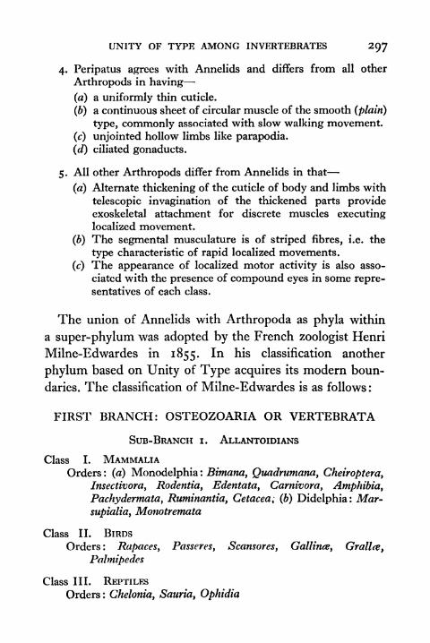
UNITY OF TYPE AMONG INVERTEBRATES 297
4. Peripatus agrees with Annelids and differs from all other
Arthropods in having
(a) a uniformly thin cuticle.
(6) a continuous sheet of circular muscle of the smooth (plain)
type, commonly associated with slow walking movement.
(c) unjointed hollow limbs like parapodia.
(d) ciliated gonaducts.
5. All other Arthropods differ from Annelids in that
(a) Alternate thickening of the cuticle of body and limbs with
telescopic invagination of the thickened parts provideexoskeletal attachment for discrete muscles executinglocalized movement.
(b) The segmental musculature is of striped fibres, i.e. the
type characteristic of rapid localized movements.
(c) The appearance of localized motor activity is also asso-
ciated with the presence of compound eyes in some repre-sentatives of each class.
The union of Annelids with Arthropoda as phyla within
a super-phylum was adopted by the French zoologist Henri
Milne-Edwardes in 1855. In his classification another
phylum based on Unity of Type acquires its modern boun-
daries. The classification of Milne-Edwardes is as follows :
FIRST BRANCH: OSTEOZOARIA OR VERTEBRATA
SUB-BRANCH i. ALLANTOIDIANS
Class I. MAMMALIAOrders : (a) Monodelphia : Bimana
y Quadrumana, Cheiroptera,
Insectivora, Rodentia, Edentata, Carnivora, Amphibia,
Pachydermata, Ruminantia, Cetacea; (b) Didelphia: Mar-
supialia, Monotremata
Class II. BIRDS
Orders: Rapaces, Passeres, Scansores, Gallince, Grallce,
Palmipedes
Class III. REPTILES
Orders : Chelonia, Sauria, Ophidia

298 PRINCIPLES OF ANIMAL BIOLOGY
SUB-BRANCH 2. ANALLANTOIDIANS
Class I. BATRACHIANS
Orders : Anura, Urodela, Perennibranchia, Cfrcilice
Class II. FISHES
Section i. Ossci
Orders : Acanthopterygii, Abdominales, Subbrachii, Apodes,
Lophobranchiij PlectognathiSection 2. Chondroptcrygii
Orders : Sturiones, Selachii, Cyclostomi
SECOND BRANCH: ENTOMOZOA OR ANNULATA
SUB-BRANCH i. ARTHROPODA
Class I. INSECTA
Orders: Coleoptera, Orthoptera, Neuroptera, Hymenoptera,Lepidoptera, Hemiptera, Diptera, Rhipiptera, Anopleura,
Thysanura
Class II. MYRIAPODAOrders : Chilognatha and Chilopoda
Class III. ARACHNIDAOrders : Pulmonaria and Trachearia
Class IV. CRUSTACEASection i. Podophthalniia
Orders : Decapoda and StomapodaSection 2. Edriophthalmi
Orders: Amphipoda y Lcemodipoda, and IsopodaSection 3. Branchipoda
Ostracoda, Phyllopoda, and Trilobitce
Section 4. Entomostraca
Orders: Copcpoda, Cladocera, Siphonostoma y Lernceida,
Cirripedia
Section 5. Xiphosura*
SUB-BRANCH 2. VERMES
Class I. ANNELIDA
Class II. HELMINTHES*To-day an order of Arachnida: it includes the King Crabs.

UNITY OF TYPE AMONG INVERTEBRATES
Class III. TURBELLARIA
Class IV. CESTOIDEA
Class V. ROTATORIA
THIRD BRANCH: MALACOZOARIA OR MOLLUSCASUB-BRANCH i. MOLLUSCA proper
Class I. CEPHALOPODA
Class II. PTEROPODA
Class III. GASTROPODA
Class IV. ACEPHALA (^ PELECYPODA or LAMELLIBRANCHIATA)
SUB-BRANCH 2. MOLLUSCOIDEA
Class I. TUNICATA
Class II. BRYOZOA
FOURTH BRANCH: ZOOPHYTESSUB-BRANCH i. RADIARIA
Class I. ECHINODERMA
Class II. ACALEPHA
Class III. CORALLARIA or POLYPI
SUB-BRANCH 2. SARCODARIA
Class I. INFUSORIA
Class II. SPONGIARIA
The Mollusca. The modern phylum Mollmca which
corresponds to the "mollusca proper"
in the preceding
arrangement illustrates Unity of Type in a more sophisticated
way than the examples we have already studied. Darwin
refers to it when he writes :
Nothing can be easier than to define a number of characters
common to all birds ; but with crustaceans any such definition has
hitherto been found impossible. There are crustaceans at the

300 PRINCIPLES OF ANIMAL BIOLOGY
opposite ends of the series which have hardly a character in common,yet the species at both ends, from being plainly allied to others and
these to others and so onwards, can be recognized as unequivocally
belonging to this and to no other class.
The characteristics which justify zoologists in placing the
several classes assigned to it in the single phylum Mollusca
cannot be summed up adequately by a few well-defined and
entirely specific characteristics. Faced with the snail, the
oyster, and the octopus as three molluscan types, the student
may well be puzzled to see any such uniformity of structural
peculiarities aswe have encountered in the scorpion, the cock-
roach, and the crayfish as examples of the Arthropoda. Yet
the unity of the group is very clear, when we study a large
number of species. Thus the nautilus is clearly similar in its
general organization to the octopus, and the nautilus is not
such a far cry from the whelk. The snail is clearly similar
to the whelk, the whelk to the limpet, and the limpet in manyrespects resembles a clam, like Nucula, that has a typical
molluscan gill (ctenidium), though the resemblance of a snail
to an oyster or mussel is not so obvious. So the zoological
student who makes his first study in the field after a preli-
minary acquaintance with text-book definitions may be
disappointed to find that the exceptions at first sight seem
to be as numerous as the rules.
To-day by general agreement the group includes two of
the Cuvierian classes whose names appeared in Lamarck's
scheme (Cephalopoda and Gastropoda), the former in-
cluding squids, octopus, etc., the latter snails, limpets, slugs.
In addition it includes Lamarck's separate class "Conchi-
fera," since usually called Lamellibranchiata (the term used
in Owen's lectures, 1855) or P^lecypoda by some contem-
porary authors. These were included along with the Tunicata
UNITY OF TYPE AMONG INVERTEBRATES 301
that are now regarded as allied to Vertebrates in Cuvier's
term Acephala. Cuvier's Pteropoda are now regarded as
an order of Gastropods. Of the Brachiopoda and Cirrho-
poda (Cirripedia) we shall speak later. They have only the
most superficial resemblance to the three major classes of
7710
den
FIG. 112 CHITON
Right, from above; left, from below, mo mouth ; want edge of mantle ;
ft foot; cten gill; an anus.
Mollusca (Cephalopoda, Gastropoda, and Lamellibran-
chiata). When we compare it with Linnaeus' order of the
same name, an assemblage which took in sea anemones,sea cucumbers, jellyfishes, most free living worms, we see
that Cuvier's Mollusca was a genuine step towards the
recognition of Unity of Type.In the modern phylum Mollusca two classes have been
separated from the older Gastropoda. These are the Iso-
pleura, or chitons, and the Scaphopoda, or elephant-tusk-
shells.
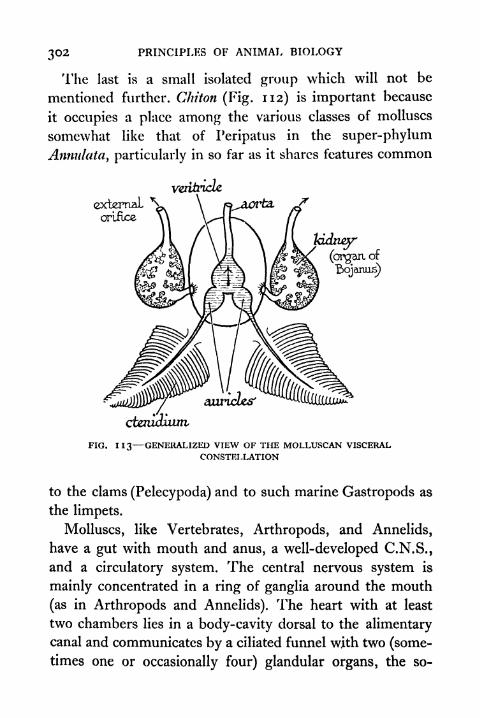
302 PRINCIPLES OF ANIMAL BIOLOGY
The last is a small isolated group which will not be
mentioned further. Chiton (Fig. 112) is important because
it occupies a place among the various classes of molluscs
somewhat like that of Peripatus in the super-phylum
Annulata, particularly in so far as it shares features common
externalorifice
wttiricle
kidney(organ of
cteniouim,
FIG. 113 GENERALIZED VIEW OF THE MOLLUSCAN VISCERALCONSTELLATION
to the clams (Pelecypoda) and to such marine Gastropods as
the limpets.
Molluscs, like Vertebrates, Arthropods, and Annelids,
have a gut with mouth and anus, a well-developed C.N.S.,
and a circulatory system. The central nervous system is
mainly concentrated in a ring of ganglia around the mouth
(as in Arthropods and Annelids). The heart with at least
two chambers lies in a body-cavity dorsal to the alimentarycanal and communicates by a ciliated funnel with two (some-times one or occasionally four) glandular organs, the so-
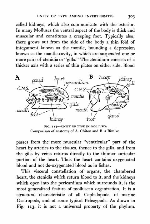
UNITY OF TYPE AMONG INVERTEBRATES 303
called kidneys, which also communicate with the exterior.
In many Molluscs the ventral aspect of the body is thick and
muscular and constitutes a creeping foot. Typically also,
there grows out from the side of the body a thin fold of
integument known as the mantle, bounding a depression
known as the mantle-cavity, in which are suspended one or
more pairs of ctenidia or "gills." The ctenidium consists of a
thicker axis with a series of thin plates on either side. Blood
.-
mouth
FIG. 114 UNITY OF TYPE IN MOLLUSCA
Comparison of anatomy of A. Chiton and B. a Bivalve.
passes from the more muscular "ventricular" part of the
heart by arteries to the tissues, thence to the gills, and from
the gills by veins returns directly to the thinner auricular
portion of the heart. Thus the heart contains oxygenated
blood and not de-oxygenated blood as in fishes.
This visceral constellation of organs, the chambered
heart, the ctenidia which return blood to it, and the kidneys
which open into the pericardium which surrounds it, is the
most generalized feature of molluscan organization. It is a
structural characteristic of all Cephalopods, of marine
Gastropods, and of some typical Pelecypods. As drawn in
Fig. 113, it is not a universal property of the phylum.

PRINCIPLES OF ANIMAL BIOLOGY
Chiton has many pairs of ctenidia, and some Cephalopodshave two pairs. Most marine Gastropods have only one gill,
pericardium/
amis'
,_-. N p&vzardium,(B) ^unt/i'
anus-
gtt,mantle, caviA N̂
central nervm?
(ugestwzghtid.
cporcuLxm,^
FIG. 115 (A) HYPOTHETICAL COMMON ANCESTOR OF CEPHALOPODS ANDGASTROPODS, (B) SHOWING TORTION OF THE BODY IN A MODERN
GASTROPOD
one auricle, and one kidney. The terrestrial species (e.g.
snails and slugs) have lost the ctenidium altogether. Though
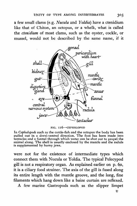
UNITY OF TYPE AMONG INVERTEBRATES
a few small clams (e.g. Nucula and Yoldid) have a ctenidium
like that of Chiton, an octopus, or a whelk, what is called
the ctenidium of most clams, such as the oyster, cockle, or
mussel, would not be described by the same name, if it
i&ntacles'
FIG. Il6 CEPHALOPOD
In Cephdlopods such as the cuttle-fish and the octopus the body has been
pulled out in a dorsi-ventral direction. The foot has been made intotentacles and a funnel through which water can be shot out to propel theanimal along. The shell is usually enclosed by the mantle and the radulais supplemented by horny jaws.
were not for the existence of intermediate types which
connect them with Nucula or Yoldia. The typical Pelecypod
gill is not a respiratory organ. As explained earlier on p. 60,
it is a ciliary food strainer. The axis of the gill is fused alongits entire length with the mantle groove, and the long, fine
filaments which hang down like a baize curtain are reflexed.
A few marine Gastropods such as the slipper limpetu
306 PRINCIPLES OF ANIMAL BIOLOGY
(Crepiduld) also use their gills in this way, and in such species,
as in many Pelecypods, there is a highly characteristic
device called the crystalline style for dealing with the carbo-
hydrate content of the diatom-diet of these animals. It is a
gelatinous rod impregnated with starch-splitting enzymeand enclosed in a groove or sac of the gut. Most Gastropods,
like Chiton and like all Cephalopods, take a more solid diet.
They have a muscular tongue (odontophore) on which is
mounted a characteristically molluscan structure called the
radula. This is a ribbon of chitinous teeth for triturating
their food. Many Gastropods and Cephalopods also have
horny jaws.
Another characteristic feature of the group is the ciliated
larva with its minute shell. It is called the Veliger (Fig. 117),
and is sometimes preceded by a larval form which resembles
the larva of marine worms, and is known as the Trochophore
(Fig. 117). The heavily yolked eggs of the Cephalopods like
the eggs of terrestrial gastropods (snails and slugs) do not
hatch out as larvae.
1. Isopleura. The mantle-fold is co-extensive with the
body, the mantle-cavity is shallow on either side carrying a
paired series of ctenidia which hang freely. The foot is
creeping and there is a distinct head, and odontophore and
radula are present. A series of unpaired dorsal plates form
the shell, e.g. Chiton.
2. Pelecypoda. As in Chiton the mantle-fold is co-
extensive with the body, but is much deeper, so that the
mantle-cavity is wrapped round the foot. The two lobes of
the mantle-fold secrete a bivalved shell. The head, odonto-
phore and radula are completely absent. A pair of ctenidia
are present, the axis being fused along its whole length with
the mantle-ridge, and the filaments, which are very

UNITY OF TYPE AMONG INVERTEBRATES 307
numerous, hang downwards like a double curtain into the
mantle-cavity. It is to this feature that the alternative name
Lamellibranchiata refers. The gill of a typical clam is not
obviously like that of other molluscs, but a series of forms
connect the more specialized type of gill, which we have
described in the case of the mussel (p. 60), characteristic of
the majority of the group, with the more typical Molluscan
ctenidium of the smaller clams like Nucula. To this class
belong the oysters, mussels, cockles, etc.
FIG. 117 CILIATED LARWE OF MOLLUSCS TROCHOPIIORE AND VELIGER
3. Scaphopoda. The Scaphopods in general plan of
organization are very much like the Lamellibranchs, but the
folds of the mantle-cavity fuse below the foot and secrete a
continuous cylindrical shell. The ctenidia have been lost.
There is a vestigial head and odontophore but no radula,
e.g. Dentalium.
4. Gastropoda. These possess a well-developed head
bearing tentacles and eyes and a creeping foot like that of
Chiton, also an odontophore with radula. In early develop-
ment the mantle-cavity is confined to the posterior region,
but as development proceeds the visceral mass becomes
twisted through two right angles so that eventually the
mantle-cavity, anus, etc., are directed forwards. There is
generally a spiral shell. In the primitive forms there are
308 PRINCIPLES OF ANIMAL BIOLOGY
two ctcnidia, more generally one, and in some forms, e.g.
snails and slugs, they have been lost. This class includes,
in addition to the last-named, the whelks, limpets, and sea
slugs.
5. Cephalopoda. The mantle-cavity is posterior and
typically contains two ctenidia. The integument below the
head is drawn out into elongated prehensile processes known
as arms and also into a funnel-like opening for the mantle-
cavity. There is nothing exactly like the creeping foot of
Chiton. An odontophore and radula are present. The shell
is single but is usually embedded in the dorsal wall, e.g.
octopus, cuttle-fish, squids, etc.
A few of the characteristic features of the Mollusca
described in the preceding survey are found in some members
of all five groups. Others are found in more than one but
not in all. Scarcely any are universal. The best way to empha-size the essential unity of type on which the recognition of
the phylum is based is therefore to draw up a table such as
that on page 309.
Leuckarfs Classification of Animals. A system which was
published in Germany in the middle of the nineteenth
century, a few years earlier than that of Milne-Edwardes,
introduces us to two new phyla, Echinodermata and Ccelen-
terata, as recognized to-day. Leuckart's system which
does not include a detailed division of Vertebrates is as
follows :
TYPE i: CCELENTERATA
Class I. POLYPIOrders : Anthozoa and Gyclicozoa
Class II. ACALEPHWEOrders : Discophorce and Ctenophorce
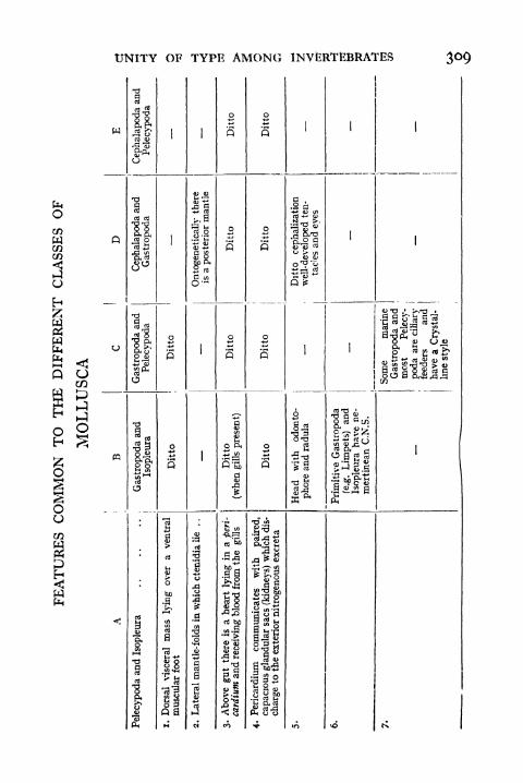
UNITY OF TYPE AMONG INVERTEBRATES 309
COWCOCO
U
Q OCO
ocj
CO
I
Gastropoda
and
Pelecypoda
-CJ
|
viscer
ar
foot
3ll
o
B
ll
Jd (
ccj -^j
with
od
and
radul

310 PRINCIPLES OF ANIMAL BIOLOGY
TYPE 2: ECHINODERMATAClass I. PELMATOZOA
Orders : Cistoidea and Crinoidea
Class II. ACTINOZOAOrders : Echinida and Asterida
ClaSS III. SCYTODERMATAOrders : Holothurice and Sipunculida
TYPE 3: VERMESClass I. ANENTERflETI
Orders : Cestodes and Acanthocephali
Class II. APODESOrders : Nemertini, Turbellarii, Trematodes, and Hirudini
Class III. CILIATI
Orders : Eryozoa and Rotifera
Class IV. ANNELIDESOrders : Nematodes, Lumbriciniy and Branchiati
TYPE 4: ARTHROPODAClass I. CRUSTACEA
Orders : Entomostraca and Malacostraca
Class II. INSECTA
Orders : Myriapoda, Arachnida (Accra, Latr.), Hexapoda
TYPE 5: MOLLUSCAClass I. TUNICATA
Orders : Ascidite and Salpce
Class II. ACEPHALAOrders : Lamellibranchiata and Brachiopoda
Class III. GASTROPODAOrders: Heterobranchia, Dermatobranchia, Clepobranchia,
PalmopodcL) and Cyclobranchia
Class IV. CEPHALOPODA
TYPE 6: VERTEBRATA

UNITY OF TYPE AMONG INVERTEBRATES 3 II
Echinoderms and Ccelenterates. The Echinodermata,
which include starfishes, sea urchins, feather stars, brittle
stars, and sea cucumbers, share one feature with the
Coelenterata with which they are associated in Cuvier's
classification. Both show a considerable measure in some
Ccelenterates complete of radial as opposed to bilateral
symmetry. In other respects they are very different.
Arthropods and Molluscs agree with Vertebrates in having
arazs
bodycavity
nerve ring mouth
FIG. Il8 DIAGRAMMATIC REPRESENTATION OF AN IDEAL ECHINODERM
a well-developed circulatory system. A rudimentary vascular
system also exists in Annelids, Brachiopods, Nemertines,and in Phoronis. The Echinodermata represent a definitely
lower grade of physiological organization and the classes,
though differing in external form, have a close similarity
in internal organization (Fig. 118). A muscular integumentwith a leathery external covering and outer layer of ciliated
epithelium is strengthened internally with calcareous plates
to which may be attached projecting spicules. It encloses a
capacious body-cavity lined by secretory epithelium and
developed 3 a pouch from the embryonic gut. The alimen-
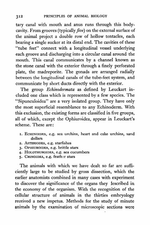
312 PRINCIPLES OF ANIMAL BIOLOGY
tary canal with mouth and anus runs through this body-
cavity. From grooves (typicallyfive) on the external surface of
the animal project a double row of hollow tentacles, each
bearing a single sucker at its distal end. The cavities of these
"tube feet" connect with a longitudinal vessel underlying
each groove and discharging into a circular canal around the
mouth. This canal communicates by a channel known as
the stone canal with the exterior through a finely perforated
plate, the madreporite. The gonads are arranged radially
between the longitudinal canals of the tube-feet system, and
communicate by short ducts directly with the exterior.
The group Echinodermata as defined by Leuckart in-
cluded one class which is represented by a few species. The
"Sipunculoidea" are a very isolated group. They have only
the most superficial resemblance to any Echinoderm. With
this exclusion, the existing forms are classified in five groups,
all of which, except the Ophiuroidea, appear in Leuckart's
scheme. These are:
1. ECHINOIDEA, e.g. sea urchins, heart and cake urchins, sand
dollars
2. ASTERIODEA, e.g. starfishes
3. OPHIUROIDEA, e.g. brittle stars
4. HOLOTHUROIDEA, e.g. sea cucumbers
5. CRINOIDEA, e.g. feath-r stars
The animals with which we have dealt so far are suffi-
ciently large to be studied by gross dissection, which the
earlier anatomists combined in many cases with experimentto discover the significance of the organs they described in
the economy of the organism. With the recognition of the
cellular structure of animals in the thirties embryologyreceived a new impetus. Methods for the study of minute
animals by the examination of microscopic sections were
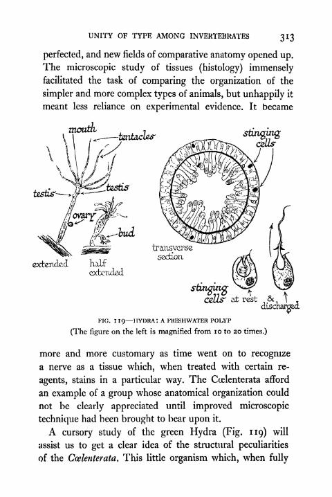
UNITY OF TYPE AMONG INVERTEBRATES 313
perfected, and new fields of comparative anatomy opened up.
The microscopic study of tissues (histology) immenselyfacilitated the task of comparing the organization of the
simpler and more complex types of animals, but unhappily it
meant less reliance on experimental evidence. It became
stingingcells'
FIG. 119 HYDRA: A FRESHWATER POLYP
(The figure on the left is magnified from 10 to 20 times.)
more and more customary as time went on to recognize
a nerve as a tissue which, when treated with certain re-
agents, stains in a particular way. The Ccelenterata afford
an example of a group whose anatomical organization could
not be clearly appreciated until improved microscopic
technique had been brought to bear upon it.
A cursory study of the green Hydra (Fig. 119) will
assist us to get a clear idea of the structural peculiarities
of the Ccelenterata, This little organism which, when fully
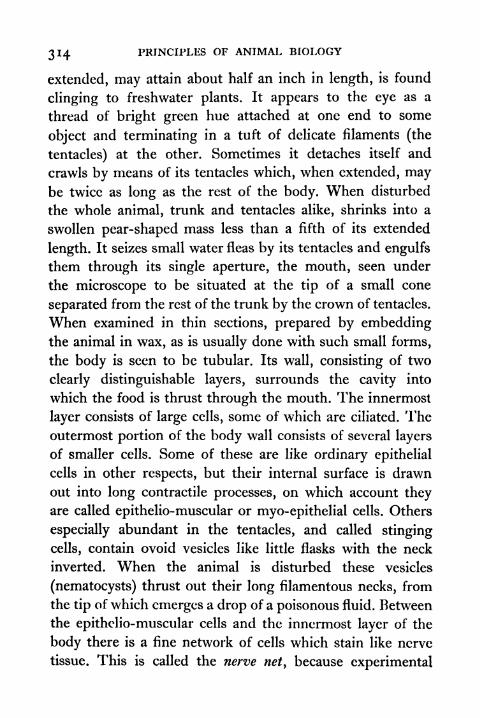
314 PRINCIPLES OF ANIMAL BIOLOGY
extended, may attain about half an inch in length, is found
clinging to freshwater plants. It appears to the eye as a
thread of bright green hue attached at one end to some
object and terminating in a tuft of delicate filaments (the
tentacles) at the other. Sometimes it detaches itself and
crawls by means of its tentacles which, when extended, maybe twice as long as the rest of the body. When disturbed
the whole animal, trunk and tentacles alike, shrinks into a
swollen pear-shaped mass less than a fifth of its extended
length. It seizes small water fleas by its tentacles and engulfs
them through its single aperture, the mouth, seen under
the microscope to be situated at the tip of a small cone
separated from the rest of the trunk by the crown of tentacles.
When examined in thin sections, prepared by embeddingthe animal in wax, as is usually done with such small forms,
the body is seen to be tubular. Its wall, consisting of two
clearly distinguishable layers, surrounds the cavity into
which the food is thrust through the mouth. The innermost
layer consists of large cells, some of which are ciliated. Theoutermost portion of the body wall consists of several layers
of smaller cells. Some of these are like ordinary epithelial
cells in other respects, but their internal surface is drawn
out into long contractile processes, on which account theyare called epithelio-muscular or myo-epithelial cells. Others
especially abundant in the tentacles, and called stinging
cells, contain ovoid vesicles like little flasks with the neck
inverted. When the animal is disturbed these vesicles
(nematocysts) thrust out their long filamentous necks, from
the tip of which emerges a drop of a poisonous fluid. Between
the epithelio-muscular cells and the innermost layer of the
body there is a fine network of cells which stain like nerve
tissue. This is called the nerve net, because experimental
UNITY OF TYPE AMONG INVERTEBRATES 315
evidence shows that the Coelenterate nerve net is a form of
conducting tissue.
From time to time cylindrical buds are formed on the
sides of the trunk. These acquire a tuft of tentacles at the
free end, and become detached to pursue an independentexistence as new individuals. But in addition to this vegeta-
tive method of reproduction, true sexual reproduction occurs
in Hydra. Cells of the outer layer of the body divide re-
peatedly, giving rise to sperms in the usual way, and the
male gonad, which is seen as a prominence on any region of
the trunk, is simply a mass of sperm covered by a thin
pellicle of epithelial cells, which eventually becomes rup-
tured liberating the gametes. An ovary may be producedon the same individual. It consists of a single large ovumlikewise covered by a thin epithelium on the outside of the
body. The sperm penetrates the ruptured wall of the ovary.
Both fertilization and early cleavage take place while the
ovum is still attached to the body of the parent.
Budding occurs in many Ccelenterates and often the buds
do not become detached, so that colonies are formed, as in
such common rock pool zoophytes as Obelia and Plumularia.
In these colonial forms the sexual organs are often developedon a special sort of bud, the medusa, which is more flattened
in shape than the vegetative hydroid. The medusa is set free
into the water and pursues an independent existence for a
time, shedding its sexual products when they are ripe. Manycolonial Ccelenterates secrete calcareous deposits around
them. Such are the corals. Some, allied to Hydra, form
colonies with large floats filled with gaseous products, the
zooids being clustered on the underside of the float which
drifts with wind and current. Physalia, the Portuguese man-
of-war, or blue-bottle, is one of these. In one respect the
316 PRINCIPLES OF ANIMAL BIOLOGY
organization of Hydra is simpler than that of the larger
Ccelenterata, the jellyfishes and sea anemones. It has no
separate muscular system. Well-developed muscle fibres
forming a continuous sheet in the outer portion of the bodywall (jellyfishes) with, in addition, discrete bands in the
inner portion (sea anemones) are often present in Ccelen-
terates. Again the gonads in the larger Coelenterata are
located in the digestive cavity. But in all they are simply
proliferations of cells of the body wall, without any ducts
or accessory structures. Stinging cells are always present,
and the conducting tissue is of the diffuse type referred to
as a nerve net. Separate afferent and efferent nerve fibres
meeting in a central nervous system are never found.
The group Coelenterata as defined by Leuckart included
the sponges, which are now usually made a separate phylum,the Porifera. With this restriction the Ccelenterata may be
defined as follows. They are cellular animals with one cavity,
the digestive cavity, communicating with the exterior by a
single aperture, the mouth. The walls of the body consist
of two distinguishable layers. These two layers, the ectoderm
and endoderm, are often separated by a thick gelatinous layer
(mesogloea). In the cellular layers true muscle-fibres and
conducting tissue may be differentiated, but there is never
present either (a) a concentration of nervous elements to
form a central nervous system, or (b) a system of reproductive
ducts. The germ cells are formed superficially either from
the ectoderm (Hydrozoa) or endoderm. From a physio-
logical point of view the main feature is the absence of a
mechanism of true reflex action. The majority of Ccelen-
terates are thus divided by modern authors :
i. Hydrozoa. Gonads external, no gullet or gastric filaments,
e.g. Hydra, Plumularia, Obelia, MiHepora, Physatia.
UNITY OF TYPE AMONG INVERTEBRATES 317
2. Scyphozoa. Gonads internal, typically with alternation of
generations, but the mcdusoid the predominant form. The medusoiddiffers from the Hydrozoan medusoid, because it has internal
feonads, and because it has gastric filaments provided with stingingcells which are the remains of radial partitions in the hydroid fromwhich it is budded off along the longitudinal axis, e.g. Aurelia,
Ilaliclystus (sedentary), R/iizostoma.
3. Actinozoa. Gonads internal, but individuals entirely sessile
with no alternation of generations. There is a deep ectodermal gulletand the digestive cavity is divided incompletely below the gullet and
completely above by radial partitions which are provided with thick
longitudinal muscle bands, e.g. sea anemones and the majority of
the corals.
A fourth group, the Ctenophora or comb-jellies, includes
a few specialized forms with predominantly bilateral sym-
metry. In development of comb-jellies the body muscles are
laid down at an early stage in embryonic life, as in more
complex animals.
The Porifera or sponges formerly placed close to the
Coelenterata are only superficially like them, in so far as
they represent a very low grade of physiological differen-
tiation. They were regarded as animals by Aristotle, but they
stand very much apart from all other animals, and such
terms as body, individual, colony or the like have very little
applicability to them. A sponge is a labyrinth of ciliated
canals communicating by pores on the surface with the
exterior. Between the canals is a gelatinous mass strengthened
by horny fibres or spicules of calcareous or siliceous material,
the exterior being invested in a thin epithelium in which
distinct cell walls are absent. They have no nervous elements,
and the reproductive organs are clusters of sperms or eggs
embedded in the gelatinous coat. The sperm wriggles its
way through the tissues of the sponge to the eggs embedded
in them.
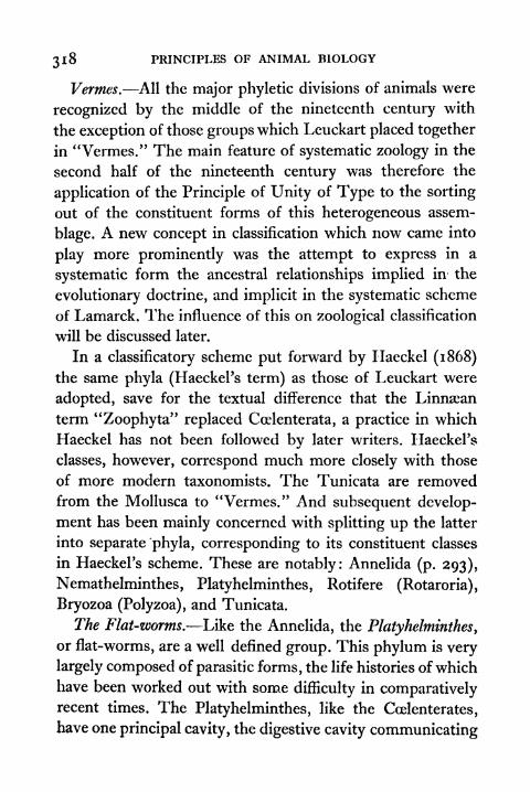
318 PRINCIPLES OF ANIMAL BIOLOGY
Vermes. All the major phyletic divisions of animals were
recognized by the middle of the nineteenth century with
the exception of those groups which Leuckart placed together
in "Vermes." The main feature of systematic zoology in the
second half of the nineteenth century was therefore the
application of the Principle of Unity of Type to the sorting
out of the constituent forms of this heterogeneous assem-
blage. A new concept in classification which now came into
play more prominently was the attempt to express in a
systematic form the ancestral relationships implied in the
evolutionary doctrine, and implicit in the systematic scheme
of Lamarck. The influence of this on zoological classification
will be discussed later.
In a classificatory scheme put forward by Haeckel (1868)
the same phyla (Haeckel's term) as those of Leuckart were
adopted, save for the textual difference that the Linnsean
term "Zoophyta" replaced Coelenterata, a practice in which
Haeckel has not been followed by later writers. HaeckePs
classes, however, correspond much more closely with those
of more modern taxonomists. The Tunicata are removed
from the Mollusca to "Vermes." And subsequent develop-
ment has been mainly concerned with splitting up the latter
into separate phyla, corresponding to its constituent classes
in HaeckeFs scheme. These are notably: Annelida (p. 293),
Nemathelminthes, Platyhelminthes ,
Rotifere (Rotaroria) ,
Bryozoa (Polyzoa), and Tunicata.
The Flat-worms. Like the Annelida, the Platyhelminthes,
or flat-worms, are a well defined group. This phylum is very
largely composed of parasitic forms, the life histories of which
have been worked out with some difficulty in comparativelyrecent times. The Platyhelminthes, like the Coelenterates,
have one principal cavity, the digestive cavity communicating
UNTTY OF TYPE AMONG INVERTEBRATES 319
with the exterior by a single aperture, the mouth. They have,
however, no sign of radial symmetry. A somewhat elaborate
system of generative organs which discharge their productsto the exterior by gonaducts, provided with an elaborate
moudi
estis
pores
FIG. 120 DIAGRAMMATIC REPRESENTATION OF AN IDEAL FLAT-WORM
Showing gut, nervous system and excretory organs on left, and repro-ductive organs on the right.
system of glands, constitutes the most characteristic feature
of the group (Fig. 120). With one or two exceptions flat-
worms are hermaphrodite. The oviduct generally receives
the secretion of shell glands and a ramifying system of yolk
glands which occupy a good deal of the internal space of
the body. Between the muscular outer wall and the epithe-
lium of the gut the interspaces are filled with a highly
characteristic mass of vacuolated cells called parenchyma.
32O PRINCIPLES OF ANIMAL BIOLOGY
There is a ramifying system of intracellular tubules called
the nephridia, communicating at one end with the exterior
and ending at the other end blindly in cells with vibratile
cilia which project into the lumen of the tube. They are
called flame cells. In general all the Platyhelminthes are
dorsoventrally compressed. A central nervous system is
present and consists of a large anterior dorsal ganglion and
lateral cords connected by transverse commissures below the
gut. Three classes are now recognized within the group :
1. Turbellaria. Free-living flat-worms in which the body is
covered by a ciliated epithelium, some of the cells of which contain
hyaline bodies known as rhabdites. The mouth is typically at the
end of an eversible pharynx on the ventral side. They are found
under rocks in fresh and marine water. A few live in mud, e.g.
Planocera, Planaria, Bipaltum.2. Trematoda. Parasitic flat-worms (flukes) in which the external
epithelium after secreting a thick cuticle degenerates. The body is
provided with suckers. A gut is present, e.g. Distomum> Polystomum,Bilharzia.
3. Cestoda. The integument and reproductive organs are verysimilar to the Trematoda, but a digestive cavity is absent. They are
all internal parasites (tape-worms), and generally the body is
divided into a number of segments, each with a complete set of
reproductive organs, e.g. Tania.
The class Nemertinea in several respects show a consider-
able advance in structural complexity upon the flat-worms,
for which reason they are elevated by some zoologists to
phyletic rank. The elongated vermiform body with terminal
mouth and anus has a rudimentary circulatory system. These
features separate them from the foregoing classes. So does
the peculiar eversible pharynx enclosed in a tubular sheath
above the alimentary canal at the anterior end of the body.On the other hand, a clear relationship to the Turbellaria
is seen in the ciliated epidermis, the cells of which contain

UNITY OF TYPE AMONG INVERTEBRATES 321
rhabdites, in the parenchymatous tissue that occupies the
space between the gut and the integument, in the nephridia
and nervous system. The Nemertinea occupy a very interest-
ing position in the animal kingdom, in some respects inter-
mediate between the Annelida and the Flat-worms, and
they might well be conceived to be survivors of a group
very closely related to the common stock of Annelids,
Molluscs, and Arthropods.
Thread-worms. Another group which includes species of
great economic importance is the Nemathelminthes, or round-
worms, which comprises both a large number of parasites,
chiefly intestinal, and free living forms in sea water and
damp soil. The majority of forms denoted by this term are
included in a single class, Nematoda, and it is doubtful
whether the other classes usually associated with them have
any close resemblance (Nematomorpha, which occur as
parasites in the intestines of insects, and Acanthocephala,
gutless intestinal parasites in Vertebrates, with a hooked
eversible proboscis). The Nematodes have an elongated bodycovered with a thin cuticle with terminal mouth and anus
at the opposite extremity. They have a muscular pharynx,but the rest of the straight alimentary canal has a wall
composed only of epithelium surrounded by cuticle. Between
the integument and the gut is a capacious cavity in which
the gonads and coiled gonaducts lie. The cavity is not lined
by a secretory epithelium like the body cavity of an Annelid.
The integument itself consists of a syncytium below the
cuticle and a single layer of longitudinal muscles whose
enormous protoplasmic processes with their nerve fibres
project into the body cavity. The similarity between the
body cavity (or coelom) of segmented worms, Vertebrates,
and Molluscs was a topic for much discussion and endless
X
322 PRINCIPLES OF ANIMAL BIOLOGY
speculation during the second half of the nineteenth century.
The fact that the "coelom" arises in development in a different
manner within the same group makes such discussion some-
what barren. To the Nematoda belong the horse thread-
worm Ascaris, the miner's worm Anchylostomum, and other
human parasites such as Trichina, Filaria, and Oxyuris, the
pin-worm of children's stools. The remaining Vermes are
isolated groups of little practical significance.
The Rotifera may be mentioned first, because they have always
proved a source of attraction to amateur microscopists. For that
reason they have been studied more thoroughly than most of the
others. The Rotifera are minute animals found in fresh water the
world over and occupy a very isolated position in the animal king-dom. Though of microscopic dimensions the largest are only just
visible to the naked eye they have a comparatively complexorganization. The body is of various shapes. Typically there is a
circle of ciliated cells at the oral end. The alimentary canal is
provided with mouth and anus and a gastric mill, but has nomuscular wall. It lies in a capacious cavity in which nephridia with
flame cells and gonads and gonaducts are also present Both the
latter open into the hind end of the gut. The C.N.S. is a circumces-
phageal ring with a large bi-lobed dorsal ganglion. They are of noeconomic importance. Many are parthenogenetic, and like the social
Hymenoptera produce their males by virgin birth and their females
from fertilized eggs. Some, like the gall-flies, and a few fresh-water
Crustacea, have a regular alternation of parthenogenetic and bi-
sexual generations. The Rotifera attracted a good deal of attention
at one time because of their superficial resemblance to the ciliated
larvae of Annelids, Molluscs, and Flat-worms.The Polyzoa are colonial animals, which in the form of the colony
and the zooids show a superficial resemblance to corals and hydroids,from which they were first separated by Vaughan Thompson. Thereis a circlet of tentacles round the oral end leading into the alimentarycanal which is provided with an anus surrounded by a body cavityin which the gonads lie. The C.N.S. is represented by a small
ganglion between mouth and anus. There is a ciliated larva some-what like the trochophore larva of Annelids. Representatives of the
group are common in rock-pools between tide marks. The Polyzoa
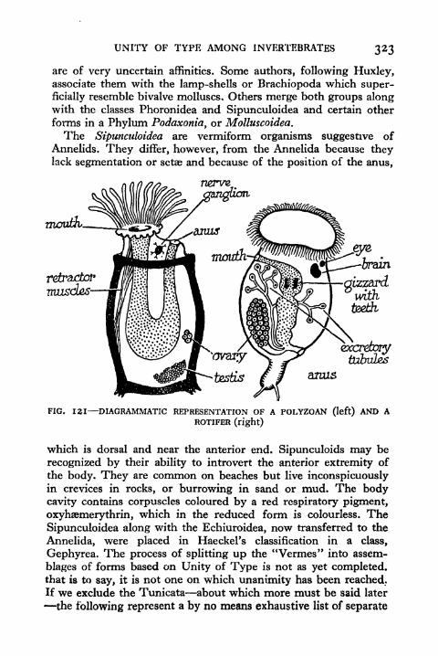
UNITY OF TYPE AMONG INVERTEBRATES 323
are of very uncertain affinities. Some authors, following Huxley,associate them with the lamp-shells or Brachiopoda which super-
ficially resemble bivalve molluscs. Others merge both groups alongwith the classes Phoronidea and Sipunculoidea and certain other
forms in a Phylum Podaxonia, or Molluscoidea.
The Sipunculoidea are vermiform organisms suggestive of
Annelids. They differ, however, from the Annelida because theylack segmentation or setae and because of the position of the anus,
FIG. 121 DIAGRAMMATIC REPRESENTATION OF A POLYZOAN (left) AND AROTIFER (right)
which is dorsal and near the anterior end. Sipunculoids may be
recognized by their ability to introvert the anterior extremity of
the body. They are common on beaches but live inconspicuouslyin crevices in rocks, or burrowing in sand or mud. The bodycavity contains corpuscles coloured by a red respiratory pigment,
oxyhaemerythrin, which in the reduced form is colourless. TheSipunculoidea along with the Echiuroidea, now transferred to the
Annelida, were placed in Haeckel's classification in a class,
Gephyrea. The process of splitting up the "Vermes" into assem-
blages of forms based on Unity of Type is not as yet completed,that is to say, it is not one on which unanimity has been reached.
If we exclude the Tunicata about which more must be said later
-the following represent a by no means exhaustive list of separate

324 PRINCIPLES OF ANIMAL BIOLOGY
phyla recognized by modern authors under HaeckePs Verrnes:
Annelida, Platyhelminthes, Nemertinea, Nemathelminthes, Roti-
fera, Brachiopoda, Polyzoa, Sipunculoidea, Phoronidea, Priapu-
loidea, Chaetognatha. But some of these terms (e.g. Polyzoa) mayinclude forms which should be further separated.
Allies of the Vertebrates. One class included in HaeckePs
Vermes is a group which has had an erratic history in
zoological classification, and like the barnacles, one whose
present position in the animal kingdom is based more on its
larval than upon its adult characteristics. The Tunicata or
sea squirts were placed at one time among the Molluscs.
This association has no justification in the light of what weknow of either their adult or larval characters. Their present
position in zoological classification, like that of the barnacles,
illustrates the extension of the Principle of Unity of Typeto the study of all stages in the development of an animal.
In Flat-worms, Nemathelminthes, Annelids, Molluscs,
and Arthropods, there is one underlying feature of similarity
in the general architecture of the body, especially in relation
to the relative positions of the C.N.S. and gut, namely,the presence of a dorsal ganglion above the mouth and a
pair of lateral nerve-cords, which tend to be approximated
ventrally below the gut, forming a circumcesphageal ring
in all except the flat-worms. In the Vertebrate phylum the
C.N.S. is wholly dorsal to the gut and arises as a median
tubular infolding of the ectoderm or outer skin of the embryo.This feature of organization together with certain minor
points is shared by a few other forms. For this reason
many authors have relegated the Vertebrata (or Craniata)
to a sub-phylum of the phylum Chordata, which is dis-
tinguished by the above characteristics, and the presence of
pharyngeal clefts and a notochord at some stages of develop-
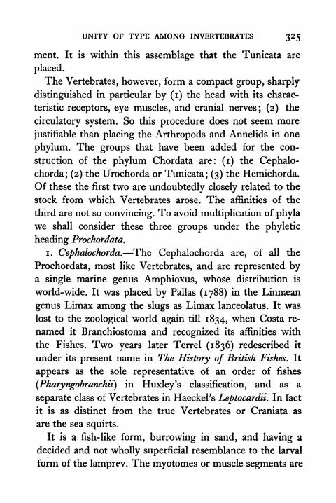
UNITY OF TYPE AMONG INVERTEBRATES 325
ment. It is within this assemblage that the Tunicata are
placed.
The Vertebrates, however, form a compact group, sharply
distinguished in particular by (i) the head with its charac-
teristic receptors, eye muscles, and cranial nerves; (2) the
circulatory system. So this procedure does not seem more
justifiable than placing the Arthropods and Annelids in one
phylum. The groups that have been added for the con-
struction of the phylum Chordata are: (i) the Cephalo-
chorda; (2) the Urochorda or Tunicata; (3) the Hemichorda.
Of these the first two are undoubtedly closely related to the
stock from which Vertebrates arose. The affinities of the
third are not so convincing. To avoid multiplication of phyla
we shall consider these three groups under the phyletic
heading Prochordata.
i. Cephalochorda. The Cephalochorda are, of all the
Prochordata, most like Vertebrates, and are represented bya single marine genus Amphioxus, whose distribution is
world-wide. It was placed by Pallas (1788) in the Linnsean
genus Limax among the slugs as Limax lanceolatus. It was
lost to the zoological world again till 1834, when Costa re-
named it Branchiostoma and recognized its affinities with
the Fishes. Two years later Terrel (1836) redescribed it
under its present name in The History of British Fishes. It
appears as the sole representative of an order of fishes
(Pharyngobranchii) in Huxley's classification, and as a
separate class of Vertebrates in Haeckel's Leptocardii. In fact
it is as distinct from the true Vertebrates or Craniata as
are the sea squirts.
It is a fish-like form, burrowing in sand, and having a
decided and not wholly superficial resemblance to the larval
form of the lamprev. The myotomes or muscle segments are
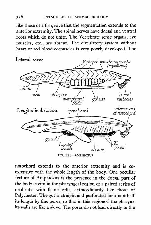
326 PRINCIPLES OF ANIMAL BIOLOGY
like those of a fish, save that the segmentation extends to the
anterior extremity. The spinal nerves have dorsal and ventral
roots which do not unite. The Vertebrate sense organs, eye
muscles, etc., are absent. The circulatory system without
heart or red blood corpuscles is very poorly developed. The
Lateral vuzwV-shaped muscle, segments
pouch, airuzm,
FIG. 122 AMPHIOXUS
notochord extends to the anterior extremity and is co-
extensive with the whole length of the body. One peculiar
feature of Amphioxus is the presence in the dorsal part of
the body cavity in the pharyngeal region of a paired series of
nephridia with flame cells, extraordinarily like those of
Polychaetes. The gut is straight and perforated for about half
its length by fine pores, so that in this regionof the pharynxits walls are like a sieve. The pores do not lead directly to the
UNITY OF TYPE AMONG INVERTEBRATES 327
exterior but into a cavity, the atrium, formed in develop-
ment by the downgrowth of two flaps of the body wall which
meet ventrally so as to constitute a closed chamber which
communicates with the exterior by a mid-ventral aperture
behind the pharyngeal region.
The entire internal wall of the pharynx and sides of the
muscles
(jIiyOTTlTOs)
mdtaplzaraL^ atrium,
FIO. 123 AMPHIOXUS TRANSVERSE SECTION THROUGIlJciLL REGION
pores are lined with ciliated epithelium, and the cilia are so
arranged as to constitute a food-net very much like the gill
of a clam in its operation. The lateral cilia which line the
pores create a current from the mouth into the atrium. Froma ventral groove or endostyle a sheet of mucus is thrown on
to the sides of the pharynx by longitudinal rows of long cilia.
The facial cilia which line the internal walls of the pharynxwash this sheet of slime with entangled food particles up to
a dorsal groove whose cilia maintain a current backwards
towards the intestine, An endostyle like that of Amphioxus
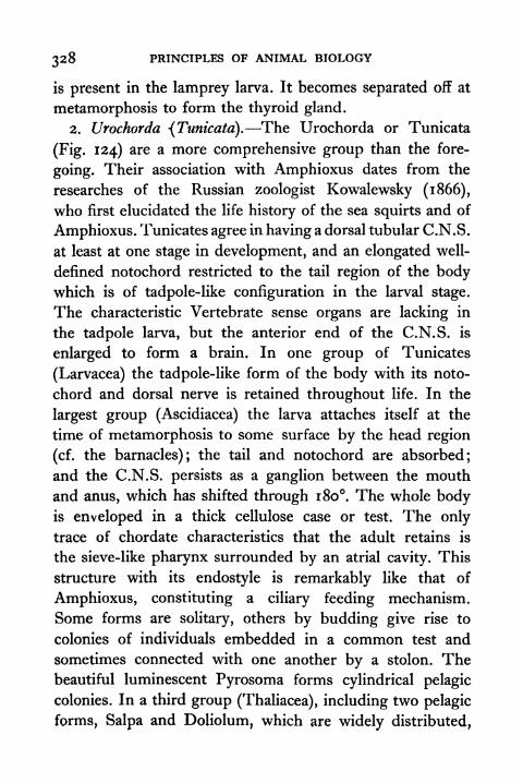
328 PRINCIPLES OF ANIMAL BIOLOGY
is present in the lamprey larva. It becomes separated off at
metamorphosis to form the thyroid gland.
2. Urochorda -(Tunicata). The Urochorda or Tunicata
(Fig. 124) are a more comprehensive group than the fore-
going. Their association with Amphioxus dates from the
researches of the Russian zoologist Kowalewsky (1866),
who first elucidated the life history of the sea squirts and of
Amphioxus. Tunicates agree in having a dorsal tubular C.N.S.
at least at one stage in development, and an elongated well-
defined notochord restricted to the tail region of the bodywhich is of tadpole-like configuration in the larval stage.
The characteristic Vertebrate sense organs are lacking in
the tadpole larva, but the anterior end of the C.N.S. is
enlarged to form a brain. In one group of Tunicates
(Larvacea) the tadpole-like form of the body with its noto-
chord and dorsal nerve is retained throughout life. In the
largest group (Ascidiacea) the larva attaches itself at the
time of metamorphosis to some surface by the head region
(cf. the barnacles); the tail and notochord are absorbed;
and the C.N.S. persists as a ganglion between the mouth
and anus, which has shifted through 180. The whole bodyis enveloped in a thick cellulose case or test. The only
trace of chordate characteristics that the adult retains is
the sieve-like pharynx surrounded by an atrial cavity. This
structure with its endostyle is remarkably like that of
Amphioxus, constituting a ciliary feeding mechanism.
Some forms are solitary, others by budding give rise to
colonies of individuals embedded in a common test and
sometimes connected with one another by a stolon. Thebeautiful luminescent Pyrosoma forms cylindrical pelagic
colonies. In a third group (Thaliacea), including two pelagic
forms, Salpa and Doliolum, which are widely distributed,
UNITY OF TYPE AMONG INVERTEBRATES 3*9
the adult form is essentially very much like a simple Asci-
dian, but it is not sedentary. These forms have a regular
alternation of sexual and asexual generations, the sexual
individuals being formed as small buds from a ventral
appendage known as the stolon. Both simple and colonial
brain (darsd ganglion)
(l)
FIG. 124 TUNICATA
(A) Colonies of a sea squirt attached to a piece of weed; (B) Longitudinalsection through single zooid of a colony; (C) (i) The tadpole-like larva
undergoing metamorphosis (2)-(3).
Ascidians are common between tide-marks on rocky shores.
The simple Ascidians (sea squirts, red-bair, etc.) are unmis-
takable objects. The colonial Ascidians are easily mistaken
for sponges.
3. Hemichorda. The Hemichorda are of much moredoubtful position than the foregoing and include two
groups. The Enteropneusta are typified by the burrowingmarine acorn-worm, Balanoglossus. The acorn-worms, distin-
guished by the conical proboscis in front of the mouth,

330 PRINCIPLES OF ANIMAL BIOLOGY
followed by a thickening of the integument known as the
collar, are placed near Amphioxus and the Tunicates, chiefly
on account of the structure of the pharynx, which is per-
forated in a manner extraordinarily similar to that of
Amphioxus. It communicates with an atrial cavity that is,
however, dorsally and not ventrally placed. A divcrticulum
from the gut extending into the proboscis region has been
compared to the notochord. The C.N.S. is represented bydorsal and ventral cords lying immediately under the epi-
dermis. The acorn-worms have a free-swimming ciliated
larva which strikingly resembles that of the sea cucumbers.
Placed near the Enteropneusta is a group of colonial orga-
nisms (Pterobranchia) superficially like Polyzoa. The zooids
have the body divided into a proboscis, collar, and trunk
like the acorn-worms. These have pharyngeal clefts and a
structure resembling the supposed notochord of Balano-
glossus. From the collar region a ring of fine tentacles
protrudes. The chordate characteristics of the Hemichorda
were first emphasized by Bateson (1884-6), whose views have
been very widely but not universally accepted by systematic
zoologists.
TOPICS FOR HOME STUDY
1 . Discuss the affinities of the Prochordata.
2. What are the structural peculiarities of:
(a) Coelenterates.
(b) Platyhelminthes.
(c) Annelids.
3. Justify the grouping of the apparently diverse Molluscan typesin one phylum.
4. Illustrate the Principle of Unity of Type by reference to
invertebrates.

CHAPTER XII
THE PRINCIPLE OF SUCCESSION WITH SPECIALREFERENCE TO THE VERTEBRATE SKELETON
T ~\ 7E have now to turn to a third step in the EvolutionaryV V argument. This is called the Principle of Geological
Succession. From ancient times, but more especially from
the end of the seventeenth century onwards, the hard remains
of animals were unearthed from time to time. For instance,
shells of molluscs which only live in water were found far
inland remote from lake, river, or sea. Such relics were
attributed by the current mythology of Christian countries to
the deluge that destroyed the contemporaries of the Noah
family, and sceptics like Voltaire, who ventured to offer more
naturalistic suggestions, were hardly more happy in their
speculations. An exception must be made in favour of Xeno-
phanes (circa 500 B.C.), and of the Arab physician Avicenna
(circa A.D. 900), who recognized fossils as remains of animals
formerly alive, and saw in them evidence of the existence of
oceans where there is now only land. A giant fossil salamander
which occurs abundantly in the Upper Miocene of Switzer-
land, closely related to the Japanese salamander Crypto-branchus japonicus, was unearthed by Scheuchzer in 1726,
and named Homo diluvii testis. The motto attached to his
figure reads :
Betriibtes Beingerust von einem alten SunderErweiche Herz und Sinn der neuen Bosheitskinder.
Which is translated (Camb. Nat. History, viii, p. 84) :
Oh, sad remains of bone, frame of poor Man of Sin,Soften the heart and mind of recent sinful kin.
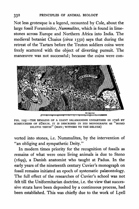
33* PRINCIPLES OF ANIMAL BIOLOGY
Not less grotesque is a legend, recounted by Cole, about the
large fossil Foraminifer, Nummulitesywhich is found in lime-
stones across Europe and Northern Africa into India. Themedieval botanist Clusius (circa 1550) says that during the
retreat of the Tartars before the Teuton soldiers coins were
freely scattered with the object of diverting pursuit. Themanoeuvre was not successful; because the coins were con-
FIG. 125 THE REMAINS OF A GIANT SALAMANDER UNEARTHED IN 1726 BYSCHEUCHZER OF ZURICH. IT IS DESCRIBED IN HIS MONOGRAPH AS "HOMO
DILUVII TESTIS" (MAN; WITNESS TO THE DELUGE)
verted into stones, i.e. Nummulites, by the intervention of
"an obliging and sympathetic Deity."
In modern times priority for the recognition of fossils as
remains of what were once living animals is due to Steno
(1699), a Danish anatomist who taught at Padua. In the
early years of the nineteenth century Cuvier's monograph on
fossil remains initiated an epoch of systematic palaeontology.
The full effect of the researches of Cuvier's school was not
felt till the Uniformitarian doctrine, i.e. the view that succes-
sive strata have been deposited by a continuous process, had
been established. This was chiefly due to the work of Lyell

THE PRINCIPLE OF SUCCESSION 333
(1830). The impiety of this new Geology did not escape
challenge. In the minutes of a meeting of the Geological
iwti&rY___
Cretaceous-_____
Jura&sv?___
"Permian,__
Carboniferous-
Dzrauan,
SiZuria/i
FIG. 126 THE SUCCESSION OF VERTEBRATES
Society of London in 1840, we read that the retiring
president, Dr. Buckland,
with a look and tone of triumph pronounced upon his opponentswho dared to question the orthodoxy of the scratches and groovesof the glacial mountains the pains of eternal itch without the
privilege of scratching (Hist* of Geol. Soc. of Lond., p. 142).

334 PRINCIPLES OF ANIMAL BIOLOGY
In what follows we shall assume the view which is now
universally accepted. The various strata of which the earth's
crust is composed have been laid down in orderly succession
during periods of time compared with which that occupied
by the history of human society is of negligible duration.
Once this conclusion was accepted the study of fossils
acquired a new interest and progressed rapidly under the
leadership of men like Owen, Cope, and their contem-
poraries. Students of fossils set themselves to compare the
characteristics of animals in different geological epochs, and
to elucidate evidences of a continuous succession of new
forms of life handed down to posterity in the record of the
rocks. Out of their studies the Principle of Succession took
shape.
The Principle of Succession may be stated in two ways.
One is that many of the more highly specialized and successful
groups of the present day did not exist at earlier periods of
the earth's history, and were preceded by forms which are
intermediate between them and representatives of surviving
groups that already existed before them. Another is that the
earliest members of the great groups usually exhibit a more
generalized type of structure than existing types. Adequatematerial for drawing these conclusions is provided only byforms which had resistant structures such as the Vertebrates,
Arthropods, and Vascular plants.
The earliest dominant group of Arthropods were the
Trilobites (Fig. 128) which appear in the lower Cambrian. Afew Crustacean types are found in mid-Cambrian rocks. Theearliest Arachnida are marine types of which some resemble
the king crab Limulus The dominant Silurian Arachnid
group was the Eurypterida, which were structurally inter-
mediate between Limulus and land scorpions. Typical land

THE PRINCIPLE OF SUCCESSION 335
'
''!/' ,-
FIG. 127 REMAINS OF ARCtLffiOPTERYX PRESERVED IN SHALE FROMSOLENHOVEN, BAVARIA

336 PRINCIPLES OF ANIMAL BIOLOGY
Arthropods, i.e. insects and spiders, appear in the coal
measures. Land scorpions which are more generalized than
anienna
uppersurface,
FIG. 128 TRILOBITE
surface.
The earliest representatives of the phylum Arthropoda found in the oldest
sedimentary rocks are the Trilobites, a group which partake of the
characteristics of all the more specialized classes which evolved later. Thusthey have a single pair of feelers like insects, bifid swimming limbs like
Crustacea, and no specialized mandibles or biting jaws such as are presentin insects and Crustacea but absent in the arachnids. The Principle of
Succession is well illustrated in Trilobites, owing to the fact that all partsof the body are enclosed in a hard case which readily leaves its impressionon the sediment where it lies or becomes fossilized.
spiders and other land Arachnids had already turned up in
the preceding epoch. If the Principle of Succession is true we
THE PRINCIPLE OF SUCCESSION 337
should expect that Trilobites are Arthropods of a more
generalized type than members of any existing classes.
Whether this is so can best be seen by asking what charac-
teristics a generalized Arthropod would have.
In modern Arthropods at least jive anterior segments are
fused to form a head. The head of many Crustacea, of the
marine Arachnid Limulus, of most insects, and of the centi-
pede Scutigera bears a pair of compound eyes. Behind the
head the succeeding segments may be differentiated in various
ways in different types. In Myriapods they are similar and
this is true of some Crustacea. The abdominal appendages of
Limulus and the mouth parts of insects and Myriapods are
biramous structures and this is the general characteristic of
Crustacean Swimmerets. Myriapods, Insects, and Crustacea
have at least one pair of antenniform appendages in front of
the mouth. The biting mandibles of insects correspond to
the basal portion of a limb, which retains a vestigial axis in
millipedes and in many Crustacea. In Arachnids several
appendages behind the mouth have biting bases (gnathites),
and no one is completely subordinated to a masticatory
function. We may therefore reconstruct a generalized Arthro-
pod type with a head composed of five segments with a pair
of feelers and compound eyes, succeeded by a number of
similar segments bearing biramous appendages with gnathites.
This in fact is a precise specification of the characteristic
features of the now extinct Trilobita.
Fossil remains of the skeletons of Vertebrates provide one
specimen which illustrates the Principle without recourse
to specialized knowledge. Archaopteryx, the first undoubted
bird which is beautifully preserved in the Bavarian litho-
graphic limestone, has many striking reptilian features
(Fig. 137). The forelimb has three complete clawed digits
Y
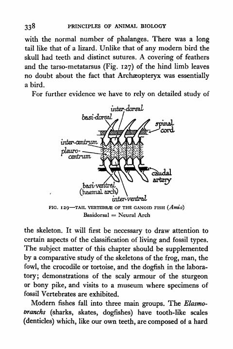
338 PRINCIPLES OF ANIMAL BIOLOGY
with the normal number of phalanges. There was a long
tail like that of a lizard. Unlike that of any modern bird the
skull had teeth and distinct sutures. A covering of feathers
and the tarso-metatarsus (Fig. 127) of the hind limb leaves
no doubt about the fact that Archaeopteryx was essentially
a bird.
For further evidence we have to rely on detailed study of
FIG. 129 TAIL VERTEBRAE OF THE GANOID FISH (Amttl)
Basidorsal = Neural Arch
the skeleton. It will first be necessary to draw attention to
certain aspects of the classification of living and fossil types.
The subject matter of this chapter should be supplemented
by a comparative study of the skeletons of the frog, man, the
fowl, the crocodile or tortoise, and the dogfish in the labora-
tory; demonstrations of the scaly armour of the sturgeonor bony pike, and visits to a museum where specimens of
fossil Vertebrates are exhibited.
Modern fishes fall into three main groups. The Elasmo-
toranchs (sharks, skates, dogfishes) have tooth-like scales
(denticles) which, like our own teeth, are composed of a hard

THE PRINCIPLE OF SUCCESSION 339
tissue called dentine overlaid by enamel. The endo-skeleton is
entirely cartilaginous. They have no operculum nor swim
bladder. The Teleostei or true bony fishes are a second groupwith the largest number of species. They appear late in the
dorsal
inter-
wntral
FIG. 130 VERTEBRflE OF AN ELASMOBRANCH (ACANTHIAS)
Jurassic epoch. The scales are thin plates of bone without
dentine or enamel. The endo-skeleton is bony. They have an
operculum, or gill-cover, and a swim bladder. They have no
spiracle. The so-called Ganoid fishes are a miscellany of sur-
vivors from very ancient stocks. Their scales are of true bone
overlaid with dentine and enamel. They have spiracles, a

340 PRINCIPLES OF ANIMAL BIOLOGY
swim-bladder which may be double, and an operculum. The
sturgeons (Chondrostei), with three living genera, are most
like the sharks. The swim-bladder of sturgeons, like that of
Teleostei,isnot a respiratory organ. That of the other Ganoids
zygapophysis
persisbzrdb
zygapophysis
transverse
process
capiiulum
FIG. 131 TRUNK VERTEBRA OF A STEGOCEPHALIAN
is a lung supplied from the last branchial artery. In one group,
the Lung fishes (Dipnoi), made up of three living genera, the
atrium is completely divided into right and left auricles, of
which the left one receives the pulmonaryvein (Fig. 55, p. 144).
They have a true postcaval vein (Fig. 60, p. 150). Thustheir circulatory arrangements are essentially similar to those
of salamanders. The remaining Ganoids are placed in four
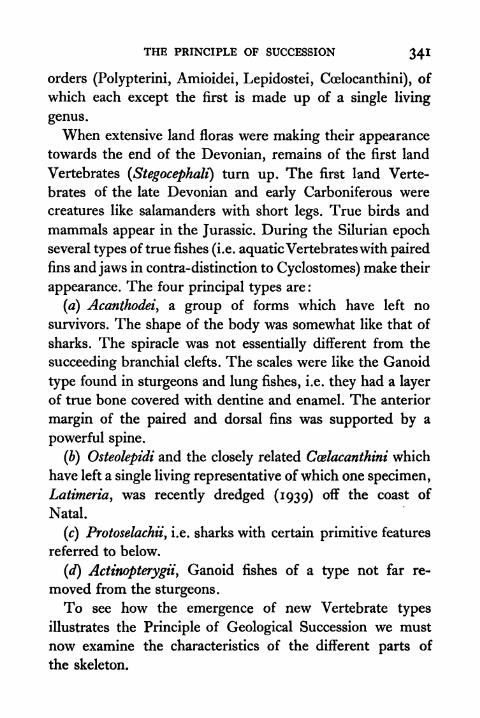
THE PRINCIPLE OF SUCCESSION 34!
orders (Polypterini, Amioidei, Lepidostei, Coelocanthini), of
which each except the first is made up of a single living
genus.
When extensive land floras were making their appearancetowards the end of the Devonian, remains of the first land
Vertebrates (Stegocephalf) turn up. The first land Verte-
brates of the late Devonian and early Carboniferous were
creatures like salamanders with short legs. True birds and
mammals appear in the Jurassic. During the Silurian epochseveral types of true fishes (i.e. aquaticVertebrateswith paired
fins and jaws in contra-distinction to Cyclostomes) make their
appearance. The four principal types are :
(a) Acanthodei9a group of forms which have left no
survivors. The shape of the body was somewhat like that of
sharks. The spiracle was not essentially different from the
succeeding branchial clefts. The scales were like the Ganoid
type found in sturgeons and lung fishes, i.e. they had a layer
of true bone covered with dentine and enamel. The anterior
margin of the paired and dorsal fins was supported by a
powerful spine.
(b) Osteolepidi and the closely related Ccelacanthini which
have left a single living representative of which one specimen,
Latimeria, was recently dredged (1939) off the coast of
Natal.
(c) Protoselachiiy i.e. sharks with certain primitive features
referred to below.
(d) Actinopterygiiy Ganoid fishes of a type not far re-
moved from the sturgeons.
To see how the emergence of new Vertebrate types
illustrates the Principle of Geological Succession we must
now examine the characteristics of the different parts of
the skeleton.

342 PRINCIPLES OF ANIMAL BIOLOGY
(i) TheAxial Skeleton. In a land Vertebrate each vertebra
consists of the following parts (Fig. 131). Over the spinal
cord is the neural arch with its characteristic articular surfaces
(zygapophyses) interlocking with those of the succeeding or
preceding vertebra. The posterior pair overlap the anterior
pair of the next vertebra behind. So the front face of a
vertebra can be recognized by the fact that the articular
surface of the zygapophysis faces upwards. On either side of
the base of the neural arch are the transverse processes with
which the ribs articulate by their tubercular extremities. The
ribs of modern Amphibia are cartilaginous vestiges with a
single head. Among birds, mammals, and some reptiles the
rib has a second or capitular head which rests against the
body or centrum of the vertebra. The latter is built up round
the notochord of the embryo, and completely replaces it in
adult life as the supporting axis of the trunk. In some
salamanders, and in the New Zealand lizard Sphenodon, both
articular faces of the centrum are concave (amphicoelous type)
and the remains of the notochord persist between them.
Most other Amphibia and Reptiles have centra which fit
tightly by a ball and socket joint. Usually the anterior face is
concave and the posterior convex (proccelous type). Mammalshave centra with flat faces. Modern birds have centra of
which each articular surface is saddle-shaped (heterocoelous
type).
At the opposite end of the Vertebrate series the axial
skeleton of Cyclostomes is represented only by the persistent
unconstricted notochord and by V-shaped cartilaginous
neural arches which lie over the spinal cord. Between this
state of affairs and the solid type vertebra characteristic
of land Vertebrates, true fishes provide examples of various
grades. In some of the Ganoids (sturgeons and lung fishes)
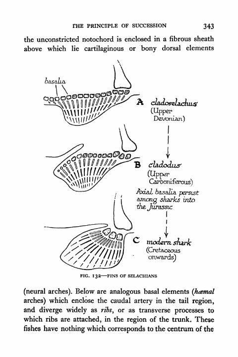
THE PRINCIPLE OF SUCCESSION 343
the unconstricted notochord is enclosed in a fibrous sheath
above which lie cartilaginous or bony dorsal elements
(UpperDevonian)
CarooTuferous)
a 6asaZia persisti among sharks into
\ -the, Jurassic
onwards)
FIG. 132-FINS OF SELACHIANS
(neural arches). Below are analogous basal elements (htemal
arches) which enclose the caudal artery in the tail region,
and diverge widely as ribs, or as transverse processes to
which ribs are attached, in the region of the trunk. These
fishes have nothing which corresponds to the centrum of the
344 PRINCIPLES OF ANIMAL BIOLOGY
land Vertebrate. The vertebrae of Teleostei and of Elasmo-
branchs are composite (Fig. 130). They have centra of the
amphicoelous type found among salamanders and in the NewZealand lizard Sphenodon. In Elasmobranchs each vertebra
has a complete neural arch which is obliquely placed above
the centrum and occupies about half the overlying space in
front or behind. Behind successive arches are interdorsal
plates which fill up the gap over the spinal cord. Small inter-
ventral plates below the centrum may also alternate with
haemal arches. In the tail region of the Ganoid Amia each
vertebra is made up of a pair of centra, a pair of dorsal
(neural), and a pair of ventral (haemal) elements (Fig. 129).
The salient points which emerge from this survey of the
axial skeleton of living Vertebrates are : (a) that in general
the vertebrae of fishes exhibit a structural duplicity which is
not found among any living land Vertebrates; and (b) that
since it is also common among fishes, the amphicoelous
vertebra is a more generalized type than others found among
living land Vertebrates. If then the Principle of Succession
is supported by the evidence derived from the peculiarities of
the axial skeleton in fossil types, two conclusions follow:
(a) the earliest land Vertebrates should have double verte-
brae; and (b) the earliest representatives of the land Verte-
brate groups should have amphicoelous vertebrae. Both these
statements are true. The Stegocephali have vertebrae which
are very different from those of living land Vertebrates. Theyhave double centra (Fig. 131). All the earliest land Verte-
brates, including the first fossil birds, had amphicoelouscentra.
(ii)The Limb Skeleton. From the standpoint of limb
specialization Vertebrates fall into three grades : (a) Cyclo-
stomes, which are limbless and have no structural charac-
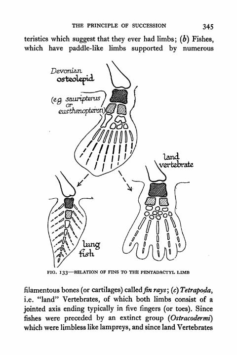
THE PRINCIPLE OF SUCCESSION 345
teristics which suggest that they ever had limbs; (b) Fishes,
which have paddle-like limbs supported by numerous
landx \wrtebrale
FIG. 133 RELATION OF FINS TO THE PENTADACTYL LIMB
filamentous bones (or cartilages) calledyw rays\ (c) Tetrapoda,
land" Vertebrates, of which both limbs consist of ai.e.
jointed axis ending typically in five fingers (or toes). Since
fishes were preceded by an extinct group (Ostracodermi)
which were limbless like lampreys, and since land Vertebrates

346 PRINCIPLES OF ANIMAL BIOLOGY
did not appear till fishes had come into being, the record of
the rocks raises two problems from the present standpoint.
One is whether we find among the earliest fishes intermediate
types which bridge the gulf between modern fishes and limb-
less types. The other is whether we find among the earliest
fishes and land Vertebrates intermediate types which bridge
the gulf between the paddle-like fin and the axiate five-
fingered leg.
The first question raises a difficulty at the outset, unless
we are able to form a likely view of what such an intermediate
type would be like. Two facts about the structure of fishes
had suggested a hypothesis which has since been supported
by the discovery of fossil types. One is that the skeletal and
integumentary structure of the paired fins with their fin rays
is remarkably like that of the dorsal fin and the ventral fin
of the caudal region. Either of these may be continuous or
broken up into discrete portions. This suggests the possible
existence of a more generalized fish type in which a lateral
fin extended on either side along the trunk region. The other
fact which lends colour to this suggestion is an essential
difference between the organization of a fish and that of
Amphioxus or of the lamprey. In the caudal region of fishes
separate muscle bands (Myotomes or Myomeres) ensheathed
in fibrous tissue correspond to each pair of spinal nerves
and to each vertebra. The structure of the trunk region is
not segmental. Several pairs of spinal nerves fuse to form
the main nerves of the legs of a Tetrapod, and a regular
succession of muscle bands corresponding to each pair of
nerves in the fish embryo cannot be distinguished in the
region anterior to the tail. To bridge the gap between fishes
and fish-like animals in which a regular arrangement of nerve
and muscle segments continues in front of the tail region,
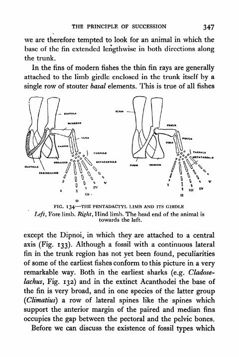
THE PRINCIPLE OF SUCCESSION 347
we are therefore tempted to look for an animal in which the
base of the fin extended lengthwise in both directions along
the trunk.
In the fins of modern fishes the thin fin rays are generally
attached to the limb girdle enclosed in the trunk itself by a
single row of stouter basal elements. This is true of all fishes
._ SCAPULA
UtTACARPAL*
FIG. 134 THE PENTADACTYL LIMB AND ITS GIRDLE
Left y Fore limb. Right, Hind limb. The head end of the animal is
towards the left.
except the Dipnoi, in which they are attached to a central
axis (Fig. 133). Although a fossil with a continuous lateral
fin in the trunk region has not yet been found, peculiarities
of some of the earliest fishes conform to this picture in a veryremarkable way. Both in the earliest sharks (e.g. Cladose-
lachus, Fig. 132) and in the extinct Acanthodei the base of
the fin is very broad, and in one species of the latter group
(Climatius) a row of lateral spines like the spines which
support the anterior margin of the paired and median fins
occupies the gap between the pectoral and the pelvic bones.
Before we can discuss the existence of fossil types which

348 PRINCIPLES OF ANIMAL BIOLOGY
bridge the gap between the fish fin and the pentadactyl limb
of land Vertebrates, we must first study the general charac-
teristics of the latter. The typical pentadactyl limb has
characteristics common to both the fore and hind member.
The names used are indicated in Fig. 134. The limb girdle
consists of three paired elements, one which is dorsal
(scapula or ilium) corresponds to the main part of the fore-
limb girdle of a fish. The other two are ventral, an anterior
U^ferc/ayic/ (epistermun)
spine.scapula*
dawcLe
coraaoid
FIG. 135 SHOULDER GIRDLE OF A MONOTREME (left) AND TYPICALMAMMAL (right)
pair (precoracoids or pubes) and a posterior pair (coracoids or
ischid). All three elements are always present in the posterior
or pelvic limb girdle of a land Vertebrate. They are generally
present in the pectoral girdle of Amphibia and of lizards. In
crocodiles and birds only one ventral element is present, and
it is not certain whether it corresponds to the precoracoidor the coracoid of lizards. With the exceptions mentioned
below the adult mammalian shoulder girdle has no separate
precoracoids or coracoids. A vestigial coracoid may be fused
with the articular facet of the scapula, which has a charac-
teristic median ridge, called the scapular spine.
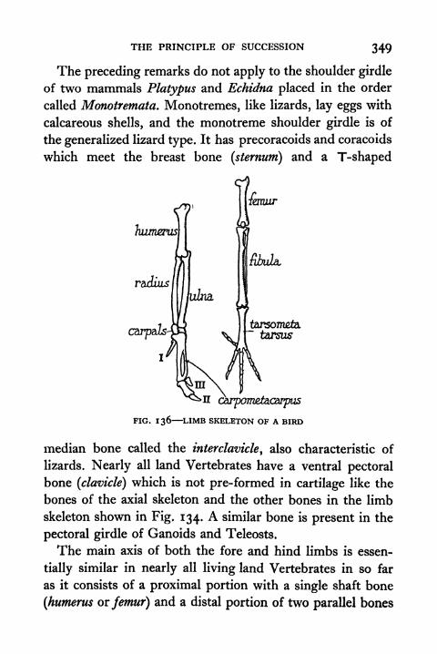
THE PRINCIPLE OF SUCCESSION 349
The preceding remarks do not apply to the shoulder girdle
of two mammals Platypus and Echidna placed in the order
called Monotremata. Monotremes, like lizards, lay eggs with
calcareous shells, and the monotreme shoulder girdle is of
the generalized lizard type. It has precoracoids and coracoids
which meet the breast bone (sternum) and a T-shaped
remur
tarsontefei"tarsus
FIG. 136 LIMB SKELETON OF A BIRD
median bone called the interclavicle, also characteristic of
lizards. Nearly all land Vertebrates have a ventral pectoral
bone (clavicle) which is not pre-formed in cartilage like the
bones of the axial skeleton and the other bones in the limb
skeleton shown in Fig. 134. A similar bone is present in the
pectoral girdle of Ganoids and Teleosts.
The main axis of both the fore and hind limbs is essen-
tially similar in nearly all living land Vertebrates in so far
as it consists of a proximal portion with a single shaft bone
(humerus or femur) and a distal portion of two parallel bones
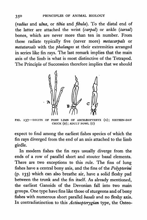
350 PRINCIPLES OF ANIMAL BIOLOGY
(radius and ulna, or tibia and fibula). To the distal end of
the latter are attached the wrist (carpal) or ankle (tarsal)
bones, which are never more than ten in number. From
these radiate typically five (never more) metacarpals or
metatarsals with the phalanges at their extremities arranged
in series like fin rays. The last remark implies that the main
axis of the limb is what is most distinctive of the Tetrapod.
The Principle of Succession therefore implies that we should
FIG. 137 DIGITS OF FORE LIMB OF ARCKffiOPTERYX (A); SIXTEEN-DAYCHICK (B) J
ADULT FOWL (c)
expect to find among the earliest fishes species of which the
fin rays diverged from the end of an axis attached to the limb
girdle.
In modern fishes the fin rays usually diverge from the
ends of a row of parallel short and stouter basal elements.
There are two exceptions to this rule. The fins of lungfishes have a central bony axis, and the fins of the Polypterini
(p. 133) which can also breathe air, have a solid fleshy padbetween the trunk and the fin itself. As already mentioned,
the earliest Ganoids of the Devonian fall into two main
groups. One type have fins like those of sturgeons and of bonyfishes with numerous short parallel basals and no fleshy axis.
In contradistinction to this Actinopterygian type, the Osteo-
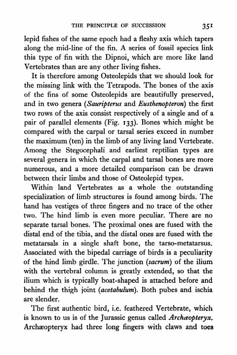
THE PRINCIPLE OF SUCCESSION 351
lepid fishes of the same epoch had a fleshy axis which tapers
along the mid-line of the fin. A series of fossil species link
this type of fin with the Dipnoi, which are more like land
Vertebrates than are any other living fishes.
It is therefore among Osteolepids that we should look for
the missing link with the Tetrapods. The bones of the axis
of the fins of some Osteolepids are beautifully preserved,
and in two genera (Sauripterus and Eusthenopterori) the first
two rows of the axis consist respectively of a single and of a
pair of parallel elements (Fig. 133). Bones which might be
compared with the carpal or tarsal series exceed in number
the maximum (ten) in the limb of any living land Vertebrate.
Among the Stegocephali and earliest reptilian types are
several genera in which the carpal and tarsal bones are more
numerous, and a more detailed comparison can be drawn
between their limbs and those of Osteolepid types.
Within land Vertebrates as a whole the outstanding
specialization of limb structures is found among birds. Thehand has vestiges of three fingers and no trace of the other
two. The hind limb is even more peculiar. There are no
separate tarsal bones. The proximal ones are fused with the
distal end of the tibia, and the distal ones are fused with the
metatarsals in a single shaft bone, the tarso-metatarsus.
Associated with the bipedal carriage of birds is a peculiarity
of the hind limb girdle. The junction (sacrum) of the ilium
with the vertebral column is greatly extended, so that the
ilium which is typically boat-shaped is attached before and
behind the thigh joint (acetabulum). Both pubes and ischia
are slender.
The first authentic bird, i.e. feathered Vertebrate, which
is known to us is of the Jurassic genus called Archceopteryx.
Archaeopteryx had three long fingers with claws and toes
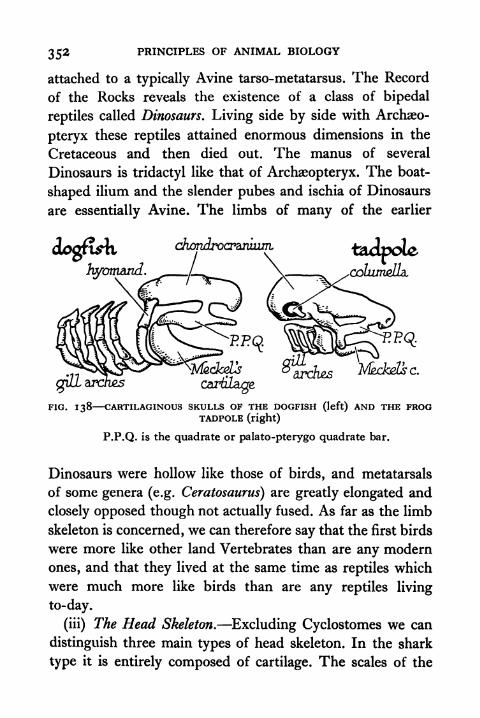
352 PRINCIPLES OF ANIMAL BIOLOGY
attached to a typically Avine tarso-metatarsus. The Record
of the Rocks reveals the existence of a class of bipedal
reptiles called Dinosaurs. Living side by side with Archaeo-
pteryx these reptiles attained enormous dimensions in the
Cretaceous and then died out. The manus of several
Dinosaurs is tridactyl like that of Archaeopteryx. The boat-
shaped ilium and the slender pubes and ischia of Dinosaurs
are essentially Avine. The limbs of many of the earlier
cartilage
FIG. 138 CARTILAGINOUS SKULLS OF THE DOGFISH (left) AND THE FROGTADPOLE (right)
P.P.Q. is the quadrate or palato-pterygo quadrate bar.
Dinosaurs were hollow like those of birds, and metatarsals
of some genera (e.g. Ceratosaurus) are greatly elongated and
closely opposed though not actually fused. As far as the limb
skeleton is concerned, we can therefore say that the first birds
were more like other land Vertebrates than are any modern
ones, and that they lived at the same time as reptiles which
were much more like birds than are any reptiles living
to-day.
(iii) The Head Skeleton. Excluding Cyclostomes we can
distinguish three main types of head skeleton. In the shark
type it is entirely composed of cartilage. The scales of the

THE PRINCIPLE OF SUCCESSION 353
skin do not contribute to it except in so far as the jaws are
fringed with thick-set denticles. It consists of the following
parts: (a) a brain-box or chondrocranium\ (b) the upper
(quadrate bar) and lower (MeckeVs cartilage) jaws ; (c) visceral
arches of serial cartilages behind the spiracles and gill clefts.
IG. 139 THE CHONDRIAL BONES OF THE MAMMALIAN SKULL
(i) Otic; (2) Basisphenoid ; (3) Presphenoid; (4) Ethmoid; (5) Orbito-
sphenoid; (6) Alisphenoid (= epipterygoid) ; (7) Exoccipital; (8) Supra-occipital; (9) Incus (
= Quadrate); (10) Malleus (= Articular); (n) Basi-
occipital; (12) Stapes (= hyomandibular or colurnella auris).
The head skeleton of a tadpole is of the same type, and
vertebrate embryos in general pass through an initial stage
which is essentially similar. In the Ganoid type the cartila-
ginous constituents mentioned already are generally con-
verted into true bone in the course of development. Thechondrocranium does not ossify as one piece. The entire
head region including the inner and outer surface of the
lower jaw, the operculum and the roof of the mouth, is
Z
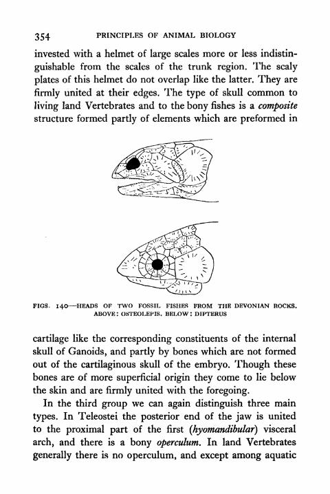
354 PRINCIPLES OF ANIMAL BIOLOGY
invested with a helmet of large scales more or less indistin-
guishable from the scales of the trunk region. The scaly
plates of this helmet do not overlap like the latter. They are
firmly united at their edges. The type of skull common to
living land Vertebrates and to the bony fishes is a composite
structure formed partly of elements which are preformed in
FIGS. 140 HEADS OF TWO FOSSIL FISHES FROM THE DEVONIAN ROCKS.ABOVE I OSTEOLEPIS. BELOW I DIPTERUS
cartilage like the corresponding constituents of the internal
skull of Ganoids, and partly by bones which are not formed
out of the cartilaginous skull of the embryo. Though these
bones are of more superficial origin they come to lie below
the skin and are firmly united with the foregoing.
In the third group we can again distinguish three main
types. In Teleostei the posterior end of the jaw is united
to the proximal part of the first (hyomandibular) visceral
arch, and there is a bony operculum. In land Vertebrates
generally there is no operculum, and except among aquatic
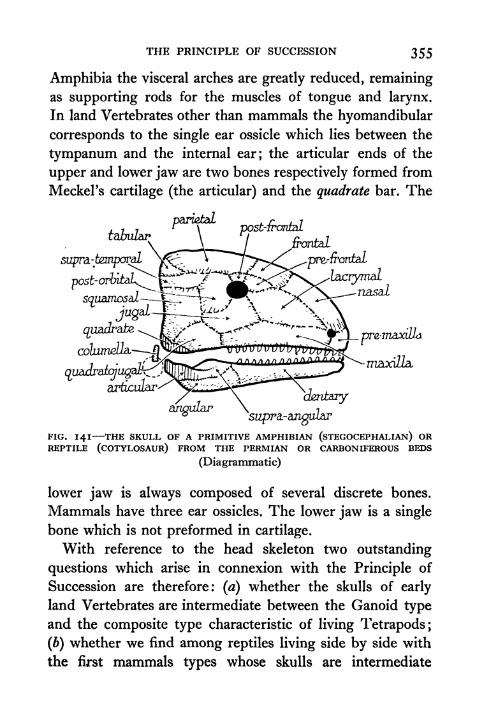
THE PRINCIPLE OF SUCCESSION 355
Amphibia the visceral arches are greatly reduced, remainingas supporting rods for the muscles of tongue and larynx.
In land Vertebrates other than mammals the hyomandibular
corresponds to the single ear ossicle which lies between the
tympanum and the internal ear; the articular ends of the
upper and lower jaw are two bones respectively formed from
Meckel's cartilage (the articular) and the quadrate bar. The
fc&zZarPai^al
PPst-"
supra-temporal
po^or&^E5^:/-"'jif>c3K^ZacrymaiP
apun^dfeqU P^V^--"^if 7 r"~ " '
/ x -\ *"
jugu-4~r-~-*^^ \
rT^E^^ss-^"1^columdla. ^Qjafci-""- VUOVcrwPl/i/uui^W
. ./'-t^fs^ ^ .iniiin^nnnnnn/Tx^ ma;)(^articular-
angular
FIG. 141 THE SKULL OF A PRIMITIVE AMPHIBIAN (STEGOCEPHALIAN) ORREPTILE (COTYLOSAUR) FROM THE PERMIAN OR CARBONIFEROUS BEDS
(Diagrammatic)
lower jaw is always composed of several discrete bones.
Mammals have three ear ossicles. The lower jaw is a single
bone which is not preformed in cartilage.
With reference to the head skeleton two outstanding
questions which arise in connexion with the Principle of
Succession are therefore: (a) whether the skulls of early
land Vertebrates are intermediate between the Ganoid type
and the composite type characteristic of living Tetrapods;
(b) whether we find among reptiles living side by side with
the first mammals types whose skulls are intermediate
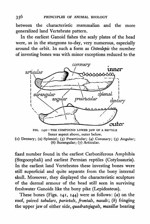
356 PRINCIPLES OF ANIMAL BIOLOGY
between the characteristic mammalian and the more
generalized land Vertebrate pattern.
In the earliest Ganoid fishes the scaly plates of the head
were, as in the sturgeons to-day, very numerous, especially
around the orbit. In such a form as Osteolepis the numberof investing bones was with minor exceptions reduced to the
coronary
articularinner
FIG. 142 THE COMPOUND LOWER JAW OF A REPTILE
Inner aspect above, outer below.
(i) Dentary; (2) Splenial; (3) Prearticular; (4) Coronary; (5) Angular;(6) Surangular; (7) Articular.
fixed number found in the earliest Carboniferous Amphibia
(Stegocephali) and earliest Permian reptiles (Cotylosauria).
In the earliest land Vertebrates these investing bones were
still superficial and quite separate from the bony internal
skull. Moreover, they displayed the characteristic sculpture
of the dermal armour of the head still seen in surviving
freshwater Ganoids like the bony pike (Lepidosteus).
These bones (Figs. 141, 144) were as follows: (a) on the
roof, paired tabulars, parietals, frontals, nasals ; (b) fringing
the upper jaw of either side, quadratojugals, maxilla bearing

THE PRINCIPLE OF SUCCESSION 357
teeth and premaxillce also bearing teeth; (c) around the
orbit postfrontah, postorbitak, jugals, lacrymals, and pre-
frontals ; (d) linking the dorsal-aspect of the head to the angle
of the jaw, supratemporals and squamosals ; (e) investing the
J.//ZX
tymparuc
FIG. 143 COMPARISON OF SKULLS OF REPTILES AND MAMMAL(Diagrammatic)
(A) Upper Permian Therapsid ; (B) Lower Triassic Therapsid ; (C) LateTriassic Therapsid; (D) Mammal. See Fig. 141 for names of bones.
outer-aspect of the lower jaw, supra-angular's, angulars, and
dentaries (the latter alone tooth-bearing). To this must be
added certain elements that are not superficial in position,
but arise from the skin and are not performed in cartilage.
In the roof of the mouth there are three pairs of teeth-
bearing bones, the palatines, transpalatines, and pterygoids,
which invest the anterior end of the quadrate cartilage.
Anterior to these on the roof of the mouth are a pair of
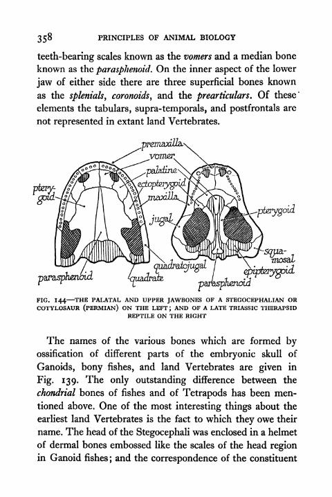
358 PRINCIPLES OF ANIMAL BIOLOGY
teeth-bearing scales known as the vomers and a median bone
known as the parasphenoid. On the inner aspect of the lower
jaw of either side there are three superficial bones known
as the splenials, coronoids, and the prearticulars. Of these*
elements the tabulars, supra-temporals, and postfrontals are
not represented in extant land Vertebrates.
FIG. 144 THE PALATAL AND UPPER JAWBONES OF A STEGOCEPHALIAN ORCOTYLOSAUR (PERMIAN) ON THE LEFT ; AND OF A LATE TRIASSIC THERAPSID
REPTILE ON THE RIGHT
The names of the various bones which are formed byossification of different parts of the embryonic skull of
Ganoids, bony fishes, and land Vertebrates are given in
Fig- *39- The only outstanding difference between the
chondrial bones of fishes and of Tetrapods has been men-
tioned above. One of the most interesting things about the
earliest land Vertebrates is the fact to which they owe their
name. The head of the Stegocephali was enclosed in a helmet
of dermal bones embossed like the scales of the head region
in Ganoid fishes; and the correspondence of the constituent

THE PRINCIPLE OF SUCCESSION 359
elements enumerated in the previous paragraph to those of
the scaly helmet of Ganoids is easily recognizable. Thus the
Stegocephali did not have a composite skull like that of all
modern Amphibia. The skulls of modern Amphibia are
much less like those of a Ganoid fish than are those of the
MALLEUS
TYMPANIC'' RING
~ 'MANDIBLE
', .MECKEl/8CARTILAGE
INCUS*
FIG. 145 HEAD OF HUMAN FCETUS
Showing the two extra ear ossicles: Malleus (articular) and Incus
(quadrate).
first fossil Amphibian types. We may indeed go farther and
say that the skulls of the first land Vertebrates were far more
like the skulls of Ganoid fishes than like those of living land
Vertebrates.
In the Permian appear land Vertebrates with more massive
limbs and limb girdles provided with well-developed ribs
which invest the trunk to form a bony chest. These earliest
reptilian forms, or Cotylosauria, of which Pariasaurus is an
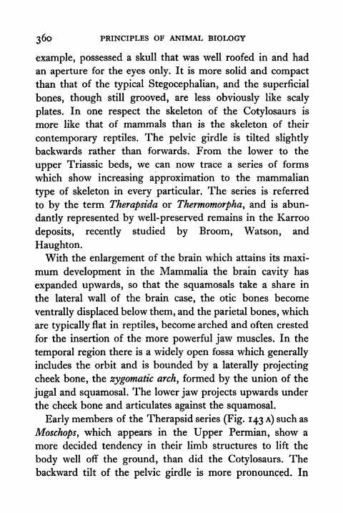
360 PRINCIPLES OF ANIMAL BIOLOGY
example, possessed a skull that was well roofed in and had
an aperture for the eyes only. It is more solid and compactthan that of the typical Stegocephalian, and the superficial
bones, though still grooved, are less obviously like scaly
plates. In one respect the skeleton of the Cotylosaurs is
more like that of mammals than is the skeleton of their
contemporary reptiles. The pelvic girdle is tilted slightly
backwards rather than forwards. From the lower to the
upper Triassic beds, we can now trace a series of forms
which show increasing approximation to the mammalian
type of skeleton in every particular. The series is referred
to by the term Therapsida or Thermomorpha, and is abun-
dantly represented by well-preserved remains in the Karroo
deposits, recently studied by Broom, Watson, and
Haughton.With the enlargement of the brain which attains its maxi-
mum development in the Mammalia the brain cavity has
expanded upwards, so that the squamosals take a share in
the lateral wall of the brain case, the otic bones become
ventrally displaced below them, and the parietal bones, which
are typically flat in reptiles, become arched and often crested
for the insertion of the more powerful jaw muscles. In the
temporal region there is a widely open fossa which generally
includes the orbit and is bounded by a laterally projecting
cheek bone, the zygomatic arch, formed by the union of the
jugal and squamosal. The lower jaw projects upwards under
the cheek bone and articulates against the squamosal.
Early members of the Therapsid series (Fig. 143 A) such as
Moschops, which appears in the Upper Permian, show a
more decided tendency in their limb structures to lift the
body well off the ground, than did the Cotylosaurs. Thebackward tilt of the pelvic girdle is more pronounced. In
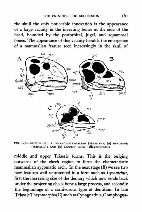
THE PRINCIPLE OF SUCCESSION 361
the skull the only noticeable innovation is the appearanceof a large vacuity in the investing bones at the side of the
head, bounded by the postorbital, jugal, and squamosalbones. The appearance of this vacuity heralds the emergenceof a mammalian feature seen increasingly in the skull of
na.
Ju
FIG. 146 SKULLS OF: (A) RHYNCHOCEPHALIAN (PERMIAN); (B) DINOSAUR(JURASSIC); AND (c) MODERN BIRD -diagrammatic
middle and upper Triassic forms. This is the bulgingoutwards of the cheek region to form the characteristic
mammalian zygomatic arch. In the next stage (B) we see two
new features well represented in a form such as Lycosuchus,
first the increasing size of the dentary which now sends back
under the projecting cheek bone a large process, and secondly
the beginnings of a carnivorous type of dentition. In late
TriassicTheromorphs(C) such asCynognathus,Gomphogna-
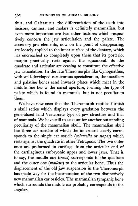
362 PRINCIPLES OF ANIMAL BIOLOGY
thus, and Galesaurus, the differentiation of the teeth into
incisors, canines, and molars is definitely mammalian, but
even more important are two other features which respec-
tively concern the jaw articulation and the palate. The
accessory jaw elements, now on the point of disappearing,
are loosely applied to the inner surface of the dentary, which
has encroached so completely upon them that its posterior
margin practically rests against the squamosal. So the
quadrate and articular are ceasing to constitute the effective
jaw articulation. In the late Theromorphs like Cynognathus,with well-developed carnivorous specialization, the maxillary
and palatine bones send inwards plates which meet in the
middle line below the narial aperture, forming the type of
palate which is found in mammals but is not peculiar to
them.
We have now seen that the Theromorph reptiles furnish
a skull series which displays every gradation between the
generalized land Vertebrate type of jaw structure and that
of mammals. We have still to account for another outstanding
peculiarity of the mammalian skull. The mammalian skull
has three ear ossicles of which the innermost clearly corre-
sponds to the single ear ossicle (columella or stapes) which
rests against the quadrate in other Tetrapods. The two outer
ones are preformed in cartilage from the articular end of
the cartilaginous embryonic upper and lower jaws. That is
to say, the middle one (incus) corresponds to the quadrateand the outer one (malleus) to the articular bone. Thus the
displacement of the oldjaw suspension in the Theromorphhas made way for the incorporation of the two distinctively
new mammalian ear ossicles. The mammalian tympanic bone
which surrounds the middle ear probably corresponds to the
angular.
THE PRINCIPLE OF SUCCESSION 363
Similarities between Theromorpha and mammals are not
confined to the skull. In the later Theromorphs the shoulder
blade has the trihedral shape which is so characteristically
FIG. 147 EVOLUTION OF THE JAW SUSPENSION OF MAMMALS(ventral aspect, after Westoll)
(a) Threnaxodon(Theromorph),(c) Ictidosaurus (Theromorph),(^) YoungGaleopithecus (Mammal), (/) Canis (Mammal). Stages (b) and (d) are
hypothetical.
The bones numbered are: (i)dentary, (2) angular, (3) articular, (4) quad-rate, (5) squamosal, (6) parasphenoid, (7) stapes, (8) jugal, (9) vomer.
mammalian. The axial skeleton of Theromorphs also dis-
plays a few features which call for special attention. Thevertebral centra of the Cotylosaurs are of the biconcave type.
In the late Theromorphs the articular facets are flatter. In
mammals they .are quite flat, so that the notochord does
not persist at all in adult life. The number of neck vertebrae

364 PRINCIPLES OF ANIMAL BIOLOGY
in Cynognathus and in mammals is fixed (seven), and the
fact that it is identical in the whale and the giraffe affords
an excellent illustration of Unity of Type. In Platypus, as in
Theromorphs, there are vestigial cervical ribs. In other
mammals they are completely fused with the vertebrae, but
the interspace between the two heads of the rib persists as a
canal on either side of the vertebra.
At the end of the Permian we find representatives of an
order of reptiles (Rhynchocephalia) which have left one
survivor in modern times, the New Zealand lizard, Spheno-
don. They differ from the Cotylosaurs in one striking
particular, the presence of two large vacuities at the side of
the skull. One, like the temporal fossa of Moschops, is
between the postorbital, squamosal, and jugal bones, and one
between the postorbital, squamosal, and parietal bones. So
the side of the head has two conspicuous bony arcades. Other
minor features of the Rhynchocephalian skeleton are the
tendency as in all modern reptiles for the pelvic girdle to be
tilted forwards, and the presence, as in birds, of prominent
projections (uncinate processes) on the posterior margin of
the ribs. From the early Triassic Rhynchocephalia we can
trace series leading in different directions to birds and to
crocodiles. The skulls of Dinosaurs had the two large
temporal vacuities of the Sphenodon skull, but in addition,
as in birds, there were large vacuities in front of the orbit.
In some of the Triassic Dinosaurs (e.g. Anchisaurus) the
postorbital bones and the lower temporal arcade were very
slender, so that apart from the presence of the former, which
has disappeared in birds, the skull is essentially avian.
Another series connects the Rhynchocephalia with the
crocodiles. It is well represented in the Triassic and Jurassic
strata. The chief change in the skull is the elaboration of the
THE PRINCIPLE OF SUCCESSION 365
characteristic false palate of the crocodiles, in which the
pterygoids as well as the palatines and maxillae participate
to shift the internal narial aperture far backwards. In addi-
tion we see the progressive solidification of the vertebral
centra. Tortoises also have a false palate of the mammalian
type. The earliest tortoises go back to the end of the Per-
mian, and possessed a skull without any lateral vacuities like
that of the Cotylosaurs. Some tortoises share with two whollyfossil aquatic or semi-aquatic orders, the Plesiosaurs and
Ichthyosaurs, a vacuity in the temporal region correspondingto the lower one in the skull of Sphenodon. The stages which
connect the tortoises with the early reptilian stocks are not
yet elucidated, and in one respect the story is discontinuous
in that the pectoral girdle of tortoises lies inside the ribs.
It is noteworthy that the Chelonian stock was emergingbefore the complete investment of the thoracic region was
effected. Lizards and snakes which are the most successful
modern Reptilia appeared last of all. The distinctive feature
of this order of reptiles (Squamatd) is the fact that the
quadratojugal has disappeared, so that the quadrate is
moveable. The lower arcade of the Sphenodon skull is thus
absent. There is an upper arcade in most lizards. With the
elimination of the postorbital bone this has disappeared in
the snakes and in geckos.
The task of elucidating the details of structure in frag-
mentary remains is often a difficult one, and the student
may justifiably be tempted to surmise that a good deal of
imagination is incorporated in the sometimes fanciful recon-
structions that are figures in many semi-popular works.
Nevertheless, there are certain broad general conclusions
now well accredited by well-nigh complete skeletons of a
large number of fossil genera and supplemented by innu-

366 PRINCIPLES OF ANIMAL BIOLOGY
merable less complete remains which confirm them. Weknow almost as much about the skeletons of Pariasaurus,
Archaeopteryx, Cynognathus, for instance, as we do about
that of a living Vertebrate. Laying aside speculative inter-
pretations and exaggerations the following points are
sustained by ample data :
(1) There was a time when the only Vertebrates were of the
Piscine type ;
(2) Previous to the appearance of land Vertebrates there existed
fish which resemble those living fish which most nearly
approach the land Vertebrates in their respiratory and
circulatory arrangements ;
(3) The first land Vertebrates with terrestrial limbs in the early
Carboniferous have skulls of which the superficial bones are
scaly plates like those which cover the head region in the
Ganoid fishes ;
(4) At a later date, in Permian times appeared reptilian forms
with large limbs and a bony chest, animals that no longerwaddled but lifted the body well off the ground ;
(5) During the time intervening between the appearance of the
first reptiles and the emergence of true mammals and birds
in the Jurassic there were reptiles which in every feature of
their organization show bird-like or mammal-like charac-
teristics seen neither in modern reptiles nor in the Permian
forms.
TOPICS FOR HOME STUDY
1. Describe the evolution of the pentadactyl limb.
2. Discuss the affinities of skeletal head pattern in modernvertebrates.
3. Describe the peculiarities of the axial skeleton in the various
vertebrate groups.
4. What are the homologues of the mammalian ear ossicles in other
vertebrates.
CHAPTER XIII
EVOLUTIONARY THEORY TO-DAY
INthe closing years of the eighteenth and the first few
decades of the nineteenth century, evolutionary specu-lation was encouraged by two circumstances. One was that
the progress of systematic comparative anatomy led to
recognition of unity of type transcending similarities of habit
and habitat. The other was the growth of materialism in the
period of intellectual ferment which immediately precededand followed the French Revolution. Without attracting
much comment from the world at large or exciting muchdiscussion among other naturalists evolution was defended
in England by Dr. Erasmus Darwin, the grandfather of
Charles Darwin, in France by Lamarck and St. Hilaire, and
in Germany by the poet Goethe, who devoted his earlier
years to anatomical pursuits and put forward an ingeniousbut entirely erroneous scheme of the homologies of the skull
bones. These pioneer evolutionists based their belief on the
prevalent superstition that acquired characters are here--
ditable. For various reasons they exerted very little influence.
The materialistic temper of the period which witnessed the
French Revolution had somewhat subsided. The fossil
record was a sealed book, modern views about the age of the
earth were not yet generally accepted, very little information
about the anatomy of the Invertebrate groups was available,
the essential nature of sexual reproduction was not fully
recognized, and scarcely anything was known about the
development of animals. Before the full significance of unityof type could be appreciated the detailed anatomy of a much.
368 PRINCIPLES OF ANIMAL BIOLOGY
wider range of animal types remained to be studied. The
existence of vestigial structures did not attract much atten-
tion until the compound microscope gave a new impetus to
embryological research which developed rapidly between
1830 and 1860, when Darwin's Origin of Species was
published.
The Principle of Succession,whichemerged with increasing
force as the chronological order of the sedimentary rocks was
established, and as their fossil relics were unearthed during
the first half of the nineteenth century, showed that the gaps
between the well-defined classes of living creatures are filled
in by intermediate forms which lived on this earth in past
epochs. Different species of animals and plants have lived at
different times in the past. Different species of animals and
plants live in different places to-day. The geological record
shows that fossil representatives of a group are usually more
alike, if they belong to the same strata, i.e. they are more
alike, if they lived about the same time. A corresponding
generalization is broadly true about related species which
live in the same geographical region. For instance, all the
species of the kangaroo family live in Australia, and all the
species of the armadillo family live in South America.
The Geographical Distribution of Animals. The scorpion
family provides a good illustration of the fact that geo-
graphical propinquity is generally associated with greater
similarity of structure. Six families of scorpions are com-
monly recognized. If we exclude the neighbourhood of the
Suez Canal and a small part of Morocco, only two families,
the Scorpionidte and the Buthidte, are represented on the
African continent. Species belonging to both these families
are also found in Asia south of the Himalayas, in Australia,
and in Central and South America. In the remotest part of

EVOLUTIONARY THEORY TO-DAY 369
the African continent, the Cape Peninsula, the Scorpionidae
are represented by one genus, Opisthophthalmus y and the
Buthidse are represented by three genera, Uroplectes, Para-
buthus, and Buthus. Opisthophthalmus and Uroplectes do not
extend north of the Equator. Parabuthus extends beyond the
Suez into Arabia, and Buthus ranges over South Asia. None
of these four genera contains species found in Australia or
America. The Cape species of Scorpionidae belong exclu-
sively to a genus which has no representatives north of the
Great Lakes. The Cape species of Buthidae belong to one
genus which is exclusively South African, to one genus with
species in the part of Asia nearest to Africa, to one genus with
species distributed throughout South Asia, and to no genera
with American and Australian representatives.
While belief in special creation was prevalent, the only
explanation offered for the connexion between geographical
propinquity and structural similarity was that animals and
plants have been placed by Providence in the station of life
to which they are best fitted. Colonial enterprise and horti-
culture both show that this is not necessarily true. At the
beginning of the nineteenth century there were no rodents
in Australia, where the rabbit has become a proverbial pest.
A few blackberry seeds transported to New Zealand spon-
sored a blackberry plague which is a serious agricultural
problem. Less than two centuries have elapsed since com-
merce with New Zealand began. There were then no indi-
genous mammals on the islands, where twenty-five imported
species are now living freely in the wild state.
Exploration and new amenities of transport during the
imperial expansion of the nineteenth century provided new
opportunities for examining other circumstances associated
with the fact that related species live in restricted localities.
AA
370 PRINCIPLES OF ANIMAL BIOLOGY
The two most obvious are : (a) the ease with which different
groups of species can travel, on account of their locomotory
organs or devices for seed dispersal, and (b) the physical
obstacles which they encounter in spreading far afield. In
general, groups of species which can most easily surmount
barriers of ocean, mountain, or desert spread themselves over
wider areas. While bats are cosmopolitan, terrestrial mam-mals and Amphibia (frogs and salamanders) are not found
on islands separated by a wide stretch of deep water from the
mainland.
The great traveller naturalists of the nineteenth century
made a close study of island faunas, and compared them
with the faunas of the nearest adjacent land. The animal and
plant populations of some islands are made up mostly of
species which also live on the nearest adjacent land areas.
Other islands are populated almost exclusively by species
which are endemic, i.e. are not found elsewhere. Islands of
the first class are generally near the mainland and are not
separated from it by a great depth of ocean. Islands of the
latter class, called oceanic islands, are separated from the
mainland by a great depth of water. They may be volcanic,
and if so have never been connected with the adjacent land
area;or they may have remained separate from it for a long
period of geological time. The Cape Verde Islands off the
west coast of North Africa are volcanic islands. New Zealand,
which is separated by a deep channel from the Australian
land mass, appears to have been separated from it in Jurassic
times.
The following tables taken from Romanes, a contemporaryof Darwin, are still representative. St. Helena, Galapagos
Islands, and the Sandwich Islands are typical oceanic islands
separated by a great depth of water from the mainland. In
EVOLUTIONARY THEORY TO-DAY 371
glacial times the British Isles connected with the mainland
of Europe. The British Isles include about a thousand islands
large and small. St. Helena is a single volcanic rock.
The geological record furnishes one clue to the meaning of
A. PECULIAR OR ENDEMIC SPECIES
B. NON-PECULIAR SPECIES
this contrast. We know that the species which now live in
Europe have changed very little since glacial times, when the
shallow channel between Britain and the mainland was
established. In other words, many British species are still
identical with European species which already existed whenBritain was still part of the European mainland. The vastly
deeper channel which separates New Zealand from Australia
points to a much longer period of isolation. So fewer existing
Australian species are identical with species which existed
372 PRINCIPLES OF ANIMAL BIOLOGY
when New Zealand was part of the same land mass. Of
1,040 species of New Zealand butterflies, 63 are Australian,
24 are cosmopolitan, and the remaining 91 per cent are
endemic.
Colonial experience furnishes a second clue to the peculiar
faunistic features of oceanic islands. Carnivorous mammals,such as stoats and foxes, which prey on rabbits elsewhere,
do not exist in Australia. Circumstances which keep down
the rabbit population of other regions were therefore
lacking, and they multiplied accordingly. Conversely the
introduction of new predatory species by colonists has
resulted in the rapid extinction of pre-existing species, like
the Dodo. Animals brought at rare intervals by drifting log-
wood and seeds dropped by birds can multiply on volcanic
islands without competition from predatory species on the
mainland. So they may survive in their new surroundings,
while their relatives on the adjacent mainland are making
way for other species.
Families (e.g. Scorpionidae) generally have a more wide-
spread distribution than the genera (e.g. Opisthophthalmus)
placed in them; and include species which have been
separate for a longer period of geological time than species
placed in a single genus. So species placed in different generaof the same family have usually had a longer period for
spreading far afield than species placed in the same genus.In short, all the facts of the geological record and of geo-
graphical distribution point to the same conclusion. Newspecies are continually appearing and old ones are continually
disappearing. Four years before Darwin's first book appearedWallace summed up the known facts about distribution in
time and space at the conclusion of his paper entitled On the
Law which has Regulated the Introduction of New Species:

EVOLUTIONARY THEORY TO-DAY 373
"every species has come into existence coincident both in
space and time with a pre-existing closely allied species."
The Theory of Natural Selection. Wallace and Darwin,
whose restatement of the evidence which had accumulated
during the first half of the nineteenth century gained general
acceptance for the modern doctrine of evolution, approached
the problem from the standpoint of the traveller-naturalist
in the light of the new knowledge gained through explora-
tion, colonization, and ocean travel. To the question why do
some types exist only in one locality and others only in a
different one, their answer was that new hereditable types
which turn up in the normal process of generation survive
in competition with others which have disappeared because
they are more fertile or more viable in the particular habitats
which they penetrate in search of food. Both Wallace and
Darwin stressed the fact that a relatively small proportion of
progeny produced in every generation survive to produce
offspring. Genetic variations will sometimes diminish the
chance of survival, and sometimes increase it. Which varia-
tions, or, as we should now say, mutant characters, favour
survival will depend on the demands imposed by a parti-
cular habitat. Thus nature selectsdifferent genotypes in different
habitats. The barriers of ocean currents, mountain ranges,
and the like perform the same function as the wire netting
or fence by which the stockbreeder or fancier perpetuates
new domestic varieties.
Darwin and Wallace concluded that natural varieties can
arise in this way from the practical experience of the horti-
culturalist, the stockbreeder, and the fancier. To-day we can
study in the laboratory how new hereditary types or mutants
arise. We know that mutants breed true to type when mated
inter se. We also know that mutant genes are not diluted by

374 PRINCIPLES OF ANIMAL BIOLOGY
inter-crossing and that new true-breeding combinations
of mutant genes can be produced by hybridization. That
natural selection of different genotypes will occur through
the spread of species into different habitats and the changingconditions of life in the situations where they find them-
selves is beyond reasonable doubt. That if this process
goes on sufficiently long they will become so unlike that they
are incapable of interbreeding, when barriers which separate
them disappear or other circumstances which prevent them
from invading the same area are removed, is a conclusion
about which no modern biologists disagree. It does not
follow from this that new species only arise in this way, and
Darwin himself did not assert it. He also recognized a second
type of species formation by sexual selection (vide infra}.
There is, in fact, no single problem of the origin of species.
There are many problems of the origins of species, arising
partly from the fact that all biologists do not always use the
word "species'* in the same sense, and partly from the fact
that new genotypes may arise in more than one way. Owingto the large-scale importation of alien types for hybridization
with a view to the production of new horticultural products,
botanists are able to draw a clear-cut distinction between
varieties and species. Individuals are classified in different
varieties if they have recognizably different hereditable pro-
perties and are still capable of producing fertile hybrids.
Whether they do so in fact may depend on whether they are
prevented by geographical circumstances or by human inter-
ference from so doing. Different species, on the other hand,
cannot interbreed successfully. Either they do not produce
offspring or produce sterile hybrids. Zoologists cannot make
the same clear-cut distinction, because far fewer possibilities
of testing this possibility under suitable conditions for the
EVOLUTIONARY THEORY TO-DAY 375
much greater number of animal species now named exist.
When intermediates occur in regions where the distribution
of two types overlaps, it is generally assumed that natural
hybridization occurs freely and the types are not regarded
as true species. When the geographical barriers to dispersal
prevent this, we have usually no means of knowing whether
it would be possible in suitable circumstances.
Thus the distinction between a geographical variety and
a species of animal is not a clear-cut one. While there is no
doubt that geographical varieties are often true species in
the making and that when closely allied species occur in the
same habitat they have invaded it after a long period of
separation during which accumulated differences have goneso far as to make them intersterile, there is also no doubt that
new species can arise in one and the same habitat. In Dar-
win's generation this led to some controversy between
botanists and zoologists, partly because the latter were more
preoccupied with the newly discovered data of geographical
distribution and hence with the fact that closely allied species
are generally found in different habitats. The occurrence of
closely allied species in the same habitat constitutes a different
problem which botanists are forced to recognize more clearly
than do zoologists for a reason which is now easy to under-
stand.
In Chapter VIII we have met with two different ways in
which new genotypes may arise. The most common is gene
mutation, which is a chemical change restricted to a particular
locus of a chromosome. There is also chromosome mutation,
as when XXY females or triploid fourth chromosome flies
are produced by non-disjunction. In one type of chromosome
mutation called polyploidy each chromosome may be repre-
sented three times, four times, etc. Although chromosome

376 PRINCIPLES OF ANIMAL BIOLOGY
mutations need not be associated with visible differences of
structure, polyploids generally show very striking somatic
peculiarities and are often partially or wholly intersterile with
the stocks from which they originate. If a polyploid of this
kind arose in a bisexual species of animal it could not perpe-
tuate itself unless there was an epidemic of similar chromo-
some mutations at the same time, and we do not know that
this occurs in nature. Since most animals are bisexual, this
method of species formation cannot be very common amonganimals. It is known to exist among some parthenogenetic
types and it could presumably occur in hermaphrodite species
which are capable of self-fertilization, or species which repro-
duce vegetatively, such as corals, sea squirts, or polyzoa. Onthe other hand, nearly all plants reproduce vegetatively. The
overwhelming majority of plants are hermaphrodite, and
unlike most hermaphrodite animals, are capable of self-
fertilization. It is now known that chromosome counts of
many closely related species of flowering plants are multiples
of the same number. Hence there is no doubt whatever that
a high proportion of plant species have arisen by chromo-
some mutation in one and the same habitat, and that though
natural selection working upon a different material basis may
exaggerate initial differences and widen the gap between
them, it is not the correct explanation of their origin.
It is also true that gene mutations may make individuals
more or less infertile with parent stock. Mutation of this
kind would have no chance of survival in a bisexual species.
On the other hand, it would offer no serious obstacle to sur-
vival in species which reproduce asexually, in self-fertilizing
hermaphrodites, or in animals, such as aphides, which have a
regular alternation of parthenogenetic and bisexual genera-
tions. Such alternation of generations also occurs in the gall
EVOLUTIONARY THEORY TO-DAY 377
flies. Of the sixty-eight species of British gall flies, fifty-
four parasitize the oak, and there is nothing to suggest that
this immense proliferation of species for one and the same
host plant is mainly due to specialization resulting from
a predilection for different parts of the plant.
Since more retarded types would have greater oppor-
tunities of interbreeding, gene mutations might lead to
species formation in one and the same environment by
retarding sexual development or by producing precocity and
by promoting earlier or later emergence of the adult from a
larval phase. In this way species formation might proceed
gradually in the same environment. Needless to say, it mightalso happen as a response to environment through natural
selection imposed by conditions of climate. A good exampleof this among animals is difficult to find, because experi-
mental studies of natural evolution are still in their infancy.
An interesting illustration from plants is the Linnean species,
Primula vera, which includes both the primrose and the
cowslip. Fertile hybrids (sometimes called oxlips) of cowslips
and primrose are easy to produce. In nature they are rare
for two reasons. Cowslips do not begin to blossom until the
flowering time of the primrose is nearly over, and primrosesare rarely found in open fields where cowslips abound. Here
we have a species difference which is incipient. If the
flowering times were more restricted, they would live side
by side in the same locality as clear-cut species, and no
botanist would hesitate to label them as such.
Another sort of species formation is foreshadowed in
Darwin's theory of sexual selection. It is obvious that animals
display secondary sexual characteristics which have no sur-
vival value except in so far as they are marks of sexual
recognition. To account for the secondary sexual differences
378 PRINCIPLES OF ANIMAL BIOLOGY
which some species such as pea-fowl display, Darwin sug-
gested that the natural process of variation may result in
different preferences of the male or of the female for its mate.
Such differences could, of course, lead to the selection of
different strains within the same habitat, and while accounting
for the exaggeration of secondary sexual characteristics
would also lead to species differentiation.
Darwin's sexual selection implies that mating does not
occur at random. From this point of view it is a particular
case ofwhat may be a more general aspect of species formation
in nature. Owing to their rarity mutants which are inter-
sterile with the parent stock have no appreciable chance of
surviving when mating occurs at random. On the other hand,
the chance that a mutant of this kind could mate and estab-
lish a pure line in a bisexual species is not small in certain
circumstances. One is that, as in many mammalian species,
a small number of individuals are produced in a litter. Theother is that owing to low density of population as with
e.g. the otter in Great Britain, brother-sister mating or, at
least, close inbreeding is obligatory. Most mutants are
recessive. Recessive individuals can be produced from
matings of two recessives, a recessive and a heterozygote,
or two heterozygotes. If the gene is rare the first two will be
very rare compared with the third. If the size of the family
is very large, a quarter of the offspring will be recessive, and
the chance that two recessives will interbreed is not small.
If the size of the family is small the chance is much larger
for the following reason. Let us suppose that the size of the
litter is two. A quarter of the offspring of all matings between
two heterozygotes will be recessive. Our census of all such
matings will include some that have two wild-type offspring,
many which contain one of each, and a few composed of two
EVOLUTIONARY THEORY TO-DAY 379
recessive offspring. Thus the proportion of recessives in
litters which contain at least one recessive will be much
greater than a quarter, and can be shown by an application
of elementary statistical principles to be more than a half.
We may sum up the preceding paragraphs ii. the following
way. Darwin's hypothesis of natural selection undoubtedlyshows how evolution may occur and does occur. Undoubtedlyit is the correct explanation of why there exist so manywide gaps between genera, families, classes, orders, and phyla ;
why these groups are so often well-defined, and why so few
intermediate types which make it difficult to construct a tidy
classification survive in nature. On the other hand, it is
undoubtedly true that selection for different habitats is not
the only way in which species are made, and although manyof Darwin's immediate followers believed so, Darwin himself
did not believe that it was. One result of the exaggerated
claims for natural selection which Darwin's followers put
forward was the difficulty of explaining certain facts about
the relations between animals and their habitats such as the
very large proportion of cave animals which have degenerate
eyes. If it were possible to find any rational basis for the
Lamarckian superstition (i.e. the so-called inheritance of
acquired characters) it might be possible to explain such
phenomena. The fact is that as far as we know, it is no
advantage for an animal that lives in the dark to have no
eyes, and though it is easy to see why animals which have
no eyes or have defective ones could manage to survive in
caves, natural selection provides no explanation of why so
many cave animals should have this characteristic.
We know that both eyeless and defective eye mutants occur
in Drosophila and in other animals. We know that the same
mutants do occur again and again. If there is no disadvantage

380 PRINCIPLES OF ANIMAL BIOLOGY
in possessing defective eyes, the proportion of individuals
which have defective eyes will increase in every generation.
So the eyeless condition or defective eyesight will eventually
become characteristic of the species as a whole not as a
result of selection for an advantageous character, but because
of the absence of such selection. If we accept this straight-
forward explanation of the fact that cave animals are optically
defective, we cannot stop there. We know that Trilobites
have compound eyes, that Limulus, the most archaic living
Arthropod, has compound eyes, that most insects have com-
pound eyes, that many Crustacea have compound eyes, and
that one myriapod genus, Scutigera, which is not cryptozoic
like other myriapods, also has compound eyes. We know
also that land Arachnids which are usually cryptozoic have
no compound eyes, that the cryptozoic wingless insects have
no compound eyes, and that myriapods which, with one
exception, are cryptozoic,* have no compound eyes. What is
true of cave animals is therefore trueof cryptozoic Arthropods.So the differentiation of species by the survival of mutants
which have useless characteristics is not confined to cave
animals.
Broadly speaking, we might say that when a species gets
into a new environment it encounters circumstances which
make new demands on the possibility of its survival and
circumstances which relax previous demands. This means
that apart from all the circumstances which promote species
formation in the same habitat, natural selection in Darwin's
sense of the term is not the only agency which accounts for
species formation in different habitats. We have also to ask
whether the mutation frequency of useless characteristics
does not make an important contribution to evolution.
*i.e. live under stones, logs, bark, leaves, etc.

EVOLUTIONARY THEORY TO-DAY 381
Although we now know some natural agencies such as tem-
perature and X-rays which increase the frequency of muta-
tion in the laboratory, we do not yet know very much about
the frequency of mutations in nature. One thing we do knowwith certainty is that some animal types such as the lampshell Lingula or the king crab Limulus have remained un-
changed over vast periods of geological time. This suggests
the possibility that the direct action of environment on the
frequency of mutation varies considerably in different species,
and therefore contributes significantly to the tempo of
evolutionary change.
That what we inherit from our ancestors is their genes
and not their characters has an important bearing on certain
features of evolution which might otherwise be puzzling.
Though we label a gene by its most striking effect on the
structure or behaviour of an animal, there is no reason to
suppose that its influence on development is confined to
such more obvious characteristics as bar-eye or yellow bodyor vestigial wings in Drosophila. In fact, we know that the
same genes have diverse effects. If a particular gene producesa difference which is advantageous or several differences
which are advantageous when taken as a whole, it will be
selected in nature. If it is also responsible for differences
which have no survival value or might by themselves be
slightly disadvantageous they will accumulate for the same
reason, and eventually become characteristic of a stock
living in a particular situation. For instance, a gene which
promotes rapid growth may be selected in circumstances
where early maturity is propitious to survival. If the result
of more rapid growth due to the presence of the same generesults in the distortion of the skull or larger body size,
selection will favour such distortion or gigantism in circum-
382 PRINCIPLES OF ANIMAL BIOLOGY
stances where these characteristics are themselves of no
advantage in the struggle for existence.
Natural Classification. The publication of Darwin's
Origin of Species in 1860 intensified interest- in the study of
comparative anatomy and the classification of animals and
plants, and encouraged some hopes which have not been
sustained by experimental discoveries which have since been
made. According to the evolutionary view unity of type is
due to common ancestry. This statement is open to no
objection if we remember that what we inherit from our
ancestors is a gene complex. Zoologists of Darwin's generation
had not got away from the habit of speaking about the in-
heritance of characters. Hence they believed it was possible
to construct a natural system of classification in which the
several divisions stand for assemblages of species which owe
their common similarities to a common ancestor who
possessed the same characteristics.
The fossil record provides abundant examples of forms
which have developed striking similarities along independentlines. Hence we may draw a rough and ready distinction
between similarities which are based on what biologists of a
past generation would have called true homology* and
similarities based on convergence. As the immediate followers
of Darwin interpreted the distinction, true homology was
generally regarded as structural similarity associated with*
Zoologists have never been entirely consistent about the use of theword "homology" at any period. Thus current text-books use the term"serial homology" in two quite different ways. Sometimes it implies a
recognition of structural similarities among organs which can bearranged in a series in one and the same individual, as when we speakof the homology of the fore and hind limbs of Vertebrates or of themandibles and walking legs of an insect. Sometimes it is used to implythat segmental organs of different species occupy the same ordinal rankin the development of such a series as when we say that the mandibles ofcentipedes are homologous with the second antennas of Crustacea (see
Appendix III).
EVOLUTIONARY THEORY TO-DAY 383
descent from a common ancestor with the same charac-
teristics. Convergent resemblance was structural similarity
of species whose common ancestor did not possess the same
structural characteristics. A distinction of this kind is quite
easy to recognize when we have enough fossil evidence on
which to base it, or when we are dealing with relatively
large and homogeneous groups. We now know that it is
not possible to make a clear distinction between true homo-
logy and convergent resemblance when we are dealing with
closely related forms and have no direct evidence about their
ancestry. Biologists brought up in the Darwinian tradition
believe that they could do so when the resemblances were
not "adaptive," i.e. when there was no reason to believe that
similarities of habitat had independently encouraged the
survival of similar variations in different stocks. From what
has been said above, it is now clear that convergence is not
restricted to useful characteristics. The fact that identical
mutations turn up repeatedly in the same species furnishes
the most reasonable explanation of non-adaptive charac-
teristics such as the reduction of the eye in cryptozoic
animals. So if similar mutations occur in closely allied
species, similarities of non-adaptive characteristics do not
justify us in believing that the common ancestor possessed
the same characteristics.
To mutant genes of various species we can now assign an
appropriate frequency or mutation rate. The gene responsible
for "sugary" as opposed to starchy endosperm in maize
turns up anew once in the production of 400,000 gametes.
Haldane estimates that the gene for haemophilia turns upabout once in 50,000 generations. During the past twenty
years chromosome maps like Fig. 94 of closely related
species of animals and plants have been constructed from
384 PRINCIPLES OF ANIMAL BIOLOGY
the data provided by hybridization of mutant types. In
such closely related species the correspondence of indi-
vidual chromosomes is easy to recognize, and bodily differ-
ences due to mutant genes can be assigned to changes at
a particular locus of a particular chromosome. By itself
the fact that similar mutant characteristics turn up in
closely related species signifies little, since different genes
known to be located on different chromosomes or on different
parts of the same chromosome may produce the same visible
effect. For instance, several albino varieties both of the
rabbit and of the fowl are visibly alike and genetically
different. On the other hand, many similar mutants in closely
allied species can now be referred to the same loci on the
same chromosome, so that chromosome maps of allied
species may resemble one another very closely. In Drosophila
simulans Sturtevant has shown that some twenty-five
mutants, similar to mutants in D. melanogaster, can be
referred to genes with similar serial order on corresponding
chromosomes. Thus yellow body, white-eye, and rudimentary
wing genes occur in the same order in both species on the
X-chromosome. A black-bodied, vestigial winged, and a
truncated-winged mutant have been found in Drosophila
simulans. Their genes occur as in D. melanogaster on the
second chromosome in the same serial relation. There is a
sepia-eyed mutant whose gene resides on the third chromo-
some as in D. melanogaster. There is the mutant "minute"
with small fine bristles in both species, with its gene located
on the fourth chromosome.
The importance of Sturtevant's work will be more clear
if we consider two species, A and B, in each of which has
appeared a series of recessive sports, a, b, c, d, e, /, and g.
Their occurrence in nature will be occasional, and may well
EVOLUTIONARY THEORY TO-DAY 385
escape the observation of the field naturalist. The geneticist,
who is on the look-out for them, can isolate each, and thus
build up a stock of A and another stock of B, each with the
same combination of mutant characters, a, b, t, d, e,f, and^.A taxonomist who visited the laboratory and examined these
cultures would find himself confronted by two species with
a group of common characteristics which other species of the
same genus Drosophila do not possess. Reassured by the
convention that these characteristics are not "adaptive/* he
would deduce the existence of a common ancestor charac-
terized with this particular constellation of mutant characters,
and therefore feel justified in erecting a new subgenus to
represent the separation of this common ancestor from the
ancestral stock of other species of Drosophila. It is possible
that Nature is continually playing practical jokes of this sort.
Hence we cannot hope to represent evolutionary relation-
ships in the final twigs of a classificatory system.
It is also very doubtful whether it is desirable to do so,
because the real reason for encouraging systematic study of
animal species is the need to identify them in the quickest
and most convenient way when we wish to know whether
we are talking about the same species or different ones. If
we classified Vertebrates and Arthropods on a strictly evolu-
tionary basis in the light of fossil evidence which we already
possess, the various orders of Myriapoda, Amphibia, and
Reptilia would be assigned the position of classes equivalent
in rank to insects and Crustacea, or to birds and mammals.
Since Myriapods have common characteristics which dis-
tinguish them from all other classes of Arthropods, and since
Amphibia and reptiles have many common characteristics
which distinguish them from other Vertebrates, such a classi-
fication would be a less serviceable key to the identification
BB

386 PRINCIPLES OF ANIMAL BIOLOGY
of a living species in the field than those which are usually
adopted in zoological text-books.
It also follows from what has been said about the occurrence
of similar mutations in related species that the laborious
efforts of anthropologists to trace the evolution of the so-
called races of mankind are largely futile. Preoccupation
with the problem of geographical distribution in Darwin's
time encouraged the study of the geographical varieties of
the human species and it was too commonly assumed that
differences of culture associated with differences of habitat
have the same basis as the physical characteristics of human
communities. This belief was entirely unwarranted. The
evolution of human culture depends largely on circumstances
which have nothing to do with changes in the genetical
make-up of individuals. Human communities are forced to
adopt different technical processes and social habits by the
natural resources and physical limitations of the places
where they live. They transmit their knowledge by speech
or by its substitute writing, and not by their gametes. The
culture level of different communities is a problem suigeneris.
It has little to do with evolution in the biological sense of
the term.
In this connexion it is important to emphasize the fact
that ethnologists do not always use the word "race" in a
strictly genetic sense. It is undoubtedly true that people wholive in some geographical regions have common hereditable
peculiarities which distinguish them from other people.
When anthropologists use the word race in this sense theyfollow the same practice as biologists. It is also a fact that
some genotypes which do not form compact communities
are more frequent in some regions than in others. For
instance, tall, long-headed people with fair hair and blue
EVOLUTIONARY THEORY TO-DAY 387
eyes, are more common in Northern Europe than in other
parts of the world. There is no evidence that there has
ever been any time in the world's history when all the
inhabitants of a particular locality had these characteristics.
Though we are entitled to speak of a Ncrdic type, the
Nordic race is a myth.The Jewish race is also a misnomer. Human beings who
are so classified include immigrants from Palestine or other
parts of Asia Minor and their proselytes among Slav and
Tartar peoples of Eastern Europe. Though there is a highconcentration of some hereditable characteristics amongmembers of the Jewish religion and their descendants because
of the taboo which forbids interbreeding with those who do
not share their peculiar dietetic preferences, no physical
characteristics distinguish all Jews as such from any other
people. Their common cultural characteristics are largely,
if not exclusively, the product of a common tradition rein-
forced by persecution and restriction of civic privileges for
many centuries.
TOPICS FOR HOME STUDY1 . How may new species arise ?
2. What important facts emerge from a study of Geographicaldistribution ?


APPENDIX I
CLASSIFICATION OF CRUSTACEA
OWING to the great variety of limb structures i.i the group andthe diverse adult modifications of species with closely similar
larval stages, the classification of the Crustacea is more difficult
than that of other Arthropod groups. The following keys are
constructed to draw attention to specially diagnostic features of
the constituent orders. The student must bear in mind that a
moron-proof guide to crustacean systematics cannot be con-structed. Most orders include types which have no very obvious
relationship to the more characteristic species. They are only
placed with the latter because of the existence of less specialized
types endowed with some of their peculiarities, or because earlier
stages of the life cycle reveal similiarities which are absent in
the adult. The profound modifications associated with parasitism,
leading in extreme cases to the disappearance of all specifically
crustacean and even of all Arthropod characters in the adult,
make itimpossible to drawup diagnoses that will apply to all membersof some of the groups. The systematic place of such specializedor degenerate types is indicated, however, either by intermediate
forms linking them with the typical members of their groups or
by the possession of characteristic larval stages. The best surveyof the Crustacea is Caiman's volume in the Oxford Treatise.
Any classification is primarily a means of identifying different
forms. The doctrine of evolution implies that a classification basedon differentiae which draw attention to a complex pattern of
resemblance broadly embodies the ancestral relations of groups.This is only true up to a point. So groups placed near one another
in a key for quick identification are not necessarily related more
closely on that account. The plan used in making the key is
based on convenience alone.
Outline Classification of Crustacea
i. Branchiopodaa. Anostraca
[b. Lipostraca (fossil)]
c. Notostracad. Diplostraca
(i) Conchostraca
(ii) Cladocera
390 PRINCIPLES OF ANIMAL BIOLOGY
2. Ostracoda
3. Cirripedia
4. Branchiura
5. Copepoda6. Malacostraca
a. Phyllocarida ( Leptostraca -= Nebaliacca)
b. Syncaridac. Peracarida
(i) Mysidacea(ii) Cumacea
[(iii) Thermosbaenacea]
[(iv) Tanaidacca]
(v) Isopoda
(vi) Amphipoda
d. Dccapoda (Eucarida)
(i) Euphausiidca
(ii) Pcnaedea
(iii) Caridea
(iv) Palinura
(v) Astacura
(vi) Anomura
(vii) Brachyura
Diagnostic Features of Sub-classes.
Posterior Region of Trunk without Limbs.
Branchiopoda
Trunk-appendages nearly always flattened and lobed
("phyllopodous") number varying greatly ("anomeristic"), genital
openings on same segments in both sexes. No mandibular palp.Shell-fold present or absent. Compound eyes present.
Ostracoda
Trunk-appendages not more than two pairs, varying in form,
genital openings in same region in both sexes. Mandibular palp
present. Shell-fold bivalve. Compound eyes present or absent.
Cirripedia
Trunk-appendages biramous, six pairs. Genital openings ondifferent somites in the two sexes ($ on first trunk somite, $behind last limbs). No mandibular palp. Shell-fold forming a
"mantle" enclosing body and limbs. Compound eyes absent.
APPENDICES 391
Branchiura
Trunk limbs biramous (except first pair), five pairs. Genital
openings on same somite in both sexes. Shell-fold absent. Com-
pound eyes present.
Copepoda
Trunk-appendages typically biramous, six pairs or fewer.
Genital openings on same somite in both sexes. Shell-fold absent.
Compound eyes absent.
Posterior Region of Trunk with Limbs.
Malacostraca
Trunk limbs biramous or pediform, in two groups ("sonnet
sequence" 8-6). Genital openings on different somites in the two
sexes ($ on 6th trunk somite, <J on 8th). Shell-fold present or
absent. Compound eyes usually present. All somites may bear
appendages except in Phyllocarida.
NOTE Trunk limbs ----- limbs behind maxilla.
SPOT KEY TO THE SUBCLASSES
1. Genital apertures on the same segment in both sexes.
. No mandibular palp; flattened appendages:
5 BRANCHIOPODAb. Mandibular palp usually present (3 and 4)
2. Genital apertures of male and female on different segments.
a. Male orifice on i3th, female on nth, all postcephalic
segments bearing appendages in sonnet sequence:
6 MALACOSTRACAb. Male orifices at end of abdominal penis, female orifice
on 5th segment; six pairs of biramous postcephalic
appendages 7 CIRRIPEDIA
3. Postcephalic appendages not exceeding three in number;shell bivalve 8 OSTRACODA
4. Postcephalic appendages more numerous and biramous; noshell-fold.
a. With paired compound eyes 9 BRANCHIURAf Without compound eyes, usually with paired egg sacs :
10 COPEPODA

3Q2 PRINCIPLES OF ANIMAL BIOLOGY
KEY TO ORDERS OF BRANCHIOPODA
1. Without shell-fold ANOSTRACA [Lipostraca (Fossil)]
2. With dorsal shell-fold NOTOSTRACA
3. With bivalve shell DIPLOSTRACA
a. Postcephalic limbs
not less than 10. Conchostraca
b. Postcephalic limbs
not more than 6. Cladocera
KEY TO CHIEF DIVISIONS OF MALACOSTRACA
1. Caudal furca present, seven abdominal somites in adult.
Bivalve shell, flattened thoracic limbs PHYLLOCARIDA
2. No caudal furca, six abdominal somites in adult.
A. No shell-fold but first thoracic somite fused with head.
No brood-pouch SYNCARIDA
B. Shell-fold present or absent, never fused with all thoracic
somites. Brood-pouch present, development direct :
PERACARIDA
C. Shell-fold fused with all thoracic somites. No brood-pouch.
Development usually with metamorphosis:EUCARIDA (Decapoda).
D. Shell-fold fused with four thoracic somites. No brood -
pouch. Development with metamorphosis. Two movable
segments in front of head HOPLOCARIDA
KEY TO CHIEF ORDERS OF PERACARIDA
1. Pedunculate eyes. Shell-fold enveloping thorax but not fused
with more than three somites. Tail-fan MYSIDACEA
2. Eyes sessile, usually coalesced. Shell-fold fused with three
or four somites, lateral lobes forming "pseudorostrum" in
front CUMACEA
3. Eyes on immovable peduncles. Shell-fold fused with twosomites TANAIDACEA
4. Eyes sessile. No shell-fold but first thoracic somite fused with
head. Abdominal limbs, biramous, lamellar ISOPODA
5. Eyes sessile. No shell-fold but first thoracic somite fused withhead. Abdominal limbs usually biramous, in two series (3-5).
(Reduced or absent in Caprellidea) AMPHIPODA
APPENDICES 393
KEY TO PRINCIPAL TRIBES OF EUCARIDA (OR DECAPODA)
i. "Petasma" on first pleopods of male. "Thelycum" or external
receptaculum seminis in female. Nauplius larva.
A. Thoracic limbs nearly uniform, rarely chelae on one pair.
Gills in one series EUPHAUSIIDEAB. First three pairs of thoracic limbs formings maxillipeds
(third pair little modified). Chelae on 4-6. Gills in three
series PENAEIDA
2,. No "petasma" in male. "Thelycum' 'rarely present. Nonaupliusstage present.
A. Thoracic limbs 45 typically chelate. Pleura of secondabdominal somite expanded CARIDEA
[B. Thoracic limbs 4-7 or 4-8 chelate ERYONIDEA]C. No chelae (except sometimes last pair in female) . . PALINURAD. Thoracic limbs 4-6 chelate ASTACURAE. Thoracic limbs 4 usually, 6 never, chelate. Abdomen
generally soft, or flexed, or asymmetrical. Uropoda rarelyabsent ANOMURA
F. Thoracic limbs 4 chelate. Abdomen reduced, flexed
symmetrical. Uropodas very rarely present as vestiges :
BRACHYURA

APPENDIX II
KEY TO THE CHIEF ORDERS OF INSECTS
I. Biting Mouth Parts.
A. Wingless continuous development.*
(a) With abdominal appendages.1. Apterygota (silver fish, bristle tails, spring tails).
(b) Without abdominal appendages.2. Mallophaga (bird lice)
B. Winged.(a) Development continuous,
(i) Without forceps.
3. Orthoptera (cockroaches, locusts, crickets, stick
and leaf insects).
4. Isoptcra (white ants).
(ii) With forceps and folded wings.
5. Dermaptera (earwigs).
(b) Interrupted development net wings.
(i) Larvae with inconspicuous caudal cerci.
6. Odonata (dragon flies).
(ii) Larvae with conspicuous caudal cerci.
7. Plecoptera (stone flies).
(c) Discontinuous development,
(i) With tegmina.
8. Colcoptera (beetles).
(ii) Without tegmina.Net winged.
9. Neuroptera (lace wings, ant lions, alder flies).
Coarse veined.
With mandibles.
10. Hymenoptera. (Bees, wasps, ants, sawflies,
gallflies).
Mandibles vestigial or absent.
11. Trichoptera (caddis flies).
* In this table a distinction is drawn between discontinuous develop-ment, in the course of which the internal organs of the larva arededifferendated and the adult organs are reconstructed from a pulp of
cells, in contradistinction to interrupted development in the course ofwhich the internal larval organs pass over to the adult.

APPENDICES 395
II. Sucking Mouth Parts.
A. Direct development.
(a) Wingless.12. Anoplura (lice).
(b) Winged.
(i) Jointed proboscis.
13. Hemiptera (plant bugs, aphids, scale insects,
water boatmen, skaters).
(ii) Unjointed proboscis.
14. Thysanoptera (thrips).
B. Discontinuous development.
(a) With four wings,
(i) With scales.
15. Lepidoptera (moths, butterflies).
(ii) Without scales.
1 6. Mecaptera (scorpion flies).
(b) With hind wings vestigial.
17. Diptera (flies, mosquitoes, keds, midges).
(c) Wingless.1 8. Siphanaptera (fleas).
III. Mouth Parts Vestigial or absent.
(a) Four wings interrupted development.
19. Ephemeroptera (may flies).
(b) Forewings vestigial discontinuous development,20. Strepsiptera (stylops).
HOMOLOGY OF INSECT MOUTHPARTS
The median hypopharynx as shown (where it can be easily seen)carries the orifices of the salivary ducts. The appendages
mandibles, maxillulae, first maxillae and second maxillae (labium)are indicated by shading. Equivalent appendages are easily
recognized in orders with biting mouthparts (Orthoptera,
Coleoptera, etc.) and in Hymenoptera, where biting (sawfly, ants),
biting and licking (wasp) or licking and nectar-sucking (bee) typesform a transparent series. The equivalence of the typical Lepi-
dopteran mouthparts is established by the Micropterygid typewith its curved galea of the first maxilla and vestigial mandibles.
Among Diptera the styliform galea of the first maxilla is easily

396 PRINCIPLES OF ANIMAL BIOLOGY
distinguished from the styliform mandibles in Tabanids\ and the
palp is also recognizable as that of the first maxilla. For this
reason the palp of other Diptera, e.g. Culex or Muscids, is re-
garded as equivalent. The equivalence of the Hcmipteran mouth
parts is not clarified by a good series of intermediate types. It is
based chiefly on comparison of the jointed probisces with the
fusion of the jointed bases of the second maxillae to form the
labium in other insects. The sucking and piercing mouthparts of
fleas (Siphanaptera), which are not included, are much like those
of Diptera, but the labium is reduced and the tube is mainlyformed by the epi-pharynx (labrum). Cross sections are drawnwith the anterior-dorsal side uppermost. In full view diagrams as
seen from the ventral side of the head the dorsal labrum epi-
pharynx is not visible. Its relative position is indicated.

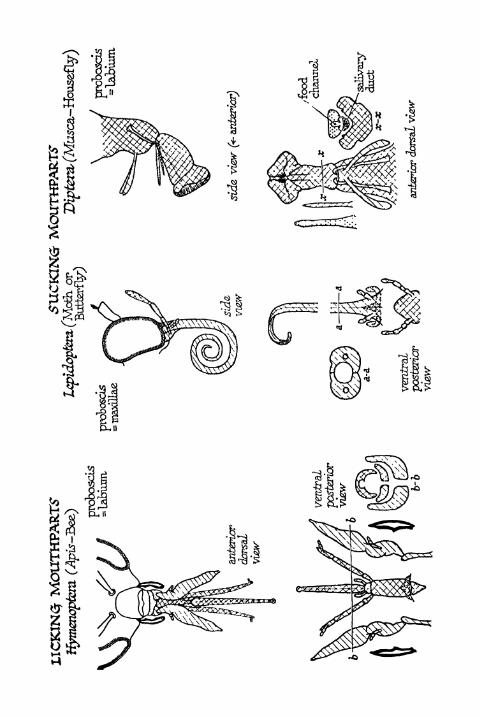
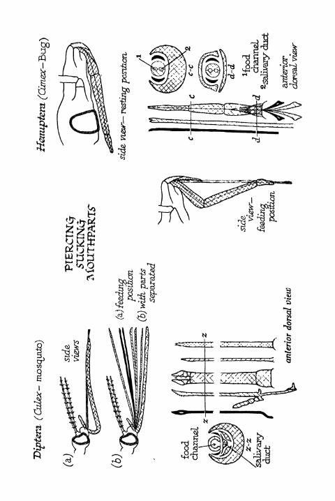
a
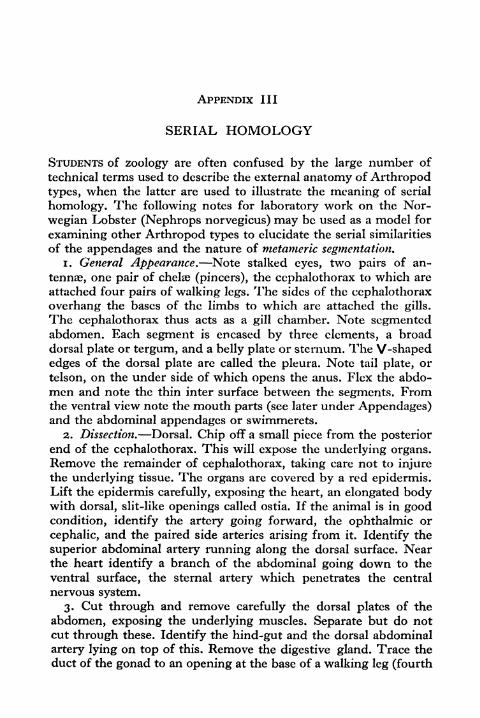
APPENDIX III
SERIAL HOMOLOGY
STUDENTS of zoology are often confused by the large number of
technical terms used to describe the external anatomy of Arthropodtypes, when the latter are used to illustrate the meaning of serial
homology. The following notes for laboratory work on the Nor-
wegian Lobster (Nephrops norvegicus) may be used as a model for
examining other Arthropod types to elucidate the serial similarities
of the appendages and the nature of metameric segmentation.1. General Appearance. Note stalked eyes, two pairs of an-
tennae, one pair of chelae (pincers), the cephalothorax to which are
attached four pairs of walking legs. The sides of the cephalothoraxoverhang the bases of the limbs to which are attached the gills.
The cephalothorax thus acts as a gill chamber. Note segmentedabdomen. Each segment is encased by three elements, a broaddorsal plate or tergum, and a belly plate or sternum. The V-shapededges of the dorsal plate are called the pleura. Note tail plate, or
telson, on the under side of which opens the anus. Flex the abdo-
men and note the thin inter surface between the segments. Fromthe ventral view note the mouth parts (see later under Appendages)and the abdominal appendages or swimmerets.
2. Dissection. Dorsal. Chip off a small piece from the posteriorend of the cephalothorax. This will expose the underlying organs.Remove the remainder of cephalothorax, taking care not to injurethe underlying tissue. The organs are covered by a red epidermis.Lift the epidermis carefully, exposing the heart, an elongated bodywith dorsal, slit-like openings called ostia. If the animal is in goodcondition, identify the artery going forward, the ophthalmic or
cephalic, and the paired side arteries arising from it. Identify the
superior abdominal artery running along the dorsal surface. Nearthe heart identify a branch of the abdominal going down to the
ventral surface, the sternal artery which penetrates the central
nervous system.
3. Cut through and remove carefully the dorsal plates of the
abdomen, exposing the underlying muscles. Separate but do notcut through these. Identify the hind-gut and the dorsal abdominal
artery lying on top of this. Remove the digestive gland. Trace the
duct of the gonad to an opening at the base of a walking leg (fourth
APPENDICES 4OI
in the case of a male, and second in the case of a female). Identifythe parts of the alimentary tract, the oesophagus, the gizzard, andthe hind-gut.
4. Remove the viscera and clean thoroughly. Identify the central
nervous system (paired strands) which lie ventrally. Separate andremove the muscles in the abdominal region. Pear 70 per cent
alcohol on to the nervous system. Trace the C.N.S. forward to
where it is enclosed by a cuticular arch. Chip away the arch andnotice the opening in the C.N.S. through which passes the sternal
artery. Note also the five ganglia in the thoracic and the six in the
abdominal region. Trace the C.N.S. to the oesophagus by separat-
ing muscle, noting the sub-cesophageal ganglion. Note the passageof the oesophagus through an opening between two strands, the
circum-oesophageal commissures, which unite forward to form the
supra-cesophageal ganglion or brain. Lift the carapace in the regionof the eyes and identify the nerves to the eyes and the antennae.
Note also the green gland at the base of the antennas.
5. Identify the following paired appendages on the intact
animal :
(a) Pre-oral antennules and antennae.
(b) Oral, completely subordinated to masticatory use, mandibles,first and second maxillae.
(c) Semi-masticatory intermediate between jaws and legs, three
pairs "maxillipedes."
(d) Post-oral chelae, and four walking limbs.
(e) Abdominal five swimmerets, one terminal or tail-paddle.
Note the biramous character of the limbs in this series, with
outer (exopodite) and inner (endopodite) branches.
6. Remove the third maxillipede of one side, extracting it at the
base (the third maxillipede lies immediately in front of the chela or
pincers). Then remove, in the following order, second maxillipede,first maxillipede, second maxilla, first maxilla, mandible, antenna,antennule. Lay these out in order, commencing with antennule, andnumber. Remove chela, four pairs of walking legs, and abdominal
appendages.
(a) Examine abdominal appendages 2-5, and the first pair of
feelers, the antennules. Two-jointed base, except the anten-
nules, which are three-jointed. This is called the protopodite.The appendage is biramous, the two similar parts called the
CO
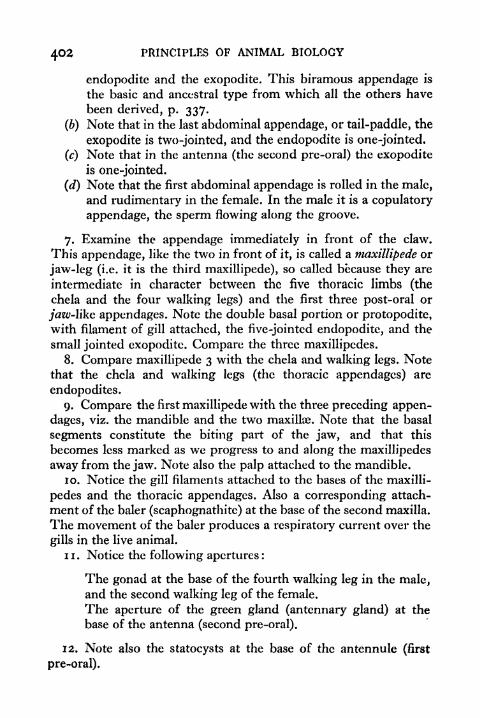
4O2 PRINCIPLES OF ANIMAL BIOLOGY
endopodite and the exopodite. This biramous appendage is
the basic and ancestral type from which all the others have
been derived, p. 337.
(6) Note that in the last abdominal appendage, or tail-paddle, the
exopodite is two-jointed, and the endopodite is one-jointed.
(c) Note that in the antenna (the second pre-oral) the exopoditeis one-jointed.
(d) Note that the first abdominal appendage is rolled in the male,and rudimentary in the female. In the male it is a copulatory
appendage, the sperm flowing along the groove.
7. Examine the appendage immediately in front of the claw.
This appendage, like the two in front of it, is called a niaxillipede or
jaw-leg (i.e. it is the third maxillipede), so called because they are
intermediate in character between the five thoracic limbs (the
chela and the four walking legs) and the first three post-oral or
jaw-like appendages. Note the double basal portion or protopodite,with filament of gill attached, the five-jointed endopodite, and the
small jointed exopodite. Compare the three maxillipedes.
8. Compare maxillipede 3 with the chela and walking legs. Notethat the chela and walking legs (the thoracic appendages) are
endopodites.
9. Compare the first maxillipede with the three preceding appen-
dages, viz. the mandible and the two maxillae. Note that the basal
segments constitute the biting part of the jaw, and that this
becomes less marked as we progress to and along the maxillipedes
away from the jaw. Note also the palp attached to the mandible.
10. Notice the gill filaments attached to the bases of the maxilli-
pedes and the thoracic appendages. Also a corresponding attach-
ment of the baler (scaphognathite) at the base of the second maxilla.
The movement of the baler produces a respiratory current over the
gills in the live animal.
11. Notice the following apertures:
The gonad at the base of the fourth walking leg in the male,and the second walking leg of the female.
The aperture of the green gland (antennary gland) at the
base of the antenna (second pre-oral).
12. Note also the statocysts at the base of the antennule (first
pre-oral).
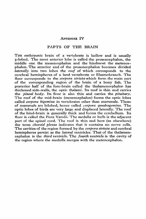
APPENDIX IV
PAPTS OF THE BRAIN
THE embryonic brain of a vertebrate is hollow and is usually3-lobed. The most anterior lobe is called the prosencephalon, themiddle one the mesencephalon and the hindmost the metence-
phalon. The anterior end of the prosencephalon becomes divided
laterally into two lobes the roof of which corresponds to thecerebral hemispheres of a land vertebrate or Elasmobranch. Thefloor corresponds to the corpora striata which form the main partof the corresponding region of the brain of a bony fish. Theposterior half of the fore-brain called the thalamencephalon hasthickened side-walls, the optic thalami. Its roof is thin and carries
the pineal body. Its floor is also thin and carries the pituitary.The roof of the mid-brain (mesencephalon) forms the optic lobes
called corpora bigemina in vertebrates other than mammals. Thoseof mammals are bilobed, hence called corpora quadragemina. Theoptic lobes of birds are very large and displaced laterally. The roof
of the hind-brain is generally thick and forms the cerebellum. Its
floor is called the Pons Varolii. The medulla or bulb is the adjacent
part of the spinal cord. The roof is thin and here (as elsewhere)the term choroid plexus indicates that it contains no nerve cells.
The cavities of the region formed by the corpora striata and cerebral
hemispheres persist as the lateral ventricles. That of the thalamen-
cephalon is the third ventricle. The fourth ventricle is the cavity ofthe region where the medulla merges with the metencephalon.


INDEX
Acalephae, 292, 299, 308
Acanthodei, 341, 347
Acephala, 292, 299, 301, 310
Actinopterygii, 341, 350
Actionozoa, 43, 310, 317Adrenal body, 24 (ill.), 153
Adrenal glands, 37 (ill.), 58, 165
Adrenalin, 154
Alimentary Canal, 55, 115 (ill.),
n6(ill.), 121, 137, 157,284,288,
293, 3ii
Allantoidians, 297
Allantois, 160 (ill.), 161 (ill.)
Amia, 344
Arnioidei, 341
Amoeba, 252-256
Amphibia, 44, 57, 58, 82, 148, 261,
266, 280, 289, 348, 385skull of, 355
Amphioxus,325,326(ill.),327(ill.),330
Amylase, 120, 122
Anallantoidians, 298
Anemones, Sea, 301, 316
Anenteroeti, 310Animalia
Articulata, 292
Mollusca, 291
Radiata, 292Vertebrata, 291
Annelida, 43, 291, 292, 293, 294, 296,
297, 298, 302, 310, 311, 318, 323,
324
Annulata, 298, 302
Anus, 114, 115, 116
Aphides, 376
Apodes, 310
Appendages, 274, 276, 285
Aptera, 261
Arachnida, 44, 261, 273, 286, 287,
288, 292, 298, 336, 337, 38o
Archoeopteryx, 335 (ill.), 337, 350,
35i, 352, 366
Aristotle, 175, 247, 249, 260
Armadillos, 175
Arteries, 130, 131, 132 (ill.), 133 (ill-),
137, 138, 139, 14, H5, H7Aorta, 138, 145, 147, 284
Branchial, 138
Carotid, 138
Cceliaco-mesentcric, 138
Iliac, 138
Pulmo-cutaneous, 138
Pulmonary, 132 (ill.), 138, 144
Renal, 138
Subclavian, 138
Systemic, 138Truncus arteriosus, 138, 147
Arthropoda, 42, 261, 266, 271, 273,
274, 279, 281 (ill.), 282, 283 (ill.),
286, 287, 289, 291, 293, 296,
297, 298, 302, 310, 311, 324, 334,
337
Ascidiacca, 328, 329Asexual reproduction, 22, 30
Asteriodea, 312
Astigmatism, 67Atomic principle, 188, 189
Atrium, 137, 148, 327Attraction sphere, 177
Auditory ossicles, 69 (ill.)
Auricles, 132 (ill.), 133 (ill.), 136 (ill.),
137, 138, 143, 144Auriculo-ventricular valve, 137, 138Aves (see also Birds), 44, 261, 289
Avicenna, 331Axial skeleton, 342
Axolotl, 16*7, 168 (ill.)
Bacteria, 121, 258, 259
Baer, von, 171
Balanoglossus, 329
Balantidium, 253
Barnacle, 249 (ill.), 288 (ill.)
406 PRINCIPLES OF ANIMAL BIOLOGY
Barry, 257
Bastian, 259
Bateson, 204, 330
Batrachians, 298
Bayliss, 152
Bee, 175, 261
Behaviour, 73, 74, 92-97, 112
Bernard, Claude, 134
Berthellot, 118
Bidder's organ, 37
Bile, 116, 122
Biogenesis, Principle of, 247-270Birds (see also Aves), 34, 59, 64, 82,
134, 181, 291, 297Limb skeleton, 349
skull, 361 (ill.)
Blacky 127
Bladder, 24 (ill.), 115 (ill.), 126
Swim-bladder, 145
Blood-Circulation of, 123, 129, 130, 131,
132 (ill.), 133, 135, 137, 138, 139
(ill.), 144, 145, 152, 283, 284
Composition of, 134, 143
Corpuscles, 142
Regulation of flow of, 139-140, 141,
152
Bone, 54 (ill.)
Bower, 44
Brachiopoda, 43, 292, 301, 311
Brain, 65, 84, 86 (ill.), 87, 88, 92, 94,
96, 153, 399
Branchiostoma, 325
Bridges, 204, 215
Bronchus, 116 (ill.), 123 (ill.).
Broom, 360
Browne, Sir Thomas, 184
Bryozoa, 299, 318
Buchner, 118
Buckland, 333Buff Orpingtons, 187
Buthidoe, 368, 369
Caffeine, 152
Calciferol, 109
Calorimeter, 101
Calvin, 130
Campodea, 276
Capillaries, 123 (ill.), 129, 130, 131,
134, 136, 137, 138, 139, 140, 141,
144, H5Carbohydrates, 102, 103, 112, 114,
134, 3o6Carbon dioxide, 77, 100, 101, 112,
114, 123, 124, 127, 128, 129,
284
Carboniferous, 355, 356
Carotin, noCartilage, 52, 54
Caseinogcn, 120
Castle, 171
Castration, 174, 175
Catalyst, 117, 118, 119, 120
Cavendish, 128
Cells, 59, 65, 66, 79, 80, 81, 115, 177Cell theory, 171, 257, 258Cellular structure, 29 (ill.), 3,
31 (ill.), 32, 35, 55, 57Central Nervous System (C.N.S.),
78, 81, 82, 83, 87, 89 (ill.), 9i,
95, 152, 156, 157, 280,281,282,
293, 302, 322, 324, 328, 330
Cephalochorda, 325
Cephalopoda, 44, 291, 299, 300, 301,
303, 304 (ill.), 305 (ill.), 306, 308,
3io, 337
Cestoda, 299, 320
Cetacea, 266
Chameleon, 18, 19, 40, 45, 47, 65, 84,
85 (ill.), 86, 90
Chelonian, 365Chemical products, 45
Chemistry of body, 102-104
Chitin, 293
Chiton, 301 (ill.), 304, 305, 306, 307,
308
Chlamydomonas, 256
Chlorophyll, 105, 112, 114, 256
Chondrostei, 340
Chordata, 324Chromatophores, 45, 46

INDEX 407
Chromosome, 176 (ill.), i?7, i?8 (ill.),
179, 180 (ill.), 181, 182 (ill.),
183 (ill.), 184, 185, 190, 191, 210,
212, 214, 215, 219 (ill.), 223 (ill.),
229
Hypothesis, 200-227 (ill.)
Map, 216-227, 375, 383, 384
Church, 44
Cilia, 59, 62 (ill.), 250, 251, 256, 257,
_ 327
Ciliary motion, 59
muscle, 64 (ill.), 66
Ciliata, 310Ciliated epithelium, 45, 59 (ill.)
Circulation
Annelida, 302
Arthropod, 302
Fish, 145
Frog, 134, 136
Human, 143-144
Mollusc, 302
Vertebrate, 302
Cirrhopoda, 292, 301
Cladosclachus, 347
Classification, Natural, 382, 387
Climatius, 347
Cloaca, 115 (ill.), 125
Clusius, 332
Cochlea, 69 (ill.), 70, 88
Cockroach, 182, 273, 274, 275, 276
(ill.), 277 (ill-), 278, 279, 284
Ccelenterata, 43, 308, 311, 313, 315,
316,318
Ccelocanthini, 341
Cole, 332
Coleoptera, 261
Colour response, 19, 20, 67, 152, 153
Conchifera, 300Conservation of Energy, 98-102
Constancy, Law of, 197Contractile vacuoles, 251
Co-ordination, 73-97
Cope, 334Corallaria or Polypi, 299
Cornea, 64 (ill.), 65, 66, 67
Correns, 190
Costa, 325
Cotylosauria, 356, 359, 360, 363, 364,
365
Crab, 175
Crayfish, anatomy of, 272-273 (ill.),
274, 278, 279, 285
Crepidula, 306
Cretinism, 105, 166
Crezv, 185
Crinoidea, 312
Cross-breeding, 189, 198, 205, 212,
228, 237
Crossing-over, 220, 221 (ill.)
Crustacea, 44, 261, 273, 279, 287,
288, 289, 292, 298, 310Classification of, 389-393
Cryptozoic, 380
Crystalline style, 306
Ctcnidium, 300, 303, 304, 305, 308
Ctenophora, 317
Cuenot, 204
Cuvier, 286, 291, 293, 300, 301, 311,
332
Cyclops, 279 (ill.)
Cyclostomes, 148, 289, 341, 342, 344,
352
Cynognathus, 361, 364, 366
Cytoplasm, 29 (ill.), 59 (ill.). 177
Dallinger, 259
Dalton, 1 88
Daphnia, 279 (ill.)
Darwin, C., 189, 299, 3 3&7, 3^8,
372, 373, 375, 377, 378, 379, 380,
382, 383, 386
Darwin, Erasmus, 367
Devonian, 350, 354De Vries, 190
Diaphragm, 116 (ill.), 124
Digestion, 114-123, 126-128, 129,
134, 282
Dinosaurs, 352
skull, 361 (ill.), 364
Dipnoi, 340, 347, 351
Diptera, 261, 354

408 PRINCIPLES OF ANIMAL BIOLOGY
Dobell, 256
Dogfish, 273, 274, 352
Doliolum, 328
Dominant, 194, 195
Doncaster, 212
Drosophilia, 180 (ill.), 183 (ill.), 204,
210, 211 (ill.), 213 (ill.), 214,
220, 225, 227, 228, 237, 379,
381, 384, 385
Drysdale, 259Ductless glands, 150-154, 165 (see
also Glands)
Ear, 8, 21, 62. 64, 68-70 (ill.)
Echidna, 268, 349
Echinodermato, 43, 292, 299, 310,
311 (ill.), 312
Echinoderms, 311, 312
Echinoidea, 312
Echivroidea, 295, 323
Ectoderm, 156 (ill.)
Ectoplasm, 251
Edwardes, Milne, 47, 297, 308Effector organs, 45-46, 73, 74, 78, 80
Egg, 24, 25, 26, 27, 28 (ill.), 30, 31,
32, 34, 35, 36, 38, 39, 40, 4i, 42,
103, 155, 158-162, 171, 179, 181,
1 86, 189, 190, 194, 197, 200
Elasmobranchs, 338, 339 (ill.), 344
Embryo, 30, 31, 35, 38, 39, 40, in,137, 145, H7, 155, 156, 158, 161,
162
Endoplasm, 251
Energy, 98-113, 4> "9Enteropneusta, 329, 330
Entomozoa, 298
Environment, 167
Enzymes, 117, 118, 119, 120, 121, 306
Epithelial folds, 164
Epithelium, 32, 293
Erepsin, 120, 122
Euglena, 256
Euryptcrida, 334
Eustacian tube, 68, 69 (ill.), 70, 115
(ill.)
Evolution, 38
Evolutionary theory to-day, 367-387
Excretion, 101, 125-128
Eye, 62, 64 (ill.), 65 (ill.), 66, 67 (ill.),
68, 88, 279, 280
Fat, 102, 103. 104 119, 122, 137, 142
Fertilization, 25 (ill.), 26, 30, 35, 37,
38, 41, 42, 155, 172, 175, 177,
178, 186, 189, 190, 191, 192,
194, 197, 231, 376
Fibres, 50, 5 1, 55, 66, 69, 7> 79, 80,
81,82,83,84,94, 115, 140, 143
Fischer, 102
Fishes, 19, 25 (ill.), 40, 46, 47, 64,
82, 86, 134, 137, 144, 145, 146,
148, 158, 162, 291, 298, 325,
338, 339, 340, 34i, 342, 344,
345, 346, 347, 350, 35i, 356, 358
Flagella, 256
Flourens, 70
Fly, 94, 205-208 fill.), 211-223
Blow, 62
Gall, 377
Fruit, 1 80, 181, 183, 206, 207 (ill.)
Stone, 1 80, 181 (see also Droso-
philia)
Foetus, human, 163 (ill.)
head, 359 (ill.)
Folt 25
Foraminifera, 253 (ill.), 332Foster
, 78 n.
Fowl, 34, 35, 36, 103, 137, 158-162,
175, 188, 189, 194, 195, 209 (ill.),
237, 238, 239
Frog (Frontispiece), 18, 19, 21, 23
(ill.), 24 (ill.), 27, 28 (ill.), 30, 32,
34, 35, 39, 42, 44, 46, 47, 49,
52 (ill.), 53 (ill-), 54, 55, 56, 57,
59, 62, 64, 65, 66, 68, 70, 73, 74,
75, 76, 77, 79, 80, 86, 88, 92, 94,
95, 96, 101, 102, 106, 107, 114,
115 (ill.), 123, 124, 125 (ill.), 129,
133 (ill.), 134, 135 (ill-), 136 (ill.),
137, 141 (ill), 142, 143, 144, 146,
INDEX 40Q
147, 150, 153, 155, 156, 157, 158,
161, 162, 164, 166, 167, 174, 175,
185, 253
Galen, 55, 7&
Ganglia, 79, 81 (ill.), 82, 83, 84 (ill.),
86,87,90,288,293
Ganoides, 338 (ill.), 339, 34, 342, 356Gastric juice, 118, 120, 122, 126, 282
Gastropoda, 44, 292, 299, 300, 301,
302, 303, 304 (ill), 306, 307, 308,
3ioGenetic \ariation, 194
Genetics, 187, 189, 234-242Genital organs, 37 (ill.)
Genotype, 375
Geographical distribution, 269-270,
368-373, 386
Geotropism, 63
Gephyrea, 323
Gerard, 248, 249
Gill, 62, 147, 162, 163, 285, 300, 303,
306
arches, 144 (ill.), 146, 147, 163, 164
clefts, 145 (ill.), 146 (ill.), 164
Glands, 32, 45, 57, 58 (ill.), 86, 142,
H3, 150-154
Glisson, 51
Glycogen, 119, 122, 124, 131, 134, 137
Goethe, 367
Goitre, 105
Gomphognatus, 361
Gonads, 27, 37, 170, 174, i75> 293
Goodrich, 172Graafian tollicles, 33
Gravity, 62, 63, 64, 72
Gullet, 59, 115, n6(ill.)
Gut, 24 (ill.), 87, 89, 115, 129, 132
(ill.), 135 (ill-), 138, H5 (ill.)
Embryonic, 55, 156 (ill.), 157
Haeckel, 252, 318, 323, 324, 325
Haemocyanin, 105, 134, 283
Haemoglobin, 103, 105, 131, 134, 282,
293
Haemophilia, 383
Haldane, J. B. S , 100, 101, 169, 170,
172, 383
Harvey, 130, 247
Haswell, 271 n.
Haughton, 360Head skeleton, ^52-366
Heart, 16 (ill.), 57, 86, 89, 106, 107,
121, 129, 130, 136, 137, 140,
141 (ill.), I44 (ill.)
Auricles, 303Sinus venosus, 137
Ventricles, 303
Hebnholtz, 75
Helminthes, 298
Hemichorda, 325, 329, 330
Henry, 103
Heredity, 167, 189, 193
Hermaphroditism, 36, 37, 376
Hertwig, 25
Heterozygous, 194, 195, 197, 378
Hexapoda, 289 (see also Insecta)
Hill, Croft, 119
Hirudinea, 43, 295
Holothuroidea, 312Homo sapiens, 261 (see also Man)Homologous structures, 33
Homology, 382, 396-398
Homozygous, 194, 245
Hooke, Robert, 126
Hormones, 152
Huxley, Julian, 100, 169, 170, 172
Huxley, T. H., 325
Hybrids, 189, 194, 200, 204, 206, 208
(ill.), 212, 245, 374, 375, 384
Hydra, 313 (ill.), 315
Hydroid, 315, 316
Hydrozoa, 43, 316
Hymenoptera, 40, 175, 261
Hypermetropia, 67
Ichthyosaurs, 365
Infusoria, 258, 292, 299Inheritance
machinery of, 186-242
problem of, 186-189
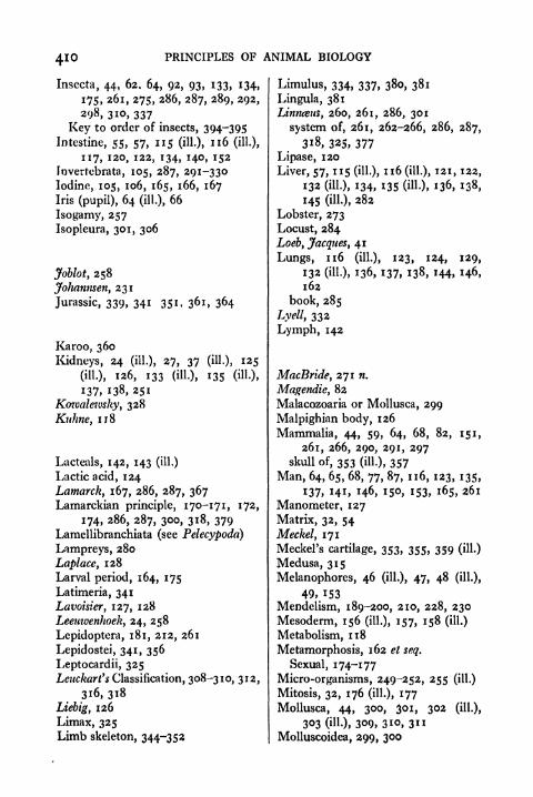
410 PRINCIPLES OF ANIMAL BIOLOGY
Insccta, 44, 62. 64, 92, 93, 133, 134,
175, 261, 2?5> 286, 287, 289, 292,
298, 3io, 337
Key to order of insects, 394-395
Intestine, 55, 57, 115 (ill.), 116 (ill.),
117, 120, 122, 134, HO, 152
Fnverrebrata, 105, 287, 291-330
Iodine, 105, 106, 165, 166, 167Iris (pupil), 64 (ill.), 66
Isogamy, 257
Isopleura, 301, 306
Joblot, 258
Johanmen, 231
Jurassic, 339, 341 351, 361, 364
Karoo, 360
Kidneys, 24 (ill.), 27, 37 (iU-)i 125
(ill.), 126, 133 (ill), 135 (ill-),
137, 138, 251
Kowalewsky, 328
Kithne, 118
Lacteals, 142, 143 (ill.)
Lactic acid, 124
Lamarck, 167, 286, 287, 367Lamarckian principle, 170-171, 172,
174, 286, 287, 300, 318, 379Lamellibranchiata (see Pelecypoda)
Lampreys, 280
Laplace, 128
Larval period, 164, 175
Latimeria, 341
Lavoisier, 127, 128
Leeuwenhoek, 24, 258
Lepidoptera, 181, 212, 261
Lepidostei, 341, 356
Leptocardii, 325Leuckart's Classification, 308-310, 312,
316,318
Liebig, 126
Limax, 325Limb skeleton, 344-352
Limulus, 334, 337, 380, 381
Lingula, 381
Linneeus, 260, 261, 286, 301
system of, 261, 262-266, 286, 287,
3i8, 325, 377
Lipase, 120
Liver, 57, 115 (ill.), 116 (ill.), 121, 122,
132 (ill.), 134, 135 (ill-), 136, 138,
145 (ill.), 282
Lobster, 273
Locust, 284
Loeb, Jacques , 41
Lungs, 116 (ill.), 123, 124, 129,
132 (ill.), 136, 137, 138, H4, H6,162
book, 285
Lyell, 332
Lymph, 142
MacBride, 271 n.
Magendie, 82
Malacozoaria or Mollusca, 299
Malpighian body, 126
Mammalia, 44, 59, 64, 68, 82, 151,
261, 266, 290, 291, 297skull of, 353 (ill.), 357
Man, 64, 65, 68, 77, 87, 116, 123, 135,
137, 141, 146, 150, 153, 165, 261
Manometer, 127
Matrix, 32, 54
Meckel, 171
Meckel's cartilage, 353, 355, 359 (ill.)
Medusa, 315
Melanophores, 46 (ill.), 47, 48 (ill),
49, 153
Mendelism, 189-200, 210, 228, 230
Mesoderm, 156 (ill.), 157, 158 (ill.)
Metabolism, 118
Metamorphosis, 162 et seq.
Sexual, 174-177
Micro-organisms, 249-252, 255 (ill.)
Mitosis, 32, 176 (ill.), 177
Mollusca, 44, 300, 301, 302 (ill.),
303 (ill.), 309, 3io, 3ii
Molluscoidea, 299, 300
Monotremata, 349
Monotreme, shoulder girdle of, 348
(ill.)
Morgan, 204
Moschops, 364
Matter, Johannes, 75, 81, 204
Muscle, 50 (ill.), 51 OH.), 55, 56, 66,
7i,75,76,77,8o,8i,86,88,ii5,
124, 134, 140
striped, 50, 55, 57, 80, 84 (ill),
85, 87, 88, 142Muscular Activity, 129, 134, 273Muscular movement, 50-52, 57 (ill.),
124, 154
Mussel, 62, 251
Mutants, 167, 205, 206, 216, 219,
245, 376, 377, 378, 379, 384
Myopia, 67
Myotomes or Myomcres, 346
Myriapoda, 44, 261, 289, 298, 337,
385
Myzostomida, 295
Nakahara, 180
Nemathelminthes, 43, 318, 324
Nematocysts, 314
Nemertina, 311, 320
Nephridia, 293, 320Nerve Fibre, 70, 79, 87, 94
afferent, 80, 81, 82, 83, 88, 90, gi
cranial, 87
efferent, 80, 81 82, 88
net, 314, 315
Vagus, 140Nervous Impulse, 75, 76 (ill.), 77 (ill.),
79,80Nervous System, 74, 75, 78, 79, 83,
88, 137, 150, 152, 153
Neurones, 79, 80, 81, 83, 87, 88, 91,
92, 94, 95
Newport, 155
Notochord, 158 (ill.), 160 (ill.)
Nucleus, 26, 29 (ill.), 30, 31 (ill-), 32,
34, 35, 47, 55, 57, 59 (ill.)
Division, 177-185 (ill.)
INDEX
Nucula, 300, 307
4IJ
Nummulites, 332
Obelia, 315
Oesophagus, 115, 116 (ill.)
Olfactory nerves, 88, 91
Oligochoeta, 38, 43, 295
Opalina, 253
Ophivroidea, 312
Opisthopthalmus, 369, 372
Optic nerve, 64 (ill.), 65, 66, 86 (ill.),
88, 90, 91 (ill.)
Organisms, 57, 118
classification of, 32, 34
development of new, 155-185
Osteolepidi, 341, 350, 351, 354, 356
Osteozoaria, 297
Ostia, 283, 284
Ostracodermi, 345
Ovaries, 24 (ill.), 27, 30, 31, 32, 33
(ill.), 34, 36, 37 OH-), 174
Oviducts, 24 (ill.), 27, 30, 34, 37 (ilU,
40
Owen, 300, 334
Oxygen, 99, 100, 101, 102, 117, 148
149, 259, 282, 285
Palaeontology, 332
Pallas, 325
Pancreas, 115 (ill.), 120, 121, 122, 152,
282
Pancreatic juice, 152
Panniza, 82
Parabuthus, 369
Paramecium, 23 (ill.), 30, 59, 250,
251, 252, 253, 254, 256, 257
Parasites, 113
Pariasaurus, 366
Parker, 271 n.
Parthenogenesis, 40-43, 376
Pasteur, 258, 259
Pavlov, 96Pean de Saint Gilles, 118
412 PRINCIPLES OF ANIMAL BIOLOGY
Pelecypoda or Lamellibranchiata, 44,
299, 300, 301, 302, 303, 306, 307
Pellagra, noPelmatozoa, 310
Penis, 37 (ill.)
Pentadactyl limb, 345, 347 (ill.)
Pepsin, 120
Peripatus, 288, 293, 295, 296, 302
Perla, 180
Permian, 360, 361, 364, 365
Pharyngobranchii, 325
Phillips, 171
Phoronis, 311
Physalia, 315
Pigeons, 70, 273
Pigmentary effector system, 45-49,
65, 90 (ill.)
Pigment cells, 153, 155, 156 (see also
Melanophores)Pineal eye, 279, 280
Pineal gland, 280
Pinna, 68 (ill.)
Pisces (see also Fishes), 44, 261, 289
Pithecanthropus, 261
Pituitary body, 86
Pituitary gland, 92, 153, 165
Placenta, 161
Plato, 247
Platyhclminthes, 43, 318, 320
Platypus, 40, 268 (ill.), 349, 3&4
Plesiosaurs, 365
Plumularia, 315Polar Bodies, 179 (ill.)
Polychoeta, 43, 294 (ill.), 295
Polypi, 292, 308
Polyploidy, 375, 376
Polypterini, 341, 350
Polyzoa, 43, 318, 322, 323 (ill.)
Porifera, 43, 316, 317
Pouchet, 259
Priestley, 127, 131
Prochordata, 325
Proteins, 102, 103, 104, 105, 113, 117,
120 121
Protista, 250, 252, 253, 256, 257
Protoplasm, 253, 256
Protoselachii, 341
Protozoa, 253, 255, 257, 258
Pseudopodia, 254, 256
Ptcrobranchia, 330
Pterodactyls, 267 (ill.)
Pteropoda, 291, 299, 301
Puberty, 174
Pyrosoma, 328
Rabbit, 55, 234-238
Radiaria, 299
Radiolaria, 253 (ill.)
Reaction, reversibility of, 118, 119
Reactivity, 17, 45, 49
Receptivity, 18, 45
Receptor organs, 20, 21, 62, 73, 74,
78
Recessive, 194
Rectum, 115, 116 (ill.)
Reflexes, 73, 74, 79, 81, 83, 84, 92,
95,96
Rennet, 120
Reproduction, 21-43, 34~38, 45
Reptilia, 44, 289, 291, 297, 385
Respiration, 112, 123-124 (ill.), 126-
128
Response, 45-73, 112
Retina, 65, 66, 67, 91 (ill.)
Rhynchocephalia, 361 (ill.), 364
Richter, 188
Roesel, 253
Romanes, 370
Rotatoria, 299
Rotifera, 40, 43, 175, 250, 258, 318,
322, 323 (ill.)
Sacculina, 175
Sacculus, 69 (ill.), 70St. Hilaire, 367
Salamander, 46, 149, 167, 168 (ill.),
175, 33i, 332 (ill.), 340, 342, 344,
370
Saliva, 121, 122
Salpa, 328

INDEX 413
Sarcoclaria, 299
Scaphognathite, 285
Scaphopoda, 301, 307
Scheuchzer, 331, 332
Schleiden, 257
Schwann, 257Sciatic nerve, 75, 76, 79, 80, 87
Scorpion, 273, 274, 275 (ill.), 279,
285, 336
Scorpionidae, 368, 369, 372
Scrotum, 37
Scutigera, 337, 380
Scyphozoa, 43, 317
Scytodermata, 310
Secretin, 152
Secretions, 45 (see also Glands), 57-59,
H2, 153
Sedgivick, 271 n.
Segmentation, 35, 39, 158, 175, 186,
287, 288, 291 (see also Egg)
Segregation, principle of, 188, 189,
190. 198 199 (ill.), 227-234 (ill.)
Selachians, fins of, 343 (ill.)
Selection, Theory of Natural, 373-387Seminal fluid, 24, 25, 29, 34, 35, 37,
38, 41, 174Sense organ, 20
Servetus, 130Sex Determination, 183, 184Sex organs, 39
Sexual reproduction, 22-30, 34-38,
40-42, in, 112,252
Sherrington, Prof., 95
Shipley, 271 n.
Shrimp, 63, 70, 71, 94
Siebold, 257
Silurian, 341
Sipunculoidea, 312, 323
Skaife, 197Skeleton: of the frog, 52, 53 (ill.),
54
Skin, 46, 47, 57, 58, 129, 164
Skull, 353-365
Spallanzani, Abbt> 43, 126, 128, 258
Species, 38
Spermathecoe, 38
Spermatozoa, 25, 26 (ill), 27 (ill.), 30,
3i, 32, 34, 35
Sphenodon, 342, 344, 364, 365
Spinal cord, 75, 78, 79, 80, 84, 85, 87,
90 (ill.), 94
Spleen, 142
Sponges (see Port/era)
Spongiaria, 299
Squamata, 365
Stannius, 82
Starling, 152
Statocyst, 71 (ill.), 94
Statolith, 70, 71 (ill.)
Stegocephali, 341, 344, 351, 358 (ill.),
359, 36o
Stegocephalian, 340 (ill.), 356, 358skull of, 355
Stflluti, 250
Steno, 332
Sterility, inStimulus, effect of, 21, 63, 64, 72, 73,
76, 78, 153
Stomach, 55, 115 (ill.), 120
Sturtevant, 384
Subold, von, 287
Succession, principle of, 269, 331-
366, 368
Sugar, 103, 119, 121, 134
Swammerdam, 56
Sympathetic ganglia, 86, 89 (ill.),
90 (ill.)
Synapsis, 83, 179, 217
Tadpole, 25, 30, 32, 33, 42, 52, 54.
144, 162, 163, 164, 165 (ill.),
166 (ill.), 167, 352 (ill.)
Teleostei, 339, 340, 344, 354
Tendon, 28, 137
Terrel, 325
Testes, 24, 27, 29, 31 (ill-), 32, 34,
36, 37 (ill.), in, 133 (ill.), 174,
175
Tetrapoda, 345, 346, 355, 35*, 362Thaliacea, 328
Therapsida, 360
4H PRINCIPLES OF ANIMAL BIOLOGY
Thermopile, 42
Thermorpha, 360, 362, 363 (ill.)
Thompson, Vaughan, 322
Thoraic duct, 142
Thyroid Glands, 105, 106, 153 (ill.),
164, 165, 166, 167, 174, 175
Tissues, 32, 59, 66, 112, 129, 131,
132 (ill.), 134, 137
Toad, 37, 46 48 49 57, 58, 70, 152,
164, 167, 175
Clawed (Xenopus), 20, 47, 48, 49 , 67-8
Trachea, 116, 123 (ill.), 134, 284, 285
Treiratocia, 320
Triassic, 360, 361, 364
Trilobita, 288
Trilobites, 281 (ill.), 334, 336 (ill.),
337, 38o
Trochophore, 306, 307 (ill.)
Trypsin, 120, 122
Tschermaky 190
Tunicata, 299, 300, 310, 318, 323, 324,
325, 328, 329 (ill.)
Turbellaria, 299, 320
Turtle, 63, 64
Tympanum, 68, 69, 70 (ill.)
Tyndall, 259
Type, Principle of unity of, 259-269,
271-330
Ureter, 24 (ill.), 37 (ill-), 125 (ill.)
Urethra, 37 (ill.), 127
Urine, 27, 152
Urinary bladder, 37 (ill.)
Urochorda (see Tunicata)
Uroplectes, 369
Urostyle, 75
Ussher, Archbishop, 247
Uterus, 37 (ill.), 40, 55
Utriculus, 69 (ill.), 70, 88
Vagina, 37 (ill.)
Vas deferens, 37 (ill.)
Vegetative reproduction, 22-23
Veins, 129, 130, 132 (ill.)
Abdominal, 135 (ill.), 136
Azygos, 149
Epigastric, 136
Femoral, 135 (ill.), 136
Hepatic, 135 (ill.)
Hepatic portal, 132, 134, 135 (ill.),
136, 137, 150, 284
Jugular, 135, 142
Musculo-Cutaneous, 135
Postcaval, 135 (ill.), 137, 138, 150
(ill.)
Precaval, 135 (ill.)
Pulmonary, 132 (ill.), 136 (ill.), 137,
141Renal portal, 135 (ill.), 136, 151
Sinus venosus, 135 (ill.), 136 (ill.),
138, 151
Subclavian, 135
Veliger, 306, 307 (ill.)
Venous circulation, 136, 137, 148,
150 (ill.), 151
Ventricle, 132 (ill.), 133 (ill-), 136 (ill.),
137, 138, 144, 145, H7Vermes, 261, 266, 298, 310, 318 (see
also Worms)Vertebral column, 52, 75, 90 (ill.)
Vertebrata, 44, 64, 79, 82, 82, 84, 87,
91, 114, 134, 149, 150, 151, 152,
158, 261, 266, 271, 273, 278, 280,
282, 287, 291, 297, 301, 310, 311,
334, 337, 34iAllies of, 324-325Skeleton of, 52
Vesalius, 51, 78
Vitamins, 108-111, 121
Voltaire, 331
Wallace, 372, 373
Watson, 360
Warburg, 42White Leghorns, 187
Whytt, 78
INDEX 415
Wohler, 102
Wolff, Caspar, 171
Worms, 38, 43, 134, 252, 291, 292, 293Flat (see also Platyhelminthes), 43,
293, 3i8, 319 (ill.), 324Segmented (see Annelida)Thread (see also Nemathelminthes),
321
X-rays, 381
Xenophanes, 331
Xenopus, 20, 47, 48, 49, 67, 68
Yoldia, 305
Zoophyla, 318
Zoophytes, 299, 315

GEORGE ALLEN & UNWIN LTDLONDON: 40 MUSEUM STREET, W.C.I
CAPE TOWN: 58-60 LONG STREETTORONTO: 91 WELLINGTON STREET WEST
BOMHAV: IS GRAHAM ROAD, BALLARD ESTATEWELLINGTON N.Z. : cS KINGS CKKSCKNT, LOWER TIuT'i
SYDNEY, N.S.\\r
: BRADIJURY HOUSE, 55 YORK STREET