Primary End Point (Six Months) Results of the Ranibizumab for Edema of the m Acula in Diabetes...
8
Primary End Point (Six Months) Results of the Ranibizumab for Edema of the mAcula in Diabetes (READ-2) Study Quan Dong Nguyen, MD, MSc, 1 Syed Mahmood Shah, MBBS, 1,2 Jeffery S. Heier, MD, 3 Diana V. Do, MD, 1 Jennifer Lim, MD, 4 David Boyer, MD, 5 Prema Abraham, MD, 6 Peter A. Campochiaro, MD, 1 for the READ-2 Study Group* Objectives: To compare ranibizumab with focal/grid laser or a combination of both in diabetic macular edema (DME). Design: Prospective, randomized, interventional, multicenter clinical trial. Participants: A total of 126 patients with DME. Methods: Subjects were randomized 1:1:1 to receive 0.5 mg of ranibizumab at baseline and months 1, 3, and 5 (group 1, 42 patients), focal/grid laser photocoagulation at baseline and month 3 if needed (group 2, 42 patients), or a combination of 0.5 mg of ranibizumab and focal/grid laser at baseline and month 3 (group 3, 42 patients). Main Outcome Measures: The primary end point was the change from baseline in best-corrected visual acuity (BCVA) at month 6. Results: At month 6, the mean gain in BCVA was significantly greater in group 1 (7.24 letters, P 0.01, analysis of variance) compared with group 2 (0.43 letters), and group 3 (3.80 letters) was not statistically different from groups 1 or 2. For patients with data available at 6 months, improvement of 3 lines or more occurred in 8 of 37 (22%) in group 1 compared with 0 of 38 (0%) in group 2 (P 0.002, Fisher exact test) and 3 of 40 (8%) in group 3. Excess foveal thickness was reduced by 50%, 33%, and 45% in groups 1, 2, and 3, respectively. Conclusions: During a span of 6 months, ranibizumab injections by the current protocol had a significantly better visual outcome than focal/grid laser treatment in patients with DME. Financial Disclosure(s): Proprietary or commercial disclosure may be found after the references. Ophthalmology 2009;116:2175–2181 © 2009 by the American Academy of Ophthalmology. *READ-2 Investigators and Team Members appear in Appendix 1 (available at http://aaojournal.org). Diabetic retinopathy is the most prevalent cause of vision loss in working-age individuals in developed countries. Severe vision loss occurs because of tractional retinal de- tachments that complicate retinal neovascularization, but the most common cause of moderate vision loss is diabetic macular edema (DME). The pathogenesis of DME is not completely understood, but hypoxia is a contributing factor. 1 Vascular endothelial growth factor (VEGF) is a hypoxia-regulated gene, and VEGF levels are increased in hypoxic or ischemic retina. Hyperglycemia also causes el- evation of VEGF, and even before there is evidence of ischemia, VEGF is elevated in diabetic retinas. 2 Injection of VEGF into mouse eyes causes breakdown of the inner blood–retinal barrier, 3 and sustained release of VEGF in the eyes of monkeys causes macular edema. 4 This combination of observations in patients and animal models led to the hypothesis that VEGF plays an important role in the patho- genesis of DME. An orally active nonselective blocker of VEGF receptors was found to significantly reduce DME, which recurred when the drug was stopped, providing the first suggestion that VEGF antagonists might provide benefit in patients with DME. 5 The development of selective antagonists of VEGF allowed for more definitive testing of the hypothesis. Ranibizumab is a Fab fragment of a humanized monoclonal antibody that binds all isoforms of VEGF-A with high affinity. In a small open-label study in patients with DME, it was found that 4 intraocular injections of 0.5 mg of ranibizumab over the span of 7 months resulted in a mean reduction in excess foveal thickening of 85% and an aver- age improvement in visual acuity of greater than 2 lines. 6 This strongly implicated VEGF in the development of DME and provided preliminary evidence that ranibizumab could provide benefit, suggesting that larger controlled clinical trials should be performed. The Early Treatment of Diabetic Retinopathy Study (ETDRS) has shown that focal/grid laser photocoagulation can reduce the risk for moderate visual loss in eyes with DME. 7 Focal/grid laser therapy is currently standard care and the gold standard with which new treatments are com- pared. We now report the results of a multicenter random- ized trial in which a regimen of intraocular injections of 2175 © 2009 by the American Academy of Ophthalmology ISSN 0161-6420/09/$–see front matter Published by Elsevier Inc. doi:10.1016/j.ophtha.2009.04.023
-
Upload
independent -
Category
Documents
-
view
1 -
download
0
Transcript of Primary End Point (Six Months) Results of the Ranibizumab for Edema of the m Acula in Diabetes...
Primary End Point (Six Months) Results ofthe Ranibizumab for Edema of the mAculain Diabetes (READ-2) Study
Quan Dong Nguyen, MD, MSc,1 Syed Mahmood Shah, MBBS,1,2 Jeffery S. Heier, MD,3 Diana V. Do, MD,1
Jennifer Lim, MD,4 David Boyer, MD,5 Prema Abraham, MD,6 Peter A. Campochiaro, MD,1 for the READ-2Study Group*
Objectives: To compare ranibizumab with focal/grid laser or a combination of both in diabetic macularedema (DME).
Design: Prospective, randomized, interventional, multicenter clinical trial.Participants: A total of 126 patients with DME.Methods: Subjects were randomized 1:1:1 to receive 0.5 mg of ranibizumab at baseline and months 1, 3, and
5 (group 1, 42 patients), focal/grid laser photocoagulation at baseline and month 3 if needed (group 2, 42 patients),or a combination of 0.5 mg of ranibizumab and focal/grid laser at baseline and month 3 (group 3, 42 patients).
Main Outcome Measures: The primary end point was the change from baseline in best-corrected visualacuity (BCVA) at month 6.
Results: At month 6, the mean gain in BCVA was significantly greater in group 1 (�7.24 letters, P � 0.01,analysis of variance) compared with group 2 (�0.43 letters), and group 3 (�3.80 letters) was not statisticallydifferent from groups 1 or 2. For patients with data available at 6 months, improvement of 3 lines or moreoccurred in 8 of 37 (22%) in group 1 compared with 0 of 38 (0%) in group 2 (P � 0.002, Fisher exact test) and3 of 40 (8%) in group 3. Excess foveal thickness was reduced by 50%, 33%, and 45% in groups 1, 2, and 3,respectively.
Conclusions: During a span of 6 months, ranibizumab injections by the current protocol had a significantlybetter visual outcome than focal/grid laser treatment in patients with DME.
Financial Disclosure(s): Proprietary or commercial disclosure may be found after the references.Ophthalmology 2009;116:2175–2181 © 2009 by the American Academy of Ophthalmology.
*READ-2 Investigators and Team Members appear in Appendix 1 (available at http://aaojournal.org).
Diabetic retinopathy is the most prevalent cause of visionloss in working-age individuals in developed countries.Severe vision loss occurs because of tractional retinal de-tachments that complicate retinal neovascularization, butthe most common cause of moderate vision loss is diabeticmacular edema (DME). The pathogenesis of DME is notcompletely understood, but hypoxia is a contributingfactor.1 Vascular endothelial growth factor (VEGF) is ahypoxia-regulated gene, and VEGF levels are increased inhypoxic or ischemic retina. Hyperglycemia also causes el-evation of VEGF, and even before there is evidence ofischemia, VEGF is elevated in diabetic retinas.2 Injection ofVEGF into mouse eyes causes breakdown of the innerblood–retinal barrier,3 and sustained release of VEGF in theeyes of monkeys causes macular edema.4 This combinationof observations in patients and animal models led to thehypothesis that VEGF plays an important role in the patho-genesis of DME.
An orally active nonselective blocker of VEGF receptorswas found to significantly reduce DME, which recurred
when the drug was stopped, providing the first suggestion© 2009 by the American Academy of OphthalmologyPublished by Elsevier Inc.
that VEGF antagonists might provide benefit in patientswith DME.5 The development of selective antagonists ofVEGF allowed for more definitive testing of the hypothesis.Ranibizumab is a Fab fragment of a humanized monoclonalantibody that binds all isoforms of VEGF-A with highaffinity. In a small open-label study in patients with DME,it was found that 4 intraocular injections of 0.5 mg ofranibizumab over the span of 7 months resulted in a meanreduction in excess foveal thickening of 85% and an aver-age improvement in visual acuity of greater than 2 lines.6
This strongly implicated VEGF in the development of DMEand provided preliminary evidence that ranibizumab couldprovide benefit, suggesting that larger controlled clinicaltrials should be performed.
The Early Treatment of Diabetic Retinopathy Study(ETDRS) has shown that focal/grid laser photocoagulationcan reduce the risk for moderate visual loss in eyes withDME.7 Focal/grid laser therapy is currently standard careand the gold standard with which new treatments are com-pared. We now report the results of a multicenter random-
ized trial in which a regimen of intraocular injections of2175ISSN 0161-6420/09/$–see front matterdoi:10.1016/j.ophtha.2009.04.023
Ophthalmology Volume 116, Number 11, November 2009
ranibizumab was compared with focal/grid laser photoco-agulation over the course of 6 months. A potential imped-iment to the use of ranibizumab is the chronic nature ofDME that could require long-term injections. A potentialproblem with focal/grid laser is that severe edema maymake treatment more technically difficult and less effective,because the edematous retina is less transparent so thatsome laser energy is absorbed by the inner retina, which isundesirable. Also, the lack of transparency makes it difficultto assess when an appropriate end point is achieved. Inject-ing ranibizumab 1 week before focal/grid laser could reducethe amount of thickening and improve the transparencyof the retina, thereby facilitating the laser treatment. Perhapsthe more precise focal/grid laser could lead to long-termstability and eliminate the need for continued injections ofranibizumab. Therefore, combination treatment with ranibi-zumab and focal/grid laser was also tested.
Materials and Methods
This is a phase II, randomized clinical trial conducted at 14 sites inthe United States through an investigator-initiated InvestigationalNew Drug granted by the Food and Drug Administration. Thestudy adhered to the guidelines of the Declaration of Helsinki, andthe protocol and consent form were approved by a local investi-gational review board for some sites and by the Western Institu-tional Review Board for others. Each subject provided writteninformed consent. The study was monitored by an independentdata and safety monitoring committee. The study is registered atwww.clinicaltrials.gov under the identifier NCT00407381.
Patient Eligibility and Exclusion CriteriaPatients (aged �18 years) with type 1 or 2 diabetes and DME wereeligible if they had reduction in visual acuity between 20/40 and20/320 and met the following criteria: (1) center subfield thicknessmeasured by optical coherence tomography (OCT) �250 �m, (2)glycosylated hemoglobin �6% within 12 months before random-ization, (3) no potential contributing causes to reduced visualacuity other than DME, (4) reasonable expectation that scatterlaser photocoagulation would not be required for the next 6months. Patients were excluded if they had received focal/gridlaser treatment within 3 months, intraocular injection of steroidwithin 3 months, or intraocular injection of a VEGF antagonistwithin 2 months. If both eyes were eligible, the eye with thegreater center subfield thickness was entered.
Study ProtocolConsenting patients were screened for the study with a medicalhistory, physical examination, measurement of best-corrected vi-sual acuity (BCVA) by an experienced examiner using the ETDRSprotocol, a slit-lamp examination, measurement of intraocularpressure, dilated funduscopic examination, an OCT evaluation, afluorescein angiogram, and laboratory tests on blood and urine.Eligible patients were randomized 1:1:1 to injections of 0.5 mg ofranibizumab alone (group 1), focal/grid laser alone (group 2), orcombination treatment consisting of injection of 0.5 mg of ranibi-zumab and focal/grid laser (group 3). Patients in group 1 receivedan injection of ranibizumab at baseline and months 1, 3, and 5.Patients in group 2 received focal/grid laser photocoagulation atbaseline and again at month 3 if center subfield thickness was
�250 �m. At baseline and month 3, patients in group 3 received2176
an intraocular injection of ranibizumab followed by focal/gridlaser treatment 1 week later. Month 6 was the primary end point ofthe study. After month 6, patients were eligible to receive intraoc-ular injections of ranibizumab no more than every 2 months orfocal/grid laser treatment no more than every 3 months if theretreatment criterion of center subfield thickness of �250 �m wasmet. Safety evaluations, measurement of BCVA, eye examina-tions, and OCT scans were done at all study visits. Fluoresceinangiography was performed at baseline and 3 and 6 months.Measurements of glycosylated hemoglobin were done at baselineand 3 and 6 months. Hematology and blood chemistry tests wereperformed at baseline and 6 months.
Administration of Study Drug
A lid speculum was inserted, and after topical anesthesia theinjection site was cleaned with 5% povidone iodine. Additionaltopical anesthesia or subconjunctival injection of 2% lidocaine wasgiven, and 0.5 mg of ranibizumab was injected through the parsplana into the vitreous cavity. The fundus was examined to ensureretinal perfusion after the injection, and patients were observed for1 hour or until intraocular pressure returned to normal. Patientswere called the day after each injection and were asked if they haddecreased vision, eye pain, unusual redness, or any new symptoms.
Focal/Grid Laser Photocoagulation
The ETDRS protocol8 with some modifications (50-�m light grayspots) was used for focal/grid laser treatment. Focal treatment wasadministered to each leaking microaneurysm, and grid treatmentwas placed in areas of thickened retina and areas of nonperfusionbetween 500 and 3000 �m from the center of the fovea.
Data Collection and Management
The Retinal Imaging Research and Reading Center (RIRRC) at theWilmer Eye Institute served as the coordinating, data management,and reading center. Personnel from the participating sites werecertified by RIRRC to perform digital fluorescein angiography andOCT based on standardized protocols developed by the RIRRC.Visual acuity examiners were required to be certified by EMMESCorporation or by a multicenter Phase II/III clinical trial. Datawere collected online using a customized version of StudyTrax(ScienceTRAX Inc., Jacksonville, FL), and training was providedto each site for use of the online system. Files for fluoresceinangiography and OCT were uploaded to the RIRRC web site.Coordinators at the RIRRC monitored the database weekly andalerted sites of missed visits or failure to upload files and followedup until these tasks were completed.
Optical Coherence Tomography
At each clinical site, OCT scans were performed by a certifiedtechnician with a StratusOCT (Carl Zeiss Meditec, Dublin, CA)using the fast macular scan protocol. This protocol consists of 6line scans that are 6.0 mm long centered on fixation and spaced 30degrees apart around the circumference of a circle. Each lineconsists of 128 A-scan measurements. With each A-scan, the OCTsoftware measures the distance between the inner surface of theretina and the anterior border of retinal pigmented epithelium-choriocapillaris. The center subfield thickness, the average of 21measurements along the central 1 mm of each of the 6 scans (totalof 126 measurements), was used as a measure of foveal thickness.Readers at the RIRRC examined the images for each OCT file to
be sure that there were no artifacts such as misidentification of inner
Nguyen et al � READ-2 Six-Month Results
or outer surface of the retina. When artifacts were present, correctedmeasurements were obtained using RetinaTomographer software(version 1.1, RIRRC, Baltimore, MD). Macular volume through-out the entire 6-mm zone is calculated using extrapolated valuesbetween the line scans. Excess foveal thickness was calculated bysubtracting the measured foveal thickness value from 212 �m, theupper limit of the normal range of center subfield thickness deter-mined from measurements on a large population of subjects.9
Excess macular volume was determined by subtracting the upperlimit of the normal range of 6.47�0.37 mm3 from the measuredvalue.
Data Safety and Monitoring Committee
An independent Data Safety and Monitoring Committee made upof 2 retina specialists with expertise in clinical trials monitoredadverse events and data at regular intervals.
Statistical Analyses
The primary outcome measure was the change in BCVA betweenbaseline and month 6. Secondary vision-related outcome measureswere the change in BCVA between baseline and month 3 and thepercentage of patients with 3 or more lines or 2 or more linesimprovement at month 6. Secondary anatomic outcomes were thechange in foveal thickness between baseline and month 6 and thepercentage of patients with elimination of 90% or 50% excessfoveal thickness.
Change from baseline in ETDRS visual acuity and change frombaseline in excess foveal thickness were compared across the 3groups at months 3 and 6 using 1-way analysis of variance withBonferroni post hoc analysis. Outcome comparison between dif-ferent time points within a group was done using a single-samplet test. Secondary anatomic and functional outcomes were com-pared using a 2-sided Fisher exact test.
Results
Baseline Characteristics of the Study Groups
The baseline characteristics of the 126 patients who were random-ized in the study are listed in Table 1. The 3 groups were balancedwith respect to mean BCVA, excess foveal thickness, and glyco-sylated hemoglobin. At baseline, 78 of the 126 patients had hy-
Table 1. Baseline Characteristics of Randomized Patients
Group 1:Ranibizumab
(n � 42)
Group 2:Laser
(n � 42)
Group 3:Ranibizumab
(n � 42)
Gender (% women) 69 55 52Race (% Caucasian) 76 64 69Age (mean yrs) 62 62 62Mean BCVA (ETDRS letters
read)24.85 28.35 24.87
Mean BCVA (Snellenequivalent)
20/80 20/80�3 20/80
Mean excess foveal thickness(�m)
198.75 227.67 262.52
Hemoglobin A1C (mean mg/dl) 7.39 7.77 7.59
BCVA � best-corrected visual acuity; ETDRS � Early Treatment Dia-
betic Retinopathy Study.percholesterolemia that required treatment, and this was balancedamong the groups: 26 patients in group 1, 29 patients in group 2,and 23 patients in group 3. There were no significant differences inany baseline characteristics among the 3 groups.
Missing Values and Early Terminations
Data were available for 115 of 126 patients at the 6-month primaryend point. Nine patients exited the study before the primary endpoint for the reasons listed in Table 2. The treatment randomiza-tion, time point of the patients’ last visit, and BCVA at baselineand last visit are also shown. One patient in group 3 died of acerebral vascular accident 6 weeks after study entry and injectionof ranibizumab. Three patients were lost to follow-up, and 3patients withdrew consent. One patient received scatter laser pho-tocoagulation by an ophthalmologist not participating in the studyand by protocol had to exit the study. One patient in group 2 hada substantial decrease in BCVA at the 3-month visit, which wasreported to be due to a combination of marked worsening of DMEand vitreous hemorrhage. The treating physician thought it was inthe patient’s best interest to exit the study and receive alternativetreatment. Four patients (1 patient in group 1, 2 patients in group2, and 1 patient in group 3) withdrew from the study beforereceiving any treatment. Two patients (both in group 1) missed thewindow for the 6-month visit. All patients who did not have6-month visit data, but had any posttreatment data at a time pointbefore month 6, had the last observation carried forward foranalysis of the primary outcome.
Primary Outcome Measure: Mean Change fromBaseline in Best-Corrected Visual Acuity
In group 2, the laser only group, 36 patients had a center subfieldthickness �250 �m at 3 months; 32 patients received additionalfocal/grid laser and 4 patients were judged to have maximum laserin the macula and were not treated. At month 6, the mean gain inBCVA was 7.24 letters in group 1, which was significantly betterthan the outcome in group 2, in which there was a mean loss of0.43 letters (Fig 1; P � 0.0001, analysis of variance with Bonfer-roni post hoc analysis). There was no statistically significant dif-ference between group 1 and group 3, in which there was a meangain of 3.8 letters (P � 0.08). Thus, with regard to the primaryoutcome of mean change in BCVA at 6 months, the currentregimen of injecting 0.5 mg of ranibizumab for 2 months and then2 additional injections 2 months apart was superior to focal/gridlaser treatment.
Secondary Vision-Related Outcome Measures
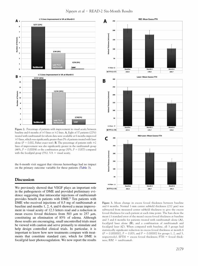
The mean change in BCVA between baseline and 3 months ingroup 1 was 3.98 letters, significantly better than the mean loss of1.48 letters in group 2 (P � 0.01, analysis of variance withBonferroni post hoc analysis), but not significantly different fromthe 1.93 letters gain (P � 0.22) in group 3 (Fig 1). Of the 37patents in group 1 for whom data were available at 6 months, 8(22%) had an improvement of 3 or more lines of BCVA and 17(46%) had an improvement of 2 or more lines (Fig 2A). None ofthe 38 patients with only focal/grid laser therapy in group 3improved by 3 or more lines and 2 patients (5%) improved by 2more lines, both significantly less than in the ranibizumab group.Three of 40 patients (8%) in group 2 who had combined treatmentimproved by 3 or more lines, and 12 patients (30%) improved by
2 or more lines.2177
c mac
Ophthalmology Volume 116, Number 11, November 2009
Secondary Anatomic Outcome Measures
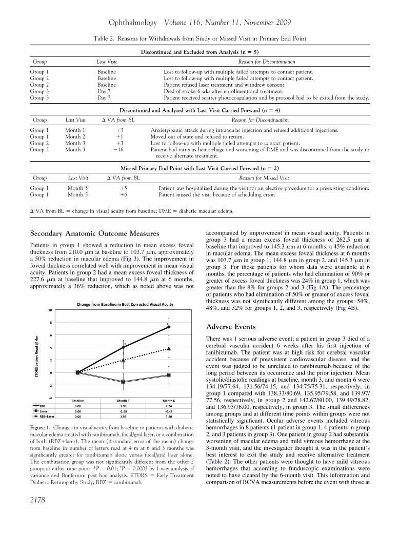
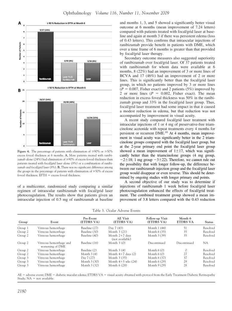
Patients in group 1 showed a reduction in mean excess fovealthickness from 210.0 �m at baseline to 103.7 �m, approximatelya 50% reduction in macular edema (Fig 3). The improvement infoveal thickness correlated well with improvement in mean visualacuity. Patients in group 2 had a mean excess foveal thickness of227.6 �m at baseline that improved to 144.8 �m at 6 months,approximately a 36% reduction, which as noted above was not
Table 2. Reasons for Withdrawals from
Discontinued and Exclu
Group Last Visit
Group 1 Baseline Lost to followGroup 2 Baseline Lost to followGroup 2 Baseline Patient refuseGroup 3 Day 7 Died of strokeGroup 3 Day 7 Patient receiv
Discontinued and Analyzed with
Group Last Visit � VA from BL
Group 1 Month 1 �3 Anxiety/panic attacGroup 1 Month 2 �1 Moved out of stateGroup 2 Month 3 �3 Lost to follow-up wGroup 2 Month 3 �16 Patient had vitreou
receive alternate
Missed Primary End Point with
Group Last Visit � VA from BL
Group 1 Month 5 �5 Patient was hospGroup 1 Month 5 �6 Patient missed t
� VA from BL � change in visual acuity from baseline; DME � diabeti
Figure 1. Changes in visual acuity from baseline in patients with diabeticmacular edema treated with ranibizumab, focal/grid laser, or a combinationof both (RBZ�laser). The mean (�standard error of the mean) changefrom baseline in number of letters read at 4 m at 6 and 3 months wassignificantly greater for ranibizumab alone versus focal/grid laser alone.The combination group was not significantly different from the other 2groups at either time point. *P � 0.01; †P � 0.0003 by 1-way analysis ofvariance and Bonferroni post hoc analysis. ETDRS � Early Treatment
Diabetic Retinopathy Study; RBZ � ranibizumab.2178
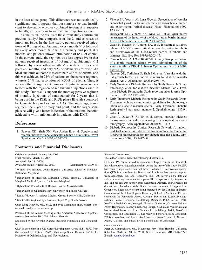
accompanied by improvement in mean visual acuity. Patients ingroup 3 had a mean excess foveal thickness of 262.5 �m atbaseline that improved to 145.3 �m at 6 months, a 45% reductionin macular edema. The mean excess foveal thickness at 6 monthswas 103.7 �m in group 1, 144.8 �m in group 2, and 145.3 �m ingroup 3. For those patients for whom data were available at 6months, the percentage of patients who had elimination of 90% orgreater of excess foveal thickness was 24% in group 1, which wasgreater than the 8% for groups 2 and 3 (Fig 4A). The percentageof patients who had elimination of 50% or greater of excess fovealthickness was not significantly different among the groups: 54%,48%, and 32% for groups 1, 2, and 3, respectively (Fig 4B).
Adverse Events
There was 1 serious adverse event; a patient in group 3 died of acerebral vascular accident 6 weeks after his first injection ofranibizumab. The patient was at high risk for cerebral vascularaccident because of preexistent cardiovascular disease, and theevent was judged to be unrelated to ranibizumab because of thelong period between its occurrence and the prior injection. Meansystolic/diastolic readings at baseline, month 3, and month 6 were134.19/77.64, 131.56/74.15, and 134.75/75.31, respectively, ingroup 1 compared with 138.33/80.69, 135.95/79.58, and 139.97/77.56, respectively, in group 2 and 142.67/80.00, 139.49/78.82,and 136.93/76.00, respectively, in group 3. The small differencesamong groups and at different time points within groups were notstatistically significant. Ocular adverse events included vitreoushemorrhages in 8 patients (1 patient in group 1, 4 patients in group2, and 3 patients in group 3). One patient in group 2 had substantialworsening of macular edema and mild vitreous hemorrhage at the3-month visit, and the investigator thought it was in the patient’sbest interest to exit the study and receive alternative treatment(Table 2). The other patients were thought to have mild vitreoushemorrhages that according to funduscopic examinations werenoted to have cleared by the 6-month visit. This information and
y or Missed Visit at Primary End Point
from Analysis (n � 5)
Reason for Discontinuation
ith multiple failed attempts to contact patient.ith multiple failed attempts to contact patient.r treatment and withdrew consent.s after enrollment and treatment.
atter photocoagulation and by protocol had to be exited from the study.
Visit Carried Forward (n � 4)
Reason for Discontinuation
ing intraocular injection and refused additional injections.efused to return.ultiple failed attempts to contact patient.orrhage and worsening of DME and was discontinued from the study toent.
Visit Carried Forward (n � 2)
Reason for Missed Visit
ed during the visit for an elective procedure for a preexisting condition.sit because of scheduling error.
ular edema.
Stud
ded
-up w-up wd lase
6 wked sc
Last
k durand rith ms hemtreatm
Last
italizhe vi
comparison of BCVA measurements before the event with those at
Nguyen et al � READ-2 Six-Month Results
the 6-month visit suggest that vitreous hemorrhage had no impacton the primary outcome variable for these patients (Table 3).
Discussion
We previously showed that VEGF plays an important rolein the pathogenesis of DME and provided preliminary evi-dence suggesting that intraocular injections of ranibizumabprovides benefit in patients with DME.6 Ten patients withDME who received injections of 0.5 mg of ranibizumab atbaseline and months 1, 2, 4, and 6 showed a mean improve-ment in visual acuity of 12.3 letters read and a reduction inmean excess foveal thickness from 503 �m to 257 �m,constituting an elimination of 85% of edema. Althoughthose results are encouraging, small uncontrolled trials mustbe viewed with caution and serve primarily to stimulate andhelp design controlled clinical trials. In particular, it isimportant to know how new treatments compare with treat-ments that constitute standard care, which for DME is
Figure 2. Percentage of patients with improvement in visual acuity betweenbaseline and 6 months of �3 lines or �2 lines. A, Eight of 37 patients (22%)treated with ranibizumab for whom data were available at 6 months improved�3 lines, which was significantly greater than 0% of patients treated with laseralone (P � 0.002, Fisher exact test). B, The percentage of patients with �2lines of improvement was also significantly greater in the ranibizumab group(46%, P � 0.00004) or the combination group (30%, P � 0.007) comparedwith the focal/grid group (5%). VA � visual acuity.
focal/grid laser photocoagulation. We now report the results
Figure 3. Mean change in excess foveal thickness between baselineand 6 months. Normal 1-mm center subfield thickness (212 �m) wassubtracted from measured center subfield thickness to give the excessfoveal thickness for each patient at each time point. The bars show themean (�standard error of the mean) excess foveal thickness at baselineand 3 and 6 months for patients treated with ranibizumab alone (A),focal/grid laser alone (B), and a combination of ranibizumab andfocal/grid laser (C). When compared with baseline, all 3 groups hadstatistically significant reduction in excess foveal thickness at month 6(P � 0.0000002, P � 0.003, and P � 0.000002 for groups 1, 2, and 3,respectively). EFTH � excess foveal thickness; FTH � foveal thick-
ness; RBZ � ranibizumab.2179
Ophthalmology Volume 116, Number 11, November 2009
of a multicenter, randomized study comparing a similarregimen of intraocular ranibizumab with focal/grid laserphotocoagulation. The results show that patients given anintraocular injection of 0.5 mg of ranibizumab at baseline
Figure 4. The percentage of patients with elimination of �90% or �50%excess foveal thickness at 6 months. A, More patients treated with ranibi-zumab alone (24%) had elimination of �90% of excess foveal thickness thanpatients treated with focal/grid laser alone (8%) or a combination of ranibi-zumab and focal/grid laser (8%). B, There was no significant difference amongthe groups in the percentage of patients with elimination of �50% of excessfoveal thickness. EFTH � excess foveal thickness.
Table 3. Ocul
Group EventPre-Event
(ETDRS VA)
Group 1 Vitreous hemorrhage Baseline (27) DayGroup 2 Vitreous hemorrhage Baseline (30) MoGroup 2 Vitreous hemorrhage Baseline (40) Mo
(Group 2 Vitreous hemorrhage and
worsening of DMEBaseline (16) Mo
Group 2 Vitreous hemorrhage Baseline (2) MoGroup 2 Vitreous hemorrhage Month 3 (4) MoGroup 3 Vitreous hemorrhage Day 7 (27) MoGroup 3 Vitreous hemorrhage Month 3 (30) MoGroup 3 Vitreous hemorrhage Month 3 (32) Mo
AE � adverse event; DME � diabetic macular edema; ETDRS VA � visua
Study; NA � not available.2180
and months 1, 3, and 5 showed a significantly better visualoutcome at 6 months (mean improvement of 7.24 letters)compared with patients treated with focal/grid laser at base-line and again at month 3 if there was persistent edema (lossof 0.43 letters). This confirms that intraocular injections ofranibizumab provide benefit in patients with DME, whichover a time frame of 6 months is greater than that providedby focal/grid laser therapy.
Secondary outcome measures also suggested superiorityof ranibizumab over focal/grid laser. Of 37 patients treatedwith ranibizumab for whom data were available at 6months, 8 (22%) had an improvement of 3 or more lines ofBCVA and 17 (46%) had an improvement of 2 or morelines. This is significantly better than the focal/grid lasergroup, in which no patients improved by 3 or more lines(P � 0.007, Fisher exact) and 2 patients (5%) improved by2 or more lines (P � 0.002, Fisher exact). The meanreduction in excess foveal thickness was 50% in the ranibi-zumab group and 33% in the focal/grid laser group. Thus,focal/grid laser treatment had some impact in that it causeda modest reduction in edema, but that reduction was notaccompanied by improvement in visual acuity.
A recent study compared focal/grid laser treatment withintraocular injections of 1 or 4 mg of preservative-free triam-cinolone acetonide with repeat treatments every 4 months forpersistent or recurrent DME.10 At 4 months, mean improve-ment in visual acuity was significantly better in the 2 triam-cinolone groups compared with the focal/grid laser group, butat the 2-year primary end point the focal/grid laser groupshowed a mean improvement of 1�17, which was signifi-cantly better than the triamcinolone groups (4 mg group,�2�18; 1 mg group �3�22). Therefore, we cannot rule outthe possibility that with longer follow-up, the difference be-tween our ranibizumab injection group and the focal/grid lasergroup would disappear or even reverse. This should be deter-mined by ongoing studies with longer primary end points.
A second objective of our study was to determine ifinjections of ranibizumab 1 week before focal/grid laserphotocoagulation enhanced the effects of focal/grid treat-ment. The combined treatment group showed a mean im-provement of 3.8 letters compared with the 0.43 reduction
dverse Events
VisitRS VA)
Follow-up Visit(ETDRS VA)
Month 6ETDRS VA Status
3) Month 1 (44) 51 Resolved(21) Month 6 (35) 35 Resolved
�7 daysvailable)
Month 3 (39) 35 Resolved
(0) Discontinued Discontinued NA
(4) Month 6 (0) 0 Resolved�7 days (2) Month 6 (0) 27 Resolved(35) Month 6 (37) 37 Resolved
�3 wks (24) Month 6 (29) 29 Resolved(28) Month 9 (29) 28 Resolved
ity obtained with protocol from the Early Treatment Diabetic Retinopathy
ar A
AE(ETD
7 (4nth 3nth 2not anth 3
nth 3nth 4nth 3nth 4nth 6
l acu
Nguyen et al � READ-2 Six-Month Results
in the laser alone group. This difference was not statisticallysignificant, and it appears that our sample size was insuffi-cient to determine whether combined treatment is superiorto focal/grid therapy or to ranibizumab injections alone.
In conclusion, the results of the current study confirm ourprevious study,6 but comparison of the 2 studies raises animportant issue. In the first study, patients received injec-tions of 0.5 mg of ranibizumab every month � 3 followedby every other month � 2 with a primary end point at 7months, and patients showed elimination of 85% of edema.In the current study, the regimen was less aggressive in thatpatients received injections of 0.5 mg of ranibizumab � 2followed by every other month � 2 with a primary endpoint at 6 months, and only 50% of edema was resolved. Anideal anatomic outcome is to eliminate �90% of edema, andthis was achieved in 24% of patients on the current regimen,whereas 54% had resolution of �50% of edema. Thus, itappears that a significant number of patients were under-treated with the regimen of ranibizumab injections used inthis study. Our results support the more aggressive regimenof monthly injections of ranibizumab for 2 years that isbeing used in the RISE and RIDE phase III trials sponsoredby Genentech (San Francisco, CA). The more aggressiveregimen, the 2-year primary end point, and the larger sam-ple size will give a better indication of the maximal benefitsachievable with ranibizumab in patients with DME.
References
1. Nguyen QD, Shah SM, Van Anden E, et al. Supplementaloxygen improves diabetic macular edema: a pilot study. Invest
Ophthalmol Vis Sci 2003;45:617–24.Footnotes and Financial Disclosures
Professor of Ophthalmology and Neuroscience.
2. Vinores SA, Youssri AI, Luna JD, et al. Upregulation of vascularendothelial growth factor in ischemic and non-ischemic humanand experimental retinal disease. Histol Histopathol 1997;12:99 –109.
3. Derevjanik NL, Vinores SA, Xiao WH, et al. Quantitativeassessment of the integrity of the blood-retinal barrier in mice.Invest Ophthalmol Vis Sci 2002;43:2462–7.
4. Ozaki H, Hayashi H, Vinores SA, et al. Intravitreal sustainedrelease of VEGF causes retinal neovascularization in rabbitsand breakdown of the blood-retinal barrier in rabbits andprimates. Exp Eye Res 1997;64:505–17.
5. Campochiaro PA, C99-PKC412-003 Study Group. Reductionof diabetic macular edema by oral administration of thekinase inhibitor PKC412. Invest Ophthalmol Vis Sci 2004;45:922–31.
6. Nguyen QD, Tatlipinar S, Shah SM, et al. Vascular endothe-lial growth factor is a critical stimulus for diabetic macularedema. Am J Ophthalmol 2006;142:961–9.
7. Early Treatment Diabetic Retinopathy Study Research Group.Photocoagulation for diabetic macular edema: Early Treat-ment Diabetic Retinopathy Study report number 1. Arch Oph-thalmol 1985;103:1796–806.
8. Early Treatment Diabetic Retinopathy Study Research Group.Treatment techniques and clinical guidelines for photocoagu-lation of diabetic macular edema: Early Treatment DiabeticRetinopathy Study report number 2. Ophthalmology 1987;94:761–74.
9. Chan A, Duker JS, Ko TH, et al. Normal macular thicknessmeasurements in healthy eyes using Stratus optical coherencetomography. Arch Ophthalmol 2006;124:193–8.
10. Diabetic Retinopathy Clinical Research Network. A random-ized trial comparing intravitreal triamcinolone acetonide andfocal/grid photocoagulation for diabetic macular edema. Oph-
thalmology 2008;115:1447–59.Originally received: January 16, 2009.Final revision: March 13, 2009.Accepted: April 9, 2009.Available online: August 22, 2009. Manuscript no. 2009-69.1 Wilmer Eye Institute, Johns Hopkins University School of Medicine,Baltimore, Maryland.2 Department of Medicine, Maryland General Hospital, University ofMaryland Medical System, Baltimore, Maryland.3 Ophthalmic Consultants of Boston, Boston, Massachusetts.4 Department of Ophthalmology, University of Illinois, Chicago.5 Retina-Vitreous Associates Medical Group, Beverly Hills, California.6 Black Hills Regional Eye Institute, Rapid City, South Dakota.
Quan Dong Nguyen, MD, MSc, and Syed Mahmood Shah, MBBS, con-tributed equally to the manuscript.
Presented at: the Annual Meeting of the American Academy of Ophthal-mology, November 10, 2008, Atlanta, Georgia.
Sponsored by the Juvenile Diabetes Research Foundation and Genentech,Inc.
QDN is a recipient of a K23 Career Development Award (EY 13552) fromthe National Eye Institute. PAC is the George S. and Dolores Doré Eccles
Financial Disclosure(s):The author(s) have made the following disclosure(s):
QDN and PAC have served as members of Expert Panels for Genentech,Inc. without receiving an honorarium during the time of this study, but JHUhas recently negotiated a contract through which JHU receives compensa-tion. QDN is a consultant for Bausch and Lomb and has research supportfrom Genentech, Inc., and Regeneron, Inc. PAC serves on the data andsafety monitoring committee for a phase III trial sponsored by Regeneron,Inc., and has research support from Genentech, Alimera, and CoMentis fordiabetic macular edema trials. Diana Do receives research support fromGenentech. These activites are being managed by the Conflict of InterestCommittee of the Johns Hopkins University School of Medicine. JSH is aconsultant for Genentech, Alcon, Allergan, Bausch and Lomb, Eyemagi-nations, Fovea, Genzyme, Heidelburg, IScience, ISTA, Jerini, LPath,NeoVista, Nodal Vision, Novagali, Novartis, Optherion, Oxigene, Paloma,Pfizer, Regeneron, Resolvyx, Schering Plough, Scyfix, and VisionCare andhas received honoriaria from Genentech, Heidelberg, Jerini, NeoVista,Optimedica, and Regeneron. JL has received honoriaria from Genentech.DB is a consultant and has received honoraria from Genentech, Novartis,Alcon, Allergan, and Pfizer. PA is a consultant for Genentech.
Correspondence:Peter A. Campochiaro, MD, Maumenee 719, Johns Hopkins UniversitySchool of Medicine, 600 N. Wolfe Street, Baltimore, MD 21287-9277.
E-mail: [email protected].2181
Ophthalmology Volume 116, Number 11, November 2009
Appendix 1. Investigators and Coordinatorsof the READ-2 Study
A. Clinical Sites1. Black Hills Regional Eye Institute:
Principal Investigator: Prema Abraham, MDCoordinator: Buffi Green, Kristi Libermont, HonorEvers
2. East Bay Retina ConsultantsPrincipal Investigator: Eugene S. Lit, MDInvestigators: Daniel A. Brinton, MD, Scott S.Lee, MDCoordinator: Scotty Renslow
3. Eye Care SpecialistsPrincipal Investigator: Erik F. Kruger, MDCoordinator: Patty Yuhas, COA
4. Illinois Retina AssociatesPrincipal Investigator: Jonathan S. Pollack, MDInvestigators: Joseph M. Civantos, MDCoordinator: Barbara J. Ciscato
5. Johns Hopkins University/Wilmer Eye InstitutePrincipal Investigator: Diana V. Do, MDInvestigators: Peter Campochiaro, MD, DanielFinkelstein, MD, Morton F. Goldberg, MD, JuliaA. Haller, MD, James T. Handa, MD, QuanDong Nguyen, MD, MSc, Edward Quinlan, MD,R. C. Andrew Symons, MBBS, PhD, FRANZCO,Jennifer U. Sung, MD, Howard Ying, MD, PhD,Ingrid Zimmer-Galler, MDCoordinators: Lisa Azzaro, COT, Anita Baird,COT, Lisa Greer, COT, MBA, Ovais Shaikh,MD, COT, Jennifer Simmons, COT
6. Midwest Eye InstitutePrincipal Investigator: Thomas Ciulla, MDCoordinator: Neelam Thukral
7. New England Retina ConsultantsPrincipal Investigator: Bradley Foster, MDCoordinator: Sharon Parker
8. Ophthalmic Consultants of BostonPrincipal Investigator: Jeffrey S. Heier, MDInvestigators: Janet J. Chieh, MD, Tina S.Cleary, MD, Gregory L. Fenton, MD, David S.Liao, MD, Jackie K. Nguyen, MD, Trexler M.Topping, MD, Torsten W. Wiegand, MD, PaulA. Yates, MDCoordinator: Lindsey Williams, Paul E. Daniel, Jr.
9. Retina Consultants of ArizonaPrincipal Investigator: Pravin U. Dugel, MDInvestigators: Jack Sipperley, MD, Donald W.Park, MD, Judy Liu, MD, Derek Y. Kunimoto,MD, Edward J. Quinlan, MD, Arthur Mollen,DO, Jaime R. Gaitan, MDCoordinator: Sarah G. Mobley, CCRC
10. Retina Consultants of Nevada - Las VegasPrincipal Investigator: Allen Thach, MDInvestigators: Roger Simon, MD, R. Jeffrey
Parker, MD, Rodney D. Hollifield, MD, Roy H.2181.e1
Loo, MD, Meher Yepremyan, MD, Irene Voo,MD, Jason C. Wickens, MDCoordinators: Janet Seybert, Cassondra Major,Mia Davis, Christy Browder, Melissa Rediker
11. Retina Institute of CaliforniaPrincipal Investigator: Thomas S. Chang, MDInvestigators: Adam Martidis, MDCoordinator: Alexandra N. Tran
12. Retina-Vitreous Associates Medical GroupPrincipal Investigator: David Boyer, MDInvestigators: Roger L. Novack, MD, PhD,Thomas G. Chu, MD, PhD, Firas M. Rahhal,MD, Janet Jill Hopkins, MD, FRCSC, HomayounTabandeh, MD, MS, FRCP, FRCS, FRCOphth,Richard H. Roe, MD, MHSCoordinator: Saba Mukarram, Tammy Gasparyan,Janet Kurokouchi
13. University of New MexicoPrincipal Investigator: Arup Das, MD, PhDInvestigators: Mark Schluter, MDCoordinator: Sheila Nemeth, COMT
14. University of Southern California/Doheny EyeInstitutePrincipal Investigator: Jennifer Lim, MD (year1); Dean Eliot, MD (year 2)Coordinator: Margaret Padilla
B. Steering CommitteePeter A. Campochiaro, MDJeffrey S. Heier, MDJennifer Lim, MDQuan Dong Nguyen, MD, MSc, Chair
C. Data Safety and Monitoring CommitteeBrian P. Conway, MD, University of VirginiaDavid Wilson, MD, Oregon Health and Science Uni-versity, Casey Eye Institute
D. Reading CenterThe Retinal Imaging Research and Reading Center atWilmerMohamed Ibrahim, MDYasir Sepah, MDIzza Khan, MDRoomasa Channa, MDSyed Mahmood Shah, MBBS
E. Data Collection and Monitoring CenterWilmer Eye Institute, Johns Hopkins UniversityKashif Janjua, MDAfsheen Khwaja, MDJohn Putzke, PhD, MSPHSyed Mahmood Shah, MBBS
F. Coordinating CenterWilmer Eye Institute, Johns Hopkins UniversityGulnar Hafiz, MD, MPHVelma Pack, BA
G. Statistical AnalysesBloomberg School of Public Health, Johns HopkinsUniversityAamir J. Khan, MD, PhD
Syed Mahmood Shah, MBBS