Febrile seizures impair memory and cAMP response-element binding protein activation
Upload
khangminh22Category
view
5download
0
Journal of
Clinical Medicine
Review
Prevention, Treatment, and Monitoring of Seizures inthe Intensive Care Unit
Micheal Strein 1, John P. Holton-Burke 2, LaTangela R. Smith 2 and Gretchen M. Brophy 1,*1 Department of Pharmacotherapy and Outcomes Science, Virginia Commonwealth University School of
Pharmacy, Richmond, VA 23298-0533, USA2 Department of Neurology, Virginia Commonwealth University Health System,
Richmond, VA 23298-0599, USA* Correspondence: [email protected]
Received: 19 June 2019; Accepted: 1 August 2019; Published: 7 August 2019�����������������
Abstract: The diagnosis and management of seizures in the critically ill patient can sometimes presenta unique challenge for practitioners due to lack of exposure and complex patient comorbidities.The reported incidence varies between 8% and 34% of critically ill patients, with many patientsoften showing no overt clinical signs of seizures. Outcomes in patients with unidentified seizureactivity tend to be poor, and mortality significantly increases in those who have seizure activitylonger than 30 min. Prompt diagnosis and provision of medical therapy are crucial in order toattain successful seizure termination and prevent poor outcomes. In this article, we review theepidemiology and pathophysiology of seizures in the critically ill, various seizure monitoringmodalities, and recommended medical therapy.
Keywords: neurocritical care; critical care; seizures; status epilepticus; electroencephalography;antiepileptic therapy
1. Introduction
Seizures and status epilepticus (SE) have a large clinical and economic impact on the care ofcritically ill patients worldwide as they are often associated with complicated and lengthy hospitaland intensive care unit (ICU) stays [1]. Neurocritical care (NCC) is a rapidly growing specialty thatspecializes in the care of critically ill patients presenting with primary neurological injuries [2]. For thesepatients, the involvement of expert NCC clinicians has led to significantly better patient outcomes.Some of the most notable NCC specialty areas include seizures and SE, ischemic and hemorrhagic stroke,and traumatic brain injury (TBI). Although seizures are not always the initial injury, critically ill patientsmay develop a secondary neurological deterioration due to ongoing intracranial pathophysiologicchanges and central nervous system (CNS) insults, leading to subsequent seizures or SE. The mostcommon secondary injuries are brain tissue hypoperfusion, brain tissue hypoxia, and excitotoxicdamage due to recurrent seizures [3]. This article will focus on the epidemiology and pathophysiologyof seizures in critically ill patients, as well as how monitoring and therapeutic strategies can aid indiagnosing and treating primary and secondary seizures and SE in this challenging population.
2. Epidemiology
The published incidence of seizures in critically ill patients is highly variable but has been reportedto range from 8% to 34% based on continuous electroencephalography (EEG) monitoring studiespublished from 1994 to 2011 [4]. The most common comorbidities and conditions associated withseizure in critical illness include a pre-existing history of epilepsy, direct CNS insults, metabolicderangements, and drug withdrawal or intoxication [4,5] (Table 1). Of the many potential CNS insults,
J. Clin. Med. 2019, 8, 1177; doi:10.3390/jcm8081177 www.mdpi.com/journal/jcm
J. Clin. Med. 2019, 8, 1177 2 of 17
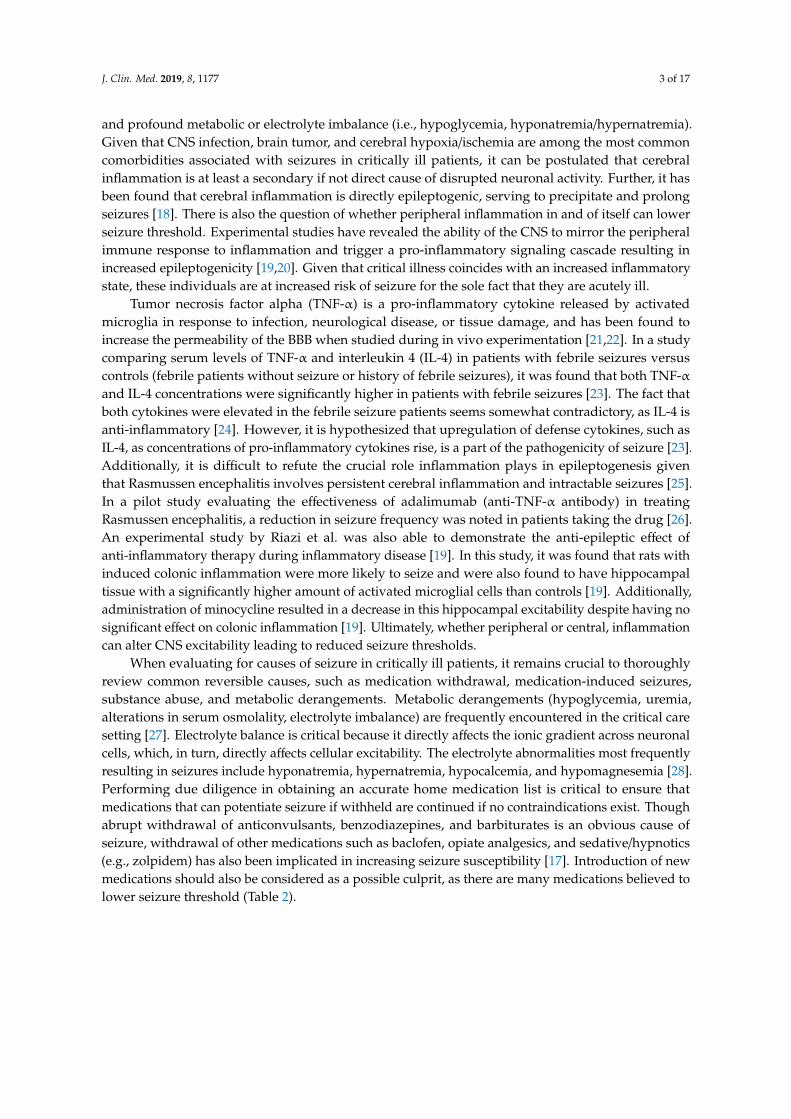
those most frequently associated with seizures are CNS infection, stroke, brain tumor, and neurosurgicalprocedures [4,5]. In critically ill patients with seizures, SE must always be considered and evenanticipated. This is especially true in comatose patients and those without return to baseline or withwaxing/waning mentation. The likelihood of capturing seizure on continuous EEG is highest in youngerpatients, those with pre-existing epilepsy, prior neurosurgical procedure, and convulsion or comatosestate prior to the start of continuous EEG monitoring [5]. Of seizures captured in one study, 34% werenonconvulsive seizures (NCSz), and of these, 76% were nonconvulsive status epilepticus (NCSE) [5].
Table 1. Neurological conditions associated with seizures and status epilepticus in critically Illpatients [4–9].
Condition
Pre-existing epilepsy Traumatic brain injuryCentral nervous system infection Ischemic stroke
Brain tumor Hypoxic ischemic encephalopathyNeurosurgical procedure Altered mental statusIntracerebral hemorrhage Drug toxicity/withdrawalSubarachnoid hemorrhage Toxic metabolic encephalopathy
Subdural hemorrhage Congenital
SE is defined as 5 or more minutes of continuous clinical and/or electrographic seizure activity,or recurrent seizure activity without recovery between seizures. Patients who do not respond tostandard treatment regimens for SE (i.e., benzodiazepine and an anticonvulsant drug) are consideredto be in refractory SE (RSE). Cases where SE continues for 24 h or more after the initiation of anesthetictherapy, including those where SE recurs during reduction or withdrawal of anesthesia, are consideredto be in super-refractory SE (SRSE) [10,11]. The annual incidence of SE in the United States (US) andworldwide is 100,000 to 152,000 and 1.2 to 5 million, respectively [12]. Young, African Americanmales appear to have a higher incidence of SE but lower associated mortality [6]. In a multicentercohort by Shin et al., SE was most commonly associated with cerebrovascular disease, substanceuse, and CNS inflammation [7]. CNS inflammation was due to infection, autoimmune encephalitis,or cryptogenic [7]. Of these, cryptogenic CNS inflammation leading to SE was most challengingto treat and considered an independent risk factor for SRSE [7]. In the US, the most commoncomorbidities associated with SE are consistent with those associated with seizures in critically illpatients, with the inclusion of cerebral anoxia and congenital disorders [6]. If the workup for theseconditions is negative, there should be high suspicion for cryptogenic new-onset refractory statusepilepticus (NORSE) and autoimmune/paraneoplastic syndromes. Liu et al. found that in patients withanti-N-methyl-d-aspartate receptor (NMDAR) encephalitis, 80.7% had seizure during the acute phase ofthe disease [13]. Fifty percent of those with seizure developed SE, with 25% of these being refractory toinitial treatment requiring multiple anticonvulsants plus anesthetic agents (midazolam/propofol) [13].Over one-third of the refractory cases were termed SRSE due to inability to withdraw or reduceanesthetic agents and resulted in patient death [13].
Neurological injuries or secondary neurological injuries from other disease states can also leadto SE (Table 1). In cardiac arrest patients, the major cause of death is hypoxic ischemic brain injurysustained during the arrest [14]. However, a high proportion of the patients that obtain return ofspontaneous circulation go on to develop seizures or SE post-resuscitation [15]. It is not known if SEcontributes to poor outcomes after cardiac arrest or if it is a consequence of the severe brain injury,and overall EEG monitoring is currently of unclear benefit in regard to patient outcomes [16].
3. Pathophysiology
Seizure results from abnormally excessive, neuronal activity as a consequence of the disruptedbalance between neuronal excitation and inhibition [17]. What leads to disruption of this balance isnot always known. Fairly recognized culprits involve breakdown of the blood–brain barrier (BBB)
J. Clin. Med. 2019, 8, 1177 3 of 17
and profound metabolic or electrolyte imbalance (i.e., hypoglycemia, hyponatremia/hypernatremia).Given that CNS infection, brain tumor, and cerebral hypoxia/ischemia are among the most commoncomorbidities associated with seizures in critically ill patients, it can be postulated that cerebralinflammation is at least a secondary if not direct cause of disrupted neuronal activity. Further, it hasbeen found that cerebral inflammation is directly epileptogenic, serving to precipitate and prolongseizures [18]. There is also the question of whether peripheral inflammation in and of itself can lowerseizure threshold. Experimental studies have revealed the ability of the CNS to mirror the peripheralimmune response to inflammation and trigger a pro-inflammatory signaling cascade resulting inincreased epileptogenicity [19,20]. Given that critical illness coincides with an increased inflammatorystate, these individuals are at increased risk of seizure for the sole fact that they are acutely ill.
Tumor necrosis factor alpha (TNF-α) is a pro-inflammatory cytokine released by activatedmicroglia in response to infection, neurological disease, or tissue damage, and has been found toincrease the permeability of the BBB when studied during in vivo experimentation [21,22]. In a studycomparing serum levels of TNF-α and interleukin 4 (IL-4) in patients with febrile seizures versuscontrols (febrile patients without seizure or history of febrile seizures), it was found that both TNF-αand IL-4 concentrations were significantly higher in patients with febrile seizures [23]. The fact thatboth cytokines were elevated in the febrile seizure patients seems somewhat contradictory, as IL-4 isanti-inflammatory [24]. However, it is hypothesized that upregulation of defense cytokines, such asIL-4, as concentrations of pro-inflammatory cytokines rise, is a part of the pathogenicity of seizure [23].Additionally, it is difficult to refute the crucial role inflammation plays in epileptogenesis giventhat Rasmussen encephalitis involves persistent cerebral inflammation and intractable seizures [25].In a pilot study evaluating the effectiveness of adalimumab (anti-TNF-α antibody) in treatingRasmussen encephalitis, a reduction in seizure frequency was noted in patients taking the drug [26].An experimental study by Riazi et al. was also able to demonstrate the anti-epileptic effect ofanti-inflammatory therapy during inflammatory disease [19]. In this study, it was found that rats withinduced colonic inflammation were more likely to seize and were also found to have hippocampaltissue with a significantly higher amount of activated microglial cells than controls [19]. Additionally,administration of minocycline resulted in a decrease in this hippocampal excitability despite having nosignificant effect on colonic inflammation [19]. Ultimately, whether peripheral or central, inflammationcan alter CNS excitability leading to reduced seizure thresholds.
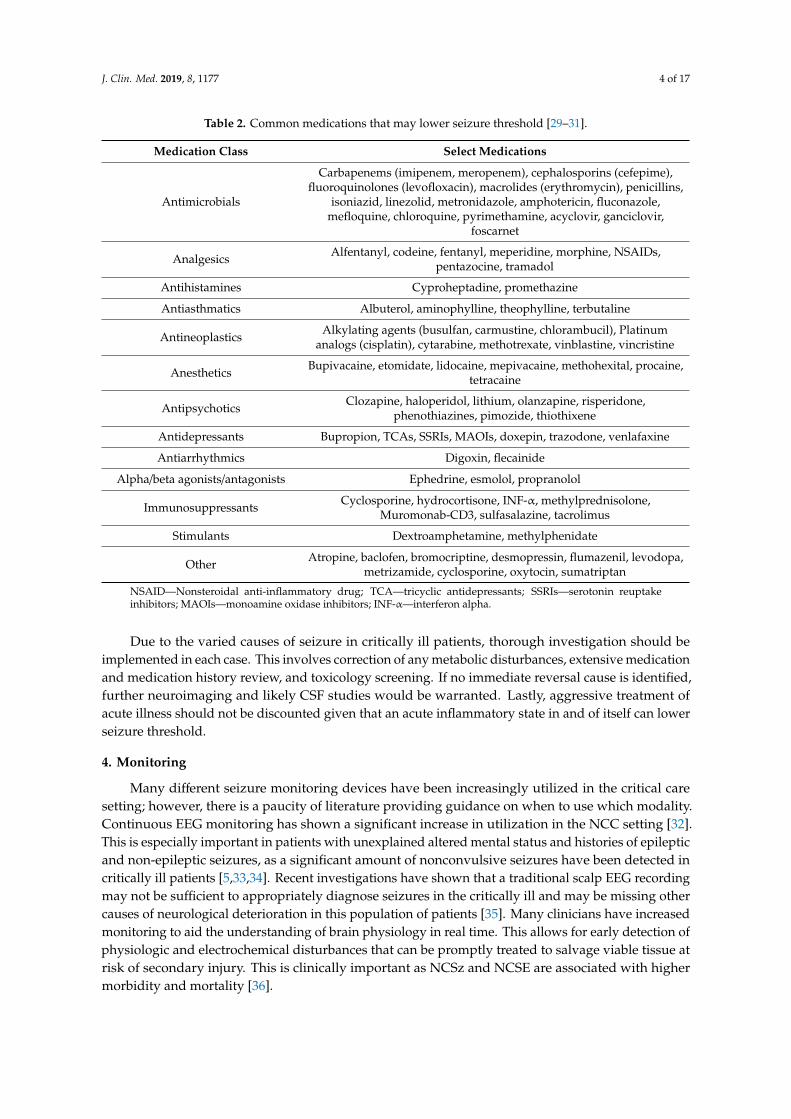
When evaluating for causes of seizure in critically ill patients, it remains crucial to thoroughlyreview common reversible causes, such as medication withdrawal, medication-induced seizures,substance abuse, and metabolic derangements. Metabolic derangements (hypoglycemia, uremia,alterations in serum osmolality, electrolyte imbalance) are frequently encountered in the critical caresetting [27]. Electrolyte balance is critical because it directly affects the ionic gradient across neuronalcells, which, in turn, directly affects cellular excitability. The electrolyte abnormalities most frequentlyresulting in seizures include hyponatremia, hypernatremia, hypocalcemia, and hypomagnesemia [28].Performing due diligence in obtaining an accurate home medication list is critical to ensure thatmedications that can potentiate seizure if withheld are continued if no contraindications exist. Thoughabrupt withdrawal of anticonvulsants, benzodiazepines, and barbiturates is an obvious cause ofseizure, withdrawal of other medications such as baclofen, opiate analgesics, and sedative/hypnotics(e.g., zolpidem) has also been implicated in increasing seizure susceptibility [17]. Introduction of newmedications should also be considered as a possible culprit, as there are many medications believed tolower seizure threshold (Table 2).
J. Clin. Med. 2019, 8, 1177 4 of 17
Table 2. Common medications that may lower seizure threshold [29–31].
Medication Class Select Medications
Antimicrobials
Carbapenems (imipenem, meropenem), cephalosporins (cefepime),fluoroquinolones (levofloxacin), macrolides (erythromycin), penicillins,
isoniazid, linezolid, metronidazole, amphotericin, fluconazole,mefloquine, chloroquine, pyrimethamine, acyclovir, ganciclovir,
foscarnet
Analgesics Alfentanyl, codeine, fentanyl, meperidine, morphine, NSAIDs,pentazocine, tramadol
Antihistamines Cyproheptadine, promethazine
Antiasthmatics Albuterol, aminophylline, theophylline, terbutaline
Antineoplastics Alkylating agents (busulfan, carmustine, chlorambucil), Platinumanalogs (cisplatin), cytarabine, methotrexate, vinblastine, vincristine
Anesthetics Bupivacaine, etomidate, lidocaine, mepivacaine, methohexital, procaine,tetracaine
Antipsychotics Clozapine, haloperidol, lithium, olanzapine, risperidone,phenothiazines, pimozide, thiothixene
Antidepressants Bupropion, TCAs, SSRIs, MAOIs, doxepin, trazodone, venlafaxine
Antiarrhythmics Digoxin, flecainide
Alpha/beta agonists/antagonists Ephedrine, esmolol, propranolol
Immunosuppressants Cyclosporine, hydrocortisone, INF-α, methylprednisolone,Muromonab-CD3, sulfasalazine, tacrolimus
Stimulants Dextroamphetamine, methylphenidate
Other Atropine, baclofen, bromocriptine, desmopressin, flumazenil, levodopa,metrizamide, cyclosporine, oxytocin, sumatriptan
NSAID—Nonsteroidal anti-inflammatory drug; TCA—tricyclic antidepressants; SSRIs—serotonin reuptakeinhibitors; MAOIs—monoamine oxidase inhibitors; INF-α—interferon alpha.
Due to the varied causes of seizure in critically ill patients, thorough investigation should beimplemented in each case. This involves correction of any metabolic disturbances, extensive medicationand medication history review, and toxicology screening. If no immediate reversal cause is identified,further neuroimaging and likely CSF studies would be warranted. Lastly, aggressive treatment ofacute illness should not be discounted given that an acute inflammatory state in and of itself can lowerseizure threshold.
4. Monitoring
Many different seizure monitoring devices have been increasingly utilized in the critical caresetting; however, there is a paucity of literature providing guidance on when to use which modality.Continuous EEG monitoring has shown a significant increase in utilization in the NCC setting [32].This is especially important in patients with unexplained altered mental status and histories of epilepticand non-epileptic seizures, as a significant amount of nonconvulsive seizures have been detected incritically ill patients [5,33,34]. Recent investigations have shown that a traditional scalp EEG recordingmay not be sufficient to appropriately diagnose seizures in the critically ill and may be missing othercauses of neurological deterioration in this population of patients [35]. Many clinicians have increasedmonitoring to aid the understanding of brain physiology in real time. This allows for early detection ofphysiologic and electrochemical disturbances that can be promptly treated to salvage viable tissue atrisk of secondary injury. This is clinically important as NCSz and NCSE are associated with highermorbidity and mortality [36].

J. Clin. Med. 2019, 8, 1177 5 of 17
Seizure monitoring modalities have been rapidly improving and are now employing bothextracranial and intracranial systems. The benefits of using an extracranial system are lack of invasiveprocedures, ease of application, and prompt monitoring. The move from routine EEGs to continuousEEGs was noted as conventional EEGs found seizures in 11% of critically ill patients, while prolongedmonitoring found seizures in 27% [37]. This was most prevalent in the first 24 h of admission, butlonger recordings may be required in comatose patients, or those with abnormalities noted on EEG.Recently, there has been a significant increase in the utilization of continuous EEG and could beviewed as a requirement in newly admitted critically ill patients with altered mental status as well asepileptic seizures [4]. Prolonged EEG recordings will initially be targeted to show slowing, corticaldepression, periodic discharges, or epileptic seizures. This is often a qualitative target which requiresthe expertise of an experienced reader of EEGs and is a useful adjunct in patients who are not improvingas expected clinically [35].
Additional information obtained via quantitative EEG (qEEG) methodology using scalp electrodesmay also be beneficial. Real-time qEEG is a computerized analysis of the digitized EEG, which allowsa modified brain mapping to be interpreted by the electroencephalographer [38]. The qEEG canprovide valuable information regarding focal slowing, frontal lobe disturbances, low magnitudes,interictal activity, as well as brain asymmetry [39]. Along with the qEEG system, the emergence ofoff-site interpretation of EEGs via a cloud based system, or tele-EEG (tEEG), has been shown to be afeasible, secure, and timely method of providing EEG service to hospitals which cannot always staff
24/7 coverage [40]. Moreover, training ICU nursing staff and clinical pharmacists to recognize thealarm system could allow a more rapid analysis of the qEEG data associated with potential seizureactivity and treatment escalation, as appropriate. Other alternatives to qEEG for providers who arenot trained to interpret brain wave activity and/or for possible NCSE have also recently come to themarket and have shown clinical efficacy, ease of use, and rapid acquisition [41].
Unfortunately, recent data have shown that a continuous EEG alone may not be sufficient to detectdeep foci of seizures or other unexplained deteriorations, at which point a high level of monitoringmay be indicated. Intracortical monitoring has shown that in patients with unexplained neurologicaldeclines, up to 60% of seizing patients may not have scalp EEG correlates [42]. This leads to the needfor either high-density EEG (HDEEG) or intracranial monitoring as treatment escalates but is typicallydriven by the available resources at the treating facility. HDEEG does show the benefit of increasingepileptic spike detection by as many as threefold, with up to 90% of temporal lobe spikes not beingfound using traditional 10–20 EEG montages [43,44].
The gold standard of spike and seizure detection remains intracranial monitoring; however,weighing the benefits and risks of this modality reserves this for the most critically ill patients [44].Intracranial monitoring is now being employed using either craniectomy and grid placement or acranial bolt system. With a triple or quadruple bolt system, many different parameters can be measuredsimultaneously [45]. In addition to intracranial EEG, monitoring other variables can be added, includingintracranial pressure, cerebral blood flow, microdialysis, and brain oxygen probes which can detectbrain tissue hypoxia, intracerebral metabolic derangements, and more quantitative information onthe brain tissue being monitored [3]. Complications to probe placement are less than 11% accordingto some sources and are generally procedure-related hemorrhage, infection, or misplacement of theprobe [46]. Inevitably, invasive monitoring is associated with higher complication rates as comparedto scalp electrodes and HDEEG [46]. Intracranial monitoring should be reserved for patients whohave unexplained alterations of mental status after undergoing continuous video EEG and risk factormodification. Due to the risks of invasive monitoring, if seizures are still suspected in spite of anunrevealing EEG, it is reasonable to trial a short course of benzodiazepines, such as lorazepam, whileobserving for improvement in mental status. Long-term antiseizure therapy has not been shown toimprove a patient’s hospital course without a clinical indication; in fact, quite the opposite. Thus,therapy should not be continued indefinitely.
J. Clin. Med. 2019, 8, 1177 6 of 17
5. Treatment
The provision of an anticonvulsive agent for seizure prophylaxis in various disease states issomething that is still widely debated. There is a paucity of data to guide clinicians as to whichdisease states should receive prophylaxis, which agent to use, and the optimal duration of prophylaxis.As discussed previously, the incidence of seizures in critically ill patients is highly variable; however,seizure prophylaxis is typically utilized in TBI, aneurysmal subarachnoid hemorrhage (aSAH),intracerebral hemorrhage, brain neoplasm, and postoperatively after craniotomy [47–49]. Currentguidelines only support the routine use of seizure prophylaxis in patients with severe TBI and suggestconsideration for use following aSAH [50,51]. Despite the indication, seizure prophylaxis should onlybe used to prevent early seizures (within 7 days), as data have not shown a benefit of prophylaxisin late-onset seizures (>7 days after incident) [52–54]. The Brain Trauma Foundation guidelinesspecifically cite use of phenytoin for prophylaxis after severe TBI due to lack of data with otheragents [51]. The American Heart Association/American Stroke Association guidelines on aSAH do notcite a specific agent, although the Neurocritical Care Society recommends against the routine use ofphenytoin for this indication, citing possible worse outcomes [50,51,55]. If a patient has a confirmedseizure at any point during hospitalization, treatment should then be instituted and continued as longas clinically indicated, as the patient is at a greater risk of recurrent seizures.
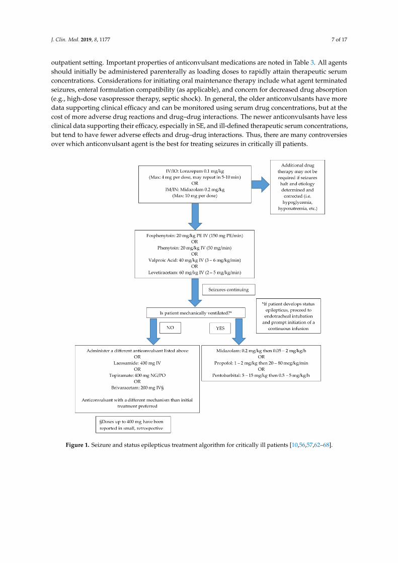
Management of seizures in the critically ill typically follows a stepwise approach (Figure 1).Initial treatment should consist of prompt administration of adequately dosed benzodiazepines.Lorazepam and midazolam are the preferred agents for intravenous (IV) and intramuscular (IM)administration, respectively [10,56,57]. Diazepam, while historically used, is not preferred for initialtherapy if lorazepam or midazolam are readily available. Diazepam has a large volume of distributionwhich results in rapid redistribution of drug out of the central nervous system to adipose tissue [6].This redistribution may result in subtherapeutic concentrations and seizure recurrence if additionalanticonvulsants are not promptly administered (e.g., within 30 min). If IV access has not been obtainedor has been lost during convulsive activity, midazolam may be administered IM or intranasally (IN).Intranasal administration should be performed with the use of a mucosal atomization device using thesame dosing strategy as IM and IV dosing. The midazolam 5 mg/mL IV product is recommended forthis route to minimize volume, and the total dose administered should be equally divided between eachnostril [58,59]. Intraosseous (IO) administration of midazolam or lorazepam may also be consideredif other routes of administration are not feasible. Standard practices for IO insertion should still beapplied, and placement should be verified by aspiration of a small amount of bone marrow followedby administration of 5–10 mL of 0.9% sodium chloride to ensure lack of resistance and to clear theneedle [60]. Regardless of the medication used and route of administration, timing of medicationadministration and appropriate dosing are of utmost importance. As seizure activity continues,synaptic gamma-aminobutyric acid (GABA) receptors (benzodiazepine pharmacologic target) begin tointernalize, resulting in a decreased efficacy of benzodiazepine therapy [61]. Some clinical concernsexist over the large doses of benzodiazepines recommended for termination of seizure activity withregard to respiratory compromise. However, studies in the prehospital setting have shown that theneed for placement of an advanced airway is more likely related to continued seizure activity ratherthan the benzodiazepines administered at the recommended doses [56,57].
Following administration of benzodiazepine, patients should be treated with longer actinganticonvulsants to aid in seizure cessation in those still seizing despite appropriately dosedbenzodiazepine therapy, or to prevent recurrent seizures in those who have achieved successfulseizure termination [10]. Patients who have had a treatable cause of seizure identified and corrected donot require additional therapy with an anticonvulsant (i.e., hypoglycemia, hyponatremia, etc.). There isno consensus on which anticonvulsant to administer after benzodiazepine therapy, so the decisionshould be patient-specific. Factors to consider include potential adverse drug effects, drug–druginteractions, hemodynamic stability, renal and/or hepatic dysfunction, serum albumin, previoushistory of anticonvulsant use, and therapeutic monitoring availability both in the inpatient and
J. Clin. Med. 2019, 8, 1177 7 of 17
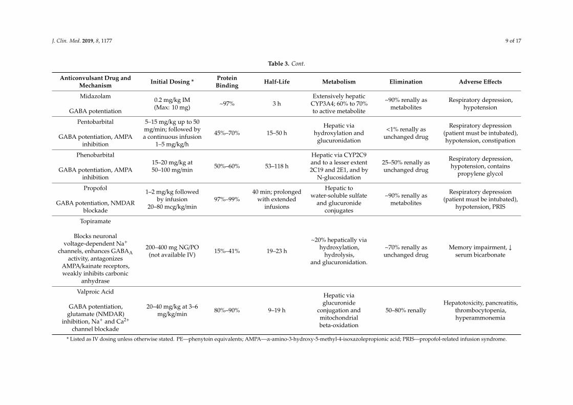
outpatient setting. Important properties of anticonvulsant medications are noted in Table 3. All agentsshould initially be administered parenterally as loading doses to rapidly attain therapeutic serumconcentrations. Considerations for initiating oral maintenance therapy include what agent terminatedseizures, enteral formulation compatibility (as applicable), and concern for decreased drug absorption(e.g., high-dose vasopressor therapy, septic shock). In general, the older anticonvulsants have moredata supporting clinical efficacy and can be monitored using serum drug concentrations, but at thecost of more adverse drug reactions and drug–drug interactions. The newer anticonvulsants have lessclinical data supporting their efficacy, especially in SE, and ill-defined therapeutic serum concentrations,but tend to have fewer adverse effects and drug–drug interactions. Thus, there are many controversiesover which anticonvulsant agent is the best for treating seizures in critically ill patients.
Figure 1. Seizure and status epilepticus treatment algorithm for critically ill patients [10,56,57,62–68].
Following administration of benzodiazepine, patients should be treated with longer acting anticonvulsants to aid in seizure cessation in those still seizing despite appropriately dosed benzodiazepine therapy, or to prevent recurrent seizures in those who have achieved successful seizure termination [10]. Patients who have had a treatable cause of seizure identified and corrected do not require additional therapy with an anticonvulsant (i.e., hypoglycemia, hyponatremia, etc.). There is no consensus on which anticonvulsant to administer after benzodiazepine therapy, so the decision should be patient-specific. Factors to consider include potential adverse drug effects, drug–drug interactions, hemodynamic stability, renal and/or hepatic dysfunction, serum albumin, previous history of anticonvulsant use, and therapeutic monitoring availability both in the inpatient and outpatient setting. Important properties of anticonvulsant medications are noted in Table 3. All agents should initially be administered parenterally as loading doses to rapidly attain therapeutic serum concentrations. Considerations for initiating oral maintenance therapy include what agent terminated seizures, enteral formulation compatibility (as applicable), and concern for decreased drug absorption (e.g., high-dose vasopressor therapy, septic shock). In general, the older anticonvulsants have more data supporting clinical efficacy and can be monitored using serum drug concentrations, but at the cost of more adverse drug reactions and drug–drug interactions. The newer anticonvulsants have less clinical data supporting their efficacy, especially in SE, and ill-defined therapeutic serum concentrations, but tend to have fewer adverse effects and drug–drug interactions.
Figure 1. Seizure and status epilepticus treatment algorithm for critically ill patients [10,56,57,62–68].

J. Clin. Med. 2019, 8, 1177 8 of 17
Table 3. Anticonvulsant medications [10,12,56,57,62,66,68–71].
Anticonvulsant Drug andMechanism Initial Dosing * Protein
Binding Half-Life Metabolism Elimination Adverse Effects
Brivaracetam
SV2A modulation
100–200 mg over atleast 2 min ≤20% ~9 h Hydrolysis and
hepatic via CYP2C19>95% renally, <10%as unchanged drug
Psychiatric disturbances,nystagmus
Diazepam
GABA potentiation
0.15 mg/kg(Max: 10 mg)
undiluted up to 5mg/min
98%
Parent drug:60–72 h
Metabolite:152–174 h
Hepatic via CYP3A4and 2C19; active
metabolites
Renally asglucuronideconjugates
Respiratory depression,hypotension (morecommon with rapid
administration)
Fosphenytoin/Phenytoin
Na+ channel blockade
20 mg/kg PE at 150mg/kg/min PE20 mg/kg at 50
mg/min
90%–95% 7–42 h
Fos: Prodrug, rapidlyhydrolyzed to
phenytoin.Hepatic via CYP2C9,
2C19, 3A4
<5% renally asphenytoin metabolites
Hypotension, phlebitis,cardiac arrhythmias.
Consider sloweradministration in elderly
Lacosamide
Enhances slow inactivationof voltage-gated
Na+ channels
200–400 mg over15–30 min <15% 13 h
Hepatic via CYP3A4,2C9, and 2C19;
inactive metabolite
~40% renally asunchanged drug
PR interval prolongation,hypotension
Levetiracetam
SV2A modulation,AMPA inhibition
3000 mg or 60 mg/kg(Max: 4500 mg) at 2–5
mg/kg/min<10% 6–8 h Nonhepatic
hydrolysis~66% renally asunchanged drug
Agitation, irritability,psychotic symptoms
Lorazepam
GABA potentiation
0.1 mg/kg(Max: 4 mg per dose,may repeat once) up
to 2 mg/min
~91% 12–18 hHepatic; rapidly
conjugated to inactivemetabolite
~88% renally asinactive metabolites
Respiratory depression,hypotension (morecommon with rapid
administration)
J. Clin. Med. 2019, 8, 1177 9 of 17
Table 3. Cont.
Anticonvulsant Drug andMechanism Initial Dosing * Protein
Binding Half-Life Metabolism Elimination Adverse Effects
Midazolam
GABA potentiation
0.2 mg/kg IM(Max: 10 mg) ~97% 3 h
Extensively hepaticCYP3A4; 60% to 70%to active metabolite
~90% renally asmetabolites
Respiratory depression,hypotension
Pentobarbital
GABA potentiation, AMPAinhibition
5–15 mg/kg up to 50mg/min; followed bya continuous infusion
1–5 mg/kg/h
45%–70% 15–50 hHepatic via
hydroxylation andglucuronidation
<1% renally asunchanged drug
Respiratory depression(patient must be intubated),hypotension, constipation
Phenobarbital
GABA potentiation, AMPAinhibition
15–20 mg/kg at50–100 mg/min 50%–60% 53–118 h
Hepatic via CYP2C9and to a lesser extent2C19 and 2E1, and by
N-glucosidation
25–50% renally asunchanged drug
Respiratory depression,hypotension, contains
propylene glycol
Propofol
GABA potentiation, NMDARblockade
1–2 mg/kg followedby infusion
20–80 mcg/kg/min97%–99%
40 min; prolongedwith extended
infusions
Hepatic towater-soluble sulfate
and glucuronideconjugates
~90% renally asmetabolites
Respiratory depression(patient must be intubated),
hypotension, PRIS
Topiramate
Blocks neuronalvoltage-dependent Na+
channels, enhances GABAAactivity, antagonizes
AMPA/kainate receptors,weakly inhibits carbonic
anhydrase
200–400 mg NG/PO(not available IV) 15%–41% 19–23 h
~20% hepatically viahydroxylation,
hydrolysis,and glucuronidation.
~70% renally asunchanged drug
Memory impairment, ↓serum bicarbonate
Valproic Acid
GABA potentiation,glutamate (NMDAR)
inhibition, Na+ and Ca2+
channel blockade
20–40 mg/kg at 3–6mg/kg/min 80%–90% 9–19 h
Hepatic viaglucuronide
conjugation andmitochondrialbeta-oxidation
50–80% renallyHepatotoxicity, pancreatitis,
thrombocytopenia,hyperammonemia
* Listed as IV dosing unless otherwise stated. PE—phenytoin equivalents; AMPA—α-amino-3-hydroxy-5-methyl-4-isoxazolepropionic acid; PRIS—propofol-related infusion syndrome.
J. Clin. Med. 2019, 8, 1177 10 of 17
Patients who have continuous seizure activity for 5 min, or at least 2 seizures without return tobaseline between seizures, are considered to be in SE, as discussed previously. Due to the significantmortality and morbidity associated with this medical emergency, prompt and aggressive treatment isrecommended [10,72]. To ensure timely administration, benzodiazepines and premade, ready-to-useanticonvulsant products should be available in automated dispensing cabinets in ICUs as well as theemergency department, unless a satellite pharmacy is located in close proximity. Additionally, prebuiltorder sets, following the Institute for Safe Medication Practices (ISMP) recommendations, should beimplemented for ease of appropriate medication ordering [73]. Patients whose seizures continue afteradministering appropriately dosed benzodiazepines and anticonvulsants are now considered to be inRSE. Establishing an institution-specific protocol detailing which anesthetic medications to utilize forRSE is also advised, as these agents are typically dosed higher than in other disease states.
6. Special Considerations
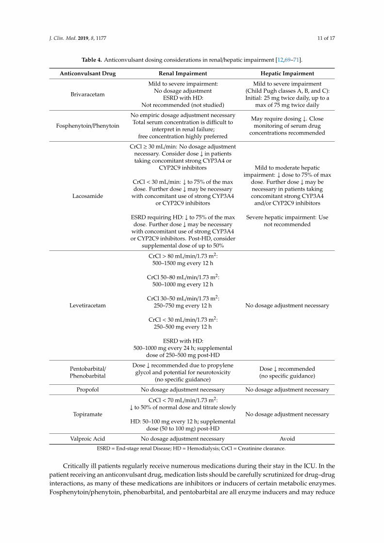
The use of continuous renal replacement therapy (CRRT) in critically ill patients is an interventionthat is becoming more and more common. Unfortunately, there is a relative paucity of data availableevaluating anticonvulsant dosing in patients on any modality of CRRT (Table 4). For this reason,any anticonvulsants that have readily available serum monitoring assays should be utilized in thissetting to assist in guiding medication dosing [69,74]. If a serum drug assay is not available, thereare a few factors that should be considered. Generally, a drug that is eliminated renally will beremoved using CRRT. The degree of removal will largely depend on the CRRT flow rate and modality(e.g., continuous venovenous hemofiltration and/or hemodialysis), the degree of protein binding(only unbound drug will be removed by CRRT), and the volume of distribution of the drug [74].Other important considerations for anticonvulsant dosing in patients on CRRT is monitoring for anyfilter down time as well as flow rate changes, as these may warrant further modification to drugdosing. While molecular weight of the drug is often cited as a consideration, the weights of availableanticonvulsants are all small enough that this factor is not of clinical significance. Unfortunately, there iscurrently not enough evidence to recommend specific anticonvulsant doses in those undergoing CRRT;therefore, the clinician must consider the characteristics of each drug when making dosing decisions,and serum drug levels should always be utilized, if available [69,74,75]. Additionally, the developmentof any adverse effects believed to be related to an anticonvulsant should prompt dosing modifications.
Similar to drug dosing in CRRT, data regarding drug dosing in patients with acute liver failureare also lacking (Table 4). Serum drug concentrations should be followed closely when available inthis unique patient population. Dosing recommendations based on Child–Pugh scores may also beconsidered, keeping in mind that these dosing recommendations were designed for patients withchronic liver disease [76]. If an anticonvulsant is to be initiated in the setting of acute liver failure, it isbest to avoid those with low hepatic extraction ratios (i.e., phenytoin, valproic acid, and phenobarbital)as clearance of these drugs will be primarily predicated upon intrinsic hepatic function.
The use of extracorporeal membrane oxygenation (ECMO) continues to rise in critically ill patients,but unfortunately, the literature surrounding specific drug dosing remains sparse. The biggest impactof ECMO on drug dosing lies in the propensity for the ECMO circuit to sequester drugs, resultingin a larger than expected volume of distribution. This phenomenon may decrease over time withcontinued dosing due to saturation of binding sites. In general, medications with a higher degreeof lipophilicity and protein binding tend to be sequestered more [77,78]. Many patients on ECMOfrequently receive concomitant CRRT, which further complicates the dosing picture and heightens theneed for therapeutic drug monitoring, if available.
J. Clin. Med. 2019, 8, 1177 11 of 17
Table 4. Anticonvulsant dosing considerations in renal/hepatic impairment [12,69–71].
Anticonvulsant Drug Renal Impairment Hepatic Impairment
Brivaracetam
Mild to severe impairment:No dosage adjustment
ESRD with HD:Not recommended (not studied)
Mild to severe impairment(Child Pugh classes A, B, and C):Initial: 25 mg twice daily, up to a
max of 75 mg twice daily
Fosphenytoin/Phenytoin
No empiric dosage adjustment necessaryTotal serum concentration is difficult to
interpret in renal failure;free concentration highly preferred
May require dosing ↓. Closemonitoring of serum drug
concentrations recommended
Lacosamide
CrCl ≥ 30 mL/min: No dosage adjustmentnecessary. Consider dose ↓ in patientstaking concomitant strong CYP3A4 or
CYP2C9 inhibitors
CrCl < 30 mL/min: ↓ to 75% of the maxdose. Further dose ↓may be necessary
with concomitant use of strong CYP3A4or CYP2C9 inhibitors
ESRD requiring HD: ↓ to 75% of the maxdose. Further dose ↓may be necessary
with concomitant use of strong CYP3A4or CYP2C9 inhibitors. Post-HD, consider
supplemental dose of up to 50%
Mild to moderate hepaticimpairment: ↓ dose to 75% of max
dose. Further dose ↓may benecessary in patients takingconcomitant strong CYP3A4
and/or CYP2C9 inhibitors
Severe hepatic impairment: Usenot recommended
Levetiracetam
CrCl > 80 mL/min/1.73 m2:500–1500 mg every 12 h
CrCl 50–80 mL/min/1.73 m2:500–1000 mg every 12 h
CrCl 30–50 mL/min/1.73 m2:250–750 mg every 12 h
CrCl < 30 mL/min/1.73 m2:250–500 mg every 12 h
ESRD with HD:500–1000 mg every 24 h; supplemental
dose of 250–500 mg post-HD
No dosage adjustment necessary
Pentobarbital/Phenobarbital
Dose ↓ recommended due to propyleneglycol and potential for neurotoxicity
(no specific guidance)
Dose ↓ recommended(no specific guidance)
Propofol No dosage adjustment necessary No dosage adjustment necessary
Topiramate
CrCl < 70 mL/min/1.73 m2:↓ to 50% of normal dose and titrate slowly
HD: 50–100 mg every 12 h; supplementaldose (50 to 100 mg) post-HD
No dosage adjustment necessary
Valproic Acid No dosage adjustment necessary Avoid
ESRD = End-stage renal Disease; HD = Hemodialysis; CrCl = Creatinine clearance.
Critically ill patients regularly receive numerous medications during their stay in the ICU. In thepatient receiving an anticonvulsant drug, medication lists should be carefully scrutinized for drug–druginteractions, as many of these medications are inhibitors or inducers of certain metabolic enzymes.Fosphenytoin/phenytoin, phenobarbital, and pentobarbital are all enzyme inducers and may reduce
J. Clin. Med. 2019, 8, 1177 12 of 17
the concentration of concurrent medications [79]. Additionally, anticonvulsant drugs with highdegrees of protein binding may displace other medications with a high degree of protein binding,resulting in an increased free fraction of one or both medications which may precipitate adverseeffects. The potential for this is heightened in critically ill patients who often have reduced plasmaalbumin and acid/base abnormalities [80–82]. One additional serious interaction to consider as theincidence of multidrug-resistant organisms increases is between carbapenem antibiotics and valproicacid. Numerous potential mechanisms exist to describe this interaction; however, it is believed thatcarbapanems inhibit an enzyme crucial to the production of the pharmacologically active moiety ofvalproic acid, resulting in significantly reduced plasma valproic acid concentrations [83–85]. In patientson valproic acid, alternatives to carbapenems should be utilized if possible. If a carbapenem must beused, patients should be started on another anticonvulsant drug prior to initiation.
7. Older Adults, Pediatrics, and Pregnancy
The initial management approach of acute onset seizures in the critically ill older adult and pediatricpopulations is similar to that of other adult patients. Medication dosing is primarily weight-based,and there are generally no modifications required for the initial dosing strategy. However, a number ofphysiological changes occur in older adults that may affect the pharmacokinetics of anticonvulsants,including possible decrease in drug absorption, increase in total body fat, decrease in total bodywater, and reduced hepatic and renal function [86]. Older adult patients may also have increasedblood–brain-barrier permeability, which lends to a higher risk of adverse effects associated withanticonvulsant use [87]. When considering anticonvulsants in this population, it is important toconsider each of these aspects, as well as concomitant disease states and medications.
Many of the older anticonvulsants that undergo hepatic metabolism may be enzyme inhibitorsor inducers. These may affect or be affected by other medications, altering serum concentrationsand potentially leading to sub- or supratherapeutic concentrations. Therefore, in the older adultpatient on numerous medications with potential for interactions, it may be prudent to assess serumdrug concentrations more frequently than in other patients to ensure efficacy and safety. In patientsreceiving newer anticonvulsants for which target serum concentrations are not as well defined,dose modifications should be made based on estimated renal function using the Cockcroft–Gaultequation and corresponding doses listed in the package inserts. Identifying anticonvulsant agents witha lower incidence of dizziness and ataxia, especially when transitioning to home care, is also importantdue to the higher risk of falling in the older adult patient population.
Many pharmacokinetic properties are different in the pediatric population and change as thepatient ages. Neonates tend to have relatively reduced fat compared to adults, whereas infants tendto have increased fat. This results in an increased volume of distribution for lipophilic drugs ininfants and a decreased volume of distribution for lipophilic drugs in neonates. Pediatric patientsalso have reduced plasma proteins, leading to a lower degree of protein binding and a higher degreeof free drug in anticonvulsants with high protein binding. Metabolism and elimination are alsoeffected due to larger relative liver and kidney sizes, resulting in a greater degree of metabolismof drugs that are extensively hepatically metabolized and increased clearance of renally eliminateddrugs, respectively [88]. Some medications have increased risk of toxicity in children (e.g., valproicacid–hepatotoxicity); therefore, the risk versus benefit of each treatment strategy should be considered.
Critically ill pregnant patients who experience seizures or SE should be treated aggressively tohalt seizure activity so additional complications are limited. Agent selection is important over thelong term due to potential teratogenic effects, but acute and chronic pharmacokinetic alterations alsoneed to be considered. During pregnancy, the plasma volume is expanded by approximately 50%,resulting in an increased volume of distribution. As pregnancy progresses, this volume expansionleads to a relative dilutional hypoalbuminemia and may result in a greater free fraction of drugs thatare highly bound to albumin (e.g., phenytoin, valproic acid). The hepatic metabolism of phenytoin hasbeen reported to increase and is possibly due to increased microsomal enzyme activity induced by
J. Clin. Med. 2019, 8, 1177 13 of 17
progesterone, so this should be considered for other hepatically metabolized anticonvulsants as well.As a result of the increased cardiac output during pregnancy, renally eliminated anticonvulsants havean increased clearance [89].
Aside from the changes in pharmacokinetics during pregnancy, the potential for a medication tocause teratogenicity should also be evaluated. In the acute setting, it is best to avoid initiation of valproicacid as it has the most data suggesting it is teratogenic. Other anticonvulsants with high teratogenicpotential include phenytoin, phenobarbital, and topiramate. Lamotrigine and levetiracetam are themost commonly recommended anticonvulsant agents for pregnant patients based on the amount ofevidence showing lower teratogenic risk.
8. Summary
The occurrence of seizures and SE in the critically ill may be attributed to a variety of factors,while the true incidence remains unknown. Increased use of prolonged monitoring techniques aswell as the development of more advanced monitoring systems may aid in bridging this knowledgegap. Further comprehension of seizure and SE incidence in critically ill patients may also allow forimproved delineation of the need for seizure prophylaxis, as this concept still remains heavily debated.For optimal outcomes, early recognition of seizure activity followed by prompt, appropriately dosedmedication therapy remains the hallmark of treating acute onset seizures and SE in critically ill patients.
Author Contributions: M.S., J.P.H.-B., L.R.S. and G.M.B. contributed equally to the creation of this manuscript.
Funding: The contents of this manuscript were developed in part under a grant from the National Institute onDisability, Independent Living, and Rehabilitation Research (NIDILRR grant number 90AR5025). NIDILRR isa Center within the Administration for Community Living (ACL), Department of Health and Human Services(HHS). The contents of this manuscript do not necessarily represent the policy of NIDILRR, ACL, HHS, and youshould not assume endorsement by the Federal Government.
Conflicts of Interest: Gretchen M. Brophy is a consultant/speaker for UCB and Sage Therapeutics. Micheal Strein,John P. Holton-Burke, and Latangela R. Smith have no conflicts of interest to disclose.
References
1. Cost of Status Epilepticus: A Systematic Review—ScienceDirect. Available online: https://www.sciencedirect.com/science/article/pii/S1059131114003021 (accessed on 18 December 2018).
2. McNett, M.; Moran, C.; Johnson, H. Evidence-Based Review of Clinical Trials in Neurocritical Care. AACNAdv. Crit. Care 2018, 29, 195–203. [CrossRef] [PubMed]
3. Ko, S.-B. Multimodality Monitoring in the Neurointensive Care Unit: A Special Perspective for Patients withStroke. J. Stroke 2013, 15, 99–108. [CrossRef] [PubMed]
4. Westover, M.B.; Shafi, M.M.; Bianchi, M.T.; Moura, L.M.V.R.; O’Rourke, D.; Rosenthal, E.S.; Chu, C.J.;Donovan, S.; Hoch, D.B.; Kilbride, R.D.; et al. The probability of seizures during EEG monitoring in criticallyill adults. Clin. Neurophysiol. 2015, 126, 463–471. [CrossRef] [PubMed]
5. Claassen, J.; Mayer, S.A.; Kowalski, R.G.; Emerson, R.G.; Hirsch, L.J. Detection of electrographic seizures withcontinuous EEG monitoring in critically ill patients. Neurology 2004, 62, 1743–1748. [CrossRef] [PubMed]
6. Brophy, G.M.; Bell, R.; Claassen, J.; Alldredge, B.; Bleck, T.P.; Glauser, T.; LaRoche, S.M.; Riviello, J.J.;Shutter, L.; Sperling, M.R.; et al. Guidelines for the Evaluation and Management of Status Epilepticus.Neurocrit. Care 2012, 17, 3–23. [CrossRef] [PubMed]
7. Shorvon, S.; Ferlisi, M. The treatment of super-refractory status epilepticus: A critical review of availabletherapies and a clinical treatment protocol. Brain J. Neurol. 2011, 134, 2802–2818. [CrossRef] [PubMed]
8. Chapter 41. Status Epilepticus | Pharmacotherapy: A Pathophysiologic Approach, 9e | AccessPharmacy |
McGraw-Hill Medical. Available online: https://accesspharmacy.mhmedical.com/content.aspx?bookid=689§ionid=45310491#57485199 (accessed on 21 September 2018).
9. Dham, B.S.; Hunter, K.; Rincon, F. The Epidemiology of Status Epilepticus in the United States. Neurocrit. Care2014, 20, 476–483. [CrossRef] [PubMed]
J. Clin. Med. 2019, 8, 1177 14 of 17
10. Shin, J.-W.; Koo, Y.S.; Kim, Y.-S.; Kim, D.W.; Kim, K.K.; Lee, S.-Y.; Kim, H.K.; Moon, H.-J.; Lim, J.-A.;Byun, J.-I.; et al. Clinical characterization of unknown/cryptogenic status epilepticus suspected as encephalitis:A multicenter cohort study. J. Neuroimmunol. 2018, 315, 1–8. [CrossRef] [PubMed]
11. Liu, X.; Yan, B.; Wang, R.; Li, C.; Chen, C.; Zhou, D.; Hong, Z. Seizure outcomes in patients with anti-NMDARencephalitis: A follow-up study. Epilepsia 2017, 58, 2104–2111. [CrossRef] [PubMed]
12. McNally, B.; Robb, R.; Mehta, M.; Vellano, K.; Valderrama, A.L.; Yoon, P.W.; Sasson, C.; Crouch, A.; Perez, A.B.;Merritt, R.; et al. Out-of-hospital cardiac arrest surveillance—Cardiac Arrest Registry to Enhance Survival(CARES), United States, October 1, 2005–December 31, 2010. Morb. Mortal. Wkly. Rep. Surveill. Summ. 2011,60, 1–19.
13. Nielsen, N.; Sunde, K.; Hovdenes, J.; Riker, R.R.; Rubertsson, S.; Stammet, P.; Nilsson, F.; Friberg, H.;Hypothermia Network. Adverse events and their relation to mortality in out-of-hospital cardiac arrestpatients treated with therapeutic hypothermia. Crit. Care Med. 2011, 39, 57–64. [CrossRef] [PubMed]
14. Crepeau, A.Z.; Rabinstein, A.A.; Fugate, J.E.; Mandrekar, J.; Wijdicks, E.F.; White, R.D.; Britton, J.W.Continuous EEG in therapeutic hypothermia after cardiac arrest: Prognostic and clinical value. Neurology2013, 80, 339–344. [CrossRef] [PubMed]
15. Won, S.-Y.; Dubinski, D.; Brawanski, N.; Strzelczyk, A.; Seifert, V.; Freiman, T.M.; Konczalla, J. Significantincrease in acute subdural hematoma in octo- and nonagenarians: Surgical treatment, functional outcome,and predictors in this patient cohort. Neurosurg. Focus 2017, 43, E10. [CrossRef] [PubMed]
16. Pruitt, P.; Naidech, A.; Ornam, J.V.; Borczuk, P. Seizure frequency in patients with isolated subdural hematomaand preserved consciousness. Brain Inj. 2019, 0, 1–5. [CrossRef] [PubMed]
17. Delanty, N.; Vaughan, C.J.; French, J.A. Medical causes of seizures. The Lancet 1998, 352, 383–390. [CrossRef]18. Vezzani, A.; Balosso, S.; Ravizza, T. Chapter 10—Inflammation and epilepsy. In Handbook of Clinical
Neurology—Epilepsy; Stefan, H., Theodore, W.H., Eds.; Elsevier: Maryland Heights, MO, USA, 2012; Volume107, pp. 163–175.
19. Riazi, K.; Galic, M.A.; Kuzmiski, J.B.; Ho, W.; Sharkey, K.A.; Pittman, Q.J. Microglial activation and TNFαproduction mediate altered CNS excitability following peripheral inflammation. Proc. Natl. Acad. Sci. 2008,105, 17151–17156. [CrossRef] [PubMed]
20. Riazi, K.; Galic, M.A.; Pittman, Q.J. Contributions of peripheral inflammation to seizure susceptibility:Cytokines and brain excitability. Epilepsy Res. 2010, 89, 34–42. [CrossRef] [PubMed]
21. Mayhan, W.G. Cellular mechanisms by which tumor necrosis factor-α produces disruption of the blood–brainbarrier. Brain Res. 2002, 927, 144–152. [CrossRef]
22. Welser-Alves, J.V.; Milner, R. Microglia are the major source of TNF-α and TGF-β1 in postnatal glial cultures;regulation by cytokines, lipopolysaccharide, and vitronectin. Neurochem. Int. 2013, 63, 47–53. [CrossRef]
23. Ha, J.; Choi, J.; Kwon, A.; Kim, K.; Kim, S.-J.; Bae, S.H.; Son, J.S.; Kim, S.-N.; Kwak, B.O.; Lee, R. Interleukin-4and tumor necrosis factor-alpha levels in children with febrile seizures. Seizure 2018, 58, 156–162. [CrossRef]
24. Zhao, W.; Xie, W.; Xiao, Q.; Beers, D.R.; Appel, S.H. Protective effects of an anti-inflammatory cytokine,interleukin-4, on motoneuron toxicity induced by activated microglia. J. Neurochem. 2006, 99, 1176–1187.[CrossRef] [PubMed]
25. Hart, Y.M.; Andermann, F.; Robitaille, Y.; Laxer, K.D.; Rasmussen, T.; Davis, R. Double pathology inRasmussen’s syndrome: A window on the etiology? Neurology 1998, 50, 731–735. [CrossRef] [PubMed]
26. Lagarde, S.; Villeneuve, N.; Trébuchon, A.; Kaphan, E.; Lepine, A.; McGonigal, A.; Roubertie, A.;Barthez, M.-A.J.; Trommsdorff, V.; Lefranc, J.; et al. Anti–tumor necrosis factor alpha therapy (adalimumab)in Rasmussen’s encephalitis: An open pilot study. Epilepsia 2016, 57, 956–966. [CrossRef] [PubMed]
27. Varelas, P.N.; Mirski, M.A. Seizures in the Adult Intensive Care Unit. J. Neurosurg. Anesthesiol. 2001, 13,163–175. [CrossRef]
28. Castilla-Guerra, L.; Fernández-Moreno, M.d.C.; López-Chozas, J.M.; Fernández-Bolaños, R. ElectrolytesDisturbances and Seizures. Epilepsia 2006, 47, 1990–1998. [CrossRef] [PubMed]
29. Hitchings, A.W. Drugs that lower the seizure threshold. Adverse Drug React. Bull. 2016, 298, 1151–1154.30. Buchanan, N. Medications which may lower seizure threshold. Aust. Prescr. 2001, 24, 51–55. [CrossRef]31. Tesoro, E.P.; Brophy, G.M. Pharmacological Management of Seizures and Status Epilepticus in Critically Ill
Patients. J. Pharm. Pract. 2010, 23, 441–454. [CrossRef] [PubMed]32. Hirsch, L.J. Continuous EEG monitoring in the intensive care unit: An overview. J. Clin. Neurophysiol. Off.
Publ. Am. Electroencephalogr. Soc. 2004, 21, 332–340. [CrossRef]
J. Clin. Med. 2019, 8, 1177 15 of 17
33. Drislane, F.W.; Lopez, M.R.; Blum, A.S.; Schomer, D.L. Detection and Treatment of Refractory StatusEpilepticus in the Intensive Care Unit. J. Clin. Neurophysiol. 2008, 25, 181–186. [CrossRef]
34. Kubota, Y.; Nakamoto, H.; Egawa, S.; Kawamata, T. Continuous EEG monitoring in ICU. J. Intensive Care2018, 6. [CrossRef] [PubMed]
35. Boly, M.; Maganti, R. Monitoring epilepsy in the intensive care unit: Current state of facts and potentialinterest of high density EEG. Brain Inj. 2014, 28, 1151–1155. [CrossRef] [PubMed]
36. Vespa, P.M.; Nenov, V.; Nuwer, M.R. Continuous EEG monitoring in the intensive care unit: Early findingsand clinical efficacy. J. Clin. Neurophysiol. Off. Publ. Am. Electroencephalogr. Soc. 1999, 16, 1–13. [CrossRef]
37. Pandian, J.D.; Cascino, G.D.; So, E.L.; Manno, E.; Fulgham, J.R. Digital video-electroencephalographicmonitoring in the neurological-neurosurgical intensive care unit: Clinical features and outcome. Arch. Neurol.2004, 61, 1090–1094. [CrossRef] [PubMed]
38. Drake, M.E.; Padamadan, H.; Newell, S.A. Interictal quantitative EEG in epilepsy. Seizure 1998, 7, 39–42.[CrossRef]
39. van Putten, M.J.A.M.; Tavy, D.L.J. Continuous quantitative EEG monitoring in hemispheric stroke patientsusing the brain symmetry index. Stroke 2004, 35, 2489–2492. [CrossRef] [PubMed]
40. Coates, S.; Clarke, A.; Davison, G.; Patterson, V. Tele-EEG in the UK: A report of over 1,000 patients. J. Telemed.Telecare 2012, 18, 243–246. [CrossRef]
41. Hobbs, K.; Krishnamohan, P.; Legault, C.; Goodman, S.; Parvizi, J.; Gururangan, K.; Mlynash, M. RapidBedside Evaluation of Seizures in the ICU by Listening to the Sound of Brainwaves: A ProspectiveObservational Clinical Trial of Ceribell’s Brain Stethoscope Function. Neurocrit. Care 2018, 29, 302–312.[CrossRef]
42. Waziri, A.; Claassen, J.; Stuart, R.M.; Arif, H.; Schmidt, J.M.; Mayer, S.A.; Badjatia, N.; Kull, L.L.; Connolly, E.S.;Emerson, R.G.; et al. Intracortical electroencephalography in acute brain injury. Ann. Neurol. 2009, 66,366–377. [CrossRef]
43. Yamazaki, M.; Terrill, M.; Fujimoto, A.; Yamamoto, T.; Tucker, D.M. Integrating dense array EEG in thepresurgical evaluation of temporal lobe epilepsy. ISRN Neurol. 2012, 2012, 924081. [CrossRef]
44. Yamazaki, M.; Tucker, D.M.; Fujimoto, A.; Yamazoe, T.; Okanishi, T.; Yokota, T.; Enoki, H.; Yamamoto, T.Comparison of dense array EEG with simultaneous intracranial EEG for interictal spike detection andlocalization. Epilepsy Res. 2012, 98, 166–173. [CrossRef] [PubMed]
45. Hutchinson, P.J.; Hutchinson, D.B.; Barr, R.H.; Burgess, F.; Kirkpatrick, P.J.; Pickard, J.D. A new cranial accessdevice for cerebral monitoring. Br. J. Neurosurg. 2000, 14, 46–48. [CrossRef] [PubMed]
46. Tavakoli, S.; Peitz, G.; Ares, W.; Hafeez, S.; Grandhi, R. Complications of invasive intracranial pressuremonitoring devices in neurocritical care. Neurosurg. Focus 2017, 43, E6. [CrossRef] [PubMed]
47. Ritter, A.C.; Wagner, A.K.; Fabio, A.; Pugh, M.J.; Walker, W.C.; Szaflarski, J.P.; Zafonte, R.D.; Brown, A.W.;Hammond, F.M.; Bushnik, T.; et al. Incidence and risk factors of posttraumatic seizures following traumaticbrain injury: A Traumatic Brain Injury Model Systems Study. Epilepsia 2016, 57, 1968–1977. [CrossRef]
48. Kodankandath, T.V.; Farooq, S.; Wazni, W.; Cox, J.-A.; Southwood, C.; Rozansky, G.; Johnson, V.; Lynch, J.R.Seizure Prophylaxis in the Immediate Post-Hemorrhagic Period in Patients with Aneurysmal SubarachnoidHemorrhage. J. Vasc. Interv. Neurol. 2017, 9, 1–4.
49. Yerram, S.; Katyal, N.; Premkumar, K.; Nattanmai, P.; Newey, C.R. Seizure prophylaxis in the neuroscienceintensive care unit. J. Intensive Care 2018, 6. [CrossRef]
50. Connolly, E.S.; Rabinstein, A.A.; Carhuapoma, J.R.; Derdeyn, C.P.; Dion, J.; Higashida, R.T.; Hoh, B.L.;Kirkness, C.J.; Naidech, A.M.; Ogilvy, C.S.; et al. Guidelines for the Management of Aneurysmal SubarachnoidHemorrhage: A Guideline for Healthcare Professionals from the American Heart Association/AmericanStroke Association. Stroke 2012, 43, 1711–1737. [CrossRef]
51. Carney, N.; Totten, A.M.; O’Reilly, C.; Ullman, J.S.; Hawryluk, G.W.J.; Bell, M.J.; Bratton, S.L.; Chesnut, R.;Harris, O.A.; Kissoon, N.; et al. Guidelines for the Management of Severe Traumatic Brain Injury. Neurosurgery2017, 80, 6–15.
52. Temkin, N.R.; Dikmen, S.S.; Anderson, G.D.; Wilensky, A.J.; Holmes, M.D.; Cohen, W.; Newell, D.W.;Nelson, P.; Awan, A.; Winn, H.R. Valproate therapy for prevention of posttraumatic seizures: A randomizedtrial. J. Neurosurg. 1999, 91, 593–600. [CrossRef]
J. Clin. Med. 2019, 8, 1177 16 of 17
53. Temkin, N.R.; Dikmen, S.S.; Wilensky, A.J.; Keihm, J.; Chabal, S.; Winn, H.R. A randomized, double-blindstudy of phenytoin for the prevention of post-traumatic seizures. N. Engl. J. Med. 1990, 323, 497–502.[CrossRef]
54. Temkin, N.R. Antiepileptogenesis and seizure prevention trials with antiepileptic drugs: Meta-analysis ofcontrolled trials. Epilepsia 2001, 42, 515–524. [CrossRef] [PubMed]
55. Diringer, M.N.; Bleck, T.P.; Claude Hemphill, J.; Menon, D.; Shutter, L.; Vespa, P.; Bruder, N.; Connolly, E.S.;Citerio, G.; Gress, D.; et al. Critical Care Management of Patients Following Aneurysmal SubarachnoidHemorrhage: Recommendations from the Neurocritical Care Society’s Multidisciplinary ConsensusConference. Neurocrit. Care 2011, 15, 211–240. [CrossRef] [PubMed]
56. Alldredge, B.K.; Gelb, A.M.; Isaacs, S.M.; Corry, M.D.; Allen, F.; Ulrich, S.; Gottwald, M.D.; O’Neil, N.;Neuhaus, J.M.; Segal, M.R.; et al. A comparison of lorazepam, diazepam, and placebo for the treatment ofout-of-hospital status epilepticus. N. Engl. J. Med. 2001, 345, 631–637. [CrossRef] [PubMed]
57. Silbergleit, R.; Durkalski, V.; Lowenstein, D.; Conwit, R.; Pancioli, A.; Palesch, Y.; Barsan, W.; NETTInvestigators. Intramuscular versus intravenous therapy for prehospital status epilepticus. N. Engl. J. Med.2012, 366, 591–600. [CrossRef] [PubMed]
58. Scheepers, M.; Scheepers, B.; Clarke, M.; Comish, S.; Ibitoye, M. Is intranasal midazolam an effective rescuemedication in adolescents and adults with severe epilepsy? Seizure 2000, 9, 417–422. [CrossRef]
59. Bhattacharyya, M.; Kalra, V.; Gulati, S. Intranasal midazolam vs rectal diazepam in acute childhood seizures.Pediatr. Neurol. 2006, 34, 355–359. [CrossRef]
60. Buck, M.L.; Wiggins, B.S.; Sesler, J.M. Intraosseous Drug Administration in Children and Adults DuringCardiopulmonary Resuscitation. Ann. Pharmacother. 2007, 41, 1679–1686. [CrossRef]
61. Goodkin, H.P.; Sun, C.; Yeh, J.-L.; Mangan, P.S.; Kapur, J. GABAA Receptor Internalization during Seizures.Epilepsia 2007, 48, 109–113. [CrossRef]
62. Glauser, T.; Shinnar, S.; Gloss, D.; Alldredge, B.; Arya, R.; Bainbridge, J.; Bare, M.; Bleck, T.; Dodson, W.E.;Garrity, L.; et al. Evidence-Based Guideline: Treatment of Convulsive Status Epilepticus in Children andAdults: Report of the Guideline Committee of the American Epilepsy Society. Epilepsy Curr. 2016, 16, 48–61.[CrossRef]
63. Kalss, G.; Rohracher, A.; Leitinger, M.; Pilz, G.; Novak, H.F.; Neuray, C.; Kreidenhuber, R.; Höfler, J.;Kuchukhidze, G.; Trinka, E. Intravenous brivaracetam in status epilepticus: A retrospective single-centerstudy. Epilepsia 2018, 59 Suppl 2, 228–233. [CrossRef]
64. Santamarina, E.; Carbonell, B.P.; Sala, J.; Gutiérrez-Viedma, Á.; Miró, J.; Asensio, M.; Abraira, L.; Falip, M.;Ojeda, J.; López-González, F.J.; et al. Use of intravenous brivaracetam in status epilepticus: A multicenterregistry. Epilepsia 2019, 0. [CrossRef] [PubMed]
65. Strzelczyk, A.; Steinig, I.; Willems, L.M.; Reif, P.S.; Senft, C.; Voss, M.; Gaida, B.; von Podewils, F.; Rosenow, F.Treatment of refractory and super-refractory status epilepticus with brivaracetam: A cohort study from twoGerman university hospitals. Epilepsy Behav. EB 2017, 70, 177–181. [CrossRef] [PubMed]
66. Farrokh, S.; Bon, J.; Erdman, M.; Tesoro, E. Use of Newer Anticonvulsants for the Treatment of StatusEpilepticus. Pharmacother. J. Hum. Pharmacol. Drug Ther. 2019, 39, 297–316. [CrossRef]
67. Strzelczyk, A.; Zöllner, J.P.; Willems, L.M.; Jost, J.; Paule, E.; Schubert-Bast, S.; Rosenow, F.; Bauer, S.Lacosamide in status epilepticus: Systematic review of current evidence. Epilepsia 2017, 58, 933–950.[CrossRef] [PubMed]
68. Treiman, D.M.; Meyers, P.D.; Walton, N.Y.; Collins, J.F.; Colling, C.; Rowan, A.J.; Handforth, A.; Faught, E.;Calabrese, V.P.; Uthman, B.M.; et al. A comparison of four treatments for generalized convulsive statusepilepticus. Veterans Affairs Status Epilepticus Cooperative Study Group. N. Engl. J. Med. 1998, 339, 792–798.[CrossRef]
69. Farrokh, S.; Tahsili-Fahadan, P.; Ritzl, E.K.; Lewin, J.J.; Mirski, M.A. Antiepileptic drugs in critically illpatients. Crit. Care 2018, 22, 153. [CrossRef] [PubMed]
70. Lexicomp. Available online: https://online.lexi.com/lco/action/home (accessed on 5 September 2018).71. Garnett, W.R. Clinical Pharmacology of Topiramate: A Review. Epilepsia 2000, 41, 61–65. [CrossRef]72. Wu, Y.W.; Shek, D.W.; Garcia, P.A.; Zhao, S.; Johnston, S.C. Incidence and mortality of generalized convulsive
status epilepticus in California. Neurology 2002, 58, 1070–1076. [CrossRef]73. Guidelines for Standard Order Sets. Available online: https://www.ismp.org/guidelines/standard-order-sets
(accessed on 3 October 2018).
J. Clin. Med. 2019, 8, 1177 17 of 17
74. Mahmoud, S.H. Antiepileptic Drug Removal by Continuous Renal Replacement Therapy: A Review of theLiterature. Clin. Drug Investig. 2017, 37, 7–23. [CrossRef]
75. Schetz, M.; Ferdinande, P.; Van den Berghe, G.; Verwaest, C.; Lauwers, P. Pharmacokinetics of continuousrenal replacement therapy. Intensive Care Med. 1995, 21, 612–620. [CrossRef]
76. Lewis, J.H.; Stine, J.G. Review article: Prescribing medications in patients with cirrhosis—A practical guide.Aliment. Pharmacol. Ther. 2013, 37, 1132–1156. [CrossRef] [PubMed]
77. Cheng, V.; Abdul-Aziz, M.-H.; Roberts, J.A.; Shekar, K. Optimising drug dosing in patients receivingextracorporeal membrane oxygenation. J. Thorac. Dis. 2018, 10, S629–S641. [CrossRef] [PubMed]
78. Shekar, K.; Roberts, J.A.; Mcdonald, C.I.; Ghassabian, S.; Anstey, C.; Wallis, S.C.; Mullany, D.V.; Fung, Y.L.;Fraser, J.F. Protein-bound drugs are prone to sequestration in the extracorporeal membrane oxygenationcircuit: Results from an ex vivo study. Crit. Care Lond. Engl. 2015, 19, 164. [CrossRef] [PubMed]
79. Lynch, T.; Price, A.L. The Effect of Cytochrome P450 Metabolism on Drug Response, Interactions, and AdverseEffects. Am. Fam. Physician 2007, 76, 391–396.
80. McElnay, J.C.; D’Arcy, P.F. Protein binding displacement interactions and their clinical importance. Drugs1983, 25, 495–513. [CrossRef]
81. Schleibinger, M.; Steinbach, C.L.; Töpper, C.; Kratzer, A.; Liebchen, U.; Kees, F.; Salzberger, B.; Kees, M.G.Protein binding characteristics and pharmacokinetics of ceftriaxone in intensive care unit patients. Br. J.Clin. Pharmacol. 2015, 80, 525–533. [CrossRef]
82. Lindow, J.; Wijdicks, E.F. Phenytoin toxicity associated with hypoalbuminemia in critically ill patients. Chest1994, 105, 602–604. [CrossRef]
83. Wen, Z.-P.; Fan, S.-S.; Du, C.; Yin, T.; Zhou, B.-T.; Peng, Z.-F.; Xie, Y.-Y.; Zhang, W.; Chen, Y.; Xiao, J.; et al.Drug-drug interaction between valproic acid and meropenem: A retrospective analysis of electronic medicalrecords from neurosurgery inpatients. J. Clin. Pharm. Ther. 2017, 42, 221–227. [CrossRef]
84. Suzuki, E.; Yamamura, N.; Ogura, Y.; Nakai, D.; Kubota, K.; Kobayashi, N.; Miura, S.; Okazaki, O.Identification of Valproic Acid Glucuronide Hydrolase as a Key Enzyme for the Interaction of Valproic Acidwith Carbapenem Antibiotics. Drug Metab. Dispos. 2010, 38, 1538–1544. [CrossRef]
85. Bede, P.; Lawlor, D.; Solanki, D.; Delanty, N. Carbapenems and valproate: A consumptive relationship.Epilepsia Open 2016, 2, 107–111. [CrossRef]
86. Klotz, U. Pharmacokinetics and drug metabolism in the elderly. Drug Metab. Rev. 2009, 41, 67–76. [CrossRef][PubMed]
87. Hutchison, L.C.; O’Brien, C.E. Changes in Pharmacokinetics and Pharmacodynamics in the Elderly Patient.J. Pharm. Pract. 2007, 20, 4–12. [CrossRef]
88. Batchelor, H.K.; Marriott, J.F. Paediatric pharmacokinetics: Key considerations. Br. J. Clin. Pharmacol. 2015,79, 395–404. [CrossRef] [PubMed]
89. Loebstein, R.; Lalkin, A.; Koren, G. Pharmacokinetic Changes During Pregnancy and Their Clinical Relevance.Clin. Pharmacokinet. 1997, 33, 328–343. [CrossRef] [PubMed]
© 2019 by the authors. Licensee MDPI, Basel, Switzerland. This article is an open accessarticle distributed under the terms and conditions of the Creative Commons Attribution(CC BY) license (http://creativecommons.org/licenses/by/4.0/).
Copyright © 2022 FDOKUMEN