Prevalence of a Nosema sp. (Microsporida: Nosematidae) in Natural Populations of Chorthippus...
7
PLEASE SCROLL DOWN FOR ARTICLE 7KLV DUWLFOH ZDV GRZQORDGHG E\ >8QLYHUVLW\ RI $UL]RQD@ 2Q 1RYHPEHU $FFHVV GHWDLOV $FFHVV 'HWDLOV >VXEVFULSWLRQ QXPEHU @ 3XEOLVKHU 7D\ORU )UDQFLV ,QIRUPD /WG 5HJLVWHUHG LQ (QJODQG DQG :DOHV 5HJLVWHUHG 1XPEHU 5HJLVWHUHG RIILFH 0RUWLPHU +RXVH 0RUWLPHU 6WUHHW /RQGRQ :7 -+ 8. %LRFRQWURO 6FLHQFH DQG 7HFKQRORJ\ 3XEOLFDWLRQ GHWDLOV LQFOXGLQJ LQVWUXFWLRQV IRU DXWKRUV DQG VXEVFULSWLRQ LQIRUPDWLRQ KWWSZZZLQIRUPDZRUOGFRPVPSSWLWOHaFRQWHQWW 3UHYDOHQFH RI D 1RVHPD VS 0LFURVSRULGD 1RVHPDWLGDH LQ 1DWXUDO 3RSXODWLRQV RI &KRUWKLSSXV EUXQQHXV 2UWKRSWHUD *RPSKRFHULQDH LQ 6RXWKHUQ 6SDLQ 3HGUR +HUQ£QGH]&UHVSR -HU]\ - /LSD &£QGLGR 6DQWLDJR£OYDUH] 2QOLQH SXEOLFDWLRQ GDWH -XQH 7R FLWH WKLV $UWLFOH +HUQ£QGH]&UHVSR 3HGUR /LSD -HU]\ - DQG 6DQWLDJR£OYDUH] &£QGLGR 3UHYDOHQFH RI D 1RVHPD VS 0LFURVSRULGD 1RVHPDWLGDH LQ 1DWXUDO 3RSXODWLRQV RI &KRUWKLSSXV EUXQQHXV 2UWKRSWHUD *RPSKRFHULQDH LQ 6RXWKHUQ 6SDLQ %LRFRQWURO 6FLHQFH DQG 7HFKQRORJ\ ٢ 7R OLQN WR WKLV $UWLFOH '2, 85/ KWWSG[GRLRUJ Full terms and conditions of use: http://www.informaworld.com/terms-and-conditions-of-access.pdf This article may be used for research, teaching and private study purposes. Any substantial or systematic reproduction, re-distribution, re-selling, loan or sub-licensing, systematic supply or distribution in any form to anyone is expressly forbidden. The publisher does not give any warranty express or implied or make any representation that the contents will be complete or accurate or up to date. The accuracy of any instructions, formulae and drug doses should be independently verified with primary sources. The publisher shall not be liable for any loss, actions, claims, proceedings, demand or costs or damages whatsoever or howsoever caused arising directly or indirectly in connection with or arising out of the use of this material.
-
Upload
independent -
Category
Documents
-
view
3 -
download
0
Transcript of Prevalence of a Nosema sp. (Microsporida: Nosematidae) in Natural Populations of Chorthippus...

PLEASE SCROLL DOWN FOR ARTICLE
Full terms and conditions of use: http://www.informaworld.com/terms-and-conditions-of-access.pdf
This article may be used for research, teaching and private study purposes. Any substantial orsystematic reproduction, re-distribution, re-selling, loan or sub-licensing, systematic supply ordistribution in any form to anyone is expressly forbidden.
The publisher does not give any warranty express or implied or make any representation that the contentswill be complete or accurate or up to date. The accuracy of any instructions, formulae and drug dosesshould be independently verified with primary sources. The publisher shall not be liable for any loss,actions, claims, proceedings, demand or costs or damages whatsoever or howsoever caused arising directlyor indirectly in connection with or arising out of the use of this material.
Biocontrol Science and Technology (2001) 11, 541±546
SHORT COMMUNICATION
Prevalence of a Nosema sp. (Microsporida: Nosematidae) in
Natural Populations of Chorthippus brunneus (Orthoptera:
Gomphocerinae) in Southern Spain
PEDRO HERNAÂNDEZ-CRESPO,1 JERZY J. LIPA2 andCAÂNDIDO SANTIAGO-AÂLVAREZ1
1 Departamento de Ciencias y Recursos AgrõÂcolas y Forestales, E.T.S.I.A.M.,Universidad de CoÂrdoba, 14080 CoÂrdoba, Spain; 2 Department of Biological
Control and Quarantine, Institute of Plant Protection, Poznan, Poland
(Received for publication 16 June 2000; revised manuscript accepted 20 January 2001)
A microsporidium Nosema sp. was isolated from the grasshopper Chorthippus brunneus(Thunberg) collected in Spain. Infected individual s were found throughout most of the seasonwhen nymphs and adults of C. brunneus were present (March±June). Other acridoids collectedin the same area were not found to be infected, indicating that this microsporidium is probablyhost speciWc. Attempts to artiWcially infect Dociostarus maroccanus (Thunberg) nymphsfailed.
Keywords: Dociostarus, grasshopper, locust
Microsporidia are common parasites of Orthoptera (Bidochka & Khatchatourians, 1991).Several Nosema species associated with acridoids have been described in Europe, NorthAmerica, Africa and Asia (Canning, 1953; Henry, 1967, 1971; Sprague, 1977a,b; Issi &Krylova, 1987; Krylova & Nurzanov, 1987; Toguebaye et al., 1988; McGuire & Streett,1989; Wang et al., 1991). Pasture and range ecosystems where acridoids occur in abundanceare more stable than most other agroecosystems, and it has been suggested that protozoanagents infecting insects may be well-established and capable of causing periodic epizootics(Maddox, 1987). Nonetheless, information concerning epizootiology or disease prevalencein ®eld populations is surprisingly sparse, and relates to only a few of the Nosemaspp. described in association with grasshoppers (Henry, 1972; Street & Woods, 1993).
The potential of Nosema species for biologica l control of locust and grasshoppers hasbeen reviewed by Bidochka and Khatchatourians (1991). Spore-based biopesticides havebeen developed, registered and used successfully in long-term biocontrol programs againstgrasshoppers (Raina et al., 1987). For this reason, our group concentrated on the study of
Correspondence to P. HernaÂndez-Crespo. Current address: C.I.B.-C.S.I.C. , Dpto. BiologõÂa de Plantas, CalleVelazquez 144, 28006 Madrid, Spain. Tel: +34 91 5611 ext. 4265; Fax: +34 91 5627 518; E-mail: [email protected]
ISSN 0958-3157 (print)/ISSN 1360-0478 (online)/01/040541-06 � 2001 Taylor & Francis Ltd
DOI: 10.1080/0958315012006757 1
Downloaded By: [University of Arizona] At: 01:01 20 November 2010
542 P. HERNAÂNDEZ-CRESPO ET AL.
a microsporidium in the genus Nosema isolated from the grasshopper Chorthippus brunneus(Thunberg) during a survey for pathogens of acridoids carried out in Southern Spain in1992 (HernaÂndez-Crespo, 1993). In this paper we provide information on life stages,prevalence of infection and tissues attacked, and compare this species with other microspori-dia isolated from Orthoptera.
From March to June 1992 specimens of various locusts and grasshoppers were collectedregularly by sweep netting in the locust breeding pasture area of La Serena (Badajoz). Somespecimens were also collected in other locust breeding areas such as BelalcaÂzar (CoÂrdoba)and La Alcudia (Ciudad Real), at approximately 50 km and 150 km to the west, respectively.Nymphs and adults were brought to the laboratory and identi®ed. They were dissected inwater or saline solution under a stereomicroscope and examined for the presence ofpathogens (Poinar & Thomas, 1984) in the gut, hypodermis, fat body and other tissues usingphase and Nomarski diVerential interference contrast light microscopy. In this paper, wedescribe the ®nding of a microsporidium in the grasshopper C. brunneus. Other pathogensidenti®ed in orthopteran species (HernaÂndez-Crespo, 1993) are published elsewhere. The lifecycle of the microsporidium was studied using Giemsa stain on microscope slides. Smearedtissue preparations were air dried, ®xed in absolute methanol for 2 min and stained with 0.25±10% (v/v) Giemsa solution for 2 h or overnight (Poinar & Thomas, 1984). Measurements wereperformed under the light microscope using an optical micrometer. Tissues of grasshoppersinfected with microsporidia were gently homogenised in water to obtain a spore suspensionthat was used for an infectivity test with fourth instar nymphs of Dociostaurus maroccanus(Thunberg). Thirty nymphs, reared in the laboratory from eggs collected in the ®eld, wereoVered discs of alfalfa leaves smeared with 5 ll of spore suspension containing approximately1 3 103 spores ll - 1 . After eating the whole leaf disc (about 24 h) insects were fed with freshalfalfa leaves and maintained at 26ëC. All dead or adult individuals were dissected and theirmidgut epithelium, fat body, tracheae, gastric caecae and Malpighian tubules were examinedunder the microscope for the presence of microsporidian spores.
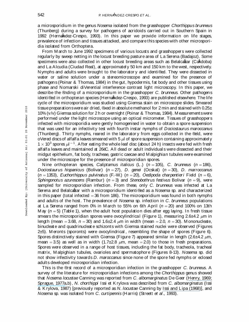
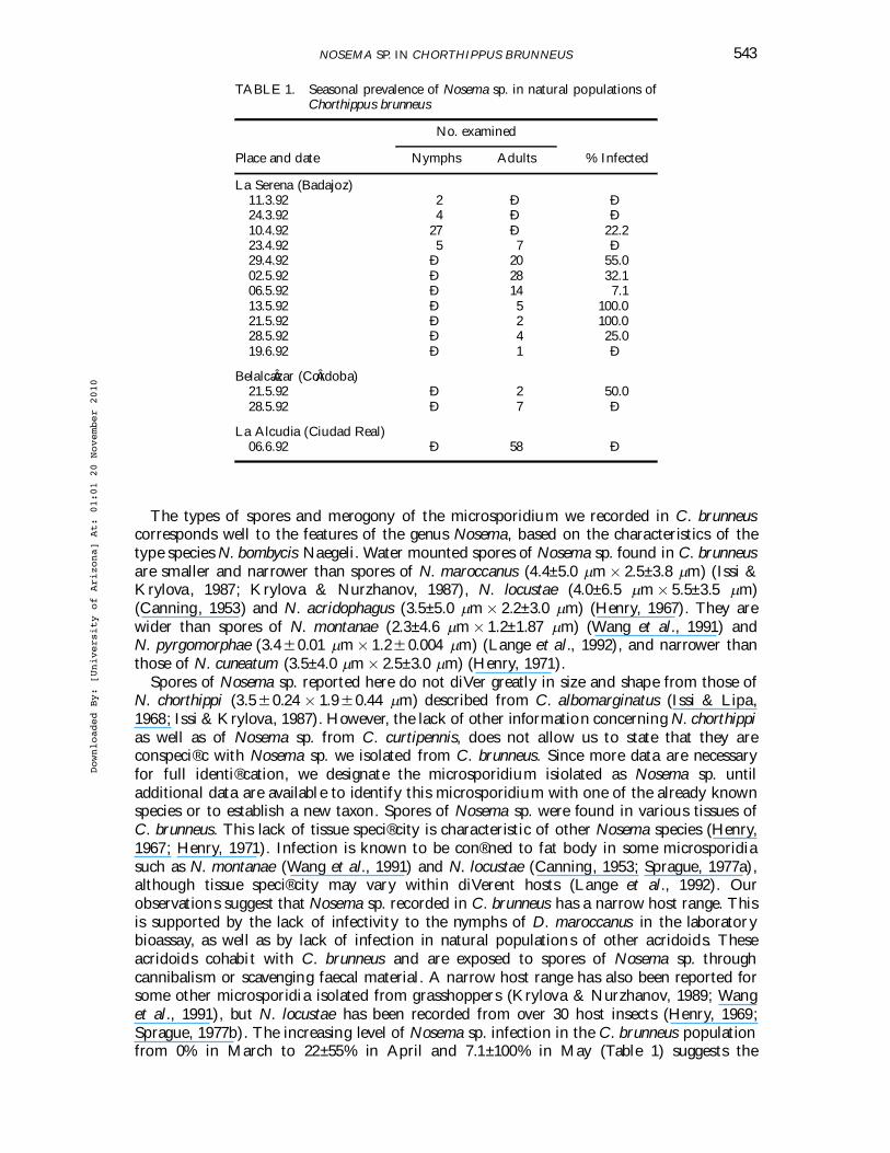
Nine orthopteran species, Caliptamus italicus (L.) (n 5105), C. brunneus (n 5186),Dociostaurus hispanicus (Bolivar) (n 527), D. genei (Ocskal) (n 530), D. marroccanus(n 51353), Euchorthippus pulvinatus (F.-W.) (n 520), Oedipoda charpentieri Field (n 56),Sphingonotus azurescens (Rambur) (n 53) and Stenobothrus festivus Bolivar (n 59), weresampled for microsporidian infection. From these, only C. brunneus was infected at LaSerena and BelalcaÂzar with a microsporidium identi®ed as a Nosema sp. and characterizedin this paper (total infected 536 from 186). The microsporidium was found in both nymphsand adults of the host. The prevalence of Nosema sp. infection in C. brunneus populationsat La Serena ranged from 0% in March to 55% on 6th April (n 520) and 100% on 13thMay (n 55) (Table 1), when the adult host population dies after egg laying. In fresh tissuesmears the microsporidian spores were ovocylindrica l (Figure 1), measuring 2.6±4.2 lm inlength (mean 53.68, n 530) and 1.6±2.4 lm in width (mean 52.0, n 530). Mononucleate,binucleate and quadrinucleat e schizonts with Giemsa stained nuclei were observed (Figures2±5). Meronts (sporonts) were ovocylindrical , resembling the shape of spores (Figure 6).Spores distinctively stained with Giemsa (Figure 7) appeared similar in length (2.6±4.2 lm,mean 53.5) as well as in width (1.7±2.8 lm, mean 52.0) to those in fresh preparations.Spores were observed in a range of host tissues, including the fat body, tracheola, trachealmatrix, Malpighian tubules, ovarioles and spermatophore (Figures 8-13). Nosema sp. didnot show infectivity towards D. maroccanus since none of the spore fed nymphs or eclosedadults developed microsporidian infection.
This is the ®rst record of a microsporidian infection in the grasshopper C. brunneus. Asurvey of the literature for microsporidian infections among the Chorthippus genus showedthat Nosema locustae Canning was reported from C. albomarginatus De Geer (Henry, 1969;Sprague, 1977a,b) , N. chorthippi Issi et Krylova was described from C. albomarginatus (Issi& Krylova, 1987) [previously reported as N. locustae Canning by Issi and Lipa (1968)], andNosema sp. was isolated from C. curtipennis (Harris) (Streett et al., 1993).
Downloaded By: [University of Arizona] At: 01:01 20 November 2010
NOSEMA SP. IN CHORTHIPPUS BRUNNEUS 543
TABLE 1. Seasonal prevalence of Nosema sp. in natural populations ofChorthippus brunneus
No. examined
Place and date Nymphs Adults % Infected
La Serena (Badajoz)11.3.92 2 Ð Ð24.3.92 4 Ð Ð10.4.92 27 Ð 22.223.4.92 5 7 Ð29.4.92 Ð 20 55.002.5.92 Ð 28 32.106.5.92 Ð 14 7.113.5.92 Ð 5 100.021.5.92 Ð 2 100.028.5.92 Ð 4 25.019.6.92 Ð 1 Ð
BelalcaÂzar (CoÂrdoba)21.5.92 Ð 2 50.028.5.92 Ð 7 Ð
La Alcudia (Ciudad Real)06.6.92 Ð 58 Ð
The types of spores and merogony of the microsporidium we recorded in C. brunneuscorresponds well to the features of the genus Nosema, based on the characteristics of thetype species N. bombycis Naegeli. Water mounted spores of Nosema sp. found in C. brunneusare smaller and narrower than spores of N. maroccanus (4.4±5.0 lm 3 2.5±3.8 lm) (Issi &Krylova, 1987; Krylova & Nurzhanov, 1987), N. locustae (4.0±6.5 lm 3 5.5±3.5 lm)(Canning, 1953) and N. acridophagus (3.5±5.0 lm 3 2.2±3.0 lm) (Henry, 1967). They arewider than spores of N. montanae (2.3±4.6 lm 3 1.2±1.87 lm) (Wang et al., 1991) andN. pyrgomorphae (3.460.01 lm 3 1.26 0.004 lm) (Lange et al., 1992), and narrower thanthose of N. cuneatum (3.5±4.0 lm 3 2.5±3.0 lm) (Henry, 1971).
Spores of Nosema sp. reported here do not diVer greatly in size and shape from those ofN. chorthippi (3.560.24 3 1.960.44 lm) described from C. albomarginatus (Issi & Lipa,1968; Issi & Krylova, 1987). However, the lack of other information concerning N. chorthippias well as of Nosema sp. from C. curtipennis, does not allow us to state that they areconspeci®c with Nosema sp. we isolated from C. brunneus. Since more data are necessaryfor full identi®cation, we designate the microsporidium isiolated as Nosema sp. untiladditional data are available to identify this microsporidium with one of the already knownspecies or to establish a new taxon. Spores of Nosema sp. were found in various tissues ofC. brunneus. This lack of tissue speci®city is characteristic of other Nosema species (Henry,1967; Henry, 1971). Infection is known to be con®ned to fat body in some microsporidiasuch as N. montanae (Wang et al., 1991) and N. locustae (Canning, 1953; Sprague, 1977a),although tissue speci®city may vary within diVerent hosts (Lange et al., 1992). Ourobservations suggest that Nosema sp. recorded in C. brunneus has a narrow host range. Thisis supported by the lack of infectivity to the nymphs of D. maroccanus in the laboratorybioassay, as well as by lack of infection in natural populations of other acridoids. Theseacridoids cohabit with C. brunneus and are exposed to spores of Nosema sp. throughcannibalism or scavenging faecal material. A narrow host range has also been reported forsome other microsporidia isolated from grasshoppers (Krylova & Nurzhanov, 1989; Wanget al., 1991), but N. locustae has been recorded from over 30 host insects (Henry, 1969;Sprague, 1977b). The increasing level of Nosema sp. infection in the C. brunneus populationfrom 0% in March to 22±55% in April and 7.1±100% in May (Table 1) suggests the
Downloaded By: [University of Arizona] At: 01:01 20 November 2010

544 P. HERNAÂNDEZ-CRESPO ET AL.
FIGURES 1±8. Nosema sp. from Chorthippus brunneus. 1Ðspores in water seen in Nomarski diVerentialinterference contrast (2775 3 ); 2Ðuninucleate schizont (1387 3 ); 3Ðdivision of uninucleateschizont (2775 3 ); 4Ðbinucleate schizont (2775 3 ); 5Ðquadrinucleate and binucleateschizont (2775 3 ); 6Ðmeront (sporont) (2775 3 ); 7ÐGiemsa-stained spores (555 3 );8Ðfat body cells ®lled with spores (1110 3 ).
occurrence of a microsporidian epizootic, although caution is required in interpreting databased on few specimens (see Table 1). Epizootics caused by N. locusta and Nosema sp. ingrasshopper populations (Henry, 1972; Streett & Woods, 1993) are well documented. Theprevalence of microsporidian infection can be highly variable from year to year (McGuire& Streett, 1989) and may result from horizontal or vertical transmission (Streett & Wood,
Downloaded By: [University of Arizona] At: 01:01 20 November 2010

NOSEMA SP. IN CHORTHIPPUS BRUNNEUS 545
FIGURES 9±13. Nosema sp. from Chorthippu s brunneus. 9Ðspores in tracheole (1110 3 ); 10Ðspores intracheal matrix (1110 3 ); 11Ðspores in Malpighian tubules (1110 3 ); 12Ðspores inspermatophore (555 3 ); 13Ðspores in ovariole (555 3 ).
1993). Prevalence of infection by Nosema sp. in C. brunneus indicates that horizontaltransmission may operate in the host ®eld populations. Moreover, the presence of spores inthe spermatophore (Figure 12) and in ovarioles (Figure 13) of adults suggests that trans-ovarian (vertical) transmission may also represent an important route of infection. Thepathology, host range and transmission of this Nosema species isolated from C. brunneus isnow under study in our laboratory.
Downloaded By: [University of Arizona] At: 01:01 20 November 2010

546 P. HERNAÂNDEZ-CRESPO ET AL.
REFERENCES
Bidochka, M.J. & Khachatourians, G.G. (1991)Microbial and protozoan pathogens of grasshoppers andlocusts as potential biocontrol agents. Biocontrol Science and Technology 1, 242±259.
Canning, E.U. (1953) A new microsporidian, Nosema locustae n. sp., from the fat body of the Africanmigratory locust, Locusta migratoria migratorioide s R. et F. Parasitology 43, 287±290.
Henry, J.E. (1967) Nosema acridophagus sp. n., a microsporidian isolated from grasshoppers. Journal ofInvertebrate Pathology 9, 331±341.
Henry, J.E. (1969) Extension of the host range of Nosema locustae in Orthoptera. Annals of the EntomologicalSociety of America 62, 452±453.
Henry, J.E. (1971) Nosema cuneatum sp. n. (Microsporida: Nosematidae) in grasshoppers (Orthoptera:Acrididae). Journal of Invertebrate Pathology 17, 164±171.
Henry, J.E. (1972) Epizootiology of infections by Nosema locustae (Canning) (Microsporidia: Nosematidae)in grasshoppers. Acrida 1, 111±120.
HernaÂndez-Crespo, P. (1993) La Langosta MediterraÂnea, Dociostaurus maroccanus (Thunberg), sus Enem-igos Naturales AutoÂctonos y el Posible Control de sus Plagas por Medio de Microorganismos PatoÂgenos.PhD Thesis, Universidad de CoÂrdoba, Spain.
Issi, I. & Krylova, S.V. (1987) Locusts microsporidia, in Saranchovye±Ekologiya i Mery Bor’by (Nikitina,M.S., Ed.). VIZR, St Petersburg, Russia, pp. 58±62 (in Russian).
Issi, I. & Lipa, J.J. (1968) Report on identi®cation of Protozoa pathogenic for insects in the Soviet Union(1961±1966), with descriptions of some new species. Acta Protozoologic a 6, 281±290.
Krylova, S.V. & Nurzhanov, A.A. (1987) Nosema sp. n. (Nosematidae) microsporidium from the Maroccanlocust Diciostaurus maroccanus Thunb. (Orthoptera). Byulleten’ Vsesoyuznogo Nauchno-Issledovatel ’skogoInstituta Zaschity Rastenii 68, 10±14 (in Russian).
Krylova, S.V. & Nurzhanov, A.A. (1989) Speci®city of microsporidium Nosema maroccanus fromMaroccan locust for the other insects. Byulletin’ Vsesoyuznogo-Issledovatel ’skogo Instituta ZaschityRastenii 74, 10±14 (in Russian).
Lange, C.E., Brito, J.M. & Henry, J.E. (1992) Characteristics of a microsporidium (Protozoa: Microspora)infecting grasshoppers (Orthoptera: Pyrgomorphidae) in Cape Verde, Africa. Journal of Protozoology 39,494±498.
Madox, J.V. (1987) Protozoan diseases, in Epizootiology of Insect Diseases (Fuxa, J.R. & Tanada, Y., Eds).John Wiley, New York, pp. 417±452.
McGuire, M.R. & Streett, D.A. (1989) Epizootiology of an undescribed microsporidium in populations ofChorthippu s curtipennis (Orthoptera: Acrididae). Journal of Invertebrate Pathology 54, 272±274.
Poinar, G.O. Jr & Thomas, G.M. (1984) Laboratory Guide to Insect Pathogens and Parasites. Plenum Press,New York.
Raina, S.K., Rai, M.M. & Khurad, A.M. (1987) Grasshopper and locust control using microsporidianinsecticides, in Biotechnology in Invertebrate Pathology and Cell CUlture. Academic Press, London,pp. 345±365.
Sprague, V. (1977a) Annoted list of species of Microsporidia, in Comparative Pathobiology : Vol. 2 Systematicsof the Microsporidia (Bulla, L.A., Jr & Cheng, T.C., Eds). Plenum Press, New York, pp. 331±334.
Sprague, V. (1977b) The zoological distribution of the Microsporidia, in Comparative Pathobiology : Vol. 2Systematics of the Microsporidia (Bulla, L.A., Jr & Cheng, T.C., Eds). Plenum Press, New York,pp. 335±385.
Streett, D.A. & Woods, S.A. (1993) Epizootiology of a Nosema sp. (Microsporida: Nosematidae) infectingthe grasshopper, Chorthippu s curtipennis (Harris) (Orthoptera: Acrididae). Canadian Entomologist 125,457±461.
Streett, D.A., Woods, S.A. & Onsager, J.A. (1993) Vertical transmission of a Nosema sp. (Microsporida:Nosematidae) infecting a grasshopper, Chorthippus curtipennis (Orthoptera: Acrididae). EnvironmentalEntomology 22, 1031±1034.
Toguebaye, B.S., Seck, A. & Marchand, B. (1988) Histopathologie et ultrastructure de Nosema prygo-morphae n. sp. (Microspora: Nosematidae) parasite de Pyrgomorpha conica tereticornis (Orthoptera:Pyrgomorphoidae). Archiv fuer Protistenkunde 136, 283±292.
Wang, L-Y., Streett, D.A. & Henry, J.E. (1991) Nosema montanae n. sp. (Microsporida: Nosematidae), aparasite from the grasshopper Melanoplus packardii (Orthoptera: Acrididae). Journal of InvertebratePathology 58, 211±218.
Downloaded By: [University of Arizona] At: 01:01 20 November 2010