Presurgical language mapping in children with epilepsy: Clinical usefulness of functional magnetic...
12
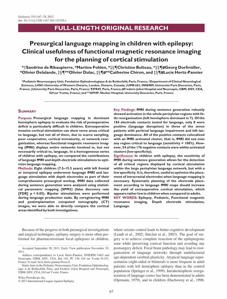
Presurgical language mapping in children with epilepsy: Clinical usefulness of functional magnetic resonance imaging for the planning of cortical stimulation *ySandrine de Ribaupierre, *Martine Fohlen, *zx{Christine Bulteau, *zx{#Georg Dorfmu ¨ ller, *Olivier Delalande, zx{**Olivier Dulac, zx{#**Catherine Chiron, and zx{#Lucie Hertz-Pannier *Pediatric Neurosurgery Unit, Fondation Ophtalmologique A de Rothschild, Paris, France; yDepartment of Clinical Neurological Sciences, LHSC-University of Western Ontario, London, Ontario, Canada; zUMR 663, INSERM, Universite ´ Paris Descartes, Paris, France; xUniversity Paris Descartes, Paris, France; {IFR49, Paris, France; #Frederic Joliot Hospital and Neurospin, I2BM, DSV, CEA, Gif sur Yvette, France; and **APHP, Necker Hospital, University Descartes, Paris, France SUMMARY Purpose: Presurgical language mapping in dominant hemisphere epilepsy to evaluate the risk of postoperative deficit is particularly difficult in children. Extraoperative invasive cortical stimulation can show some areas critical to language, but not all of them, due to scarce sampling, poor cooperation, cortical immaturity, or network reor- ganization, whereas functional magnetic resonance imag- ing (fMRI) displays entire networks involved in, but not necessarily critical to, language. In a homogeneous series of children with epilepsy, we compared the contributions of language fMRI and depth electrode stimulations to opti- mize language mapping. Methods: Eight children (7.5–15.5 years) with left frontal or temporal epilepsy underwent language fMRI and lan- guage stimulation with depth electrodes as part of their comprehensive presurgical workup. fMRI data collected during sentence generation were analyzed using statisti- cal parametric mapping (SPM2) (false discovery rate [FDR] p < 0.05). Bipolar stimulations were performed during language production tasks. By coregistering fMRI and postimplantation computed tomography (CT) images, we were able to directly compare the cortical areas identified by both investigations. Key Findings: fMRI during sentence generation robustly showed activation in the whole perisylvian regions with lit- tle reorganization (left hemisphere dominant in 7). Of the 184 electrode contacts tested for language, only 8 were positive (language disruption) in three of the seven patients with periictal language impairment and left lan- guage dominance. All of the positive contacts colocalized with an fMRI activated cluster, that is, fMRI did not miss any region critical to language (sensitivity = 100%). How- ever, 54 of the 176 negative contacts were within activated clusters (low specificity). Significance: In children with epilepsy, the sensitivity of fMRI during sentence generation allows for the detection of all critical regions displayed by cortical stimulation within the large perisylvian language network, but with a low specificity. It is, therefore, useful to optimize the place- ment of intracranial electrodes when language mapping is necessary. Systematic planning of the electrode place- ment according to language fMRI maps should increase the yield of extraoperative cortical stimulation, which appears rather low in children when compared to adults. KEY WORDS: Epilepsy, Pediatric, Functional magnetic resonance imaging, Depth electrode stimulation, Language. Because of the progress in both presurgical investigations and surgical techniques, epilepsy surgery is more often per- formed for pharmacoresistant focal epilepsies in children, where seizure control leads to better cognitive development (Lendt et al., 2002; Sinclair et al., 2003). The goal of sur- gery is to achieve complete resection of the epileptogenic zone while preserving cortical function and avoiding any postsurgery deficit. Focal brain pathology may lead to reor- ganization of language networks through multifactorial age-dependent cerebral plasticity. Atypical language repre- sentation (right-sided or bilateral) is more frequent in adult patients with left hemisphere epilepsy than in the control population (Springer et al., 1999). Intrahemispheric reorga- nization of language cortex has been demonstrated in adults (Ojemann, 1979), and in children (Duchowny et al., 1996; Accepted September 30, 2011; Early View publication November 29, 2011. Address correspondence to Lucie Hertz-Pannier, INSERM U663 and Neurospin, I2BM, DSV, CEA, Bat 145, PC 156, Gif sur Yvette 91191, France. E-mail: [email protected] Study done in the Pediatric Neurosurgery Unit, Fondation Ophtalmolog- ique A de Rothschild, Paris, and Frederic Joliot Hospital and Neurospin, I2BM, DSV, CEA, Gif sur Yvette, France. Wiley Periodicals, Inc. ª 2011 International League Against Epilepsy Epilepsia, 53(1):67–78, 2012 doi: 10.1111/j.1528-1167.2011.03329.x FULL-LENGTH ORIGINAL RESEARCH 67
Transcript of Presurgical language mapping in children with epilepsy: Clinical usefulness of functional magnetic...
Presurgical language mapping in children with epilepsy:
Clinical usefulness of functional magnetic resonance imaging
for the planning of cortical stimulation*ySandrine de Ribaupierre, *Martine Fohlen, *zx{Christine Bulteau, *zx{#Georg Dorfmuller,
*Olivier Delalande, zx{**Olivier Dulac, zx{#**Catherine Chiron, and zx{#Lucie Hertz-Pannier
*Pediatric Neurosurgery Unit, Fondation Ophtalmologique A de Rothschild, Paris, France; yDepartment of Clinical Neurological
Sciences, LHSC-University of Western Ontario, London, Ontario, Canada; zUMR 663, INSERM, Universite Paris Descartes, Paris,
France; xUniversity Paris Descartes, Paris, France;{IFR49, Paris, France; #Frederic Joliot Hospital and Neurospin, I2BM, DSV, CEA,
Gif sur Yvette, France; and **APHP, Necker Hospital, University Descartes, Paris, France
SUMMARY
Purpose: Presurgical language mapping in dominant
hemisphere epilepsy to evaluate the risk of postoperative
deficit is particularly difficult in children. Extraoperative
invasive cortical stimulation can show some areas critical
to language, but not all of them, due to scarce sampling,
poor cooperation, cortical immaturity, or network reor-
ganization, whereas functional magnetic resonance imag-
ing (fMRI) displays entire networks involved in, but not
necessarily critical to, language. In a homogeneous series
of children with epilepsy, we compared the contributions
of language fMRI and depth electrode stimulations to opti-
mize language mapping.
Methods: Eight children (7.5–15.5 years) with left frontal
or temporal epilepsy underwent language fMRI and lan-
guage stimulation with depth electrodes as part of their
comprehensive presurgical workup. fMRI data collected
during sentence generation were analyzed using statisti-
cal parametric mapping (SPM2) (false discovery rate
[FDR] p < 0.05). Bipolar stimulations were performed
during language production tasks. By coregistering fMRI
and postimplantation computed tomography (CT)
images, we were able to directly compare the cortical
areas identified by both investigations.
Key Findings: fMRI during sentence generation robustly
showed activation in the whole perisylvian regions with lit-
tle reorganization (left hemisphere dominant in 7). Of the
184 electrode contacts tested for language, only 8 were
positive (language disruption) in three of the seven
patients with periictal language impairment and left lan-
guage dominance. All of the positive contacts colocalized
with an fMRI activated cluster, that is, fMRI did not miss
any region critical to language (sensitivity = 100%). How-
ever, 54 of the 176 negative contacts were within activated
clusters (low specificity).
Significance: In children with epilepsy, the sensitivity of
fMRI during sentence generation allows for the detection
of all critical regions displayed by cortical stimulation
within the large perisylvian language network, but with a
low specificity. It is, therefore, useful to optimize the place-
ment of intracranial electrodes when language mapping is
necessary. Systematic planning of the electrode place-
ment according to language fMRI maps should increase
the yield of extraoperative cortical stimulation, which
appears rather low in children when compared to adults.
KEY WORDS: Epilepsy, Pediatric, Functional magnetic
resonance imaging, Depth electrode stimulation,
Language.
Because of the progress in both presurgical investigationsand surgical techniques, epilepsy surgery is more often per-formed for pharmacoresistant focal epilepsies in children,
where seizure control leads to better cognitive development(Lendt et al., 2002; Sinclair et al., 2003). The goal of sur-gery is to achieve complete resection of the epileptogeniczone while preserving cortical function and avoiding anypostsurgery deficit. Focal brain pathology may lead to reor-ganization of language networks through multifactorialage-dependent cerebral plasticity. Atypical language repre-sentation (right-sided or bilateral) is more frequent in adultpatients with left hemisphere epilepsy than in the controlpopulation (Springer et al., 1999). Intrahemispheric reorga-nization of language cortex has been demonstrated in adults(Ojemann, 1979), and in children (Duchowny et al., 1996;
Accepted September 30, 2011; Early View publication November 29,2011.
Address correspondence to Lucie Hertz-Pannier, INSERM U663 andNeurospin, I2BM, DSV, CEA, Bat 145, PC 156, Gif sur Yvette 91191,France. E-mail: [email protected]
Study done in the Pediatric Neurosurgery Unit, Fondation Ophtalmolog-ique A de Rothschild, Paris, and Frederic Joliot Hospital and Neurospin,I2BM, DSV, CEA, Gif sur Yvette, France.
Wiley Periodicals, Inc.ª 2011 International League Against Epilepsy
Epilepsia, 53(1):67–78, 2012doi: 10.1111/j.1528-1167.2011.03329.x
FULL-LENGTH ORIGINAL RESEARCH
67
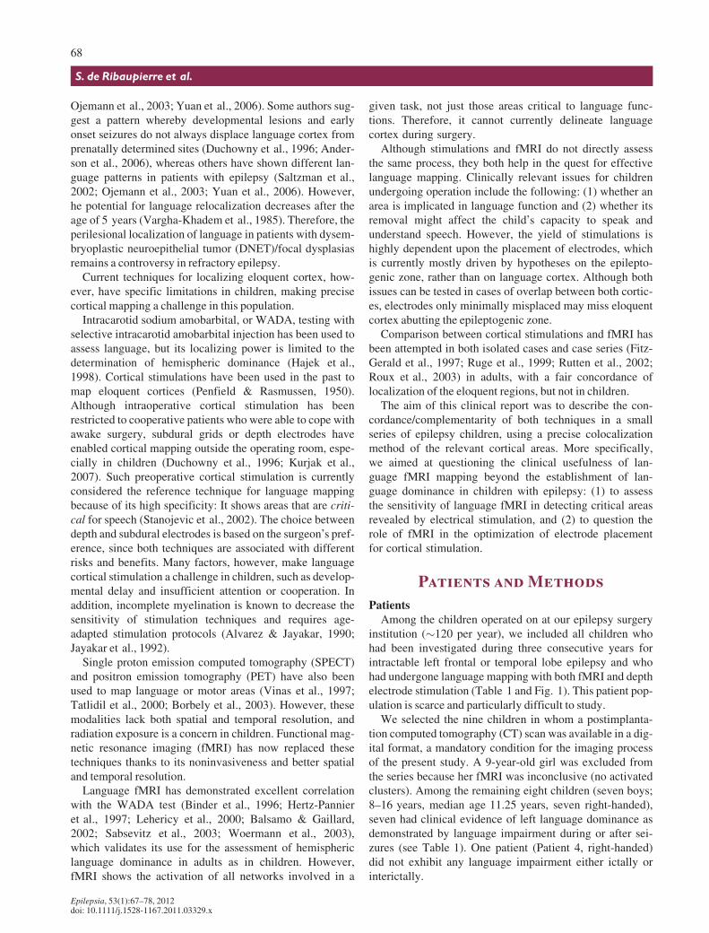
Ojemann et al., 2003; Yuan et al., 2006). Some authors sug-gest a pattern whereby developmental lesions and earlyonset seizures do not always displace language cortex fromprenatally determined sites (Duchowny et al., 1996; Ander-son et al., 2006), whereas others have shown different lan-guage patterns in patients with epilepsy (Saltzman et al.,2002; Ojemann et al., 2003; Yuan et al., 2006). However,he potential for language relocalization decreases after theage of 5 years (Vargha-Khadem et al., 1985). Therefore, theperilesional localization of language in patients with dysem-bryoplastic neuroepithelial tumor (DNET)/focal dysplasiasremains a controversy in refractory epilepsy.
Current techniques for localizing eloquent cortex, how-ever, have specific limitations in children, making precisecortical mapping a challenge in this population.
Intracarotid sodium amobarbital, or WADA, testing withselective intracarotid amobarbital injection has been used toassess language, but its localizing power is limited to thedetermination of hemispheric dominance (Hajek et al.,1998). Cortical stimulations have been used in the past tomap eloquent cortices (Penfield & Rasmussen, 1950).Although intraoperative cortical stimulation has beenrestricted to cooperative patients who were able to cope withawake surgery, subdural grids or depth electrodes haveenabled cortical mapping outside the operating room, espe-cially in children (Duchowny et al., 1996; Kurjak et al.,2007). Such preoperative cortical stimulation is currentlyconsidered the reference technique for language mappingbecause of its high specificity: It shows areas that are criti-cal for speech (Stanojevic et al., 2002). The choice betweendepth and subdural electrodes is based on the surgeon’s pref-erence, since both techniques are associated with differentrisks and benefits. Many factors, however, make languagecortical stimulation a challenge in children, such as develop-mental delay and insufficient attention or cooperation. Inaddition, incomplete myelination is known to decrease thesensitivity of stimulation techniques and requires age-adapted stimulation protocols (Alvarez & Jayakar, 1990;Jayakar et al., 1992).
Single proton emission computed tomography (SPECT)and positron emission tomography (PET) have also beenused to map language or motor areas (Vinas et al., 1997;Tatlidil et al., 2000; Borbely et al., 2003). However, thesemodalities lack both spatial and temporal resolution, andradiation exposure is a concern in children. Functional mag-netic resonance imaging (fMRI) has now replaced thesetechniques thanks to its noninvasiveness and better spatialand temporal resolution.
Language fMRI has demonstrated excellent correlationwith the WADA test (Binder et al., 1996; Hertz-Pannieret al., 1997; Lehericy et al., 2000; Balsamo & Gaillard,2002; Sabsevitz et al., 2003; Woermann et al., 2003),which validates its use for the assessment of hemisphericlanguage dominance in adults as in children. However,fMRI shows the activation of all networks involved in a
given task, not just those areas critical to language func-tions. Therefore, it cannot currently delineate languagecortex during surgery.
Although stimulations and fMRI do not directly assessthe same process, they both help in the quest for effectivelanguage mapping. Clinically relevant issues for childrenundergoing operation include the following: (1) whether anarea is implicated in language function and (2) whether itsremoval might affect the child’s capacity to speak andunderstand speech. However, the yield of stimulations ishighly dependent upon the placement of electrodes, whichis currently mostly driven by hypotheses on the epilepto-genic zone, rather than on language cortex. Although bothissues can be tested in cases of overlap between both cortic-es, electrodes only minimally misplaced may miss eloquentcortex abutting the epileptogenic zone.
Comparison between cortical stimulations and fMRI hasbeen attempted in both isolated cases and case series (Fitz-Gerald et al., 1997; Ruge et al., 1999; Rutten et al., 2002;Roux et al., 2003) in adults, with a fair concordance oflocalization of the eloquent regions, but not in children.
The aim of this clinical report was to describe the con-cordance/complementarity of both techniques in a smallseries of epilepsy children, using a precise colocalizationmethod of the relevant cortical areas. More specifically,we aimed at questioning the clinical usefulness of lan-guage fMRI mapping beyond the establishment of lan-guage dominance in children with epilepsy: (1) to assessthe sensitivity of language fMRI in detecting critical areasrevealed by electrical stimulation, and (2) to question therole of fMRI in the optimization of electrode placementfor cortical stimulation.
Patients and Methods
PatientsAmong the children operated on at our epilepsy surgery
institution (�120 per year), we included all children whohad been investigated during three consecutive years forintractable left frontal or temporal lobe epilepsy and whohad undergone language mapping with both fMRI and depthelectrode stimulation (Table 1 and Fig. 1). This patient pop-ulation is scarce and particularly difficult to study.
We selected the nine children in whom a postimplanta-tion computed tomography (CT) scan was available in a dig-ital format, a mandatory condition for the imaging processof the present study. A 9-year-old girl was excluded fromthe series because her fMRI was inconclusive (no activatedclusters). Among the remaining eight children (seven boys;8–16 years, median age 11.25 years, seven right-handed),seven had clinical evidence of left language dominance asdemonstrated by language impairment during or after sei-zures (see Table 1). One patient (Patient 4, right-handed)did not exhibit any language impairment either ictally orinterictally.
68
S. de Ribaupierre et al.
Epilepsia, 53(1):67–78, 2012doi: 10.1111/j.1528-1167.2011.03329.x
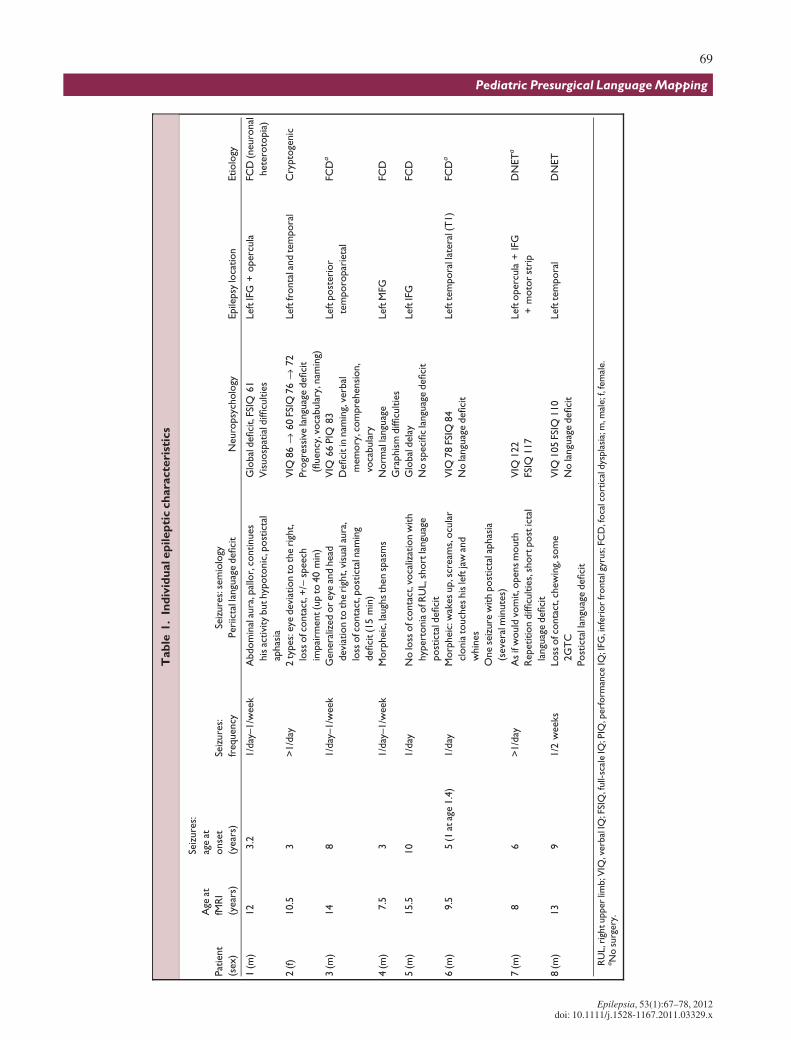
Tab
le1.
Ind
ivid
ualep
ilep
tic
ch
ara
cte
rist
ics
Pat
ient
(sex)
Age
at
fMR
I
(year
s)
Seiz
ure
s:
age
at
onse
t
(year
s)
Seiz
ure
s:
freq
uency
Seiz
ure
s:se
mio
logy
Peri
icta
llan
guag
edefici
tN
euro
psy
cholo
gyEpile
psy
loca
tion
Etiolo
gy
1(m
)12
3.2
1/d
ay–1/w
eek
Abdom
inal
aura
,pal
lor,
continues
his
activi
tybuthyp
oto
nic
,post
icta
l
aphas
ia
Glo
bal
defici
t,FS
IQ61
Vis
uosp
atia
ldiffi
cultie
s
Left
IFG
+operc
ula
FCD
(neuro
nal
hete
roto
pia
)
2(f
)10.5
3>
1/d
ay2
types:
eye
devi
atio
nto
the
righ
t,
loss
ofc
onta
ct,+
/)sp
eec
h
impai
rment(u
pto
40
min
)
VIQ
86
fi60
FSIQ
76
fi72
Pro
gress
ive
langu
age
defici
t
(fluen
cy,v
oca
bula
ry,n
amin
g)
Left
fronta
land
tem
pora
lC
rypto
genic
3(m
)14
81/d
ay–1/w
eek
Genera
lized
or
eye
and
head
devi
atio
nto
the
righ
t,vi
sual
aura
,
loss
ofc
onta
ct,p
ost
icta
lnam
ing
defici
t(1
5m
in)
VIQ
66
PIQ
83
Defici
tin
nam
ing,
verb
al
mem
ory
,com
pre
hensi
on,
voca
bula
ry
Left
post
erio
r
tem
poro
par
ieta
l
FCD
a
4(m
)7.5
31/d
ay–1/w
eek
Morp
hei
c,la
ugh
sth
en
spas
ms
Norm
alla
ngu
age
Gra
phis
mdiffi
cultie
s
Left
MFG
FCD
5(m
)15.5
10
1/d
ayN
olo
ssofc
onta
ct,v
oca
lizat
ion
with
hyp
ert
onia
ofR
UL,s
hort
langu
age
post
icta
ldefici
t
Glo
bal
dela
y
No
speci
fic
langu
age
defici
t
Left
IFG
FCD
6(m
)9.5
5(1
atag
e1.4
)1/d
ayM
orp
hei
c:w
akes
up,s
cream
s,ocu
lar
clonia
touch
es
his
left
jaw
and
whin
es
One
seiz
ure
with
post
icta
laphas
ia
(seve
ralm
inute
s)
VIQ
78
FSIQ
84
No
langu
age
defici
t
Left
tem
pora
llat
eral
(T1)
FCD
a
7(m
)8
6>
1/d
ayA
sif
would
vom
it,o
pens
mouth
Repetition
diffi
cultie
s,sh
ort
post
icta
l
langu
age
defici
t
VIQ
122
FSIQ
117
Left
operc
ula
+IF
G
+m
oto
rst
rip
DN
ET
a
8(m
)13
91/2
weeks
Loss
ofc
onta
ct,c
hew
ing,
som
e
2G
TC
Post
icta
llan
guag
edefici
t
VIQ
105
FSIQ
110
No
langu
age
defici
t
Left
tem
pora
lD
NET
RU
L,r
ightupper
limb;V
IQ,v
erb
alIQ
;FSI
Q,f
ull-
scal
eIQ
;PIQ
,perf
orm
ance
IQ;I
FG,i
nfe
rior
fronta
lgyr
us;
FCD
,foca
lcort
ical
dys
pla
sia;
m,m
ale;f
,fem
ale.
aN
osu
rgery
.
69
Pediatric Presurgical Language Mapping
Epilepsia, 53(1):67–78, 2012doi: 10.1111/j.1528-1167.2011.03329.x
Methods
fMRI paradigmAfter having obtained parental consent and child assent,
according to our institutional review board approval,language fMRI was performed on a 3-Tesla research mag-net (Bruker, Erlangen, Germany) using a modified gradi-ent-echo echo-planar sequence (22 axial slices, resolution3.7 · 3.7 · 5 mm3, TR = 5 s). Children were especiallytrained before testing to optimize comprehension and per-formance, after careful explanation was given about thecovert condition that they would perform during fMRIusing the same timing and paradigm as in MRI, but inovert condition and with different words. Among the fourexpressive and receptive tasks performed in our local pro-cedure (Hertz-Pannier et al., 2001), the candidate task forthe present study consisted of generation of sentences,performed covertly to minimize the risk of head and facialmovements. In our experience, this expressive languagetask, close to the widely used verb generation task, andstrongly lateralizing, has several major advantages in chil-dren, although no control of performance can be obtainedduring scanning: it is amenable to children with IQs down
to 50, does not require much in the way of attentionalresources or heavy working memory load, and optimizesthe yield of fMRI in terms of assessment of hemisphericdominance, when compared to other language paradigms.In this series, our pragmatic goal with fMRI was to screenthe entire language network (as well as its lateralization),rather than to find specific language areas. Patients werepresented with a concrete word via headphones every 5 s.They were then asked to silently generate a simple sen-tence (subject-verb-object) using the presented word. Sub-jects were instructed not to vocalize their sentences inorder to minimize artifacts from face movements. Theywere given five nouns in each activation block, and acti-vation blocks alternated with blocks of rest of the sameduration (total 3 min 35 s). To ensure good comprehen-sion of the auditory stimuli, these were presented duringthe silent interval formed by the grouping of the sliceacquisition gradients (loud beeps) over 1.4 s within eachTR of 5 s. During rest, patients were asked to stop think-ing of words and to listen to the magnet noise.
In one child who failed the sentence generation (Patient1), word repetition was used instead, with the presentationof concrete words through the headphones in the magnet.
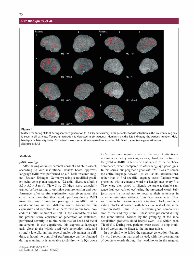
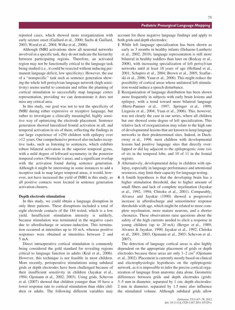
Figure 1.
Surface rendering of fMRI during sentence generation (p < 0.05 per cluster) in the patients. Robust activation in the prefrontal regions
is seen in all patients. Temporal activation is detected in six patients. Numbers on the left indicating the patient number. HLI,
hemispheric laterality index. *In Patient 1, word repetition was used because the child failed the sentence generation task.
Epilepsia ILAE
70
S. de Ribaupierre et al.
Epilepsia, 53(1):67–78, 2012doi: 10.1111/j.1528-1167.2011.03329.x

Two words were presented in each activation block, andactivation blocks alternated with blocks of rest of the sameduration. The patient was asked to silently repeat the wordsimmediately after hearing them. During rest, he was toremain quiet and listen to the magnet noise.
Each child underwent a three-dimensional (3D) inversionrecovery (IR)-prepped T1 acquisition for anatomic localiza-tion of activated clusters.
Electrodes implantation and stimulationDepth electrode implantation had been planned for sei-
zure monitoring after careful review of video–electroen-cephalography (EEG) and MRI, and was performed withina mean interval of 6 months around the fMRI. As all but onechild had periictal language impairment, we expected aproximity between critical language cortex and the epilepto-genic zone. We slightly modified the implantation design inthe last child included in the series (Patient 2) to place someelectrodes within the fMRI activated clusters, surroundingthe hypothetical epileptogenic zone. Depth electrodes wereimplanted using a frameless stereotactic robotic system(NeuroMate; Schaerer-Mayfield, Venissieux, France) com-bined with an imaging workstation for trajectory planningand electrode positioning (Voxim software; IVS Solutions,Chemnitz, Germany). Following the implantation, all chil-dren underwent a CT scan to confirm each electrode’s exactposition.
The electrodes were stimulated using the Jayakar proto-col during 1 week of EEG-recording (Jayakar et al., 1992).Square pulses of current were applied either to two adjacentcontacts or to two contacts separated by a third one (bipolarstimulation). The stimulations began at an intensity of1 mA, a high frequency of 50 Hz, a 0.3 ms pulse of alternat-ing polarity and a train duration of 3–5 s. Stimulation inten-sity was increased by steps of 1 mA until eitherpoststimulation discharges were seen on EEG, a clinicalresponse with speech arrest occurred, or a seizure was trig-gered.
In the children who also had electrodes in either the motorcortex or the sensorimotor area (SMA; n = 3, 8–16 years), apositive response was obtained at an average of 2 mA. Post-stimulation discharges were observed with intensities vary-ing from 1.6–10 mA. Therefore, the maximal stimulationintensity was 10 mA.
The language tasks used for stimulations all involved‘‘ecologic’’ language production, and consisted of counting,reading a book aloud, or spontaneously generating speech,depending on child’s abilities and the site of stimulation, tokeep an acceptable length of the procedure. A response wasconsidered positive when the child displayed a clear andtransient arrest in language activity during stimulation.
Fusion of fMRI and CT scan imagesTo obtain the same reference space for depth electrodes
and fMRI, the postimplantation CT scan was aligned with
the anatomic MRI data and resliced using MRIcro (Version1.39) (Rorden & Brett, 2000) and SPM2.
Fusion of CT and fMRI images was performed using twodifferent softwares: Anatomist (CEA, Orsay, France, http://brainvisa.info) and MRIcro (Version 1.39). Fusion withboth software packages was similar, as well as the numberof electrode contacts either within or outside of activatedareas.
AnalysisAll stimulation data were reviewed and grouped as posi-
tive or negative.The fMRI data were analyzed using SPM2 (Wellcome
Institute for Cognitive Neuroscience, University College,London, United Kingdom). The first three images of eachrun were discarded to allow stabilization of the longitudinalmagnetization. The remaining nonnormalized images wererealigned to the first image to correct for head motion duringacquisition, coregistered to the anatomic image, resliced,and smoothed (full width at half maximum [FWHM] of5 mm). Images were then analyzed using pixel-wise thresh-olds of p < 0.05 and p < 0.01 (FDR), corrected at a cluster-level of p < 0.05. The obtained clusters were then saved as a3D image.
In an additional analysis to assess hemispheric languagedominance, hemispheric laterality indices were calculatedby automatically summing all activated clusters in the leftand right hemispheres (excluding basal ganglia, occipitalcortex, and midline pixels) after linear normalization, andcomputing Left ) Right/Left + Right ratios (Binder et al.,1996).
Sensibility and specificity of the fMRI were calculatedusing the stimulation results as the reference.
Results
Language representation at fMRIfMRI during sentence generation elicited consistent acti-
vation in the usual perisylvian network in all patients, con-firming the robustness of both the task and the analysisthreshold, albeit with considerable interindividual variation,as expected in this population (Fig. 1). Activation of theprefrontal cortex (left inferior frontal gyrus) was found in allsubjects, whereas posterior temporal activation was seen insix of them.
In the seven patients with periictal language impairment,fMRI confirmed the clinically suspected left hemispherelanguage dominance with positive hemispheric lateralityindex (HLI) (between 0.1 and 1; Fig. 1). In Patient 1 whereword repetition was used, temporal activation was ratherbilateral, contrasting with a clearly left lateralized activationin Broca’s area, resulting in an laterality index (LI) of 0.1.In the right-handed child without periictal language mani-festations (Patient 4), the dominance side was atypical witha right-sided lateralization (HLI )0.28).
71
Pediatric Presurgical Language Mapping
Epilepsia, 53(1):67–78, 2012doi: 10.1111/j.1528-1167.2011.03329.x
Language stimulation using depth electrodesAmong the 184 depth electrodes contacts stimulated in
total, 8 disclosed a positive result, that is, with a language
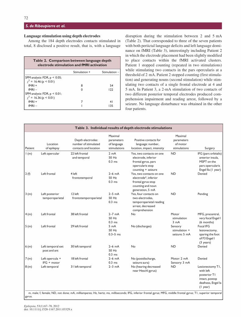
disruption during the stimulation between 2 and 5 mA(Table 2). That corresponded to three of the seven patientswith both periictal language deficits and left language domi-nance on fMRI (Table 3), interestingly including Patient 2in which the electrode placement had been slightly modifiedto place contacts within the fMRI activated clusters.Patient 1 stopped counting (repeated in two stimulations)while stimulating two contacts in the pars opercularis at athreshold of 2 mA. Patient 2 stopped counting (first stimula-tion) and generating nouns (second stimulation) while stim-ulating two contacts of a single frontal electrode at 4 and5 mA. In Patient 3, a 2-mA stimulation of two contacts oftwo different posterior temporal electrodes produced com-prehension impairment and reading arrest, followed by aseizure. No language disturbance was obtained in the otherfour patients.
Table 2. Comparison between language depth
electrode stimulation and fMRI activation
Stimulation + Stimulation )
SPM analysis: FDR, p < 0.05;
v2 = 16.46 (p < 0.01)
fMRI + 8 54
fMRI ) 0 122
SPM analysis: FDR, p < 0.01;
v2 = 16.36 (p < 0.01)
fMRI + 7 41
fMRI ) 1 135
Table 3. Individual results of depth electrode stimulations
Patient
Location
of epilepsy
Depth electrodes:
number of stimulated
contacts and location
Maximal
parameters
of language
stimulations
Positive contacts for
language: number,
location, impact, intensity
Maximal
parameters
of motor
stimulations Surgery
1 (m) Left opercular 22 left frontal
and temporal
2 mA
50 Hz
0.3 ms
Yes, two contacts on one
electrode, inferior
frontal gyrus, pars
opercularis stop
counting + seizure
ND IFG (pars orbitalis)
anterior insula,
MSPT on the
pars opercularis
Engel IIa (1 year)
2 (f) Left frontal 4 left
frontotemporal
2–6 mA
50 Hz
0.3 ms
Yes, two contacts on one
electrode*, inferior
frontal gyrus stop
counting and noun
generation, 5 mA
ND Denied
3 (m) Left posterior
temporoparietal
12 left
frontotemporoparietal
2–5 mA
50 Hz
0.3 ms
Yes, four contacts on
two electrodes,
temporoparietal reading
arrest, decreased
comprehension
ND Pending
4 (m) Left frontal 38 left frontal 2–7 mA
50 Hz
0.3 ms
No Motor
stimulation
3 mA
MFG, precentral,
very focal Engel I
(6 months)
5 (m) Left frontal 29 left frontal 5 mA
50 Hz
0.3–5 ms
No (discharges) Sensory
stimulation +
seizure: 5 mA
Focal IFG
lesionectomy,
sparing the foot
of F3 Engel I
(3 years)
6 (m) Left temporal ext
post and ant
30 left temporal 2–6 mA
50 Hz
0.3 ms
No ND Denied
7 (m) Left opercula +
IFG + motor
18 left frontal 2–6 mA
0.3 ms
No (postdischarge,
seizure aura)
Motor: 2 mA
Sensory: 3 mA
Denied
8 (m) Left temporal 31 left temporal 2–3 mA No (hearing decreased
near Heschl gyrus)
ND Lesionectomy T1,
with left
posterior T1
intact, postop
deafness, Engel Ia
(1 year)
m, male; f, female; ND, not done; mA, milliamperes; Hz, hertz; ms, milliseconds; IFG, inferior frontal gyrus; MFG, middle frontal gyrus; T1, superior temporalgyrus.
72
S. de Ribaupierre et al.
Epilepsia, 53(1):67–78, 2012doi: 10.1111/j.1528-1167.2011.03329.x

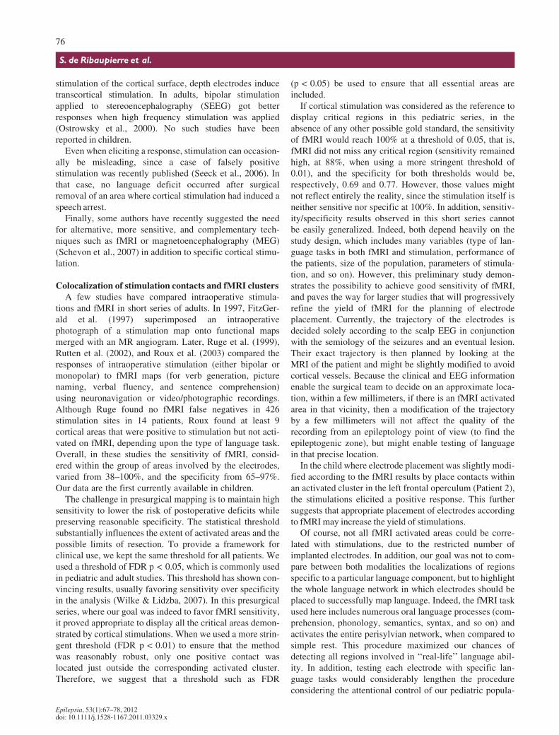
Colocalization of depth electrodes and fMRIAt the individual level, the colocalization of each positive
contact with fMRI-activated clusters was excellent (parsopercularis of the left inferior frontal gyrus in Patient 1(Fig. 2A–C); inferior frontal gyrus in Patient 2; temporopa-rietal junction in Patient 3; Table 2).
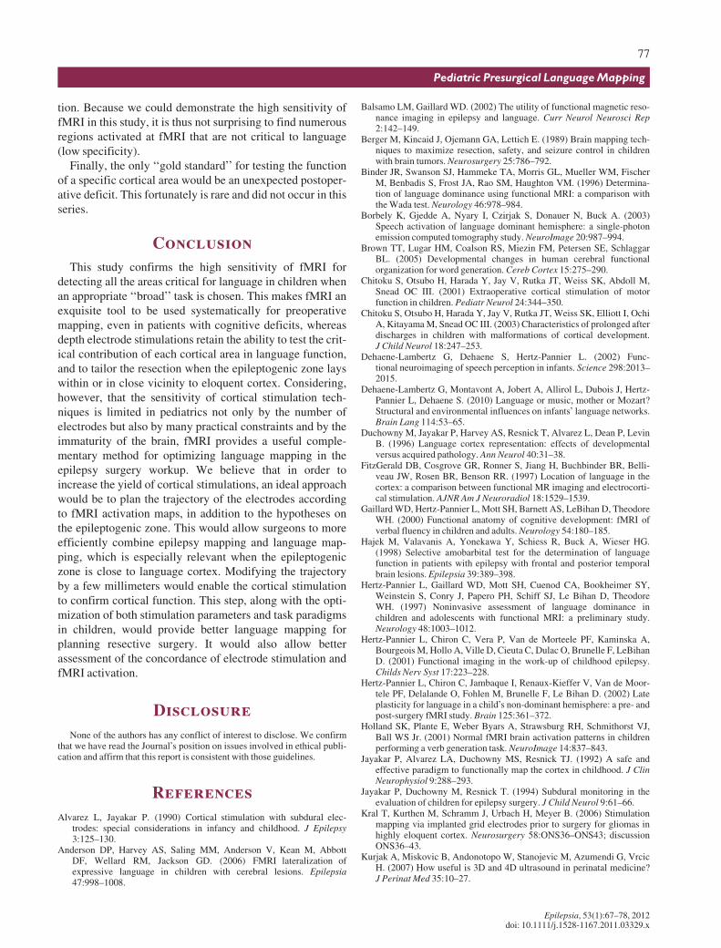
Although, as expected, a large number of negative con-tacts were outside activated areas, some were within or atthe border of activated clusters [superior temporal sulcus inPatient 1; posterior portion of the middle temporal gyrusin Patients 3 and 6; inferior and middle frontal gyri, precen-tral gyrus, and SMA in Patients 4, 5, and 7; and superior
A B
C
D
E
Figure 2.
Excellent colocalization of fMRI activation and positive electrode contact (Patient 1). (A–C) Axial, coronal, and sagittal views of
the CT–fMRI fusion showing positive contacts located in an activated cluster in the pars opercularis of the left inferior frontal gyrus.
(D–E) Postoperative CT scan showing the resection with the pars opercularis left in place. No postoperative language deficit and
Engel I at 12 months follow-up.
Epilepsia ILAE
A B
C
Figure 3.
Low specificity of fMRI activation
(Patient 8). (A–C) Axial, coronal,
and sagittal views of the CT-fMRI
fusion, where the electrodes within
the activated areas were negative to
stimulations. Frontal depth
electrodes were decided because of
the rapid spread of the seizures to
the frontal area on the scalp EEG to
exclude the possibility of the inferior
frontal gyrus being part of the
epileptogenic zone. Left temporal
dysembryoplastic neuroepithelial
tumor resected (lesionectomy).
One year after surgery the patient is
seizure free.
Epilepsia ILAE
73
Pediatric Presurgical Language Mapping
Epilepsia, 53(1):67–78, 2012doi: 10.1111/j.1528-1167.2011.03329.x
temporal gyrus and inferior frontal gyrus in Patient 8(Fig. 3A–C)].
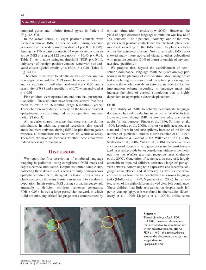
In the whole series, all eight positive contacts werelocated within an fMRI cluster activated during sentencegeneration at the widely used threshold of p < 0.05 (FDR).Among the 176 negative contacts, 54 were located within anactive fMRI cluster and 122 were not (v2 = 16.46, p < 0.01,Table 2). At a more stringent threshold (FDR p < 0.01),only seven of the eight positive contacts were within an acti-vated cluster (global results v2 = 16.36, p < 0.01, Table 2,Fig. 4A,B).
Therefore, if we were to take the depth electrode simula-tion as gold standard, the fMRI would have a sensitivity of 1and a specificity of 0.69 when analyzed at p < 0.05; and asensitivity of 0.88 and a specificity of 0.77 when analyzed atp < 0.01.
Five children were operated on and none had postopera-tive deficit. These children have remained seizure-free for amean follow-up of 16 months (range 6 months–3 years).Three children were denied surgery on the basis of bilateralepileptogenic foci or a high risk of postoperative languagedeficit (Table 3).
All surgeries spared the areas that were positive duringstimulation. In addition, planned resections also sparedareas that were activated during fMRI despite their negativeresponse at stimulation (in the Broca or Wernicke area).Therefore, we have no feedback whether those areas wereindeed necessary for language.
Discussion
We report the first description of combined languagemapping in pediatrics, using coregistered fMRI maps anddepth electrode stimulation. Despite its limited sample size,collecting these data in such a series of fairly homogeneousepileptic children with stringent inclusion criteria was achallenge, given the many limitations inherent to a pediatricpopulation. In this series, fMRI during a broad language taskamenable to deficient children (sentence generation,FDR < 0.05) showed a large perisylvian network in whichit did not miss any critical language areas demonstrated by
cortical stimulation (sensitivity = 100%). However, theyield of depth electrode language stimulation was low (8 of184 contacts; 3 of 7 patients). Notably, one of the threepatients with positive contacts had the electrode placementmodified according to the fMRI map, to place contactswithin the activated clusters. Not surprisingly, fMRI alsoshowed many more activated clusters, either colocalizedwith negative contacts (30% of them) or outside of any con-tact (low specificity).
We propose that, beyond the establishment of hemi-spheric dominance, language fMRI be systematically per-formed in the planning of cortical stimulation, using broadtasks including expressive and receptive processing thatactivate the whole perisylvian network, in order to plan theimplantation scheme according to language maps andincrease the yield of cortical stimulation that is highlydependent on appropriate electrode placement.
fMRIThe ability of fMRI to reliably demonstrate language
dominance has led to a decline in the use of the WADA test.However, even though fMRI is now everyday practice inadults for that purpose (Binder et al., 1996; Springer et al.,1999; Lehericy et al., 2000), it is not yet fully accepted as astandard of care in pediatric epilepsy because of the limitednumber of published studies (Hertz-Pannier et al., 1997,2002; Balsamo & Gaillard, 2002; Wilke et al., 2003, 2006;Szaflarski et al., 2006; Yuan et al., 2006). Expressive taskssuch as word fluency or verb generation are the most lateral-ized tasks and provide better correlation with invasive meth-ods like the WADA test than receptive tasks (Lehericyet al., 2000). Generation of sentences, an easy task largelyamenable to impaired children, activates a large left perisyl-vian network, comprising both expressive and receptive lan-guage areas (Broca and Wernicke) as well as the usualcortical areas found to be coactivated in various languagetasks (Muller et al., 1997; Vigneau et al., 2006). In this ser-ies, seven of the eight children showed clear left dominance.These children had little reorganization despite early leftperisylvian epilepsy, as it was found in other studies (Duch-owny et al., 1996; Liegeois et al., 2004), unlike some
A BFigure 4.
Threshold effect. (A) At FDR
p < 0.05, the electrode contacts
that are positive to stimulation are
within an activated area. (B) At
FDR p < 0.01, the activated area
around the electrode contacts is no
longer detected.
Epilepsia ILAE
74
S. de Ribaupierre et al.
Epilepsia, 53(1):67–78, 2012doi: 10.1111/j.1528-1167.2011.03329.x
reported cases, which showed more reorganization withearly seizure onset (Gaillard et al., 2000; Sachs & Gaillard,2003; Wood et al., 2004; Wilke et al., 2006).
Although fMRI activations show all neuronal networksinvolved in a specific task, they do not indicate the hierarchybetween participating regions. Therefore, an activatedregion may not be functionally critical to the language taskbeing studied (i.e., it could be resected without inducing per-manent language deficit, low specificity). However, the useof a ‘‘nonspecific’’ task such as sentence generation show-ing the whole left perisylvian language network (high sensi-tivity) seems useful to constrain and refine the planning ofcortical stimulation to successfully map language cortexrepresentation, providing we can demonstrate it does notmiss any critical area.
In this study, our goal was not to test the specificity offMRI during either expressive or receptive language, butrather to investigate a clinically meaningful, highly sensi-tive way of optimizing the electrode placement. Sentencegeneration showed lateralized frontal activation in all, andtemporal activation in six of them, reflecting the findings inour large experience of >250 children with epilepsy over>12 years. Our comprehensive protocol also includes recep-tive tasks, such as listening to sentences, which exhibitsrobust bilateral activation in the superior temporal gyrus,with a mild degree of leftward asymmetry in the posteriortemporal cortex (Wernicke’s area), and a significant overlapwith the activation found during sentence generation.Although it might be interesting in some instances to add areceptive task to map larger temporal areas, it would, how-ever, not have increased the yield of fMRI in this study, asall positive contacts were located in sentence generationactivation clusters.
Depth electrode stimulationIn this study, we could obtain a language disruption in
only three patients. These disruptions included a total ofeight electrode contacts of the 184 tested, which is a lowyield. Insufficient stimulation intensity is unlikely,because stimulation was terminated in the negative casesdue to afterdischarge or seizure-induction. This termina-tion occurred at intensities up to 10 mA, whereas positiveresponses were obtained at intensities between 2 and5 mA.
Direct intraoperative cortical stimulation is commonlybeing considered the gold standard for revealing regionscritical to language function in adults (Kral et al., 2006).However, this technique is not feasible in most children.More recently, perioperative stimulations using subduralgrids or depth electrodes have been challenged because oftheir insufficient sensitivity in children (Jayakar et al.,1994; Ojemann et al., 2002, 2003). Using grids, Schevonet al. (2007) showed that children younger than 10 have alower response rate to cortical stimulation than older chil-dren or adults. The following different variables may
account for these negative language findings and apply toboth grids and depth electrodes:1 While left language specialization has been shown as
early as 3 months in healthy infants (Dehaene-Lambertzet al., 2002, 2010), language representation is still morebilateral in healthy toddlers than later on (Redcay et al.,2008), with increasing specialization of left perisylviannetworks until at least 18 years of age (Holland et al.,2001; Schapiro et al., 2004; Brown et al., 2005; Szaflar-ski et al., 2006; Yuan et al., 2006). This might reduce thepossibility of cortical areas whose unilateral left stimula-tion would induce a speech disturbance.
2 Reorganization of language distribution has been shownmore frequently in subjects with early brain lesions andepilepsy, with a trend toward more bilateral language(Hertz-Pannier et al., 1997; Springer et al., 1999;Liegeois et al., 2004; Yuan et al., 2006). This, however,was not clearly the case in our series, where all childrenbut one showed some degree of left specialization. Thisrelative lack of reorganization may relate to the high rateof developmental lesions that are known to keep languagenetworks in their predetermined sites. Indeed, in Duch-owny et al., 1996. most children with developmentallesions had positive language sites that directly over-lapped or did lay adjacent to the epileptogenic zone (sixof six in the temporal lobe, and 10 of 11 in the frontalregion).
3 Alternatively, developmental delay in children with epi-lepsy, especially in language performance and attentionalresources, may limit their capacity for language testing.
4 A fourth hypothesis is that the developing brain has ahigher stimulation threshold, due to higher amount ofsmall fibers and lack of complete myelination (Jayakaret al., 1992, 1994; Chitoku et al., 2001). Comparably,Alvarez and Jayakar (1990) showed a progressiveincrease in afterdischarge and sensorimotor responsethresholds with age, which might be related to more com-plete myelination, more mature neurons, and a shorterchronaxis. These observations raise questions about thesafety of the high currents needed to elicit a response inyoung children (up to 20 mA) (Berger et al., 1989;Alvarez & Jayakar, 1990; Jayakar et al., 1992; Chitokuet al., 2001, 2003; Ojemann et al., 2003; Schevon et al.,2007).The detection of language cortical areas is also highly
dependent on the appropriate placement of grids or depthelectrodes because these areas are only 1–2 cm2 (Ojemannet al., 2002). Placement is currently mostly based on clinicaland electrophysiologic hypotheses on the epileptogenicnetwork, as it is impossible to infer the precise cortical orga-nization of language from anatomic data alone. Geometricdifferences between grids and depth electrodes (grids:3–5 mm in diameter, separated by 1 cm; depth electrodes:2 mm in diameter, separated by 1.5 mm) also influencethe stimulated volume. Although subdural grids allow
75
Pediatric Presurgical Language Mapping
Epilepsia, 53(1):67–78, 2012doi: 10.1111/j.1528-1167.2011.03329.x
stimulation of the cortical surface, depth electrodes inducetranscortical stimulation. In adults, bipolar stimulationapplied to stereoencephalography (SEEG) got betterresponses when high frequency stimulation was applied(Ostrowsky et al., 2000). No such studies have beenreported in children.
Even when eliciting a response, stimulation can occasion-ally be misleading, since a case of falsely positivestimulation was recently published (Seeck et al., 2006). Inthat case, no language deficit occurred after surgicalremoval of an area where cortical stimulation had induced aspeech arrest.
Finally, some authors have recently suggested the needfor alternative, more sensitive, and complementary tech-niques such as fMRI or magnetoencephalography (MEG)(Schevon et al., 2007) in addition to specific cortical stimu-lation.
Colocalization of stimulation contacts and fMRI clustersA few studies have compared intraoperative stimula-
tions and fMRI in short series of adults. In 1997, FitzGer-ald et al. (1997) superimposed an intraoperativephotograph of a stimulation map onto functional mapsmerged with an MR angiogram. Later, Ruge et al. (1999),Rutten et al. (2002), and Roux et al. (2003) compared theresponses of intraoperative stimulation (either bipolar ormonopolar) to fMRI maps (for verb generation, picturenaming, verbal fluency, and sentence comprehension)using neuronavigation or video/photographic recordings.Although Ruge found no fMRI false negatives in 426stimulation sites in 14 patients, Roux found at least 9cortical areas that were positive to stimulation but not acti-vated on fMRI, depending upon the type of language task.Overall, in these studies the sensitivity of fMRI, consid-ered within the group of areas involved by the electrodes,varied from 38–100%, and the specificity from 65–97%.Our data are the first currently available in children.
The challenge in presurgical mapping is to maintain highsensitivity to lower the risk of postoperative deficits whilepreserving reasonable specificity. The statistical thresholdsubstantially influences the extent of activated areas and thepossible limits of resection. To provide a framework forclinical use, we kept the same threshold for all patients. Weused a threshold of FDR p < 0.05, which is commonly usedin pediatric and adult studies. This threshold has shown con-vincing results, usually favoring sensitivity over specificityin the analysis (Wilke & Lidzba, 2007). In this presurgicalseries, where our goal was indeed to favor fMRI sensitivity,it proved appropriate to display all the critical areas demon-strated by cortical stimulations. When we used a more strin-gent threshold (FDR p < 0.01) to ensure that the methodwas reasonably robust, only one positive contact waslocated just outside the corresponding activated cluster.Therefore, we suggest that a threshold such as FDR
(p < 0.05) be used to ensure that all essential areas areincluded.
If cortical stimulation was considered as the reference todisplay critical regions in this pediatric series, in theabsence of any other possible gold standard, the sensitivityof fMRI would reach 100% at a threshold of 0.05, that is,fMRI did not miss any critical region (sensitivity remainedhigh, at 88%, when using a more stringent threshold of0.01), and the specificity for both thresholds would be,respectively, 0.69 and 0.77. However, those values mightnot reflect entirely the reality, since the stimulation itself isneither sensitive nor specific at 100%. In addition, sensitiv-ity/specificity results observed in this short series cannotbe easily generalized. Indeed, both depend heavily on thestudy design, which includes many variables (type of lan-guage tasks in both fMRI and stimulation, performance ofthe patients, size of the population, parameters of stimula-tion, and so on). However, this preliminary study demon-strates the possibility to achieve good sensitivity of fMRI,and paves the way for larger studies that will progressivelyrefine the yield of fMRI for the planning of electrodeplacement. Currently, the trajectory of the electrodes isdecided solely according to the scalp EEG in conjunctionwith the semiology of the seizures and an eventual lesion.Their exact trajectory is then planned by looking at theMRI of the patient and might be slightly modified to avoidcortical vessels. Because the clinical and EEG informationenable the surgical team to decide on an approximate loca-tion, within a few millimeters, if there is an fMRI activatedarea in that vicinity, then a modification of the trajectoryby a few millimeters will not affect the quality of therecording from an epileptology point of view (to find theepileptogenic zone), but might enable testing of languagein that precise location.
In the child where electrode placement was slightly modi-fied according to the fMRI results by place contacts withinan activated cluster in the left frontal operculum (Patient 2),the stimulations elicited a positive response. This furthersuggests that appropriate placement of electrodes accordingto fMRI may increase the yield of stimulations.
Of course, not all fMRI activated areas could be corre-lated with stimulations, due to the restricted number ofimplanted electrodes. In addition, our goal was not to com-pare between both modalities the localizations of regionsspecific to a particular language component, but to highlightthe whole language network in which electrodes should beplaced to successfully map language. Indeed, the fMRI taskused here includes numerous oral language processes (com-prehension, phonology, semantics, syntax, and so on) andactivates the entire perisylvian network, when compared tosimple rest. This procedure maximized our chances ofdetecting all regions involved in ‘‘real-life’’ language abil-ity. In addition, testing each electrode with specific lan-guage tasks would considerably lengthen the procedureconsidering the attentional control of our pediatric popula-
76
S. de Ribaupierre et al.
Epilepsia, 53(1):67–78, 2012doi: 10.1111/j.1528-1167.2011.03329.x
tion. Because we could demonstrate the high sensitivity offMRI in this study, it is thus not surprising to find numerousregions activated at fMRI that are not critical to language(low specificity).
Finally, the only ‘‘gold standard’’ for testing the functionof a specific cortical area would be an unexpected postoper-ative deficit. This fortunately is rare and did not occur in thisseries.
Conclusion
This study confirms the high sensitivity of fMRI fordetecting all the areas critical for language in children whenan appropriate ‘‘broad’’ task is chosen. This makes fMRI anexquisite tool to be used systematically for preoperativemapping, even in patients with cognitive deficits, whereasdepth electrode stimulations retain the ability to test the crit-ical contribution of each cortical area in language function,and to tailor the resection when the epileptogenic zone layswithin or in close vicinity to eloquent cortex. Considering,however, that the sensitivity of cortical stimulation tech-niques is limited in pediatrics not only by the number ofelectrodes but also by many practical constraints and by theimmaturity of the brain, fMRI provides a useful comple-mentary method for optimizing language mapping in theepilepsy surgery workup. We believe that in order toincrease the yield of cortical stimulations, an ideal approachwould be to plan the trajectory of the electrodes accordingto fMRI activation maps, in addition to the hypotheses onthe epileptogenic zone. This would allow surgeons to moreefficiently combine epilepsy mapping and language map-ping, which is especially relevant when the epileptogeniczone is close to language cortex. Modifying the trajectoryby a few millimeters would enable the cortical stimulationto confirm cortical function. This step, along with the opti-mization of both stimulation parameters and task paradigmsin children, would provide better language mapping forplanning resective surgery. It would also allow betterassessment of the concordance of electrode stimulation andfMRI activation.
Disclosure
None of the authors has any conflict of interest to disclose. We confirmthat we have read the Journal’s position on issues involved in ethical publi-cation and affirm that this report is consistent with those guidelines.
References
Alvarez L, Jayakar P. (1990) Cortical stimulation with subdural elec-trodes: special considerations in infancy and childhood. J Epilepsy3:125–130.
Anderson DP, Harvey AS, Saling MM, Anderson V, Kean M, AbbottDF, Wellard RM, Jackson GD. (2006) FMRI lateralization ofexpressive language in children with cerebral lesions. Epilepsia47:998–1008.
Balsamo LM, Gaillard WD. (2002) The utility of functional magnetic reso-nance imaging in epilepsy and language. Curr Neurol Neurosci Rep2:142–149.
Berger M, Kincaid J, Ojemann GA, Lettich E. (1989) Brain mapping tech-niques to maximize resection, safety, and seizure control in childrenwith brain tumors. Neurosurgery 25:786–792.
Binder JR, Swanson SJ, Hammeke TA, Morris GL, Mueller WM, FischerM, Benbadis S, Frost JA, Rao SM, Haughton VM. (1996) Determina-tion of language dominance using functional MRI: a comparison withthe Wada test. Neurology 46:978–984.
Borbely K, Gjedde A, Nyary I, Czirjak S, Donauer N, Buck A. (2003)Speech activation of language dominant hemisphere: a single-photonemission computed tomography study. NeuroImage 20:987–994.
Brown TT, Lugar HM, Coalson RS, Miezin FM, Petersen SE, SchlaggarBL. (2005) Developmental changes in human cerebral functionalorganization for word generation. Cereb Cortex 15:275–290.
Chitoku S, Otsubo H, Harada Y, Jay V, Rutka JT, Weiss SK, Abdoll M,Snead OC III. (2001) Extraoperative cortical stimulation of motorfunction in children. Pediatr Neurol 24:344–350.
Chitoku S, Otsubo H, Harada Y, Jay V, Rutka JT, Weiss SK, Elliott I, OchiA, Kitayama M, Snead OC III. (2003) Characteristics of prolonged afterdischarges in children with malformations of cortical development.J Child Neurol 18:247–253.
Dehaene-Lambertz G, Dehaene S, Hertz-Pannier L. (2002) Func-tional neuroimaging of speech perception in infants. Science 298:2013–2015.
Dehaene-Lambertz G, Montavont A, Jobert A, Allirol L, Dubois J, Hertz-Pannier L, Dehaene S. (2010) Language or music, mother or Mozart?Structural and environmental influences on infants’ language networks.Brain Lang 114:53–65.
Duchowny M, Jayakar P, Harvey AS, Resnick T, Alvarez L, Dean P, LevinB. (1996) Language cortex representation: effects of developmentalversus acquired pathology. Ann Neurol 40:31–38.
FitzGerald DB, Cosgrove GR, Ronner S, Jiang H, Buchbinder BR, Belli-veau JW, Rosen BR, Benson RR. (1997) Location of language in thecortex: a comparison between functional MR imaging and electrocorti-cal stimulation. AJNR Am J Neuroradiol 18:1529–1539.
Gaillard WD, Hertz-Pannier L, Mott SH, Barnett AS, LeBihan D, TheodoreWH. (2000) Functional anatomy of cognitive development: fMRI ofverbal fluency in children and adults. Neurology 54:180–185.
Hajek M, Valavanis A, Yonekawa Y, Schiess R, Buck A, Wieser HG.(1998) Selective amobarbital test for the determination of languagefunction in patients with epilepsy with frontal and posterior temporalbrain lesions. Epilepsia 39:389–398.
Hertz-Pannier L, Gaillard WD, Mott SH, Cuenod CA, Bookheimer SY,Weinstein S, Conry J, Papero PH, Schiff SJ, Le Bihan D, TheodoreWH. (1997) Noninvasive assessment of language dominance inchildren and adolescents with functional MRI: a preliminary study.Neurology 48:1003–1012.
Hertz-Pannier L, Chiron C, Vera P, Van de Morteele PF, Kaminska A,Bourgeois M, Hollo A, Ville D, Cieuta C, Dulac O, Brunelle F, LeBihanD. (2001) Functional imaging in the work-up of childhood epilepsy.Childs Nerv Syst 17:223–228.
Hertz-Pannier L, Chiron C, Jambaque I, Renaux-Kieffer V, Van de Moor-tele PF, Delalande O, Fohlen M, Brunelle F, Le Bihan D. (2002) Lateplasticity for language in a child’s non-dominant hemisphere: a pre- andpost-surgery fMRI study. Brain 125:361–372.
Holland SK, Plante E, Weber Byars A, Strawsburg RH, Schmithorst VJ,Ball WS Jr. (2001) Normal fMRI brain activation patterns in childrenperforming a verb generation task. NeuroImage 14:837–843.
Jayakar P, Alvarez LA, Duchowny MS, Resnick TJ. (1992) A safe andeffective paradigm to functionally map the cortex in childhood. J ClinNeurophysiol 9:288–293.
Jayakar P, Duchowny M, Resnick T. (1994) Subdural monitoring in theevaluation of children for epilepsy surgery. J Child Neurol 9:61–66.
Kral T, Kurthen M, Schramm J, Urbach H, Meyer B. (2006) Stimulationmapping via implanted grid electrodes prior to surgery for gliomas inhighly eloquent cortex. Neurosurgery 58:ONS36–ONS43; discussionONS36–43.
Kurjak A, Miskovic B, Andonotopo W, Stanojevic M, Azumendi G, VrcicH. (2007) How useful is 3D and 4D ultrasound in perinatal medicine?J Perinat Med 35:10–27.
77
Pediatric Presurgical Language Mapping
Epilepsia, 53(1):67–78, 2012doi: 10.1111/j.1528-1167.2011.03329.x
Lehericy S, Cohen L, Bazin B, Samson S, Giacomini E, Rougetet R, Hertz-Pannier L, Le Bihan D, Marsault C, Baulac M. (2000) Functional MRevaluation of temporal and frontal language dominance compared withthe Wada test. Neurology 54:1625–1633.
Lendt M, Gleissner U, Helmstaedter C, Sassen R, Clusmann H, Elger CE.(2002) Neuropsychological outcome in children after frontal lobeepilepsy surgery. Epilepsy Behav 3:51–59.
Liegeois F, Connelly A, Cross JH, Boyd SG, Gadian DG, Vargha-KhademF, Baldeweg T. (2004) Language reorganization in children withearly-onset lesions of the left hemisphere: an fMRI study. Brain127:1229–1236.
Muller RA, Rothermel RD, Behen ME, Muzik O, Mangner TJ, ChuganiHT. (1997) Receptive and expressive language activations forsentences: a PET study. Neuroreport 8:3767–3770.
Ojemann GA. (1979) Individual variability in functional localization of lan-guage. J Neurosurg 50:164.
Ojemann JG, Ojemann GA, Lettich E. (2002) Cortical stimulation mappingof language cortex by using a verb generation task: effects of learningand comparison to mapping based on object naming. J Neurosurg97:33–38.
Ojemann SG, Berger MS, Lettich E, Ojemann GA. (2003) Localization oflanguage function in children: results of electrical stimulation mapping.J Neurosurg 98:465–470.
Ostrowsky K, Isnard J, Ryvlin P, Guenot M, Fischer C, Mauguiere F.(2000) Functional mapping of the insular cortex: clinical implication intemporal lobe epilepsy. Epilepsia 41:681–686.
Penfield W, Rasmussen T. (1950) The cerebral cortex of man. A clinicalstudy of localization of function. Macmillan, New York.
Redcay E, Haist F, Courchesne E. (2008) Functional neuroimaging ofspeech perception during a pivotal period in language acquisition. DevSci 11:237–252.
Rorden C, Brett M. (2000) Stereotaxic display of brain lesions. BehavNeurol 12:191–200.
Roux FE, Boulanouar K, Lotterie JA, Mejdoubi M, LeSage JP, BerryI. (2003) Language functional magnetic resonance imaging inpreoperative assessment of language areas: correlation with directcortical stimulation. Neurosurgery 52:1335–1345; discussion 1345–1337.
Ruge MI, Victor J, Hosain S, Correa DD, Relkin NR, Tabar V, Brennan C,Gutin PH, Hirsch J. (1999) Concordance between functional magneticresonance imaging and intraoperative language mapping. StereotactFunct Neurosurg 72:95–102.
Rutten GJ, Ramsey NF, van Rijen PC, Noordmans HJ, van Veelen CW.(2002) Development of a functional magnetic resonance imaging proto-col for intraoperative localization of critical temporoparietal languageareas. Ann Neurol 51:350–360.
Sabsevitz DS, Swanson SJ, Hammeke TA, Spanaki MV, Possing ET,Morris GL III, Mueller WM, Binder JR. (2003) Use of preoperativefunctional neuroimaging to predict language deficits from epilepsysurgery. Neurology 60:1788–1792.
Sachs BC, Gaillard WD. (2003) Organization of language networks inchildren: functional magnetic resonance imaging studies. Curr NeurolNeurosci Rep 3:157–162.
Saltzman J, Smith ML, Scott K. (2002) The impact of age at seizure onseton the likelihood of atypical language representation in children withintractable epilepsy. Brain Cogn 48:517–520.
Schapiro MB, Schmithorst VJ, Wilke M, Byars AW, Strawsburg RH, Hol-land SK. (2004) BOLD fMRI signal increases with age in selected brainregions in children. Neuroreport 15:2575–2578.
Schevon C, Carlson C, Zaroff C, Weiner H, Doyle W, Miles D, Lajoie J,Kuzniecky R, Pacia S, Vazquez B, Luciano D, Najjar S, Devinsky O.(2007) Pediatric language mapping: sensitivity of neurostimulation andWada testing in epilepsy surgery. Epilepsia 48:539–545.
Seeck M, Pegna AJ, Ortigue S, Spinelli L, Dessibourg CA, Delavelle J,Blanke O, Michel CM, Landis T, Villemure JG. (2006) Speech arrestwith stimulation may not reliably predict language deficit after epilepsysurgery. Neurology 66:592–594.
Sinclair DB, Aronyk K, Snyder T, McKean J, Wheatley M, Bhargava R,Hoskinson M, Hao C, Colmers W. (2003) Pediatric temporal lobectomyfor epilepsy. Pediatr Neurosurg 38:195–205.
Springer JA, Binder JR, Hammeke TA, Swanson SJ, Frost JA, BellgowanPS, Brewer CC, Perry HM, Morris GL, Mueller WM. (1999) Languagedominance in neurologically normal and epilepsy subjects: a functionalMRI study. Brain 122:2033–2046.
Stanojevic M, Hafner T, Kurjak A. (2002) Three-dimensional (3D) ultra-sound – a useful imaging technique in the assessment of neonatal brain.J Perinat Med 30:74–83.
Szaflarski JP, Holland SK, Schmithorst VJ, Byars AW. (2006) fMRI studyof language lateralization in children and adults. Hum Brain Mapp27:202–212.
Tatlidil R, Xiong J, Luther S. (2000) Presurgical lateralization of seizurefocus and language dominant hemisphere with O-15 water PET imag-ing. Acta Neurol Scand 102:73–80.
Vargha-Khadem F, O’Gorman AM, Watters GV. (1985) Aphasia and hand-edness in relation to hemispheric side, age at injury and severity of cere-bral lesion during childhood. Brain 108:677–696.
Vigneau M, Beaucousin V, Herve PY, Duffau H, Crivello F, Houde O,Mazoyer B, Tzourio-Mazoyer N. (2006) Meta-analyzing left hemi-sphere language areas: phonology, semantics, and sentence processing.NeuroImage 30:1414–1432.
Vinas FC, Zamorano L, Mueller RA, Jiang Z, Chugani H, Fuerst D, MuzikO, Mangner TJ, Diaz FG. (1997) [15O]-water PET and intraoperativebrain mapping: a comparison in the localization of eloquent cortex.Neurol Res 19:601–608.
Wilke M, Lidzba K. (2007) LI-tool: A new toolbox to assess lateralizationin functional MR-data. J Neurosci Methods 163:128–136.
Wilke M, Holland SK, Ball WS Jr. (2003) Language processing during nat-ural sleep in a 6-year-old boy, as assessed with functional MR imaging.AJNR Am J Neuroradiol 24:42–44.
Wilke M, Lidzba K, Staudt M, Buchenau K, Grodd W, Krageloh-Mann I.(2006) An fMRI task battery for assessing hemispheric language domi-nance in children. NeuroImage 32:400–410.
Woermann FG, Jokeit H, Luerding R, Freitag H, Schulz R, Guertler S,Okujava M, Wolf P, Tuxhorn I, Ebner A. (2003) Language lateraliza-tion by Wada test and fMRI in 100 patients with epilepsy. Neurology61:699–701.
Wood AG, Harvey AS, Wellard RM, Abbott DF, Anderson V, Kean M,Saling MM, Jackson GD. (2004) Language cortex activation in normalchildren. Neurology 63:1035–1044.
Yuan W, Szaflarski JP, Schmithorst V, Schapiro M, Byars A, StrawsburgRH, Holland S. (2006) fMRI shows atypical language lateralization inpediatric epilepsy patients. Epilepsia 47:593–600.
78
S. de Ribaupierre et al.
Epilepsia, 53(1):67–78, 2012doi: 10.1111/j.1528-1167.2011.03329.x