One Pot Synthesis and Characterization of Alginate Stabilized Semiconductor Nanoparticles
Upload
independentCategory
view
0download
0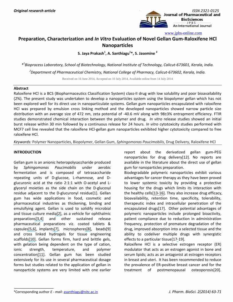
Original research article ISSN 2321-0125
www.jpbs-online.com
*Corresponding author E - mail: [email protected] J. Pharm. BioSci. 2(2014) 63-71
Preparation, Characterization and In Vitro Evaluation of Novel Gellan Gum-Raloxifene HCl Nanoparticles
S. Jaya Prakash1, A. Santhiagu1*, S. Jasemine 2
*1Bioprocess Laboratory, School of Biotechnology, National Institute of Technology, Calicut-673601, Kerala, India.
2Department of Pharmaceutical Chemistry, National College of Pharmacy, Calicut-673602, Kerala, India.
Received on 16 June 2014, Accepted on 10 July 2014, Available online from 14 July 2014
Abstract Raloxifene HCl is a BCS (Biopharmaceutics Classification System) class-II drug with low solubility and poor bioavailability (2%). The present study was undertaken to develop a nanoparticles system using the biopolymer gellan which has not been explored well for its direct use in nanoparticulate systems. Gellan gum nanoparticles encapsulated with raloxifene HCl was prepared by emulsion cross linking method and the developed nanoparticles showed narrow particle size distribution with an average size of 472 nm, zeta potential of -40.6 mV along with 98±3% entrapment efficiency. FTIR studies demonstrated chemical interaction between the polymer and drug. In vitro release studies showed an initial burst release within 30 min followed by a continuous release for 24 hours. In vitro cytotoxicity studies performed with MCF7 cell line revealed that the raloxifene HCl-gellan gum nanoparticles exhibited higher cytotoxicity compared to free raloxifene HCl.
Keywords: Polymer Nanoparticles, Biopolymer, Gellan Gum, Sphingomonas Paucimobilis, Drug Delivery, Raloxifene HCl
INTRODUCTION Gellan gum is an anionic heteropolysaccharide produced by Sphingomonas Paucimobilis under aerobic fermentation and is composed of tetrasaccharide repeating units of D-glucose, L-rhamnose, and D-glucuronic acid at the ratio 2:1:1 with O-acetyl and L-glyceryl moieties as the side chain on the D-glucosyl residue adjacent to the D-glucuronyl residue[1]. Gellan gum has wide applications in food, cosmetic and pharmaceutical industries as thickening, binding and emulsifying agent. Gellan is used to solidify microbial and tissue culture media[2], as a vehicle for ophthalmic preparations[3,4] and other sustained release pharmaceutical preparations viz. coated tablets & capsules[5,6], implants[7], microspheres[8], beads[9] and cross linked hydrogels for tissue engineering scaffolds[10]. Gellan forms firm, hard and brittle gels, with gelation being dependent on the type of cation, ionic strength, temperature, and polymer concentration[11]. Gellan gum has been studied extensively for its use in several pharmaceutical dosage forms but studies related to the application of gellan in nanoparticle systems are very limited with one earlier
report about the derivatized gellan gum-PEG nanoparticles for drug delivery[12]. No reports are available in the literature about the direct use of gellan gum for nanoparticles preparation. Biodegradable polymeric nanoparticles exhibit various advantages for cancer therapy as they have been proved to lower systemic toxicity by providing a protective housing for the drugs which limits its interaction with the healthy cells[13-16]. They also increase drug efficacy, bioavailability, retention time, specificity, tolerability, therapeutic index and intracellular penetration of the encapsulated drugs[17]. Other potential advantages of polymeric nanoparticles include prolonged bioactivity, patient compliance due to reduction in administration frequency, protection of premature degradation of the drug, improved absorption into a selected tissue and the ability to codeliver multiple drugs with synergistic effects to a particular tissue[17-19]. Raloxifene HCl is a selective estrogen receptor (ER) modulator that acts as an estrogen agonist in bone and serum lipids; acts as an antagonist at estrogen receptors in breast and uteri. It has been recommended to reduce the prevalence of ER-positive breast cancer and for the treatment of postmenopausal osteoporosis[20].

64
Prakash et al., J. Pharm. BioSci. 2(2014) 63-71
Raloxifene HCl was shown to inhibit the proliferation of breast cancer cell line MCF-7[21], inhibit the growth of RT4 urothelial carcinoma cells through ER-dependent induction of apoptosis indicative of its possible use in the treatment of bladder cancer[22], prevent capase-3-dependent apoptosis in human chondrocytes[23] and induce apoptosis in multiple myeloma cells[24]. Despite known to be a therapeutically valuable drug, raloxifene HCl has poor water solubility and is classified by Biopharmaceutics Classification System (BCS) under Class II drugs[25]. It has very low bioavailability of 2%, due to low aqueous solubility and extensive first pass metabolism[26]. Various methods have been proposed to overcome the poor aqueous solubility of raloxifene HCl like solid dispersion formulation[27], self-microemulsifying drug delivery system[28], inclusion complexes[29] and co-grinding[26]. Reports about the nanoparticle based delivery systems for raloxifene HCl are limited with few previous reports about solid lipid nanoparticles[30], raloxifene HCl nanoparticles prepared using rapid expansion of supercritical system[31], raloxifiene loaded solid dispersion nanoparticles[27] and raloxifene HCl loaded nanoparticles using synthetic aliphatic polyesters[32] to improve the solubility of raloxifene HCl. In this present study we report the preparation of raloxifene HCl loaded gellan gum nanoparticles (GgR); characterization, in vitro drug release and in vitro cytotoxicity on human breast carcinoma cell line MCF-7 of the developed nanoparticle formulation. MATERIALS AND METHODS
Materials Raloxifene HCl was obtained from Orchid chemical and pharmaceutical Ltd, Chennai, India as gift sample. Di-octyl sodium sulfosuccinate (AOT), methylene chloride, isopropyl alcohol and mannitol were purchased from Sigma-Aldrich. All other chemicals used were of reagent grade. Gellan gum was produced by a recombinant Sphingomonas Paucimobilis strain developed in our laboratory. Production, isolation and purification of gellan gum
Gellan gum was produced by recombinant Sphingomonas Paucimobilis in a 5L fermentor (Solaris, Italy) with a working volume of 3L and the conditions
were maintained as described in our previous work[33]. Gellan gum was recovered using the method described previously[34], with slight modification. The fermented broth was diluted ten-folds with distilled water to reduce viscosity, heated for 15 min and cooled then pH was adjusted to 10 followed by heating in a constant water bath for 10 min. The heated broth was cooled; pH was brought down to 7 and centrifuged at 10000 rpm for 1 hour to separate the cells. The supernatant was heated to 95°C and gellan was recovered by precipitation with three volumes of isopropanol. The precipitate was then washed with large volume of isopropanol and freeze dried; the above steps were repeated two times to remove fermentation residues. Dried gellan was dissolved in deionized water (1%, w/v), 1% KCl and the solution was heated to 95°C. The polymer was recovered by precipitation with three volumes of isopropanol followed by centrifugation at 10000 rpm for 1 hour and freeze dried.
Synthesis of GgR nanoparticles
Gellan gum nanoparticles were prepared by employing emulsion cross-linking method[35] with slight modification. Briefly, a solution of purified gellan gum (10 mg/ml) and raloxifene HCl (500 µg/ml) were prepared in deionized water (pH 8.0) and methanol respectively. Each one ml of the solution was emulsified with 3 ml of AOT solution in methylene chloride (10% w/v) by sonication (Sonics, Vibracell, USA) over ice bath for 1 min. Secondary w/o/w emulsion was prepared by emulsifying the primary emulsion into 15 ml aqueous solution of polyvinyl alcohol (PVA, 3.5%, w/v) by sonication for 3 min over ice bath. Aqueous calcium chloride solution (5 ml, 60%, w/v) was added gradually to the above emulsion with continuous stirring. Methylene chloride was removed under vacuum using a rotary vacuum evaporator (Heidolph, Germany) and the nanoparticles formed were recovered by centrifugation at 10,000 rpm for 1 hour. Nanoparticles were washed twice with deionized water, centrifuged to remove PVA and unentrapped drug. The pellet so obtained was resuspended in water and lyophilized.
Characterization of GgR nanoparticles
FTIR spectrum of gellan gum, raloxifene HCl, and GgR nanoparticles were recorded over the wave region of 500 to 4000 cm-1, at a resolution of 4 cm-1, using FTIR

65
Prakash et al., J. Pharm. BioSci. 2(2014) 63-71
spectrophotometer (Perkin- Elmer, USA). The stability of the nanoparticles system was evaluated by zeta potential measurements. Zeta potential and average particle size analysis was performed by DLS-Zeta Sizer Nano series (Malvern). The measurement was done at 25°C for 2 min. The morphology of the prepared GgR nanoparticles was examined by scanning electron microscope (Hitachi SU6600) with a Schottky field emission electron source operated under low vacuum condition. The sample was kept at a working distance of 10.9 mm from the electron gun and was illuminated with an electron beam operated at an accelerating voltage of 15 kV and 2 µm magnifications. The secondary electron scattered from the surface of the nanoparticle were captured using Everhart- Thornly secondary electron detector and used for imaging purpose.
Encapsulation efficiency
Encapsulation efficiency, which is the percentage of the actual amount of drug encapsulated in the polymeric carrier relative to the total amount of drug taken for nanoparticles preparation, was calculated using the following equation:
%Encapsulation Efficiency = (Actual drug loading/ Theoretical drug loading) × 100
To calculate the actual drug loading, an accurately weighed quantity of nanoparticles was sonicated in 10 ml of methanol for 5 min, filtered through 0.45 µ syringe filter. The concentration of raloxifene HCl was estimated by a HPLC system (Shimadzu, Japan, Model LC-6AD2100) with C18 analytical column (enable C18G 5 µm: 250×4.6 mm, Spincotech, India). The analytes were detected at 287 nm with PDA detector (Model SPD-M20A) using acetonitrile and phosphate buffer solution (pH 3.0, 35:65, volume ratio) as mobile phase. Each analysis was performed in triplicate and the flow rate of the mobile phase was 1.0 ml/min at 40°C.
In vitro drug release of GgR nanoparticles
In vitro drug release of the GgR nanoparticles formulation were investigated in acetate buffer pH 2.2 and phosphate buffer pH 7.4. Weighed quantity of nanoparticles were loaded in a dialysis membrane (Cutoff 45 kDa) and kept in beaker containing 100 ml of
dissolution medium with constant stirring at 37⁰C. About 3 ml of sample was taken at regular intervals from the dissolution buffer and replaced with equal volume of fresh buffer. The amount of raloxifene HCl released was estimated by HPLC analysis as described above. Each analysis was done in triplicate.
In vitro cytotoxicity
The cytotoxicity of GgR nanoparticles and raloxifene HCl was measured by MTT assay in human mammarian cancer cell line MCF-7. The cells were cultured in Dulbecco’s modified eagle’s medium (DMEM) supplemented with 5-10% fetal calf serum, 4 mM glutamine and 0.1% penicillin/streptomycin. Cells were seeded at 3×10³ cells/well in 96 well plates and incubated for 24 hours to allow adherence of the cells before the administration of samples for testing. After incubation the cells were incubated with appropriate concentrations of free raloxifene HCl (20-500 nM), GgR nanoparticles (20-500 nM raloxifene HCl equivalent) and unloaded Gg nanoparticles separately in 96-well plates and incubated for 24 hours at 37°C with humidified 5% CO2. DMSO (0.1% v/v) was used as a vehicle control. After 24 hours of incubation, 50 µl of MTT (2 mg/ml) was added to each well and then incubated for 4 hours. After 4 hours of incubation the medium was removed and the purple formazan crystal formed due the reduction of tetrazolium salt only by metabolically active cells[36] was dissolved in 200 μl DMSO. The absorbance of the dissolved formazan was taken at 570 nm using a Bio-Rad microplate reader and the percentage of cell viability was calculated from the absorbance. All experiments were done in triplicate and statistical analysis was performed using Prism Software.
RESULTS AND DISCUSSION
Synthesis and characterization of GgR nanoparticles Gellan gum is an anionic, water soluble heteropolysaccharide which forms insoluble gels on addition of calcium or sodium ions. AOT (Di-octyl sodium sulfosuccinate) is an anionic surfactant containing polar sulphosuccinate head group with large branching Di-octyl side chain. AOT forms reverse micelles in non-polar solvents such as methylene chloride and acquires bilayer structure in multiple emulsions due to its double chain amphiphilic nature and also forms insoluble salt with

66
Prakash et al., J. Pharm. BioSci. 2(2014) 63-71
Figure 1. FTIR spectra of (A) raloxifene HCl, (B) gellan gum and (C) GgR nanoparticles
cationic calcium ions. The aqueous core of gellan gum was entrapped into the reverse micelles formed by the AOT in methylene chloride and further emulsified in the aqueous phase using PVA as a secondary emulsifier. Due to unique properties of the surfactant-polymer system used in this study, the nanoparticles formed are expected to have a calcium crosslinked core composed of gellan and AOT head groups encircled by a hydrophobic matrix made of AOT side chains with the drug encapsulated in the core[35]. FTIR spectra of raloxifene HCl, gellan gum and GgR nanoparticles were studied to check any interaction between gellan gum and raloxifene HCl. As given in Fig. 1, gellan gum shows characteristic peaks at 3414 cm-1
(OH-stretching), 2924.09 cm-1 (C-H stretching), two strong peaks at 1612 cm-1and 1413 cm-1 for asymmetric and symmetric –COO stretching and at 1028 cm-1 for C-O stretching for alky ether. Raloxifene HCl exhibits characteristic peaks at 3215 cm-1, 3145.01 cm-1 for functional N-H and Ph-OH bonds respectively. It also shows other characteristic peaks at 2958 cm-1 due to the aromatic C-H stretching, 1642 cm-1 for C-O-C
stretching, 1596 cm-1 for C=O stretching, 1464.18 cm-1 for S- benzothiofuran, 1258.74 cm-1 for C-O stretching, 905 cm-1 for benzene ring and 806 cm-1 for thiophene C-H bond. The interaction between polymer and drug can be identified by the shifting or disappearance of the frequency of functional characteristic peaks. As evident from Fig. 1 the FTIR spectrum of GgR nanoparticles indicated a shift in –OH peak (3414 cm-1 ) of gellan to lower frequency (3386 cm-1) and disappearance of COO stretching (1612 cm-1& 1413 cm-1) of gellan gum[37]. Disappearance of raloxifene HCl characteristic peaks at 3215 cm-1 and 3145.01 cm-1 for functional N-H and phenolic -OH bonds[32] also confirmed the interaction of the polymer with drug. The particle size distribution of the formulation was
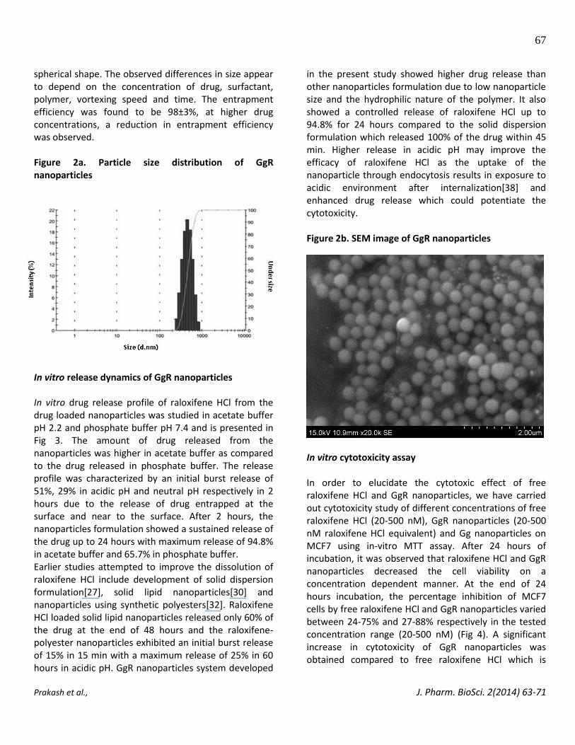
determined by light scattering (Fig. 2a) and the particles showed narrow size distribution (PDI<0.5) with a mean diameter of 472 nm. The zeta potential value of the GgR nanoparticles was around -40 mV which revealed that the nanoparticles exhibit good stability. The morphology of GgR nanoparticles was examined by SEM. It was observed from the SEM image (Fig. 2b) that GgR nanoparticles exhibit smooth surface and regular
67
Prakash et al., J. Pharm. BioSci. 2(2014) 63-71
spherical shape. The observed differences in size appear to depend on the concentration of drug, surfactant, polymer, vortexing speed and time. The entrapment efficiency was found to be 98±3%, at higher drug concentrations, a reduction in entrapment efficiency was observed. Figure 2a. Particle size distribution of GgR nanoparticles
In vitro release dynamics of GgR nanoparticles In vitro drug release profile of raloxifene HCl from the drug loaded nanoparticles was studied in acetate buffer pH 2.2 and phosphate buffer pH 7.4 and is presented in Fig 3. The amount of drug released from the nanoparticles was higher in acetate buffer as compared to the drug released in phosphate buffer. The release profile was characterized by an initial burst release of 51%, 29% in acidic pH and neutral pH respectively in 2 hours due to the release of drug entrapped at the surface and near to the surface. After 2 hours, the nanoparticles formulation showed a sustained release of the drug up to 24 hours with maximum release of 94.8% in acetate buffer and 65.7% in phosphate buffer. Earlier studies attempted to improve the dissolution of raloxifene HCl include development of solid dispersion formulation[27], solid lipid nanoparticles[30] and nanoparticles using synthetic polyesters[32]. Raloxifene HCl loaded solid lipid nanoparticles released only 60% of the drug at the end of 48 hours and the raloxifene-polyester nanoparticles exhibited an initial burst release of 15% in 15 min with a maximum release of 25% in 60 hours in acidic pH. GgR nanoparticles system developed
in the present study showed higher drug release than other nanoparticles formulation due to low nanoparticle size and the hydrophilic nature of the polymer. It also showed a controlled release of raloxifene HCl up to 94.8% for 24 hours compared to the solid dispersion formulation which released 100% of the drug within 45 min. Higher release in acidic pH may improve the efficacy of raloxifene HCl as the uptake of the nanoparticle through endocytosis results in exposure to acidic environment after internalization[38] and enhanced drug release which could potentiate the cytotoxicity. Figure 2b. SEM image of GgR nanoparticles
In vitro cytotoxicity assay In order to elucidate the cytotoxic effect of free raloxifene HCl and GgR nanoparticles, we have carried out cytotoxicity study of different concentrations of free raloxifene HCl (20-500 nM), GgR nanoparticles (20-500 nM raloxifene HCl equivalent) and Gg nanoparticles on MCF7 using in-vitro MTT assay. After 24 hours of incubation, it was observed that raloxifene HCl and GgR nanoparticles decreased the cell viability on a concentration dependent manner. At the end of 24 hours incubation, the percentage inhibition of MCF7 cells by free raloxifene HCl and GgR nanoparticles varied between 24-75% and 27-88% respectively in the tested concentration range (20-500 nM) (Fig 4). A significant increase in cytotoxicity of GgR nanoparticles was obtained compared to free raloxifene HCl which is

68
Prakash et al., J. Pharm. BioSci. 2(2014) 63-71
Figure 3. In vitro drug release profile of raloxifene HCl from GgR nanoparticles in acetate buffer pH 2.2 (▲) and
phosphate buffer pH 7.4(■) at 37°C
Figure 4. In vitro cytotoxic effect of raloxifene HCl and GgR nanoparticles on MCF-7. Student T test,* p<0.05
evident from the reduction in IC50 value of raloxifene HCl from 200 nM (free raloxifene HCl) to 100 nM (GgR
nanoparticles). Unloaded gellan gum nanoparticles did not show significant toxicity on MCF-7 as given in Fig 4.

69
Prakash et al., J. Pharm. BioSci. 2(2014) 63-71
CONCLUSION In the present study we report for the first time, the possibility to entrap hydrophobic raloxifene HCl with gellan gum using surfactant. The prepared nanoparticles exhibited spherical shape, narrow particle size distribution, negative zeta potential and high entrapment efficiency. About 94.8% of raloxifene HCl was released in acetate buffer at the end of 24 hours from the GgR nanoparticles which was better than other nanoparticle systems reported for raloxifene HCl delivery. In vitro cytotoxicity assessment revealed that the GgR nanoparticles system showed better cytotoxicity than the free raloxifene HCl. Further studies on the optimization of concentration of the drug, surfactant and polymer could result in a better system with a controlled release of the drug. These results suggest that gellan gum nanoparticle system can be a better system to deliver poorly water soluble hydrophobic drugs like raloxifene HCl. The outcome of this study also indicated the suitability of gellan gum for nanoparticles based drug delivery systems. ACKNOWLEDGMENT We would like to thank Mr. S. Balamurugan, School of Nano science & Technology, NIT Calicut for assistance to perform SEM and DLS analysis. REFERENCES 1. O’Neill, M.A., Selvendran, R.R., Morris, V.J., 1983.
Structure of the acidic extracellular gelling polysaccharide produced by Pseudomonas elodea. Carbohydr Res. 124, 123-133.
2. Lin, C.C., Casida, L.E., 1984. Gelrite as a gelling agent in media for the growth of thermophilic microorganisms. Appl Environ Microbiol. 47, 427- 429.
3. Meseguer, G., Buri, P., Plazonnet, B., Rozier, A., Gurny, R., 1996. Gamma scintigraphic comparision of eye drops containing pilocarpine in healthy volunteers. J Ocul Pharmacol Ther. 12, 481-488.
4. Carlfors, J., Edsman, K., Peterson, R., Jornving, K., 1998. Rheological evaluation of gelrite in situ for opthalmic use. Eur J Pharm Sci. 6(2), 113-119.
5. Ike-Nor, U.O., Ofoefule, S.I., Chukwu, A., 2006. Evaluation of gellan gum as a potential
pharmaceutical adjuvant: Binding properties in tablets containing a poorly water soluble and poorly compressible drug, J Drug Deliv Sci Technol. 16, 397–401.
6. Alhaique, F., Santucci, E., Carafa, M., Coviello, T., Murtas, E., Riccieri, F.M., 1996. Gellan in sustained release formulations: preparation of gel capsules and release studies. Biomaterials. 17(20), 1981-1986.
7. Li, J., Kamath, K., Dwivedi, C., 2001. Gellan film as an implant for insulin delivery. J Biomater Appl. 15, 321–343.
8. Jana, S., Das, A., Nayak, A.K., Sen, K.K., Basu, S.K., 2013. Aceclofenac-loaded unsaturated esterified alginate/gellan gum microspheres: In vitro and in vivo assessment. Int J Biol Macromol. 57, 129–137.
9. Fattah, E.A., Gran, D.J., Gabr, K.E., Meshali, M.M., 1998. Physical characterstics and release behaviour of salbutamol sulphate beads prepared with different ionic polysaccharide. Drug Dev Ind Pharm. 24, 541-547.
10. Karthika, J.S., Vishalakshi, B., 2013. Synthesis, swelling behaviour, salt- and pH- sensitivity of crosslinked gellan gum-graft-poly (acrylamide-co-itaconic acid)hydrogels. Der Pharma Chem. 5(2), 185-192.
11. Herrera, M.G.S., Berli, C.L., Padilla, L.P.M., 2008. Physicochemical and rheological properties of oil-in-water emulsions prepared with sodium caseinate/gellan gum mixtures. Food Hydrocoll. 22(5), 934–942.
12. Palaniappan, R., 2010. Gellan-gum nanoparticles and method of making and using the same. US patent No 0112076 A1.
13. Hagen, T.L.T., Seynhaeve, A.L., Tiel, S.T., Ruiter, D.J., Eggermont, A.M,. 2002. Pegylated liposomal tumor necrosis factor-alpha results in reduced toxicity and synergistic antitumor activity after systemic administration in combination with liposomal doxorubicin (Doxil) in soft tissue sarcoma-bearing rats. Int J Cancer. 97(1), 115-120.
14. Bansal, M., Gupta, S., Pal, U.S., Pal, A., 2013. Magainin, isolated from Xenopus laevis, induced Apoptosis in Human Cervical Carcinoma Cells. J Pharm BioSci. 1, 1-3.
15. Gabizon, A.A., 1992. Selective tumor localization and improved therapeutic index of anthracyclines encapsulated in long-circulating liposomes. Cancer

70
Prakash et al., J. Pharm. BioSci. 2(2014) 63-71
Res. 52, 891–896. 16. Northfelt, D.W., Martin, F.J., Working, P.,
Volberding, P.A., Russell, J., Newman, M., Amantea, M.A., Kaplan, L. D., 1996. Doxorubicin encapsulated in liposomes containing surface-bound polyethylene glycol: Pharmacokinetics, tumor localization, and safety in patients with AIDS-related Kaposi’s sarcoma. J Clin Pharmacol. 36, 55–63.
17. Alexis, F., Basto, P., Levy‐Nissenbaum, E., Radovic
‐Moreno, A.F., Zhang, L., Pridgen, E., wang, A.Z.,Marein,S.L., Westerhof, K., Molnar, L.K., Farokhzad, O.C., 2008. HER-2-targeted nanoparticle-affibody bioconjugates for cancer therapy. ChemMedChem. 3(12), 1839–1843.
18. Deshmukh, K., Amin, P., 2013. Formulation and evaluation of solid-lipid nanoparticles based 0.1% soy isoflavone dermal gels. J Pharm BioSci. 1, 7-18.
19. Zhang, L., Radovic‐Moreno, A.F., Alexis, F., Gu, F.X., Basto, P.A., Bagalkot, V., Jon, S., Langer, R.S., Farokhzad, O.C., 2007. Co-delivery of hydrophobic and hydrophilic drugs from nanoparticle-aptamer
bioconjugates. ChemMedChem. 2(9), 1268–1271. 20. Kavas, A., Cagatay, S.T., Banerjee, S., Keskin, D.,
Tezcaner, A., 2013. Potential of raloxifene in reversing osteoarthritis-like alterations in rat chondrocytes: An in vitro model study. J Biosci. 38(1), 135–147.
21. Sato, M., Glasebrook, L.A., Bryant, H.U., 1994. Raloxifene: A selective estrogen receptor modulator. J Bone Miner Metab. 12(2), S9-S20.
22. Hoffman, K.L., Lerner, S.P., Smith, C.L., 2012. Raloxifene inhibits growth of RT4 urothelial carcinoma cells via estrogen receptor-dependent induction of apoptosis and inhibition of proliferation. Horm Cancer. 4, 24–35.
23. Hattori, Y., Kojima, T., Kato, D., Matsubara, H., Takigawa, M., Ishiguro, N., 2012. A selective estrogen receptor modulator inhibits tumor necrosis factor-α-induced apoptosis through the ERK1/2 signaling pathway in human chondrocytes. Biochem Biophys Res Commun. 421418–421424.
24. Olivier, S., Close, P., Castermans, E., De Leval, L., Tabruyn, S., Chariot, A., Franchimont, N., 2006. Raloxifene-induced myeloma cell apoptosis: A study of nuclear factor-κB inhibition and gene expression signature. Mol Pharmacol. 69, 1615– 1623.
25. Teeter, J.S., Meyerhoff, R.D., 2002. Environmental fate and chemistry of raloxifene hydrochloride.
Environmental Toxicology and Chemistry. 21, 729–736.
26. Garg, A., Singh, S., Rao, V.U., Bindu, K., Balasubramaniam, J., 2009. Solid state interaction of raloxifene HCl with different hydrophilic carriers during co-grinding and its effect on dissolution rate. Drug Dev Ind Pharm. 35, 455–470.
27. Tran, T.H., Poudel, B.K., Marasini, N., Woo, J.S., Choi, H.G., Yong, C.S., Kim, J.O., 2013. Development of raloxifene-solid dispersion with improved oral bioavailability via spray-drying technique. Arch Pharmacal Res. 36, 86–93.
28. Thakkar, H., Nangesh, J., Parmar, M., Patel, D., 2011. Formulation and characterization of lipid-based drug delivery system of raloxifene-microemulsion and self-microemulsifying drug delivery system. J Pharm Bioallied Sci. 3, 442–448.
29. Patil, P.H., Belgamwar, V.S., Patil, P.R., Surana, S.J., 2013. Solubility enhancement of raloxifene using inclusion complexes and cogrinding method. J Pharm. 2013, 1-9.
30. Burra, M., Jukanti, R., Janga, K.Y., Sunkavalli, S., Velpula, A., Ampati, S., Jayaveera, K.N., 2013. Enhanced intestinal absorption and bioavailability of raloxifene hydrochloride via lyophilized solid lipid nanoparticles. Adv Powder Technol. 24, 393–402.
31. Keshavarz, A., Karimi-Sabet, J., Fattahi, A., Golzary, A., Rafiee-Tehrani, M., Dorkoosh, F.A., 2012. Preparation and characterization of raloxifene nanoparticles using Rapid Expansion of Supercritical Solution (RESS). J Supercrit Fluids. 63, 169-179.
32. Bikiaris, D., Karavelidis, V., Karava, E., 2009. Novel biodegradable polyesters synthesis and application as drug carriers for the preparation of raloxifene HCl loaded nanoparticles. Molecules, 14(7), 2410–2430.
33. Banik, R.M., Santhiagu, A., 2006. Improvement in production and quality of gellan gum by Sphingomonas Paucimobilis under high dissolved oxygen tension levels. Biotechnol Lett. 28(17), 1347–1350.
34. Dreveton, E., Monot, F., Lecourtier, J., Ballerina, D., Choplin, L., 1996. Influence of fermentation hydrodynamics on gellan gum physico-chemical characteristics. J Ferment Bioeng. 82, 272-276.
35. Mahesh, D.C., Khdair, A., Patil, Y., Handa, H., Mao, G., Jayanth, P., 2007. Polymer-surfactant nanoparticles for sustained release of water-soluble drugs. J pharm Sci. 96(12), 3379-3389.

71
Prakash et al., J. Pharm. BioSci. 2(2014) 63-71
36. Mosmann, T., 1983. Rapid colorimetric assay for cellular growth and survival: application to proliferation and cytotoxicity assays. J lmmunol Methods. 65, 55-63.
37. Elmowafy, E.M., Awad, G.A., Mansour, S., El-Shamy, Ael-H., 2008. Release mechanisms behind polysaccharides-based famotidine controlled release matrix tablets. AAPS PharmSciTech. 9(4), 1230.
38. Bareford, L.M., Swaan, P.W., 2007. Endocytic mechanisms for targeted drug delivery. Adv Drug Deliv Rev. 59, 748–758.
Copyright © 2022 FDOKUMEN