A novel route to the synthesis of PP g-PMMA copolymer via ATRP reaction initiated by Si–Cl bond
Available online at www.sciencedirect.com
ng C 28 (2008) 572–577www.elsevier.com/locate/msec
Materials Science and Engineeri
Preparation and study of in vitro bioactivity of PMMA–co–EHAcomposites filled with a Ca3(PO4)2–SiO2–MgO glass
P.P. Lopes a, B.J.M. Leite Ferreira b,⁎, N.A.F. Almeida b, M.C. Fredel a,M.H.V. Fernandes b, R.N. Correia b
a Department of Mechanical Engineering, Federal University of Santa Catarina - UFSC, Trindade/Caixa Postal: 476 - Florianópolis/SC - 88040-900, Brazilb CICECO, Department of Ceramics and Glass Engineering, University of Aveiro, Campus de Santiago, 3810-193 Aveiro, Portugal
Received 20 October 2006; received in revised form 4 June 2007; accepted 13 June 2007Available online 19 June 2007
Abstract
The nature of the orthopaedic implant surface affects the interaction with cells and subsequent bone formation. The bone/cement interface incement-held prostheses is considered to be the main cause of fracture leading to implant revision. It is thought that the introduction of a bioactivephase, such as bioglass, in the cement may permit a more stable interface by encouraging direct bone apposition rather then encapsulation of theimplant by fibrous tissue. In this work new poly(methylmethacrylate) (PMMA) based composites filled with 0, 30, 40 and 50 (wt.%) of a Ca3(PO4)2–SiO2–MgO glass, were processed. The prepared composites consist of a poly(methylmethacrylate)–co–(ethylhexylacrylate) (PMMA–co–EHA) matrix filled with a glass (G7), with nominal composition 33.26CaO, 28.07P2O5, 23.03SiO2, 15.64MgO (wt.%). The in vitro bioactivityof the composites was assessed by determining the changes in surface morphology and composition, by X-ray diffraction (XRD) and scanningelectron microscopy coupled with X-ray energy dispersive spectroscopy (SEM-EDS), after soaking in a simulated body fluid (SBF) for periods ofup to 21 days at 37 °C. Inductively coupled plasma (ICP) was used to assess the evolution of ionic concentrations in the SBF solution. The resultsobtained confirmed the growth of a hydroxyapatite (HA)-like layer on the surface of the prepared composites. As expected, HA layer formationwas faster for composites prepared with higher glass content.© 2007 Elsevier B.V. All rights reserved.
Keywords: Bioactive glasses; PMMA-based composites; Hydroxyapatite-like layer; In vitro bioactivity
Table 1Chemical composition of the PMMA-based composites investigated (wt.%)
1. Introduction
Fixation of the majority of prostheses in the past has beenperformed using poly(methylmethacrylate) (PMMA) bonecement. However, an unresolved problem with using PMMAas bone cement is a thickening of the intervening fibrous tissuelayer, which leads to aseptic loosening of the cement in somecases [1,2]. To improve fixation of PMMA to the host bone,various composites with bioactive materials have beendeveloped and studied [3–5]. Bioactive materials (e.g. glasses,sintered HA, glass–ceramics) are able to bond to living bonethrough a hydroxyapatite (HA) layer formed onto their surfaces
⁎ Corresponding author. Tel.: +351 234 370 261; fax: +351 234 425 300.E-mail address: [email protected] (B.J.M. Leite Ferreira).
0928-4931/$ - see front matter © 2007 Elsevier B.V. All rights reserved.doi:10.1016/j.msec.2007.06.002
[6,7].A similar layer is reported to form on the surface of thesematerials in vitro, after soaking in liquids with ionic concen-tration similar to the human blood plasma [5,8–10]. Previousstudies performed in glasses from the Ca3(PO4)2–SiO2–MgOsystem, have already shown bulk superficial reactivity in theform of a HA-like layer [11–13], when immersed in a simulatedbody fluid (SBF). The objective of the present work was to
Sample identification MMA EHA Glass, G7
Composite C5 25 25 50Composite C4 30 30 40Composite C3 35 35 30Matrix M 50 50 –
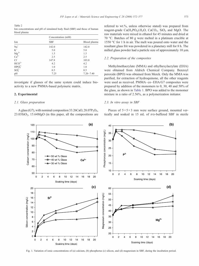
Table 2Ion concentrations and pH of simulated body fluid (SBF) and those of humanblood plasma
Concentration (mM)
Ion SBF Blood plasma
Na+ 142.0 142.0K+ 5.0 5.0Mg2+ 1.5 1.5Ca2+ 2.5 2.5Cl− 147.8 103.0HCO3− 4.2 4.2HPO4
2− 1.0 1.0SO4
2− 0.5 0.5pH 7.25 7.20–7.40
573P.P. Lopes et al. / Materials Science and Engineering C 28 (2008) 572–577
investigate if glasses of the same system could induce bio-activity to a new PMMA-based polymeric matrix.
2. Experimental
2.1. Glass preparation
Aglass (G7), with nominal composition 33.26CaO, 28.07P2O5,23.03SiO2, 15.64MgO (in this paper, all the compositions are
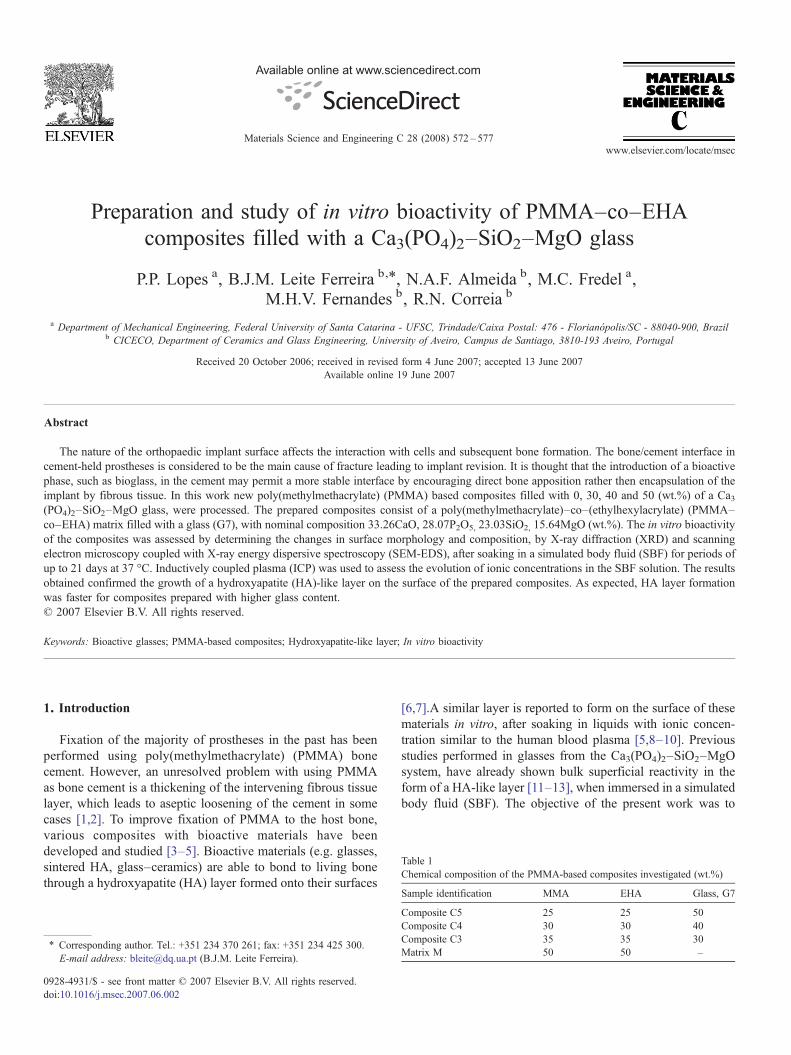
Fig. 1. Variation of ionic concentrations of (a) calcium, (b) phosphorus (
referred to wt.%, unless otherwise stated) was prepared fromreagent-grade Ca(H2PO4).H2O, CaCO3, SiO2 and MgO. Theraw materials were mixed in ethanol for 45 minutes and dried at70 °C. Batches of 80 g were melted in a platinum crucible at1550 °C for 1 h in air. The melt was poured onto water and theresultant glass frit was powdered in a planetary mill for 8 h. Themilled glass powder had a particle size of approximately 10 μm.
2.2. Preparation of the composites
Methylmethacrylate (MMA) and ethylhexylacrylate (EHA)were obtained from Aldrich Chemical Company. Benzoylperoxide (BPO) was obtained from Merck. Only the MMAwaspurified, for extraction of hydroquinone, all the other reagentswere used as received. PMMA–co–EHA/G7 composites wereprepared by addition of the monomers to 0, 30, 40 and 50% ofthe glass, as shown in Table 1. BPO was added to the monomermixture in a ratio of 2.56%, as a polymerization initiator.
2.3. In vitro assay in SBF
Pieces of 5×5×3 mm were surface ground, mounted ver-tically and soaked in 15 mL of tris-buffered SBF in sterile
c) silicon, and (d) magnesium in SBF, during the incubation period.
574 P.P. Lopes et al. / Materials Science and Engineering C 28 (2008) 572–577
polyethylene containers maintained at 37 °C. The SBF solutionhad a similar composition to that of human plasma, as shown inTable 2, and was previously filtered trough a Millipore 0.22 μmsystem. Soaking periods were 1, 3, 7, 14 and 21 days. Theconcentrations of calcium (Ca), phosphorous (P), silicon (Si),and magnesium (Mg) were determined for each period byinductively coupled plasma spectroscopy (ICP, Jobin Yvon, JY70 plus). Formation of the calcium phosphate (CP) surface layerwas followed by X-ray diffraction (XRD, Rigaku GeigerflexDmax-C with CuKα radiation) and scanning electron micros-copy coupled with X-ray energy dispersive spectroscopy (SEM-EDS, Hitachi S-4100, 25 kVacceleration voltage, beam current10 μA).
3. Results and discussion
3.1. Changes in SBF composition
Changes in the concentrations of Ca, P, Si and Mg ions inSBF due to immersion of the composites are shown in Fig. 1.
Fig. 2. Cu Kα XRD patterns of the surface of composite (a) prepared w
Between 0 and 1 day in SBF there is a rapid increase in Ca, P, Siand Mg concentration, for all the composites studied, due tofiller dissolution. Between 1 and 3 days a concomitant depo-sition of calcium phosphate occurs as shown by the fall in theCa and especially in P concentrations in composite C5, Fig. 1aand b. During this period the SBF solution in contact withcomposites C4 and C3 presented less variation in Ca and Pconcentrations while Si and Mg concentrations continuedto rise. From 3 to 14 days the Ca and P concentrations stilldecreased, for composite C5. Composites C4 and C3 alsopresented a minor decrease in the Ca and P profile during thisperiod. During the same period the Si and Mg concentrationscontinued to increase. Between 14 and 21 days the Ca andP concentration profiles are more stable, with comparativelyminor changes for the three composites studied. This factsuggests a reformulation of the calcium phosphate deposits –perhaps with morphological implications – rather than appo-sition. A later increase in soluble Ca and P, for compositeC5, is thought to result from detachment of portions of thedeposit.
ith 50% and (b) 30% (wt) of glass, during the immersion period.
575P.P. Lopes et al. / Materials Science and Engineering C 28 (2008) 572–577
In general, the ICP analyses reveal that composites preparedwith more glass content exhibit a smaller initial increase in Caand P solution concentrations, Fig. 1a and b. Usually, onewould expect that composites with higher filler concentrationshould release more Ca and P into the SBF solution. However,considering the Ca and P concentration profiles we are led toassume that although the composite C5 releases more ions tothe solution than C4, as confirmed by the Si analysis shownin Fig. 1c, it also induces a more rapid formation of the CPlayer — resulting in a lower concentration of Ca and P ions insolution. The same can be said about C4 relatively to C3. In aprevious study, glasses from the same system in bulk form,showed ability to quickly (less than 1 day) form a CP layer,
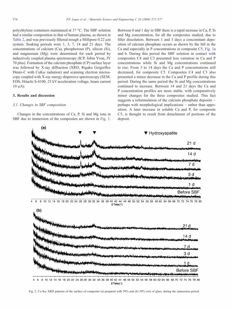
Fig. 3. SEMmicrographs of the surface of composites C5 and C4 before – (a) and (b),(The codes indicate the composite number, mentioned in Table 1, and the immersion
without decreasing the Ca and P solution concentrations in thesame length of time [13]. In Mg concentration profiles there aresmall differences between the composites, the more interestingone comes from comparing C5 and C4 between 14 and 21 daysof immersion, since it could result from incorporation of Mgions, from the SBF solution, into the CP layer formed on thesurface of composite C5.
3.2. Formation and characterization of the surface layer
XRD of composite surfaces C5 and C3 during immersion inSBF, are shown in Fig. 2. After 1 day in SBF the XRD pattern isstill very similar to the pattern before immersion. After 3 days
respectively – and soaking in SBF for 3 – (c) and (d) – and 21 days— (e) and (f).time. For example, C5–3d means composite 5 after 3 days in SBF).
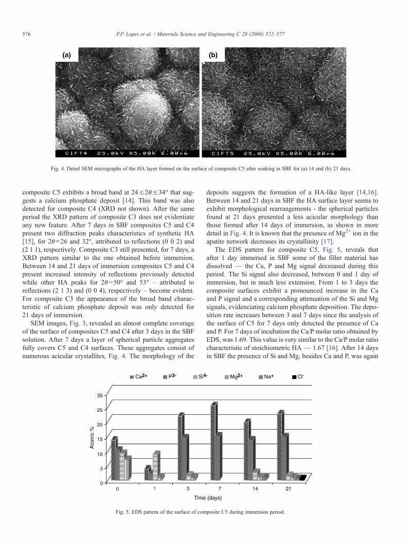
Fig. 4. Detail SEM micrographs of the HA layer formed on the surface of composite C5 after soaking in SBF for (a) 14 and (b) 21 days.
576 P.P. Lopes et al. / Materials Science and Engineering C 28 (2008) 572–577
composite C5 exhibits a broad band at 24≤2θ≤34° that sug-gests a calcium phosphate deposit [14]. This band was alsodetected for composite C4 (XRD not shown). After the sameperiod the XRD pattern of composite C3 does not evidentiateany new feature. After 7 days in SBF composites C5 and C4present two diffraction peaks characteristics of synthetic HA[15], for 2θ=26 and 32°, attributed to reflections (0 0 2) and(2 1 1), respectively. Composite C3 still presented, for 7 days, aXRD pattern similar to the one obtained before immersion.Between 14 and 21 days of immersion composites C5 and C4present increased intensity of reflections previously detectedwhile other HA peaks for 2θ=50° and 53° – attributed toreflections (2 1 3) and (0 0 4), respectively – become evident.For composite C3 the appearance of the broad band charac-teristic of calcium phosphate deposit was only detected for21 days of immersion.
SEM images, Fig. 3, revealed an almost complete coverageof the surface of composites C5 and C4 after 3 days in the SBFsolution. After 7 days a layer of spherical particle aggregatesfully covers C5 and C4 surfaces. These aggregates consist ofnumerous acicular crystallites, Fig. 4. The morphology of the
Fig. 5. EDS pattern of the surface of com
deposits suggests the formation of a HA-like layer [14,16].Between 14 and 21 days in SBF the HA surface layer seems toexhibit morphological rearrangements - the spherical particlesfound at 21 days presented a less acicular morphology thanthose formed after 14 days of immersion, as shown in moredetail in Fig. 4. It is known that the presence of Mg2+ ion in theapatite network decreases its crystallinity [17].
The EDS pattern for composite C5, Fig. 5, reveals thatafter 1 day immersed in SBF some of the filler material hasdissolved — the Ca, P and Mg signal decreased during thisperiod. The Si signal also decreased, between 0 and 1 day ofimmersion, but in much less extension. From 1 to 3 days thecomposite surfaces exhibit a pronounced increase in the Caand P signal and a corresponding attenuation of the Si and Mgsignals, evidenciating calcium phosphate deposition. The depo-sition rate increases between 3 and 7 days since the analysis ofthe surface of C5 for 7 days only detected the presence of Caand P. For 7 days of incubation the Ca/P molar ratio obtained byEDS, was 1.69. This value is very similar to the Ca/P molar ratiocharacteristic of stoichiometric HA — 1.67 [16]. After 14 daysin SBF the presence of Si and Mg, besides Ca and P, was again
posite C5 during immersion period.
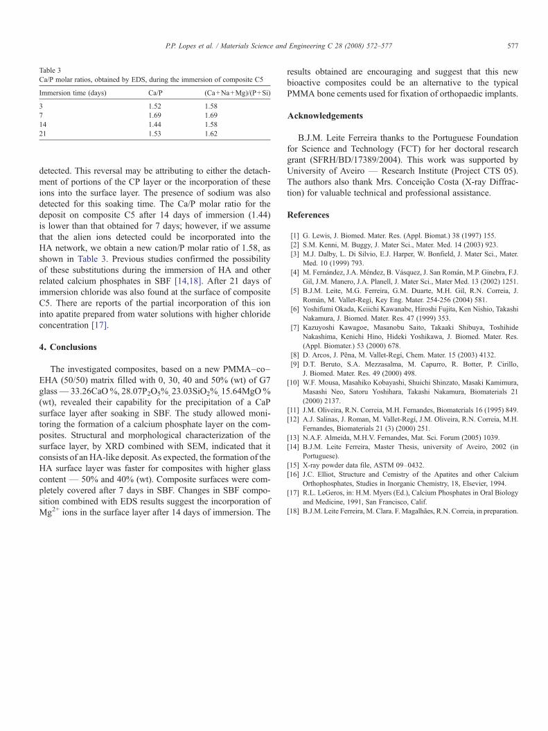
Table 3Ca/P molar ratios, obtained by EDS, during the immersion of composite C5
Immersion time (days) Ca/P (Ca+Na+Mg)/(P+Si)
3 1.52 1.587 1.69 1.6914 1.44 1.5821 1.53 1.62
577P.P. Lopes et al. / Materials Science and Engineering C 28 (2008) 572–577
detected. This reversal may be attributing to either the detach-ment of portions of the CP layer or the incorporation of theseions into the surface layer. The presence of sodium was alsodetected for this soaking time. The Ca/P molar ratio for thedeposit on composite C5 after 14 days of immersion (1.44)is lower than that obtained for 7 days; however, if we assumethat the alien ions detected could be incorporated into theHA network, we obtain a new cation/P molar ratio of 1.58, asshown in Table 3. Previous studies confirmed the possibilityof these substitutions during the immersion of HA and otherrelated calcium phosphates in SBF [14,18]. After 21 days ofimmersion chloride was also found at the surface of compositeC5. There are reports of the partial incorporation of this ioninto apatite prepared from water solutions with higher chlorideconcentration [17].
4. Conclusions
The investigated composites, based on a new PMMA–co–EHA (50/50) matrix filled with 0, 30, 40 and 50% (wt) of G7glass— 33.26CaO%, 28.07P2O5%, 23.03SiO2%, 15.64MgO%(wt), revealed their capability for the precipitation of a CaPsurface layer after soaking in SBF. The study allowed moni-toring the formation of a calcium phosphate layer on the com-posites. Structural and morphological characterization of thesurface layer, by XRD combined with SEM, indicated that itconsists of an HA-like deposit. As expected, the formation of theHA surface layer was faster for composites with higher glasscontent — 50% and 40% (wt). Composite surfaces were com-pletely covered after 7 days in SBF. Changes in SBF compo-sition combined with EDS results suggest the incorporation ofMg2+ ions in the surface layer after 14 days of immersion. The
results obtained are encouraging and suggest that this newbioactive composites could be an alternative to the typicalPMMA bone cements used for fixation of orthopaedic implants.
Acknowledgements
B.J.M. Leite Ferreira thanks to the Portuguese Foundationfor Science and Technology (FCT) for her doctoral researchgrant (SFRH/BD/17389/2004). This work was supported byUniversity of Aveiro — Research Institute (Project CTS 05).The authors also thank Mrs. Conceição Costa (X-ray Diffrac-tion) for valuable technical and professional assistance.
References
[1] G. Lewis, J. Biomed. Mater. Res. (Appl. Biomat.) 38 (1997) 155.[2] S.M. Kenni, M. Buggy, J. Mater Sci., Mater. Med. 14 (2003) 923.[3] M.J. Dalby, L. Di Silvio, E.J. Harper, W. Bonfield, J. Mater Sci., Mater.
Med. 10 (1999) 793.[4] M. Fernández, J.A. Méndez, B. Vásquez, J. San Román, M.P. Ginebra, F.J.
Gil, J.M. Manero, J.A. Planell, J. Mater Sci., Mater Med. 13 (2002) 1251.[5] B.J.M. Leite, M.G. Ferreira, G.M. Duarte, M.H. Gil, R.N. Correia, J.
Román, M. Vallet-Regí, Key Eng. Mater. 254-256 (2004) 581.[6] Yoshifumi Okada, Keiichi Kawanabe, Hiroshi Fujita, Ken Nishio, Takashi
Nakamura, J. Biomed. Mater. Res. 47 (1999) 353.[7] Kazuyoshi Kawagoe, Masanobu Saito, Takaaki Shibuya, Toshihide
Nakashima, Kenichi Hino, Hideki Yoshikawa, J. Biomed. Mater. Res.(Appl. Biomater.) 53 (2000) 678.
[8] D. Arcos, J. Pena, M. Vallet-Regí, Chem. Mater. 15 (2003) 4132.[9] D.T. Beruto, S.A. Mezzasalma, M. Capurro, R. Botter, P. Cirillo,
J. Biomed. Mater. Res. 49 (2000) 498.[10] W.F. Mousa, Masahiko Kobayashi, Shuichi Shinzato, Masaki Kamimura,
Masashi Neo, Satoru Yoshihara, Takashi Nakamura, Biomaterials 21(2000) 2137.
[11] J.M. Oliveira, R.N. Correia, M.H. Fernandes, Biomaterials 16 (1995) 849.[12] A.J. Salinas, J. Roman, M. Vallet-Regí, J.M. Oliveira, R.N. Correia, M.H.
Fernandes, Biomaterials 21 (3) (2000) 251.[13] N.A.F. Almeida, M.H.V. Fernandes, Mat. Sci. Forum (2005) 1039.[14] B.J.M. Leite Ferreira, Master Thesis, university of Aveiro, 2002 (in
Portuguese).[15] X-ray powder data file, ASTM 09–0432.[16] J.C. Elliot, Structure and Cemistry of the Apatites and other Calcium
Orthophosphates, Studies in Inorganic Chemistry, 18, Elsevier, 1994.[17] R.L. LeGeros, in: H.M. Myers (Ed.), Calcium Phosphates in Oral Biology
and Medicine, 1991, San Francisco, Calif.[18] B.J.M. Leite Ferreira, M. Clara. F. Magalhães, R.N. Correia, in preparation.
Copyright © 2022 FDOKUMEN