Synthesis, Crystal Structure and Spectroscopy Properties of Na3AZr(PO4)3 (A: Mg, Ni) and...
8
ISSN 2321-807X 2532 | Page July 31, 2014 Synthesis, crystal structure and spectroscopy studies of a new non-centrosymmetric organic cation nitrate H.Saadouni a , D. E. Janzen b , M.Rzaigui a and W.Smirani Sta* ,a a-Laboratoire de chimie des matériaux, Faculté des sciences de Bizerte, 7021, Zarzouna, Tunisia b- Department of Chemistry and Biochemistry, St Catherine University, St Paul, Minnesota, 55105, USA *Corresponding author: [email protected] ABSTRACT The synthesis, crystal structure and spectroscopic studies are reported for the salt m-anisidinium nitrate. A single-crystal X-ray investigation has shown that this compound crystallizes in the non-centrosymmetric space group Cc with the lattice parameters: a = 5.744(3) Å, b = 14.968(6) Å, c = 10.178(4) Å; = 96.910(7) o ; V = 868.6(6)Å 3 ; Z = 4. The structure was solved from 1973 independent reflections with R1 = 0.033 and wR2 = 0.088. The hydrogen atoms of the protonated amine undergo hydrogen bonding interactions with oxygen atoms of three different nitrate anions forming 2-dimensional sheets. Solution NMR results are consistent with the X-ray structure. A measured optical band gap of 3.35eV indicates m- anisidinium nitrate is a wide-band-gap dielectric material. Indexing terms/Keywords Organic-inorganic hybrid; Crystal structure; IR Spectroscopy; NMR spectroscopy; Band-gap. Council for Innovative Research Peer Review Research Publishing System Journal: Journal of Advances in Chemistry Vol. 10, No. 3 [email protected] www.cirjac.com
-
Upload
independent -
Category
Documents
-
view
3 -
download
0
Transcript of Synthesis, Crystal Structure and Spectroscopy Properties of Na3AZr(PO4)3 (A: Mg, Ni) and...

ISSN 2321-807X
2532 | P a g e J u l y 3 1 , 2 0 1 4
Synthesis, crystal structure and spectroscopy studies of a new
non-centrosymmetric organic cation nitrate
H.Saadounia, D. E. Janzenb, M.Rzaiguia and W.Smirani Sta*,a
a-Laboratoire de chimie des matériaux, Faculté des sciences de Bizerte, 7021, Zarzouna, Tunisia
b- Department of Chemistry and Biochemistry, St Catherine University, St Paul, Minnesota, 55105, USA
*Corresponding author: [email protected]
ABSTRACT
The synthesis, crystal structure and spectroscopic studies are reported for the salt m-anisidinium nitrate. A single-crystal X-ray investigation has shown that this compound crystallizes in the non-centrosymmetric space group Cc with the lattice
parameters: a = 5.744(3) Å, b = 14.968(6) Å, c = 10.178(4) Å; = 96.910(7)o; V = 868.6(6)Å3
; Z = 4. The structure was
solved from 1973 independent reflections with R1 = 0.033 and wR2 = 0.088. The hydrogen atoms of the protonated amine undergo hydrogen bonding interactions with oxygen atoms of three different nitrate anions forming 2-dimensional sheets. Solution NMR results are consistent with the X-ray structure. A measured optical band gap of 3.35eV indicates m-
anisidinium nitrate is a wide-band-gap dielectric material.
Indexing terms/Keywords
Organic-inorganic hybrid; Crystal structure; IR Spectroscopy; NMR spectroscopy; Band-gap.
Council for Innovative Research Peer Review Research Publishing System
Journal: Journal of Advances in Chemistry
Vol. 10, No. 3
www.cirjac.com

ISSN 2321-807X
2533 | P a g e J u l y 3 1 , 2 0 1 4
1. INTRODUCTION
Nonlinear optical materials have attracted much attention because of their potential applications in optoelectronic technologies [1] and [2]. These applications depend upon the various properties of the materials, such as transparency, birefringence, refractive index, dielectric constant, thermal, photochemical and chemical stability. Inorganic crystals have excellent mechanical and thermal properties but possess relatively low optical nonlinearities because of the lack of π- electron delocalization. Organic crystals have large nonlinear susceptibilities compared to inorganic crystals. However, these crystals have certain limitations such as increased optical absorption, narrow transparency window and poor mechanical and thermal stability. Combining the high optical nonlinearity and chemical flexibility of organics with thermal stability and excellent transmittance of inorganics, semiorganic materials have been proposed and are attracting a great deal of attention in the field of nonlinear optics. Interestingly, the simple inorganic acid nitric acid can be used to access nitrate salts with organic cations. Among crystals of organic cations with nitrate anions, the most interesting for us are those that are non-centrosymmetric because of their non-linear optical properties. Several examples of nitrate salts exhibiting second harmonic generation have been reported by Kaminskii et al. [3]. In an attempt to find new materials suitable for nonlinear optical applications, we have prepared and report here the characterization of a new mixed organic–inorganic non-centrosymmetric nitrate.
2. EXPERIMENTAL
2.1 Material Preparation
Crystals of m-anisidinium nitrate were obtained by mixing, in stoichiometric ratio, a solution of nitric acid (1 M) with an ethanolic solution of m-anisidine. The mixture was stirred for 2 hours. The obtained solution was slowly evaporated at room temperature. After one week of evaporation at 293 K, X-ray quality yellow crystals appeared in the solution.
2.2 Physical measurement
X-ray diffraction measurements were made on a Rigaku XtaLAB mini diffractometer [4]. A crystal was placed on the tip of a Mitigen micromount and X-ray intensity data were measured at low temperature (173(2) K) with graphite
monochromated Mo K radiation ( = 0.71075 Å). Preliminary sets of cell constants were calculated from reflections harvested from one set of 12 images with frame times of 30 s. A randomly oriented region of reciprocal space was surveyed to the extent of one sphere and to a resolution of 0.77 Å . Three major sections of frames were collected with 1.0
steps in at three different settings. The intensity data were corrected for absorption and decay [4]. Final cell constants were calculated from the xyz centroids of 1820 strong reflections from the actual data collection after integration [4]. The structure was solved and refined using SHELXL-97 [5]. A direct-methods solution was calculated that provided most of the non-hydrogen atoms from the E-map. Full-matrix least-squares/difference Fourier cycles were performed that located the remaining nonhydrogen atoms. All non-hydrogen atoms were refined with anisotropic displacement parameters. Most of the hydrogen atoms were placed in ideal positions and refined as riding atoms with relative isotropic displacement with the exception of H1A-C. H1A, H1B, and H1C were located in the electron density map, their x,y,z positions were refined, and their thermal parameters were treated as riding. The final refinement cycles led to R1= 0.033 and wR2= 0.088. The absolute structure was deduced based on Flack parameter [6], -0.2(10), using 973 Friedel pairs. As the intensity of high angle reflections were weak, the ambiguity in the Flack parameter is not surprising.
The infrared spectrum of solid C7H10NO.NO3 was recorded on a Nicolet IR200 FT-IR Spectrometer at ambient temperature.
The UV-Vis absorption (water solution) and optical diffuse reflectance spectra (solid) were measured at room temperature with a Perkin Elmer Lambda 11 UV/Vis spectrophotometer in the range of 200-800 nm.
Solution 13
C and 1H NMR spectra were measure with a JEOL 400MHz NMR in D2O. A small amount of CH3CN was added
as an internal reference (reference shifts of 1.94ppm for 1H, 118.69 ppm for
13C)
3. RESULTS AND DISCUSSION
3.1 X-ray diffraction analysis
Crystal data summary of intensity data collection and structure refinement are reported in Table 1. The final atomic coordinates and Beq or Biso are given in Table 2.
ISSN 2321-807X
2534 | P a g e J u l y 3 1 , 2 0 1 4
Table 1. Crystal data and structure refinement
Empirical formula C7H10N2O4
Formula weight(g.mol-1
) 186.17
Temperature (K) 173
Wavelength (Å) 0.71075
Crystal system monoclinic
Space group Cc
a (Å) 5.744(3)
b (Å) 14.968(6)
c (Å) 10.178(4)
β (°) 96.910(7)
Volume (Å3) 868.6(6)
Z 4
Dcalc (g/cm-3
) 1.423
Absorption coefficient (cm-1
) 1.177
F(000) 392
Crystal size (mm3) 0.45 × 0.20 × 0.15
Reflections collected 4457
Unique reflections / No. Variables 1973/131
Goodness-of-fit on F² 1.043
Final R indices [I > 2 sigma (I)] R1=0.033 and wR2= 0.088
Flack parameter -0.2 (10)
Largest diff. peak and hole (e/Å-3
) 0.12 and -0.23
Table 2. Atomic coordinates and equivalent isotropic thermal parameters
Atoms X(σ) Y(σ) Z(σ) Beq.or B*iso
O1 0.8651(3) 1.11200(8) 0.3508(2) 3.66(3)
O2 1.2171(3) 0.77220(8) 0.1576(2) 3.50(3)
O3 0.8462(3) 0.75548(9) 0.1741(2) 4.08(3)
O4 1.0808(3) 0.79636(9) 0.3436(2) 3.54(3)
N1 0.5772(3) 0.80966(9) 0.4084(2) 2.70(3)
N2 1.0478(3) 0.77446(8) 0.2241(2) 2.80(3)
C1 0.5764(3) 0.90603(9) 0.4355(2) 2.35(3)
C2 0.7271(3) 0.9605(1) 0.3766(2) 2.41(3)
C3 0.7255(3) 1.0520(1) 0.4034(2) 2.59(3)
C4 0.5748(3) 1.0860(1) 0.4888(2) 2.83(3)
ISSN 2321-807X
2535 | P a g e J u l y 3 1 , 2 0 1 4
C5 0.4274(3) 1.0295(2) 0.5457(2) 3.18(3)
C6 0.4249(3) 0.9383(1) 0.5196(2) 3.00(3)
C7 1.0037(4) 1.0803(2) 0.2534(2) 3.54(4)
H1A 0.613(4) 0.779(2) 0.482(3) 3.1(4)
H2 0.8297 0.9363 0.3192 2.89
H1B 0.434(5) 0.793(2) 0.370(3) 4.3(5)
H4 0.5742 1.1481 0.5076 3.40
H5 0.3253 1.0531 0.6038 3.82
H6 0.3220 0.8994 0.5586 3.60
H7A 0.9017 1.0522 0.1809 4.25
H7B 1.1173 1.0364 0.2935 4.25
H7C 1.0873 1.1306 0.2189 4.25
H1C 0.686(5) 0.793(2) 0.358(3) 4.0(5)
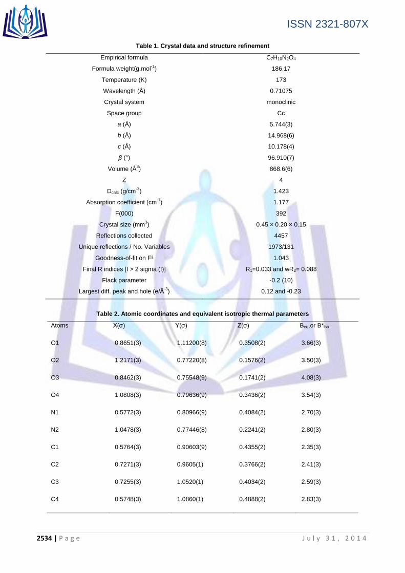
The asymmetric unit of the title compound, C7H10NO.NO3 (Figure 1), contains one nitrate anion and one m-anisidinium cation.
Figure 1. ORTEP view of C7H10NO.NO3, showing 50% probability ellipsoids
The structure consists of a protonated m-anisidine cation with a nitrate anion. No solvent or disorder was present in the
structure. The cation is quite planar with a mean deviation from a least-squares plane formed by atoms N1, O1, C1-C7 of 0.0255 Å. All hydrogens except H1A-C were refined as riding atoms. H1A, H1B, and H1C were located in the electron density map, their x,y,z positions were refined, and their thermal parameters were treated as riding. The cations are arranged within the unit cell in layers located at b = 0 and ½. Nitrates are in layers between the cations at b =1/4 and ¾
(Figure 2). While the nearly planar cations are parallel with nearest neighbor cations (3.63 Å interplanar spacing) , no interaction is present.
ISSN 2321-807X
2536 | P a g e J u l y 3 1 , 2 0 1 4
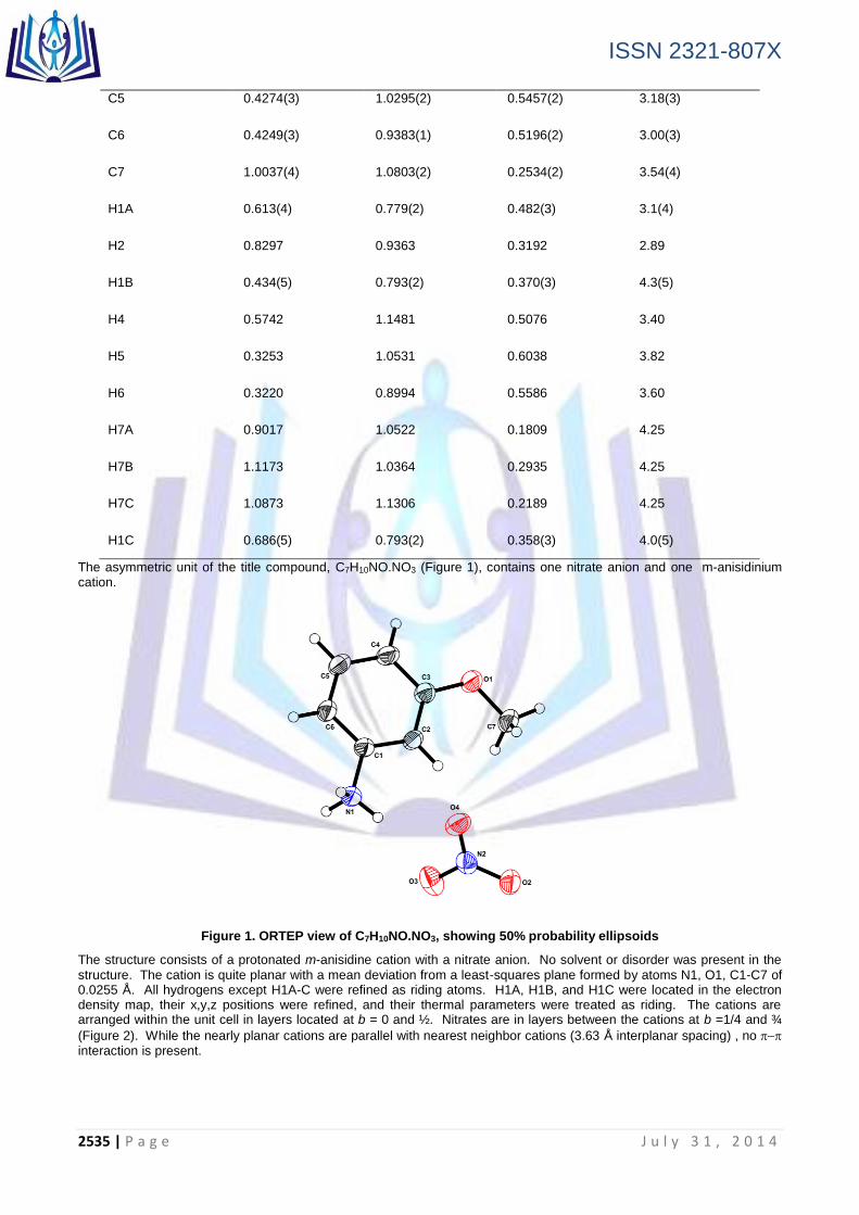
Figure 2. Two dimensional (2D) layer composed of nitrates anions and ammonium cations with hydrogen bonds as dotted lines
Organic cation and nitrate anion have normal valence bond lengths and angles (Table 3), as observed in the structures of 2-methoxyanilinium nitrate [7].
Table 3. Selected bond distances (Å) and angles (°) for C7H10NO.NO3
atom atom distance atom atom distance
O2 N2 1.250(2) O3 N2 1.241(2)
O4 N2 1.252(2) N1 C1 1.469(2)
O1 C3 1.357(2) O1 C7 1.425(3)
C1 C2 1.377(3) C1 C6 1.379(3)
C2 C3 1.397(3) C3 C4 1.395(3)
C4 C5 1.373(3) C5 C6 1.390(3)
The hydrogen atoms of the protonated amine (H1A, H1B, H1C) undergo hydrogen bonding interactions with oxygen atoms of three different nitrate anions. O1 of the methoxy group does not participate as a hydrogen bond acceptor (Table 4). All hydrogen bonding occurs in two-dimensional sheets (ac planes) in the regions where cation layers meet the nitrate layers (Figure 3).
Table 4. Principal intermolecular distances (Å) and bond angles (°) of the hydrogen bonding scheme
Donor H Acceptor D...A D-H H...A D-H...A
N1 H1A O21 2.845(3) 0.89 1.96(3) 171(2)
N1 H1B O42 2.855(3) 0.90 2.02(3) 154(3)
N1 H1C O4 3.049(3) 0.89 2.29(3) 143(2)
Symmetry Operators:
(1) X+1/2-1,-Y+1/2+1,Z+1/2 (2) X-1,Y,Z

ISSN 2321-807X
2537 | P a g e J u l y 3 1 , 2 0 1 4
Frequency (cm-1)
Figure 3. Crystal packing of C7H10NO.NO3
3.2 Infrared Spectroscopy
The infrared spectrum of the crystalline nitrate is shown in Figure 4. To assign the IR peaks to vibrational modes, we examined the modes and frequencies observed in similar compounds [8].
The planar NO3- free ion (D3h) has six vibrational degrees of freedom. They form four fundamental modes: antisymmetric
stretching (3) at approximately 1330 cm-1
(doubly degenerate, both IR and Raman active), symmetric stretching (1) at
1040 cm-1
(Raman active), an out-of-plane bending (2) at 800 cm-1
(IR active) and an in-plane bending (4) at 730 cm-1
(doubly degenerate, both IR and Raman active) [9]. Two bands in the infrared spectrum are observed at 835 and 820 cm
-1
as very weak and weak, respectively. They originate from the bending type of vibrations of nitrate anions. The splitting of this mode may be ascribed to a lowering of symmetry of the NO3
- ion from D3h to C2v or Cs.
The remaining observed bands in the spectrum can be assigned to CH3, CH, NH3+, and skeletal symmetric and
asymmetric stretching and deformation modes [10]. A broad band extending from 3395 to 2609 cm–1
is observed in the IR spectrum. This broad band must be due to the symmetric and asymmetric stretching modes of NH3, CH3 and bending.
Bands in the 1300-1000 cm-1
are attributed to the stretching of the organic groups C-C), C-N) and is characteristic of 1,3-substituted benzene rings. The shifting of the stretching and bending vibrations of the NH3 group from the free state value confirms the formation of hydrogen bonds of varying strengths in the crystal. Frequencies in the range 1469 - 1305
cm–1
are attributed to s(CH3), as(CH3), s(CH) and as(CH). The presence of C=C stretching vibrations of the aromatic ring is consistent with the absorption bands at 1608 and 1480 cm
−1.
Figure 4. IR absorption spectrum of C7H10NO.NO3

ISSN 2321-807X
2538 | P a g e J u l y 3 1 , 2 0 1 4
3.3 UV-Vis Absorption and Diffuse Reflectance
The UV-Vis absorption spectrum revealed two bands at 276nm and 288nm. These bands correspond respectively to the π →π* transition of the aromatic ring and n →π* transition of the nitrate NO3
⁻ anion. The optical diffuse reflectance
spectrum (Figure 5) indicates an optical band gap of 4.35 eV, hence, the t i t le compound is a wide band-gap dielectric material. A similar high gap energy value was observed in N,N’-diphenylguanidinium nitrate at 3.9eV [10].
Figure 5. UV diffuse reflectance spectrum for C7H10NO.NO3
3.4 13C and 1H NMR Spectroscopy
Figure 6 illustrates the solution 13
C NMR spectrum of C7H10NO.NO3. As expected by symmetry, seven carbon peaks are
present, six of which are aromatic. The aromatic carbons are found at 159.55 ; 130.75 ; 130.48 ; 114.85 ; 114.27 and
108.54ppm. The two quaternary aromatic carbons (159.95 and 130.48) are indicated by lesser integration. The peak at 55.17 ppm corresponds to the aliphatic carbon of methoxy group.
Figure 6. 13
C NMR Spectrum for C7H10NO.NO3
Figure 7 illustrates the 1H NMR spectrum of C7H10NO.NO3. The singlet at 3.75 ppm is assigned to the methoxy OC(7)H3
protons. The triplet at 7.36 ppm, the two doublets of doublets of doublets at 6.97 ppm and at 6.93 ppm and the triplet at 6.89 ppm are attributed to H2, H4, H6 and H5, respectively. H2, H4 and H6 are meta magnetically coupled to each other. H4 is also ortho coupled to H5. Likewise, coupling is present between H6 and H5, where H6 is located at the ortho
position relative to H5. The NH3 protons do not appear as they have likely exchanged with D of the D2O.

ISSN 2321-807X
2539 | P a g e J u l y 3 1 , 2 0 1 4
Figure 7. 1H NMR Spectrum for C7H10NO.NO3
3.5 Conclusion
A non-centrosymmetric material, C7H10NO.NO3, with Cc space group symmetry has been prepared and structurally characterized. The atomic arrangement of this nitrate consists of a three-dimensional network of R
2
2(16) graph-set motifs
of nitrate anions and m-anisidinium cations connected by N−H…O and C−H…O hydrogen bonding interactions. The NMR results are as expected based on the symmetry of substitution in the cation. The diffuse reflectance data indicate an energy gap 4.35 eV which is a typical of a dielectric material. This compound may be a potential candidate for application as a new second-order non linear optical (NLO) material.
Acknowledgement
We acknowledge the NSF–MRI grant No. 1125975 "MRI Consortium Acquisition of a Single Crystal X-ray Diffractometer for a Regional PUI Molecular Structure Facility".
REFERENCES
[1] Marcy, H. O., Warren, L. F., Webb, M. S., Ebbers, C. A., Velsko, S. P., Kennedy, G. C. and Catella, G. C., Appl. Opt. 31 (1992) 5051.
[2] Wang, X. Q., Xu, D., Yuan, D. R., Tian, Y. P., Yu, W. T., Sun, S. Y., Yang, Z. H., Fang, Q., Lu, M. K., Yan, Y. X., Meng, F. Q., Guo, S. Y., Zhang, G. H. and Jiang, M. G., Mater. Res. Bull. 34 (1999) 2003.
[3] A.A. Kaminskii, J. Hulliger, H. Eichler, J. Hanuza, J. Findeisen, P. Egger, Dokl. Akad. Nauk. 364 (1999) 761 (in
Russian).
[4]CrystalClear: Rigaku Corporation, 1999. CrystalClear Software User's Guide, Molecular Structure Corporation, (c) 2000.J.W.Pflugrath, Acta Cryst. D55 (1999) 1718.
[5]SHELX97: Sheldrick, G.M. Acta Cryst. A64 (2008) 112.
[6]Flack, H. D. Acta Cryst. A39 (1983) 876.
[7] K. Bouchouit, Z. Sofiani, B. Derkowska, S. Abed, N. Benali-cherif, M. Bakasse, B. Sahraoui, optics commu. 278
(2007)180.
[8] M.K. Marchewka, A. Pietraszko, J. Phys. Chem. Solids 66 (2005) 1039
[9] K. Nakamoto, Infrared Spectra of Inorganic and Coordination Compounds, Wiley, New York, 1963.
[10] G. Saravana Kumar, P. Murugakoothan, Spectrochim. Acta, Part A, 131 (2014) 17.