Syntheses and crystal structures of new vanadium(IV) oxyphosphates M(VO)2(PO4)2 with M=Co, Ni
Upload
independentCategory
view
4download
0
American Mineralogist, Volume 98, pages 1261–1269, 2013
0003-004X/13/0007–1261$05.00/DOI: http://dx.doi.org/10.2138/am.2013.4465 1261
In situ dehydration behavior of veszelyite (Cu,Zn)2Zn(PO4)(OH)3⋅2H2O: A single-crystal X-ray study
Rosa Micaela Danisi,1,* ThoMas aRMbRusTeR,1 biljana lazic,1 Predrag Vulić,2 ReinhaRD KainDl,3,4 radoVan dimitrijeVić,2 anD VolKeR KahlenbeRg4
1Mineralogical Crystallography, Institute of Geological Sciences, University of Bern, Freiestrasse 3, 3012 Bern, Switzerland2Laboratory of Crystallography, Faculty of Mining and Geology, University of Belgrade, Djusina 7, 11000 Belgrade, Serbia
3MATERIALS—Institute for Surface Technologies and Photonics, Joanneum Research, Leobner Strasse 94, 8712 Niklasdorf, Austria4Institute of Mineralogy and Petrography, University of Innsbruck, Innrain 52f, 6020 Innsbruck, Austria
absTRacT
The rare mixed copper-zinc phosphate mineral veszelyite (Cu,Zn)2Zn(PO4)(OH)3⋅2H2O [space group P21/c, a = 7.5096(2), b = 10.2281(2), c = 9.8258(2) Å, β = 103.3040(10)°, V = 734.45(3) Å3] was investigated by in situ temperature-dependent single-crystal X-ray structure refinements. The atomic arrangement of veszelyite consists of an alternation of octahedral and tetrahedral sheets. The Jahn-Teller distorted CuO6 octahedra form sheets with eight-membered rings. The tetrahedral sheet composed of PO4 and ZnO3(OH) tetrahedra shows strong topological similarities to that of cavansite, gismondine, and kipushite.
Diffraction data of a sample from Zdravo Vrelo, near Kreševo (Bosnia and Herzegovina) have been measured in steps of 25 up to 225 °C. Hydrogen positions and the hydrogen-bond system were determined experimentally from the structure refinements of data collected up to 125 °C. At 200 °C, the hydrogen-bonding scheme was inferred from bond-valence calculations and donor-acceptor distances. The hydrogen-bond system connects the tetrahedral sheet to the octahedral sheet and also braces the Cu sheet.
At 150 °C, the H2O molecule at H2O2 was released and the Cu coordination (Cu1 and Cu2) de-creased from originally six- to fivefold. Cu1 has a square planar coordination by four OH groups and an elongate distance to O3, whereas Cu2 has the Jahn-Teller characteristic elongate bond to H2O1. The unit-cell volume decreased 7% from originally 734.45(3) to 686.4(4) Å3 leading to a formula with 1 H2O pfu. The new phase observed above 150 °C is characterized by an increase of the c axis and a shortening of the b axis. The bending of T-O-T angles causes an increasing elliptical shape of the eight-membered rings in the tetrahedral and octahedral sheets. Moreover a rearrangement of the hydrogen-bond system was observed.
At 225 °C, the structure degrades to an X-ray amorphous residual due to release of the last H2O molecule at H2O1. The stronger Jahn-Teller distortion of Cu1 relative to Cu2 suggests that Cu1 is fully occupied by Cu, whereas Cu2 bears significant Zn. H2O1 is the fifth ligand of Cu2. Zn at Cu2 is not favorable to adopt planar fourfold coordination. Thus, if the last water molecule is expelled the structure is destabilized.
This study contributes to understanding the dehydration mechanism and thermal stability of super-gene minerals characterized by Jahn-Teller distorted octahedra with mixed Cu, Zn occupancy.
Keywords: Veszelyite, Jahn-Teller effect, dehydration, crystal structure, hydrogen bonding
inTRoDucTion
Veszelyite, (Cu,Zn)2Zn(PO4)(OH)3⋅2H2O, is a rare mixed copper-zinc phosphate mineral named after A. Veszeli (1820–1888), a Hungarian mining engineer, who discovered the species.
The atomic arrangement of veszelyite consists of an alterna-tion of octahedral and tetrahedral sheets (Ghose et al. 1974). The octahedral sheet is made up of distorted Cu2+O6 octahedra joined to form a network of eight-membered rings parallel to
the b-c plane. The tetrahedral sheet is composed by alternating PO4 and ZnO3(OH) tetrahedra forming undulating pyroxenoid-like chains extended parallel to c. Adjacent chains have the tetrahedral apices pointing alternatively up and down along the a axis. These chains are laterally joined forming fourfold and eightfold rings. The tetrahedral sheets are linked directly to the octahedral sheets to assemble a typical layer structure. The eight-membered rings formed by CuO6 octahedra are elongated and oriented in the same direction as the eight-membered rings formed by PO4 and ZnO3(OH) tetrahedra. These eightfold rings create channels along a, the walls of which are decorated by H2O molecules. The H2O molecules are loosely bonded to the * E-mail: [email protected]
DANISI ET AL.: IN SITU DEHYDRATION BEHAVIOR OF VESZELYITE1262
Cu2+ atoms. The long Cu-H2O distances, between 2.34 and 2.62 Å, are due to the distorted octahedral coordination according to the Jahn-Teller effect (Jahn and Teller 1937).
The mineral used in this study originates from Zdravo Vrelo, near Kreševo (Bosnia and Herzegovina). At this locality, vesze-lyite occurs in baryte veins as isometric blue to dark green crys-tals in association with tetrahedrite, pyrite, covellite, malachite, and other minerals (Janjić et al. 1973). The paragenesis belongs to low-temperature hydrothermal activities, in which veszelyite was formed in the oxidation zone (Vulić et al. 2007). Vesze-lyite is regarded a supergene mineral characteristic of Cu-Zn mining activity. The mineral was identified for the first time in Morawitza, Romania (Zsivny 1932), and has also been reported from the Hisaichi (Arakawa) mine, Japan (Sadanaga and Bunno 1974) and the Kamioka mine, Gifu-Prefecture, Honshu, Japan (Sakurai et al. 1952); Kipushi, Zaire (Lhoest 1995); Kabwe, Zambia (formerly Broken Hill in northern Rhodesia) (Zsivny 1932); Wanlockhead, Scotland (Green 1990); and the Gold Hill mining district, Utah (El-Shatoury and Whelan 1970). The world’s largest veszeylite crystals of approximately 5 cm were discovered in the Black Pine mine 12 km northwest of Philips-burg, Montana (Waisman 1992). In addition, the mineral occurs at the La Esperanza mine Zacapoaxtly district, Puebla, Mexico (Panczner 1987). Veszeylite was used as blue-green Maya pig-ment on funerary paraphernalia, such as masks, miniatures, and vases found in Calakmul, Mexico (Moreno et al. 2008a).
The substitution of Zn at the octahedral sites varies from (Cu1.91Zn0.09)Zn(PO4)(OH)3⋅H2O at the Arakawa mine, Japan (Zsivny 1932), to (Cu1.47Zn0.53)Zn(PO4)(OH)3⋅H2O at the Ka-mioka mine, Japan (Harada 1954).
Originally motivated by the similarity of the complex PO4-ZnO3(OH) sheet in veszelyite with topologically identi-cal sheets of SiO4 tetrahedra in cavansite (Danisi et al. 2012) and SiO4-AlO4 tetrahedra in gismondine or amicite (Fischer 1963; Alberti and Vezzalini 1979), the purpose of this study is to determine the hydrogen-bond system and to understand the dehydration behavior of veszelyite. The H2O molecules in veszelyite located in the eight-membered rings appear at first glance to be somewhat zeolitic in character. Thus, the knowledge of the structural modifications induced by heating is a first at-tempt in understanding the flexibility and stability of the layer structure of veszeylite.
expeRiMenTal MeThoDs
Single-crystal X-ray diffractionThe chemical composition of veszelyite from Zdravo Vrelo was determined
by electron microprobe analysis (Vulić et al. 2007) as (Cu1.76Zn0.24)2Zn(PO4)(OH3)⋅2H2O.
A veszelyite single crystal from Zdravo Vrelo, near Kreševo (Bosnia and Herzegovina) of approximate dimensions 0.1 × 0.1 × 0.2 mm was selected for the structural study and mounted in an open 0.1 mm diameter quartz-glass capil-lary. Single-crystal X-ray diffraction data were collected with a Bruker APEX II SMART diffractometer using MoKα (λ = 0.71073 Å) X-ray radiation with 50 kV and 30 mA X-ray power. To study in situ dehydration, complete data sets were collected in steps of 25 °C up to 225 °C using a self-constructed temperature controlled hot nitrogen blower. Before data collections the crystal was kept at least 30 min at the next measuring temperature.
CCD area-detector data were integrated and an empirical absorption correction was applied using the Apex2 v. 2011.4-1 software package (Bruker 2011). Data-collection parameters and refinement parameters are given in Table 1. Neutral atom
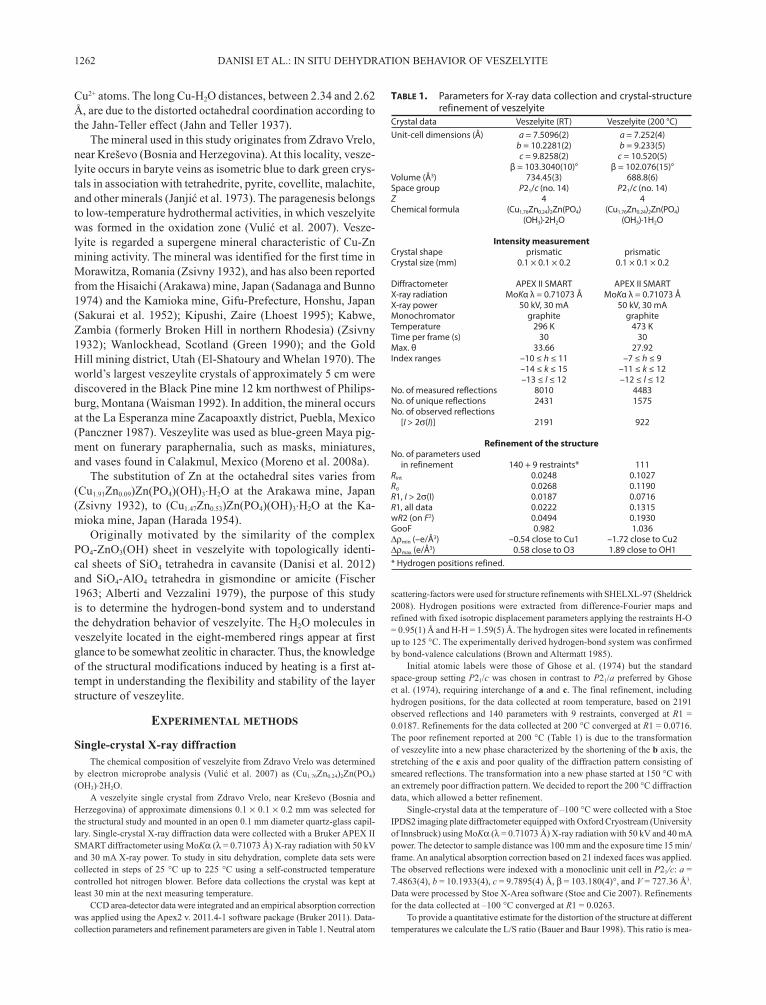
Table 1. Parameters for X-ray data collection and crystal-structure refinement of veszelyite
Crystal data Veszelyite (RT) Veszelyite (200 °C)Unit-cell dimensions (Å) a = 7.5096(2) a = 7.252(4) b = 10.2281(2) b = 9.233(5) c = 9.8258(2) c = 10.520(5) β = 103.3040(10)° β = 102.076(15)°Volume (Å3) 734.45(3) 688.8(6)Space group P21/c (no. 14) P21/c (no. 14)Z 4 4Chemical formula (Cu1.76Zn0.24)2Zn(PO4) (Cu1.76Zn0.24)2Zn(PO4) (OH3)·2H2O (OH3)·1H2O
Intensity measurementCrystal shape prismatic prismaticCrystal size (mm) 0.1 × 0.1 × 0.2 0.1 × 0.1 × 0.2 Diffractometer APEX II SMART APEX II SMARTX-ray radiation MoKα λ = 0.71073 Å MoKα λ = 0.71073 ÅX-ray power 50 kV, 30 mA 50 kV, 30 mAMonochromator graphite graphiteTemperature 296 K 473 KTime per frame (s) 30 30Max. θ 33.66 27.92Index ranges –10 ≤ h ≤ 11 –7 ≤ h ≤ 9 –14 ≤ k ≤ 15 –11 ≤ k ≤ 12 –13 ≤ l ≤ 12 –12 ≤ l ≤ 12No. of measured reflections 8010 4483No. of unique reflections 2431 1575No. of observed reflections [I > 2σ(I)] 2191 922
Refinement of the structure No. of parameters used in refinement 140 + 9 restraints* 111Rint 0.0248 0.1027Rσ 0.0268 0.1190R1, I > 2σ(I) 0.0187 0.0716R1, all data 0.0222 0.1315wR2 (on F2) 0.0494 0.1930GooF 0.982 1.036∆ρmin (–e/Å3) –0.54 close to Cu1 –1.72 close to Cu2∆ρmax (e/Å3) 0.58 close to O3 1.89 close to OH1* Hydrogen positions refined.
scattering-factors were used for structure refinements with SHELXL-97 (Sheldrick 2008). Hydrogen positions were extracted from difference-Fourier maps and refined with fixed isotropic displacement parameters applying the restraints H-O = 0.95(1) Å and H-H = 1.59(5) Å. The hydrogen sites were located in refinements up to 125 °C. The experimentally derived hydrogen-bond system was confirmed by bond-valence calculations (Brown and Altermatt 1985).
Initial atomic labels were those of Ghose et al. (1974) but the standard space-group setting P21/c was chosen in contrast to P21/a preferred by Ghose et al. (1974), requiring interchange of a and c. The final refinement, including hydrogen positions, for the data collected at room temperature, based on 2191 observed reflections and 140 parameters with 9 restraints, converged at R1 = 0.0187. Refinements for the data collected at 200 °C converged at R1 = 0.0716. The poor refinement reported at 200 °C (Table 1) is due to the transformation of veszeylite into a new phase characterized by the shortening of the b axis, the stretching of the c axis and poor quality of the diffraction pattern consisting of smeared reflections. The transformation into a new phase started at 150 °C with an extremely poor diffraction pattern. We decided to report the 200 °C diffraction data, which allowed a better refinement.
Single-crystal data at the temperature of –100 °C were collected with a Stoe IPDS2 imaging plate diffractometer equipped with Oxford Cryostream (University of Innsbruck) using MoKα (λ = 0.71073 Å) X-ray radiation with 50 kV and 40 mA power. The detector to sample distance was 100 mm and the exposure time 15 min/frame. An analytical absorption correction based on 21 indexed faces was applied. The observed reflections were indexed with a monoclinic unit cell in P21/c: a = 7.4863(4), b = 10.1933(4), c = 9.7895(4) Å, β = 103.180(4)°, and V = 727.36 Å3. Data were processed by Stoe X-Area software (Stoe and Cie 2007). Refinements for the data collected at –100 °C converged at R1 = 0.0263.
To provide a quantitative estimate for the distortion of the structure at different temperatures we calculate the L/S ratio (Bauer and Baur 1998). This ratio is mea-

DANISI ET AL.: IN SITU DEHYDRATION BEHAVIOR OF VESZELYITE 1263
sured between opposite oxygen sites along the shortest (S) and longest (L) cross sections of the eight-membered rings of the octahedral and the tetrahedral sheets.
TG/DTA analysisTG/DTA was conducted at the Department of Chemistry and Biochemistry,
University of Bern, with a Mettler Toledo TGA/SDTA851 instrument using a sample from Zdravo Vrelo, Bosnia and Herzegovina. The sample of 11.28 mg was ground to a fine powder and placed into a 70 µL alox crucible. Between 25 and 600 °C a heating rate of 5 °C/min was used with a gas flow rate of 20 mL/min. The analysis was conducted under a dry helium atmosphere.
Raman spectroscopyConfocal Raman spectra of single crystals were obtained with a HORIBA
JOBIN YVON LabRam-HR 800 Raman micro-spectrometer. The sample was excited by the 515 nm emission line of a 100 mW Ar+-laser under an OLYMPUS 100× objective (N.A. = 0.9). The size and power of the laser spot on the surface were approximately 1 µm and 5 mW, respectively. The scattered light was dispersed by a grating with 1800 lines/mm and collected by a 1024 × 256 open electrode CCD detector. The spectral resolution, determined by measuring the Rayleigh line, was about 1.4 cm–1. Third-order polynomial and convoluted Gauss-Lorentz func-tions were applied for background correction and band fitting. The wavenumber accuracy of about 0.5 cm–1 was achieved by adjusting the zero-order position of the grating and was regularly checked with a neon spectral calibration lamp.
ResulTs
Atomic coordinates and displacement parameters for the veszelyite structure at room temperature and 200 °C are given in Tables 2 and 3, respectively. The atomic coordinates and displacement parameter at –100 °C are reported in Table 41.
Selected distances and angles of hydrogen bonds under ambient conditions and at 200 °C are in Table 5.
Table 61 compares the interatomic distances at room tem-perature and 200 °C. Results of bond-valence calculations for veszelyite at room temperature and 200 °C are reported in Table 71. Observed Raman bands (cm–1) and possible assignments in the spectrum of veszelyite at ambient conditions are indicated in Table 8. Table 91 lists the L/S ratios of the longest to the shortest cross-sections of eight-membered rings of veszelyite at different temperature.
Discussion
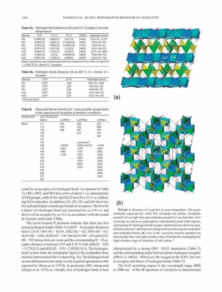
The structure of veszelyite under ambient conditionsThe structural study under ambient conditions confirmed
that veszelyite is built by alternating tetrahedral and octahedral sheets (Figs. 1–2) in agreement with the results by Ghose et al. (1974), Berry (1948), and Vulić et al. (2007).
The H positions were determined from –100 to +125 °C without substantial change in the related hydrogen bonding scheme (Fig. 1). Bond-valence calculations (Brown and Alter-matt 1985) were used to corroborate the experimentally derived hydrogen-bond system. Ignoring the contribution of hydrogen bonds in the bond-valence sums (bvs) of oxygen atoms, a bvs <0.5 valence units (v.u.) indicates a H2O molecule while a bvs of about 1 v.u. is typical of OH groups. Moreover, a bvs below 2 v.u. but greater than 1.5 v.u. suggests that the oxygen atom
Table 2a. Atomic coordinates and Ueq (Uiso) (Å2) values for veszelyite at RT
Site Atom Occ. x y z Ueq/Uiso
Cu1 Cu 1 0.49119(3) 0.073201(19) 0.12977(2) 0.01159(6)Cu2 Cu,Zn* 1 0.46075(3) 0.25309(2) 0.35875(2) 0.01117(6)Zn Zn 1 0.06969(3) 0.076074(18) 0.21045(2) 0.01146(6)P P 1 0.06592(6) 0.29887(4) 0.41457(5) 0.00931(9)O1 O 1 0.97533(18) 0.15924(13) 0.03207(13) 0.0152(3)O2 O 1 0.98311(17) 0.16643(12) 0.35793(13) 0.0138(3)O3 O 1 0.27509(17) 0.28533(12) 0.46986(13) 0.0128(2)O4 O 1 0.0246(2) 0.40001(12) 0.29562(15) 0.0161(3)OH1 O 1 0.33809(18) 0.08578(11) 0.26875(14) 0.0119(2)H1 H 1 0.359(5) 0.023(3) 0.340(3) 0.080†OH2 O 1 0.62201(17) 0.21779(12) 0.23336(14) 0.0128(2)H2 H 1 0.740(2) 0.190(3) 0.280(3) 0.080†OH3 O 1 0.59993(17) 0.40805(11) 0.46354(14) 0.0112(2)H3 H 1 0.7248(19) 0.386(4) 0.488(4) 0.080†H2O1 O 1 0.6558(2) 0.38343(15) 0.03669(16) 0.0252(3)H4 H 1 0.760(3) 0.370(4) 0.001(4) 0.080†H5 H 1 0.656(5) 0.334(3) 0.117(2) 0.080†H2O2 O 1 0.3199(2) 0.40648(15) 0.19194(17) 0.0238(3)H6 H 1 0.323(5) 0.374(4) 0.103(2) 0.080†H7 H 1 0.202(3) 0.402(3) 0.209(4) 0.080†* See discussion.† Uiso of hydrogen was fixed.
Table 2b. Atomic coordinates and Ueq (Å2) values for veszelyite at 200 °CSite Atom Occ. x y z Ueq
Cu1 Cu 1 0.4798(2) 0.08719(14) 0.11512(13) 0.0290(4)Cu2 Cu,Zn* 1 0.4742(2) 0.22291(15) 0.36711(14) 0.0315(5)Zn1 Zn 1 0.0541(2) 0.07741(14) 0.19716(14) 0.0317(5)P4 P 1 0.0902(5) 0.2150(3) –0.0771(3) 0.0282(8)O1 O 1 –0.0130(12) 0.1742(9) 0.0301(8) 0.035(2)O2 O 1 –0.0201(12) 0.1634(10) 0.3432(8) 0.038(2)O3 O 1 0.2947(12) 0.2645(8) –0.0172(8) 0.0302(19)O4 O 1 –0.1070(15) –0.0902(9) 0.1639(10) 0.051(3)OH1 O 1 0.3324(13) 0.0610(8) 0.2501(8) 0.034(2)OH2 O 1 0.6023(13) 0.2449(8) 0.2223(9) 0.037(2)OH3 O 1 0.6005(11) 0.1039(7) –0.0372(8) 0.0282(18)H2O1 O 1 0.6902(17) 0.0653(11) 0.4712(13) 0.067(5)* See discussion.
Table 3a. Anisotropic displacement parameters (Å2) for veszelyite at RTSite U11 U22 U33 U23 U13 U12
Cu1 0.01235(11) 0.01255(10) 0.01121(12) –0.00326(7) 0.00543(8) –0.00264(7)Cu2 0.01099(11) 0.01187(10) 0.01168(12) –0.00293(7) 0.00472(8) –0.00128(7)Zn 0.01111(10) 0.01221(9) 0.01126(12) –0.00107(7) 0.00295(8) –0.00146(6)P 0.0084(2) 0.01001(17) 0.0099(2) –0.00132(14) 0.00289(16) 0.00010(14)O1 0.0129(6) 0.0211(6) 0.0130(7) 0.0051(5) 0.0059(5) –0.0001(5)O2 0.0125(6) 0.0129(5) 0.0167(7) –0.0043(5) 0.0049(5) –0.0018(4)O3 0.0081(6) 0.0164(5) 0.0139(7) –0.0021(5) 0.0024(5) –0.0005(4)O4 0.0195(7) 0.0140(5) 0.0164(7) 0.0035(5) 0.0071(6) 0.0056(5)OH1 0.0105(6) 0.0131(5) 0.0124(7) 0.0008(4) 0.0032(5) –0.0009(4)OH2 0.0112(6) 0.0145(5) 0.0132(6) –0.0020(5) 0.0039(5) –0.0013(4)OH3 0.0102(6) 0.0120(5) 0.0123(7) 0.0013(4) 0.0043(5) 0.0015(4)H2O1 0.0322(9) 0.0237(7) 0.0192(8) 0.0026(6) 0.0049(7) 0.0018(6)H2O2 0.0182(7) 0.0324(8) 0.0221(8) –0.0024(6) 0.0075(6) –0.0009(6)
Table 3b. Anisotropic displacement parameters (Å2) for veszelyite at 200 °C
Site U11 U22 U33 U23 U13 U12
Cu1 0.0366(9) 0.0255(7) 0.0274(9) –0.0030(5) 0.0125(7) –0.0023(6)Cu2 0.0373(10) 0.0261(7) 0.0335(10) –0.0075(6) 0.0129(7) –0.0041(6)Zn1 0.0377(9) 0.0271(7) 0.0313(9) –0.0030(5) 0.0095(7) –0.0037(6)P 0.0295(18) 0.0237(14) 0.0314(18) 0.0032(11) 0.0064(15) –0.0041(12)O1 0.036(5) 0.035(4) 0.035(5) 0.011(4) 0.009(4) –0.001(4)O2 0.033(5) 0.051(5) 0.038(5) –0.017(4) 0.020(4) –0.003(4)O3 0.036(5) 0.029(4) 0.027(5) 0.004(3) 0.011(4) –0.005(3)O4 0.061(7) 0.041(5) 0.056(6) –0.015(4) 0.022(6) –0.022(5)OH1 0.043(5) 0.026(4) 0.038(5) –0.003(3) 0.021(4) 0.003(4)OH2 0.040(6) 0.032(4) 0.041(5) –0.001(4) 0.014(4) –0.007(4)OH3 0.031(5) 0.023(4) 0.033(5) 0.000(3) 0.013(4) 0.006(3)H2O1 0.062(8) 0.049(7) 0.091(10) 0.023(6) 0.019(7) 0.016(5)
1 Deposit item AM-13-711, Tables 4, 6, 7, and 9; Figure 9; and CIFs are on deposit. Deposit items are available two ways: For a paper copy contact the Business Office of the Mineralogical Society of America (see inside front cover of recent issue) for price information. For an electronic copy visit the MSA web site at http://www.minsocam.org, go to the American Mineralogist Contents, find the table of contents for the specific volume/issue wanted, and then click on the deposit link there.
DANISI ET AL.: IN SITU DEHYDRATION BEHAVIOR OF VESZELYITE1264
could be an acceptor of a hydrogen bond. As reported in Table 7a, OH1, OH2, and OH3 have a bvs of about 1 v.u. characteristic of OH groups, while H2O1 and H2O2 have bvs <0.5 v.u. indicat-ing H2O molecules. In addition, O1, O2, O3, and O4 show low bvs and participate at hydrogen bonds as acceptors. The bvs for a donor of a hydrogen bond was increased by ca. 0.8 v.u. and the bvs of an acceptor by ca. 0.2, in accordance with the model by Ferraris and Ivaldi (1988).
The seven located H positions indicate that there are five strong hydrogen bonds (Table 5) with H···O acceptor distances below 2.0 Å: OH1-H1···H2O1, OH2-H2···O2, OH3-H3···O1, H2O1-H5···OH2, H2O2-H7···O4. The H2O1-H4···O2 and H2O2-H6···O3 interactions are weak and the corresponding H···O ac-ceptor distance is between 2.07 and 2.45 Å with d(H2O1···O2) = 3.373(2) Å and d(H2O2···O3) = 2.8958(19) Å. The hydrogen-bond system links the tetrahedral sheet to the octahedral sheet and also interconnects the Cu sheet (Fig. 3a). The hydrogen bond system delineated in this study is only in partial agreement to that reported by Ghose et al. (1974). In particular, OH1 interpreted (Ghose et al. 1974) as virtually free of hydrogen bond is here
Table 5a. Hydrogen bond distances (Å) and O-H···O angles (°) at room temperature
Species D-H H···A D···A <(DHA) Hydrogen bondOH 0.940(10) 1.888(13) 2.811(2) 166(4) OH1-H1···H2O1OH 0.940(10) 1.829(12) 2.7568(18) 169(3) OH2-H2···O2OH 0.941(10) 1.888(10) 2.8286(18) 179(3) OH3-H3···O1H2O 0.937(10) 2.451(13) 3.373(2) 168(3) H2O1-H4···O2H2O 0.940(10) 1.705(13) 2.626(2) 166(3) H2O1-H5···OH2H2O 0.938(10) 2.07(2) 2.8958(19) 146(3) H2O2-H6···O3H2O 0.941(10) 1.736(15) 2.646(2) 162(4) H2O2-H7···O4Notes: H positions were determined with the restraint O-H is 0.95(1) Å and H-H = 1.59(5) Å. D = donor; A = acceptor.
Table 5b. Hydrogen bond distances (Å) at 200 °C, D = donor; A = acceptor
Species D-H D···A Hydrogen bondOH 0.95* 2.96 OH1-H1···OH2OH 0.95* 2.87 OH2-H2···O2OH 0.95* 2.82 OH3-H3···O1H2O 0.95* 2.82 H2O1-H4···O3H2O 0.95* 2.87 H2O1-H5···O2* D-H was fixed.
FiguRe 1. Structure of veszelyite at room temperature. The green tetrahedra represent Zn, while PO4 tetrahedra are yellow. Octahedra around Cu2 are light blue and octahedra around Cu1 are dark blue. H2O molecules are shown as pink spheres with attached small white spheres representing H. Hydrogen-bond acceptor interactions are shown by gray dashed connectors. (a) Projection along the b axis showing the tetrahedral and octahedral sheets, (b) view of the veszelyite structure parallel to a showing the four- and eight-member rings of tetrahedra overlapping the eight-member rings of octahedra. (Color online.)
characterized by a strong OH1···H2O1 interaction (Table 5) and the corresponding angle between donor-hydrogen-acceptor (DHA) is 166(4)°. Moreover, the oxygen at the H2O1 site acts as acceptor and donor of hydrogen bonds (Table 5).
The O-H stretching region in the wavelength range 4000 to 3000 cm–1 of the IR spectrum of veszelyite is characterized
Table 8. Observed Raman bands (cm–1) and possible assignments in the spectrum of veszelyite at ambient conditions
Assignment ν(Zn-O),ν(Cu-O), lattice ν2(OPO)s ν4(OPO)a ν1(PO)sBand 111 439 539 833 115 470 556 882 124 486 607 929 130 624 951 143 967 151 152 overtones? 164 ν3(PO)a ν(OH),ν(H2O)? νs(OH) 174 1025 1379 2664 189 1045 1587 2852 197 1108 1632 3184 199 1805 3286 243 1970 3302 261 2042 3497 278 2233 3425 318 3566 364 3390 377 3555 387

DANISI ET AL.: IN SITU DEHYDRATION BEHAVIOR OF VESZELYITE 1265
FiguRe 2. Structure of veszelyite at 200 °C. (a) Projection along the b axis, (b) View of the veszelyite structure parallel to a. Colors as in Figure 1. (Color online.)
FiguRe 3. Hydrogen-bond system of veszelyite. (a) Hydrogen bonding at RT. H2O molecules are shown as pink spheres and the numbers 1 and 2 refer to H2O1 and H2O2 sites, respectively. (b) Hydrogen bonding at 200 °C, the black arrows indicate the direction to the acceptor. Colors as in Figure 1. (Color online.)
by two peaks at 3530 (sharp), 3270 (broad) cm–1, with two shoulders at 3350 and 3170 cm–1 (Ghose et al. 1974). Accord-ing to the hydrogen bond length vs. IR frequency correlation by Libowitzky (1999), absorptions between 3270 and 3530 cm–1 correspond to donor-acceptor (O···O) distances of ca. 2.73–2.93 Å, which agrees with the D-A distances in Table 5. The two strong hydrogen bonds H2O1-H5···OH2 and H2O2-H7···O4 with D-A distances of about 2.6 Å should show absorp-tion peaks at ca. 2730 cm–1, not observed in the IR spectrum reported by Ghose et al. (1974). However, two broad bands at 2664 and 2852 cm–1 besides 8 further bands between 3184 and 3555 cm–1 were observed in the Raman spectrum collected on Zdravo Vrelo veszelyite shown in Figure 4. The large number of bands in this region can be assigned to stretching vibrations of hydroxyl and H2O units and are consistent with the complex hydrogen bond system.
The Raman spectrum of veszelyite below 2500 cm–1 matches with the spectra presented in Moreno et al. (2008b). It is char-acterized by the most intense band (amplitude) at 967 cm–1, medium intensity bands at 624, 486, 470 387, 377, 364, 243,
and 130 cm–1 and numerous weaker bands (Table 8). Scattering measurements on phosphates in aqueous solutions (Nakamoto 1978), Zn phosphate minerals (Frost 2004) and ab initio studies of hydroxyapatite (Corno et al. 2006) assign the Raman bands in water-bearing phosphates to ν3 asymmetric P-O stretching modes ∼1020–1095 cm–1, ν1 symmetric P-O stretching modes around 950–990 cm–1, ν4 asymmetric OPO bending ∼530–660 cm–1, ν2 symmetric OPO bending modes ∼400–490 cm–1 and complex lattice modes involving Zn-O and Cu-O vibrations <400 cm–1.
The origin of the additional bands at 833 and 882 cm–1 and from 1379–2233 cm–1 is unclear. Compared to other zinc and zinc calcium phosphates the symmetry of veszelyite is lower and the structure more complex, resulting in a higher number of theoretical possible vibrational modes. Further explanations might be vibrational overtones caused by second-order Raman scattering processes or vibrational modes of H2O and OH groups. The existence of translational modes of OH groups at 335 and 630 cm–1 was reported for hydroxyapatite (Corno et al. 2006 and references therein).

DANISI ET AL.: IN SITU DEHYDRATION BEHAVIOR OF VESZELYITE1266
Dehydration upon heatingThe observed dehydration beginning at 150 °C is preceded
by a slight increase of volume due to thermal expansion. This dehydration step is associated with a complete loss of H2O at the H2O2 site. After the release of H2O2, the Cu coordination of Cu1 and Cu2 declined from originally six- to fivefold. Copper is no longer in distorted octahedral coordination but occupies a distorted square-based pyramid (Fig. 5). H2O1 moves toward a position in the center of the eight-membered rings (Figs. 1–2). Moreover, the Cu2-H2O1 distance decreased from 2.4441(16) to 2.248(10) Å. Simultaneous to the water loss, the unit-cell volume decreased by 7% from originally 734.45(3) to 686.4(4) Å3 leading to a formula with 1 H2O pfu (Fig. 6). The new phase observed above 150 °C is characterized by a lengthening of the c axis and shortening of the b axis (Table 1). The expansion of the c axis is related to strain relaxation within the four- and eight-membered rings and to the deformation imposed by the distortion of the T-O-T angles. This T-O-T bending causes distortion of the tetrahedral and octahedral sheets (Fig. 2). The L/S ratio of the eight-membered rings of the tetrahedral sheet and the octahedral sheet increases between room temperature and 200 °C yielding more pronounced elliptical shape of the channels with dehydration (Table 9). Moreover, the bases of the tetrahedra forming sheets parallel to (100) describe a puck-ered surface as result of the strain relaxation within the eight-membered rings (Fig. 2). In contrast, at room temperature the bases of tetrahedra form flat layers (Fig. 1). The deformations of the tetrahedral sheets are monitored by variations of T-O-T angles at different temperature (Table 6). The P-O1-Zn and P-O2-Zn angles increased from 132.42° and 120.59° at room
FiguRe 5. Displacement ellipsoids for Cu1 and Cu2 polyhedra in veszelyite at RT and 200 °C. The probability for ellipsoids is 0.75. (Color online.)
FiguRe 4. Deconvolution of the Raman spectrum of veszelyite in the range 2500–4000 cm–1. Dots = measured Raman spectrum, thin curves = fitted Gauss-Lorentz functions, thick curve = sum curve of fitted functions.

DANISI ET AL.: IN SITU DEHYDRATION BEHAVIOR OF VESZELYITE 1267
temperature to 135.6° and 123.1° at 200 °C, respectively. The P-O4-Zn angle decreased from 131.23° to 127.5°.
The deformation of the framework imposed by the release of water implies a rearrangement of the hydrogen-bond system (Fig. 3b). The poor data quality and refinement at 200 °C did not allow a determination of the H positions. The evaluation of the hydrogen-bond system at 200 °C is based on bond-valence calculations (Brown and Altermatt 1985) and donor-acceptor distances (Table 5b). As reported in Table 7b, the increased bond-valence sum (without hydrogen bond contributions) at O4 is due to the shortening of the P-O4 distance. O4 at 200 °C is no longer fixed by a hydrogen bond, which is in contrast to our structural data below 150 °C. The bonds OH2-H2···O2 and OH3-H3···O1 are preserved at 200 °C (Table 5). The H2O1 site rearranges the hydrogen bonds generating two new interactions: H2O1-H4···O3 and H2O1-H5···O2. The strong interaction H2O1-H5···OH2 with a D-A distance of 2.63 Å at room temperature is no more observed. The D-A distance H2O1···O2 shortens from 3.37 Å at room temperature to 2.87 Å at 200 °C. The OH1 site generates a new hydrogen bond to OH2 with a D-A distance of 2.96 Å. As observed at room temperature, the hydrogen-bond system at 200 °C strengthens the connection between the tetra-hedral and the octahedral sheet (Fig. 3b).
Already at 125 °C (before release of H2O2), O4 shows increased anisotropy in thermal motion parallel to a and per-pendicular to the P-O4-Zn connection. This behavior is depicted in Figure 7 in terms of temperature dependence of Ueq. At first glance it appears surprising that H2O2, which is expelled first, has lower Ueq than H2O1. This is explained by two bonds of H2O2 to Cu1 and Cu2, whereas H2O1 is only bonded to Cu2. Above 125 °C O4 shows the strongest thermal disorder (Figs. 7 and 8) comparable to H2O1. The sites from O1 to O3 and the oxygen atoms of the hydroxyl groups show the same behavior with a slight increase of the Ueq after 125 °C.
Our dehydration experiments were carried out up to 225 °C. At this temperature we observed that the color changed from blue to green accompanied by a breakdown of the partially
Temperature (°C)
0 20 40 60 80 100 120 140 160 180 200 220
Uni
t cel
l vol
ume
(Å3 )
680
690
700
710
720
730
740
750
H2O2
FiguRe 6. Development of unit-cell volume vs. temperature for in situ dehydration experiments of veszelyite. The size of the symbols is larger than the associated e.s.d. values.
Temperature (°C)
0 20 40 60 80 100 120 140 160 180 200 220
Ueq
(Å2 )
0.00
0.01
0.02
0.03
0.04
0.05
0.06
0.07
O1O2O3O4OH1OH2OH3H2O1H2O2
FiguRe 7. Ueq for oxygen sites at different temperatures for
veszelyite. The size of the symbols is larger than the associated e.s.d. values.
FiguRe 8. Thermal ellipsoids for P and Zn polyhedra in veszelyite at RT and 200 °C. The probability for ellipsoids is 0.75. (Color online.)

DANISI ET AL.: IN SITU DEHYDRATION BEHAVIOR OF VESZELYITE1268
dehydrated structure. With the removal of the last H2O mol-ecule at H2O1, the crystal structure collapsed. At first glance we would have expected that after the release of H2O at H2O1 Cu adopted square planar fourfold coordination preserving the crystal structure. A loss of H2O1 would not lead to unreasonable bond-valence sums for oxygen or Cu (Table 7b).
Few natural examples of crystal structures with Cu2+ in pla-nar fourfold-coordination are reported in the literature: elyite Pb4Cu(SO4)O2(OH)4⋅H2O (Kolitsch and Giester 2000), johillerite Na(Mg,Zn)3Cu(AsO4)3 (Tait and Hawthorne 2004), henmilite Ca2Cu(OH)4[B(OH)4]2 (Nakai et al. 1986), and cuprorivaite CaCuSi4O10 (Bensch and Schur 1995).
However, a crystalline anhydrous veszelyite phase was not observed and the reason is attributed to the substitution of Cu by Zn in the octahedral sheet. As the Cu2 octahedron is less distorted than Cu1, Zn substitution is more probable for Cu2. Looking at the probability ellipsoids in Figure 5, there is no clear indication of preferential occupancy of Zn at Cu1 or Cu2. We would expect that a Jahn-Teller distorted octahedron with mixed Zn, Cu occupancy shows smeared probability ellipsoids along the Jahn-Teller active ligands. The problem of Zn prefer-ence for different Cu sites in veszelyite and other structures is addressed by Mellini and Merlino (1978). On the base of the different octahedral distortions of the two sites, they assume that Cu1 is fully occupied by Cu, whereas Cu2 is partially substituted by Zn. The ∆L value (difference between the average values of axial and equatorial bond distance) for Cu1 indicates 100% Cu content. This hypothesis supports the idea that if H2O at H2O1 is released Zn cannot adopt planar fourfold coordination and for this reason the breakdown of the structure occurs.
The weight loss between 130 and 220 °C seen in our thermo-gravimetric analysis (Fig. 91) correlates with the transitions at 150 °C in our study. The second step between 220 and 290 °C is related to the loss of the last H2O molecule at the H2O1 site. The last step (290–600 °C) corresponds to the loss of OH groups. The total mass lost during the 25 to 600 °C heating excursion sums up to 17.5 wt% (Fig. 91).
Similarities and differences between veszelyite and related structures
The veszelyite structure has some similarity to that of the zeolite-like cavansite framework [Ca(VO)Si4O10⋅4H2O] (Evans 1973) and gismondine (CaAl2Si2O8⋅4H2O) (Vezzalini et al. 1993). These structures have tetrahedral sheets composed of four- and eight-membered rings in common with the same up and down arrangement of tetrahedral apices in adjacent chains.
In cavansite, the dehydration proceeds in four steps within the same space group with only minor impact on framework distortion and contraction (Danisi et al. 2012). The removal of the last H2O molecule causes structural destruction in both structures. In particular, the lowest coordination limit for the cations (Cu or Ca) bonded to the H2O molecules appears to be five. In veszelyite, Zn-substituted Cu2 cannot adopt square planar fourfold coordination while Ca in cavansite, after losing the last H2O molecule, cannot preserve the fivefold coordination thus causing structural breakdown.
An interesting analogy could be drawn between veszelyite and kipushite. In both structures the sheet composed by alter-
nating PO4 and ZnO3(OH) tetrahedra is very similar (Piret et al. 1985). The kipushite structure is also built of sheet of ZnO4 and PO4 tetrahedra neighbored by two octahedral sheets like in veszelyite but every other tetrahedral sheet is replaced by isolated PO4 tetrahedra. In both structures, Zn fully occupies a tetrahedral site and partially substitutes Cu in octahedral coor-dination. The up and down arrangement of tetrahedral apices in adjacent chains in veszelyite corresponds to the arrangement in kipushite.
Tetrahedral sheets composed by four- and eight-membered rings have been reported also for the layered hydrous zinc phosphate Mg[ZnPO4(H2O)]2⋅10H2O (Kahlenberg et al. 2008). The building units are PO4 and ZnO3(H2O) but in contrast to veszelyite the up and down arrangement of tetrahedral apices in adjacent chains is different.
A comparable open eight-membered octahedral sheet struc-ture has been found in bayldonite (Ghose and Wan 1979). The Cu2+O6 octahedra show the usual Jahn-Teller distortion and Zn partially substitutes copper in octahedral coordination. As in veszelyite, the Cu2 site appears to be the most probable site for Zn substitution.
acKnowleDgMenTsThe authors are deeply grateful D. Djordjević for supplying us a sample of
veszelyite from Zdravo Vrelo (Bosnia and Herzegovina) for studying the dehy-dration behavior. The constructive reviews of Stefano Merlino and Reinhard X. Fischer are highly appreciated.
ReFeRences ciTeDAlberti, A. and Vezzalini, G. (1979) The crystal structure of amicite, a zeolite.
Acta Crystallographica, B35, 2866–2869.Bauer, T. and Baur, W.H. (1998) Structural changes in the natural zeolite gismon-
dine (GIS) induced by cation exchange with Ag, Cs, Ba, Li Na, K and Rb. European Journal of Mineralogy, 10, 133–147.
Bensch, W. and Schur, M. (1995) Crystal structure of calcium copper phyllode-caoxotetrasilicate, CaCuSi4O10. Zeitschrift für Kristallographie, 210, 530.
Berry, L.G. (1948) Structural crystallography of lazulite, scorzalite, and veszelyite. American Mineralogist, 33, 750.
Brown, I.D. and Altermatt, D. (1985) Bond-valence parameters obtained from a systematic analysis of the Inorganic Crystal Structure Database. Acta Crystal-lographica, B41, 244–247.
Bruker (2011) SAINT, SADABS. Bruker AXS Inc., Madison, Wisconsin.Corno, M., Busco, C., Civalleri, B., and Ugliengo, P. (2006) Periodic ab initio
study of structural and vibrational features of hexagonal hydroxyapatite Ca10(PO4)6(OH)2. Physical Chemistry Chemical Physics, 8, 2464–2472.
Danisi, R.M., Armbruster, T., and Lazic, B. (2012) In situ dehydration behavior of zeolite-like cavansite: a single crystal X-ray study. American Mineralo-gist, 97, 1874–1880.
El-Shatoury, H.M. and Whelan, J.A. (1970) Mineralization in the gold hill mining district, Tooele County, Utah. Utah Geological and Mineralogical Survey, Bulletin 83, 1–37.
Evans, H.T. Jr. (1973) The crystal structures of cavansite and pentagonite. Ameri-can Mineralogist, 58, 412–424.
Ferraris, G. and Ivaldi, G. (1988) Bond valences vs bond length in O…O hydrogen bonds. Acta Crystallographica, B44, 341–344.
Fischer, K.F. (1963) The crystal structure determination of zeolite gismondite CaAl2Si2O8⋅4H2O. American Mineralogist, 48, 664–672.
Frost, R.L. (2004) An infrared and Raman spectroscopic study of natural zinc phosphates. Spectrochimica Acta Part A: Molecular and Biomolecular Spectroscopy, 60, 1439–1445.
Ghose, S. and Wan, C. (1979) Structural Chemistry of copper and zinc minerals VI. Bayldonite, (Cu,Zn)3Pb(AsO4)2(OH)2: a complex layer structure. Acta Crystallographica, B35, 819–823.
Ghose, S., Leo, S.R., and Wan, C. (1974) Structural chemistry of copper and zinc minerals. Part I. Veszelyite, (Cu,Zn)2Zn(PO4)(OH)3⋅2H2O: a novel type of sheet structure and crystal chemistry of copper-zinc substitution. American Mineralogist, 59, 573–581.
Green, D. (1990) Veszelyite a mineral new to Britain, from Wanlockhead, Scotland. Journal of Mines and Minerals, 8, 6–7.
Harada, Z. (1954) Chemical analyses of Japanese minerals (III). Journal of the

DANISI ET AL.: IN SITU DEHYDRATION BEHAVIOR OF VESZELYITE 1269
of kipushite, a new copper-zinc phosphate from Kipushi, Zaire. Canadian Mineralogist, 23, 35–42.
Sadanaga, R. and Bunno, M. (1974) The Wakabayashi mineral collection. The University Museum, the University of Tokio Bulletin no. 7, 177 p.
Sakurai, K., Nagashima, H., and Sorita, E. (1952) Kamiokalite, a new zinc copper phosphate mineral from Kamioka Mine, Japan. Syumi-no-Tigaku (Amateur Geologist), 5, 170–175.
Sheldrick, G.M. (2008) A short history of SHELX. Acta Crystallographica, A64, 112–122.
Stoe and Cie (2007) X-Area 1.39. Darmstadt, Germany.Tait, K.T. and Hawthorne, F.C. (2004) Johillerite from Tolbachik, Kamchatka
Peninsula, Russia: crystal-structure refinement and chemical composition. Canadian Mineralogist, 42, 717–722.
Vezzalini, G., Quartieri, S., and Alberti, A. (1993) Structural modifications induced by dehydration in the zeolite gismondine. Zeolites, 13, 34–42.
Vulić, P., Kahlenberg, V., Lazic, B., Kaindl, R., Dimitrijević, R., and Djordjević, D. (2007) Veszelyte from the locality Zdravo Vrelo near Kreševo (Bosnia and Herzegovina): its mineralogical characterization and absolute crystal structure. XIV Conference of the Serbian Crystallographic Society (28–30 June 2007), p. 16–17. Abstracts, Vršac, Serbia.
Waisman, D. (1992) Minerals of the Black Pine Mine, Granite county, Montana. Mineralogical Record, 23, 477–483.
Zsivny, V. (1932) Ueber den Veszelyit von Vaskö (Moravicza). Zeitschrift für Kristallographie, 82, 87–110.
Manuscript received January 14, 2013Manuscript accepted March 28, 2013Manuscript handled by G. dieGo Gatta
Faculty of Science, Hokkaido University, Series 4, Geology and Mineralogy, 8(4), 289–348.
Jahn, H.A. and Teller, E. (1937) Stability of polyatomic molecules in degenerate electronic states. I. Orbital degeneracy. Proceedings of the Royal Society of London, Series A, Mathematical and Physical Sciences, 161, 220–235.
Janjić, S., Đorđević, D., Jovanović, R. and Bugarski, P. (1973) Pojava vescelita (veszelyita) u području Kreševa (Jugoslavija). Geološki Glasnik, 17, 181–192.
Kahlenberg, V., Tessadri, R., Többens, D.M., Wertl, W., and Rössler, A. (2008) Mg[ZnPO4(H2O)]2⋅10H2O—a layered hydrous zinc phosphate retrieved from an industrial filter cake residual. Zeitschrift für Anorganische und Allgemeine Chemie, 634, 1181–1186.
Kolitsch, U. and Giester, G. (2000) Elyite, Pb4Cu(SO4)O2(OH)4⋅H2O: Crystal structure and new data. American Mineralogist, 85, 1816–1821.
Libowitzky, E. (1999) Correlation of O-H stretching frequencies and O-H hy-drogen bond lenghts in minerals. Monatshefte für Chemie, 130, 1047–1059.
Lhoest, J.J. (1995) The Kipushi Mine. Mineralogical Record, 26, 163–192.Mellini, M. and Merlino, S. (1978) Ktenasite, another mineral with
[(Cu,Zn)2(OH)3O]– octahedral sheets. Zeitschrift für Kristallographie, 147, 129–140.
Moreno, R.G., Mathis, F., Mazel, V., Dubus, M., Calligaro, T., and Strivay, D. (2008a) Discovery and characterization of an unknown blue-green Maya pigment: veszelyite. Archaeometry, 50, 658–667.
Moreno, R.G., Strivay, D., and Gilbert, B. (2008b) Maya blue-green pigments found in Calakmul, Mexico: a study by Raman and UV-visible spectroscopy. Journal of Raman Spectroscopy, 39, 1050–1056.
Nakai, I., Okada, H., Masutomi, K., Koyama, E., and Nagashima, K. (1986) Hen-milite, Ca2Cu(OH)4[B(OH)4]2, a new mineral from Fuka, Okayama Prefecture, Japan. American Mineralogist, 71, 1234–1239.
Nakamoto, K. (1978) Infrared and Raman spectra of inorganic and coordination compounds, 448 p. Wiley, New York.
Panczner, W.D. (1987) Minerals of Mexico, 459 p. Van Nostrand Reinhold Company, New York.
Piret, P., Deliens, M., and Piret-Meunier, J. (1985) Occurrence and crystal structure
Copyright © 2022 FDOKUMEN