Pregnancy, Child Nutrition, and Oral Health
418
Nutrition and Oral Medicine Edited by Riva Touger-Decker, P h D , RD , FADA David A. Sirois, DMD , P h D Connie C. Mobley, P h D , RD Nutrition and Oral Medicine Edited by Riva Touger-Decker, P h D , RD , FADA David A. Sirois, DMD , P h D Connie C. Mobley, P h D , RD
-
Upload
independent -
Category
Documents
-
view
1 -
download
0
Transcript of Pregnancy, Child Nutrition, and Oral Health

Nutritionand
Oral MedicineEdited by
Riva Touger-Decker, PhD, RD, FADA
David A. Sirois, DMD, PhD
Connie C. Mobley, PhD, RD
Nutritionand
Oral MedicineEdited by
Riva Touger-Decker, PhD, RD, FADA
David A. Sirois, DMD, PhD
Connie C. Mobley, PhD, RD

NUTRITION AND ORAL MEDICINE
Touger_Decker_FM_Final2 07/27/2004, 1:50 PM1

NUTRITION AND HEALTH
Adrianne Bendich, Series Editor
Nutrition and Oral Medicine, edited by Riva Touger-Decker, David A. Sirois, andConnie C. Mobley, 2005
The Management of Eating Disorders and Obesity, Second Edition, edited by David J.Goldstein, 2005
Preventive Nutrition: The Comprehensive Guide for Health Professionals, Third Edition,edited by Adrianne Bendich and Richard J. Deckelbaum, 2005
IGF and Nutrition in Health and Disease, edited by M. Sue Houston, Jeffrey M. P.Holly, and Eva L. Feldman, 2005
Epilepsy and the Ketogenic Diet, edited by Carl E. Stafstrom and Jong M. Rho, 2005Handbook of Drug–Nutrient Interactions, edited by Joseph I. Boullata and Vincent T.
Armenti, 2004Nutrition and Bone Health, edited by Michael F. Holick and Bess Dawson-Hughes,
2004Diet and Human Immune Function, edited by David A. Hughes, L. Gail Darlington,
and Adrianne Bendich, 2004Beverages in Nutrition and Health, edited by Ted Wilson and Norman J. Temple,
2004Handbook of Clinical Nutrition and Aging, edited by Connie Watkins Bales and
Christine Seel Ritchie, 2004Fatty Acids: Physiological and Behavioral Functions, edited by David I. Mostofsky,
Shlomo Yehuda, and Norman Salem, Jr., 2001Preventive Nutrition: The Comprehensive Guide for Health Professionals, Second Edition,
edited by Adrianne Bendich and Richard J. Deckelbaum, 2001Nutritional Health: Strategies for Disease Prevention, edited by Ted Wilson and Norman
J. Temple, 2001Clinical Nutrition of the Essential Trace Elements and Minerals: The Guide for Health
Professionals, edited by John D. Bogden and Leslie M. Klevey, 2000Primary and Secondary Preventive Nutrition, edited by Adrianne Bendich
and Richard J. Deckelbaum, 2000The Management of Eating Disorders and Obesity, edited by David J. Goldstein,
1999Vitamin D: Physiology, Molecular Biology, and Clinical Applications, edited by Michael
F. Holick, 1999Preventive Nutrition: The Comprehensive Guide for Health Professionals, edited by
Adrianne Bendich and Richard J. Deckelbaum, 1997
Touger_Decker_FM_Final2 07/27/2004, 1:50 PM2

HUMANA PRESSTOTOWA, NEW JERSEY
NUTRITION
AND ORAL MEDICINE
Edited by
RIVA TOUGER-DECKER, PhD, RD, FADADepartment of Primary Care, School of Health-Related Professions,Department of Diagnostic Sciences, New Jersey Dental School,University of Medicine and Dentistry of New Jersey, Newark, NJ
DAVID A. SIROIS, DMD, PhDDepartment of Oral Medicine, New York University College of Dentistry,New York, NY
CONNIE C. MOBLEY, PhD, RDDepartment of Professional Studies, University of Nevada Las Vegas Schoolof Dental Medicine, Las Vegas, NV
Foreword by
DOMINICK P. DEPAOLA, DDS, PhDThe Forsyth Institute, Boston, MA
Touger_Decker_FM_Final2 07/27/2004, 1:50 PM3

© 2005 Humana Press Inc.999 Riverview Drive, Suite 208Totowa, New Jersey 07512
www.humanapress.com
All rights reserved. No part of this book may be reproduced, stored in a retrieval system, or transmitted in any form or by anymeans, electronic, mechanical, photocopying, microfilming, recording, or otherwise without written permission from the Publisher.
All papers, comments, opinions, conclusions, or recommendations are those of the author(s), and do not necessarily reflect the views of thepublisher.
Due diligence has been taken by the publishers, editors, and authors of this book to assure the accuracy of the information publishedand to describe generally accepted practices. The contributors herein have carefully checked to ensure that the drug selections anddosages set forth in this text are accurate and in accord with the standards accepted at the time of publication. Notwithstanding, asnew research, changes in government regulations, and knowledge from clinical experience relating to drug therapy and drug reactionsconstantly occurs, the reader is advised to check the product information provided by the manufacturer of each drug for any changein dosages or for additional warnings and contraindications. This is of utmost importance when the recommended drug herein is anew or infrequently used drug. It is the responsibility of the treating physician to determine dosages and treatment strategies forindividual patients. Further it is the responsibility of the health care provider to ascertain the Food and Drug Administration statusof each drug or device used in their clinical practice. The publisher, editors, and authors are not responsible for errors or omissionsor for any consequences from the application of the information presented in this book and make no warranty, express or implied, withrespect to the contents in this publication.
Cover design by Patricia F. ClearyProduction Editor: Robin B. Weisberg
For additional copies, pricing for bulk purchases, and/or information about other Humana titles,contact Humana at the above address or at any of the following numbers: Tel.: 973-256-1699;Fax: 973-256-8341; E-mail: [email protected] or visit our website at http://humanapress.com
This publication is printed on acid-free paper.ANSI Z39.48-1984 (American National Standards Institute) Permanence of Paper for Printed Library Materials.
Photocopy Authorization Policy:Authorization to photocopy items for internal or personal use, or the internal or personal use of specific clients is granted by HumanaPress, provided that the base fee of US $25.00 per copy is paid directly to the Copyright Clearance Center (CCC), 222 Rosewood Dr.,Danvers MA 01923. For those organizations that have been granted a photocopy license from the CCC, a separate system of paymenthas been arranged and is acceptable to the Humana Press. The fee code for users of the Transactional Reporting Service is 1-58829-192-8/05 $25.00.
Printed in the United States of America. 10 9 8 7 6 5 4 3 2 1
eISBN 1-59259-831-5
Library of Congress Cataloging-in-Publication Data
Nutrition and oral medicine / edited by Riva Touger-Decker, David A. Sirois, and Connie C. Mobley ;foreword by Dominick P. DePaola. p. ; cm. Includes bibliographical references and index. ISBN 1-58829-192-8 (alk. paper) 1. Teeth--Care and hygiene. 2. Dental public health. 3. Mouth--Care and hygiene. I. Touger-Decker, Riva.II. Sirois, David. III. Mobley, Connie C. [DNLM: 1. Oral Health. 2. Diet. 3. Mouth Diseases--etiology. 4. Nutrition Disorders--complications.5. Nutrition. WU 113.7 N9757 2004] RK61.N88 2004 617.6'01--dc22
2004001616
Touger_Decker_FM_Final2 07/27/2004, 1:50 PM4

Dedication
v
Former Israeli Prime Minister Golda Meir once said “I never did anything alone.Whatever was accomplished in this country was accomplished collectively” (1). Rarelyare accomplishments solo products. They are the result of team work and mutual respect.For the most part, the first person, nominative case plural is the operative pronoun ofaccomplishment, not the singular. In this regard, our colleagues, students, and fellowpractitioners constitute the corporate “we” of accomplishment. We dedicate this work toyou with this simple sentence: “We did it!”
Thank you to our parents, who gave us the guidance, inspiration, and encouragementto succeed in anything we do and to our spouses and our children without whose support,patience, and understanding we never would have been able to complete this book.
Thank you to our students, colleagues, faculty, and staff at the University of Medicineand Dentistry of New Jersey, University of Texas Health Sciences Center at San AntonioDental School, and New York University College of Dentistry for providing insight,support, challenges and opportunities to advance the education, practice, and researchsurrounding nutrition and oral medicine.
Thank you to the editors and staff at Humana Press, in particular Dr. Adrianne Bendich,for their foresight in seeing the need for this book and their guidance in the technicalphases of production and publication.
REFERENCE
1. Meir G. Quote from Remarks to Egyptian President Anwar el-Sadat during his unprecedented visit toIsrael, news summaries 21 Nov 77. Bartleby.com. Internet: http://www.bartleby.com/63/31/231.html(Accessed 10 May 2003).
Touger_Decker_FM_Final2 07/27/2004, 1:50 PM5

Touger_Decker_FM_Final2 07/27/2004, 1:50 PM6

vii
Series Editor’s Introduction
The Nutrition and Health series of books have an overriding mission to provide healthprofessionals with texts that are considered essential because each includes (1) a synthe-sis of the state of the science; (2) timely, in-depth reviews by the leading researchers intheir respective fields; (3) extensive, up-to-date fully annotated reference lists; (4) adetailed index; (5) relevant tables and figures; (6) identification of paradigm shifts andthe consequences; (7) virtually no overlap of information between chapters, but targeted,interchapter referrals; (8) suggestions of areas for future research; and (9) balanced, data-driven answers to patient /health professionals’ questions that are based on the totality ofevidence rather than the findings of any single study.
The series’ volumes are not the outcome of a symposium. Rather, each editor has thepotential to examine a chosen area with a broad perspective, both in subject matter as wellas in the choice of chapter authors. The international perspective, especially with regardto public health initiatives, is emphasized where appropriate. The editors, whose trainingsare both research- and practice-oriented, have the opportunity to develop a primaryobjective for their book, define the scope and focus, and then invite the leading authoritiesfrom around the world to be part of their initiative. The authors are encouraged to providean overview of the field, discuss their own research, and relate the research findings topotential human health consequences. Because each book is developed de novo, thechapters are coordinated so that the resulting volume imparts greater knowledge than thesum of the information contained in the individual chapters.
Nutrition and Oral Medicine, edited by Riva Touger-Decker, David Sirois, and ConnieMobley, clearly exemplifies the goals of the Nutrition and Health series. The editors areinternationally recognized leaders in the field of dentistry and nutrition. Moreover, theyare excellent communicators and have worked tirelessly to develop a book that is destinedto be the benchmark in the field because of its extensive, practice-based, in-depth chap-ters covering the most important aspects of the effects of diet and its nutrient componentson the development, growth, maintenance, disease prevention, as well as disease treat-ment in the oral cavity and pharynx. The editors have chosen 24 of the most well-recognized and respected authors to contribute the 18 informative chapters in the volume.The foreword, contributed by Dominick DePaola, highlights the book’s clear, well-organized information about the interactions between nutrition and dentistry that can beused daily by both health professionals and students. Unique appendices, including thelatest Dietary Reference Intake charts, oral nutrition risk-assessment tools, a body massindex table, a cranial nerve assessment chart, and diet education tools are provided for thereader, further assuring that this volume will be the key resource for professionals in thefields of nutrition, dietetics, and dentistry. Finally, to fulfill the objectives of the editorsto provide clear guidance to readers, where appropriate, chapters include a chart entitled“Guidelines for Practice” that includes columns for oral health professionals and nutri-tion professionals and examines their roles in both prevention and intervention.
Touger_Decker_FM_Final2 07/27/2004, 1:50 PM7

viii Series Editor’s Introduction
The book chapters are logically organized to provide the reader with all of the basicsin both oral health and nutrition. The unique chapters in first section, “Synergistic Rela-tionships Between Nutrition and Health,” include a comprehensive discussion of theterms “diet quality” and “nutrition,” numerous comprehensive tables that compare thedietary intake recommendations from authoritative sources including the USDA’sDietary Guidelines for Americans; the Dietary Reference Intakes from the NationalAcademy of Sciences; and recommendations from the World Health Organization, theAmerican Heart Association, the American Diabetes Association, and the NationalCholesterol Education Program. Tables that describe dietary practices to optimize oralhealth in infants and young and older children are also included. The process of toothdevelopment and the consequences of tooth loss and caries development; oral healthissues in children with special needs; effects of oral surgery and tooth replacement; andthe role of culture, ethnicity, religious practices, societal, and technical influences onfood choices and feeding practices are also reviewed in the comprehensive chapters in thefirst section.
There are two ways to look at the interactions between nutrition and oral health: factorsthat affect the oral cavity and their consequences to nutritional status, and vice versa,nutrient effects—with more emphasis on deficiencies and their effects on oral physiol-ogy. The second section contains two complementary, in-depth chapters that examine the“Synergistic Relationships Between Oral and General Health” with an emphasis onnutrition. For example, areas covered in the two chapters include the gingival responsesto plaque, pregnancy, menstruation, puberty, diabetes, and other life events. The inter-actions between diet and caries formation, effects of smoking, orofacial pain, and dys-phagia are included as well. Detailed tables that list drugs that can affect nutritional statusand oral health, drugs that alter food absorption, and clinical manifestations of vitamindeficiencies that can be detected in the oral cavity provide the reader with clear, easilyaccessible information.
Cutting-edge discussions of the “Relationship Between Nutrition and Oral Health” arepresented in the four chapters in the following section. Areas covered include enameldevelopmental defects resulting from poor maternal dietary intake; specific discussionsof essential nutrients and the oral signs of marginal micronutrient deficiencies; the criticalrequirement for salivary secretion and an intact oral mucosa; nutritional factors that affectthe risk for oral cancers and, additionally, the effects of oral cancers on nutritional status,development of dental plaque and its prevention. Critical issues involved with tooth lossand subsequent loss of alveolar bone; loss of soft tissues in the oral cavity because ofcongenital defects, accidents, cancer, or other causes are examined in the light of theirnutritional consequences. Other topics include oral pain, immune disorders, diminishedtaste and smell, and the current state of the science with regard to the use of complemen-tary and alternative medicine practices in the oral cavity. Of great importance, the editorsand authors have provided chapters that balance the most technical information withdiscussions of its importance for patients as well as graduate nutrition and dental students,related health professionals and research-based academicians.
The fourth section examines “Selected Diseases/Conditions With Known Nutritionand Oral Health Relationships.” Separate in-depth, cutting-edge chapters address theeffects of diabetes, oral and pharyngeal cancer, human deficiency virus infection,
Touger_Decker_FM_Final2 07/27/2004, 1:50 PM8

osteoporosis, and wound healing. Each of the chapters begins with an overall review ofthe disease and its systemic effects, and then discusses the specific consequences to theoral cavity. Numerous detailed tables and figures assist the reader in comprehending thecomplexities of each of the disease states and their effects on taste, salivary secretion,mastication, swallowing, and absorption of nutrients. With regard to diabetes, the dentalteam can be of great service in identifying patients with undiagnosed diabetes as 70% ofall adults visit the dentist at least once a year and oral signs of diabetes occur early indisease development. This chapter contains many detailed tables that describe the stepsin diabetes surveillance, treatment guidelines for oral infections often seen in diabetics,oral drugs used in diabetes treatment and dosing regimes, another table on the types ofinsulins used and their dosing schedules, and goals for medical nutrition therapy indiabetics, as well as a list of relevant websites. With regard to oral and pharyngealcancers, it is critical to note that smoking and alcoholic beverages are linked to 75% ofcases. The associations between these cancers and intakes of fruits, vegetables, breads,grains, cereals, meats, dairy, and fiber are shown in detailed tables. Information is alsoprovided concerning dietary supplements and the evidence of their actions in precancer-ous oral lesions.
The next chapter looks in depth at the effects of HIV infection and treatment in the oralcavity. As with diabetes, the first signs of HIV infection may be seen in the mouth by theoral health team. The clinical signs of immune suppression in the oral cavity may bemanifest in viral infections that can include herpes, cytomegalovirus, Epstein-Barr virus,human herpes virus (which can result ultimately in Kaposi’s sarcoma) and human pap-illoma virus—all of which are tabulated. There is also detailed information on the sourcesof fungal and bacterial infections. Finally, there are in-depth guidelines for the nutritionalmanagement of oral HIV and related infections.
Autoimmune diseases, reviewed in the next chapter, are also often diagnosed byevaluating the changes seen in the oral cavity. The example used is pernicious anemia,an autoimmune disease directly related to nutrition as it results in vitamin B12 deficiency;its initial symptoms include a red and smooth tongue and general weakness. Otherautoimmune diseases discussed include systemic lupus erythematosus, rheumatoidarthritis, and Sjogren’s syndrome. The clinical features for each disease that are reviewedinclude both general diagnosis and treatment, oral complications of the disease, and itsclinical management, with separate sections for drugs and nutritional management. Thechapter on osteoporosis includes a careful review of the clinical literature on the possiblelinks between loss of systemic and alveolar bone (in the jaw). There appears to be arelationship between osteoporosis in women and periodontal disease. There is a compre-hensive review of the current anti-osteoporosis therapies as well as the consistent find-ings that low intakes of calcium and vitamin D increase the risk of osteoporosis, as wellas periodontal disease and alveolar bone loss in the jaw. The final chapter in this sectiondeals with wound healing and reviews the general physiology of wound healing andnutrients that have been found to enhance the process. There are many potential types ofinjury to the oral cavity including physical, chemical, thermal, and mechanical injuries.Lifestyle choices such as chewing tobacco, and diseases such as kidney disease, andcertain medications that impair wound healing are also discussed in depth. Thus, the sixchapters in this section provide a wealth of clinical information that focuses on provid-
Series Editor’s Introduction ix
Touger_Decker_FM_Final2 07/27/2004, 1:50 PM9

x Series Editor’s Introduction
ing detailed data in well-organized tables so that readers have important information attheir fingertips.
The last section of the book includes two unique chapters on “Nutrition and OralMedicine: Education and Practice.” The first chapter explores the value of using a noveltool, the “Oral Nutrition Health Risk Assessment” tool in the evaluation of patients. Byusing the tabulated set of questions, the health care provider can determine the patient’shealth history, use of drugs and dietary supplements, and eating habits. This tool can bevery effective in identifying some of the serious eating disorders such as bulimia andanorexia that can be hidden from family members or never identified early in the diseaseprocess by other health providers. The emphasis in this chapter is on the importance ofthe team approach, including the dietitian, to have a full evaluation of the dental patient.The final chapter provides an integrated plan for the inclusion of nutrition informationin the dental education process. Nutrition education should be a core component of bothpre- and postdoctoral education and continue to be updated with targeted continuingeducation courses. Tables are provided that indicate the required areas of focus for boththe nutrition and dental professionals in order for the dental education to be optimal.Certainly, this comprehensive volume could easily serve as the text for such educationprograms.
Hallmarks of all the chapters include complete definitions of terms with the abbrevia-tion fully defined for the reader and consistent use of terms between chapters. There arenumerous relevant tables, graphs, and figures, as well as up-to-date references; all chap-ters include a conclusion section that provides the highlights of major findings. Thevolume contains a highly annotated index and within chapters, readers are referred torelevant information in other chapters.
This important text provides practical, data-driven resources based on the totality ofthe evidence to help the reader evaluate the critical role of nutrition, especially in at-riskpopulations, in optimizing health, and preventing diseases in the oral cavity. Theoverarching goal of the editors is to provide fully referenced information to health pro-fessionals so they may have a balanced perspective on the value of foods and nutrientsthat are routinely consumed and how these help to maintain oral health.
In conclusion, Nutrition and Oral Medicine provides dietitians and dental profession-als in many areas of research and practice with the most up-to-date, well-referenced, andeasy-to-understand volume on the importance of nutrition in optimizing oral healthcurrently available. Actually, all health professionals can find valuable information aboutthe links between systemic and oral health and nutrition in this benchmark volume. Thistext will serve the reader as the most authoritative resource in the field to date and is a verywelcome addition to the Nutrition and Health series.
Adrianne Bendich, PhD, FACN
Series Editor
Touger_Decker_FM_Final2 07/27/2004, 1:50 PM10

xi
Foreword
Many of the nation’s most significant health problems are linked to poor dietarypractices and under- and overnutrition. Most notably, a recent report of the SurgeonGeneral of the United States indicated that a large proportion of the population is over-weight with attendant risks for a host of chronic diseases including, among others, car-diovascular disease, diabetes, some forms of cancers, and hypertension. Consequently,there has been a flurry of activity by the food industry, including food manufacturers, thefederal government, the academic community, and consumer groups to increase publicawareness of the deadly risks of excessive food consumption and the importance of abalanced diet for nutrition and systemic health. The problem, however, is neither simplenor easy to reverse.
It does not help that virtually each day, the public is bombarded with conflictingnutritional messages including the nutrient du jour that is associated with cancer, only tobe reversed in a few weeks with a confounding study, and the plethora of dietary regimesespoused in best selling books, by charismatic salespeople and seemingly endlessinfomercials. Even the staid “Food Guide Pyramid” has come under attack because itdoes not address portion sizes, “good” fats, and on and on. In total, the sum of all thesecommentaries, whether based on good science or pseudoscience is a confused consumerwho does not know what or when to believe. The net result is a public at risk for disease!
So, too, oral health and nutrition! This is a field that has moved forward ever so slowlymainly because of a dearth of investigators and educators adequately trained in nutrition.Additionally, the field of nutrition and oral health has been plagued by a legacy of poor,uncontrolled and flawed nutrition studies; a legacy of oversimplification of researchresults; a legacy of clinical practitioners who either were true believers that diet andnutrition was critically linked to the diagnosis and treatment of oral disease and disorders,or clinical practitioners who did not believe that nutrition had anything to do with oraldisease and disorders, in spite of studies to the contrary. In addition, because of the lackof education and training and paucity of insurance reimbursement for nutrition care,many educators, practitioners and, thus, the consumer are confused and consequentlyremain at risk for oral disease and disorders. Both the general diet and nutrition dilemmaas well as the oral health issue are compounded by the lack of funding of nutrition scienceby the government and by the small proportion of the health care dollar (less than 5%)that is spent on prevention!
In my considered view, the common denominator between overnutrition, poor dietarypractices, and risk of chronic and oral diseases is the lack of an evidence-based approachto nutrition, medicine, and dental medicine. In this text, the editors (Touger-Decker,Mobley, and Sirois) call upon some of the finest investigators, educators, and clinicalpractitioners to provide the evidence linking (or not) nutrition and dietary practices to oraldiseases and disorders. To be sure, in the past decade, nutrition research has seen a majorshift in emphasis from epidemiology and food intake studies to outcomes research,
Touger_Decker_FM_Final2 07/27/2004, 1:50 PM11

xii Foreword
clinical studies, molecular biology, genetics and physiology. Thus, the evidence base forlinking nutrition causally or as risk factor(s) to specific diseases and disorders, includingoral disease, is about to explode.
Indeed, with the vast array of information available about the human genome, it isanticipated that the role of nutrients and, by implication, dietary practices on gene expres-sion, nutrient-relevant metabolomics and proteomics will enable an understanding of theresponse of whole systems to nutrients (1). The application of these “nutrigenomic” toolswill provide a fundamental basis for understanding how, for example, craniofacialdefects can be prevented; how oral cancer can be detected early; how periodontal inflam-mation and metabolic stress are related; and how early molecular biomarkers for indi-viduals can be used to reduce the risk of oral diseases and disorders.
In this same regard, with the growing data that links systemic health and well-beingto oral infection, particularly in the areas of low birthweight, cardiovascular disease,stroke, and diabetes, the understanding of gene–nutrient interactions can result in evenmore profound discoveries. For example, there is growing evidence that the nutrition afetus receives, as measured by low birthweight, may influence the risk of adult onsetdiabetes, heart disease, and some cancers. Specific nutrients can create “epigenetic”changes such that the sequence of a specific genome does not change, but, selectivenutrients can silence or activate specific genes. If one puts together the growing evidencerelating oral infection to low birthweight, it would be easy to speculate how the mother’sspecific nutrient intake patterns could affect gene expression, and how the combinationof oral infections and poor dietary practices can lead to major risk for chronic disease laterin life! However, similar to the folate–neural tube relationship, before the clinical prac-titioner will be able to use this information for the public good, he or she must wait untilthe preponderance of evidence favors dietary intervention. Unfortunately, that has notalways been the case!
Although there is much promise in applying the excitement of nutrigenomics to con-ditions of oral health and disease, there are also many other relationships that must beexplored that have direct implications to the public. Tantamount to these relationships isthe understanding that nutrients and dietary intake patterns must be the basis for preven-tion of diseases and disorders. Indeed, messages to the public must integrate contempo-rary dietary practice knowledge with scientific nutritional evidence and consumerbehavior in order to be effective. In this regard, it is critical that single nutrients are notlabeled good or evil, but that there is an understanding that dietary intake patterns them-selves can affect specific nutrient absorption, transport, and utilization. Witness therecent publication in Nature (2) that demonstrates that although consumption of plain,dark chocolate results in an increased total antioxidant capacity and the (-)epicatechincontent of blood plasma, these effects are markedly reduced when the chocolate is con-sumed with milk. These data are consistent with previous dietary recommendationslinking intake of nutrients to dental caries. For it is not only the extent and frequency ofthe fermentable carbohydrates that contributes to caries risk, but the specific foodsingested and the order in which the foods are consumed.
Because nutrition is one of the truly integrative sciences, where molecules derivedfrom the genome could be traced to use in metabolic pathways, it is likely that dataforthcoming from genomics and proteomics will provide confirmatory evidence of the
Touger_Decker_FM_Final2 07/27/2004, 1:50 PM12

Foreword xiii
real role of nutrition and dietary practices to diseases and disorders. It is also likely thatthe forthcoming discoveries will demonstrate that the physiologic system of the humandoes not discriminate between a tissue found in the liver from one found in the oral cavity.Thus, contemporary nutrition and dietary practices must pay close attention to the evi-dence derived from these nutrient–genetic metabolic studies in order to provide students,the practitioner, and the consumer with the evidence necessary to prevent, diagnose, andtreat human disease. It is also likely that these types of data will enable the public tounderstand that health and reduced risk of future disease, including oral disease, is whatnutrition is all about.
The bottom line is that dietary recommendations and practices must be based onscientific evidence and the scientific community and the education community must notbe afraid to change these recommendations if the evidence dictates. In order to get there,we need more education and training practices in nutrition; more linkages betweennutrition, medicine, and dentistry; greater involvement of dietetics and clinical nutritionin medical/dental practice; improved funding of nutritional science; informed clinicalpractitioners and consumers; and a serious evidence-based approach to the science andart of the exciting and dynamic world of nutrition and oral medicine practice! Providingthe reader with the evidence to make informed clinical judgments about risk andintervention are what this text is about.
Dominick P. DePaola, DDS, PhD
President and CEOThe Forsyth Institute, Boston, MA
REFERENCES
1. Miller M, Kersten S. Nutrigenomics: goals and strategies. Nature Reviews/Genetics 2003; 4:315–322.2. Serafini M, Bugianesi R, Maiani G, Valtuena S, DeSantis S, and Crozier A. Plasma antioxidants from
chocolate. Nature 2003;424:1013.
Touger_Decker_FM_Final2 07/27/2004, 1:50 PM13

Touger_Decker_FM_Final2 07/27/2004, 1:50 PM14

Preface
Nutrition and Oral Medicine was written to fill an existing void in the nutrition anddental literature. The primary aims of this book are to target the known complex, mul-tifaceted relationships between diet/nutrition and oral health. The reader will find chap-ters focusing on oral and dental diseases and disorders, oral manifestations of systemicdiseases, and discussions of the synergy between oral tissues and nutrients. Specifictopics, such as diet and head, neck, and oral cancers, are examined in the light of nutritionintervention strategies. Oral and systemic diseases and orofacial pain syndromes areaddressed via their relationship and impact on nutrition status, the impact of medicationsand treatments on the oral cavity and nutrition status. Suggested management strategiesare paired with selected topics. Cutting-edge research issues regarding the relationshipof individual antioxidants, trace elements, polyphenols, and other nutrient substrates andoral health/disease are covered. The links between compromised immunity, oral infec-tions, systemic disease, and nutrient deficiencies in relation to oral diseases and systemicdiseases with oral manifestations are included as well as the impact of impaired hostdefense on oral and nutrition health.
The book is divided into five sections: oral and general health, nutrition and generalhealth, nutrition and oral health, select oral and systemic diseases with known nutritionand oral health interfaces, and nutrition and oral health education and practice. Chaptersin each section examine the research and practice relative to the topic as well as addresscurrent issues. Several screening and education tools are included for our readers to usefor educational purposes. We hope our colleagues in dental, dietetics, allied health, andmedical education and practice, as well as students in the fields of nutrition/dietetics,dentistry, and related disciplines whose research, practice, and education includes nutri-tion and oral medicine find Nutrition and Oral Medicine a valuable resource.
Riva Touger-Decker, PhD, RD, FADA
David A. Sirois, DMD, PhD
Connie C. Mobley, PhD, RD, LD
xv
Touger_Decker_FM_Final2 07/27/2004, 1:50 PM15

Touger_Decker_FM_Final2 07/27/2004, 1:50 PM16

Contents
Series Editor’s Introduction ........................................................................................ vii
Foreword ....................................................................................................................... xi
Preface .......................................................................................................................... xv
Contributors ................................................................................................................. xix
Value-Added eBook/PDA .......................................................................................... xxi
I SYNERGISTIC RELATIONSHIPS BETWEEN NUTRITION AND HEALTH
1 Impact of Dietary Quality and Nutrition on General HealthStatus ............................................................................................. 3
Connie C. Mobley and Teresa Marshall
2 Pregnancy, Child Nutrition, and Oral Health ................................. 17Connie C. Mobley and Elizabeth Reifsnider
3 Age-Related Changes in Oral Health Status: Effects on Dietand Nutrition ............................................................................... 31
Carole A. Palmer
4 Impact of the Environment, Ethnicity, and Culture on Nutritionand Health ................................................................................... 45
Joanne Kouba
II SYNERGISTIC RELATIONSHIPS BETWEEN ORAL AND GENERAL HEALTH
5 Bidirectional Impact of Oral Health and General Health ............... 63Angela R. Kamer, David A. Sirois, and Maureen Huhmann
6 Impacts and Interrelationships Between Medications,Nutrition, Diet, and Oral Health ................................................. 87
Miriam R. Robbins
III RELATIONSHIP BETWEEN NUTRITION AND ORAL HEALTH
7 Oral Consequences of Compromised Nutritional Well-Being ..... 107Paula J. Moynihan and Peter Lingström
8 Nutritional Consequences of Oral Conditions and Diseases ........ 129A. Ross Kerr and Riva Touger-Decker
9 Complementary and Alternative Medical Practicesand Their Impact on Oral and Nutritional Health .................... 143
Ruth M. DeBusk and Diane Rigassio Radler
10 Emerging Research and Practices Regarding Nutrition, Diet,and Oral Medicine .................................................................... 167
Shelby Kashket and Dominick P. DePaola
xvii
Touger_Decker_FM_Final2 07/27/2004, 1:50 PM17

IV SELECT DISEASES AND CONDITIONS WITH KNOWN NUTRITION
AND ORAL HEALTH RELATIONSHIPS
11 Diabetes Mellitus: Nutrition and Oral Health Relationships ....... 185Riva Touger-Decker, David A. Sirois, and Anthony T. Vernillo
12 Oral and Pharyngeal Cancer .......................................................... 205Douglas E. Morse
13 Human Immunodeficiency Virus .................................................. 223Anita Patel and Michael Glick
14 Autoimmune Diseases ................................................................... 241David A. Sirois and Riva Touger-Decker
15 Osteoporosis ................................................................................... 261Elizabeth A. Krall
16 Wound Healing .............................................................................. 273Marion F. Winkler and Suzanne Makowski
V EDUCATION AND PRACTICE
17 Approaches to Oral Nutrition Health Risk Assessment ............... 287Riva Touger-Decker and David A. Sirois
18 Oral Medicine and Nutrition Education ........................................ 299Riva Touger-Decker and David A. Sirois
Appendices
A. Oral Nutrition Risk Assessment Tools ......................................................... 307
B. Body Mass Index Table ................................................................................. 311
C. Dietary Reference Intake Charts ................................................................... 313
D. Cranial Nerve Assessment ............................................................................ 353
E. Diet Education Tools ..................................................................................... 357
Index ........................................................................................................................... 379
xviii Contents
Touger_Decker_FM_Final2 07/27/2004, 1:50 PM18

Contributors
RUTH M. DEBUSK, PhD, RD • DeBusk Communications, LLC, Tallahassee, FLDOMINICK P. DEPAOLA, DDS, PhD • CEO, Forsyth Institute, Boston, MAMICHAEL GLICK, DMD • Department of Diagnostic Sciences, Division of Oral Medicine,
New Jersey Dental School, University of Medicine and Dentistry of New Jersey,Newark, NJ
MAUREEN HUHMANN, MS, RD • School of Health Related Professions, Universityof Medicine and Dentistry of New Jersey , and Cancer Institute of New Jersey,New Brunswick, NJ
ANGELA R. KAMER, DDS, PhD • Department of Periodontics, New York University Col-lege of Dentistry, New York, NY
SHELBY KASHKET, PhD • Forsyth Institute, Boston, MAA. ROSS KERR, DDS, MSD •Department of Oral Medicine, New York University College
of Dentistry, New York, NYJOANNE KOUBA, MS, RD• Dietetic Internship, Loyola University Chicago, Chicago, ILELIZABETH A. KRALL, PhD, MPH • Department of Health Policy & Health Services
Research, Boston University Goldman School of Dental Medicine, Boston, MAPETER LINGSTRÖM, DDS, PhD • Department of Cardiology, Goteborg University,
Faculty of Odontology,Goteborg, SwedenSUZANNE MAKOWSKI, MED, RD, LDN, RDH • Department of Food and Nutrition, Rhode
Island Hospital, Providence, RITERESA MARSHALL, PhD, RD • Department of Preventive Medicine and Community
Dentistry, University of Iowa, Iowa City, IACONNIE C. MOBLEY, PhD, RD • Department of Professional Studies, University
of Nevada Las Vegas School of Dental Medicine, Las Vegas, NVDOUGLAS E. MORSE, DDS, PhD • Department of Epidemiology and Health Promotion,
New York University College of Dentistry, New York, NYPAULA J. MOYNIHAN, PhD, RD • School of Dental Medicine, University of Newcastle
upon Tyne, Newcastle upon Tyne, UKCAROLE A. PALMER, EdD, RD • Division of Nutrition and Health, Tufts University
Dental School, Boston, MAANITA PATEL, DMD • Department of Diagnostic Sciences, University of Medicine
and Dentistry of New Jersey , New Jersey Dental School, Newark, NJELIZABETH REIFSNIDER, RN, PhD • Department of Chronic Nursing, School of Nursing,
University of Texas Health Science Center at San Antonio, San Antonio, TXDIANE RIGASSIO RADLER, MS, RD • Department of Primary Care, School
of Health-Related Professions, Department of Diagnostic Sciences, New JerseyDental School, University of Medicine and Dentistry of New Jersey, Newark, NJ
xix
Touger_Decker_FM_Final2 07/27/2004, 1:50 PM19

MIRIAM R. ROBBINS, DDS, MS • Department of Oral Medicine, New York UniversityCollege of Dentistry, New York, NY
DAVID A. SIROIS, DMD, PhD • Department of Oral Medicine, New York UniversityCollege of Dentistry, New York, NY
RIVA TOUGER-DECKER, PhD, RD, FADA • Department of Primary Care, Schoolof Health-Related Professions, and Department of Diagnostic Sciences, NewJersey Dental School, University of Medicine and Dentistry of New Jersey,Newark, NJ
ANTHONY T. VERNILLO, DDS, PhD • Department of Oral Pathology, New YorkUniversity College of Dentistry, New York, NY
MARION F. WINKLER, MS, RD, LDN, CNSD • Division of Biology and Medicine,Department of Surgery/Nutritional Support Service, Rhode Island Hospitaland Brown University, Providence, RI
xx Contributors
Touger_Decker_FM_Final2 07/27/2004, 1:50 PM20

Value-Added eBook/PDA
This book is accompanied by a value-added CD-ROM that contains an Adobe eBookversion of the volume you have just purchased. This eBook can be viewed on yourcomputer, and you can synchronize it to your PDA for viewing on your handheld device.The eBook enables you to view this volume on only one computer and PDA. Once theeBook is installed on your computer, you cannot download, install, or e-mail it to anothercomputer; it resides solely with the computer to which it is installed. The license providedis for only one computer. The eBook can only be read using Adobe® Reader® 6.0 soft-ware, which is available free from Adobe Systems Incorporated at www.Adobe.com.You may also view the eBook on your PDA using the Adobe® PDA Reader®software thatis also available free from Adobe.com.
You must follow a simple procedure when you install the eBook/PDA that will requireyou to connect to the Humana Press website in order to receive your license. Please readand follow the instructions below:
1. Download and install Adobe® Reader® 6.0 softwareYou can obtain a free copy of Adobe® Reader® 6.0 software at www.adobe.com*Note: If you already have Adobe® Reader® 6.0 software, you do not need to reinstall it.
2. Launch Adobe® Reader® 6.0 software3. Install eBook: Insert your eBook CD into your CD-ROM drive
PC: Click on the “Start” button, then click on “Run”At the prompt, type “d:\ebookinstall.pdf” and click “OK”*Note: If your CD-ROM drive letter is something other than d: change the abovecommand accordingly.MAC: Double click on the “eBook CD” that you will see mounted on your desktop.Double click “ebookinstall.pdf”
4. Adobe® Reader® 6.0 software will open and you will receive the message“This document is protected by Adobe DRM” Click “OK”*Note: If you have not already activated Adobe® Reader® 6.0 software, you will be
prompted to do so. Simply follow the directions to activate and continue installation.Your web browser will open and you will be taken to the Humana Press eBookregistration page. Follow the instructions on that page to complete installation. Youwill need the serial number located on the sticker sealing the envelope containingthe CD-ROM.
If you require assistance during the installation, or you would like more informationregarding your eBook and PDA installation, please refer to the eBookManual.pdf locatedon your CD. If you need further assistance, contact Humana Press eBook Support by e-mail at [email protected] or by phone at 973-256-1699.
*Adobe and Reader are either registered trademarks or trademarks of Adobe SystemsIncorporated in the United States and/or other countries.
xxi
Touger_Decker_FM_Final2 07/27/2004, 1:50 PM21

Touger_Decker_FM_Final2 07/27/2004, 1:50 PM22

Chapter 1 / General Health Status 1
SYNERGISTIC RELATIONSHIPS
BETWEEN NUTRITION AND HEALTH
I
01_Touger_001_016_F 7/8/04, 10:37 AM1

2 Part I / Nutrition and Health
01_Touger_001_016_F 7/8/04, 10:37 AM2

Chapter 1 / General Health Status 3
3
From: Nutrition and Oral MedicineEdited by: R. Touger-Decker, D. A. Sirois, and C. C. Mobley © Humana Press Inc., Totowa, NJ
1 Impact of Dietary Quality andNutrition on General Health Status
Connie C. Mobley and Teresa Marshall
1. INTRODUCTION
Dietary quality and nutrition are important in the promotion and maintenance of healththroughout the entire life span and inclusive among the multiple determinants of chronicdiseases. They occupy a prominent position in disease prevention and health promotion.When combined with other modifiable risk factors, such as tobacco or physical activity,diet and nutrition may have an additive or multiplier effect for an array of chronic diseases,including cardiovascular disease, diabetes, obesity, cancer, osteoporosis, and dental dis-eases (1). Furthermore, nutritional status is a primary determinant of responses to medicaltherapies effective in the treatment of an array of physical and iatrogenic conditions.
This chapter discusses the synergistic relationships of diet and nutrition with otherdeterminants of health and provides a contemporary perspective of related nutritionresearch. Foundations for practices that include diet and nutrition with respect to bothprimary and secondary prevention and management of prevalent diseases are reviewed.
2. DEFINTION OF TERMS
Nutrition is the sum of dietary quality and physiological and biological activity nec-essary to maintain life. The multidimensional impact of nutritional status on healthreflects the intricacies of nutrition. Dietary quality is often expressed in terms of agri-cultural or industrial sources of food, nutrient content, organoleptic appeal, variety, andadequacy. Food and foodstuffs are chemical compounds configured by nature or formu-lated by manmade processes to mimic nature. Beyond human milk for infants, there isno one food that meets all nutritional needs and thus it is a combination of foods andadequacy of the diet that defines quality. Dietary quality to a large extent defines health.Between 20 and 30 biologically distinct types of foods are required for healthy diets inthe course of 1 wk (1). Diets evolve over time, are influenced by many factors, andrepresent complex sociodemographic and political/economic environments associatedwith availability and accessibility of food. Nutrition is a scientific term that describeshow diets meet energy output and balance the needs and demands of cellular activity,growth, and tissue maintenance. An optimum diet, via the nutritional processes, sup-
01_Touger_001_016_F 7/8/04, 10:37 AM3

4 Part I / Nutrition and Health
ports general health, health promotion, and disease prevention but has yet to be preciselyidentified for each individual.
Both undernutrition and overnutrition are forms of malnutrition. Nutrient inadequa-cies as well as the malabsorption, utilization, or excretion of a nutrient, can result inundernutrition, whereas excessive intakes of nutrients represent toxicities and overnutri-tion. Over several million years of human evolution, nutrients and physical activity haveinfluenced gene expression and defined opportunities for both health and susceptibilityto disease. Environmental factors determine which individuals among those who aresusceptible will develop an illness or chronic condition.
Nutritional status is a measurement of the extent to which an individual’s definedphysiological need for nutrients are being met by his or her dietary patterns and choices.Thus, these measurements entail review of dietary intake, biochemical markers of nutri-ent status, and anthropometric changes.
Dietary guidelines are developed based on population-based nutritional status sum-maries and their associations with indices of disease risk and defined health status. Theyprovide clinicians with markers for both assessing health status and recommendinglifestyle behaviors to enhance positive health outcomes.
3. DIET, PHYSICAL ACTIVITY, AND CHRONIC DISEASE3.1. Dietary Excess and Inadequate Physical Activity
Chronic diseases are largely preventable and associated with lifestyle behaviors—diet, physical activity, and tobacco use. Excessive intakes of energy-dense, highly pro-cessed foods and a lack of physical activity contribute to obesity and obesity-relateddiseases (e.g., insulin resistance, hyperlipidemias, cardiovascular disease, cancer). Theprevalence of obesity has increased among all age groups, and obesity-related diseasesare observed at increasingly younger ages. Among adults living in the United States, theprevalence of obesity increased by 74% between 1991 and 2001 (2). Among youngerobese adults, decreased life expectancy is increased (3). Social and economic costs ofobesity and obesity-related diseases are enormous, accounting for approx 9.4% of UShealth care expenditures (4).
3.1.1. OBESITY
Obesity is defined as excessive body fat, and typically assessed by evaluating theweight for height relationship expressed as the body mass index (BMI; e.g., weight in kg/height in m2). National Heart, Lung and Blood Institute (NHLBI) guidelines diagnoseobesity using the BMI of 25–29.9—overweight; 30–34.9—class I obesity; 35–39.9—class II obesity, and over 40—class III obesity (5). Energy consumed in excess of require-ments is converted to fat and stored in adipocytes or fat cells. Adipocyte capacity islimited; once full, preadipocytes differentiate, increasing adipocyte numbers. Duringweight loss, adipocytes may decrease in size, but do not go away, making weight lossdifficult (6). Obesity-related diseases are more closely associated with adipocyte size andlocation (e.g., central deposition) than with total body fat, thus reduction of adipocyte sizeis an appropriate weight-loss goal (7).
Weight loss is achieved when energy expenditure exceeds energy intake. Achieve-ment and maintenance of ideal body weight (i.e., a return to both normal number and sizeof adipocytes) is virtually impossible and not an appropriate goal for most obese individu-
01_Touger_001_016_F 7/8/04, 10:37 AM4

Chapter 1 / General Health Status 5
als. Contemporary research suggests that a 10% weight loss is sufficient to decrease riskof obesity-related diseases, and is reasonable to achieve and maintain (8). Because lossof all excess body fat is not realistic, prevention of obesity through the establishment ofappropriate dietary and activity patterns early in life is important. Food characteristics(e.g., energy-dense, processed), eating behaviors (e.g., binging, compulsive overeating),and environmental factors (e.g., marketing, portion sizes, accessibility) contribute toincreased energy intake, whereas sedentary leisure activities (e.g., television, videogames), safety issues (e.g., latch-key kids, absence of sidewalks or parks), and techno-logical advances (e.g., automobiles, labor-saving devices) are thought to be responsiblefor lower energy requirements. A diet based on low-fat, minimally processed foodsconsistent with the US Department of Agriculture (USDA) Food Guide Pyramid (9)described in Table 1, and regular physical activity are recommended for prevention andtreatment of obesity.
3.1.2. INSULIN RESISTANCE
Insulin resistance is characterized by impaired insulin function. Removal of glucoseand free fatty acids from serum is reduced with insulin resistance; excess insulin issecreted to compensate and maintain normal serum glucose and free fatty acid concen-trations. If resistance reaches the threshold where compensation is lost, serum glucoselevels increase and the individual meets criteria for type 2 diabetes (10). Insulin resistancewithout elevated glucose is not benign; resulting hypertension and dyslipidemia (e.g.,small, dense low-density lipoprotein [LDL], hypertriglyceridemia and decreased high-density lipoprotein [HDL]) increase risk of cardiovascular disease (CVD). Individualswith central adiposity are at increased risk for insulin resistance and type 2 diabetes;reduction of adipocyte size has been shown to improve insulin response.
Insulin resistance is improved by weight loss and physical activity (11). High carbo-hydrate diets, particularly those high in simple sugars, appear to aggravate insulin resis-tance. Therefore, diets moderate in both carbohydrate and fat, with emphasis on complexcarbohydrate and unsaturated fats are recommended (12,13).
3.1.3. HYPERLIPIDEMIA
Hyperlipidemias include a spectrum of disorders in which serum lipid levels areabnormal. Serum lipid levels are subject to genetic and environmental influences; bothdiet and physical activity can modify serum lipid levels. Hypercholesterolemia, particu-larly elevated LDL cholesterol and low HDL cholesterol, are associated with an increasedrisk of CVD (14). Hypertriglyceridemia, which often presents with insulin resistance, isalso associated with increased risk of CVD.
Weight loss and physical activity improve serum lipid levels. Additionally, a decreasein dietary total fat, saturated fat, and cholesterol will decrease serum cholesterol levels.Hypertriglyceridemia is improved by a diet containing moderate complex carbohydrates,low simple sugars, moderate fat, and limited alcohol (14).
3.1.4. HYPERTENSION
High blood pressure is defined by the NHLBI as a systolic pressure of 140 mmHg orgreater, diastolic pressure of 90 mmHg or greater, or use of antihypertensive medication(6). As the BMI of individuals has risen in the United States, so has the prevalence ofhypertension associated with insulin resistance and risk for CVD and stroke (14).
01_Touger_001_016_F 7/8/04, 10:37 AM5

6P
art I / Nutrition and H
ealthTable 1Daily Food Pattern Guides
Food Guide Pyramid for Americans a DASH Food Guide b
Food group Serving size No. of servings Food group Serving size No. of servings
Fats, oils, None specified Use sparingly Fats and oils 1 teaspoon soft margarine, 2–3and sweets 1 tablespoon low-fat mayonnaise,
2 tablespoons light salad dressing,1 teaspoon vegetable oil
Sweets 1 tablespoon sugar, jelly, or jam, 5 per week1/2 ounce jelly beans,8 ounces lemonade
Milk, yogurt, 1 cup milk or yogurt, 2–3 Low-fat or 8 ounces milk, 3and cheese 1 1/2 ounces natural cheese fat-free dairy 1 cup yogurt,
2 ounces process cheese, foods 1 1/2 ounce cheese
Meat, poultry, 2 1/2–3 ounces cooked lean beef, 2–3 Meat, poultry 3 ounces cooked meats, 2fish, dry beans, pork, lamb, veal, poultry or fish. and fish poultry or fisheggs, and nuts 1/2 cup cooked beans or egg or
2 tablespoons peanut butter or1/3 cup nuts count as 1 ounce meat
Fruit 1 piece fruit, 2–4 Fruits 6 ounces fruit juice, 51/2 cup chopped, cooked 1 medium fruit,or canned fruit, 1/4 cup dried fruit,3/4 cup fruit juice 1/2 cup fresh, frozen, or canned fruit
Vegetable 1/2 cup raw leafy vegetables, 2–4 Vegetables 1 cup raw leafy vegetables, 4–51/2 cup other vegetables, 1/2 cup cooked vegetables,cooked or chopped raw, 6 ounces vegetable juice3/4 cup vegetable juice
Bread, cereal, 1 slice bread, 6–11 Grains and 1 slice bread, 8rice, and pasta 1 ounce ready-to-eat cereal, grain products 1 ounce dry cereal,
1/2 cup cooked rice, pasta, or cereal 1/2 cup cooked rice, pasta, or cereal
a Source: From ref. 43, recommended values based on 1600–2800 calories per day.b Source: From ref. 37, recommended values based on 2000 calories per day.DASH, Dietary Approaches to Stop Hypertension Trial.
6
01_Touger_001_016_F
7/8/04, 10:37 AM
6

Chapter 1 / General Health Status 7
Lifestyle changes, including weight management, diet modification, and physicalactivity are recommended for management of hypertension (1,15). Diets limited insodium from processed foods, reduced in calories and saturated fat, but adequate incalcium, magnesium, and phosphorous may lower blood pressure (16,17).
3.1.5. CARDIOVASCULAR DISEASE
CVD, the primary cause of mortality in the United States, has been the focus ofnutrition and physical activity intervention trials since the 1940s. Beginning with theFramingham studies in 1967, physical activity was found to reduce the risk of heartdisease, whereas obesity was found to increase the risk (18).
CVD is characterized by atherosclerotic disease in the vessels supporting the heart.Associated risk factors include obesity, insulin resistance, hypertension, and dyslip-idemia. Prevention and management of CVD targets risk factors and includes reductionof energy intake and increased physical activity (19,20).
3.1.6. CANCER
Cancer, defined as a disease of deoxyribonucleic acid (DNA), is characterized byuncontrolled growth of cells secondary to initiating and promoting factors and failure ofthe body to inhibit the uncontrolled growth. Dietary factors have been associated withinitiation (e.g., aflatoxin, nitrosamines) and promotion (e.g., salt, fat) (1). Obesity andexcessive alcohol intake increase risk of certain cancers. Antioxidants (e.g., vitamins A,C, and E) and folic acid found in fruits and vegetables and vitamin B12 found in animalproducts are thought to decrease risk of cancer. Dietary recommendations to preventcancer include weight management, moderate energy and fat intakes, and a diet rich infruits and vegetables (21).
3.2. Dietary Deficiency and Diseases
Dietary deficiencies occur less frequently than dietary excesses in the United States,yet remain a significant public health burden, particularly for vulnerable populations.Poverty and environmental barriers are associated with insufficient food intake, relianceon highly processed foods, narrow food choices, and limited nutrient intakes. Youngchildren and the elderly are particularly vulnerable (22).
3.2.1. PROTEIN ENERGY MALNUTRITION
Protein energy malnutrition (PEM) is characterized by weight, stature, or weight forstature indices below the fifth percentile for age. PEM is the result of inadequate energyor protein to maintain weight and support growth. Diets characterized by insufficientenergy and protein are typically deficient in multiple nutrients. The etiology of growthfailure may be multifactorial. In addition to physical signs, individuals with PEM mayexhibit cognitive delays, behavioral problems, and emotional problems secondary toPEM (23,24).
Management of PEM includes identification and resolution of the underlying problem(e.g., access to food, dysphagia, food preparation barriers). Provision of appropriate andadequate foodstuff, including a variety of foods from all food groups, vitamin and mineralsupplementation when food acceptance is severely limited, and limitation of energy-dense beverages with structured meals and snacks are recommended (22).
01_Touger_001_016_F 7/8/04, 10:37 AM7

8 Part I / Nutrition and Health
3.2.2. ANEMIAS
Anemia is characterized by reduced red blood cell volume, insufficient hemoglobin,and reduced oxygenation of body tissues. Nutritional anemias are caused by inadequateiron, folic acid, or vitamin B12 intakes. Iron-deficiency anemia is a public health burdenof infants, young children, and pregnant women, particularly those living in poverty. Inaddition to anemia, iron deficiency is associated with growth failure, impaired immunefunction, learning difficulties, and behavioral problems (25). Folic acid deficiencies havebeen identified as a risk for neural tube defects and, through associations with elevatedhomocysteine levels, CVD (26). Vitamin B12 deficiencies are associated with cognitivedeclines in the elderly and elevated homocysteine levels with increased risk of CVD (27).
Management of nutritional anemias requires careful identification of the deficientnutrient; folic acid supplementation will correct a vitamin B12 deficiency anemia, but notconcurrent neurological damage associated with the vitamin B12 deficiency. Iron-defi-cient anemia may be treated with supplemental iron and iron-containing foods (28). Folicacid-deficient anemia is treated with supplemental folic acid and folic acid-containingfoods. Supplemental folic acid is recommended for women at risk for pregnancy (29).Vitamin B12 deficiency is treated with supplemental vitamin B12 and vitamin B12-con-taining foods (e.g., animal products) if the diet is inadequate; supplemental vitamin B12ifgastric hydrochloric acid production is reduced; and by injection of vitamin B12 if intrin-sic factor, which is required for absorption, is limited (30). Table 2 lists dietary sourcesof iron, folic acid, and vitamin B12.
3.2.3. OSTEOPOROSIS
Osteoporosis is characterized by decreased bone density and quality and is associatedwith an increased risk of fracture (31). Although fractures do not typically occur until olderages, osteoporosis may be considered a pediatric disease because most bone accrual occursduring this life stage. Bone accrual is influenced by genetic and environmental factors,including diet, physical activity, and body size. Inadequate dietary calcium and insuffi-cient vitamin D (either dietary or sunlight exposure) are associated with reduced bonedensity.
Maximization of bone density is a primary strategy for prevention of osteoporosis.Diets high in calcium and vitamin D (e.g., dairy products) and limited in low nutrientbeverages (e.g., soft drinks) are recommended (1).
Table 2Dietary Sources of Iron, Folic Acid, and Vitamin B12
Iron Folic acid Vitamin B12
Meat Green leafy vegetables MeatEgg yolk Lean beef Milk and dairy foodsLegumes Wheat EggsWhole or enriched grains EggsDark green vegetables FishDark molasses Dry beansShrimp Asparagus
Broccoli
01_Touger_001_016_F 7/8/04, 10:37 AM8

Chapter 1 / General Health Status 9
3.2.4. IMMUNOCOMPROMISED
Nutrition and the immune system are intertwined; malnutrition increases the risk ofinfection and infection depletes nutrient reserves. During severe infection, both increasedenergy and protein requirements and decreased intakes contribute to PEM. Effectsof micronutrient deficiencies are less obvious, but they also affect the function of theimmune system. Specifically, deficiencies of iron, zinc, and vitamin A have been asso-ciated with altered immune functions (23).
Diets providing adequate vitamins and minerals as well as sufficient protein and energyare necessary to support a functional immune system. During infection, nutrient require-ments increase, and supplemental therapy may be required. Anorexia, secondary to theinfection, may further compromise energy and nutrient intakes and nutritional status.
4. DIETARY GUIDELINES FOR OPTIMUM HEALTH
One of the greatest challenges in contemporary United States health care is the shiftfrom the management of acute episodes of illness toward the management of chronicconditions. Although the health care provider manages acute episodes, effectiveness ofchronic disease management ultimately depends on the patient’s lifelong adherence todrug, diet, and exercise regimens and response to symptoms (32). Five chronic dis-eases—heart disease, cancers, stroke, chronic obstructive pulmonary diseases, and dia-betes—account for more than 66% of all deaths in the United States. Approximately 75%of health care costs each year are attributed to treatment of chronic diseases. In 2000,more than $76 billion in health care costs was associated with physical inactivity. Morethan $33 billion in medical costs and $9 billion in lost productivity have been attributedto poor nutrition and incidence of heart disease, cancer, stroke, and diabetes (33).
Scientific evidence indicates that when clear and compelling health information isconveyed, the public is engaged. Communication strategies to inform and influenceindividual and community decisions on health promotion and disease prevention dependon documented evidence-based physical activity and dietary guidelines (33). Longitudi-nal multicenter clinical trails, epidemiological evidence, and the expert opinion of gov-ernment agencies, researchers, and academicians have lead to a better understanding ofthe role of dietary and physical activity in health promotion. Evolving guidelines serveas a framework and set policy for the interpretation and implementation of healthy choices.
4.1. Comparative OverviewBenchmarks used to determine the direction and framework for establishing dietary
practices appropriate for decreasing risk for disease and health promotion have beenexpressed in a variety of formats. General guidelines or messages for making dietarychoices are generally nonspecific but designed to increase awareness and establish guid-ing principles. Other guidelines are more targeted and prescriptive in nature. These fallinto two categories: food pattern guides and targeted recommendations for specific nu-trients that are associated with evidence from observational studies.
4.1.1. GENERAL GUIDELINES
Broad dietary and physical activity concepts based on significant population-basedresearch have been published by a variety of government agencies and health-basedorganizations. Dietary Guidelines for Americans, the cornerstone for diet and nutrition
01_Touger_001_016_F 7/8/04, 10:37 AM9

10P
art I / Nutrition and H
ealth
Table 3Agency and Health-Based Organization Dietary Guidelines
Dietary Guidelines for Americans a American Cancer Society b National Cancer Institute c American Heart Association d
Aim for Fitness by aiming for a Adopt a physically active life. Avoid obesity. A Healthy Body Weighthealthy weight and being physically Maintain a healthy weight Match energy intake to energy needs,active each day. throughout life. with appropriate changes to achieve
weight loss when indicated.
Build a healthy base by using the Eat a variety of healthy foods, Include a variety of fruits and A Healthy Eating PatternFood Guide Pyramid to guide your with an emphasis on plant sources. vegetables in the daily diet. Include a variety of fruits, vegetables,food choices, choosing a variety of grains, low-fat to nonfat dairygrain (especially whole grains), products, fish, legumes, poultry,fruits, and vegetables daily and lean meats.keeping food safe to eat.
Choose sensibly by choosing a Minimize consumption of A Desirable Blood Cholesteroldiet that is low in saturated fat, salt-cured, salt-pickled, and Lipoprotein Profilecholesterol, and sodium and and smoked foods. Limit foods high in saturated fat andmoderate in total fat, as well as cholesterol; and substitute unsaturatedsugar from beverages and foods. fat from vegetables, fish, legumes, nuts.
If you drink alcoholic beverages, If you drink alcoholic beverages, Consume alcoholic beverages A Desirable Blood Pressuredo so in moderation. limit consumption. in moderation, if at all. Limit salt and alcohol; maintain a
healthy body weight and a dietwith emphasis on vegetables, fruits, andlow-fat/nonfat dairy products.
a Source: From ref. 43b Source: From ref. 21c Source: From ref. 44d Source: From ref. 45
10
01_Touger_001_016_F
7/8/04, 10:37 AM
10

Chapter 1 / General Health Status 11
messages, was first published in 1980, followed by four intermittent revisions (34). Themost recent version, published in 2000, reflected recommendations based on currentscientific knowledge from the Departments of Health and Human Services (HHS) andAgriculture (USDA) on how dietary intake may reduce risk for major chronic diseasesand how a healthy diet may improve nutrition (34). Table 3 represents the recent versionsof the evolution of guidelines. Organizations focused on decreasing cancer or heartdisease risk have chosen to express guidelines in a comparable format. These conceptualstatements are designed to increase awareness and promote action related to associationsbetween lifestyle behaviors and chronic disease risk. In some instances, they may repre-sent suggested choices that are not yet fully substantiated by significant causal data.
4.1.2. FOOD PATTERN GUIDELINES
The clinician is encouraged to translate general guidelines to meet the needs of indi-viduals. The Food Guide Pyramid for Americans (9) represents recommendations forselecting a variety of foods in amounts leading to successful implementation of the dietaryguidelines. It is primarily a nutrition education tool and food guidance system used toillustrate balance and variety within the realm of scientific nutrition evidence. Over time,it has been modified and adapted for special groups, like children and the elderly, basedon new knowledge and needs and will likely continue to change as new findings identifythe role of foods in health promotion (35). When similar pictorial representations ofvarious international dietary guidelines were compared, it was concluded that recommen-dations for individuals to consume larger amounts of fruits, vegetables, and grains andmoderate amounts of meats, milk, and dairy products were consistent. Major differencesin suggested dietary patterns were attributed to cultural differences (36). Table 1 describesthe components of the Food Guide Pyramid for Americans (9).
Attempts to further assist the public in interpreting dietary behaviors research effortshave focused on the role of daily dietary patterns. In Table 1, recommendations formanaging hypertension were outlined according to findings from the Dietary Approachesto Stop Hypertension trial (15). The trial was designed to test eating patterns rather thanspecific nutrient intake related to hypertension because so many nutrients play an inter-dependent role in maintenance of nutritional status (37).
4.1.3. NUTRIENT INTAKE RECOMMENDATIONS
Dietary quality is further expressed in terms of nutrient composition. The evidence tosupport recommendations for both macronutrients and micronutrients represents a grow-ing body of knowledge specifically targeting a variety of diseases.
The Institute of Medicine of the National Academy of Sciences (38) and the WorldHealth Organization (1) have published extensive documents in support of dietary rec-ommendations for health promotion. A summary of guidelines for caloric distributionfrom energy nutrients and additional nutrients of major concern are listed in Table 4along with similar recommendations from other health-based agencies. Recommenda-tions for protein, carbohydrate, and fat distribution in the daily diet are similar. Addi-tionally, recommendations for specific nutrients like simple sugars and types of fat arenot specified by all agencies. The unequivocal scientific evidence to clearly define whatis optimum nutrition remains undefined. However, findings do support food guiderecommendations broader in scope. Essentially, diets need to provide adequate protein
01_Touger_001_016_F 7/8/04, 10:37 AM11

12P
art I / Nutrition and H
ealth
12
Table 4Daily Recommendations for Major Nutrient Intakes by Percentage of Calories and for Dietary Cholesterol, Dietary Fiber, and SodiumChloride
World Health National Cholesterol American AmericanInstitute of Medicine a Organization b Education Program c Heart Association d Diabetes Association e
Nutrient Percentage calories
Protein 10–35 10–15 15 15 15–20Carbohydrate 45–65 55–75 50–60 55 or <Simple sugar <25 <10Dietary fat 20–35 15–30 25–35 <30 60–70
Monounsaturated �20 �15Polyunsaturated 6–10 >10 <7 15–20N-6 polyunsaturated 5–10 5–8N-3 polyunsaturated 6–1.2 1–2Saturated <10 <7 <7 7–10Trans-fatty acids <1
Daily values
Dietary Cholesterol <300mg <200 mg <200 mg 200–300 mgDietary Fiber 25–38g 20–30 gSodium Chloride 2–5g <6 g <6 g
a Source: From ref. 38b Source: From ref. 1c Source: From ref. 14d Source: From ref. 45e Source: From ref. 13
�
01_Touger_001_016_F
7/8/04, 10:37 AM
12

Chapter 1 / General Health Status 13
for tissue regeneration and maintenance plus a variety of carbohydrates to supportcellular and systemic functions. Dietary fat intake determinations define the importanceof the unsaturated fatty acids in health promotion.
The Dietary Reference Intakes offer standards for recommended amounts of specificelements, macronutrients, and vitamins to be included in a dietary pattern (38–42). Thesemultiple sets of values include (Estimated Average Requirements, RecommendedDietary Allowances, Adequate Intakes, and Tolerable Upper Intake Levels) for desig-nated age groups, physiologic states, and sexes. Like the former Recommended DietaryAllowances these values replace, they are intended to meet the needs of healthy individu-als over time. A detailed explanation of these values for individuals at different life stagesis found in Appendix C.
5. SUMMARY
General health status can have an impact on dental diagnosis and treatment outcomes.Diet and nutrition are major indicators of status and can mediate the course of oral healthoutcomes. Likewise, oral health status has an impact on nutritional status. The synergybetween oral health, general health, and nutrition is dynamic and should be viewed froma global perspective to provide clinical and community interventions targeted towardhealth promotion. The similarities among dietary guidelines and recommendations spe-cific to the major chronic conditions and diseases help to create oral health and nutritionmessages with multiple intents and outcomes.
Guidelines for Practice
Oral Health Professional Nutrition Professional
Prevention • Assess dietary patterns and body • Include oral health screening inmass index in identifying routine routine physical assessmentmedical history for patients
• Provide messaging to patients aboutthe synergy between general andoral health
Intervention • Provide guidelines that promote • Conduct dietary assessment andoral health and support dietary nutrition education with dentalchanges necessary to decrease risk patients/clientsfor chronic and disabling diseases
• Tailor dietary counseling to include• Develop a referral protocol to a guidelines to promote optimum oral
nutrition professional for compre- health and disease risk reductionhensive dietary counseling andfollow-up • Develop a referral protocol to a
dental professional for oral healthmaintenance
01_Touger_001_016_F 7/8/04, 10:37 AM13

14 Part I / Nutrition and Health
REFERENCES
1. World Health Organization. Diet, Nutrition, and the Prevention of Chronic Diseases. Report of a WHO/FAO Consultation. Geneva:WHO Technical Report Series 916, 2002.
2. Task Force of the American Society for Clinical Nutrition. The evidence relating six dietary factors tothe nation’s health. Am J Clin Nutr 1979; 32(Suppl.):2621–2748.
3. Mokdad AH, Ford ES, Bowman, BA, et al. Prevalence of obesity, diabetes, and obesity-related healthrisk factors. JAMA 2003; 289(1):76–79.
4. Fontaine KR, Redden DT, Wang C, Westfall AO, Allison DB. Years of life lost due to obesity. JAMA2003; 289(2):187–193.
5. Colditz GA. Economic costs of obesity and inactivity. Med Sci Sports Exerc 1999; 31(11S):S663–S667.6. National Institutes of Health, National Heart, Lung and Blood Institute Obesity Education Initiative
Expert Panel. Clinical Guidelines on the Identification, Evaluation, and Treatment of Overweight andObesity in Adults: The Evidence Report. NIH Publication No. 98-4083, 1998.
7. Spiegelman BM, Flier JS. Adipogenesis and obesity: rounding out the big picture. Cell 1996; 87:377–389.8. Yajnik CS. The lifecycle effects of nutrition and body size on adult adiposity, diabetes and cardiovas-
cular disease. Obesity Reviews 2002; 3(3):217–224.9. U.S. Department of Agriculture. Human Nutrition Information Service. The Food Guide Pyramid. 1992.
Home and Garden Bull. No. 252, (updated 1996).10. Oster G, Thompson D, Edelsberg J, Bird AP, Colditz GA. Lifetime health and economic benefits of
weight loss among obese persons. Am J Public Health 1999; 89(10):1536–1542.11. Expert Committee. Report of the Expert Committee on the Diagnosis and Classification of Diabetes
Mellitus. Diabetes Care 2003; 26:S5–S20.12. Diabetes Prevention Program Research Group. Reduction in the incidence of type 2 Diabetes with
lifestyle intervention and metformin. N Engl J Med 2002; 346(6):393–403.13. American Diabetes Association. Evidence-based nutrition principles and recommendations for the treat-
ment and prevention of diabetes and related complications. Diabetes Care 2003; 26(Suppl. 1): S51–S52.14. National Cholesterol Education Program (NCEP) Expert Panel. Third Report of the National Choles-
terol Education Program (NCEP) Expert Panel on Detection, Evaluation, and Treatment of High BloodCholesterol in Adults (Adult Treatment Panel III) final report. Circulation 2002; 106(25):3143–3421.
15. Lopes HF, Martin KL, Nashar K, Morrow JD, Goodfriend TL, Egan BM. DASH diet lowers bloodpressure and lipid-induced oxidative stress in obesity. Hypertension 2003; 41(3):422–430.
16. Sacks FM, Katan M. Randomized clinical trials on the effects of dietary fat and carbohydrate on plasmalipoproteins and cardiovascular disease. Am J Med 2002; 113(Suppl. 9B):13S–24S.
17. The Trials of Hypertension Prevention Collaborative Research Group. Effects of weight loss and sodiumreduction intervention on blood pressure and hypertension incidence in overweight people with high-normal blood pressure. The Trials of Hypertension Prevention, phase II. Arch Intern Med 1997; 157:657–667.
18. Kannel WB. Habitual level of physical activity and risk of coronary heart disease: the Framinghamstudy. Can Med Assoc J 1967 96(12):811–812.
19. Neuhouser ML, Miller DL, Kristal AR, Barnett MJ, Cheskin LJ. Diet and exercise habits of patientswith diabetes, dyslipidemia, cardiovascular disease or hypertension. J Am College Nutr 2002; 21(5):394–401.
20. Lalonde L, Gray-Donald K, Lowensteyn I, et al. The Canadian Collaborative Cardiac AssessmentGroup. Comparing the benefits of diet and exercise in the treatment of dyslipidemia. Preventive Medi-cine 2002; 35(1):16–24.
21. Byers T, Nestle M, McTiernan, A., et al. American Cancer Society 2001 Nutrition and Physical ActivityGuidelines Advisory Committee. American Cancer Society guidelines on nutrition and physical activityfor cancer prevention: Reducing the risk of cancer with healthy food choices and physical activity.Cancer J Clin 2002; 52(2):92–119.
22. Reilly JJ. Understanding chronic malnutrition in childhood and old age: role of energy balance research.Proc Nutr Soc 2002; 61(3):321–327.
01_Touger_001_016_F 7/8/04, 10:37 AM14

Chapter 1 / General Health Status 15
23. Keusch GT. The history of nutrition: malnutrition, infection and immunity J Nutr 2003; 133(1): 336S–340S.
24. Caballero B. Global patterns of child health: the role of nutrition. Ann Nutr Metab 2002; 46(Suppl 1):3–7.
25. Beard J. Iron deficiency alters brain development and functioning. J Nutr 2003; 133(5 Suppl. 1):1468S–14672S.
26. Remacha AF, Souto JC, Ramila E, Perea G, Sarda MP, Fontcuberta, J. Enhanced risk of thromboticdisease in patients with acquired vitamin B12 and/or folate deficiency: role of hyperhomocysteinemia.Ann Hematol 2002; 81(11):616–621.
27. Bender DA. Megaloblastic anaemia in vitamin B12 deficiency. Br J Nutr 2003; 89(4):439–441.28. Viteri F. Iron supplementation for the control of iron deficiency anemia in populations at risk. Nutr Rev
1997; 55:189–203.29. Honein MA, Paulozzi LJ, Mathews TJ, Erickson JD, Wong LC. Impact of folic acid fortification of the
US food supple on the occurrence of neural tube defects. JAMA 2001; 285:2981–2986.30. Elia M. Oral or parenteral therapy for B12 deficiency. Lancet 2003; 352(9142):1721–1722.31. Harvey N, Cooper C. Determinants of fracture risk in osteoporosis. Current Rheumatology Reports
2003; 5(1):75–81.32. DeBusk RF, West JA, Miller NH, Taylor CB. Chronic disease management: treating the patient with
disease(s) vs treating disease(s) in the patient. Arch Int Med 1999; 159(22):2739–2742.33. Centers for Disease Control and Prevention. The Promise of Prevention. Reducing the Health and
Economic Burden of Chronic Disease. Atlanta, GA: Department of Health and Human Services, Centersfor Disease Control and Prevention, 2003.
34. Keenan DP, Abusabha R. The fifth edition of the Dietary Guidelines for Americans: Lessons learnedalong the way. J Am Diet Assoc 2001; 101(6):631–634.
35. Davis CA, Britten P, Myers EF. Past, present, and future of the food Guide Pyramid. J Am Diet Assoc2001; 101(8):881–885.
36. Painter J, Rah J, Lee Y. Comparison of International Food Guide pictorial representations. J Am DietAssoc 2002; 102(4):483–489.
37. Vogt TM, Appel LJ, Obarzanek E, et al. Dietary Approaches to Stop Hypertension: rationale, design andmethods. JADA 1999; 99(Suppl.):S12–S18.
38. Institute of Medicine. Dietary Reference Intakes for Energy, Carbohydrates, Fiber, Fatty Acids Choles-terol, Protein and Amino Acids. Washington DC: National Academy Press, 2002.
39. Institute of Medicine. Dietary Reference Intakes for Calcium, Phosphorus, Magnesium, Vitamin D, andFluoride. Washington DC: National Academy Press, 1997.
40. Institute of Medicine. Dietary Reference Intakes for Thiamin, Riboflavin, Niacin, Vitamin B6, Folate,Vitamin B12, Pantothenic Acid, Biotin, and Choline. Washington DC: National Academy Press, 1998.
41. Institute of Medicine. Dietary Reference Intakes for Vitamin C, Vitamin E, Selenium, and Carotenoids.Washington DC: National Academy Press, 2000.
42. Institute of Medicine. Dietary Reference Intakes for Vitamin A, Vitamin K, Arsenic, Boron, Chromium,Copper, Iodine, Iron, Manganese, Molybdenum, Nickel, Silicon, Vanadium, and Zinc. Washington DC:National Academy Press, 2001.
43. Nutrition and Your Health: Dietary Guidelines for Americans, 5th Ed. Home and Garden Bulletin No.232. Washington, DC: U.S. Department of Agriculture and U.S. Department of Health and HumanServices, 2000.
44. National Cancer Institute. Action Guide For Healthy Eating. NIH Publication No. 96-3877, 1996.45. Krauss RM, Eckel RH, Howard B, et al. AHA Dietary Guidelines: Revision 2000: A statement for
healthcare professionals from the Nutrition Committee of the American Heart Association. Circulation2000; 102(18): 2284–2299.
01_Touger_001_016_F 7/8/04, 10:37 AM15

01_Touger_001_016_F 7/8/04, 10:37 AM16
Chapter 2 / Pregnancy, Child Nutrition, Oral Health 17
17
From: Nutrition and Oral MedicineEdited by: R. Touger-Decker, D. A. Sirois, and C. C. Mobley © Humana Press Inc., Totowa, NJ
2 Pregnancy, Child Nutrition,and Oral Health
Connie C. Mobley and Elizabeth Reifsnider
1. INTRODUCTION
Growth and development begin with pregnancy and continue through adolescence andearly adulthood. Growth is defined as increases in cell size caused by processes of cellmultiplication involving hyperplasia, hypertrophy, and accretion. Development involvesa progressive maturation, differentiation, or specialization that results in a final physical,emotional, psychological, and cognitive biological state. The effects of nutrition aremanifested throughout this process and in all tissues and structures in the body (1).
Both general nutrition and dietary intake of specific nutrients have been associatedwith oral growth and development. Dietary choices throughout life can have a primaryeffect on the tooth structure, whereas nutritional status exerts a systemic effect on theintegrity and maintenance of other oral tissue (2). However, research in this area has beenlimited to animal studies and a few preliminary human investigations. Lack of an exten-sive body of evidence to identify associations between specific nutrients and optimumoral growth and development has resulted in limited interpretations of the precise inter-actions.
The purpose of this chapter is to examine the synergy between oral health developmentand nutritional status from conception to adolescence and to review past, present, andfuture research in this area according to stages of development that include pregnancy,infancy, early childhood, and school-aged children. Finally, this chapter identifies issuesin managing oral health and promoting nutrition for special-needs individuals duringpregnancy and childhood.
2. PRENATAL AND PERINATAL NUTRITIONAND TOOTH DEVELOPMENT
The nutritional intake of the pregnant woman has specific and global effects on thedentition of her child. The specific effects are related to the formation of the enamel anddentin of the primary and permanent teeth during fetal development. The global effectsare related to the amount of weight she gains, her diet’s overall nutritional quality, howmuch her infant weighs at birth, and the gestational age of her infant—that is, whether
02_Touger_017_030_F 7/8/04, 10:38 AM17

18 Part I / Nutrition and Health
the infant is delivered at term or preterm. Primary teeth begin to form at 6 wk of gesta-tional age when cells in the fetal oral cavity begin to differentiate and form tooth buds.The dentin layer forms first and then the enamel layer is deposited (3). Mineralizationbegins at 4 mo in utero, and central incisors (the teeth that erupt first) have 83% of theirenamel formed by the time of birth (3). Insults from teratogens or lack of crucial nutrientsduring pregnancy can have significant impact on the nearly developed primary teeth andthe beginnings of the permanent teeth. Infants are born with all the primary teeth andmany permanent unerupted teeth in varying stages of development (4,5). A review ofearly childhood caries (ECC) and hypoplasia in infants and children in developing coun-tries revealed that these conditions were most closely associated with a general underly-ing nutritional deficiency state (malnutrition or undernutrition) in the perinatal period (6).A clear relationship has been found between specific dietary nutrients during criticalperiods of calcification and poorly calcified teeth, which reduces caries resistance (7).
2.1. Maternal Protein Energy Malnutrition
Maternal malnutrition, specifically protein energy malnutrition (PEM), has globaleffects. A pregnant woman needs to gain enough weight to support the fetus, placenta,and associated gain in maternal tissues to support pregnancy and lactation. Generally, aweight gain of 27.5 lbs (12.5 kg) is considered adequate, but the recommended weightgain for a specific woman depends on her prepregnancy body mass index (BMI) (8,9).The requirement for protein increases by 20%, to a total of approx 60 g per day (10)depending on body weight and age. A woman who enters pregnancy underweight withlow protein stores and who does not gain sufficient weight during pregnancy to supportthe fetus and her increased metabolism is at risk of delivering a low birth weight (<2500g) and/or preterm infant (11).
Low birth weight and preterm delivery are associated with enamel hypoplasia of theprimary and permanent teeth. Nutritional deficiencies during pregnancy can affect toothsize, timing of tooth eruption, defects in enamel mineralization, and salivary gland for-mation and can create increased susceptibility to caries (12,13). Furthermore, develop-mental defects of enamel and enamel hypoplasia are seen as a result of malnutritionduring pregnancy and early childhood (14–16). When nutritional deficiencies or toxici-ties occur during “critical periods” of oral tissue development, consequences can bepermanent and irreversible (17). Turnover time for oral soft tissue is between 3 and 7 d.This is more rapid than in other tissue and may increase oral tissue needs for nutrientsbeyond those in tissue with longer turnover rates (3). Female rats fed a low-protein dietgave birth to rats with smaller molars, impaired salivary gland function (which affectscaries resistance), and delayed eruption of the first and second molars (7). When femalerats consumed protein- and calorie-deficient diets, the molars and incisors of the off-spring weighed less than than those of the normally fed control group (18). These defectscan be permanent and irreversible because of the absence of enamel and dentin regenera-tions once tooth eruption occurs (19,20). Eruption of primary teeth has been delayed inlongitudinal studies of stunted infants compared to healthy infants and associated witha risk of caries later in life (21).
2.2. Maternal Micronutrient Malnutrition
Deficiencies in micronutrients such as vitamins A, C, and D and minerals such ascalcium, phosphorus, fluoride, iron, and iodine have an effect on developing dentition (22).
02_Touger_017_030_F 7/8/04, 10:38 AM18

Chapter 2 / Pregnancy, Child Nutrition, Oral Health 19
Vitamin A deficiency is widespread and often accompanies protein–calorie malnutri-tion. It is especially prevalent when dairy product and fresh fruit and vegetable intakesare limited (23). It can exert its effect on dentition through the global effect of malnutri-tion as well as from a specific deficiency. Vitamin A deficiency can increase the risk ofmaternal mortality and is associated with preterm birth, intrauterine growth retardation,and low birth weight (24). A vitamin A deficiency that occurs during gestation results indecreased epithelial tissue development, tooth morphogenesis dysfunction, and decreasedodontoblast differentiation in fetal development (3). It is thought that a lack of vitaminA produces chemical changes in the dentin that reduce the extent of mineralization (25).However, vitamin A can be teratogenic when ingested in the form of retinoic acid ana-logs, given to treat severe cases of acne vulgaris. The -carotene form of vitamin A foundin fruits and vegetables is not teratogenic (26).
Vitamin C plays a role in the integrity of osteoblasts, fibroblasts, chondroblasts, andodontoblasts (27–29). It is necessary for synthesis of collagen, which contributes to theorganic matrix for the deposition of calcium phosphate crystals that occurs during bonemineralization (27,30). In vitro odontogenesis is constrained by vitamin C deficiencycaused by dedifferentiation of odontoblasts, which leads to a cessation of dentin produc-tion (27,30). A positive correlation (r = 0.245, p = 0.033) was reported between higherintakes of vitamin C and changes in bone density in 76 subjects at 10–20 wk gestation(31). Ogawara (30) studied vitamin C deficiency in rats and concluded that it causes amarked reduction in dentin formation. The recommended dietary intake (RDI) for vita-min C is increased by 67% during pregnancy and requires daily ingestion because it is awater-soluble vitamin. Many fresh fruits and vegetables provide excellent sources. Along-term deficiency of vitamin C leads to scurvy, which can cause swollen, bleedinggums and loss of teeth. Vitamin C also works in concert with vitamin A, as collagenand calbindin (a product of vitamin A), to promote mineralization and development ofteeth (27).
Vitamin D deficiency most often occurs when the dietary intake of vitamin D-fortifiedmilk is deficient or when pregnant women choose clothing that inhibits exposure tosunlight (32). Effects of vitamin D deficiency by itself are difficult to separate from theeffects of calcium and phosphorus deficiency because vitamin D causes the effect ofhypomineralization through lowering plasma calcium levels (3,33). The effects of cal-cium, phosphorus, and vitamin D deficiency result in lowered levels of plasma calcium,hypoplastic effects/hypomineralization, compromised tooth integrity, and a delay intooth eruption. Permanent as well as primary teeth are affected. Likewise, animal andhuman studies indicate that excess vitamin D can result in disturbances in tooth calcifi-cation and hypoplasia that are not reversible (17).
The demands of calcification of the fetal skeleton and mineralization of the primary teethresult in an increased need for calcium during gestation. The permanent teeth begin miner-alization just before fetal maturity is reached (7). The RDI for calcium increases to 1000 mgper day for pregnant women (33). Yet, many women do not ingest this amount, even thoughcalcium is found in all dairy foods, many fruits and vegetables, and some dietary sourcesof protein (34). Hypomineralization of primary teeth from deficiencies of vitamin D, cal-cium, and phosphorus can contribute to increased susceptibility to ECC (35,36).
Fluoride increases the resistance of teeth to decay by increasing mineralization. Fluoridesupplementation does not seem to benefit the developing fetus (37) even though it diffuses
02_Touger_017_030_F 7/8/04, 10:38 AM19

20 Part I / Nutrition and Health
across the placental barrier and is incorporated into fetal bones and teeth. The uptake offluoride into calcified teeth is most dramatic during infancy but decreases with age (3).
Iron and iodine are minerals closely associated with promoting oral health because oftheir effect on the development of fetal dentition (22,38). They are crucial for the healthof the developing fetus. Iron-deficiency anemia is a serious condition for a pregnantwoman (11). It can result in reduced oxygen-carrying capacity in the mother, leading tohypoxia for developing fetal tissues and reduced iron deposition in these tissues. Iodineis critical for the formation of thyroid hormones, and its deficiency can cause mentalretardation in a fetus. Because it may be difficult for a pregnant woman to meet the dietaryreference intake for iron exclusively from foods, iron supplementation of 30–60 mg isrecommended (11). Iodine consumption can be easily accomplished through inclusion ofiodized salt in the diet. However, for women on salt-restricted diets because of hyperten-sion, elemental iodine is available in selected prenatal supplements.
Table 1 lists vitamin and mineral recommendations for promotion of fetal and infanthealth (39,40). Dietary sources are listed, and recommendations for supplementation arenoted. A prenatal vitamin and mineral supplement has become part of routine prenatalcare for women planning a pregnancy.
3. INFANT AND EARLY CHILDHOODFEEDING PRACTICES AND ORAL HEALTH
Rate of growth during both fetal life and infancy has been associated with long-termconsequences for bone health as well as cardiovascular risk and brain development ininfants (41). Dietary and feeding practices have a primary influence on infant growth.The World Health Organization (WHO) recommended exclusive breastfeeding up to orbeyond 2 yr of age (42). Recommendations were not given on introduction of solid foodsfor formula-fed infants even though there is evidence that introduction before 4 mo ofage may be associated with increased body fat, higher BMI, increased incidence ofrespiratory illness in later childhood, and increased risk for ECC. A report includingseveral cohorts of greater than 2000 infants born in the United Kingdom in the mid-1990s noted that weaning occurred on average 4 mo earlier than recommendations fromthe Department of Health and that this earlier weaning was associated with less positivehealth behaviors and significantly higher infant body weights at 6 mo of age (41). Asignificant relationship was identified between reported weaning practices and ECC inmore than 1000 Hispanic children of women participating in the Women, Infants, andChildren (WIC) Supplemental Feeding Program in south Texas, which suggests thatfeeding practices may modulate the incidence of ECC (43).
The fluoride content of breast milk is low (3,44). Fluoride inhibits demineralization,encourages remineralization, and increases the stability of the tooth enamel (3). Fluorideis most effective when ingested during infancy beginning at 6 mo of age through supple-mented community water or through the use of infant formula constituted with fluori-dated water that contains less than allowable optimal amounts of fluoride (0.3 ppm).Fluoride supplementation (in the form of drops) is not recommended during the first 6 moof life (45). In the absence of access to fluoridated water, the infant’s or child’s physicianmay prescribe fluoride-containing vitamin supplements. Guidelines for fluoride supple-mentation have been established for children between 6 mo and 16 yr of age, based onthe fluoride ion level in the drinking water and the use of other fluoride sources (46).
02_Touger_017_030_F 7/8/04, 10:38 AM20

Chapter 2 / Pregnancy, C
hild Nutrition, O
ral Health
21
Table 1Daily Vitamin and Mineral Recommendations During Pregnancy
Recommended Quantity in standard SupplementationVitamin intake a prenatal supplement Some recommended food sources recommended?
A 770 RE Fish oils, dark-green vegetables, and deeply colored fruits No B1 1.4 mg Green leafy vegetables, lean pork, soy milk, enriched whole grain breads and cereals No B2 1.4 mg Green vegetables, eggs, milk, meats No B6 1.9 mg 2 mg Wheat germ, pork, cereals, legumes No
B12 2.6 µg Meats, poultry, fish, shellfish, milk, eggs, cheese NoC 85 mg 50 mg Dark-green vegetables, citrus fruits NoD 5 µg 5 µg Fortified milk, egg yolks, fatty fish NoE 15 mg Polyunsaturated plant oils, wheat germ, tofu, avocado, sweet potatoes NoK 90 mg Leafy green vegetables, cabbage, cheese No
Folate 600 µg 400–600 µg Dark-green leafy vegetables, beans, peas, lentils YesNiacin 18 mg NE Peanut butter, lean ground beef, chicken, tuna, shrimp No
Mineral
Iron 27 mg 30 mg Spinach, broccoli, tofu, shrimp, iron-fortified cereals YesCalcium 1000–1300 mg 250 mg Dairy products including milk, yogurt and cheese; leafy green vegetables; almonds; No b
calcium-fortified foodsPhosphorus 700–1250 mg All animal foods (meats, fish, poultry, eggs, milk) No
Zinc 11–13 mg 15 mg Lentils, shrimp, crab, turkey, pork, lean ground beef, eggs, tofu No c
a Source: Institute of Medicine, refs. 33,40.b Supplementation is recommended if foods high in calcium are not consumed.c Both iron and copper compete with zinc at absorption sites; therefore, zinc supplementation is recommended when elemental iron supplementation exceeds 60 mg/d.RE, retinol equivalents; NE, niacin equivalents.
21
02_Touger_017_030_F
7/8/04, 10:38 AM
21

22 Part I / Nutrition and Health
3.1. Health Consequences of Early Childhood Caries (ECC)
The simultaneous presence of cariogenic microorganisms, fermentable carbohydrate,and a susceptible tooth and host initiate the infectious and transmissible disease knownas dental caries in adults and ECC in children. At a microscopic level, the biology of cariesis the same for adults and children. However, in children this disease can have a negativeimpact on a child’s diet, nutritional status, sleep patterns, psychological status, and, lateron, school attendance. Children have been reported to experience pain with ECC thatadults would not endure. ECC has been described as a virulent form of caries that startssoon after teeth erupt, and it proceeds rapidly to involve primary molars and canines (6).
Children who develop ECC are more likely to experience delayed development. Sev-eral ECC interventions have been identified in the literature as important for the amelio-ration of a commonly coexisting condition of ECC and the condition of growth faltering(45,47,48). Acs et al. (49) found that correcting ECC could lead to an accelerated velocityof weight gain, resulting in the improvement of the growth faltering. Long-term effectsof reversed growth faltering on health status or, conversely, prevention of childhoodobesity are unknown. Because ECC has been closely associated with underlying nutri-tional deficiencies in the perinatal period (6), it is likely, as the disease progresses, thatdevelopmental eating behaviors and nutritional status are threatened.
3.2. Recommended Infant and Early ChildhoodFeeding Practices to Promote Oral Health
Nutrient- and energy-dense liquids are supplemented and partially replaced with solidfoods in the infant’s diet when developmental readiness occurs. Thus, introduction ofpureed and solid foods is recommended to parallel physiological, emotional, and cogni-tive development. Guidelines to ensure maintenance of adequate nutrient intake to sup-port growth, development, and oral health are in sync. Introduction of foods to the dietto diminish the transmission of oral bacteria from the caregiver to the child and to includea variety of textures and consistency may be important to craniofacial and occlusaldevelopment. Animal studies have shown that animals fed only soft diets had smallermaxillary and mandibular arch widths and lengths than those fed hard diets (50). Avariety of foods further supports nutrient needs and enhances mastication, which leadsto salivary production beneficial in decreasing caries risk. Table 2 outlines recommen-dations for oral health promotion among infants and young children based on policystatements of the American Academy of Pediatric Dentists and the American Academyof Pediatrics (51,52).
4. SCHOOL-AGE CHILDREN AND ORAL HEALTH
Approximately 50% of children have experienced dental decay or caries by the ageof 8 despite the fact that oral health has improved in the past few decades. This diet-dependent infectious bacterial disease remains one of the most common diseases ofchildhood. According to the Surgeon General’s Report on Oral Health in America, it isfive times as common as asthma and seven times as common as hay fever. Some racialor ethnic minorities, homeless and migrant populations, children with disabilities, andthose with human immunodeficiency virus (HIV) infection often present with the mostadvanced forms of caries. If those with dental caries remain untreated, serious general
02_Touger_017_030_F 7/8/04, 10:38 AM22

Chapter 2 / Pregnancy, Child Nutrition, Oral Health 23
health problems can result. It has been reported that among low-income children, almost50% of caries remain untreated. Thus, these children may experience pain, inability tochew well and eat, failure to gain weight, and embarrassment because of discolored anddamaged teeth. These circumstances can reduce a child’s capacity to succeed in schooland can alter self-esteem and health behavioral self-efficacy. Dental-related illnessresults in more than 51 million lost school hours each year. Likewise, head, neck, andmouth trauma resulting from violence, sports, falls, and motor vehicle crashes createadditional oral health problems for children (22).
4.1. Food Choices and Frequency of Dietary Intake
Caries risk throughout childhood remains dependent on the synergy existing amongoral bacterial count, general health status, fluoride exposure, and dietary patterns. Dietis one of the significant factors in modulating the direction of the dynamic demineral-ization and remineralization process of the tooth enamel. Diet cannot alter the percent-age of acidogenic bacteria in dental plaque, but the dietary composition and frequencyof exposure can alter the pH of the environment adjacent to the tooth as well as promotesaliva production (53). Saliva buffers dental plaque pH and promotes tooth reminerali-zation and oral clearance. When plaque pH is measured following the consumption ofa fermentable food or beverage, it can drop from neutral (pH 7.0) to a critical level below5.7 within a 5-min time frame. It takes approx 40–60 min and a maximum of 120 minto return to neutral, particularly if interventions such as oral hygiene or xylitol/sorbitolsugar-free gums are not introduced (54). Thus, food choices and frequency of dietaryintake are major determinants of caries risk.
Table 2Recommended Dietary Guidelines and Practicesfor Oral Health Promotion of Infants and Young Children
Birth to 6 mo • Whether breast- or bottle-feeding is the method of choice, infant feeding schedulesshould encourage routine consumption of milk rather than on-demand feeding.
• Discourage bedtime bottles and nocturnal feeding after eruption of first tooth.
• Diminish transmission of bacteria from caregiver to infant by wiping the gums afterfeedings and implementing oral hygiene when the first tooth erupts.
• Promote introduction of water in bottles as appropriate but not until routine feeding iswell established.
• Instruct mothers to avoid introduction of food until the infant doubles the birth weightor weighs at least 13 lbs or reaches 6 mo of age.
6–12 mo • Promote weaning from the bottle in combination with the introduction of a cup andspoon.
• Promote introduction of food to encourage self-feeding.
1–3 yr • Stress the value of mealtime and snacks and the importance of variety and moderation.Offer no more that 1 cup/d of fruit juices and only at meals and avoid all carbonatedbeverages during the first 30 mo of life.
From refs. 51,52.
02_Touger_017_030_F 7/8/04, 10:38 AM23

24 Part I / Nutrition and Health
4.2. Snacking and School EnvironmentsThe school environment provides a venue for a significant portion of a child’s expe-
riences with lifestyle behaviors that effect health. Using well-defined protocols to distin-guish snack and meal consumption patterns, nationally representative data from threelarge surveys conducted between 1977 and 1996 described increased snacking preva-lence in US children (55). Groups of foods eaten within a 15-min time span were countedas one snack. Children between the ages of 2 and 5 yr and 6 and 11 yr reported significantincreases in the number of snacks eaten daily as well as the kilocalories from those snacks.In 1996, 90% of children snacked, whereas only 80% did so in 1977. Total daily energyintake from snacks rose from about 18% to 25% during this time frame. Snack intakereflected an increased consumption of soft drinks, potato and corn chips, and other kindsof salty snacks and a simultaneous drop in fruit, vegetable, and milk consumption. Energyconsumption from meals remained constant over time, and children between 6 and 11 yrdecreased their intake of calcium and protein; however, there was no change in vitaminA and fiber intake from these foods. Although it was reported that the average size ofsnacks and the energy per snack remained relatively constant, the number of snackingoccasions had increased significantly. These trends are related to both childhood obesityand dental health status (55).
A smaller study conducted in Finland reported that children who had candy and juicemore than once a week when they were 3 yr old generally consumed more sucrose 3 yrlater and had more visible plaque, leading to a higher incidence of carious lesions by theage of 6 yr (56). Those who had sweets once a week or less had less plaque and fewerlesions at 6 yr of age.
Snacking is essential to the dietary pattern of children for adequate nutritional support.Because both the food and beverage snack choices and the frequency of their consump-tion have implications for caries risk, guidelines for nutrition educators can augment andenhance their role in the promotion of children’s overall health and well-being. Table 3(2,20,55–61) suggests specific guidelines for children between the ages of 3 and 10 yr.
5. SPECIAL NEEDS CHILDREN, NUTRITION, AND ORAL HEALTH
5.1. Cleft Lip and PalateStudies suggest that the development of cleft lip and palate are related to a gene–
environment interaction (62). Studies have shown that maternal multivitamin supplementuse protects against cleft lip and palate (63,64). In a Danish population, higher levels ofvitamin A intake from multivitaminsand liver sources also seemed to protect against cleftlip and palate (65). Further studies have suggested that the protection was not entirelyexplained by multivitamin use, indicating that adequate levels of vitamin A may berequired for normal development of the primary palate (62). Other authors have identifiedother risk factors, including cigarette smoking and alcohol consumption, use of anticon-vulsant drugs, and exposure to organic solvents. A protective effect has been shown forsupplementation with folic acid (66). Maternal diabetes has also been implicated as anetiologic agent in cleft lip and palate. Mothers with diabetes were found to be 1.352 times(95% confidence interval, 1.004–1.821; p < 0.05) more likely than nondiabetic mothersto have a newborn with cleft lip or palate (67). Early glycemic control by expectantmothers may be an important factor in decreasing the incidence of this congenital anomaly.
02_Touger_017_030_F 7/8/04, 10:38 AM24

Chapter 2 / Pregnancy, C
hild Nutrition, O
ral Health
25
25
Table 3Oral Health Nutrition Messages for 3- to 10-Yr-Old Children and Their Caregivers
Message Rationale Reference
Starchy, sticky, or sugary foods should be eaten with nonsugary foods. The pH will rise if a nonsugary item that stimulates saliva is eaten 55immediately before, during, or after a challenge.
Combine dairy products with a meal or snack. Dairy products (nonfat milk, yogurt) enhance remineralization and 56contain calcium.
Combine chewy foods such as fresh fruits and vegetables with Chewy, fibrous foods induce saliva production and buffering capacity. 2fermentable carbohydrates.
Space eating occasions at least 2 h apart and limit snack time Fermentable carbohydrates eaten sequentially one after another 20to 15 min. promote demineralization.
Limit bedtime snacks Saliva production declines during sleep. 57
Limit consumption of acidic foods such as sports drinks, juices, Acidic foods promote tooth erosion that increases risk for caries. 58and sodas.
Combine proteins with carbohydrates in snacks. Proteins act as buffers and are cariostatic. 2,60Examples: tuna and crackers, yogurt and berries, apples and cheese, cookies and milk.
Combine raw and cooked or processed foods in a snack. Raw foods encourage mastication and saliva production, whereas 2,60cooked or processed foods may be more available for bacterialmetabolism if eaten alone.
Encourage use of xylitol/sorbitol-based chewing gum and candies 5 min exposure is effective in increasing saliva production and dental 59,61immediately following a meal or snack.a plaque pH. Excessive use may cause gastrointestinal distress.
Sugar-free chewable vitamin–mineral supplements and syrup-based Sugar-free varieties are available and should be suggested for high-medication should be recommended. caries risk groups.
Encourage children with pediatric GERD to adhere to dietary guidelines. GERD increases risk for dental erosion.
a Not recommended for children under the age of 4 yr.GERD, gastroesophageal reflux disease.
02_Touger_017_030_F
7/8/04, 10:38 AM
25

26 Part I / Nutrition and Health
All of the above studies of possible etiology reveal that adequate preconceptual andprenatal nutrition has an important role in the primary prevention of cleft lip and palate.
Dietary recommendations for infants born with a cleft are no different from those forother infants. The delivery method of breast milk or formula can be altered to ensureadequate nutrient intake in preparation for successful future surgical outcomes (69).Chappelle and Nunn (68) showed that only 34% of 12-yr-old children with cleft lip andpalate were free of caries. All the 4-yr-olds examined in their investigation had evidenceof erosion of enamel in the primary teeth (incisors and first molars) and 56% of the 12-yr-olds had erosion of permanent teeth (incisors and first permanent molars). Lin and Tsai (70)found that children with clefts who took a bottle to bed showed an increased risk ofdeveloping ECC. The parents or caretakers of bottle-fed children allowed use of thebottle beyond 1 yr of age and also showed a lack of motivation to perform regularpreventive dental home care for their children. This suggests that oral health promotionprograms should begin in infancy for children with clefts and their parents, teachingthem to wean their children from a bottle by age 1 yr, avoid nocturnal nursing bottles,and practice good oral hygiene from infancy.
5.2. Neural Tube DefectsThe etiology of neural tube defects (NTDs) is unknown, although it is thought to be the
result of a genetic predisposition manifested by the environment. It results from the dis-ruption of the orderly formation of the vertebral arches and spinal cord that occurs betweenthe fourth and sixth week of embryogenesis. According to recent evidence, NTDs may berelated to the interaction of a genetic propensity for the defect and a deficiency of folic acidor poor metabolism of folic acid. Multivitamins containing folic acid taken during the first6 wk of pregnancy can prevent more than 50% of NTD occurrence (71).
There are two common forms of NTDs: spina bifida occulta and myelomeningocele(meningomyelocele) (72). The form of NTD expressed as myelomeningocele has manyassociated problems, such as hydrocephalus, paralysis, orthopedic deformities, and geni-tourinary abnormalities. Children with closed spina bifida present with skin covering thearea. The defect occurs in the vertebral column with tissue protrusion through a bonycleft. It usually is observed as a tuft of hair, a dimple, or a pigmented area above a defect.Sometimes with growth, children with this condition may develop foot weakness orbowel and bladder sphincter disturbances (73).
Children with NTDs often are on long-term liquid medications to manage these con-ditions. When these medications have high sucrose content, caries may result (74). Chil-dren with spina bifida have associated circumstances that make them prime candidatesfor dental caries. Many of these children tend to be overweight, which can be attributedto frequent snacking and impaired mobility because of the nature of their disability (73).Caloric intake for these children often exceeds energy expended and is coupled with alack of stimulation from physical activities. Caregivers may use food as a demonstrationof affection or as a reward. Without good oral hygiene, this creates a perfect environmentfor oral bacteria, making the teeth susceptible to decay. These children often are seen forextensive dental rehabilitation (75).
Few reliable statistics on how many children with spina bifida experience dental cariescan be found. These children, however, are at greater risk for periodontal disease than thegeneral population because of their disabilities and potential defects in the developmentalformation of their enamel (76).
02_Touger_017_030_F 7/8/04, 10:38 AM26

Chapter 2 / Pregnancy, Child Nutrition, Oral Health 27
5.3. Mentally and Physically HandicappedChildren with mental retardation may be prone to childhood dental caries because their
neurological impairment can make dental care difficult or impossible. Often, childrenwith this disability need assistance with eating, clothing, and toileting from caregivers.Given all of these needs, diligent oral hygiene may be considered less important and thusmay be neglected (71). Dental health in children with special needs is as essential asimmunizations, regular physical examinations, and attention to injury prevention. Chil-dren with mental retardation may acquire dental caries because of poor dental hygiene,poor nutritional intake, and long-term medication therapy (77).
The dental status and treatment needs of Israeli children and adults with mental retar-dation were studied in a random sample of 387 subjects. Findings confirm high dentalmorbidity such as decayed and missing teeth and significant oral health differences bylevel of retardation, age, and dental clinic status. Specialized training designed to addresstreatment issues with handicapped populations is recommended for dental providers andthe staffs of institutions to enhance oral health outcomes (78).
Parents and caregivers of handicapped children should be educated to clean the gumsand mouths of children who do not have teeth yet with moist gauze. Children withdisabilities need an appropriately sized toothbrush and require assistance with brushing.The use of an electric toothbrush can be beneficial for children who have poor plaqueremoval technique because of their lack of manual dexterity (79).
6. SUMMARY
Nutrition and diet are significant determinants of oral health status during pregnancy,infancy, and childhood. Adequate nutrition of the fetus during pregnancy that is extendedinto childhood increases the likelihood of establishing health behaviors that promotepositive outcomes. Conversely, malnutrition can contribute to low birthweight, pretermdelivery, failure to thrive, obesity, and altered growth patterns associated with increasedrisks for oral diseases. Dental professionals should address oral health issues specific tostages of growth and development and promote parenting behaviors in keeping withachieving good health.
Guidelines for Practice
Oral Health Professional Nutrition Professional
Prevention • Educate women who are planning • Include oral health screening in routinepregnancies, are pregnant, or are prenatal, infant, and child dietaryparenting about the role of nutrition in assessment activitiesthe oral health of infants and children • Provide messaging to parents about the
synergy between nutrition and risk fororal disease
Intervention • Provide guidelines that promote oral • Conduct dietary assessment andhealth and support dietary changes nutrition education with dental patients/necessary to decrease risk for ECC clients who are parenting
• Develop a referral protocol to a nutrition • Tailor dietary counseling to includeprofessional for comprehensive dietary guidelines to promote optimum oralcounseling and monitoring health and disease risk reduction
• Develop a referral protocol to a dentalprofessional for oral health maintenance
02_Touger_017_030_F 7/8/04, 10:38 AM27

28 Part I / Nutrition and Health
REFERENCES1. Marshall TA. Diet and nutrition in pediatric dentistry. Dent Clin North Am 2003; 47:279–303.2. Mobley CC. Nutrition and dental caries. Dent Clin North Am. 2003; 47:319–336.3. DePaola DP, Faine M, Palmer C. Nutrition in relation to dental medicine. In Shils M, Olson J, Shike M,
Ross AC, eds.. Modern Nutrition in Health and Disease, 9th Ed. Baltimore, MD: Williams and Wilkins,1999:1099–1124.
4. Fitzsimons D, Dwyer JT, Palmer C, Boyd LD. Nutrition and oral health guidelines for pregnant women,infants and children. J Am Diet Assoc 1998; 98:182–186.
5. Jontell M, Linde A. Nutritional aspects of tooth formation. World Rev Nutr Diet 1986; 48:114–136.6. Davis GN. Early childhood caries: a synopsis. Community Dent Oral Epidemiol 1988; 26:106–116.7. DePaola DP, Kuftinec MM. Nutrition in growth and development of oral tissues. Dent Clin North Am
1976; 20:441–459.8. Institute of Medicine. Nutrition During Pregnancy. Washington DC: National Academy Press, 1990.9. Rossner S. Obesity and pregnancy. In Bray G, Bouchard C, James W., eds. Handbook of Obesity New
York, NY: Marcel Dekker, 1998, 775–790.10. Luke B, Johnson T, Petrie R. 1993 Clinical Maternal–Fetal Medicine. Boston: Little, Brown, 1993.11. Reifsnider E. Gill SL. Nutrition for the childbearing years. J Obstet Gynecol Neonatal Nurs 2000; 29:
43–55.12. Bailit HL. Dental variation among populations: an anthropologic view. Dent Clin North Am 1975;
19(1):125–139.13. Dorsky R.Nutrition and oral health. Gen Dent 2001; 49(6):576–582.14. Li Y, Navia JM, Bian J-Y. Prevalence and distribution of developmental enamel defects in primary
dentition of Chinese children 3–5 years old. Community Dent Oral Epidemiol 1995; 23(2):72–79.15. Rugg-Gunn AJ, Al-Mohammadi SM, Butler TJ. Effects of fluoride level in drinking water, nutritional
status, and socio-economic status on the prevalence of developmental defects of dental enamel inpermanent teeth in Saudi 14 year old boys. Caries Res. 1997; 31:259–267.
16. Seow WK. Enamel hypoplasia in the primary dentition: a review. J Dent Child 1991; 58:441–452.17. Giunta J L. Dental changes in hypervitaminosis D. Oral Surg Oral Med Oral Pathol Oral Radiol Endod
1998; 4:410–413.18. Nakamoto T, Mallak H M, Miller SA. The effect of protein-energy malnutrition on the growth of tooth
germs in newborn rats. J Dent Res 1979; 58(3):1115–1122.19. Dreizen S. The importance of nutrition in tooth development. J Sch Health 1973; 63(2):114–115.20. Tinanoff N. Palmer CA. Dietary determinants of dental caries and dietary recommendations for pre-
school children. J Public Health Dent 2000; 60(3):197–209.21. Ismail A I. The role of early dietary habits in dental caries development. Spec Care Dentist 1998;
18(1):40–45.22. U.S. Department of Health and Human Services. Oral Health in America: A Report of the Surgeon
General—Executive Summary. Rockville, MD: U.S. Department of Health and Human Services,National Institute of Dental and Craniofacial Research, National Institutes of Health, 2000.
23. Ladipo O.A. Nutrition in pregnancy: mineral and vitamin supplements. Am J Clin Nut 2000; 72:280S–290S.
24. Ramakrishnan U, Manjrdkar R, Rivera J, Gonzalez-Cossio T, Martorell R. 1999 Micronutrients andpregnancy outcome: a review of the literature. Nutr Res 2000; 19:103–159.
25. Harris SS, Navia JM. In vivo and in vitro study of the effects of vitamin A deficiency on rat third molardevelopment. J Dent Res 1986; 65(12):1445–1448.
26. Worthington-Roberts B. The role of maternal nutrition in the prevention of birth defects. Perinatal NutrRep 1996; 2(3):12.
27. Fontana M. Vitamin C (ascorbic acid): clinical implications for oral health—a literature review. CompendContin Educ Dent 1994; 15(7):916–929.
28. Dixon SJ, Wilson JX. Adaptative regulation of ascorbate transport in osteoblastic cells. J Bone MinerRes 1992; 17(6):675–681.
29. Nizel AE, Papas AS. Nutrition in Clinical Dentistry, 3rd Ed. Philadelphia, PA: W.B. Saunders, 1989.30. Ogawara M, Aoki K, Okiji T, Suda H. Effect of ascorbic acid deficiency on primary and reparative
dentinogenesis in non-ascorbate-synthesizing ods rats. Arch Oral Biol 1997; 42(10):695–704.
02_Touger_017_030_F 7/8/04, 10:38 AM28

Chapter 2 / Pregnancy, Child Nutrition, Oral Health 29
31. Clark DE, Navia JM, Manson-Hing LR, Duncan HE. Evaluation of alveolar bone in relation to nutri-tional status during pregnancy. J Dent Res 1990; 69(3):890–895.
32. Bawden JW. Calcium transport during mineralization. Anat Rec 1989; 224(2):226–233.33. Institute of Medicine. Dietary Reference Intakes for Calcium, Phosphorus, Magnesium, Vitamin D, and
Fluoride. Washington DC: National Academy Press, 1997.34. Giddens JB, Krug SK, Tsang RC, Guo S, Miodovnik M, Prada JA. Pregnant adolescent and adult women
have similarly low intakes of selected nutrients. J Am Diet Assoc, 2000; 100(11):1334–1340.35. Alvarez JO. Nutrition, tooth development, and dental caries. Am J Clin Nutr 1995; 61:410S–416S.36. Jontell M, Linde A. Nutritional aspects of tooth formation. World Rev Nutr Diet 1986; 48:114–136.37. Leverett DH, Adair SM,Vaughan BW, Proskin HM, Moss ME. Randomized clinical trial of the effect
of prenatal fluoride supplements in preventing dental caries. Caries Res 1997; 31(3):174–179.38. Dorsky R. Nutrition and oral health. Gen Dent 2001; 49(6):576–582.39. National Research Council. Recommended Dietary Allowances.10th Ed. Washington DC: National
Academy Press, 1989.40. Institute of Medicine. Dietary Reference Intakes: Thiamine, Riboflavin, Niacin, Vitamin B-6, Folate,
Vitamin B-12, Pantothenic Acid, Biotin, and Choline. Washington DC: National Academy Press, 1998.41. Fewtrell MS, Lucas A, Morgan JB. Factors associated with weaning in full term and preterm infants.
Arch Dis Child Fetal Neonatal Ed 2003; 88(4):F296–301.42. World Health Organization. Diet, Nutrition, and the Prevention of Chronic Diseases. Report of a WHO/
FAO Consultation. WHO Technical Report Series 916, 2002.43. Garcia-Godoy F, Mobley CC, Jones DL. Caries and Feeding Patterns in South Texas Preschool Chil-
dren. San Antonio, TX: University of Texas Health Science Center at San Antonio, 199544. van Palenstein Helderman WH, Mabelya L, Van’t Hof MA, Konig KG. Two types of intraoral distri-
bution of fluorotic enamel. Community DentOral Epidemiol 1997; 25(3):251–255.45. Fitzsimons D, Dwyer JT, Palmer C, Boyd LD. Nutrition and oral health guidelines for pregnant women,
infants and children. J Am Diet Assoc 1998; 98:182–186.46. American Dietetic Association. The impact of fluoride on health. J Am Diet Assoc 2000; 100:1208–
1213.47. Acs G, Lodolini G, Shulman R, Chussid S. The effect of dental rehabilitation on the body weight of
children with failure to thrive: case reports. Compend Contin Educ Dent 1998; 19(2):164–168, 170–171.48. Elise CE, Fields HW. Failure to thrive: review of the literature, case reports, and implications for dental
treatment. Pediatr Dent 1990; 12(3):185–189.49. Acs G, Shulman R, Ng MW, Chussid S. The effect of dental rehabilitation on the body weight of children
with early childhood caries. Pediatr Dent 1999; 21(2):109–113.50. Beecher RM, Corruccini RS, Freeman M. Craniofacial correlates of dietary consistency in a nonhuman
primate. J Craniofac Genet Dev Biol 1983; 3(2):193–202.51. American Academy of Pediatrics. Policy statement: oral health risk assessment timing and establish-
ment of the dental home. Pediatrics 2003; 111(5):1113–1116.52. American Academy of Pediatric Dentistry. Journal of the American Academy of Pediatric Dentistry
Reference Manual 1996–97. Pediatric Dentistry 1996; 18(6): 25–30.53. Konig KG. Diet and oral health. Int Dent J 2000; 50:162–174.54. Newbrun E. Cariology. Chicago: Quintessence, 198955. Jahns L, Siega-Riz MS, Popkin BM. The increasing prevalence of snacking among US children from
1977 to 1996. J Pediatr 2001; 138:493–498.56. Karjalainen S, Soderling E, Sewon L, Lapinleimu H, Simell O. A prospective study on sucrose consump-
tion, visible plaque and caries in children from 3 to 6 years of age. Community Dent Oral Epidemiol2001; 29(2):136–142.
57. Geddes DAM. Diet patterns and caries. Adv Dent Res 1994; 8:221–224.58. Dairy foods and oral health. In Miller GD, Jarvis JK, McBean LD. Handbook of Dairy Foods and
Nutrition. 2nd Ed. Washington DC: CRC Press 2000: ,291–310.59. Edgar WM, O’Mullane. Saliva and Oral Health. London, UK: British Dental Association, 1996.60. Rugg-Gunn AJ. Nutrition and Dental Health. Oxford, UK: Oxford University Press, 1993.61. Machiulskiene V, Nyvad B, Baelum V. Caries preventive effect of sugar-substituted chewing gum.
Community Dent Oral Epidemiol 2001; 29:279–288.
02_Touger_017_030_F 7/8/04, 10:38 AM29

30 Part I / Nutrition and Health
62. Mitchell LE, Murray JC,, O’Brien S, Christensen K. Retinoic acid receptor alpha gene variants, multi-vitamin use, and liver intake as risk factors for oral clefts: a population-based case-control study inDenmark, 1991–1994. Am J Epidemiol 2003; 158(1):69–76.
63. Mastroiacovo P, Mazzone T, Addis A, et. al. High vitamin A intake in early pregnancy and majormalformations: a multicenter prospective controlled study. Teratology 1999; 59:7–11.
64. Mills JL, Simpson JL, Cunningham GC, Conley MR, Rhoads GG. Vitamin A and birth defects. Am JPublic Health 1997; 177:31–36.
65. Shaw D, Ray A, Marazita M, Field L. Further evidence of a relationship between the retinoic acidreceptor alpha locus and nonsyndromic cleft lip with or without cleft palate (CL +/– P) Am J Hum Genet1993; 53(5):1156–1157.
66. Leite IC, Paumgartten FJ, Koifman S. Chemical exposure during pregnancy and oral clefts in newborns.Cadernos de Saude Publica 2002; 18(1):17–31.
67. Spilson SV, Kim HJ, Chung KC. Association between maternal diabetes mellitus and newborn oral cleft.Ann Plast Surg 2001; 47(5):477–481.
68. Chapple J, Nunn J. Abstracts from other journals. Int J Paediatr Dent 2001; 11(6):457.69. Redford-Badwal DA, Mabry K, Frassinelli JD. Impact of cleft lip and/or palate on nutritional health and
oral–motor development. Dent Clin North Am 2003; 47:305–317.70. Lin YT, Tsai CL. Caries prevalence and bottle-feeding practices in 2-year-old children with cleft lip,
cleft palate, or both in Taiwan. Cleft Palate Craniofac J 1999; 36(6):522–526.71. Milunsky A, Jick H, Jick SS, et. al. Multivitamin/folic acid supplementation in early pregnancy reduces
the prevalence of neural tube defects. JAMA 1989; 262(20):2847–2852.72. Hudson M. Dental surgery in pediatric patients with spina bifida and latex allergy. AORN J 2001;
74(1):56–57, 59–63, 65–66, 69–70, 72–78.73. Farley J, Dunleavy MJ. Myelodysplasia. In Jackson P, Vessey J., eds. Primary Care of the Child with
a Chronic Condition. 3rd Ed. St. Louis, MO: Mosby, 2000, 658–674.74. Shaw L. The role of medications in dental caries formation: need for sugar-free medication for children.
Pediatrician 1989; 16:153–155.75. Haskins DR. Pediatric dental rehabilitation procedures in the OR. AORN J 1996; 64(4):573–579.76. McDonald RE, Avery DR, Stookey GK. Dental caries in the child and adolescent. In McDonald RE,
Avery DR, eds. Dentistry for the Child and Adolescent, 7th Ed. St. Louis, MO:Mosby, 2000, 221–222.77. Helpin ML, Rosenberg HM. Beyond brushing and flossing. In Batshaw ML, ed. Children with Disabili-
ties, 4th Ed. Baltimore, MD: Paul Brooks, 1977, 643–655.78. Shapira J, Efrat J, Berkey D, Mann J. Dental health profile of a population with mental retardation in
Israel. Spec Care Dent 1998; 18(4):149–155.79. Dean JA, Hughes CV. Mechanical and chemotherapeutic home oral hygiene. In McDonald RE, Avery
DR, eds. Dentistry for the Child and Adolescent, 7th Ed. St. Louis, MO: Mosby, 2000, 253.
02_Touger_017_030_F 7/8/04, 10:38 AM30

Chapter 3 / Aging and Oral Health 31
31
From: Nutrition and Oral MedicineEdited by: R. Touger-Decker, D. A. Sirois, and C. C. Mobley © Humana Press Inc., Totowa, NJ
3 Age-Related Changes in Oral HealthStatusEffects on Diet and Nutrition
Carole A. Palmer
1. INTRODUCTION
The oral cavity is the vehicle through which the body receives and processes nourish-ment. Multiple oral tissues work together to support these vital functions. These oraltissues include muscles of mastication and deglutition (including the tongue), oralmucosa, teeth and periodontal tissues, salivary glands and their secretions, and taste andsmell receptors. As a result, problems that occur in the oral cavity can affect the abilityand desire to bite, chew, and swallow foods. For example, soft-tissue diseases and con-ditions can make it difficult and painful to masticate and swallow and, correspondingly,dental caries or missing teeth can make it difficult to bite and chew.
Compromised oral function has been linked to decreased self-esteem and a decline inthe quality of life (1). Adults with missing teeth or loose dentures may avoid certain socialactivities because they are embarrassed to speak, smile, or eat in front of others. Bothsystemic health and quality of life are compromised when oral conditions affect eatingand food choices (2). This chapter reviews how changes in oral soft and hard tissue effectdiet and nutrition including life-cycle issues specific to aging.
2. DENTATE STATUS AFFECTS DIET AND NUTRITION
Of adults aged 25 and older, 11% have lost all of their natural teeth. This numberincreases to 30% for people over age 65 and is even higher for those living at the povertylevel (3). Despite this continued problem with tooth loss, most people are keeping anincreasing number of their teeth into old age. Over the next three decades, the averagenumber of teeth retained among the elderly is predicted to rise to 25.9 (4) from the currentaverage of 20. Although the proportion of edentulous patients in the elderly populationis declining, the total number of persons with no teeth in the United States is likely toremain constant at 9 million (4).
Although more people are keeping their natural teeth longer in developed countries,a large number of people still depend on removable dentures. The third National Healthand Nutrition Examination Survey (NHANES III), a US survey conducted between 1988and 1994, reported that 44% of those aged 75 or older were missing all of their teeth (5).
03_Touger_031_044_F 7/8/04, 10:38 AM31

32 Part I / Nutrition and Health
Half of all Americans aged 55 and older wore a partial or complete denture, whereas 60%of denture wearers reported one or more problems with their dentures (6).
2.1. Masticatory Ability
Research consistently has demonstrated that reduced chewing efficiency is associatedwith decreasing numbers of teeth, removable partial dentures as compared with a similarnumber of natural teeth, and complete dentures as compared with natural dentition (7). Theposition of natural teeth, the number of pairs of opposing tooth surfaces, and their functionare thought to be more accurate indicators of masticatory ability than the total number ofteeth in the mouth (8). Aging itself has little effect on chewing efficiency, but the reducedmuscle mass associated with aging may cause reduced oral motor function (9,10).
2.1.1. NUMBER OF TEETH
Changes in dentition can have a profound impact on eating, although perceived chew-ing efficiency with dental prostheses may be a more likely determinant of food accep-tance than measured function (11).
Compiled evidence from the 1950s to the 2000s indicated that tooth loss affectsobjective measures of masticatory performance. Early studies implied a significantdecrease of masticatory efficiency in 75% of complete denture wearers compared todentate subjects. Later research indicated that losses in masticatory efficiency becauseof tooth loss were closer to 30 to 40%. However, the data suffer from differences inresearch methods, test food choices, and test populations (12).
Masticatory ability is more a function of dentition status than of age (12). The BostonVeterans’ Administration Dental Longitudinal Study showed that dentate status was theprimary determinant of masticatory ability. The number of existing teeth affects masti-catory performance and food selection (11,13–16). The presence of removable denturesand the possession of fewer than 20 teeth are both linked to reduced chewing ability.Chewing longer and swallowing larger food particles compensates for reduced chewingability (17). As adults age, they tend to increase the number of chewing strokes and chewlonger to prepare food for swallowing (15). Masticatory efficiency in wearers of a com-plete denture has been found to be approx 80% lower than in people with intact naturaldentition (18).
The degree of dental impairment also determines chewing performance. When objec-tive measures of chewing ability were assessed, individuals with intact dentitions chewedthe best, followed in descending order by those with partially compromised dentitions(24–29 teeth) or compromised dentitions (20–26 teeth). People with one or two fulldentures had the poorest chewing performance (18).
The Swallowing Threshold Test Index, an objective measure of chewing ability,assesses the number of chewing strokes required to reduce a hard food such as a rawcarrot to a small enough particle size for swallowing and is used as a gold standard. Whenfunctional feeding skills were measured in 79 healthy adults aged 60–97 yr, these skillswere not age dependent but were dependent on whether subjects wore full dentures (19).
2.1.2. FUNCTIONAL TOOTH UNITS
When Hatch et al. (20) tested a multivariate model of masticatory performance fordentate subjects, the number of functional tooth units and bite force were confirmed as
03_Touger_031_044_F 7/8/04, 10:38 AM32

Chapter 3 / Aging and Oral Health 33
the key determinants of masticatory performance. Other investigators noted that olderdentate adults reported difficulty chewing one or more of the following foods: steaks,chops, fibrous meats, raw carrots, celery, fresh apple, lettuce or spinach salad, and cookedvegetables (21).
Results from the British National Diet and Nutrition Survey (NDNS), conducted with753 free-living British subjects aged 65 yr and older, found that 17% reported that theirpatterns of daily living, especially eating and speaking, on a regular basis were impactedby oral conditions. The impact levels were lowest in dentate subjects with the greaternumber of teeth. Among those whose dental status had an impact on eating, 25% said thatthe impact was severe and 42% said that they notice the impact on a daily basis. Theseperceptions were associated with inability to eat or difficulty in eating a range of 16common foods (22). Approximately one in five dentate subjects said that they had dif-ficulty eating or could not eat raw carrots, apples, well-done steak, or nuts. Perceivedchewing ability increased with increasing numbers of natural teeth and pairs of opposingposterior teeth (23).
In summary, evidence to date has confirmed an association between food choices andmasticatory efficiency by demonstrating that decreasing numbers of teeth leads to pro-gressive alteration in food choices. The greatest effect is noted among those who areedentulous (7). Table 1 lists common foods that present difficulties among people withdentures. Table 2 provides guidelines for eating with dentures.
2.2. Diet Quality and Nutritional StatusAlthough oral conditions can exacerbate systemic conditions (e.g., diabetes), the
extent to which tooth loss and dental disease alone can adversely affect nutritional healthis not completely known (12). As the degree of dental impairment increases, the qualityof the diet seems to decrease (24–26). This deterioration in diet quality is thought toincrease risk for several systemic diseases (27). Among a sample of noninstitutionalizedelders, those wearing one or two complete dentures had a 20% decline in the nutrientquality of their diets when compared to dentate peers (28). Indeed, the lifestyle factorslinked most closely to inferior diets were low median family income, low educational
Table 1Foods Difficult for Complete Denture Wearers to Chew
Foods with seedsand small particles Hard foods Sticky foods
Nuts Meat (steak) Chewing gumTomatoes Apples (whole)Jam with seeds Toast (crusts) Soft breadPears Raw fruit Sticky candyFigs Salad (carrot, celery)Grapes Corn on the cobCoconutRiceSeedsCrunchy peanut butter
From ref. 7.
03_Touger_031_044_F 7/8/04, 10:38 AM33

34 Part I / Nutrition and Health
attainment, and wearing dentures. In the Nutrition and Oral Health Study (NOHS), 247partially or fully dentate middle-aged and elderly subjects experienced declining intakesof vitamin A, fiber, and calcium with decreasing numbers of teeth (29). This may bebecause dentate adults tend to eat more fruits and vegetables than full-denture wearers(30,31).
Among 324 frail elders (mean age of 85) participating in a Swiss study, malnutritionas measured by serum albumin (<33g/L) and body mass index (BMI = < 21 kg/m2) wasmost associated with either wearing dentures with defective bases or not wearing den-tures at all. In the dentate subjects in this sample, corresponding identifiers were thenumber of occluding pairs of teeth (five or fewer, natural or prosthetic), the number ofretained roots, and the presence of mobile teeth. In both groups, those with compromisedoral functional status had a significantly lower BMI and serum albumin concentrationthan those with intact dentition (32). Papas (29) and Lamy (33) both reported similarfindings.
In 110 frail adults with an average age of 77 yr who were admitted to a rehabilitationunit, the number of oral problems, including limited chewing ability, was the mostimportant predictor of weight loss (34). Older adults with poor-fitting dentures mayexperience weight loss because of a low calorie intake or limited variety of foods (35).
The US Department of Agriculture’s Nutritional Status Study of 691 independent-living elders aged 60–98 yr found that poor diet quality was associated with self-reportof partial or full dentures. In a random subsample of 181 subjects, significant correlationswere found between quality of nutrient intake reported in 3-d food records and degree oftooth loss. Analysis of 53 nutrients showed that the dentate subjects had diets of signifi-
Table 2Guidelines for Eating With Complete Dentures
Getting started
1. Begin with soft foods such well-cooked cereals, boiled eggs, and very tender or ground meats andgradually add firmer foods to the diet.
2. Chew small precut pieces of hard foods, such as apples, with the back teeth.3. Divide food between both sides of the mouth and avoid chewing on only one side at a time.4. Avoid holding liquids and beverages in the mouth before swallowing because this may loosen a
lower denture.5. Eat easy-to-bite foods, such as sandwiches, by applying pressure against the back of the front teeth as
the mouth slowly closes.6. Avoid sticky (peanut butter) or hard-to-chew (hard salami) sandwiches.
Food choices
1. Choose a variety of foods to provide an adequately balanced diet.2. Carefully select leafy vegetables, such as lettuce, as they may adhere to the polished denture surface.3. Replace fibrous or hard-to-chew meats with ground meats, poultry, fish, eggs, beans, or legumes.4. Cream, stew, or roast tender cuts of meat.5. Replace sticky foods with chewable high-fiber foods that will increase saliva and oral clearance.6. Eat doughy foods, such as breads, with liquids to decrease their tendency to stick to dentures, or toast
bread when possible.7. Choose steamed or canned vegetables and chopped or shredded raw vegetables and fruit.
Adapted with permission from the University of Texas Health Science Center at San Antonio Dental School,Department of Community Dentistry
03_Touger_031_044_F 7/8/04, 10:38 AM34

Chapter 3 / Aging and Oral Health 35
cantly higher nutritional quality than did those who were partially or totally edentulous(25). The denture wearers consumed more refined carbohydrates and sucrose than den-tate adults (29). As the number of teeth declined, vitamin A, crude fiber, and calciumintakes declined, whereas dietary cholesterol intakes increased (29).
Subjects in the NDNS study who were edentate had intakes of most nutrients that werelower than in dentate subjects. Intakes of nonstarch polysaccharides, protein, calcium,nonheme iron, niacin, and vitamin C were significantly lower in edentate subjects. Indi-viduals with 21 or more teeth consumed a greater amount of most nutrients (36).
Edentulous subjects in Connecticut and 10 European countries, without prostheticreplacements, had significantly reduced intakes of carbohydrate and vitamin B6 whencompared to dentate subjects (37). Intakes of vitamins B1 and C, dietary fiber, calcium,and iron were also lower in the edentulous group.
Changes in food habits associated with denture wearing may ultimately affect generalhealth. The Health Professionals Study reported that the diets of edentulous subjectscontained fewer vegetables, less carotene and dietary fiber, and more dietary cholesterol,saturated fat, and total calories than diets of persons who had 25 or more teeth (38).Edentulous, middle-aged Swedish men and women were heavier and had lower levels ofhigh-density lipoprotein (HDL) cholesterol than dentate adults, and edentulous womenhad significantly higher plasma total cholesterol and triglycerides than dentate women(39). Table 3 summarizes the implications of dentures on nutrition.
Nutritional deficiencies are not inevitable, however. Masticatory variables are notnecessarily related to diet quality. Dietary quality indices defined by the Healthy EatingIndex (40) were examined in a community-based sample of 731 subjects, aged 37–81yr. Masticatory variables were not found to be related to differences in the indices,suggesting that chewing-related factors are not necessarily predictors of overall dietquality (41). Using analyses based on the same index, NHANES data from 5958 partici-pants ages 50 yr and over showed that dental health is closely associated with nutritionalstatus. Impaired dentition in this population was defined by number of posterior occlud-ing pairs of teeth. Those wearing dentures and having fewer than five pairs of posteriorteeth reported diets of poorer quality than those with increasing numbers of pairs ofteeth. This was associated with lower fruit intake, higher fat intake, lower dietary fiberintake, and higher BMIs (42).
Some individuals compensate for decline in masticatory ability by choosing processedor cooked foods rather than fresh and by chewing longer before swallowing. Others,however, may eliminate entire food groups from their diet (11,15). Nutrient intakes of
Table 3Nutrition-Related Implications for Denture Wearers
• Diet quality decreases as the number of missing teeth increases.• Dentures do not necessarily improve diet quality.• Denture wearers present with higher serum cholesterol, higher body mass index, higher prevalence of
type 2 diabetes, and higher sugar and carbohydrate intakes vs dentate people.• Upper dentures can impair taste and swallowing function.• Denture wearers experience accelerated alveolar bone resorption coupled with lower dietary calcium
intake.
03_Touger_031_044_F 7/8/04, 10:38 AM35

36 Part I / Nutrition and Health
individuals with impaired dentition can fall below minimum recommendations, espe-cially if the eating pattern is already marginal in quality and the patient is subjected tosudden insults such as illness, loss of taste, inability to chew, or changes in economicstatus and living situation (12,38,43). In addition, a high number of smokers and formersmokers is found among the edentulous, and adults who smoke generally have poorerdiets (38).
2.3. Sensory Perception
Taste buds on the tongue and oropharynx are responsible for the sensations of bitter,sweet, salty, and sour. Tactile and temperature sensations in the tongue help discernflavor. Depressed senses of taste and smell have been attributed to the aging process,disease, medications, smoking, lack of tongue cleaning, and other dental problems. Indeed,many oral conditions can directly or indirectly affect smell and taste. These include oralinfections, oral lesions, salivary gland dysfunction, poorly fitting dentures, and systemicdiseases (44).
Diminished salivary secretions have an adverse effect on many oral functions such aschewing, tasting, swallowing, speaking, and wearing dentures successfully. There mayalso be burning or soreness of oral mucosa, increased susceptibility to oral candidiasis,and an increase in the incidence of severe caries (45,46). Salivary gland dysfunction and/or inadequate saliva contribute to the inability to obtain proper nutrition via difficultiesin lubricating, masticating, tolerating, tasting, and swallowing food (47). Elders withxerostomia experienced a reduction in taste and food perception, and these subjects alsoexperienced significant (p < 0.001) deficiencies of dietary fiber, potassium, vitamin B6,iron, calcium, and zinc as compared to the 1989 recommended dietary allowances (47).Compared to people with normal salivary flow, those with xerostomia have been shownto have significantly lower caloric intake and inadequate intakes of fiber, potassium,vitamin B6, iron, calcium, and zinc, despite the cause of the xerostomia (47). Craniofacialand oral surgery, chemotherapy, and radiotherapy to head and neck regions can also causetemporary or permanent changes in taste and smell (12).
A full upper denture can impede optimal taste and swallowing ability and may contrib-ute to choking. Taste sensitivity can be reduced when an upper denture covers the hardpalate (which contains taste buds). When the upper palate is covered, it is also difficultfor a person to determine the location of food in the mouth, thus making swallowing lesswell coordinated (48,49). Dentures are considered to be a major contributing factor todeaths from choking for this reason (50).
Considerable differences exist between older and younger people regarding sensoryperception and pleasantness of foods (51). Several studies have also shown that peoplewith poor odor perception have lower nutrient intakes than those with good odor percep-tion. Dental status may be a contributing factor to odor perception in those who have otherhealth conditions as well (53). Tongue brushing can help increase taste sensation for olderpatients (52).
2.4. Digestion and Gastrointestinal Function
The chewing of foods is important for the initiation of food digestion. Tosello showedthat subjects with “a natural set of teeth” had significantly less gastrointestinal pathologythan did partially edentulous subjects (54). More specifically, poor oral function may lead
03_Touger_031_044_F 7/8/04, 10:38 AM36

Chapter 3 / Aging and Oral Health 37
to a semisolid or soft consistency of the alimentary bolus. This bolus, resulting from alow-fiber diet, may be the origin of gastric disorders (43). In support of this hypothesis,the use of gastrointestinal (GI) drugs appears to be higher in adult edentulous subjectswith poor masticatory ability (55). Correcting the composition of the alimentary bolusmay help avoid these GI pathologies (43).
In young and middle-aged adults, dental erosion and alterations in taste are associatedwith chronic vomiting, duodenal ulcer, and gastroesophageal reflux disease (GERD).Heartburn, chronic cough, hoarseness, asthma, and idiopathic pulmonary fibrosis haveall been associated with GERD (56). Demineralization of the hard dental tissues, particu-larly at restoration margins, increases risk for dental caries and possibly tooth loss.Elevating the head of the bed, abstinence from alcohol and tobacco, avoidance of fattyand spicy foods, avoidance of foods and liquids 2–3 h before bedtime, and use of liquidantacids are generally recommended to control adverse effects.
3. ORAL HEALTH CARE INTERVENTION AND DIET AND NUTRITION
In 2000, the Surgeon General’s Report on Oral Health in America stated (57): “Oraldiseases are progressive and cumulative and become more complex over time. They canaffect our ability to eat, the foods we choose, how we look, and the way we communicate.These diseases can affect economic productivity and compromise our ability to work athome, at school, or on the job. Health disparities exist across population groups at allages.” Oral health care includes interventions targeted to prolonging a positive oral healthstatus and quality of life. However, these interventions can alter dietary intake and pos-sibly nutritional status.
3.1. Oral Surgery and Maxillomandibular Fixation
Oral surgery and maxillomandibular fixation temporarily alter food intake and maycontribute to weight loss. Nutritional requirements are increasing as a result of surgicalstress simultaneous to the time when eating well becomes most difficult. After toothextractions or gingival surgery, some patients will avoid eating because of local pain orfear that eating will cause pain. Oral surgical procedures may pose nutritional risks ifpatients are severely underweight; have had a recent loss of 10% of body weight or more;abuse alcohol or other substances; or take steroids, immunosuppressants, or antitumoragents that have catabolic properties (58). Reported weight loss may be an indicator ofinadequate caloric intake. Fatigue, a common patient complaint following surgery, mayalso lead to skipping meals and reduced desire to eat.
Proactive diet suggestions may help prevent potential negative nutritional consequencesof extensive oral surgery. For the first day or two after extensive oral surgery, a high-calorieliquid diet, including cool or cold foods (ice cream, milk shakes, eggnog, and chilledpuddings) will help keep the local area comfortable. Adding snacks of fruit-juice popsicles,milk shakes, or liquid supplements provides additional calories The patient should consumeeight or more glasses of fluids, water, fruit juices, or milk every 24 h. By the second or thirdday after surgery, the patient should be able to consume a soft diet. The typical oral surgerypatient should be able to return to a normal diet within a week, but people with multipleextractions throughout the mouth may need to maintain a soft diet for a longer time. Table4 includes suggestions that may be helpful to these patients.
03_Touger_031_044_F 7/8/04, 10:38 AM37

38 Part I / Nutrition and Health
Intermaxillary fixation (wiring the maxilla and mandible together) may be necessaryafter major reconstructive jaw surgery or to immobilize a fractured jaw and may lastseveral weeks. During this period, patients may be at increased nutritional risk becauseof impaired eating ability. If the jaw wiring is prolonged, food intake may decreasedramatically. Weight loss is common after jaw fixation owing to the limited volume offood eaten. If patients have suffered injury, they will often be in negative nitrogen balanceas well, indicating breakdown of body protein and increased risk for malnutrition.
A liquid or blenderized diet is generally indicated during jaw immobilization (59), andsix to eight small meals a day may be required to obtain adequate calories and protein.Blenderized liquid foods that can pass through a straw are generally well tolerated (60). Liquidsupplements given to healthy orthognathic surgery patients for 6 wk after surgery aidedpatients in maintaining nitrogen balance, body weight, and balanced nutrient intake (61).
3.2. Tooth ReplacementThe overall risk for caries in individuals aged 45 and older has not decreased appre-
ciably. The increase in restorative care needed between 1990 and 2030 will be highest inadults over the age of 44 yr (62). The prevalence of root caries and the number of restored
Table 4Dietary Guidelines for Biting or Chewing Difficulties Associated With Dental Procedures
Eating fruits and vegetables
• Turn fruits and vegetables into chopped salads.Fruit salad in bite-size pieces made from fresh cut-up fruit.Chopped greens, tomatoes, cucumbers.Corn salsa (take fresh corn off the cob and mix with salsa).
• Choose either very soft breads and crackers or very crisp ones (limit those that are “chewy” such asbagels, soft rolls, bread crusts).
• If raw vegetables are tough to chew even chopped, steam or stir fry for 5 min and then chop and chillfor a salad.
• Pasta salads (pasta mixed with chopped vegetables or beans and chilled).
Snacks foods that are easy to eat
• Bite-size tortilla chips broken in half.• Bite-size rice cakes.• Fruit smoothies: frozen fresh fruit and yogurt or milk in the blender (high in calcium also).• Frozen berries or grapes (great to suck on).• Flour tortilla pizza strips (flour tortillas with a layer of salsa and cheese, microwave for 1 min, then
cut in strips.
Drinking liquids
• Drink at least 8 cups of liquid (= amount of water) daily.
Adequate fiber
• Dietary fiber intake may decrease; using these tips for eating chopped and cut food will help maintainfiber intake.
• If needed or desired, try 1/2 to 1 cup of bran cereal daily and include high-fiber foods such as corn,blueberries, cooked dried beans.
Adapted with permission from University of Medicine and Dentistry of New Jersey.
03_Touger_031_044_F 7/8/04, 10:38 AM38

Chapter 3 / Aging and Oral Health 39
teeth will be greatest in the elderly population, requiring caries risk management thatincludes dietary modification.
The effect of dentures on nutritional status is highly variable among individuals, andtooth replacement with complete upper and lower dentures does not completely restoremasticatory function and sensory ability (26,63,64). Replacing poorly fitting dentureswith new ones does result in better retention, better stability, and improved occlusion.Replacing posterior teeth by fixed or removable prostheses increases masticatory muscleactivity and reduces chewing time and the number of chewing strokes until swallowing.However, as previously discussed in this chapter, tooth replacement alone has not shownto markedly improve dietary intakes (7,19,24,65,66).
Implant-supported mandibular dentures also improve masticatory function, particu-larly biting force. However, no significant improvement in food selection or nutrientintake has been noted in patients exchanging optimal complete dentures for implant-supported dentures (62,67,68).
3.3. Temporomandibular Disorders
Dysfunction of the temporomandibular region, where the mandible joins the tempo-ral bone, can result in pain, discomfort, and the inability to open the mouth widely. Theseimpediments can limit biting and chewing ability (69) and may alter food choices.Although the exact causes of temporomandibular disorders (TMD) are not well under-stood, the most commonly documented causes include injury to the joints or muscles ofthe jaw, occlusal problems such as poor bite or misaligned jaw, and habits such asclenching, bruxing, or excessive gum chewing. TMD is more common among post-menopausal women receiving hormone replacement therapy and young women usingoral contraceptives (70).
3.4. Dysphagia
Dysphagia (difficulty in swallowing) is most commonly associated with stroke, butpoor dentition (natural or artificial), head and neck radiation or other anticancer therapy,surgery involving oral structures, esophagitis, or severe trauma to the oral facial areasmay also cause dysphagia (15,71). Dysphagia, in turn, can result in reduced caloric intakeand a decrease in important food components such as fiber. Chapter 5 discusses dysph-agia in greater detail.
3.5. Special Populations
Throughout the life cycle, oral health status, promotion, and care are defined by specialneeds and concerns. Chapter 2 describes in detail considerations specific to the oral healthand nutritional well-being of infants and children.
4. SUMMARY
Oral health status can affect food choices and ultimately impact dietary adequacy,nutritional status, and general health. Lack of teeth can affect chewing ability and foodchoices. However, research to date has not unequivocally shown an effect on nutritionalstatus (72). One major implication of dentures seems to be the life-threatening risk ofchoking (50).
03_Touger_031_044_F 7/8/04, 10:38 AM39

40 Part I / Nutrition and Health
Inadequate methods for obtaining accurate diet data, including failure to control forconfounding variables, have made research in the area of oral status and nutrition difficultto interpret. However, it is clear that oral status can affect food choices and quality of life.
Dental professionals should assess the dietary patterns of patients as a measure ofsuccessful oral health care. Changes in oral health status as a result of changes in generalhealth or alteration in dentition necessitate the implementation of nutrition education toaccommodate temporary or prolonged changes in mastication, salivation, and oral softtissue health. These initiatives should include referral to a dietetic professional if meritedand resources for dental patients that include nutrition education materials.
Allied health professionals in healthcare facilities and public health arenas should beassessing the adequacy of mastication, the presence of pain, and physical changes in oraltissue to initiate referral to a dental professional and nutrition education appropriate tofacilitate optimum oral health and oral health care.
Guidelines for Practice
Oral Health Professional Nutrition Professional
Prevention • Include routine dietary screening for • Include oral health screening in routinepatients who report difficulty chewing physical assessmentor swallowing or oral pain • Provide messaging to patients about the
• Integrate nutrition education into oral interaction between nutrition andhealth promotion messaging maintenance of oral health
Intervention • Provide guidelines to support dietary • Conduct dietary assessment andchanges necessary to accommodate nutrition education with edentulousdentition patients/clients
• Develop a referral protocol to a nutrition • Tailor dietary counseling to includeprofessional for comprehensive dietary guidelines to promote optimumcounseling and follow-up masticatory function for patients with
compromised dentition or thoserecovering from dental treatment
REFERENCES
1. Gift HC, Redford M. Oral health and quality of life. Clin Geriatr Med 1992; 8(3):673–683.2. Hollister MC, Weintraub JA. The association of oral status with systemic health, quality of life, and
economic productivity. J Dent Educ 1993; 57(12):901–912.3. U.S. Department of Health and Human Services. Oral Health in America: A Report of the Surgeon
General. Rockville, MD: U.S. Department of Health and Human Services, National Institute of Dentaland Craniofacial Research, National Institutes of Health, 2000.
4. Ettinger R. Changing dietary patterns with changing dentitions: how do people cope? Spec Care Dentist1998; 18:33–39.
5. Marcus S, Drury TF, Brown LJ, Zion GR. Tooth retention and tooth loss in the permanent dentition ofadults: United States, l988–1991. J Dent Res 1996 ; 75(special issue):684–695.
6. Redford M, Drury TF, Kingman A, Brown LJ. Denture use and the technical quality of dental prosthe-ses among persons 18–74 years of age: United States, l988–1991. J Dent Res 1996; 75(special issue):714–725.
7. Walls AW, Steelem JG, Sheiham A, Marcenes W, Moynihan PJ. Oral health and nutrition in olderpeople. J Public Health Dent 2000; 60(4):304–307.
8. Hildebrandt GH, Dominguez BL, Schork MA, Loesche WJ. Functional units, chewing, swallowing, andfood avoidance among the elderly. J Prosthet Dent 1997; 77:588–595.
03_Touger_031_044_F 7/8/04, 10:38 AM40

Chapter 3 / Aging and Oral Health 41
9. Baum B, Bodner L. Aging and oral motor function: evidence for altered performance among olderpersons. J Dent Res 1983; 62:2–6.
10. Newton J, Yemm R, Abel RW, Menhinick S. Changes in human jaw muscles with age and dental state.Gerodontology 1993; 10:16–22.
11. Wayler A, Muench ME, Kapur K, Chauncy HH. Masticatory performance and food acceptability inpersons with removable partial dentures, full dentures, and intact natural dentition. J Gerontol 1984;39:284–289.
12. Ship JA, Duffy V, Jones JA, Langmore S. Geriatric oral health and its impact on eating. J Am GeriatrSoc 1996; 44(4):456–464.
13. Feldman R, Kapur KK, Alman JM, Muench MR, Chauncey HH. Aging and mastication: changes inperformance and in the swallowing threshold with natural dentition. J Am Geriatr Soc 1980; 28:97–103.
14. Wayler A, Kapur KK, Feldman RS, Chauncey HH. Effects of age and dentition status on measures offood acceptability. J Gerontol 1982; 37:294–299.
15. Wayler A, Chauncey HH. Impact of complete dentures and impaired natural dentition on masticatoryperformance and food choice in healthy aging men. J Prosthet Dent 1983; 49:427–433.
16. Feldman R, Alman J, Muench MR, Chauncey HH. Longitudinal stability and masticatory function ofhuman dentition. Gerodontol 1984; 3:107–113.
17. Budtz-Jorgensen E, Chung JP, Mojon P. Successful aging: the case for prosthetic therapy. J PublicHealth Dent 2000; 60(4):308–312.
18. Kapur K, Soman SD. Masticatory performance and efficiency in denture wearers. J Prosthet Dent 1964;14:687–694.
19. Fucile S, Wright PM, Chan I, Yee S, Langlais ME, Gisel EG. Functional oral-motor skills: do theychange with age? Dysphagia 1998; 13(4):195–201.
20. Hatch JP, Shinkai RS, Sakai S, Rugh JD, Paunovich ED. Determinants of masticatory performance indentate adults. Arch Oral Biol 2001; 46(7):641–648.
19. Hatch JP. Oral function and diet quality in a community-based sample. J Dent Res 2001; 80(7):1625–1630.
21. Foerster U, Gilbert GH, Duncan P. Oral functional limitation among dentate adults. J Public Health Dent1998; 58:202–209.
22. Sheiham A, Steele JG, Marcenes W, Tsakos G, Finch S, Walls AW. Prevalence of impacts of dental andoral disorders and their effects on eating among older people: a national survey in Great Britain. Com-munity Dent Oral Epidemiol 2001; 29(3):195–203.
23. Sheiham A, Steele JG, Marcenes W, Finch S, Walls AW. The impact of oral health on stated ability toeat certain foods: findings from the National Diet and Nutrition Survey of Older People in Great Britain.Gerodontol 1999; 16(1):11–20.
24. Gunne H. The effect of removable partial dentures on mastication and dietary intake. Acta OdontolScand 1985; 42:269–278.
25. Papas AS, Palmer CA, Rounds MC, Russell RM. The effects of denture status on nutrition. Spec CareDent 1998; 18(1):17–25.
26. Krall E, Hayes C, Garcia R. How dentition status and masticatory function affect nutrient intake. J AmDental Assoc 1998; 129:1261–1269.
27. Ritchie CS, Joshipura K, Hung H, Douglass CW. Nutrition as a mediator in the relation between oral andsystemic disease: associations between specific measures of adult oral health and nutrition outcomes.Crit Rev Oral Biol Med 2002; 13(3):291–300.
28. Papas AS, Palmer CA, Rounds MC, et al. Longitudinal relationships between nutrition and oral health.Ann NY Acad Sci 1989; 561:124–142.
29. Papas AS, Joshi A, Giunta JL, Palmer CA. Relationships among education, dentate status, and diet inadults. Spec Care Dent 1998; 18(1):26–32.
30. Greska L, Parraga IM, Clark CA. The dietary adequacy of edentulous older adults. J Prosthet Dent 1995;73:142–145.
31. Ranta K, Tuominen R, Paunio I, Seppanen R. Dental status and intake of food items among an adultFinnish population. Gerodontics 1988; 4:32–35.
32. Mojon P, Budtz-Jorgensen E, Rapin CH. Relationship between oral health and nutrition in very oldpeople. Age and Aging 1999; 28(5);463–468.
03_Touger_031_044_F 7/8/04, 10:38 AM41

42 Part I / Nutrition and Health
33. Lamy M, Mojon PH, Kalykakis G, Legrand R, Butz-Jorgensen E. Oral status and nutrition in theinstitutionalized elderly. J Dent 1999; 27(6):443–448.
34. Sullivan DH, Martin W, Flaxman N, Hagen JE. Oral health problems and involuntary weight loss in apopulation of frail elderly. J Am Geriatr Soc 1993; 41(7):725–731.
35. Knapp A. Nutrition and oral health in the elderly. Dent Clin North Am 1989; 33:109–125.36. Sheiham A, Steele JG, Marcenes W, et al. The relationship among dental status, nutrient intake, and
nutritional status in older people. J Dent Res 2001; 80(2):408–413.37. Fontijn-Tekamp FA, van ’t Hof MA, Slagter AP, van Waas MA. The state of dentition in relation to
nutrition in elderly Europeans in the SENECA Study of 1993. Eur J Clin Nutr 1993; 50:S117–122.38. Joshipura K, Willett WC, Douglass CW. The impact of edentulousness on food and nutrient intake. J Am
Diet Assoc 1996; 127:459–467.39. Johansson I, Tidehag P, Lundberg V, Hallmans G. Dental status, diet, and cardiovascular risk factors in
middle-aged people in northern Sweden. Community Dent Oral Epidemiol 1994; 22(6):431–436.40. Bowman SA, Lino M, Gerrior SA, Basiotis PP. The Healthy Eating Index: 1994–96. CNPP-5. Wash-
ington, DC: USDA, Center for Nutrition Policy and Promotion, 1998.41. Shinkai RS, Hatch JP, Sakai S, Mobley CC, Saunders MJ, Rugh JD. Oral function and diet quality in
a community-based sample. J Dent Res 2001; 80(7):1625–1630.42. Sahyoun NR, Lin CL, Krall E. Nutritional status of the older adult is associated with dentition status.
J Am Diet Assoc 2003; 103:61–66.43. Brodeur J, Laurin D, Vallee R, Lachapelle D. Nutrient intake and gastro-intestinal disorders related to
masticatory performance in the edentulous elderly. J Prosthet Dent 1993; 70:468.44. Ship J. Gustatory and olfactory considerations: examination and treatment in general practice. J Am
Dent Assoc 1993; 124:55–62.45. Rieke JW, Hafermann MD, Johnson JT, et al. Oral pilocarpine for radiation-induced xerostomia: inte-
grated efficacy and safety results from two prospective randomized clinical trials. Int J Radiat Oncol BiolPhys 1995; 31(3):661–669.
46. Atkinson J, Fox PC. Salivary gland dysfunction. Clin Geriatr Med 1992; 8:499–511.47. Rhodus NL, Brown J. The association of xerostomia and inadequate intake in older adults. J Am Diet
Assoc 1990; 90(12):1688–1692.48. Manley R, Pfaffman C, Lathrop DD, Keyser J. Oral sensory thresholds of persons with natural and
artificial dentitions. J Dent Res 1952; 31:305–312.49. Henkin R, Christianson RL. Taste thresholds in patients with dentures. J Am Dent Assoc 1967; 75:
118–120.50. Anderson D. Death from improper mastication. Int Dent J 1977; 27:349–354.51. Schiffman S. Perceptions of taste and smell in elderly persons. Crit Rev Food Sci Nutr 1993; 33:
17–26.52. Winkler S, Garg AK, Mekayarajjananonth T, Bakseen LG, Khan E. Depressed taste and smell in
geriatric patients. J Am Dent Assoc 1999; 130(12):1759–65.53. Griep MI, Verleye G, Franck AH, Collys K, Mets TF, Massart DL. Variation in nutrient intake with
dental status, age and odour perception. Eur J Clin Nutr 1996; 50(12):816–825.54. Tosello A, Foti B, Sedarat C, et al. Oral functional characteristics and gastrointestinal pathology: an
epidemiological approach. J Oral Rehabil 2001; 28:668–672.55. Laurin D, Brodeur JM, Bourdages J, Vallee R, Lachapelle D. Fibre intake in elderly individuals with
poor masticatory performance. J Can Dent Assoc 1994; 60:443–449.56. Barron RP, Carmichael RP, Marcon MA, Sandor GKB. Dental erosion in gastroesophageal reflux
disease. J Can Dent Assoc 2003; 69(2):84–89.57. U.S. Department of Health and Human Services. Oral Health in America: A Report of the Surgeon
General—Executive Summary. Rockville, MD: U.S. Department of Health and Human Services, Na-tional Institute of Dental and Craniofacial Research, National Institutes of Health, 2000.
58. Patten J. Nutrition and Wound Healing. Compend 1995; 16:200–212.59. Nelson J, Moxness KE, Jensen MD, Gastineau CF. Mayo Clinic Diet Manual: Handbook of Nutrition
Practices. Vol. 7th Ed. St. Louis, MO: Mosby, 1994.60. Soliah L. Clinical effects of jaw surgery and wiring on body composition: a case study. Dietetic Currents
1987; 14:13–16.
03_Touger_031_044_F 7/8/04, 10:38 AM42

Chapter 3 / Aging and Oral Health 43
61. Olejko R, Fonseca RJ. Preoperative nutritional supplementation for the orthognathic surgery patient. JOral Maxillofac Surg 1984; 42:573–578.
62. Anusavice KJ. Dental caries: risk assessment and treatment solutions for an elderly population. CompendCont Ed Dent 2002; 23:12–20.
63. Sandstrom B, Lindquist LW. The effect of different prosthetic restorations on the dietary selection inedentulous patients: a longitudinal study of patients initially treated with optimal complete dentures andfinally with tissue-integrated prostheses. Acta Odontol Scand 1987; 45(6);423–428.
64. Norlen P, Steen B, Birkhed D, Bjorn A-L. On the relations between dietary habits, nutrients, and oralhealth in women at the age of retirement. Acta Odontol Scand 1993; 51:277–284.
65. Garrett N, Perez P, Elbert C, Kapur KK. Effects of improvements of poorly fitting dentures and newdentures on masticatory performance. J Prosthet Dent 1996; 75:269–275.
66. Gunne H, Wall AK. The effect of new complete dentures on mastication and dietary intake. Acta OdontolScand 1985; 43:257–268.
67. Geertman ME, Boerrigter EM, Van’t Hof MA, et al. Two-center trial of implant-retained mandibularoverdentures: chewing ability. Community Dent Oral Epidemiol 1996; 24:79–84.
68. Sebring N, Guckes AD, Li S-H, McCarthy GR. Nutritional adequacy of reported intake of edentuloussubjects treated with conventional or implant suppported mandibular dentures. J Prosthet Dent 1995;74:358–363.
69. Stegenga B, de Bont LG, de Leeuw R, Boering G. Assessment of mandibular function impairmentassociated with temporomandibular joint osteoarthrosis and internal derangement. Orofacial Pain 1993;7(2):183–195.
70. LeResche L, Saunders K, Von Korff MR, Barlow W, Dworkin SF. Use of exogenous hormones and riskof temporomandibular pain. Pain 1997; 69:153–160.
71. Galvan TJ. Dysphagia: going down and staying down. Am J Nurs 2001; 101(1):37–43.72. Rugg-Gunn AJ. The value of teeth in nutrition. In Rugg-Gunn AJ, ed. Nutrition and Dental Health. New
York, NY: Oxford University Press 1993, 322–337.
03_Touger_031_044_F 7/8/04, 10:38 AM43

03_Touger_031_044_F 7/8/04, 10:38 AM44

Chapter 4 / Environment, Ethnicity, and Culture 45
45
From: Nutrition and Oral MedicineEdited by: R. Touger-Decker, D. A. Sirois, and C. C. Mobley © Humana Press Inc., Totowa, NJ
4 Impact of Environment, Ethnicity,and Culture on Nutrition and Health
Joanne Kouba
1. INTRODUCTION
The physiological response to hunger and taste preferences are powerful stimuli influ-encing food choices, but the environment combined with the individual’s personal his-tory likely has an equal influence (1). In a contemporary society representing manycultures and differing demographics, it is important to understand how each of thesevariables interacts. Health professionals can best address the dietary and nutritional needsof the population when the mediating forces can be identified and accommodated inattempts to promote healthful food choices. This chapter considers the external influ-ences of dietary patterns, nutrition, and health on dietary needs on the basis of culturalexperiences associated with environments and ethnicity.
2. FOOD AND CULTURE
One set of characteristics that contributes to humanity is how food is managed, whetherone considers large aggregates of a nation or a small grouping of a nuclear family. Distinctpatterns of behavior related to eating have developed among population groups. Throughthe ages, people have managed to move from a pattern of locating food for one meal tocultivating plants and raising livestock to last through several seasons. Currently, sophis-ticated manufacturing systems that can produce and store food for years in many coun-tries have expanded the food supply. Besides food production, social norms are culturalphenomena worthy of consideration within the health care context.
Maslow’s hierarchy of needs theory provides a framework for understanding theexternal influences on nutritional status (2). For example, physiological need for food forsurvival is at the bottom of the hierarchy. The next level addresses food security. Meetingneeds for belonging to a group is at the subsequent level. Food not only fulfills physicalneeds but also may provide a sense of emotional or social nourishment. The complexrules of etiquette are used to demonstrate refinements in social norms related to food.Food habits are often used to illustrate social status within political or environmentalsituations. Self-realization or actualization is achieved when movement has occurredthrough all levels, at which point personal preference governs food choices.
04_Touger_045_060_F 7/8/04, 10:39 AM45

46 Part I / Nutrition and Health
Culture refers to beliefs, attitudes, values, customs, and habits accepted by a commu-nity. Commonly, cultural practices are outward expressions of daily life. Cultural influ-ences also effect internal decision making and behaviors related to values, family roles,status, and nonverbal communication. Ethnicity defines cultural membership that isbased on more than nation of origin or race (2). Successive generations have changedtraditional dietary patterns. This “acculturation” is often a result of food availability andchange in work and school routines in a new country where societal influences change(2). This frequently results in various ethnic groups adopting foods typifying choices ina new environment. Assimilation refers to the loss of cultural identify by immigrants ordescendants with adoption of the majority culture (2). Other factors that effect food habitsare income, occupation, geographic area, religious considerations, education, food pro-duction and distribution systems, and access to food assistance programs.
3. INFLUENCE OF RELIGIOUS PRACTICES ON DIETARY PATTERNS
Spirituality, faith, and religion influence many people’s lives. Food is an importantpart of religion whether during formal services or as part of one’s daily habits. Judaism,Christianity, Islam, Hinduism, and Buddhism are religions with explicit dietary laws andpractices. Because of the range of ethnic mixes within various religions, data have notbeen documented relating health status to religion. Understanding religious dietary prac-tices is more useful in facilitating dietary practices to promote health within the religiousdietary laws.
3.1. JudaismJudaism is practiced by millions of individuals in the world. Most of the 6 million Jews
living in the United States are Ashkenazi, with ancestors who came from Germany,eastern Europe, or France (2). There are also Sephardim, whose ancestors migrated fromthe Middle East, Spain, or southern Europe. There are five sects including hasidism,orthodoxy, conservative, reformed, and reconstructionist. Kosher is the term that is usedto refer to the Jewish dietary laws. The term literally means “fit.”
Kosher dietary laws allow for including mammals that have cloven feet and chew theircud, along with the milk of that animal (3). Dietary pattern laws include prohibitingcombined meat (including poultry) and dairy product consumption (4), as well as shell-fish or other fish without fins or scales. If dairy foods have been consumed, then at least1 h must pass before meat can be eaten
In addition to the daily kosher dietary laws, there are special feast and fast days. TheSabbath is observed from sundown on Friday until sundown on Saturday (5). Challah,a braided soft bread, is a part of the Sabbath meal. Apples dipped in honey or honey cakesare commonly served to symbolize wishes for a sweet year at the Jewish New Yearcelebration, Rosh Hashanah. Yom Kippur is a day of fasting and penitence 10 d after RoshHashanah. During the 8 days of Passover, no leavened bread or any other food that mayrise is eaten. Matzoh, the unleavened bread, is consumed during this week and replacesflour (3–5). There are other feast and fast days for those who strictly observe kosherdietary laws.
3.2. ChristianityThere are three primary branches of the Christian religion: Roman Catholicism, East-
ern Orthodox Christianity, and Protestant Christianity. The latter encompasses, among
04_Touger_045_060_F 7/8/04, 10:39 AM46

Chapter 4 / Environment, Ethnicity, and Culture 47
others, the Presbyterian, Methodist, Anglican, Episcopalian, and Lutheran denomina-tions. Roman Catholicism incorporates guidelines for days of fast and abstinence. Mostcommonly, Catholics fast on Ash Wednesday (the first day of Lent), every Friday in Lent,and Good Friday before Easter Sunday. Fasting in the Catholic religion allows one fullmeal per day and additional small snacks. Abstinence prohibits consumption of meat orpoultry, but fish, seafood, eggs, and dairy are allowed. Some Catholics may still observethe practice of abstinence every Friday, but this dietary law was eliminated in the UnitedStates in 1966 (2).
The Eastern Orthodox religion proscribes many days for fasting when meat, fish,animal products, dairy, or eggs may not be eaten. Older or more devout members of theEastern Orthodox religion sometimes eliminate olive oil on fast days as well. Theseinclude every Wednesday and Friday (except the week after Christmas, Easter, andTrinity Sunday) during the entire Advent and Lent seasons and several other holy periods.
Most Protestant denominations do not have specific dietary laws or fast days. Mor-mons, or members of the Church of Jesus Christ of the Latter Day Saints, prohibit the useof tobacco, alcoholic beverages, tea, and coffee and fast one day monthly. Seventh DayAdventists (SDAs) also observe the Sabbath, from sundown on Friday to sundown onSaturday, as a day of prayer and rest and follow an ovolacto vegetarian diet with nuts andbeans. They avoid alcohol, tobacco, coffee, tea, and spicy seasonings such as pepper.Although not all SDAs are vegetarian, pork and shellfish are avoided by most.
3.3. Islamism
Members of the Islamic faith are called Muslims. It is estimated that 2 million Muslimslive in the United States (6) and many more live throughout the world. As in other faiths,Muslims may be of African, Middle Eastern, Asian Indian, or any other ethnic descent.Comparable to that of other faiths, there is a range of how one practices the Islamic faithfrom very traditional to more liberal lifestyle patterns, including dietary laws. Food isconsidered important for good health and survival, whereas overeating, wasting food, andnot sharing are considered improper. Haram are foods that are not permitted. This includespork, shellfish, birds and animals of prey, improperly slaughtered animals, and alcoholicbeverages (2,7). Similar to the kosher labeling symbols, the Islamic Food and NutritionCouncil of America labels foods with a symbol to indicate compliance with dietary laws.Fasting involves adults avoiding food, beverages, alcohol, smoking, and sexual inter-course from dawn to sunset. During the month of Ramadan, Muslim adults are requiredto fast during the day only. Fasting rules do not apply to those who are ill, traveling,pregnant or lactating women, the elderly, and those involved in strenuous labor.
3.4. Hinduism and Buddhism
Eastern religions include Hinduism and Buddhism. Many Hindu are vegetarianbecause, although the religion does not specifically prohibit consumption of meat, it issuggested that those who avoid it will be rewarded. The cow is considered sacred andtaboo for human consumption. Pork and certain fish are generally avoided. Fasting ispracticed monthly and on holy days. However, the extent and types of food avoided varyamong individuals according to family and local customs. Many Buddhists follow anovolacto vegetarian diet.
04_Touger_045_060_F 7/8/04, 10:39 AM47

48 Part I / Nutrition and Health
4. ETHNIC AND REGIONAL INFLUENCESON DIETARY PATTERNS AND HEALTH
Ethnicity refers to aspects of relationships between groups who consider themselves,and are regarded by others, as being culturally distinctive. It is different from race basedon genetics. The difficulty arises in trying to define race, given that worldwide interracialmingling blurs distinctions (8). Yet, dietary patterns continue to be identified with ethnicgroups who have maintained practices by virtue of their cultural origin or environmentalinfluences. Likewise, persons throughout society have adopted ethnic cuisines based ontaste and desirability. How these have specifically influenced health is still a question tobe answered, but there appear to be significant associations between ethnic food ways andpossible risk for chronic disease. Table 1 lists examples of specific popular foods asso-ciated with select ethnic groups.
4.1. European and Scandinavian InfluencesWith the considerable length of time in the United States and also intermarriage
between various European and non-European ethnicities, there is widespread dispersionand intermingling of Americans from European ancestry throughout urban, suburban,and rural America. Some of the largest ethnic groups originated in Europe, influencingthe majority culture in this country. What we may think of as “typical American” foodhabits actually originated in Europe. As Europeans moved to the Americas, the originalcuisine was blended with native ingredients and other regional influences to an adoptedfare.
About 45 million Americans claim northern European heritage, so it is not surprisingthat food ways of the British Isles and France have left a mark on meal patterns (9). Inaddition, the popular Cajun and Creole American regional cuisines developed fromFrench, Spanish, African, and Native American cooking in Louisiana (10). In general,meal patterns included a breakfast of cereal, toast, breads, eggs, bacon, and sausages. Thecontemporary adaptation has resulted in lighter breakfasts during the week and largerbreakfasts on weekends and light items for lunch. A template of meat, starch, vegetables,bread, and dessert for dinner still prevails in today’s society.
Most immigration from southern Europe was from Italy with lesser numbers fromPortugal and Spain. Italian cuisine varies greatly by region and locally available fooditems (11). Core foods in the dietary patterns of Russia and the central European countriesincluding Germany, Austria, Hungary, Romania, the Czech Republic, and Poland listedin Table 1 are those grown in cold climates (10,12,13). Many adaptations of this cuisineare regular items in “American” foods exemplified in the popularity of pretzels, stickybuns, pickles, and hot dogs (wienerwurst).
The Scandinavian countries include Sweden, Norway, Denmark, and Iceland. Scan-dinavian food habits illustrate simple and hearty dishes. Pickling, curing, smoking, andsalting were commonly used to preserve foods and these foods are still popular today.
4.1.1. RELATED NUTRITION AND HEALTH ISSUES
Information on the influences of dietary patterns of northern, southern, or centralEuropeans on food choices is minimal. The significant extent of assimilation into main-stream culture and intermarriage within successive generations and lower migration inrecent decades than other minority groups make it difficult to sift through the limited
04_Touger_045_060_F 7/8/04, 10:39 AM48

Chapter 4 / E
nvironment, E
thnicity, and Culture
49Table 1Examples of Common Food Selections and Habits Associated With Dietary Practices of Selected Ethnic Groups
Ethnic group Animal foods Starches/vegetables Unique foods Related habits/practices
European and Scandinavian influences
British Isles Roast beef, ham, leg of mutton, Potatoes, white, leavened breadsausage, bacon, eggs, cheese
France Sauces, imported cheese, Cajun/Creole cuisine noted forquiche use of onions, tomatoes,
peppers, celery, garlic, andspices in gumbos andjambalayas
Italy Italian cheeses and sauce Filled or unfilled pastas in Olive oil, basil, garlic, andvarious shapes, pizza, polenta anchovy hot oil dip
Central Europeanb,c,d,e Eggs, dairy products, pork, Potatoes, cabbage, rye, wheat, Foods flavored with paprika, Sauerbraten is a classic,countries and Russia beef, chicken, sausages and barley grains poppy seeds, caraway, dill, marinated beef roast (Germany, Austria, (kielbasa and wursts), pickled garlic, horseradish, mace, Hungary, Romania, and smoked fish Stuffed dumplings (piergo and marjoram, mustard, almonds, the Czech Republic, Pelmeni), noodles chestnuts, pecans, and walnuts and Poland)
Scandanavian Beef, pork, fish Rye and buckwheat breads, Flavoring with caraway, dill, Pickled, cured, smoked, salted (Sweden, Norway, oats, pancakes, pastries, fennel, garlic, horseradish, foods are commonly used; Denmark and noodles, barley, potatoes, lemon, ginger, mustard, tarra- dairy is consumed daily Iceland) cabbage, legumes, apples, gon, allspice, cloves, vine-
berries gar, almonds, and other nuts
African influencesf
Angola, Nigeria, Variety and smoked meats Corn, millet, barley, wheat, Allspice, cardamom, cayenne, Diet is rich in fruit andSenegal, Sierra Leone, rice, yams, legumes, okra cinnamon, cloves, cumin, gin- vegetables; diet evolved intoand Liberia ger, nutmeg, nuts, and coconut traditional southern American
cuisine; pork fat and lard usedfor seasoning
Continued on next page
49
04_Touger_045_060_F
7/8/04, 10:39 AM
49

50P
art I / Nutrition and H
ealth
50
Table 1 (Continued)Examples of Common Food Selections and Habits Associated With Dietary Practices of Selected Ethnic Groups
Ethnic group Animal foods Starches/vegetables Unique foods Related habits/practices
Mexican and Central American influencesg,h
Mexican Beans, poultry, beef, goat, Filled tortillas (enchiladas, Dried chilies, cumin, cinnamon, Milk consumption is low;pork, seafood, cheese tacos, quesadillas, burritos), cocoa, nuts, tropical fruits, beans and rice are common
cactus, chilies, jicama, yucca, and pumpkin seeds staples; salsa is a popularsquash, yeast, condimentand sweet breads.
Central American Red and black beans, ground Pickled vegetables, empanadas Coconut milk, sour orangemeat, sausage, cheese (fried, filled breads) juice mixed with peppers
Asian influencesi
Chinesej Legumes, poultry, fish, soy Rice, egg rolls, spring rolls Soy sauce, spicy sauce, fiery Congee is a common breakfastfilled with vegetables, noodles chili pastes, nuts, seeds gruel; dairy products are rare;
vegetable oil use is common
Japanese Fish, soybeans, tofu Rice, noodles, seaweed, algae, Wasabi (horseradish) Pickling is popular; raw fish asmushrooms, gourds, bamboo sushi is popular; green tea isshoots, radish preferred
Korean Fish, beef, poultry Rice, noodles, seaweed, Garlic, ginger, fish sauce Kimchi is a pickle relish; driedcabbage, eggplant, adzuki beans fish is common
Asian Indiansk,l Legumes Rice, flat bread, potatoes, mango Curry, masalas, chutneys Pureed lentils and curry sidedishes are common; many arevegetarian; yogurt dishes arepopular
Southeast Asia Fish, seafood, poultry, Rice, noodles Bamboo shoots, bean sprouts, Fried rice and stir-fries areand Philippines legumes, soy cassava, heart of palm, water popular
chestnut, fish pastes, soy sauce,lime, flavored chili, coconut milk
a From ref. 2. d From ref. 12. g From ref. 33. j From ref. 45.b From ref. 10. e From ref. 13. h From ref. 34. k From ref. 79.c From ref. 11. f From ref. 20. i From ref. 67. l From ref. 80.
04_Touger_045_060_F
7/8/04, 10:39 AM
50

Chapter 4 / Environment, Ethnicity, and Culture 51
data. It is assumed that diet, nutrition, and health concerns parallel those of the overallUS population (2). The major adaptations that have occurred are a shift in the proportionof macronutrients in the diet to less complex carbohydrates from staple items such asbread, legumes, pasta, noodles, and vegetables to increased dietary sources of proteinand fat from meat and cheeses. Information on the nutrition status of European andScandinavian populations suggests inadequate dietary iron intakes associated with highphytate (fiber) and tea consumption and low intakes of vitamin C, which enhances ironuptake (14). Martinchik et al. reported low dietary intakes of riboflavin and calcium andmajor dental health problems in 10- to 15-yr-olds in Russia (15,16). Risk for cardiovas-cular disease (CVD) is a concern in middle-aged men in Russia because high fat anddietary cholesterol intakes are reported to be approx 500 mg/d on average (17,18).
4.2. African-American Influences
The majority of African Americans are descendants of West African countries includ-ing Angola, Nigeria, Senegal, Sierra Leone, and Liberia. Besides forced migration to theAmericas, Africans were also transported to the Caribbean islands during the 17th and18th centuries (19).
Core foods in the African diet include an array of complex carbohydrates with anabundance of vegetables and fruits (20). With the establishment of the African-Americanculture in the southern United States, the traditional African diet evolved to include cornproduct-based dishes (cornmeal grits, corn pone, spoon bread, hominy, hushpuppies,hoecake) as staple items, garden-grown vegetables, and variety of meat cuts such as feet,knuckles, chitterlings, and organs. Pork fat or lard replaced palm oil. Common disheswere hearty vegetable and bean stews, thickened with okra or sassafras and seasoned withsmall pieces of smoked meat. The dietary patterns of African Americans have beencharacterized as including a high intake of fried foods, cornbread, greens, sweet potatoes,and fruit drinks, with a lower intake of cereals and dairy products (2).
4.2.1. RELATED NUTRITION AND HEALTH ISSUES
Robinson and Hunter reported the nutrient intake of urban African Americans toinclude diets composed of 42.7% dietary fat from total calories, 14.6% saturated fatfrom calories (compared to the recommended maximum of 10%), a sodium intake thatis 150% of recommendations, and a dietary fiber intake that is 30% of optimal (21).Significant heterogeneity exists in nutrient patterns among African Americans (22).Health concerns of the African-American population have changed as this populationhas moved from an agricultural-based lifestyle characterized by dietary patterns thatwere low in fat and high in fiber, fruits, and vegetables with frequent malnutrition todietary patterns characterized by a high animal fat and refined food intake and overnu-trition (19). Emerging problems include an increased prevalence of CVD, hyperten-sion, kidney diseases, obesity, diabetes mellitus, and cancer (23). Infant mortality rate(IMR), based on the 1998 baseline data, is 13.8 per 1000 live births for the US African-American population compared to a rate of 7.2 for the overall US population (23).
Luke et al. identified the trend of body mass index (BMI) increasing in women livingin West Africa, the Caribbean, and the United States from 22.6 to 23.5 to 30.8, respec-tively (19). Prevalence of obesity (defined as BMI �30) ranging from 5% in Nigerianmen to 49% in US women is associated with increased risk of type 2 diabetes mellitus
04_Touger_045_060_F 7/8/04, 10:39 AM51

52 Part I / Nutrition and Health
(T2DM). Of the African-American adult population, 34% are at a healthy weight, com-pared to 42% of the overall US adult population (23). Secondary complications of T2DMsuch as end-stage renal disease, retinopathy, and amputations in the African-Americanpopulation are a major concern (24–26).
Review of age-adjusted prevalence of hypertension for men from Nigeria, Jamaica,and the United States shows a pattern similar to that of obesity and T2DM (27). Researchhas focused on the extent to which this is genetic compared to environmental, althoughnutritional factors are considered to account for a majority of these cases (27–30).Although coronary heart disease (CHD) is the leading cause of death in the United Statesfor both the African-American and white populations, the African-American populationhas experienced less of a decline in mortality associated with CHD. This differentialmay be caused by lifestyle factors related to lipid levels, hypertension, smoking, andphysical activity.
4.3. Mexican and Central American Influences
It is projected that by 2050, the US Hispanic population will be 96.5 million, or 24%of the total US population, with Mexican Americans constituting about 64% of the USHispanic population and the largest ethnic group in this country (31). Similar to manyother cultural and ethnic groups, there is a wide range of food habits in Hispanics relatedto the strength of cultural influences and degree of acculturation (32).
Major contributions of this cuisine in terms of complex carbohydrate sources includethe popular tortillas (flat bread made from a variety of corns or flour), a one-dish mealaccompaniment (33,34). Flour tortillas are more popular in the United States than thecorn tortillas preferred in Mexico. Beans are a low-cost staple source of complex carbo-hydrate, fiber, water-soluble vitamins, and iron often combined with rice. The Mexicancuisine is rich in vegetables used as ingredients in many mixed dishes. Approximately100 varieties of chilies are used as the basic flavoring in many dishes (33,34) and salsasmade with fresh, raw chilies, tomatoes, tomatillos, onions, cilantro, citrus juice, or fruitaccompany meals.
Mexican Americans are likely to adapt meal composition and cycle to the “typical”American pattern of a small breakfast and lunch with a large dinner compared to thetraditional Mexican meal pattern of four to five meals daily, with the largest in the earlyafternoon.
Central America includes Belize, Guatemala, El Salvador, Honduras, Nicaragua, CostaRica, and Panama. The cuisine includes many similarities with that of Mexico but thereare specific regional differences in preferences for bean dishes.
4.3.1. RELATED NUTRITION AND HEALTH ISSUES
Nutritional concerns in the diets of Hispanic Americans have been reported to includehigher intakes of fat, meat, cheese, sugar-sweetened beverages, butter, and dressingsand lower intakes of protein, fruit, vegetables (including beans), compared to non-Hispanics (2). Dixon et al. concluded that, in general, those born in Mexico consumedsignificantly less fat and more fiber; vitamins A, C, E, and B6; folate; calcium; potas-sium; and magnesium than either group born in the United States (35). Similarly,Polednak found that education and level of acculturation were not associated with fatintake in Hispanic adults in New York and Connecticut; although the male gender and
04_Touger_045_060_F 7/8/04, 10:39 AM52

Chapter 4 / Environment, Ethnicity, and Culture 53
younger individuals reported higher fat intakes from whole milk, red meat, and frenchfries (36). Prevalence of iron-deficiency anemia, 6.2% compared to 2.3%, has beenreported in Mexican American women compared to non-Hispanic whites despite com-parable intakes in iron and vitamin C (37).
Prevalence of obesity, overweight, and T2DM has been reported in Mexican Ameri-cans (22). After adjustments for age and socioeconomic status, Winkleby noted thatMexican American women continued to exhibit significantly higher levels of BMI,blood pressure, T2DM, and physical inactivity than white women (38). Higher levels ofrisk factors, not actual CVD morbidity and mortality, have been suggested among USHispanics vs non-Hispanics (39). Conversely, Pandey et al. and Goff et al. found higherlevels of CVD mortality in Mexican Americans in Texas compared to the non-Hispanicwhite population (40,41). After studying Hispanic Health and Nutrition ExaminationSurvey (HHANES) data on hypertension in Mexican American, Cuban American, andPuerto Rican women, Goslar et al. found that age, BMI, and level of acculturation weresignificantly associated with hypertension in all three subgroups (42).
Diabetes, particularly T2DM, was found to be 1.9 times more prevalent in theMexican American population compared to non-Hispanic whites based on NHANESIII data (43). According to the Healthy People 2010 objectives, approx 30% ofMexican American adults are at a healthy weight compared to 42% for the overallpopulation (23).
Data are lacking for the total Hispanic population and other subgroups related todental health. Based on Healthy People 2010 data, 57% of Mexican Americans aged 15yr or more have experienced dental caries compared to 61% of the total US population.However, for younger ages, there are more dental caries among Mexican Americansthan among all US children (44).
4.4. Asian Influences
Asian cuisine encompasses the food of many nations including the People’s Republicof China, Japan, the Republic of China (Taiwan), the Democratic People’s Republic ofKorea, the Republic of Korea, Thailand, Vietnam, India, and the Philippines (8). Thereare commonalities in Asian dietary patterns.
Core foods in the Chinese diet vary by region but generally are carbohydrate-basedand include rice and wheat. Rice is a symbol of prosperity, good fortune, health, andblessings of life (45) and is traditionally eaten from a small separate bowl, whereasfruits are not frequently eaten, but do provide an occasional snack or dessert. Soy is asignificant source of protein and calcium in the Asian diet.
Japanese dietary patterns have parallels to Chinese patterns. Seaweed is used in sushiand soups and eaten as a dish by itself. Consumption of raw fish as sushi and sashimi ispopular, whereas consumption of dairy has been typically low. One characteristic Japa-nese flavoring is wasabi, a green horseradish condiment that can be very hot and spicy.
The Korean diet includes kimchi, a pickled relish that is eaten along with rice andmeats or mixed with soups. Garlic, ginger, peppers, toasted seeds and nuts, fish sauces,and spicy chilies are key flavorings in this cuisine (2). The traditional Korean diet is lowin fat, cholesterol, and calcium and high in carbohydrates and sodium.
There is a significant diversity within the Asian Indian population in terms of reli-gions, languages, and dietary patterns. Many Asian Indian Americans observe vegetar-
04_Touger_045_060_F 7/8/04, 10:39 AM53

54 Part I / Nutrition and Health
ian dietary practices that make intake of animal tissue, fish, and seafood taboo. Fer-mented dairy products, such as yogurt, are a common source of protein that is consumedalmost daily in the traditional Indian diet.
The southeast Asian countries of Vietnam, Cambodia, the Philippines, and Laos haveculinary influences of China and France, Spain, and other European countries (2). Fla-vorings and seasonings range from hot and spicy in the Thai diet, which uses a varietyof chilies and curries, to relatively mild in the Cambodian diet. Dairy products are notconsumed in high amounts in the traditional diets.
4.4.1. NUTRITION AND HEALTH ISSUES RELATED TO ASIAN INFLUENCES
Similar to other cultural groups, change in dietary patterns occurs in Chinese andChinese Americans with increasing length of stay in the new country and with shifts tomore “Americanized” diets (46). Wu et al. found tofu intake in Asian American womento be twice that of American women (62 times compared to 30 times per year) (47). Othershifts in eating patterns are noteworthy for increased intakes of soft drinks. Parallel to thechanges in eating patterns, nutrient quality of the diet also changes. The traditionalChinese diet is low in fat and dairy, and high in complex carbohydrates and sodium.However, Chinese descendants report a shift to higher fat, protein, sugar, and cholesterolintakes with increasing length of time in the United States (48).
Other health concerns of Chinese Americans include lactose intolerance and increasingrates of colorectal and breast cancers compared to rates in China (48,49). Asian Americanwomen have been noted to have lower rates of breast cancer compared to those of theoverall American population but higher than those of native Asian women (23,50). Obe-sity, CVD, infant mortality, and hypertension rates have been reported to be lower inpopulations of Chinese Americans. Baseline 1996 Healthy People 2010 objectives notedthe IMR was lower for the Asian population than total IMR of 7.2 per 1000 live births (23).
The traditional Japanese diet is low in fat and cholesterol and higher in sodium andsmoked or cured foods. When Kudo studied second- and third-generation JapaneseAmerican women, second-generation women reported consuming more total meats, fish,vegetables, and legumes than third-generation women, who reported consuming moreAmerican-type snack foods, sugar-sweetened soft drinks, and alcoholic beverages (51,52).Traditional Japanese have experienced higher rates of hypertension, cerebrovascularaccidents (CVA), and stomach cancer, and lower IMR, colorectal and breast cancer, andCVD. Increased rates of certain cancers and T2DM have been associated with adoptionof Western diets (53). Lauderdale et al. reported that Asian American women have lowerbone density and more osteoporosis, although fewer hip fractures, than white women inthe United States (54). Both colorectal and gastric cancer have increased dramatically inJapanese migrants to the United States (55), but rates have not been as high as the rates forthose in Japan (56). The Japanese American population has a higher prevalence of T2DMthan Japanese living in Japan (57–59).
Korean Americans experience a disproportionately higher incidence of stomach can-cer compared to the total US population (60). Dietary etiological factors including fre-quent consumption of salted and fermented fish products, nitrates, sodium, and hot andspicy foods are thought to be related. Hypertension and hyperlipidemia have been foundto be lower in Korean Americans (61).
04_Touger_045_060_F 7/8/04, 10:39 AM54

Chapter 4 / Environment, Ethnicity, and Culture 55
On average, Asian Indians who have migrated to Western countries report diets withthe macronutrient distribution for carbohydrate, protein, and fat to be approx 49%, 14%,and 36% of the total caloric intake, respectively (62). This suggests higher fat intakes thanis currently recommended and that is part of the traditional Indian diet (62). CVD andT2DM occur at rates of one to four times that of the US population and higher than nativeAsian Indians in acculturated groups (63).
Because of the frequent use of frying as a cooking method, the Filipino diet is tradi-tionally higher in fat (64). Hypertension and T2DM are prevalent in this group, althoughdata are not available to support associations with dietary variables.
Vitamin and mineral deficiency states have been observed in recent immigrants fromsoutheast Asia, including deficiencies of iron (particularly in children and refugees) andvitamins B6, D, and E; folacin; calcium; phosphorus; potassium; and magnesium inpregnant women (65,66). Nutritional problems are compounded by intestinal parasites,liver disease, tuberculosis, and dental problems (66).
4.5. Other Cultural InfluencesThis discussion does not allow space for a detailed review of each specific cultural
influence. With technology, the globalization of health has led to a better understandingof cultural practices related to health status.
4.5.1. NUTRITION AND HEALTH ISSUES RELATED TO SELECTED CULTURAL INFLUENCES
Health issues of interest among the Greeks include lower levels of CHD, which maybe attributable to “Mediterranean” dietary patterns associated with high intakes ofmonounsaturated fatty acids (from olive oil), a generous antioxidant intake from a highfruit and vegetable intake, and fish, with lower intakes of red meat and saturated fat (67).
Based on HHANES and Health and Nutrition Examination Survey II data for CubanAmericans and Puerto Ricans in New York City, lower rates of hypertension were notedcompared to age-adjusted data for white and African-American populations (68). How-ever, Bauer et al. reported higher sodium, calcium, and potassium intakes in mainlandPuerto Rican populations compared to African-American or EuroAmerican populations(69). These conflicting results imply the need for additional studies designed to identifythe profound effects of culture and ethnicity on dietary patterns and health outcomes.
5. SOCIETAL INFLUENCES ON DIETARY PATTERNS AND HEALTH
From an ethnographic perspective, cultural influences on diet and nutrition may notbe as influential on dietary patterns and health as societal trends, mass media messages,and peer pressure. With advances in transportation systems including railroads, automo-biles, and trucks, the variety and quantity of foods available to Americans have greatlyincreased and are not limited to seasonal availability from local farmers. Railroads,coupled with the invention of mobile refrigeration systems, allowed milk to be providedin abundance to children in New York City (70). Citrus fruits became available tomidwesterners in the winter. These advances improved diet quality. This increasedmobility in the last century has led to the creation of “drive-in” and now “drive-through”restaurants. Additionally, the food manufacturing industry has created transportable foodsmade for busy lifestyles. Examples include prepackaged breakfast burritos, corn dogs on
04_Touger_045_060_F 7/8/04, 10:39 AM55

56 Part I / Nutrition and Health
a stick, prepackaged bagel sandwiches, breakfast bars, and yogurt in a tube. It is easierthan ever to eat a meal without sitting down at a table or using a dish or eating utensil.
5.1. Technological Influences on Dietary Patterns
The workplace influences our diets and nutritional health. During agrarian periods inhistory, the largest meal was eaten in the early afternoon after a morning of heavy labor.The second meal of the day was eaten in the late afternoon. With the industrial revolution,meal patterns shifted to a quick breakfast and lunch to accommodate the demands of themechanized workplace. The last meal of the day was eaten in the home later in the dayand became the larger meal. In the last several decades, more women have begun workingoutside of the home, which created a market for convenience foods for all family mem-bers. As a result, an abundance of innovative convenience food products from prepack-aged lunches and stir-fry “kits” to french fries for the microwave have exploded for usein the home, in restaurants, and by personal chef services.
The food supply has expanded dramatically with the advent of the industrial revolu-tion, technological improvements, and growth of nutritional science. Trends in foodsupply data in the United States indicated that overall energy, fat, fruit, vegetable, and softdrink (including vending machines) availability has increased in the last 25 yr (71).Added fat consumption doubled between 1908 and 1998 (71). American consumersfrequented restaurants in increasing numbers (72). Brand-name identification (a regu-larly practiced concept in shopping), supermarkets, and advertisements have become bigbusiness and major industries. This has created new health promotion challenges. Thepractice of schools entering into contracts with food manufacturers to place soft drink orbrand-name items into school cafeterias is an example of controversial practices withpotential negative health consequences for America’s youth (71,73).
A discussion of secular trends in diet cannot ignore the influential role of media,including radio, newspapers, magazines, signage, personal computers, and television.These modes of communication include featured articles or programs as well as adver-tisements. The phenomenon of television has sparked inventions such as the “TV din-ner,” created more reasons to snack, and may be changing the family dynamics ofmealtimes in our society (70). Not only does television provide cues to eat, it is associatedwith limiting physical activity (71). In addition to persuasive messages from MadisonAvenue, news segments of the media are vying for one’s attention through reports of thelatest cancer prevention nutrient, ways to reduce cholesterol, or a weight loss program.This has led to some degree of confusion and skepticism by the public about nutritioninformation (74–76). Of those surveyed in 1995, 78% responded that concepts of“healthy” foods were very likely to change completely within 5 yr (74). The need forhealth care practitioners, dietitians, and nutritional scientists to accurately convey dietand health messages will increase (74,77).
The internet has influenced dietary choices. Davison et al. examined dietary dataavailable on the internet and determined that 45% of the dietary information was incon-sistent with the principles of the Canadian Guidelines for Healthy Eating (78). Sourceswere largely private vendors or individual web pages. However, reliable health-promot-ing messages and campaigns also existed such as the “Five-A-Day for Better Health”initiative of the National Cancer Institute (71).
04_Touger_045_060_F 7/8/04, 10:39 AM56

Chapter 4 / Environment, Ethnicity, and Culture 57
6. SUMMARY
Health care professionals recognize and work with the many influential facets that maydetermine dietary behaviors. These include individual factors such as taste preferences,religious practices, ethnicity, and societal factors such as those governed by work sched-ules, food industry practices, or mass media messages. Acknowledging the intricaciesthat determine the interaction among these influences will become increasingly impor-tant with the continued mobilization and globilization of societies and cultures.
REFERENCES
1. Glanz K, Basil M, Maibach E, Goldberg J, Snyder D. Why Americans eat what they do: taste, nutrition,cost, convenience, and weight control concerns as influences on food consumption. J Am Diet Assoc1998; 98(10):1118–1126.
2. Kittler PG, Sucher KP. Food and Culture. Belmont, CA: Wadsworth, 2001, 199–225.3. Greenberg B. How to run a traditional Jewish household. Northvale, NJ: Aronson, 1985.4. Greenwald Z, Greenwald Z, Gonopolsky M. Vol. 3: The Kosher Kitchen. New York, NY: Philipp
Feldheim 1997.5. Hirsh A, Miksch A. Our Food: The Kosher Kitchen. New York, NY: Doubleday, 1993.6. U.S. State Department. (Website: http://uninfo.state.gov/usa/islam/demograph.htm). Accessed 4/19/02.7. McKennis, AT. Caring for the Islamic patient. AORN J 1999; 69(6):1185–1206.8. Ericksen, TH. Ethnicity and Nationalism: Anthropoligical Perspectives. London, UK: Pluto Press, 2002.9. U.S. Census Bureau.
10. Zibart E. Ethnic Food Lover’s Companion. Birmingham, AL: Menasha Ridge Press, 2001.11. Smith J. The Frugal Gourmet on Our Immigrant Ancestors. New York, NY: William Morrow 1990, 70–
83, 100–152, 221–232, 484–492.12. Brizova J. The Czechoslovak Cookbook. New York, NY: Crown , 1965, 1–59, 91–129.13. Schargenberg H. The Cuisines of Germany. New York, NY: Poseidon Press, 1980, 280–321.14. Kohlmeier L, Mendez M, Shalnova S, Martinchik A, Chakraborty H, Kohlmeier M. Deficient dietary
iron intakes among women and children in Russia. Am J Public Health 1998; 88(4):576–580.15. Martinchik AN, Baturin AK, Helsing E. Nutrition monitoring of Russian schoolchildren in a period of
economic change: a World Health Organization multicenter survey, 1992–1995. Am J Clin Nutr 1997;65(4): 1215S–1219S.
16. Turner C, Zagirova A, Frolova L, Courts FJ, Williams WN. Oral health status of Russian children withunilateral cleft lip and palate. Cleft Palate Craniofac J 1998; 35(6):489–494.
17. Cockerham WC. Health lifestyles in Russia. Soc Sci Med 2000; 51(9):313–324.18. Charzewska J. Gaps in dietary survey methodology in Eastern Europe. Am J Clin Nutr 1994; 59(S):157S–
160S.19. Luke A, Cooper RS, Prewitt TE, Adeyemo AA, Forrester TE. Nutritional consequences of the African
diaspora. Annu Rev Nutr 2001; 21:47–71.20. Harris J. The African Cookbook: Tastes of a Continent. New York, NY: Simon and Schuster, 1998, 1–72.21. Robinson ME, Hunter PH. Nutritional assessment of a predominantly African American inner city clinic
population. Wis Med J 2001; 100(9):32–38.22. Bronner YL, Harris E, Ebded TL, Hossain MB, Nowverl A. Historical assessment of nutrition studies
using only African American study subjects: gender, socioeconomic status, and geographic location.Ethn Dis 2001; 11(1):134–143.
23. U.S. Department of Health and Human Services. Healthy People 2010. 2nd Ed. With understanding andimproving health and objectives for improving health. 2 Vols. Washington, DC: U.S. GovernmentPrinting Office, November 2000.
24. Cowie CC, Port PK, Wolfe RA, Savage PJ, Moll PP, Hawthorne VM. Disparities in incidence of end-stage renal disease according to race and type of diabetes. N Eng J Med 1989; 321:1074–1079.
04_Touger_045_060_F 7/8/04, 10:39 AM57

58 Part I / Nutrition and Health
25. Harris EL, Feldman S, Robinson CR, Sherman S, Georgopoulos A. Racial differences in the relationshipbetween blood pressure and risk of retinopathy among individuals with NIDDM. Diabetes Care 1993;16:748–754.
26. Lavery LA, Ashry HR, vanHoutum W, Pugh JA, Harkless LB, Basu S. Variation in the incidence andproportion of diabetes-related amputations in minorities. Diabetes Care 1996; 19:48–52.
27. Cooper RS, Rotimi C, Ataman S, McGee D, Osotimehim B. The prevalence of hypertension in sevenpopulations of West African origin. Am J Public Health 1997; 87:160–169.
28. Knuiman JT, West CE, Katan MB, Hautvast JGAJ. Total cholesterol and high density lipoproteincholesterol levels in populations differing in fat and carbohydrate intake. Arteriosclerosis 1987; 7:612–619.
29. Okosum IS, Forrester TE, Rotimi CN, Lsotimehim BO, Muna W, Cooper RS. Abdominal obesity in sixpopulations of West African descent: prevalence and population attributable fraction of hypertension.Obesity Res 1999; 7:453–462.
30. Okosun IS, Cooper RS, Rotimi CN, Osotimehin B, Forrester T. Association of waist circumference withrisk of hypertension and type 2 diabetes in Nigerians, Jamaicans, and African-Americans. Diabetes Care1998; 21(11):1836–1842.
31. Day JC. Population projections of the United States by age, sex, race and Hispanic origin: 1995–2050.Washington, DC: U.S. Bureau of Census.
32. Wallendorf M, Reilly MD. Ethnic migration, assimilation, and consumption. J Con Res 1983; 10:292–302.33. Kennedy D. The Cuisines of Mexico. New York, NY: Harper and Row, 1972, 3–49, 59–106, 278–295.34. Bayless R. Mexico One Plate at a Time. New York, NY: Scribner, 2000, 139–293; 353–360.35. Dixon LB, Sundquist J, Winkleby M. Differences in energy, nutrient, and food intakes in a U.S. sample
of Mexican American women and men: findings from the Third National Health and Nutrition Exami-nation Survey, 1998–1994. Am J Epidemiol 2000; 152(6):548–557.
36. Polednak, AP. Use of selected high fat foods by Hispanic adults in the northeastern U.S. Ethn Health1997; 2(1–2):-71–76.
37. Frith-Terhune AL, Cogswell ME, Khan LK, Will JC, Ramakrishnan U. Iron deficiency anemia: highprevalence in Mexican American than in non-Hispanic white females in the third National Health andNutrition Examination Survey, 1988–1994. Am J Clin Nutr 2000; 72(4):963–968.
38. Winkleby MA, Kraemer H, Ahn DK, Varady AN. Ethnic and socioecnomic differences in cardiovas-cular disease risk factors: findings for women from the Third National Health and Nutrition ExaminationSurvey, 1988–1994. JAMA 1998; 280(4):356–362, 4(3):367–376.
39. Luepker R. Cardiovascular disease among Mexican Americans. JAMA 2001; 110(2):147–148.40. Pandey KE, Labarthe DR, Goff, DC. Community-wide coronary heart disease mortality in Mexican
Americans equals or exceeds that in non-Hispanic whites: the Corpus Christi Heart Project. Am J Med2001; 110:81–87.
41. Goff DC, Ramsey VC. Mortality after hospitalization for myocardial infarction among Mexican Ameri-cans and non-Hispanic whites: the Corpus Christi Heart Project. Ethn Dis 1993; 3:55–63.
42. Goslar PW, Macera CA, Castellanos LG, Hussey JR, Sy FS, Sharpe PA. Blood pressure in Hispanicwomen: the role of diet, acculturation, and physical activity. Ethn Dis 1997; 7(20):106–113.
43. Harris MI, Flegal KM, Cowie C, et al. Prevalence of diabetes, impaired fasting glucose, and impairedglucose tolerance in U.S. adults: the Third National Health and Nutrition Examination Survey, 1988–1994. Diabetes Care 1998; 21(4):518–524.
44. Nurko AC, Aponte-Merced L, Bradley EL, Fox L. Dental caries prevalence and dental health care ofMexican American workers’ children. J Dent Children 1998; 65:65–72.
45. Lo EY. Chinese Kitchen. New York, NY: William Morrow, 1999, 1–39, 105–110.46. Satia JA, Patterson RE, Taylor VM, et al. Use of qualitative methods to study diet, acculturation, and
health in Chinese-American women. J Am Diet Assoc 2000; 100(8):934–940.47. Chau P, Lee H, Tseng R, Downes NJ. Dietary habits, health beliefs and food practices of elderly Chinese
women. J Am Diet Assoc 1990; 90:579–580.48. Wu Ha, Ziegler RG, Horn-Ross PL, et al. Tofu and risk of breast cancer in Asian Americans. Cancer
Epidemiol Biomarkers Prev 1996; 5(11):901–906.
04_Touger_045_060_F 7/8/04, 10:39 AM58

Chapter 4 / Environment, Ethnicity, and Culture 59
49. Sun WY, Wu JS. Comparison of dietary self-efficacy and behavior among American- born and foreign-born Chinese adolescents residing in New York City and Chinese adolescents in Guangzhou, China. JAm Coll Nutr 1997; 16(2):103–104.
50. Ziegler RG, Hoover RN, Nomure AM, et al. Relative weight, weight change, height, breast cancer riskin Asian-American women. J Natl Cancer Inst 1996; 88(10):650–660.
51. Lands EM, Hamazaki T, Yamazaki K, et al. Changing dietary patterns. Am J Clin Nutr 1990; 51:991–993.52. Kudo Y, Falciglia GA, Couch SC. Evaluation of meal patterns and food choices of Japanese American
females born in the United States. Eur J Clin Nutr 2000; 54(8):665–670.53. Tsunehara CH, Neonetti DL, Fujimoto WY. Diet of second-generation Japanese American men with and
without non-insulin dependent diabetes. AmJ Clin Nutr 1990; 52:731–738.54. Lauderdale DS, Jacobsen SJ, Furner SE, Levy PS, Brody JA, Goldberg J. Hip fracture incidence among
elderly Asian American populations. Am J Epidemiol 1997; 146:502–509.55. Marchand LL. Combined influence of genetic and dietary factors on colorectal cancer incidence in
Japanese Americans. J Natl Cancer Inst 1999; 26(monograph):101–105.56. Kamineni A, Williams MA, Schwartz SM, Cook LS, Weiss NS. The incidence of gastric carcinoma in
Asian migrants to the U.S. and their descendants. Cancer Causes Control 1999; 10(1):77–83.57. Fujimoto WY, Bergstrom RW, Boyko EJ, et al. Preventing diabetes: applying pathophysiological and
epidemiological evidence. Br J Nutr 2000; 82(S2):S173–S176.58. Hara H, Egusa G, Yamakido M. Incidence of non-insulin–dependent diabetes mellitus and its risk in
Japanese-Americans. Diabetic Med 1996; 13(S6):S133–S142.59. Alexander GR, Mor JM, Kogan MD, Leland MNL, Keiffer E. Pregnancy outcomes of U.S. born and
foreign-born Japanese Americans. Am J Public Health 1996; 86(6):820–824.60. Kim J, Chan M. Nutrition and health of Korean Americans: a review of the literature. Top Clin Nutr
2000; 16(1):59–70.61. Lee S, Sobal J, Frongillo EA. Acculturation and health in Korean Americans. Soc Sci Med 2000;
51(2):159–173.62. Kamath SK, Murillo G, Chatterton RT, et al. Breast cancer risk factors in two distinct ethnic groups:
Indian and Pakistani vs American premenopausal women. Nutr Cancer 1999; 35(1):16–26.63. Hughes, IO. Insulin, Indian origin and ischaemic heart disease. Intl J Card 1990; 26:1–4.64. DiPasquale-Davis J, Hopkins S. Health behaviors of an elderly filipino group. Public Health Nurs 1997;
14(2):118–122.65. Ikeda JP, Ceja DR, Glass RS, Harwood JO, Lucke KA, Sutherland JM. Food habits of Hmong living in
central California. J Nutr Ed 1991; 23:168–175.66. Newman V, Norcross W, McDonald R. Nutrient intake of low-income Southest Asian pregnant women.
J Am Diet Assoc 1991; 91:793–799.67. Kremezi A. The foods of the Greek islands. New York, NY: Houghton Mifflin, 1995, 1–15, 147–142.68. Pappas F, Gergen PJ, Carroll M. Hypertension prevalence and the status of awareness, treatment, control
in the Hispanic Health and Nutrition Examination Survey, 1982–84. Am J Public Health 1990; 80(12):1431–1436.
69. Bauer UE, Mayne ST. Do ethnic differences in dietary cation intake explain the ethnic differences inhypertension prevalence? Ann Epidemiol 1997; 7(7):479–485.
70. Gordon BM. Why we choose the foods we do. Nutr Today 1983; 3:17–24.71. French S, Story M, Jeffrey R. Environmental influences on eating and physical activity. Ann Rev Public
Health 2001; 22:309–315.72. National Restaurant Association. (Website: http://www.restaurant.org/research/pocket/index.htm).
Accessed 3/4/02.73. Nestle M. Soft drink “pouring rights”: marketing empty calories to children. Public Health Rep 2000;
115(4):308–319.74. Wellman NS, Scarbrough FE, Ziegler RG, Lyle B. Do we facilitate the scientific process and the
development of dietary guidance when findings from single studies are publicized? Am J Clin Nutr 1999;70(5):802–805.
75. Worsley A. Perceived reliability of sources of health information. Health Ed Res 1989;367–376.
04_Touger_045_060_F 7/8/04, 10:39 AM59

60 Part I / Nutrition and Health
76. Rowe SB. Food for thought: an in-depth look at how the media report nutrition, food safety, and health.Am Med Writers Assoc J 2000; 15(4):5–9.
77. Ayoob K. Commentary on dietitian and the media: consumers want us and we need to be there. TopicsClin Nutr 2001; 16(2):1–7.
78. Davison K, Guan S. The quality of dietary information on the World Wide Web. J Can Dietetic Assoc1996; 57(4):137–141.
79. Jaffrey M. Far Eastern cookery. New York, NY: Harper and Row, 1989; 22–25, 30-4411.80. Roden, C. The new book of Middle Eastern Food. New York, NY: Alfred Knopf,2000; 3–53, 109–141,
365–400.
04_Touger_045_060_F 7/8/04, 10:39 AM60

Chapter 5 / Bidirectional Impact 61
SYNERGISTIC RELATIONSHIPS
BETWEEN ORAL AND GENERAL HEALTH
II
05_Touger_061_086_F 7/8/04, 10:39 AM61

62 Part II / Oral and General Health
05_Touger_061_086_F 7/8/04, 10:39 AM62

Chapter 5 / Bidirectional Impact 63
63
From: Nutrition and Oral MedicineEdited by: R. Touger-Decker, D. A. Sirois, and C. C. Mobley © Humana Press Inc., Totowa, NJ
5 Bidirectional Impactof Oral Health and General Health
Angela R. Kamer, David A. Sirois,and Maureen Huhmann
1. INTRODUCTION
Oral manifestations of systemic disease or its treatment have been the subject of anextensive body of biomedical literature; several textbooks cover the subject in great detail(1,2). More recently, the impact of oral health on systemic health and disease has beenthe subject of intense investigation, leading to provocative ideas about oral risk factorsfor systemic disease and novel approaches to health promotion and disease prevention.This collective understanding of the reciprocal oral and systemic risks for disease willrequire, more than ever before, an expanded scope of dental, medical, and nutritionprofessional training to achieve competency in risk assessment and risk reduction. Trans-lation into practice will require consultation among and collaboration between cliniciansand should lead to innovative health care delivery systems that efficiently bring thesecaregivers and services to the patient.
The scope of the bidirectional impact of oral and general health is so extensive thatonly a survey of selected topics can be explored in this chapter. From the perspective offive oral conditions (periodontal disease, caries, mucosal disease, dysphagia, and chronicorofacial pain), this chapter reviews the bidirectional relationship between selectedsystemic illness and each oral condition. Table 1 summarizes a broader range of oral-systemic problems that have demonstrated bidirectional impact and should alwaysprompt the primary-care provider to initiate evaluation by the dentist, registered dieti-tian, or physician. Several of these, and related topics, are discussed in other chaptersof this book.
2. PERIODONTAL DISEASE
2.1. Systemic Disease Impact on Periodontal DiseasePeriodontal diseases are a heterogeneous group of diseases that affect supporting
structures of the teeth. Their diagnosis is based on specific signs and symptoms. In 1999,a new classification system was developed based on the current knowledge of the etiol-
05_Touger_061_086_F 7/8/04, 10:39 AM63

64 Part II / Oral and General Health
ogy and pathogenesis of periodontal diseases. In this classification system, significantattention has been given to infectious periodontal disease caused by bacteria in the dentalplaque such as dental plaque-induced gingival diseases, chronic periodontitis, aggressiveperiodontitis, and necrotizing periodontal diseases. Although periodontopathic bacteriaare needed for initiation, maintenance, and progression of these infectious periodontaldiseases, the response of the host to the bacterial challenge is just as important in deter-mining the expression of the specific periodontal disease. An important corollary is thatany systemic condition that is able to modulate the bacterial makeup or the host responseto the bacterial challenge can constitute a risk factor for periodontal diseases. Identifyingand addressing factors that significantly contribute to the development and progressionof periodontal diseases is also important in the effective management of such patients.Some of the most frequent conditions identified as modifying the expression of periodon-tal diseases are smoking, diabetes mellitus, immunosuppression, and conditions relatedto hormonal changes.
To better understand the relationship between systemic conditions and the possibleimpact that they may have on periodontal diseases, one must appreciate the maintenance
Table 1Selected Disorders or Events for Which a Clinical Pathway IncludingReferral Between Dental, Medical, and Nutrition Clinicians is Recommended
Systemic
• Diabetes• Pregnancy (for periodontal disease assessment and care)• Elderly patients, especially those taking multiple medications• Cardiac valvular disorders (risk for infective endocarditis)• Systemic immunosuppressive therapy (organ transplant, primary immunologic disorders)• Pathologic immunosuppression (HIV, AIDS, malignancy)• Osteoporosis• Autoimmune disorders (Sjogren’s syndrome, lupus erythematosis, rheumatoid arthritis, pemphigus
vulgaris, cicatricial pemphigoid)• >10% unexplained weight changes in the past 6 mo• Tobacco use• Coagulation disorders• Malabsorption• Neurodegenerative disorders affecting physical ability• Hemodialysis
Oral
• Dysphagia• Chronic orofacial pain• Oral or pharyngeal cancer (pre-, intra-, and posttherapy)• Chronic or recurrent oral mucosal lesions• High caries risk (three or more carious lesions in the past 12 mo)• Xerostomia
05_Touger_061_086_F 7/8/04, 10:39 AM64

Chapter 5 / Bidirectional Impact 65
of a fine balance between two main factors, bacterial plaque and the host response to thisbacterial challenge. Host response mechanisms encompass both the inflammatory andthe immune responses. Although these mechanisms appear independently, they act inconcert to resolve the bacterial challenge, inducing changes in vasculature and variousinflammatory and immune cells (2a,2b). These responses are coordinated by a multitudeof mediators including interleukin (IL)-1 IL-6, and tumor necrosis factor (TNF)- thateventually activate the effectors of periodontal inflammation and destruction such asproteinases and osteoclasts.
Under normal conditions, the balance between bacteria and host response is main-tained, and a clinical healthy periodontal state is achieved. When this balance is disturbedby any factor affecting either arm of the balance, periodontal disease may occur. Severalsystemic conditions can modulate the expression of periodontal disease by affecting thehost response, and these modulating factors can intervene at any level in the chain ofpathogenesis of periodontal diseases.
2.1.1. PLAQUE-INDUCED GINGIVAL DISEASE
Plaque-induced gingival diseases are inflammatory conditions of the gingiva, calledgingivitis, and they are characterized by the absence of clinical attachment loss. Clinicalsigns of gingivitis include erythema; edema of the gingiva; changes in contour, consis-tency, and texture; and bleeding on provocation. Plaque-induced gingivitis is a commonoccurrence in both children and adults (3). However, several systemic conditions such ashormonal-associated physiological (pregnancy, menstrual cycle, puberty) or pathologi-cal conditions (e.g., diabetes mellitus), nutrition, blood disorders, and various medica-tions have been found to significantly increase the risk of gingivitis.
2.1.2. PREGNANCY-ASSOCIATED GINGIVITIS
Pregnancy is associated with an increase in prevalence, severity, and extent of gingi-vitis. The prevalence of gingivitis in pregnant women can vary between 30 and 100%(4,5) and may be higher in woman with previous pregnancies (4,5). During pregnancy,gingivitis increases in severity as early as the second month of pregnancy, increasesfurther as the pregnancy advances, reaches a maximum around months 7 and 8 , decreasesin month 9 (6,7), and returns to prepregnancy values in the postpartum period (8,9). Thesechanges are independent of plaque accumulation (9), suggesting that the host factors areinvolved in its pathogenesis. The clinical signs of pregnancy gingivitis are similar to thesigns of plaque-induced gingivitis. For example, there is a significant increase in inflam-matory indexes such as gingival bleeding, pocket depth, and gingival crevicular fluid(10). The percentage of bleeding sites may almost double in pregnant women comparedto nonpregnant women (9). The percentage of pregnant woman with pockets deeper than4 mm may be twice that of nonpregnant women (11). In a significant percentage ofwomen, there may also be significant enlargement of the gingiva, particularly affectingareas of previous gingival inflammation.
2.1.3. MENSTRUAL CYCLE-ASSOCIATED GINGIVITIS
Changes in gingival appearance may also occur in relation to the menstrual cycle.These changes may be less obvious and may manifest themselves as increases in gingivalcrevicular fluid. It appears that this increase occurs particularly during the ovulationperiod and depends on the presence of bacterial plaque (12,13).
05_Touger_061_086_F 7/8/04, 10:39 AM65

66 Part II / Oral and General Health
2.1.4. PUBERTY-ASSOCIATED GINGIVITIS
Puberty is a stage in the development of human beings characterized by sexual matu-ration. Studies suggest that the prevalence, severity, and extent of gingivitis increase inchildren reaches a peak around the age of 12–14 and decreasing thereafter, with signifi-cantly lower values by the age of 16–17 (14). The increase in the severity and extent ofgingivitis appear to parallel sexual maturation rather than plaque index, suggesting anenhanced response of gingival tissues to the presence of dental plaque and supporting therole of puberty in the pathogenesis of gingivitis (15).
One of the most significant changes occurring during pregnancy, menstrual cycle, andpuberty are major hormonal shifts, and these hormonal changes have been proposed asone of the mechanisms responsible for the increase in the expression of gingivitis duringthese physiological states (10,16). During pregnancy, there is a significant increase ingonadotropins in the first trimester and progesterone and estrogens in the second and thirdtrimesters. The menstrual cycle is also associated with significant surges in progesteroneand estrogen, although these changes are less dramatic than those seen in pregnancy.During puberty, the changes in the hormonal makeup vary between girls and boys. Ingirls, there is a significant increase in estrogen and progesterone; in boys, testosteronereaches significantly high values. The main mechanisms by which sex hormones affectperiodontal tissues are the effects on the elements of the host response such as increasedblood vessel permeability, vasodilation, decreased epithelial keratinization, reduction inneutrophil chemotaxis, and changes in T cells and signal transduction molecules (4,17).Studies have shown that the bacterial makeup changes during pregnancy and puberty,with a significant increase in the proportion of anaerobic to aerobic bacteria, particularlythe periodontopathogen Provotella intermidia, probably because of the ability of thesebacteria to use progesterone and estrogen as growth factors (9).
2.1.5. DIABETES-ASSOCIATED GINGIVITIS
The relationship between diabetes and periodontal disease is also discussed in Chap-ter 11; the topic is reviewed here in the context of diabetes risk factors for periodontaldisease. Diabetes mellitus (DM) is an endocrine disorder with significant metabolicdisturbances. Type 1 diabetes (T1DM) is characterized by destruction of the pancreatic
-cells through autoimmune mechanisms with consequent decrement or complete deple-tion of the insulin. Type 2 DM (T2DM) is characterized by the inability of insulin toexert its functions on target tissues because of deficiencies in its transduction system.T1DM constitutes 5–10% of all diagnosed diabetes, generally has its onset before theage of 21, and affects approx 1 in 400–500 children and adolescents. T2DM constitutes90–95% of all diagnosed cases of diabetes and has its onset in adulthood, although it hasbecome increasingly diagnosed in children and adolescents (18).
Although gingivitis is prevalent in children, adolescents, and adults, DM adds anadditional risk to its development (3,19). In adults, the prevalence of people with diabeteshaving gingival inflammation is higher than in people without diabetes, whereas inchildren and adolescents with diabetes, the prevalence of gingivitis may almost doublecompared to healthy children and adolescents (20,21). When evaluated in children, theseverity and extent of gingivitis may also be significantly increased (22–27) and maydepend on glycemic control (25,28). Moderate to severe gingivitis is twice as extensivein subjects with diabetes compared to subjects without diabetes (24).
05_Touger_061_086_F 7/8/04, 10:39 AM66

Chapter 5 / Bidirectional Impact 67
2.1.6. CHRONIC PERIODONTITIS: DIABETES MELLITUS AS A RISK FACTOR
Periodontitis is characterized by features similar to plaque-induced gingivitis but, inaddition, there is clinical attachment loss and often bone loss. Approximately 35% of thedentate US adults between 30 and 90 yr of age have periodontal disease (29). Periodontaldisease prevalence increases with age, reaching approx 50% among persons over the ageof 55 yr (30). Diabetes is considered a risk factor for periodontal diseases. Unlike gin-givitis, periodontitis is uncommon in children. Children with T1DM do not appear to beat an increased risk of developing periodontitis. In adolescents, however, T1DM mayincrease the susceptibility to periodontitis (23,31). For example, one study reported thatnearly 10% of adolescents with T1DM had periodontitis compared to less than 2% ofadolescents without T1DM (23). During adolescence, there may be an amplified inflam-matory reaction of the gingiva to gingival plaque (32). It is therefore possible that thepresence of diabetes adds an additional element to this already-modified host response,resulting in periodontal breakdown.
T2DM affects approx 15 million Americans (33). The prevalence of this conditionamong children younger than 10 yr of age, although historically low, is steadily increas-ing concurrent with the rise in childhood obesity (34,35). The prevalence and charac-teristics of periodontitis in children with T2DM has not received adequate study. To ourknowledge, there is only one study evaluating periodontal disease in children under 10yr of age (36), and unfortunately in that study, the data analysis aggregated periodontalfindings for children, adolescents, and young adults. Therefore, any conclusions relat-ing T2DM to periodontal disease in children cannot be drawn. Considering the lowprevalence of this condition among children, however, acquiring more data would bechallenging.
The status of periodontal disease in adolescents with T2DM is also poorly defined.Among studies investigating the relationship between T2DM and periodontal disease,only four included adolescents, and all subjects were Pima Indians. One study includedonly three subjects aged 15–19 yr (37), and the other three investigations combinedpersons aged 15–34 yr of age in the presentation of study findings (38,39). Nevertheless,the prevalence of periodontal disease among subjects with diabetes aged 15–34 was fiveto six times higher than the prevalence among subjects without diabetes (38,39), suggest-ing that younger people with T2DM are at particularly high risk of developing periodon-tal disease. Further conclusions about the relationship between T2DM and periodontaldisease in adolescents would be premature.
T2DM in adolescents is considered to have reached epidemic proportions, and it ispredicted that its incidence will continue to rise (40). There is a disproportionate increasein T2DM among children and adolescent minorities compared to whites (41), and thesepopulations are also at an increased risk for complications of diabetes. Therefore, com-plications related to their diabetes, including periodontal disease, are expected to rise.Moreover, some of the characteristics of these populations such as race, obesity, and poorglycemic control of their diabetes (40,42,43) may add additional risk factors for peri-odontal breakdown (44–46), resulting in unusually aggressive periodontal diseases.Therefore, there is an imperative need to characterize the status of periodontal disease inadolescents with T2DM and to evaluate its magnitude as a health problem. Until addi-tional studies are conducted, it is prudent, from a practical point of view, to assume thatchildren and adolescents with T2DM are at an elevated (high) risk of developing peri-
05_Touger_061_086_F 7/8/04, 10:39 AM67

68 Part II / Oral and General Health
odontal diseases. As such, appropriate measures should be taken to diagnose and, ifnecessary, treat them.
In adults, considerable evidence exists suggesting that T1DM and T2DM increase therisk for periodontitis (47). For example, the reported odds ratios for periodontal diseaseamong subjects with T2DM ranged from 2 to 4, meaning that the odds of having peri-odontitis in patients with T2DM are two to four times the odds of having periodontitis insubjects without T2DM (37,38,48,49). The presence of periodontitis in patients withdiabetes appears to depend on factors such as metabolic control, duration of diabetes, andpossibly the age of the patient. A number of studies showed that poor metabolic controlincreased the likelihood of periodontitis in persons with diabetes. Patients with poormetabolic-controlled T2DM may have two times more sites with attachment loss of morethan 5 mm relative to subjects with good metabolic control (50). In patients with T2DM,the odds of having severe periodontal disease in patients with fair-to-good glycemiccontrol (HbA1c <9%) were approx 50% higher than among subjects without diabetes,whereas for subjects with poor glycemic control (HbA1c >9%), the odds of severe peri-odontal disease were 200% higher than those subjects without diabetes (49). Thesefindings suggest that there may be a dose–response relationship between periodontaldisease risk and glycemic control. Some investigations suggest that patients who havehad diabetes for more than 10 yr and those aged 30–50 yr and smokers may also be athigher risk of periodontal breakdown (23,49,51). However, more clinical studies areneeded to clarify the significance of these factors.
2.2. Periodontal Disease Impact on Cardiovascular Disease and Pregnancy
The host response to infection (production of inflammatory mediators) and the directproducts of bacteria (endotoxin, lipopolysaccharides) appear to play a major role in theproposed relationship between periodontal disease and cardiovascular disease or prema-ture birth. Several microbiological and immunological events associated with periodon-tal disease that can influence systemic health are reviewed below. In terms of theinflammatory events associated with periodontal disease, these same events can havevariable effects on different organ systems (i.e., cardiovascular, genitourinary).
2.2.1. CARDIOVASCULAR DISEASES
Recent evidence suggests that periodontal disease could affect the expression ofsystemic diseases such as cardiovascular diseases and diabetes as well as cause adversepregnancy outcomes (52–56). As with other microorganisms, it is conceivable thatperiodontal bacteria could induce damage at sites distant from the location of initialcolonization. This phenomenon, called focal infection, received considerable attentionin the early literature but later fell out of favor. More recently, the concept of focalinfection has been revisited as more and more evidence has accumulated to implicateperiodontal disease in the pathogenesis of a variety of systemic diseases.
There are a number of plausible mechanisms by which periodontopathic bacteriacould induce distant pathology. One such mechanism is via direct bacterial access to thesystemic circulation and its subsequent metastasis to various anatomical sites. There issignificant evidence for this mechanism in both the periodontal and general dental litera-ture; for example, bacteremia induced by tissue manipulation may result in metastaticimplantation of bacteria at damaged or prosthetic heart valves. Alternatively, bacterial
05_Touger_061_086_F 7/8/04, 10:39 AM68

Chapter 5 / Bidirectional Impact 69
products such as endotoxin may gain access to the circulation and affect various patho-logical processes at distant sites. Still another pathogenic mechanism involves the hostresponse. Challenged with bacteria, the host produces a multitude of mediators includingcytokines and other inflammatory proteins that again gain access to the circulatory sys-tem, with resulting systemic effects.
The term cardiovascular disease encompasses several pathological conditions involv-ing the heart and vascular system. The major cardiovascular diseases are coronary heartdiseases, cerebrovascular diseases, and peripheral vascular diseases. The most commonpathologic basis for these diseases is atherosclerosis. The major risk factors for athero-sclerosis and cardiovascular disease include dyslipidemia, hypertension, obesity, smok-ing, and diabetes, and together, these factors account for more than two-thirds of allmyocardial infarctions (MI), the most common acute coronary syndrome (57). However,acute coronary syndrome occurs in people without the “classical” risk factors, suggestingthat other risk factors exist.
In addition to the traditionally recognized risk factors for cardiovascular disease,significant evidence now exists to suggest that inflammation is also an important riskfactor. Several systemic markers defining the inflammatory status of an individual havebeen assessed in clinical studies to determine their role in predicting cardiovascularevents. These systemic markers include proinflammatory cytokines (IL-6 and TNF- ),inflammatory-sensitive proteins (C-reactive protein [CRP], serum amyloid A [SAA],serum amyloid P, complement proteins, and fibrinogen) and adhesion molecules (58–62). Other inflammatory-sensitive proteins found to be associated with an increased riskof cardiovascular events are fibrinogen (63) and SAA (64). One study assessed the riskof MI in humans in relation to the level of five inflammatory-sensitive proteins. Thesubjects were grouped according to the presence of major cardiovascular risk factors(dyslipidemia, hypertension, smoking, and diabetes), with “low” risk being defined ashaving no major risk factors and “high” risk as having more than two risk factors. Theresults of this study revealed that in both the low- and high-risk groups, the risk of MIincreased with the number of elevated inflammatory-sensitive proteins. In the low-riskgroup, subjects with at least three elevated inflammatory-sensitive proteins had morethan three times the risk of MI relative to subjects without an elevated level of inflam-matory-sensitive proteins. This increase in risk was detectable only after 10 yr, suggest-ing that chronic inflammatory processes may have delayed effects on the pathogenesisof cardiovascular events. This study also suggested that elevations of inflammatorymarkers that may occur in periodontal disease could increase the risk of cardiovascularevents even decades after exposure.
The role of the inflammatory process in the pathogenesis of cardiovascular disease isfurther suggested by studies demonstrating that anti-inflammatory agents, such as aspi-rin, and drugs with a serum CRP-lowering effects, such as statins, also lower the risk forcardiovascular events, again suggesting that CRP and perhaps other inflammatory mark-ers are in the pathogenic chain (65,66). These inflammatory markers could be derivedfrom the liver (CRP) or some other damaged tissues. Some of these inflammatory mark-ers such as IL-6 and TNF- could be spilled into the plasma from diseased periodontaltissue and further stimulate the liver production of acute phase proteins (CRP). Thus, itappears that in addition to the “classical” risk factors, chronic inflammatory conditionssuch as periodontal disease may constitute risk factors for cardiovascular diseases. It is
05_Touger_061_086_F 7/8/04, 10:39 AM69

70 Part II / Oral and General Health
notable that Periostat (low-dosage doxycycline hyclate), a drug used to treat periodontaldisease, has also been found to decrease CRP levels by 60%.
2.2.2. ATHEROSCLEROSIS
Atherosclerosis is a disease that affects the mid- and large-sized arteries, and itscharacteristic lesions are formed within the intimal layer of the vessels. Morphologicalcharacterization of atherosclerotic lesions clearly shows that they are inflammatorylesions. Moreover, the atheromatous lesions are particularly prone to complications inareas of significant inflammatory progression (67). The molecular pathogenesis of ath-erosclerosis appears to recapitulate the molecular events of an evolving inflammatoryprocess, with macrophages and lymphocytes playing a significant role. Endothelialdysfunction leading to the initiation of the atherosclerosis process may be caused byhemodynamic (hypertension), chemical (cholesterol, cigaret smoking, glycated pro-teins), immunologic (immune complexes), viral, bacterial, endocrine, paracrine factors(cytokines such as IL-1, IL-6 and TNF- , angiotensin II), and inflammatory-sensitiveproteins (e.g., CRP). Because atherosclerosis is an inflammatory process, modulatinginflammatory processes may contribute not only to the initiation of the atheroscleroticlesion but may have significant effects in the progression and complication of theselesions and subsequent clinical events. Periodontal diseases are inflammatory condi-tions with the potential of influencing the atherosclerosis pathogenesis.
2.2.3. CLINICAL STUDIES EXPLORING THE ASSOCIATION
BETWEEN PERIODONTAL AND CARDIOVASCULAR DISEASE
Both mechanistic and association clinical studies have been conducted to examine thepossible involvement of periodontal diseases in the pathogenesis of cardiovascular dis-eases. Mechanistic studies have evaluated serological, pathological, and inflammatorymarker data in order to assess the biological plausibility of a relationship between peri-odontal and cardiovascular disease. One seroepidemiological study from Finland foundthat the risk of having coronary heart disease in subjects with an antibody response totwo periodontal pathogens (Porphyromonas gingivalis and Actinobacillus actino-mycetemcomitans) was increased by 50% (68), results consistent with studies showingthat antibody levels to P. gingivalis are significantly higher in subjects with elevated CRP(69). Another study showed that patients testing positive for Bacteroides forsythus intheir periodontal pockets were three times more likely and subjects testing positive forP. gingivalis were 2.5 times more likely to have suffered a nonfatal MI. The evaluationof atheroma samples has demonstrated that periodontal pathogens are often present. Inaddition, the intimamedia wall thickness is considered a measure of subclinical athero-sclerosis, and it is associated with cardiovascular events (70). When subjects with andwithout periodontal disease were studied, it was found that those persons with severeperiodontal disease were 30% more likely to have a thick carotid arterial wall (71).
Several studies examining various populations reported an association betweenelevated levels of CRP and periodontal disease (72–76), as well as degree of overweightor obesity as measured by body mass index (BMI). For example, data derived from thethird National Health and Nutrition Examination Survey (NHANES III) showed thatsubjects with extensive periodontal disease had significantly higher CRP levels thansubjects without periodontal disease (75) and that this difference was higher in subjects
05_Touger_061_086_F 7/8/04, 10:39 AM70

Chapter 5 / Bidirectional Impact 71
without other risk factors for elevated CRP. These results are supported by another studyexamining the Atherosclerosis Risk in Communities (ARIC) population (77). The ARICstudy was designed to investigate the etiology and natural history of atherosclerosis andclinical cardiovascular diseases in four US communities. In that investigation, subjectswith periodontal disease were twice as likely to have elevated CRP levels among thosepersons with a low BMI but not among those with a high BMI, suggesting that if CRPproduction is stimulated by other causes, the stimulation coming from periodontal dis-ease may be influenced by other factors and not add a further increment in its values (77).At least one other study, however, failed to show increased CRP levels in subjects withperiodontal diseases (78), although methodological limitations may be responsible.Recent studies suggest that even modest elevations in CRP levels may increase the riskfactors for cardiovascular diseases. Periodontal disease-induced increases in CRP levelsmay be just enough to constitute a risk for cardiovascular diseases. Since other inflam-matory markers have been found to be associated with cardiovascular disease, these arealso being investigated to determine a possible link between periodontal and cardiovas-cular diseases.
Numerous association studies have been conducted to examine the relationshipbetween periodontal disease and cardiovascular events. Some of these investigationshave suggested a clinically important relationship (79,80); however, other studies havenot found an association (81–83), thus questioning the causal effect of periodontaldisease in the pathogenesis of the cardiovascular diseases. These inconsistencies acrossstudies may result from the fact that both study populations and methodologicalapproaches varied by investigation. In addition, when the strength of the relationshipis in the slight to moderate range, it is expected that some studies would not show anyassociation. A recent meta-analysis of nine cohort studies found a relative risk of 1.19,a result consistent with a relatively weak relationship (84). Interestingly, several stud-ies including the meta-analysis found that in younger subjects the association betweenperiodontal disease and cardiovascular disease was greatest in younger individuals(83,85). These results are consistent with the suggestion that periodontal disease maybe an important risk factor for cardiovascular disease in some but not all individuals.
Continued work is necessary to determine what factors in patients with periodontaldisease additionally predict risk for cardiovascular disease as well as alternate explana-tions for the observed association between periodontal and cardiovascular disease (86).Future studies should explore confounding factors (i.e., shared risk factors for periodon-tal and cardiovascular diseases) that could explain the observed associations betweenperiodontal and cardiovascular diseases. Additional prospective, case-controlled studiesare needed to clearly demonstrate that proposed associations are real and independent ofcommon risk factors.
2.2.4. PREGNANCY AND PRETERM BIRTH
An estimated 10,000 babies are born each day in the United States. Of these babies,800 are born at low birth weight and 150 at very low birth weight (87,88); more than 50%will die, and a significant number will have permanent deficits. A growing body ofevidence has revealed an increased risk for premature birth in women with varyingdegrees of periodontal disease (89–92). As discussed in the preceding section on car-diovascular disease, the cytokines and other inflammatory mediators produced in gingi-
05_Touger_061_086_F 7/8/04, 10:39 AM71

72 Part II / Oral and General Health
vitis or periodontitis result in local tissue destruction and also enter the bloodstream,where they may reach significant levels and affect placental tissues. Numerous studieshave demonstrated that genitourinary tract infection increases the risk for prematurerupture of membranes and premature labor (93,94). The bacteria found in periodontaldisease include Gram-negative rods and anaerobes similar to those found in vaginalinfection (95,96). Even at a distant site such as the mouth, these bacteria can lead to therelease of cell-wall components that trigger the maternal release of factors that caninitiate labor or, by spreading to the upper genital tract, could lead to membrane ruptureand premature labor similar to that seen vaginal infection. Several published reportshave demonstrated vaginal infection with organisms that originate from oral (periodon-tal) sites (97–99). Thus, periodontal disease may influence pregnancy and gestationaldevelopment by direct infection of the genital tract as well as the release of inflamma-tory mediators that trigger from a distance hormonal events leading to premature laborand premature birth (100).
One recent controlled clinical trial demonstrated a significant reduction of prematurebirth associated with prenatal periodontal therapy (101), and several case-control stud-ies have demonstrated increased risk for premature birth in women with periodontaldisease compared to those without disease (90,92,102), and risk varied with degree ofperiodontal disease. Although there remains considerable work ahead to understand themechanisms and causal relationship, even at this stage public health recommendationsare changing to emphasize dental and periodontal health as an important component ofprenatal care.
3. CARIES
Dental caries is an infectious microbial disease that results in dissolution and destruc-tion of calcified tooth structure. Caries and periodontal disease are probably the mostcommon chronic infectious diseases in the world (103). In industrialized nations, out-standing progress has been made over the past 50 yr in reducing the incidence andconsequences of dental caries: by 1991, 50% of children aged 5–17 yr had no caries intheir permanent dentition (104), with 80% of the caries observed among only 25% of thechildren. This high-risk group represents children with primarily socioeconomic riskfactors for disease. Over the past 25 yr, the percentage of adults with no caries increasedfrom 15.7% to 19.6% in those aged 18–34 yr and from 12 to 13.5% in those aged 35–54yr (105,106). The trend in older adults has been worsening, with the percentage of teethfree of caries and restorations declining from 10.6 to 7.9% in those aged 55–64 yr andfrom 9.6 to 6.5% in those aged 65–74 yr (105). In particular, there is increased risk forroot caries in these older individuals who have experienced varying degrees of gingivalrecession because of periodontal disease (107,108). Although fluoridation has had thesingle largest impact on the incidence of caries, improved oral hygiene, diet, education,and overall health have also played major roles (109).
Although the most common risk factors for caries are lack of daily tooth brushing,frequency of carbohydrate intake, low income, low education, and lack of communityfluoridation, any systemic or local disease that results in salivary hypofunction may dra-matically increase the risk for and incidence of dental caries (110,111). Impaired salivarygland neurosecretory action caused by pharmacologic blockade is a common side effectof many medication classes (particularly antihypertensive and antidepressant medica-
05_Touger_061_086_F 7/8/04, 10:39 AM72

Chapter 5 / Bidirectional Impact 73
tions), and any person with three or more carious lesions in 1 ar and who is taking a newmedication should be evaluated for possible drug side effects (112) (see Chapter 6).Several autoimmune disorders (most notable Sjogren’s syndrome) can result in salivarygland injury, hyposalivation, and increased caries incidence (see Chapter 14). Radiationtherapy for the treatment of malignancy in the head and neck regions, when the majorsalivary glands are included in the treatment field, can result in profound xerostomia andrampant caries (113) (see Chapter 12).
Dental caries can result in pain, loss of teeth, and limited ability to chew, thus poten-tially affecting short- and long-term ability to bite and chew foods, limiting food selec-tion. Any patient with three or more carious lesions in the past 2 mo is considered at highrisk for future caries and should be evaluated for underlying causes (diet, hygiene, xeros-tomia, and medications).
4. ORAL MUCOSAL DISEASE
Painful oral mucosal lesions can result in a number of local, systemic, and nutrition-related problems: impaired chewing ability because of pain, damaged epithelial integrityas a portal for infection, altered taste, and a variety of complications related to treatmentof the disorder (see Chapters 6, 8, and 14). Representative oral mucosal disorders includelocal (oral cancer, aphthous stomatitis, reactive or traumatic lesions, recurrent [herpetic]viral lesions, candidiasis) and systemic disorders with oral manifestations (lichen planus,pemphigus vulgaris, benign mucous membrane pemphigoid, lupus erythematosis). Theprevalence of these disorders and an assessment of the related public health need hasrecently been reviewed (114). The first large-scale study of oral soft tissue lesions waspublished by Knapp in 1971 (115). Of 181,388 young adult army inductees, 1.36% hadoral soft tissue lesions. However, in a study of 14,749 adults over 35 yr of age, 10% hadoral soft tissue lesions (116). Axell (117) assessed the prevalence of oral mucosal dis-eases in 20,333 adults and found more than 60 different conditions. The prevalence of themore common lesions were as follows: herpes labialis, 17.38%; aphthous stomatitis,17.7%; leukoplakia, 9.95%; snuff dippers’ lesions, 8.04%; fibroepithelial polyp, 3.25%;denture stomatitis, 16.02%; angular cheilitis, 3.76%; benign migratory glossitis, 8.45%;and lichen planus, 1.89%. The original Health and Nutrition Examination Survey(HANES I) (1971–1974) study showed that 9.5% of 20,749 persons ages 1 to 74 yr hadoral soft tissue lesions (118). Bouquot et al. (119,120) found that 10.3% of the 23,616 whoparticipated in a mass oral screening had at least one oral soft tissue lesion and of these,25% had more than one lesion. Of the 10.3% with lesions, nearly 17% had oral leuko-plakia, and 7% of these were carcinoma or severe dysplasia. In the HANES II study, Handand Whitehill (121) reported that 23.1% of 629 Iowans over 65 yr of age had oral mucosallesions. Studies performed by Kleinman et al. (122) have demonstrated that approx 10%of the population has oral soft tissue lesions at any one time. The more common lesionsnoted included recurrent aphthous stomatitis, 46%; recurrent herpetic infections, 17–38%; oral sores, 8.4%; leukoplakia, 5%; and lichen planus, 1%. The clinical significanceof proper evaluation of these lesions cannot be underestimated. Sciubba et al. (123)reported in a nationwide survey that 12.3% of 945 epithelial lesions judged to be clini-cally suspicious by specialists in oral and maxillofacial pathology, oral and maxillofacialsurgery, and oral medicine were dysplastic or malignant.
05_Touger_061_086_F 7/8/04, 10:39 AM73

74 Part II / Oral and General Health
5. CHRONIC OROFACIAL PAINPersistent orofacial pain can lead to impaired ability to eat, reduced socializing, and
depression. The painful conditions can result from local orofacial disorders or as regionalmanifestations of systemic illness. Pain is the most common reason for patients to seekhealth care. Chronic pain, both medical and dental, is a leading cause of disability, secondonly to respiratory infections for lost work days, and by far the leading reason for long-term disability. As detailed in the World Workshop on Oral Medicine in 1988, billionsof dollars are expended annually for the diagnosis and treatment of pain, a significantportion of which is spent on inappropriate or ineffective diagnostic and treatment modali-ties for temporomandibular disorders (TMD) (124). In a study of 45,711 US adults,Lipton et al. (125) demonstrated that approx 22% of the population, or 39 million persons,have orofacial pain. Nearly half of this pain is caused by toothache, but approx 10 millionpeople have painful oral lesions, 4.3 million have TMD and related pain, 500,000 haveface or cheek pain, and 300,000 complain of burning mouth symptoms. These findingswere recently confirmed by Riley et al. (126), who reported that of 5860 older adultsinterviewed (mean age, 73 ± 6.1 yr), 32% had painful oral sores, 50% had burning mouth,56% reported jaw joint pain, 61% had facial pain, and 62% had toothache pain (126). Oraldiscomfort has also been reported in 45–60% of menopausal women (127). Additionally,patients are seen by dental providers with pain that can be diagnosed as vascular, neuro-logic, atypical facial pain, and other poorly understood head and neck pain syndromes.Cases of migraine have been documented to occur in 43 per 1000 persons, and neuralgiahas been reported to occur in 2 per 1000 persons (128). Oral burning might occur in upto 15% of adults (129).
6. DYSPHAGIASwallowing dysfunction, better known as dysphagia, is common after acute stroke; it
is also associated with traumatic brain injury, Parkinson’s disease, aging, and the treat-ment and physiology of certain malignancies. Dysphagia can result in numerous compli-cations including aspiration of food, chest infection, compromised nutritional status,increased mortality, increased length of stay, and overall disability (see Table 2)(130,131).
Table 2Causes of Dysphagia
Neurogenic Myogenic• Stroke • Muscular dystrophy• Multiple sclerosis • Myasthenia gravis• ALS • Aging• Diabetic neuropathy• Cerebral palsy Other• Guillain-Barré • Reumatologic disorders
• Connective tissue disordersObstructive • Vagotomy• Candidiasis • Gastrointestinal resection• Head and neck cancer
ALS, amyotrophic lateral sclerosis.
05_Touger_061_086_F 7/8/04, 10:39 AM74

Chapter 5 / Bidirectional Impact 75
Clinical characteristics of dysphagia include dysphonia, dysarthria, abnormal gag,abnormal volitional cough, cough and/or voice change after swallow, abnormal lip clo-sure and tongue movement, lingual discoordination, delayed oral and pharyngeal transit,incomplete oral clearance, regurgitation, pharyngeal pooling, delayed or absent triggerof swallow, and dyspraxia (132).
There are two main types of dysphagia: oropharyngeal and esophageal dysphagia.Oropharyngeal dysphagia can be related to neurogenic disorders, decreased salivation,oropharyngeal lesions, weakness of lips, decreased oral sensitivity, Sjogren’s syndrome(dry mouth), or cognitive disorders. Esophageal dysphagia can be related to obstructivedisorders, motility disorders, or motor dysfunction.
6.1. Complications of Dysphagia
Documented complications of dysphagia include increased length of stay, chestinfections, disability/decreased functional status, decreased nutrition status, increasedlikelihood of discharge to institutionalized care, and increased mortality (133–135).The “silent aspiration,” or aspiration without a reflexive cough, is a serious concernwith dysphagic individuals and is often the cause of several of the above-mentionedcomplications (136). Silent aspiration accounts for 40 to 70% of aspiration in patientswith dysphagia. Those patients who do silently aspirate often present with pneumoniaand chest infections (137). Smithard et al. in 1996, illustrated that patients with dysph-agia secondary to acute stroke as assessed at the bedside had a 33% incidence of chestinfections, double that of patients with a “safe swallow” (133). Increased length of stayand risk of mortality are also higher in these subjects (133–135).
6.2. Nutritional Implications of Dysphagia
Dysphagia usually results in insufficient food intake. In most incidences, this is be-cause of the difficulty in consuming an adequate volume. Malnutrition occurs secondaryto insufficient protein intake, as well as an overall lack of calories and micronutrients.Dietary intake may be affected for long periods of time (132,134). Malnutrition signifi-cantly impedes the recovery process (138). Nutrition status changes, as indicated bychanges in skin-fold thickness and albumin, are apparent in patients who exhibit dysph-agia (133). Smithard found that deterioration of nutrition status began to improve 1 moafter appropriate diet and speech intervention (133).
6.3. Dysphagia Screening
Dysphagia screening is defined as “a procedure designed to detect any clinical indi-cation of potential neurological deglutition dysfunction” (132). Early screening andtreatment of dysphagia leads to more cost-effective treatment and improved quality ofcare and ensures optimal outcome (134,139).
The Registered Dietitian Dysphagia Screening Tool, designed by Brody et al., utilizesmedical record review, patient questioning, and observation of a meal (Table 3). Obser-vation includes assessment of drooling of liquids and solids, cough during or after aswallow, facial or tongue weakness, difficulty with secretions, pocketing, change invoice quality, posture and head control, percentage of meal consumed, eating time, andpresence of voluntary and dry cough (140).
05_Touger_061_086_F 7/8/04, 10:39 AM75

76 Part II / Oral and General Health
Table 3Registered Dietitian (RD) Dysphagia Screening Tool (2003)
To be completed at the time of initial chart review by RD
Medical Record # __________________________ Date of Admission _______________________
Subject # _________________________________
Gender __________________________________ Date of Screen __________________________
I. INITIAL CHART REVIEW
MD/RN Documentation of:
Difficulty Swallowing: Fluids Y NChoking During or After Meals Y N
If diagnosis and/or diet order meet screening criteria: patient appropriate for screening AND/OR: If one or more of the above are yes: patient appropriate for screening
Screening to be completed Y NInappropriate for screening due to alternate mode of feeding Y NPt already referred or seen by SLP for Bedside Dysphagia Y NEvaluation Y N
II. COGNITION
Is the patient alert and able to remain awake during the interview? Y N Is the patient able to follow basic directions?
Open your mouth Y NStick out your tongue Y N
If NO on either and patient is on an oral diet:
AT RISK—Refer to SLP for Bedside Dysphagia Evaluation.Evaluate for nutrition support.Complete Data Collection Form.
III. OBSERVE DURING A MEAL
Observe and ask patient if they exhibit or have a recent history (within the last month) of any of the following: Observed Self-Reported
Drooling of: liquids? Y N Y Nsolids? Y N Y N
Coughing: during a swallow? Y N Y Nafter a swallow? Y N Y N
Facial or tongue weakness? Y N Y NDifficulty managing own secretions? Y N Y NPocketing? Y N Y NChange in voice quality (wet/gurgly)? Y N Y NPoor head control or posture? Y N Y NFailure to consume >50% of meal? Y N Y NProlonged eating time? Y N Y N
IV. CONDUCT
Voluntary Cough Present? Y NDry Swallow Present? Y N
Number of symptoms observed: ________________
Number of symptoms self-reported: _____________
Adapted with permission from Rebecca Brody, MS, RD, CNSD
05_Touger_061_086_F 7/8/04, 10:39 AM76

Chapter 5 / Bidirectional Impact 77
Other screening methods for dysphagia include the Burke Dysphagia Screening Test,which incorporates the individual consuming 3 oz of water, the Standardized SwallowingAssessment; the Hinds and Wiles timed test; and the Smithard et al. Bedside SwallowingAssessment (132). All of these tools look at the following similar clinical features andcontain a component with or without the administration of water. These features includeadministration of water to assess for holding, leakage, coughing, choking, breathless-ness, and quality of voice (141,142). The literature reviewed indicates that a registerednurse, registered dietitian, physician, or speech–language pathologist (SLP) can performthese screenings in patients (132,133,137–140,143–148).
6.4. Diagnosis of DysphagiaBedside dysphagia evaluations are performed by the SLP and help to identify patients
at risk for dysphagia and increase timely and appropriate diet and dysphagia therapy(130). The evaluation includes observation of a range of textures and consistencies,resulting in a detailed description of the clinical function of component phases of swal-lowing, usually accompanied by judgment of degree of dysfunction and aspiration risk(132). It must be performed by a certified SLP. Clinical assessment focuses on oral–motor and oral–sensory function, protective reflexes, and respiratory status. Observa-tions are made on level of arousal, cognitive–linguistic status, and perception (149).Treatment including alteration in the consistencies of foods and the use of swallowingtherapies improves swallowing function in these patients. This leads to improvements innutritional status and overall health (131).
6.5. Dysphagia TreatmentOnce the presence of dysphagia is identified, treatment should commence. Goals for
treatment may include the promotion of good nutrition status, weight maintenance, dimi-nution of the risk of aspiration, promotion of eating independence, and enjoyment ofmealtime (150). The treatment of dysphagia typically includes oral motor exercises,swallowing techniques, positioning during feeding, and diet modification. Elmstahl et al.found in 1999 that the combination of these techniques reduced the degree of oral andpharyngeal dysfunction in acute stroke patients. This in turn led to improved nutritionstatus, as indicated by increases in albumin and total iron-binding capacity (131).
6.6. Diet ManagementEating and drinking have important social, cultural, and emotional meaning in all
societies (151,152). Initiating safe oral nutrition and hydration is a priority for patientswith dysphagia (140). A diet order is recommended once the degree of dysphagia isassessed. Modifications in diet can include changes in food consistencies and/or liquidconsistencies, or even elimination of intake by mouth and initiation of tube feeding.Liquid or pureed foods are sometimes the only consistency tolerated by patients withmechanical disorders that cause dysphagia but may not be the most appropriate choicefor individuals with oropharyngeal dysphagia. Semisolid consistencies that are easy tochew are more often the best choices in this population (153). Cohesive foods that aremoist are easier to manage for most individuals with dysphagia (150). Liquids often haveto be thickened to decrease transit time and allow for protection of the airway (150,153).The patient should take nothing by mouth if aspiration is present. Withholding oral intake
05_Touger_061_086_F 7/8/04, 10:39 AM77

78 Part II / Oral and General Health
of food and fluid reduces the volume of material aspirated and may reduce the severityof the pneumonia (154).
In 2002, the American Dietetic Association published the National Dysphagia DietTask Force’s National Dysphagia Diet. The diet has four levels of consistencies: dysph-agia puree, dysphagia mechanically altered, dysphagia advanced, and regular (Table 4).There are also four levels of National Dysphagia Diet liquid consistencies: thin liquids(low viscosity), nectar-like liquids (medium viscosity), honey-like liquids (viscosity ofhoney), and spoon-thick liquids (viscosity of pudding) (155).
Compliance with the puree diet can be a problem for patients who previously con-sumed whole foods. Patients often find these foods unappetizing, especially if they arecoming from a baby food jar. Several food service companies now provide individualized
Table 4Stages of National Dysphagia Diet
Stage Description Examples
Dysphagia puree • Uniform • Smooth hot cereals cooked to a• Pureed “pudding” consistency• Cohesive • Mashed potatoes• “Pudding-like” texture • Commercial slurried/pureed bread
• Applesauce or pureed fruit• Pureed meat• Pureed pasta or rice• Pureed vegetable• Yogurt• Custard
Dysphagia mechanically altered • Moist • Soft pancakes moistened with syrup• Soft-textured • Cooked cereals• Easily forms a bolus • Dry cereals moistened with milk
• Canned fruit (excluding pineapple)• Soft, moist cakes• Moist ground meat• Cottage cheese• Scrambled eggs• Tuna salad (no large chunks)• Potato salad• Well-cooked noodles in sauce/gravy• Well-cooked, diced vegetables
Dysphagia advanced • Regular foods • Moist breads (with butter, jelly, etc.)(with the exception • Well-moistened cereals of very hard, sticky, • Peeled soft fruits (peach, plum, kiwi) or crunchy foods) • Tender, thin-sliced meats
• Eggs• Rice• Baked potato (without skin)• Tender, cooked vegetables• Shredded lettuce
Regular • All foods • No restrictions
05_Touger_061_086_F 7/8/04, 10:39 AM78

Chapter 5 / Bidirectional Impact 79
as well as bulk varieties of pureed foods for the hospitality industry. Unfortunately, theseitems are often very expensive, preventing their widespread use.
Patient positioning is also a very important part of feeding a patient with dysphagia.The patient must be sitting upright and should be supervised. Correct anatomical align-ment is necessary for the passage of food through the pharynx and esophagus. Patientsshould be well supported whether they are in a chair or a bed.
7. SUMMARY
This chapter highlights bidirectional relationships between oral and systemic healthand disease. What emerges are several themes of interaction that the clinician should beaware of: inflammation, infection, oral function, and salivation. Oral disease with aninflammatory component (primarily although not exclusively periodontal disease) canaffect systemic immune and hormonal status, increasing risk for cardiovascular diseaseand pregnancy complications; this scope of influence will likely expand as this new areaof investigation continues. Likewise, systemic inflammatory diseases (autoimmune andimmune mediated) can have significant oral manifestations, including chronic mucosalulceration and salivary gland hypofunction. Oral infection is common (caries and peri-odontal disease) and can be life threatening in patients with other systemic risk factorssuch as cardiac valve disorders (infective endocarditis) and immunsuppression caused bydisease or medical therapy; oral infection is also the initiating event in the aforementionedinflammatory disorders. Oral function, including dysphagia, can be impaired wheneverthere is a painful condition (mucosal, periodontal, dental, musculoskeletal, or neuro-pathic) and affect oral intake of nutrients; these oral disorders may be primary or second-ary to a number of neurological, rheumatological, and orthopedic problems. Saliva isessential for normal oral immune surveillance, mastication, swallowing, and digestion.Hyposalivation is a common complication of medical management of a wide variety ofmedical illnesses as well as radiation and chemotherapy for malignancy.
Physicians, dentists, and registered dietitians need improved cross training in oral–systemic–nutrition relationships. Greater awareness of the signs, symptoms, and man-agement strategies for oral–systemic disease must be translated into clinical practice andinclude clinical pathways for referral and/or consultation for patients with high-riskfindings or illnesses (see Table 1).
REFERENCES
1. Greenberg MS, Glick M, eds. Burket’s Oral Medicine: Diagnosis and Treatment. 10th Ed. Hamilton,Ontario: BC Decker Inc., 2003
2. Little J, Falace D, Miller C, Rhodus N, eds. Dental management of the medically compromised patient.6th Ed. St. Louis, MO: Mosby Inc., 2002
2a. Genco RJ. Host responses in periodontal diseases: current concepts. J Periodontol 1992; 63(4 Suppl.):338–355.
2b. Kornman KS, Page RC, Tonetti MS. The host response to the microbial challenge in periodontitis:assembling the players. Periodontol 2000 1997; 14:33–53.
3. Jenkins WM, Papapanou PN. Epidemiology of periodontal disease in children and adolescents.Periodontol 2000 2001; 26:16–32.
4. Hugoson A. Gingival inflammation and female sex hormones: a clinical investigation of pregnantwomen and experimental studies in dogs. J Periodontal Res 1970; (Suppl. 5):1–18.
5. Loe H. Physiology of the gingival pocket. Acad Rev Calif Acad Periodontol 1965; 13(1):6–14.
05_Touger_061_086_F 7/8/04, 10:39 AM79

80 Part II / Oral and General Health
6. Tilakaratne A, Soory M, Ranasinghe AW, Corea SM, Ekanayake SL, de Silva M. Periodontal diseasestatus during pregnancy and 3 months post-partum, in a rural population of Sri-Lankan women. J ClinPeriodontol 2000; 27(10):787–792.
7. Loe H. Periodontal disease in pregnancy. 3. Response to local treatment. Acta Odontol Scand 1966;24(6):747–759, .
8. Cohen DW, Shapiro J, Friedman L, Kyle GC, Franklin S. A longitudinal investigation of the periodontalchanges during pregnancy and fifteen months post-partum. 2. J Periodontol 1971; 42(10):653–657.
9. Kornman KS, Loesche WJ. The subgingival microbial flora during pregnancy. J Periodontal Res 1980;15(2):111–122.
10. Mariotti A. Sex steroid hormones and cell dynamics in the periodontium. Crit Rev Oral Biol Med 1994;5(1):27–53.
11. Miyazaki H, Yamashita Y, Shirahama R, et al. Periodontal condition of pregnant women assessed byCPITN. J Clin Periodontol 1991; 18(10):751–754.
12. Holm-Pedersen P, Loe H. Flow of gingival exudate as related to menstruation and pregnancy. J Peri-odontal Res 1967; 2(1):13–20.
13. Lindhe J, Attsfrom R. Gingival exudation during the menstrual cycle. J Periodontal Res 1967; 2(3):194–198.
14. Sutcliffe P. A longitudinal study of gingivitis and puberty. J Periodontal Res 1972; 7(1):52–58.15. Mombelli A, Gusberti FA, van Oosten MA, Lang NP. Gingival health and gingivitis development
during puberty: a 4-year longitudinal study. J Clin Periodontol 1989; 16(7):451–456.16. Amar S, Chung KM. Influence of hormonal variation on the periodontium in women. Periodontol 2000
1994; 6:79–87.17. Miyagi M, Aoyama H, Morishita M, Iwamoto Y. Effects of sex hormones on chemotaxis of human
peripheral polymorphonuclear leukocytes and monocytes. J Periodontol 1992; 63(1):28–32.18. National Diabetes Statistics 2000. Available at (Website: http://diabetes.niddk.nig.gov/dm/pubs/statis-
tics/index/htm).19. American Academy of Periodontology position paper 2003. Available at (Website: http://www.perio.
org/resources-products/posppr2.html).20. de Pommereau V, Pare C, Bordais P, Robert JJ. Insulin-dependent diabetes and periodontal disease in
young patients. Ann Pediatr (Paris) 1992; 38(4):235–239.21. Rylander H, Ramberg P, Blohme G, Lindhe J. Prevalence of periodontal diseases in young diabetics.
J Clin Periodontal 1987; 14:38–43.22. Bernick SM, Cohen DW, Baker L, Laster L. Dental disease in children with diabetes mellitus. J
Periodontol 1975; 46(4):241–245.23. Cianciola LJ, Park BH, Bruck E, Mosovich L, Genco RJ. Prevalence of periodontal disease in insulin-
dependent diabetes mellitus (juvenile diabetes). J Am Dent Assoc 1982; 104(5):653–660.24. de Pommereau V, Dargent-Pare C, Robert JJ, Brion M. Periodontal status in insulin-dependent diabetic
adolescents. J Clin Periodontol 1992; 19(9 Pt 1):628–632.25. Gislen G, Nilsson KO, Matsson L. Gingival inflammation in diabetic children related to degree of
metabolic control. Acta Odontol Scand 1980; 38(4):241–246.26. Novaes Junior AB, Pereira AL, de Moraes N, Novaes AB. Manifestations of insulin-dependent diabetes
mellitus in the periodontium of young Brazilian patients. J Periodontol 1991; 62(2):116–122.27. Pinson M, Hoffman WH, Garnick JJ, Litaker MS. Periodontal disease and type I diabetes mellitus in
children and adolescents. J Clin Periodontol 1995; 22(2):118–123.28. Karjalainen KM, Knuuttila ML. The onset of diabetes and poor metabolic control increases gingival
bleeding in children and adolescents with insulin-dependent diabetes mellitus. J Clin Periodontol 1996;23(12):1060–1067.
29. Albandar JM, Brunelle JA, Kingman A. Destructive periodontal disease in adults 30 years of age andolder in the United States, 1988–1994. J Periodontol 1999; 70(1):13–29.
30. Albandar JM, Rams TE. Global epidemiology of periodontal diseases: an overview. Periodontol 20002002; 29:7–10.
31. Harrison R, Bowen WH. Periodontal health, dental caries, and metabolic control in insulin-dependentdiabetic children and adolescents. Pediatr Dent 1987; 9(4):283–286.
32. Mariotti A. Dental plaque-induced gingival diseases. Ann Periodontol 1999; 4(1):7–19.
05_Touger_061_086_F 7/8/04, 10:39 AM80

Chapter 5 / Bidirectional Impact 81
33. National Diabetes Statistics 2002. Available at (Website: http://diabetes.niddk.nig.gov/dm/pubs/statis-tics/index/htm).
34. Fagot-Campagna A. Emergence of type 2 diabetes mellitus in children: epidemiological evidence. JPediatr Endocrinol Metab 2000; 13(Suppl. 6):1395–1402.
35. Fagot-Campagna A, Pettitt DJ, Engelgau MM, et al. Type 2 diabetes among North American children andadolescents: an epidemiologic review and a public health perspective. J Pediatr 2000; 136(5):664–672.
36. Shlossman M, Knowler WC, Pettitt DJ, Genco RJ. Type 2 diabetes mellitus and periodontal disease. JAm Dent Assoc 1990; 121(4):532–536.
37. Taylor GW, Burt BA, Becker MP, Genco RJ, Shlossman M. Glycemic control and alveolar bone lossprogression in type 2 diabetes. Ann Periodontol 1998; 3(1):30–39.
38. Emrich LJ, Shlossman M, Genco RJ. Periodontal disease in non-insulin-dependent diabetes mellitus.J Periodontol 1991; 62(2):123–131.
39. Nelson RG, Shlossman M, Budding LM, et al. Periodontal disease and NIDDM in Pima Indians.Diabetes Care 1990; 13(8):836–840.
40. Rosenbloom AL, Joe JR, Young RS, Winter WE. Emerging epidemic of type 2 diabetes in youth.Diabetes Care 1999; 22(2):345–354.
41. Pinhas-Hamiel O, Dolan LM, Daniels SR, Standiford D, Khoury PR, Zeitler P. Increased incidence ofnon-insulin-dependent diabetes mellitus among adolescents. J Pediatr 1996; 128(5 Pt 1):608–615.
42. Auslander WF, Thompson S, Dreitzer D, White NH, Santiago JV. Disparity in glycemic control andadherence between African-American and Caucasian youths with diabetes: family and communitycontexts. Diabetes Care 1997; 20(10):1569–1575.
43. Neufeld ND, Raffel LJ, Landon C, Chen YD, Vadheim CM. Early presentation of type 2 diabetes inMexican-American youth. Diabetes Care 1998; 21(1):80–86.
44. Albandar JM, Rams TE. Risk factors for periodontitis in children and young persons. Periodontol 20002002; 29:207–222.
45. Iacopino AM, Cutler CW. Pathophysiological relationships between periodontitis and systemic dis-ease: recent concepts involving serum lipids. J Periodontol 2000; 71(8):1375–1384.
46. Wood N, Johnson RB, Streckfus CF. Comparison of body composition and periodontal disease usingnutritional assessment techniques: Third National Health and Nutrition Examination Survey (NHANESIII). J Clin Periodontol 2003; 30(4):321–327.
47. Ryan ME, Carnu O, Kamer AR. The influence of diabetes on the periodontal tissues. J Am Dent Assoc2003; 134:34S–40S.
48. Persson RE, Hollender LG, MacEntee MI, Wyatt CC, Kiyak HA, Persson GR. Assessment of periodon-tal conditions and systemic disease in older subjects. J Clin Periodontol 2003; 30(3):207–213.
49. Tsai C, Hayes C, Taylor GW. Glycemic control of type 2 diabetes and severe periodontal disease in theU.S. adult population. Community Dent Oral Epidemiol 2002; 30(3):182–192.
50. Tervonen T, Oliver RC. Long-term control of diabetes mellitus and periodontitis. J Clin Periodontol1993; 20(6):431–435.
51. Thorstensson H, Hugoson A. Periodontal disease experience in adult long-duration insulin-dependentdiabetics. J Clin Periodontol 1993; 20(5):352–358.
52. Armitage GC. Periodontal infections and cardiovascular disease: how strong is the association? OralDis 2000; 6(6):335–350.
53. Genco RJ, Trevisan M, Wu T, Beck JD. Periodontal disease and risk of coronary heart disease. JAMA2001 ; 285(1):40–41.
54. Madianos PN, Bobetsis GA, Kinane DF. Is periodontitis associated with an increased risk of coronaryheart disease and preterm and/or low birth weight births? J Clin Periodontol 2002; 29(Suppl. 3):22–36;discussion 37–38.
55. Taylor GW. The effects of periodontal treatment on diabetes. J Am Dent Assn 2003; 134(Suppl.):41S–48S.
56. American Academy Periodontology position paper 1998. Available at (Website: http://www.perio.org/resources-products/posppr2.html).
57. Stamler J, Stamler R, Neaton JD, et al. Low risk-factor profile and long-term cardiovascular andnoncardiovascular mortality and life expectancy: findings for 5 large cohorts of young adult and middle-aged men and women. JAMA 1999; 282(21):2012–2018.
05_Touger_061_086_F 7/8/04, 10:39 AM81

82 Part II / Oral and General Health
58. Mendall MA, Patel P, Asante M, et al. Relation of serum cytokine concentrations to cardiovascular riskfactors and coronary heart disease. Heart 1997; 78(3):273–277.
59. Ridker PM, Rifai N, Pfeffer M, Sacks F, Lepage S, Braunwald E. Elevation of tumor necrosis factor-alpha and increased risk of recurrent coronary events after myocardial infarction. Circulation 2000;101(18):2149–2153.
60. Ridker PM, Rifai N, Stampfer MJ, Hennekens CH. Plasma concentration of interleukin-6 and therisk of future myocardial infarction among apparently healthy men. Circulation 2000; 101(15):1767–1772.
61. Ridker PM, Cushman M, Stampfer MJ, Tracy RP, Hennekens CH. Inflammation, aspirin, and therisk of cardiovascular disease in apparently healthy men. N Engl J Med 1997; 336(14):973–979.
62. Sakkinen P, Abbott RD, Curb JD, Rodriguez BL, Yano K, Tracy RP. C-reactive protein and myocardialinfarction. J Clin Epidemiol 2002; 55(5):445–451.
63. Maresca G, Di Blasio A, Marchioli R, Di Minno G. Measuring plasma fibrinogen to predict stroke andmyocardial infarction: an update. Arterioscler Thromb Vasc Biol 1999; 19(6):1368–1377.
64. Ridker PM. Novel risk factors and markers for coronary disease. Adv Intern Med 2000; 45:391–418.65. Albert MA, Staggers J, Chew P, Ridker PM. The pravastatin inflammation CRP evaluation (PRINCE):
rationale and design. Am Heart J 2001; 141(6):893–898.66. Jialal I, Devaraj S. Inflammation and atherosclerosis: the value of the high-sensitivity C-reactive protein
assay as a risk marker. Am J Clin Pathol 2001; 116(Suppl.):S108–S115.67. van der Wal AC, Becker AE, van der Loos CM, Das PK. Site of intimal rupture or erosion of thrombosed
coronary atherosclerotic plaques is characterized by an inflammatory process irrespective of the domi-nant plaque morphology. Circulation 1994; 89(1):36–44.
68. Pussinen PJ, Jousilahti P, Alfthan G, Palosuo T, Asikainen S, Salomaa V. Antibodies to periodontalpathogens are associated with coronary heart disease. Arterioscler Thromb Vasc Biol 2003; 23(7):1250–1254.
69. Rahmati MA, Craig RG, Homel P, Kaysen GA, Levin NW. Serum markers of periodontal disease statusand inflammation in hemodialysis patients. Am J Kidney Dis 2002; 40(5):983–989.
70. Chambless LE, Heiss G, Folsom AR, et al. Association of coronary heart disease incidence with carotidarterial wall thickness and major risk factors: the Atherosclerosis Risk in Communities (ARIC) Study,1987–1993. Am J Epidemiol 1997; 146(6):483–494.
71. Beck JD, Elter JR, Heiss G, Couper D, Mauriello SM, Offenbacher S. Relationship of periodontaldisease to carotid artery intima-media wall thickness: the atherosclerosis risk in communities (ARIC)study. Arterioscler Thromb Vasc Biol 2001; 21(11):1816–1822.
72. Craig RG, Yip JK, So MK, Boylan RJ, Socransky SS, Haffajee AD. Relationship of destructive peri-odontal disease to the acute-phase response. J Periodontol 2003; 74(7):1007–1016.
73. Ebersole JL, Machen RL, Steffen MJ, Willmann DE. Systemic acute-phase reactants, C-reactive pro-tein and haptoglobin, in adult periodontitis. Clin Exp Immunol 1997; 107(2):347–352.
74. Noack B, Genco RJ, Trevisan M, Grossi S, Zambon JJ, De Nardin E. Periodontal infections contributeto elevated systemic C-reactive protein level. J Periodontol 2001; 72(9):1221–1227.
75. Slade GD, Offenbacher S, Beck JD, Heiss G, Pankow JS. Acute-phase inflammatory response toperiodontal disease in the U.S. population. J Dent Res 2000; 79(1):49–57.
76. Wu T, Trevisan M, Genco RJ, Falkner KL, Dorn JP, Sempos CT. Examination of the relation betweenperiodontal health status and cardiovascular risk factors: serum total and high density lipoproteincholesterol, C-reactive protein, and plasma fibrinogen. Am J Epidemiol 2000; 151(3):273–282.
77. Slade GD, Ghezzi EM, Heiss G, Beck JD, Riche E, Offenbacher S. Relationship between periodontaldisease and C-reactive protein among adults in the Atherosclerosis Risk in Communities study. ArchIntern Med 2003; 163(10):1172–1179.
78. Ide M, McPartlin D, Coward PY, Crook M, Lumb P, Wilson RF. Effect of treatment of chronic peri-odontitis on levels of serum markers of acute-phase inflammatory and vascular responses. J Clin Perio-dontol 2003; 30(4):334–340.
79. Arbes SJ Jr, Slade GD, Beck JD. Association between extent of periodontal attachment loss and self-reported history of heart attack: an analysis of NHANES III data. J Dent Res 1999; 78(12):1777–1782.
80. Beck J, Garcia R, Heiss G, Vokonas PS, Offenbacher S. Periodontal disease and cardiovascular disease.J Periodontol 1996; 67(10 Suppl.):1123–1137.
05_Touger_061_086_F 7/8/04, 10:39 AM82

Chapter 5 / Bidirectional Impact 83
81. Hujoel PP, Drangsholt M, Spiekerman C, Derouen TA. Peridontal disease and coronary heart diseaserisk. JAMA 2002; 284:1406–1410.
82. Joshipura KJ, Rimm EB, Douglass CW, Trichopoulos D, Ascherio A, Willett WC. Poor oral health andcoronary heart disease. J Dent Res 1996; 75(9):1631–1636.
83. Tuominen R, Reunanen A, Paunio M, Paunio I, Aromaa A. Oral health indicators poorly predictcoronary heart disease deaths. J Dent Res 2003; 82(9):713–718.
84. Janket SJ, Baird AE, Chuang SK, Jones JA. Meta-analysis of periodontal disease and risk of coronaryheart disease and stroke. Oral Surg Oral Med Oral Pathol Oral Radiol Endod 2003; 95(5):559–569.
85. DeStefano F, Anda RF, Kahn HS, Williamson DF, Russell CM. Dental disease and risk of coronaryheart disease and mortality. Br Med J 1993; 306(6879):688–691.
86. Joshipura K, Ritchie C, Douglass C. Strength of evidence linking oral conditions and systemic disease.Compend Contin Educ Dent 2000; 21(Suppl.):12–23.
87. National Center for Health Statistics, 1998. Available at (Website: http://www.modimes.org.healthlibrary2/infanthealthstatistics/aveday2001.htm).
88. March of Dimes Perinatal Data Center. Final natality data, July 2000. Available at (Website: http://www.modimes.org.healthlibrary2/infanthaelthstatistics/preterm.htm).
89. Jeffcoat M, Geurs N, Reddy M, Glodenberg R, Hauth J. Current evidence regarding periodontal diseaseas a risk factor in preterm birth. Ann Periodontol 2001; 6:183–188.
90. Romero B, Chiquito C, Elejalde L, Bernardoni C. Relationship between periodontal disease in pregnantwomen and the nutritional condition of their newborns. J Periodontol 2002; 73:1177–1183.
91. Dasanayake A, Boyd D, Madianos P, Offenbacher S, Hills E. The association between Porphyromonasgingivalis-specific maternal serum IgG and low birth weight. J Periodontol 2001; 72(11):1491–1497.
92. Dasanayake A. Poor periodontal health of the pregnant woman as a risk factor for low birth weight. AnnPeriodonal 1998; 3:206–212.
93. Hay PE, Lamont RF, Taylor-Robinson D, Morgan DJ, Ison C, Pearson J. Abnormal bacterial coloni-zation of the genital tract and subsequent preterm delivery and late miscarriage. Br Med J 1994;308:295–298.
94. Kempe A, Wishe B,Arkan SE, et al. Clinical determinants of racial disparity in very low birth weight.N Engl J Med 1992; 327:969–973.
95. Socransky SS, Haffajee AD. The bacterial etiology of destructive periodontal disease: current concepts.J Periodontol 1992; 63:322–331.
96. Zambon JJ. Periodontal diseases: microbial factors. 1996; 1:879–925.97. Dixon NG, Ebright D, Defrancesco MA, Hawkins RE. Orogential contact: a cause of chorioamnionitis?
Obstet Gynecol 1994; 84:654–655.98. Wallace RJ. Capnocytophaga on the fetal surface of the placenta of a patient with ruptured membranes
at 39 weeks gestation. Am J Obstet Gynecol 1986; 155:228–229.99. Mercer LJ. Capnocytophaga isolated from the endometrium as a cause of neonatal sepsis: a case report.
J Reprod Med 1985; 30:67–68.100. Offenbacher S, Jared HL, O’Rielly PG, et al. Potential pathogenic mechanisms of periodontitis-asso-
ciated pregnancy complications. Ann Periodontol 1998; 3:233–250.101. Lopez N, Smith P, Gutierrez. Periodontal therapy may reduce the risk of preterm low birth weight in
women with periodontal disease: a randomized controlled trial. J Periodontol 2002;73:911–924102. Offenbacher S, Katz V, Fertik G et al. Periodontal infection as a possible risk factor for preterm low birth
weight. J Periodontol 1996;67(Suppl.):1103–1113103. Ettinger RL. Epidemiology of dental caries. Dent Clin N Amer 1999;43(4)679–694104. Kaste LM, Selwitz RH, Oldakowski RJ, Brunelle JA, Winn DM, Brown LJ. Coronal caries in the primary
and permanent dentition of children and adolescents 1–17 years of age. J Dent Res 1996;75:631–641105. US Department of Health and Human Services. US Public Health Service. Oral health in America: a
report of the surgeon general. Rockville, MD: National Institutes of Health, 2000.106. Featherstone J. The science and practice of caries prevention. J Am Dent Assoc 2000; 131:887–899.107. Steele JG, Sheiham A, Marcenas W, Fay N, Walls AW. Clinical and behavioral risk indicators for root
caries in older people. Gerodontolgy 2001; 18(2):95–101.108. Ship JA, Pillemer SR, Baum BJ. Xerostomia and the geriatric patient. J Amer Geriatr Soc 2002;
50(3):535–543.
05_Touger_061_086_F 7/8/04, 10:39 AM83

84 Part II / Oral and General Health
109. Unell L, Soderfeldt B, Halling A, Birkhed D. Explanatory models for clinically determined and symp-tom-related caries indicators in an adult population. Acta Odontol Scand 1999; 57:132–138.
110. Guggenheimer J, Moore PA. Xerostomia: etiology, recognition, and treatment. J Am Dent Assoc 2003;134:61–69.
111. Tenovuo J. Salivary parameters of relevance for assessing caries activity in individuals and populations.Community Dent Oral Epidemiol. 1997; 25(1):82–86.
112. Narhi TO, Meurman JH, Ainamo A. Xerostomia and hyposalivation: causes, consequences and treat-ment in the elderly. Drugs Aging 1999; 15:103–116.
113. D’Hondt E, Eisbruch A, Ship J. The influence of pre-radiation salivary flow rates and radiation dose onparotid salivary gland dysfunction in patients receiving radiotherapy for head and neck cancer. SpecCare Dent 1998; 18:102–108.
114. Miller CS, Epstein JB, Hall EH, Sirois DA. Changing oral care needs in the United States: the continuingneed for oral medicine. Oral Surg Oral Med Oral Path Radiol Endo 2001; 91(1):34–44.
115. Knapp MJ. Oral disease in 181,338 consecutive oral examinations. J Am Dent Assoc 1971; 83:1288–1293.
116. Ross NM, Gross E. Oral findings based on an automated multiphasic health screening program. Bib-liographic links. J Oral Med 1971; 26:21–26.
117. Axell T. A prevalence study of oral mucosal lesions in an adult Swedish population. Odontol Rev 1976;27:1–103.
118. National Health and Nutrition Examination Survey (NHANES I). Dental, ages 1–74 years (tape no.4235). Hyattsville, MD: U.S. Dept of Health and Human Services, Public Health Service, Centers forDisease Control, National Center for Health Statistics, 1979.
119. Bouquot JE, Gundlach KK. Oral exophytic lesions in 23,616 white Americans over 35 years of age. OralSurg Oral Med Oral Pathol 1986; 62:284–291.
120. Bouquot JE, Gorlin RJ. Leukoplakia, lichen planus, and other oral keratoses in 23,616 white Americansover the age of 35 years. Bibliographic links. Oral Surg Oral Med Oral Pathol 1986; 61:373–381.
121. Hand JS, Whitehill JM. The prevalence of oral mucosal lesions in an elderly population. J Am DentAssoc 1986; 112:73–76.
122. Kleinman DV, Swango PA, Niessen LC. Epidemiologic studies of oral mucosal conditions:methodologic issues. Community Dent Oral Epidemiol 1991; 19:129–140.
123. Sciubba JJ, for the U.S. collaborative Oral CDX study group. Improving detection of precancerous andcancerous oral lesions. J Am Dent Assoc 1999; 130:1445–1457.
124. Millard HD, Mason DK, eds. World Workshop in Oral Medicine. Chicago, IL: Year Book MedicalPublishers, 1989.
125. Lipton JA, Ship JA, Larach-Robinison D. Estimated prevalence and distribution of reported orofacialpain in the United States. J Am Dent Assoc 1993; 124:115–121.
126. Riley JL, Gilbert GH, Heft MW. Health care utilization by older adults in response to painful orofacialsymptoms. Pain 1999; 81:67–75.
127. Ben Aryeh H, Gottlieb I, Ish-Shalom S, Szargel H, Laufer D. Oral complaints related to menopause.Maturitas 1996; 24:185–189.
128. Adams PF, Marano MA. National Health Interview Survey 1994. National Center for Health Statistics,Vital Health Statistics Series 10. Hyattsville, MD: U.S. Department of Health and Human Services, 1994.
129. Tammiala-Salonen T, Hiidenkari T, Parvinen T. Burning mouth in a Finnish adult population. Commu-nity Dent Oral Epidemiol 1993; 21:67–71.
130. Smithard DG, O’Neill PA, Park C, et al. Complications and outcome after acute stroke: does dysphagiamatter? Stroke 1996; 27(7):1200–1204.
131. Elmstahl S, Bulow M, Ekberg O, Petersson M, Tegner H. Treatment of dysphagia improves nutritionalconditions in stroke patients. Dysphagia 1999; 14:61–66.
132. Perry L, Love CP. Screening for dysphagia and aspiration in acute stroke: a systematic review. Dysph-agia. 2001; 16:7–18.
133. Smithard DG, O’Neill PA, Park C, et al. Complications and outcome after acute stroke: does dysphagiamatter? Stroke 1996: 27(7):1200–1204.
134. Elmstahl S, Bulow M, Ekberg O, Petersson M, Tegner, H. Treatment of dysphagia improves nutritionalconditions in stroke patients. Dysphagia 1999; 14:61–66.
05_Touger_061_086_F 7/8/04, 10:39 AM84

Chapter 5 / Bidirectional Impact 85
135. Odderson IR, Keaton JC, McKenna BS. Swallow management in patients on an acute stroke pathway:quality is cost effective. Arch Phys Med Rehab 1995; 76:1130–1133.
136. Hammond CAS, Goldstein LB, Zajac DJ, Gray L, Davenport PW, Bolser DC. Assessment of aspirationrisk in stroke patients with quantification of voluntary cough. Neurology 2001; 56:502–506.
137. Daniels SK, Ballo LA, Mahoney MC, Foundas AL. Clinical predictors of dysphagia and aspiration risk:outcome measures in acute stroke patients. Arch Phys Med Rehab 2000; 81:1030–1033.
138. Bending A. Meeting the challenges of managing dysphagia. Community Nurse 2001; 7(1):13–16.139. Hinds NP, Wiles CM. Assessment of swallowing and referral to speech and language therapists in acute
stroke. Q J Med 1998; 919(12):829–835.140. Brody RA, Touger-Decker R, VonHagen S, O’Sullivan Maillet J. Role of registered dietitians in dysph-
agia screening. J Am Diet Assoc 2000; 100(9):1029–1037.141. Wood P, Emick-Herring B. Dysphagia: a screening tool for stroke patients. J Neurosci Nurs 1997; 29(5):
325–329.142. Perry L. Screening swallowing function of patients with acute stroke. 1. Identification, implementation,
and initial evaluation of a screening tool for use by nurses. J Clin Nurs 2001; 10:463–473.143. Perry L. Screening swallowing function of patients with acute stroke. 2. Detailed evaluation of the tool
used by nurses. J Clin Nur 2001; 10:474–481.144. Doggett DL, Tappe KA, Mitchell MD, Chapell D, Coates V, Turkelson CM. Prevention of pneumonia
in elderly stroke patients by systematic diagnosis and treatment of dysphagia: an evidence-based com-prehensive analysis of literature. Dysphagia 2001; 16:279–295.
145. DePippo KL, Holas MA, Reding MJ. The Burke Dysphagia Screening Test: validation of its use inpatients with stroke. Arch Phys Med Rehab 1994; 75:1284–1286.
146. DePippo KL, Holas MA, Reding MJ. Validation of the 3-oz water swallow test for aspiration followingstroke. Arch Neurol 1992; 49:1259–1261.
147. Davies S. Dysphagia in acute strokes. Nursing Standard 1999; 13(30):49–54.148. Ellul J, Barer D. Intraobserver reliability of a standardized bedside swallowing assessment. Cerebrovasc
Dis 1996; 6(Suppl. 2):152–153.149. Davies S. Dysphagia in acute strokes. Nursing Standard 1999; 13(30):49–54.150. Dorner B. Tough to swallow. Today’s Dietitian 2002; 8:28–31.151. Negus E. Stroke induced dysphagia in the hospital: the nutritional perspective. Br J Nur 1994; 3(6):
263–268.152. Shaw C, Power J. Nutritional management of patients with dysphagia. Brit J Community Nurs 1999;
4(7):338–343.153. Groher ME. Dysphagia: Diagnosis and Management. Boston, MA: Butterworth-Heinemann, 1997.154. Kidd D, MacMahon J, Lawson J, Nesbitt R. The natural history and clinical consequences of aspiration
in acute stroke. Q J Med 1995; 88(6):409–413.155. National Dysphagia Diet Task Force. National Dysphagia Diet: Standardization for Optimal Care.
Chicago, IL: American Dietetic Association, 2002.
05_Touger_061_086_F 7/8/04, 10:39 AM85

05_Touger_061_086_F 7/8/04, 10:39 AM86

Chapter 6 / Medications, Nutrition, Diet, and Oral Health 87
87
From: Nutrition and Oral MedicineEdited by: R. Touger-Decker, D. A. Sirois, and C. C. Mobley © Humana Press Inc., Totowa, NJ
6 Impacts and InterrelationshipsBetween Medications, Nutrition,Diet, and Oral Health
Miriam R. Robbins
1. INTRODUCTION
Long-term medication therapy is common, allowing increasing numbers of people tocontrol chronic illnesses and achieve improved quality of life. However, use of medica-tions is rarely without side effect or risk, making prevention and management of medica-tion complications more challenging. As patients with chronic illness age, it is morecommon to encounter multiple medications directed at multiple organ systems, com-pounding the challenge of managing the adverse effects of, and interactions among,multiple medications. Drug–nutrient interactions and nutritional deficiencies are a poten-tial primary problem with many medications, which can also lead to secondary oral healthcomplications. Likewise, medications can affect primarily the oral environment and havesecondary nutritional and diet consequences. This chapter reviews common drug-inducednutrient deficits and their mechanisms, drug-induced adverse oral conditions, and selectedinterrelationships between medications and nutritional and oral health.
2. DRUGS AND NUTRITIONAL STATUS
Drug-induced nutritional deficiencies may develop through various mechanisms,occurring through different physiologic pathways. Drugs can interfere with synthesis ofnutrients and alter the ability to transport, store, or metabolize nutrients. Nutrient deple-tion can result either by preventing nutrient absorption, enhancing nutrient elimination,or both. Drugs, even in therapeutic doses, can interfere with nutrient utilization, especiallywhen the intake of nutrients is less than the demand or when nutrient stores are depleted(1). Vitamin and mineral deficiencies can result from poor nutrient absorption caused bybinding of nutrients to drugs, increased excretion, or impaired utilization. The inhibitionof gastrointestinal (GI) absorption of vitamins by cholestyramine; the enhancement ofpotassium, magnesium, and zinc excretion by thiazide diuretics; and the acceleration ofvitamin D metabolism and calcium depletion by anticonvulsants such as phenytoin andphenobarbital are well-documented examples (2).
06_Touger_087_104_F 7/8/04, 10:39 AM87

88 Part II / Oral and General Health
Drugs can produce damage to the exocrine pancreas, causing decreased production orrelease of pancreatic enzymes, which leads to decreased digestion of fat, protein, andstarch. Some drugs decrease the absorption of macronutrients including sugars, fats,amino acids, vitamins, and minerals, causing nutritional deficiencies depending on therelative absorptive capacity of the small intestine (3). Drugs can additionally have a directdepressant or stimulating effect on appetite and food intake.
Nutritional deficiencies and drug–nutrient reactions are more likely to occur amongelderly patients taking multiple drugs for chronic illness who may also have other risks(economic, masticatory efficiency, access) for inadequate nutritional intake. Commonlyprescribed drugs for chronic illness that disproportionately affect the elderly and can leadto significant health problems such as megaloblastic and iron-deficiency anemia, hyper-kalemia, hyper- or hypocalcemia, osteomalacia, and osteoporosis, hip fractures, de-creased wound healing, organic brain disease, and peripheral neuropathies includeanalgesics, anti-inflammatories, anticonvulsants, antidepressants and other psychotro-pics, antihypertensives, GI protectors, and hormone replacement therapy (4). Drug-in-duced oral lesions, also prevalent among the elderly, can be a direct adverse effect of adrug or a result of drug-induced vitamin deficiencies (5). Painful oral lesions can furtherimpair the patient’s ability to maintain adequate nutritional intake.
3. EFFECTS OF DRUGS ON NUTRIENTABSORPTION AND METABOLISM
Most drugs taken orally are absorbed by the small intestine via transport across themucosal membrane into the bloodstream. Once absorbed, the drug can be distributed infat, muscle, or water, or bound to proteins, metabolized, and used by the body. Oncemetabolized, the drug is usually inactivated by the liver, which turns it into a form thatcan be readily eliminated by the kidneys. Chronic diseases or drug-induced cellulardamage of intestinal mucosa can reduce drug absorption by damaging the transportersthat carry drugs or nutrients into the bloodstream or interfering with the structuralintegrity of the villi. Decreased liver function can slow drug metabolism, especially inolder adults, allowing more of the active form of a drug to be available for longer periodsof time. A decline in renal function, especially caused by chronic diseases such asdiabetes, can significantly decrease elimination of drugs (6). Decreased absorption,increased elimination, or altered metabolism of drugs can contribute to drug-inducednutritional deficiencies, mostly in the form of vitamin or mineral deficiencies. Table 1summarizes drugs that can result in nutritional deficits, and Table 2 summarizes foodsthat can alter drug absorption.
Primary malabsorption occurs when drugs depress uptake of nutrients from the smallintestine without interfering with metabolism. Drugs can alter nutrient absorption inmany ways: blocking transport systems, changing the pH of the stomach, inactivatingintestinal enzymes, removing bile salts from the sites of absorption of fat-soluble vita-mins, or reducing bacterial flora. Commonly used drugs that cause malabsorption includeantacids, laxatives, tetracycline, neomycin, colchicine, and cholestyramine (used to lowerserum cholesterol) (7). Abuse of antacids can lead to phosphate depletion and osteoma-lacia (vitamin D deficiency). Antacids also affect the absorption of riboflavin (B2), cop-per, and iron. Tetracycline reduces calcium absorption by binding to calcium and formingan insoluble complex that is excreted. Bile acid sequestrants’ cholesterol-lowering agents
06_Touger_087_104_F 7/8/04, 10:39 AM88

Chapter 6 / Medications, Nutrition, Diet, and Oral Health 89
can cause alterations in the absorption of B12, folic acid, iron, and the fat-soluble vitamins.Chronic use of medications that decrease the gastric acid can cause B12 deficiency becausean acidic pH is necessary to free B12 from its protein-bound state and be available to reactwith intrinsic factor. Iron also requires acidic gastric conditions for optimal absorption,and deficiency can result from reduced gastric acid production. Drugs, such asmetoclopramide, that increase gastric emptying, can cause rapid emptying of food intothe small intestine, which results in reduced nutrients absorption from inadequate timefor gastric digestion or saturation of absorption systems. Mineral oil and overuse oflaxatives can lead to fat-soluble vitamin deficiencies by increasing the rate of intestinalmotility, speeding transit time, and decreasing the amount of time available for nutrientabsorption (8). Increased intestinal motility can also result in inadequate time for potas-sium reabsorbtion by the large intestine and can lead to hypokalemia and abnormal heartrhythms.
Secondary malabsorption is caused by the interference of a drug with the absorptionor metabolism of another nutrient (9). The primary example are drugs that alter themetabolism of vitamin D, which is responsible for stimulating calcium and phosphorusabsorption to levels that support normal bone mineralization (1). Vitamin D deficiencyleads to inadequate calcium absorption, decreased calcium plasma levels, and reabsorp-tion of calcium to maintain plasma levels (10). Glucocorticosteroids can cause a loweredproduction of a biologically active vitamin D metabolite, which leads to a consequentdecrease in calcium absorption. They also have a direct action on bone mineralization bycausing mobilization of calcium from the skeleton (1). Demineralization and fracturescan result in steroid-induced osteopenia and osteoporosis with prolonged use (11), espe-cially in postmenopausal women. Vitamin D deficiency in patients on chronic anticon-vulsant drugs such as phenobarbital and phenytoin is caused by acceleration at the siteof hepatic conversion of the vitamin and its active metabolite (25-hydroxycholecalciferol)to an inactive form, causing decreased serum calcium levels. Osteomalacia may occurwithin months of the initiation of anticonvulsant therapy, especially in patients withinadequate calcium intakes (12). In both cases, supplemental calcium with vitamin Dshould be provided routinely to avoid such risks.
Once absorbed, a nutrient is usually metabolized through enzyme pathways, providinganother opportunity for drugs to interact and affect nutrient metabolism. Drugs canfunction as antivitamins by combining with or inhibiting enzyme systems required for theconversion of vitamins to their coenzyme or active form (1). Often, this interference withthe body’s utilization of a specific nutrient is the desired therapeutic outcome. Folate isnecessary for cells and tissues that rapidly divide, such as cancer cells. Methotrexate, bybinding to dihydrofolate reductase enzyme, prevents the conversion of folic acid (fromdihydrofolate) to its biologically active form (tetrahydrofolate) and inhibits the growthof these cells. Coumarin anticoagulants function as antagonists to vitamin K activity bycausing the retention of the inactive form of vitamin K, thereby inhibiting the coagulationprocess (1). With some drugs, the effect on nutrient metabolism is recognized as anavoidable side effect. Isoniazid and L-dopa both inhibit the normal metabolism of vitaminB6, often causing neuropathies that can be reversed with supplements. Anticonvulsants,especially phenytoin and phenobarbital, not only cause vitamin D and K deficiency, butwith prolonged use also cause folic acid deficiency (9) by inducing the liver enzymes thatmetabolize these vitamins.
06_Touger_087_104_F 7/8/04, 10:39 AM89

90P
art II / Oral and G
eneral Health
90
Table 1Drugs That Can Result in Nutrient Deficits
Drug Examples Nutrient Deficit Possible Clinical Signs
Alcohol Folic acid, thiamine, vit. C, niacin, Peripheral neuropathy, angular stomatitis, vit. B2, vit. B6, vit. B12, Mg, zinc cheilitis, glossitis
Analgesics Salicylates Folate, vit. C Anemia
Antacids Aluminum hydroxide, sodium Folic acid, Ca, Mg, Cu, Fe, phosphate, Osteomalacia bicarbonate Zn, K
Antibacterial agents General All B vitamins, vit. C, and KTrimethoprim Folic acid Anemia, glossitis, cheilitisSulfa Folic acid Anemia, glossitis, cheilitisTetracyclines Vit. B6, vit. B12, Ca, Mg, Zn Peripheral neuropathy, angular stomatitis,
cheilitis, glossitisAminoglycosides (Neomycin) Vit. B12, vit. A, vit. K; Ca, Fe, Mg, K+
Anticoagulants Coumadin Vit. K Hemorrhage
Anticonvulsants Phenobarbital, phenytoin, primidone Folic acid, vit. B12, vit. D, Ca OsteomalaciaCarbamazepine, Tegretol Folic acid Megaloblastic anemia, glossitis, cheilitis
Oral hypoglycemic agents Sulfonylureas ThiamineBiguanides (metformin) Vit. B12 Megaloblastic anemia
Anti-inflammatory agents Corticosteroids Vit. D, Ca, Zn, K Osteopenia, osteoporosisPrednisone Ca OsteopeniaGout medication (colchicine) Vit. K, vit. B12, vit. A, K, NaSulfasalazine Folic acid Anemia, glossitis, cheilitisAspirin Folic acid, vit. C, Fe, protein Anemia, glossitis, cheilitisNSAIDs Folic acid Anemia, glossitis, cheilitis
Anti-Parkinson drugs L-Dopa Vit. B6 Peripheral neuropathy, angular stomatitis, cheilitis, glossitis
06_Touger_087_104_F
7/8/04, 10:39 AM
90

Chapter 6 / M
edications, Nutrition, D
iet, and Oral H
ealth91
91
Antiretroviral agents AZT, Retrovir, Ribavirn, Zidovudine Cu, Zn
Antituberculosis drugs Isoniazid Vit. B6, vit. D, vit. E, Ca, Niacin Pellagra, peripheral neuropathy, angular stomatitis, cheilitis, glossitis
Para-amino salicylic acid Folic acid, vit. B12 Megaloblastic anemia
Antiulcer agents H2 receptor antagonists, cimetidine, Folic acid, vit. B12, vit, D, Pernicious anemia, neuropathy, dsypnea, ranitidine Ca, Fe, Zn redness and burning of the tongueProton pump inhibitors Vit. B12 Pernicious anemia, neuropathy, redness
and burning of the tongue
Cardiovascular agent Cardiac glycosides Ca, MgPotassium chloride Vit. B12 Pernicious anemia, neuropathy, redness
and burning of the tongueDihydrofolate reductase Methotrexate Folic acid inhibitors
Diuretics/antihypertensives Thiazides Mg, Zn, K, NaPotassium-sparing Folic acid, Ca Megaloblastic anemia, anorexia, glossitis, cheilitisLoop diuretics Vit. B1, vit. B6, Mg, Zn, K Peripheral neuropathy, angular stomatitis, cheilitis,
glossitisHydralazines Vit. B6, Mg Peripheral neuropathy, angular cheilitisTriamterene Folic acid
Female hormones Oral contraceptives Vit. B2, vit. B6, vit. B12, folic acid, Peripheral neuropathy, angular stomatitis, cheilitis, vit. C, Mg, Zn glossitis
Hypocholesterolemic HMG-CoA reductases Folic acid Anemia, glossitis, cheilitis agents Bile sequestrants Folic acid, vit. B12, vit. A, vit. D, Anemia, glossitis, cheilitis
vit. E, Fe, fat
Laxatives Mineral oil Vit. A, vit. D, vit. E, vit. K OsteomalaciaPhenolphthalein Vit. D, K, Ca,
Tranquilizers Chlorpromazine Vit. B12 Anemia, glossitis, cheilitis
06_Touger_087_104_F
7/8/04, 10:39 AM
91

92 Part II / Oral and General Health
Alcohol affects nutrient metabolism primarily through its affect on the liver and otherorgans. Alcohol-induced abnormal pancreatic function leads to changes in vitamin ab-sorption and utilization. Alcohol interferes with thiamine metabolism, resulting in defi-ciency that in its extreme form can cause Wernicke-Korsakoff syndrome, a chronicdegenerative neurologic disorder. Alcoholics also can develop peripheral neuropathiessecondary to thiamine and vitamin B6 deficiency and pellagra, secondary to niacin defi-ciency (13). Folate deficiency is also frequent in chronic alcoholics because of interfer-ence with absorption, storage, and conversion to its active form, as well as altered vitaminA metabolism.
Vitamins and minerals are necessary for a number of metabolic functions. Require-ments vary with life stage (see Chapter 1). Some vitamins act as coenzymes for biochemi-cal reactions, while others function as hormones. The family of B vitamins is composedof coenzymes and precursors of coenzymes that are essential for a large number ofmetabolic reactions. Therefore, inadequate intake of one often impairs utilization ofothers. For example, thiamine (vitamin B1), riboflavin (vitamin B2), niacin (vitamin B3),biotin, and pantothenic acid are involved with energy-release mechanisms, includingglycolysis and the tricarboxylic acid cycle. Thiamine pyrophosphate, the active form ofthiamine (vitamin B1) is the cofactor of an enzyme that catalyses the oxydative decar-boxylation of pyruvate to acetyl coenzyme A for the Krebs cycle. Riboflavin (vitamin B2)and niacin (vitamin B3) are required for oxidoreduction reactions associated with the
Table 2Food-Altering Drug Absorption
Decreased
Acetaminophen Doxycycline Penicillin G and VAlcohol Erythromycin RifampinAmpicillin Erythromycin stearate Sulfadiazine sodiumAspirin Hydrochlorothiazide TetracyclineAtenolol Isoniazide TheophyllineCaptopril Levodopa TolbutamideCiprofloxacin Metronidazole
Delayed
Acetaminophen Digoxin NifedipineAmoxicillin Furosemide QuinidineAspirin Glipizide Potassium ionCefaclor Indoprofen SulfadiazineCephalexin Indomethacin SufadimethoxineCephradine Ketoprofen SulfanilamideCimetidine Midazolam Valproic acidDiclofenac
Increased
Chlorothiazide Hydrazaline NitofurantoinDiazepam Labetalol PropanololErythromycin ethylsuccinate Lithium RiboflavinGriseofulvin Metoprolol Spirolactone
Adapted from refs. 1 and 4.
06_Touger_087_104_F 7/8/04, 10:39 AM92

Chapter 6 / Medications, Nutrition, Diet, and Oral Health 93
release of energy from carbohydrate, protein, or fat metabolism. Pyridoxine (vitamin B6)is a coenzyme for reactions of protein metabolism, glycogen release from the liver andmuscles, and the synthesis of the neurotransmitter -aminobutyric acid (GABA). Pan-tothenic acid and biotin are particularly important in fatty-acid synthesis and glucogenisis.Vitamin A is an essential hormone for maintaining the structural and functional integrityof the epithelial membranes. Vitamin K is necessary for the synthesis of blood-clottingfactors II, VII, IX, and X by acting as a cofactor of the carboxylase that forms -carboxyglutamate in the precursor proteins of those clotting factors. Vitamin D is asteroid hormone that in its biologically active form, 1,25-dihydroxy vitamin D3 [1,25-(OH)2D3 ,or calcitriol], functions primarily to regulate calcium and phosphorous homeo-stasis. Most vitamins cannot be synthesized in the body and must be absorbed from foodsources, either in an active form or as precursors that are then transformed into activeentities. Vitamin K, biotin, and small amounts of vitamin B12 are produced by intestinaltract flora; folate needs to be activated by intestinal enzymes. Vitamin D is synthesizedfrom a precursor in the skin by sunlight and is found in enriched milk and cereal products.
Based on their solubility characteristics, vitamins are classified as fat or water soluble.The fat-soluble vitamins include A, D, E, and K, which circulate with lipoproteins in theblood before reaching their target tissues. Excess amounts of these vitamins can be storedin fat tissue and utilized when needed, compensating for short-term intake deficits. Sincefat-soluble vitamins are readily absorbed and stored, doses in excess of the dietary ref-erence intake (DRI) should be avoided because of the risk of toxicities, particularly withvitamin A. An exception may be vitamin E, which, when taken in large doses, haspharmacologic properties as an antioxidant. The B vitamins (B1, B2, B6, B12, niacin,panthothenic acid, biotin, and folic acid) and vitamin C are water soluble and are absorbeddirectly into the bloodstream. Frequent consumption of these vitamins is necessary tomaintain necessary tissue levels because there is limited storage, and excesses are quicklyeliminated by the kidneys. Trace elements and minerals, such as calcium and magnesium,are needed in large quantities by the body as essential building blocks, while iron, zinc,copper, iodine, and a number of other trace elements have limited but essential functions(14) and are needed in much smaller amounts.
Whether drug-induced nutritional deficits translate into frank nutrient deficienciesdepends on several factors, including the overall nutrition status of the individual. Inad-equate diet and subsequent low store of nutrients can increase the chance of nutritionaldrug depletion, causing clinical symptoms of nutrient deficits. For example, nursing-home patients on anticonvulsants who get little exposure to sunlight and marginal dietaryintake of vitamin D have a higher incidence of developing osteomalacia and osteoporosis(15). The length of time that a drug is taken also has significant impact on depletion ofnutrients. The incidence of vitamin D deficiency in patients on anticonvulsants increasesthe longer the drug is taken (15). Multiple drug regimens also may increase the risk ofnutritional deficiency if the drugs affect the same absorption or metabolic pathway.Individuals with tuberculosis and concurrent malnutrition taking isoniazid and para-amino salicylic acid can develop B6, B12, and folic acid deficiency and subsequentlydevelop pellagra (niacin deficiency) because B6 is needed to convert tryptophan to niacin.Increased body fat, decreased muscle mass, or dehydration can also effect the distributionof lipophilic (such as benzodiazapines, digitoxin, and synthetic steroids [16]) or hydro-philic (as warfarin, procainamide, atenolol, quinidine, propanolol, theophylline, digoxin,
06_Touger_087_104_F 7/8/04, 10:39 AM93

94 Part II / Oral and General Health
and cimetidine [17]) drugs, causing increased drug concentrations that can lead to dis-placement of nutrients from plasma proteins or tissue-binding sites. This can lead toincreased excretion of the affected nutrient, either free or in drug–nutrient complexes.
Drugs used to treat chronic diseases can also lead to increased nutrient requirements.For example, select drugs such as sulfasalzine (Azulfadine), INH, and methotrexatecontribute to folic acid deficiencies in patients with inflammatory bowel disease,tuberculosis, and rheumatoid arthritis (8). Individuals with end-stage renal disease maydevelop deficiencies in riboflavin, vitamin C, and folic acid if they are not givensupplemental doses of these nutrients because of the nature of the disease and use ofdiuretics and dialysis. Patients with chronic renal disease also have impaired absorp-tion of calcium secondary to decrease in renal production of 1,25-dihydrocholecal-ciferol.
Intestinal enzymes such as pepsinogen, amylase, and lipase, which help to digest foodand bacteria are needed to help absorb vitamins B1, B2, B6, B12, and vitamin K. Malab-sorption of these nutrients can be caused by drugs that kill off these bacteria, such asbroad-spectrum antibiotics, or drugs that damage the lining of the intestines, thus reduc-ing absorption and enzyme action (1). Table 3 summarizes the clinical manifestations ofspecific vitamin deficiencies.
4. DRUGS AND FOOD INTAKE
Drugs can affect appetite as well as GI function, resulting in alterations in food intake.Drugs affecting appetite may have either a central or peripheral effect. Some drugs suchas antidepressants and prednisone may increase appetite and cause weight gain, whereasothers such as chemotherapeutic agents contribute to weight loss by causing anorexia,nausea, and vomiting. Many drugs (see Table 1) have oral side effects such as xerosto-mia or stomatitis that can affect food intake. A wide variety of drugs can cause changesin taste or smell alterations. Potassium iodide is secreted into the saliva, producing aconstant unpleasant taste that is difficult to eradicate. Chlorpromazine and metronida-zole cause a persistent metallic taste that inhibits food intake. Penicillamine causes zincdepletion, leading to loss of taste and loss of desire for food (18). Digitalis, especiallyin patients with decreased renal function, can cause marked nausea and vomiting.Biguanides, used as oral hypoglycemic agents, cause impaired appetite and decreasedfood intake. Chemotherapy drugs, especially cisplatin, actinomycin D, adriamycin,dacarbazine, streptozocin, nitrosoureas, nitrogen mustard, and cyclophosphamide, andfolic acid analogs (19) induce nausea, vomiting, anorexia, and subsequent weight loss.In patients who may have marginal intake of required nutrients, drugs causing anorexiacan result in nutritional deficiencies.
5. FOOD AND DRUG INTERACTIONS
Absorption of drugs may be decreased or more infrequently increased by the presenceof food, which changes the pH, osmolality, secretions and motility of the gastrointestinaltract (Table 2) (9). Drug bioavailability may be affected by direct interactions betweenthe drug and food, such the formation of drug–protein complexes or chelation withpolyvalent metal ions (20). For example, tetracycline forms an insoluble complex withcalcium, rendering it ineffective when taken with dairy products (9). Food may act as a
06_Touger_087_104_F 7/8/04, 10:39 AM94

Chapter 6 / Medications, Nutrition, Diet, and Oral Health 95
mechanical barrier, preventing access to the epithelial surface of the GI tract, increasetransit time, or indirectly inhibit drug absorption because of increased digestion by GIsecretions. Drugs taken with food may remain in the stomach for extended periods andbe broken down before they can be absorbed in the intestines. This is especially signifi-cant with drugs that are meant to be rapidly absorbed or have a short half-life (4). Delayedgastric clearance of such drugs as erythromycin, capoten (Captopril), penicillin, digoxin,and levodopa would cause more of the drug to be metabolized in the stomach and less ofthe active drug available for absorption, causing a suboptimal clinical response (21).Nutrients can bind to drugs and make them unavailable. Iron binds with tetracycline,
Table 3Clinical Manifestations of Specific Vitamin Deficiencies
Deficiency Manifestations
Fat-Soluble Vitamins
Vitamin A Night blindness, impaired protein utilization, impaired healing, impairedbone and tooth formation
Vitamin D Bone loss, osteoporosis, low blood calcium, rickets/osteomalacia, muscleweakness, impaired tooth development
Vitamin E Rare: anemia, lethargy, neuromuscular diseaseVitamin K Impaired blood clotting, bleeding gums, bruising, bleeding after injury or
surgery
Water-Soluble Vitamins
Vitamin B1(thiamine) Impaired gastric acid production, tingling and numbness in fingers andtoes, stomatitis, beriberi (disorder of the nervous and cardiovascularsystem), and Wernicke-Kosakoff syndrome
Vitamin B2 (riboflavin) Impaired metabolism of carbohydrates, fats, and proteins; impaired cellgrowth; seborrheic dermatitis; angular cheilitis; “magenta” tongue; splittongue; burning of oral mucosa; aphthous ulcers
Niacin Pellagralike symptoms of dermatitis, dementia, diarrhea, angular stomatitis,lesions of tongue and oral mucosa, raw erythematous tongue
Vitamin B6 (pyroxidine) Hypochromic anemia, decreased antibody formation, dermatitis of face andscalp, burning feet, peripheral neuropathy, carpal tunnel syndrome,angular stomatitis, cheilitis, glossitis
Vitamin B12 (cobalamine) Pernicious anemia, hypotension, neuropathy, fatigue, dsypnea, redness andburning of the tongue
Folic acid Megaloblastic anemia, anorexia, glossitis, cheilitis, problems with clottingand bruising, birth defects
Vitamin C (ascorbic acid) Compromised immune system, impaired wound healing, petechiae,bleeding gums and loosening of teeth, damage to nerves, eyes andvascular system
Minerals
Calcium Osteopenia, osteoporosis, muscle cramps, joint aches, heart rhythmirregularities, numbness in arms and legs, rickets
Iron Angular cheilitis, glossitis, dysphagia, mucosal thinning, ulcerations,anemia
Zinc Hypogeusia, dysgeusia, delayed wound healing
06_Touger_087_104_F 7/8/04, 10:39 AM95

96 Part II / Oral and General Health
ciprofloxacin (Cipro), ofloxacin (Floxin), and enoxacine (Penetrax), causing reducedabsorption and blood levels (21). Fluid intake may affect how well water-soluble drugsare absorbed. Excessive consumption of foods rich in vitamin K can alter prothrombintimes in patients on coumadin. Patients taking coumadin do not have to avoid foods highin vitamin K, such as liver, broccoli, brussel sprouts, and green leafy vegetables (e.g.,spinach, Swiss chard, coriander, collards, cabbage), but it is important that the patientkeep his or her intake consistent so that the amount of vitamin K eaten remains the same.
Certain foods can affect the rate of drug metabolism or elimination. Grapefruit juice,even consumed 24 h before taking certain drugs, can inhibit an intestinal enzyme neces-sary for drug breakdown, resulting in a more potent drug effect. Examples of these drugsinclude carbamazepine (Tegretol), amiodizine (Norvasc), nifedepine (Procardia),felodipine (Plendil), cisapride (Propulsid) (22), and atorvastatin (Lipitor). Foods canaffect the rate of elimination of a drug by changing the pH of the urine. If the pH of theurine is modified, it is possible that there will be prolonged effects of a drug by increasingthe un-ionized form in the urine (favoring reabsorption) or expedited elimination bypromoting the excretion of the ionized form. If the urine has a low pH, weakly basic drugssuch as amitriptyline and chloroquine are excreted, because they form water-soluble salts(4). Foods that can cause acidification of the urine include meat, fish, chicken, seafood,eggs, cheese, peanut butter, and most processed carbohydrates such as breads and pasta.Conversely, the action of some drugs is prolonged if the pH of the urine is high becausethere is decreased ionization and therefore reduced elimination and increased absorptionby the kidneys. Low protein diets, chronic antacid use, citrus fruits, milk products, andmost vegetables can raise urine pH (23). This alkalinizing effect of the diet leads to lessof the ionized form of basic drugs (such as gentamicin, quinine, or procainamide) pre-sented in the renal tubule, leading to more of the drug being reabsorbed. Similarly, theelimination half-life of ephedrine and fexofenadine (Allegra) is increased in the presenceof an alkaline urine pH, possibly leading to increased level of the drug unless the dosingschedule is adjusted (24).
Interactions between drugs and foods or alcohol can result in systemic reactions,some of which can be life threatening. Tyramine, an amine that appears in some plantand animal products that have been fermented or aged, is a potent pressor that can causemarked elevation in blood pressure and, in severe cases, hypertensive crisis. Monoam-ine oxides, found in the liver, GI tract, and adrenergic nerve endings, metabolize tyraminebefore it can reach systemic circulation (25). Monoamine oxidase inhibitators (MAOIs)include drugs used to treat severe depression such as isocarboxazid (Marplan),phenelzine sulfate (Nardil) and tranylcypromine sulfate (Parnate), seligiline (Atapryl,Eldepryl, and Selpak) used to treat Parkinson’s disease, and the antineoplastic drugprocarbazine hydrochloride. If a patient taking these drugs eats foods containingtyramine, it is not metabolized and can cause a rapid increase in blood pressure andincreased incidence of cerebrovascular accidents (CVAs). Examples of foods and bev-erages that contain tyramine include beer, ale, red wines including Chianti, vermouth,homemade breads, cheese, crackers (with cheese), sour cream, bananas, red plums, figs,raisins, avocados, fava beans, Italian broad beans, green bean pods, eggplant, pickledherring, chicken and beef liver, dry sausages, canned meats, salami, yogurt, soup cubes,commercial gravies, chocolate, and soy sauce (26). All of these foods should be avoidedby individuals taking MAOIs.
06_Touger_087_104_F 7/8/04, 10:39 AM96

Chapter 6 / Medications, Nutrition, Diet, and Oral Health 97
Alcohol is absorbed as a drug and is metabolized as both a nutrient and a drug. Whenconsumed in excess, it causes malabsorption or decreased absorption of many nutrients,in particular intestinal absorption of the fat- and water-soluble vitamins (27). In addi-tion, there is malabsorption of folate, thiamin, vitamin B12, calcium, magnesium, fattyacids, and protein in individuals with excess consumption (27). Alcohol can causehypoglycemic reactions in individuals taking oral hypoglycemic agents, which whencombined with these agents can cause mental confusion, weakness, hypotension, andloss of consciousness. Tetraethylthiuram disulfide (Disulfiram or Antabuse) producesa “disulfiram” reaction (flushing, headache, nausea, vomiting, and chest and abdominalpain) (28) when even very small amounts of alcohol are consumed. A similar reactionis seen with alcohol and other drugs, including metronidazole, oral hypoglycemic agentssuch as the sulfonylureas, hydrochloride, chloramphenicol, and griseofulvin. Alcoholhas an additive or synergistic central nervous system (CNS) depressant effect whencombined with barbiturates. It increases the risk of phenytoin-induced folate deficiencyand can cause hypotension when combined with coronary vasodilators, such as nitro-glycerin (28). Alcohol dissolves capsules and increases the rate of dissolution and ab-sorption of drugs given in capsule form. It potentiates the adverse nutritional effects ofother drugs that cause anorexia, malabsorption, vitamin antagonism, and mineral wast-ing (29). Health care providers should be aware of the possibility of interactions withalcohol, both with the medications they prescribe and with other medications peoplemay take, and advise patients accordingly.
6. DRUGS THAT AFFECT ORALHEALTH AND NUTRITIONAL INTAKE
Many prescription and over-the-counter medications have the potential to causeadverse oral conditions. Drug-induced oral lesions or disorders occur frequently, espe-cially among the elderly who take multiple medications to treat chronic diseases. Syn-ergistic effects between medications can increase the incidence of such adverse events,either directly as an adverse effect of the drug or indirectly as the result of drug-inducedvitamin deficiencies (5). These effects can present as part of a general systemic effect,a specific oral adverse effect to the systemic use of a drug, or a local effect secondaryto direct contact with the oral mucosa. The presence of these adverse drug effects canimpair appetite and food intake and adversely affect the nutritional status of the patient.Table 4 (30,31) summarizes common drug-induced adverse oral conditions.
6.1. XerostomiaXerostomia is the perception of dry mouth and may or may not be associated with
actual hyposalivation. Xerostomia is probably the most frequent, undesirable effect ofdrugs. Commonly used classes of medications such as anticholinergics, centrally actingantihypertensives, antihistamines, antipsychotics, narcotic analgesics, hypnotics, musclerelaxants, anticonvulsants, antineoplastics, antidepressants, and diuretics (Table 4) (32)can all cause a decrease of normal salivary secretion. Drug-induced xerostomia can causealterations in taste and difficulty in chewing and swallowing, leading to decreased foodintake, subclinical malnutrition, and reduced resistance to disease and stress, especiallyamong the elderly (33). Lack of lubrication can cause mucosal irritation, inflammation,and ulceration, and make it difficult for the patient to wear removable dental prostheses.
06_Touger_087_104_F 7/8/04, 10:39 AM97

98 Part II / Oral and General Health
Xerostomia increases the development of candidal infections, often leading to com-plaints of pain, burning, and altered taste. Decreases in salivary flow leads to decreasedenzymatic debridement of plaque, decreased buffering and neutralization of acid formedin the tooth decay process, and increased accumulation of food debris, resulting inincreased caries and periodontal disease (34).
6.2. Changes in Taste SensationsMany drugs induce abnormalities of taste and smell. The alteration may be a decreased
sensitivity in taste perception (hypogeusia), a total loss in the ability to taste (ageusia),or an unpleasant or altered taste sensation (dysgeusia) (35). Although ageusia is rare,drugs can give rise to dysgeusia or hypogeusia by interfering in the chemical compositionand amount of saliva, by directly affecting taste-receptor function or signal transduction(either stimulating or desensitizing), or by adversely affecting the renewal process oftaste buds (35). The effect of drugs on taste may also be mediated by their actions on tracemetals such as copper, zinc, and nickel (36). Reduction in salivary flow may concentrateelectrolytes in the saliva, resulting in a salty or metallic taste. Alterations in taste cancause anorexia, food aversion, and weight loss.
Classifications of medications that can alter taste or smell include antihistamines,anticonvulsants, antidepressants, antihistamines, antihypertensives, anti-inflammatories,antimicrobials, antineoplastics, asthma medication, bronchodilators, lipid-loweringdrugs, muscle relaxants, and vasodilators (37). Griseofulvin, metronidazole (Flagyl),capoten (Captopril), penicillamine, and metformin (Glucophage) are frequently associ-ated with altered taste. Up to 4% of patients treated with angiotensin-converting enzyme(ACE) inhibitors may have dysgeusia, although this adverse effect is self-limiting and
Table 4Drug-Induced Adverse Oral Conditions
Category Examples of drugs
Xerostomia Anti-anxiety, anticonvulsants, anticholinergics, antidepressants(tricyclics and SSRIs), antihistamines/decongestants, HAART,protease inhibitors, antihypertensives, anti-Parkinson’s, anti-psychotics, antispasmodics, appetite suppressants, disopyramide,diuretics, hypnotics, muscle relaxants, narcotics, over-the-countersleep aids, systemic bronchodilators, methyldopa, clonidine,reserpine, -blocker
Soft-tissue reaction Ampicillin, anticoagulants, aspirin, captopril, barbiturates,(erythema, fixed eruptions, sulfonamides, penicillamine, gold salts, chloroquine, isoniazid, glossitis, stomatitis, ulceration) nonsteroidal anti-inflammatory drugs, indomethacin, cytotoxic
antineoplastic drugs, tetracycline, methyldopaMovement disorders/dysphagia Antipsychotics, lithium, levodopa, amoxapine, metoclopramide
Gingival overgrowth Diltiazem, nifedipine, phenytoin, cyclosporin
Taste changes Enalapril, captorpril, D-penicillamines, griseofulvin, metronidazole,chlorhexidine, carbenicillin, chloral hydrate, diltiazem, gold salts,guanethidine, iron, nifedipine, ethambutol, sulfasalazine, azathiprine,allopurinol, phenylbutazone, baclofen, levodopa, carbamazepine
Adapted from refs. 30 and 31.
06_Touger_087_104_F 7/8/04, 10:39 AM98

Chapter 6 / Medications, Nutrition, Diet, and Oral Health 99
reversible within a few months, even with continued therapy (38). Therapies such asHAART, protease inhibitors, clarithromycin, and lansoprazole (Prevacid) therapy forHeliobacter pylori infection, terbinafine, pentamidine, and isotretinoin (Accutane) maycause some degree of loss of taste or altered taste. Taste disturbances tend to be self-limiting and often reversible in 2–3 mo following discontinuation of the medication. Itmay be feasible to contact the patient’s physician to see whether there are substitutemedications that can be used that have less effect on either salivary flow or taste. Possibleapproaches to relief from dysgeusia include increased use of flavoring agents during foodpreparation, substituting alternative protein sources if the patient is unable to toleratemeat, and use of artificial saliva as needed. Optimal oral hygiene is essential. Use of a“swish and spit” with iced lemon water can temporarily decrease dysgeusia and increasepatient comfort and satisfaction with subsequent meals (39). Patients should be reassuredthat, in most cases, taste will return to baseline. If the patient has persistent taste distur-bances, a referral to an oral medicine specialist would be appropriate.
6.3. Stomatitis/Glossitis
Stomatitis (inflammation of the mucosal lining of the mouth) from medications canbe caused by both local effects and systemically mediated responses. Pain from mucosallesions can be severe and can interfere with eating. Some cases of drug-induced stoma-titis have no clinical presentation other than erythema, whereas other cases can be cat-egorized as allergic stomatitis, lichenoid drug eruptions, lupus erythematouslikeeruptions, pemphiguslike drug reactions, and erythema multiforme (40). Ulceration ofthe oral mucosa is a common side effect of a wide variety of antineoplastic agents,including methotrexate, 5-fluorouracil, doxorubicin, daunorubicin, bleomycin, andmelphalen through inhibition of epithelial cell mitosis (41). Allergic reactions can occureither locally from contact with the medication or from systemic administrations ofthe drug. Lichenoid drug eruptions have been linked to a number of medications, includ-ing antibiotics (tetracycline, penicillin, sulfonamides, nitrofurantoin, isoniazid, para-amino salicylic acid, streptomycin, ketoconazole, griseofulvin), oral hypoglycemics(sulfonylureas), antihypertensives ( -adrenergic blocking agents [methyldopa]); ACEinhibitors (captopril; reserpine), nonsteroidal anti-inflammatory agents (indomethacin,azulfidine, phenylbutazone, naproxen) (42), and heavy metals (especially gold com-pounds) (43). Secondary oral effects may be seen with drug-induced vitamin deficien-cies (Table 2), including the B-complex vitamins, iron, vitamin C, and vitamin A.Thiaminedeficiency (often the result of chronic alcoholism) may lead to painful mucosaand small vesicles on the buccal mucosa and tongue. Riboflavin (vitamin B2) and pyri-doxine (vitamin B6) deficiency has been associated with angular cheilitis, atrophic glos-sitis, burning mouth, and mucosal ulcerations. Isoniazid, which inhibits the metabolismof B6, can be associated with peripheral neuritis affecting the cranial nerves that supplythe oral cavity. Acute deficiencies in niacin, folic acid, or vitamin B12 can cause the oralmucosa to become red, swollen, and tender to the touch. Pernicious anemia caused byvitamin B12 deficiency and folic acid deficiency can cause painful glossitis and gingivi-tis, as well as atrophy of the papillae of the tongue, resulting in a “bald” tongue.Long-term use of methotrexate, cholestyramine (Questran), colchicines, sodiumaminosalicylate, ethanol, metformin (Glucophage), methyldopa, cimetidine (Tagamet),allopurinol (Zyloprim), and oral contraceptives have all been implicated in vitamin B12
06_Touger_087_104_F 7/8/04, 10:39 AM99

100 Part II / Oral and General Health
deficiency (43). Niacin deficiency (pellagra) also causes an atrophic glossitis. VitaminC deficiency causes red, spongy gingiva that bleeds easily and, in severe cases, can causeextensive destruction of the periodontium (1). When prescribing drugs with a well-documented history of causing vitamin deficiencies, concomminant vitamin supple-ments should be prescribed. A good example of this would be the use of vitamin B6 withisoniazid or prescribing folic acid supplements for patients taking medications knownto cause folic acid deficiencies, such as anticonvulsant medications (e.g., dilantin, pheny-toin, and primidone), oral hypoglycemic agents (metformin), anti-inflammatory agents(sulfasalazine, which is used to control inflammation associated with Crohn’s diseaseand ulcerative colitis), diuretics (triamterene), and methotrexate (44).
6.4. Dysphagia
Drugs that cause xerostomia can interfere with a patient’s ability to swallow. Antip-sychotic drugs can cause laryngeal dystonia early in treatment and tardive dyskinesiaswith chronic treatment that can cause dysphagia. Drug-induced esophageal injury canalso cause difficulty in swallowing, usually as result of nonchewable tablets or capsulesthat get lodged within the esophagus, dissolve, and release their concentrated contents,causing direct mucosal damage (45). An important warning sign is a dull, aching pain inthe chest or shoulder after taking the drug. Almost 100 different drugs are known to causeesophageal damage (46) (see Table 5; 47,48). Many factors influence the severity ofdrug-induced injury to the esophagus. Chemical and physical properties of the drug, suchas its chemical formula, pH, concentration of medication, drug formulation, and size andshape of the tablet or capsule, play a role. Other factors include delay in transit time ofthe drug and duration of contact with esophageal mucosa. When gelatin capsules areadministered with inadequate liquid, they may become sticky as they dissolve and delaytransit time of the drug (45). When a gelatin capsule becomes lodged in the esophagus,it may be difficult to dislodge with repeated swallows of water. Preexisting swallowingproblems (esophageal dysmotility conditions, stroke) or anatomical abnormalities (esoph-ageal strictures) may alter the passage of the drug to the stomach (45).
Aspirin, tetracycline, quinidine, potassium chloride, vitamin C, and iron can all causeesophageal ulcers (49). Alendronate (Fosamax) in particular can cause esophagitis,including severe ulcerations. Patients should take alendronate with at least 180 mL ofwater and remain in an upright position for at least 30 min before eating to preventesophageal ulceration.
The best treatment for drug-induced esophageal damage is the discontinuation of theproblematic drug. Patients should be instructed to swallow several sips of water to lubri-cate the throat before taking a tablet or capsule and then take the medication with at least8 oz of liquid. Tablets or capsules should be swallowed while in an upright or sittingposition, and the patient should not lie down immediately after taking a tablet or capsuleto ensure that the solid dosage forms pass through the esophagus and into the stomach.Patients should also avoid irritating foods such as citrus juices and alcohol (45).
6.5. Gingival HyperplasiaEnlargement and overgrowth of the gingiva was originally recognized in patients
using phenytoin and more recently in the calcium channel blocker class of cardiac drugs,cyclosporine, and the anticonvulsant valproate sodium. An inverse relationship appears
06_Touger_087_104_F 7/8/04, 10:39 AM100

Chapter 6 / Medications, Nutrition, Diet, and Oral Health 101
to exist between oral hygiene and the degree of gingival enlargement. In rare cases, theextent of the gingival hyperplasia is such that it impedes the patient’s ability to eat, andexcess tissue must be surgically removed.
7. CONCLUSION
The interactions between drugs and nutrition are becoming more important given theaging of the population and the increased usage of multiple medications on a chronicbasis. Unwanted outcomes include reduction in the intended response to a therapeuticdrug because of diet-induced changes in drug bioavailability or metabolism, drug-induced nutritional deficiencies, and drug–food/drug–nutrient incompatibility reactions(50). Risk of drug–nutrient interactions and their outcomes depends on the patient’s age,physiologic status including renal and hepatic function, number of drugs being taken,and the diet. Predictions of the risk of drug and nutrient interaction may be possible ifthe characteristics of prescribed drugs and diet are known. Diagnosis of a clinical prob-lem as an adverse drug–nutrient interaction depends on recognizing that the problemappeared after the drug has been prescribed and/or a change in diet has occurred. Labo-ratory tests may be useful in clarifying the diagnosis, especially with drug-inducedvitamin deficiencies. Avoidance of drug–nutrient interactions depends on knowledge ofthe risk as well as avoidance of drug–nutrient or drug–food intake that imposes a high-risk situation (50). Careful monitoring of the oral cavity for signs of drug–nutrientvitamin deficiencies (such as atrophic glossitis) can allow intervention before a patient’s
Table 5Drugs Associated With Esophageal Damage
Acid-containing products Clindamycin (Cleocin)Doxycycline (Vibramycin)Erythromycin (Ery-tabs, E-mycin)Minocycline (Minocin)Pentamidine (NebuPent)Tetracycline (Sumycin)
Antiarrhythmics Quinidine (Quinaglute, Cardioquin)
Aspirin Bayer Aspirin, others
Bisphosphonates Alendronate (Fosamax)Tiludronate (Skelid)
Iron-containing products FeoSol, Feratab, Slow FE, Fer-Iron, others
Methylxanthines Theophylline (Theo-Dur, Unidur, Slo-Bid)
Nonsteroidal anti-inflammatory drugs Ibuprofen (Advil, Motrin)Indomethacin (Indocin)Ketoprofen (Orudis)Naproxen (Aleve, Naprosyn)
Potassium chloride K-Dur, K-Tabs, Klor-Con, Micro-K, Slow-K, others
Vitamin C (ascorbic acid) products
Adapted from ref. 45, 47, and 48.
06_Touger_087_104_F 7/8/04, 10:39 AM101

102 Part II / Oral and General Health
nutritional status is further compromised. Use of sialagogues and topical fluorides canprevent damage of the oral mucosa and teeth as the result of drug-induced xerostomia.In summary, it is incumbent on the members of the health-care team to have a currentknowledge of drug–nutrient interrelationships and to understand how adverse drugreactions can have a negative impact on a patient’s nutritional status. Review and fre-quent update of all medications that a patient is taking, both prescribed and over thecounter, and appropriate dietary evaluation with an eye to the patient’s underlyingmedical condition are vital in order to anticipate and possibly prevent and appropriatelytreat unwanted interactions.
Guidelines for Practice
Nutrition Professional Oral Health Professional
Assessment • Physical examination with • Physical examination with• Prolonged medication attention to evidence of attention to evidence of
program/multiple nutritional deficiency nutritional deficiency medications/compromised • Clinical signs of deficiencies • Clinical signs of deficiencies patient � Altered taste � Altered taste (elderly, systemic disease) � Angular chelitis � Angular chelitis
� Bleeding � Bleeding� Glossitis � Glossitis� Stomatitis � Stomatitis
• Attention to adequacy of diet and • Look for common drug-need for supplements induced oral side effects� Evaluation of daily diet with
respect to nutritional adequacy� Elderly� Borderline or inadequate
dietary habits� Chronically ill
� Dietary alterations� Nutritional supplementation� Medication dose changes� Medication changes
Prevention • Awareness of possible nutritional • Topical fluoride for patientseffects of medication(s) on xerostomic drugs
• Awareness of various adverse effects • Sialagogues• Awareness of incompatibilities • Medications taken with
associated with combination of adequate water and in uprightmedications position
• Update patient’s medication history • Periodic evaluations of dailyfrequently diet and review of systems� Prescription and physical exam� Over-the-counter
Intervention • Appropriate supplements when • Palliative relief of symptomsindicated
• Encourage adequate intake • Referral to registered dietitian• With physician, adjustment of dosage as needed for diet intervention
up or down to accommodate for sys-temic disease
06_Touger_087_104_F 7/8/04, 10:39 AM102

Chapter 6 / Medications, Nutrition, Diet, and Oral Health 103
REFERENCES
1. Roe DA. Drug-Induced Nutritional Deficiencies, 2nd Ed. Westport, CT: Avi Publishing, 1985.2. Rikans LE. Drugs and nutrition in old age. Life Sci 1986; 39(12):1027–1036.3. Branda R. Effects of drugs on cellular transport of nutrients. In Winick M, ed. Nutrition and Drugs. New
York, NY: John Wiley, 1983, 13–29.4. Lamy PP. Drug–nutrient interactions in the aged. In Watson RR, ed. Handbook of Nutrition in the Aged,
2nd Ed. Boca Raton, FL: CRC Press, 1994, 165–200.5. Lamy ML. Drugs and oral health. The Maryland 1984; 60(7):125–135.6. Blumberg J, Couris R. Pharmacology, nutrition and the elderly: interactions and implications. In Chernoff
R, ed. Geriatric Nutrition: The Health Professionals Handbook. Gaithersburg, MD: Aspen Publishers,1999L, 342–365.
7. Roe DA. Drugs, diet and nutrition. J Dent Children 1978; September–October:68–70.8. Moseley V. Medications and malnutrition: cause and effect. J S C Med Assoc 1980; 76(7):339–342.9. Zeman FJ. Drugs and Nutritional Care in Clinical Nutrition and Dietetics. Lexington, MA: D.C. Heath,
1983, 49–75.10. Mohs ME. Assessment of nutritional status In Watson RR, ed. Handbook of Nutrition in the Aged,
2nd Ed. Boca Raton, FL: CRC Press, 1994, 145–164.11. Roe DA. Drug-Induced Nutritional Deficiencies. In Roe DA, ed. Diet and Drug Interactions. Westport,
CT: Avi Publishing, 1989, 83–103.12. Dickerson JW. Some adverse effects of drugs on nutrition. Royal Soc Health J 1978; 98(6):261–265.13. Zeman FJ Liver Disease and Alcoholism in Clinical Nutrition and Dietetics. Lexington, MA: D.C.
Heath, 1983, 441–470.14. Roe DA. Effects of drugs on nutrition. Life Sci 1974; 15(7):1219–1234.15. Roe DA. Medications and nutrition in the elderly. Primary Care 1994; 21:135–147.16. Eddington ND. Pharmacokinetics. In Roberts J, Snyder DL, Friedman E, ed. Handbook of Pharmacol-
ogy of Aging. Boca Raton, FL: CRC Press, 1996, 1–22.17. Bressler R. Adverse drug reactions. In Bressler R, Katz MD, ed. Geriatric Pharmacology. New York,
NY: McGraw-Hill, 1993, 41–61.18. Roe DA. Drug-induced malnutrition in geriatric patients. Compr Ther 1977; 3(10):24–28.19. Casciato DA. Supportive care. In Casciato DA, and Lowitz BB, ed. Manual of Clinical Oncology, 3rd
Ed. Boston, MA: Little, Brown, 1996, 61–63.20. Viswanathan CT, Welling, PG. Food effects on drug absorption. In Roe DA, ed. Drugs and Nutrition in
the Geriatric Patient. New York, NY: Churchill Livingstone, 1984, 47–70.21. Kirk KJ. Significant drug–nutrient interactions. Am Fam Phys 1995; 51:1175–1182.22. Dresser GK, Bailey DG. Grapefruit juice–drug interactions. Clin Pharmacol Ther 2000; 68:28–34.23. Neuvonen PJ, Kivistoe KT. The clinical significance of food–drug interactions: a review. Med J Austral,
1989, 150:36–41.24. Lambert ML. Drug and diet interactions. Am J Nurs 1975; 75(3):402–406.25. Roe DA. Interactions between drugs and nutrients. Med Clin North Am 1979; 63:998.26. Holt GA. Food & Drug Interaction. Chicago, IL: Precept Press, 1998, 319–326.27. Roe DA. Alcohol and the Diet. Westport, CT: Avi Publishing, 1979.28. Roe DA. Drug-Induced Reactions to Alcohol and Food in Diet and Drug Interactions. Westport, CT: Avi
Publishing, 1989, 59–71.29. Lieber CS. Alcohol, protein nutrition and liver injury. In Winik M, ed. Nutrition and Drugs. New York,
NY: Wiley-Interscience, 1983, 49–71.30. Lewis IK, Hanlen JT, Hobbens MJ, Beck JD. Use of medications with potential oral adverse drug
reactions. Special Care Dent 1993; 12(4):171–176;31. Abdhollahi M, Radfar M. A review of drug-induced oral reactions. J Contemp Dent Prac 2003; 1:10–31.32. Lamy PP. Drugs, older adults and oral health. J Gerontol Nurs 1985; 11(10):36–37.33. Soon JA. Effects of drug therapy on oral health of older adults. Can Dent Hygiene/Probe 1992;
3:(26):118–120.34. Seymour RA. Oral and dental disorders. In Davies DM, Ferner RE, DeGlanville H, eds. Davies’s
Textbook of Adverse Drug Reactions, 5th Ed. London, UK: Chapman & Hall Medical, 1998, 234–250.
06_Touger_087_104_F 7/8/04, 10:39 AM103

104 Part II / Oral and General Health
35. Porter SR, Scully C. Adverse drug reactions in the mouth. Clin Dermatol 2000; 18(5):525–532.36. Guggenheimer J. Oral manifestations of drug therapy. Dent Clin North Am 2002; 46:857–868.37. Shiffman SS. Taste and smell losses in normal aging and disease. JAMA 1997; 278(16):1257–1362.38. Henkin RI. Drug-induced taste and smell disorders. Drug Safety 1994; 11:318–377.39. Frankmann CB. Medical nutrition therapy for neoplastic disease. In Mahan K, Escott-Stump S, eds.
Krause’s Food, Nutrition and Diet Therapy, 10th Ed. Philadelphia, PA: WB Saunders, 2000, 867–888.40. Wright JM. Oral manifestations of drug reactions. Dent Clin North Am 1984; 28(3):529–543.41. Sonis ST. Oral complications of cancer therapy. In DeVita VT, Hellman S, Rosenberg SA, eds. Cancer:
Principles and Practice of Oncology, 5th Ed. Philadelphia, PA: Lippincott-Raven, 1997, 2385–2393.42. Korstanje MJ. Drug-induced mouth disorders. Clin Exp Dermatol 1995; 20(1):10–18.43. Duxbury AJ. Systemic pharmacotherapy. In Jones JH, Mason DK, eds. Oral Manifestations of Systemic
Disease, 2nd Ed. London, UK: Bailliere Tindall, 1990, 411–479.44. Linker CA. Folic acid deficiency. In Tierney LM, et al., eds. Current Medical Diagnosis and Treatment
2002, 41st Ed. New York, NY: Lange Medical/McGraw-Hill, 2002, 524–525.45. Boyce HW Jr . Drug-induced esophageal damage: diseases of medical progress. Gastrointest Endosc
1998; 47:547–550.46. Kikendall JW. Pill-induced esophageal injury. In: Castell DO, Richter JE, eds. The Esophagus. Phila-
delphia, PA: Lippincott Williams & Wilkins, 1999.47. Sliwa JA, Lis S. Drug-induced dysphagia. Arch Phys Med Rehabil. 1993; 74:445–447.48. Balzer KM. Drug-induced dysphagia. Intl J MS Care 2000; 2(1):6.49. U.S. Department of Health and Human Services. Harmful effects of medicines on the adult digestive
system. NIH Publication No. 97-3421, September 1992.50. Roe DA. Diet, nutrition and drug reactions. In Shils ME, Olson JA, Shike M, eds. Modern Nutrition in
Health and Disease, 8th Ed. Philadelphia, PA: Lea and Febiger, 1994, 1399–1416.
06_Touger_087_104_F 7/8/04, 10:39 AM104

Chapter 7 / Compromised Nutrition 105
RELATIONSHIP BETWEEN
NUTRITION AND ORAL HEALTH
III
07_Touger_105_128_F 7/8/04, 10:39 AM105

106 Part III / Nutrition and Oral Health
07_Touger_105_128_F 7/8/04, 10:39 AM106

Chapter 7 / Compromised Nutrition 107
107
From: Nutrition and Oral MedicineEdited by: R. Touger-Decker, D. A. Sirois, and C. C. Mobley © Humana Press Inc., Totowa, NJ
7 Oral Consequences of CompromisedNutritional Well-Being
Paula J. Moynihan and Peter Lingström
1. INTRODUCTION
Nutrition affects the development of the teeth and the development and maintenanceof the oral tissues. Often, the very early signs of suboptimum nutritional status are firstseen in the mouth, which has been described as a “mirror of nutritional status.” Dentalhealth professionals are therefore in a position to be the first to notice compromisednutrition, and a sound knowledge of the symptoms and signs will enable them to take theappropriate action. A role for dental health professionals in providing dietary advice isbeing encouraged, and therefore knowledge of the consequences of compromised nutri-tional status on the mouth is essential in order to fulfill this important role. The dentalhealth professional should recognize when a patient needs to be referred to a dietitian,and, likewise, dietitians should recognize the oral symptoms of nutritional deficiencies.
In this chapter, compromised nutritional well-being encompasses general undernutri-tion such as protein energy malnutrition (PEM), deficiencies of specific nutrients, anddiets that fall short of current recommendations for intakes of food types, such as a lowintake of fruits and vegetables.
This chapter discusses each oral disease or disorder in turn, relating the condition tocompromised nutrition. The conditions covered include enamel developmental defects,dental caries, impaired salivary gland function, disorders of the oral mucosa includingnoma and oral cancer, and periodontal disease.
2. COMPROMISED NUTRITIONAL STATUSAND ENAMEL DEVELOPMENTAL DEFECTS
Calcification of enamel of deciduous incisors begins at 14 wk in utero and continuesuntil about 4 mo after birth. Lesions to the enamel of the deciduous incisors are thereforemost likely to be caused by perinatal or neonatal insults. Simultaneously with calcifica-tion of the deciduous dentition, the tooth germs for the permanent teeth develop. Miner-alization of the permanent dentition starts with the first molars around birth, and all teeth(with the exception of the third molars) have normally started to mineralize before 3 yrof age. The crowns are complete at 5 to 7 yr of age, and root formation is complete
07_Touger_105_128_F 7/8/04, 10:39 AM107

108 Part III / Nutrition and Oral Health
between 10 and 15 yr of age. A full description of the development of the dentition isprovided by Rugg-Gunn 1993 (1).
Developmental defects may be broadly classified into opacities (white or yellow areasof opaque enamel) that are caused largely by excess fluoride ingestion, or “hypoplasia,”a term that describes surface defects of enamel (pits, fissures, or larger areas of missingenamel). There are many causes of enamel developmental defects including congenitaldefects, effects of drugs, trauma, infection, and metabolic disturbances (2); compromisednutritional status is just one cause. Most attention has focused on excess ingestion offluoride as a cause of opacities and disturbances of calcium and phosphorus metabolismand deficiencies of vitamins A and D and protein as nutritional causes of hypoplasia.Damage to the teeth during the development is permanent damage that affects the aes-thetics of the teeth and the susceptibility to dental diseases.
2.1. Protein Energy Malnutrition and Enamel Developmental Defects
The most common enamel abnormality in severely malnourished children is linearenamel hypoplasia (LEH) (3,4), characterized by a horizontal groove on the labialsurface that stains posteruption. In developing countries, hypoplasia is common andlinked to malnutrition in children. Both malnutrition in early childhood and impairedmaternal nutritional status can result in hypoplasia. Enwonwu (5) reported the preva-lence of hypoplasia to be approx 21% in 4-yr-old Nigerian children from low socioeco-nomic backgrounds compared with 0% in affluent areas. Almost 75% of the defectswere caused by prenatal damage. Sweeney (4) reported a prevalence of LEH of 73% inchildren with third-degree malnutrition compared with 24% in children with lessermalnutrition. The reason why malnourished children were more susceptible to hypopla-sia was unclear until Nikiforuk and Fraser found, in a study of subjects with disturbancesof calcium and/or phosphate metabolism, that hypocalcemia was associated with hypo-plasia (6). In chronic undernutrition, hypocalcemia develops as a result of diarrhea andthus may explain the association between PEM and defective enamel development.Acute diarrheal disease may also lead to low serum levels of vitamin A, which maycontribute to the development of hypoplasia.
Opacities are largely caused by excessive fluoride ingestion; however, PEM has alsobeen shown to exacerbate the effect of excessive fluoride ingestion, causing more severeenamel opacities (7).
2.2. Deficiencies of Vitamins A and D and Enamel Hypoplasia
Vitamin A is important in maintaining normal epithelial tissue integrity andhistodifferentiation of the tooth. Vitamin A functions in the normal differentiation of theenamel organ, which is an essential prerequisite for tooth development to occur normally.Numerous animal studies have shown that a deficiency of vitamin A impairs histo-differentiation of the tooth (8). Vitamin D is intricately involved in mineralization pro-cesses, and, therefore, a deficiency of this vitamin will impair normal mineralization.
The early half of the 20th century was an exciting time in the discovery of the vita-mins, and during this period Lady May Mellanby conducted extensive studies on theeffects of the vitamins on the development of the teeth and their resistance to disease (9–12). In 1918 (9), she reported that dogs reared on a diet deficient in vitamin D haddelayed development of teeth that were poorly aligned with deficient calcification of
07_Touger_105_128_F 7/8/04, 10:39 AM108

Chapter 7 / Compromised Nutrition 109
enamel. Mellanby noticed the high prevalence of hypoplasia in the primary dentition ofBritish children and suggested that this was caused by vitamin deficiencies, especiallyvitamins A and D (10). She attributed the improvements in British children’s teethbetween 1929 and 1943 to the improved diet of the wartime food policy, which providedcheap milk; free cod liver oil to pregnant mothers, infants, and young children; fortifi-cation of white flour with calcium carbonate; and the addition of vitamins A and D tomargarine (11).
Mellanby examined teeth of children in England and in India and found that grosshypoplasia was slightly more prevalent in India; however, the prevalence of mild hypo-plasia was similar in the two countries. She attributed the higher gross hypoplasia inIndia to the poor diet and greater diarrheal disease and the lower-than-expected mildhypoplasia to the high exposure to sunlight and resulting good status of vitamin D (12).In support of these observations, Cockburn et al. (13) found that supplementation withvitamin D during pregnancy resulted in raised cord-blood vitamin D levels and reducedincidence of enamel defects in deciduous teeth in the offspring.
2.3. Association Between Hypoplasia of Enamel and Dental Caries
In addition to the aesthetic disadvantages of enamel defects, there is evidence thathypoplasia is associated with an increased risk of caries (14–17). Mellanby (14) studiedmore than 300 teeth from at least 200 British children aged 2–13 yr. Nearly all teeth withhypoplasia were carious, whereas only 25% of teeth without hypoplasia were carious. InGuatemalan children aged 2–6 yr, 48% of children without hypoplasia were caries freebut only 31% of children with hypoplasia were caries free (15). In a study of Chinesechildren, Li et al. (16) found that almost 25% of children aged 3–5 yr had hypoplasia, andonly 7% of these children were caries free compared with 21% of children withouthypoplasia. Matee et al. (17) found a higher incidence of nursing-bottle caries in youngchildren with hypoplasia.
3. THE EFFECT OF COMPROMISEDNUTRITIONAL STATUS ON DENTAL CARIES
3.1. PEM
The worldwide distribution of severity of dental caries has changed markedly sincethe late 1970s, with a marked decrease in prevalence in developed countries but anincrease in many developing countries that parallels an increase in sugars consumption.Where sugars are available in developing countries, the level of caries observed is higherthan expected from observations in industrialized countries, and this has led to thehypothesis that malnutrition enhances the cariogenic effects of sugars (5,18). This hasbeen demonstrated in animal experiments, which have shown increased caries in ratpups whose dams received a protein-deficient diet during pregnancy and lactation (19).However, the milk quantity as well as quality would be affected in such an experiment,making if difficult to separate the effect of deficient calories from deficient protein.Further research showed that addition of protein alone was able to restore the low levelof caries observed in the well-fed rats.
There are three potential mechanisms to explain how deficiency of protein, energy,or both increase caries susceptibility. First, malnutrition results in defectively formed
07_Touger_105_128_F 7/8/04, 10:39 AM109

110 Part III / Nutrition and Oral Health
enamel that is poorly calcified (see the discussion of hypoplasia above) and thereforesusceptible to dental decay. Second, undernutrition causes delayed eruption of the den-tition, and this affects the caries prevalence at any given age (5, 20–22). Enwonwu foundthat malnourished children aged 7–24 mo had 2.5 fewer erupted teeth compared withwell-nourished children of a similar age. Alvarez and colleagues (20–22) conducted aseries of investigations into nutritional status and eruption and exfoliation of dentitionin Peruvian children from a poor area of Lima. Occurrence of caries as a function of timewas shifted to the right and occurred 15 mo later in malnourished children comparedwith well-nourished children. This suggested that one reason for a higher level of cariesin malnourished children at age 6 was the delay in exfoliation of the primary dentition.When the study was expanded to look at the permanent dentition (21), eruption of thepermanent dentition occurred 2.5 yr later, and hence caries prevalence at age 12 waslower because many of the secondary teeth had not erupted. The third mechanism bywhich PEM increases caries susceptibility is through its effects on salivary gland func-tion and saliva quality. The effects of compromised nutritional status on salivary glandfunction are considered in a later section.
3.2. Vitamin Deficiencies and Dental Caries3.2.1. VITAMIN D
Mellanby postulated that vitamin D deficiency was the cause of dental caries in Britishschoolchildren because deficiency of this vitamin resulted in hypoplasia that rendered theteeth susceptible to dental decay. She tested her vitamin-deficiency theory in interventionstudies and found that initiation and spread of caries was lower and hardening of pre-carious lesions was higher in children receiving dietary supplements (23). Childrensupplemented with cod liver oil developed fewer caries lesions in erupting permanentteeth (but not erupted teeth) compared with a control group receiving olive oil. Manyinvestigators did not accept the vitamin theory and rightly claimed that the effect ofdietary sugars was of greater significance. Mellanby modified her view to say that vita-min D was a factor during the development of teeth rather than the factor responsible forcaries.
Vitamin D deficiency is rare nowadays except in countries with limited natural sun-light. However, a modern intervention study in which full-spectrum lighting wasinstalled into classrooms in Alberta, Canada, found that fewer caries developed in theschoolchildren compared with children from classrooms with conventional lighting (24).In some religions, females fully veil themselves and receive little vitamin D through theaction of sunlight on the skin. This renders them susceptible to deficiency of vitamin Dand may result in increased caries susceptibility in their offspring. An intervention studyconducted in Scotland in which pregnant mothers received a vitamin D supplement fromthe 12th wk of pregnancy resulted in fewer caries in the children at age 3 yr compared withthe children of a control group who did not receive a vitamin D supplement (13). Defi-ciency of vitamin D during the development of the teeth is likely to cause defects in theenamel that render the tooth more susceptible to dental caries.
3.2.2. VITAMIN A
Animal experiments have shown that deficiency of vitamin A causes gross structuralchanges in teeth, and this increases risk of dental caries (25–27). Vitamin A deficiency
07_Touger_105_128_F 7/8/04, 10:39 AM110

Chapter 7 / Compromised Nutrition 111
is often associated with diarrhea and PEM and may increase susceptibility to dental cariesby causing hypoplasia and also by affecting normal salivary function.
In conclusion, PEM and deficiencies of specific nutrients per se will not cause dentalcaries in the absence of dietary sugars. Even in severely malnourished communities,dental caries is rare when intake of sugar is below 10 kg/yr. Likewise, optimum nutri-tional status will not protect against dental caries if the frequency and amount of intakeof dietary free sugars is too high.
4. THE EFFECT OF COMPROMISED NUTRITIONALSTATUS ON SALIVARY SECRETION AND COMPOSITION
Saliva is a complex liquid consisting of secretions from the major and minor salivaryglands where a number of physiological factors influence the composition of wholesaliva. About 99% of saliva is water, whereas the remainder consists mainly of proteinsand electrolytes. One of its major functions is to protect the soft tissues of the oral cavity.Other important functions are to clear the oral cavity of food residues, buffer the detri-mental effects of strong acids and bases, provide ions to enhance the degree ofremineralization of the teeth, and be involved in the preparation of food for digestion, aswell as functioning in both taste and speech. Thus, there is an overwhelming risk thatfactors that compromise salivary gland function may also alter oral host defense andimpact on oral health.
Nutritional deficiencies have been found to affect both salivary gland formation andfunction as well as saliva composition. Research into saliva covers a broad spectrumof factors that influence saliva, including the degree of malnutrition, timing of thedeficiency, consistency of diet, salivary gland function, and the source of saliva. Inrelation to gland function, it is particularly important to determine the period whenmalnutrition takes place—for example, in utero and during lactation, in young age orin older age. Much of the research on nutrition and saliva has been conducted onanimals.
4.1. Protein DeficiencyBoth moderate and severe protein malnutrition result in alterations to salivary gland
growth and function (28–30). The results reported have varied depending on the point inthe life cycle at which malnutrition occurred. When protein deficiency takes place at anearly stage, the size of the submandibular gland has been found to be irreversibly reduced(31). The effects that take place during these early stages of development may not becompletely reversed when a diet adequate in protein is subsequently provided. Proteinmalnutrition induced in young adult rats resulted in increased parotid gland weight anda decreased -adrenoceptor density, whereas submandibular gland weight and -celldensity were both reduced (29). However, several studies have shown no effect on glandweight when protein malnutrition is induced in aged animals (32).
Johnson et al. (30) evaluated the effect on parotid gland growth and secretory func-tion of animals fed diets with different protein concentrations (20% or 7%) and differ-ent consistencies (powdered or solid). A reduced parotid gland weight was found in therats fed the low protein (7%) diet. This reduced gland weight was observed for the low-protein diet when it was fed as either a solid or powdered form. However, the parotidgland weight observed in the animals fed the low-protein diets was similar to the
07_Touger_105_128_F 7/8/04, 10:39 AM111

112 Part III / Nutrition and Oral Health
animals fed a normal protein diet (20%) in a powdered form. The composition of theparotid gland saliva was higher in proline-rich proteins in both groups of animals onthe low-protein diets. The flow rate of parotid saliva was significantly reduced by boththe diet consistency and the dietary protein concentration. Although protein deficiencyhas a marked effect on salivary gland growth and function, diet texture, which affectsthe requirement for mastication, was also found to be a significant factor. This issupported by other researchers where a change in saliva volume (33), gland weight(34), and salivary composition (32–34) has been found during protein malnutrition.The majority of the work has been conducted on animals. However, although some-what difficult to extrapolate the results to humans, one might expect someone withprotein malnutrition to have a reduced gland function.
4.2. Vitamin and Mineral DeficienciesEssential vitamins and minerals must be readily available for optimal gland function.
It has been shown that when fed low-calcium diets, rats may develop a salivary secretiondysfunction (35). Vitamin A has also been found to affect the morphology and functionof salivary glands in animals (36,37), and it has been observed that vitamin D deficiencyreduces parotid gland secretion (38). Different explanations have been suggested, but afunction disorder in the salivary gland has been postulated to be the most likely expla-nation. For vitamin D, it has been proposed that the vitamin is necessary for the synthesisof proteins, which in turn is essential for the utilization of extracellular calcium in thesecretion process (38). Other effects of specific nutrient deficiencies on saliva cited in theliterature include a reduction in acidic proline-rich proteins in parotid saliva of zinc-deficient rats (39) and reduced saliva secretion rate and impaired peroxidase protectionin iron-deprived rats (40). It is possible that reduced saliva calcium content may reducethe capacity for the hard tissues to remineralize.
4.3. Effects of Childhood Malnutrition on Saliva
In 8- to 12-yr-old Indian children with moderate to severe PEM, extensive analysesof their salivary production and composition revealed the following findings: (a) adecreased stimulated secretion rate that was related to the severity of PEM, but nodifference in unstimulted secretion rate; (b) a lower content of calcium and chlorideions and total protein secretion in stimulated saliva; and (c) impaired immunologicaland agglutinating defense factors in unstimulated saliva (41). The magnitude ofdecrease in salivary protein concentration and arginase activity increased with in-creased severity of PEM, which has led to the suggestion that protein content of salivaand its arginase activity may be used as an index of PEM in the early stages of thedisease (42).
4.4. Effects of Adult Moderate Malnutrition on SalivaIn one of the few studies carried out on human adults where a low energy (300 kcal/
d) liquid diet was given for 7 d, a reduction in stimulated salivary secretion rate, phos-phate, and calcium ion concentrations were found (43). This occurred even when ab-sence of chewing was compensated for. In a previous study by the same investigators,a significant decrease in secretion rate, phosphate, and sialic acid concentration ofstimulated whole saliva was observed (44).
07_Touger_105_128_F 7/8/04, 10:39 AM112

Chapter 7 / Compromised Nutrition 113
5. COMPROMISED NUTRITIONAL WELL-BEINGAND DISORDERS OF THE ORAL MUCOSA
The oral tissues are composed of cells with a rapid turnover and so require a regularadequate supply of nutrients to maintain their integrity. Nutritional deficiencies lead toatrophy of the oral mucosa and can lead to thinning, inflammation, and ulceration of theoral mucosa and loss of filiform papillae on the lingual mucosa, resulting in glossitis(inflammation of the tongue). Nutritional deficiencies also cause atrophy, inflammation,and fissures to the labial mucosa of the lips, and a common feature of compromisednutritional well-being is angular chelitis (sores at the corners of the mouth), largelybecause of the high turnover of cells in the labial commissures. A suboptimum nutritionalstatus will also increase the susceptibility to oral infections, including candidiasis, andsecondary staphylococcal infections to damaged, inflamed tissues. The effects of specificnutritional deficiencies on the oral mucosal tissues are summarized in Table 1.
A deficiency of vitamin B12 causes reversible dysplasic changes to the oral mucosa andrecurrent ulcers (45,46). A deficiency of thiamine causes burning-mouth syndrome (47),as do deficiencies of riboflavin and B6 (48). B12 deficiency has been associated withstomatitis (inflammation of the oral mucosa), which is common in patients with perni-cious anemia.
PEM is associated with a smooth, red glossitis affecting in particular the anteriormargins of the tongue. This condition is often referred to as “scarlet tongue.” A deficiencyof vitamin B12 also causes atrophy of the lingual papillae, but the condition responds wellto vitamin therapy and is reversed within 3 wk. Glossitis is common in patients with irondeficiency anemia. However, glossitis is an early symptom of iron deficiency appearingprior to anemia. The severity of glossitis in iron deficiency is not as severe as thatobserved in deficiencies of vitamin B12 or folate. Cheilosis, or inflammation of the lipsis a common sign of deficiency of the B vitamin complex, being associated with deficien-cies of riboflavin, folate, and pyridoxine (Fig. 1).
5.1. Noma
Noma (also known as cancrum oris) is a gangrenous lesion of early childhood in whichthe perioral flesh is destroyed. The disease is exclusive to malnourished and poor com-munities and occurs most commonly in Africa, where the prevalence is 1–7 per 1000population. Cases of noma have also been reported in Asian and Pacific countries (49).
Details of the pathogenesis have been reported elsewhere (50). In brief, compromisednutritional status and/or viral or other infections result in impaired immune function.Next, oral ulcers appear, such as acute ulcerative gingivitis, and these are exacerbated bymalnutrition and/or viral infections. The ulcerated tissue provides a site of entry fororganisms, including Fusobacterium necrophorum and Prevotella intermedia, that causethe gangrenous lesion (51). Mortality from noma is very high (70–90%) (52). The aftereffects are severe aesthetic and functional lesions. The prevention of this disfiguringdisease relies on prevention of malnutrition in poor countries.
5.2. Oral Cancer
Cancers of the mouth and pharynx are the fifth most common cancers in the world andthe seventh most common causes of death from cancer (53). This is further detailed in
07_Touger_105_128_F 7/8/04, 10:39 AM113

114P
art III / Nutrition and O
ral Health
114
Table 1The Role of Some Vitamins and Minerals in the Oral Tissues and the Oral Signs of Deficiency
Nutrient Dietary source Function Oral sign of deficiency
Vitamin A Carotenoids (found in dark-green and yellow Epithelial differentiation Mucosal keratinization and leukoplakia;fruits and vegetables (not citrus). Preformed Cheilitis; Hypoplasia if deficiency occursvitamin found in oily fish, liver, eggs, and during mineralization of the enamelfortified margarine
Thiamin (B1) Fortified wheat flour and breakfast cereals, Coenzyme thiamine pyrophosphate Oral sensitivity; Burning mouth syndrome;milk, eggs, yeast extract functions in energy metabolism Reduced taste perception
Riboflavin (B2) Dairy products and eggs, fortified breakfast Flavoproteins: coenzymes involved in Angular cheilitis; Glossitis; Recurrent aphthaecereals, liver, kidney, and whole grains energy metabolism
Niacin (B3) Dairy products, liver, meat, eggs, yeast Nucleotide coenzyme involved in energy Mucosal atrophy and stomatitis; Glossitis;extract, pulses metabolism Angular cheilitis
Vitamin B6 Liver, meat, fish, whole grains, milk, and Coenzyme involved in amino acid Glossitis; Cheilitis; Burning mouth syndrome;peanuts metabolism Ulceration; Lip fissures
Folate Liver, kidney, green leafy vegetables, oranges, Purine and pyrimidine synthesis Glossitis; Stomatosis; Recurrent aphthae;pulses, and fortified breakfast cereals (forti- Angular cheilitis; Candidosisfied flour in the United States and Canada)
Vitamin B12 Meat, fish, eggs, dairy products, fortified Purine and pyrimidine synthesis Atrophic glossitis; Stomatitis; Recurrentbreakfast cereals aphthae; Dysplasia; Angular cheilitis;
CandidosisVitamin C Citrus fruits, berries, potatoes, green vegeta- Antioxidant involved in redox reactions Recurrent aphthae; Angular cheilitis;
bles, bell peppers, parsley Gingivitis/periodontitisVitamin D Oily fish, fortified margarine, eggs, sunlight Calcium homeostasis Hypoplasia if deficiency occurs during tooth
mineralizationVitamin E Vegetable oils, sunflower seeds, whole An antioxidant None
grains, eggsVitamin K Vegetables, pulses, liver Formation of clotting factors Gingival bleeding; Post extraction hemorrhageIron Meat, fish, dark-green vegetables, pulses, Hemoglobin and myoglobin formation; Glossitis; Angular cheilitis; Mucosal atrophy
cocoa, fortified breakfast cereals enzyme component (increases susceptibility to carcinoma);Candidosis
Zinc Shellfish, fish, meat, poultry, dairy products, A component of >70 enzymes Taste disturbancespulses
Selenium Richest source is animal products Enzyme component in glutathione peroxi- May be protective against oral cancer (high levelsdase; protects from oxidative damage promote caries )
07_Touger_105_128_F
7/8/04, 10:39 AM
114

Chapter 7 / Compromised Nutrition 115
Chapter 12. Oral cancer includes cancer of the tongue, gums, floor of the mouth andother mucosal surfaces, lips, and salivary glands. Oral cancer has a relatively highincidence in developing countries, and recent trends show an increase in Europe, Japan,and Australia. The incidence is generally higher in men than in women. Tobacco use andalcohol are the main etiological factors, causing damage to cells that results in compen-satory hyperproliferation and increases the likelihood of unrepaired DNA damage. Thisdamage, across generations of cells, results in an abnormal clone or precancerous lesion(leucoplakia). Leucoplakia is reversible, but further damage will ultimately result incarcinoma.
There has been much research into the associations between diet and oral cancer,including individual micronutrients, food types, and dietary supplementation. Arens (54)stated that regional variations in the intake of some micronutrients may be implicated inthe geographical distribution of oral cancer in the United Kingdom, and, as early as 1933,Orr (55) suggested that the level of fruit and vegetable consumption determined differ-ences in the risk of oral cancer in India.
5.2.1. MINERAL DEFICIENCY AND ORAL CANCER
There is some evidence for an association between deficiency of iron and oral cancer.In Sweden, a fall in the incidence of sideropenic anemia coincided with a fall in theincidence of oral cancer (56). A higher susceptibility to oral cancer is associated withiron-deficiency states (57). Case-control studies are few and show conflicting results:Rich and Radden (58) showed that elderly women with oral cancer had lower serum ironstatus compared with controls. However, Chyou et al. (59) found no association betweeniron status and cancer of the mouth. The evidence is therefore sparse, and the WorldCancer Research Fund report “Food Nutrition and the Prevention of Cancer” stated thatthere was insufficient evidence to draw conclusions regarding iron status and oral cancer.
Fig. 1. Angular Cheilitis: vertical fissuring of lips as a consequence of B-vitamin deficiency.
07_Touger_105_128_F 7/8/04, 10:39 AM115

116 Part III / Nutrition and Oral Health
A few studies have investigated possible associations between selenium status andoral-cancer risk. Rogers found that low selenium concentration of toenails (as an indexof status over the past year) was related to increased risk of oral cancer. This relationshipwas not observed for other minerals, including zinc and calcium (60). Other studies havefound low red blood cell concentrations of selenium and higher plasma concentrationsof selenium in patients with oral cancer compared with control subjects without cancer(61). In a cohort of US adults, studied between 1974 and 1990, high serum seleniumconcentrations were associated with increased risk of oral cancer (62). On the basis of thelimited number of studies, firm conclusions about the role of selenium in relation to oralcancer risk cannot be drawn.
5.2.2. VITAMIN STATUS AND RISK OF ORAL CANCER
The associations between diet and cancer have been researched for decades; however,most recent interest has focused on a potentially protective role of the antioxidant vita-mins A, C, and E and the carotenoids, in particular -carotene. The antioxidant vitaminsscavenge potentially mutagenic free radicals from damaged cells.
Convincing evidence exists largely from case-control studies for a protective role ofvitamin C. Overall, studies indicate that vitamin C intakes in the uppermost quartile areassociated with half the risk of cancers of the oral tissues (63–67). A low serum concen-tration of vitamin C has also been found in patients with precancerous oral lesions.Tobacco and alcohol abuse both result in a lower status of vitamin C.
Evidence for an association between vitamin E status and oral cancer is limited. Somestudies have found lower serum vitamin E concentrations in patients with oral cancerscompared with controls (68), whereas others report no association (69). A limited numberof intervention studies show long-term supplementation with vitamin E to reduce risk(68,70).
Case-control studies investigating the association between retinol status and oral can-cer and leucoplakia have also produced conflicting results. Some studies found thatretinol intakes were positively associated with increased risk (65,71), and others havefound low retinol status to be associated with increased risk of oral cancer (69,72).
Precursors of vitamin A can modulate differentiation of the oral mucosa, and retinoidscan suppress oral leukoplakia (73). A low intake of carotenoids has been associated withincreased risk of oral cancer in a small number of studies in humans (66). A higherincidence of leukoplakia has been reported in subjects with a low carotenoid status(74,69), and intervention studies have shown carotenoids to reduce the occurrence ofleukoplakia in humans (75,76). However, intervention with carotenoids in experimentalanimals has produced mixed results.
A meta-analysis by MacFarlane (77), in which data from several studies were statis-tically combined to provide an analysis with greater power than any one individual study,looked at the effects of tobacco, alcohol, fruits and vegetables, macronutrients, vitaminC, and nonstarch polysaccharide (dietary fiber) on oral cancer. Dietary fiber and vitaminC status were inversely related to disease risk across study populations, and the greatestreduction in risk from vitamin C occurred in heavy smokers.
5.2.3 LOW INTAKE OF FRUIT AND VEGETABLES AND RISK OF ORAL CANCER
Overall intervention with individual vitamins has produced inconsistent results thatsuggest that the individual vitamins per se may not be the sole bioactive component and
07_Touger_105_128_F 7/8/04, 10:39 AM116

Chapter 7 / Compromised Nutrition 117
could serve as a marker for other bioactive substances found in the same food sources.This view is supported by the convincing evidence that diets high in fruits and vegetablesoffer protection against oral cancer, and therefore low intake of fruits and vegetablesincreases risk for the disease. Some research has investigated the protective role ofdifferent types of individual fruits or vegetables.
The protective effect of fruits and vegetables remains after adjusting for tobacco useand alcohol consumption. The majority of studies that have investigated fruits and veg-etables as a broad category have found a protective association (64,67,78–80). Likewise,the majority of studies that have specifically looked at fruit have found significant asso-ciations between level of intake and oral cancer risk, daily consumption halving the riskcompared with less than daily consumption (65,67,80–84). McLaughlin (65) investi-gated the protective role of a number of fruits and vegetables and found that the strongestprotection was offered by fruits.
Considering individual vegetables and fruits, the evidence is most consistent for car-rots, citrus fruits, and green vegetables (67,80,82). However, evidence of a protectiveeffect of cruciferous vegetables is not as convincing, with the majority of studies notfinding an association (67,84). Francheschi (80) reported a protective effect of greenpeppers, and Hirayama (85) found a reduced risk of oral cancer with an increased intakeof yellow and green vegetables in Japan. In an ecological study of 59 countries that usedfood intake data from food balance sheets and mortality data from oral cancer, a protec-tive association was found between cabbage consumption and oral cancer. Case-controlstudies have found conflicting evidence for a protective effect of legume intake on oralcancer (64,67,84). In conclusion, evidence from case-control studies strongly indicatesa protective role of fruits and vegetables; however, well-designed dietary interventionstudies that prospectively investigate the association between fruit and vegetable intakeand risk of oral cancer are required.
6. NUTRITIONAL DEFICIENCIES AND PERIODONTAL DISEASE
Periodontal disease includes the gingival inflammation and the further developmentof loss of soft-tissue attachment to the tooth and resorption of the alveolar bone, ulti-mately leading to tooth loss. The disease is found primarily among adults with increasednumbers in older age but can also be found in younger individuals (86,87).
Although convincing scientific evidence supports the fact that the pathogenesis ofperiodontitis involves anaerobic oral bacteria and that tissue damage occurs as a resultof the complex interaction between pathogenic bacteria and the host’s response to infec-tion, several local and systemic factors are known to be associated with the risk or theseverity of the periodontal disease (88,89). Nutrition is known to be important for main-taining periodontal health, and different dietary aspects have been put forward as possibleaggravating factors.
The two potential mechanisms in the relation of diet to gingival and periodontal healthare diet in relation to plaque formation and the effect of different nutritional deficiencieson the periodontal tissues. Research into the local effect of diet on plaque has focusedlargely on the effect of an abrasive diet in reducing plaque formation. Although animalstudies have shown a relationship between diet and plaque formation (90,91), the signifi-cance to humans is questionable because of differences in tooth morphology. It is alsounclear whether, in addition to the influence of diet on amount of plaque present, this
07_Touger_105_128_F 7/8/04, 10:39 AM117

118 Part III / Nutrition and Oral Health
relationship extends further to influence the development of gingivitis and periodontitis.A more important influence of diet on periodontal health is the relationship betweensuboptimal nutritional status and periodontal disease, where detrimental metabolicchanges associated with compromised nutritional status increase susceptibility to peri-odontal problems. Attention has been drawn mainly to the role of vitamin C, calcium,vitamin D, and antioxidants in different chronic stages of disease.
6.1. History of Vitamin C and Scurvy
Vitamin C (ascorbic acid) is one of the most well-documented nutrients in relation toperiodontal disease, and severe vitamin C deficiency is known to lead to scorbutic gingi-vitis (scurvy), characterized by ulcerative gingivitis and rapid periodontal pocket devel-opment with tooth exfoliation (92). During vitamin C deficiency, a lack of collagenformation and an increase in the permeability of endotoxin from the oral mucosa have beenobserved in histological studies (93). Studies have demonstrated that vitamin C enhancesmotility of polymorphonuclear leukocytes, and deficiency therefore decreases hostimmune responses (94,95). A rapid reduction in bleeding on probing after supplementa-tion with vitamin C has been found, suggesting that the shifts in gingival indices are relatedto vascular changes or early inflammatory pathways (96). This implies that vitamin Cstatus may influence early stages of gingival inflammation, particularly crevicular bleed-ing. Another suggested mechanism by which vitamin C depletion influences periodontalhealth is by decreasing bactericidal activity against Actinimyces viscosus (97).
Studies in animals have shown that a diet low in vitamin C increases susceptibilityof the periodontium to chronic inflammation (98), and acute vitamin C deficiencyincreases the permeability of the gingival sulcular epithelium. These are changes thatoccur prior to development of scorbutic gingivitis (99). The periodontal effects ofvitamin C deficiency in humans show contradictory results. Although some epidemio-logical and experimental evidence has failed to demonstrate any significant etiologicalrelationship between vitamin C deficiency and the periodontal disease (100,101), oth-ers have reported a direct relationship between gingival inflammation and vitamin Cstatus (96,102). When the effects of vitamin C depletion and supplementation on peri-odontal health were studied in experimental conditions in healthy nonsmoking menaged 19–28 yr, it was found that vitamin C deficiency influenced the early stages ofinflammation, while no mucosal pathoses or changes in plaque accumulation or prob-ing depths were noted during any of the periods of depletion or supplementation (96).The number of healthy sites observed after supplementation with 600 mg/d of ascorbicacid was reduced during the following period of depletion to 5 mg/d. These results aresupported by Leggott et al. (102), who found a significant increase in gingival bleedingafter a period with vitamin C depletion, while no significant changes in plaque accu-mulation, probing pocket depth, or attachment level during the study were found.
6.2. Relevance of Vitamin C Deficiency Today
Acute scurvy is a rare occurrence in industrialized societies; however, studies onvitamin C and its effect on the extracellular matrix and on immunologic and inflam-matory responses provide a rationale for hypothesizing that vitamin C is a risk factorfor periodontal disease. Epidemiological studies that have examined the relationshipbetween vitamin C and periodontal disease show contradictory results, and there is great
07_Touger_105_128_F 7/8/04, 10:39 AM118

Chapter 7 / Compromised Nutrition 119
controversy concerning the amount of vitamin C necessary for health. The levels ofvitamin C required to prevent or cure scurvy could conceivably be quite different fromthose necessary for the optimal beneficial effects of vitamin C.
The role of dietary vitamin C as a contributing risk factor for periodontal disease hasbeen investigated using data from the Third National Health and Nutrition ExaminationSurvey (NHANES III), and the results have been compared between smokers and non-smokers (92). The periodontal health of 12,419 adults aged 20 or older was compared totheir dietary vitamin C intake. The dietary intake of vitamin C showed a statisticallysignificant relationship to periodontal disease in current and former smokers as measuredby clinical attachment. It was concluded that those with the lowest intake of vitamin C,and who also smoke, are likely to show the greatest clinical effect on the periodontaltissues. Thus, a weak but significant dose–response increase of risk for periodontaldisease in lower vitamin C intake groups was found.
The effect of megadoses of vitamin C on nondeficient individuals has received littleattention. A relationship between low levels of vitamin C and impaired wound healinghas been demonstrated, and it has been suggested that gingival tissues undergoing heal-ing could benefit from increased levels of vitamin C. In healthy young adult males,classified according to periodontal status, one single intravenous dose of 500 mg ofascorbic acid resulted in statistically significant correlations between gingival status andascorbic acid levels in whole blood and urine (103). This is in contrast to a more recentstudy by Woolfe et al. (104), who evaluated the relationship of vitamin C supplementa-tion to gingival clinical parameters. An intake of 1 g vitamin C per day for 6 wk in normalhuman subjects did not have an effect on the gingival response to initial therapy, andidentical gingival responses to periodontal therapy were found in control and vitamin C–supplemented patients. Final blood vitamin C levels appeared to have increased mini-mally, suggesting that excesses of the vitamin were excreted in the urine. On the basisof the best available evidence, there are no benefits to the periodontal patient of takingvitamin C supplements and the dietary reference intake (DRI) may be easily met throughconsumption of a healthy, balanced diet.
6.3. Dietary Calcium and Periodontal Disease
Dietary calcium has long been considered a candidate to modulate periodontal disease,and deficiency has been associated with changes in collagen synthesis and structure inoral connective tissue (105). Both animal and human studies have indicated a relationshipbetween calcium intake, bone mineral density (BMD), and tooth loss (106). Calciumdeficiency has, in animal studies, been associated with osteopenia or resorption of thealveolar bone (105,107,108). Because calcium intake affects alveolar BMD, it is possiblethat it also affects periodontal disease, but to what extent has not been elucidated.
Human studies have not been conclusive in establishing a link between periodontaldisease and dietary calcium. In a double-blind study, calcium supplementation (1 g) for180 d without any subsequent periodontal treatment did not influence the periodontalstatus of patients with moderate to advanced periodontal disease (109). The periodontalstatus of patients with a low calcium intake did not differ from those receiving adequatecalcium. Other studies have reported positive associations between calcium supplemen-tation and periodontal conditions (110,111). Periodontal disease has, in humans, beenfound to be reversed when the patients were given 1 g calcium per day for 180 d (111).
07_Touger_105_128_F 7/8/04, 10:39 AM119

120 Part III / Nutrition and Oral Health
In a study of more than 12,000 adults, Nishida et al. (106) found a statistically significantassociation between lower dietary calcium intake and periodontal disease in young malesand females and for older males. For the females with the lowest level of dietary calciumintake, there was a 54% greater risk of periodontal disease.
6.4. Other Nutrients and Periodontal DiseaseA recent study of older adults reported that an intake of calcium and vitamin D,
maintained at the US DRI values (in order to prevent osteoporosis) had a beneficialeffect on tooth loss (112). However, the number of teeth lost depended on self-reports,and the authors conclude that these findings need to be confirmed in an intervention trial.Although many human studies have evaluated the effect of both calcium and vitamin Ddeficiency, only a limited number of studies have looked at vitamin D separately. In oneof the few studies, young rats were fed diets deficient in calcium, calcium plus vitaminD, or vitamin D (113). Whereas the first two diets resulted in obvious effects on theperiodontal tissues, the rats were found to be insensitive to vitamin D deficiency as longas an adequate supply of calcium was available. In one of the few studies evaluating theintake of a wide range of nutrients, dietary protein and vitamin A together with calciumwere found to correlate to periodontal disease (114).
6.5. Antioxidant Nutrients and Periodontal DiseaseIn response to periodontal pathogens, neuthrophils release oxidants, proteinases, and
other destructive factors (115). Different antioxidants, present in all body fluids andtissues, may protect against tissue-destructive effects of oxidants. These include, forexample, ascorbic acid (vitamin C), -tocopherol (vitamin E), and -carotene found inextracellular fluid and dietary-derived components such as uric acid, nonprotein thiols,and glutathione (116). In relation to periodontal disease, it has been found that P. gingivalistriggers the release of cytokines, resulting in increased activity of polymorphonucleocytes(PMN) and that increased oxidative damage to gingival tissue, periodontal ligament, andalveolar bone may occur (116). A similar increase in PMN cells has been found after only3 d plaque accumulation during experimental gingivitis (117).
Data considering antioxidant status and periodontal disease are rare, but a reducedsalivary antioxidant activity in patients suffering from periodontal disease has beenreported (116). In another study of only 28 healthy and 7 diseased subjects, no differencein salivary antioxidant capacity between diseased and healthy patients was found (118).It has been suggested that an improved understanding of the role that antioxidants playin periodontal health and disease and the influence of diet and nutrition on antioxidantstatus may lead to a possible nutritional strategy for the treatment of periodontal disease(116). However, there is no evidence to support supplementation with megadoses ofantioxidant vitamins; once again, an adequate intake may be obtained by consumption ofa healthy, balanced diet.
6.6. Nutrition and Diet in Relation to Dental Plaque
It is important to bear in mind the overall pathogenesis of periodontal disease and thatthe changes seen may vary both inter- and intraindividually in relation to the presence ofdental plaque. Good dietary practices and optimal nutritional status are important inreducing the severity of inflammatory periodontal lesions but are likely to be of limited
07_Touger_105_128_F 7/8/04, 10:39 AM120

Chapter 7 / Compromised Nutrition 121
value if the stimuli from dental plaque are not removed. There is still little knowledgeabout the interaction between the presence or absence of plaque and coexisitance ofnutritional deficiencies. Further studies are needed to clarify the role of nutrition inperiodontal disease and to determine the extent to which adequate dietary intake willaffect the initiation, progression, or treatment of periodontal disease.
7. SUMMARY OF THE RELEVANCE TO CURRENT-DAY DENTISTRY
Although severe nutritional deficiencies are rare in the Western world, they are com-monly found in developing countries. In the latter, oral diseases are not the main targetfor health authorities where general health aspects take priority. However, it is generallybelieved that malnutrition impairs innate and adaptive defenses of the host and that theseverity of oral infections can be intensified, leading to their development into life-threatening diseases such as noma (50). As one tool in the combat against the differentoral diseases discussed in this chapter, an optimal dietary intake of nutrients should betaken care of. Generally optimal oral health, including reduced levels of dental caries andperiodontitis, is considered essential in order to retain a high number of teeth into olderage and thereby have the ability to consume a healthy, varied diet. There is evidence thatloss of teeth may be associated with suboptimal intake of nutrients (119–121).
The primary oral infectious diseases, dental caries and periodontitis, can today bekept at a low level by different preventive tools such as adequate exposure to fluoride,restricted free sugars consumption (with respect to dental caries), and oral hygiene anddifferent antimicrobial agents (with respect to periodontal disease). One may speculatewhether the nutritional status is of lesser importance during high preventive measures.Unfortunately, there is still a lack of knowledge to which extent different nutrientsinterfere with the initiation and progression of certain oral diseases. Little is also knownabout how this differs among individuals and in relation to variation in a general healthperspective. This is of particular interest for the increased number of older persons seenworldwide.
Today, many nutritional aspects are well understood but seldom considered in formu-lating and executing a treatment plan. Patients may not be aware of the effects of diet and/or nutritional status on the development and maintenance of a healthy mouth, includingteeth free from caries and signs of periodontal disease. It is important that all dental careproviders look for potential signs of nutritional deficiencies or nutrition-related problemsin all patients, but particularly for those with chronic diseases. The link between nutritionand oral health may vary from subtle to overt.
8. CONCLUSIONS AND RECOMMENDATIONS
An important consideration is that many of the oral consequences of compromisednutritional status discussed in this chapter may themselves directly or indirectly result inan aggravation of the malnutrition. This applies not only to developing low-incomecountries, because a clear example is the housebound older adult living in industrializednations. One example is that a poor diet in earlier life results in tooth loss in later life.Tooth loss then limits the diet and compromises nutritional status, which may thenpredispose to other oral conditions and impaired general health. A second example is areduced salivary secretion (which is common in older age) that results in sore gingiva and
07_Touger_105_128_F 7/8/04, 10:39 AM121

122 Part III / Nutrition and Oral Health
tongue, which then increases the susceptibility to infections such as candida. This sub-sequently results in swallowing difficulties and thereby limits dietary intake, furthercompounding poor nutrition. A reduced food intake of soft character will result in lessactivity for the salivary glands and a further decrease in saliva flow—the result is adownward spiral, a squirrel-wheel from which it is hard to escape (Fig. 2). Basically, anyof the variables listed in the figure can start the wheel to turn.
To summarize the main issues addressed in this chapter:
1. Dental structures are influenced by nutritional status only during the period of toothformation. Thereafter, deficient nutrition has no direct influence on tooth structure (butmay influence the supporting structures of the teeth and the oral mucosa).
2. Adequate early feeding programs in developing countries are important in avoidingenamel defects and compromised salivary secretion, both of which may increase suscep-tibility to dental caries, the latter having more widespread oral consequences.
3. The early signs of nutritional deficiencies are seen in the oral soft tissues and includethinning, inflammation, and ulceration. Malnutrition also impairs immune responses andmay predispose to life-threatening diseases of the oral soft tissues such as noma.
4. Oral cancer is the fifth most common cancer worldwide, and there is good evidence thatvitamin C is protective, but evidence for other vitamins is less consistent. However,vitamin C may serve as a proxy for other bioactive components in fruits and vegetables;there is an increasing body of evidence that suggests that diets high in fruits and somevegetables protect against oral cancer.
5. An optimal nutritional status is important in reducing the origin and severity of periodon-tal disease but is likely to be of limited value if the stimuli from dental plaque are not
Fig. 2. The squirrel wheel of poor nutrition and oral health.
07_Touger_105_128_F 7/8/04, 10:39 AM122

Chapter 7 / Compromised Nutrition 123
removed. Further research is needed in order to identify to what extent dietary modifi-cation and/or supplementation will modulate periodontal disease and tooth loss.
6. Nutritional status and oral health are reciprocally related, and each one affects the other—a downturn in nutrition impairs oral function, and this in turn compromises nutritionalwell-being.
REFERENCES1. Rugg-Gunn A. Nutrition and Dental Health. Oxford, UK:Oxford University Press, 1993, 15–35.2. Pindbord J J. Aetiology of developmental enamel defects not related to fluorosis. Int Dent J 1982;
32:123–134.3. Infante PF, Gillespie GM. An epidemiological study of linear enamel hypoplasia of deciduous anterior
teeth in Guatemalan children. Arch Oral Biol 1974; 19:1055–1061.4. Sweeney EA, Saffir AJ, Leon R. Linear hypoplasia of deciduous incisor teeth in malnourished children.
Am J Clin Nutr 1971; 24:29–31.5. Enwonwu CO. Influence of socio-economic conditions on dental development in Nigerian children.
Arch Oral Biol 1973; 18:95–107.6. Nikiforuk G, Fraser D. The aetiology of enamel hypoplasia: a unifying concept. J Ped 1981; 98:888–893.7. Rugg-Gunn AJ, Al-Mohammadi SM, Butler TJ. Malnutrition and developmental defects of enamel in
2- to 6- year-old Saudi boys. Caries Res 1998; 32:181–192.8. Jontell K, Linde A. Nutritional aspects of tooth formation. World Rev NutrDiet 1986; 48:114–136.9. Mellanby M. An experimental study of the influence of diet on teeth formation. Lancet 1918; 2:767–770.
10. Mellanby M. The role of nutrition as a factor in resistance to dental caries. Br Dent J 1937; 62: 241–252.11. Mellanby M, Coumoulos H. The improved dentition of 5-year-old London schoolchildren. Br Med J
1944; 1:837–840.12. Mellanby M, Martin WJ. Dental structure and disease in some 5-year-old Indian school children
compared with the same age group in London. Arch Oral Biol 1962; 7:733–750.13. Cockburn F, Belton NR, Purvis RJ, Giles MM. Maternal vitamin D intake and mineral metabolism in
mothers and their newborn infants. Br Med J 1980; 2:11–14.14. Mellanby M. The relation of caries to the structure of teeth. Br Dent J 1923; 44:1–13.15. Infante PF, Gillespie GM. Enamel hypoplasia in relation to caries in Guatemalan children. J Dent Res
1977; 56:493–498.16. Li Y, Navia JB, Bian JY. Caries experience in deciduous dentition of rural Chinese children aged 3–
5 years old in relation to the presence or absence of enamel hypoplasia. Caries Res 1996; 30:8–15.17. Matee MIN, van’t Hof MA, Maselle SY, Mitx FHM. Nursing caries, linear hypoplasia and nursing and
weaning habits in Tanzanian infants. Comm Dent Oral Epidemiol 1994; 22:289–293.18. Alvarez JO, Navia JM. Nutritional status, tooth eruption and dental caries: a review. Am J Clin Nutr
1989; 49:417–426.19. Navia JM, DiOrio LP, Menaker L, Miller J. Effect of undernutrition during the perinatal period on
caries development in the rat. J Dent Res 1970; 49:1091–1098.20. Alvarez JO, Lewis CA, Saman C, et al. Chronic malnutrition, dental caries, and tooth exfoliation in
Peruvian children aged 3–9 years. Am J Clin Nutr 1988; 48:368–372.21. Alvarez JO, Eguren JC, Caceda J, Navia JM. The effect of nutritional status on the age distribution of
dental caries in the primary teeth. J Dent Res 1990; 69:1564–1566.22. Alvarez JO, Dieguez-Marino B, Caceda J, Navia JM. A longitudinal study of infant malnutrition and
dental caries. J Dent Res 1991; 70:339, abstr 590.23. Mellanby M, Pattinson CL. The action of vitamin D in preventing the spread and promoting the arrest
of caries in children. Br Med J 1928; 2:1079–1082.24. Hargreaves JA, Thompson GW. Ultra-violet light and dental caries in children. Caries Res 1989;
23:389–392.25. Wolbach SB, Howe PR. The incisor teeth of albino rats and guinea pigs in vitamin A deficiency and
repair. Am J Pathol 1933; 9:275–294.26. Salley JJ, Bryson WF, Eshleman JR. The effect of chronic vitamin A deficiency on dental caries in
Syrian hamsters. J Dent 1959; 38:1038–1043.
07_Touger_105_128_F 7/8/04, 10:39 AM123

124 Part III / Nutrition and Oral Health
27. Harris SS, Navia JM. Vitamin A deficiency and caries susceptibility of rat molars. Arch Oral Biol 1980;25:415–421.
28. Johansson I, Alm P. Effect of moderate protein-deficiency on ultrastructure in parotid and subman-dibular acinar cells in the adult rat. Scand J Dent Res 1989; 97:505–510.
29. Johansson I, Ryberg M. The effects of moderate protein deficiency on -adrenoceptor density in ratparotid and submandibular salivary glands. Arch Oral Biol 1991; 36:591–594.
30. Johnson DA, Lopez H, Navia JM. Effects of protein deficiency and diet consistency on the parotidgland and parotid saliva of rats. J Dent Res 1995; 74:1444–1452.
31. Menaker L, Navia JM. Effect of undernutrition during the perinatal period on caries development inthe rat. 5. Changes in the whole saliva volume and protein content. J Dent Res 1974; 53:592–597.
32. Naini A, Morgan B, Mandel ID. Effect of protein malnutrition on the composition of submandibularglands of aged rats. Arch Oral Biol 1989; 34:985–988.
33. Johansson I, Ericson T. Saliva composition and caries development during protein deficiency and-receptor stimulation or inhibition. J Oral Pathol 1987; 16:145–149.
34. Anderson LC. Effects of diabetes and dietary manipulation on rat parotid gland secretory response tosympathetic nerve stimulation. Comp Biochem Physiol 1991; 98:363–366.
35. Wang P-L, Shirasu S, Shinohara M, et al. Salivary amylase activity of rats fed a low calcium diet.Jpn J Pharmacol 1998; 78:279–283.
36. Trowbridge HO. Salivary gland changes in vitamin A deficient rats. Arch Oral Biol 1969; 14:891–900.
37. Anzano MA, Lamb AJ, Olsen JA. Impaired salivary gland secretory function following the inductionof rapid, synchronous vitamin A deficiency in rats. J Nutr 1981; 111:496–504.
38. Glijer B, Peterfy C, Tenenhouse A. The effect of vitamin D deficiency on secretion of saliva by ratparotid gland in vivo. J Physiol 1985; 363:323–334.
39. Johnson DA, Alvares OF. Zinc deficiency–induced changes in rat parotid salivary proteins. J Nutr1984; 114:1955–1964.
40. Johansson I, Fagernäs C. Effect of iron-deficiency anaemia on saliva secretion rate and compositionin the rat. Arch Oral Biol 1994; 39:51–56.
41. Johansson I, Lenander-Lumikari M, Saellström A-K. Saliva composition in Indian children withchronic protein-energy malnutrition. J Dent Res 1994; 73:11–19.
42. Agarwal PK, Agarwal KN, Agarwal DK. Biochemical changes in saliva of malnourished children. AmJ Clin Nutr 1984; 39:181–184.
43. Johansson I, Ericson T. Effect of chewing on the secretion of salivary components during fasting.Caries Res 1986; 20:141–147.
44. Johansson I, Ericson T, Steen L. Studies of the effect of diet on saliva secretion and caries develop-ment: the effect of fasting on saliva composition of female subjects. J Nutr 1984; 114:2010–2020.
45. Wray D, Fergason MM, Mason DK, Hutcheon AW, Dagg JH. Recurrent aphthae: treatment withvitamin B12, folic acid and iron. Br Med J 1975; 2:490–493.
46. Theaker H, Porter SR. Vitamin B12 deficiency and dysplasia of the oral mucosa. Oral Surg Oral Med1989; 67:81–83.
47. Foy H, Kondi A, Mbaya V. Serum vitamin B12 and folate levels in normal and riboflavin deficientbaboons. Br J Haematol 1966; 12:239–245.
48. Lamey P, Hammond A, Allan BF, McIntosh WB. Vitamin status of patients with burning mouthsyndrome and the response to replacement therapy. Br Dent J 1986; 160:81–83.
49. Bourgeois DM, Leclercq MH. The World Health Organization initiative on noma. Oral Dis 1999;5:172–174.
50. Enwonwu CO, Phillips RS, Falkler WA Jr . Nutrition and oral infectious diseases: state of the science.Compend Contin Educ Dent 2002; 23:431–448.
51. Costini B, Larroque G, Duboscq JC, Montandon D. Noma or cancrum oris: etiopathogenic and noso-logic aspects. Med Trop 1995; 55:263–273.
52. Bourgeois DM, Diallo B, Frieh C, Leclercq MH. Epidemiology of the incidence of orofacial noma: astudy of cases in Kakar, Senegal, 1981–1993. Am J Trop Med 1999; 61:909–913.
53. World Cancer Research Fund/American Institute for Cancer. Food, Nutrition and the Prevention ofCancer: A Global Perspective, 1997.
07_Touger_105_128_F 7/8/04, 10:39 AM124

Chapter 7 / Compromised Nutrition 125
54. Arens U. Oral health, diet and other factors. The Report of the British Nutrition Foundation Task Force.British Nutrition Foundation, 1999.
55. Orr IM. Oral cancer in betel nut chewers in Travancore. Lancet 1933; 2:575–580.56. Larsson LG, Sandstrom A, Westling P. Relationship of Plummer-Vinson disease to cancer of the upper
alimentary tract in Sweden. Cancer Res 1975; 35:3308–3316.57. Warnakuylasuriya KAAS, Prabhu SR. Anaemia in the tropics. In Prabju SR, Wilson DS , Daftary DK,
Johnson NW, eds., Oral Disease in the Tropics. Oxford, UK:Oxford University Press, 1992, 325–339.58. Rich AM, Radden BG. Squamous cell carcinoma of the oral mucosa: a review of 244 cases in Australia.
J Oral Path 1984; 13:459–471.59. Chyou P, Nomura AM, Stemmermann GN. Diet, alcohol, smoking and cancer or the upper aerodigestive
tract: a prospective study among Hawaii Japanese men. Int J Cancer 1995; 60:616–621.60. Rogers MAM, Thomas DB, Davies S, Weiss NS, Vaughan TL, Nevissi AE. A case-control study of
oral cancer and pre-diagnostic concentrations of selenium and zinc in nail tissue. Int J Cancer 1991;48:182–188.
61. Goodwin W J, Lane H W, Bradford K, et al. Selenium and glutathione peroxidase levels in patients withepidermoid carcinoma of the oral cavity and oropharynx. Cancer 1983; 51:110–115.
62. Zheng W, Blot WH, Diamond EL, et el. Serum, micronutrients and the subsequent risk of oral andpharyngeal cancer. Cancer Res 1993; 53:795–798.
63. Marshall J, Graham S, Mettlin C, Shedd D, Swanson M. Diet in the epidemiology of oral cancer. NutrCancer 1983; 5:96–106.
64. Notani PN, Jayant K. Role of diet in upper aerodigestive tracts cancers. Nutr Cancer 1987; 10:103–113.65. McLaughlin JK, Grindley G, Block G, et al. Dietary factors in oral and pharyngeal cancer. J Natl Cancer
Inst 1988; 80:1237–1243.66. Rossing MA, Baughan TL, McNight B. Diet and pharyngeal cancer. Int J Cancer 1989; 44:593–597.67. Grindley G, McLaughlin JK, Block G. Diet and oral and pharyngeal cancer among blacks. Nutr Cancer
1990; 14:219–225.68. Knekt P, Aromaa A, Maatela J, et al. Vitamin E and cancer prevention. Am J Clin Nutr 1991;
53:283S–286S.69. Ramaswamy G, Rao VR, Kumaraswamy SV, Anatha N. Serum vitamins status in oral leucoplakias:
a preliminary study oral oncology. Eur J Cancer 1996; 32B:120–122.70. Barone J, Taioli E, Herbert JR, Wynder EL. Vitamin supplement use for oral and oesophageal cancer.
Nutr Cancer 1992; 18:31–41.71. Grindley G, McLaughlin JK, Block G, Blot WJ, Gluch M, Fraumeni JF Jr. Vitamin supplement use and
reduced risk of oral and pharyngeal cancer. Am J Epidemiol 1992; 135:1083–1092.72. Middleton B, Byers T, Marshall J, Graham S. Dietary vitamin A and cancer: multisite case control
study. Nutr Cancer 1986; 8:106–115.73. Scully C, Boyle P. Vitamin A related compounds in the chemoprevention of potentially malignant oral
lesions and carcinoma. Eur J Cancer 1992; 28B:87–90.74. Prasad MPR, Krishna TP, Pasricha S, Qureshi MA, Krishnaswamy K. Diet and oral cancer: a case
control study. Nutr Cancer 1992; 18:85–93.75. Stich HF, Rosin MP, Hornby AP, Mathew B, Sankaranarayanan R, Nair MK. Remission of oral
leukoplakias and micronuclei in tobacco quid chewers treated with beta-carotene and with beta-caro-tene plus vitamin A. Int J Cancer 1988; 42:195–199.
76. Garewal HS. Emerging role of beta-carotene and other antioxidant nutrients in prevention or oralcancer. Arch Otolaryn Head Neck Surg 1995; 121:141–144.
77. Macfarlane GJ. The Epidemiology of Oral Cancer. Thesis. Bristol, UK: University of Bristol, 1993.78. Jafarey NA, Mahmood Z, Savidi SH. Habits and dietary pattern of cases of carcinoma of the oral cavity
and oropharynx. J Pak Med Assoc 1977; 27:340–343.79. Oreggia E, De Stefani E, Correa P, Fierro L. Risk factors for cancer of the tongue in Uruguay. Cancer
1991; 67:180–183.80. Francheschi S, Barro S, La Vecchia C, Bidoli E, Negri E, Talamini R. Risk factors for cancer of the
tongue and mouth a case control study from Northern Italy. Cancer 1992; 70:2227–2233.81. Winn DM, Zieger RG, Pickle LW, Gridley G, Blot WJ, Hoover RN. Diet in the etiology of oral and
opharyngeal cancer among women from southern United States. Cancer Res 1984; 44:1216–1222.
07_Touger_105_128_F 7/8/04, 10:39 AM125

126 Part III / Nutrition and Oral Health
82. Franco EL, Kowalski LP, Oliveira AV, et al. Risk factors for oral cancer in Brazil: a case control study.Int J Cancer 1989; 43:992–1000.
83. La Vecchia C, Negri E, D’Avanzo B, Boyle P, Francheschi S. Dietary indicators of oral and pharyngealcancer. Int J Epidemiol 1991; 20:39–44.
84. Zheng W, Blot WJ, Shu XV, et al. Risk factors for oral and pharyngeal cancer in Shanghai withemphasis on diet. Cancer Epidemiol Biomarkers Prev 1992: 1:441–448.
85. Hirayama T. A large scale cohort study on cancer risks by diet with special reference to the risk reducingeffect of green yellow vegetable consumption. Intl Symp Princess Takamatsu Cancer Res Fund 1985;16:41–53.
86. Albandar JM, Rams TE. Global epidemiology of periodontal diseases: an overview. Periodontology2000 2002;29:7–10.
87. Albandar JM, Tinoco EMB. Global epidemiology of periodontal diseases in children and young per-sons. Periodontology 2002; 29:153–176.
88. Genco RJ. Current view of risk factors for periodontal disease. J Periodontol 1996; 67(Suppl.):1041–1049.
89. Kornman KS, Löe H. The role of local factors in the etiology of periodontal diseases. Periodontology1993; 2:83–97.
90. Jensen L, Logan E, Finney O, et al. Reduction in accumulation of plaque, stain and calculus in dogsby dietary means. J Vet Dent 1995; 12:161–163.
91. Logan EI, Finney O, Hefferren JJ. Effects of a dental food on plaque accumulation and gingival healthin dogs. J Vet Dent 2002; 19:15–18.
92. Nishida M, Grossi SG, Dunford RG, Ho AW, Trevisan M, Genco RJ. Dietary vitamin C and the riskfor periodontal disease. J Peridontol 2000; 71:1215–1223.
93. Alfano MC, Miller SA, Drummond JF. Effect of ascorbic acid deficiency on the permeability andcollagen biosynthesis of oral mucosal epithelium. Ann NY Acad Sci 1975; 258:253–263.
94. Goetzl EJ, Wasserman SI, Gigli I, Austen KF. Enhancement of random migration and chemotacticresponse of human leukocytes by ascorbic acid. J Clin Invest 1974; 53:813–818.
95. Sandler JA, Gallin JI, Vaughan M. Effects of serotonin, carbamylchlorine and ascorbic acid on leuko-cyte cyclic GM and chemotaxis. J Cell Biol 1975; 67:480–484.
96. Leggott PJ, Robertson PB, Rothman DL, Murray PA, Jacob RA. The effect of controlled ascorbic aciddepletion and supplementation on periodontal health. J Periodontol 1986; 57:480–485.
97. Goldschmidt MC, Masin WJ, Brown LR, Wyde PR. The effect of ascorbic acid deficiency on leukocytephagocytisis and killing of Actinimyces viscous. Int J Vitam Nutr Res 1988; 58:326–334.
98. Alvares O, Altman LC, Springmeyer S, Ensign W, Jacobson K. The effect of subclinical ascorbatedeficiency on periodontal health in nonhuman primates. J Periodontol Res 1981; 16:628–636.
99. Alvares O, Siegel I. Permeability of gingival sulcular epithelium in the development of scorbuticgingivitis. J Oral Pathol 1981; 10:40–48.
100. Ismail AI, Burt BA, Eklund SA. Relation between ascorbic acid intake and periodontal disease in theUnited States. J Am Dent Assoc 1983; 107:927–931.
101. Vogel RI, Lamster IB, Wechsler SA, Macedo B, Hartley LJ, Macedo JA. The effects of megadoses ofascorbic acid on PMN chemotaxis and experimental gingivitis. J Periodontol 1986; 57:472–479.
102. Leggott PJ, Robertson PB, Jacob RA, Zambon JJ, Walsh M, Armitage GC. Effects of ascorbic aciddepletion and supplementation on periodontal health and subgingival microflora in humans. J Dent Res1991; 70:1531–1536.
103. Shannon IL, Gibson WA. Intravenous ascorbic acid loading in subjects classified as to periodontalstatus. J Dent Res 1965; 44:355–361.
104. Woolfe SN, Kenney EB, Hume WR, Carranza FA Jr . Relationship of ascorbic acid levels of blood andgingival tissue with response to periodontal therapy. J Clin Periodontol 1984; 11:159–165.
105. Oliver WM, Leaver AG, Scott PG. The effect of deficiencies of calcium or of calcium and vitamin Don the rate of oral collagen synthesis in the rat. J Period Res 1972; 7:29–34.
106. Nishida M, Grossi SG, Dunford RG, Ho AW, Trevisan M, Genco RJ. Calcium and the risk for peri-odontal disease. J Periodontol 2000; 71:1057–1066.
107. Henrikson PA. Periodontal disease and calcium deficiency: an experimental study in the dog. ActaOdontol Scand 1968; 26(Suppl. 50):1–132.
07_Touger_105_128_F 7/8/04, 10:39 AM126

Chapter 7 / Compromised Nutrition 127
108. Lundgren S, Rosenquist JB. Short term bone healing in calcium deficiency osteopenia and disuseosteopenia: experimental studies in adult rats. Scand J Dent Res 1992; 100:337–339.
109. Uhrbom E, Jacobson L. Calcium and periodontitis: clinical effect of calcium medication. J ClinPeriodontol 1984; 11:230–241.
110. Spiller WF Jr . A clinical evaluation of calcium therapy for periodontal disease. Dent Digest 1971;77:522–526.
111. Krook L, Lutwak L, Whalen JP, Henrikson P, Lesser GV, Uris R. Human periodontal disease: mor-phology and response to calcium therapy. Cornell Vet 1972; 62:32–53.
112. Krall EA, Wehler C, Garcia RI, Harris SS, Dawson-Hughes B. Calcium and vitamin D supplementsreduce tooth loss in the elderly. Am J Med 2001; 111:452–456.
113. Oliver WM. The effect of deficiencies of calcium, vitamin D or calcium and vitamin and of variationsin the source of dietary protein on the supporting tissues of the rat molar. J Peridont 1969; 4:56–59.
114. Freeland JH, Cousins RJ, Schwartz R. Relationship of mineral status and intake to periodontal disease.Am J Clin Nutr 1976; 29:745–749.
115. Enwonwu CO. Interface of malnutrition and periodontal diseases. Am J Clin Nutr1995; 61(Suppl.):430S–436S.
116. Sculley DV, Langley-Evans SC. Salivary antioxidants and periodontal disease status. Proc Nutr Soc2002; 61:137–143.
117. Zhang J, Kashket S, Lingström P. Evidence of the early onset of gingival inflammation following short-term plaque accumulation. J Clin Peridontal 2002; 29:1082–1085, 2003; 30:278.
118. Moore S, Calder KAC, Millar NJ, Rice-Evans CA. Antioxidant activity of saliva and periodontaldisease. Free Radic Res 1994; 21:417–425.
119. Carlos JP, Wolfe MD. Methodological and nutritional issues in assessing the oral health of agedsubjects. Am J Clin Nutr 1989; 50(Suppl.):1210–1218, 1231–1235.
120. Steele JG, Sheiham A, Marcenes W, Walls AWG. National diet and nutrition survey: people aged 65and over. 2. Report of the oral health survey. London, UK: The Stationary Office, 1998.
121. Moynihan PJ, Butler T J, Thomason J M, Jepson NJA. Nutrient intake in partially dentate patients: theeffect of prosthetic rehabilitation. J Dent 2000; 28:557–563.
07_Touger_105_128_F 7/8/04, 10:39 AM127

07_Touger_105_128_F 7/8/04, 10:39 AM128

Chapter 8 / Oral Diseases and Conditions 129
129
From: Nutrition and Oral MedicineEdited by: R. Touger-Decker, D. A. Sirois, and C. C. Mobley © Humana Press Inc., Totowa, NJ
8 Nutritional Consequencesof Oral Conditions and Diseases
A. Ross Kerr and Riva Touger-Decker
1. INTRODUCTION
Oral health status is influenced by numerous oral diseases and conditions, includingloss of teeth and supporting dental alveolar bone, xerostomia, loss of taste and smell,orofacial pain, oral movement disorders, and others. Other major factors include gen-eral health, socioeconomic status (SES), nutritional well-being, and dietary habits (1).Diseases of the oral cavity, both local and systemic, can have a significant impact onability to consume an adequate diet and consequently maintain optimal nutrition status.The impact of tooth loss, edentulism, and removable prostheses on dietary habits, dietadequacy, masticatory function, olfaction, and gastrointestinal (GI) disorders has beendocumented (2–11). Inadequate intake of fruits, vegetables, and whole grains is com-mon in edentulous individuals or those individuals with maxillary and mandibularcomplete dentures, resulting in an inadequate intake of dietary fiber and vitamins A andC (5,9,10). Disorders of taste and smell can affect appetite and salivary flow and com-promise dietary intake. Orofacial pain, salivary disorders, and oral movement disorderscan also have a negative impact on appetite and impair normal oral function and eatingability. The relationship between oral disease and nutrition is synergistic. Oral diseases,along with acute, chronic, and terminal systemic diseases with oral manifestations,impact functional ability to eat as well as nutritional well-being (11).
1.1. Nutrition Consequence FollowingPhysical Loss of Masticatory Apparatus1.1.1. TOOTH AND ALVEOLAR BONE LOSS
AND THE EFFECTS OF DENTAL REHABILITATION
Data from the third National Health and Nutrition Examination Survey (NHANES III)conducted from 1988 to 1991 showed that approx 60% of US adults were missing at leastone tooth, and 10% were edentulous (12). Rates of edentulism correlated strongly withage, with the average number of teeth and a rate of edentulism in the over-75 age groupestimated to be nine teeth per person and 44%, respectively. Despite the increasingnumber of senior citizens, the rate of tooth loss and edentulism has been declining and
08_Touger_129_142_F 7/8/04, 10:40 AM129

130 Part III / Nutrition and Oral Health
is estimated to drop by approx 50% over the next 30 yr (13). Nevertheless, a significantnumber of adults, particularly the elderly, have tooth loss in the United States today, andit is natural to hypothesize that this group is at nutritional risk (11). Moynihan demon-strated that edentulous patients have about one-fifth the chewing ability of their dentatecounterparts (8). Many consume increasing amounts of medications (including laxativesand anti-reflux agents) for GI disorders (14).
Many studies have clearly established that masticatory function is reduced followingtooth loss (2,3,9,10,15). Masticatory function is measured experimentally by assessingthe particle-size distribution of food when chewed for a given number of strokes, oftento the point before swallowing (masticatory performance), by counting the number ofstrokes to reduce a food to a given particle size (masticatory efficiency), or by the forceexerted during chewing (masticatory force). Using the Swallowing Threshold TestIndex to measure masticatory performance of a test food in four dentition status groupsof all ages (intact [14–16 teeth bilaterally], partially compromised [10–13 teeth on oneside and 14–16 teeth on the other], compromised [10–13 teeth bilaterally], and a com-plete denture group), there was a graded reduction in masticatory function with progres-sive loss of dentition, with denture wearers showing the poorest performance (2).Persons with intact dentitions, followed closely by persons with partially compromiseddentitions (with or without removable partial dentures), on average demonstrated asuperior level of masticatory performance (>80%), compared to persons with compro-mised dentition or complete denture wearers, who demonstrated significantly lowerand inadequate performance (<80%) (3). A study employing two different methodolo-gies to measure masticatory function yielded similar findings (9), although it wasobserved that some persons with complete dentures had a high degree of function,comparable to persons with intact dentitions. This wide variation in masticatory perfor-mance was demonstrated in 367 subjects with complete dentures. Approximately halfof these subjects demonstrated inadequate performance, primarily because of lack ofsatisfaction secondary to poor denture stability (10).
In the clinical setting, however, masticatory function is not measured. It is incumbenton the practitioner to ask the patient simple questions about his or her eating ability (16).Such questions (see Chapter 17) are as follow:
• Do you have any difficulty biting, chewing, swallowing foods?If yes, specify biting, chewing, swallowing (which one[s])?If yes, what specific foods are difficult?
• If you wear dentures, do you keep them in your mouth for eating?Which ones do you remove and why?
In patients with complete dentures, masticatory function is increased followingreplacement by implant-borne prostheses or by functionally corrected new dentures(17), although it does not return to the performance levels seen in patients with an intactdentition. Allen and McMillan (18) compared individuals receiving implant-supporteddentures, fixed bridges, and complete conventional dentures. Although individuals whoreceived implant-supported dentures showed improved ability to chew as compared tothe other two groups, food selection habits did not increase significantly in this groupas compared to the other groups. These findings confirm those of others (19,20) thatimprovement in chewing ability does not necessarily mean improved dietary habits;individualized diet counseling may be needed in order to improve food choices.
08_Touger_129_142_F 7/8/04, 10:40 AM130

Chapter 8 / Oral Diseases and Conditions 131
Masticatory ability is subjective and relates to both the individual’s self-assessedbiting or chewing function and associated food preferences. Masticatory ability is cor-related with masticatory function. Using surveys to assess perceived chewing difficultyand food acceptance (taste, texture, and frequency of ingestion) of 13 test foods withdiffering chewing hardness, it was demonstrated that both perception of chewing ability(2) and food acceptance (3) are correlated with masticatory function, with the greatesteffect seen in edentulous persons. Younger patients (under 50 yr old) with compromiseddentitions demonstrated increased perceived chewing difficulty compared to olderpatients, indicating adaptation over time (20). In a study (20) exploring diet adequacyand nutrition risk status before and after denture insertion in individuals getting newcomplete dentures, diet adequacy was assessed using a 3-d food diary, and nutrition riskstatus was determined using the Nutrition Screening Initiative (21) DETERMINE Check-list. Three months after denture insertion, 45% of the patients were at moderate nutritionrisk; the three most common risk factors were eating alone (63%), living alone (45.6%),and continued tooth or mouth problems making eating difficult (40.4%). Diet adequacycompared to the food guide pyramid was poor; intake of all food groups with the excep-tion of fats, sweets, and oils was below the recommended number of servings for allgroups. Mean values for calcium, zinc, and vitamin B6 was below the recommendeddaily allowances (RDA). Despite new dentures, 40% of subjects continued to experienceperceived difficulty biting and chewing. This study demonstrated that although newdentures may improve functional chewing ability, they do not necessarily yield im-proved patient perception of chewing ability or improved diet quality.
Given that many foods with high nutrient value, such as fruits and vegetables, are moredifficult to chew, the hypothesis that reduced masticatory function and ability can havea deleterious impact on nutritional intake seems strong, yet these relationships have beendifficult to prove, and the conclusions of numerous studies remain equivocal. The fieldof dietary analysis is evolving, and many of the early studies assessing nutritional statusin subjects with tooth loss are methodologically flawed, including cross-sectionally ratherthan longitudinally designed studies, lack of power because of small sample size, inad-equate or inconsistent nutritional data collection or analysis, and failure to control oradjust for numerous potential confounding factors (such as age, gender, SES, caloricintake, medical status and medications, smoking, alcohol intake, salivary flow, anthro-pometric data). It is also difficult to compare results from studies because of a widevariation in both population composition (independent or free-living vs institutionalized,elderly vs adult, low vs high SES, and a myriad of populations categorized by discretevs continuous variables of dentition or masticatory functional status) and nutritionalassessment (single nutrient vs dietary pattern methods of measuring nutritional intake,comparison to control groups or RDA values, and inclusion vs exclusion of variousobjective measures of overall nutritional status). Furthermore, nutritional status is itselfa risk factor for tooth loss, and the impact of poor dietary habits preceding tooth loss mayperpetuate following tooth loss and replacement.
Shinkai et al. used the Interactive Healthy Eating Index as a measurement of dietquality in assessing dentition and nutrition status; results showed that diet quality did notdiffer by dentition status in a heterogeneous group of 731 independently living subjects(22). Oral function was not a predictor of overall diet quality in this study. Severalstudies have found significant changes in nutritional intakes in subjects with compro-
08_Touger_129_142_F 7/8/04, 10:40 AM131

132 Part III / Nutrition and Oral Health
mised dentitions (21–24). The design of the first NHANES, a large cross-sectional studyof 13,479 adults aged 18–74 yr, included a 24-h dietary recall and collected informationabout tooth loss. Controlling for multiple variables, results indicated edentulous adultshad significantly lower intakes of vitamin C, protein, and iron, and a lower consumptionof fruits and vegetables compared to adults with teeth (23).
The Health Professionals Follow-Up Study collected information from 49,501 subjectsto assess the nutritional consequences of tooth loss (25). Data collected included demo-graphic and behavioral traits, number of teeth, medical history, and the estimated intakefrequency of 131 food items over a 1-yr period. This approach was validated by collectingdetailed 2-wk recalls from 127 subjects and showed Pearson correlation coefficients rang-ing from r = 0.64–0.77 for various nutrients. The nature of the population facilitated validdata and, in addition, the investigators adjusted for total calorie intake and other potentialconfounders. Edentulous subjects consumed, on average, significantly less dietary fiber,carotene, vegetables, fresh apples, pears, and carrots, and consumed more saturated fat andcholesterol compared to subjects with 25 or more teeth. The actual differences in intakebetween these two groups were relatively small, ranging from 2 to 13%, although, becauseof the higher SES and access to dental care of the patient population studied, the differ-ences in the general population are likely to be higher. Papas et al. (26) studied nutritionand dentition status in 691 subjects in New England. All were over 60 yr old and thepopulation was 95% white. Individuals who reported difficulty biting, chewing, andswallowing were more likely (80 vs 60% in males, 75 vs 66% in females) to wear dentures.Denture wearers had lower intakes of calories, protein, and folate than nonwearers.
In a cohort of 490 British seniors, the nutrient intake was associated with dental status(27). Nutritional intake was measured by 4-d recall and hematologic and biochemicalmeasurement of various nutrients. Confounding factors were controlled by multivariateanalysis. Edentulous independently living subjects consumed fewer calories, protein,intrinsic and milk sugars, fiber, calcium, nonheme iron, niacin, and vitamin C comparedto dentate subjects. Only plasma nutrient levels of vitamin C and retinol were signifi-cantly reduced in the edentulous group. Fiber and sugar intake was significantly asso-ciated with both the number of teeth and occluding pairs of posterior teeth.
The health-related consequences of the nutritional differences in intake because ofdentition status in the previously cited studies suggest that edentulous persons may be athigher risk for cardiovascular or GI diseases and cancer, along with increased associatedmorbidity and mortality. Persons with deficient masticatory function were shown to bemore likely to have GI disorders, as concluded in a study of 367 elderly noninstitutional-ized people, linking poor masticatory performance to a significantly increased prevalencein the use of medications to treat GI disorders (10). In a study evaluating the relationshipsbetween a functional measure of dental status, a nutritional intake profile, and mortalityin a cohort of 1189 elderly community subjects in Italy, the authors concluded that dentalstatus is among the major nutritional predictors of long-term mortality (28).
Although eating ability may impact food choices, the reported relationships betweendentition status and nutrition status and diet adequacy has been variable for many reasons.A primary consideration not often cited is behavior patterns relative to food choices,which are difficult to change (18). Individuals with compromised dentition adjust theireating habits over time; once new dentures are placed, despite instructions, they may relyon previous eating habits, food preferences, and patterns because it has been typical
08_Touger_129_142_F 7/8/04, 10:40 AM132

Chapter 8 / Oral Diseases and Conditions 133
behavior for an extended time period. In some instances, individuals may be afraid to tryto eat a greater variety of foods that are perceived to be difficult to chew. The durationfor all of the studies cited has been short; a longitudinal study of individuals with com-promised dentition would more accurately characterize the impact of compromised den-tition on morbidity, mortality, and disease risk. Despite the lack of adequate longitudinaldata, the impact of compromised dentition on food habits is well documented. It isimperative for dietitians and dental practitioners to address dietary habits in the officesetting. It is of vital importance to question all patients regarding perceived ability to bite,chew, and swallow foods, changes in eating habits secondary to oral dysfunction, andreasons why their eating habits have changed (see Chapter 17). Following insertion ofpermanent or removable prostheses, dental practitioners should instruct patients on abalanced diet and discuss how their ability to bite, chew, and swallow has been modified(see Chapter 3). Follow-up visits should include questions on changes in eating habitsand reasons why or why not eating ability has or has not changed. Early interventionand frequent follow-up can help promote adjustment to a varied, balanced diet forindividuals of all ages. Dietetics practitioners should question patients about use ofremovable prostheses, examine the dentition to note presence and degree of edentulism,and counsel patients accordingly as well as refer patients as needed to dentists.
1.2. Loss of Jaw or Soft Tissues (Tongue/Palate/Floor of Mouth/Oropharynx)
Loss of jaw or soft tissues may occur congenitally, the most common example beingcleft lip or palate, or following surgical resection of head and neck tumors. The nutritionalimpact of cleft lip or palate can be severe if a newborn is unable to feed, particularlybecause these babies have a low birthweight. The combined use of a palatal obturator andlactation education has been shown to reduce feeding time, increase volume intake,decrease infant fatigue, and result in favorable growth (29). Alternatively, when bottlefeeding is preferred, successful feeding has been obtained using either a standard glassbaby bottle with a crosscut nipple (30,31) or a squeezable cleft palate nurser (31) andfeeding the infant in the sitting position and burping frequently.
A team approach to management is critical for optimal systemic, oral, and nutritionalwell-being. Dental, medical, and dietetics professionals along with the caregiver canplan strategies to achieve optimal dietary intake during all phases of rehabilitation.
Approximately 30,000 Americans are diagnosed with oropharyngeal cancer annu-ally. Treatment involves a number of modalities including surgical resection, radio-therapy, and, in some cases, chemotherapy. More than 60% of patients withoropharyngeal cancer have advanced disease at the time of diagnosis (32) and up to 30–50% of this population are malnourished (33), presumably because of the increasedmetabolic requirements and the impact of tumor growth on the masticatory apparatus.Diet and nutrition issues of cancers of the head, neck, and oral cavity are addressed inChapter 12.
1.3. Nutrition Consequence of Oral Pain1.3.1. ODONTOGENIC PAIN
Odontogenic pain may arise as a result of disease or injury to the dental pulp or theperiodontal apparatus. Patients with early pulpal disease, known as reversible pulpitis,
08_Touger_129_142_F 7/8/04, 10:40 AM133

134 Part III / Nutrition and Oral Health
have symptoms of a fleeting sharp pain elicited by exposure to a stimulus such as coldor sweet. Chronic and untreated pulpal disease progresses to an irreversible pulpitis, acondition resulting in pulpal death. This produces symptoms of poorly localized linger-ing pain described as boring or gnawing. It is aggravated by eating or exposure to a coldstimulus, as well as by lying down (i.e., many patients awake from their sleep withpulpitis pain). Pulpitis progression produces even more pain, frequently severe in nature,aggravated by heat, and often relieved by application of cold. Occasionally, chronicpulpitis results in the spontaneous development of pain. Pulpal death and necrosis canlead to an acute apical periodontitis and an acute apical abscess, both of which may causea severe throbbing pain localized to the tooth involved, and regional lymphadenopathy.Ultimately, the abscess can disseminate into a cellulitis and fascial space infection, whichwill cause facial swelling, pain in the regional lymph nodes, fever, malaise, difficultyeating or opening the mouth, and dysphagia. In extreme cases, the infection and affiliatedinflammatory products can become life threatening if vital structures become involved(e.g., dyspnea caused by compromised airway, infection of mediastinum, cavernoussinus). Periodontal pain is usually associated with an acute periodontal abscess, whichcan also result in serious sequelae.
Because odontogenic pain is generally acute in nature, prompt and effective treat-ment by tooth restoration, root-canal therapy, extraction, or periodontal curettage willresult in the resolution of pain. As such, the nutritional impact is generally short-livedunless the treatment for the pain results in loss of teeth and associated sequelae (seeSection 1.1.1.). However, in patients who are at risk for or have malnutrition and/or lackaccess to emergency care, the inability to chew may have a pronounced nutritionalimpact. Appetite and eating may be compromised by frequent use of analgesics, whichmay impact GI function and/or narcotics used for severe pain, which may diminishappetite (see Chapter 6).
1.3.2. CHRONIC OROFACIAL PAIN
In chronic pain disorders such as myofascial pain (MFP), eating ability may behampered by pain. In studies of individuals with MFP, masticatory muscle functionmay fatigue more quickly than in individuals without MFP and raise pain scores (34,35).Raphael et al. (35) studied patients with MFP; of 61 participants, 88.5% indicateddifficulty chewing, 86.9% indicated difficulty eating hard foods, and 24.6% indicateddifficulty eating soft foods. A trend, although not significant, was found for individualswho reported eating difficulty for hard foods; these subjects consumed a greater per-centage of calories from protein. Those who experienced difficulty eating soft foodswhen compared to those who did not report this difficulty had a reduced intake of totalcalories (although not significant). Although comparison of the macro- and micronu-trient intake of MFP patients to the US population revealed no significant differencein nutrient intake per se, when multiple linear regression was used, a significant differ-ence in dietary fiber intake was noted for individuals with higher pain levels. Therewas a significant negative relationship between pain severity and dietary fiber intake(p < 0.01). The results of this study supported the concept that intake of high-fiberfoods is more difficult for individuals with MFP. Dietary recommendations for patientswith MFP should include high-fiber foods that do not require extensive masticatoryfunction.
08_Touger_129_142_F 7/8/04, 10:40 AM134

Chapter 8 / Oral Diseases and Conditions 135
1.3.3. MUCOSAL PAIN
Oral or pharyngeal mucosal pain can result from a number of different disorders andcan be categorized as acute or chronic. Mucosal pain generally occurs following theerosion or ulceration of the epithelium, leading to the exposure of the underlying mucosalnociceptors and, depending on the etiology, a variable release of mediators of inflamma-tion that further sensitize the exposed nociceptors. Common causes for acute mucosalpain in otherwise well-nourished immunocompetent patients include traumatic ulcer-ations secondary to ill-fitting dentures, faulty restorations, or sharp teeth; minor recurrentaphthous stomatitis (also known as canker sores); mucosal burning secondary to xeros-tomia or candidiasis; and herpes virus infections, of which intraoral herpes simplex(HSV) or varicella zoster virus (VZV) infections are the most common. Many of theseconditions are short-lived (7–10 d), self-limiting, or easily treated, and have minimalnutritional impact.
However, nutritional consequences are far more likely in mucosal disorders that takelonger than 2 wk to resolve, recur frequently, are not self-limiting without treatment, areso severe that eating and swallowing solid food is impossible, or, if they occur in patientswho are predisposed to malnutrition such as the elderly or other immunocompetentpatients. Recurrent aphthous stomatitis can result in delayed healing, particularly if theulcers are large, occur in crops, or recur frequently.
Erosive lichen planus, oral mucous membrane pemphigoid, pemphigus vulgaris, anda number of other rare immune-mediated disorders can involve multiple sites on theoropharyngeal mucosa, and, unless they are treated successfully, mastication and swal-lowing can be severely compromised. Infectious diseases, normally self-limiting in theimmunocompetent patient, can become widespread and may lead to severe oral mucosalpain. Examples include viral infections such as HSV, VZV, and cytomegalovirus; fungalinfections such as candidiasis and deep fungal infections; and bacterial infections suchas necrotizing ulcerative gingival or periodontal infections. Disorders related to humanimmunodeficiency virus (HIV) are addressed in Chapter 13; lesions associated withautoimmune diseases are discussed in Chapter 14.
In all of these disorders, diet management should be individualized on the basis of thelocation and extent of the lesion(s). Use of a straw may be helpful to avoid contact ofliquids with lesions. Common themes in diet management for individuals with mucosallesions are as follows:
• try foods at room temperature or cool; avoid extremes in temperature, hot or cold;• avoid citrus and acidic juices and fruits including oranges, grapefruit, lemon, lime, tomato,
and pineapple;• use herbs instead of spices and hot sauces for seasoning;• keep foods easy to chew, limiting rough textured foods such as bread crusts, bagels,
pretzels, crackers, chips, nuts, fruit skins, and salads;• try fruit smoothies: frozen fruit (berries, bananas, melon) and milk or yogurt in the
blender;• try mild gravies;• cut all food well prior to eating; and• keep a water bottle handy at all times; drink with and between meals and snacks.
Clinical features and their associated oral findings and disorders, along with nutri-tional considerations, are addressed in Table 1.
08_Touger_129_142_F 7/8/04, 10:40 AM135

136P
art III / Nutrition and O
ral Health
Table 1Abnormal Oral Findings: Associated Local and Systemic Diseases
Clinical feature Associated finding Associated disorders Nutritional considerations
Xerostomia Excessive dental caries, candidiasis, Drug-induced xerostomia, head and Increase fluids, minimize cariogenic foods,dysphagia, dysgeusia, burning neck irradiation, Sjögren’s syndrome, modify food consistency and flavoring,mouth or tongue diabetes evaluate glucose control in diabetes
Burning mouth or tongue May be associated with mucosal Anemia, diabetes, candidiasis Determine etiology of anemia (iron, folate,erythema or atrophy, glossitis B12), modify food consistency and
flavoring, evaluate glucose control
Angular cheilitis Dry, cracked, fissured corner of the Dehydration, anemia, ill-fitting dentures Determine etiologymouth (drooling)
Candidiasis White and/or red removable patches Immunodeficiency, diabetes Determine etiologyon the oral mucosa
Difficulty chewing Partial or complete edentulism, poor Cranial nerve disorders (see Appendix D) Determine etiology, referral for medicalocclusion, ill-fitting dentures nutrition therapy
136
08_Touger_129_142_F
7/8/04, 10:40 AM
136

Chapter 8 / Oral Diseases and Conditions 137
1.4. Nutritional Consequences of OralMovement Disorders (e.g., Parkinson’s)
Patients with oral movement disorders may be at risk for nutritional consequences.Many oral movement disorders are associated with pathologic alterations in the basalganglia or their neuronal interconnections, although some are less clearly understood.Movement disorders are characterized either by an excess of movement (hyperkinesia ordyskinesia) or by a paucity of movement (hypokinesia or bradykinesia). Different neu-rological diseases may be characterized clearly by one type of dyskinesia (e.g., ticsassociated with Tourette’s syndrome) or by a combination of overlapping movementdisorders (e.g., dystonia, resting tremor, and bradykinesia associated with Parkinson’sdisease [PD]). In descending order of prevalence, the most common movement disorderis essential tremor, followed by PD, dystonia, and drug-induced dyskinesias.
Of the little research carried out in this area, most has focused on PD, a slowlyprogressive and debilitating degenerative disease caused by destruction in the dopam-inergic neurons in the substantial nigra of the brain. Patients with PD have muscularrigidity (leading to difficulty in cutting up and moving food into their mouths, andchewing), difficulty swallowing (characterized by increased transit time and impairedtongue coordination), and diminished smell, and eventually they develop dementia.Furthermore, the medications they take, especially levodopa, can lead to xerostomia andGI sequelae such as nausea, vomiting, or constipation. All of these are risk factors formalnutrition, and these patients are often underweight and or malnourished.
Damage to the developing central nervous system may result in significant impairmentin oral–motor function and can potentially contribute to feeding difficulty in disabledchildren and result in nutritional impairment. Early recognition, assessment, and treat-ment of an infant with neurological impairment that is compromising the normal feedingprocess is crucial. The elderly may be at risk for age-related dimunition of oral–motorfunction (36).
1.5. Nutritional Consequences of XerostomiaSalivary hypofunction is estimated to affect approx 5% of the US population (37),
primarily the elderly, and has multiple causes including medical problems, medications(including antidepressants, anticholinergics, antispasmodics, antihistamines, anti-hypertensives, sedatives, and others), head and neck radiotherapy, and Sjögren’s syn-drome. Healthy saliva production is essential to the protection of the oral mucosae andteeth and for taste perception, food bolus formation, swallowing, speech, and earlydigestion. Hyposecretion of saliva exemplifies the importance of salivary flow: it causesfood to stick to oral structures and compromises a person’s ability to swallow food;predisposes persons to dental caries, which can result in dental pain and infection;causes hypogeusia or dysgeusia; and puts persons at risk for candidiasis, which cancause mucosal burning. Quality of life and nutritional intake can be affected by xeros-tomia. Although salivary function remains intact in healthy elderly persons (38), theelderly population has a higher prevalence of the risk factors for xerostomia, and mostof the research linking xerostomia to nutritional consequences is focused on this popu-lation, particularly in the institutionalized elderly. Sjögren’s syndrome is an autoim-mune exocrinopathy that is in part characterized by severe salivary hypofunction causedby infiltration of lymphocytes into the salivary gland parenchyma (see Chapter 14).
08_Touger_129_142_F 7/8/04, 10:40 AM137

138 Part III / Nutrition and Oral Health
This disease serves as a good model for xerostomia. Elderly patients (>65) with thisdisease were shown to have significantly lower nutritional intake compared tononxerostomic controls (39). In a study of 28 free-living patients diagnosed withSjögren’s syndrome and abnormal unstimulated and stimulated salivary flow rateswere matched (for age, medical conditions, and medication usage, and anthropometricdata) with 24 control subjects with salivary flow rates greater than 0.5 ml/min; subjects,however, were not matched for other potentially important confounding factors such asmasticatory function. A 5-d dietary record and patient interview was conducted. Notonly did the xerostomia group show a significant reduction in intake of all 12 nutrientsevaluated (in particular, fiber, potassium, zinc, and vitamin B6) compared to the controlgroup, but their intakes were also below RDA levels.
Xerostomia is potential major contributing etiological factor for nutritional deficien-cies in the elderly (40). Dietary intake of 43 elderly institutionalized subjects from anextended-care facility (21 subjects with sialometry-proven xerostomia and 22 closelymatched control subjects without xerostomia) was analyzed based on a 5-d food diary.All meals were identical, and a videotape was taken to ensure documentation of alldietary intakes. Additional data collected by interview included food preferences, tasteperception, self-perception of health, medication usage, and a number of anthropomet-ric measurements used to assess nutritional status (height, weight, body composition,and body mass index [BMI]). There was a markedly greater proportion of subjects inthe xerostomia group who had diets deficient in nutrients. Furthermore, in contrast tothe control group in which no subject complained about the food, all of the xerostomicinstitutionalized subjects perceived the taste and quality of the food to be poor. Anthro-pomorphic measures revealed a significantly lower BMI in the xerostomic subjectscompared to the control group, and abnormal serum albumin levels (an index of proteinnutriture) were below normal in more than 50% of the xerostomic patients comparedto only one subject from the control group. Because nutritional deficits are not uncom-mon in the free-living elderly population, the investigators also performed a nutritionalassessment and interviews on 24 xerostomic free-living seniors and found that thisgroup had a similar prevalence of nutritional deficits in their diets. It is, however,important to distinguish between dietary nutrient deficits and frank nutritional deficien-cies. Recognition of dietary nutrient deficits early can prevent the development of franknutrient deficiencies.
Sialometry-proven xerostomia was a significant risk factor for malnutrition in a cohortof 99 Swiss elderly, nonpsychiatric, hospitalized patients with a variety of medical con-ditions (41). There was a significant association between symptoms of oral dryness in51% of the cohort (e.g., dry mouth during the day, difficulty speaking, frequent waterintake, and problems with denture wear) and abnormal unstimulated flow (<0.1 mL/min)in 17% and stimulated flow (<0.5 mL/min) in 26% of the cohort, respectively. The subjectswith reduced flow rates demonstrated a significant reduction in anthropometric measuresof nutrition status (BMI, triceps skin thickness, and arm circumference) and serum albuminlevels compared to the subjects in the cohort with normal salivary flow tests.
Diet management guidelines for xerostomia may be found in the appendix. Of centralimportance is the need for individuals to keep a water bottle around at all times andpractice good oral hygiene. Frequent rinsing with plain water will prevent accumulationof food remnants, particularly fermentable carbohydrates in the oral cavity, which could
08_Touger_129_142_F 7/8/04, 10:40 AM138
Chapter 8 / Oral Diseases and Conditions 139
lead to increased caries risk. Dental and dietetics practitioners should provide patientswith guidelines and refer to each other as needed to maximize patient care.
1.6. Nutritional Consequences of Diminished Taste and Smell
Adjusted national estimates of the 1994 Disability Supplement to the National HealthInterview Survey reported prevalence rates of 1.4 and 0.6% for olfactory and gustatoryproblems, respectively, that these rates increase exponentially with age (42), and thatolfactory function may decline with age more than taste. Risk factors for diminished tasteand smell include nervous system disorders, chronic renal and liver disease, endocrinedisorders, medication use, and a multitude of disorders affecting the nasal and oropha-ryngeal regions (43). Our capacity to taste and smell allows us to distinguish the flavorand aroma of foods or drinks, whether pleasant or unpleasant. Indeed, alterations in thesesenses significantly impact our quality of life (44).
The clinical assessment of olfaction begins with threshold and suprathreshold testing,usually with phenyl ethyl alcohol and pyridine, followed by identification testing of astandard battery of odorants (45). A performance average on the two tests yields a com-posite score on a scale from 0 (anosmia) to 7 (normosmia). Weiffenbach has provided athorough review of the assessment of taste (46), which should involve both subjective andobjective tests since there can be discordance between the two. Generally, threshold,suprathreshold, and identification testing is performed for the four standard tastants:sucrose (sweet), sodium chloride (salty), citric acid (acidic), and quinine (bitter).
As with other variables that can have a nutritional impact, the nutritional consequencesof diminished taste and smell are difficult to assess. An uncontrolled nutritional assess-ment of two cohorts (young: 25–43 yr, and elderly: >60 yr) of patients with smell dysfunc-tion reported changes in eating habits and a decreased enjoyment of food, although therewas little overall impact on nutritional intake compared to the RDA values (47). Thesefindings were confirmed in a mixed population of 310 patients referred for a chemosen-sory dysfunction (48 with no diagnosis, 64 subjects with hyposmia, 106 with anosmia,30 with dysosmia, 31 with phantogeusia, and 31 with multiple diagnoses), where a reduc-tion in food enjoyment was noted by 61–90% of the subjects, although overall there wasno significant impact on the nutritional intakes by this population, as measured by 3-ddietary recalls and body weight changes, compared to a control group (48). However, inboth of these studies there was a wide variation of nutritional impact, and a dramaticimpact was noted in some patients with chemosensory disorders; this variation likelydepends on the type, duration, and cause of the disorder, and also by both the individual’sdiet and food-related activities before the onset of the disorder and his or her ability tocompensate. Patients with aversive taste disorders may be more prone to lose weightbecause of the avoidance of certain foods, while, conversely, patients with diminishedtaste or smell may gain weight in an effort to find satisfying food combinations.
A common nutritional management strategy for patients with chemosensory disordersis to compensate for missing sensations by increasing the textural quality of foods andstimulating the sensory branches of the trigeminal nerves by adding hot foods such aspepper, horseradish, or mint. For patients with olfactory disorders who can detect primarytaste qualities, compensation with salty, sweet, or acidic foods can greatly enhance foodappreciation, although overuse of salty and sweet foods can have an impact on medicalconditions such as hypertension or diabetes, respectively (49).
08_Touger_129_142_F 7/8/04, 10:40 AM139

140 Part III / Nutrition and Oral Health
2. CONCLUSIONS AND IMPLICATIONS FOR PRACTICE
Integration of nutrition and diet management with oral diseases and symptomatologywill yield improved oral, nutritional, and systemic health. Diseases of the oral cavity aswell as other systemic diseases with oral manifestations can impact nutritional well-being and ability to consume a regular diet. However, despite the significant impacts thatcompromised dentition and oral health can have on oral intake, dietary quality can beachieved when nutrition counseling is a routine component of dental practice (8,20,43).In the dental office, tailored dietary guidelines on specific causes of eating difficulties(i.e., dentition, xerostomia, lesions) should be reviewed with and provided to patients(see Appendix E). In instances where nutrition status is also compromised or when thepatient has other medically compromising conditions, a referral to a registered dietitianfor medical nutrition therapy may be warranted. Similarly, in the dietetics practice set-ting, patients or clients exhibiting any of the oral manifestations covered should beprovided with dietary guidance to maximize eating ability and reduce discomfort as wellas with a referral to a dentist for oral intervention.
Nutrition risk may be minimized and/or avoided with early intervention, proper dietinstruction, and referral to the appropriate health professional (see Chapter 17). The roleof the dietetics and dental professional is similar: assess nutrition/oral implications andcauses of complaints or findings, provide guidelines, and refer to the appropriate pro-vider.
REFERENCES
1. Budtz-Jorgensen E, Chung JP, Mojon P. Successful aging: the case for prosthetic therapy. J PublicHealth Dent 2000; 60(4):308–312.
2. Wayler AH, Chauncey HH. Impact of complete dentures and impaired natural dentition on masticatoryperformance and food choice in healthy aging men. J Prosthet Dent 1983; 49(3):427–433.
3. Chauncey HH, Muench ME, Kapur KK, Wayler AH. The effect of the loss of teeth on diet and nutrition.Intl Dent J 1984; 34(2):98–104.
4. Joshipura KJ, Willett WC, Douglass CW. The impact of edentulousness on food and nutrient intake. JAm Dent Assoc 1996; 127(4):459–467.
5. Brodeur JM, Laurin D, Vallee R, Lachapelle D. Nutrient intake and gastrointestinal disorders related tomasticatory performance in the edentulous elderly. J Prosthet Dent 1993; 70(5):468–473.
6. Greska LP, Parraga IM, Clark CA. The dietary adequacy of edentulous older adults. J Prosthetic Dent1993; 70:468–473.
7. Kapur KK, Soman SD. Masticatory performance and efficiency in denture wearers. J Prosthet Dent1994; 14:687–694.
8. Moynihan P, Bradbury J. Compromised dental function and nutrition. Nutrition 2001; 17:177–178.9. Gunne HS. Masticatory efficiency and dental state: a comparison between two methods. Acta Odontolog
Scand 1985; 43(3):139–146.10. Joshipura KJ, Kent RL, DePaola PF. Gingival recession: intra-oral distribution and associated factors.
J Periodontol 1994; 65(9):864–871.11. American Dietetic Association. Position of the American Dietetic Association: Oral Health and Nutri-
tion. J Am Diet Assoc 2003; 103:615–625.12. Marcus SE, Drury TF, Brown LJ, Zion GR. Tooth retention and tooth loss in the permanent dentition
of adults: United States, 1988–1991. J Dent Res 1996; 75(Spec No):684–695.13. Thompson GW, Kreisel PS. The impact of the demographics of aging and the edentulous condition on
dental care services. J Prosthet Dent 1998; 79(1):56–59.14. Brodeur JM, Laurin D, Vallee R, Lachapelle D. Nutrient intake and gastrointestinal disorders related to
masticatory performance in the edentulous elderly. J Prosthet Dent 1993; 70:468–473.
08_Touger_129_142_F 7/8/04, 10:40 AM140

Chapter 8 / Oral Diseases and Conditions 141
15. Manley RS, Shiere FR. The effect of dental deficiency on mastication and food preference. Oral SurgOral Med Oral Pathol 1950; 3:674–685.
16. Touger-Decker R. Clinical and laboratory assessment of nutrition status. Dent Clin North Am 2003;47(2):259–278.
17. Ettinger RL. Changing dietary patterns with changing dentition: how do people cope? Special Care Dent1998; 18(1):33–39.
18. Allen F, McMillan A. Food selection and perceptions of chewing ability following provision of implantand conventional prostheses in complete denture wearers. Clin Oral Impl Res 2002; 13:320–326.
19. Shinkai RSA, Hatch JP, Sakai S, Mobley CC, Saunders MJ, Rugh JD. Oral function and diet quality ina community-based sample. J Dent Res 2001; 80(7):1625–1630.
20. Touger-Decker R, Schaeffer M, Flinton R, Steinberg L. Impact of diet counseling on diet and nutritionstatus post-denture. J Dent Res 1997; 76(AADR Abstract):172.
21. Nutrition Screening Initiative. DETERMINE Checklist. Washington DC: Nutrition Screening Initia-tive, 1992.
22. Shinkai RS, Hatch JP, Sakai S, Mobley CC, Saunders MJ, Rugh JD. Oral function and diet quality ina community-based sample. J Dent Res 2001; 80(7):1625–1630.
23. Burt BA, Eklund SA, Landis R, Larkin FA, Guire KE, Thompson FE. Diet and dental health, a study ofrelationships: United States, 1971–74. Vital & Health Statistics, Series 11: Data From the NationalHealth Survey 1982; 11(225):1–85.
24. Baxter JC. The nutritional intake of geriatric patients with varied dentitions. J Prosthet Dent 1984;51(2):164–168.
25. Joshipura KJ, Willett WC, Douglass CW. The impact of edentulousness on food and nutrient intake. JAm Dent Assoc 1996; 127(4):459–467.
26. Papas AS, Palmer CA, Rounds MC, Russell RM. The effects of denture status on nutrition. Special Carein Dentistry 1998; 18:17–25.
27. Sheiham A, Steele JG, Marcenes W, et al. The relationship among dental status, nutrient intake, andnutritional status in older people. J Dent Res 2001; 80(2):408–413.
28. Appollonio I, Carabellese C, Frattola A, Trabucchi M. Influence of dental status on dietary intake andsurvival in community-dwelling elderly subjects. Age Ageing 1997; 26(6):445–456.
29. Turner L, Jacobsen C, Humenczuk M, Singhal VK, Moore D, Bell H. The effects of lactation educationand a prosthetic obturator appliance on feeding efficiency in infants with cleft lip and palate. Cleft PalateCraniofac J 2001; 38(5):519–524.
30. Pashayan HM, McNab M. Simplified method of feeding infants born with cleft palate with or withoutcleft lip. Am J Dis Children 1979; 133(2):145–147.
31. Brine EA, Rickard KA, Brady MS, et al. Effectiveness of two feeding methods in improving energyintake and growth of infants with cleft palate: a randomized study. J Am Diet Assoc 1994; 94(7):732–738.
32. Cancer Facts and Figures 2002. Atlanta: American Cancer Society, 2002.33. van Bokhorst-de van der S, van Leeuwen PA, Kuik DJ, et al. The impact of nutritional status on the
prognoses of patients with advanced head and neck cancer. Cancer 1999; 86(3):519–527.34. Svensson P, Arendt-Nielsen L, Houe L. Muscle pain modulates mastication: an experimental study in
humans. J Orofac Pain 1998: 12:7–16.35. Raphael K, Marbach J, Touger-Decker R. Dietary fiber intake in patients with MFP. J Orofac Pain 2002;
16:39–47.36. Baum BJ, Bodner L. Aging and oral motor function: evidence for altered performance among older
persons. J Dent Res 1983; 62(1):2–6.37. Miller CS, Epstein JB, Hall EH, Sirois D. Changing oral care needs in the United States: the continuing
need for oral medicine. Oral Surg Oral Med Oral Path Oral Rad Endodont 2001; 91(1):34–44.38. Ship JA, Pillemer SR, Baum BJ. Xerostomia and the geriatric patient. J Am Geriatr Soc 2002; 50(3):
535–543.39. Rhodus NL. Qualitative nutritional intake analysis of older adults with Sjogren’s syndrome.
Gerodontology 1988; 7(2):61–69.40. Rhodus NL, Brown J. The association of xerostomia and inadequate intake in older adults. J Am Dietet
Assoc 1990; 90(12):1688–1692.
08_Touger_129_142_F 7/8/04, 10:40 AM141

142 Part III / Nutrition and Oral Health
41. Dormenval V, Budtz-Jorgensen E, Mojon P, Bruyere A, Rapin CH. Associations between malnutrition,poor general health and oral dryness in hospitalized elderly patients. Age Ageing 1998; 27:123–128.
42. Winkler S, Garg AK, Mekayarajjananonth T, Bakaeen LG, Khan E. Depressed taste and smell ingeriatric patients. J Am Dent Assoc 1999; 130(12):1759–1765.
43. Ettinger RL. Changing dietary patterns with changing dentition: how do people cope? Special Care Dent1998; 18(1):33–39.
44. Ship JA, Duffy V, Jones JA, Langmore S. Geriatric oral health and its impact on eating. J Am GeriatrSoc 1996; 44(4):456–464.
45. Cowart BJ. Relationships between taste and smell across the adult life span. Ann NY Acad Sci 1989;561:39–55.
46. Weiffenbach JM. Assessment of chemosensory functioning in aging: subjective and objective proce-dures. Ann NY Acad Sci 1989; 561:56.
47. Ferris AM, Duffy VB. Effect of olfactory deficits on nutritional status: does age predict persons at risk?Ann NY Acad Sci 1989; 561:113–123.
48. Mattes RD, Cowart BJ. Dietary assessment of patients with chemosensory disorders. J Am Diet Assoc1994; 94(1):50–56.
49. Duffy VB, Ferris AM. Nutritional management of patients with chemosensory disturbances. Ear NoseThroat J 1989; 68(5):395–397.
08_Touger_129_142_F 7/8/04, 10:40 AM142

Chapter 9 / CAM and Oral Health 143
143
From: Nutrition and Oral MedicineEdited by: R. Touger-Decker, D. A. Sirois, and C. C. Mobley © Humana Press Inc., Totowa, NJ
9 Complementary and AlternativeMedical Practices and Their Impacton Oral and Nutritional Health
Ruth M. DeBusk and Diane Rigassio Radler
1. INTRODUCTION
Complementary and alternative medical (CAM) practices refer to those therapeuticchoices that are outside of conventional, mainstream options within the United Statesand cover a broad range of approaches to healing. Although not widely accepted in theUnited States, many CAM practices are indigenous to other cultures worldwide andconsidered by those to be traditional health care practices. The increasing use of CAMpractices within the United States has been documented since the early 1990s, but littleis known about their efficacy or long-term safety. The ongoing challenge for the UShealth care system has been to assess the appropriateness of a variety of CAM modalitiesusing the best research designs presently available. Several of these modalities haveimplications for dental practice and are the focus of this chapter.
1.1. What Is CAM?
The authoritative body for complementary and alternative medicine in the UnitedStates is the National Center for Complementary and Alternative Medicine (NCCAM)within the National Institutes of Health. NCCAM describes CAM as “those treatmentsand health care practices not taught widely in medical schools, not generally used inhospitals, and not usually reimbursed by medical insurance companies,” and as “thosehealth care and medical practices that are not currently an integral part of conventionalmedicine” (1). NCCAM notes that the term “conventional medicine” refers to medicineas practiced by those with medical doctor (MD) or doctor of osteopathy (DO) degrees andmay be called allopathy, Western, regular, or mainstream medicine (1). “Complemen-tary” refers to practices that are adjunctive to conventional practice. “Alternative” refersto practices that are used instead of conventional practices. “Integrative,” an emergingterm, refers to the practice of combining allopathic, complementary, and alternativeapproaches to deliver health care that is superior to any one modality alone (1).
09_Touger_143_166_F 7/8/04, 10:40 AM143

144 Part III / Nutrition and Oral Health
NCCAM groups CAM practices into five domains: alternative medical systems, mind–body interventions, biologically based treatments, manipulative and body-based meth-ods, and energy therapies (1). These are explained in more detail in Table 1.
1.2. Trends in CAM Usage in the United StatesCAM has been increasing in popularity in the United States for the past two decades,
with documented escalation in usage during the 1990s. A survey conducted in 1990 byEisenberg and colleagues is widely considered to have served as a wake-up call forhealth care professionals concerning the growing popularity of CAM in the UnitedStates (2). In this landmark survey of 1539 respondents, the researchers disclosed thatone in three Americans who participated in the survey had used at least one CAMtherapy during the past year and that visits to CAM providers totaled 425 million, whichexceeded the number of visits to all US primary care physicians that year. The majorityof those who used CAM therapies for serious medical conditions also sought care fromtheir conventional physicians, but approx 72% did not disclose their CAM activities tothese physicians. Furthermore, most (estimated at 75%) of the expenditures associatedwith the use of CAM therapy were paid out of pocket.
When consumers were surveyed in 1997, an even greater number of participants hadused a CAM modality (42 vs 34% in 1990), out-of-pocket expenditures were similar, andthe same percentage of users did not share their CAM activities with their conventionalphysicians (3). In another survey in which lifetime use and age at onset were examined,67.6% of 2055 respondents had used at least one CAM therapy in their lifetimes (4). Forthe population as a whole, lifetime use increased with age. This study concluded that thetrend for CAM therapies was strong and that there would be a continuing demand forCAM therapies for the foreseeable future.
In a survey designed to examine CAM use in the United States, Astin found those mostlikely to be using CAM were white, well-educated, upper middle-class individuals with
Table 1Types of Complementary and Alternative Medicine Practice*
Type Characteristics
Alternative medical systems Include such approaches as homeopathy, natural medicine,and TOM.
Mind–body interventions Focus on the mind’s ability to influence the body’s functionand symptoms and include such approaches as hypnosis;meditation; dance, music, and art therapies; and prayer andmental healing.
Biologically based treatments Include special diets, herbal and other dietary supplements,and orthomolecular therapy (use of vitamin regimens atlevels significantly greater than the daily recommendedintake).
Manipulative and body-based methods Include methods involved in the manipulation or movementof the body, such as chiropractic, osteopathy, and massage.
Energy therapies Refer to energy fields that may originate within the body(biofields) or those that originate outside the body, such aselectromagnetic fields.
*Source: National Center for Complementary and Alternative medicine (Website: www.nccam.nih.gov).
09_Touger_143_166_F 7/8/04, 10:40 AM144

Chapter 9 / CAM and Oral Health 145
disposable income (5). They were not necessarily dissatisfied with mainstream medicine;they were simply more comfortable with CAM options, which they found to be morecongruent with their personal health philosophies. A similar conclusion was reached byEisenberg and coworkers when they surveyed adults who saw a medical doctor and alsoused CAM therapies (6). Of the 831 people surveyed, there was no widespread dissatis-faction with conventional medicine. Instead, 79% felt the combination was superior toeither approach alone.
Patterns of CAM use in dentistry have not been surveyed but are assumed to be similarto the characteristics to those disclosed by prior research (7). However, the lack ofscientific support of alternative therapies in dentistry has given these remedies a poorreputation, and many consider the treatments to be quackery. Evidence should be sought,either in support of or against the use of a particular therapy and assessed by dentalpractitioners in patient care (8).
1.3. Concerns About Efficacy and Safety of CAM Therapies
Among the key issues regarding CAM use that concern health care providers is theuncertainty over efficacy and safety of many CAM practices. Many of these modalitieshave been used in other cultures for centuries. In the United States, however, consumersare quick to adapt therapies that have not been validated through research, such as locallygrown botanicals, that may differ from those of other countries in terms of species, soil,water, and growing conditions. The creation of NCCAM has been a major step forwardin laying the groundwork for investigating efficacy and safety of various CAM modali-ties. The mission of NCCAM is to subject CAM practices to rigorous scientific scrutiny,train CAM researchers, and provide credible information to both consumers and healthcare professionals (9–11). Primary attention is being given to studies that focus on thesafety and efficacy of natural products, the pharmacological action of natural productsand their interactions with commonly used medications, and the effectiveness of variousCAM practices, such as acupuncture and chiropractic. For those CAM modalities that arefound to be health promoting, the expectation is that they will be integrated into main-stream medicine. The rise in demand and availability, ease of obtaining products, andlimited proof of efficacy and safety are cause for concern. Dental practitioners andconsumers need to emphasize critical thinking and evaluation when considering CAMtherapies (7).
Among the clinical trials supported by NCCAM, several may be of interest to dentalprofessionals (12). One study is investigating the use of naturopathic medicine and tra-ditional Oriental medicine (TOM) compared with usual care for women with temporo-mandibular disorders (TMDs). Another related study is assessing the ability ofacupuncture, chiropractic therapy, and bodywork therapy compared with usual care inmanaging the pain associated with TMD in both men and women. A third study willassess the effectiveness of natural medicine for chronic periodontitis and explore thevariables that influence successful outcomes when naturopathic approaches are com-bined with conventional care. A fourth study will examine the potential for herb–druginteractions and will focus on the ability of 10 of the most popular herbal products toaffect (inhibit or induce) two key cytochrome P450 enzymes, the 3A4 and 2D6 isozymes,which are known to be major drug-metabolizing enzymes.
09_Touger_143_166_F 7/8/04, 10:40 AM145

146 Part III / Nutrition and Oral Health
1.4. Dietary Supplement Health and Education Act of 1994
The growing interest in CAM by US consumers has been fueled in part by the explo-sion in the number of dietary supplements that have entered the market in the past decadesince the passage of the Dietary Supplement Health and Education Act of 1994 (DSHEA,pronounced “de-SHAY”). Congress passed DSHEA as an amendment to the federalFood, Drug, and Cosmetic Act (13). DSHEA included several provisions that set dietarysupplements and dietary supplement ingredients apart from the usual regulations thatapply to food and drugs. It defined “dietary supplement,” established a new frameworkfor ensuring safety, provided guidelines for the use of claims that could be made aboutdietary supplements and for literature that was displayed where supplements were sold,established labeling requirements for dietary supplements, and granted the Food andDrug Administration (FDA) the authority to establish good manufacturing practices(GMPs) for the production of dietary supplements.
DSHEA defined a dietary supplement as a product intended to supplement the diet thatcontains a vitamin, a mineral, an herb or other botanical, an amino acid, or a combinationof these. Additionally, a dietary supplement is intended for ingestion in pill, capsule,tablet, or liquid form; is not represented for use as a conventional food or as the sole itemof a meal or diet; and is labeled as a “dietary supplement” (13). All dietary supplementsmust be labeled as such and must carry the following disclaimer: “This statement has notbeen evaluated by the Food and Drug Administration. This product is not intended todiagnose, treat, cure, or prevent any disease.” This disclaimer is mandatory for all supple-ments, regardless of whether they are effective in improving health. It conveys no judg-ment on the part of the FDA as to whether the supplement is effective for its intendedpurpose.
To help consumers decipher how much is known about the intended purpose of adietary supplement, DSHEA further established regulations for product claims. There aretwo types of claims that are of particular help to supplement users: health claims andstructure–function claims. Health claims are the gold standard and make a direct linkagebetween a dietary ingredient and a health benefit and have substantial documentation, inthe form of clinical study data, behind the claim. An example of a health claim is: “Dietshigh in calcium may reduce the risk of osteoporosis.” Dietary supplements cannot claimto decrease risk of a disease unless the claim has been cleared by the FDA as a healthclaim.
The second type of claim, the structure–function claim, is more commonly seen andis more general, stating that a particular supplement may promote a healthier condition,but may not make a direct link between the supplement and a disease. Structure–functionclaims cannot state that a supplement will prevent, treat, or cure a disease. An exampleof an allowable structure–function claim would be: “Soy isoflavones promote bonehealth,” because it makes no reference to a disease state.
The passage of DSHEA has had profound effects on the availability of dietary supple-ments to the public, and the number of supplements has greatly increased with consumerdemand. Perhaps the primary concern among consumers and health care professionals iswhether these supplements are safe. In contrast to drugs, dietary supplements are notsubjected to premarket approval for safety and efficacy, nor are there, as yet, widespreadstringent manufacturing standards for these products. The FDA, however, has the author-
09_Touger_143_166_F 7/8/04, 10:40 AM146

Chapter 9 / CAM and Oral Health 147
ity to implement GMP quality standards and to remove from the market any products thatare found to be unsafe.
The main difference between how dietary supplements and drugs (both prescriptionand over the counter) are brought to market lies with the degree of scrutiny that productsare given prior to being released to the market. Drugs undergo extensive safety andefficacy testing, which forms the basis for FDA approval of a drug’s entry into themarketplace, whereas dietary supplements do not have to undergo this level of premarkettesting and approval. As a result, there is considerably more room for an unscrupulousdietary supplement manufacturer to market an impure or ineffective product than fora drug manufacturer to do so and, indeed, several reports of such problems have beendocumented (14). Information on dietary supplements with adverse effects can be foundon the FDA website at www.fda.gov. In addition to safety concerns, there is often limitedscientific documentation that a particular supplement is effective for its intended pur-pose. The present atmosphere in the natural products industry is one of “buyer beware.”However, numerous high-quality manufacturers have voluntarily adopted GMPs similarto those of the pharmaceutical industry and are producing excellent products based on thescientific literature and tested in clinical studies. Further, the industry itself has movedto put in place protocols in which a company subjects its products, facilities, and manu-facturing procedures to evaluation by independent third parties. Among the more rigor-ous certification programs are those offered by the US Pharmacopeia (www.usp.org) andNSF International (www.nsf.org).
2. DENTAL-RELATED CAM
Good oral health is an essential foundation for optimal overall health. The recognitionthat oral health is integral to general health is reinforced in a position of the AmericanDietetic Association and in Healthy People 2010 (15,16). The Surgeon General’s Reporton Oral Health (17) underscores the fact that diseases and limitations of the oral cavitymay impact general health and wellness. Dental practitioners are concerned with prevent-ing disorders that affect the health of the oral cavity and alleviating or preventing symp-tomatic manifestations of systemic diseases.
2.1. Alternative Medical SystemsAmong the more popular alternative medical systems are homeopathy, natural medi-
cine, and TOM, which includes acupuncture.
2.1.1. HOMEOPATHY
The word “homeopathy” derives from the Greek homeos, meaning similar, andpathos, meaning suffering. Homeopathy (18–21) is a medical system developed in thelate 18th century by the German physician Samuel Hahnemann and is based on the “lawof similars,” which refers to the concept of like curing like. When a homeopathicmedication (“remedy”) is administered to a healthy person, the individual will developsigns and symptoms. If an ill person with similar signs and symptoms is given the sameremedy, the ill person often becomes well. The administration of a homeopathic remedyto a well person and the careful noting of all signs and symptoms is called a “proving.”One of the fundamental principles of homeopathy is that only a single remedy is admin-istered at a time.
09_Touger_143_166_F 7/8/04, 10:40 AM147

148 Part III / Nutrition and Oral Health
A second principle of homeopathy is that the minimum dose is given, so that sideeffects are rare. The original solution is serially diluted until not even a single moleculeof the original solute remains. At each dilution, the solution is shaken vigorously, aprocess called “dynamizing” or “succussion.” Because, theoretically, no solute is presentin the final dilution, which is the form used as the homeopathic medication, there isconcern among Western scientists that any effect represents simply a placebo effect (22).Some scientists have suggested that the process of preparing the remedy could alter themolecular structure of water, which may be an important factor in the efficacy of theremedy. In a systematic review of prospective clinical trials of homeopathy, Linde et al.(23) concluded that the majority of trials suggested positive results beyond that of aplacebo effect but not conclusively so. However, the theory underlying homeopathy hasnot yet been explained by modern chemistry (24).
2.1.1.1. Dental Implications
Homeopathic remedies are generally considered nontoxic and are not known to causecomplications with dental procedures or surgery. Homeopathic preparations and indica-tions for use are readily available on the Internet for sale by for-profit marketers. Dentalpractitioners should be concerned that patients may try to relieve symptoms themselveswithout consulting a homeopathic provider or in lieu of other proven treatments. Goldsteinand Epstein argue that homeopathic philosophy lacks a rational basis and that, eventhough the preparations may pose no significant risk, irrational care may not be harmless(25). If a patient wishes to use a homeopathic remedy, a referral to a qualified homeopathwould be prudent.
2.1.2. NATURAL MEDICINE
Natural medicine (26–29) has been a Western medical system since the end of the19th century. Unlike other health care systems, natural medicine is not associated withany one therapy or modality. Instead, it is an eclectic system of health care in whichpractitioners combine several elements of today’s CAM with training in conventionalmedicine’s basic and clinical sciences. Practitioners span the spectrum from using only“nature’s cures” to extensive manipulation of the body’s biochemistry and physiologyusing natural medicines. However, all have in common natural medicine’s basic prin-ciples of “the healing power of nature” (vis medicatrix naturae), “first do no harm,”“find the cause,” “treat the whole person,” preventive medicine, wellness, and the “doc-tor as teacher” (28). Considerable focus is on teaching individuals how to preventdisease and to work within the body’s natural ability to heal itself.
Natural medicine is licensed in many US states. Licensed practitioners can be expectedto have completed a 4-yr graduate-level medical school whose curriculum is strikinglysimilar to that of allopathic medicine. In addition, the natural medicine physician istrained in clinical nutrition, acupuncture, homeopathy, botanical medicine, and psychol-ogy. Natural medicine physicians integrate many conventional and complementaryapproaches. Among the cornerstones of the natural medicine approach are food, dietarysupplements, physical activity, and spirituality.
2.1.2.1. Dental Implications
Most natural medicines and therapies are noninvasive and designed to promote intrin-sic healing and would not be expected to have negative side effects from a dental health
09_Touger_143_166_F 7/8/04, 10:40 AM148

Chapter 9 / CAM and Oral Health 149
perspective. However, the use of dietary supplements may have implications for dentalcare. Certain dietary supplements can promote bleeding or enhance the action of anes-thetics (specifics follow in the Subheading 2.2.2.). It is important, therefore, that dentalprofessionals discuss with the patient the dietary supplements being taken and identifypotentially problematic ones prior to initiating dental care.
2.1.3. TRADITIONAL ORIENTAL MEDICINE
Traditional Oriental medicine (30,31) encompasses the ancient medical practices ofChina, Japan, and other Asian countries. TOM is a holistic practice in which the bodyis viewed as being in a state of harmony and balance, both within itself and with theenvironment in which it lives. Disharmony signals disease, and the therapeutic goal isto bring the individual back into balance. The body and mind are unified, and a univer-sal life force of chi (pronounced “chee” in Chinese and “kee” in Japanese) is believedto flow throughout the individual along specific channels or “meridians.” Maintainingthe free flow of chi is essential for health. TOM practitioners use a combination ofacupressure and acupuncture, herbal remedies, and meditative exercise to achievebalance.
TOM practitioners are keen observers and include parameters in their examinationsnot typically used in the West. Practitioners ask many questions and observe the voice,emotions, and general demeanor of the patient. They conduct a visual examination thatincludes body movement, facial colors, and the tongue. TOM practitioners believe theycan tell much about the health of the inner organs by observing the health of the tongue:its color, moisture, coating, fit within the oral cavity, and presence of abnormalities. Theyare careful listeners and also rely on smell to detect disharmony within the body. Finally,they use a touching exam to determine the person’s sensitivity to touch and to evaluatepulses, which provides insight into the state of the individual’s chi.
In TOM, disharmony results when the flow of chi is out of balance. Acupressure andacupuncture are used to correct the balance of chi. Acupressure uses the pressure of thefingers, palm, or elbow to stimulate specific sites along the meridians, whereas acupunc-ture uses hair-thin needles to stimulate these sites. TOM practitioners also use dietarytherapy and herbal medicines to rebalance the body.
2.1.3.1. Dental Implications
Individuals may seek acupressure or acupuncture for TMD, gagging, myofascial pain,or postoperative pain. Many find these treatments relaxing and helpful for pain relief,and they are not thought to present a health hazard as long as the acupuncture needleshave been properly sterilized or the practitioner uses disposable single-use needles. Theuse of Chinese herbs, however, may potentially interfere with dental therapy. Becausethese mixtures are typically complex, it can be difficult to determine which active con-stituents are present and how these might interact with anesthetics and other medica-tions. The dental practitioner may want to contact the TOM provider to determine whatis in the mixture and for consultation in light of planned dental treatment.
2.1.4. ACUPUNCTURE
The flow of chi moves through 59 channels or “meridians” in the body. In an unhealthyindividual, the flow of chi is impaired. As stated previously, the goal of acupuncture (32–35) is to rebalance the chi so that it flows freely along the meridians throughout the body.
09_Touger_143_166_F 7/8/04, 10:40 AM149

150 Part III / Nutrition and Oral Health
Thin needles are inserted into specific points along the meridians. The select acupuncturepoints and the angle and depth of the needles are believed to be important. Needles aresometimes twirled or stimulated with electric current or heat to increase their effect.Acupuncture is particularly helpful in the treatment of chronic pain conditions. It isthought that the needles stimulate the release of natural endorphins that mitigate pain.In a 1997 consensus statement, a panel convened by NCCAM reviewed the existingresearch and concluded that, although the data were equivocal, acupuncture was a prom-ising modality for a number of applications, including postoperative dental pain. Theyfurther recommended that acupuncture be incorporated into conventional medicine andthat studies into its mechanism of action be conducted (33).
2.1.4.1. Dental Implications
Acupuncture has become popular in the United States for the treatment of chronic pain,particularly low back pain but also for temporomandibular, myofascial, and postopera-tive dental pain. As an analgesic, acupuncture may be an alternative for patients withsevere allergic reactions to conventional medications. In a review of scientific validityof acupuncture in dentistry, Rosted reported that the use of acupuncture in treating tem-poromandibular pain may be valuable but its use as a dental analgesic needs additionalresearch to claim efficacy (36).
Acupuncture has also been used to treat xerostomia (37–39). Johnstone et al. reportedpreliminary results in 50 patients who had xerostomia following radiation therapy forhead and neck malignancies (38). Treatment (2–15 sessions, mean 5 sessions) providedsubstantive relief to 35 of the 50 patients studied. In a retrospective study of 70 patientswith xerostomia of different etiologies, Blom and Lundeberg found that, after 24 acu-puncture treatments, there was a significant difference in salivary flow rates (39). Fur-thermore, continuing acupuncture therapy maintained the improvement in flow rate.Clinicians may find that acupuncture applied to a specific point on the wrist is effectivein preventing the gagging reflex (40). Even thumb pressure (acupressure) applied to thispoint has been shown to be somewhat effective.
2.1.5. MIND–BODY INTERVENTIONS
Mind–body medicine (31,41–44) recognizes that the mind plays a key role in health.It views the mind and the body as a unified whole and believes that they should not betreated as separate entities (42,43). Animal and human research into the biochemistry ofemotion is providing examples of how mind–body communication might take place (44).A number of mind–body interventions are used to promote a state of relaxation in thebody. Meditation, hypnosis, yoga, progressive muscle relaxation, biofeedback, and musictherapy are perhaps the better-known applications to dental therapy. Meditation involvesdeep-breathing exercises and focused attention to effect muscle relaxation. Dr. HerbertBenson introduced the concept of the “relaxation response” in the early 1970s (41). In thisapproach he brought together a number of different types of relaxation and meditationtechniques, with a common outcome of a state of consciousness that reduced heart rate,blood pressure, breathing rate, brain-wave patterns, and, often, pain. Mindfulness medi-tation derives from the Buddhist tradition and is the concept of staying in the presentmoment. It underlies all of the mind–body interventions and promotes a state of enhancedrelaxation and insightfulness. Hypnosis is a state between sleep and wakefulness that
09_Touger_143_166_F 7/8/04, 10:40 AM150

Chapter 9 / CAM and Oral Health 151
allows for relaxation and deep concentration. Yoga is often described as the “science ofthe spirit.” Its postures, breathing exercises, and meditation have become popular formsof mind–body interventions in the United States. Progressive muscle relaxation makesone aware of the tension throughout the body by teaching methods to release that tension.Typically, one begins with deep breathing, followed by systematically tensing each partof the body from the toes to the head and then releasing the tension, generating a waveof relaxation over the entire body. Biofeedback is a process of monitoring the tension inthe body in response to thoughts, feelings, and sensations. Electrodes are attached to thebody area that is tense, and the monitoring equipment provides feedback to the patientthrough an audible or visual signal. The patient is told to use thoughts and feelings toaffect the amount of tension in the area being monitored. As muscles contract, the signalis rapid; as muscles relax, the signal is slowed. With training, patients learn to slow thesignal through thoughts, feelings, and actions. Music therapy is yet another effectivemethod for reducing stress and pain levels (45).
2.1.5.1. Dental Applications
Patients may use mind–body interventions to manage fear of dental therapy or tominimize pain. Published research and case reports have suggested that some individu-als are helped by a combination of one or more of these approaches to control needlephobia, gag reflex, pain, or fear and may be used as an adjunct to conventional anesthesia(46–48).
2.2. Biologically Based Treatments2.2.1. DIETARY
In general, dietary strategies to reduce the risk of caries focus on limiting the numberof times the teeth are exposed to fermentable carbohydrates without oral hygiene inter-vention. Individualized diet recommendations may be provided by a registered dietitian(RD) for those with inflammation, pain, or other limitations in mastication. There are noCAM-based diets developed specifically for dentistry. Individuals with cancer may betempted to try various diet regimens that purport to reverse or cure the disease. Often thesediets are too restrictive to meet nutrient needs, and the patient should be referred to an RDto ensure nutritional adequacy.
2.2.2. DIETARY SUPPLEMENTS
Dietary supplements can be conveniently divided into botanicals, which include herbsand other plant materials with potential health benefits, and nutritionals, which are essen-tially all other dietary supplements, such as vitamins, minerals, amino acids, fatty acids,and metabolites. Dietary supplements of primary interest to dental professionals fallmainly into the categories of anticoagulants, anti-inflammatory agents, antimicrobialagents, immune stimulants, and metabolic stimulants. Anti-inflammatory and antimicro-bial agents have a direct application to oral health, whereas the anticoagulants and stimu-lants that increase immune response or metabolic rate may interfere with dental treatment.It is important to know which anticoagulant medication, botanical, or nutritional supple-ment, or combination of these, a patient may be taking because of a potential cumulativeeffect that may increase bleeding. The primary anticoagulant dietary supplements thatpatients may be taking are Ginkgo biloba, garlic, ginger, vitamin E in high doses, and
09_Touger_143_166_F 7/8/04, 10:40 AM151

152 Part III / Nutrition and Oral Health
omega-3 fatty acids such as -linolenic acid, eicosapentaneoic acid, and docosahexaneoicacid. Similarly, the use of metabolic stimulants, such as ephedra, guarana, and maté, mayenhance the effects of epinephrine. There are still many unknowns in the use of dietarysupplements. It would be wise to adopt a policy of having patients discontinue all dietarysupplements 2–3 wk prior to any procedures that require anesthesia (49).
2.2.2.1. Botanicals (Phytotherapy)
Patients presenting for dental care may be using botanicals (50,51) for the treatmentof dental symptoms or for an unrelated chronic disease. If the supplement is used foran oral problem, the dental practitioner should be aware and ascertain if it is in lieu ofa conventional treatment. Whether the botanical is being used for treatment of a sys-temic disease or for disease prevention, the practitioner should evaluate it for safety,efficacy, and potential interactions with planned procedures. Botanicals that may be ofconcern to the dental practitioner are listed in Table 2 (52–68).
2.2.2.2. Nutrition Supplements
Table 3 (53,68–74) includes various supplements with consumer uses and concernsrelevant to the dental practitioner. Patients should be encouraged to consume a varietyof foods for nutrient adequacy and be advised that it is the position of the AmericanDietetic Association that supplemental vitamins and minerals or both may be benefi-cial in the circumstance that a well-balanced diet cannot be maintained (69). No sci-entific evidence has been found in published literature that a select combination ofvitamins and minerals in supplement form will enhance oral health or prevent oraldisease.
2.3. Manipulative and Body-Based Methods2.3.1. CHIROPRACTIC
Chiropractic (31, 75–78), like natural medicine, embraces the belief that the bodyhas the inherent ability to heal itself and that the practitioner’s role is to facilitate thisnatural propensity. Developed in the late 1800s by Daniel David Palmer, chiropracticis rooted in the belief that proper alignment of the spine impacts the health of the body.Wellness and prevention are the guiding principles in chiropractic. Treatment isnoninvasive, and practitioners do not employ drugs or surgery. Instead, chiropractorsfocus on keeping the nervous system healthy and removing any musculoskeletal prob-lems (called “subluxations”) that may interfere with optimal functioning of the nervoussystem.
The term “chiropractic” derives from the Greek chiropraktikos, which means “effec-tive treatment by hand.” Chiropractors manually manipulate the body to promote optimalfunctioning of the nervous system by aligning the spine properly and relieving muscletension. The chiropractic philosophy is based on two fundamental precepts: the impor-tance of the influence that the structure and condition of the body have on its physiologi-cal functioning and the importance of the mind–body connection in promoting healingand maintaining health.
Chiropractic is a popular CAM modality, particularly for those seeking relief fromchronic pain, such as low back pain or temporomandibular or fibromyalgia pain.Although popular with consumers, conventional medical practitioners remain skep-
09_Touger_143_166_F 7/8/04, 10:40 AM152

Chapter 9 / C
AM
and Oral H
ealth153
Table 2Botanical Supplements
Supplement General use(s) Oral use(s) Concerns Additional reference(s)*
Aloe vera Anti-inflammatory Mouth rinse May cause diarrhea, malabsorption,electrolyte imbalance when ingested
Bilberry Relieve diabetic neuropathy, As an astringent rinse to treat (52)antioxidant, improve eye- mildly inflamed mucous mem-sight branes of mouth and throat
Calendula Anti-inflammatory, reduce Applied topically to treat May cause allergic reactions in http://www.herbmed.orgswelling, improve wound inflammation of oral mucosa individuals sensitive to members ofhealing the Asteraceae (daisy) family
Cayenne Used topically for pain relief Topical application for pain Capsaicin is active compound in chili/ http://www.clinicaltrials.govrelief for toothache, trige- cayenne peppers; irritant to skin andminal neuralgia. NIDCR eyes; topical use onlyto test effectiveness ofcapsaicin in pain controlafter third molar extraction
Echinacea Upper respiratory infections, Locally applied as antimicro- Research has not demonstrated efficacy (49,53)wound healing bial for oral health, inflam- in oral health; not to be used with
mation of mouth immunosupressants or hepatotoxicdrugs, or in patients with AIDS,tuberculosis, or autoimmune diseases;use to be discontinued prior to surgery
Ephedra Stimulant, ergogenic aid Tachycardia, hypertension, (49,54)arrhythmias; possible additive effectwith epinephrine; reduces effectivenessof prednisone
Continued on next page
153
09_Touger_143_166_F
7/8/04, 10:40 AM
153

154P
art III / Nutrition and O
ral Health
154
Table 2 (Continued)Botanical Supplements
Supplement General use(s) Oral use(s) Concerns Additional reference(s)*
Feverfew Migraine headaches, If leaves are chewed, may cause mouth ulcers, (53)menstrual symptoms, sore tongue, swollen lips, and taste changes;inflammation may interfere with blood clotting; avoid with
use of aspirin, NSAIDS; may cause allergicreactions in individuals sensitive to membersof the Asteraceae (daisy) family
Garlic Cardiovascular disease Inflammation of oral mucosa, Additive effects with anticoagulant agents; (49,55,56)(decrease blood pressure or aqueous extract for the halitosis unless using “odorless” and LDL cholesterol) treatment and prevention of preparations
oral candidiasis
Ginger Nausea or vomiting Antiemetic; stimulates saliva Inhibits platelet aggregation (57)and gastric juices
Ginkgo Increases peripheral and Inhibits platelet aggregation; discontinue (49)biloba cerebral circulation at least 36 h prior to surgery
Ginseng, General tonic, stimulant, Interferes with platelet aggregation and possibly (49,53)Panax improve stress with other stimulants; may decrease blood
resistance glucose levels in type 2 diabetes; avoiduse with estrogens or corticosteroids
Goldenseal Anti-inflammatory May increase saliva production; mouth (58)and throat irritations in high doses
Green Tea Cancer prevention Oral and esophageal cancer Additional research needed (59,60)prevention; reduce risk ofdental caries
09_Touger_143_166_F
7/8/04, 10:40 AM
154

Chapter 9 / C
AM
and Oral H
ealth155
155
Guarana Contains caffeine; used Avoid use with caffeine, ephedra, or other CNS (61)in weight loss aids, stimulants; possible additive effect withreduces fatigue epinephrine
Kava Antianxiety Additive effect with sedatives; implicated (49)in liver failure http://nccam.nih.gov/
Maté Stimulant to relieve Additive effect with other CNS stimulants, maymental and physical increase blood pressure, contains caffeine;pain; headache possible additive effect with epinephrine
Mexican Supportive treatment Astringent properties may be (62) sanguinaria for coughs when useful adjunct with usual oral(Polygonum taken systemically hygiene to reduce gingivitis aviculare L.) when applied topically
or used as rinse
Stevia Sweetener Use in place of sugar in food May help reduce caries risk (63)and beverages
St John’s Mild depression, Topical application may relieve Interaction with medications including digoxin, (49,66) Wort viral infections TMJ pain cyclosporin, indinavir, statins, oral contracep-(Hypericum tives, nonsteroidal anti-inflammatory agents, perforatum) and some preoperative drugs such as midazolam
and diazepam; may also cause xerostomia
Tea Tree Oil Topical use for Topically for bacterial and Oil should not be ingested; contact http://www.herbmed.org(Melaleuca bacterial and fungal fungal infections of mucosa, dermatitis has been reported alternifolia) infections of the skin including candidiasis
Valerian Antianxiety Additive effect with barbiturates and other (53)CNS depressants
Yohimbe Impotence, exhaustion, To increase saliva flow in May elevate blood pressure; do not use indiabetic neuropathy those with xerostomia kidney or liver disease, anxiety or panic
disorders, or with psychoactive drugs
* General sources: refs. 67,68.NIDCR, National Institute of Dental and Craniofacial Research; NSAIDs, nonsteroidal anti-inflammatory drugs; LDL, low-density lipoprotein; CNS, central nervoussystem; TMJ, temporomandibular joint.
09_Touger_143_166_F
7/8/04, 10:40 AM
155

156P
art III / Nutrition and O
ral Health
Table 3Nutritional Supplements
Supplement General use(s) Oral use(s) Concerns Additional reference(s)*
Calcium Bone density Integrity of maxilla and mandible Inadequate calcium may be implicated (68)in tooth loss and periodontal disease
Coenzyme Cardiovascular health, Periodontal disease treatment Research has not demonstrated efficacy (69) Q10 adjunctive therapy for in treating periodontal disease
symptoms of congestiveheart failure
Lysine An essential amino acid in Aphthous and herpes ulcers Additional research is needed to deter- (70,71)human metabolism mine effective dose
Omega-3 Decrease inflammation TMD Possible additive effect with anti- http://www.OneMedicine.comfatty acids coagulants
Vitamin C Collagen formation, Integrity of gingiva and improved Research has not demonstrated benefit (72)among others healing of vitamin C supplementation for perio-
dontal disease or oral mucosa healing
Vitamin E Lipid-soluble antioxidant General antioxidant Possible additive effect with anti-coagulants
Zinc Immune stimulant Avoid use with immunosuppressives (53)
*General sources: refs. 67,68.TMD, temporomandibular disorders.
156
09_Touger_143_166_F
7/8/04, 10:40 AM
156

Chapter 9 / CAM and Oral Health 157
tical that spinal manipulation is connected with the body’s ability to heal. NCCAM hasestablished the Consortial Center for Chiropractic Research for the purposes of sub-jecting chiropractic to rigorous clinical trials and investigating whether chiropractic isan effective and safe therapeutic modality. All 50 states require chiropractors to belicensed, which involves completing a 4-yr program at a nationally accredited collegeof chiropractic and successfully passing a national certifying examination.
2.3.1.1. Dental Implications
Chiropractic is noninvasive and does not involve drugs. Practitioners may adjust thehead and neck region to relieve chronic temporomandibular pain. They may also recom-mend dietary supplements that could have implications for dental care, particularly forthose supplements that promote bleeding or enhance the action of anesthetics (refer toSubheading 2.2.2.). Dental professionals should discuss with the patient the dietarysupplements being taken and identify potentially problematic ones prior to initiatingdental care.
2.3.2. OSTEOPATHY
Osteopathy (31,79) is a holistic healing system first practiced by Andrew Taylor Stillin the late 1800s. Similar to chiropractic, osteopathy includes a form of therapeuticmanipulation that seeks to restore optimal flexibility and mobility to the body to relievepain and promote well-being. Originally, osteopaths used long-lever manipulation,which involves the arms and legs as fulcrums for bending and twisting the body. Chi-ropractic, in contrast, used short-lever manipulation that focused on the protruding partsof the spinal vertebrae. Today, however, osteopathic medicine training is essentially thesame as that of medical doctors except that osteopaths receive additional training in themusculoskeletal system. DOs are fully trained and licensed physicians who may pre-scribe drugs, perform surgery, and utilize all accepted modalities to maintain and restorehealth. Many osteopaths serve as primary care physicians because of their commitmentto treating the whole person. Rather than being an alternative therapy per se, osteopathyis a conventional medical system that includes the alternative practice of therapeuticmanipulation.
2.3.2.1. Dental Applications
A patient with myofascial pain or fibromyalgia may use osteopathic manipulation tominimize pain. Osteopathy would not be expected to interfere with dental therapy.
2.3.3. MASSAGE
Massage (31) is an ancient practice commonly used in Chinese, Greek, and Romancultures. Using their fingers and palms, massage therapists knead or otherwise manipu-late soft tissues to relax sore, tired muscles; dispel tension from the body; and improvelymphatic circulation. The benefits of massage are not well documented in the scientificliterature, and it is not clear how much of the perceived benefit comes from the bodyworkitself and how much to the attention and touch that are an integral part of massage therapy.
In the United States, three types of massage are commonly available: European mas-sage, deep-tissue massage, and pressure-point techniques. Swedish massage, a type ofEuropean massage, is perhaps the best known in the United States. This type of massage
09_Touger_143_166_F 7/8/04, 10:40 AM157

158 Part III / Nutrition and Oral Health
involves long gliding strokes, kneading, and friction. Rolfing is a popular type of deep-tissue massage and involves working with the fascia to loosen the sheaths that cover themuscles, which is thought to allow the body to be held erect with less muscular effort.Acupressure is an example of pressure-point massage and involves pressing with thefingers on the acupressure points throughout the body (see Subheading 2.1.3. on Tradi-tional Oriental Medicine). Shiatsu massage is a popular Japanese form of this pressure-point technique.
2.3.3.1. Dental Implications
Individuals may use massage therapy to ease chronic myofascial pain, fibromyalgia,or TMD. Research in the 1980s suggested that ice massage of the webbed area betweenthe thumb and index finger was helpful in relieving dental pain, but the practice is notwidely used (80). Massage with aromatherapy has been suggested as an effective alter-native in relieving symptoms of teething (81). Massage is not known to cause harm andmay actually provide relief to those with chronic pain and allow for relaxation prior todental treatment.
3. PRACTICAL APPLICATIONS
3.1. Potential CAM–Medication InteractionsMany consumers believe it is safe to take herbs and supplements because they are
“natural.” However, it is important to keep in mind that dietary supplements are subjectto the same mechanisms of interactions as pharmaceuticals. Pharmacokinetic or pharma-codynamic interactions may occur with food, medications, or other supplements. Phar-macokinetic interactions are related to absorption, displacement, and metabolism, whilepharmacodynamic mechanisms refer to additive, opposing, or transport changes. Forexample, kava and valerian may have additive effects when combined with alcohol orprescribed sedatives; anticoagulants such as garlic can have additive effects with war-farin; and St John’s wort has been implicated in pharmacokinetic interactions with sev-eral drugs using similar metabolic pathways (49,82). Current information on druginteractions and advisories can be obtained from the NCCAM Website at http://nccam.nih.gov/health/.
3.2. Working With Patients Who Are Using CAM Modalities
The health practitioner has a professional responsibility to the patient to protect,permit, promote, and partner, described as follows. The practitioner must “first do noharm” and protect the patient from toxic or ineffective therapies that would be used inplace of efficacious treatments. The practitioner should, however, also permit the patientto choose safe treatments and adjunctive treatments with no demonstrated side effects,as long as the therapy is accessible, not prohibitive in cost, and does not displace provenremedies (83–85). Health practitioners can promote dialogue for safe and effectivetreatments and help consumers discern good information and reputable therapies andtherapists. Finally, practitioners should partner with their patients to comanage illness,provide direction to resources and evidence, and provide professional input into thedecision process. Partnership is advisable also for monitoring progress and response totreatment if a patient makes an informed decision to engage in CAM.
09_Touger_143_166_F 7/8/04, 10:40 AM158

Chapter 9 / CAM and Oral Health 159
An open dialogue with all patients is essential and maintaining one should be discussedas part of routine care. Pertinent questions to ask are “what, why, when, where, and how,”outlined as follows. The first question to ask is, “What are you taking?” Alternatively, thedental practitioner could ask, “Are you taking any supplements?” but that may put thepatient on the defense. Assuming that patients are taking supplements may let the patientsknow that the practitioner is receptive to a discussion. This discussion should include useof any vitamins, minerals, botanicals, or other dietary supplements. It may be followedby a question about any other treatments or therapies used that may be considered comple-mentary or alternative. A patient may not think to inform her dental provider about theacupuncture treatments she is having for headaches, but this may in fact lead you todiscover other symptoms and therapies not previously disclosed. Also included in the“what” question is, “What dosage do you take?” This should include how much per tabletor capsule or how much of an herb is brewed in tea, and should also include the frequencywith which the patient consumes the supplement or participates in treatment. Dentalimplications will differ in the patient who takes ginger tea occasionally for nausea versusanother patient who takes concentrated ginger in capsules twice each day.
The second question is, “Why are you taking it?” Is it to remedy a symptom, to treata disease, or is it for disease prevention? A practitioner may discover a patient who istaking garlic and would question whether he is taking it to treat oral candidiasis or for theprevention of cardiovascular disease, both of which would have very different implica-tions to the practitioner.
The next question is, “When did you start taking it?” (or, in the case of a therapy, howlong ago did the patient start using the treatment) to find out how long the patient has beenon the regimen. Often in the case of dietary supplements, symptom relief or treatmentmay not appear noticeable for several months. If, however, the patient indicates that hehas taken the supplement for 4 mo without any relief, it may be advisable to discontinuethe regimen and try another approach. Another important consideration to ask is, “Whenin the day do you take it?” to discern whether there are potential pharmacokinetic inter-actions with food or medications or whether the patient takes a remedy at a particular timewhen symptoms seem most severe.
During the interview, the practitioner should ask, “Where did you obtain your informa-tion and where do you buy the product?” These are important questions to determinewhether the patient regularly sees a CAM provider or whether the information was obtainedfrom advertising or testimonials. The origin of purchase will disclose whether the patientis buying directly from the person making recommendations or selecting independentlywithout guidance. Finally, the practitioner should ask, “How is it working?” Is the patientreceiving the results that he thought he would? Have the symptoms been completely alle-viated or just diminished? Are there any side effects attributable to the treatment?
These questions may at first seem daunting to a practitioner who is not fully knowl-edgeable about various supplements. It is intimidating to consider starting a dialogueabout CAM therapies because of a limited knowledge base. The first step is to learn aboutcommon supplements most likely to be used in the population you serve. Familiarity withterminology may ease any tension in the discussion. Inquiry to the patient can also beeducational to understand his or her rationale for using a treatment. Additionally, andperhaps most important, in order to increase the likelihood of honest responses, patientsshould understand why a dental practitioner inquires about CAM.
09_Touger_143_166_F 7/8/04, 10:40 AM159

160 Part III / Nutrition and Oral Health
3.3. Choosing Quality CAM3.3.1. CHOOSING QUALITY SUPPLEMENTS
In evaluating the potential for a supplement, consider the following five factors: methodof action, available research, adverse effects, legality of use, and professional ethics (86).If patients make an informed decision to take supplements, advise them to buy from well-known, reputable companies to reduce the risk of supplement contamination and adul-teration (87). Consumers should be educated to look for quality control standards. Oneexample is the Dietary Supplement Verification Program (DSVP) set up in 2000 by theUnited States Pharmacopoeia (USP), an independent, nongovernmental organization, torespond to the need to assure consumers that dietary supplements contain the type andamount of ingredients listed on the label (88). Manufacturers of dietary supplementsvoluntarily participate in this program. The rigorous program subjects products to scien-tific testing for purity, accuracy of ingredient labeling, and quality manufacturing prac-tices. A product that passes the USP standards may bear the DSVP certification mark.
Another similar voluntary certification program for dietary supplements is offered byNSF International (89). The certification process includes product testing, GMP inspec-tions, and ongoing monitoring in exchange for the NSF mark.
The USP and NSF International certifications are rigorous and ensure quality at alllevels of the manufacturing process. Another potentially useful source of information isConsumerLabs.com. This company evaluates supplements for labeling accuracy andconsistency of product units. ConsumerLabs.com is an independent testing laboratorythat investigates whether the label accurately reflects what is in the product container,whether the product is free of contaminates, whether the product disintegrates in a rea-sonable amount of time so that it can be absorbed by the body, and whether there isconsistency among each unit (such as a tablet or capsule) within the product. If a productsuccessfully meets these criteria, ConsumerLabs.com awards its CL Seal of Approval tothat product. Test results are available at Website: www.consumerlabs.com. Summariesof the findings are available for free; full reports are available by subscription for a modestfee (90).
These types of certifications are useful to consumers and health care practitioners.They help to identify quality products and increase confidence in the product selected.Consumer and practitioner collaboration is critical to safe and rational use of supple-ments (85).
3.3.2. EVALUATING A PRACTITIONER
The Website address, http://nccam.nih.gov/health/decisions/index.htm, within theNCCAM Website, has a number of tips for consumers who wish to explore CAM thera-pies. Topics include selecting complementary and alternative therapies, examining thepractitioner’s expertise, considering the cost, partnering with a health care provider, andfinding credible sources of information about complementary and alternative therapies.Additionally, therapies requiring licenses for practice, such as natural medicine, have acredentialing organization that may include a referral system or a process to check thequalifications of a particular therapist. For example, licensed natural medicine practitio-ners can be located at Website: http://www.naturopathic.org. Dental practitioners shouldfamiliarize themselves with local CAM practitioners to get to know the resources likelyused by patients and to establish a rapport for future consultation with these practitioners.
09_Touger_143_166_F 7/8/04, 10:40 AM160

Chapter 9 / CAM and Oral Health 161
3.3.3. LEGAL ISSUES
All health practitioners must advise patients of potential harm or risk involved withany therapy, whether it is considered conventional or CAM. In order for health practitio-ners to continue to practice comprehensively and ethically, they must be familiar withCAM therapies related to dental practice and treatment (91). Malpractice refers to treat-ment that deviates from the accepted standards of care, which, in dentistry, are based onscientific knowledge and proven practice. CAM may eventually prove to be scientificallyvalid and accepted into mainstream practice, but many of the therapies are not thoroughlystudied at this time.
Informed consent is always obtained in medical or dental practice for conventionaltreatments and now needs to be considered for CAM therapy. The practitioner is facedwith keeping up with contemporary findings in CAM research and use, communicatingthe risks and benefits of the therapy or of the lack of standard care if the patient shouldchoose only the alternative therapy, and obtaining and documenting informed consentfrom a competent patient (92). Working together for the optimal solution should providethe patient with the best options from which to choose and the best outcomes in patientcare. For example, a patient undergoing dental surgery may use music therapy during theprocedure to decrease anxiety and acupuncture following the procedure to reduce post-operative pain and minimize the need for pain medication.
4. SUMMARY
CAM health care practices are popular. There are five major categories of CAM:alternative medicine therapies (homeopathy, natural medicine, TOM); mind–body inter-ventions (meditation, prayer, and spirituality); biologically based treatments (specialdiets, use of dietary supplements); body-based methods (chiropractic, massage, osteopa-thy); and energy therapies (energy healing, electromagnetism). Consumers typically usethese approaches to augment rather than to substitute for conventional care. Among theCAM modalities likely to be encountered by dental professionals are the use of homeo-pathic remedies for pain, infection, or swelling; acupuncture for pain; mind–body thera-pies to reduce anxiety and pain; dietary supplements for general health promotion and totreat specific disorders; and manipulative techniques to decrease pain.
Homeopathic remedies are readily available to consumers and considered to be non-toxic. However, to maximize effectiveness, they should be used under the guidance ofa qualified homeopathic provider rather than the patient using them to self-medicate.Acupuncture appears to be beneficial for postoperative dental pain and possibly forTMD and xerostomia. It, too, should be administered by a qualified provider. Osteo-pathic, chiropractic, and massage manipulation are also used to minimize pain. None ofthese modalities is invasive or known to interfere with dental therapy and may providerelief from myofascial pain or fibromyalgia for many patients. Numerous mind–bodyinterventions are used by patients to minimize pain or decrease fear of dental therapy:biofeedback, hypnosis, meditation, programmed relaxation, music therapy, and yoga.The most popular biologically based therapies is the use of dietary supplements. Themain classes of supplements that have implications to dental therapy are the anticoagu-lant, anti-inflammatory, and antimicrobial agents, and the immune and metabolic stimu-lants. It is important for the dental professional to ask patients what they are taking andto know how their supplements may potentially interact with anesthetics and other
09_Touger_143_166_F 7/8/04, 10:40 AM161

162 Part III / Nutrition and Oral Health
dental medications. Chinese herbs are often complex mixtures and may not be fromreliable sources. Dental professionals should work with the patient’s TOM provider toevaluate any potential problems, particularly with herbal mixtures.
Clearly, it is important for the dental professional to be aware of the CAM modalitiesbeing used by patients and to review whether they might interfere with dental therapy.An important finding in surveying CAM users is that they often do not volunteer infor-mation to their health care providers concerning their use of CAM therapies. Anonjudgmental approach to the patient’s choices fosters a comfortable environment andprovides an opportunity to educate patients about efficacy and safety of the variousmodalities and to provide credible resources for further self-education.
REFERENCES
1. National Center for Complementary and Alternative Medicine. What Is Complementary and AlternativeMedicine (CAM)? Available at Website: (http://www.nccam.nih.gov/health/whatiscam/#1). AccessedJuly 15, 2002.
2. Eisenberg DM, Kessler RC, Foster C, et al. Unconventional medicine in the United States: prevalence,costs, and patterns of use. N Engl J Med 1993; 328:246–252.
3. Eisenberg DM, Davis RB, Ettner SL, et al. Trends in alternative medicine use in the United States, 1990–1997: results of a follow-up national survey. JAMA 1998; 280:1569–1575.
4. Kessler RC, Davis RB, Foster DF, et al. Long-term trends in the use of complementary and alternativemedical therapies in the United States. Ann Intern Med 2001; 135:262–268.
5. Astin JA. Why patients use alternative medicine: results of a national study. JAMA 1998; 279:1548–1553.
6. Eisenberg DM, Kessler RC, Van Rompay MI, et al. Perceptions about complementary therapies relativeto conventional therapies among adults who use both: results from a national survey. Ann Intern Med2001; 135:344–351.
7. Goldstein BH. Unconventional dentistry. 2. Practitioners and patients. J Can Dent Assoc 2000; 66:381–383.
8. Jacobsen PL, Epstein JB, Cohan RP. Understanding “alternative” dental products. Gen Dent 2001;49:616–620.
9. Harlan WR Jr. New opportunities and proven approaches in complementary and alternative medicineresearch at the National Institutes of Health. J Altern Complement Med 2001; 7(Suppl. 1):S53–S59.
10. Harlan WR Jr. Research on complementary and alternative medicine using randomized controlled trials.J Altern Complement Med 2001; 7(Suppl. 1):S45–S52.
11. National Center for Complementary and Alternative Medicine. About the National Center for Comple-mentary and Alternative Medicine. Available at Website: (http://nccam.nih.gov/about/aboutnccam/index.htm). Accessed July 15, 2002.
12. ClinicalTrials.gov. Linking patients to clinical research. Available at Website: (http://clinicaltrials.gov).Accessed June 28, 2002.
13. Food and Drug Administration, Center for Food Safety and Applied Nutrition. Dietary SupplementHealth and Education Act of 1994. Public law 103-417.94. Available at Website: (http://vm.cfsan.fda.gov/~dms/dietsupp.html). Accessed June 28, 2002.
14. Food and Drug Administration. MedWatch: 2002 Safety Information. Available at Website: (http://www.fda.gov/medwatch/SAFETY/2002/safety02.htm). Accessed July 15, 2002.
15. Touger-Decker R, Mobley C. Oral health and nutrition: position of ADA. J Am Diet Assoc 2003; 103:615–625.
16. US Department of Health and Human Services. Healthy People 2010: Understanding and ImprovingHealth. 2nd Ed. Washington, DC: U.S. Government Printing Office, 2000.
17. US Department of Health and Human Services. Oral Health in America: A Report of the SurgeonGeneral. Rockville, MD: US Department of Health and Human Services, National Institute of Dentaland Craniofacial Research, National Institutes of Health, 2000.
09_Touger_143_166_F 7/8/04, 10:40 AM162

Chapter 9 / CAM and Oral Health 163
18. Jacobs J, Moskowitz H. Homeopathy. In Micozzi MS, ed. Fundamentals of Complementary and Alter-native Medicine. New York, NY: Churchill Livingstone, 1996, 67–78.
19. Lange A. Homeopathy. In Pizzorno JE, Murray MT, eds. Textbook of Natural Medicine. New York, NY:Churchill Livingstone, 1999, 335–343.
20. Reilly D. The puzzle of homeopathy. J Altern Complement Med 2001; 7(Suppl. 1):S103–S109.21. Vickers A, Zollman C. ABC of Complementary Medicine: Homoeopathy. Br Med J 1999; 319:1115–1118.22. Linde K, Clausius N, Ramirez G, et al. Are the clinical effects of homeopathy placebo effects? A meta-
analysis of placebo-controlled trials. Lancet 1997; 350:834–843.23. Linde K, Clausius N, Ramierez G, et al. Systematic Reviews of Complementary Therapies: An Anno-
tated Bibliography. 3. Homeopathy. Available at Website: (http://www.biomedcentral.com/1472-6882/1/4). Accessed July 15, 2002.
24. Brunette DM. Alternative therapies: abuses of scientific method and challenges to dental research. JProsthet Dent 1998; 80:605–614.
25. Goldstein BH, Epstein JB. Unconventional Dentistry. 4. unconventional dental practices and products.J Can Dent Assoc 2000; 66:564–568.
26. Bradley RS. Philosophy of naturopathic medicine. In Pizzorno JE, Murray MT, eds. Textbook of NaturalMedicine. New York, NY: Churchill Livingstone, 1999, 41–49.
27. Pizzorno JE. Naturopathic Medicine. In Micozzi MS, ed. Fundamentals of Complementary and Alterna-tive Medicine. New York, NY: Churchill Livingstone, 1996, 163–181.
28. Pizzorno JE, Murray MT, eds. Textbook of Natural Medicine. New York, NY: Churchill Livingstone, 1999.29. Smith MJ, Logan AC. Naturopathy. Med Clin North Am 2002; 86:173–184.30. Ergil KV. Chinese Medicine: China’s Traditional Medicine. In Micozzi MS, ed. Fundamentals of
Complementary and Alternative Medicine. New York, NY: Churchill Livingstone, 1996, 183–230.31. Fugh-Berman A. Alternative Medicine: What Works. Tucson, AZ: Odonian Press, 1996.32. National Center for Complementary and Alternative Medicine. Acupuncture Information and Resources.
Available at Website: (http://nccam.nih.gov/health/acupuncture/). Accessed July 15, 2002.33. Acupuncture. NIH Consensus Statement Online 1997; 15(5):1–34. Available at Website: (http://odp.od.
nih.gov/consensus/cons/107/107_statement.htm). Accessed June 30, 2002.34. Berman BM. Clinical applications of acupuncture: an overview of the evidence. J Altern Complement
Med 2001; 7(Suppl. 1):S111–S118.35. Linde K, Clausius N, Ramierez G, et al. Systematic Reviews of Complementary Therapies: An Anno-
tated Bibliography. 1. Acupuncture. Available at Website: (http://www.biomedcentral.com/1472-6882/1/3). Accessed July 15, 2002.
36. Rosted P. The use of acupuncture in dentistry: a review of the scientific validity of published papers. OralDis 1998; 4:100–104.
37. Rydholm M, Strang P. Acupuncture for patients in hospital-based home care suffering from xerostomia.J Palliat Care 1999; 15(4):20–23.
38. Johnstone PA, Niemtzow RC, Riffenburgh RH. Acupuncture for xerostomia: clinical update. Cancer2002; 94:1151–1156.
39. Blom M, Lundeberg T. Long-term follow-up of patients treated with acupuncture for xerostomia and theinfluence of additional treatment. Oral Dis 2000; 6:15–24.
40. Lu DP, Lu GP, Reed JF 3rd. Acupuncture/acupressure to treat gagging dental patients: a clinical studyof anti-gagging effects. Gen Dent 2000; 48:446–452.
41. Benson H. The Relaxation Response. New York: Outlet Books, 1993.42. Jacobs GD. Clinical applications of the relaxation response and mind–body interventions. J Altern
Complement Med 2001; 7(Suppl. 1):S93–S101.43. Jacobs GD. The physiology of mind–body interactions: the stress response and the relaxation response.
J Altern Complement Med 2001; 7(Suppl. 1):S83–S92.44. Pert CB, Dreher HE, Ruff MR. The psychosomatic network: foundations of mind–body medicine.
Altern Ther Health Med 1998; 4:30–41.45. Kerkvliet GJ. Music therapy may help control cancer pain. J Natl Cancer Inst 1990; 82:350–352.46. Gardea MA, Gatchel RJ, Mishra KD. Long-term efficacy of biobehavioral treatment of temporoman-
dibular disorders. J Behav Med 2001; 24:341–359.47. Patel B, Potter C, Mellor AC. The use of hypnosis in dentistry: a review. Dent Update 2000; 27:198–202.
09_Touger_143_166_F 7/8/04, 10:40 AM163

164 Part III / Nutrition and Oral Health
48. Shaw AJ, Welbury RR. The use of hypnosis in a sedation clinic for dental extractions in children: reportof 20 cases. J Dent Child 1996; 63:418–420.
49. Ang-Lee MK, Moss J, Yuan CS. Herbal medicines and perioperative care. JAMA 2001; 286:208–216.50. Linde K, Clausius N, Ramierez G, et al. Systematic Reviews of Complementary Therapies: An Anno-
tated Bibliography. 2. Herbal Medicine. Available at Website: (http://www.biomedcentral.com/1472-6882/1/5). Accessed July 15, 2002.
51. Vickers A, Zollman C. ABC of complementary medicine: herbal medicine. Br Med J 1999; 319:1050–1053.
52. Anonymous. Monograph. Vaccinium myrtillus (bilberry). Altern Med Rev 2001; 6:500–504.53. Miller LG. Herbal medicinals: selected clinical considerations focusing on known or potential drug–
herb interactions. Arch Intern Med 1998; 158:2200–2211.54. Haller CA, Benowitz NL. Adverse cardiovascular and central nervous system events associated with
dietary supplements containing ephedra alkaloids. N Engl J Med 2000; 343:1833–1838.55. Ghannoum MA. Inhibition of Candida adhesion to buccal epithelial cells by an aqueous extract of Allium
sativum (garlic). J Appl Bacteriol 1990; 68:163–169.56. Ankri S, Mirelman D. Antimicrobial properties of allicin from garlic. Microbes Infect 1999; 1:125–129.57. Ernst E, Pittler MH. Efficacy of ginger for nausea and vomiting: a systematic review of randomized
clinical trials. Br J Anaesth 2000; 84:367–371.58. Anonymous. Monograph. Berberine. Altern Med Rev 2000; 5:175–177.59. Hamilton-Miller JM. Anti-cariogenic properties of tea (Camellia sinensis). J Med Microbiol 2001;
50:299–302.60. Hsu S, Lewis JB, Borke JL, et al. Chemopreventive effects of green tea polyphenols correlate with
reversible induction of p57 expression. Anticancer Res 2001; 21:3743–3748.61. Haller CA, Jacob P 3rd, Benowitz NL. Pharmacology of ephedra alkaloids and caffeine after single-dose
dietary supplement use. Clin Pharmacol Ther 2002; 71:421–432.62. Gonzalez Begne M, Yslas N, Reyes E, et al. Clinical effect of a Mexican sanguinaria extract (Polygonum
aviculare L.) on gingivitis. J Ethnopharmacol 2001; 74:45–51.63. Kinghorn AD, Kaneda N, Baek NI, Kennelly EJ, Soejarto DD. Noncariogenic intense natural sweeten-
ers. Med Res Rev 1998; 18:347–360.64. Moore LB, Goodwin B, Jones SA, et al. St. John’s wort induces hepatic drug metabolism through
activation of the pregnane X receptor. Proc Natl Acad Sci U S A 2000;97:7500–7502.65. Piscitelli SC, Burstein AH, Chaitt D, et al. Indinavir concentrations and St John’s wort. Lancet 2000;
355:547–548.66. Ruschitzka F, Meier PJ, Turina M, et al. Acute heart transplant rejection due to Saint John’s wort. Lancet
2000; 355:548–549.67. Jellin JM, Gregory P, Batz F, et al. Pharmacist’s Letter/Prescriber’s Letter Natural Medicines Compre-
hensive Database. 4th ed. Stockton, CA: Therapeutic Research Faculty; 2002; Website: (http://www.naturaldatabase.com). Accessed July 15, 2002.
68. Blumenthal M, Goldberg A, Brinckmann, J. Herbal Medicine. Expanded Commission E Monographs.Austin, TX: American Botanical Council; and Boston: Integrative Medicine Communications, 2000.
69. American Dietetic Association. Position on Food Fortification and Dietary Supplements. J Am DietAssoc 2001; 101:115. Available at Website: (http://www.eatright.com/positions.html#4). AccessedJuly 15, 2002.
70. Nishida M, Grossi SG, Dunford RG, Ho AW, Trevisan M, Genco RJ. Calcium and the risk for periodon-tal disease. J Periodontol 2000; 71:1057–1066.
71. Al-Hasso S. Coenzyme Q10: a review. Hosp Pharm 2001; 36:51–55.72. Poland JM. Current therapeutic management of recurrent herpes labialis. Gen Dent 1994; 42:46–50.73. Wright EF. Clinical effectiveness of lysine in treating recurrent aphthous ulcers and herpes labialis. Gen
Dent 1994; 42:40–42.74. Nishida M, Grossi SG, Dunford RG, et al. Dietary vitamin C and the risk for periodontal disease. J
Periodontol 2000; 71:1215–1223.75. Cherkin DC, Mootz RD, eds. Chiropractic in the United States: training, practice and research. AHCPR
Publication No. 98-N002. published 1997. Available at Website: (http://www.chiroweb.com/archives/ahcpr/uschiros.htm). Accessed June 30, 2002.
09_Touger_143_166_F 7/8/04, 10:40 AM164

Chapter 9 / CAM and Oral Health 165
76. Redwood D. Chiropractic. In Micozzi MS, ed. Fundamentals of Complementary and Alternative Medi-cine. New York, NY: Churchill Livingstone, 1996:91–110.
77. Vickers A, Zollman C. ABC of complementary medicine: the manipulative therapies: osteopathy andchiropractic. Br Med J 1999; 319:1176–1179.
78. Berman BM, Swyers JP. Complementary medicine treatments for fibromyalgia syndrome. BaillieresBest Pract Res Clin Rheumatol 1999; 13:487–492.
79. Wagner GN. Osteopathy. In Micozzi MS, ed. Fundamentals of Complementary and Alternative Medi-cine. New York, NY: Churchill Livingstone, 1996:79–89.
80. Melzack R, Guite S, Gonshor A. Relief of dental pain by ice massage of the hand. Can Med Assoc J 1980;122:189–191.
81. McIntyre GT, McIntyre GM. Teething troubles? Br Dent J 2002; 192:251–255.82. Fugh-Berman A. Herb–drug interactions. Lancet 2000; 355:134–138.83. Eisenberg DM. Advising patients who seek alternative medical therapies. Ann Intern Med 1997; 127:
61–69.84. Delbanco TL. Bitter herbs: mainstream, magic, and menace. Ann Intern Med 1994; 121:803–804.85. Bauer BA. Herbal therapy: what a clinician needs to know to counsel patients effectively. Mayo Clin
Proc 2000; 75:835–841.86. Stephens MB. Ergogenic aids: powders, pills and potions to enhance performance. Am Fam Physician
2001; 63:842–843.87. Scott GN, Elmer GW. Update on natural product–drug interactions. Am J Health Syst Pharm 2002;
59:339–347.88. US Pharmacopeia. Dietary Supplement Verification Program Overview. Available at Website: (http://
www.usp-dsvp.org/background/overview.html). Accessed May 28, 2002.89. NSF International. Dietary Supplement Certification Program. Available at Website: (http://www.nsf.
org/Dietary/). Accessed May 28, 2002.90. ConsumerLab.com. Independent tests of herbal, vitamin, and mineral supplements. Available at Website:
(http://www.consumerlabs.com). Accessed July 15, 2002.91. Goldstein BH. Unconventional dentistry. 5. Professional issues, concerns and uses. J Can Dent Assoc
2000;66: 608–610.92. Ernst E, Cohen MH. Informed consent in complementary and alternative medicine. Arch Intern Med
2001; 161:2288–2292.
ADDITIONAL RESOURCES
Selected Print ResourcesMcGuffin M, Hobbs C, Upton R, Goldberg A eds. American Herbal Products Association’s Botanical Safety
Handbook: Guidelines for the Safe Use and Labeling for Herbs of Commerce. Boca Raton, FL: CRCPress, 1997.
Blumenthal M, Busse WR, Goldberg A, et al. eds. The Complete German Commission E Monographs:Therapeutic Guide to Herbal Medicines. Klein S, Rister RS, trans. Austin, TX: American BotanicalCouncil; Boston: Integrative Medicine Communications, 1998. Also available from the AmericanBotanical Council Website: (http://www.herbalgram.org).
Blumenthal M, Goldberg A, Brinckmann, J. Herbal Medicine. Expanded German Commission E Mono-graphs. Austin, TX: American Botanical Council; Boston: Integrative Medicine Communications, 2000.
DeBusk RM, Treadwell PR. Herbs as Medicine: What You Should Know. Tallahassee, FL: DeBusk Com-munications, 2000.
DeSmet PAGM. The safety of herbal products. In Jonas WB, Levin JS, eds. Essentials of Complementaryand Alternative Medicine. Philadelphia, PA: Lippincott Williams and Wilkins, 1999.
Foster S, Tyler VE. Tyler’s Honest Herbal: A Sensible Guide to the Use of Herbal and Related Remedies.4th Ed. New York, NY: Haworth Press, 1999.
PDR for Herbal Medicines. 2nd Ed. Montvale, NJ: Medical Economics Company, 2000.PDR for Nutritional Supplements. Montvale, NJ: Medical Economics Company, 2001.
09_Touger_143_166_F 7/8/04, 10:40 AM165

166 Part III / Nutrition and Oral Health
American Botanical Council; quarterly publication, subscription information available at Website:(www.herbalgram.com).
Herr SM. Herb–Drug Interaction Handbook. 2nd Ed. Nassau, NY: Church Street Books, 2002.Integrative Medicine Access. Professional Reference to Conditions, Herbs, and Supplements. Boston: Inte-
grative Medicine Communications, 2000.Jellin JM, Gregory P, Batz F, et al. Pharmacists Letter/Prescribers Letter Natural Medicines Comprehensive
Database. 4th Ed. Stockton, CA: Therapeutic Research Faculty, 2002; updated daily online available bysubscription at Website: (http://www.naturaldatabase.com). Micozzi MS, ed. Fundamentals of Comple-mentary and Alternative Medicine. New York, NY: Churchill Livngstone,1996.
Selected WebsitesAgricultural Research Services’ Dr. Duke’s Phytochemical and Ethnobotanical Database
http://www.ars-grin.gov/duke/index.htmlAmerican Botanical Council
http://www.herbalgram.orgAlternative Medicine Homepage (University of Pittsburgh)
http://www.pitt.edu/~cbw/altm.htmlBratman, S. ed. The Natural Pharmacist Encyclopedia
http://www.tnp.comCAM Clearinghouse
http://nccam.nih.govNCCAM Clearinghouse, PO Box 7923, Gaithersburg, MD 20898-7923;Toll Free: 1-888-644-6226; International: 301-519-3153 TTY;(for deaf or hard-of-hearing callers): 1-866-464-3615; (Toll-free) Fax: 1-866-464-3616;(Toll-free) Fax-on-Demand Service: 1-888-644-6226;Email: info@nccam. nih.gov
CAM on PubMed (a subset of the National Library of Medicine’s Pub Med free literature search servicelimited to CAM literature)http://www.nlm.nih.gov/nccam/camonpubmed.html
IBIDS database; includes consumer or peer review searcheshttp://lobo.nal.usda.gov/WebZ/Authorize?sessionid=0
FDA-authorized health claims for food and dietary supplementshttp://www.cfsan.fda.gov/~dms/flg-6c.html
NCCAM Fact Sheetshttp://nccam.nih.gov/fcp/factsheets
NCCAM Fact Sheets: Questions and Answers About Coenzyme Q10http://cis.nci.nih.gov/fact/9_16.htm
National Center for Complementary and Alternative Medicine–sponsored studieshttp://nccam.nih.gov/ne/clinical-trial
Natural Medicines Comprehensive Databasehttp://www.naturaldatabase.com
WHO Traditional Medicine Strategy 2002–2005http://www.who.int/medicines/organization/trm/orgtrmmain.shtml
09_Touger_143_166_F 7/8/04, 10:40 AM166

Chapter 10 / Emerging Research and Practices 167
167
From: Nutrition and Oral MedicineEdited by: R. Touger-Decker, D. A. Sirois, and C. C. Mobley © Humana Press Inc., Totowa, NJ
10 Emerging Researchand Practices RegardingNutrition, Diet, and Oral Medicine
Shelby Kashket and Dominick P. DePaola
1. INTRODUCTION
The field of nutrition had its early heyday in the first half of the 20th century, only tobe superseded by the rapidly growing fields of biochemistry, molecular biology, immu-nology, and genomics in the latter part of the century. At the present time, questions areonce again being asked about nutrients, but this time they are being directed towardgenomics, proteomics, cell signaling, developmental biology, cancer biology, tissueengineering, and much more. Like much of science, the questions relating nutrition tospecific states of health and disease that have not yet been defined are the most intriguing.The aim of this chapter, therefore, is to guide the reader to areas of uncertainty and to leavethe development of new approaches to defining the nutrition–disease relationship to theimagination and genius of the student, scientist, clinical practitioner, or entrepreneur.
2. ORAL HARD TISSUES
2.1. Dental CariesCaries have been investigated for many years and the role of fluoride in preventing
caries is well described. However, the focus has shifted to include the deleterious effectsof the agent on tooth development. Fomon et al. (1) examined the effects of dosage ininfants and young children and concluded that the prevalence of fluorosis is likely tocontinue to increase. Fluorosis has increased in communities with water fluoridation aswell as in communities without fluoridation. Recommendations to limit the occurrenceof fluorosis included the use of low-fluoride water in the preparation of infant formulas,adult supervision of brushing, and the application of stringent criteria to the administra-tion of fluoride supplements to children. Measures of the toxic effects of fluoride appearto be an area that will continue to require further research. It is critical to point out,however, that the introduction of fluoride to the communal water supply is still the mosteffective way to prevent dental caries and has been hailed as one of the world’s tengreatest public health advances.
10_Touger_167_182_F 7/8/04, 10:41 AM167

168 Part III / Nutrition and Oral Health
Alvarez and Navia and their coworkers (2) described the delayed eruption of thedentition in malnourished children and the increased incidence of caries in these teeth.Even one mild to moderate malnutrition episode during the first year was sufficient toincrease caries in both the deciduous and permanent dentition. Malnutrition is wide-spread throughout the world and affects millions of children and adults and, as a result,further study of the phenomenon would be very worthwhile.
In more affluent countries, the effects of nutrition take on quite another aspect, namely,the increased frequency of consumption of sugars and other fermentable carbohydratesin the diet. Added to the frequent suboptimal level of oral care in certain communities,this leads to an increased prevalence of caries despite the widespread availability offluoride.
Konig (3) referred to studies showing that most adolescents can remain caries-free iforal hygiene is practiced, despite a reported persisting, high per capita level of sugarconsumption. Lingstrom et al. (4) stressed the role of starches in caries etiology. Thedifferent forms of starch and their differential susceptibilities to amylolytic degradationprobably determine their bioavailability and potential cariogenicity. Studies on the rela-tion between the glycemic indices (i.e., the effect of the ingestion of a given food on bloodglucose levels) of starchy foods and cariogenicity could help to better understand therelative contributions of starches in caries etiology. Investigations continue on the effectsof simple (extrinsic) sugars (4–6) and on the development of new non- or low-cariogenicsugar replacers (i.e., sugar alcohols or polyols) and sweeteners (7).
Konig (3) concluded that the risks from high-carbohydrate foods, under conditionsof proper oral hygiene, do not require any dietary recommendations other than those thatare already prescribed for good general health. Indeed, Soderling (8), in summarizinga symposium on nutrition, diet, and oral health in the 21st century, pointed out that inmost central and eastern European countries, current and expected future improved oralhygiene and the use of fluoride-containing toothpaste have made recommendations oncariogenic foods less necessary at population levels. Furthermore, targeted interventioninvolving dietary recommendations was seen as best directed at individuals and popu-lation groups with existing dental health problems.
2.2. Remineralization
Highly promising research in recent years involves the study of enamel remineraliza-tion and the reversibility of the demineralization process (9). The application of micro-radiographic and other new procedures, in combination with intraoral assay devices, hasmade such studies possible (10). Calcium-containing foods have been found to enhanceremineralization and, indeed, milk and cheeses have been shown to be effective both inreducing demineralization and enhancing remineralization (11). In addition, gum chew-ing has been shown to rapidly reverse the drop in plaque pH that is induced after theconsumption of a wide variety of foods (12) and to enhance remineralization (13). Szokeet al. (14) demonstrated that after-meal gum chewing reduced the rate of caries develop-ment, whereas Machiulskiene et al. (15) reported that the effect was caused by the chew-ing process itself and was not influenced by the nature of the sugar-free sweetener (i.e.,sorbitol or xylitol) in the gum.
Root caries has drawn increasing attention especially as the older population growsin number, as teeth are retained into the older years, and as the use of diuretics and other
10_Touger_167_182_F 7/8/04, 10:41 AM168

Chapter 10 / Emerging Research and Practices 169
medications continues to increase. Concern is being expressed about the role of cari-ogenic foods, as well as erosive foods and beverages, on the relatively soft dentinalmineral. The techniques used to study enamel caries are being applied to the study ofroot caries.
2.3. Vaccines
Caries vaccines have been under development for many years and are close to clinicaltesting and development for commercial application (16). There is a serious need for suchvaccines both in developing and more affluent countries. Pockets of dental disease existin underserved and low-income populations in Europe and the United States despite thewidespread availability of fluoride. Interestingly, the influence of nutrients on theresponse to these vaccines has not been studied, although it is possible that the impactof malnutrition on the effectiveness of such vaccines may be serious inasmuch as nutri-ents can affect the immune response (see Subheading 3.1.5.).
2.4. Tissue Engineering
Tissue-engineering techniques are being used to create viable tooth substitutes (17).In this procedure, cells that were enzymatically dissociated from a tooth germ wereseeded into a polyglycollic acid scaffold and implanted into a rat omentum or into a thirdmolar tooth socket. Analyses showed that, after 20–30 wk, a variety of enamel and dentinstructures had formed. The procedure, when developed further, could be highly suitablefor the study of nutritional factors in tooth formation.
3. ORAL SOFT TISSUES
3.1. Periodontal Tissues
Periodontal disease is an infectious process and the search for nutritional factors andintervention have generally proven to be of limited significance. Most information relatesto severe nutritional deficiencies in humans and animals, while minor nutritional defi-ciencies in animal studies have not been found to convincingly affect periodontal health.However, Enwonwu (18) has pointed out that good nutrition may be a useful adjunct indelaying and/or mitigating severe inflammatory lesions while at the same time promotinghealing, but is of very limited value if the chronic inflammatory stimuli from dentalplaque are not removed. Munoz et al. (19) assessed the effects of a multivitamin supple-ment simply as an adjunct to home care in patients with periodontal disease and foundsignificant reductions in clinical parameters after 60 d. Gingival indices and pocketdepths were reduced but no changes were found in bleeding indices or attachment loss.Enwonwu (20) has further reviewed the potential role of malnutrition in the pathogenesisof inflammatory periodontal disease and has concluded that malnutrition influences theoral microbial ecology, the reparative process, the host inflammatory response, and/orthe invasion and/or destruction of the periodontium. Furthermore, since inflammatoryperiodontal diseases are episodic in nature, syntheses and/or availability of factors andnutrients that are compromised by malnutrition can have a powerful effect on the progressof periodontal diseases (21,22). These factors and nutrients include essential amino acids,essential fatty acids, dietary energy, and several micronutrients, such as zinc, calcium,retinol, and ascorbate (20) (see Fig. 1).
10_Touger_167_182_F 7/8/04, 10:41 AM169

170 Part III / Nutrition and Oral Health
In addition, cellular depletion of antioxidant nutrients, such as zinc, -tocopherol,-carotene, ascorbic acid, and glutathione, promotes immunosuppression, accelerated
replication rate of RNA viruses and increased disease progression (20). Therefore,malnutrition can intensify oral infection severity, which leads to life-threatening con-ditions (20). New correlates with systemic diseases and new modeling of biologicalmechanisms have opened the way for a new look at the role of nutrition in periodontaland other soft-tissue diseases that promise exciting future research advancements.
3.2. Relation to Systemic DiseasesA highly promising and active area of research that has developed in recent years deals
with the study of the apparent interrelation between periodontal disease and a variety ofsystemic diseases, including cardiovascular disease, diabetes mellitus, stroke, and chronicdegenerative diseases such as ulcerative colitis, as well as pregnancy outcomes. In onesuch recent report, Wu et al. (23) examined the association between periodontal diseaseand cerebrovascular accidents (CVA). Data taken from the first National Health andNutrition Examination Survey (NHANES) and its follow-up study found periodontitis tobe a significant risk factor for CVA, in particular nonhemorrhagic stroke. Hazard ratiosfor relative risks of association were lower for gingivitis and endentulousness. Ratios fortotal and fatal CVA were lower but similar to those for nonhemorrhagic stroke. Increasedrelative risks were found for white men and women and African Americans.
Genco et al. (24) reviewed the epidemiologic studies reporting an association betweenperiodontal and cardiovascular diseases and concluded that there appears to be a mod-erate association. However, a causal relationship between the two has not been studied.Genco points out that animal and laboratory studies have provided evidence to supportthe view that subgingival pathogenic microorganisms are associated with myocardialinfarction. Other studies from the same laboratory (25) reported evidence that a number
Fig. 1. Model of complex interactions of infections, nutritional status, and immune function (20).Reprint permission from the Compendium of Continuing Education in Dentistry.
10_Touger_167_182_F 7/8/04, 10:41 AM170

Chapter 10 / Emerging Research and Practices 171
of inflammatory mediators were elevated in the sera of subjects with periodontal disease,cardiovascular disease, or both, supporting the premise that local infection can lead tochanges in the inflammatory mediators and that these, in turn, can impact inflammation-associated atherosclerosis. However, the relationship has been questioned, and Gencoet al. (24) wondered whether the treatment of periodontal disease could reduce the riskfor heart disease. A multisite clinical study, funded by the National Institute of Dentaland Craniofacial Research to assess whether intervention can reduce risk for cardiovas-cular disease is currently ongoing.
Joshipura et al. (26) have drawn attention to the possibility of confounding factors(i.e., shared risk factors for oral and systemic diseases) that could result in oversimpli-fied associations between periodontal and cardiovascular diseases. Thus, missing teeth,themselves markers of previous dental disease, may lead to dietary alterations and thusto increased systemic disease. Common elements such as bacteremia and inflammatorymediators (e.g., cytokines) (24) offer possible mechanisms whereby one factor contrib-utes to both. Indeed, Saito et al. (27) argued that cardiovascular disease and periodon-titis are both associated with increased levels of visceral fat and the latter may be aconfounding factor. Nevertheless, the current evidence suggests causal associationsbetween chronic periodontal disease and tooth loss with cardiovascular disease, bacte-rial endocarditis, pregnancy outcomes, and all-cause overall mortality. Joshipura (28)reviewed epidemiologic studies relating oral conditions with stroke and peripheralvascular disease and found some significant associations in seven out of nine studiesthat evaluated tooth loss and periodontal disease as risk factors for the vascular dis-eases. It was not clear whether any causal relationship existed. There appears to begeneral agreement that further studies are needed to clearly demonstrate that suchassociations are real and independent of common risk factors. It is too early to drawclinical recommendations from the current data.
Tsai et al. (29) attempted to eliminate a number of potentially confounding variablesin a study of the association between diabetes mellitus and periodontal disease. Such anassociation has been proposed for many years but the evidence has not been consistent(30). Reviews (31,32) point out the similar incidences of both diseases in the population,and the apparently higher incidence and severity of periodontitis in diabetic patients.Conversely, the prevalence of diabetes is higher in patients with periodontitis than inperiodontally healthy individuals. However, studies are not always consistent and therelation becomes less clear when attempts are made to correlate glycemic control and theseverity of periodontal disease. Soskolne and Klinger (31) outlined two possible mecha-nisms to explain associations: (a) a direct relationship such that the metabolic abnormali-ties of diabetes directly exacerbate the bacteria-induced, localized inflammation, and (b)a genetic predisposition in which a given individual will develop one or both diseases.Grossi and Genco (30) proposed an up-regulation of cytokine production by lipopolysac-charides and other products of periodontal microorganisms that may amplify advancedglycation end product (AGE)-mediated responses (see below) that would, in turn, exac-erbate tissue destruction as seen in diabetes. They considered this to be a two-wayrelationship between diabetes and periodontal disease. The role of diet in maintaininggood control in diabetes is well known, and studies such as this point to the need for acontinuing examination of the role of diet in periodontitis under conditions of systemicdisease.
10_Touger_167_182_F 7/8/04, 10:41 AM171

172 Part III / Nutrition and Oral Health
Krall (33) reviewed the implications for the connection between periodontal diseaseand osteoporosis (see Chapter 15). Hormone replacement therapy and nutritional supple-ments such as vitamin D and calcium are used to treat osteoporosis and appear also toreduce the rate of alveolar bone loss in postmenopausal women, as well as in men. Toothretention is improved and the risks for edentulism are correspondingly reduced. Nishidaet al. (34) assessed the role of calcium intake as a potential modulator of periodontaldisease by examining the data from the third National Health and Nutrition ExaminationSurvey (NHANES III). They found a statistically significant association of lower dietarycalcium intake with periodontal disease in young males and females and in older males(40–59 yr). Conversely, a calcium intake of 800 mg or more per day appeared to reducethe risk of periodontal disease in females. These results are consistent with the common-ality of pathology in oral and the systemic diseases, and the parallel response to dietaryfactors. They stress the importance of assessing oral effects especially for new medica-tions that are being developed for the treatment of osteoporosis.
3.3. Plaque Microbiota
The significance of microbial biofilms and microbial diversity has only recentlybecome a focus for the general biologist, yet oral biologists have been investigatingthese systems (i.e., dental plaque) for a long time and have contributed greatly to ourunderstanding of the impact of nutrition on biofilms. Earlier studies involved mainly themicrobiota of supragingival plaque and the nutritional effects on these and their relationto the development of caries. However, considerable attention has been placed in recentyears on determining the composition of the microorganisms of subgingival plaque andattempting to understand the role of the numerous species and strains, and their inter-actions, in the initiation and progression of periodontal disease. The use of molecularprobes has greatly enhanced the rapid identification of these microorganisms in subgin-gival plaque and a number of putative pathogens have been identified (35). However,little attention has been given to the possible nutritional effects on the subgingivalmicrobiota. These microorganisms derive nutrients from the host tissues but are alsosubjected to the food elements that are delivered to the gingival tissue via the blood. Itis not known what influence these blood-borne nutrients may have on the subgingivalmicroorganisms. Interestingly, Maiden et al. (36) demonstrated the partial inhibitoryeffect of glucose on the growth of Bacteroides forsythus in vitro, with the concomitantrelease of high levels of the toxic metabolite methylglyoxal. The latter was also foundto be elevated in the gingival crevicular fluid from periodontitis pockets (37), leadingone to speculate about the possibility that the protein-reactive agent may contribute toinflammatory episodes in patients with periodontitis. Since blood sugar levels vary withtime, the effects on periodontal tissues may be episodic and not readily detectable, yetthese may be cumulative and of major significance. Indeed, diabetic patients who areknown to experience fluctuating blood sugar levels have been found to exhibit signs oftissue damage with increased levels of AGEs. Glucose (or some of its oxidation prod-ucts), as well as methylglyoxal, can interact with proteins nonenzymatically to formAGEs. Lalla et al. (38) proposed that cellular receptors for advanced glycation endproducts play a central role in oral infection, exaggerated inflammatory host responses,and destruction of alveolar bone in diabetes. Similarly, cytokines and other inflamma-tory mediators, while known to accumulate with increasing bacterial load (39,40), may
10_Touger_167_182_F 7/8/04, 10:41 AM172

Chapter 10 / Emerging Research and Practices 173
be significantly affected by transient blood sugar and nutrient levels. These and otherfindings point strongly to the need to reevaluate the role of nutrients in periodontaldiseases. The relation may be quite complex and not obvious to the casual observer.
3.4. ObesitySaito et al. (41) reported a significant association between obesity and periodontitis
and suggested that the latter may be exacerbated by some conditions associated withobesity. Subsequent studies by this group showed that waste–hip ratio (i.e., upper bodyobesity), body mass index (BMI), and body fat were significant risk indicators for peri-odontitis and, more specifically, that risk increased primarily in subjects with high waist–hip ratios and elevated BMI (27). Obesity is a known risk factor for Type 2 diabetes,cardiovascular disease, and other chronic diseases (42) and, not surprisingly, Grossi andGenco (30) reported the interrelationship between obesity, diabetes, and periodontaldisease. Preliminary studies at the Forsyth Institute (43) have shown that obese individu-als, as determined by elevated BMI, were more likely to exhibit periodontitis and hadhigher proportions of B. forsythus in their subgingival plaque than nonobese individuals.Thus, the evidence for an association between obesity and periodontal disease appears tobe growing, although the nature of the association is not known at present. A number ofmechanisms are possible. Other systemic diseases may exacerbate the conditions thatfavor the development of periodontal disease as suggested by Saito and coworkers. Onthe other hand, periodontal disease may be yet another manifestation of a common set ofmechanisms involving increased caloric intake with a concomitant increased exposureto specific sugars or other nutrients, the uptake of specific fats, or the shared exposure toinflammatory mediators resulting from increased metabolic stresses. Obesity may resultin decreased blood flow to the periodontal tissues and may generally affect the hostimmune responses (44). Gemmell et al. (45) reported that adipose tissue can secretetumor necrosis factor (TNF)- , which mediates endotoxin-induced tissue injury in peri-odontal tissue, whereas Grossi and Genco (30) found that periodontal treatment resultedin improved diabetic control. These disparate observations, and others, tend to supportthe view that the interrelationship between periodontal disease and a variety of systemicconditions or diseases does indeed exist and is complex, and requires more careful study.
3.5. Immune Responses
It has long been accepted that nutrients can have a profound effect on immune function(46). However, this relationship is most easily seen in instances of malnutrition (20).Thus, protein energy malnutrition is probably the most common cause of immunodefi-ciency, while scurvy leads to readily observable effects on the gingiva. In vitro studieshave supplemented clinical observations and have provided mechanisms whereby thespecific roles of given nutrients have been explained. The focus has generally been onantioxidants such as vitamin E and selenium as well as on vitamins A and D, zinc, andfatty acids. These have been shown to regulate immune function, and studies have shownthe benefits of increasing the intake of such nutrients. The review by Field et al. (46)covers specifics of some these findings, and reference to such observations (especiallythe interesting results with polyunsaturated fatty acids) have been made in this text. Twopoints stand out from many studies: (a) clinical studies on the efficacy of nutrient addi-tives on the immune system are complex, inconsistent, and difficult to interpret even
10_Touger_167_182_F 7/8/04, 10:41 AM173

174 Part III / Nutrition and Oral Health
when mechanisms have been worked out at the cellular laboratory animal level; and (b)even less is known about effects on the oral system.
Studies with polyunsaturated fatty acids (PUFA), specifically n-3 PUFA, illustratethe current state of the field (4). Feeding PUFA to animals has resulted in reductions intumor growth and survival; however, high levels of n-3 PUFA (>10% of total fat) fed toanimals or humans can suppress lymphocyte proliferation or activation, natural killercell activity, macrophage activation, and TNF- production. More moderate amountsmay enhance immune functions so that there appears to be a transition between positiveand negative effects that depends on concentration, and probably also on the diseaseload. Dietary n-3 PUFA can lead to changes in cell membrane composition, alterationsin eicosanoid production and signal transduction, and modulations in gene expression(see below). Despite increasing knowledge about the function of n-3 PUFA, it is clearthat more work has to be done to delineate the optimal conditions under which n-3 PUFAcan stimulate the immune response in healthy subjects as well as in patients sufferingfrom various diseases. The same conclusions apply to other macro- and micronutrients.Grimble (22) carried out a meta-analysis of a number of large randomized placebo-controlled trials using immunonutrition (i.e., supplementation with nutrients known tomodulate inflammatory response to injury and infection) in humans and found improve-ments in patient recovery. However, the author was concerned about the difficulty indemonstrating effectiveness of immunonutrition and pointed to uncertainties in antioxi-dant status of subjects as well as genotypic variations. The data suggested that not allgenotypes respond the same way to the nutritional therapy. Clearly, new factors arebeing discovered and all of these must be taken into consideration before meaningfulclinical applications can be developed and tested.
3.6. Gene ExpressionThe findings that nutrients can regulate gene expression has brought about a renewed
interest in nutrition and stimulated the interest of researchers in genomics, medicine, andvarious other disciplines but has not yet brought about much interest in oral biology.These approaches and concepts have been applied to studies of diabetes, obesity, cardio-vascular disease, and numerous other health issues. Amino acids, fatty acids, steroids,and trace minerals, among numerous plant and animal products, have been shown toregulate key target genes generally involved in their metabolism (47–50). Clark (51) andJump and Clark (52) showed that PUFAs down-regulate genes that control fatty acidsynthesis and storage, and up-regulate genes that enhance fatty acid oxidation. This shiftin partitioning of fatty acids between storage and catabolism can serve to explain manydietary observations on the beneficial effects of linoleic acid (50). Furthermore, suchmechanisms may play critical roles in obesity and diabetes, two conditions that have beenassociated with periodontal disease. Gene regulation may be one of the common factorsthat play a vital role in all of these conditions. Furthermore, gene polymorphism has beenshown to modulate the response to nutrients (53) and may help to explain the differencesobserved in the responses of different individuals toward nutrient and other therapies.Studies such as these clearly fall into the new field of “nutrigenomics” (54) (Table 1).
Within this field, nutrients are seen as “dietary signals that ... influence gene andprotein expression and, subsequently, metabolite production.” High-throughputgenomics, as well as proteomics and metabolomics tools, are used to study the effects of
10_Touger_167_182_F 7/8/04, 10:41 AM174

Chapter 10 / Emerging Research and Practices 175
dietary constituents. Micro- and macronutrients and the effects of genotypic diversity onthe responses to these can be studied in these systems. In other words, nutrigenomicsbridges the gap between current gene research and traditional nutrition research. Mullerand Kersten (54) reviewed the work done so far on gene expression profiling, includingstudies on aging, diabetes, and cancer but did not list studies on oral biological systems,although they urge a systems-wide approach. The application of these new tools to oralsystems is needed and will undoubtedly lead to new understandings of the mechanismsby which nutrients regulate homeostasis and modulate oral disease processes.
3.7. Polyphenols/AntioxidantsPolyphenols exist widely in the plant kingdom and are major constituents of many of
the foods that humans eat (55,56). Despite earlier concerns about the adverse effect ofcertain polyphenols on protein digestibility, interest in polyphenols has been revived inrecent years because of the antioxidant and free-radical scavenging activities of many ofthese plant metabolites. Thus, phenolic antioxidants can interfere with the oxidation oflipids by reacting with free radicals to form relatively stable phenoxy radical intermedi-ates. In addition, polyphenols are effective metal chelators and so can interfere with theformation of free oxygen radicals. As a result, their use in the prevention of cardiovas-cular diseases, cancer, and other diseases has been actively pursued. However, relativelylittle attention has been given to plant antioxidants, their use in the treatment of oraldisease, and the effects of nutrition on the levels of these factors in the oral cavity.
Table 1Transcription-Factor Pathways Mediating Nutrient–Gene Interactions
Nutrient Compound Transcription factor
Macronutrients
Fats Fatty acids PPARs, SREBPS, LXR, HNF4, ChREBPCholesterol SREBPs, LXRs, FXR
Carbohydrates Glucose USFs, SREBPs, ChREBPProteins Amino acids C/EBPs
Micronutrients
Vitamins Vitamin A RAR, RXRVitamin D VDRVitamin E PXR
Minerals Calcium Calcineurin/NF-ATsIron IRP1 IRP2Zinc MTF-1
Other food componentsFlavonoids ER, NF– B, AP1Xenobiotics CAR, PXR
AP1, activating protein 1; CAR constitutively active receptor; C/EBP, CAAT/enhancer bindingprotein; ChREBP, carbohydrate responsive element binding protein; ER, estrogen receptor; FXR,farnesoid X receptor; HNF, hepatocyte nuclear factor; IRP, iron regulatory protein; LXR, liver Xreceptor; MTF1, metal-responsive transcription factors; NF– B, nuclear factor– B; NF-AT, nuclearfactor of activated T cells; PPAR, peroxisome proliferator-activated receptor; PXR, pregnane Xreceptor; RAR, retinoic acid receptor; RXR, retinoid X receptor; SREBP, sterol-responsive-elementbinding protein; USF, upstream stimulatory factor; VDR, vitamin D receptor. (From ref. 54.)
Reprint permission from Nutrition Reviews.
10_Touger_167_182_F 7/8/04, 10:41 AM175

176 Part III / Nutrition and Oral Health
Waddington et al. (57) and Sculley and Langley-Evans (58) reviewed the evidencefor the participation of reactive oxygen species (ROS) in the destruction of periodontaltissue in disease. ROS oxidation products such as connective tissue metabolites havebeen detected in gingival crevicular fluid, consistent with the role of ROS in tissuedestruction. In vitro studies showing the deleterious effects of ROS on the extracellularmatrix support these findings. Microorganisms such as Porphyromonas gingivaliscan trigger the release of cytokines (e.g., interleukin [IL]-8 and TNF- ), leading toincreases in tissue polymorphonucleocyte numbers and activity, which, in turn, pro-duce ROS. Although blood plasma contains levels of antioxidants such as ascorbate,albumin, and urate that reflect dietary intake, the primary antioxidant in saliva is urateand its relation to diet is unclear. It is significant that levels of salivary antioxidantactivity have been found to be reduced in periodontal patients (59). There is a need tounderstand more about the effects of antioxidants in periodontal disease and the effi-cacy of diet in delivering these compounds especially to the sites of active periodontaldamage.
Plant polyphenols are known also to bind to proteins and to inhibit enzymes, as wellas to be antibacterial. Considerable work has been done on the effects of tea, cocoa, andother plant polyphenols on cariogenic microorganisms (60–64). Several of the studiesshowed that the polyphenols inhibited the glucosyltransferase of Streptococcus mutans(61,62) and salivary amylase (60). Ho (65) recently reported the inhibitory effects ofcertain tannins from Vaccinium vitis-idaea L on P. gingivalis and Prevotella intermedia.Mao et al. (66) showed that polyphenols (more specifically, flavonols) from cocoamodulated IL-1 , IL-2, IL-4, and IL-5 production by peripheral blood mononuclearcells, whereas Song et al. (67) reported that polyphenols (procyanidin oligomers) inhib-ited several metalloproteinases in gingival crevicular fluid and the trypsinlike enzymesfrom Treponema denticola and P. gingivalis. It remains unclear whether dietarypolyphenols play a role in vivo in modulating subgingival enzymes such as thoseexamined in the latter study, and whether alterations in diet could improve the outcome.Interestingly, in a pilot study Hirasawa et al. (68) applied cellulose strips containing agreen tea polyphenol (catechin) in pockets of periodontal patients once a week for 8 wkand found that pocket depth and the proportion of black pigmented rods were signifi-cantly reduced. Peptidase activity in the gingival crevicular fluid was also reduced.
3.8. Vitamin CReports on the modulating effect of vitamin C on periodontal disease have been in the
literature for a long time; however, the data are not entirely convincing. In a study byNishida et al. (69) on vitamin C as a risk factor, the authors found a weak but statisticallysignificant effect for the overall study population, especially in current and former smok-ers. Odds ratios fell from 1.30 for those taking 0–29 mg vitamin C/d, to 1.16 for thosetaking 100–179 mg/d, compared to those taking 180 mg vitamin C/d.
Neiva et al. (70) reviewed the data in the literature on the effects of the nutritionalelements vitamin B complex, vitamin C, and dietary calcium on wound healing, peri-odontal disease status, and response to periodontal therapy. They found that a numberof studies reported associations between nutritional supplements and periodontal sta-tus, while others reported positive effects on therapeutic outcomes. However, theirconclusion was that the efficacy of such supplementation has yet to be determined.
10_Touger_167_182_F 7/8/04, 10:41 AM176

Chapter 10 / Emerging Research and Practices 177
4. ORAL FUNCTION AND NUTRITIONThe link between oral health status and diet, especially when dental function is com-
promised, has long been accepted. However, recent studies suggest that the relation isnot clear. Shinkai et al. (71) reported that masticatory variables, including masticatoryperformance, bite force, number of posterior functional tooth units, temporomandibularjoint disorder, and dentition status, were not related to diet quality in a general commu-nity-based sample. Walls et al. (72), however, found in a review of the literature aprogressive alteration in food choice with decreasing numbers of teeth that were asso-ciated with altered blood levels of key nutrients. Sheiham et al. (73) studied an olderpopulation (65 yr and older) and found that, while the intakes of most nutrients, includingnonstarch polysaccharides, protein, calcium, nonheme iron, niacin, and vitamin C, werelower in edentate subjects than dentate subjects, the differences did not appear to affecthematological constituents. Moynihan and Bradbury (74) reviewed recent research andnoted that improvement of chewing ability with the introduction of new dentures andimplant-supported prostheses did not significantly improve diet. An interesting insightwas given by the authors: they concluded that individuals, once compromised byedentulism, may no longer be aware of dietary insufficiencies and may require dietarycounseling if good eating habits are to be regained.
Ritchie et al. (75) evaluated 56 publications from 1966 through July 2001 that relatedoral conditions with nutrition, the limits being that the articles were in English, sampledmore than 30 subjects, and used objective oral health measures. The data suggested thattooth loss affected nutrient intake such that the risk for several systemic diseases couldbe affected. Xerostomia and altered taste appeared to result in poorer nutrition, butthere was little information relating periodontal disease and nutrition. The authorsconcluded that, altogether, there is a paucity of well-designed studies addressing oralhealth and nutrition and that more studies are needed with larger sample sizes and bettercontrol of confounders before the relation between oral health and nutrition can beunderstood. The need for a better understanding is made more compelling by the recentfindings of relationships between oral health and systemic diseases.
5. INTERDISCIPLINARY RESEARCHA recurring theme in the discussion so far has been the relationship between oral and
systemic health. At the very least, such relationships have already been shown to existin a number of specific cases. These relatively recent findings bring home the point thatstudies in oral health/dentistry have to become more closely tied to studies in systemicdisease—that is, oral and systemic research must become interdisciplinary. Not onlydo studies in oral health have to encompass nutrition, molecular biology, immunology,genomics, proteomics, and the other sciences, but they must reach out and becomeinvolved in studies in medicine and its related sciences. Indeed, this was one of themajor conclusions of a workshop on nutrition and oral infectious disease that was heldin 2000 at the Forsyth Institute. In summary, the goal was put forward that mechanisticstudies aimed at understanding the connection between oral infections, immunity/inflammation, and systemic diseases should involve multidisciplinary projects withinterdisciplinary collaborations using modern technologies.
Such an approach should open many new avenues of research. Questions arise aboutthe commonality of the determinants of systemic and oral disease. Not only can systemic
10_Touger_167_182_F 7/8/04, 10:41 AM177

178 Part III / Nutrition and Oral Health
disease be caused by infectious agents of oral origin, but both types may have a commonsource in the genetic profile of an individual. The expression of molecular markers maynot be limited to systemic diseases but may have counterparts in the mouth. By consid-ering oral diseases from this point of view, it becomes clear that the oral biologist/dentistmust begin to utilize the new technologies that are becoming available at an ever-increasing rate. New markers of inflammation, infection, genetic polymorphisms, tissuedestruction, and the like can lead to new insights into oral and related systemic diseases.
From this perspective, the study of the effects of nutritional status may be seen asaffecting both the oral and the systemic status of an individual. It follows that clinicalstudies of nutrition would be most productive if both aspects are examined. Additionally,the technologies that are already being used in many studies of the systemic componentscould be made available for the study of the oral biology with the result that entirely newpatterns might be detected. A similar approach for the study of nutritional supplementsor dietary additives—that is, the simultaneous examination of systemic and oral sequellaewith the use of up-to-date biomarkers—could be most productive and save time in thelong run. New dietary products, including, for example, genetically modified foods,antioxidants, and phytochemicals, will make this multidisciplinary approach not onlydesirable, but necessary. In addition, the newer findings in the neurosciences and behav-ioral sciences are opening new approaches to the understanding of dietary habits and thetherapeutic modification of diet. The connection between oral biology and nutrition andthese newly developing areas cannot be ignored and may lead to entirely new and excitingways of studying and treating nutritional deficiencies, especially those associated withsystemic disease.
6. INTERNATIONAL PERSPECTIVE
The third goal that came out of the workshop on nutrition and oral infectious diseasewas the collaboration between scientists and investigative clinicians on an internationalscale. The problems that face researchers in developed countries are encountered andoften exacerbated in less-developed countries. The same approaches that could lead tobetter understanding of nutritional diseases can be applied in the latter. Indeed, conditionsin the less-developed nations offer new opportunities for studying the interrelationaleffects of nutrition on oral and systemic diseases. In addition, ethnic and religious prac-tices can often be examined in local quasi-isolated conditions and open the possibilitiesfor gaining new insights into the effects of these practices on health and disease. Also,the effects of genetic variation in response to diet and the effects of newly introducedfoods (including genetically modified foods) can be evaluated on the whole individual.The opportunities for new, innovative, and socially important studies are considerableand would be best achieved if carried out with a multidisciplinary, high-technologyapproach.
7. THE SCIENTIST AND THE COMMUNITY
It is important that, as scientists, we speak out on scientific issues. The scientific andhealth communities have an obligation to educate the public about scientific studies,results, and work in progress in the simplest, most direct, and easy-to-understand fashion.It will continue to be our responsibility not only to carry out the best, most imaginative
10_Touger_167_182_F 7/8/04, 10:41 AM178

Chapter 10 / Emerging Research and Practices 179
research, but also to keep the public informed. In addition, it will be increasingly impor-tant to inform government agencies, as well as companies that produce foods and relatedproducts, of the benefits that nutrition research has for humanity and the need for themto continue to support this research. Nutrition researchers and practitioners will be neededwith special talents in these areas, who will be able to speak responsibly to and for thecommunity.
REFERENCES
1. Fomon SJ, Ekstrand J, Ziegler EE. Fluoride intake and prevalence of dental fluorosis: trends in fluorideintake with special attention to infants. J Public Health Dent 2000; 60:131–139.
2. Alvarez JO. Nutrition, tooth development, and dental caries. Am J Clin Nutr 1995; 61(Suppl.):410S–416S.3. Konig KG. Diet and oral health. Int Dent J 2000; 50:162–174.4. Lingstrom P, van Houte J, Kashket S. Food starches and dental caries. Crit Rev Oral Biol Med 2000; 11:
366–80.5. Moynihan P. The British Nutrition Foundation Oral Task Force Report: issues relevant to dental health
professionals. Br Dent J 2000; 188:308–312.6. Sheiham A. Dietary effects on dental disease. Public Health Nutr 2001; 4:569–591.7. McNutt K. Sugar replacers and the FDA noncariogenicity claim. J Dent Hygiene 2000; 74:36–408. Soderling E. Summary Report: Nutrition, diet and oral health in the 21st century. Int Dent J 2001; 51:
389–391.9. Kashket S. Historical review of remineralization research. J Clin Dent 1999; 10:56–63.
10. Proceedings, Workshop on Technological Advances in Intra-oral Model Systems Used to AssessCariogenicity. J Dent Res 1992; 71(Spec Issue):801–956.
11. Kashket S, DePaola DP. Cheese consumption and the development and progression of dental caries.Nutr Rev 2002; 60:97–103.
12. Jensen ME, Wefel JS. Human plaque pH responses to meals and the effects of chewing gum. Br DentJ 1989; 167:204–208.
13. Hall AF, Creanor SL, Strang R, et al. The effect of sucrose-containing chewing-gum use on in situenamel lesion remineralization. Caries Res 1995; 29:477–482.
14. Szoke J, Banoczy J, Proskin HM. Effect of after-meal sucrose-free gum-chewing on clinical caries. JDent Res 2001; 80:1725–1729.
15. Machiulskiene V, Nyvad B, Baelum V. Caries preventive effect of sugar-substituted chewing gum.Community Dent Oral Epidemiol 2001; 29:278–288.
16. Smith DJ. Dental caries vaccines: prospects and concerns. Crit Rev Oral Biol Med 2002; 13:335–349.17. Young CS, Vacanti JP, Terada S, Bartlett JD, Yelick PC. Immunohistochemical analyses of
bioengineered tooth tissues. J Dent Res 2002; 81(Spec Issue A):A-146.18. Enwonwu CO. Interface of malnutrition and periodontal diseases. Am J Clin Nutr 1995; 61(Suppl.):
430S–436S.19. Munoz CS, Kiger RD, Stephens JA, Kim J, Wilson AC. Effects of a nutritional supplement on periodon-
tal status. Compend Contin Educ Dent 2001; 22:425–428.20. Enwonwu CO, Phillips RS, Falkler WA. Nutrition and oral infectious diseases: state of the science.
Compend Contin Educ Dent 2002; 23:431–448.21. Enwonwu CO. Cellular and molecular effects of malnutrition and their relevance to periodontal disease.
J Clin Periodontol 1994; 21(10):643–657.22. Grimble, RF. Nutritional modulation of immune function. Proc Nutr Soc 2001; 60:389–397.23. Wu T, Trevisan M, Genco RJ, Dorn JP, Falkner KL, Sempos CT. Periodontal disease and risk of
cerebrovascular disease: the first national health and nutrition examination survey and its follow-upstudy. Arch Intern Med 2000; 160:2749–2755.
24. Genco R, Offenbacher S, Beck J. Periodontal disease and cardiovascular disease: epidemiology andpossible mechanisms. J Am Dent Assoc 2002; 133(Suppl.):14S–22S.
25. Glurich I, Grossi S, Albini B, et al. Systemic inflammation in cardiovascular and periodontal disease:comparative study. Clin Diag Lab Immunol 2002; 9:425–432.
10_Touger_167_182_F 7/8/04, 10:41 AM179

180 Part III / Nutrition and Oral Health
26. Joshipura K, Ritchie C, Douglass C. Strength of evidence linking oral conditions and systemic disease.Compend Contin Educ Dent 2000; 21(Suppl.):12–23.
27. Saito Tssimazaki Y, Koga T, Tsuzuki M, Oshima A. Relationship between upper body obesity andperiodontitis. J Dent Res 2001; 80:1631–1636.
28. Joshipura K. The relationship between oral conditions and ischemic stroke and peripheral vasculardisease. J Am Dent Assoc 2002; 133(Suppl.):23S–30S.
29. Tsai C, Hayes C, Taylor GW. Glycemic control of type 2 diabetes and severe periodontal disease in theUS adult population. Community Dent Oral Epidemiol 2002; 30:182–192.
30. Grossi SG, Genco RJ. Periodontal disease and diabetes mellitus: a two-way relationship. Ann Periodontol1998; 3:51–61.
31. Soskolne WA, Klinger A. The relationship between periodontal diseases and diabetes: an overview. AnnPeriodontol 2001; 6:91–98.
32. Salvi GE, Lawrence HP, Offenbacher S, Beck JD. Influence of risk factors on the pathogenesis ofperiodontitis. Periodontol 2000 1997; 14:173–201.
33. Krall EA. The periodontal–systemic connection: implications for treatment of patients with osteoporo-sis and periodontal disease. Ann Periodontol 2002; 6:209–213.
34. Nishida M, Grossi SG, Dunford RG, Ho AW, Trevisan M, Genco RJ. Calcium and the risk for periodon-tal disease. J Periodontol 20001; 71:1057–1066.
35. Socransky SS, Haffajee AD, Cugini MA, Smith C, Kent RL Jr. Microbial complexes in subgingivalplaque. J Clin Periodontol 1998; 25:134–144.
36. Maiden MFJ, Pham C, Kashket S. Glucose toxicity effect and accumulation of methylglyoxal by theperiodontal anaerobe Bacteroides forsythus. Anaerobe 2004; 10:27–32.
37. Kashket S, Maiden M, Haffajee A, Kashket ER. Accumulation of methylglyoxal in the gingival crev-icular fluid of chronic periodontitis patients. J Clin Periodontol 2003; 30:364–367.
38. Lalla E, Lamster IB, Stern DM, Schmidt AM. Receptor for advanced glycation end products, inflam-mation, and accelerated periodontal disease in diabetes: mechanisms and insights into therapeuticmodalities. Ann Periodontol 2001; 6:113–118.
39. Heasman PA, Collins JG, Offenbacher S. Changes in crevicular fluid levels of interleukin-1 beta,leukotriene B4, prostaglandin E2, thomboxane B2 and tumour necrosis factor alpha in experimentalgingivitis in humans. J Periodontol Res 1993; 28:241–247.
40. Zhang J, Kashket S, Lingstrom P. Evidence for the early onset of gingival inflammation following short-term plaque accumulation. J Clin Periodontol 2003; 29:1082–1085; 30:278.
41. Saito T, Shimazaki Y, Yamashita Y, Koga T, Tsuzuki M, Sakamoto M. Association between periodon-titis and exercise capacity. Periodontal Insights 1999; 62:9–12.
42. Kopelman PG. Obesity as a medical problem. Nature 2000; 404:635–643.43. Haffajee AD, Socransky SS, Carpino EA, Teles RP. Relation of BMI to periodontal, microbial and host
parameters. J Dent Res 2004; 83(Spec Iss A)abstract 173.44. Das UN. Is obesity an inflammatory condition? Nutrition 2001; 17:953–966.45. Gemmell E, Marshall RJ, Seymour GJ. Cytokines and prostaglandins in immune homeostasis and tissue
destruction in periodontal disease. Periodontol 2000 1997; 14:112–143.46. Gemmell E, Marshall RJ, Seymour GJ. Cytokines and prostaglandins in immune homeostasis and tissue
destruction in periodontal disease. Periodontol 2000 1997; 14:112–143.47. Averous J, Bruhat A, Mordier S, Fafournoux P. Recent advances in the understanding of amino acid
regulation of gene expression. J Nutr 2003; 133(Suppl. 1):2040S–2045S.49. Moore JB, Blanchard RK, Cousins RJ. Dietary zinc modulates gene expression in murine thymus:
results from a comprehensive differential display screening. Proc Natl Acad Sci USA 2003; 100:3883–3888.
49. Hesketh JE, Villette S. Intracellular trafficking of micronutrients from gene regulation to nutrient re-quirements. Proc Nutr Soc 2002; 61:405–414.
50. Olson RE. Nutrition and genetics: an expanding frontier. Am J Clin Nutr 2003; 78:201–208.51. Clark SD. Polyunsaturated fatty acid regulation of gene transcription: a mechanism to improve energy
balance and insulin resistance. Br J Nutr 2000; 83(Suppl.):S59–S66.52. Jump DB, Clark SD. Regulation of gene expression by dietary fat. Annu Rev Nutr 1999; 19:63–90.
10_Touger_167_182_F 7/8/04, 10:41 AM180

Chapter 10 / Emerging Research and Practices 181
53. Loktionov A. Common gene polymorphisms and nutrition: emerging links with pathogenesis of mul-tifactorial chronic diseases (review). J Nutr Biochem 2003; 14:426–451.
54. Muller M, Kersten S. Nutrigenomics: goals and strategies. Nature Rev (Genetics) 2003; 4:315–322.55. Battino M, Bullon P, Wilson M, Newman H. Oxidative injury and inflammatory periodontal diseases:
the challenge of anti-oxidants to free radicals and reactive oxygen species. Crit Rev Oral Biol Med 1999;10:458–476.
56. Bravo L. Polyphenols: chemistry, dietary sources, metabolism, and nutritional significance. Nutr Rev1998; 56:317–333.
57. Waddington RJ, Moseley R, Embery G. Reactive oxygen species: a potential role in the pathogenesisof periodontal diseases. Oral Dis 2000; 6:136–137.
58. Sculley DV, Langley-Evans SC. Salivary antioxidants and periodontal disease status. Proc Nutr Soc2002; 61:137–143.
59. Diab-Ladki R, Pellat B, Chahine R. Decrease in the total antioxidant activity of saliva in patients withperiodontal diseases. Clin Oral Investig 2003; 7:103–107.
60. Zhang J, Kashket S. Inhibition of salivary amylase by black and green teas and their effects on theintraoral hydrolysis of starch. Caries Res 1998; 32:233–238.
61. Paolino VJ, Kashket S. Inhibition by cocoa extracts of biosynthesis of extracellular polysaccharides byhuman oral bacteria. Arch Oral Biol 1985; 30:359–363.
62. Hattori M, Kusumoto IT, Namba T, Ishigami T, Hara Y. Effect of tea polyphenols on glucan synthesisby glucosyltransferase from Streptococcus mutans. Chem Pharm Bull (Tokyo) 1990; 38:717–720.
63. Hara Y. Antioxidative action of tea polyphenols. 1. Am Biotechnol Lab 1994; 12:48.63a.Wu CD, Wei GX. Tea as a functional food for oral health. Nutrition 2002; 18:443–444.64. Hwang BY, Roberts SK, Chadwick LR, Wu CD, Kinghorn A. Antimicrobial constituents from gold-
enseal (the rhizomes of Hydrastis canadensis) against select oral pathogens. Planta Med 2003; 69:623–627.
65. Ho KY, Tsai CC, Huang JS, Chen CP, Lin TC, Lin CC. Antimicrobial activity of tannin components fromVaccinium vitis-idaea L. J Pharm Pharmacol 2001; 53:187–191.
66. Mao TK, Van der Water J, Keen CL, Schmitz HH, Geshwind ME. Effect of flavonols and their relatedoligomers on the secretin of interleukin-5 in peripheral blood mononuclear cells. J Med Food 2002;5:17–22.
67. Song SE, Choi BK, Kim SN, et al. Inhibitory effect of procyanidin oligomer from elm cortex on thematrix metalloproteinases and proteases of periodontopathogens. J Periodontal Res 2003; 38:282–289.
68. Hirasawa M, Takada K, Makimura M, Otake S. Improvement of periodontal status by green tea catechinusing a local delivery system: a clinical pilot study. J Periodontal Res 2002; 37:433–438.
69. Nishida M, Grossi SG, Dunford RG, Ho AW, Trevisan M, Genco RJ. Dietary vitamin C and the risk forperiodontal disease. J Periodontol 2000b; 71:1215–1223.
70. Neiva RF, Steigenga J, Al-Shammari KF, Wang HL. Effects of specific nutrients on periodontal diseaseonset, progression and treatment. J Clin Periodontol 2003; 30:579–589.
71. Shinkai RS, Hatch JP, Sakai S, Mobley CC, Saunders MJ, Rugh JD. Oral functions and diet quality ina community-based sample. J Dent Res 2001; 80:1625–1630.
72. Walls AW, Steele JG, Sheiham A, Marcenes W, Moynihan PJ. Oral health and nutrition in older people.J Public Health Dent 2000; 60:304–307.
73. Sheiham A, Steele JG, Marcenes W, et al. The relationship among dental status, nutrient intake, andnutritional status in older people. J Dent Res 2001; 80:408–413.
74. Moynihan P, Bradbury J. Compromised dental function and nutrition. Nutrition 2001; 17:177–178.75. Ritchie CS, Joshipura K, Hung HC, Douglass CW. Nutrition as a mediator in the relation between oral
and systemic disease: associations between specific measures of adult oral health and nutrition out-comes. Crit Rev Oral Biol Med 2002; 13:291–300.
10_Touger_167_182_F 7/8/04, 10:41 AM181

10_Touger_167_182_F 7/8/04, 10:41 AM182

Chapter 11 / Diabetes Mellitus 183
SELECT DISEASES AND CONDITIONS
WITH KNOWN NUTRITION
AND ORAL HEALTH RELATIONSHIPS
IV
11_Touger_183_204_F 7/8/04, 10:41 AM183

184 Part IV / Select Diseases and Conditions
11_Touger_183_204_F 7/8/04, 10:41 AM184

Chapter 11 / Diabetes Mellitus 185
185
From: Nutrition and Oral MedicineEdited by: R. Touger-Decker, D. A. Sirois, and C. C. Mobley © Humana Press Inc., Totowa, NJ
11 Diabetes MellitusNutrition and Oral Health Relationships
Riva Touger-Decker, David A. Sirois,and Anthony T. Vernillo
1. INTRODUCTION
Diabetes mellitus (DM) is the most common endocrine metabolic disorder and itsdiagnosis is increasing at an alarming rate (1). No definitive cure exists for this disorder,which is the third leading cause of mortality and morbidity in the United States. In 1980,slightly less than 6 million Americans were diagnosed with diabetes. In 2000, this numberdoubled to about 12 million reported cases. Adults age 65 and older account for approx40% of the population with diabetes. In 2002, this number rose to about 13 millionindividuals diagnosed with diabetes, with roughly 5 million individuals with undiag-nosed diabetes (2). This increased incidence is multifactorial in nature: increased screen-ing efforts, rising rates of obesity, and longer life spans have all contributed to the risingincidence of diabetes in the United States.
DM is a pernicious disease. The relative risk for individuals with diabetes acquiringend-stage renal disease is 25 times that of individuals without diabetes. The relative riskfor patients with diabetes having a limb amputated because of diabetic complications ismore than 40 times that of normal. More than 20,000 amputations per year are performedon patients with DM, representing nearly 50% of all nontraumatic amputations. Therelative risk of an individual with diabetes becoming blind is 20 times greater than for otherindividuals (3). Myocardial infarction is 10 times more likely in the patient with DM.
The dental professional is in a prime position to improve the detection of undiagnoseddiabetes (approx 25% of patients with diabetes are undiagnosed) and the surveillance ofglycemic control: 70% of all adults visit the dentist at least once a year. Because dietmanagement is the cornerstone of diabetes management, medical nutrition therapy pro-vided by a registered dietitian (RD) is an essential component of effective diabetesmanagement. Effective health care partnerships between dentists, dietitians, and physi-cians is the ideal scenario for optimal disease management. The role for each health careprovider is included in the most recent recommendations for diabetes surveillance (Table1). Earlier detection, treatment, and management of diabetes will improve quality of lifeand reduce disease-associated morbidity and mortality.
11_Touger_183_204_F 7/8/04, 10:41 AM185

186 Part IV / Select Diseases and Conditions
In addition to the systemic complications of diabetes (hypertension, neuropathy, neph-ropathy, impaired immunity, coronary artery disease), several important oral complica-tions may develop or be worsened as a result of poorly controlled diabetes: candidiasis,caries, gingivitis, periodontitis, xerostomia, mucosal disease, and oral burning and otherdysesthesias. Conversely, there is evidence indicating that poor oral health can affectglycemic control. This chapter reviews the relationships between oral, systemic, andnutritional risk factors for diabetes and diabetes-related oral disorders, which have alsobeen extensively reviewed in a supplement of the Journal of the American Dental Asso-ciation (4).
2. CLINICAL CLASSIFICATIONS OF DIABETES MELLITUS,ETIOLOGY, AND PATHOGENESIS
2.1. Type 1 Diabetes MellitusThe presentation of hyperglycemia is the hallmark of DM, as are its chronic metabolic
complications; these are generally more severe in the person with Type 1 diabetes mel-litus (T1DM). Such complications include increased susceptibility to infection anddelayed healing; microvascular disease and associated neuropathy, retinopathy, and neph-ropathy; and accelerated atherosclerosis with associated myocardial infarction, stroke,atherosclerotic aneurysms, and amputation (macrovascular disease). The developmentof these secondary lesions in the diabetic patient relates largely to the severity and chronicduration of hyperglycemia. In general, the major classic findings of hyperglycemia with
Table 1Recommended Diabetes Surveillance Program
At each regular diabetes visit:• Measure weight and blood pressure• Inspect feet• Review self-monitoring glucose record• Review/adjust medications• Recommend regular use of aspirin for cardiovascular disease prevention• Review self-management skills, dietary needs, and physical activity• Determine Medicare benefits eligibility for MNT and diabetes self-management education• Counsel on smoking cessation and alcohol use
Twice a year:• Obtain HbA1C in patients meeting treatment goals with stable glycemia (quarterly if not)• Refer for oral/dental exam
Annually:• Obtain fasting lipid profile• Obtain serum creatinine and urinalysis for protein and microalbumin• Refer for dilated eye exam• Perform comprehensive foot exam• Administer influenza vaccine
Usually only once:• Administer pneumococcal vaccination
From the National Diabetes Education Program, NIH Publication NDEP-12, Website: (www.ndep.nih.gov), revised February 2003. MNT, medical nutrition therapy.
11_Touger_183_204_F 7/8/04, 10:41 AM186

Chapter 11 / Diabetes Mellitus 187
polyuria, polydipsia, polyphagia, weight loss, and fatigue occur in the setting of new-onset diabetes in young patients whose disease is caused by profound insulin deficiency(Type 1) (5). T1DM, or insulin-dependent diabetes mellitus (IDDM), constitutes approx3–5% of all cases of diabetes mellitus and is related to autoimmune-mediated destructionof the insulin-producing -pancreatic islet cells. Thus, these patients are prone to diabeticketoacidosis, an acute and potentially life-threatening metabolic complication, and arecompletely dependent on exogenous insulin to sustain life. Ketoacidosis may developrapidly and lower the pH of the blood, leading to coma and death. The onset of signs andsymptoms of diabetes in these patients is relatively abrupt and usually occurs at a youngage (mean of 15 yr), although T1DM may arise at any age. The destruction of the -cellsin T1DM has been linked to the presence of certain major histocompatibility locusantigens (HLA), some of which are also associated with other autoimmune diseases (5).These autoimmune diseases include Hashimoto’s thyroiditis (hypothyroidism) andAddison’s disease (primary chronic adrenal insufficiency). The only genes definitelyassociated with T1DM are those of the major HLA. In T1DM, 95% of persons affectedexpress either HLA-DR3 or HLA-DR4, or both, compared with 40% of the generalpopulation (6). Thus, the overall clinical management of the individual with Type 1diabetes must take into careful consideration the potential for the development of otherautoimmune-mediated endocrine diseases characterized by hypofunction.
2.2. Type 2 Diabetes Mellitus
Type 2 diabetes mellitus (T2DM) accounts for approx 95% of all cases of diabetes. Theinsulin levels of affected patients may be normal, increased, or decreased, but there is noprofound insulin deficiency. However, over many years, the majority of individuals withT2DM show a continual decrease in their insulin levels. The etiology and pathogenesisof T2DM may be more heterogeneous with multiple biochemical or molecular lesions(5). These lesions may include impaired insulin secretion; a defect at the insulin receptor;a defect distal to the insulin receptor; or a defect in the hepatic uptake of glucose, con-tributing to insulin intolerance. These patients are not prone to ketoacidosis under basalconditions and are not completely dependent on exogenous insulin to sustain life. How-ever, insulin treatment for patients with T2DM (25–30% of cases) can improve controlof hyperglycemia.
The hyperglycemia in T2DM is not caused by autoimmune destruction of -cells; itis rather a failure of those cells to meet an increased demand for insulin (impaired insulinsecretion). Obesity is an overwhelming risk factor and is frequently associated withT2DM (6). Obesity and its associated high serum cholesterol can also further exacerbateaccelerated atherosclerosis that is frequently a preexisting component of clinical diabe-tes. The diagnosis of T2DM usually occurs after age 40. Eighty percent of adult diabeticsare obese or have a history of obesity. Among adults who are at least 25% over their idealbody weight, one out of five has elevated fasting blood sugar levels, and three out of fivehave abnormal oral glucose tolerance tests (5). Obesity increases insulin levels anddecreases the concentration of insulin receptors or their sensitivity in tissue, includingskeletal muscle and fat (clinical insulin resistance). Exercise increases the number ofinsulin receptors and improves insulin sensitivity, whereas a sedentary lifestyle is associ-ated with glucose intolerance. Regular exercise with weight loss is associated with adecreased incidence of T2DM after adjusting for body mass index (5).
11_Touger_183_204_F 7/8/04, 10:41 AM187

188 Part IV / Select Diseases and Conditions
Multifactorial inheritance is also a clinical factor in the development of T2DM (6).However, a family history is not an absolute prerequisite for the development of diabetes.In fact, patients develop diabetes without any known family history. There is usually astronger family history in T2DM than in T1DM. Sixty percent of patients with T2DMhave either a parent or a sibling with the disease. In some populations, notably the PimaIndians of Arizona and the natives of Nauru in the Gilbert Islands of the Pacific, one-thirdto one-half of all persons are afflicted with T2DM. When one member of a monozygotictwinship has the disease, the second identical twin is almost invariably affected (90%concordance). However, there is no association with genes of the major histocompatibil-ity complex similar to that occurring in T2DM. Despite the high familial prevalence ofT2DM, the precise mode of inheritance remains undefined. As stated above, constitu-tional factors such as obesity and the amount of exercise influence the expression of thisdisease.
3. ORAL COMPLICATIONS OF DIABETES MELLITUS
The oral complications of uncontrolled DM may include, but are not necessarilylimited to, infection, poor healing, increased incidence and severity of caries, candidiasiswith its protean presentations, gingivitis and periodontal disease, periapical abscesses,xerostomia, altered taste, and burning mouth (3). The oral complications in patients withuncontrolled DM are most likely related to the excessive loss of fluids through excessiveurination (polyuria), the altered response to infection, the microvascular changes, andpossibly the increased glucose concentrations in saliva (salivary hyperglycemia). Care-ful evaluation of glycemic control is critical in determining the risk assessment forprogression to the oral complications from diabetes.
3.1. XerostomiaA high percentage of persons with diabetes experience xerostomia and its related
complications (7–10). When the normal environment of the oral cavity is altered becauseof a decrease in salivary flow or alterations in salivary composition, a healthy mouth canbecome more susceptible to painful dental caries. Dry and atrophic oral mucosa can alsoresult from xerostomia, potentially accompanied by mucositis, ulceration, and desqua-mation as well as opportunistic bacterial, viral, or fungal infections and an inflamed,depapillated tongue. Coupled with impairment in cell-mediated immunity, xerostomia isthus a devastating oral complication. Difficulty in lubricating, masticating, tasting, andswallowing are among the most serious complications of xerostomia and may contributeto impaired nutritional intake. Nutrition management for xerostomia focuses on adequatefluid with all meals and snacks and use of moist foods (see the appendix for nutritionguidelines). Aside from topical treatments for xerostomia, improved metabolic controlof diabetes may mitigate against its complications. Therefore, it is critical that the dentistand nutritionist work together to develop an effective dietary management plan for thediabetic patient with xerostomia.
3.2. CariesThe relationship between DM and caries has been investigated with conflicting con-
clusions. Risk factors for caries that may be influenced by diabetes include the quality andquantity of saliva, and salivary immune surveillance. Additionally, salivary hyperglyce-
11_Touger_183_204_F 7/8/04, 10:41 AM188

Chapter 11 / Diabetes Mellitus 189
mia may contribute to increased caries risk (11,12). Several studies have shown cariesincidence that is higher (13–20) or lower (21–25) in individuals with diabetes comparedto those without diabetes. This contradiction is most likely the result of study design,cohort characteristics, and degree of diabetes control.
Regular oral and dental examination, as an integral component of diabetes care, isimperative to detect caries and render corrective and preventive therapy at the earliestopportunity. Any patient with three or more carious lesions during a 12-mo period shouldbe considered at the highest risk for future caries, and preventive steps should be taken:sealants, fluoride, and diet evaluation or modification as needed.
3.3. Oral Candidiasis
Oral candidiasis is an opportunistic fungal infection commonly associated with hyper-glycemia and is a frequent complication of diabetes (26). Oral lesions associated withcandidal infection include median rhomboid glossitis (central papillary atrophy), atro-phic glossitis, denture stomatitis, pseudomembranous candidiasis (thrush), and angularcheilitis (26). Candida albicans is a constituent of the normal oral microflora. It rarelycolonizes and infects the oral mucosa unless there are predisposing factors. These includeimmunologically compromised conditions, as seen prototypically in malignancy, che-motherapy, human immunodeficiency virus infection and acquired immunodeficiencysyndrome, or, particularly, poorly or marginally controlled diabetes mellitus; individualswho wear dentures and practice poor oral hygiene; and the chronic use of broad-spectrumantibiotics. The contribution of xerostomia with the loss of saliva and its protectivemechanisms, compromised immune function, and salivary hyperglycemia that providesa potential substrate for fungal growth are the major contributing factors for oral candidi-asis in diabetes.
Guggenheimer (28) showed that more subjects with T1DM than control subjectswithout T1DM (15.1 vs 3.0%) were found with clinical manifestations of candidiasis,including median rhomboid glossitis (MRG), denture stomatitis, and angular cheilitis.Subjects with T1DM were also more likely to have Candida pseudohyphae in theircytologic smears as well as increased pseudohyphae in their counts. Diabetic subjectswith MRG had a longer duration of T1DM as well as the microvascular complicationsof nephropathy and retinopathy. Denture stomatitis was associated with smoking, retin-opathy, higher counts of candidal pseudohyphae, poor glycemic control, and a longerduration of T1DM. Three factors in this particular study were significantly associatedwith the presence of candidal pseudohyphae: cigarette smoking, the use of dentures, andelevated HbA1c levels, indicative of marginal to poor metabolic control (28). Clearly,cigarette smoking and glycemic control are modifiable risk factors. The dentist mustprovide the patient with information on smoking cessation and monitor that patient’sprogress carefully and must also work closely with the patient’s physician and nutritionistto ensure improved glycemic control and minimize the risk for these opportunistic fungalinfections. Management strategies for oral candidiasis are summarized in Table 2.
3.4. Noncandidal-Associated Oral LesionsA number of soft tissue abnormalities other than candidal infections have also been
reported with DM (30). These include aphthous stomatitis (31,32), benign migratoryglossitis (geographic tongue) (33), fissured tongue (34), hyperplastic gingivitis (35),
11_Touger_183_204_F 7/8/04, 10:41 AM189

190 Part IV / Select Diseases and Conditions
lichen planus (36,37), and parotid gland enlargement (38). Although many studies haveevaluated oral lesions in patients with DM, there have been conflicting findings concern-ing the prevalence of some noncandidal associated conditions (39–41). It is not clearwhether the type of diabetes, the duration, or the degree of glycemic control account forsome of these contradictory findings. Another recent study examined the prevalence andcharacteristics of noncandidal oral lesions in T1DM (30). Nearly twice as many subjectswith diabetes as those without the disease were found with one or more oral soft tissuelesions (44.7 vs 25.0%), and all had elevated HbA1c levels. Subjects with diabetes alsohad significantly higher prevalence rates for noncandidal lesions such as fissured tongue,irritation fibroma, and traumatic ulcers. Elevated glycosylated hemoglobin levels werenoted in all patients, indicating marginal to poor glycemic control. Working with theregistered dietitian (RD), the dentist can provide the patient with effective diet guidelinesto minimize the recurrence of oral complications and promote healing from trauma orinfection. It is equally imperative to treat traumatic ulcers with antibiotics to preventsecondary infections, particularly in the poorly controlled individual with diabetes.Patients with persistent noncandidal oral lesions should be referred to a dental oralmedicine specialist for evaluation and treatment.
Table 2Regimens for Treatment of Oral Candidiasis
Agent Duration Label
Treatment for Oral Candidiasis
Topical• Clotrimazole troches a 2 wk Dissolve 1 10 mg troche in mouth 5 times a day• Nystatin vaginal suppositories b 2 wk Dissolve 1 tablet (100,000 U) in mouth 6 to 8 times a daySystemic• Fluconazole 2 wk 100 mg/d• Ketoconazole c 2 wk 200 mg/d• Itraconazole d 2 wk 200 mg/d
Treatment for Angular Cheilitis
Antifungal cream 2 wk Apply to affected area 4 times a day• Clotrimazole 1%• Miconazole 2%• Ketoconazole 2%Combination creams e 2 wk Apply to affected area 3 times a day• Hydrocortisone-iodoquinol cream• Betamethasone-dipropionate-clotrimazole cream• Triamcinolone-nystatin cream
a Use with caution because of sugar contentb Although this preparation is not designed for oral use, clinicians have found it useful for the treatment of
oral candidiasis when the sugar content of other topical anticandidal medications is of concern. A sugarlessflavored lozenge maybe dissolved simultaneously in the mouth to mask the taste of Nystatin.
c Must use with caution; monitor for hepatotoxicity with liver function tests.d Should be used for resistant strains of C. albicans.e Some clinicians have found combination creams more effective than antifungal medications alone in the
treatment of angular cheilitis. These include combination preparations of topical hydrocortisone, antifungalagents, and hydrocortisone-iodoquinol cream, which combines an antifungal–antibacterial agent with an anti-inflammatory antipruritic.
11_Touger_183_204_F 7/8/04, 10:41 AM190

Chapter 11 / Diabetes Mellitus 191
3.5. Periodontal DiseaseThe relationship between DM and periodontal disease and infection is bidirectional,
with evidence for worsening of one disease being correlated with poor control of theother. Periodontal infection may complicate the severity of DM and the degree of meta-bolic glucose control (24,25,27,42–47). Individuals with diabetes are more likely todevelop periodontal disease: the longer the duration of the diabetes, the greater theseverity of the periodontal disease (19,29,31,48–52). Indeed, periodontal disease hasbeen referred to as the “sixth complication of DM” (53). The susceptibility to periodontaldisease in diabetes, although related primarily to the presence of dental bacterial plaque,may also be related to several diabetes-associated pathological events. These includeimpairment in cell-mediated immunity such as neutrophil (polymorphonucleocyte)chemotaxis and macrophage function (54,55) and vascular disease. In most cases, thedental clinician can manage the well-controlled person with T1DM or T2DM in a mannerconsistent with a healthy individual without diabetes. The well-controlled person withdiabetes generally does not require antibiotics following surgical procedures. However,the administration of antibiotics during the postsurgical phase is appropriate, particularlyif there is significant infection, pain, and stress. Several papers have reported an addi-tional therapeutic benefit from tetracyclines, principally as inhibitors of human matrixmetalloproteinase activity (e.g., collagenase) and bone resorption; this inhibitory effectis independent of the antimicrobial action of tetracyclines, providing an added dimensionto the therapeutic management of periodontitis (56–62). Preventive periodontal therapymust be included in the comprehensive care of the patient with diabetes. Therapy includesan initial assessment of the risk of oral disease progression, explicit oral hygiene instruc-tion, dietary assessment and instruction, and frequent periodic examinations and prophy-lactic dental cleaning.
The dentist can perform periodontal surgical procedures, although it is important for thepatient to maintain a normal diet during the postsurgical phase to avoid hypoglycemia andensure effective healing and repair. The practitioner should review any previous history ofdiabetic complications, determine the most recent test results (e.g., glycosylated hemoglo-bin and postprandial blood glucose levels), and maintain an ongoing dialogue with thepatient’s physician and nutritionist. Supportive periodontal therapy should be provided atrelatively close intervals (2–3 mo) because some studies indicate a slight, but persistent,tendency to progressive periodontal destruction, despite effective metabolic control(19,29,31,48,50,52). The dentist, nutritionist, and physician can work as a team to closelymonitor oral hygiene maintenance, diet, and glycemic control in the diabetic patient.
3.6. Oral Burning/DysesthesiaPatients with diabetes may complain of a continuous burning sensation, usually in-
volving the tip and dorsum of the tongue or the palate. Associated underlying mucosaldisorders (lichen planus, candidiasis) should be identified and treated, resulting in reliefof the burning sensation. Burning may also be a sign of vitamin deficiency (B12, zinc),and these levels should be examined and corrected if abnormal. Burning in the absenceof physical or biochemical changes may represent a diabetes-related oral sensory neur-opathy, and several reports have explored this possibility, although none in a rigorous andvalid manner (63–66). When a physical or biochemical abnormality cannot be found, thesymptoms of burning mouth can be improved using adjuvant neuropathic analgesic
11_Touger_183_204_F 7/8/04, 10:41 AM191

192 Part IV / Select Diseases and Conditions
medications such as a tricyclic antidepressant or clonazepam. However, an unfortunateside effect of these medications is xerostomia, which may compound any existing alter-ations in saliva and increase risk of candidiasis and dental caries.
3.7. Altered TasteDysgeusia can occur in patients with diabetes and may adversely affect food choices
and intake of nutrients. Altered taste in individuals with diabetes is often caused byaltered salivary chemistry (secondary to diabetes out of control), xerostomia, burningmouth or tongue, and/or candidiasis; all possibilities should be explored to determineany possible underlying disorder. Possible approaches for relief from dysgeusia includeincreased use of flavoring agents during food preparation, substituting alternative proteinsources if unable to tolerate meat, and use of artificial saliva as needed.
4. MANAGEMENT GOALS FOR THE PATIENT WITH DIABETES
The dietitian, dentist, and physician each have specific roles in optimal care for thepatient with diabetes. Responsibilities include screening for nutritional, oral, and sys-temic disease as well as surveillance of glycemic control. Tables 1 and 3 summarize thegoals of diabetes management and a recommended surveillance program, respectively,for patients with diabetes.
4.1. Oral/Medical ScreeningBoth disciplines should incorporate medical history questions consistent with the
Standards of Medical Care for Patients with Diabetes Mellitus (67). Specific questionsregarding prior activity, metabolic control, and diet prescriptions are important. Ques-tions regarding metabolic control will yield important information regarding past andpotential future episodes of hyper- or hypoglycemia. Such information should include thefollowing laboratory data: HbA1C; total, low- (LDL) and high-density lipoprotein (HDL)cholesterol; triglycerides; and fasting glucose as well as weight and any history of weightchange. Previous diet and medication controls, frequency, severity and cause of episodes
Table 3Treatment Goals for the ABCs of Diabetes
A1C <7%Preprandial plasma glucose 90–130 mg/dLPeak postprandial plasma glucose <180 mg/dL
Blood Pressure (mmHg)Systolic Diastolic
Hypertension definition >140 and/or >90Treatment goal <130 and <80
Cholesterol: Lipid Profile (mg/dL)LDL <100HDL Men >40 Women >50Triglycerides <150
From the American Diabetes Association Clinical Practice Recommendations,Diabetes Care 2003; 26 (Suppl. 1):S3–S50.
11_Touger_183_204_F 7/8/04, 10:41 AM192

Chapter 11 / Diabetes Mellitus 193
of hyper- and hypoglycemia, and other medications and over-the-counter products shouldbe routinely asked of patients on initial and periodic follow-up exams. Prior diet prescrip-tions should be compared to current dietary habits based on the diet history taken; this willprovide some level of assessment of patient compliance and health attitude. In additionto the oral exam, an overall physical exam including height and weight, blood pressure,skin, and cranial nerves should be conducted. Subsequent referrals to appropriate healthprofessionals should be provided on the basis of the exam findings and patient needs.Tables 1 and 3 (from the National Diabetes Education Program) summarize currentrecommended diabetes surveillance and management strategies; important roles areclearly stated for dentist and RD.
4.2. Team Approach to Diabetes Management
All health professionals must be cognizant of the medical, dental, and nutrition goalsfor management of diabetes, be aware of the resources available and refer the patient tothe appropriate health care provider. No discipline functions within a vacuum; in particu-lar, in regard to dentistry and nutrition, there is sufficient evidence to support the need forawareness of diabetes management goals, screening, and referral mechanisms.
It is incumbent on dietetic and dental professionals to screen individuals with diabetesfor nutrition and oral health risk, provide appropriate education, and refer to other healthprofessionals as needed. The relationship between oral manifestations of diabetes anddiet/nutrition is a complex two-way street. Oral manifestations of diabetes challenge andcompromise eating ability and consequently diet quality and nutrient intake, ultimatelyimpacting nutrition status. Clearly, the role of each discipline is different in this process;both roles are outlined below. A detailed review of oral nutrition risk assessment may befound in Chapter 17.
5. CO-MORBIDITIES AND DIABETES
The term “diabesity” has been coined to refer to the strong relationship betweendiabetes and obesity. Diabetes and obesity incidence are increasing at escalating propor-tions across the life span; T2DM in children, once considered rare, is becoming increas-ingly common with the increased incidence of obesity in children. Obesity remains adominant modifiable risk factor for diabetes for adults and children.
Metabolic syndrome refers to the combination of the following risk factors: abdominalobesity, T2DM, insulin resistance, dyslipidemia, and hypertension (68). Obesity andcentral abdominal adiposity (waist circumference of 40 in for men, 35 in for women) aretwo independent predictors of metabolic syndrome that have an additive effect on riskfactors for metabolic syndrome (68).
5.1. Screening
5.1.1. THE ROLE OF THE RD
Medical nutrition therapy (MNT) for individuals with all types of DM is a recognizedessential component of care. The American Diabetes Association Clinical Practice Rec-ommendations focus on treatment and prevention of diabetes and the related complica-tions (69) and are described in the subsequent section of this chapter. Qualified dieteticsprofessionals need to incorporate questions and guidelines on dental sequellae of diabe-
11_Touger_183_204_F 7/8/04, 10:41 AM193

194 Part IV / Select Diseases and Conditions
tes into MNT as well as refer patients for dental care (70). As a component of the healthhistory and review of systems, the RD can ask about difficulties with biting, chewing, andswallowing, and the presence and severity of oral diseases and conduct a brief oral exam(70,71). The oral exam by the RD should include a screen of the head and neck area,cranial nerves, and intraoral area to note integrity of the soft tissue, dentition, and occlu-sion (71,72). Although it is not the role of the RD to diagnose or treat oral symptomatol-ogy or disease, it is incumbent on the RD to note any “nonnormal” finding, determine itsimpact on eating ability, and refer patients appropriately to the dentist (72).
The RD will also complete a medical history and screen as described in the previoussection and an in-depth diet history. The diet history taken by the RD differs from thatsuggested for the dentist. As clearly elucidated in the American Diabetes Association’snutrition guidelines (73,74), it is the role of the RD to provide MNT but “it is essentialthat all team members be knowledgeable about nutrition therapy and supportive of theperson with diabetes.” The outcome of the RD screen will be one or more of the following:provide MNT for the patient, refer the patient to a dentist for evaluation and treatment,refer the patient to his or her primary care provider, or refer the patient to another healthprofessional. MNT is described in a subsequent section.
5.1.2. THE ROLE OF THE DENTIST
The role of the dentist in screening individuals for nutrition risk includes questionsabout metabolic control of their disease, history of hypo- and hyperglycemia incidents,and questions regarding diet. Sample diet history questions are outlined in Chapter 17,Table 4 and Appendix A.
Patient responses to questions regarding difficulties with biting, chewing, and swal-lowing provide insight into the functional abilities of the oral cavity. Although the dentalprofessional will address these issues as part of a dental treatment plan, it is likewise hisor her responsibility to provide some guidelines on how to eat when dentition or oralfunction is compromised and refer patients as appropriate to an RD. Common scenarioswhere masticatory function can be compromised in a patient with diabetes and appropri-ate consultation or referral to an RD is justified include partial or complete edentulism,removable prostheses, postoperative periods when pain may significantly affect oralintake of nutrients, and chronic disorders affecting mastication such as xerostomia andchronic orofacial pain of myofascial, musculoskeletal, neuropathic, or mucosal origins.
6. GOALS OF MEDICATION MANAGEMENT OF DIABETES MELLITUS
6.1. Tight Control: Revisiting and Redefining an Established Concept
Current diabetes treatment goals are summarized in Table 3. Both acute and chronic,prolonged exposure to hyperglycemia is the primary factor responsible for the develop-ment of diabetic complications (75). The common biochemical basis for complicationsis hyperglycemia-mediated formation of nonenzymatic advanced glycation end products(AGEs). AGEs are chemically irreversible, glucose-derived compounds that form slowlyand continuously in plasma and tissues as a function of blood glucose concentration andhave been linked to the development of diabetic complications such as renal failure (76).
The glycated or glycosylated hemoglobin test (HbA1c) is widely used to assess gly-cemic control (77). It also has prognostic value as shown in the the Diabetes Control and
11_Touger_183_204_F 7/8/04, 10:41 AM194

Chapter 11 / Diabetes Mellitus 195
Complications Trial (DCCT ) in 1993 (78); the randomized prospective 6-yr study inJapan (Kumamoto study, 1995) (79); and in the UK Prospective Diabetes Study Groupin 1998 (80). The HbA1c measures glucose that binds to blood hemoglobin within thecirculating erythrocytes and remains attached for the life cycle of the red blood cell. Thus,hemoglobin is a marker to measure the glucose pool. Hence, it is the preferred test for themedical evaluation of diabetic control because it measures the blood glucose levels overa period of 8 to 12 wk. Continuous or prolonged hyperglycemia as measured by theglycated hemoglobin test is associated with chronic toxicity and tissue damage. Theglycated hemoglobin can be elevated significantly with prolonged, severe hyperglyce-mia. Target therapeutic ranges for the HbA1C are below 7% (American Diabetes Asso-ciation) or 6% (American College of Endocrinolgy); Table 4 summarizes the correlationbetween glycated hemoglobin and mean plasma glucose (77). However, the glycatedhemoglobin is not the most complete expression of the degree of hyperglycemia; theHbA1c measurement alone is not sufficient to assess diabetes control.
In 1995, the DCCT showed that other features of diabetic glucose control not reflectedin the HbA1c might add to or modify the risk of complications (81). For example, clinicaldata show that the risk and severity of complications may be even more highly dependenton the extent of 1 to 2 h postprandial (after meal) excursions of blood glucose (acuteglycemia and toxicity) (76). Hyperglycemia after a meal is associated with increased freeradical production that can lead to tissue damage. Hyperglycemia 2 h postload is asso-ciated with an increased risk of death, independent of fasting blood glucose (82). Dataalso show that the risk of microvascular (nerve, eye, and kidney) disease increases withprogression in postprandial glucose levels from 180 to 260 mg/dL (79). Hence, tightcontrol in current therapy now includes a shift to a new focus: constant, daily, glucosemonitoring (a battery-operated glucometer) often before and after meals to target post-prandial levels (Table 3) and minimize the occurrence of acute hyperglycemia and acutetoxicity. The battery-operated glucometer is a home monitoring device that enables thepatient to obtain and record blood glucose data. The patient should perform this test, onaverage, at least four to six times per day. Walking this metabolic tightrope also carriesrisk: the patient may fall into profound hypoglycemia (insulin shock) with multipleinsulin injections, or severe hyperglycemia with ketoacidosis (diabetic coma) on theinsulin pump.
Oral hypoglycemic agents and insulin preparations currently available for treatingdiabetes mellitus are summarized in Tables 5 and 6 (87).
Table 4Correlation Between GlycosylatedHemoglobin and Mean Plasma Glucose Level
Glycosylated hemoglobin (%) Mean plasma glucose (mg/dL)
6 1357 1708 2059 240
10 27511 31012 345
11_Touger_183_204_F 7/8/04, 10:41 AM195

196 Part IV / Select Diseases and Conditions
Table 6Insulin Preparations
Type Action Onset time (h) Peak time (h) Duration (h)
Human
Regular Short–intermed 0.5 2–4 6–12NPH Intermed 1.5 4–6 14–16Lente Intermed 3.0 7–9 14–20Ultralente Intermed–long 4.0 8–14 24
Analogs
Lispro Rapid 0.25 1.25 2.5Insulin Aspart Rapid 0.25 1.25 2.5Glargine Long acting 2.00 none >24
Insulin Mixtures
Humulin 70/30 NPH 70% + Regular 30% 0.50 4.0 14–16Novolin 70/30 NPH 70% + Regular 30% 6.50 4.0 14–16Humulin 50/50 NPH 50% + Regular 50% 0.50 4.0 14–16Humalog 75/25 NPH 75% + Humalog 25% 0.25 2.5 14–16Novolog 70/30 NPH 70% + Novolog 30% 0.25 2.5 14–20
Table 5Oral Hypoglycemic Agents
Class Drug Dosage Doses/day
Insulin Secretogogues
Sulfonylureas Chlorpropramide 100–500 mg 1Glibizide 5–40 mg 1–2Glyburide 1.5–2 mg 1–2Glimedpride 1–8 mg 1
Nonsulfonylureas Nateglinide 180–360 mg 2–4Repaglinide 0.5–16 mg 2–4
Insulin Sensitizers
Biguanides Metformin 500–2250 mg 2–3Thiazolidinediones Pioglitazone 15–45 mg 1
Rosiglitazone 4–8 mg 1–2Delay Carb Absorption
-glucosidase inh Acarbose 25–300 mg 3Miglitol 25–300 mg 3
Combination Agents Rosiglitazone + Metformin 2Metformin + Glyburide 1–2Metformin + Glipizide 1–2
6.2. Medical Nutrition Therapy (MNT)Diet management is the cornerstone of diabetes management. Clearly, there are sev-
eral approaches to diet management of diabetes. Independent of the strategy selected isthe need for MNT with a qualified provider, namely the RD. Goals of MNT are outlinedin Table 7 (73). MNT in diabetes care includes a comprehensive nutrition assessment
11_Touger_183_204_F 7/8/04, 10:41 AM196

Chapter 11 / Diabetes Mellitus 197
that addresses current diet intake, eating patterns, lifestyle, metabolic status and control,readiness to change, goal-setting, diet instruction, and monitoring of response to therapy.Individuals at increased risk should be referred to an RD for MNT. The high-risk patientpopulation includes persons at risk for oral problems because of poor glycemic controland those with diabetes who face dental (oral) procedures that will affect their functionalability to eat. Examples include individuals undergoing oral surgical procedures inwhich eating ability will be compromised for several days or longer (dental implants,extensive periodontal surgery, multiple extractions), those getting full dentures, or thosegetting reconstructive surgery.
Medicare reimbursement for MNT for individuals with diabetes supports referrals toan RD by dental professionals for individuals with diabetes. In most states, MNT by anRD with several follow-up visits is a benefit covered by all third-party payers andMedicaid. In addition to referring patients as appropriate to an RD, the oral healthprofessional should reinforce the need to adhere to the diabetic diet, integrate oralhygiene into daily routines, and modify diet consistencies as needed to manage oralconditions and surgeries.
Table 7Goals of Medical Nutrition Therapy
Goals for all persons with diabetes
• Attain and maintain desirable metabolic outcomes� Normal blood glucose values� Lipid profile consistent with risk reduction for macrovascular complications� Blood pressure levels associated with reduced risk for vascular disease
• Prevent and treat associated chronic complications of diabetes
� Modify nutrient intake and lifestyle to prevent obesity, dyslipidemia, cardiovascular disease,hypertension, nephropathy
• Improve health through healthy food choices and physical activity
• Address individual nutritional needs considering personal and cultural preferences and lifestyle
Goals for specific situations
• In youth with T1DM, provide adequate energy for normal growth and development, integrateinsulin regimens into usual eating and physical activity habits
• In youth with T2DM, facilitate changes in eating and physical activity habits that reduce insulinresistance and improve metabolic status
• In women who are pregnant and lactating, provide adequate energy and nutrients needed for optimaloutcomes
• In older adults, provide for the nutritional and psychological needs of an aging individual
• In individuals treated with insulin or insulin secretagogues, provide self-management education fortreatment (and prevention) of hypoglycemia, acute illnesses, and exercise-related blood glucoseproblems
• In people at risk for diabetes, decrease risk via encouraging physical activity and promotion of foodchoices that facilitate moderate weight loss or at least prevent weight gain
Adapted from ref. 74.
11_Touger_183_204_F 7/8/04, 10:41 AM197

198 Part IV / Select Diseases and Conditions
6.3. Nutrition Goals of Diabetes ManagementThe nutrition goals for diabetes management of the American Diabetes Association
(73,74) are stated in Table 7. The evidence supporting these goals is provided for eachof the macro- and micronutrients, alcohol, and energy needs is clearly stated as “A level”(strongest) to “B” and “C” level (weakest).
General recommendations regarding energy intake and obesity focus on provision ofcalories to promote weight loss and improve insulin resistance and glycemic control inindividuals with insulin resistance (A-level evidence). Lifestyle modification programsincluding education, reduced fat (<30% of total calories) and energy intake, regularphysical activity, and ongoing contact between participant and provider can promotelong-term weight loss. Additionally, behavior modification and exercise are usefuladjunctive therapies to support weight-loss initiatives. Energy intake should be deter-mined based on weight management goals.
Expert consensus indicates that carbohydrate should comprise 60 to 70% of energyintake. However, the metabolic profile and need for weight loss should be consideredwhen determining the macronutrient composition. A-level evidence for carbohydrateindicates that foods containing whole grains, fruits, vegetables, and low-fat milk shouldbe included. The total amount of carbohydrate at meals and snacks should be consid-ered as more important than the source or type of the carbohydrate. Glycemic responseis influenced by many factors including the amount of carbohydrate, type of sugar(fructose, glucose, lactose, sucrose), nature of the starch, food processing, and cookingand form as well as the presence of other food components. The literature at this time(73,74) does not support use of glycemic index as a strategy to control diabetic diets,rather total carbohydrate. Sucrose-containing foods may be included but they shouldbe substituted for other types of carbohydrates or covered with insulin. Nonnutritivesweeteners may be used. Dietary fiber recommendations are similar to those for thegeneral population.
Expert consensus regarding protein intake indicates that it should comprise 15 to20% of the total energy intake when renal function is normal. There is no A-levelevidence regarding protein in the diabetic diet; B-level evidence indicates that althoughthe protein requirement for individuals with diabetes may be more than the recom-medned dietary allowance, it should not be greater than “usual” intake. The primarygoal regarding dietary fat in the diet for diabetes is limitation of dietary saturated fatand cholesterol. A-level evidence supporting fat intake indicates that less than 10% oftotal energy intake should come from saturated fats and that those with an elevatedLDL-C (>100mg/dL) may benefit by less than 7% from saturated fat. Dietary choles-terol should be less than 300 mg/d (those with a LDL-C >100mg/dL should have lessthan 200 mg/d). B-level evidence supports that total energy intake from saturated fatshould be reduced when weight loss is a goal and intake of transunsaturated fattyacids should be minimized. B-level evidence also supports the fact that reduced-fatdiets over time can contribute to a “modest” loss of weight and improvement in lipidlevels. C-level evidence regarding fat supports a polyunsaturated fat intake of less than10% of total energy needs.
Although there is no A-level evidence supporting added levels of micronutrient orantioxidant intake for individuals with diabetes, B-level evidence indicates no evi-dence of benefit from vitamin, mineral, or trace-element supplementation for individu-
11_Touger_183_204_F 7/8/04, 10:41 AM198

Chapter 11 / Diabetes Mellitus 199
als with diabetes in the absence of a deficiency. The exceptions to this include folatefor the prevention of neural tubular birth defects and calcium for prevention of bonedisease, both recommendations for all individuals independent of diabetes. In addition,the B-level evidence also indicates that routine supplementation of antioxidants is notadvised as there are no data regarding the long-term efficacy or safety of supplementaldoses. B-level evidence regarding alcohol indicates that intake should be limited to onedrink per day for women and two for men.
The reader is encouraged to consult the references for more detailed discussion onmanagement of T1DM and T2DM and special situations. Other recommended clinicalresources include the American Diabetes Association (Website: www.diabetescare.org)and the American Dietetic Association (Website: www.eatright.org).
6.4. The Diabetic Diet
Individuals with diabetes should be seen by an RD for MNT on diagnosis in orderto determine the appropriate diabetic diet and establish individualized goals and mealplans. In addition to the traditional diabetes exchange system diet, there is also carbo-hydrate counting, the diabetes food guide pyramid, and the glycemic index.
The diabetes food guide pyramid (83) is similar to the US Food Guide Pyramid butis composed of six food groups. The largest group, grains and starchy vegetables, formsthe base of the pyramid, and fats, sweets, and alcohol are the smallest group is at thetip, indicating that one should consume the least amount of servings from each group.The remaining groups—vegetables, fruits, dairy, and protein foods—form the middleof the pyramid. An individualized diabetes diet care plan will include a specific num-ber of servings from each group depending on the individual’s goals and energy andnutrient needs. It is important to distribute food groups at each meal and snack.
The diabetes exchange lists are published by the American Dietetic Association andAmerican Diabetes Association (84) and are another guide often used for individual-ized meal planning. Exchange systems are also available for several nationalities tofacilitate keeping to one’s ethnic food preferences. The exchange system is based ondistribution of foods on the basis of their nutrient composition and includes a carbohy-drate group that contains starch, fruit, milk, and other carbohydrates; and vegetablelists; a meat and meat substitutes group that includes categories based on the fat con-tent; a fat group; and lists for vegetarian alternatives, fast foods, and other carbohy-drates, including cakes, pies, puddings, and cookies.
Carbohydrate counting is another tool for individuals with diabetes to use in makingfood choices and planning meals (85). It is based on the principle that total carbohy-drate intake influences blood glucose the most. Using this method, individuals withdiabetes calculate the grams of carbohydrate they eat at meals and snacks. Individualsmust learn to use carbohydrate counting along with the principles of balanced nutritionand the food guide pyramid in planning their meals.
The glycemic index (GI) of a carbohydrate is the rise in plasma glucose (abovebaseline) relative to that induced by a standard, usually 50 g, glucose or “white breadchallenge” (86). GI values are based on digestion and absorption postprandially.Clearly, dietary fiber content and other factors influence the GI of individual foods.There is no consensus as to its usefulness and practicality as means of dietary manage-ment of DM.
11_Touger_183_204_F 7/8/04, 10:41 AM199

200 Part IV / Select Diseases and Conditions
7. SUMMARY
Given the systemic nature of DM and its sequellae, it is clear that nutrition and oralhealth are central to health maintanence and prevention of complications in individualswith diabetes. Dietetics and dental professionals in collaboration with physicians canprovide a coordinated approach to care. Recognition and appreciation of the role of eachdiscipline in disease management is critical to successful patient care.
Guidelines for Practice
Nutrition–Diet Oral Health
Assessment • Nutrition risk evaluation • Intra/extra oral exam of hard and soft• Determine level of risk and need for tissue, cranial nerves
intervention (registered dietician [RD], • Determine level of risk and need fordentist, medical, other) intervention (RD, dentist, medical,
• Medical nutrition therapy (MNT) to other)include diet, clinical, anthropometric,and biochemical assessment
Prevention • Periodic comprehensive assessment • Periodic oral examinations andand MNT to determine compliance prophylaxis
• Reinforce guidelines for care • Reinforce guidelines for care
Intervention • MNT • Dental care as needed
REFERENCES
1. Diabetes Public Health Resource. Centers for Disease Control. Website: (http://www.cdc.gov/diabetes/statistics/prev/national/fig1.htm). Accessed December 23, 2003.
2. Centers for Disease Control and Prevention. National diabetes fact sheet: general information andnational estimates on diabetes in the United States, 2002. Atlanta, GA: U.S. Department of Health andHuman Services, Centers for Disease Control and Prevention, 2003. Website: (http://www.diabetes.org/info/facts/facts_natl.jsp). Accessed December 23, 2003.
3. Diabetes. In Little JW, Falace DA, Miller CS, Rhodus NL, eds. Dental Management of the MedicallyCompromised Patient. St. Louis, MO: Mosby, 1997, 387–409.
4. Special Supplement. J Am Dent Assoc 2003; 134.5. Mullen AG Jr. Endocrinologic problems. In Goroll AH, May LA, Mulley AG Jr, eds. Primary Care
Medicine: Office Evaluation and Management of the Adult Patient. Philadelphia, PA: J.B. Lippincott,1995, 524–527.
6. Whitehead C. Diabetes. In Rubin E, Farber JL, eds. Pathology. Philadelphia, PA: J.B. Lippincott, 1994,1148–1160.
7. Field EA, Longman LP, Bucknall R, et al. The establishment of a xerostomia clinic: a prospective study.Br J Oral Maxillofac Surg 1997; 35(2):96–103.
8. Chavez EM, Bornell LN, Taylor GW, Ship JA. A longitudinal analysis of salivary flow in controlsubjects and older adults with type 2 diabetes. Oral Surg Oral Med Oral Pathol Oral Radiol Endod 2001;91:166–173.
9. Gilbert GH, Heft MW, Duncan RP. Mouth dryness as reported by older Floridians. Community DentOral Epidemiol 1993; 21:390–397.
10. Moore PA, Guggenheimer J, Etzel KR, Weyant RJ, Orchard T. Type I diabetes mellitus, xerostomia, andsalivary flow rates. Oral Surg Oral Med Oral Pathol Oral Radiol Endod 2001; 92:281–292.
11. Feller RP, Shannon IL. The secretion of glucose by the parotid gland. J Am Assoc Dent Res 1975; 54:570.12. Tenovuo J, Alanen P, Larjava H, Vilikari J, Lehtonen OP. Oral health of patients with insulin-dependent
diabetes mellitus. Scand J Dent Res 1986; 94:338–346.
11_Touger_183_204_F 7/8/04, 10:41 AM200

Chapter 11 / Diabetes Mellitus 201
13. Moore PA, Weyant RJ, Etzel KR, et al. Type 1 diabetes mellitus and oral health: assessment of coronaland root caries. Community Dent Oral Epidemiol 2001; 29:183–194.
14. Lin BP, Taylor GW, Allen DJ, Ship JA. Dental caries in older adults with diabetes mellitus. Spec CareDent 1999; 19:8–14.
15. Twetman S, Johansson I, Birkland D, Nederfors T. Caries incidence in young type 1 diabetes melli-tus patients in relation to metabolic control and caries-associated risk factors. Caries Res 2002; 36:31–35.
16. Tewtman S, Nedefors T, Stahl B, Aronson S. Two year longitudinal observations of salivary status anddental caries in children with diabetes mellitus. Pediatr Dent 1992; 14:184–188.
17. Tavares M, Depaola P, Soparkar P, Joshipura K. The prevalence of root caries in a diabetic population.J Dent Res 1991; 70:979–983.
18. Karajalainen KM, Knuuttila ML, Kaar ML. Relationship between caries and level of metabolic balancein children and adolescents with insulin dependant diabetes mellitus. Caries Res 1997; 31:13–18.
19. Emrich L, Schlossman M, Genco R. Periodontal disease in non-insulin dependant diabetes mellitus. JPeriodontol 1991; 62:123–131.
20. Canepari P, Zerman N, Cavalleri G. Lack of correlations between salivary Streptococcus mutans andlactobacillicounts and caries in IDDM children. Minerva Stomatol 1989; 43:501–505.
21. Kirk JM, Kinirons MJ. Dental health of young insulin dependant diabetic subjects in Northern Ireland.Community Dent Health 1991; 8:335–341.
22. Matsson L, Koch G. Caries frequency in children with controlled diabetes. Scand J Dent Res 1975;83:327–332.
23. Meurman JH, Collin HL, Niskaken L, et al. Saliva in non-insulin dependant diabetic patients and controlsubjects: the role of the autonomic nervous system. Oral Surg Oral Med Oral Pathol Oral Radiol Endod1998; 86:69–76.
24. Taylor GW. Bidirectional interrelationships between diabetes and periodontal diseases: an epidemio-logical perspective. Ann Periodontol 2001; 6:99–112.
25. Grossi SG, Skrepcinski FB, DeCaro T, Zambon JJ, Cummins D, Genco RJ. Response to periodontaltherapy in diabetics and smokers. J Periodontol 1996; 67(Suppl. 10):1094–1102.
26. Samarananayake LP. Host factors and oral candidosis. In Samarananayake LP, MacFarlane TW, eds.Oral Candidosis. London, UK: Wright, 1990, 66–103.
27. Grossi SG, Skrepcinski FB, DeCaro T, Zambon JJ, Cummins D, Genco RJ. Treatment of periodontaldisease in diabetics reduces glycated hemoglobin. J Periodontol 1997; 68:713–719.
28. Guggenheimer J, Moore PA, Rossie K, et al. Insulin-dependent diabetes mellitus and oral soft tissuepathologies. II. Prevalence and characteristics of Candida and candidal lesions. Oral Surg Oral Med OralPathol Oral Radiol Endod 2000; 89:570–576.
29. Tervonen T, Oliver RC. Long term control of diabetes mellitus and periodontitis. J Clin Periodontol1993; 20:431–435.
30. Guggenheimer J, Moore PA, Rossie K, et al. Insulin-dependent diabetes mellitus and oral soft tissuepathologies. 1. Prevalence and characteristics of non-candidal lesions. Oral Surg Oral Med Oral PatholOral Radiol Endod 2000; 89:563–569.
31. Ryan ME, Oana C, Kamer A. The influence of diabetes on the periodontal tissues. J Am Dent Assoc2003; 134(Suppl.):34S–39S.
32. Lorini R, Scaramuzza A, Vitali L, et al. Clinical aspects of celiac disease in children with insulindependant diabetes mellitus. J Pediatr Endocrinol Metab 1996;9(Suppl. 1):101–111.
33. Wysocki GP, Daley TD. Benign migratory glossitis in patients with juvenile diabetes. Oral Surg OralMed Oral Pathol 1987; 63:68–70.
34. Farman AG. Atrophic lesions of the tongue: a prevalence study among 176 diabetic patients. J OralPathol 1976; 5:255–264.
35. Van Dis ML, Allen CM, Neville BW. Erythematous gingival enlargement in diabetic patients: a reportof four cases. J Oral Maxillofac Surg 1988; 46:794–798.
36. Bagan-Sebastian JV, Milian-Masanet MA, Penarrocha-Diago M, Jiminez Y. A clinical study of 205patients with oral lichen planus. J Oral Maxillofac Surg 1992; 50:116–118.
37. Petrou-Amerikanou C, Markopoulos AK, Belazi M, Karamitsos D, Papanayotou P. Prevalence of orallichen planus in diabetes mellitus according to the type of diabetes. Oral Dis 1998; 4:37–40.
11_Touger_183_204_F 7/8/04, 10:41 AM201
202 Part IV / Select Diseases and Conditions
38. Russotto SM. Asymptomatic parotid gland enlargement in diabetes mellitus. Oral Surg Oral Med OralPathol 1981; 52:594–598.
39. Ben-Aryeh H, Serouya R, Kanter Y, Szargel R, Laufer D. Oral health and salivary composition indiabetic patients. J Diabetes Comp 1993; 7:57–62.
40. Borghelli RF, Pettinari IL, Chuchurru JA, Stirparo MA. Oral lichen planus in patients with diabetes. OralSurg Oral Med Oral Pathol 1993; 75:498–500.
41. Van Dis ML, Parks ET. Prevalence of oral lichen planus in patients with diabetes mellitus. Oral SurgOral Med Oral Pathol 1995; 79:696–700 .
42. Taylor GW. The effects of periodontal treatment on diabetes. J Am Dent Assoc 2003; 134(Suppl.):41S–48S.
43. Grossi SG, Genco RJ. Periodontal disease and diabetes mellitus: a two-way relationship. Ann Periodontol1998; 3(1):51–61.
44. Taylor GW. Periodontal treatment and its affect on glycemic control: a review of the evidence. Oral SurgOral Med Oral Pathol Oral Radiol Endod 1999; 87:311–316.
45. Stewart JE, Wager KA, Friedlander AH, Zadeh HH. The effect of periodontal treatment on glycemiccontrol in patients with type 2 diabetes mellitus. J Clin Periodontol 2001; 28:306–310.
46. Taylor GW, Burt BA, Becker MP. Severe periodontitis and risk for poor glycemic control in patientswith non-insulin dependant diabetes mellitus. J Periodontol 1996; 67(Suppl. 10):1085–1093.
47. Iacopino AM. Periodontitis and diabetes interrelationships: role of inflammation. Ann Periodontol2001; 6:125–137.
48. Taylor GW, Burt BA, Becker MP, Genco RJ, Schlossman M. Glycemic control and alveolar bone lossprogression in type 2 diabetes. Ann Periodontol 1998; 3:30–39.
49. Grossi S. Treatment of periodontal disease and control of diabetes: an assessment of the evidence andneed for future research. Ann Periodontol 2001; 6:138–145.
50. Tsai C, Hayes C, Taylor GW. Glycemic control of type 2 diabetes and severe periodontal disease in theUS adult population. Community Dent Oral Epidemiol 2002; 30:182–192.
51. Oliver RC, Tervonen T. Diabetes: a risk factor for paeriodontitis in adults? J Periodontol 1994; 65(Suppl. 5):530–538.
52. Moore PA, Weyant RJ, Mongelluzzo MB, et al. Type 1 diabetes mellitus and oral health: assessment ofperiodontal disease. J Periodontol 1999; 70:409–417.
53. Loe H. Periodontal disease: the sixth complication of diabetes mellitus. Diabetes Care 1993; 16:329–334.54. Genco R, VanDyke TE, Levine MJ, Nelson RD, Wilson ME. Molecular factors influencing neutrophil
defects in periodontal disease. J Dent Res 1986; 65:1379–1391.55. Drell DW, Notkins AL. Multiple immunological abnormalities in patients with type-I (insulin-depen-
dent) diabetes mellitus. Diabetologica 1987; 30:132–143.56. Vernillo AT, Ramamurthy NS, Golub LM, Greenwald RA, Rifkin BR. Tetracyclines as inhibitors of
bone loss in vivo. In Cohen MM Jr, Baum BJ, eds. Studies in Stomatology and Craniofacial Biology.Amsterdam, The Netherlands: IOS Press, 1997, 499–522.
57. Rifkin BR, Vernillo AT, Golub LM, Ramamurthy NS. Modulation of bone resorption by tetracyclines.Annals NY Acad Sci 1994; 732:165–180.
58. Vernillo AT, Ramamurthy NS, Golub LM, Rifkin BR. The nonantimicrobial properties of tetracyclinefor the treatment of periodontal disease. Curr Opin Periodontol 1994; 2:111–118.
59. Rifkin BR, Vernillo AT, Golub LM. Blocking periodontal disease progression by inhibiting tissue-destructive enzymes: a potential therapeutic role for tetracyclines and their chemically-modified ana-logs. J Periodontol 1993; 64:819–827.
60. Ramamurthy NS, Vernillo AT, Greenwald RA, et al. Reactive oxygen species activate and tetracyclinesinhibit rat osteoblast collagenase. J Bone Min Res 1993; 8:1247–1253.
61. Golub LM, Ramamurthy NS, McNamara TF, Greenwald RA, Rifkin BR. Tetracyclines inhibit connec-tive tissue breakdown: new therapeutic implications for an old family of drugs. Crit Rev Oral Biol Med1991; 2:297–321.
62. Golub LM, Suomalainen K, Sorsa T. Host modulation with tetracyclines and their chemically modifiedanalogues. Curr Opin Dent 1992; 2:80–90.
63. Ship JA, Grushka M, Lipton J, Mott A, Sessle B, Dionne R. Burning mouth syndrome: an update. J AmDent Assoc 1995; 126:842–853.
11_Touger_183_204_F 7/8/04, 10:41 AM202

Chapter 11 / Diabetes Mellitus 203
64. Grushka M, Epstein JB, Gorsky M. Burning mouth syndrome Am Fam Physician 2002; 65:615–620.
65. Kalina RE. Seeing into the future: vision and aging. West J Med 1997; 167:253–257.66. Ship JA, Duffy V, Jones JA, Langmore S. Geriatric oral health and its impact on eating. J Am Geriatr
Soc 1996; 44:456–464.67. American Diabetes Association. Standards of medical care for patients with diabetes mellitus. Diabetes
Care 2002; 25(Suppl. 1):S33–S49.68. Hauer H. Insulin resistance and the metabolic syndrome: a challenge of the new millennium. Eur J Clin
Nutr 2002; 56(Suppl. 1):S25–S29.69. American Diabetes Association. Evidence-based nutrition principles and recommendations for the
treatment and prevention of diabetes and related complications. Diabetes Care 2003; 26(Suppl. 1):S51–S61.
70. Touger-Decker R, Sirois D. Dental care of the person with diabetes. In Powers MA, ed. Handbook ofDiabetes Nutrition Management. 2nd Ed. Rockville, Md: Aspen Publishers, 1996.
71. American Dietetic Association. American Dietetic Association Position Paper: Nutrition and OralHealth. Journal of the American Dietetic Association 2003; 103(5):615–625.
72. Rigassio D, Touger-Decker R. Physical assessment skills for the R.D. Today’s Dietitian 2000; 2(9):24–26.
73. American Diabetes Association. Evidence-based nutrition principles and recommendations for thetreatment and prevention of diabetes and related complications. Diabetes Care 2003; 26(Suppl. 1):S51–S61.
74. American Diabetes Association. Evidence-based nutrition principles and recommendations for thetreatment and prevention of diabetes and related complications. Diabetes Care 2002; 25(Suppl. 1):S50–S60.
75. Diabetes Control and Complications Trial Research Group. Epidemiology of severe hyperglycemia inthe diabetes control and complications trial. Am J Med 1991; 90:450–459.
76. Ceriello A. The emerging role of postprandial hyperglycemic spikes in the pathogenesis of diabeticcomplications. Diabet Med 1998; 15(3):188–193.
77. American Association of Clinical Endocrinologist and the American College of Endocrinology. Medi-cal guidelines for the management of diabetes mellitus: the AACE system of intensive diabetes self-management—2002 update. Endocr Pract 2002; 8(Suppl. 1)41–82.
78. Diabetes Control and Complications Trial Research Group. The effect of intensive treatment of diabeteson the development and progression of long-term complications in insulin-dependent dependent mel-litus. N Engl J Med 1993; 329:977–986.
79. Ohkubo Y, Kishikawa H, Araki E, et al. Intensive insulin therapy prevents the progression of diabeticmicrovascular complications in Japanese patients with noninsulin dependent diabetes mellitus: a ran-domized prospective 6-year study. Diabetes Res Clin Pract 1995; 28:103–117.
80. UK Prospective Diabetes Study Group (UKPDS). Lancet 1998; 352:837–853.81. Diabetes Control and Complications Trial Research Group. Effect of intensive diabetes management on
macrovascular and microvascular events and risk factors in the diabetes control and complications trial.Diabetes 1995; 44:968–983.
82. Hanefeld M, Temelkova-Kurktschiew T. The postprandial state and the risk of atherosclerosis. DiabetMed 1997; 14:S6–S11.
83. Diabetes Food Guide Pyramid. American Diabetes Association. Website: (http://www.diabetes.org/health/nutrition/foodpyramid/foodpyramid.jsp). Accessed December 23, 2003.
84. Meal Planning Exchange Lists. American Diabetes Association. http://www.diabetes.org/health/nutri-tion/exchanges/exchangelist.jsp. Accessed December 23, 2003.
85. Diabetes Care and Education Dietetic Practice Group. Website: (http://www.dce.org/). AccessedDecember 23, 2003.
86. Jenkins D, Kendall C, Augustin L, et al. Glycemic index: overview of implications in health anddisease. Am J Clin Nutr 2002; 76(Suppl.):266S–73S.
87. Robertson C, Drexler A, Vermillo A. Update on diabetes diagnosis and management. J Am Dent Assn2003; 134(5):165–235.
11_Touger_183_204_F 7/8/04, 10:41 AM203

204 Part IV / Select Diseases and Conditions
Selected WebsitesAmerican Diabetes Association
www.diabetes.orgCenters for Disease Control: Division of Diabetes Translation
www.cdc.gov/diabetes/index.htmCenters for Disease Control: Diabetes Project Children and Diabetes
www.cdc.gov/diabetes/projects/diab_child.htmlThe Spanish Federation of Associations of Educators in Diabetes (F.E.A.E.D)
http://www.feaed.org/MEDLINEplus Diabetes
www.nlm.nih.gov/medlineplus/diabetes.htmlNational Diabetes Education Program
http://ndep.nih.govNational Institute of Diabetes and Digestive and Kidney Diseases—Nutrition
www.niddk.nih.gov/health/nutrition.htm
11_Touger_183_204_F 7/8/04, 10:41 AM204

Chapter 12 / Oral and Pharyngeal Cancer 205
205
From: Nutrition and Oral MedicineEdited by: R. Touger-Decker, D. A. Sirois, and C. C. Mobley © Humana Press Inc., Totowa, NJ
12 Oral and Pharyngeal Cancer
Douglas E. Morse
1. INTRODUCTION
Worldwide, in the year 2000, there were approx 450,000 new cases of and 240,000deaths attributable to cancer of the lip, oral cavity, pharynx, and salivary glands (ICD-9140–149) (1). A diagnosis of cancer at these sites is important because it can result infacial disfigurement, speech impairment, chewing and/or swallowing difficulties, mentalanguish, a decreased quality of life, and reduced survival.
In the United States, approx 75% of all newly diagnosed oral and pharyngeal cancers(OPCs) are attributable to tobacco and alcohol consumption (2); however, other modi-fiable exposures, including diet and nutrition, mouthwash use, socioeconomic status,occupation, and oral hygiene, have also been evaluated as potential risk or preventivefactors. This chapter reviews findings from studies that have investigated diet and nutri-tion in relation to OPC risk.
2. NUTRITIONAL EPIDEMIOLOGY
Before presenting the relevant findings, a brief description of the methodologicalapproach utilized in epidemiological studies of diet and nutrition is provided. Most suchinvestigations employ a case–control study design. Using this approach, investigatorsidentify persons with the disease of interest (cases) and persons without the disease(controls). In a case–control study of OPC, the case group is made up of persons withOPC, whereas the control group is composed of persons without such a history.
Information on past exposures of interest (e.g., tobacco use, alcoholic beverage con-sumption, and diet/nutrition) is then obtained from the cases and controls, with food-frequency questionnaires often used to procure information on previous dietary habits.Typically, food-frequency questionnaires include questions on numerous food items andask the study participants to estimate how many times a day, week, or month they ate, onaverage, certain foods (e.g., apples) during their adult life or during a specified timeperiod. The specific food items included in such questionnaires can and do vary acrossstudies.
In some instances, the investigators focus on a particular food item (e.g., tomatoes),whereas in other situations, the researchers define a food group for investigation (e.g.,
12_Touger_205_222_F 7/8/04, 10:43 AM205

206 Part IV / Select Diseases and Conditions
fruits and vegetables). In still other instances, researchers calculate the level of macro-and micronutrients in the diet by multiplying the average nutrient content within a typicalserving of each food item included on the questionnaire and summing across the variousfoods eaten.
On the basis of the dietary information provided by study subjects, researchers thencompare intake patterns in the case and control groups. If ingestion of a given foodincreases the risk of disease, cases would be more likely than controls to report havingconsumed that food in the past or to have consumed the food on a more frequent basis.The reverse would be true if the food had a protective effect.
Findings from case–control studies of OPC are often reported in terms of odds ratios(ORs). The OR is a measure of association and in the case of rare diseases such as OPC,serves as a good estimate of another epidemiologic measure, the relative risk (RR). TheRR is a useful measure because it relates the risk of disease (in this instance, OPC risk)among those subjects who have a history of a given exposure relative to the risk of diseaseamong those persons who did not have the exposure. Both ORs and RRs can range inmagnitude from 0 to infinity. An OR or RR equal to 1.0 suggests no relationshipbetween exposure and the disease. An OR or RR greater than 1.0 indicates a positiveassociation between exposure and disease—that is, people with the exposure of interestare more likely to have developed the disease than persons without the exposure. On theother hand, an OR or RR less than 1.0 indicates a negative, or inverse, relationshipbetween the exposure and disease being studied—that is, individuals with the exposureare less likely to have developed the cancer than are those persons who were unexposed.
The magnitude of the OR and RR in studies of OPC also imparts additional informa-tion regarding the relationship between a given disease and exposure. As stated previ-ously, in studies of OPC, the OR is a good estimate of the RR. In that instance, an OR of2.0 indicates a twofold (100%) increase in the risk of cancer among persons with aparticular exposure relative to the risk among those persons who were unexposed. Simi-larly, an OR of 3.0 indicates a threefold (200%) increase in the risk of cancer among theexposed relative to the unexposed. As already indicated, ORs falling below 1.0 areinterpreted as suggesting a protective effect between a given exposure and disease. Incase–control studies of OPC, an OR of 0.5 represents a 50% reduction in the risk of suchcancers among those persons with the exposure compared to those who are unexposed.Similarly, an OR of 0.3 is interpreted as a 70% reduction in risk.
ORs and RRs are also often reported as having been adjusted for factors such assmoking and alcoholic beverage consumption. Although the manner in which suchadjustment takes place is beyond the scope of this chapter, the purpose of adjusting ORsand RRs is to remove the potentially confounding effect of these factors from the rela-tionship being studied (3). For example, one might hypothesize that coffee drinking isrelated to the risk of oral cancer. However, smoking is known to increase the risk of oralcancer, and smokers are more likely to drink coffee than are nonsmokers (i.e., theassociation between coffee drinking and oral cancer could be confounded by smoking).If a research project identified a positive association between coffee drinking and oralcancer risk (i.e., OR >1.0), it would be important to remove the potentially confoundingeffect of tobacco smoking from the estimated OR for coffee drinking and oral cancer;otherwise, the unadjusted (crude) OR could merely reflect confounding by smoking. Instudies of diet and nutrition in relation to cancer of the oral cavity and pharynx, it is
12_Touger_205_222_F 7/8/04, 10:43 AM206

Chapter 12 / Oral and Pharyngeal Cancer 207
imperative that the investigators take into consideration such potential confounders astobacco and alcohol use. For that reason, each of the studies cited here has adjusted itsreported ORs for tobacco and alcohol consumption, often in addition to other potentialconfounders.
One might question the usefulness of conducting a dietary study in which the data isself-reported. After all, most of us cannot remember what we ate 2 d or 1 wk ago. Howthen can we expect others to recall what they ate over the past year or over their adultlifetime? In fact, from a study perspective it would be ideal if we could remember andreport everything we ever ate; however, that is clearly not feasible. Instead, nutritionalepidemiologists often seek to obtain “best” estimates of dietary consumption and, on thebasis of those estimates, form groups with various levels of reported ingestion. Thegroups are often defined on the basis of the frequency distribution of intake as reportedwithin the control group. For example, if one-third of the controls reported eating fewerthan four servings of fruit per week, study subjects (cases and controls) reporting thatlevel of fruit consumption would be placed in the “low” fruit-intake group. Similarly, ifone-third of the controls reported eating 10 or more fruit servings per week, the “high”fruit-intake group would be defined accordingly. In this example, the “intermediate”intake group would be composed of those cases and controls who reported consuming 4or more, but fewer than 10, fruit servings per week. Although each subject’s estimatedfruit consumption is no doubt a little high or low, one could argue that persons in the low-intake group most likely eat less fruit than persons in the intermediate or high consump-tion groups. By examining trends in the risk (or odds) of developing a given disease overincreasing consumption levels of a given food group or item (e.g., low, intermediate,high), epidemiologists can evaluate whether that food is associated with an increased,decreased, or no risk of disease. If, for example, the ORs for consuming increasing levelsof fruit are 1.0 (the OR for low consumption relative to itself), 0.7 (the OR for interme-diate relative to low consumption), and 0.5 (high relative to low consumption), evidencefor a protective pattern of risk exists. If a statistical test reveals that the trend is unlikelyto have occurred by chance (i.e., if the p value is <0.05), the trend is said to be statisticallysignificant.
Still, one might argue that food intake data are simply based on guesses and do notreflect, or even approach, reality. If that is true, one would expect that trends in risk overvarious levels of food intake would appear randomly or not at all. On the other hand, ifa trend is observed with consistency across studies even given the fuzziness of the data,support for a relationship is strengthened.
3. FINDINGS FROM STUDIES OF OPC IN RELATIONTO DIET AND NUTRITION
A number of epidemiological studies have suggested that certain dietary factors, orthe lack thereof, may be linked to the risk of OPC. Early studies linked Plummer-Vinson(or Patterson-Kelly) syndrome, a disease complex related to deficiencies in iron, ribo-flavin, and other vitamins, to the development of primarily hypopharyngeal cancer inwomen (4,5), but other investigations prior to the 1980s provided little evidence relatingdiet to OPC risk (6,7). Since the early 1980s, however, a number of epidemiologicstudies in various regions of the world have reported associations between the intake ofsome foods or nutrients and the risk of OPC.
12_Touger_205_222_F 7/8/04, 10:43 AM207

208 Part IV / Select Diseases and Conditions
3.1. Food Groups and Items
3.1.1. FRUITS AND VEGETABLES
Among studies investigating a possible link between OPC and the intake of variousfood groups and items, the most consistent finding has been an inverse associationbetween cancers at these anatomical sites and the consumption of fruits and/or veg-etables (8–25). That is, the risk of OPC tends to decrease with increased fruit andvegetable intake. Table 1 presents findings from selected studies that investigated therelationship between OPC and the consumption of fruits and vegetables.
3.1.1.1. Fruits
With few exceptions, high, relative to low, fruit consumption has been linked to areduced risk of OPC. In a large case–control study conducted in the United States (9,11),risk reductions on the order of 40 to 80% were observed for high, relative to low, totalfruit consumption among both blacks and whites and among males and females. More-over, when fruit subgroups were analyzed separately, high intakes of citrus, dark yellow,and other fruits—including watermelon, strawberries, apples, pears, and bananas—were always associated with a reduced OPC risk across the racial and gender groups(9,11). In a more recent report from a prospective cohort study involving more than34,000 Iowan women followed for 14 yr, total fruit intake was again associated with aprotective effect in terms of upper aerodigestive tract (oral/pharyngeal, esophageal,gastric, and laryngeal) cancers, but the finding was not statistically significant (notshown in Table 1) (21). A large case–control study conducted in northeastern and centralItaly also linked most types of fruit to a reduced risk of OPC, although the reductionassociated with high consumption was greater for citrus (50%) than noncitrus (30%)fruits and statistically significant for citrus fruits only (23). Notably, the beneficialeffects associated with citrus-fruit intake in that study were derived from the consump-tion of both the whole fruit and fruit juices.
3.1.1.2. Vegetables
Findings regarding vegetable intake have also suggested a protective effect, but notnecessarily across all studies or across all vegetable and population subgroups. In thelarge US case–control study, patterns of OPC risk reduction were seen for total veg-etable consumption among black males, to a lesser extent among females, but not amongwhite males, whose ORs remained at or near 1.0 for each level of consumption (9,11).In that study, however, vegetables most likely to be eaten raw, including lettuce, cucum-bers, fresh tomatoes, carrots, and coleslaw, were associated with 60 to 80% reductionsin OPC risk among high, relative to low, consumers in both races and genders. In thecohort study of Iowan women, an inverse relationship was observed for total vegetableintake in relation to the risk of upper aerodigestive tract cancer, although, as with fruits,the finding was not statistically significant (21). That study did, however, report astatistically significant reduction in cancer risk with regard to the intake of yellow andorange vegetables. In the Italian case–control study (23), a high intake of both raw andcooked vegetables was associated with approx 50% reductions in OPC risk.
Although individual vegetable items have not been consistently included across stud-ies, carrots (9,12,16,20,23,25,26) and tomatoes (9,12,16,17,20,23,24) have emerged as
12_Touger_205_222_F 7/8/04, 10:43 AM208

Chapter 12 / Oral and Pharyngeal Cancer 209
Table 1Selected Studies Investigating an Association Between OPC and the Consumptionof Fruits and Vegetables
Geographic Gender studied Adjusted odds ratios TrendAuthor region Food group/item by consumption level p-value
1 2 3(low) (high)
Winn et al. Southern, Females
1984 USA Fruits & vegetables 1.0 0.7 0.5 a 0.002
1 2 3 4(low) (high)
McLaughlin et al. USA White males
1988 Fruits 1.0 0.6 0.4 0.4 b <0.001Vegetables 1.0 1.1 1.0 1.0 0.69
White females
Fruits 1.0 0.9 0.8 0.5 0.01Vegetables 1.0 1.1 0.7 0.8 0.20
Gridley et al. USA Black males
1990 Fruits 1.0 0.8 0.9 0.2 c 0.006Vegetables 1.0 0.9 0.4 0.3 0.004
Black females
Fruits 1.0 0.6 0.5 0.6 0.66Vegetables 1.0 0.4 1.1 0.8 0.92
1 2 3(low) (high)
Zheng et al China, Males
1992 Shanghai Fruits Oranges/tangerines 1.0 0.40 0.40 d �0.05 Other fruits 1.0 0.44 0.66 NSVegetables Dark yellow 1.0 0.52 0.32 �0.05 Dark green 1.0 0.41 0.42 �0.05 (except bok choy)
Females
Fruits Oranges/tangerines 1.0 0.33 0.42 �0.05 Other fruits 1.0 0.31 0.83 NS Vegetables Dark yellow 1.0 0.79 0.78 NS Dark green 1.0 0.87 0.69 NS (except bok choy)
De Stefani et al. Uruguay Males
1994 Fresh Fruits 1.0 0.7 0.6 e NRVegetables 1.0 0.4 0.4 NR
Continued on next page
12_Touger_205_222_F 7/8/04, 10:43 AM209

210 Part IV / Select Diseases and Conditions
Table 1 (Continued)Selected Studies Investigating an Association BetweenOPC and the Consumption of Fruits and Vegetables
Geographic Gender studied Adjusted odds ratios TrendAuthor region Food group/item by consumption level p-value
1 2 3(low) (high)
Levi et al. Switzerland, Males & females
1998 Vaud Fruits Citrus fruits 1.0 0.34 0.38 f <0.01 Other fruits 1.0 0.42 0.22 <0.01
Vegetables Raw vegetables 1.0 0.63 0.30 <0.01 Cooked vegetables 1.00 0.42 0.14 <0.01
1 2 3 4 5(low) (high)
Franceschi et al. Italy, Males & females
1999 Northeast Fruits & Central Citrus Fruits 1.0 0.8 0.7 0.6 0.5 g <0.01
Other Fruits 1.0 0.7 0.7 0.8 0.7 NSVegetables Raw vegetables 1.0 0.4 0.5 0.5 0.4 <0.01 Cooked vegetables 1.0 0.8 1.0 0.7 0.5 <0.01
Garrote et al. Cuba Males & females
2001 Fruits 1.0 0.71 0.43 h <0.05Vegetables 1.0 0.67 0.78 0.49
a Crude ORs, but the logistic model which adjusted simultaneously for race, education, tobacco use, alcoholconsumption, interview respondent type, relative weight, dentures, missing teeth, gum-tooth quality, mouthwashuse, 3+ meals per day, residence, urban/rural residence, and other food groups simultaneously, “yielded adjustedRRs not different from the crude RRs.”
b Adjusted for smoking and drinking.c Adjusted for smoking and drinking, and calories.d Adjusted for smoking and education. “After adjustment for smoking and education, adjustment for other
factors such as age, alcohol consumption, and income did not substantially change the ORs of oral cancer withdietary factors.”
e Adjusted for smoking (pack-yrs), total alcohol consumption, education, age, and residence.f Adjusted for smoking, alcohol, education, age, sex, and non-alcohol total energy.g Adjusted for smoking, alcohol, education, age, sex, center, and total energy.h Adjusted for smoking, alcohol, education, age, sex, residence, and other major food groups.NR: Not reported; NS: Not statistically significant
potentially protective in a majority of studies, although the findings have not always beenstatistically significant. There is also evidence to suggest that diversity in terms of thefruits and vegetables consumed is inversely related to the risk of OPC (18).
Not all investigations have evaluated the association between fruit and/or vegetableconsumption and the risk of OPC in smokers and drinkers; however, among those studies
12_Touger_205_222_F 7/8/04, 10:43 AM210

Chapter 12 / Oral and Pharyngeal Cancer 211
that have, findings suggest that the effect of both smoking and drinking may be attenu-ated among persons with a diet high in these foods (8,9,11,13,24–26). For example, inone case–control study of OPC in southern US women, smokers with a low consumptionof fruits and vegetables had over a 300% increase in cancer risk relative to nontobaccousers with a low intake of fruits and vegetables (the referent group). However, smokerswith a high consumption of fruits and vegetables had only a 60% increase in risk (againrelative to the referent group) (8). In other words, smoking increased the risk of OPCamong these women, but a high consumption of fruits and vegetables was associatedwith a notable reduction in that excess risk.
3.1.2. BREADS, GRAINS, AND CEREALS
Some studies suggest that bread, grain, and cereal consumption may also be related tothe risk of OPC. In the case–control study of OPC among women living in the southernUnited States, Winn et al. reported a statistically significant protective effect associatedwith a high, relative to low, consumption of breads and cereals (8). In the subsequent largeUS case–control study, however, no clear pattern of increased or decreased risk wasobserved in relation to overall grain consumption among whites, while among blacks, anincreased intake was associated with nonsignificant increases in risk (9,11). Despitethese inconsistencies, some studies suggest that whole-grain bread, pasta, and cerealintake may be related to a protective effect (8,9,12,21,26–28). For example, in a Swisscase–control study, a high intake of whole-grain foods was associated with a 40% reduc-tion in the risk of OPC (28), whereas the cohort study of Iowan women revealed a 50%reduction (21). However, the large Italian study found no relationship between consum-ing whole-grain bread at least weekly and OPC risk (23). Clearly, further study is requiredin order to more fully understand the possible association between whole-grain foods andthe risk of OPC.
3.1.3. MEATS
Whereas some studies have reported increased risks associated with a high, relativeto low, consumption of meat in general (9,18,20,22), other studies have reported pro-tective or inconsistent patterns (11,13,16,26,29). In a Brazilian investigation, charcoal-grilled meat was linked to an elevated risk of oral cancer (10), but studies in the UnitedStates have found little evidence of such a relationship (8,9,11). In a similar vein, somestudies have suggested that salted meat and fish (13,29) or nitrite-containing (9,11) orprocessed meats (23) could be associated with increased risks of OPC in one or anotherpopulation or subgroup; however, these potential associations have not been suffi-ciently corroborated and require further study. Although fish consumption has beenidentified as potentially protective in relation to OPC risk in a majority of studies(9,11,14,16,18,23,25,30), other investigations have reported findings that do not sup-port such a relationship (8,13,20).
3.1.4. DAIRY FOODS
Dairy foods have not been consistently related to the risk of OPC. Whereas high milkintake has been linked to a reduced risk of cancer at these sites in some studies (16,18,26),other investigations have not found a protective effect (23,29). Conversely, high, rela-tive to low, cheese intake has been associated with an increased risk in some (12,20) but
12_Touger_205_222_F 7/8/04, 10:43 AM211

212 Part IV / Select Diseases and Conditions
not other (23,26) studies. The link between egg consumption has also varied by study,with some reports suggesting a positive relationship with OPC risk (12,18,23) and otherssuggesting the opposite (16). Similar inconsistencies have been observed with regard tothe use of butter (12,15,23,26).
To date, in terms of food groups, fruits and vegetables are the best established dietarylink to OPC risk. Moreover, the associations generally remain even after adjusting forsmoking, drinking, and other potential confounders. In addition to studying specificfood groups and items, researchers have also investigated potential relationships be-tween OPC risk and specific components of the various foods consumed.
3.2. Fiber and Micronutrients
3.2.1. FIBER
There is evidence that fiber intake is inversely associated with OPC. Studies in theUnited States, Australia, China, and Italy have all reported decreases in oral and pharyn-geal cancer risk of 40% or more in persons with a high, relative to low, intake of totalfiber (9,11,16,31–33). Whether the observed reductions in OPC risk are a function of thefiber per se; markers of a diet high in fruits, vegetables, and whole grains; or some othermechanism has not been entirely resolved.
3.2.2. MICRONUTRIENTS
3.2.2.1. Dietary Micronutrients
Various dietary micronutrients, including vitamins, provitamins, and minerals, havealso been investigated for potential associations with OPC risk. Findings from studiesreported in the early to mid-1980s suggested a possible inverse relationship betweendietary vitamin A and OPC risk among men (34,35), but not among women (34).In those studies, however, the researchers were limited, both in terms of the food itemsincluded in the dietary questionnaire—which did not include, for example, liver—andby the US Department of Agriculture (USDA) food composition tables of the day—which did not differentiate between retinol (vitamin A from primarily animal sources)and provitamin A carotenoids, such as -carotene, derived primarily from plants(36). Subsequent studies, aided by more advanced food composition tables, haveallowed for the partitioned evaluation of dietary retinol and carotene intake in relationto OPC risk.
In terms of retinol intake, findings have been inconsistent regarding the risk ofOPC, with some evidence suggesting that a high intake is associated with an increasedrisk, but other evidence revealing little, if any, association. In the large US study,for example, patterns of increased risk were observed with increasing dietary retinolamong black and white males and among white females (9,11). For black females,however, no clear patterns of increasing or decreasing risk were observed with increas-ing retinol intake (11). Similarly, retinol intake was not clearly associated with anincreased or decreased risk of OPC in a case–control study conducted in Italy andSwitzerland (37).
With regard to dietary carotene, some studies (16,37) have suggested a protectiveassociation in relation to OPC while findings from other investigations have been more
12_Touger_205_222_F 7/8/04, 10:43 AM212

Chapter 12 / Oral and Pharyngeal Cancer 213
equivocal or suggested no effect (9,11,13,22,31). On the basis of the large US study,McLaughlin et al. (9) reported a 60% reduction in cancer risk among high consumersof fruit carotene, but little risk reduction for persons with a high intake of carotene fromvegetable sources. However, findings from a Chinese study conducted in Beijingrevealed 40% and 50% reductions in oral cancer risk with high, relative to low, dietaryintakes of carotene from fruits and vegetables, respectively (16). A similar study inShanghai, however, revealed no evidence of a protective relationship (13). During theearly 2000s, findings from two European case–control studies were reported. Althougha Greek study found no relationship between carotene intake and OPC risk (22), a largeinvestigation conducted in Italy and Switzerland revealed strong protective effects thatgenerally increased with the amount of carotene consumed (37).
In addition to carotene, the dietary intake of various other antioxidants, primarilyvitamins C and E, have been investigated in relation to OPC risk. With regard to vitaminC, a high, relative to low, dietary intake has generally been associated with a decreasedrisk of OPC in studies conducted in various regions of the world, including the US,China, Australia, Greece, Switzerland, and Italy (9,11,16,22,32,35,37,38), althoughsome studies have found only little (31) or no protective effect (13). In the large UScase–control study, for example, men and women who consumed a high level of dietaryvitamin C had a 40% or more reduction in their risk of OPC compared to persons witha low intake (9,11). On the other hand, evidence for an association between vitamin Eand OPC is generally weak, although one large case–control study did report a mod-erate-to-strong inverse relationship (37).
The dietary intake of various B vitamins—namely, thiamin, niacin, riboflavin, pyri-doxine, and folic acid—has also been evaluated in terms of OPC risk (9,11,22,31,37).Each of these vitamins has been identified as protective in one or more studies; however,conflicting findings have been reported as well, and more research is needed in order tofurther clarify these potential associations.
Likewise, some minerals, including calcium, iron, phosphorous, and potassium,have each been assessed in relation to OPC risk by at least two investigations(9,11,16,22,31,37), but it is premature to conclude that any of these minerals is clearlyrelated to OPC risk.
3.2.2.2. Vitamin Supplementation
The relationship between vitamin supplementation and OPC risk has received lim-ited attention. In three studies carried out in the United States, there was little evidenceof an association between cancer risk at these sites and the use of multivitamins per se(38–40). On the other hand, in the largest and most comprehensive of the three studies,persons who reported taking one or more individual supplements for vitamins A, C,E, or B-complex for at least 6 mo had a 30% reduction in risk (39). In that study,however, only vitamin E demonstrated a statistically significant risk reduction (OR =0.5) after taking into account the fact that persons who use one vitamin supplement mayalso be using another (i.e., after adjusting for all other vitamins supplements). More-over, the 50% reduction in risk with vitamin E supplementation was remarkably con-sistent across levels of tobacco and alcohol use, fruit and vegetable consumption, theuse of other supplements, and level of dietary vitamin E intake. That is, for example,among persons with a high consumption of tobacco and alcohol, those who took vita-
12_Touger_205_222_F 7/8/04, 10:43 AM213

214 Part IV / Select Diseases and Conditions
min E supplements had half the risk of those who did not. A protective effect withvitamin E supplementation was also evaluated and observed in one other study (40), butadditional research is necessary to determine whether the vitamin E supplementationper se or some other correlate, such as lifestyle, is responsible for the apparent riskreduction.
3.2.2.3. Serum Micronutrients
Apart from analyzing dietary and supplemental micronutrient intake, some research-ers have compared serum micronutrient levels in persons with and without cancer as away of further identifying and understanding relationships between specific nutrientsand cancer. One approach has been to obtain blood from both OPC patients and individu-als without cancer, measure specific serum micronutrient levels, and compare the levelsin the two groups. Such an approach has merits, but cannot rule out the possibility thatthe disease might have modified the serum levels of micronutrients. Another studydesign has also been used that compares micronutrient levels in serum that was collectedwell in advance of a given individual’s diagnosis of cancer. In one approach, blood isdrawn from apparently healthy individuals and the serum is frozen and then stored foran extended period of time, often decades. The subjects are then followed to determinewhich individuals develop the disease of interest. Ultimately, the serum samples fromidentified cases and appropriate controls are analyzed to determine whether there areany notable differences in the various micronutrient levels being tested. The advantageof this approach is that the disease had not occurred at the time that the blood was drawn,and, therefore, the disease could not have influenced the serum levels being studied.Consequently, this design allows for the exploration of serum micronutrients that poten-tially impact the risk of future disease.
Three studies in the 1990s used the approach described above to evaluate possibleassociations between serum micronutrients and OPC (41–43). Each of these investiga-tions found an apparent protective relationship between high, relative to low, serum
-carotene levels and the subsequent development of OPC, although the findings werenot always statistically significant. In addition, and although evaluated in only two ofthe three studies, serum levels of other carotenoids, including -carotene and
-crytoxanthine, were also lower in cases than in their pair-matched controls (42,43).However, findings regarding serum levels of vitamin E, including - and -tocopherol,were inconsistent across studies.
Before leaving the topic of micronutrients, it is important to underscore the fact thatalthough we are learning more about the manner in which various nutrients mightinfluence the risk of various cancers, we are still far from our goal of understanding theapparent effects associated with diet, particularly those diets high in fruits and veg-etables. We know, for example, that -carotene and other carotenoids can quenchsinglet oxygen and capture free radicals, thereby helping to prevent the induction ofgenetic mutations and immune dysfunction. We also know that vitamin C can inhibitthe formation of nitrosamine and act as an immune response enhancer. It is less clear,however, how various nutrients function together. For example, although there is evi-dence suggesting that certain antioxidants can act synergistically in some cancers,Negri et al. (37) found evidence of the opposite effect (antagonism) when the relation-ship between various antioxidants was evaluated in relation to OPC risk. Clearly,
12_Touger_205_222_F 7/8/04, 10:43 AM214

Chapter 12 / Oral and Pharyngeal Cancer 215
additional research and improved methodological tools are needed in order to furtherour understanding as to how nutritional factors influence the risk of OPC.
4. FINDINGS FROM STUDIES OF PRECANCEROUS ORAL LESIONSAND CONDITIONS IN RELATION TO DIET AND NUTRITION
One criticism of studies that investigate the relationship between diet and OPC risk isthat symptoms associated with OPC could impact dietary intake and thereby influencethe reporting of preillness diets by cases in case–control studies (44). Such biased report-ing could give rise to the identification of misleading associations between OPC risk anddietary and/or nutritional factors.
The study of some precancerous oral lesions such as leukoplakia (i.e., a predomi-nantly white lesion of the oral mucosa that cannot be categorized as any other definablelesion [45]) is particularly useful in addressing this concern because such lesions aregenerally asymptomatic and therefore far less likely to initiate dietary changes. Thesame can be said for studying oral epithelial dysplasia (OED), a histopathologic diag-nosis associated with an increased risk of oral cancer, particularly when the dysplasiais identified in asymptomatic oral lesions. Some investigations have also studied therelationship between diet and nutrition and oral submucous fibrosis (OSF), a precancer-ous condition characterized in its later stages by palpable fibrous bands in the mucosaof the upper digestive tract and seen most often in individuals living in or migrating fromareas in Asia, particularly the Indian subcontinent. Unlike oral leukoplakia, however,OSF is often associated with a burning sensation when consuming spicy foods, a char-acteristic that could obviously lead to dietary alterations.
Some investigations focusing on oral precancer in relation to diet and nutrition haveevaluated a possible link between the oral lesions or conditions and the intake of specificfoods or food groups. For example, one study of male Indian tobacco users revealedmarginally significant protective effects with the consumption of fruits (OSF) and veg-etables (leukoplakia) as well as a significant inverse relationship between fiber intake andthe risk of both leukoplakia and OSF (46). Another Indian study of oral leukoplakia andOSF involving tobacco users and a US investigation of OED risk each reported findingsconsistent with a protective effect associated with fruit and/or vegetable consumption(47,48).
Among those investigations studying the potential link between various precancerouslesions or conditions and selected dietary micronutrients, vitamin C (46,47,49,50) and
-carotene, iron, and zinc (47,49) have each been identified as having potentially protec-tive effects by at least two studies, but further research is required to verify and ultimatelyunderstand the mechanisms through which such associations are manifest.
5. FINDINGS FROM STUDIES OF CANCER CHEMOPREVENTION
Cancer chemoprevention is the use of chemical agents to prevent cancer. Promisingoral cancer chemopreventive agents have been evaluated in human intervention trialsby assessing their effectiveness in reversing oral leukoplakia and, alternatively, in pre-venting second primary and recurrent cancers. Both synthetic and naturally occurringagents have been tested. The synthetic agents have included the vitamin A derivatives
12_Touger_205_222_F 7/8/04, 10:43 AM215

216 Part IV / Select Diseases and Conditions
13-cis-retinoic acid, fenretinide, and etretinate, but these will not be discussed furtherbecause they are outside the focus of this chapter. The naturally occurring agents haveincluded vitamins A, C, and E, -carotene, and selenium, which are generally admin-istered as pills or capsules at high supplemental levels.
Most chemoprevention trials of oral premalignancy have focused on vitamin A and-carotene, and both nutrients have been shown to reverse oral leukoplakia (51–56).
Many of these trials, however, were not placebo controlled and should be interpretedcautiously. In addition, vitamin A administered at high doses is often accompanied bytoxic side effects.
One study has reported on the effectiveness of -carotene in preventing second pri-mary and recurrent cancers among persons diagnosed with a previous stage I or IIcarcinoma of the head and neck (57). In that randomized clinical trial and after a meanfollow-up of 51 mo, the -carotene group experienced a 30% decrease in the risk ofsecond or recurrent head and neck cancers, a 15% reduction in total mortality, but a 45%increase in the risk of lung cancer, although none of those findings were statisticallysignificant. The increased risk of lung cancer associated with -carotene supplementa-tion has also been observed in earlier cancer chemoprevention trials focusing on end-points other than OPC (58,59) and underscores the fact that there is still much to learnregarding the role of nutrients in cancer and cancer prevention.
6. CONCLUSIONS AND RECOMMENDATIONS FOR CLINICIANS
Around the world, OPC affects hundreds of thousands of individuals each year. In theUnited States and other Western countries, tobacco use and alcohol consumption are theprimary causative agents, although at least some aspects of diet and nutrition also appearto play an important role. Most studies investigating a link between diet and OPC, andto a lesser extent precancer, have reported a protective effect associated with the con-sumption of fruits and vegetables. Some investigations also suggest that the consump-tion of whole-grain foods as well as a diet high in fiber intake may also be protective.Studies investigating specific dietary micronutrients in relation to OPC have most oftenidentified vitamin C as being associated with a reduced risk. In addition, persons withhigh serum levels of -carotene have been found to have reduced risks of OPC evenwhen the blood was taken years before the diagnosis of OPC. It is not clear, however,whether these micronutrients are directly responsible for the risk reduction, or whetherthey are markers for some other responsible factor. Multivitamins have not emerged asprotective in terms of OPC, and while two case–control studies have suggested thatvitamin E supplementation could be protective, it is too early to recommend this supple-ment to patients. Finally, although various potential oral cancer chemopreventive agentshave been and are being evaluated for their effectiveness and safety, the use of theseagents remains experimental at this time.
7. SOURCES FOR ADDITIONAL INFORMATION
Further information regarding diet and nutrition in relation to OPC can be obtained byaccessing the websites for the National Cancer Institute and the American Cancer Society(ACS).
12_Touger_205_222_F 7/8/04, 10:43 AM216

Chapter 12 / Oral and Pharyngeal Cancer 217
Guidelines for Practice
Oral Health Professional Nutrition Professional
Prevention • Encourage smoking cessation. • Encourage smoking cessation.
• Discourage more than moderate alcohol • Discourage more than moderate alcoholconsumption. consumption.
• Conduct screening of all patients • Promote the National Cancer Institute’speriodically for early signs of oral Five A Day of fruit and vegetablecancer and precancerous lesions and servings and the ACS Diet for Cancerconditions. Prevention.
• Promote the National Cancer Institute’s • ACS Diet Guidelines (60):Five A Day of fruit and vegetable The following is reprinted from ref. 60.servings and the American CancerSociety (ACS) Diet for Cancer
� Eat a variety of healthful foods, with
Prevention.an emphasis on plant sources.
� Eat five or more servings of avariety of vegetables and fruitseach day.
� Choose whole grains in preferenceto processed (refined) grains andsugars.
� Limit consumption of red meats,especially those high in fat andprocessed.
� Choose foods that maintain ahealthful weight.
� Adopt a physically active lifestyle.
� Adults: engage in at least moderateactivity for 30 min or more on 5 ormore days of the week.
� Children and adolescents: engagein at least 60 min per day ofmoderate to vigorous physicalactivity at least 5 d per week.
� Maintain a healthful weightthroughout life.
� Balance caloric intake withphysical activity.
� Lose weight if currently over-weight or obese.
� If you drink alcoholic beverages,limit consumption.
Intervention • Provide dental care consistent with needs; • Provide MNT consistent with treatmentmake referrals as appropriate for medical and posttreatment needs.nutrition therapy (MNT). • Recommendations of the ACS (61):
� Plan diet based on nutrient needs andindividual side-effects experiences
• Recommendations for after treatmentends (taken from ref 61).
Continued on next page
12_Touger_205_222_F 7/8/04, 10:43 AM217

218 Part IV / Select Diseases and Conditions
Guidelines for Practice
Oral Health Professional Nutrition Professional
� Ask your registered dietitian (RD) towork with you to develop a balancedeating plan.
� Choose a variety of foods from allthe food groups. Use the ACSGuidelines for Nutrition for CancerPrevention.
� Try to eat at least five to sevenservings a day of fruits andvegetables, including citrus fruitsand dark-green and deep-yellowvegetables.
� Eat plenty of high-fiber foods, suchas whole-grain breads and cereals.
� Decrease the amount of fat in yourmeals by baking or broiling foods.
� Choose low-fat milk and dairyproducts.
� Avoid salt-cured, smoked, andpickled foods.
� Drink alcohol only occasionally ifyou choose to drink.
� If you are overweight, considerlosing weight by reducing theamount of fat in your diet andincreasing your activity. Chooseactivities that youenjoy. Check with your doctorbefore starting any exerciseprogram.
REFERENCES
1. Parkin DM, Bray F, Ferlay J, Pisani P. Estimating the world cancer burden: Globocan 2000. Int J Cancer2001; 94:153–156.
2. Blot WJ, McLaughlin JK, Winn DM, et al. Smoking and drinking in relation to oral and pharyngealcancer. Cancer Res 1988; 48:3282–3287.
3. Rothman J, Greenland S. Modern Epidemiology. Philadelphia, PA: Lippencott-Raven Publishers, 1998.4. Wynder EL, Hultberg S, Jacobson F, Bross IJ. Environmental factors in cancer of the upper alimentary
tract: Swedish study with special reference to Plummer-Vinson (Paterson-Kelly) syndrome. Cancer1957; 10:470–487.
5. Larsson LG, Sandstrom A, Westling P. Relationship of Plummer-Vinson disease to cancer of theupper alimentary tract in Sweden. Cancer Res 1975; 35:3308–3316.
6. Martinez I. Factors associated with cancer of the esophagus, mouth, and pharynx in Puerto Rico. J NatlCancer Inst 1969; 42:1069–1094.
7. Graham S, Dayal H, Rohrer T, et al. Dentition, diet, tobacco, and alcohol in the epidemiology of oralcancer. J Natl Cancer Inst 1977; 59:1611–1618.
12_Touger_205_222_F 7/8/04, 10:43 AM218

Chapter 12 / Oral and Pharyngeal Cancer 219
8. Winn DM, Ziegler RG, Pickle LW, Gridley G, Blot WJ, Hoover RN. Diet in the etiology of oraland pharyngeal cancer among women from the southern United States. Cancer Res 1984; 44:1216–1222.
9. McLaughlin JK, Gridley G, Block G, et al. Dietary factors in oral and pharyngeal cancer. J NatlCancer Inst 1988; 80:1237–1243.
10. Franco EL, Kowalski LP, Oliveira BV, et al. Risk factors for oral cancer in Brazil: a case–controlstudy. Int J Cancer 1989; 43:992–1000.
11. Gridley G, McLaughlin JK, Block G, et al. Diet and oral and pharyngeal cancer among blacks. NutrCancer 1990; 14:219–225.
12. Franceschi S, Bidoli E, Baron AE, et al. Nutrition and cancer of the oral cavity and pharynx in north-east Italy. Int J Cancer 1991; 47:20–25.
13. Zheng W, Blot WJ, Shu XO, et al. Risk factors for oral and pharyngeal cancer in Shanghai, with emphasison diet. Cancer Epidemiol Biomarkers Prev 1992; 1:441–448.
14. Notani PN, Jayant K. Role of diet in upper aerodigestive tract cancers. Nutr Cancer 1987; 10:103–113.15. Franceschi S, Barra S, La Vecchia C, Bidoli E, Negri E, Talamini R. Risk factors for cancer of the
tongue and the mouth: a case–control study from northern Italy. Cancer 1992; 70:2227–2233.16. Zheng T, Boyle P, Willett WC, et al. A case–control study of oral cancer in Beijing, People’s Republic
of China: associations with nutrient intakes, foods and food groups. Eur J Cancer B Oral Oncol 1993;29B:45–55.
17. Franceschi S, Bidoli E, La Vecchia C, Talamini R, D’Avanzo B, Negri E. Tomatoes and risk of digestive-tract cancers. Int J Cancer 1994; 59:181–184.
18. Levi F, Pasche C, La Vecchia C, Lucchini F, Franceschi S, Monnier P. Food groups and risk of oral andpharyngeal cancer. Int J Cancer 1998; 77:705–709.
19. De Stefani E, Deneo-Pellegrini H, Mendilaharsu M, Ronco A. Diet and risk of cancer of the upperaerodigestive tract. 1. Foods. Oral Oncol 1999; 35:17–21.
20. Garrote LF, Herrero R, Reyes RM, et al. Risk factors for cancer of the oral cavity and oro-pharynx inCuba. Br J Cancer 2001; 85:46–54.
21. Kasum CM, Jacobs DR Jr, Nicodemus K, Folsom AR. Dietary risk factors for upper aerodigestive tractcancers. Int J Cancer 2002; 99:267–272.
22. Petridou E, Zavras AI, Lefatzis D, et al. The role of diet and specific micronutrients in the etiology oforal carcinoma. Cancer 2002; 94:2981–2988.
23. Franceschi S, Favero A, Conti E, et al. Food groups, oils and butter, and cancer of the oral cavity andpharynx. Br J Cancer 1999; 80:614–620.
24. Sanchez MJ, Martinez C, Nieto A, et al. Oral and oropharyngeal cancer in Spain: influence of dietarypatterns. Eur J Cancer Prev 2003; 12:49–56.
25. Tavani A, Gallus S, La Vecchia C, et al. Diet and risk of oral and pharyngeal cancer: an Italian case–control study. Eur J Cancer Prev 2001; 10:191–195.
26. La Vecchia C, Negri E, D’Avanzo B, Boyle P, Franceschi S. Dietary indicators of oral and pharyngealcancer. Int J Epidemiol 1991; 20:39–44.
27. Chatenoud L, Tavani A, La Vecchia C, et al. Whole grain food intake and cancer risk. Int J Cancer 1998;77:24–28.
28. Levi F, Pasche C, Lucchini F, Chatenoud L, Jacobs DR Jr, La Vecchia C. Refined and whole grain cerealsand the risk of oral, oesophageal and laryngeal cancer. Eur J Clin Nutr 2000; 54:487–489.
29. De Stefani E, Oreggia F, Ronco A, Fierro L, Rivero S. Salted meat consumption as a risk factor for cancerof the oral cavity and pharynx: a case–control study from Uruguay. Cancer Epidemiol Biomarkers Prev1994; 3:381–385.
30. Fernandez E, Chatenoud L, La Vecchia C, Negri E, Franceschi S. Fish consumption and cancer risk. AmJ Clin Nutr 1999; 70:85–90.
31. Marshall JR, Graham S, Haughey BP, et al. Smoking, alcohol, dentition and diet in the epidemiologyof oral cancer. Eur J Cancer B Oral Oncol 1992; 28B:9–15.
32. Kune GA, Kune S, Field B, et al. Oral and pharyngeal cancer, diet, smoking, alcohol, and serum vitaminA and beta-carotene levels: a case–control study in men. Nutr Cancer 1993; 20:61–70.
33. Soler M, Bosetti C, Franceschi S, et al. Fiber intake and the risk of oral, pharyngeal and esophagealcancer. Int J Cancer 2001; 91:283–287.
12_Touger_205_222_F 7/8/04, 10:43 AM219

220 Part IV / Select Diseases and Conditions
34. Middleton B, Byers T, Marshall J, Graham S. Dietary vitamin A and cancer: a multisite case–controlstudy. Nutr Cancer 1986; 8:107–116.
35. Marshall J, Graham S, Mettlin C, Shedd D, Swanson M. Diet in the epidemiology of oral cancer. NutrCancer 1982; 3:145–149.
36. Mayne ST, Graham S, Zheng TZ. Dietary retinol: prevention or promotion of carcinogenesis in humans?Cancer Causes Control 1991; 2:443–450.
37. Negri E, Franceschi S, Bosetti C, et al. Selected micronutrients and oral and pharyngeal cancer. Int JCancer 2000; 86:122–127.
38. Rossing MA, Vaughan TL, McKnight B. Diet and pharyngeal cancer. Int J Cancer 1989; 44:593–597.39. Gridley G, McLaughlin JK, Block G, Blot WJ, Gluch M, Fraumeni JF Jr. Vitamin supplement use and
reduced risk of oral and pharyngeal cancer. Am J Epidemiol 1992; 135:1083–1092.40. Barone J, Taioli E, Hebert JR, Wynder EL. Vitamin supplement use and risk for oral and esophageal
cancer. Nutr Cancer 1992; 18:31–41.41. Knekt P, Aromaa A, Maatela J, et al. Serum micronutrients and risk of cancers of low incidence in
Finland. Am J Epidemiol 1991; 134:356–361.42. Nomura AM, Ziegler RG, Stemmermann GN, Chyou PH, Craft NE. Serum micronutrients and upper
aerodigestive tract cancer. Cancer Epidemiol Biomarkers Prev 1997; 6:407–412.43. Zheng W, Blot WJ, Diamond EL, et al. Serum micronutrients and the subsequent risk of oral and
pharyngeal cancer. Cancer Res 1993; 53:795–798.44. Marshall JR, Boyle P. Nutrition and oral cancer. Cancer Causes Control 1996; 7:101–111.45. Axell T, Pindborg JJ, Smith CJ, van der Waal I for the International Collaborative Group on Oral White
Lesions. Oral white lesions with special reference to precancerous and tobacco- related lesions: conclu-sions of an international symposium held in Uppsala, Sweden, May 18–21, 1994. J Oral Pathol Med1996; 25:49–54.
46. Gupta PC, Hebert JR, Bhonsle RB, Sinor PN, Mehta H, Mehta FS. Dietary factors in oral leukoplakia andsubmucous fibrosis in a population-based case control study in Gujarat, India. Oral Dis 1998; 4:200–206.
47. Gupta PC, Hebert JR, Bhonsle RB, Murti PR, Mehta H, Mehta FS. Influence of dietary factors on oralprecancerous lesions in a population-based case–control study in Kerala, India. Cancer 1999; 85:1885–1893.
48. Morse DE, Pendrys DG, Katz RV, et al. Food group intake and the risk of oral epithelial dysplasia in aUnited States population. Cancer Causes Control 2000; 11:713–720.
49. Hebert JR, Gupta PC, Bhonsle RB, et al. Dietary exposures and oral precancerous lesions in SrikakulamDistrict, Andhra Pradesh, India. Public Health Nutr 2002; 5:303–312.
50. Kaugars GE, Riley WT, Brandt RB, Burns JC, Svirsky JA. The prevalence of oral lesions in smokelesstobacco users and an evaluation of risk factors. Cancer 1992; 70:2579–2585.
51. Silverman S, Renstrup G, Pindberg JJ. Studies in oral leukoplakias. 3. Effects of vitamin A compar-ing clinical, histopathologic, cytologic and hematologic responses. Acta Odont Scandinav 1963; 21:271–292.
52. Garewal HS, Meyskens FL Jr, Killen D, et al. Response of oral leukoplakia to beta-carotene. J Clin Oncol1990; 8:1715–1720.
53. Garewal HS, Katz RV, Meyskens F, et al. Beta-carotene produces sustained remissions in patients withoral leukoplakia: results of a multicenter prospective trial. Arch Otolaryngol Head Neck Surg 1999;125:1305–1310.
54. Stich HF, Hornby AP, Mathew B, Sankaranarayanan R, Nair MK. Response of oral leukoplakias to theadministration of vitamin A. Cancer Lett 1988; 40:93–101.
55. Stich HF, Rosin MP, Hornby AP, Mathew B, Sankaranarayanan R, Nair MK. Remission of oralleukoplakias and micronuclei in tobacco/betel quid chewers treated with beta-carotene and with beta-carotene plus vitamin A. Int J Cancer 1988; 42:195–199.
56. Sankaranarayanan R, Mathew B, Varghese C, et al. Chemoprevention of oral leukoplakia with vitaminA and beta carotene: an assessment. Oral Oncol 1997; 33:231–236.
57. Mayne ST, Cartmel B, Baum M, et al. Randomized trial of supplemental beta-carotene to prevent secondhead and neck cancer. Cancer Res 2001; 61:1457–1463.
58. Omenn GS, Goodman GE, Thornquist MD, et al. Effects of a combination of beta carotene and vitaminA on lung cancer and cardiovascular disease. N Engl J Med 1996; 334:1150–1155.
12_Touger_205_222_F 7/8/04, 10:43 AM220

Chapter 12 / Oral and Pharyngeal Cancer 221
59. The Alpha-Tocopherol, Beta Carotene Cancer Prevention Study Group. The effect of vitamin E andbeta carotene on the incidence of lung cancer and other cancers in male smokers. N Engl J Med 1994;330:1029–1035.
60. American Cancer Society. ACS Recommendations for Nutrition and Physical Activity for CancerPrevention. American Cancer Society 2001 Nutrition and Physical Activity Guidelines Advisory Com-mittee. Approved by the American Cancer Society National Board of Directors on November 1, 2001.Website: (http://www.cancer.org/docroot/PED/content/PED_3_2X_Recommendations.asp?sitearea=PED). Accessed January 2, 2004.
61. American Cancer Society. Nutrition for Cancer Patients .American Cancer Society. Website: (http://www.cancer.org/docroot/MBC/MBC_6_1_things_to_think_about.asp). Accessed January 2, 2004.
12_Touger_205_222_F 7/8/04, 10:43 AM221

12_Touger_205_222_F 7/8/04, 10:43 AM222

Chapter 13 / HIV 223
223
From: Nutrition and Oral MedicineEdited by: R. Touger-Decker, D. A. Sirois, and C. C. Mobley © Humana Press Inc., Totowa, NJ
13 Human Immunodeficiency Virus
Anita Patel and Michael Glick
1. INTRODUCTION
Oral health is an integral component of overall health and well-being in all patients.However, for an immunocompromised patient, many common oral conditions may havea significant impact on quality of life. Intraoral pain, which is a common complaintamong patients with human immunodeficiency virus (HIV), will compromise patients’ability to maintain adequate and appropriate oral intake. Furthermore, the polypharma-copeia used to treat HIV and associated opportunistic infections may cause an increasein the incidence of caries. Changes in the levels of salivary flow caused by medicationsand the high sucrose content of nutritional supplements play a significant role in theprogression of caries rate (1).
Oral manifestations of HIV disease are frequently the initial signs of the disease andrepresent clinical signs of continuous immune suppression. Side effects of medicationsand underlying systemic and local opportunistic infections may also manifest by oralchanges. Because there is a strong correlation with immune status and the manifestationof oral lesions, any abnormal oral finding may significantly influence clinical treatmentdecisions (2). The use of oral lesions as clinical markers for immune suppression anddisease progression is utilized for intervention therapy, as well as in different stagingprotocols (3). Unfortunately, the majority of oral lesions are extremely painful and mayalso be refractory to conventional therapy. As a consequence, oral manifestations greatlyhamper nutritional intake and influence general well-being. It is of vital importance thatoral health care providers, physicians, and nutritionists work together during the manage-ment of HIV-infected patients.
2. HIV/ACQUIRED IMMUNODEFICIENCY SYNDROME
HIV, the known etiologic agent that causes acquired immunodeficiency syndrome(AIDS), is a lentivirus belonging to the larger family of retroviruses. This is a family ofRNA viruses that is most notable for its possession of a viral reverse transcriptase (an RNA-dependent DNA polymerase) that transcribes viral RNA into proviral DNA, which is laterincorporated into the host-cell genome. The T-helper lymphocytes, generally referred toas CD4 cells, are infected by HIV and are subsequently depleted during the progression ofHIV disease. These cells are essential for the coordination of many critical immunological
13_Touger_223_240_F 7/8/04, 10:43 AM223

224 Part IV / Select Diseases and Conditions
functions. Consequently, the loss of these cells results in the progressive destruction ofimmune functions and is associated with worsening of the clinical course (4).
In 1981, the Centers for Disease Control and Prevention (CDC) noted a surprisingincrease in a rare form of cancer accompanied by progressive immune deteriorationamong homosexual and bisexual men in New York and San Francisco. The neoplasticgrowth was initially referred to as “gay cancer,” and the condition causing immunedeterioration, with its associated opportunistic infections, was named gay-relatedimmune deficiency (GRID). Later it was recognized that “gay cancer” was a neoplasm,usually not seen in young males, called Kaposi’s sarcoma (KS). Subsequent to discov-ering that nonhomosexual and nonbisexual men developed the same type of immunedeterioration referred to as GRID, the name of this condition was changed to AIDS.
HIV is a disorder that is characterized by the development of opportunistic infections,malignancies, neurological dysfunction, and other conditions associated with immunedeterioration. AIDS is defined as a clinical stage where individuals infected with HIVhave developed specific diseases and conditions, according to a surveillance case defi-nition put forth by the CDC, or have reached a level of immune deterioration determinedby a specific level of remaining CD4 cells (3).
Transmission of HIV requires contact with body fluids containing free virions orinfected cells. HIV may be present in any fluid or exudates that contain plasma or lym-phocytes, blood, semen, vaginal secretions, breast milk, saliva, or wound exudates. Thisvirus has not been known to be transmitted by casual contact.
HIV is actually caused by two similar retroviruses, HIV-1 and HIV-2. Both viruses arefound primarily in different geographical areas, yet infection with either virus ultimatelyresults in the development of AIDS. HIV-1 is known to cause most of the AIDS cases inthe Western hemisphere; Europe; Asia; and central, south, and east Africa; whereas,HIV-2 is the primary agent of AIDS documented cases in west Africa.
The HIV/AIDS epidemic in the United States has entered its third decade with no curein sight. Although there was an initial decline and later a plateau in the death rate, anincrease in new infections was noted in 1999–2000. It is possible to estimate that 40,000to 50,000 new infections occur annually in the United States, which projects to an esti-mated 850,000 to 950,000 individuals living with HIV in the United States in 2004 (5).
Oral manifestations during initial HIV infection are common and they have beenreported among the earliest documented cases of the disease (6). Patients may presentwith nonspecific oral ulcerations, a sore throat, exudative pharyngitis, and oral candidi-asis, which may be clinical presentations of the acute seroconversion syndrome (7–9).Signs of fever, fatigue, weight loss, and myalgia are also related to initial HIV infection(8). Occasionally, a macular erythematous nonpruritic rash may be found on the trunk andextremities (9). A thorough and comprehensive patient history, including sexual history,is one of the key factors in the differential diagnosis of HIV disease. An HIV antibodytest is strongly recommended for patients engaging in high-risk sexual activity or sharingneedles, or if there are reasons to assume that HIV infection has occurred. The durationof the acute phase of this nonspecific illness usually lasts from a few days to 14 d (8). Thepatient should be referred to an infectious disease clinic for a comprehensive evaluationand treatment.
In this chapter various oral lesions and conditions that may appear during the courseof HIV disease and during treatment are described.
13_Touger_223_240_F 7/8/04, 10:43 AM224
Chapter 13 / HIV 225
3. ORAL LESIONS
Numerous classification systems have been put forth describing and characterizingoral changes that occur in patients infected with HIV (3,10,11). Most commonly, orallesions are described according to etiology, and specific etiologic agents when possible.
3.1. Viral EtiologyTable 1 describes the viral sources of oral lesions common in HIV disease.
3.1.1. HERPES SIMPLEX VIRUS INFECTIONS
As of 2003, there are eight different types of herpes viruses that are known to infecthumans; of these, at least six have been associated with lesions in the oral cavity (12). Orallesions caused by herpes simplex viruses are the most easily recognized in the spectrumof the manifestations associated with viruses (13). Lesions associated with cytomega-lovirus (CMV), varicella zoster virus (VZV), and human herpes virus 8 are more uncom-mon in non-HIV-infected individuals and pose more of challenge to recognize (12,14).
Although both herpes simplex virus type 1 and type 2 (HSV-1 and HSV-2) can causeoral ulcerations, HSV-1 ulcers are generally found in and around the oral cavity, whereasHSV-2 ulcers are more commonly associated with genital manifestations. When, and if,HSV-2 produces oral ulcerations, the recurrence rate tends to be higher than those causedby HSV-1. Oral ulcerations produced by HSV-1 are more common than HSV-2 in HIV-infected individuals (13).
The lesions may present as single or multiple, localized or generalized vesicles onintraoral surfaces and/or on the lips. The ulcers are usually small, oval, irregular, andshallow, with a diameter of 2 to 4 mm. In the immunocompromised patient, coalescentlesions may form large ulcers that can be hemorrhagic and covered by a pseudomembra-nous plaque. A raised white border may surround chronic ulcers that have persisted formore than 3 wk (13).
In the immunocompromised patient, lesions may occur on any intraoral surface.However, in the immunocompetent individual the lesions are generally restricted to themore keratinized surfaces.
Ulcerations associated with HSV can be very painful and tend to make eating, swal-lowing, and speaking difficult. As a consequence, this may lead to dehydration, weightloss, and restricted oral intake, which could compromise or alter medication and nutri-tional regimens.
In the immunocompromised host, the healing time of HSV-associated ulcers may bedelayed and may be misdiagnosed as aphthous ulcers or CMV-associated ulcers. Aprecise diagnosis is of great importance, as treatment, prognosis, and significance of thelesions may relate to a disseminated disease state of the herpes virus.
A diagnosis can be established by various methods: a cytologic staining for multi-nucleated giant cells, viral culture from the lesion, biopsy for detecting occurrence ofviral intranuclear inclusions, or the use of monoclonal antibodies. As a rule, HSV-associated intraoral lesions are not used as markers for changes in immune states,although frequent or large confluent ulcers may indicate advanced HIV disease. Toreduce the severity of lesions, the recommended treatment is either acyclovir 800 mgfive times a day or valacyclovir 500 mg two to three times a day. Caution should be usedwhen prescribing valacyclovir for immunocompromised patient, as this medication
13_Touger_223_240_F 7/8/04, 10:43 AM225

226P
art IV / Select D
iseases and Conditions
Table 1Viral Infections
Clinical Differential NutritionalLesions Presentation Diagnosis Treatment Significance Marker for HIV Implications
HSV Solitary, multiple, or con- Apthous ulcers, Acyclovir 800 mg five times Increases in frequency Not generally; Dehydration,fluentvesicles; keratinized CMV a day or valacyclovir 500 mg and severity based on indicates disease restricted oral intakemucosa; painful BID for 14 d progression of HIV progression
CMV Nonspecific >5 mm ulcers, HSV, recurrent Ganciclovir; acyclovir CD4 cell count < 200 mm3 Not generally; Dehydration,nonkeratinized mucosa; aphthous ulcers (solely for oral lesions) indicates disease restricted oral intakepainful progression
VZV Unilateral, generally on HSV Supportive and preventive; occa- No relation found Not generally Dehydration,palate, along division of sionally with oral or, if needed, restricted oral
intake5th cranial nerve; similar intravenous acyclovirto HSV lesions; painful
EBV Lateral borders of the Hyperplastic or Acyclovir 800 mg five CD4 cell count <200 mm3; Yes Nonetongue; a white, vertical, chronic times a day for 14 d viral load >20,000hyperkeratotic striae; candidiasis copies/mLcannot be wiped orrubbed off; asymptomatic
HHV8 Red, purplish-blue macules or Bacillary Radiation, surgical intervention; CD4 cell count <100 mm3 Yes Difficulty eatingnodules; hard/soft palate, (epithelioid) vinblastine sulfate 0.1mg/mm2 or with larger and/orgingival; painful; caused by angiomatosis, sodium tetradecyl sulfate traumatized lesionstrauma lymphoma 0.1 mg/mm2
HPV Hyperplastic, papillomatous Fibroma Topical podophyllum resin 25%; No relation found No Interference withor verrucous intralesional injections one to two chewing
times a wk with 1 million IU/cm2
of the lesion, along with subcutan-eous interferon- injections of 3million IU two to three times per wk
HSV, herpes simplex virus; CMV, cytomegalovirus; VZV, varicella zoster virus; EBV, Epstein-Barr Virus; HHV8, human herpes virus 8; HPV, human papillomavirus.
226
13_Touger_223_240_F
7/8/04, 10:43 AM
226

Chapter 13 / HIV 227
may produce thrombotic thrombocytopenic purpura and hemolytic uremic syndrome inthis patient population.
3.1.2. CMV
CMV, human herpes virus 5, can cause multiorgan dysfunction in an immunocompro-mised patient (15,16). The intraoral presentation of CMV indicates severe immunosup-pression, as measured by the CD4 cell count of less than 100 mm3 and is usually linkedwith disseminated CMV infection (2,17,18). The lesions are generally nonspecific ulcersthat range from a few millimeters to 1 to 2 cm in diameter, primarily on the gingiva orthe palate, but they can also be found on other mucosal surfaces. The ulcers may beshallow or deep with an eroded base and tend to be extremely painful (19). Because ofthe ulcers, the nutritional status of the patient may be severely compromised. Pain addedto prolonged healing of the ulcers may cause dehydration because of inadequate oral andnutritional intake.
The differential diagnoses should include recurrent aphthous ulcers and HSV lesions.To make a definitive diagnosis, a biopsy demonstrating perivascular inflammation andlarge basophilic intranuclear inclusions of CMV is required. An diagnosis of oral CMVrequires additional workup to rule out ophthalmologic or other CMV-related disease and/or lesions (20). Intraoral manifestations of CMV can be treated with ganciclovir (21).
3.1.3. VZV
VZV causes two well-defined diseases: chickenpox (varicella) and shingles (herpeszoster). After an initial infection, the virus remains latent in the dorsal root ganglia.Reactivation produces shingles in adult and immunocompromised patients. Shingles maycommonly be seen during the progression of an individual’s HIV disease. Although intra-oral lesions have been noted, they are not common. If lesions do appear intraorally, theyare typically found unilaterally along a division of the fifth cranial nerve, generally on thepalate (14). Lesions tend to resemble HSV-associated ulcers, but they are typically clus-tered unilaterally and are larger in size, and they too are painful. A significant correlationbetween intraoral VZV presentation and the immune status of an individual with HIV hasyet to be shown (22). Because of the painful nature of the lesions, especially in the acutephase, oral intake and consequently nutrition status may be affected. Treatment is usuallysupportive and preventive in nature. Although oral valacyclovir has been utilized, intra-venous acyclovir may be indicated in severely immunocompromised patients (14).
3.1.4. EPSTEIN-BARR VIRUS
Initially described in HIV-infected males, oral hairy leukoplakia (OHL), a lesion asso-ciated with Epstein-Barr virus (EBV), was initially thought to be an HIV-specific mani-festation (23,24). However, OHL is found not only among HIV-infected individuals, buthas also been reported among other immunocompromised groups, and occasionally inimmunocompetent individuals (25,26). For an individual presenting with OHL and anunknown HIV status, an HIV test is strongly recommended, as the occurrence of OHL inindividuals with HIV far exceeds the incidence of the lesion among other groups ofpatients.
OHL is commonly seen on the lateral borders of the tongue as white, vertical, hyper-keratotic striae sometimes extending onto the ventral or dorsal surfaces. The lesioncannot be wiped or rubbed off. As it is asymptomatic, the patient may not be aware of its
13_Touger_223_240_F 7/8/04, 10:43 AM227

228 Part IV / Select Diseases and Conditions
presence, and it may be found incidentally during a routine intraoral examination. Occa-sionally, the lesion may appear as white patches on other intraoral surfaces. A definitivediagnosis of OHL must demonstrate the presence of EBV in the lesion (10). A differentialdiagnosis of OHL includes hyperplastic or chronic candidiasis. Candida albicans hasbeen shown to be present in over 50% of the lesions (22). OHL is frequently found inpatients with CD4 cells below 200 cells per mm3 and is further associated with a viral loadof 20,000 copies/mL or greater, irrespective of the CD4 cell count and the use of anantiretroviral regimen (2,26–28).
Since OHL lesions are painless and not transmissible, treatment for OHL is generallyinitiated based on the patient’s request when the lesion causes aesthetic problems orimpairs masticatory functions. Therapy with acyclovir 800 mg five times a day for at least10 to -14 d is usually successful. In some individuals, recurrence is common and prophy-lactic therapy of acyclovir 800 mg daily is recommended (22).
3.1.5. HUMAN HERPES VIRUS 8
KS, traditionally known as an angiomatous tumor, was first described in 1872 by theAustrian dermatologist Moritz Kaposi. This form of tumor was known to predominantlyaffect men of Mediterranean decent in their sixth decade of life. However, since the onsetof the HIV epidemic, the epidemiology of KS in the United States has changed dramati-cally. Today, KS is considered to be the most common neoplasm associated with HIVdisease, mostly affecting homosexual and bisexual men. Because of its presence in menwith multiple sexual contacts, it was hypothesized that KS may be caused by a transmis-sible pathogen. Studies have confirmed this suspicion, implicating the human herpesvirus 8 (HHV8) as the etiologic agent for KS (29,30). The presence of HHV8 in practi-cally all KS lesions strongly suggests that this virus is associated with the developmentof the lesion (31).
More than 90% of KS lesions are found either on the hard and/or soft palate, with thegingivae being the second most common site (32). Lesions usually present as red, pur-plish, or blue macules, or nodules. In the early macular stage, the lesions are mostlyasymptomatic and the patient may not be aware of their presence. Larger lesions mayinterfere with normal oral functions and become painful, mostly from secondary traumaor ulcerations. KS lesions may grow to such an extent that they may interfere with apatient’s ability to eat, speak, and/or swallow.
Although lesions have been noted during all stages of HIV disease, intraoral KSgenerally presents when the patient’s CD4 cell count drops below 100 cells per mm3.Extrapalatal lesions and the change from a macular to a nodular presentation representa progression and poorer prognosis. Similar to cutaneous lesions, intraoral KS is morefrequently found in patients with increased incidence of sexually transmitted diseases.
A differential diagnosis must include physiologic pigmentation, bacillary (epithe-lioid) angiomatosis, and lymphoma. A biopsy is required for a definitive diagnosis of KS.
Therapies are aimed primarily at reducing the size and number of lesions. The mostcommon treatments are systemic chemotherapy, radiation therapy, and surgical interven-tions. Smaller localized lesions can be treated by direct injection into the lesion withchemotherapeutic agent such as vinblastine sulfate 0.1 mg/mm2 or sodium tetradecylsulfate 0.1 mg/mm2. Such local therapies are generally successful, although they may notbe long lasting (33–35).
13_Touger_223_240_F 7/8/04, 10:43 AM228

Chapter 13 / HIV 229
3.1.6. HUMAN PAPILLOMA VIRUS
Human papilloma virus (HPV) is the known etiologic agent that causes warts in theoral cavity. Multiple lesions can occur in large areas of the mouth. The lesions aregenerally asymptomatic, unless traumatized. However, they frequently tend to inter-fere with mastication and may be disfiguring. HPV can present on all oral mucosalsurfaces, although they have a predilection for the inside of the lips and the gingiva(22,36). Various clinical forms have been described as hyperplastic, papillomatous, orverrucous. Oral squamous cell papillomas manifest as solitary or multiple, exophytic,pedunculated papules with a cauliflowerlike or pebbled surface. Condyloma acuminataare larger, white to pink nodules, with a cauliflowerlike or pebbled surface. The com-mon wart, or verruca vulgaris, may appear as a firm and sessile, exophytic, whitelesion. This lesion has a hyperkeratinized superficial epithelium with a slight invagi-nation at the center of the lesion. Heck’s disease, or focal epithelial hyperplasia, mayappear as areas that are smooth, pebbled, or cauliflowerlike; large; solitary or multiple;whitish; hyperplastic; and slightly elevated. The differential diagnosis of oral HPVincludes traumatic fibromas.
Although the incidence and prevalence of oral lesions have decreased since the insti-tution of more effective anti-HIV medications, there has been a rise in the incidence oforal HPV (28,37). It has been proposed that the increased incidence of HPV amongpatients with HIV who are taking highly active antiretroviral therapy (HAART) may berelated not to immune suppression, but rather to immune reconstitution (38).
Multiple successful treatment regimens have been reported (36). Surgical removalwith a scalpel, laser ablation, cryotherapy, topical application of keratinolytic agents,and intralesional injections of antiviral agents have been used. Podophyllum resin 25%may be used topically to reduce the size of the lesion and for removal of smaller lesions.Larger lesions can be treated using intralesional injections of interferon- once or twicea week with 1million IU per cm2 of the lesion, accompanied with subcutaneous injec-tions of 3 million IU two to three times aweek for up to 3 mo (36). Eradication of thelesions is possible over time using a combination of therapies, even though the recur-rence rate is high.
3.2. Fungal Etiology
Table 2 describes the fungal sources of oral lesions common in HIV disease.
3.2.1. CANDIDIASIS
C. albicans exists as a harmless inhabitant of the mucosal surfaces of the oral cavityin many immunocompetent individuals, generally without any signs of clinical mani-festation. Candidiasis, or candidosis, has always been recognized as one of the mostcommon and earliest intraoral manifestations of immune suppression (39). In itself,candidiasis is not pathognomonic for HIV disease, but oral candidiasis may be an earlysign of infection and a marker for disease progression, independent of CD4 lymphocytecount (27). The clinical diagnosis should be verified by laboratory tests. Tests that maybe used for a definitive diagnosis include cytological smear with potassium hydroxide,biopsy and staining for tissue infiltration of pathogenic agents (spores or hyphae), orroutine culture. Four different clinical presentations of oral candidiasis have beendescribed: pseudomembranous, erythematous, hyperplastic, and angular chelitis.
13_Touger_223_240_F 7/8/04, 10:43 AM229

230P
art IV / Select D
iseases and Conditions
230
Table 2Fungal Infections
Oral Clinical Differential NutritionalManifestations Presentation Diagnosis Treatment Significance Marker for HIV Implications
Pseudomembranous White or yellowish, Nystatin oral pastille, one to CD4 cells below 400 Not generally, In severe cases, eatingcandidiasis single or confluent two pastilles dissolved slowly cells/mm3 indicates early and swallowing can be
plaques; easily rubbed four to five times a day; clotri- immune uncomfortableoff; any surface mazole oral troche 10 mg, suppression
1 troche dissolved five timesa day; nystatin oral suspen-sion; fluconazole one to two100 mg tablets QD; ketocona-zole one to two 200 mg tabletsQD taken with food; itracona-zole two 100 mg capsules QD
Erythematous Red or atrophic Same as pseudomembranous Any stage of HIV Not generally, Longstanding lesionscandidiasis patches on any surface, candidiasis indicates early may produce a burning
however, greater on the immune sensationtongue and hard palate suppression
Hyperplastic White or discolored Leukoplakia, Fluconazole, one to two CD4 cell count below Yes, severe Burning sensation and acandidiasis plaques; solitary or OHL 100 mg tablets QD; ketocon- 100 cells per mm3; immune feeling of having a “large
confluent; cannot be azole one to two 200 mg high predilection for suppression ball of cotton in therubbed or wiped off tablets QD taken with food; either having or devel- mouth”; xerostomia;
itraconazole two 100 mg oping esophageal eating and swallowingcapsules QD candidiasis may be uncomfortable
Angular chelitis Radiating red fissures; Ketoconazole ointment 2%; No Difficulty opening thecommissures of the lips apply to affected areas four mouth because of pain
times a day for 14 d;miconazole ointment 2%;apply to affected areas fourtimes a day for 14 d
Histoplasmosis Ulcerations to Amphotericin B; Severe immune Yes Advanced stages,granulomas ketoconazole; itraconazole suppression difficulty eating
13_Touger_223_240_F
7/8/04, 10:43 AM
230

Chapter 13 / HIV 231
3.2.1.1. Pseudomembranous Candidiasis
Commonly known as thrush, pseudomembranous candidiasis presents as white oryellowish, single or confluent plaques that can be easily rubbed off from the oral mucosa.The plaques may be found on any intraoral surface and leave an erythematous or bleedingsurface when rubbed off. Patients are frequently not aware of the intraoral presentation.However, in more severe cases, eating and swallowing can be uncomfortable, which mayhave a negative impact on ability to eat and drink and, consequently, on nutritional status.The manifestation is an early indication of immune suppression, with CD4 cell countsbelow 400 cells per mm3 (40).
3.2.1.2. Erythematous Candidiasis
Also known as atrophic candidiasis, erythematous candidiasis presents as red or atro-phic patches. This lesion may affect any intraoral mucosal surfaces but has a predilectionfor the tongue and the hard palate. On the dorsal surface of the tongue, it manifests as areasof depapillation, sometimes creating smooth, erythematous patches. The erythematousform of candidiasis presents alone or in combination with the pseudomembranous form.Affected areas usually appear without a distinct border, thus making it difficult to markthe extent of the lesion. Long-standing lesions may be accompanied with a burningsensation or even ulcerations (14). The lesions can appear throughout the course of HIVdisease, although they are generally seen during the earliest stages of immune suppres-sion (22).
Treatment of both the pseudomembranous and erythematous forms of candidiasis isusually effective. Troches and mouth rinses are beneficial for patients with CD4 cellcounts above 150 to 200 cells per mm3. In patients with more severe immune suppression,systemic medications should be instituted.
Several different factors determine what medication is to be used. Troches are moreconvenient and practical than mouth rinses. However, decreased salivary flow makes itharder to dissolve the troches in the mouth. The type and use of systemic medications aredictated by patient’s immune status, liver status, and compliance. Topical antifungalagents include nystatin oral pastille, one or two or pastilles dissolved slowly four or fivetimes daily; clotrimazole oral troche 10 mg, one troche dissolved five times a day; andnystatin or itraconazole oral suspensions. Patients using topical antifungal agents needto be instructed in meticulous oral hygiene and home fluoride rinses as most of theseformulations contain cariogenic sugars. Topical agents with low sugar content are vagi-nal preparations of clotrimazole or nystatin. One vaginal troche is dissolved in the mouththree times a day. The taste of the vaginal formulation is less agreeable to patients andmay affect compliance.
The most frequently used systemic antifungal agents are fluconazole one or two 100mg tablets daily; ketoconazole one or two 200 mg tablets daily to be taken with food; oritraconazole two 100 mg capsules daily. Unfortunately, oral candidiasis has a fairly highrecurrence rate, and maintenance therapy may be indicated.
3.2.1.3. Hyperplastic Candidiasis
Frequently known as chronic candidiasis, hyperplastic candidiasis is an uncommonfeature among ambulatory HIV-infected patients. This form of candidiasis is observedprimarily in debilitated patients with severe immune suppression, represented by CD4
13_Touger_223_240_F 7/8/04, 10:43 AM231

232 Part IV / Select Diseases and Conditions
cell counts below 100 cells per mm3. Patients with hyperplastic candidiasis have a highpredilection for having, or developing, esophageal candidiasis, which is an AIDS-defin-ing illness (2).
Hyperplastic candidiasis presents as white or discolored plaques that may be solitaryor confluent but, in contrast to pseudomembranous candidiasis, cannot be wiped orrubbed off the mucosa. Patients tend to complain of an oral burning sensation and afeeling of having a “large ball of cotton in mouth.” The lesions may be found on anyintraoral surface but have a high prevalence on the hard or soft palate.
Chronic candidiasis is often misdiagnosed as leukoplakia or, when it presents on thetongue, as OHL. Even though the clinical manifestation of the lesion is pathognomonic,the presence of hyphae and blastospores on a smear confirms a diagnosis. Treatment forthis type of candidiasis involves systemic antifungal agents, which may include intrave-nous formulations.
3.2.1.4. Angular Chelitis
Angular chelitis presents as radiating red fissures, occasionally with a pseudomembra-nous cover, from the corner of the mouth. This condition is frequently observed in olderindividuals with ill-fitting complete dentures and is not pathognomonic for HIV disease.It is commonly found together with xerostomia and may worsen during the cold wintermonths.
Successful treatment of angular chelitis is accomplished by application of a topicalantifungal agent such as clotrimazole, miconazole, ketoconazole cream, or nystatin oint-ment in combination with a topical antibacterial agent.
3.2.2. HISTOPLASMOSIS
Infections with histoplasmosis can be found in individuals who work near dust-pro-ducing activities in high endemic areas, such as the Ohio and Mississippi valleys. His-toplasmosis is caused by inhaling the spores of Histoplasma capsulatum, which is foundin the soil throughout the world. Disturbances of contaminated soil can cause the sporesto become airborne. Although rare in areas that are nonendemic, endemic areas haveexhibited prevalence rates of approx 70% of the adult population having a positivehistoplasmin test (41). In the immunocompetent host, most infections by H. capsulatumare subclinical and self-limiting. However, in the immunocompromised patient, H. cap-sulatum can cause a serious opportunistic infection. Although not correlated with CD4cell count, histoplasmosis was included in the AIDS-defining illnesses category in 1985(3). Many cases of histoplasmosis are disseminated by the time of oral manifestations(42). The clinical presentations vary along a spectrum of ulcerations to granulomas. Adefinitive diagnosis is based on multiple factors, including clinical examination, serol-ogy, light microscopy, and culture (42). The treatment modality of choice is intravenousamphotericin B, however itraconazole and ketoconazole have also been used success-fully to treat disseminated histoplasmosis in immunocompromised patients (42,43).
3.3. Bacterial Etiology3.3.1. PERIODONTAL DISEASE
Periodontal disease is a common condition found in both HIV-positive and HIV-negative individuals. HIV-infected individuals may show signs of a more rapidly pro-
13_Touger_223_240_F 7/8/04, 10:43 AM232

Chapter 13 / HIV 233
gressive type of conventional chronic periodontal disease, but this is not a consistentfeature. Acute, rapidly progressive periodontal conditions may be early signs of immunesuppression and HIV infection (22). Studies have demonstrated an association betweenaggressive periodontal conditions in HIV-infected individuals and a progressive deterio-ration of individuals’ immune status (44,45). Even though different periodontal condi-tions in the HIV-infected individual are linked to the person’s immune status, they are notassociated with a change in the microflora (22). There are three types of periodontalconditions that have been associated with HIV disease: linear gingival erythema (LGE),necrotizing ulcerative gingivitis (NUG), and necrotizing ulcerative periodontitis (NUP)(11,46).
3.3.1.1. Linear Gingival Erythema
An erythematous 2–3 mm red band at the gingival margin, disproportional to plaqueaccretion, characterizes LGE with an equal distribution around the teeth. There is noulceration, no increase in pocket depth with periodontal attachment loss, and minimalbleeding on probing (22). Occasionally, punctuated or diffuse erythema is noted on theattached gingiva near the alveolar mucosa. The condition may be asymptomatic. Thispresentation does not have a strong correlation with HIV disease (22). The differentialdiagnosis can be quite extensive and include localized effect secondary to dry mucosaassociated with open-mouth breathing, localized candidiasis, oral lichen planus, mucousmembrane pemphigoid, hypersensitivity reaction presenting as plasma cell gingivitis,Geotrichum candidum infection, and thrombocytopenia. The presentation of LGE maybe caused by a subgingival candida infection (22).
LGE will typically not respond to routine dental scaling and root planing. However,concomitant use of chlorhexidine gluconate (0.12%) mouth rinse twice a day for up to3 mo generally produces a notable improvement. The patient should be advised to swishwith 15 mL of the solution for 30 s and then expectorate. Addition of a topical antifungalagent may be advantageous. Meticulous oral hygiene is essential for both the treatmentand maintenance.
3.3.1.2. NUG and NUP
Both NUG and NUP may represent different stages in a spectrum of the same severeperiodontal condition. NUG is classically limited to the gingiva without related peri-odontal attachment loss, whereas NUP is recognized by the loss of periodontal attach-ment and ulceration of the adjacent alveolar mucosa. Both lesions manifest in the acutephase and may vary from initial lesions with restricted necrosis at the top of the papillaeto involvement of the complete attached gingiva, accompanied by tooth mobility andbone sequestering (44). Individuals with NUP generally complain of severe deep-seatedjaw pain, spontaneous bleeding from the gingiva, and, in chronic cases, tooth movement(47–49). Characteristically, foeter ex oris, or halitosis, is also present. Left untreated,NUP may progress with 1–2 mm of soft and hard tissue destruction per week. NUP hasbeen linked with severe immune suppression and CD4 cell counts below 100 cells permm3 (44). During the acute stage of these lesions, patient’s oral intake may be severelylimited.
Several conditions should be included in a differential diagnoses: benign mucousmembrane pemphigoid, erythema multiforme, and acute forms of leukemia (22).
13_Touger_223_240_F 7/8/04, 10:43 AM233

234 Part IV / Select Diseases and Conditions
The first step in the treatment of both NUG and NUP is debridement and antibiotictherapy with metronidazole 250–500 mg or tetracycline 250-500 mg four times a dayfor 7 d. Pain relief is rapid and tissue healing begins quickly after initial therapy.Concomitant antifungal therapy is recommended, as there is a greater chance of asecondary candidal infection. Chlorhexidine gluconate 0.12% mouth rinse, twice a dayswish for 30 s and then expectorate, is recommended for both treatment and mainte-nance therapy (22).
3.4. Conditions With Nonspecific Etiology3.4.1. NECROTIZING STOMATITIS
Necrotizing stomatitis is a rapidly progressive localized ulceration that causes necro-sis of the soft tissue overlying bone (50). The lesion may be extremely painful andinterfere with eating, speech, and swallowing. This presentation is generally seen inpatients who are severely immunosuppressed, with the CD4 cell counts below 100 cellsper mm3 (2).
Differential diagnosis should include aggressive forms of aphthous ulcers. It has beenhypothesized that necrotizing stomatitis may be a more widespread type of NUP (22).
Treatment includes thorough debridement of the necrotic areas. When possible, a stentshould be fabricated and placed over the affected areas as protection against trauma andas a carrier for topical application of medication. Topical glucocorticosteroids such asclobetasole or fluocinonide gel, or dexamethasone elixir mouth rinse 0.5 mg/5 mL, for10 to 14 d may be used. In more severe and resistant cases, systemic steroid formulation,such as prednisone up to 20 mg four times a day for 7 d may be indicated. The patientshould be instructed to use chlorhexidine gluconate 0.12% rinse twice a day. Concurrentusage of a systemic antibiotic, metronidazole or tetracycline, can prevent bacterial super-infections and promote a faster healing phase (22).
3.4.2. APHTHOUS ULCERS
Minor recurrent aphthous ulcers generally manifest as 2–5 mm diameter small ulcersand resolve within 5 to 7 d. Recurrent aphthous ulcers (RAUs) appear on less-keratinizedtissue of the oral cavity, such as inside the lips or on the floor of the mouth or buccalmucosa. Minor RAUs are characterized by their frequent recurrence rate, often at thesame site, and are generally associated with stressful events (22). They can be slightlypainful, yet heal without scarring. This type of ulcer is common in the general populationand can be misdiagnosed as recurrent herpes virus infection. Swishing and expectoratingwith viscous lidocaine 2–4% solution, an analgesic mouth rinse, is recommended todecrease oral discomfort. This treatment greatly restores a patient’s ability to eat, drink,chew, and swallow.
Recurrent ulcers that present as greater than 10 mm in diameter, with a crateriform anddeeply eroded base, are known as major recurrent aphthous ulcers. These ulcers persistfor more than 3 wk and tend to heal with scarring. Major RAUs severely hinder thepatient’s ability to eat, speak, and swallow (51). Although no clear etiology has beenestablished for these ulcers, several theories have been put forth: stress, vitamin defi-ciency, diet, hormonal changes, trauma, and immune dysfunction (14,52). In patientswith HIV disease, major RAUs have been correlated with severe immune suppressionand CD4 cell count below 100 cells per mm3 (51).
13_Touger_223_240_F 7/8/04, 10:43 AM234

Chapter 13 / HIV 235
Treatment of major RAU is crucial to restore masticatory function and reduce intraoralpain. Systemic or topical glucocorticosteroids may be administered as the first step intherapy. Prednisone 20 mg tablets three to four times a day for 7 d is the recommendedsystemic therapy regimen. Dexamethasone elixir 0.5 mg/5 mL, as a topical rinse, 15 mLfour times a day for 7 d can also be used. Frequently, an antibiotic and an antifungal agentare prescribed to prevent superinfections. Topical medications may increase patientcompliance for nutritional intake and maintaining treatment regimen. Thalidomide 100–200 mg/d has been used to treat oral and esophageal ulcerations with limited success (53).There are some adverse reactions noted in people who use thalidomide, including som-nolence, rash, and peripheral sensory neuropathy (53).
3.5. Neoplastic Conditions
The association between HIV and non-Hodgkin’s lymphomas (NHLs) was recog-nized early in the HIV epidemic and in 1985 it was added to the list of AIDS-definingdiseases (3). This neoplasm is rare, appearing in approx 3% of AIDS diagnoses world-wide (54). The lesion may present as a large, painful, ulcerated or exophytic mass on anymucosal surface and may be accompanied with tooth mobility, dentoalveolar pain,widened periodontal ligament, and progressive paresthesia (14,22). The average CD4cell count in patients diagnosed with NHL is below 100 cells per mm3 (14).
Prior to HAART, NHL represented the second most common neoplasm associatedwith AIDS, after KS (55). With the implementation of HAART, the incidence of KS caseshas decreased, whereas patients developing NHL have remained relatively the same asprior to HAART (55).
A biopsy is required for a definitive diagnosis, and an appropriate referral to anoncologist is indicated for initiation of treatment. Therapy may include surgical exci-sion, chemotherapy, and/or radiation therapy (14). Unfortunately, NHL is associatedwith an extremely poor prognosis, with an average survival rate of approx 6–9 mo afterdiagnosis (56).
4. NUTRITIONAL MANAGEMENTOF ORAL MANIFESTATIONS OF HIV
Dental intervention in conjunction with nutrition management is an essential compo-nent of care at the earliest stage of HIV infection because of the magnitude and impactof HIV-associated oral diseases on dietary intake and nutritional status. Oropharyngealfungal infections and viral diseases, including the accompanying painful ulcerationsalong with stomatitis and periodontitis, are associated with pain and can lead to reducedoral intake. Often esophagitis and oral and esophageal candidiasis can cause painfulmastication, drinking, and swallowing, further compromising appetite and intake. KS,depending on the size and location in the oral cavity, has the combined effect of compro-mising oral intake and increasing nutrient needs.
Oropharyngeal fungal infections may cause a burning, painful mouth and dysphagia.Very hot and cold foods or beverages, spices, and sour or tart foods also may be painfuland should be avoided. Consumption of temperate, moist foods without added spicesshould be encouraged. Small, frequent meals followed by rinsing with lukewarm wateror brushing to reduce the risk of dental caries are helpful. Once the type and extent of oralmanifestations are identified, a nutrition care plan can be developed.
13_Touger_223_240_F 7/8/04, 10:43 AM235

236 Part IV / Select Diseases and Conditions
Stomatitis can cause severe pain and ulceration of the gingiva, oral mucosa, and palate,which makes eating painful. Xerostomia, or dry mouth, secondary to medications or otherconcurrent disease can further compromise intake. Efforts to stimulate saliva productionusing pharmacologic agents and citrus-flavored, sugar-free candies may ease eatingdifficulty (see Appendix B). Dietary guidelines focus on the use of moist foods withoutadded spices, increased fluid consumption with and between all meals and snacks, andjudicious food choices. Problems with chewy (steak), crumbly (cake, crackers), dry(chips), and sticky (peanut butter) foods are common in individuals with severe xeros-tomia, and avoiding these foods may help a great deal with eating. Water with a lemon-lime twist, citrus-flavored seltzers, and sucking on frozen grapes may help. Good oralhygiene habits are important to reduce the risk of tooth decay and should be practicedafter all meals and snacks. Xylitol-flavored gums and mints may help reduce the risk ofassociated decay.
5. CONCLUSIONS
Oral manifestations are commonly the first signs of an underlying systemic disease ina patient. This chapter discusses some of the more frequently found oral lesions associ-ated with HIV disease, their clinical presentations, and current treatment modalities. Theintegration of dental management with nutrition care contributes to improved systemic,oral, and nutritional well-being and response to treatment. Collaboration across disci-plines in detection, referral, and early intervention of oral and nutrition-diet-related prob-lems are important for comprehensive care of the individual with HIV or AIDS.
Guidelines for Practice
Oral Health Professional Nutrition Professional
Prevention • Conduct regular screening examinations • Conduct oral screen as part offor oral disease. comprehensive nutrition assessment;
• Assess nutrition risk and provide appro- refer nonnormal findings to a dentist.priate referrals for medical nutrition • Provide MNT to patients along withtherapy (MNT) in all individuals with referrals for dental care on a routineHIV and AIDS. basis.
Intervention • Provide dental care and prophyaxis as • Routinely conduct oral screen on allneeded. patients and refer for and reinforce
• Review appetite, weight change, intake, importance of dental treatment.and oral factors affecting eating ability • Provide dietary guidelines consistentat all visits and refer as needed for MNT with symptoms to promote attainingby a registered dietitian. and maintaining nutritional well-being.
REFERENCES
1. Sirois D. Oral manifestations of HIV disease. Mount Sinai J Med 1998; 65(Oct/Nov):322–332.2. Glick M, Muzyka BC, Lurie D, Salkin LM. Oral manifestations associated with HIV-related disease as
markers for immune suppression and AIDS. Oral Surg Oral Med Oral Pathol 1994; 77:344–349.3. CDC. 1993 Revised classification system for HIV infection and expanded surveillance case definition
for AIDS among adolescents and adults. MMWR 1992; 41:1–10.4. Anker M, Schaaf D. WHO Report on global surveillance of epidemic-prone infectious diseases.
Communicable Disease Surveillance and Response. WHO/CDS/CSR/ISR/2000.1. Available at
13_Touger_223_240_F 7/8/04, 10:43 AM236

Chapter 13 / HIV 237
Website: (http://www.who.int/emc-documents/surveillance/whocdscsrisr20001c.html). AccessedSeptember 3, 2002.
5. Fleming PL, Byers PA, Sweeney DD, Karon JM, Janssen RS. HIV prevalence in the United States,2000. Seattle, WA: 9th Conference on Retroviruses and Opportunistic Infections, February 24–28,2002. Abstract 11.
6. CDC. Pneumocystis pneumonia: Los Angeles. MMWR 1981; 30:250.7. Roos MT, Lange JM, de Goode RE, et al. Viral phenotype and immune response in primary human
immunodeficiency virus type 1 infection. J Inf Dis 1992; 165:427–432.8. Schacker T, Collier AC, Hughes J, Shea T, Corey L. Clinical and epidemiologic features of primary HIV
infection. Ann Int Med 1996; 124:257–264.9. Kahn JO, Walder BD. Current concepts: acute human immunodeficiency virus type 1 infection. New
Engl J Med 1998; 339:33–39.10. Greenspan JS, Barr CE, Sciubba JJ, at al. Oral manifestations of HIV infection: definitions, diagnostic
criteria and principles of therapy. Oral Surg Oral Med Oral Pathol 1992; 73:142–144.11. EEC-Clearinghouse on oral problems related to HIV infection and WHO collaborating center on oral
manifestations of the human immunodeficiency virus: an update of the classification and diagnosticcriteria of oral lesions in HIV-infection. J Oral Pathol and Med 1991; 20:289–291.
12. Glick M, Siegel MA. Viral and fungal infections of the oral cavity in immunocompetent patients. InfectDis Clin North Am 1999; 13:817–831.
13. Glick M. Recurrent oral herpes simplex virus infection: clinical aspects. Compend Contin Educ Dent2002; 23(Suppl. 2):4–8.
14. Kademani D, Glick M. Oral ulcerations in individuals infected with human immunodeficiency virus:clinical presentations, diagnosis, management, and relevance to disease progression. Quintessence Int1998; 29:523–534.
15. Mattes FM, McLauglin JA, Emery VC, Clark DA, Griffiths PD. Histopathological detection of owl’seye inclusions is still specific for cytomegalovirus in the era of human herpesviruses 6 and 7. J ClinPathol 2000; 53:612–614.
16. Drew WL. Cytomegalovirus infection in patients with AIDS. J Infect Dis 1988; 158:449–456.17. Spector SA, Hsia K, Grager M, et al. Cytomegalovirus (CMV) DNA load is an independent predictor
of CMV disease and survival in advanced AIDS. J Virol 1999; 73:7027–7030.18. Spector SA, Wong R, Hsia K, Pilcher M, Stempien MJ. Plasma cytomegalovirus (CMV) DNA predicts
CMV disease and survival in AIDS patients. J Clin Invest. 1998; 101:497–502.19. Syrjanes S, Leimola-Virtanen R, Schmidt-Westhausen A, Reichart PA. Oral ulcers in AIDS patients
frequently associated with cytomegalovirus (CMV) and Epstein-Barr virus (EBV) infections. J OralPathol Med 1999; 28:204–209.
20. Schrier RD, Freeman WR, Wiley CA, McCutchan JA, the HNRC Group. Immune predispositions forcytomegalovirus retinitis in AIDS. J Clin Invest 1995; 95:1741–1746.
21. Spector SA, Hsia K, Grager M, et al. Cytomegalovirus (CMV) DNA load is an independent predictorof CMV disease and survival in advanced AIDS. J Virol 1999; 73:7027–7030.
22. Glick M, Berthold P. Oral manifestations of HIV infection. In Buckley RM, Gluckman SJ, eds. HIVinfection in primary care. Philadelphia, PA: W.B. Saunders, 2002, 132–140.
23. Greenspan D, Greenspan JS, Conant M, et al. Oral “hairy” leukoplakia in male homosexuals: evidenceof association with both papillomavirus and herpes-group virus. Lancet 1984; 2:831–834.
24. Greenspan JS, Greenspan D, Lennette ET, et al. Replication of Epstein-Barr virus within epithelial cellsof oral hairy leukoplakia: an AIDS-associated lesion. N Engl J Med 1985; 313:1564–1571.
25. Felix DH, Watret K, Wray D, Southam JC. Hairy leukoplakia in an HIV-negative, nonimmunosuppressedpatient. Oral Surg Oral Med Oral Pathol 1992; 74:563–566.
26. Greenspan D, Greenspan JS. HIV-related oral disease. Lancet 1996; 348:729–733.27. Patton LL. Sensitivity, specificity, and positive predictive value of oral opportunistic infections in adults
with HIV/AIDS as markers of immune suppression and viral burden. Oral Surg Oral Med Oral PatholOral Radiol Endod 2000; 90:182–188.
28. Patton LL, McKaig R, Strauss R, Rogers D, Eron JJ. Changing prevalence of oral manifestations ofhuman immunodeficiency virus in the era of the protease inhibitor therapy. Oral Surg Oral Med OralPathol Oral Radiol Endod 2000; 89:299–304.
13_Touger_223_240_F 7/8/04, 10:43 AM237
238 Part IV / Select Diseases and Conditions
29. Chang Y, Cesarman E, Pessin MS, et al. Identification of herpes-like DNA sequences in AIDS-associ-ated Kaposi’s sarcoma. Science 1994; 266:1865–1869.
30. O’Neill E, Henson TH, Ghorbani AJ, Land MA, Webber BL, Garcia JV. Herpes virus-like sequencesare specifically found in Kaposi’s sarcoma lesions. J Clin Pathol; 49:306–308.
31. Leao JC, Porter S, Scully C. Human herpesvirus 8 and oral health care: an update. Oral Surg Oral MedOral Pathol Oral Radiol Endod 2000; 90:694–704.
32. Gorsky M, Epstein JB. A case series of acquired immunodeficiency syndrome patients with initialneoplastic diagnoses on intraoral Kaposi’s sarcoma. Oral Surg Oral Med Oral Pathol Oral Radiol Endod2000; 90:612–617.
33. Muzyka BC, Glick M. Sclerotherapy for the treatment of nodular intraoral Kaposi’s sarcoma in patientswith AIDS. N Engl J Med 1993; 328:210–211.
34. Epstein JB, Scully C. HIV infection: clinical features and treatment of thirty-three homosexual men withKaposi’s sarcoma. Oral Surg Oral Med Oral Pathol Oral Radiol Endod 1991; 71:38–41.
35. Epstein JB, Lozado-Nur R, McLeod A, Spinelli J. Oral Kaposi’s sarcoma in acquired immunodeficiencysyndrome: review of management and report of the efficacy of intralesional vinblastine. Cancer 1989;64:2424–2430.
36. Lozada-Nur F, Glick M, Schubert M, Silverberg I. Use of intralesional interferon-alpha for the treatmentof recalcitrant oral warts in patients with AIDS: a report of 4 cases. Oral Surg Oral Med Oral Pathol OralRadiol Endod 2001; 92:617–622.
37. Greenspan D, Cancola AJ, MacPhail LA, Cheikh B, Greenspan JS. Effect of highly active antiretroviraltherapy on frequency of oral warts. Lancet 2001; 357:1411–1412.
38. King MD, Reznik DA, O’Daniels CM, et al. Human papillomavirus-associated oral warts among humanimmunodeficiency virus-seropositive patients in the era of highly active antiretroviral therapy: anemerging infection. Clin Infec Dis 2002; 34:641–648.
39. Campo J, Del Romero J, Castilla J, Garcia S, Rodriguez C, Basones A. Oral candidiasis as a clinicalmarker related to viral load, CD4 lymphocytes count and CD4 lymphocyte percentage in HIV-infectedpatients. J Oral Pathol Med 2002; 31:5–10.
40. Phelan JA, Begg MD, Lamster IB, et al. Oral candidiasis in HIV infection: predictive value andcomparison of findings in injecting drug users and homosexual men. J Oral Pathol Med 1997; 26:237–243.
41. Ng KH, Siar CH. Review of oral histoplasmosis in Malaysians. Oral Surg Oral Med Oral Pathol OralRadiol Endod 1996; 81:303–307.
42. Warnakulasuriya KAAS, Harrison, JD, Johnson, NW, Edwards S, Taylor C, Pozniak, AL. Localizedoral histoplasmosis lesions associated with HIV infection. J Oral Pathol Med 1997; 26:294–296.
43. Economopoulou P, Laskaris G, Kittas C. Oral histoplasmosis as an indicator of HIV infection. Oral SurgOral Med Oral Pathol Oral Radiol Endod 1998; 86:1–6.
44. Glick M, Muzyka BC, Salkin LM, Lurie D. Necrotizing ulcerative periodontitis: a marker for immunedeterioration and a predictor for the diagnosis of AIDS. J Periodontol 1994; 65:393–397.
45. Lucht E, Heimdahl A, Nord CE. Periodontal disease in HIV-infected patients in relation to lymphocytesubsets and specific micro-organisms. J Clin Periodontol 1991; 18:252–256.
46. Robinson PG. Which periodontal changes are associated with HIV infection? J Clin Periodontol 1998;25:278–285.
47. Narani N, Epstein JB. Classifications of oral lesions in HIV infection. J Clinical Periodontol 2001;28:137–145.
48. Winkler JR, Robertson PB. Periodontal disease associated with HIV infection. Oral Surg Oral Med OralPathol 1992; 73:145–150.
49. Greenspan JS, Barr CE, Sciubba JJ, Winkler JR, USA Oral AIDS Collaborative Group. Oral manifes-tations of HIV infection: definitions, diagnostic criteria, and principles of therapy. Oral Surg Oral MedOral Pathol 1992; 73:142–144.
50. Muzyka BC, Glick M. HIV infection and necrotizing stomatitis. Gen Dent 1994:66–68.51. Muzyka BC, Glick M. Major aphthous ulcers in patients with HIV disease. Oral Surg Oral Med Oral
Pathol 1994; 77:116–120.52 Ship JA. Recurrent apthous stomatitis: an update. Oral Surg Oral Med Oral Pathol Oral Radiol Endod
1996; 81:141–147.
13_Touger_223_240_F 7/8/04, 10:43 AM238

Chapter 13 / HIV 239
53. Weidle PJ. Thalidomide for aphthous ulcers in patients infected with the human immunodeficiencyvirus. Am J Health Syst Pharm 1996; 53:368–378.
54. Tirelli U, Franceschi S, Carbone A. Current issues in cancer: malignant tumours in patients with HIVinfection. Br Med J 1994; 308:1148–1153.
55. Tirelli U, Spina M, Gaidano G, Vaccher E, Franceschi S, Carbone A. Epidemiological, biological andclinical features of HIV-related lymphomas in the era of highly active antiretroviral therapy. AIDS2000; 114:1675–1688.
56. Lozada-Nur F, de Sanz S, Silverman S Jr, et al. Intraoral non-Hodgkin’s lymphoma in seven patients withacquired immunodeficiency syndrome. Oral Surg Oral Med Oral Pathol 1994; 82:173–178.
13_Touger_223_240_F 7/8/04, 10:43 AM239

13_Touger_223_240_F 7/8/04, 10:43 AM240

Chapter 14 / Autoimmune Diseases 241
241
From: Nutrition and Oral MedicineEdited by: R. Touger-Decker, D. A. Sirois, and C. C. Mobley © Humana Press Inc., Totowa, NJ
14 Autoimmune Diseases
David A. Sirois and Riva Touger-Decker
1. INTRODUCTION
Several autoimmune diseases are particularly important to oral health and nutritionbecause of their direct impact on the oral mucosa, masticatory apparatus, salivaryglands, teeth and supporting structures, oral pain, or mechanical ability to chew. Forsome disorders, the earliest signs of systemic illness are found in the oral cavity, andthese remain as significant manifestations of the primary immune disorder. Addition-ally, the medical management of autoimmune diseases can alter oral immune surveil-lance as well as introduce significant metabolic and nutritional complications. Thischapter explores the impact on oral health and nutrition of the following autoimmunediseases: systemic lupus erythematosus (SLE), rheumatoid arthritis (RA), perniciousanemia (PA), and Sjögren’s syndrome (SS), and the mucocutaneous disorders of pem-phigus vulgaris and benign mucous membrane pemphigoid; type 1 diabetes mellitus,another important autoimmune disorder, is discussed separately in Chapter 11. Thedentist may be the first to diagnose some of these conditions owing to their oral presen-tation, but he or she also has the responsibility to manage oral complications of thedisease and its treatment as well as identify health promotion strategies to minimizelong-term sequelae. Referral to a registered dietitian (RD) is an important aspect of totalpatient care and provides the best strategy for optimal disease management and healthpromotion. Likewise, the RD may see patients with these diseases for medical nutritiontherapy (MNT); it is incumbent on the RD to ask patients about oral pain and dysfunc-tion, conduct an oral screen, and refer patients appropriately to a dentist.
2. GENERAL FEATURES OF AUTOIMMUNEDISEASES AND THEIR TREATMENT
Autoimmune diseases share the feature of an immunologic disorder resulting in pro-duction of antibodies directed against normal tissue. The signs and symptoms of eachdisorder depend on the target tissue or tissues involved. In addition to the presence ofan auto-antibody or evidence of other self-reactivity, criteria for autoimmune diseaseinclude the presence of the auto-antibody or lymphocytic infiltrate in the pathologiclesion and demonstration that the relevant auto-antibody or T cell can cause tissue pathol-ogy in laboratory models or by transplacental transmission. Although not exclusively,
14_Touger_241_260_F 7/8/04, 10:44 AM241

242 Part IV / Select Diseases and Conditions
these disorders generally occur more frequently in women than in men and usually inindividuals over age 40. Autoimmune diseases may be organ specific (pemphigus vul-garis, mucous membrane pemphigoid) or non-organ specific (i.e., multiple organ sys-tems affected) (SLE, SS, RA). Individuals with one autoimmune disease are more likelyto develop another; thus, it is not uncommon to find that patients have overlappingsymptoms and multiple diagnoses (i.e., SLE and SS). Table 1 summarizes the targetantigens of the autoimmune disorders discussed in this chapter, and each disorder isexplored in greater detail in subsequent sections.
In general, autoimmune disorders are incurable and require continuous or periodictreatment to minimize disease-related sequelae. Treatment may be directed primarily atthe immune system with the objective of reducing autoantibody production or immuneactivation, at protection or replacement of the affected target tissue or cell, at relievingsymptoms, or a combination of the above. The common long-term use of immunosup-pressant and immunomodulating medications presents significant risk for complicationssuch as opportunistic infection, increased risk for cancer, and a myriad of potentiallyserious metabolic and biochemical problems. Progress in the development of genetherapy, tissue transplantation, and monoclonal antibody therapy provides the promisein the near future of more specific treatments with fewer side effects. Table 2 summarizesthe spectrum of therapies for treating the autoimmune disorders discussed in this chapter,and Table 3 shows the related systemic, oral, and nutritional complications of thesemedications; details are discussed in subsequent sections.
3. ORGAN-SPECIFIC AUTOIMMUNE DISORDERS
PA, pemphigus vulgaris (PV) and benign mucous membrane pemphigoid (BMMP)represent autoimmune diseases with organ-specific antigen targets (see Table 1). Eachhas unique oral, systemic, and nutritional implications for the disease itself as well as forits therapy.
Table 1Target Antigens of Selected Autoimmune Disorders
Presence in autoimmune disease (% of cases)
Non-organ-specific autoimmune disorders Organ-specific disorders
Auto-antibody type SLE RA SS PA PV BMMP
Antinuclear 96–100 30–60 95Anti-native DNA 60 0–5 0Anti-rheumatoid factor 20 72–85 75Anti-Sm 10–30 0 0Anti-Ro 15–25 0–5 60–70Anti-La 5–20 0–2 60–70Anti-intrinsic factor 60Anti-gastric parietal cell 90Desmoglein 3 100Desmoglein 1 50Bullous pemphoid antigen 2 100
14_Touger_241_260_F 7/8/04, 10:44 AM242

Chapter 14 / Autoimmune Diseases 243
3.1. Pernicious Anemia3.1.1. PATHOGENESIS
Anemia is a decrease in oxygen-carrying capacity of red blood cells (RBC) and canresult from RBC destruction, inadequate RBC production, or blood loss (1,2). PA resultsfrom B12 (cobalamine) deficiency, most often caused by absorption deficiency ratherthan dietary deficiency (1–3). Cobalamine is an essential compound in folate metabo-lism, and a lack of either folate or cobalamine results in a defect in DNA synthesis andthe associated megaloblastic changes in cells with relatively high turnover such ashematopoietic precursors and gastrointestinal (GI) epithelium. Cytoplasmic develop-ment proceeds normally while cell division is slowed, resulting in large cells with anincreased ratio of RNA to DNA. Megaloblastic anemia is certain when the mean corpus-cular volume exceeds 100 fL. Other conditions that can lead to cobalamine deficiencyand megaloblastic anemia include gastrectomy, small-intestine bacterial overgrowth,diverticulosis, scleroderma, tapeworm, tropical sprue, alcoholism, and medications suchas neomycin and colchicine.
Cobalamine cannot be synthesized, and dietary sources are limited to animal proteinfoods (meat, fish, poultry, eggs, and dairy), although many enriched and fortified grainproducts have added B12 (4). With a recommended daily allowance of only 2.5 µg anda storage of approx 4 mg in various body compartments, the onset of symptoms ofanemia usually occurs after several years of inadequate consumption or absorption.
Table 2Selected Systemic Therapies for Autoimmune Disorders
Common indications
Drug Class PA PV BMMP SLE SS RA
Prednisone Glucocorticoid 1 1 2 3 2Mycophenolate mofentil Immunosupp 1 2 2 3 3Azathioprine Immunosupp 2 2 2 3 3Dapsone Antileprosy/Disease modifying 2 1 2 3 2Cyclophosphamide Alkylating agent 2 2 2 3Cyclosporin Immunosupp 2 2 2 2Methotrexate Immunosupp; Antimetabolite 2 2 2 2Gold Disease modifying 4 3 3 3 2NSAIDs Anti inflammatory 4 3 1 2 1IVIg n/a 2 3 4Hydroxychloroqine Antimalarial 3 2 2Plasmapheresis n/a 3 3 3Topical, injectable therapies Immunosupp 2 1 2 2Replacement therapy (B12) 1 3Palliative therapy 1 3 (e.g., artificial tears, sialagogue, analgesic)
1, first-line therapy; 2, second- or third-line therapy or in combination with 1; 3, on rare occasions; 4, notapplicable.
14_Touger_241_260_F 7/8/04, 10:44 AM243

244P
art IV / Select D
iseases and Conditions
244
Table 3Selected Adverse Effects of Therapies for Autoimmune Disorders
Selected adverse effects
Drug Class Oral Systemic Nutritional
Prednisone Glucocorticoid Candidiasis Adrenal insufficiency Appetite changePsychosis Weight gainMood swings DyspepsiaPeptic ulcer OsteoporosisOsteoporosis HyperglycemiaEdemaInsomniaHyperglycemiaHypertension
Mycophenolate mofetil Immunosupp Candidiasis Leukopenia DyspepsiaThrombocytopenia DiarrheaGI ulceration GI ulcerationHypertension ConstipationEdema AnemiaAnemiaHeadacheAbdominal painConstipationInsomnia
Azathioprine Immunosupp Candidiasis Leukopenia AnorexiaThrombocytopenia VomitingAnemia DiarrheaGI hypersensitivity AnemiaHepatotoxicityMalaise
14_Touger_241_260_F
7/8/04, 10:44 AM
244

Chapter 14 / A
utoimm
une Diseases
245
245
Dapsone Antileprosy/ Erythema multiforme Hemolytic anemia AnorexiaDisease-modifying drug Hepatotoxicity Vomiting
Fever NauseaMalaise AlbuminuriaArthralgia
Cyclophosphamide Alkylating agent Candidiasis Cardiomyopathy NauseaStomatitis Malignancy Vomiting
Heart failure AnorexiaLeukopenia DiarrheaThrombocytopenia
Cyclosporine Immunosupp Gingival hyperplasia Seizures DiabetesCandidiasis Leukopenia Nausea
Thrombocytopenia VomitingNephrotoxicity DiarrheaHepatotoxicityDiabetesHypertension
Methotrexate Immunosupp Candidiasis Anemia NauseaAntimetabolite Erythema multiforme Leukopenia Vomiting
Stomatitis Thrombocytopenia DiarrheaHepatotoxicity AnemiaNephrotoxicitySeizuresEncephalopathyPulmonary fibrosis
Hydroxychloroquine Antimalarial Candidiasis Anemia NauseaAgranulocytosis VomitingThrombocytopenia DiarrheaSeizures Weight loss
Anemia
Topical, injectable steroids Immunosupp Candidiasis Minimal glucocorticoid effectsMucosal atrophy
14_Touger_241_260_F
7/8/04, 10:44 AM
245

246 Part IV / Select Diseases and Conditions
Intrinsic factor (IF) produced in gastric parietal cells binds to cobalamine and protects itfrom degradation until it can be absorbed in the ileum. Anti-IF antibodies and antibodiesdirected against gastric parietal cells result in a lack of IF and subsequent poor absorptionof cobalamine and megaloblastic anemia. Correct identification of the underlying cause(i.e., folate vs cobalamine deficiency) is important, rather than empirical folate therapy,which will improve only the anemia and not the neurological symptoms of a cobalaminedeficiency (paresthesia, impaired neurocognitve function).
3.1.2. CLINICAL FEATURES
3.1.2.1. General Features
Symptoms of anemia may include weakness, light-headedness, shortness of breath,palpitations, and angina. Physical findings in the patient with florid cobalamin deficiencyinclude pale skin, with slightly icteric skin and eyes, and rapid pulse. There may beanorexia with varying degrees of weight loss. Megaloblastosis of the small intestineepithelium may result in diarrhea, resulting in malabsorption and contributing to weightloss. A hallmark feature of PA is the neurological symptoms of peripheral paresthesia,weakness, ataxia, and even dementia. Early demyelination can progress to axonal degen-eration and neuronal death. These neurological symptoms are a direct result of the cobal-amine deficiency (not the anemia) and can be permanent even after correction of both thecobalamine deficiency and the related anemia. Empirical treatment of undiagnosed PAusing folate can correct the anemia but mask the underlying cobalamine deficiency, thusplacing the patient at risk for worsening neurological symptoms.
3.1.2.2. Oral Features
Oral findings can occur quite early in PA and include atrophic glossitis (red andsmooth tongue), burning mouth, and aphthous stomatitis. Any patient over age 40 withunexplained weakness and these oral findings should have a complete blood count withRBC indices and a B12 and folate level. A thorough diet, nutrition, and health history isalso important to detect other contributing factors.
3.1.3. DIAGNOSIS
Diagnostic tests for pernicious anemia include the Shilling Test, which uses radio-labeled B12 to demonstrate inadequate absorption and urinary excretion of cobalamine,and anti-IF (60% of patients) or antiparietal cell (90% of patients) antibodies.
3.1.4. TREATMENT
There is no treatment directed at reducing the auto-antibody production. Once thediagnosis of PA is established, replacement therapy with monthly parenteral B12 or oraldaily doses of B12 leads to rapid correction of symptoms (2,5,6). Prognosis is excellent,although established neurological symptoms may remain. Continuous evaluation andmonitoring are necessary because of the increased prevalence of gastric polyps andpossibility of gastric carcinoma in later years (7).
3.1.5. ORAL HEALTH AND NUTRITION COMPLICATIONS AND MANAGEMENT
3.1.5.1. Oral Complications and Management
The oral complications of PA (glossitis, burning mouth, aphthous stomatitis) gener-ally improve with adequate cobalamine replacement. If burning persists and significantly
14_Touger_241_260_F 7/8/04, 10:44 AM246

Chapter 14 / Autoimmune Diseases 247
impacts quality of life, adjuvant analgesic medications for neuropathic pain (low dosesof tricyclic antidepressants and/or clonazepam) provide relief in the majority of patients.Periodic aphthous ulcerations can be managed by early and brief treatment with moderateto potent topical steroid ointments such as clobetesol or triamcinalone; patients withfrequent or persistent lesions should be referred to a dentist oral medicine specialist foradditional care.
3.1.5.2. Nutritional Management
MNT for the management of PA, like other diseases or disorders, begins with acomprehensive nutrition assessment (refer to Chapter 17 for a more detailed discussion)by an RD. Although the dentist may detect oral nutrition manifestations and difficultieseating, the level of nutrition intervention required for such individuals should be pro-vided only by an RD. The dentist should provide baseline education relative to manage-ment of eating difficulties (see Appendix E ) and refer patients to an RD.
Nutrition assessment of such individuals includes a thorough diet history including theuse of any dietary supplements or special diets that may impact vitamin B12 absorption,metabolism, or excretion as well as overall nutrition and GI status. Signs and symptomsof oral dysfunction should be explored for impact on oral function (biting, chewing, andswallowing). Other concurrent diseases or surgical histories (e.g., gastrectomy) that mayaffect B12 status should also be explored. A clinical and physical assessment of nutritionstatus should be completed to determine overall nutritional well-being. Energy andnutrient needs should be based on goals for weight (gain, loss, and maintenance), priorintake, and estimated needs in light of current therapies. Use of dietary supplements andother forms of complementary and alternative medicine by individuals with autoimmunediseases is common and should be questioned in detail (see Chapter 9). Select supple-ments can impact oral health and function as well as interact with medications to affectnutritional well-being. Individuals should be cautioned against the use of any supple-ments that can affect vitamin B nutriture and immune function.
The tables at the end of this chapter and the diet education materials in Appendix Eprovide detailed instructions on diet management of oral sequellae. Nutritional manage-ment should be planned concurrently with the treating physician to complement pharma-cologic therapies. Diet management must address oral dysfunction, side effects of anymedications, concurrent diseases, and cultural and personal preferences. Constant moni-toring by an RD is necessary to achieve goals. Oral function, appetite, side effects ofmedications, clinical parameters, level of fatigue, nutrient intake (via computerizedassessment of energy and nutrient intake), and weight should be monitored regularly bythe RD. The dentist in practice should monitor patient weights; question patients regard-ing oral function, appetite, and intake; and refer accordingly to an RD.
3.2. Pemphigus Vulgaris (PV)3.2.1. PATHOGENESIS
PV is a mucocutaneous autoimmune bullous disease. Although there are several typesof pemphigus (vulgaris, foliaceous, vegetans, and paraneoplastic), 80% of all patientswith pemphigus have PV (8,9). PV affects both sexes equally and is more common amongJews, particularly Ashkenazi Jews. Although rare pediatric cases have been reported,PV most commonly develops during the fourth to sixth decade of life (8,9). PV is char-
14_Touger_241_260_F 7/8/04, 10:44 AM247

248 Part IV / Select Diseases and Conditions
acterized by auto-antibodies directed against desmosome-associated protein antigens(desmoglein 3 in 100% of cases, desmoglein 1 in 50% of cases) found in epithelial andepidermal intercellular substance (10). Because the desmosome is the primary attach-ment mechanism between keratinocytes, the inflammatory destruction of that attachmentleads epithelial separation, formation of characteristic fluid-filled bullae, and subsequentulceration. When the epithelial attachment is compromised or destroyed, even minormucosal trauma may result in epithelial separation (acantholysis) and bullae formation.The bullae quickly rupture, leaving a relatively nonspecific, shallow ulceration with anirregular border.
3.2.2. CLINICAL FEATURES
3.2.2.1. General Features
The typical presentation of PV is multiple, chronic, shallow mucocutaneous ulcer-ations, preceded by bullae, occurring most often in patients over age 40. The lesions donot heal without treatment and often develop following minor trauma (referred to as theNikolsky sign). Althogh the oral presentation of PV is by far the most common first siteof lesions, and the exclusive site for approx 25% of patients, it is not well recognized inthe mouth, and, unfortunately, the oral presentation is often associated with significantdiagnostic delays (11,12).
3.2.2.2. Oral Features
Multiple, shallow, chronic oral mucosal ulcerations are the typical presentation of PV.Generalized desquamative gingivitis is another common presentation of oral PV, appear-ing as generalized gingival erosion and erythema. Other diseases to be considered in thedifferential diagnosis include benign mucous membrane pemphigoid and erosive lichenplanus; several other disorders may be considered but are exceptionally rare. The oralcomponent of PV is the presenting sign in 80% of patients with PV, and the exclusive sitein 24% of patients (11,12). Despite this predominant oral presentation, the oral presen-tation is not well recognized and is associated with considerable diagnostic delays (11,12).Oral complications of PV include painful gingival and mucosal lesions that can preventadequate oral hygiene and intake of food, resulting in risks for significant dental andnutritional complications, and complications related to immunosuppressant medicationsused in the treatment of PV (see relevant sections below).
3.2.3. DIAGNOSIS
A perilesional biopsy must be performed and submitted for both routine (hematoxylinand eosin) and direct immunofluorescent (immunoglobulin G [IgG]) stains (8–10).Additionally, circulating antibodies (IgG) are detectable in 80 to 90% of patients withPV; the titer is generally correlated with the level of clinical disease (13) and is a usefulmeasure of treatment effectiveness and disease activity.
3.2.4. TREATMENT
PV is a serious, chronic, incurable disease that can lead to death. The goal of treatmentis elimination of lesions by reduction in auto-antibody production using systemic therapy;local treatment is of limited value with the exception of topical or injection steroidmedication to manage lesions recalcitrant to therapy. First-line therapy is usually pred-nisone in moderate to high doses (1–2 mg/kg) (14,15). Doses of prednisone can be
14_Touger_241_260_F 7/8/04, 10:44 AM248

Chapter 14 / Autoimmune Diseases 249
reduced while maintaining therapeutic benefit with the use of adjuvant medications suchas azathiopine, cyclophosphamide, methotrexate, and dapsone (see Table 2). Myco-phenolate mofetil is a newer immunosuppressant medication with a more desirable sideeffect profile compared to prednisone and can be effective as first-line and adjuvant(steroid-sparing) therapy (16). In addition to clinical improvement, monitoring of circu-lating auto-antibody using indirect immunofluorescence is useful for assessing treatmentresponse (13). With aggressive treatment, a complete resolution of lesions is common,although many patients remain on lower maintenance doses of prednisone or the adjuvantmedications owing to the chronic nature of the illness.
3.2.5. ORAL HEALTH AND NUTRITION COMPLICATIONS AND MANAGEMENT
3.2.5.1. Oral Complications and Management
The oral lesions of PV can be painful. Two-thirds of patients with PV find their orallesions more problematic than the skin lesions (11). Especially when there is a gingivalcomponent, oral hygiene can be very difficult, and patients avoid cleaning their teeth nearthe gingivae. As a result, plaque accumulation can lead to significant caries and gingivaland periodontal disease. Adequate management of the oral lesions of PV is essential sothe patient can maintain a healthy oral environment; tooth loss can be very problematicbecause replacement by a removable prosthesis may not be possible in the presence ofa blistering mucosal disease. Recalcitrant oral lesions can be managed with injectable andpotent topical steroids; referral to a dentist oral medicine specialist is indicated. Thepatient with oral PV should be on an aggressive health-promotion and disease-preventionplan with frequent recall and dental prophylaxis. Diet evaluation and referral to an RDare essential.
Immunosuppressant medications prescribed to treat PV increase the risk for severaloral disorders: candidiasis, stomatitis, and erythema multiforme (see Table 3). Systemicantifungal therapy (fluconazole) is indicated when there is oral candidiasis (Table 4).Oral lesions that are a result of medications such as methotrexate and cyclophasphamidemay require substitution with an alternative medication.
3.2.5.2. Nutritional Manag5ement
PV increases nutrition risk by virtue of the medications used to manage the disease,the oral and systemic sequellae of the disease, and the patient’s quality of life. There areno specific nutrient needs associated with the disease; in contrast, nutrient needs and dietmodifications are varied and individual to every patient based on the manifestations ofthe PV and the treatment plan. Common symptoms to look for include anorexia second-ary to the associated pain, significant weight loss during acute phases of the diseaseprocess, and risk for dehydration. Dehydration is not uncommon depending on the loca-tion and extent of oral lesions; at times a percutaneuous enteral gastrostomy feeding tubeis needed to provide for energy, nutrient, and fluid needs. When diet alone cannot meetenergy and nutrient needs, oral supplements can be used to augment diet. Efforts tocontrol weight loss and combat the unpleasant systemic side effects of medicationsshould be initiated early with a referral to an RD for MNT. When steroid therapy isinitiated, serum glucose and lipid values should be monitored regularly, and individualsshould be evaluated for risk for or presence of osteoporosis. Initiation of calcium supple-mentation early (see Chapter 15) can prevent problems with osteopenia or osteoporosis.
14_Touger_241_260_F 7/8/04, 10:44 AM249

250 Part IV / Select Diseases and Conditions
Nutrition screening and assessment guidelines for dental and dietetics professionalsare detailed in Chapter 17. Specific diet guidelines for xerostomia and oral dysfunctionare provided in Appendix E. In the diet management of PV, topical anesthetics immedi-ately prior to mealtime and use of a straw may help facilitate eating. Frequent and coor-dinated follow-up by the RD, dentist, and physician is critical to the management ofpatients with PV and other autoimmune diseases.
3.3. Benign Mucous Membrane Pemphigoid3.3.1. PATHOGENESIS
BMMP, like PV, is an autoimmune disorder with auto-antibodies directed against theepithelium (8,14,15). However, BMMP is limited to the mucosa, with the target antigen(bullous pemphigoid antigen) being found at the basement membrane zone, where theinflammation results in a separation from the underlying connective tissue, bullae forma-tion, and then nonspecific ulceration. BMMP does not generally involve the skin sur-faces, that variant of pemphigoid being termed bullous pemphigoid (16). The clinicalpresentation is similar to that of a patient with PV: multiple, chronic oral ulcerationspreceded by bullae. Only a biopsy with routine and direct immunofluorescence stainingcan definitively distinguish between the two disorders.
Table 4Oral Candidiasis Treatment Regimens
Agent Duration Label
Treatment for Oral CandidiasisTopical
Clotrimazole troches a 2 wk Dissolve 1 10 mg troche in mouth 5 times/dNystatin vaginal suppositories b 2 wk Dissolve 1 tablet (100,000 U) in mouth 6–8 times/d
SystemicFluconazole 2 wk 100 mg/dKetoconazole c 2 wk 200 mg/dItraconazole d 2 wk 200 mg/d
Treatment for Angular CheilitisAntifungal Cream 2 wk Apply to affected area 4 times/d
Clotrimazole 1%Miconazole 2%Ketoconazole 2%
Combination Creams e 2 wk Apply to affected area 3 times/dHydrocortisone–iodoquinol creamBetamethasone–dipropionate–clotrimazole creamTriamcinolone–nystatin cream
a Use with caution because of sugar content.b Although this preparation is not designed for oral use, clinicians have found it useful for the treatment of
oral candidiasis when the sugar content of other topical anticandidal medications is of concern. A sugarlessflavored lozenge maybe dissolved simultaneously in the mouth to mask the taste of nystatin.
c Must use with caution; monitor for hepatotoxicity with liver function tests.d Should be used for resistant strains of C. albicans.e Some clinicians have found combinations creams more effective than antifungal medications alone in the
treatment of angular cheilitis. These include combination preparations of topical hydrocortisone, antifungalagents, and hydrocortisone–iodoquinol cream, which combines an antifungal–antibacterial agent with an anti-inflammatory antipruritic.
14_Touger_241_260_F 7/8/04, 10:44 AM250

Chapter 14 / Autoimmune Diseases 251
3.3.2. CLINICAL FEATURES
3.3.2.1. General Features
The presentation of BMMP is nearly identical to that of PV, with the exception that itdoes not involve skin surfaces. However, it can involve extraoral mucosal surfaces suchas the genital mucosa, conjunctivae, esophagus, and airway. Nikolsky sign (bulla andblister formation following minor trauma) occurs in BMMP, although not as reliably asin PV. Unlike PV, healing of BMMP lesions is often associated with cicatrix, or scar,formation, and a synonymous term for BMMP is cicatricial pemphigoid. The scars canbe significant, leading to oral, pharyngeal, and esophageal strictures that limit motilityand function. BMMP is a chronic, incurable disease that can be controlled with systemic,and at times local, potent anti-inflammatory medications.
3.3.2.2. Oral Features
Oral findings in BMMP include multiple, shallow, chronic ulcers that are preceded bybullae. Generalized desquamative gingivitis is a common presentation of BMMP. Cica-trix formation can limit oral opening and flexibility of oral mucosa. Differential diagnosisincludes PV and erosive lichen planus. The oral cavity is the predominant site of BMMP.Oral complications of PV include painful gingival and mucosal lesions that can preventadequate oral hygiene and intake of food, resulting in risks for significant dental andnutritional complications, and complications related to immunosuppressant medicationsused in the treatment of PV (see relevant subheadings below).
3.3.3. DIAGNOSIS
Like PV, a definitive diagnosis of BMMP requires a perilesional biopsy and subse-quent routine and DIF staining to confirm the presence of subepithelial antibody.
3.3.4. TREATMENT
Although prednisone is often a drug of first choice to rapidly control the disease, it isnot typically used in large doses for prolonged periods (8,14,15). Dapsone, in additionto its antileprosy properties, is also very effective in controlling BMMP and has a moredesirable side-effect profile than prednisone (17,18). It is important to determine whetherthe patient has a G6PD deficiency because these patients are at much greater risk fordapsone-induced hemolysis and methemoglobinemia. Adjuvant medications listed inTable 3 are helpful in refractory disease. Finally, BMMP is more responsive than PV to localtreatment using potent topical (clobetesol, halbetesol) or injectable steroid medications.
3.3.5. ORAL HEALTH AND NUTRITION COMPLICATIONS AND MANAGEMENT
3.3.5.1. Oral Complications and Management
The oral BMMP can be painful and can make it very difficult for patients to adequatelyclean their teeth near the gingivae. As a result, plaque accumulation can lead to significantcaries and gingival and periodontal disease. Adequate management of the oral lesions ofBMMP is essential so the patient can maintain a healthy oral environment; tooth loss canbe very problematic because replacement by a removable prosthesis may not be possiblein the presence of a blistering mucosal disease. Recalcitrant oral lesions can be managedwith injectable and potent topical steroids, and referral to a dentist oral medicine special-ist is indicated. The patient with BMMP should be on an aggressive heath-promotion and
14_Touger_241_260_F 7/8/04, 10:44 AM251

252 Part IV / Select Diseases and Conditions
disease-prevention plan with frequent recall and dental prophylaxis. Diet evaluation andreferral to an RD are essential when there is evidence that food choices are limited orinadequate.
Immunosuppressant medications prescribed to treat BMMP increase the risk forseveral oral disorders: candidiasis, stomatitis, and erythema multiforme (see Table 3).Systemic antifungal therapy (fluconazole) is indicated when there is oral candidiasis(Table 4). Oral lesions that are a result of medications such as methotrexate and cyclo-phasphamide may require substitution with an alternative medication.
3.3.5.2. Nutritional Management
Nutrition management is similar to that described for PV. Although guidelines fordiet management are provided, it is important to recognize that the elevated energy andnutrient needs are challenged by compromised ability to consume adequate nutrition,pain, and altered quality of life. An RD should be consulted early for nutrition interven-tion. When diet alone cannot meet energy and nutrient needs, oral supplements will beused to augment diet. Likewise, when seeing patients with BMMP, it is incumbent onthe RD to integrate oral hygiene instruction into counseling strategies and communicateregularly with the physician and dentist.
4. NON-ORGAN-SPECIFIC AUTOIMMUNE DISORDERS
4.1. Systemic Lupus Erythematosus4.1.1. PATHOGENESIS
SLE is an autoimmune disease that affects multiple organ systems. Approximately90% of patients are female, generally of childbearing age. Multiple auto-antibodies arefound, the only disease-specific auto-antibody being anti-Sm, a small protein associatedwith RNA; antibody to double-stranded DNA is also highly suggestive (19,20). Theprincipal tissue damage in SLE comes from the deposition of immune complexes andsmall vessel vasculitis in multiple organs: heart, lungs, kidneys, joints, skin, nervoussystem, and GI tract. The manifestations of the disease depend on the organ systemaffected, and presentation can vary widely for the same reason.
4.1.2. CLINICAL FEATURES
4.1.2.1. General Features
Any young women presenting with multiorgan system symptoms should be evaluatedfor SLE. The clinical manifestations of SLE vary widely depending on the organ systemaffected. The more commonly encountered problems are briefly described here. Thesymptoms have periods of exacerbation and remission, and a patient may have periodsof relatively inactive disease followed by a lupus “flare.”
Arthralgia and myaglia are common and intense. Typical malar (“butterfly”) rash isa fixed, erythematous rash over the nose and extending as far as the chin and ears. Therash is photosensitive, and damaging exposure to sun can also result in a generalizedmaculopapular rash. Immune complexes deposited in the glomeruli result in glomeru-lonephritis in about 50% of patients and can be rapidly progressive and result in renalfailure. Neural involvement can lead to impaired cognition, headache, seizures, and avariety of other nonspecific neurological symptoms. Small-vessel vasculitis can result
14_Touger_241_260_F 7/8/04, 10:44 AM252

Chapter 14 / Autoimmune Diseases 253
in thrombus formation and increased clotting. Libman-Sacks vegetations involving theheart valves can increase the risk for infective endocarditis. Hemolysis leading toanemia and thrombocytopenia can be severe. Pericarditis, pleurisy, and pneumonitisare not uncommon cardiopulmonary manifestations of SLE. Nausea, diarrhea, andvague abdominal discomfort are common GI symptoms, and intestinal vasulitis is aserious event that can lead to perforation.
4.1.2.2. Oral Features
SLE affects several areas of the orofacial region: oral mucosal ulceration, glossitis,salivary gland inflammation and fibrosis (similar to SS), and inflammation of the tem-poromandibular joints (TMJ) (21,22). Thus, the patient with SLE can experience pain-ful oral ulcers, xerostomia, burning tongue and mouth, and arthralgia. When takingimmunosuppressive medications to control SLE symptoms, the patient may addition-ally develop oral candidasis and mucositis.
4.1.3. DIAGNOSIS
Four of eleven findings are sufficient to establish a diagnosis of SLE: malar rash,discoid rash, photosensitivity, oral ulcers, arthritis, serositis, renal disease, neurologi-cal disease, hematologic disorder, immunologic disorder, or anti-Sm and anti-ds-DNAantibodies (19,20).
4.1.4. TREATMENT
SLE is incurable, and most patients experience continuous symptoms of varyingintensity along with periods of disease flares. Mild disease activity is treated symptom-atically, and those patients with life-threatening manifestations (renal, cardiopulmo-nary) require aggressive immunosuppressive therapy primarily with prednisone and incombination with adjuvant immunosuppressant and cytotoxic medications (Table 2).Symptomatic treatment includes the judicious use of nonsteroidal anti-inflammatorydrugs (NSAIDs), antimalarial agents such as hydroxychloroquine, and adjuvant anal-gesics for neuropathic burning pain (tricyclic antidepressants).
4.1.5. ORAL HEALTH AND NUTRITION COMPLICATIONS AND MANAGEMENT
4.1.5.1. Oral Complications and Management
The oral complications of SLE include oral ulcers, xerostomia, glossitis, and arthritis(21). There is no specific treatment for these disorders, and most improve with treatmentof the SLE. Xerostomia and immunosuppressants place the patient at increased risk fororal candidiasis, periodontal disease, and caries; frequent recall is important to minimizethe risk. Oral candidiasis should be treated with systemic antifungal therapy (fluconazole)or topical antifungal therapy (Table 4). Dental sealants and fluoride may reduce risk forcaries. Topical steroid medications (clobetesol, triamcinolone) can be beneficial for focaloral ulcerations. Oral conditions that do not respond to therapy should be managed byreferral to a dentist oral medicine specialist. Diet evaluation and referral to a registereddietitian can reduce risk for oral disease and are essential when there are GI symptomsor evidence of disease or treatment-related malabsorption.
Approximately 20% of patients with SLE have Libman-Sacks vegetations on cardiacvalves, placing them at increased risk for infective endocarditis (23). The dentist shouldconsult with the physician to determine whether such lesions exist and follow the Ameri-
14_Touger_241_260_F 7/8/04, 10:44 AM253

254 Part IV / Select Diseases and Conditions
can Heart Association recommendations for endocarditis prophylaxis. Many patientswith SLE may also have a coagulopathy (platelet and clotting factor antibodies) andshould have a coagulation profile prior to extensive oral surgery, including managementof the bleeding, following consultation with the physician.
Finally, it is possible that dental treatment or acute dental disease could trigger a lupusflare. Although there is little to reasonably be done to avoid this, patient education aboutthe possibility is important so there is minimal delay in responding to the flare.
4.1.5.2. Nutritional Management
The recommendations for nutrition screening and assessment in Chapter 17 apply incases of SLE as well. However, given its systemic nature, SLE can affect the GI tract fromentry to exit. The manifestations of the disease, organ systems and joints affected, andpresentation as well as the management of the disease all impact nutrition and diet needsand goals. Any individual presenting with SLE should receive MNT from an RD, includ-ing a comprehensive nutrition assessment from the outset. The RD will assess functional(oral motor, cranial nerve, integrity of the oral cavity), anthropometric (height, weight,body mass index, percent body fat), clinical (signs and symptoms of nutrient deficien-cies), and biochemical parameters (serum albumin, prealbumin, glucose, and cholesterolas well as liver, renal, and immune function tests) parameters, and determine nutrientneeds and route(s) of feeding. When diet alone cannot meet energy and nutrient needs,oral supplements will be used to augment diet. If needed, a tube feeding may be used forshort- or long-term maintenance of nutritional well-being.
4.2. Rheumatoid Arthritis4.2.1. PATHOGENESIS
RA is an autoimmune disease affecting multiple organ systems, although the princi-pal characteristic is persistent inflammatory synovitis. The synovitis and subsequentjoint pain and destruction can vary widely among patients. The disease affects womenthree times more frequently than men, and the majority of patients are in the fourth orfifth decades at the time of diagnosis (24). Although auto-antibodies are detected in themajority of patients with RA (about 50% antinuclear antibody and 80% rheumatoidfactor), not all patients will demonstrate auto-antibodies. Although it is unknown whatevents initiate the inflammatory response, the hallmark synovial inflammation is char-acterized by infiltration of T-lymphocytes, macrophages, and fibroblasts (25).
4.2.2. CLINICAL FEATURES
4.2.2.1. General Features
The onset of painful peripheral joints in a symmetric fashion is the usual presentationof RA, often accompanied by nonspecific weakness and fatigue. Peripheral joint stiffnessand swelling develop over time. The disease occurs more frequently in women than men,and generally between the ages of 35 and 50. The disease can be progressive and debili-tating, leading to joint fibrosis and ankylosis.
4.2.2.2. Oral Features
Although oral signs of RA are not common, they do occur, and certainly there canbe oral complications to RA treatment. Approximately 50% of patients with RA willhave involvement of the TMJ determined by a variety of imaging techniques. However,
14_Touger_241_260_F 7/8/04, 10:44 AM254

Chapter 14 / Autoimmune Diseases 255
probably less than half this number develop symptoms such as jaw stiffness, TMJ pain,and limited range of motion. Certainly, a small number of patients have aggressive andprogressive destruction of the TMJ, leading to fibrosis, anklyosis, and anterior openbite because of condylar destruction. When SS occurs with RA (not uncommon),xerostomia can increase the risk for caries and periodontal disease. There is one reportthat suggests that alveolar bone loss may be a complication of RA independent ofxerostomia (26).
4.2.3. DIAGNOSIS
The diagnosis of RA requires the presence of at least four out of seven criteria: morningstiffness, arthritis of three or more joints, arthritis of the hand joints, symmetric arthritis,rheumatoid nodules, serum rheumatoid factor, or radiographic changes consistent withRA (27,28). As discussed in the pathogenesis section, auto-antibodies are not found inall patients with RA (Table 1).
4.2.4. TREATMENT
The objectives of RA treatment are relief of pain, reduction of inflammation, protec-tion of articular structures, maintenance of function, and control of systemic involve-ment. There is no cure for RA. Physical therapy is a major component of RA to minimizepain and maximize function. Medical management of RA generally first utilizesNSAIDs, although there is increasing evidence that low-dose glucocorticoids providegood disease control with minimal side effects (29,30). Intra-articular injection of anti-inflammatory medications is common. A class of medications referred to as disease-modifying anti-rheumatic drugs is used for patients not responding to other RAmedications (Table 2) (31).
4.2.5. ORAL HEALTH AND NUTRITION COMPLICATIONS AND MANAGEMENT
4.2.5.1. Oral Complications and Management
If signs of TMJ involvement are found (condylar erosion, pain, limited opening),consultation with the physician and an oral medicine specialist is useful for consideringmedication adjustment or intra-articular injection; in rare instances, advanced TMJdestruction may require total joint replacement. When manual dexterity of the handsis affected, the patient’s ability to clean the teeth can be significantly impaired. Indi-vidual assessment to identify strategies to improve the ability to clean the teeth isessential and includes customized toothbrush holders and irrigation and other devicesto allow periodontal hygiene (i.e., floss holder). If xerostomia is present, caries preven-tion should include diet counseling, fluoride, and sealants where appropriate. If immu-nosuppressant medications are prescribed, there is increased risk for oral candidiasisand/or stomatitis (seen with some cytotoxic medications; see Table 3). Candidiasistreatment recommendations are summarized in Table 4. Severe TMJ ankylosis and/ordestruction can significantly affect the ability to eat. Referral to an RD is essential toevaluate and manage nutritional deficiency.
4.2.5.2. Nutritional Management
Nutrition management of RA is similar to that cited for other autoimmune diseases,particularly SLE. However, the side effects of medications used to treat the disease mayhave severe side effects on appetite and intake, causing a significant impact on nutritional
14_Touger_241_260_F 7/8/04, 10:44 AM255

256 Part IV / Select Diseases and Conditions
well-being. Diet management focuses on level of oral and joint function, dexterity, andrange of motion of the head, arms, and hands. Nutrient and energy needs vary with theinitial status of the individual, the organ systems affected, and the pharmacologic man-agement of the patient. Early intervention is critical to successful management. Thereader is referred to the other sections of this chapter and the Appendix for diet educationmaterials and management strategies. Frequent follow-up is important as changes inmedications and exacerbations of disease may impact diet and subsequently nutritionstatus.
4.3. Sjögren’s Syndrome4.3.1. PATHOGENESIS
SS is a chronic, multiorgan system autoimmune disease that affects most dramati-cally the exocrine lacrimal and salivary glands, resulting in profoundly dry eyes andmouth. It occurs in women about nine times more frequently than men, and most com-monly begins between the ages of 30 and 40. It may occur alone (primary SS) orfrequently in combination with another autoimmune or connective tissue disease (sec-ondary SS) (32,33). Auto-antibodies to Ro/SS-A and La/SS-B antigens are common andrelatively disease specific but not necessary; ANA is very common but not diseasespecific (Table 1) (34). The disease is characterized by a multifocal lymphocytic infil-tration of the salivary and lacrimal glands, leading to acinar atrophy and parenchymalfibrosis; patients are left with decreased tear and saliva production. Extraglandularinvolvement also occurs primarily because of immune complex vasculitis. A smallnumber of patients with auto-antibodies and multiorgan system manifestations are atrisk for developing lymphoma (35).
4.3.2. CLINICAL FEATURES
4.3.2.1. General Features
Symptoms of dry eyes and dry mouth are gradual in onset and slowly progressive.Eye irritability and photosensitivity are common eye complaints. Inability to swallowfood or the need to wash it down with fluids is a common oral complaint. Some patientsdevelop enlarged parotid salivary glands. The symptoms can become profound andlead to corneal injury, rampant caries and other oral diseases, and affect oral intake ofnutrients.
4.3.2.2. Oral Features
Oral features of SS include diminished saliva pooling and dry oral mucosa. Smoothsurface caries, dental erosion, candidiasis, and mucosal irritation or injury occur when themouth is dry.
4.3.3. DIAGNOSIS
Diagnostic criteria for SS have been the subject of considerable debate, with as manyas five varying criteria sets (36–38). Most practitioners agree that there must exist evi-dence of decreased gland function, auto-antibodies, and a minor salivary gland biopsydemonstrating multifocal lymphocytic infiltration. Some have recommended diagnosticcriteria based only on symptoms, although this is not widely accepted.
14_Touger_241_260_F 7/8/04, 10:44 AM256

Chapter 14 / Autoimmune Diseases 257
4.3.4. TREATMENT
There is no cure for SS, and treatment focuses largely on minimizing the symptoms.Artificial tears help protect the eyes. Saliva substitutes are available but are not easyto use or particularly effective. When there is evidence of remaining gland function,sialagogue medications (pilocarpine, cevimeline) can be very effective in producingmore saliva. Steroid and NSAIDs have not shown any benefit in saliva or tear produc-tion.
4.3.5. ORAL HEALTH AND NUTRITION COMPLICATIONS AND MANAGEMENT
4.3.5.1. Oral Complications and Management
The oral complications of SS can be serious and can dramatically affect quality oflife (39). Xerostomia leading to rampant caries can be very difficult to treat and resultin infection, pain, loss of teeth, and inability to chew properly. Removable prosthesesare not tolerated because of the dry mucosa, irritation, and lack of retention. Candidi-asis can occur because of xerostomia (see Table 4 for treatment). Every effort shouldbe made to prevent disease and promote health. Fluoride and frequent evaluation areessential. Diet evaluation and referral to an RD are critical elements of comprehensivecare to minimize caries risk and optimize nutritional value of foods when eating abilityis impaired.
4.3.5.2. Nutritional Management
Nutrition management of the patient with SS is challenged by insufficient or totallack of saliva, which serves to moisten food and facilitate mastication of foods, move-ment of foods in the oral cavity, and swallowing. Depending on the degree of salivaryfunction remaining, eating ability is compromised. Dietary guidelines for xerostomiaare provided in Appendix E. Patients should be instructed to always carry a water bottle,drink fluids with meals, increase fluidity of mealtime foods (gravies, sauces, soups),and avoid “coarse” foods that may irritate the mucosa such as hard bread crusts. Tem-perate foods with spices and seasonings adjusted to fit individual tolerances should bepromoted. Small frequent meals are often preferable as eating time may be prolonged.When patients complain of altered taste, simple taste testing in the office to determinetolerable tastes (sweet, salt, bitter, sour) can be done, and those tastes that are bettertolerated can be emphasized. If dairy products are not tolerated, alternative calciumsources should be identified or a calcium supplement should be recommended.
5. SUMMARY
Patients with autoimmune disorders present significant challenges to oral and nutri-tional health. Both the disease itself and its medical management can have profoundimpact on oral health and nutrition status, biting, chewing and swallowing ability, diet,and quality of life. Optimal patient management requires coordination and collaborationbetween medical, dental, and nutrition health care providers to minimize risk for disease,manage active disease, and improve quality of life.
14_Touger_241_260_F 7/8/04, 10:44 AM257

258 Part IV / Select Diseases and Conditions
Guidelines for Practice
Nutrition and Diet Oral Health
Assessment • Comprehensive nutrition assessment of • Physical examinationdiet and supplement intake, anthropo- • Mucosalmetrics (height, weight, body mass • Teethindex), biochemical profile, clinical • Periodontiumassessment. • Saliva quantity and quality
• Individualized diet based on symptoms, • Symptomsdegree of oral compromise, other • Painsequellae and chronic diseases. • Dry mouth
• Screen for osteoporosis risk. • Oral burning• As-needed multivitamin and mineral • Impaired chewing
supplements.• Referral to physician and/or dentist
as needed.
Prevention • Frequent monitoring of above assess- • Frequent recall and dental prophylaxis.ment parameters. • Sialagogues, prescription fluoride,
• Referral to physician and/or dentist sealants for patients with xerostomia.as needed. • Modified techniques for improved oral
hygiene.• Antibiotic prophylaxis based on
immune status or cardiac risk.• Medical consult regarding health status
and management strategies.• Referral to RD.• Avoid situations that may precipitate
disease flare-up.
Intervention • Frequent monitoring of above assess- • Frequent recall and dental prophylaxis.ment parameters. • Sialagogues, prescription fluoride,
• Referral to physician and/or dentist sealants for patients with xerostomia.as needed. • Modified techniques for improved oral
• Modify diet according to symptoms, hygiene.changes in nutritional status, and oral • Medications that address specific oralpain and dysfunction. components of autoimmune illness.
• Monitor drug–nutrient reactions or • Referral to oral medicine specialist and/sequallae of drugs on diet and nutrition or RD.status, and adjust diet accordingly.
REFERENCES
1. Babior BM. The megaloblastic anemias. In Beutler E, Lichtman M, Coller B, Kipps T, Seligsohn U, eds.Williams’ Hematology, 6th Ed. New York, NY: McGraw-Hill, 2000.
2. Toh BH. Pernicious anemia. N Engl J Med 1997; 337:1441–1448.3. Carmel R. Malabsorption of cobalamin. Baillieres Clin Haematol 1995; 8:639–655.4. Lee GR, Herbert V. Nutritional factors in the production and function of erythrocytes. In Lee GR,
Foerster J, Paraskevas F, Greer JP, Rodgers GM, Lukens JN, eds. Wintrobe’s Clinical Hematology. 10thEd. Giza, Egypt: Mass Publishing, 1999, 228–266.
5. Kuzminski AM, Del Giacco EJ, Allen RH, et al. Effective treatment of cobalamin deficiency with oralcobalamin. Blood 1998; 92:1191–1196.
6. Lederle FA. Oral cobalamin for pernicious anemia: medicine’s best kept secret? JAMA 1991; 265:94–95.
7. Hsing AW. Pernicious anemia and subsequent cancer. Cancer 1993; 71:745.
14_Touger_241_260_F 7/8/04, 10:44 AM258

Chapter 14 / Autoimmune Diseases 259
8. Fine JD. Management of acquired bullous skin diseases. N Eng J Med 1995; 333:1475–1484.9. Korman NJ. Pemphigus. J Am Acad Dermatol 1988; 18:1219–1238.
10. Eversole LR. Immunopathology of oral mucosal ulcerative, desquamative, and bullous disease. OralSurg Oral Med Oral Pathol 1994; 77:555–571.
11. Sirois DA, Fatahzadeh M, Roth R, Ettlin D. Diagnostic patterns and delays in pemphigus vulgaris:experience with 99 patients. Arch Dermatol 2000;136(12):1569–1570.
12. Sirois D, Leigh JE, Sollecito TP. Oral pemphigus vulgaris preceding cutaneous lesions: recognition anddiagnosis. J Am Dent Assoc 2000; 131(8):1156–1160.
13. Fitzpatrick RE, Newcomer VD. The correlation of disease activity and antibody titres in pemphigus.Arch Dermatol 1980; 116:285–290.
14. Chrysommlis F, Ioannides D, Teknetzis A, et al. Treatment of oral pemphigus vulgaris. Int J Dermatol1994; 33:803–807.
15. Stanley JR. Therapy of pemphigus vulgaris. Arch Dermatol 1999; 135:76–77.16. Enk AH. Mycophenolate is effective in the treatment pemphigus vulgaris. Arch Dermatol 1999; 135:
54–56.14. Anhalt GJ. Pemphigoid: bullous and cicatricial. Dermatol Clin 1990; 8:701–712.15. Korman NJ. Bullous pemphigoid: the latest in diagnosis, prognosis and therapy. Arch Dermatol 1998;
134:1137–1141.16. Venning VA, Frith PA, Bron AJ, et al. Mucosal involvement in bullous and cicatricial pemphigoid: a
clinical and immunopathological study. Br J Dermatol 1988; 118:7.17. Rogers RS, Mehregan DA. Dapsone therapy of cicatricial pemphigoid. Sem Dermatol 1988; 7:201.18. Ciarrocca KN, Greenberg MS. A retrospective study of the management of oral mucous membrane
pemphigoid with dapsone. Oral Surg Oral Med Oral Pathol Oral Radiol Endod 1999; 88:159–163.19. Boumpas DT, Fessler BJ, Austin, III HA, et al. Systemic lupus erythematosus: emerging concepts. 1.
Renal, neuropsychiatric, cardiovascular, pulmonary and hematologic disease. 2. Dermatologic and jointdisease, the antiphospholipid antibody syndrome, pregnancy and hormonal therapy, morbidity andmortality, and pathogenesis. Ann Intern Med 1995; 123:42, 940.
20. Arkachaisri T, Lehman TJ. Systemic lupus erythematosus and related disorders of childhood. Curr OpinRheumatol 1999; 11:384–392.
21. De Rossi SS, Glick M. Lupus erythematosus: considerations for dentistry. J Am Dent Assoc 1998;129:330–339.
22. Rhodus NL, Johnson DK. The prevalence of oral manifestations of systemic lupus erythematosus.Quintessence Int 1990; 21:461–465.
23. Miller CS, Egan RM, Falace DA, Rayens MK, Moore CR. Prevalence of infective endocarditis inpatients with systemic lupus erythematosus. J Am Dent Assoc 1999; 130(3):387–392.
24. Michet C. Update on the epidemiology and of the rheumatic diseases. Curr Opin Rheumatol 1998;10:129–135.
25. Lipsky PE, Davis LS. The central involvement of T cells in rheumatoid arthritis. Immunologist 1998;6:121.
26. Kaber UR, Gleissner C, Dehne F, et al. Risk for periodontal disease in patient with longstanding rheu-matoid arthritis. Arthritis Rheum 1997; 40:2248–2251.
27. Kwoh CK. American College of Rheumatology guidelines for the management of rheumatoid arthritis.Arthritis Rheum 1996; 39:713.
28. Harrison BJ. The performance of the 1987 ARA classification criteria for rheumatoid arthritis in apopulation-based cohort of patients with early inflammatory polyarthritis. J Rheumatol 1998; 25:2324.
29. Wolfe F, Hawley D. The long-term outcomes of rheumatoid arthritis: a prospective 18-year study of 823patients. J Rheumatol 1998; 25:2108.
30. Abu-Shakra M. Clinical and radiographic outcomes of rheumatoid arthritis patients not treated withdisease-modifying drugs. Arthritis Rheum 1998; 41:1190.
31. Rau R. Long-term combination therapy of refractory and destructive rheumatoid arthritis with methotr-exate (MTX) and intramuscular gold or other disease-modifying antirheumatic drugs compared to MTXmonotherapy. J Rheumatol 1998; 25:1485.
32. Tzioufas AG, Moutopoulos HM. Sjögren’s syndrome. In JH Klippel, PA Dieppe eds. Rheumatology.London, UK: Mosby, 1998.
14_Touger_241_260_F 7/8/04, 10:44 AM259

260 Part IV / Select Diseases and Conditions
33. Fox RI, Kang HI. Pathogenesis of Sjögren’s syndrome. Rheum Dis Clin North Am 1992; 18:517–538.34. Fox RI, Howell FV, Bone RC, Michelson P. Primary Sjögren’s syndrome and immunologic features.
Sem Arthritis Rheum 1984; 14:77–105.35. Kassan SS, Thomas TL, Moutosopoulos HM, et al. Increased risk of lymphoma in sicca syndrome. Ann
Intern Med 1978; 89:888–892.36. Fox RI, Saito I. Criteria for diagnosis of Sjögren’s syndrome. Rheum Dis Clin North Am 1994; 20(2):
391–407.37. Vitali C, Moutsopoulos HM, Bombardieri S, for the The European Community Study Group on diag-
nostic criteria for Sjögren’s syndrome. Sensitivity and specificity of tests for ocular and oral involvementin Sjögren’s syndrome. Ann Rheum Dis 1994; 53:637–647.
38. Fox R. Classification criteria for Sjögren’s syndrome. Rheum Dis Clin North Am 1994; 20:391–407.39. Atkinson JC. Sjögren’s syndrome: oral and dental considerations. J Am Dent Assoc 1993; 124:74–86.
14_Touger_241_260_F 7/8/04, 10:44 AM260

Chapter 15 / Osteoporosis 261
261
From: Nutrition and Oral MedicineEdited by: R. Touger-Decker, D. A. Sirois, and C. C. Mobley © Humana Press Inc., Totowa, NJ
15 Osteoporosis
Elizabeth A. Krall
1. INTRODUCTION
Tooth loss and periodontal diseases become more prevalent with advancing age andresult from the interaction of many factors, including local oral conditions, poor hygienepractices, lack of access to dental care, genetics, and systemic diseases. Osteoporosis ischaracterized by a decline in bone density and quality that predisposes to fracture and isone of several systemic diseases that has been hypothesized to increase the risks ofperiodontal disease and tooth loss (1). Although the number of studies of these associa-tions continues to grow as precise methods to measure both oral and systemic bonemineral density are developed, a physiological mechanism for the associations betweenosteoporosis and these oral conditions has yet to be clearly identified. The majority ofhuman studies to date have not been able to distinguish between causation and mereassociation.
2. BONE LOSS IN ADULTS
Skeletal tissue exists in a dynamic state. Bone mineral at discrete sites throughout theskeleton is continuously being broken down (resorbed) by the osteoclasts and reformedby osteoblasts in a series of coordinated, or coupled, actions. The resorptive phase of thisremodeling process is estimated to last 10 d, while replacement of lost bone continues forapprox 3–4 mo (2). In young, healthy adults, the two phases are balanced so that theamount of bone mineral remains fairly constant. In middle-aged and older adults and indisease states such as osteoporosis, the phases tend to be uncoupled and dominated byresorption. The result is a net loss of bone mineral over time.
The decline in bone mineral content and the structural deterioration in osteoporosis areevident microscopically. The compact cortical layer that forms the exterior shell becomesthin, and the normally dense network of calcified trabeculae in the bone interior alsobecomes thin and disconnected. On appearance, the bone is abnormally porous, whichgives the disease its name. These changes result in bone that is fragile and susceptible tofracture even with a low amount of trauma.
This loss of mineral can be measured by dual or single x-ray absorptiometry to deter-mine a person’s bone mineral density (BMD) at specific skeletal sites. BMD is compared
15_Touger_261_272_F 7/8/04, 10:44 AM261

262 Part IV / Select Diseases and Conditions
to values for a healthy, young, gender-specific reference population. The risk of fracturedoubles for each 1 standard deviation increment below the reference value. The WorldHealth Organization defines osteoporosis in women as BMD that is more than 2.5 stan-dard deviations below the average value for a young healthy female, and osteopenia asBMD between 1 and 2.5 standard deviations below average (3).
3. EPIDEMIOLOGY OF OSTEOPOROSIS
In the United States, it is estimated that at least 8 million women and 2 million menhave osteoporosis, and another 34 million individuals have osteopenia (4). After age 50,the lifetime risk of any fracture of the hip, spine, or distal forearm is nearly 40% for whitewomen and 13% for white males (5). Age and gender are two important risk factors forosteoporosis. A woman’s risk is higher because on average, she will generally haveachieved a lower peak bone mass as a young adult than a man, and her bone loss isaccelerated during the 5 to 10 yr surrounding the menopause. A number of medicalconditions and medications may also result in osteoporosis as a secondary outcome. Riskfactors for primary and secondary osteoporosis are shown in Table 1. Known risk factorsfor periodontal disease also include age, smoking, race, socioeconomic status, diabetes,and genetic susceptibility (6). Although osteoporosis and periodontal disease share somerisk factors (age, smoking, and ethnicity), the relationships are not entirely consistent inboth diseases. The prevalence of periodontal disease is greater with age and cigarette use,but lower in whites than in blacks (6), whereas age, cigarette smoking, and white ethnicityall increase the risk of osteoporosis.
Table 1Common Risk Factors for Primary and Secondary Osteoporosis
Primary osteoporosis
• Advanced age• Female sex• Postmenopause• White or Asian ethnicity• Family history of osteoporosis or low-trauma fracture• Low body weight• Low dietary intakes of calcium and vitamin D• Low physical activity level• Cigarette smoking
Secondary osteoporosis
• Hypogonadism (male and female)• Use of glucocorticoid or anticonvulsant medication• Excess thyroid hormone• Alcoholism
15_Touger_261_272_F 7/8/04, 10:44 AM262
Chapter 15 / Osteoporosis 263
4. EFFECTS OF OSTEOPENIAAND OSTEOPOROSIS ON THE ORAL CAVITY
Periodontal disease and tooth loss become more prevalent with increased age (7,8), soit may be expected that a large proportion of the elderly population will exhibit these oralconditions concurrently with signs of osteoporosis or osteopenia. However, many epide-miologic studies find that individuals with poor bone status at systemic skeletal sites tendto have more oral bone loss, periodontal disease, and tooth loss than similarly agedindividuals with good skeletal bone status (1,9–14), which suggests that the relationshipis more than coincidental. Other studies report no associations (15–18). Each study haslimitations. The strength of the evidence must be evaluated in the context of subjectselection, sample size, and study design.
Given the complex causes of oral and systemic bone loss, associations between thetwo are likely to be moderate or weak, and large sample sizes are needed to ensureadequate statistical power. Randomized clinical trials offer the best evidence for acausal relationship, but none are available to date. The next best level of evidenceconsists of prospective studies that simultaneously follow changes in systemic bone andoral status over time. Finally, cross-sectional studies offer weak evidence for or againsta causal relationship. Findings should be controlled for potential confounders such asage or years since menopause, smoking status, and hormone replacement status. Thecurrent state of the knowledge is such that most of the human studies of associationsbetween systemic bone status and oral outcomes are cross-sectional and do not alwayscontrol for multiple confounders. The prospective studies and more comprehensivecross-sectional studies are described in more detail below.
4.1. Periodontal Diseases: Alveolar Bone Loss
Periodontal disease involves the destruction of alveolar bone tissue by bacteria-induced inflammation. Although bacteria are necessary to initiate and continue theinflammatory response, osteopenia and osteoporosis might act as host characteristicsthat influence the initial bone quantity and quality. Few studies have included directmeasurements of oral bone loss. Payne et al. (9) assessed changes in alveolar bone heightover a period of 2 yr in 17 postmenopausal women with osteoporosis at the spine and21 women with normal BMD. The osteoporotic women exhibited significantly moresites with alveolar bone loss than women with normal spine BMD. Moderate correla-tions between alveolar bone height and BMD of the hip, spine, and forearm have beenreported in several small cross-sectional studies of women comprising a wide age range(1,19–22). In contrast, Elders et al. (15) reported no association between spine BMD andloss of alveolar bone height on molars and premolars in healthy women between the agesof 46 and 55. Subjects in this latter study were all early postmenopausal with goodperiodontal health on average.
4.2. Clinical Measures of Periodontal Disease
Other studies (10,15–17) examined the relationship of systemic bone status to clinicalmeasures of periodontal disease such as clinical attachment loss (CAL) and periodontalpocket depth (PPD). In a cross-sectional analysis of data from 11,655 participants in thethird National Health and Nutrition Examination Survey (NHANES III), BMD of the
15_Touger_261_272_F 7/8/04, 10:44 AM263

264 Part IV / Select Diseases and Conditions
total femur was associated with average CAL values and percent of sites with moderateto severe CAL in women (10). The association appeared to depend on the level of calculussuch that women with very high or extremely high calculus index scores had up to 2.5times as many sites with CAL greater than 7 mm as women with comparable calculus butnormal BMD. Similar trends were seen in men. Others found only weak or no associa-tions between BMD and probing pocket depth or CAL (15,16). CAL and PPD valueswere higher in women with low BMD at multiple sites in a study of elderly women, butthe magnitude of the relationships was reduced when controlled for age, smoking, andnumber of teeth (16). One group (17) reported an opposite relationship: scores of theCommunity Periodontal Index of Treatment Needs were higher among women withhigher BMD, but these data were not adjusted for potential confounders. The findingsfrom these studies do not offer strong support for an association between BMD andclinical periodontal indices.
4.3. Tooth LossThe most consistent studies in support of a positive association are prospective and
large cross-sectional studies of elderly subjects in which tooth loss is the dependentvariable. In a prospective observational study of postmenopausal women, rates of boneloss at the hip, spine, and total body over a 7-yr period were higher in women whoconcurrently lost teeth than in women who retained teeth (11). In women who lost teethduring that period, bone loss was three times faster at the hip and total body than inwomen who lost no teeth. The risk of tooth loss increased by at least 50% for each 1%per year increment in the rate of systemic bone loss. However, there was no informationon other oral factors, such as caries burden, that cause tooth loss.
May et al. (12) reported that self-reported number of teeth remaining correlated withBMD of the hip and spine in more than 1400 men and women aged 65 and older. AverageBMDs among edentate men were 5 to 9% lower than fully dentate men when adjusted forbody mass index (BMI), age, and smoking status. A similar trend of smaller magnitude wasseen in women, but the difference was not statistically significant. Krall et al. (13) reportedsignificant correlations between number of teeth retained and BMD of the forearm, spine,and hip in a group of 329 postmenopausal women after having controlled for BMI, meno-pausal age, and smoking status. Daniell (14) studied 208 women aged 60–69 yr and foundthat those with low cortical bone area of the metacarpals were more likely to have becomeedentate after the start of menopause, a trend that was most striking in nonsmokers.
In contrast, Weyant et al. (16) saw no association between BMD at four skeletal sitesand number of teeth remaining in a group of 292 elderly women using multivariateanalyses. Two studies of early postmenopausal women also reported no associations ofteeth remaining with spine (15,18) or hip BMD (18).
4.4. Alveolar Bone Height in Edentate and Partially Dentate Subjects
Erosion of alveolar bone after tooth extraction is common and may cause denture fit toworsen over time. Findings from several (22–25) studies generally suggest a correlationbetween remaining bone height in the mandible and BMD at systemic sites, but not allstudies agree (26), and the strength of the relationship may not be consistent in all regionsof the jawbone (24). More studies are needed to determine how other factors such as age,mastication forces, and length of time since tooth extraction affect the relationship.
15_Touger_261_272_F 7/8/04, 10:44 AM264

Chapter 15 / Osteoporosis 265
5. EFFECTS OF OSTEOPOROSIS PREVENTIONAND TREATMENT STRATEGIES ON THE ORAL CAVITY
5.1. Medications for the Treatment of OsteoporosisApproved treatments for osteoporosis include hormone replacement therapy (HRT),
the hormone calcitonin, and antiresorptive medications. The bis-phosphonate class ofantiresorptive medication has been shown to prevent alveolar bone resorption and pre-serve mandibular bone mass in animals (27). In humans, the therapy that has been studiedmost extensively is HRT, either estrogen alone or a combination of estrogen and proges-tin. Three large population-based studies of postmenopausal women found that womenwho have ever used HRT retain more teeth than nonusers and have a lower likelihood ofbeing edentate (28–30) independent of age, smoking, and other factors. Duration of usewas also related to tooth retention. For each 4-yr increment in the duration of HRT use,the number of teeth retained increased by one and the risk of edentulism decreased byabout 25% (28,29). Payne, Reinhardt, and colleagues reported that estrogen sufficiencyis associated with preservation of alveolar bone density and less frequent CAL (9,31–33).HRT appears to also have beneficial effects on soft periodontal tissues, since it is asso-ciated with less bleeding on probing (33,34).
5.2. Nutritional Approaches to Osteoporosis5.2.1. CALCIUM AND VITAMIN D
A randomized, controlled trial in children demonstrated that increasing intake ofcalcium from an average of about 900 mg/d to 1600 mg/d enhanced the rate of increasein bone density during growth (35). Several placebo-controlled studies in older adults ofcalcium and vitamin D supplements showed that higher intakes of these nutrients slowthe rate of bone mineral loss (36–38) and reduce the risk of fracture (39). The usual dietaryintakes of calcium and vitamin D among subjects in these studies generally were below800 mg and 200 IU/d, respectively, and the content of the supplements ranged from 500to 1200 mg of calcium (36,39) and 200 to 800 IU of vitamin D (38,39). Osteoporosis-likechanges in oral bone of animals can be prevented by a diet containing an adequate amountof calcium (40–42), but clinical signs of periodontal disease are not affected. A 1-yrcontrolled trial of calcium supplementation in humans did not find a difference in peri-odontal disease indices between the calcium and placebo groups (43), but it is possiblethat a longer follow-up time is needed to observe an effect.
A prospective analysis in 552 older men suggested that higher calcium intake levelsmay be beneficial in slowing alveolar bone loss (44). Progression of alveolar bone lossat each tooth was defined as a change from minimal bone loss (80% or more of alveolarbone remaining around the tooth) to bone loss (less than 80% of bone remaining), and thetotal number of teeth with progression was computed for each participant. Men whosecalcium intakes were above 1000 mg/d had fewer teeth exhibiting progression of alveolarbone loss over an average 4-yr follow-up period (2.0 ± 2.5 teeth) compared to men withcalcium intakes below this level (2.6 ± 2.0 teeth, p = 0.04), controlling for age, initialnumber of teeth, smoking status, vitamin D intake, caries status, and clinical periodontaldisease status.
Nishida et al. (45) analyzed dietary intake surveys and periodontal data in more than12,000 adults from NHANES III. After controlling for age and smoking status, there was
15_Touger_261_272_F 7/8/04, 10:44 AM265

266 Part IV / Select Diseases and Conditions
an inverse association between calcium intake level (from diet only) and the odds ofhaving periodontal disease (defined as mean periodontal attachment loss 1.5 mm)among younger adults. The odds were nearly double in both men and women whosecalcium intake was below 800 mg/d relative to a higher intake. Although these findingssupport an association between low dietary calcium and increased risk of periodontaldisease, the study was limited by its cross-sectional design and lack of data on calciumsupplement use.
Calcium intake was associated with a reduction in the risk of tooth loss in a 5-yr studyof elderly men and women (46). During the first 3 yr of the study, when placebos andsupplements containing 500 mg calcium and 700 IU vitamin D per day were randomlyassigned, the odds of losing any teeth were reduced by more than half in the supplementedgroup. In a 2-yr follow-up of the same individuals, the odds of tooth loss were again about50% lower in subjects whose total calcium intake was at least 1000 mg/d compared tothose who consumed less than 1000 mg.
Calcium and vitamin D intakes have been associated with erosion of edentate regionsof the alveolar ridge. In several small cross-sectional studies, subjects with low calciumor vitamin D intake tended to have more severe erosion (22,47). Initiation of supplementscontaining 750 mg of calcium and 375 IU of vitamin D shortly after tooth extractionresulted in less alveolar bone loss 1 yr later (48). Recommended intake levels for calciumand vitamin D in the United States (49) are shown in Table 2.
6. PROFESSIONAL AND PRACTICE ISSUES
The elderly are at increased risk of osteoporosis and osteopenia as well as tooth lossand periodontal disease. The available evidence supports the hypothesis that poor sys-temic bone status contributes to tooth loss and periodontal disease but is not conclusive.If such a relationship exists, the impact to an individual elderly patient is likely to besmall. Good oral hygiene, professional prophylaxis, and avoidance of smoking playimportant roles in the preservation of alveolar bone and teeth. However, given the
Table 2Adult Dietary Reference Intake Values of Calciumand Vitamin D in the United States (49)
Age Calcium (mg/d) Vitamin D (IU [µg]/d)
0–6 mo 210 200 [5]6–12 mo 270 200 [5]
1–3 yr 500 200 [5]4–8 yr 800 200 [5]
9–13 yr 1300 200 [5]14–18 yr 1300 200 [5]19–30 yr 1000 200 [5]31–50 yr 1000 200 [5]51–70 yr 1200 400 [10]
70 yr and older 1200 600 [15]
15_Touger_261_272_F 7/8/04, 10:44 AM266

Chapter 15 / Osteoporosis 267
increasing number of elders in Western societies and the widespread presence ofosteopenia and osteoporosis, instituting measures to prevent systemic bone loss couldhave a beneficial impact on oral health of the population as a whole. The dental practi-tioner can increase the patient’s knowledge of risk factors, especially poor nutrition andsmoking that affect both periodontal health and systemic bone health. It is important tostress good nutrition and lifestyle habits in childhood and early adulthood before thesigns of osteoporosis become evident.
HRT used in the treatment and prevention of osteoporosis appears to reduce alveolarbone loss and the risk of tooth loss. Recent information on the adverse effects of estrogen-progestin combination therapy (50) will alter the pattern of HRT usage in the future.However, because many women have used HRT in the past, documentation of its useshould be part of the patient records. Use of any osteoporosis drug should be recorded formale and female patients.
7. CONCLUSIONS AND RECOMMENDATIONSBASED ON EVIDENCE TO DATE
The existing studies generally support the hypothesis that osteoporosis and osteopeniaare associated with oral health, particularly tooth loss in the elderly. However, themajority of the evidence to date comes from cross-sectional studies that cannot deter-mine whether changes in the skeleton preceded the oral conditions or result from riskfactors common to both conditions. Prospective studies are needed. Randomized clinicaltrials of agents for osteoporosis prevention could also provide useful information on therelationship if measurements of oral bone and teeth are integral parts of the study design.Even though the clinical relevance of the association between osteoporosis and oralhealth is not known, it remains prudent for dental patients to maintain good calcium andvitamin D nutrition for the benefit of systemic bone health at least, and possibly for oralhealth as well.
8. RESOURCES FOR THE PRACTITIONER
Knowledge of a dental patient’s osteoporosis status is important, just as it is impor-tant to know whether the patient has heart disease or diabetes. But many people withosteoporosis are unaware that they have the disease. Accurate and cost-effective screen-ing for osteoporosis requires equipment to measure bone densitometry of individualsat moderate and high risk, but that is beyond the scope of a dental or nutritional practice.However, several brief, self-administered osteoporosis risk questionnaires have beendeveloped that can not only be employed as a first step in a multistage screening butalso serve to increase osteoporosis awareness. For example, the Simple CalculatedOsteoporosis Risk Estimation tool assigns scores for the presence or absence of majorrisk factors for osteoporosis such as age, race, underweight, estrogen therapy, rheuma-toid arthritis, and previous fracture (51). Total scores above a particular thresholdindicate a possible increased risk for osteoporosis. Other questionnaires contain addi-tional items such as smoking, alcohol use, and physical exercise.
It must be pointed out that although the risk assessment questionnaires by themselveshave good sensitivity (individuals with osteoporosis will likely be correctly categorizedas “high risk” by the questionnaires), specificity (healthy individuals are categorized as
15_Touger_261_272_F 7/8/04, 10:44 AM267

268 Part IV / Select Diseases and Conditions
“low risk”) is only moderate (52), and there will be a large number of false-positivesamong those who complete the risk questionnaires. Dental providers and their patientsneed to understand that a follow-up examination with the primary care physician and abone density scan are essential in order to make a diagnosis. Examples of osteoporosisrisk questionnaires can be found at the following websites:
http://www.osteofound.org/osteoporosis/pdf/risk_test_e.pdf
http://www.askapharmacist.com/osteoform.htm
The following websites provide free information about osteoporosis: disease facts,risk factors and prevention, diagnosis, and treatments. Educational materials can also beordered from the following organizations:
The National Osteoporosis Foundation, http://www.nof.org.
International Osteoporosis Foundation, http://www.osteofound.org
National Institutes of Health Bone Diseases Center, http://www.osteo.org
Guidelines for Practice
Oral Health Professional Nutrition Professional
Prevention • Screen all patients for osteoporosis risk • Screen all patients for osteoporosis riskfactors. factors.
• Refer high-risk patients to physician • Refer high-risk patients to physician forfor evaluation. evaluation.
• Encourage calcium-rich diet and • Encourage calcium-rich diet andexercise. exercise.
• Review calcium-rich food sources andcalcium supplements as needed.
Intervention • Include an osteoporosis item on all med- • Include an osteoporosis item on allical history forms for new patients and medical history forms for new patientswhen updating health status of current and when updating health status ofpatients. Although osteoporosis is most current patients. Although osteoporosiscommon in postmenopausal women, is most common in postmenopausalapprox 20% of cases of osteoporosis women, approx 20% of cases ofare older men (4). Premenopausal osteoporosis are older men (4).women with a history of abnormal Premenopausal women with a history ofabsence of menstrual periods can also abnormal absence of menstrual periodsbe at increased risk (4). can also be at increased risk (4).
• Document current and past use of osteo- • Document current and past use ofporosis drugs in the patient’s medical osteoporosis drugs in the patient’sand dental records. Currently approved medical and dental records. Currentlymedications include alendronate, approved medications includerisedronate, calcitonin, estrogen/ alendronate, risedronate, calcitonin,hormone therapy, parathyroid hormone, estrogen/hormone therapy, parathyroidand raloxifene. hormone, and raloxifene.
• Have a brief osteoporosis risk question- • Have a brief osteoporosis risknaire available for the patient to complete questionnaire available for the patientor take home. to complete or take home.
• Encourage a balanced nutritional intake. • Encourage a balanced nutritional intake.
• Encourage smoking cessation. • Encourage smoking cessation.
15_Touger_261_272_F 7/8/04, 10:44 AM268

Chapter 15 / Osteoporosis 269
REFERENCES
1. Wactawski-Wende J, Grossi SG, Trevisan M, et al. The role of osteopenia in oral bone loss and periodon-tal disease. J Periodontol 1996; 67(Suppl.):1076–1084.
2. Mundy GR. Bone remodeling. In Favus MJ, ed. Primer on the Metabolic Bone Diseases and Disordersof Mineral Metabolism. Philadelphia, PA: Lippincott Williams & Wilkins, 1999, 30–38.
3. The WHO Study Group. Assessment of Fracture Risk and Its Application to Screening for Postmeno-pausal Osteoporosis. Geneva, Switzerland: World Health Organization, 1994.
4. National Osteoporosis Foundation. Fast facts on osteoporosis. Website: (http://www.nof.org/osteoporo-sis/stats.htm). Accessed July 31, 2003.
5. Melton LJ III, Chrischilles EA, Cooper C, Lane AW, Riggs BL. How many women have osteoporosis?J Bone Miner Res 1992; 7:1005–1010.
6. Pihlstrom BL. Periodontal risk assessment, diagnosis and treatment planning. Periodontology 20002001; 25:37–58.
7. Albandar JM, Brunelle JA, Kingman A. Destructive periodontal disease in adults 30 years of age andolder in the United States, 1988–1994. J Periodontol 1999; 70:13–29.
8. Marcus SE, Drury TF, Brown LJ, Zion GR. Tooth retention and tooth loss in the permanent dentitionof adults: United States, 1988–1991. J Dent Res 1996; 75(Spec Issue):684–695.
9. Payne JB, Reinhardt RA, Nummikoski PV, Patil KD. Longitudinal alveolar bone loss in postmenopausalosteoporotic/osteopenic women. Osteoporosis Int 1999; 10:34–40.
10. Ronderos M, Jacobs DR, Himes JH, Pihlstrom BL. Associations of periodontal disease with femoralbone mineral density and estrogen replacement therapy: cross-sectional evaluation of adults fromNHANES III. J Clin Periodontol 2000; 27:778–786.
11. Krall EA, Garcia RI, Dawson-Hughes B. Increased risk of tooth loss is related to bone loss at the wholebody, hip, and spine. Calc Tiss Int 1996; 9:433–437.
12. May H, Reader R, Murphy S, Khaw KT. Self-reported tooth loss and bone mineral density in older menand women. Age Ageing 1995; 24:217–221.
13. Krall EA, Dawson-Hughes B, Papas A, Garcia RI. Tooth loss and skeletal bone density in healthypostmenopausal women. Osteoporosis Int 1994; 4:104–109.
14. Daniell HW. Postmenopausal tooth loss: contributions to edentulism by osteoporosis and cigarettesmoking. Arch Intern Med 1983; 143:1678–1682.
15. Elders PJ, Habets LL, Netelenbos JC, van der Linden LW, van der Stelt PF. The relation betweenperiodontitis and systemic bone mass in women between 46 and 55 years of age. J Clin Periodontol 1992;19:492–496.
16. Weyant RJ, Pearlstein ME, Churak AP, Forrest K, Famili P, Cauley JA. The association betweenosteopenia and periodontal attachment loss in older women. J Periodontol 1999; 70:982–991.
17. Klemetti E, Collin HL, Forss H, Markkanen H, Lassila V. Mineral status of skeleton and advancedperiodontal disease. J Clin Periodontol 1994; 21:184–188.
18. Earnshaw SA, Keating N, Hosking DJ, et al., for the EPIC study group. Tooth counts do not predict bonemineral density in early postmenopausal Caucasian women. Int J Epidemiol 1998; 27:479–483.
19. Streckfus CF, Johnson RB, Nick T, Tsao A, Tucci M. Comparison of alveolar bone loss, alveolar bonedensity and second metacarpal bone density, salivary and gingival crevicular fluid interleukin-6 concen-trations in healthy premenopausal and postmenopausal women on estrogen therapy. J Gerontol SeriesA: Biol Sci Med Sci 1997; 52:M343–M351.
20. Engel MB, Rosenberg HM, Jordan SL, Holm K. Radiological evaluation of bone status in the jaw andin the vertebral column in a group of women. Gerodontol 1994; 11:86–92.
21. Southard KA, Southard TE, Schlechte JA, Meis PA. The relationship between the density of the alveolarprocesses and that of post-cranial bone. J Dent Res 2000; 79:964–969.
22. Kribbs PJ, Smith DE, Chesnut CH III. Oral findings in osteoporosis. 2. Relationship between residual ridgeand alveolar bone resorption and generalized skeletal osteopenia. J Prosthet Dent 1983; 50:719–724.
23. Kribbs PJ, Chesnut CH III, Ott SM, Kilcoyne RF. Relationships between mandibular and skeletal bonein an osteoporotic population. J Prosthet Dent 1989; 62:703–707.
24. Klemetti E, Vainio P. Effect of bone mineral density in skeleton and mandible on extraction of teeth andclinical alveolar height. J Prosthet Dent 1993; 70:21–25.
15_Touger_261_272_F 7/8/04, 10:44 AM269

270 Part IV / Select Diseases and Conditions
25. Klemetti E, Vainio P, Lassila V, Alhava E. Cortical bone mineral density in the mandible and osteoporo-sis status in postmenopausal women. Scand J Dent Res 1993; 101:219–223.
26. Mercier P, Inoue S. Bone density and serum minerals in cases of residual alveolar ridge atrophy. JProsthet Dent 1981; 46:250–255.
27. Reddy MS, Weatherford TW III, Smith CA, West BD, Jeffcoat MK, Jacks TM. Alendronate treatmentof naturally–occurring periodontitis in beagle dogs. J Periodontol 1995; 66:211–217.
28. Krall EA, Dawson-Hughes B, Hannan MT, Wilson PWF, Keil DP. Postmenopausal estrogen use andtooth retention. Am J Med 1997; 102:536–542.
29. Grodstein F, Colditz GA, Stampfer MJ. Post-menopausal hormone use and tooth loss: a prospectivestudy. J Amer Dent Assoc 1996; 127:370–377.
30. Paganini-Hill A. The benefits of estrogen replacement therapy on oral health: the Leisure World cohort.Arch Intern Med 1995; 155:2325–2329.
31. Payne JB, Zachs NR, Reinhardt RA, Nummikoski PV, Patil K. The association between estrogen statusand alveolar bone density changes in postmenopausal women with a history of periodontitis. J Periodontol1997; 68:24–31.
32. Reinhardt RA, Payne JB, Maze C, Babbitt M, Nummikoski PV, Dunning D. Gingival fluid IL-1beta inpostmenopausal females on supportive periodontal therapy: a longitudinal 2-year study. J ClinPeriodontol 1998; 25:1029–1035.
33. Reinhardt RA, Payne JB, Maze CA, Patil KD, Gallagher SJ, Mattson JS. Influence of estrogen andosteopenia/osteoporosis on clinical periodontitis in postmenopausal women. J Periodontol 1999; 70:823–828.
34. Norderyd OM, Grossi SG, Machtei EE, et al. Periodontal status of women taking postmenopausalestrogen supplementation. J Periodontol 1993; 64:957–962.
35. Johnston CC Jr, Miller JZ, Slemenda CW, et al. Calcium supplementation and increases in bone mineraldensity in children. N Engl J Med 1992; 327:82–87.
36. Dawson-Hughes B, Harris SS, Krall EA, Dallal GE. A controlled trial of calcium and vitamin Dsupplementation on bone density in men and women 65 years of age or older. N Engl J Med 1997; 337:670–676.
37. Prince R, Devine A, Dick I, et al. The effects of calcium supplementation (milk powder or tablets) andexercise on bone density in postmenopausal women. J Bone Miner Res 1995; 10:1068–1075.
38. Peacock M, Liu G, Carey M, et al. Effect of calcium or 25OH vitamin D3 dietary supplementation onbone loss at the hip in men and women over the age of 60. J Clin Endocrinol Metab 2000; 85:3011–3019.
39. Chapuy MC, Arlot ME, Duboeuf F, et al. Vitamin D3 and calcium to prevent hip fractures in the elderlywomen. N Engl J Med 1992; 327:1637–1642.
40. Bissada NF, DeMarco TJ. The effect of a hypocalcemic diet on the periodontal structures of the adultrat. J Periodontol 1974; 45:739–745.
41. Oliver WM. The effect of deficiencies of calcium, vitamin D or calcium and vitamin D and ofvariations in the source of dietary protein on the supporting tissues of the rat molar. J Periodont Res1969; 4:56–69.
42. Ferguson HW, Hartles RL. The effects of diets deficient in calcium or phosphorus in the presence andabsence of supplements of vitamin D on the secondary cementum and alveolar bone of young rats. ArchOral Biol 1964; 9:647–658.
43. Uhrbom E, Jacobson L. Calcium and periodontitis: clinical effect of calcium medication. J ClinPeriodontol 1984; 11:230–241.
44. Krall EA. The periodontal-systemic connection: implications for treatment of patients with osteoporosisand periodontal disease. Ann Periodontol 2001; 6:209–213.
45. Nishida M, Grossi SG, Dunford RG, Ho AW, Trevisan M, Genco RJ. Calcium and the risk for periodon-tal disease. J Periodontol 2000; 71:1057–1066.
46. Krall EA, Wehler C, Harris SS, Garcia RI, Dawson-Hughes B. Calcium and vitamin D supplementsreduce tooth loss in the elderly. Am J Med 2001; 111:452–456.
47. Wical KE, Swoope CC. Studies of residual ridge resorption. 2. The relationship of dietary calcium andphosphorus to residual ridge resorption. J Prosthet Dent 1974; 32:13–22.
48. Wical KE, Brussee P. Effects of a calcium and vitamin D supplement on alveolar ridge resorption inimmediate denture patients. J Prosthet Dent 1979; 41:4–411.
15_Touger_261_272_F 7/8/04, 10:44 AM270

Chapter 15 / Osteoporosis 271•
49. Bloch AS, Shils ME. National and international recommended dietary reference values. In Shils, ME,Olson JA, Shike M, Ross AC, eds. Modern Nutrition in Health and Disease. Baltimore, MD: Williams& Wilkins, 1999, A19–A48.
50. Writing Group for the Women’s Health Initiative Investigators. Risks and benefits of estrogen plusprogestin in healthy postmenopausal women: principal results from the Women’s Health Initiativerandomized controlled trial. JAMA 2002; 288:321–333.
51. Lydick E, Cook K, Turpin J, Melton M, Stine R, Byrnes C. Development and validation of a simplequestionnaire to facilitate identification of women likely to have low bone density. Am J Manag Care1998; 4:37–48.
52. Geusens P, Hochberg MC, van der Voort DJ, et al. Performance of risk indices for identifying low bonedensity in postmenopausal women. Mayo Clin Proc 2002; 77:629–637.
15_Touger_261_272_F 7/8/04, 10:44 AM271

15_Touger_261_272_F 7/8/04, 10:44 AM272

Chapter 16 / Wound Healing 273
273
From: Nutrition and Oral MedicineEdited by: R. Touger-Decker, D. A. Sirois, and C. C. Mobley © Humana Press Inc., Totowa, NJ
16 Wound Healing
Marion F. Winkler and Suzanne Makowski
1. INTRODUCTION
A wound is defined as any physical break in tissue continuity. Wounds differ depend-ing on the type and severity, mechanism of wounding, location, and desired outcome. Theact of wounding or injury disrupts anatomical continuity, tissue function, and cellularintegrity (1). Alterations in wound healing may result in impaired or delayed healing.Impaired wound healing often occurs in the presence of concurrent injury or infection asin a patient who has diabetes or has received radiation therapy, and almost always leadsto a poor wound outcome (2). A delayed or poorly healing wound may have decreasedtensile strength or low collagen accumulation but may eventually heal to normal (3).Delayed wound healing, especially in the context of stress-induced immune suppressionmay result in increased infection, scarring, poor esthetic outcome, and poor regenerativepotential (4). Tissues in the oral cavity are capable of remarkable regeneration and repair.Depending on the severity and location of tissue damage and whether the source of injuryor infection is eliminated, oral tissue will typically completely heal within 2 wk (5).Healing results rapidly in the majority of instances because of the rich vascular nature ofthe oral cavity. Nutritional deficiencies and other factors such as smoking, chronic dis-ease, and medications can make oral tissue more susceptible to injury and impact thehealing process.
2. WOUND-HEALING PHYSIOLOGY
Oral tissues capable of repair include fibrous connective tissue, epithelium, and bone.Other tissue such as enamel does not have this capability. Cellular immune responseplays a major role in wound healing by protecting against infection, preparing the woundfor healing, and regulating repair (4). Following injury to tissue, there is vasoconstrictionand clot formation. The resulting increase in blood flow through the capillary beds isassociated with increased permeability to leukocytes and plasma into the injured tissue.Cytokines (interleukin [IL]-1, IL-8) and tumor necrosis factor regulate phagocytic activ-ity. Phagocytic cells clear away damaged tissue and regulate fibroblast and epithelial cellinvolvement in healing. Epithelialization begins at the edges of the wound and progressesin a sheet of cells toward its center. Clotting and epithelialization help maintain tissuehydration, strengthen the site of tissue injury, and provide a barrier limiting bacterial
16_Touger_273_284_F 7/8/04, 10:44 AM273

274 Part IV / Select Diseases and Conditions
influx and foreign material contact, which may impede healing (1). Connective tissuehealing proceeds through three classical phases that can be summarized as follows:
1. Inflammatory phase: vascular and cellular response; hemostasis; cleaning of cellulardebris and removal of infectious agents; secretion of factors that stimulate new epithe-lium; and migration of polymorphonuclear neutrophils, macrophages, and lympohocytes
2. Fibroblast phase: epithelial regeneration; new capillary formation; collagen synthesis;wound granulation; epithelialization and scar formation; vascularization
3. Remodeling phase: tissue repair; collagen synthesis and degradation; scar formation;wound strengthening.
Each step in the wound-healing process requires energy provided through carbohy-drate and fat metabolism. Protein and amino acids contribute structurally to the woundas part of the ground substance in the form of proteoglycans and glycoproteins. Proteinis also necessary for synthesis and cellular multiplication. Normal cellular functionrequires electrolytes, minerals, and vitamins that serve as enzymatic cofactors for manyof the steps in the wound-healing process. Vitamin C, because of its function in collagensynthesis, plays a major role in wound healing. In the oral cavity, vitamin C is involvedin maintenance of tissue including the periodontal ligament, formation of bone matrixand teeth, and maintenance of the integrity of blood vessel walls (6). Table 1 summarizesthe role of nutrients involved in normal wound healing. The reader is also encouragedto review Chapter 7 for a more detailed discussion on nutrients and the oral cavity.
3. INJURY TO THE ORAL CAVITY
Tissue damage in the oral cavity may be caused by physical (incision, crushing,extreme heat or cold), chemical (caustic materials applied to oral tissues, anti-cancerdrugs), mechanical (trauma during mastication, mucosal tearing from dental appli-
Table 1Role of Nutrients in the Wound-Healing Process
Nutrient Role/function
Protein Cell multiplication; enzyme synthesis; collagen and other connective tissue synthesis
Carbohydrate Source of energy; part of ground substance
Fatty acids Part of cell membrane; prostaglandin synthesis
Vitamin C Cofactor in hydroxylation of proline and lysine; collagen synthesis
Vitamin A Epithelial maintenance; glycoprotein and proteoglycan synthesis; stimulation of cellulardifferentiation in fibroblast and collagen formation
Vitamin D Calcium homeostasis; calcium and phosphate absorption
Vitamin E Maintenance of cell membrane
Calcium Required by tissue collagenases; bone and tooth formation
Zinc Transcription of RNA; cellular proliferation; protein synthesis
Iron Involved in hydroxylation of proline and lysine
Copper Component of lysyl oxidase; erythropoiesis
16_Touger_273_284_F 7/8/04, 10:44 AM274

Chapter 16 / Wound Healing 275
ances, unconscious biting), or infectious (microorganism invasion of oral tissue) dis-ruption. Injury to tissue in the oral cavity results in inflammation. The extent and dura-tion of the inflammatory response is related to the injury. This can range from localinflammation limited to the site of injury or to a systemic response characterized byfever, leukocytosis, and lymphadenopathy. Classical signs of inflammation includeerythema, heat, edema, pain, and loss of normal tissue function. Serous or purulentexudate may be present.
The size of a wound in the oral cavity depends on the insult. Wounds are usuallysuperficial, displaying a yellow center with a red margin. A severe ulcer becomes sur-rounded by a white margin, is deep and indurated, and may resemble a malignancy.Traumatic lesions occur most frequently on the tongue, labial mucosa, alveolar ridge,buccal mucosa, palate, and lips. Traumatic ulceration may become chronic if the under-lying cause is not corrected—for example, repair of sharp-edged or fractured teeth orcontrol of excessive tongue rubbing. Burns that may occur after ingestion of hot food ordrink most frequently affect the palate and tongue. Pain, erythema, and mucosal slough-ing are manifestations of mild burns, whereas vesicles and ulceration are characteristicof more severe burns. Burns caused by chewing on an electrical cord typically result insevere necrosis. The injured area is painless and blood-free and develops a black eschar.If the burn extends intraorally, the tongue and gingiva and developing tooth germs maybe affected. Lesions on the hard palate may occur as a result of extensive smoking.Referred to as nicotine stomatitis, these lesions appear erythematous with increasedkeratinization and opacification and often are accompanied by inflamed salivary glands(5). Tobacco chewers may develop white lesions in the mucobuccal fold with atypicalepithelialization. Acute necrotizing ulcerative gingivitis is an endogenous oral infectionassociated with poor oral hygiene, smoking, emotional stress, and systemic disorderssuch as leukemia, aplastic anemia, HIV infection, immunosuppressive states, and mal-nutrition. The interdental soft tissue has ulcerations that appear necrotic with excessivebleeding. In the oral cavity, infection, impaired healing, and overload are considered themajor etiologic factors for the loss of oral implants (7). Nonnutritional factors such ascorrection of high muscle attachments and vestibular deficiencies creates a proper envi-ronment for soft tissue healing associated with implants (8). Nutritional deficiencies canadversely affect the outcome of implant surgery and rehabilitation, particularly in geri-atric patients (9).
4. ORAL TISSUE REPAIR
Optimal oral tissue healing is a desired clinical outcome in oral mucosal disease,trauma, oral surgery, and periodontal disease and treatment. Healing occurs by primaryintention when the edges of the wound are surgically approximated with sutures. Woundsusually have little tissue granulation formation, which results in minimal scarring. Whenthere is large loss of tissue and clot formation, healing occurs by secondary intention. Thisoften results in increased granulation tissue and increased scar formation. Wounds com-plicated by infection may require the wound site to be left open without surgically joiningthe edges until resolution or control of the infection. Healing of injury to bone involvesthe creation of bone tissue produced by osteoblasts. This process can be impaired byremoval of osteoblast-producing tissue, increased movement, edema, or infection (5).
16_Touger_273_284_F 7/8/04, 10:44 AM275

276 Part IV / Select Diseases and Conditions
Scar formation can be minimized in the oral cavity by avoidance of bacterial contamina-tion and excellent wound management including thorough irrigation to remove anyforeign material within the wound, removal of all necrotic tissue, prevention of ischemia,and control of wound tension with proper suturing and well-positioned flaps (1).
Surgical technique including hemostasis and adequate suturing and proper woundmanagement have the greatest influence on healing of mucosal wounds. Occlusive dress-ings used alone or in combination with topical anesthestics and anti-inflammatory agentsprotect the suture line and help minimize trauma to the wound from normal activity andeating (1). Strenuous activity and excessive exertion should be avoided to reduce stresson the wound margins and to prevent dehiscence for the first 24 h postoperatively. Gentlebrushing with a soft nylon toothbrush rinsed in warm water is useful in minimizingtrauma to oral and periodontal wounds. Oral rinses with chlorhexidine may be beneficialwith routine oral hygiene; however, use of hydrogen peroxide is discouraged because thismay delay healing or disrupt the normal flora in the oral cavity (10). Alcohol should alsobe avoided.
5. PERIODONTAL DISEASE AND TREATMENT
An early form of periodontal disease is gingivitis, characterized by inflammation ofthe gingival tissue. The gingival tissue appears red and swollen and is prone to bleedingvery easily. Local factors that promote the accumulation of bacterial plaque eventuallyleading to gingivitis include calculus, defective restorations, food impaction, and poortooth alignment. With effective plaque removal and control, gingival tissue can oftenreturn to normal without permanent damage. However, as gingivitis progresses to earlyperiodontitis, there is continued gingival inflammation with early bone loss and deepperiodontal pockets. These pockets become deeper, tooth mobility increases, and toothloss occurs as the destruction of the tissues in the periodontium and alveolar bone progress.Anaerobic bacterial plaque that colonizes the teeth and surrounding periodontal tissueand their by-products, including endotoxins, initiates gingival inflammation, which thenpromotes the inflammatory host response. As the inflammatory process continues, it candestroy surrounding cells, connective tissue, and bone (11). Depending on the severityof the periodontal disease and the extent of tissue damage, various periodontal treatmentprocedures are available.
Effective plaque control is essential for optimal oral tissue repair. Personal oralhygiene in conjunction with periodic dental prophylaxis and, if necessary, antimicrobialagents to help destroy the pathogenic bacteria flora are essential. Often these techniqueswill be sufficient to resolve current gingivitis. If periodontal disease has progressed toperiodontitis, scaling and root planing is often necessary to mechanically remove sub-gingival plaque, diseased tissue, and toxic substances (12). Scaling and root planingcombined with effective plaque control may be sufficient to promote periodontal heal-ing in some cases of periodontitis (13,14).
Periodontal surgery may be necessary in severe cases of periodontal disease. The goalsof periodontal surgery are to reduce or eliminate periodontal pockets, improve access toroot surfaces that require treatment, treat osseous defects, and attempt to create newattachment. Surgical methods routinely used include gingivectomy/gingivoplasty andapically positioned flap with or without osseous surgery. Techniques such as guided
16_Touger_273_284_F 7/8/04, 10:44 AM276

Chapter 16 / Wound Healing 277
tissue regeneration using nonresorbable and bioresorbable membranes may be effectivefor the treatment of infrabony defects and furcation (15,16). In the immediate postopera-tive period, patients are advised to consume a “dental” soft diet (see Appendix) and toavoid foods that may irritate tissue such as popcorn and raw vegetables. In this respecta soft diet refers to the consumption of nonirritating (avoiding citrus, spicy, and coarsetexture or seeded foods or beverages), cutting all foods into bite-size pieces, and avoidingvery hot foods to avoid irritating an already inflamed oral mucosa. Patients are alsoinstructed to eliminate alcohol, refrain from smoking, and avoid rigorous rinsing.
6. NUTRIENT DEFICIENCY AND ROLE OF SUPPLEMENTATION
Nutritional deficiencies may alter the host’s resistance to disease, compromise theintegrity of the tissues of the periodontium, and impair oral tissue repair and regenera-tion. The epithelial lining of the gingival sulcus has a very fast turnover rate of about 3–6 d (17). Because of the high turnover rate of epithelial and connective tissue cells of theperiodontium, optimal nutrients are necessary for the synthesis of DNA and RNA andprotein. Malnutrition, particularly protein-calorie malnutrition, causes marked changesin the oral microbial environment, resulting in increased pathogenic anaerobic organ-isms, enhanced binding of bacteria to oral mucosal cells, diminished acute phase proteinresponses, and an alteration in cytokine function that can augment the severity of oralinfection and disease progression (18). Optimal nutritional status is therefore importantin promoting wound healing and in mitigating the severity of oral inflammation andperiodontal disease (19). It is prudent to encourage an adequate diet and sufficientnutrient intake. The role of specific nutrients in optimizing oral tissue wound healing isan active area of research.
Vitamin C, or ascorbic acid, is essential in the metabolism of the amino acids hydrox-yproline and hydroxylysine, which form collagen in the periodontium. Histologically,a deficiency of vitamin C has been shown to decrease collagen formation by affectingthe hydroxylation of proline and by increasing the permeability of the oral mucosa tobacterial endotoxins, thereby affecting the barrier function of the epithelium (20). In anexperimental setting of vitamin C depletion, gingival bleeding significantly increasedand then returned to baseline following repletion (21). These findings suggest thatgingival bleeding may be improved with adequate vitamin C repletion, but gingivitiswill continue until the bacterial plaque is effectively removed despite correction of anydeficiency (21). Severe vitamin C deficiency may result in scorbutic gingivitis, charac-terized by ulceration, rapid development of periodontal pockets, and tooth exfoliation(6). Despite associations between vitamin C and dental health, there is little evidence todemonstrate a correlation between ascorbic acid deficiency and the prevalence of peri-odontal disease (22). There is no data to support the role of supplemental vitamin C inwound healing in the absence of deficiency.
Zinc serves as a cofactor for the enzymes responsible for cellular proliferation and isneeded for the transcription of RNA, cellular replication, and collagen formation. Zincdeficiency impairs wound healing by reducing the rate of epithelialization, decreasingwound strength, and diminishing collagen synthesis. It has been shown in beagle dogsthat oral zinc supplementation leads to increased healing in gingival tissue (23). Simi-larly, feline oral health is improved with the use of zinc ascorbate gel as an oral antiseptic
16_Touger_273_284_F 7/8/04, 10:44 AM277

278 Part IV / Select Diseases and Conditions
(24). In humans, zinc-containing dental materials are frequently used in clinical den-tistry. Small quantities of zinc oxide-eugenol diffuse through the dentin to the pulp andexert anti-inflammatory and local anesthetic effects on the dental pulp. Temporaryfillings with this compound may facilitate pulpal healing (25,26). Clinical studies haveshown that the administration of zinc may enhance wound healing in conditions of lowzinc nutriture, but zinc supplementation in the presence of normal zinc status has notbeen shown to speed the healing process (27).
Glutamine, an essential amino acid, is a principal nutrient for the gastrointestinal (GI)mucosa and helps maintain GI structure and function. The role of glutamine in the healingof oral mucositis is an emerging area of study. Oral mucositis is often a complicationfollowing chemotherapy with or without radiation. Some studies have demonstratedpositive results when patients swish and swallow an oral glutamine suspension duringand after chemotherapy. Results of these studies indicate a reduction in the incidence,duration, and severity of chemotherapy-induced mucositis (28,29). Although the resultsare conflicting, adjunctive therapy utilizing oral glutamine suspension may benefit can-cer patients undergoing chemotherapy in helping to minimize complications and discom-fort from oral mucositis.
Antioxidants including vitamin C, -carotene, selenium, and vitamin E also offerpotential benefits to oral-tissue wound healing. It has long been hypothesized that tissuedestruction in periodontal disease may be promoted by extended exposure to reactiveoxygen species (ROS) that are created during the periodontal inflammatory process bypolymorphonuclear leukocytes (30). The free-radical scavenging capabilities of antioxi-dants may help to protect periodontium cell structures from tissue damage mediated byROS. Investigators are currently studying the antioxidant activity of saliva in healthyindividuals and in patients with periodontal diseases (30–33). The possible therapeuticeffects of antioxidants in preventing or treating periodontal disease is an active area ofresearch (34). The potential role of nutritional therapy in the treatment of periodontitiswill likely follow (35).
One interesting randomized, controlled clinical study compared the use of nutritionalsupplements and nutraceuticals in conjunction with routine oral hygiene for 60 d on thereduction of gingivitis, bleeding, probing depth, and attachment levels in patients withperiodontal disease vs healthy individuals (36). There were no significant changes forattachment levels with either the experimental or control group; however, in a subset ofpatients with deep pocket depths (>4 mm), there was significant improvement in thegingival index and periodontal pocket depth from baseline to 60 d in the experimentalgroup. This concept merits further investigation but data at present are insufficient tosupport routine supplementation.
7. LIFESTYLE AND MEDICAL FACTORS
Smoking, chronic diseases, and medications can also affect oral tissue healing. Smok-ing negatively impacts wound healing and periodontal health. The deleterious effects onthe periodontium include alterations of the periodontal tissue vasculature and bacterialmicroflora, inhibitory effects of immunoglobulin levels, and antibody responses to plaquebacteria (37). Although the mechanism of how tobacco smoking influences periodontaldisease is not completely understood, smoking is thought to interfere with protease
16_Touger_273_284_F 7/8/04, 10:44 AM278

Chapter 16 / Wound Healing 279
inhibitors, thereby affecting the inflammatory response (38). Smoking may also increasethe risk of oral lesions such as nicotine stomatitis in heavy smokers, cause abnormalepithelium in tobacco chewers, and increase the risk of squamous cell carcinoma of thelips and skin.
Diabetes mellitus, because of the complications of peripheral neuropathy andmicrovascular disease, is associated with poor wound healing and alveolar bone loss.Elevated blood glucose levels may cause endothelial damage with potential occlusionof capillary vessels as well as hyperglycemia-induced leukocyte dysfunction, decreasedchemotaxis, and phagocytosis resulting in impaired wound healing and increased riskof infection (27,39). A model designed to study the combination of infection andadvanced glycation end product-mediated cytokine up-regulation explains the increasein tissue destruction seen in diabetic periodontitis and how periodontal infection maycomplicate the severity of diabetes and the degree of metabolic control (40). Defectivecollagen metabolism in diabetics is also thought to be a factor in delayed wound heal-ing. Hyperglycemia in animal studies is associated with increased collagenase andprotease activity in rat gingiva and impaired vascular wound healing (41). Uncon-trolled or poorly controlled diabetes mellitus is associated with increased frequencyand severity of oral infections, including periodontal disease and dental caries (42,43).Patients with diabetes who wear dentures also have an increased frequency of candidacolonization and candida-associated denture stomatitis compared to denture wearerswith normal blood glucose (44).
Patients with renal failure or renal insufficiency may also be susceptible to impairedwound healing because of oral mucosal infections, dental caries, gingivitis, and peri-odontal disease (45). Uremic stomatitis, characterized by erythemic thickening of thebuccal mucosa, ulceration, gingival and mucosal hemorrhage, and ecchymoses mayoccur in dialysis patients (46).
There are a variety of medications associated with impaired wound healing. Antico-agulation therapy may cause hemorrhage, petechiae, ecchymoses, or purpura of oralmucosal tissue (47). Steroids, nonsteroidal anti-inflammatory drugs, and other immu-nosuppressive agents can also increase the risk of oral fungal and other opportunisticinfections. Because the process of wound healing begins with inflammation, the use ofanti-inflammatory agents may delay the healing process. Vitamin A given perioperativelymay counteract the anti-inflammatory effects of steroids (48). The surgical literaturerecommends 25,000 IU vitamin A supplementation perioperatively for patients withsevere injuries and for those receiving steroids except for patients on immunosuppresivetherapy following transplantation (3,49). Because of direct irritation to skin or mucosaltissue, as well as the resultant immunosuppression, patients receiving radiation therapyand chemotherapy are also at high risk for impaired wound healing. Acute and chroniccomplications of the oral tissues following chemotherapy, bone marrow transplantation,and local radiation may include mucositis, stomatitis, salivary gland dysfunction, andgingival and oral mucosal hemorrhage. Mucosal lesions may become portals for theinvasion of pathogens and increase susceptibility to oral microbial infection (50,51).These complications may also increase the risk of dental caries and periodontal disease.Patients who have undergone radiation treatment to the head and neck are also at risk ofdevelopment of osteonecrosis of the mandible or maxilla because of diminished bloodsupply to the bone.
16_Touger_273_284_F 7/8/04, 10:44 AM279

280 Part IV / Select Diseases and Conditions
8. SUMMARY AND CONCLUSIONS
Delayed or impaired oral wound healing can often be minimized or prevented byconducting a thorough comprehensive health history, medication review, and generalnutrition risk assessment. Practitioners should focus especially on recent trauma or infec-tion, increased nutrient requirements associated with physiologic states (pregnancy, lac-tation, growth), metabolic disorders and disease (diabetes, renal failure, malignancy), useof medications that may impact nutritional status or wound healing, and smoking. Thephysical exam of the oral cavity should focus on signs of nutritional deficiency, oralmanifestations of disease or treatment, and other dental pathology including caries andphysical condition of the teeth as well as oral conditions that may impact ability to usethe oral cavity functionally for eating. Personal oral hygiene and plaque control areessential for oral health and wound healing. Prompt attention to oral trauma is imperative.For patients in whom there is a potential for increased risk of infection, maintenance ofsutures for a longer period of time, excision of ragged wound edges to reduce vascularity,and antibiotic prophylaxis may be indicated (52). Because of the relationship betweennutrition and oral health, there is an ongoing need for practitioners to make recommen-dations about diet, nutrition, and lifestyle choices (53,54). There is also an increasing rolefor nutrition assessment, evaluation, and intervention in patients with oral wound pathol-ogy and periodontal disease.
Severe oral pathology or recent oral surgery can often contribute to chewing and/orswallowing difficulties temporarily or permanently and may severely limit intake ofnutritious foods. Without the appropriate adjustments in diet, individuals have the poten-tial for excessive weight loss, dehydration, protein-energy malnutrition, vitamin/mineraldeficiencies, and poor oral wound healing. The goal of nutrition therapy is to provideadequate calories, protein, vitamins, minerals, and fluids in a consistency best toleratedby the individual. Avoidance of foods with extreme temperatures and high acid, salt, orspice content, and dry coarse foods may be sufficient to limit discomfort when chewingor swallowing. Dietary texture modifications to mechanical soft or pureed may be nec-essary, and in some situations only liquids can be tolerated. Commercial nutritionalsupplements can be used as a meal replacement or to augment nutritional intake. Oftenindividuals are encouraged to consume small, frequent meals and/or nutritional supple-ments as tolerated. A multivitamin with mineral supplement containing 100% of therecommended daily intake may be suggested. Individuals must also be encouraged tomaintain good oral hygiene care and follow the recommendations of their oral health carepractitioner. In some extreme cases, an oral analgesic may be prescribed. In most cases,these modifications will be temporary, and the individual can return to normal eating afteroral healing is completed.
16_Touger_273_284_F 7/8/04, 10:44 AM280

Chapter 16 / Wound Healing 281
REFERENCES
1. Certosimo F, Nicoll B, Nelson R, Wolfgang M. Wound healing and repair: a review of the art andscience. Gen Dent 1998; July/August:362–369.
2. Winkler M. Surgery and wound healing. In Skipper A, ed. Dietitian’s Handbook of Enteral andParenteral Nutrition. Gaithersburg, MD: Aspen Publishers, 1998.
3. Albina JE. Nutrition and wound healing. J Parenter Enteral Nutr 1994; 18:367–376.4. Rozlog LA, Kiecolt-Glaser JK, Marucha PT, Sheridan JF, Glaser R. Stress and immunity: implications
for viral disease and wound healing. J Periodontol 1999; 70:786–792.5. Fehrenbach MJ, Lemborn UE, Phelan JA. Inflammation and repair. In Ibsen O, Phelan J, eds. Oral
Pathology for the Dental Hygienist. 3rd Ed. Philadelphia, PA: W.B. Saunders, 2000, 43–101.6. Nishida M, Grossi SG, Dunford RG, Ho AW, Trevisan M, Genco RJ. Dietary vitamin C and the risk
for periodontal disease. J Periodontol 2000; 71:1215–1223.7. Esposito M, Hirsch J, Lekholm U, Thomsen P. Differential diagnosis and treatment strategies for
biologic complications and failing oral implants: a review of the literature. Int J Oral Maxillofac Implants1999; 14:473–490.
8. Cranin AN. Implant surgery: the management of soft tissues. J Oral Implantol 2002; 28:230–237.9. Winkler S, Mekayarajjananonth T, Garg AK, Tewari DS. Nutrition and the geriatric implant patient.
Implant Dent 1997; 6:291–294.10. Rees TD, Orth CF. Oral ulcerations with use of hydrogen peroxide. J Periodontal 1986; 57:689–692.11. Kornman KS, Loe H. The role of local factors in the etiology of periodontal diseases. Periodontol 2000
1993; 2:83–97.12. O’Leary TJ. The impact of research on scaling and root planing. J Periodontol 1986; 57:69–75.13. Axelsson P, Lindhe J. The significance of maintenance care in the treatment of periodontal disease. J Clin
Periodontol 1981; 8:281–294.14. Badersten A, Nilveus R, Egelberg J. Four-year observations of basic periodontal therapy. J Clin
Periodontol 1987; 14:438–444.15. Heard RH, Mellonig JT. Regenerative materials: an overview. Alpha Omegan 2000 1993; 4: 51–58.16. Eickholz P, Kim TS, Hausmann E, Holle R. Long-term results of guided tissue regeneration therapy with
non-resorbable and bioabsorbable barriers. Infrabony defects. Abstract. IADR/AADR/CADR 80thGeneral Session, March 6–9, 2002.
17. Alfano MC. Controversies, perspectives, and clinical implications of nutrition in periodontal disease.Dent Clin North Am 1976; 20:519–548.
18. Enwonwu CO, Phillips RS, Falkler WA. Nutrition and oral infectious diseases: state of the science.Compend Contin Educ Dent 2002; 23:431–438.
19. Enwonwu CO. Interface of malnutrition and periodontal diseases. Am J Clin Nutr 1995; 61:430S–436S.
Guidelines for Practice
Oral Health Professional Nutrition Professional
Prevention • Conduct thorough health history and • Conduct oral exam as part of nutritionmedication review. assessment.
• Screen patients for nutrition risk as part • Provide guidelines for balancedof oral exam. nutrition and recommendations for
supplements as needed.• Encourage daily oral hygiene.
Intervention • Determine impact of planned surgery • Provide medical nutrition therapyon oral function and provide guidelines tailored to individual nutrient needs andaccordingly. oral function.
• Ask patients about alterations in intake • Adjust diet as appropriate as woundand changes in weight on postoperative healing progresses.visits and refer as needed to a registered • Monitor intake to ensure balanceddietitian. nutrition to promote wound healing.
16_Touger_273_284_F 7/8/04, 10:44 AM281

282 Part IV / Select Diseases and Conditions
20. Alfano MC, Miller SA, Drummond JF. Effect of ascorbic acid deficiency on the permeability andcollagen biosynthesis of oral mucosal epithelium. Ann NY Acad Sci 1975; 258:253–363.
21. Leggott PJ, Robertson PB, Jacob RA, Zambon JJ, Walsh M, Armitage GC. Effects of ascorbic aciddepletion and supplementation on periodontal health and subgingival microflora in humans. J Dent Res1991; 70:1531–1536.
22. Ismail AI, Burt BA, Eklund SA. Relation between ascorbic acid intake and periodontal disease in theUnited States. J Am Dent Assoc 1983; 107:927–931.
23. Ehrlichman RJ, Seckel BR, Bryan DJ, Moschella CJ. Common complications of wound healing. SurgClin North Am 1991; 71:1323–1351.
24. Williamson CE, Yukna RA, Gandor DW. Zinc concentration in normal and healing gingival tissue inbeagle dogs. J Periodontol 1984; 55:170–174.
25. Clarke DE. Clinical and microbiological effects of oral zinc ascorbate gel in cats. J Vet Dent 2001;18:177–183.
26. Markowitz K, Moynihan M, Liu M, Kim S. Biologic properties of eugenol and zinc oxide-eugenol: aclinically oriented review. Oral Surg Oral Med Oral Pathol 1992; 73:729–737.
27. Pitt Ford TR, Andreasen JO, Dorn SO, Kariyawasam SP. Effect of various zinc oxide materials as root-end fillings on healing after replantation. Int Endod J 1995; 28:273–278.
28. Anderson PM, Schroeder G, Skubitz KM. Oral glutamine reduces the duration and severity of stomatitisafter cytotoxic cancer chemotherapy. Cancer 1998; 83:1433–1439.
29. Cockerham MB, Weinberger BB, Lerchie SB. Oral glutamine for the prevention of oral mucositisassociated with high-dose paclitaxel and melphalan for autologous bone marrow transplantation. AnnPharmacother 2002; 34:300–303.
30. Waddington RJ, Moseley R, Embery G. Reactive oxygen species: a potential role in the pathogenesisof periodontal diseases. Oral Dis 2000; 6:138–151.
31. Diab-Ladki R, Pellat B, Chahine R. Decrease in the total antioxidant activity of saliva in patients withperiodontal diseases. Clin Oral Investig 2003; 7:103–107.
32. Sculley DV, Langley-Evans SC. Periodontal disease is associated with lower antioxidant capacity inwhole saliva and evidence of increased protein oxidation. Clin Sci 2003; 105:167–172.
33. Brock GR, Matthews J, Butterworth C, Chapple ILC. Plasma and crevicular fluid antioxidant defensein periodontitis and health. Abstract. IADR/AADR/CADR 80th General Session, March 6–9, 2002.
34. Battino M, Bullon P, Wilson M, Newman H. Oxidative injury and inflammatory periodontal diseases:the challenge of anti-oxidants to free radicals and reactive oxygen species. Crit Rev Oral Biol Med 1999;10:458–476.
35. Sculley DV, Langley-Evans SC. Salivary antioxidants and periodontal disease status. Proc Nutr Soc2002; 61:137–143.
36. Munoz CA, Kiger RD, Stephens JA, Kim J, Wilson AC. Effects of a nutritional supplement on periodon-tal status. Compend Contin Educ Dent 2001; 22:425–432.
37. American Academy of Periodontology. The pathogenesis of periodontal diseases. J Periodontol 1999;70:457–470.
38. Persson L, Bergstrom J, Ito H, Gustafsson A. Tobacco smoking and neutrophil activity in periodontaldisease. J Periodontol 2001; 72:90–95.
39. Rudolph DM. Why won’t this wound heal? Amer J Nurs 2002; 102:24DD–24HH.40. Grossi SG, Genco RJ. Periodontal disease and diabetes mellitus: a two-way relationship. Ann Periodontol
1998; 3:51–61.41. Grant-Theule DA. Periodontal disease, diabetes, and immune response: a review of current concepts.
J West Soc Periodontol Periodontal Abstr 1996; 44:69–77.42. Lin BP-J, Taylor GW, Allen DJ, Ship JA. Dental caries in older adults with diabetes mellitus. Spec Care
Dent 1999; 19:8–14.43. Taylor GW, Burt BA, Becker MP, Genco RJ, Shlossman M. Glycemic control and alveolar bone loss
progression in type 2 diabetes. Ann Periodontol 1998; 3:30–39.44. Dorocka-Bobkowska B, Budtz-Jorgensen E, Wloch S. Non–insulin-dependent diabetes mellitus as a
risk factor for denture stomatitis. J Oral Pathol Med 1996; 25:411–415.45. Naugle K, Darby ML, Bauman DM, Lineberger LT, Powers R. The oral health status of individuals on
renal dialysis. Ann Periodontol 1998; 3:197–205.
16_Touger_273_284_F 7/8/04, 10:44 AM282

Chapter 16 / Wound Healing 283
46. DeRossi SS, Glick M. Dental considerations for the patient with renal disease receiving hemodialysis.J Am Dent Assoc 1996; 127:211–219.
47. Ghezzi EM, Ship JA. Systemic disease and their treatments in the elderly: impact on oral health. J PubHealth Dent 200; 60:289–296.
48. Anstead GM. Steroids, retinoids, and wound healing. Adv Wound Care 1998; 11:277–285.49. Levenson SM, Demetriou AA. Metabolic factors. In Cohen IK, Diegelmann RF, Lindblad WJ, eds.
Wound Healing: Biochemical and Clinical Aspects. Philadelphia, PA: W.B. Saunders, 1992, 248–273.50. Peterson DE, D’Ambrosio JA. Diagnosis and management of acute and chronic oral complications of
nonsurgical cancer therapies. Dent Clin North Am 1992; 36:945–966.51. Carl W. Oral complications of local and systemic cancer treatment. Curr Opin Oncol 1995; 7:320–324.52. Marciani RD. Critical systemic and psychosocial considerations in the management of trauma in the
elderly. Oral Surg Oral Med Oral Pathol Oral Radiol Endod 1999; 87:272–280.53. Position of the American Dietetic Association. Oral health and nutrition. J Am Diet Assoc 1996; 96:
184–189.54. Dorsky R. Nutrition and oral health. Gen Dent 2001; November/December:576–582.
Selected WebsitesA Comprehensive Color Photo Atlas of Dentistry: Wound Healing, Dental Images
www.drdorfman.com/atlas/at204American Academy of Periodontology
www.perio.orgAmerican Academy of Wound Management
www.aawm.orgAssociation for the Advancement of Wound Care
www.AAWC1.comCenter for Stress and Wound Healing
www.med.ohio-state.edu/mindbody/Comprehensive Training in Oral and Craniofacial Sciences (CTOC)
www.ctoc.osu/edu/wound_healing.htmlInternational Association for Dental Research and American Association for Dental Research
www.iadr.comJournal of Wound Care
www.journalofwoundcare.comNational Institute of Dental and Craniofacial Research (NIDCR)
www.nidr.nih.govThe Wound Care Information Network
www.medicaledu.comThe Wound Healing Society
www.woundheal.orgUCLA Dental Research Unit: Wound Healing Research Center
www.dent.ucla.edu/drlWound Healing Research Group, University of Wales College of Medicine, Wales, UK
www.uwcm.ac.uk/study/dentistry/osmp/wound/wound.htm
16_Touger_273_284_F 7/8/04, 10:44 AM283

16_Touger_273_284_F 7/8/04, 10:44 AM284

Chapter 17 / Oral Nutrition Health Risk Assessment 285
EDUCATION AND PRACTICEV
17_Touger_285_298_F 7/8/04, 10:45 AM285

286 Part V / Education and Practice
17_Touger_285_298_F 7/8/04, 10:45 AM286

Chapter 17 / Oral Nutrition Health Risk Assessment 287
287
From: Nutrition and Oral MedicineEdited by: R. Touger-Decker, D. A. Sirois, and C. C. Mobley © Humana Press Inc., Totowa, NJ
17 Approaches to Oral NutritionHealth Risk Assessment
Riva Touger-Decker and David A. Sirois
1. INTRODUCTION
Oral health plays a significant role in overall and nutritional health. This role becomesincreasingly important for patients with oral or medical illness, patients with physicallimitations that affect chewing ability, and patients who take medications that affectimmune surveillance, oral ecology, and oral physiology. The primary focus of thischapter is to describe approaches to oral nutrition and diet risk evaluation for both thedietetics and dental professional. It is the responsibility of dietetics and dental profes-sionals to conduct baseline evaluation of oral and nutrition status, provide appropriateeducation, and refer patients accordingly to the appropriate health professional (1). Thesynergistic relationship between diet/nutrition and integrity of the oral cavity and itspotential impact on general health support the need for oral nutrition risk assessment asa routine part of dental care (2,3). Nutrition and oral health can have a significant impacton general health (1,2,4,5). Compromised diet intake can lead to decreased intake ofessential nutrients and subsequent malnutrition, which increases the risk for systemicand oral disease.
Table 1 outlines conditions associated with compromised nutrition and oral health.The list in Table 1 is by no means all inclusive-there are many oral and systemic diseasesthat are associated with oral and nutrition risk. The risk assessment process for dental anddietetics professionals differs somewhat in the approach; however, the outcomes andgoals should be similar. Intervention needs may include referral to the appropriate pro-vider, either the registered dietitian (RD) or dentist, provision of baseline education, anda plan for follow-up.
Table 2 outlines a model from the American Dietetic Association Position on OralHealth and Nutrition (1). The goal of nutrition risk assessment is early detection andintervention to reduce the incidence and severity of nutrition risk and maximize responseto treatment. Nutrition risk assessment, completed on the patient’s initial and periodicreassessment appointments, takes approx 8–10 min; the amount of time that may be savedsecondary to providing this assessment and subsequent care or referral is not quantifiable.The dental professional may refer a patient to an RD for medical nutrition therapy (MNT).
17_Touger_285_298_F 7/8/04, 10:45 AM287

288 Part V / Education and Practice
Table 2Approaches to Oral Nutrition Management for the Dietetics and Dental Professional
Dietetics professional
• Conduct intra-/extraoral screening and cranial nerve exams.
• Include oral health screening in nutrition assessment protocols (i.e., cranial nerve function, occlusion,edentulism, masticatory ability, swallowing, salivary adequacy).
• Recognize oral manifestations of systemic diseases and provide patients with guidelines to maximizeoral intake.
• Confer with and refer patients (via consults) to dental practitioners for management of oral diseasesand/or risk factors for oral diseases.
• Consult with dental professionals in interpretation of oral nutrition assessment findings and planningin the long-term care setting.
Dental professional
• Include diet screening, education, and referral for oral infectious disease prevention/control, optimalmasticatory function, and management of other oral diseases/treatments as a component of compre-hensive dental care.
• Collaborate with dietetics professionals in delivery of MNT and provision of oral health care in long-term care settings.
• Provide diet and nutrition guidelines for health promotion and disease prevention to patients andprovide guidelines for diet to maximize oral intake.
• Consult with and refer patients (via consult) to dietetics professionals for management of nutritionrisk caused by compromised oral health (e.g., caries, immunosuppressive disorders, xerostomia,diabetes, oral surgery, cancer).
Adapted with permission from ref. 1 .
Table 1Conditions Associated With Compromised Nutritional and Oral Health Status
Anorexia MenopauseAutoimmune disorders Multiple sclerosisBulimia Musculoskeletal disordersCardiovascular disease Neoplastic diseaseCraniofacial anomalies OsteoporosisDevelopmental disorders Physical/mental handicapsDiabetes PolypharmacyDisorders of taste and smell Poor dentition/edentulismEarly childhood caries Protein-energy malnutrition/wastingEnd-stage renal disease QuadriplegiaErosion Radiation therapyFad dieting/nutrition quackery Salivary dysfunctionGastrointestinal disorders Specific disease statesHypertension Substance abuse (alcohol and/or drugs)Immunocompromising conditions Transplant surgery
(e.g., cancer, HIV infection, AIDS) Ulcers in the oral cavityInfectious diseases Von Willenbrandt’s diseaseJuvenile periodontitis Weight statusKaposi’s sarcoma XerostomiaLupus Zinc deficiency
17_Touger_285_298_F 7/8/04, 10:45 AM288

Chapter 17 / Oral Nutrition Health Risk Assessment 289
MNT is defined as “nutritional diagnostic, therapy, and counseling services for the pur-pose of disease management which are furnished by a registered dietitian or nutritionprofessional” (6).
2. DETERMINING NUTRITION RISK:THE ROLE OF THE DENTAL PROFESSIONAL
Nutrition risk evaluation at a minimum includes subjective statements relative to diet,oral health, biting and chewing ability, and weight history as well as objective assessmentof height, weight, and the condition of the oral cavity. The extent to which laboratory dataand other components are used depends, in part, on the type of dental practice and theoverall health and disease history of the patient. Patients with complex medical historieswho take multiple medications may require a more extensive physical and laboratoryassessment.
Nutrition risk factors are defined as “characteristics that are associated with anincreased likelihood of poor nutritional status” (7). Oral health nutrition risk factors areoutlined in Table 3. Nutrition risk is based on the type and extent of risk factors present.The elderly patient who lives alone, has lost more than 10 lbs in 6 mo and has difficultychewing is at significant risk, as is the 35-yr-old woman who tests positive for humanimmunodeficiency virus (HIV) and presents with candidiasis. It is always prudent torefer any patient for whom you suspect nutrition or diet concerns to an RD for MNT.
The outcomes of nutrition risk assessment are based on individual needs and mayinclude the following:
• basic diet evaluation and education by the dental professional to meet oral and nutritionalhealth needs; this service both promotes health and prevents disease by focusing on riskidentification and risk reduction;
• basic oral health screening by the RD and referral to a dentist;• MNT by an RD;• referral to social services for food and supplement resources in the community; and• referral to an RD for a nutrition consult and MNT; this service is both interventional and
preventive, identifying strategies to minimize nutrition-related disease as well as inter-ventions to improve oral and systemic health.
Table 3Oral Nutrition Risk Factors
Acute/Chronic disease(s) Inadequate diet(see Table 1) Altered taste
Masticatory compromise Poverty(biting/chewing /swallowing difficulty) Substance abuse
Unintentional weight change PolypharmacyExtensive caries Acute oral painDental procedures altering ability Oral infections
to eat a usual diet Oral surgeryUlcerations/lesions XerostomiaCranial nerve disorders Chronic orofacial pain
17_Touger_285_298_F 7/8/04, 10:45 AM289
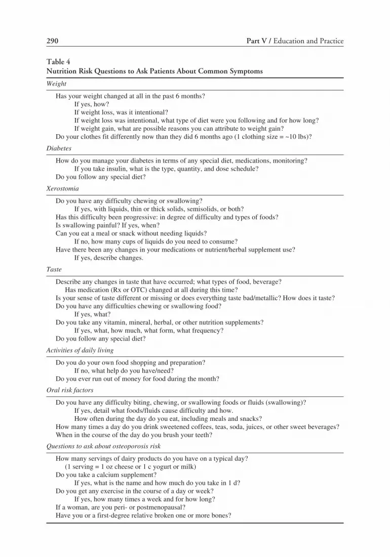
290 Part V / Education and Practice
Table 4Nutrition Risk Questions to Ask Patients About Common Symptoms
Weight
Has your weight changed at all in the past 6 months?If yes, how?If weight loss, was it intentional?If weight loss was intentional, what type of diet were you following and for how long?If weight gain, what are possible reasons you can attribute to weight gain?
Do your clothes fit differently now than they did 6 months ago (1 clothing size = ~10 lbs)?
Diabetes
How do you manage your diabetes in terms of any special diet, medications, monitoring?If you take insulin, what is the type, quantity, and dose schedule?
Do you follow any special diet?
Xerostomia
Do you have any difficulty chewing or swallowing?If yes, with liquids, thin or thick solids, semisolids, or both?
Has this difficulty been progressive: in degree of difficulty and types of foods?Is swallowing painful? If yes, when?Can you eat a meal or snack without needing liquids?
If no, how many cups of liquids do you need to consume?Have there been any changes in your medications or nutrient/herbal supplement use?
If yes, describe changes.
Taste
Describe any changes in taste that have occurred; what types of food, beverage?Has medication (Rx or OTC) changed at all during this time?
Is your sense of taste different or missing or does everything taste bad/metallic? How does it taste?Do you have any difficulties chewing or swallowing food?
If yes, what?Do you take any vitamin, mineral, herbal, or other nutrition supplements?
If yes, what, how much, what form, what frequency?Do you follow any special diet?
Activities of daily living
Do you do your own food shopping and preparation?If no, what help do you have/need?
Do you ever run out of money for food during the month?
Oral risk factors
Do you have any difficulty biting, chewing, or swallowing foods or fluids (swallowing)?If yes, detail what foods/fluids cause difficulty and how.How often during the day do you eat, including meals and snacks?
How many times a day do you drink sweetened coffees, teas, soda, juices, or other sweet beverages?When in the course of the day do you brush your teeth?
Questions to ask about osteoporosis risk
How many servings of dairy products do you have on a typical day?(1 serving = 1 oz cheese or 1 c yogurt or milk)
Do you take a calcium supplement?If yes, what is the name and how much do you take in 1 d?
Do you get any exercise in the course of a day or week?If yes, how many times a week and for how long?
If a woman, are you peri- or postmenopausal?Have you or a first-degree relative broken one or more bones?
17_Touger_285_298_F 7/8/04, 10:45 AM290

Chapter 17 / Oral Nutrition Health Risk Assessment 291
2.1. HistoryPatient history is an important component of nutrition evaluation. Medical history will
reveal information about acute, chronic, and terminal diseases that may impact on oraland nutritional well-being. There are more than 100 systemic diseases with oral manifes-tations (7). In addition to asking patients about their medical, surgical, and drug history,diet and nutrition risk evaluation questions may also be asked. It is likely that the dentaland dietetics professional will take a complete patient history as part of routine care.Supplemental questions relative to oral health and nutrition are in Table 4.
Unintentional weight change may signal potential nutrition deficits, lack of money forfood, or systemic disease. A weight change of more than 10 lbs in less than 6 mo isconsidered a significant risk factor in any individual (8). Weight loss is characterized byloss of body-fat stores and lean body mass. Patients should be weighed during initial visitsand subsequent check-ups. The initial-visit weight provides a baseline for comparison forfuture reference and self-reported weight. Reliance on self-reported weights does notprovide the professional with a baseline from which to monitor weight status. Weightchange typically is associated with either a change in eating habits or systemic disease.Eating habit changes may be intentional with a goal of weight loss or gain or caused byoral or systemic health issues influencing appetite or functional ability to eat. In eithercase, the result is a change in nutrient intake.
There are several web-based programs available to calculate weight change and bodymass index. Table 5 includes some easy-to-use formulas for calculating weight statistics.Some provide access to computerized nutrient analysis programs, including the USDepartment of Agriculture’s Interactive Healthy Eating Index (9).
Table 5Formulas for Weight and Body Mass Index and Other Anthropometric Calculations
Wt change is calculated based on usual body wt:
today's wtusual wt
× 100 = % wt change Ex:today's wt = 50 lbsusual wt = 100 lbs
× 100 = % wt change
Current (today’s) wt < usual wt = negative wt change Current wt > usual wt = positive weight change
Ranges of desirable height and weight for adults: rule of thumb
Men = 106 lbs for first 5 ft + 6 lbs/inEx: 5'6" man = 106 + (6 inches × 6 lbs/inch) = 106 + 36 = 142 lbs ± 10% desirable wt
Women = 100 lbs for first 5 ft + 5 lbs/inEx: 5'2" woman = 100 lbs + (2 inches × 5 lbs/inch) = 100 lbs + 10 = 110 lbs ± 10%
Note: ±10% = based on frame size; weight range may be 10% greater or less than the calculated wt
Body mass index
Body wt in kilograms (kg)
(ht in meters)2
or Wt (in lbs) × 703 divided by (height in inches)2
If patient does not know his or usual weight, desirable weight can be used to calculate % desirableweight. This will also give an indication of body stores. Weight alone does not differentiate betweenbody fat vs muscle mass.
17_Touger_285_298_F 7/8/04, 10:45 AM291

292 Part V / Education and Practice
2.1.1. MEDICAL HISTORY
Medical history will reveal individual information about acute or chronic diseases orimmunologic disorders that are risk factors for individuals with concurrent oral or dentalproblems that affect their ability to consume their usual diet. Acute or chronic diseasesor immunologic disorders are risk factors in patients with concurrent oral and dentalproblems that affect their ability to consume their usual diet. The 65-yr-old woman whosechief complaint is, “I can’t chew my food,” is at risk if she has type 2 diabetes mellitus.Diabetes is associated with oral manifestations, particularly when blood sugars areelevated and other disease sequelae are evident. These manifestations, including peri-odontal disease, dysgeusia, increased caries risk, candidiasis, burning tongue, and poorwound healing, particularly when blood sugars are elevated, may in turn impact appetite,eating ability, and, finally, oral intake (10). Neuropathies and opportunistic microbialinfections in the oral cavity affect oral health, nutrition status, and inevitably diabetescontrol. Complications including burning tongue, gingivitis, and periodontal diseases,along with xerostomia, are common in individuals with diabetes.
Autoimmune diseases such as pemphigus vulgaris increase nutrition risk by virtue ofthe oral and facial sequelae of the disease and the medications used to manage the disease(2,3). Inflammatory arthritides have associated medication side effects, and joint pain ormechanical limitation may compromise eating ability. Steriod medications used to man-age these diseases increase risk of diabetes and nitrogen (protein) and calcium losses, thusincreasing protein and calcium needs. The xerostomia associated with Sjögren’s syn-drome increases risk for dental caries, periodontitis, and oral mucosal injury or pain,which may make eating difficult or painful (2). Temporomandibular joint pain may resultin limited opening of the mouth and compromised masticatory ability.
Head and neck and oral cancers affect nutrition and oral health status. Surgery toremove tumors in the oral cavity may have severe functional effects on eating ability.Radiation to the oral cavity can destroy taste as well as the quality and quantity of saliva.Chemotherapy can cause anorexia, stomatitis, nausea, and vomiting, ultimately compro-mising nutrition status.
Individuals with HIV infection or acquired immunodeficiency syndrome (AIDS) areat increased risk of oral infections and disease manifestations that alter functional andsensory functions of the oral cavity (11). Oral complications and malnutrition may occursecondary to the disease process and associated gastrointestinal, metabolic, immune,pharmacological, and psychosocial sequellae. Altered micronutrient metabolism maycontribute to oral manifestations and subsequent malnutrition, further compromisingoral integrity and the ability to combat infections. Nutrition screening by the dentalprofessional with a particular focus on the integrity of the oral cavity and medications andtheir combined impact on oral function is important. A referral to an RD for MNT isroutine in this population.
2.1.2. MEDICATIONS AND OTHER OVER-THE-COUNTER PRODUCTS
Adults in the United States are using increasing amounts of over-the-counter (OTC)and prescription medications (12). Drug use tends to be greatest in the elderly (12).Medications used to treat the oral and systemic manifestations of HIV infection, cancer,autoimmune diseases, and cardiovascular diseases, as well as local oral diseases, mayalso affect a person’s ability to ingest, digest, and absorb an adequate diet. Common
17_Touger_285_298_F 7/8/04, 10:45 AM292

Chapter 17 / Oral Nutrition Health Risk Assessment 293
consequences of several classes of medications—including antiretroviral, antiviral, an-tifungal, antiparasitic, antihypertensive, antidepressant, antihistamine, narcotic, seda-tive, and antineoplastic agents—include xerostomia, stomatitis, reduced salivary flow,altered taste, and/or oral ulcers. All of these factors impact food choices, quality andquantity of foods consumed, and the local effect of foods on the oral cavity, leading tocompromised nutrient intake (13). Patients should be carefully questioned about use ofprescription and OTC drugs, as well as use of herbs, minerals, vitamins, and other dietarysupplements. It is up to the provider to review the potential drug–drug, drug–nutrient,drug–symptom, and drug–supplement interactions with patients and guide them appro-priately.
2.1.3. DIETARY SUPPLEMENTS
Chapter 6 in this text is dedicated to this topic. Select dietary supplements may alterthe integrity of the oral cavity, interfere with action of prescription or OTC medications,and/or alter response to a select therapy. Supplements impacting the immune system suchas echinacea may interfere with the actions of immunosuppressing medications second-ary to its immune enhancing effects. Other supplements such as ma huang, or ephedra,may have systemic side effects. The principal alkaloids of ephedra are ephedrine andpseudoephedrine. When epinephrine- and ephedrine-containing supplements are takensimultaneously, the resultant stimulatory effects, including increased blood pressure andheart rate and central nervous system stimulation, may be additive (P. Gregory, personalcommunication, April 2002). The impact of the combination is difficult to estimatebecause it depends on how much of each agent is taken and the actual ephedrine contentof the ephedra-containing supplement consumed. According to the Natural MedicinesComprehensive Database, ephedrine has a short half-life (2.5–3.6 h); it would there-fore be prudent to recommend that consumption of ephedrine-containing supplementsbe withheld for 24 h prior to a dental procedure in which a local anesthetic with epineph-rine would be administered.
Part of a routine history and even follow-up appointments should include questionsabout use of dietary supplements, form, dosage, and frequency. The Physician’s DeskReference for Herbal Supplements (14), the Natural Medicines Database (15), or theCommission E Monographs (16) can be used to evaluate potential symptoms and sideeffects of these dietary supplements.
2.1.4. ORAL EXAM FINDINGS
Although the oral exam is a routine component of the initial workup in the dentaloffice, the diet and nutrition implications are often not considered. Whereas dental pro-fessionals may do a comprehensive oral exam, their role is to detect nonnormal findingsor symptoms relative to diet and nutrition, provide basic education, and refer patients tothe RD. Dietetics professionals can readily add these parameters to their initial physicalexam with the caveat that their role is to detect non-normal findings and refer patients tothe dentist. The RD can provide baseline guidance relative to oral health and nutrition.Table 6 provides guidelines for dietetics professionals conducting a nutrition-focusedoral examination.
Patients with biting, chewing, or swallowing difficulties may be at risk depending onthe presence of other risk factors and length and severity of the problem. Simple guide-lines on modification of food form and consistency may be adequate for individuals
17_Touger_285_298_F 7/8/04, 10:45 AM293

294 Part V / Education and Practice
without other nutrition risk factors. Denture wearers or individuals who will be gettingdentures for the first time need education on diet modification during the initial fitting andadjustment phases. However, if other risk factors are present, individualized counselingmay be indicated.
Individuals with moderate to extensive caries (based on American Dental Associationcriteria) are at risk and should have their treatment planned for diet and nutrition evalu-ation and counseling. Individuals with soft-tissue lesions, oral pathologies, orofacial painor disorders that interfere with the ability to eat, or exam findings suggestive of nutritiondeficits should be referred to the RD for MNT.
2.1.5. EATING DISORDERS
The dental professional has a distinct role in detecting eating disorders, includinganorexia and bulemia, given some of the changes that are evident on head, neck, and oral
Table 6Functional Oral Nutrition Risk Evaluation
Structure Patient-focused exam Management
Lips • Dryness; sensation; cracking or • Alter diet texture and consistencyfissuring, swelling; history of blistersor ulcers
Gingiva and • Soreness/pain; bleeding spontan- • Alter diet texture, temperature,oral mucosa eously, change in appearance; and consistency
swelling, growths, discharge; • Screen for oral cancer, nutrientbad taste; halitosis deficiencies
• Red or white patches/lesions• Erosion/ulceration; focal pigmen-
tation; erythema
Teeth • Toothache/pain; looseness and • Adjust diet, consistency; evaluatemobility; dental prosthesis caries risk; consider altered taste/(removable or fixed); edentulism smell
Tongue • Soreness/pain; burning; rough • Screen for systemic disease,patches; dryness; cracking or fissur- nutrient deficiencies; alter dieting; growths; change in taste; ulcers texture
Temporomandibular • Difficulty or painful opening; • Change diet consistency, foodgrinding sounds on joint opening/ “hardness”; limit “chewy” foodschewing with limited range or pain;weakness of chewing muscles
Salivary glands • Mucosal dryness; too little or too • Increase fluids; evaluate formuch saliva; drooling; change in dysgeusia, dysphagia; limitcolor, consistency, difficulty swal- spices, “hard” foods; reviewlowing dry food, altered taste, dry changes in Rx; evaluate zinc
statuseyes; gland pain or swelling
Neck • Tender/swollen lymph nodes, other • Medical consultswellings
Skin • Change in appearance; rashes, sores, • Medical consultlumps; itching
Note: For each section, ask about patient complaints, duration of symptom(s), and any changes in appearance,size, acuity, frequency, and pain.
17_Touger_285_298_F 7/8/04, 10:45 AM294

Chapter 17 / Oral Nutrition Health Risk Assessment 295
exams. Bitemporal wasting, thinning hair, and boney prominences are symptomatic ofanorexia nervosa. Swollen salivary glands, redness in the back of the throat, lingualerosion, and symptoms of frequent regurgitation, sometimes tinged with blood, are com-mon in bulemia. Individuals with eating disorders are at oral and nutrition risk. Suchpatients should be referred to the RD for evaluation and possible referral to an eatingdisorder program. Diet evaluation for cariogenic risk and nutritional adequacy and indi-vidualized counseling focused on oral health should be completed. Referrals for compre-hensive medical care should be provided.
Questions regarding self-imposed diet modifications, “fad” diets, provide insight intoabnormal eating patterns that may precipitate nutrient deficits. Any questions on appro-priateness or potential risk of the diet should be referred to the RD. Patient questioningshould refer to the types, quantity, and duration of supplement use and rationale for use.
2.1.6. OSTEOPOROSIS
Osteoporosis is increasingly common in women (one in two women) and men (one ineight men) (17). The relationship between osteoporosis and oral health is increasinglyevident (18,19), particularly in reference to periodontal disease and implant surgery.Chapter 15 provides a detailed insight into oral and nutrition health and osteoporosis. Thedental and dietetics professionals should be familiar with high-risk individuals and ques-tion them about any history of osteoporosis and whether they have undergone a screeningor diagnostic evaluation. Risk factors for osteoporosis (17) include the following:
• long-term (more than 1 mo) steroid use;• hyperparathyroidism, hyperthyroidism, end-stage renal disease;• history of organ transplants treated with cyclosporine and steroids;• spinal cord injury;• women who are perimenopausal or of small stature;• family history of osteoporosis;• prolonged low calcium intake combined with inactivity; and• prolonged lack of exposure to sunlight.
2.1.7. DIETARY HABITS
Questions regarding previous and current diet habits should focus on dietary pat-terns, nutritional adequacy, and factors influencing eating and fluid intake. Table 4 (20)provides suggested questions on factors influencing xerostomia, masticatory function,occlusion, and other risk factors. Responses to these questions will help the dental anddietetics professional identify the impact of oral disease and symptoms on ability toconsume foods and fluids. It is assumed that the RD will approach this section of patientassessment in a more detailed manner than the dental professional and provide moretargeted nutrition and diet recommendations. However, it is incumbent on the dentalprofessional to ask questions regarding oral function and diet as well as specific ques-tions about diet intake. The following paragraph provides a strategy that is time sensi-tive and perhaps more reasonable for the dental office setting. It is the strategy used inseveral dental schools as part of routine patient care.
Dietary habit assessment should include general questions about food habits (see theAppendix for nutrition risk evaluation and diet evaluation forms for use in dental prac-tices). The dietary habit assessment will provide insight into caries risk of the diet as well
17_Touger_285_298_F 7/8/04, 10:45 AM295

296 Part V / Education and Practice
as specific questions about typical daily diet routines. A 24-h recall, or recall of a typicalday’s diet, will provide a “snapshot” of the patient’s diet pattern and a global assessmentof nutrient intake. Simply asking a patient to recall everything he or she had to eat anddrink on a previous or typical day and recording approximate times, specific foods, andportion sizes is all that is needed. This intake can then be analyzed in one of two ways.For a general assessment of dietary adequacy, the foods and fluids can be quantified intowhere they fit in the food guide pyramid. Once that is done, one has a general picture ofthe individual’s ability to meet the food group recommendations for the food guidepyramid and any missing or excessive intakes by food group. For a more specific assess-ment of nutrient adequacy, the diet may be analyzed using one of many computerizednutrient analysis packages. The Interactive Healthy Eating Index (9), available free on theinternet, may be used. This provides an assessment of actual calories and macro- andmicronutrient intake according to the individual’s gender, age, and body size.
Although a thorough diet and nutrition history and analysis are routine for the dieteticsprofessional as a component of MNT, questions regarding oral dysfunction and a clinicalexam of the oral cavity are not necessarily routine practice. The RD is encouraged tointegrate the components of oral nutrition risk assessment in the tables included in thischapter, along with a brief intra- and extraoral examination (1), into nutrition assessmentprotocols.
2.1.8. FINAL STEPS
Once the health professional has completed the assessment of risk factors, the deter-mination of nutrition and oral health risk and needs for intervention must occur. In respectto the dental professional, this may include a brief discussion with the patient aboutrecommendations to improve diet and suggestions for diet modification based on iden-tified oral dysfunction or disease, as well as referral to an RD for MNT. In contrast, thedietetics professional will be able to provide comprehensive nutrition care and a dietmodified to meet oral disease or dysfunction, along with a referral to a dental professionalfor dental care.
3. CONCLUSIONS
There are no hard and fast rules to determine nutrition risk levels in dental practice.Nutrition risk is more accurately based on the type and extent of risk factors and theirimpact on oral function. Examples of conditions or diseases associated with nutrition riskare in each of the chapters of this text.
REFERENCES
1. American Dietetic Association. Oral health and nutrition: position of the American Dietetic Association.J Amer Dietet Assoc 2003; 103:615–625.
2. DePaola DP, Mobley C, Touger-Decker R. Nutrition and oral medicine. In Handbook of Nutrition andFood. New York, NY: CRC Press, 2001, 1113–1134.
3. National Institute of Dental and Craniofacial Research. Spectrum Series. The Oral-Systemic HealthConnection. May 1999. Website: (http://www.nidcr.nih.gov/spectrum/NIDCR2/2menu.htm). AccessedApril 19, 2002.
4. Papas AS, Palmer CA, Rounds MC, Russell RM. The effects of denture status on nutrition. Special Carein Dentistry 1998; 18:17–25.
17_Touger_285_298_F 7/8/04, 10:45 AM296

Chapter 17 / Oral Nutrition Health Risk Assessment 297
5. Ritchie CS, Joshipura K, Silliman RA, Miller B, Douglas CW. Oral health problems and significantweight loss among community-dwelling older adults. J Gerontol Series A Biol Sci Med Sci 2000;55:M366–371.
6. H.R.5661 Medicare, Medicaid, and SCHIP Benefits Improvement and Protection Act of 2000. Sec. 105.Coverage of Medical Nutrition Therapy Services for Beneficiaries with Diabetes or a Renal Disease.Website: (http://thomas.loc.gov/cgi-bin/query/D?c106:1:./temp/~c106Wgp2IQ:e29796). AccessedAugust 21, 2002.
7. American Academy of Family Practice. Nutrition Screening Initiative. Website: (http://www.aafp.org/nsi/). Accessed April 19, 2002.
8. Saltzman E, Mogensen K. Physical assessment. In Coulston AM, Rock CL, Monsen ER eds. Nutritionin the Prevention and Treatment of Disease. New York, NY: Academic Press, 2001, 43–55.
9. Anonymous. Interactive Healthy Eating Index. Website: (http://147.208.9.133/Default.asp). AccessedApril 19, 2002.
10. Slavkin HC, Baum BJ. Relationship of dental and oral pathology to systemic illness. JAMA 2000; 284:1215–1217.
11. Sirois DA. Oral manifestations of HIV disease. Mt Sinai J Med 1998; 65:322–332.12. Kaufman D, Kelly J, Rosenberg L, et al. Recent patterns of medication use in the ambulatory adult
population of the United States. JAMA 2002; 287(3):337-344.13. Position of the American Dietetic Association and the Canadian Dietetic Association: nutrition interven-
tion in the care of persons with human immunodeficiency virus infection. J Am Diet Assoc 2000; 100:708–717.
14. Greenwald J, Brendler T, Jaenicke C, eds. PDR for Herbal Medicines, 1st ed. Montvale, NJ: MedicalEconomics, 1998.
15. Jellin JM, ed. Natural Medicines Comprehensive Database. Stockton, CA: Therapeutic Research Fac-ulty, 2002.
16. American Botanical Council. The Complete German Commission E Monographs: Therapeutic Guideto Herbal Medicines. Newton, MA: Integrative Medicine Communications, 1998.
17. National Osteoporosis Foundation. Disease Statistics: Fast Facts. Website: (http://www.nof.org/osteoporosis/stats.htm). Accessed July 2, 2002.
18. Krall EA, Wehler C, Garcia RI, Harris SS, Dawson-Hughes B. Calcium and vitamin D supplementsreduce tooth loss in the elderly. Am J Med 2001; 111:452–456.
19. Wactawski-Wende J. Periodontal diseases and osteoporosis: association and mechanisms. AnnPeriodontol 2001; 6:197–208.
20. Touger-Decker R. New Jersey Dental School Oral Medicine Clinic Nutrition Manual. 7th Ed. Newark,NJ: New Jersey Dental School, unpublished manual, 2002.
17_Touger_285_298_F 7/8/04, 10:45 AM297

17_Touger_285_298_F 7/8/04, 10:45 AM298

Chapter 18 / Nutrition Education 299
299
From: Nutrition and Oral MedicineEdited by: R. Touger-Decker, D. A. Sirois, and C. C. Mobley © Humana Press Inc., Totowa, NJ
18 Oral Medicineand Nutrition Education
Riva Touger-Decker and David A. Sirois
1. INTRODUCTION
In order to ensure the integration of nutrition and oral health education (and viceversa), it is essential that a body of knowledge for both dietetics and dentistry be delin-eated (1). A clear understanding of the synergy between oral health and nutrition shouldbe promoted in all health professions’ education programs. Nutrition education as acomponent of pre- and postdoctoral dental education and continuing dental education isimportant in order to translate theory into practice. Likewise, oral health and diseaseeducation as a component of preprofessional and graduate dietetic education and continu-ing professional education is necessary. This chapter addresses both dental and dieteticsprofessional education and consumer education. Consumer education is critical to ensurethe integration of oral and diet/nutrition health behaviors into lifestyles.
2. NUTRITION AND ORAL HEALTHEDUCATION OF HEALTH PROFESSIONALS
In the practice setting, collaborative efforts between dental and dietetics professionalsto promote interdisciplinary health care teams will foster successful strategies related tooral health and nutrition. Table 1 (1) represents a paradigm for collaboration and referralbetween dietetics and dental professionals to address oral health and nutrition issues inpractice.
To achieve this paradigm, it is essential to foster core knowledge development startingon the preprofessional level. Basic levels of care skills, including risk identification andreferral for intervention, are becoming essential for health professionals (2) as clientsseek comprehensive health care. The Institute of Medicine (IOM) study supports com-prehensive training of dental professionals to ensure that they can “assess and treat thewhole patient, not just the mouth” (2). The need for preventive health, including institu-tion of improved feeding practices (2), is clearly supported in this report. The 1998 PewHealth Professions Commission Report recommends that relationships between dentistsand allied health professionals be developed and expanded and that interdisciplinarycompetence be required of all health professionals (3).
18_Touger_299_306_F 7/8/04, 10:45 AM299

300 Part V / Education and Practice
Table 1Dietetics and Dental Professional Role Modeling to Achieve Effective Integration of Oral Healthand Nutrition Service in Health Promotion and Disease Prevention and Intervention
1. Dietetics professional
Clinical Setting
• Conduct intra-/extraoral screening and cranial nerve exams.• Integrate oral health screening as a component of nutrition assessment protocols (i.e., cranial nerve function,
occlusion, edentulism, masticatory ability, swallowing, salivary adequacy).• Recognize oral manifestations of systemic diseases and provide patients with guidelines to maximize oral
intake.• Confer with and refer patients (via consults) to dentists for management of oral symptoms of diseases and/
or risk factors for oral diseases.• Consult with dental professionals in interpretation of oral nutrition assessment findings and planning in the
long-term care setting.
Community Setting
• Establish partnerships with dental professionals in community and private practice settings.• Develop and implement collaborative oral health and nutrition screening/education programs in schools,
worksites, and health maintenance organizations.• Promote collaborative education on nutrition and oral health among dietetics and dental professionals.• Develop nutrition education messages that encourage oral health.• Promote oral health in school and community nutrition programs.
Research Setting
• Promote collaborative nutrition and oral health research initiatives.• Design and conduct nutrition/diet components of oral health research initiatives.• Identify and support integration of oral health issues (e.g., screening, disease, management, education) as a
component of nutrition research.
2. Dental professional
Clinical Setting
• Include diet screening, education, and referral for oral infectious disease prevention/control, optimalmasticatory function, and management of other oral diseases/treatments as a component of comprehensivedental care.
• Collaborate with dietetics professionals in delivery of MNT and provision of oral health care in long-term caresettings.
• Provide diet and nutrition guidelines for health promotion and disease prevention to patients and provideguidelines for diet to maximize oral intake.
• Consult with and refer patients (via consult) to dietetics professionals for management of nutrition risk causedby compromised oral health (e.g., caries, immunosuppressive disorders, xerostomia, diabetes, oral surgery,cancer).
Community Setting
• Establish partnerships with dietetics professionals in community and private practice settings to promotenutrition/diet screening and education in dental practice.
• Develop and implement collaborative oral health and nutrition screening/education initiatives in schools,worksites, and health care organizations.
• Promote collaborative education on nutrition and oral health among dietetics and dental professionals.• Develop oral health messages that integrate nutrition and diet education.• Promote diet and nutrition as a component of school and community oral health programs.
Research Setting
• Promote collaborative oral health and nutrition research initiatives.• Design and conduct oral health component of nutrition /diet research initiatives.• Identify and support integration of nutrition topics as a component of oral health research as appropriate.
Adapted from ref. 1.
18_Touger_299_306_F 7/8/04, 10:45 AM300

Chapter 18 / Nutrition Education 301
2.1. Nutrition in Dental Education
Dental accreditation standards and the 2000 Competencies for the New Dentist (4,5)do not specify predoctoral nutrition education competencies. Although the 1990 stan-dards for nutrition in dental education specifically addressed knowledge of basic nutri-tion, the role of diet and nutrients in health and oral diseases, and nutrition counseling asit relates to oral health, the 1998 standards focus on broad-based competency statements.Nutrition as a component of oral health education is not explicitly stated; it is implied,however, throughout several areas of the competencies and standards. The biomedicalsciences standards do not address specific sciences; rather, they must “ensure an in-depthunderstanding of basic biological principles, consisting of a core of information on fun-damental structures, functions and interrelationships of the body systems” (5). Similarly,“in-depth information” must be provided to develop understanding of oral health, oraldisease, oral epidemiology, and the role of diet and nutrition in the etiology, diagnosis,prevention, and treatment of oral disease. Knowledge of principles of nutrition and dietand their clinical application in practice is implied throughout the document (5) becauseit provides the underpinning for achievement of several of the competencies. The accredi-tation standards for the majority of advanced specialty postdoctoral education programsvary in the specificity with which they address nutrition. The specialty postdoctoralprogram in periodontics does address knowledge of “principles of nutrition, especiallyas they relate to patient evaluation, disease processes, and wound healing” (6). Otherpostdoctoral programs include competencies focusing on management of medicallycompromised patients, those with chronic and terminal diseases, and those who haveundergone surgical interventions. Implied in these required competencies is knowledgeof diet and nutrition as they relate to comprehensive dental management (7).
In the last decade of the 20th century, several national surveys documented that cur-riculum hours devoted to nutrition are low compared to time dedicated to other curricu-lum areas (8–12). These surveys and more recent papers (1) have, as the core of theirrecommendations, the integration of nutrition in the dental curriculum (10–12) through-out the 4 yr of didactic and clinical coursework.
Development of a core curriculum on nutrition and oral health for medical, dental,and allied health education has been suggested (13), along with the need to survey thestate of the art of nutrition in dental education and vice versa (13). Similarly, neither thestandards nor the competencies state the need for demonstration of clinical competencein dental school; however, the need for this is clear in order to translate knowledge intopractice. These recommendations are supported by the original Healthy People (HP)2000 (14), the more recent HP 2010 (15), and the Competencies for Health Care Prac-titioners for 2005 (3). They highlight the need for expanded access to care by healthproviders in coordinated teams. Consistent with professional needs and federal andprivate association recommendations, suggested nutrition curriculum topics in under-graduate dental education include diet and nutrition screening, intervention, and refer-ral; formulation of diet orders; and education strategies (1–13,15,16) (see Table 2).Historically, the focus of nutrition education in dental schools has been on oral infectiousdisease management. However, consistent with the rest of health care, the focus hasshifted to health promotion, disease prevention, and intervention in oral and systemicdiseases (4,10–12). There is sufficient evidence to support the need for competency in
18_Touger_299_306_F 7/8/04, 10:45 AM301

302 Part V / Education and Practice
Table 2Didactic and Practice Components of a Curriculum Model for Dietetics and Dental EducationPrograms to Promote Collaboration and Multiskilling in Nutrition and Oral Health
Dietetics education (ADA competencies)
1. Baccalaureate Programa. Didactic topics• Oral anatomy and physiology• Oral manifestations of acute, chronic, terminal, and systemic diseases• Oral sequellae of medications, chemo-, and radiation therapies• Primary diseases of the oral cavity and their effects on taste, smell, and masticationb. Clinical Experiences• Field visits to dental schools/clinics• Work with dental students in clinical/community settings• Oral health screening questions as a component of nutrition assessment activities
2. Dietetic Internship/Coordinated Program Competencies• Conduct nutrition screening and diet counseling in dental school and community dental clinic rotations• Integrate oral health screening questions in diet/nutrition assessment activities• Integrate appropriate oral health guidelines into the conduct of diet counseling and education• Participate in oral health and nutrition research• Perform basic physical assessment including oral and cranial nerve screening exams• Design nutrition care plans for patients with compromised oral health
3. Graduate Education• Design oral health and nutrition research• Perform oral physical assessment exams including intra-/extraoral screening and cranial nerve
examinations
4. Continuing Professional Education• Collaboration between dietetics and dental professionals in case presentations, multidisciplinary care
meetings, conferences about diseases and the lifespan, multidisciplinary seminars• Training opportunities using different media; for example, distance learning, CD-ROMs, videotapes
Dental education
1. Doctoral Programa. Didactic topics• Nutritional biochemistry• Nutrition and oral health throughout the life span• Diet education and intervention relative to oral health/disease• Effect(s) of oral disease(s), symptomatology and their treatment(s) on diet and nutrition status• Diet/nutrition screening, education, and referral in dental practice• Diet/nutrition concerns and management strategies of high-risk patientsb. Clinical experiences• Self-evaluation of diet• Training techniques for diet evaluation and education• Nutrition risk screening and diet education relative to oral health of patients• Consultation and supervised practice with registered dietitians and/or dietetic technicians
2. Graduate Programs• Oral health and nutrition research• Integration of nutrition screening and diet education into dental and oral disease specialties• Collaborative education endeavors on related topics with dietetics programs
3. Continuing Professional Education• Collaboration between dietetics and dental professionals in case presentations, multidisciplinary care
meetings, conferences about diseases and the life span, multidisciplinary seminars• Multiskilling training opportunities using different media; for example, distance learning, CD-ROMs,
videotapes
From ref. 1.
18_Touger_299_306_F 7/8/04, 10:45 AM302

Chapter 18 / Nutrition Education 303
basic nutrition care by dentists (1,15). Dental visits provide a unique opportunity fordental professionals to integrate lifestyle management (diet and smoking cessation) intotreatment and long-term care plans.
Nutrition is clearly delineated in the Accreditation Standards for Dental HygieneEducation Programs (17). The biomedical science component of the curriculum mustinclude nutrition. Clinical instruction competencies indirectly address competency in theability to evaluate an individual’s diet and provide diet education relative to oral health.In the practice setting, it may be the dental hygienist who completes the patient healthassessment and any diet or nutrition evaluation, subsequently providing oral hygiene andhealth promotion instruction including diet.
2.2. Oral Health in Dietetics EducationIn a similar venue to dental education and nutrition, oral health education is not out-
lined as a specific competency or criterion in the standards of education for dieteticseducation (18). However, the standards do require that entry-level dietetic educationprograms with a nutrition therapy emphasis include competency in the performance ofbasic physical assessment. This should include novice-level competency in the perfor-mance of a head, neck, and oral screening exam. Because a healthy, functional oral cavityis a necessary part of mastication and digestion, oral health concepts should be incorpo-rated into didactic and clinical training in baccalaureate, preprofessional, and graduatelevels of dietetics education (Table 2) (19).
The clinical training of dietetics professionals during dietetic internship or coordinatedprogram clinical experiences should include opportunities for clinical experiences in den-tal school clinic or dental office settings (1). Students in these programs should be givenopportunities to work side by side with dental students or residents in the clinical settingto provide nutrition and diet intervention as a component of oral health management.Competencies in oral screening (head and neck exams, intra- and extraoral assessment, andcranial nerve exams) should be taught to students in the preprofessional setting (19).
At the graduate and professional level, academic and continuing education programsshould include research and applications as they relate to medical and nutrition manage-ment of orally compromised patients. The conduct of oral assessments and identificationof dental risk should be included (3,19).
The educational outcomes of such training include competencies in the identificationof nonnormal conditions of the oral cavity; oral dysfunction factors affecting biting,chewing, drinking, and swallowing; and potential nutrient deficiencies. As individualsprogress from entry-level to beyond entry-level and advanced-level training, so will theircompetency in the performance of these outcome competencies. The value of thesecompetencies in nutrition assessment, monitoring, and evaluation across the life span inhealth and disease will significantly impact development of nutrition care plans and dietprotocols.
3. A MODEL FOR COLLABORATIONIN NUTRITION AND ORAL HEALTH EDUCATION
The continual shifting of the social and economic realities of today’s health caresystem have had a dramatic effect on the preparation and training of health professionals,including dietetics professionals, dentists, and allied dental personnel. The American
18_Touger_299_306_F 7/8/04, 10:45 AM303

304 Part V / Education and Practice
Dietetics Association position on nutrition education of health professionals advocates“that nutrition education is an essential component of the curricula for the majority ofhealth care professionals. Curricula should include nutrition principles and identificationof nutrition risk factors” (15).
The need for the recognition of nutrition as an essential part of training for dentalprofessionals and as an important component of educational programs for dietetic andother health professionals (20) is clearly delineated in the joint World Health Organiza-tion (WHO)/Food and Agricultural Organization expert consultants’ recommendationthat encouraged this joint approach by international organizations including the WHOand the Food and Agricultural Organization (20). In order to be successful, educators andleaders in oral health and nutrition must promote this dual-content area in the curriculumsof other allied health professions. Dental and dietetics professionals need to form net-works with other health professionals (e.g., physicians, physician assistants, speech andlanguage pathologists, nurses) to advance health promotion and preventive and commu-nity health initiatives that promote oral health and nutrition as they relate to generalhealth. These new models of health care provider cross-training and collaboration willrequire innovative approaches to new models of health care delivery if training is tosuccessfully carry over to practice. Recommendation 5 of the IOM study states: “Toprepare future practitioners for more medically based modes of oral health care and moremedically complicated patients, dental educators should work with their colleagues inmedical schools and academic health centers” (2).
4. SUMMARY AND NEXT STEPS
This chapter has provided a historical perspective on nutrition and dental educationalong with 21st-century initiatives and recommendations for nutrition education of den-tal professionals and oral health education of dietetics professionals (Tables 1 and 2). Acollaborative effort of members of both disciplines including academicians, clinicians,and administrators is needed to actualize the recommendations into practice. The prac-tical model envisioned as an outcome of the recommendations within this chapter in-cludes teams of registered dietitian nutrition experts in dental schools and dental facultyin dietetics programs, with both disciplines represented on accreditation boards for di-etetics and dentistry. Professional initiatives and federal grants to build such curriculummodels and a forum for creating and sharing model curricula would provide the infra-structure to grow and develop these models. Opportunities for collaboration across dis-ciplines exist.
REFERENCES1. American Dietetic Association. Position of the American Dietetic Association: Oral health and nutrition.
J Am Diet Assoc 2003; 103:615–625.2. Institute of Medicine. Dental education at the crossroads: summary. J Dent Educ 1995; 59(1):7–15.3. Pew Health Professions Commission. Recreating Health Professional Practice for a New Century:
The Fourth Report of the Pew Health Professions Commission. Center for the Health Professions,December 1998.
4. Commission on Dental Accreditation. Accreditation Standards for Dental Education Programs. Chi-cago: American Dental Association, 1998.
5. American Dental Education Association. The 2000 competencies for the new dentist. Website:(www.adea.org). Accessed January 5, 2004.
18_Touger_299_306_F 7/8/04, 10:45 AM304

Chapter 18 / Nutrition Education 305
6. Commission on Dental Education. Accreditation Standards for Advanced Specialty Education Pro-grams in Periodontics. American Dental Association, 2001.
7. Commission on Dental Accreditation. Accreditation Standards for Advanced Education Programs inGeneral Practice Residency. Chicago, IL: American Dental Association, 1998.
8. DePaola DP, Jacobs JHH, Slim LH. Nutrition education in United States and Canadian schools ofdentistry. J Am Diet Assoc 1982; 81:580–583.
9. Faine MP. Nutrition in the dental curriculum: Seattle model. J Dent Educ 1990; 54:510–512.10. Palmer CA. Applied nutrition dental education: issues and challenges. J Dent Educ 1990; 54:513–518.11. Touger-Decker R, Gilbride JA. Nutrition education of dental students and professionals. Top Clin Nutr
1997; 12(3)23–32.12. DePaola D. Nutrition and oral infectious disease workshop recommendations. Boston, MA: The Forsyth
Institute, 2000. Website: (www.forsyth.org/nutrition/recommendations.htm). Accessed on May 10,2002.
13. National Center for Health Statistics. Healthy People 2000 Final Review. Hyattsville, MD: PublicHealth Service, 2000.
14. U.S. Department of Health and Human Services. Healthy People 2010. 2nd Ed. With Understanding andImproving Health and Objectives for Improving Health. 2 vols. Washington, DC: U.S. GovernmentPrinting Office, 2000.
15. Commission on Dental Education. Accreditation Standards for Dental Hygiene Education Programs.Chicago: American Dental Association, 2000.
16. Position of the American Dietetic Association: nutrition education of health professionals. J Am DietAssoc 1998; 98:343–346.
17. Unpublished draft report of the Joint WHO/FAO expert consultation: Diet, nutrition and the preventionof chronic diseases. April 2002.
18. American Dietetic Association. Knowledge, skills and competencies for entry level dietitian educationprograms. In Commission on Accreditation for Dietetics Education Accreditation Handbook. Chicago,IL: The American Dietetic Association, 2002. Website: (http://www.eatright.org/Member/Files/CADEHandbook.pdf). Accessed January 5, 2004.
19. Mackle T, Touger-Decker R, O’Sullivan Maillet J, Holland B. Registered dietitians’ use of physicalassessment parameters in practice. J Am Diet Assoc 2003; 103(12); 1632–1638.
20. Unpublished draft report of the Joint WHO/FAO expert consultation: Diet, nutrition and the preventionof chronic diseases. April 2002.
18_Touger_299_306_F 7/8/04, 10:45 AM305

18_Touger_299_306_F 7/8/04, 10:45 AM306

Appendix A / Oral Nutrition Risk Assessment Tools 307
Appendix AOral Nutrition Risk Assessment Tools
Append_A_307_310_F 7/8/04, 10:45 AM307

308 Appendix A / Oral Nutrition Risk Assessment Tools
Append_A_307_310_F 7/8/04, 10:45 AM308

Appendix A / Oral Nutrition Risk Assessment Tools 309
Append_A_307_310_F 7/8/04, 10:45 AM309

310 Appendix A / Oral Nutrition Risk Assessment Tools
Append_A_307_310_F 7/8/04, 10:45 AM310

Appendix B / Body Mass Index Table 311
Appendix BBody Mass Index Table
Append_B_311_312_F 7/8/04, 10:45 AM311

From National Institutes of Health, National Heart, Lung and Blood Institute, Website: http://www.nhlbi.nih.gov/guidelines/obesity/bmi_tbl.htm
312
Append_B
_311_312_F7/8/04, 10:45 A
M312

Appendix C / Dietary Reference Intake Charts 313
Appendix CDietary Reference Intake Charts
Append_C_313_352_F 7/8/04, 9:59 AM313

314A
ppendix C / D
ietary Reference Intake C
harts
314
Append_C
_313_352_F7/8/04, 9:59 A
M314

Appendix C
/ Dietary R
eference Intake Charts
315
315
Append_C
_313_352_F7/8/04, 9:59 A
M315

316A
ppendix C / D
ietary Reference Intake C
harts
316
Append_C
_313_352_F7/8/04, 9:59 A
M316

Appendix C
/ Dietary R
eference Intake Charts
317
317
Append_C
_313_352_F7/8/04, 9:59 A
M317

318A
ppendix C / D
ietary Reference Intake C
harts
318
Append_C
_313_352_F7/8/04, 9:59 A
M318

Appendix C
/ Dietary R
eference Intake Charts
319
319
Append_C
_313_352_F7/8/04, 9:59 A
M319

320A
ppendix C / D
ietary Reference Intake C
harts
320
Append_C
_313_352_F7/8/04, 9:59 A
M320

Appendix C
/ Dietary R
eference Intake Charts
321
321
Append_C
_313_352_F7/8/04, 9:59 A
M321

322A
ppendix C / D
ietary Reference Intake C
harts
322
Append_C
_313_352_F7/8/04, 9:59 A
M322

Appendix C
/ Dietary R
eference Intake Charts
323
323
Append_C
_313_352_F7/8/04, 9:59 A
M323

324A
ppendix C / D
ietary Reference Intake C
harts
324
Append_C
_313_352_F7/8/04, 9:59 A
M324

Appendix C
/ Dietary R
eference Intake Charts
325
325
Append_C
_313_352_F7/8/04, 9:59 A
M325

326A
ppendix C / D
ietary Reference Intake C
harts
326
Append_C
_313_352_F7/8/04, 9:59 A
M326

Appendix C
/ Dietary R
eference Intake Charts
327
327
Append_C
_313_352_F7/8/04, 9:59 A
M327

328A
ppendix C / D
ietary Reference Intake C
harts
328
Append_C
_313_352_F7/8/04, 9:59 A
M328

Appendix C
/ Dietary R
eference Intake Charts
329
329
Append_C
_313_352_F7/8/04, 9:59 A
M329

330A
ppendix C / D
ietary Reference Intake C
harts
330
Append_C
_313_352_F7/8/04, 9:59 A
M330

Appendix C
/ Dietary R
eference Intake Charts
331
331
Append_C
_313_352_F7/8/04, 9:59 A
M331

332A
ppendix C / D
ietary Reference Intake C
harts
332
Append_C
_313_352_F7/8/04, 9:59 A
M332

Appendix C
/ Dietary R
eference Intake Charts
333
333
Append_C
_313_352_F7/8/04, 9:59 A
M333

334A
ppendix C / D
ietary Reference Intake C
harts
334
Append_C
_313_352_F7/8/04, 9:59 A
M334

Appendix C
/ Dietary R
eference Intake Charts
335
335
Append_C
_313_352_F7/8/04, 9:59 A
M335

336A
ppendix C / D
ietary Reference Intake C
harts
336
Append_C
_313_352_F7/8/04, 9:59 A
M336

Appendix C
/ Dietary R
eference Intake Charts
337
337
Append_C
_313_352_F7/8/04, 9:59 A
M337

338A
ppendix C / D
ietary Reference Intake C
harts
338
Append_C
_313_352_F7/8/04, 9:59 A
M338

Appendix C
/ Dietary R
eference Intake Charts
339
339
Append_C
_313_352_F7/8/04, 9:59 A
M339

340A
ppendix C / D
ietary Reference Intake C
harts
340
Append_C
_313_352_F7/8/04, 9:59 A
M340

Appendix C
/ Dietary R
eference Intake Charts
341
341
Append_C
_313_352_F7/8/04, 9:59 A
M341

342A
ppendix C / D
ietary Reference Intake C
harts
342
Append_C
_313_352_F7/8/04, 9:59 A
M342

Appendix C
/ Dietary R
eference Intake Charts
343
343
Append_C
_313_352_F7/8/04, 9:59 A
M343

344A
ppendix C / D
ietary Reference Intake C
harts
344
Append_C
_313_352_F7/8/04, 9:59 A
M344

Appendix C
/ Dietary R
eference Intake Charts
345
345
Continued on next page
Append_C
_313_352_F7/8/04, 9:59 A
M345
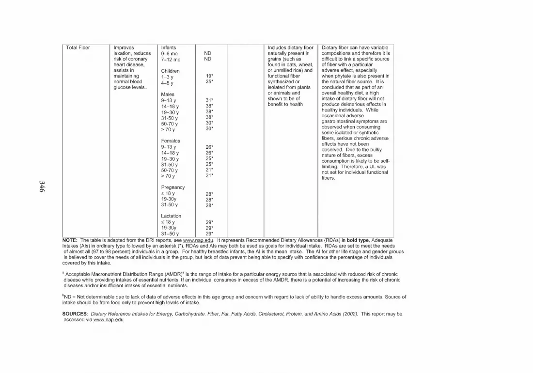
346A
ppendix C / D
ietary Reference Intake C
harts
346
Append_C
_313_352_F7/8/04, 9:59 A
M346

Appendix C
/ Dietary R
eference Intake Charts
347
347
Continued on next page
Append_C
_313_352_F7/8/04, 9:59 A
M347

348A
ppendix C / D
ietary Reference Intake C
harts
348
Append_C
_313_352_F7/8/04, 9:59 A
M348

Appendix C
/ Dietary R
eference Intake Charts
349
349
Continued on next page
Append_C
_313_352_F7/8/04, 9:59 A
M349

350A
ppendix C / D
ietary Reference Intake C
harts
350
Append_C
_313_352_F7/8/04, 9:59 A
M350

Appendix C
/ Dietary R
eference Intake Charts
351
351
Append_C
_313_352_F7/8/04, 9:59 A
M351

352A
ppendix C / D
ietary Reference Intake C
harts
352
Append_C
_313_352_F7/8/04, 9:59 A
M352

Appendix D / Cranial Nerve Assessment 353
Appendix DCranial Nerve Assessment
Append_D_353_356_F 7/8/04, 10:46 AM353

354A
ppendix D / C
ranial Nerve A
ssessment
354
Cranial Nerve Assessment
Cranial Nerve Function Examination Presentation
I. Olfactory S Sensation to nasal mucosa. Smell. S Test presence and ability to distin- S Limitation in ability to smell.guish scents using fragrant foods,flavor extracts, or spices.
II. Optic S Visual acuity. Visual field. Convey S Stand behind patient with arms S Limitation in ability to see.visual information from the retina. extended outward. Bring arms
together and have patient identifywhen they can see your hands.
III. Oculomotor M Motor function to eye, pupillary M Test movement of eye with “H” M Limitation in ability to move eyeconstriction to light. or “box” test. Test pupillary light downward, upward or toward nose
reaction with penlight. (adduction), unable to adapt to lightTest III, IV, VI together. and dark. Ptosis (drooping eyelid).
IV. Trochlear M Motor function to superior oblique Test III, IV, VI together. M Limitation in ability to move eyemuscle. downward and inward. Double vision.
V. Trigeminal a S Sensation to dura, skin of forehead, S Test face along the mandible, S Decreased facial or oral sensation.scalp, roof of nasal cavity, nose, maxilla, and forehead with sharp • Droolingskin around the eye, cheek, lip, (pinprick), dull (tongue depressor), • Pocketing of foodteeth, hard palate, ear, chin, lining and light touch (cotton swab). M Weak jaw closing, asymmetricof cheek and gums. M Have patient open and close the opening, muscle atrophy.
M Mastication. Motor function to jaw. Then have patient resist • Easily fatiguestemporalis, masseter, medial and attempts of examiner to close jaw. • Food leakagelateral pterygoid, mylohyloid.
VI. Abducens M Motor function to lateral rectus Test III, IV, VI together. M Limitation in ability to follow objectmuscle (for precise movement of with eye, or move away from nosethe eye for visual tracking or fixa- (abduction).tion on an object).
Append_D
_353_356_F7/8/04, 10:46 A
M354

Appendix D
/ Cranial N
erve Assessm
ent355
355
VII. Facial a S Taste anterior 2/3 of tongue, palate. S Test taste sensation of sweet, salt, S Altered taste.M Facial expressions, muscles of lips and sour. M Facial asymmetry, unequal nasolabial
and cheeks. M Have patient smile to show teeth, folds, poor lip seal.A Secretion of lacrimal, sublingual, raise eyebrows, close eyelids • Drooling
and submandibular glands. tightly. • Pocketing of foodA Decreased saliva.
• Increased oral preparatory stage.
VIII. Vestibulocochlear S Sensation from cochlea for hearing S Rub hair or fingers near patient’s S Deafness, vertigo. (Acoustic) and from semicircular canals and ears.
vestibule for equilibrium.
IX. Glossopharyngeal a S Taste posterior 1/3 of tongue; sensa- S Test taste sensation of bitter. S Altered taste.tion to pharynx, tonsils, soft palate. Gag reflex: Touch posterior No pharyngeal elevation.
M Elevation of soft palate; swallowing. pharynx with a cotton swab. • Increased risk of aspiration.A Secretion of parotid gland. Look for pharyngeal elevation M Limited palate elevation.
and tongue retraction. • PocketingM Have patient say “AH.” Look for • Prolonged eating time
bilateral soft palate elevation and A Decreased saliva.midline uvula.
X. Vagus a S Taste base of tongue and epiglottis. Test IX and X together. (See above.) (See above.)Sensation to soft palate, upper larynx.
M Movement of soft palate, pharynx,and larynx.
XI. Spinal Accessory M Motor function to vagus, sternoma- M Test neck range of motion. Have M Limitation in ability to raise shouldersstoid, and trapezius muscles. patient lift shoulders with and with- or turn head, muscle atrophy and
out pressure. Have patient turn head weakness.with and without pressure.
XII. Hypoglossal a M Movement of tongue. M Protrude tongue. Should be midline. M Limitation in tongue movement orTest tongue strength with tongue deviation.blade.
S Sensor response; M motor response; A autonomic response; a CN involved in dysphagia screening.From Norman, W. Cranial Nerves. Website: (http://mywebpages.comcast.net/wnor/cranialnerves.htm). Accessed October 24, 2003.
Append_D
_353_356_F7/8/04, 10:46 A
M355

Append_D_353_356_F 7/8/04, 10:46 AM356

Appendix E / Diet Education Tools 357
Appendix EDiet Education Tools
The authors share the following diet education materials that may bereproduced for patient education purposes only without permission.However, we ask that the appropriate references citation to the origina-tors of the materials be listed. These materials may not be reproduced tobe sold or used in any publication without written permission.
Append_E_357_378_F 7/8/04, 10:46 AM357

358 Appendix E / Diet Education Tools
Append_E_357_378_F 7/8/04, 10:46 AM358

Appendix E / Diet Education Tools 359
Continued on next page
Append_E_357_378_F 7/8/04, 10:46 AM359

360 Appendix E / Diet Education Tools
Append_E_357_378_F 7/8/04, 10:46 AM360

Appendix E / Diet Education Tools 361
Append_E_357_378_F 7/8/04, 10:46 AM361

362 Appendix E / Diet Education Tools
Append_E_357_378_F 7/8/04, 10:46 AM362

Appendix E / Diet Education Tools 363
Continued on next page
Append_E_357_378_F 7/8/04, 10:46 AM363

364 Appendix E / Diet Education Tools
Append_E_357_378_F 7/8/04, 10:46 AM364

Appendix E
/ Diet E
ducation Tools
365
365
Continued on next page
Append_E
_357_378_F7/8/04, 10:46 A
M365

366A
ppendix E / D
iet Education T
ools
366
Continued on next page
Append_E
_357_378_F7/8/04, 10:46 A
M366

Appendix E
/ Diet E
ducation Tools
367
367
Continued on next page
Append_E
_357_378_F7/8/04, 10:46 A
M367

368A
ppendix E / D
iet Education T
ools
368
Append_E
_357_378_F7/8/04, 10:46 A
M368

Appendix E / Diet Education Tools 369
Continued on next page
Append_E_357_378_F 7/8/04, 10:46 AM369

370 Appendix E / Diet Education Tools
Append_E_357_378_F 7/8/04, 10:46 AM370

Appendix E / Diet Education Tools 371
Continued on next page
Append_E_357_378_F 7/8/04, 10:46 AM371

372 Appendix E / Diet Education Tools
Append_E_357_378_F 7/8/04, 10:46 AM372

Appendix E / Diet Education Tools 373
Append_E_357_378_F 7/8/04, 10:46 AM373

374 Appendix E / Diet Education Tools
Append_E_357_378_F 7/8/04, 10:46 AM374

Appendix E / Diet Education Tools 375
Append_E_357_378_F 7/8/04, 10:46 AM375

376 Appendix E / Diet Education Tools
Append_E_357_378_F 7/8/04, 10:46 AM376

Appendix E / Diet Education Tools 377
Append_E_357_378_F 7/8/04, 10:46 AM377

Append_E_357_378_F 7/8/04, 10:46 AM378

Index 379
379
Index
A
Abducens nerveassessment, 354
Acarbose, 196tAcculturation, 46Acoustic nerve
assessment, 355Acquired immunodeficiency syndrome (AIDS),
223–224, 292Activities of daily living
questions concerning, 290tAcupuncture, 149–150Acute necrotizing ulcerative gingivitis, 275Advanced glycation end products (AGE), 194African-American influences
dietary patterns, 51–52AGE, 194Aged
drugs, 88, 292–293frail, 34–35
AIDS, 223–224, 292Alcohol, 92Algorithm, 97Aloe vera, 153t
-Linolenic aciddietary reference intake, 349
-Tocopheroldietary reference intake, 326
Alternative medical systems, 147–151Alveolar bone
heightedentate subjects, 264–265
loss, 129–130diabetes mellitus, 279osteoporosis, 263
American Heart Associationdietary guidelines, 10t
Amino acidsdietary reference intake, 351
Anemia, 8. See also Pernicious anemiairon-deficiency, 20nutritional, 8
Aneurindietary reference intake, 320
Angular cheilitis, 115f, 230tHIV, 232
Anorexia nervosa, 295Antioxidant nutrients
periodontal disease, 120Antioxidants, 175–176Aphthous ulcers
HIV, 234–235Arsenic
dietary reference intake, 328Arthritides
inflammatory, 292Arthritis
rheumatoid, 254–256Ascorbic acid
dietary reference intake, 324Asian influences
dietary patterns, 53–55Atherosclerosis
periodontal disease impact on, 70Autoimmune disease, 241–258
medicationsadverse effects, 244t–245t
non-organ specific, 252–257organ-specific, 242–252target antigens, 242t
Azathioprine, 244t
B
Bacteroides forsythus, 172Benign mucous membrane pemphigoid, 250–252
-Carotenewound healing, 278
Bilberry, 153tBiofeedback, 151Biologically based treatments, 151–152Biotin
dietary reference intake, 314Biting difficulties
dietary guidelines, 38tdiet education tools, 376
Blenderized diet, 38Body-based methods, 152–158Body mass index
formulas, 291t
Touger_Decker_Index_Final 07/27/2004, 1:54 PM379

380 Index
table, 312web-based calculations, 291
Bone loss, 261–262alveolar, 129–130
diabetes mellitus, 279osteoporosis, 263
Bone marrow transplantationimpairing wound healing, 279
Bone mineral density, 261–262Borondietary reference intake, 329Botanicals, 152, 153t–155tBread
oral and pharyngeal cancer, 211Breast milk
fluoride, 20Buddhism, 47Bulimia, 295
C
Calciferoldietary reference intake, 325
Calcium, 156tdietary
periodontal disease, 119–120dietary reference intake, 266t, 330diet education tools, 359–361
Calendula, 153tCAM. See Complementary and alternative medi-
cine (CAM)Cancer, 7. See also Oral and pharyngeal cancer
gay, 224HIV, 235oral, 113–117
chemoprevention, 215–216squamous cell, 279
lips, 279Cancrum oris, 113Candida albicans, 189Candida pseudohyphae, 189Candidiasis
erythematous, 230tHIV, 231
HIV, 229–232hyperplastic, 230tHIV, 231–232oral
diabetes mellitus, 189treatment of, 190t, 250t
pseudomembranous, 230tCarbohydrates
counting, 199dietary reference intake, 345
intakediabetes mellitus, 198
Cardiovascular disease, 7risk, 5
Caries, 72–73, 109–111, 167–168, 294diabetes mellitus, 188–189early childhood, 22enamel hypoplasia, 109risk, 38–39vaccines, 169
Caroteneoral and pharyngeal cancer, 212
Cayenne, 153tCentral American influences
dietary patterns, 52–53Cereals
oral and pharyngeal cancer, 211Cheilitis
angular, 115f, 230tHIV, 232
Chemotherapyimpairing wound healing, 279
Chewing difficultiesdietary guidelines, 38tdiet education tools, 376
Children. See also Early childhood; Infantsoral health nutrition messages, 25tsalivary secretions, 112school-age
food choices, 23oral health, 22–24
special needs, 24–27young
recommended dietary guidelines for oralhealth promotion, 23t
Chiropractic, 152–158Chlorpropamide, 196tCholesterol
dietarydaily recommendations, 12t
dietary reference intake, 350Choline
dietary reference intake, 315Christianity, 46–47Chromium
dietary reference intake, 331Chronic disease
cost, 9Chronic orofacial pain, 74, 134Chronic periodontitis
diabetes mellitus, 67–68Cleft lip/palate, 24–26Clinical pathways
Touger_Decker_Index_Final 07/27/2004, 1:54 PM380

Index 381
recommended for selected disorders, 64tClotrimazole
for angular cheilitis, 190tfor oral candidiasis, 190t
CMV, 226tHIV, 227
Cobalamindietary reference intake, 323
Coenzyme Q10, 156tCollaboration model
nutrition and oral health education, 303–304Commission E Monographs, 293Complementary and alternative medicine (CAM),
143–162defined, 143–144dental-related, 147–158efficacy, 145legal issues, 161medication interactions, 158practitioners
evaluating, 160–161quality, 160safety, 145types, 144tusage trends, 144–145working with patients using, 158–159
Complete denturesguidelines for eating well with, 34t
Compromised nutritional and oral health statusconditions associated with, 288t
Compromised nutritional well-beingoral consequences of, 107–123
Connective tissue healingphases, 274
Convenience food, 56Copper
dietary reference intake, 332Core curriculum, 301Cost
chronic disease, 9Cranial nerve assessment, 354–355Culture, 46Cyclophosphamide, 245tCyclosporine, 245tCysteine
dietary reference intake, 351Cytomegalovirus (CMV), 226t
HIV, 227
D
Daily food pattern guides, 6tDairy food
oral and pharyngeal cancer, 211–212
Dapsone, 245tDASH food guide, 6tDehydroascorbic acid (DHA)
dietary reference intake, 324Dental education
curriculum model, 302tnutrition, 301–303
Dental professionalsdetermining nutrition risk, 289–296diabetes screening, 194integrating oral health and nutrition service in
health promotion, 300toral nutrition management, 288t
Dentate statusdiet and nutrition, 31–37
Dentists. See Dental professionalsDentures, 36, 294
completeguidelines for eating well with, 34t
diet education tools, 362, 365–368foods difficult to chew, 33tnutrition-related implications, 35t
DHAdietary reference intake, 324
Diabetes-associated gingivitis, 66Diabetes mellitus, 185–200, 292
alveolar bone loss, 279clinical classification, 186–188clinical resources, 199co-morbidities, 193–194diet education tools, 362–364, 369–372dysesthesia, 191–192etiology, 186–188exchange list, 199medical nutrition therapy, 196–197, 197tmedication management, 194–200noncandidal-associated oral lesions, 189–191nutrition goals, 198–199oral complications, 188–192pathogenesis, 186–188questions concerning, 290tscreening
dentist, 194surveillance program, 186ttreatment of, 192–193, 192twebsites, 204wound healing, 279
Diabetic diet, 199–200Diet
blenderized, 38deficiency, 7defined, 3diabetic, 199–200
Touger_Decker_Index_Final 07/27/2004, 1:54 PM381

382 Index
education programcurriculum model, 302toral health, 303
education tools, 358–377biting difficulties, 376
guidelines, 9–13agency and organization, 10tAmerican Heart Association, 10tbiting difficulties, 38tdefined, 4
habitsassessment, 295–296
liquid, 38micronutrients
oral and pharyngeal cancer, 212–213oral health care intervention, 37–39patterns
ethnic and regional influences, 48–55quality, 33–36
defined, 3Dietary calcium
periodontal disease, 119–120Dietary cholesterol
daily recommendations, 12tDietary fiber
daily recommendations, 12t@IE:>Dietary guidelineschewing difficulties, 38t
Dietary lawsKosher, 46
Dietary reference intake, 13-linolenic acid, 349-tocopherol, 326
amino acids, 351aneurin, 320arsenic, 328ascorbic acid, 324biotin, 314calciferol, 325calcium, 266t, 330charts, 314cholesterol, 350choline, 315chromium, 331cobalamin, 323copper, 332cysteine, 351DHA, 324elements, 328–345fat, 347fiber, 346fluoride, 333
folacin, 316folate, 316folic acid, 316histidine, 351iodine, 334iron, 335isoleucine, 351leucine, 351linoleic acid, 348lysine, 351macronutrients, 345–352magnesium, 336manganese, 337methionine, 351molybdenum, 338niacin, 317nickel, 339nicotinic acid, 317pantothenic acid, 318valine, 351vanadium, 343vitamin A, 321vitamin B2, 319vitamin B6, 322vitamin B12, 320, 323vitamin C, 324vitamin D, 266t, 325vitamin E, 326vitamin K, 327vitamins, 314–327zinc, 344
Dietary Supplement Health and Education Act of1994, 146–147
Dietary supplements, 151–152, 293Dietary Supplement Verification Program
(DSVP), 160Diet education tools
calcium, 359–361chewing difficulties, 376dentures, 362, 365–368diabetes mellitus, 362–364, 369–372post surgical, 373snacks, 377sore mouth, 374taste, 375xerostomia, 358
Dietetics professionalsintegrating oral health and nutrition service
in health promotion, 300tpractice guidelines, 288t
Digestion, 36–37Disease
abnormal oral findings, 136t
Touger_Decker_Index_Final 07/27/2004, 1:54 PM382

Index 383
Drugs, 292–293affecting oral health and nutritional intake,
97–101, 98tassociated with esophageal damage, 101tfood intake, 94nutrient absorption and metabolism, 88–94nutritional status, 87–88resulting in nutrient deficits, 90t–91t
DSVP, 160Dysesthesia
diabetes mellitus, 191–192Dysphagia, 39, 74–79, 100
causes, 74tcomplications, 75diagnosis of, 77diet management, 77–79nutritional implications, 75screening, 75–77, 76ttreatment of, 77
E
Early childhoodcaries, 22feeding practices, 20–22
promoting oral health, 22Eating disorders, 294–295EBV, 226t
HIV, 227–228Echinacea, 153t, 293Edentate subjects
alveolar bone height, 264–265Edentulous, 35Education
dentalcurriculum model, 302tnutrition, 301–303
dietcurriculum model, 302toral health, 303
diet tools, 358–377biting difficulties, 376
oral medicine and nutrition, 299–304Elderly
drugs, 88, 292–293frail, 34–35
Elementsdietary reference intake, 328–345
Enameldevelopmental defects, 107–109hypoplasia, 18linear hypoplasia, 108
Energy intakediabetes mellitus, 198
Ephedra, 153tEphedrine, 293Epstein-Barr virus (EBV), 226t
HIV, 227–228Erythematous candidiasis, 230t
HIV, 231Ethnic groups
food selections and habits, 49t–50tEuropean influences
dietary patterns, 48–50Exchange list
diabetes, 199
F
Facial nerveassessment, 355
Fatdietary reference intake, 347
Fat-soluble vitamins, 93Fatty acids
omega-3, 156tpolyunsaturated
dietary reference intake, 348saturated
dietary reference intake, 350trans
dietary reference intake, 350Feverfew, 154tFiber
dietarydaily recommendations, 12t
dietary reference intake, 346oral and pharyngeal cancer, 212
Floor of mouthloss, 133
Fluconazolefor oral candidiasis, 190t
Fluoride, 19–20breast milk, 20dietary reference intake, 333
Fluorosis, 167Folacin
dietary reference intake, 316Folate
dietary reference intake, 316Folic acid
dietary reference intake, 316dietary source, 8t
Foodaltering drug absorption, 92tconvenience, 56culture, 45–46daily pattern guides, 6t
Touger_Decker_Index_Final 07/27/2004, 1:54 PM383

384 Index
dairyoral and pharyngeal cancer, 211–212
DASH guide, 6thabits, 45–46pattern guidelines, 11
daily, 6tFood–drug interactions, 94–97Food guide pyramid, 6t
diabetes, 199Frail elders, 34–35Fruit
oral and pharyngeal cancer, 208, 209t–210toral cancer, 116–117
Functional teeth units, 32–33
G
Garlic, 154tGastroesophageal reflux disease (GERD), 37Gay cancer, 224Gay-related immune deficiency (GRID), 224Gene expression, 174–175GERD, 37Ginger, 154tGingiva
examination, 294tGingival hyperplasia, 100–101Gingivitis, 276–277
acute necrotizing ulcerative, 275diabetes-associated, 66menstrual cycle-associated, 65pregnancy-associated, 65puberty-associated, 66scorbutic, 277
Ginkgo biloba, 154tGinseng, 154tGlibizide, 196tGlimedpride, 196tGlossitis, 99–100Glossopharyngeal nerve
assessment, 355Glutamine
wound healing, 278Glyburide, 196tGlycemic index, 199Glycosylated hemoglobin
mean plasma glucose, 195tGlycosylated hemoglobin test (HbA1C), 194Goldenseal, 154tGrains
oral and pharyngeal cancer, 211Grapefruit juice, 96Green tea, 154tGRID, 224
Guarana, 155tGuidelines. See also Practice guidelines
diet, 9–13agency and organization, 10tAmerican Heart Association, 10tbiting difficulties, 38tchewing difficulties, 38tdefined, 4infants, 23t
H
HbA1C, 194HDL, 5Health professionals
oral medicine and nutrition education, 299–303Health status, 3–13Height
formulas, 291tHerpes simplex virus (HSV), 226t
HIV, 225–227HHV8, 226t
HIV, 228High blood pressure, 5–7High-density lipoprotein (HDL), 5Hinduism, 47Histidine
dietary reference intake, 351Histoplasmosis, 230t
HIV, 232HIV. See Human immunodeficiency virusHomeopathy, 147–148HPV, 226t
HIV, 229HSV, 226t
HIV, 225–227Human herpes virus 8 (HHV8), 226t
HIV, 228Human immunodeficiency virus (HIV), 223–236,
292angular cheilitis, 232aphthous ulcers, 234–235cancer, 235EBV, 227–228erythematous candidiasis, 231histoplasmosis, 232hyperplastic candidiasis, 231–232linear gingival erythema, 233necrotizing stomatitis, 234oral lesions, 225–235
bacterial, 232–234fungal, 229–232, 230tviral, 225–229, 226t
oral manifestationsnutritional management, 235–236
Touger_Decker_Index_Final 07/27/2004, 1:54 PM384

Index 385
Human papilloma virus (HPV), 226tHIV, 229
Hydroxychloroquine, 245tHypercholesterolemia, 5Hyperglycemia
type 2 diabetes mellitus, 187Hyperlipidemia, 5Hyperplastic candidiasis, 230t
HIV, 231–232Hypertension, 5–7
defined, 5Hypertriglyceridemia, 5Hypnosis, 150–151Hypoglossal nerve
assessment, 355Hypoglycemic agents, 196t
I
Immune response, 173–174Immunocompromised, 9Immunosuppressive agents
impairing wound healing, 279Infants
feeding practices, 20–22recommended dietary guidelines for oral
health promotion, 23tInflammatory arthritides, 292Insulin
preparations, 196tresistance, 5
Interdisciplinary research, 177–178Intermaxillary fixation, 38Internet influences
dietary patterns, 56Iodine, 20
dietary reference intake, 334Iron, 20
dietary reference intake, 335dietary source, 8t
Iron-deficiency anemia, 20Islamism, 47Isoleucine
dietary reference intake, 351Itraconazole
for oral candidiasis, 190t
J
Jawloss, 133
Judaism, 46
K
Kaposi’s sarcoma, 224Kava, 155t
Ketoconazolefor angular cheilitis, 190tfor oral candidiasis, 190t
Kosher dietary laws, 46
L
LDL, 5LEH, 108Leucine
dietary reference intake, 351Linear enamel hypoplasia (LEH), 108Linear gingival erythema
HIV, 233Linoleic acid
dietary reference intake, 348Linolenic acid
dietary reference intake, 349Lips
examination, 294tsquamous cell carcinoma, 279
Liquid diet, 38Low birth weight, 18Low-density lipoprotein (LDL), 5Lysine, 156t
dietary reference intake, 351
M
Macronutrientsdietary reference intake, 345–352
Magnesiumdietary reference intake, 336
Malignancy. See CancerMalnutrition
defined, 4salivary secretions, 112
Manganesedietary reference intake, 337
Manipulative methods, 152–158Maslow’s hierarchy of needs, 45Massage, 157–158Masticatory ability, 32Masticatory apparatusphysical loss, 129–133Maté, 155tMaternal micronutrient malnutrition, 18–19Maternal protein energy malnutrition, 18Maxillomandibular fixation, 37–39Meat
oral and pharyngeal cancer, 211Medical history, 292–293Medications. See DrugsMeditation, 150
mindfulness, 150
Touger_Decker_Index_Final 07/27/2004, 1:54 PM385

386 Index
Menstrual cycle-associated gingivitis, 65Mentally handicapped, 27Metformin, 196tMethionine
dietary reference intake, 351Methotrexate, 245tMexican influences
dietary patterns, 52–53Mexican sanguinaria, 155tMiconazole
for angular cheilitis, 190tMicrobiota
plaque, 172–173Micronutrient malnutrition
maternal, 18–19Micronutrients
dietoral and pharyngeal cancer, 212–213
oral and pharyngeal cancer, 212–215serum
oral and pharyngeal cancer, 214–215Miglitol, 196tMind–body interventions, 150–151Mindfulness meditation, 150Mineral deficiency
clinical manifestations, 95toral cancer, 115–116oral signs of, 114tsalivary secretions, 112
Minerals, 92, 93Molybdenum
dietary reference intake, 338, 340Mormons, 47Mucosal pain, 135Mucositis
oral, 278Muslims, 47Mycophenolate mofetil, 244tMyelomeningocele, 26
N
Nateglinide, 196tNational Cancer Institute
dietary guidelines, 10tNational Center for Complementary and Alterna-
tive Medicine (NCCAM), 143National Dysphagia Diet, 78, 78tNatural medicine, 148–149Natural Medicines Database, 293NCCAM, 143Neck
examination, 294tNecrotizing stomatitis
HIV, 234
Neoplasms. See CancerNeural tube defects, 26Niacin
dietary reference intake, 317Nickel
dietary reference intake, 339Nicotine stomatitis, 275, 279Nicotinic acid
dietary reference intake, 317Noma, 113Noncandidal-associated oral lesions
diabetes mellitus, 189–191Nonsteroidal anti-inflammatory drugs (NSAID)
impairing wound healing, 279NSAID
impairing wound healing, 279NUG
HIV, 233–234NUP
HIV, 233–234Nutrient–gene interactions
transcription-factor pathways, 175tNutrient intake
recommendations, 11, 12tNutrition. See also Oral nutrition
defined, 3oral health care intervention, 37–39perinatal
tooth development, 17–20prenatal
tooth development, 17–20risk assessment
outcomes, 289risk questions
about common symptoms, 290tNutritional anemia, 8Nutritional status, 33–36
defined, 4Nutritional supplements, 152, 156tNutritional well-being
compromisedconditions associated with, 288toral consequences of, 107–123
Nutrition professionalspractice guidelines
child nutrition, 27diabetes, 200HIV, 236medications, 102oral and pharyngeal cancer, 217–218osteoporosis, 268pregnancy, 27
Nystatinfor oral candidiasis, 190t
Touger_Decker_Index_Final 07/27/2004, 1:54 PM386

Index 387
O
Obesity, 4–5, 173defined, 4type 2 diabetes mellitus, 187
Oculomotor nerveassessment, 354
Odds ratio, 206Odontogenic pain, 133–134Older adults
drugs, 88, 292–293frail, 34–35
Olfactory nerveassessment, 354
Omega-3 fatty acids, 156tOptic nerve
assessment, 354Oral and pharyngeal cancer, 205–218
nutritional epidemiology, 205–207resources, 216studies, 207–215
Oral burningdiabetes mellitus, 191–192
Oral cancer, 113–117chemoprevention, 215–216
Oral candidiasisdiabetes mellitus, 189treatment of, 190t, 250t
Oral cavity injury, 274–275Oral disease
nutritional consequences, 129–139Oral exam, 293–294Oral function
nutrition, 177Oral hard tissues, 167–169Oral health
age-related changes, 31–40bidirectional impact, 63–79nutrition messages for children, 25t
Oral health professionalspractice guidelines, 13, 27
child nutrition, 27diabetes, 200HIV, 236medications, 102oral and pharyngeal cancer, 217osteoporosis, 268pregnancy, 27
Oral hypoglycemic agents, 196tOral lesions
noncandidal-associateddiabetes mellitus, 189–191
precancerous, 215Oral movement disorders, 137
Oral mucosadisease, 73, 113–117examination, 294t
Oral mucositis, 278Oral nutrition
health risk assessment, 287–296management
approaches to, 288trisk assessment tools, 308–310risk evaluation, 294trisk factors, 289t
questions concerning, 290tOral pain, 133–134Oral soft tissues, 169–176Oral surgery, 37–39Oral tissue repair, 275–276Osteopathy, 157Osteopenia
oral cavity, 263–266Osteoporosis, 8, 261–268, 295
alveolar bone loss, 263calcium, 265epidemiology of, 262medications, 265nutritional approaches, 265oral cavity, 263–266professional and practice issues, 266–267questions concerning, 290tresources, 267–268risk factors, 262trisk questionnaires, 268treatment
effects on oral cavity, 265vitamin D, 265websites, 268
Overnutritiondefined, 4
Over-the-counter products, 292–293
P
Painchronic orofacial, 74, 134mucosal, 135odontogenic, 133–134oral, 133–134
Pantothenic aciddietary reference intake, 318
Parkinson’s disease, 137Partially dentate subjects
alveolar bone height, 264–265Patient history, 291–292PEM. See Protein energy malnutrition (PEM)Pemphigus vulgaris, 247–250, 292
Touger_Decker_Index_Final 07/27/2004, 1:54 PM387

388 Index
Perinatal nutritiontooth development, 17–20
Periodontal disease, 63–72, 117–121, 120antioxidant nutrients, 120clinical measures, 263–264diabetes mellitus, 191HIV, 232–233impact on cardiovascular disease, 68–70
clinical studies, 70impact on pregnancy and preterm birth, 71–72osteoporosis, 263relation to systemic disease, 170–172treatment of, 276–277
Periodontal tissues, 169–170Periodontitis
chronicdiabetes mellitus, 67–68
Permanent teethmineralization, 19
Pernicious anemia, 243–247clinical features, 246diagnosis of, 246nutrition complications, 246–247pathogenesis, 243–246treatment of, 246
Pharyngeal cancer. See Oral and pharyngealcancer
Phenylalaninedietary reference intake, 351
Physical activityinadequate, 4
Physically handicapped, 27Physician’s Desk Reference for Herbal Supple-
ments, 293Phytotherapy, 152Pioglitazone, 196tPlaque, 120–121
control, 276inducing gingival disease, 65microbiota, 172–173
Polyphenols, 175–176, 176Polyunsaturated fatty acids
dietary reference intake, 348, 349Practice guidelines
dietetics professionals, 288tnutrition professionals
child nutrition, 27diabetes, 200HIV, 236medications, 102oral and pharyngeal cancer, 217–218osteoporosis, 268pregnancy, 27
oral health professionals, 13, 27child nutrition, 27diabetes, 200HIV, 236medications, 102oral and pharyngeal cancer, 217osteoporosis, 268pregnancy, 27
Precancerous oral lesions, 215Prednisone, 244tPregnancy
daily vitamin and mineral recommendations,21t
gingivitis, 65Prenatal nutrition
tooth development, 17–20Preterm delivery, 18Progressive muscle relaxation, 151Protein
deficiencysalivary secretions, 111–112
dietary reference intake, 351intake
diabetes mellitus, 198Protein-calorie malnutrition, 19
wound healing, 277Protein energy malnutrition (PEM), 7
dental caries, 109–110enamel developmental defects, 108maternal, 18
Pseudomembranous candidiasis, 230tHIV, 231
Pteroylpolyglutamatesdietary reference intake, 316
Puberty-associated gingivitis, 66
R
Radiationimpairing wound healing, 279
Reactive oxygen species (ROS), 176Registered Dietitian
diabetes screening, 193–194Relative risk, 206Relaxation response, 150Religious practices
dietary patterns, 46–47Remineralization, 168–169Renal failure
wound healing, 279Repaglinide, 196tRetinol
oral and pharyngeal cancer, 212Rheumatoid arthritis, 254–256
Touger_Decker_Index_Final 07/27/2004, 1:54 PM388

Index 389
Riboflavindietary reference intake, 319
Risk assessmentnutrition, 294t, 308–310
outcomes, 289osteoporosis, 268
Risk factorsoral nutrition, 289tosteoporosis, 262t
ROS, 176Rosiglitazone, 196t
S
Salivary glandsexamination, 294t
Salivary secretions, 36, 111–112children, 112
Saturated fatty acidsdietary reference intake, 350
Scandinavian influencesdietary patterns, 48–50
School-age childrenfood choices, 23oral health, 22–24
Scientistcommunity, 178–179
Scorbutic gingivitis, 277Screening
diabetes mellitus, 192–194dysphagia, 75–77, 76t
Scurvyvitamin C, 118
Seleniumdietary reference intake, 341wound healing, 278
Sensory perception, 36Serum micronutrients
oral and pharyngeal cancer, 214–215Seventh Day Adventists, 47Silicon
dietary reference intake, 342Sjogren’s syndrome, 256–257Skin
examination, 294tsquamous cell carcinoma, 279
Smelldiminished, 139
Smokingwound healing, 275, 278–279
Snacksdiet education tools, 377school-age children, 24
Societal influencesdietary patterns, 55–56
Sodiumdaily recommendations, 12t
Sore mouthdiet education tools, 374
Spina bifida occulta, 26Spinal accessory nerve
assessment, 355Squamous cell carcinoma, 279
lips, 279St. John’s wort, 155tSteroids, 245t
impairing wound healing, 279Stevia, 155tStomatitis, 99–100
necrotizingHIV, 234
nicotine, 275, 279Supplements
dietary, 151–152, 293nutritional, 152, 156t
Swallowing Threshold Test Index, 32, 130Swedish massage, 157–158Systemic disease
impact on periodontal disease, 63–68Systemic lupus erythematosus, 252–254
T
Tastechanges in, 98–99diabetes mellitus, 192diet education tools, 375diminished, 139questions concerning, 290t
Taste buds, 36Team approach
diabetes mellitus, 193Tea tree oil, 155tTechnological influences
dietary patterns, 56Teeth
examination, 294tfunctional units, 32–33loss of, 129–130
osteoporosis, 264number, 32replacement of, 38–39
Television influencedietary patterns, 56
Temporomandibular disorders, 39, 74Temporomandibular joint
examination, 294tThiamin
dietary reference intake, 320
Touger_Decker_Index_Final 07/27/2004, 1:54 PM389

390 Index
Threoninedietary reference intake, 351
Tissue engineering, 169Tocopherol
dietary reference intake, 326Tongue
examination, 294tloss, 133
Trace elements, 93Traditional Oriental medicine, 149Trans fatty acids
dietary reference intake, 350Trigeminal nerve
assessment, 354Trochlear nerve
assessment, 354Tryptophan
dietary reference intake, 351Type 1 diabetes mellitus, 186–187Type 2 diabetes mellitus, 187–188
obesity, 187Tyramine, 96Tyrosine
dietary reference intake, 351
U
Undernutritiondefined, 4
V
Vagus nerveassessment, 355
Valerian, 155tValine
dietary reference intake, 351Vanadium
dietary reference intake, 343Varicella zoster virus (VZV), 226t
HIV, 227Vegetables
oral cancer, 116–117, 208–211, 209t–210tVestibulocochlear nerve
assessment, 355Vitamin(s), 92
dietary reference intake, 314–327fat-soluble, 93oral and pharyngeal cancer, 213–214
Vitamin Adeficiency, 19
dental caries, 110–111enamel hypoplasia, 108–109
dietary reference intake, 321
Vitamin B2
dietary reference intake, 319Vitamin B6
dietary reference intake, 322Vitamin B12
dietary reference intake, 320, 323dietary source, 8t
Vitamin C, 156t, 176–177deficiency, 19, 118–119dietary reference intake, 324scurvy, 118wound healing, 277, 278
Vitamin Ddeficiency, 19
dental caries, 110enamel hypoplasia, 108–109
dietary reference intake, 266t, 325osteoporosis, 265
Vitamin deficiencyclinical manifestations, 95toral signs of, 114tsalivary secretions, 112
Vitamin E, 156tdietary reference intake, 326wound healing, 278
Vitamin Kdietary reference intake, 327
VZV, 226tHIV, 227
W
Websitesdiabetes mellitus, 204osteoporosis, 268
Weightformulas, 291tquestions concerning, 290tunintentional change, 291web-based calculations, 291
Weight loss, 4–5Wound healing, 273–281
diabetes mellitus, 279lifestyle, 278–279nutrients, 274tnutrient supplementation, 277–278physiology, 273–274
X
Xerostomia, 97–98, 137–139, 292diabetes mellitus, 188diet education tools, 358questions concerning, 290t
Touger_Decker_Index_Final 07/27/2004, 1:54 PM390

Index 391
Y
Yoga, 151Yohimbe, 155tYoung children
recommended dietary guidelines for oralhealth promotion, 23t
Z
Zinc, 156tdietary reference intake, 344wound healing, 277–278
Touger_Decker_Index_Final 07/27/2004, 1:54 PM391

Touger_Decker_Index_Final 07/27/2004, 1:54 PM392

ABOUT THE EDITORS
Dr. Touger-Decker is an Associate Professor, Director of the Gradu-ate Programs in Clinical Nutrition in the UMDNJ School of HealthRelated Professions (SHRP) and Director of the Nutrition Division atthe New Jersey Dental School (NJDS). She completed her BS atNew York State University College at Buffalo and her MA and PhDdegrees in Clinical Nutrition from New York University.
Dr. Touger-Decker’s academic and research interests includenutrition and oral diseases, oral manifestations of systemic disease, andnutrition education of dietetics, dental and medical students, andpractitioners. Her research experience includes nutrition education ofdental students, technology-based approaches to graduate education,and nutrition and oral diseases.
She is recognized as a speaker internationally and has published inrefereed journals and texts. Dr. Touger-Decker has received numerousawards including the American Dietetic Association Medallion Award and Excellence in Educationawards from the American Dietetic Association Foundation and the American Society forClinical Nutrition.
Dr. Sirois received his DMD degree from the University of Pennsyl-vania where he is also completed the PhD in neuroscience and clinicalspecialty in oral medicine. Presently, he is Chairman of Oral Medicineand Head, Division of Reconstructive and Comprehensive Care at NYUCollege of Dentistry. Dr. Sirois is a Diplomat of the American Boardof Oral Medicine. He is a nationally recognized expert in oral medicineand lectures widely on topics related to oral mucosal disease, salivarygland disorders, chronic pain, and nutrition in dentistry. In addition tohis academic and research responsibilities at NYU, Dr. Sirois also pro-vides patient care in a private, faculty practice setting at NYU Hospital-Tisch Medical Center, where he is also a member of the Departmentsof Neurology and Surgery. He has published widely in the clinical andbasic sciences, focusing on oral mucosal disease, medicine anddentistry, and trigeminal sensory physiology and pain.
Dr. Mobley is a Professor of Nutrition at The University of NevadaLas Vegas School of Dental Medicine and was previously at The Uni-versity of Texas Health Science Center in San Antonio. She completednutrition/dietetic degrees and clinical internships at The University ofSouthwestern Louisiana, Florida International University, Texas A&MUniversity, and the University of Oklahoma Medical Center. She is afunded investigator in several hallmark multicenter National Institutesof Health dietary intervention studies examining the prevention andmanagement of obesity and diabetes in children and adults. Her contri-butions as an investigator and clinical instructor include infant andchild dental nutrition, aging and oral health, and nutrition education indental schools. She has been an invited speaker at internationalmeetings. She has numerous publications in books, monographs, andrefereed journals and serves on local, state, and national public healthcommittees. She has received awards from public broadcasting radio for her work in women’s health,the American Cancer Society, and was selected as Distinguished Scientist by the Texas DieteticAssociation.
Touger-Decker_Bio_pg 07/27/2004, 1:57 PM1

Dr. Adrianne Bendich is Clinical Director of CalciumResearch at GlaxoSmithKline Consumer Healthcare, where sheis responsible for leading the innovation and medical programs insupport of TUMS and Os-Cal. Dr. Bendich has primary respon-sibility for the direction of GSK’s support for the Women’s HealthInitiative intervention study. Prior to joining GlaxoSmithKline,Dr. Bendich was at Roche Vitamins Inc., and was involved withthe groundbreaking clinical studies proving that folic acid-contain-ing multivitamins significantly reduce major classes of birth defects.Dr. Bendich has co-authored more than 100 major clinical researchstudies in the area of preventive nutrition. Dr. Bendich is recognizedas a leading authority on antioxidants, nutrition, immunity, andpregnancy outcomes, vitamin safety, and the cost-effectiveness ofvitamin/mineral supplementation.
In addition to serving as Series Editor for Humana Press and initiating the development ofthe 15 currently published books in the Nutrition and Health™ series, Dr. Bendich is the editorof nine books, including Preventive Nutrition: The Comprehensive Guide for Health Profession-als. She also serves as Associate Editor for Nutrition: The International Journal of Applied andBasic Nutritional Sciences, and Dr. Bendich is on the Editorial Board of the Journal of Women’sHealth and Gender-Based Medicine, as well as a past member of the Board of Directors of theAmerican College of Nutrition.
Dr. Bendich was the recipient of the Roche Research Award, a Tribute to Women andIndustry Awardee, and a recipient of the Burroughs Wellcome Visiting Professorship in BasicMedical Sciences, 2000–2001. Dr. Bendich holds academic appointments as Adjunct Professorin the Department of Preventive Medicine and Community Health at UMDNJ, Institute ofNutrition, Columbia University P&S, and Adjunct Research Professor, Rutgers University,Newark Campus. She is listed in Who’s Who in American Women.
ABOUT THE SERIES EDITOR
7x10_Bendich_Bio_pg_2004-3 07/27/2004, 2:01 PM1

Nutrition and Oral Medicine
A volume of Nutrition and Health™
Nutrition and Oral MedicineISBN: 1-58829-192-8 E-ISBN: 1-59259-831-5
humanapress.com
CONTENTS
Edited by
Riva Touger-Decker, PhD, RD, FADADepartment of Primary Care, School of Health-Related Professions, Department of Diagnostic Sciences,
New Jersey Dental School, University of Medicine and Dentistry of New Jersey, Newark, NJ
David A. Sirois, DMD, PhDDepartment of Oral Medicine, New York University College of Dentistry, New York, NY
Connie C. Mobley, PhD, RDDepartment of Professional Studies, University of Nevada Las Vegas School of Dental Medicine, Las Vegas, NV
Foreword by
Dominick P. DePaola, DDS, PhDThe Forsyth Institute, Boston, MA
Nutrition and Oral Medicine
With a growing evidence base linking nutrition with specific diseases and disorders—including oral disease—and theadvent of genomics and proteomics illuminating the response of whole systems to nutrients, it is clear that nutrition willbe playing an even more significant role in the prevention, diagnosis, and treatment of human disease. In Nutrition andOral Medicine, internationally recognized leaders in dentistry and nutrition review the now ample evidence concerningthe effects of diet and its nutrient components on the development, growth, maintenance, prevention, and treatment ofthe oral cavity. Topics range from the relationship between oral and general health, nutrition and general health, andnutrition and oral health, to select oral and systemic diseases with known nutrition and oral health interfaces. Specifictopics, such as the relationship between diet and cancers of the head, neck, and mouth are examined in light of nutri-tional intervention strategies. Oral and systemic diseases and orofacial pain syndromes are considered in terms of theirimpact on both nutritional status and the medications and treatments used for the oral cavity. Cutting-edge researchissues on the relationship of individual antioxidants, minerals, trace elements, polyphenols, and other nutrient sub-strates to oral health and disease are covered. Where appropriate, optimal management strategies are also presented.
Data-driven and practice-based, Nutrition and Oral Medicine offers benchmark reviews of the many links connectingnutrition and dietary practices to oral diseases and disorders, and constitutes a powerful demonstration that nutrition isall about health and reducing risk of future disease.
I Synergistic Relationships Between Nutrition and Health. Im-pact of Dietary Quality and Nutrition on General Health Status. Preg-nancy, Child Nutrition, and Oral Health. Age-Related Changes in OralHealth Status: Effects on Diet and Nutrition. Impact of the Environment,Ethnicity, and Culture on Nutrition and Health. II Synergistic Rela-tionships Between Oral and General Health. Bidirectional Im-pact of Oral Health and General Health. Impacts and InterrelationshipsBetween Medications, Nutrition, Diet, and Oral Health. III Relation-ship Between Nutrition and Oral Health. Oral Consequences ofCompromised Nutritional Well-Being. Nutritional Consequences of OralConditions and Diseases. Complementary and Alternative Medical Prac-tices and Their Impact on Oral and Nutritional Health. EmergingResearch and Practices Regarding Nutrition, Diet, and Oral Medicine.IV Select Diseases and Conditions With Known Nutrition and
■ Benchmark studies of the links connecting (or not)nutrition to oral diseases
■ “Guidelines for Practice” sections to summarize manage-ment strategies
■ Useful in the classroom, clinical training, and clinicalpractice
■ Discussion of drugs that can affect nutritional status andoral health
Oral Health Relationships. Diabetes Mellitus: Nutrition and OralHealth Relationships. Oral and Pharyngeal Cancer. Human Immunode-ficiency Virus. Autoimmune Diseases. Osteoporosis. Wound Healing.V Education and Practice. Approaches to Oral Nutrition Health RiskAssessment. Oral Medicine and Nutrition Education. Appendices. A. OralNutrition Risk Assessment Tools. B. Body Mass Index Table. C. DietaryReference Intake Charts. D. Cranial Nerve Assessment. E. Diet Educa-tion Tools. Index.
■ Impact of diabetes, cancer, osteoporosis, nutritiondeficiencies, and HIV infection on oral health
■ Therapeutic and nutritional management of autoimmunediseases
■ Quick and easy nutrition and oral health screening tools fordentists, dietitians, and dental hygienists
■ Valuable information on the links between systemic andoral health and nutrition and diet