
PRACE ORYGINALNE PRACE POGLĄDOWE PRACE ...
116
PISMO SEKCJI POLSKIEGO TOWARZYSTWA KARDIOLOGICZNEGO: CHORÓB SERCA U KOBIET, ECHOKARDIOGRAFII, ELEKTROKARDIOLOGII NIEINWAZYJNEJ I TELEMEDYCYNY, KARDIOLOGII DZIECIĘCEJ, KARDIOLOGII EKSPERYMENTALNEJ, INTERWENCJI SERCOWONACZYNIOWYCH, NIEWYDOLNOŚCI SERCA, REHABILITACJI KARDIOLOGICZNEJ I FIZJOLOGII WYSIŁKU, INTENSYWNEJ TERAPII KARDIOLOGICZNEJ I RESUSCYTACJI, RYTMU SERCA, WAD ZASTAWKOWYCH SERCA, FARMAKOTERAPII SERCOWONACZYNIOWEJ ISSN 2353–7752 nr 3/tom 14/2019 www.journals.viamedica.pl/folia_cardiologica PRACE ORYGINALNE Those who are more ill refuse help to get healthier? Dlaczego bardziej chorzy rezygnują z szansy na wyzdrowienie? Magdalena Krawczyk et al. str. 223 Sinus rhythm recovery in patients with atrial fibrillation Przywracanie rytmu zatokowego u chorych z migotaniem przedsionków Anna Szpotowicz et al. str. 230 Detection of ischaemia with myocardial perfusion imaging Diagnostyka choroby niedokrwiennej serca za pomocą scyntygrafii perfuzyjnej Agnieszka Stępień et al. str. 235 PRACE POGLĄDOWE Ostra niewydolność wątroby po podaniu amiodaronu Acute liver failure after administration of amiodarone Olga Wajtryt, Tadeusz M. Zielonka str. 242 Rola edukacji zdrowotnej u chorych z HF The role of health education in HF patients Jolanta Kolasa i wsp. str. 252 Nowe możliwości terapii osób z zespołem metabolicznym New treatment options for the patients with metabolic syndrome Beata Wożakowska-Kapłon str. 264 PRACE KAZUISTYCZNE Apico-mitral axis during PTMC Oś koniuszkowo-mitralna w trakcie zabiegu PTMC Santosh Kumar Sinha et al. str. 279 Acute radial collapse of a well deployed 3 rd generation stent Ostre zapadnięcie się całkowicie rozprężonego stentu III generacji Santosh Kumar Sinha et al. str. 283 ‘Pharmacological distal protection’ with intragraft administration of diltiazem „Dystalna ochrona farmakologiczna” przez podanie diltiazemu do pomostu naczyniowego Santosh Kumar Sinha et al. str. 289
-
Upload
khangminh22 -
Category
Documents
-
view
0 -
download
0
Transcript of PRACE ORYGINALNE PRACE POGLĄDOWE PRACE ...

PISMO SEKCJI POLSKIEGO TOWARZYSTWA KARDIOLOGICZNEGO: CHORÓB SERCA U KOBIET, ECHOKARDIOGRAFII, ELEKTROKARDIOLOGII NIEINWAZYJNEJ I TELEMEDYCYNY, KARDIOLOGII DZIECIĘCEJ, KARDIOLOGII EKSPERYMENTALNEJ, INTERWENCJI SERCOWONACZYNIOWYCH, NIEWYDOLNOŚCI SERCA, REHABILITACJI KARDIOLOGICZNEJ I FIZJOLOGII WYSIŁKU, INTENSYWNEJ TERAPII KARDIOLOGICZNEJ I RESUSCYTACJI, RYTMU SERCA, WAD ZASTAWKOWYCH SERCA, FARMAKOTERAPII SERCOWONACZYNIOWEJ
ISSN 2353–7752nr 3/tom 14/2019
www.journals.viamedica.pl/folia_cardiologica
PRACE ORYGINALNE
Those who are more ill refuse help to get healthier?Dlaczego bardziej chorzy rezygnują z szansy na wyzdrowienie?Magdalena Krawczyk et al.
str. 223
Sinus rhythm recovery in patients with atrial fibrillationPrzywracanie rytmu zatokowego u chorych z migotaniem przedsionkówAnna Szpotowicz et al.
str. 230
Detection of ischaemia with myocardial perfusion imagingDiagnostyka choroby niedokrwiennej serca za pomocą scyntygrafii perfuzyjnejAgnieszka Stępień et al.
str. 235
PRACE POGLĄDOWE
Ostra niewydolność wątroby po podaniu amiodaronuAcute liver failure after administration of amiodaroneOlga Wajtryt, Tadeusz M. Zielonka
str. 242
Rola edukacji zdrowotnej u chorych z HFThe role of health education in HF patientsJolanta Kolasa i wsp.
str. 252
Nowe możliwości terapii osób z zespołem metabolicznymNew treatment options for the patients with metabolic syndromeBeata Wożakowska-Kapłon
str. 264
PRACE KAZUISTYCZNE
Apico-mitral axis during PTMCOś koniuszkowo-mitralna w trakcie zabiegu PTMCSantosh Kumar Sinha et al.
str. 279
Acute radial collapse of a well deployed 3rd generation stentOstre zapadnięcie się całkowicie rozprężonego stentu III generacjiSantosh Kumar Sinha et al.
str. 283
‘Pharmacological distal protection’ with intragraft administration of diltiazem„Dystalna ochrona farmakologiczna” przez podanie diltiazemu do pomostu naczyniowegoSantosh Kumar Sinha et al.
str. 289

ORGANIZATOR
PATRONATMEDIALNY
–
XI Konferencja
X Zimowe Spotkanie Sekcji Farmakoterapii Sercowo-Naczyniowej
Polskiego Towarzystwa Kardiologicznego

REDAKTOR NACZELNA/Editor-in-ChiefBeata Wo akowska-Kap on (Kielce)
ZAST PCA REDAKTOR NACZELNEJ/Deputy EditorJacek Kubica (Bydgoszcz)
SEKRETARZ REDAKCJI/SecretaryIwona Gorczyca (Kielce)
REDAKTOR PROWADZ CA/Managing EditorJoanna Gajkowska (Gda sk)
DZIA “KARDIOLOGIA W OBRAZACH”//Section “Images in Cardiology”Jaros aw D. Kasprzak ( ód )
DZIA „NIEWYDOLNO SERCA”/Section ”Heart Failure”Ma gorzata Lelonek ( ód )
DZIA „KARDIOCHIRURGIA”/Section ”Cardiac Surgery”Ireneusz Haponiuk (Gda sk)
DZIA „ELEKTROTERAPIA”/Section ”Electrotherapy”Jacek Gajek (Wroc aw)
DZIA „DIAGNOSTYKA KARDIOLOGICZNA”//Section ”Cardiology Investigations”Andrzej Cacko (Warszawa)
DZIA „KARDIOLOGIA I PRAWO”//Section ”Cardiology and low”Kamila Ko anda (Kielce)
Folia Cardiologica, ISSN 2353–7752 (pod wcze niejszym tytu em Folia Cardiologica Excerpta, ISSN 1896–2475) jest czasopismem wydawanym 6 razy w roku przez wydawnictwo VM Media sp. z o.o. VM Group sp.k., ul. wi tokrzyska 73, 80–180 Gda sk, tel. 58 320 94 94, faks 58 320 94 60, www.fc.viamedica.pl
Adres Redakcji: I Klinika Kardiologii i Elektroterapii, wi tokrzyskie Centrum Kardiologii, Uniwersytet Jana Kochanowskiego, ul. Grunwaldzka 45 25–736 Kielce, tel. 41 36 71 510, faks 41 36 71 396
Czasopismo (pod wcze niejszym tytu em Folia Cardiologica Excerpta) jest indeksowane w bazach Crossref, DOAJ (Directory of Open Access Journals), Google Scholar, Index Copernicus (80,03 pkt.), Ministerstwa Nauki i Szkolnictwa Wy szego (2015 r., 9 pkt.), Polskiej Bibliogra� i Lekarskiej, ROAD i Ulrich’s Periodicals Directory.
Za prenumerat czasopisma „Folia Cardiologica” przys uguje 5 pkt. edukacyjnych*
(*na podstawie rozporz dzenia ministra zdrowia z 6 pa dziernika 2004 r. w sprawie sposobów dope nienia obowi zku doskonalenia zawodowego lekarzy i lekarzy dentystów; Dz.U. nr 231, poz. 2326 z 22 pa dziernika 2004 r.)
Prenumerata: cena prenumeraty papierowej dla osób indywidualnych wynosi 160 z , a dla instytucji 320 z . Prenumerata elektroniczna jest bezp atna.Zamówienia drog elektroniczn : www.fc.viamedica.pl; e-mail: [email protected]
Reklamy: Nale y si kontaktowa z wydawnictwem VM Media sp. z o.o. VM Group sp.k.
Dzia Reklam: ul. wi tokrzyska 73, 80–180 Gda sk, tel. 58 320 94 94, e-mail: [email protected] tre reklam redakcja nie ponosi odpowiedzialno ci.
Wszelkie prawa zastrze one, w cznie z t umaczeniem na j zyki obce. aden fragment tego czasopisma zarówno tekstu, jak i gra� ki nie mo e by wykorzystywany w jakiejkolwiek formie. W szczególno ci zabronione jest dokonywanie reprodukcji oraz przek adanie na j zyk mechaniczny lub elek-troniczny, a tak e utrwalanie w jakiejkolwiek postaci, przechowywanie w jakimkolwiek uk adzie pami ci oraz transmitowanie — w formie elektronicznej, mechanicznej czy za pomoc fotokopii, mikro� lmu, nagra , skanów b d w jakikolwiek inny sposób, bez wcze niejszej pisemnej zgody wydawcy. Prawa wydawcy podlegaj ochronie przez krajowe prawo autorskie oraz konwencje mi dzynarodowe, a ich naruszenie jest cigane na drodze karnej.
Nota prawna: http://journals.viamedica.pl/folia_cardiologica/about/legalNote
Zasady edycji i informacje dla autorów: wszelkie informacje dotycz ce zakresu tematycznego pisma, zasad deponowania prac, przebiegu procesu recenzji i publikacji tekstów zamieszczono na stronie internetowej: www.journals.viamedica.pl/folia_cardiologica
„Folia Cardiologica” jest o� cjalnym pismem Sekcji Polskiego Towarzystwa Kardiologicznego: Chorób Serca u Kobiet, Echokardiogra� i, Elektrokardiologii Nieinwazyjnej i Telemedycyny, Kardiologii Dzieci cej, Kardiologii Eksperymentalnej, Interwencji Sercowo-Naczyniowych, Niewydolno ci Serca,
Rehabilitacji Kardiologicznej i Fizjologii Wysi ku, Intensywnej Terapii Kardiologicznej i Resuscytacji, Rytmu Serca, Wad Zastawkowych Serca, Farmakoterapii Sercowo-Naczyniowej
RADA REDAKCYJNA/Editorial BoardDániel Aradi (Budapeszt, W gry)
Iwona Cygankiewicz ( ód , Polska)
Krzysztof J. Filipiak (Warszawa, Polska)
Stefano De Servi (Pavia, W ochy)
Sergio Dubner (Buenos Aires, Argentyna)
Mariusz G sior (Zabrze, Polska)
Zbigniew G sior (Katowice, Polska)
Piotr Hoffman (Warszawa, Polska)
Claudio Hadid (Buenos Aires, Argentyna)
Violeta Iric-Cupic (Kragujevac, Serbia i Czarnogóra)
Young-Hoon Jeong (Jinju, Korea Po udniowa)
Jaros aw Ka mierczak (Szczecin, Polska)
Ewa Lewicka (Gda sk, Polska)
Gregory Lip (Birmingham, Zjednoczone Królestwo)
Grzegorz Opolski (Warszawa, Polska)
Siniša Pavlovi (Belgrad, Serbia i Czarnogóra)
Lech Polo ski (Zabrze, Polska)
Piotr Pruszczyk (Warszawa, Polska)
Jolanta Siller-Matula (Wiede , Austria)
Ilke Sipahi (Cleveland, Stany Zjednoczone)
Jerzy K. Wranicz ( ód , Polska)
Giuseppe Specchia (Pavia, W ochy)
Waldemar Wysoki ski (Rochester, Stany Zjednoczone)
Wojciech Zar ba (Rochester, Stany Zjednoczone)
Copyright © 2019 Via Medica www.viamedica.pl
Opinie prezentowane w artyku ach nie musz by zgodne z opiniami redakcji.

Warszawa,
www.
INTERDYSCYPLINARNIE RÓŻNE PROBLEMY, RÓŻNE SPOJRZENIA
ORGANIZATOR PATRONATODDZIAŁ WARSZAWSKI
Komitet Naukowy:prof. dr hab. n. med. Andrzej Januszewiczdr hab. n. med. Aleksander Prejbisz, prof. nadzw.dr hab. n. med. Marek Kabat, prof. nadzw.dr hab. n. med. Piotr Dobrowolski

Spis tre ci/Table of Contents
PRACE ORYGINALNE/ORIGINAL PAPERS
Those who are more ill more often refuse help to get healthier: the cardiac rehabilitation acceptance paradoxDlaczego bardziej chorzy rezygnuj z szansy na wyzdrowienie? Paradoks podejmowania decyzji o udziale w rehabilitacji kardiologicznej
Magdalena Krawczyk, Marcin Dominiak, Ma gorzata Kurpesa, Tomasz Rechci ski, Emilia Walas-Frankiewicz, Jaros aw D. Kasprzak 223
Sinus rhythm recovery in patients with atrial � brillation treated with non-vitamin K antagonist oral anticoagulants is safe without transoesophageal echocardiography evaluation — a preliminary reportPrzywracanie rytmu zatokowego u pacjentów z migotaniem przedsionków leczonych doustnymi antykoagulantami nieb d cymi antagonistami witaminy K jest bezpieczne bez przezprze ykowego badania echokardiogra� cznego — doniesienie wst pne
Anna Szpotowicz, Iwona Gorczyca, Ma gorzata Krzciuk, Beata Wo akowska-Kap on 230
M ODA KARDIOLOGIA/YOUNG CARDIOLOGY
Detection of ischaemia with myocardial perfusion imaging using 99mTc-MIBI SPECT in patients suspected of having, or who have, stable coronary artery diseaseDiagnostyka choroby niedokrwiennej serca za pomoc scyntygra� i perfuzyjnej z u yciem izotopu 99mTc-MIBI u pacjentów choruj cych lub diagnozowanych w kierunku stabilnej choroby wie cowej
Agnieszka St pie , Katarzyna Holcman, Przemys aw Pozna ski, Mateusz Jakosz, Rados aw Cieciak, Magdalena Kostkiewicz 235
PRACE POGL DOWE/REWIEV PAPERS
Ostra niewydolno w troby po p odaniu amiodaronuOlga Wajtryt, Tadeusz M. Zielonka 242
Acute liver failure after administration of amiodaroneOlga Wajtryt, Tadeusz M. Zielonka 247
Rola edukacji zdrowotnej u chorych z niewydolno ci sercaJolanta Kolasa, Cezary Maciejewski, Aleksandra Zych, Pawe Balsam, Marcin Grabowski 252
The role of health education in heart failure patientsJolanta Kolasa, Cezary Maciejewski, Aleksandra Zych, Pawe Balsam, Marcin Grabowski 258
Nowe mo liwo ci terapii osób z zespo em metabolicznymBeata Wo akowska-Kap on 264
New treatment options for patients with metabolic syndromeBeata Wo akowska-Kap on 272
2019, tom 14, nr 3

PRACE KAZUISTYCZNE/CASE REPORTS
Importance of apico-mitral axis during percutaneous transmitral commissurotomyZnaczenie osi koniuszkowo-mitralnej w trakcie przezskórnej komisurotomii mitralnej
Santosh Kumar Sinha, Puneet Aggarwal, Mahmadula Razi, Ramesh Thakur 279
Acute radial collapse of a well deployed 3rd generation stent during bifurcation percutaneous coronary interventionOstre zapadni cie si ca kowicie rozpr onego stentu III generacji w trakcie przezskórnej interwencji wie cowej w obr bie rozwidlenia t tnic
Santosh Kumar Sinha, Mahmodula Razi, Kumar Himanshu, Puneet Aggarwal 283
‘Pharmacological distal protection’ with intragraft administration of diltiazem as pre-treatment during saphenous vein graft intervention„Dystalna ochrona farmakologiczna” przez podanie diltiazemu do pomostu naczyniowego w ramach leczenia wst pnego podczas interwencji w obr bie pomostu naczyniowego z y y odpiszczelowej
Santosh Kumar Sinha, Siddarth Samrat, Kumar Himanshu, Mahmodulla Razi 289
Amiodaron — tarczyca — arytmia. Taniec na linie endokrynologa i kardiologa Pacjent z poamiodaronowymi zaburzeniami funkcji tarczycyPiotr Koprowicz, Jakub W odarczyk, Ma gorzata Kurpesa, Jaros aw D. Kasprzak, Tomasz Rechci ski 294
Amiodarone — thyroid — arrhythmia. Dif� cult cooperation between endocrinologist and cardiologist. A patient with amiodarone-induced thyroid disturbancesPiotr Koprowicz, Jakub W odarczyk, Ma gorzata Kurpesa, Jaros aw D. Kasprzak, Tomasz Rechci ski 298
Double left brachiocephalic vein: a rare systemic vein anomaly and a potential source of complication during CIED proceduresPodwójna lewa y a ramienno-g owowa — rzadka anomalia y systemowych i potencjalne ryzyko powik a procedur CIED
Przemys aw Stolarz, El bieta Barbara wi to , Roman Steckiewicz 302
NIEWYDOLNO SERCA/HEART FAILURE
Kardiomiopatia po ogowa — wci niepoznana. Aktualny stan wiedzyKiet Anh Nguyen, Kamila Gniady, Ma gorzata Lelonek 305
Peripartum cardiomyopathy: still unknown. The current state of knowledgeKiet Anh Nguyen, Kamila Gniady, Ma gorzata Lelonek 315
KARDIOCHIRURGIA/CARDIAC SURGERY
A previously asymptomatic two month-old infant with unique presentation of cor triatriatum sinister, right partial anomalous pulmonary venous return, and critical supravalvular stenosis of mitral valveDotychczas bezobjawowe dwumiesi czne niemowl z unikatow prezentacj trójprzedsionkowego lewego serca, prawego cz ciowego nieprawid owego sp ywu y p ucnych oraz krytycznej nadzastawkowej stenozy mitralnej
Filip Pawliczak, Katarzyna Ostrowska, Jacek Moll, Jadwiga A. Moll 324
KomentarzIreneusz Haponiuk, Katarzyna Gierat-Haponiuk 328

www.journals.viamedica.pl/folia_cardiologica
Folia Cardiologica 2019tom 14, nr 3
DOI: 10.5603/FC.2019.0066Copyright © 2019 Via Medica
ISSN 2353–7752Od Redaktora
Drodzy Czytelnicy,trzeci tegoroczny numer Folia Cardiologica wype niaj prace oryginalne, trzy prace pogl dowe i opisy przypadków. Szczegó owe dyscypliny s reprezentowane przez dzia y „Niewydolno serca” oraz „Kar-diochirurgia”.
W otwieraj cej numer pracy oryginalnej dr n. med. Magdalena Krawczyk i wsp. z Kliniki Kardiologii Uniwersytetu Medycznego w odzi zadaj intryguj ce pytanie: „Dlaczego bardziej chorzy rezygnuj z szansy na wyzdrowienie?” i opisuj paradoks podejmowania przez pacjentów decyzji o udziale w rehabilitacji kar-diologicznej. Jak si okazuje, z badania, do którego w czono 137 chorych po ostrym zespole wie cowym, zach caj c ich do udzia u w trwaj cym 3 tygodnie programie stacjonarnej rehabilitacji kardiologicznej, bardziej chorzy pacjenci s mniej sk onni do udzia u w rehabilitacji kardiologicznej. Ta konstatacja uzmy-s awia, jak czasem wybiórczo traktujemy proces leczenia pacjentów z chorob wie cow , gdy sukces skutecznej rewaskularyzacji nieco przes ania ca okszta t procesu leczenia i rehabilitacji z uwzgl dnieniem psychiki pacjenta i jego gotowo ci do aktywnych, przed u onych dzia a s u cych poprawie stanu zdrowia.
Autorzy kolejnej pracy oryginalnej, zatytu owanej „Sinus rhythm recovery in patients with atrial � brillation treated with non-vitamin K antagonist oral anticoagulants is safe without transoesophageal echocardiography evaluation — a preliminary report”, lek. Anna Szpoto-wicz i wsp. z dwóch o rodków kardiologicznych województwa wi tokrzyskiego, Oddzia u Kardiologii Szpitala w Ostrowcu wi tokrzyskim i I Kliniki Kardiologii i Elektroterapii wi tokrzyskiego Szpitala Zespolonego w Kielcach, przekonuj , e mo na bezpiecznie wykonywa kardiowersj elektryczn u chorych z migotaniem przedsionków przyjmuj cych przez co najmniej 21 dni przed procedur leki z grupy doustnych leków przeciwkrzepliwych nieb d cych antagonistami witaminy K (NOAC). Wprawdzie w wytycznych legitymizuje si takie post powanie, ale wci jest niema o o rodków, do których nale y zreszt równie O rodek pisz cej te s owa, w których przezprze ykowe badanie echokardiogra� czne wykonuje si rutynowo przed zabiegiem kardiowersji u chorych przygotowywanych do zabiegu za pomoc leków z grupy NOAC. Pozycj tych leków mo na ju uzna za ugruntowan w terapii chorych z migotaniem przedsionków — nadszed czas na podsumowywanie do wiadcze dotycz cych NOAC w szczególnych sytuacjach klinicznych. Powy sza praca stanowi doniesienie wst pne obejmuj ce 170 pacjentów i zapewne zapowiada kolejne opracowania z udzia em liczniejszej grupy pacjentów.
Niezmiennie wyra amy satysfakcj z ka dej pracy ukazuj cej si w dziale „M oda kardiologia”. W niniejszym numerze Folii we wspo-mnianym dziale przedstawiamy prac Agnieszki St pie i wsp. pod tytu em „Detection of ischaemia with myocardial perfusion imaging using 99mTc-MIBI SPECT in patients suspected of having, or who have, stable coronary artery disease” napisan pod kierunkiem wybitnego polskiego diagnosty w dziedzinie obrazowania kardiologicznego — prof. Magdaleny Kostkiewicz. W artykule tym Autorzy z Krakowskiego Szpitala Specjalistycznego im. Jana Paw a II analizuj zgodno wskaza do badania scyntygra� cznego metod tomogra� i emisyjnej pojedynczego fotonu (SPECT, single-photon emission computed tomography) u pacjentów diagnozowanych w kierunku choroby wie cowej lub z rozpoznan ju chorob ze wskazaniami podanymi w wytycznych Europejskiego Towarzystwa Kardiologicznego.
Zach camy do lektury pozosta ych prac, ycz c Pa stwu niezmiennej satysfakcji z obcowania z naszym dwumiesi cznikiem równie w okresie wakacyjno-urlopowym. ycz c (umiarkowanego) s o ca, a równie pogody ducha, wypoczynku i mi ych wra e wakacyjno--urlopowych, egnamy si z Pa stwem i zapraszamy do kolejnych numerów Folia Cardiologica po wakacjach.
Redaktor Naczelna
prof. dr hab. n. med. Beata Wo akowska-Kap on


223www.journals.viamedica.pl/folia_cardiologica
Folia Cardiologica 2019 tom 14, nr 3, strony 223–229 DOI: 10.5603/FC.2019.0053 Copyright © 2019 Via Medica
ISSN 2353–7752
praca oryginalna
Address for correspondence: dr n. med. Magdalena Krawczyk, Klinika Kardiologii, Katedra Kardiologii, Uniwersytet Medyczny w Łodzi, ul. Kniaziewicza 1/5, 91–347 Łódź, Poland, e-mail: [email protected]
Those who are more ill more often refuse help to get healthier: the cardiac rehabilitation acceptance paradox
Dlaczego bardziej chorzy rezygnują z szansy na wyzdrowienie? Paradoks podejmowania decyzji o udziale w rehabilitacji kardiologicznej
Magdalena Krawczyk, Marcin Dominiak, Małgorzata Kurpesa●iD, Tomasz Rechciński●iD, Emilia Walas-Frankiewicz, Jarosław D. Kasprzak●iD
Chair and Department of Cardiology, Medical University of Lodz, Lodz, Poland
AbstractIntroduction. Coronary heart disease is the leading cause of death in industrialised countries. Cardiac rehabilitation allows improvement of functioning after acute coronary syndrome (ACS). It is important to identify the factors that have an impact upon treatment and rehabilitation decisions. We aimed to assess the impact of the clinical characteristics of ACS pts upon the decision to participate in an early inpatient cardiac rehabilitation programme (ICR).Material and methods. 137 patients after ACS (70% ST-elevation myocardial infarction) treated with primary percuta-neous coronary angioplasty were enrolled in the study. Quality of life questionnaire (EuroQol-5D with Visual Analogue Scale) and depression score (Beck Depression Inventory) was collected at discharge from cardiology department (5 ± ± 2 days after ACS). All patients were asked to participate in a three-week-long ICR programme. Depending on approval or refusal, the patients were divided into two subgroups.Results. The group without ICR had lower median values of red blood count, haemoglobin and haematocrit compared to the rehabilitation group. The objectors had higher total cholesterol, low-density lipoproteins cholesterol, and Gensini score (40 ± 22.6 vs. 35 ± 24.3, p = 0.02). There was a strong trend towards a lower median depression level in the ICR group compared to controls (8 ± 8.8 vs. 12 ± 8.5, p = 0.06). No other statistically significant differences were identified between the groups.Conclusions. Patients who refuse cardiac rehabilitation had a worse clinical profile compared to those who accepted ICR. A higher depression score was a borderline significant predictor of refusing ICR. Paradoxically, sicker patients are less willing to take part in rehabilitation and require more encouragement as part of their individualised treatment.
Key words: resignation, paradox, cardiac rehabilitationFolia Cardiologica 2019; 14, 3: 223–229
Introduction
Ischaemic heart disease is one of the most common causes of death in industrialised countries, and is thus a world--leading health problem. More than 7 million people die of the disease annually, which corresponds to 12.8% of overall
mortality [1]. Myocardial infarction, a marker of the disease, occurs predominantly in adult and productive people, which means that the problem is not confined to patients and their families, but involves the whole of society [2].
Numerous studies have confirmed the advanta-ges of participation in a suggested early cardiological

224
Folia Cardiologica 2019, vol. 14, no. 3
www.journals.viamedica.pl/folia_cardiologica
rehabilitation programme (ICR). The actions involved aim at patient adaptation to a new lifestyle and his/her best achievable fitness, therefore having a holistic and complex impact on a patient. This allows not only improvement of somatic indices, but also of inseparable mental function [3], which should lead to an improvement of the subjectively perceived quality of life. This term is understood as being the way in which a person perceives his/her position in the world and associated possibilities of reaching goals and realising interests. Health-related quality of life (HRQL) assumes that health is an indispensible factor for optimal quality of life. Its subjective assessment may influence both the decision regarding therapy or rehabilitation, and the course of those processes [4, 5]. Scientists have stres-sed the role of cardiology rehabilitation as a method for improving not only somatic, but also mental, function [6]. Rehabilitation allows also restoration of social roles and a return to professional life. Participation in a cardiology rehabilitation programme favours a pro-health lifestyle and a reduction of major risk factors [7]. The efficacy of cardiology rehabilitation has also been the subject of nu-merous meta-analyses. Their results indicate that ICR not only significantly reduces cardiac-associated mortality, but also overall mortality. A statistically significant reduction of mortality in the group of people subject to cardiology rehabilitation was observed both in a one-year and a two--year observation, and reached the level of 47% compared to the group of non-participants [8, 9].
Benefits associated with participation in the ICR may involve also factors such as quality of life. This may be lar-gely improved due to the elimination of symptoms hindering everyday functioning, including reduction of cardiology complaints, weariness, stress, and improvement of overall psychosocial function [10].
Depression is a condition that influences both the development of cardiological diseases, and the decision on treatment and a real assessment of its efficacy. Depres-sion may contribute to an increased risk of cardiovascular diseases, and modify their course. The results of clinical trials indicate that the incidence of severe depression in ischaemic heart disease patients, including those with myo-cardial infarction, is 16–23%, and depression symptoms are diagnosed in 31–60% of those patients [11].
The consequences of developing depression symptoms may be serious in patients with coronary disease. Results of studies on patients with a history of myocardial infarction indicate anything from a two-fold to a four-fold higher risk of subsequent coronary events, cardiac-related death, and all cause mortality [12, 13].
Depression is also associated with severe course and inferior prognosis, including the increased risk of death, of coronary disease patients and of previously healthy individuals. The diagnosis of depression in patients with ischaemic heart disease poses a serious problem. Patients
are often afraid of being stigmatised as mentally ill, and sometimes they are unaware of their condition and related symptoms. They tend to minimise the intensity of encounte-red depression symptoms, and, if they focus their attention on them, they tend to explain them as being the result of the co-existent somatic disease. Most often they mention persistent tiredness, loss of energy, and some untypical somatic problems, including dyspnoea, palpitations, and non-specific pains in the chest [14, 15].
The purpose of this study was to assess the effect of clinical features of the health of patients with a history of acute coronary syndrome on the process of making a de-cision regarding whether to take part in a programme of stationary cardiological rehabilitation.
Material and methods
A group of 137 patients with acute coronary syndrome [non-ST-elevation myocardial infarction (NSTEMI), ST-ele-vation myocardial infarction (STEMI)] hospitalised in the Cardiology Department was recruited. Patients who gave their written consent to participate in the study were asked to fill out questionnaires regarding their quality of life and their perceived depression level, and to return them on the discharge day [5 ± 2 days after the myocardial infarction (MI)]. All patients were invited to a three-week ICR pro-gramme. Depending on whether they agreed or refused, pa-tients were divided into two subgroups: I (N = 98 patients) comprising those accepting the invitation to participate in the programme; and II (N = 39 patients), those refusing to participate.
Following their discharge from the hospital, patients in Group I continued treatment in stationary conditions at the Department of Cardiology Rehabilitation. Psychologi-cal rehabilitation was a significant and regular element of that treatment. Provided workshops involved elements of psychotherapy and psycho-education, as well as relaxation training and exercises aimed at preparation for better stress handling. Patients in the rehabilitated group were asked to fill out the aforementioned questionnaires during the last week of the cardiology rehabilitation programme, and once again three months after discharge from the Cardio-logy Department. Also patients in Group II were asked to answer the same questionnaires three months after the acute coronary event.
EuroQol EQ-5DQuality of life was tested using the EuroQol EQ-5D que-stionnaire with the Visual Analogue Scale (VAS). EQ-5D is a standardised tool for tests of health-related quality of life, allowing clinical and economic evaluation of health status in a population. The descriptive part of EQ-5D investigates five dimensions of health: ability to move, self-care, usual activity, pain/discomfort, and anxiety/depression. Each

225www.journals.viamedica.pl/folia_cardiologica
Magdalena Krawczyk et al., The sicker refuse getting healthier
dimension has three possible answers: no problems, some problems, or serious problems. A subject is asked to defi-ne his/her health condition by ticking one of these three answers for each of the five dimensions.
The visual analogue scale EQ-VAS resembles the scale of a thermometer, and allows definition of the current health condition. Subjects are asked to draw a line connecting the words ‘Your health status today’ to a selected point on the scale that seems to them to be the best description of their current health condition [16].
The Beck Depression InventoryIntensity of depression symptoms was measured with the Beck Depression Inventory (BDI). This scale involves 21 points determining the most common symptoms of depres-sion. A subject determines the intensity of each symptom according to his/her subjective judgement, scoring each point from 0 to 3, where 0 stands for absence of symptoms, and 3 stands for the highest intensity of a symptom. Scores are totalled. A final score of 0–9 indicates no depression, a score of 10–19 suggests mild depression, a score of 20–25 indicates moderate depression, and a score over 26 may suggest severe depression.
To be able to correctly diagnose depression disorders, a psychiatric examination is necessary. However, consi-dering the low availability of that type of diagnostics in a hospital setting, and the demand for a simple screening diagnostic process, a psychological test was applied, allo-wing estimation of the intensity of depression symptoms that could suggest existence of the disease.
BDI was chosen for this study because it is a tool of re-latively high psychometric reliability [15]. A great advantage of BDI is also its convenience, as the scale allows indepen-dent assessment of existing depression symptoms within a definite unit of time (so symptoms developing within the last month were analysed in the study).
Results of laboratory tests were analysed in both groups: lipid profile [total cholesterol, low-density lipopro-teins (LDL), high-density lipoproteins (HDL), triglycerides], arterial pressure, body mass index (BMI), and blood cell count. Results of parameters acquired from standard electrocardiogram (ECG) investigations have been also ela-borated [17], as well as angiographic investigation results, including the number of atherosclerotic blood vessels and the Gensini score. These figures were taken from patients’ medical files provided at discharge from the Cardiology Department after acute coronary syndrome.
Results
Characteristics of the study groupOne hundred and thirty seven patients after acute coronary event (70% STEMI), aged 31–65 years participated in the study. Group I, patients participating in the programme of
stationary cardiology rehabilitation, consisted of 98 sub-jects [women N = 25 (25.5%); mean age 56 ± 6.7 years; mean ejection fraction (EF) 45 ± 7.6%]. Group II, patients refusing to participate in cardiology rehabilitation, consi-sted of 39 patients [women N = 11 (28.2%); mean age 56 ± 6.8 years; mean EF 46 ± 7.7%]. Detailed characteristics are set out in Table 1.
Table 1. Baseline characteristics of study group*
Parameter Value
Number of patients [N] 137Age [years] 57 (31–65)Body mass index [kg/m2] 27.8 (18–41)Female gender, N [%] 36 (26.28)Heart failureEjection fraction [%] 45.3 ± 7.7Angiographic parametersNumber of vessels with coronary artery stenosis
1 (0–4)
Gensini score 35 (0–136)Medical historyAcute coronary syndrome, N [%] 24 (17.52)Smoking, N [%] 61 (44.53)Hypertension, N [%] 83 (60.58)Diabetes mellitus type 2, N [%] 26 (18.98)Biochemical parametersSodium [mg/dL] 139.09 ± 2.77Potassium [mg/dL] 4.07 ± 0.41Chloride [mg/dL] 101.2 ± 3.0Glucose [mg/dL] 101 (70–313)Creatinine [mg/dL] 0.93 (0.47–2.6)Urea [mg/dL] 31 (12–77)Lipid profileTotal cholesterol [mg/dL] 203 (95–414)LDL cholesterol [mg/dL] 128.8 ± 39.2HDL cholesterol [mg/dL] 45 (17–98)Triglycerides [mg/dL] 146 (32–732)MorphologyErythrocytes × 106/μL 4.5 (3.39–5.87)Leukocytes × 103/μL 8.9 (4.6–16.7)Haemoglobin [g/dL] 14.1 (4.48–17.7)Haematocrit [%] 41.2 (13.2–51.5)Mean corpuscular haemoglobin concentration [mg/dL]
34.2 (29.4–36)
Platelets × 103/μL 218 (119–478)Psychological parametersBeck Depression Inventory 10 (0–40)Quality of life (Visual Analogue Scale) 58.3 ± 19.8
*Data presented as mean ± standard deviation or number (percentage); LDL — low-density lipo-proteins; HDL — high-density lipoproteins

226
Folia Cardiologica 2019, vol. 14, no. 3
www.journals.viamedica.pl/folia_cardiologica
The population of patients refusing cardiology reha-bilitation was in demographic terms similar to Group I. However, some significant differences in the results of additional investigations were observed between the groups. Compared to Group I, Group II demonstrated lower median values of red blood count (RBC) (4.24 ± ± 0.42 × 10⁶/μL vs. 4.53 ± 0. × 106/μL, p = 0.009), haemoglobin (Hgb) level (13.5 ± 1.93 mg/dl vs. 14.3 ± ± 1.17 mg/dl; p = 0.042), and hematocrit (HTC) (39.1 ± ± 5.5% vs. 41.8 ± 5.1%, p = 0.0008). Higher total cho-lesterol level (226 ± 56 mg/dL vs. 189.5 ± 50 mg/dL, p = 0.049), LDL cholesterol (149 ± 37 mg/dL vs. 124 ± ± 39.2 mg/dL, p = 0.021) was found in Group II, as well as more advanced atherosclerosis of coronary arteries according to the Gensini score (40 ± 22.6 vs. 35 ± 24.3, p = 0.0187). A strong trend towards lower median value of results in the BDI was observed in the rehabilitated group compared to Group II (8 ± 8.8 vs. 12 ± 8.5, p = 0.0578). Results are presented in Table 2. Considering various types of distributions, Table 2 presents results as a medi-an value (interquartile interval, the p value from t-Student or Mann-Whitney test).
Neither group of patients was significantly different from each other in terms of quality of life. Both in individual categories of the EQ-5D test, the descriptive part of which tests five dimensions of health: ability to move, self-care, usual activity, pain/discomfort, and anxiety/depression, and in the VAS-assessed QoL, there were no statistically significant differences between the groups. In none of the groups did the level of intensity of depression symptoms, or the level of quality of life, change significantly during the three-month observation period.
Discussion
According to the World Health Organisation (WHO), ischae-mic heart disease is the fourth most serious health problem worldwide. Its aetiopathogenesis is complex. However, a growing body of evidence indicates a role for psychologi-cal factors in both its development and course, and in the therapeutic process and rehabilitation. Those factors also have an important effect on the self-assessment of health conditions and the process of making healthcare decisions associated with that assessment.
Depressive mood disorders may also be a consequen-ce of cardiological problems, and often remain neglected [18]. Depressed patients are at a higher risk of coronary disease, and patients with coronary disease more often report mood disorders. The co-existence of both conditions leads to in-depth analysis of their mutual correlations [19].
This may mean that patients who underestimate their symptoms may tend to delay their visit to a doctor and the-rapy, which may account for an inferior health condition at admission to a hospital. Such patients may also be unaware
of the consequences of their condition, which may lead to them underestimating risks and necessary therapeutic actions, including stationary cardiology rehabilitation [20]. When confronted with the fact that their symptoms are chronic and persistent, these patients may evaluate their condition as not susceptible to treatment, which would have a significant effect on their decision whether or not to take part in cardiology rehabilitation [21].
Study results indicate that a cardiology rehabilitation programme is more frequently rejected by people who are in a less favourable health condition, including increased total cholesterol level, LDL level, and inferior atheroscle-rosis indices according to the Gensini score. This may also mean that their previous functioning had not been altered despite the occurrence of first complaints, or that attempted changes had been ineffective [22].
Participation in rehabilitation assumes a change of life-style to a more active one, necessary control of the current heath condition, and the introduction of a diet. In order for this to happen, a patient has to understand that his/her complaints have been caused by their previous lifestyle, and has to take responsibility for his/her own health. Data obtained from analysis of results of the study suggests that inferior health condition of patients post acute coronary event may contribute to the decision not to participate in the cardiology rehabilitation programme.
This is a new observation, one that has never been mentioned in the literature before.
This observation adds additional value to this study, as it allows better understanding of those patients who reluc-tantly participate in a cardiology rehabilitation programme.
The single-centre character and relatively low count of the analysed population, and particularly a low percentage of patients in the control group, are limitations of our study. The study was directed at determining the characteristics of a patient who refuses cardiology rehabilitation, and included no effects of other factors, including those related to the me-dical personnel, and non-medical (family, social) reasons for refusal. For that reason, the results presented here demon-strate a patient’s choice as a consequence of his/her health profile. Another possible limitation is the fact that the Beck De-pression Inventory is insufficient to diagnose depression, but only indicates the level of intensity of depression symptoms.
Conclusions
Patients who reject participation in a stationary cardiology rehabilitation programme present an inferior clinical pro-file, in terms of laboratory and angiographic parameters, compared to patients who accept in-patient rehabilitation after an acute coronary event. Differences in blood cell count may reflect inferior health condition (an ischaemic and inflammatory aspect) that may lead to insufficient eva-luation of the patient’s own abilities, physical fitness and

227www.journals.viamedica.pl/folia_cardiologica
Magdalena Krawczyk et al., The sicker refuse getting healthier
Table 2. Differences between study group and control group*
Parameter Study group Control group p**
N 98 39Age [years] 57 (31–65) 59 (40–65) 0.5Body mass index [kg/m2] 28 (18–41) 27 (18–40) 0.8Female, N [%] 25 (26) 11 (28) 0.8Ejection fraction [%] 46 (26–60) 47 (27–63) 0.4Angiographic parametersNumber of vessels with coronary artery stenosis:
1 (0–4) 1 (0–3) 0.07
• 1 vessel 68 25 0.3• 2 vessels 18 4 0.2• 3 vessels 12 10 0.06Gensini score 35 (0–136) 40 (11–112) 0.02Medical historyAcute coronary syndrome, N [%] 16 (16) 8 (21) 0.6Smoking, N [%] 43 (43) 18 (46) 0.9Hypertension, N [%] 59 (60) 24 (62) 1.0Diabetes mellitus type 2, N [%] 20 (20) 6 (15) 0.6Biochemical parametersSodium [mg/dL] 139 (131–146) 139 (134–144) 0.7Potassium [mg/dL] 4.07 (3–5.4) 4,22 (3.2–4.8) 0.2Chloride [mg/dL] 101 (93–110) 101 (95–111) 0.4Glucose [mg/dL] 101 (74–277) 107 (70–313) 0.6Creatinine [mg/dL] 0.92 (0.47–2.6) 0.95 (0.6–1.4) 0.7Urea [mg/dL] 31 (12–77) 30.5 (15–54) 0.9Lipid profileTotal cholesterol [mg/dL] 189.5 (106–414) 227 (95–319) 0.05LDL cholesterol [mg/dL] 125 (40–246) 149 (67–214) 0.02HDL cholesterol [mg/dL] 45 (24–78) 40 (19–68) 0.9Triglycerides [mg/dL] 144 (32–732) 160 (51–437) 0.5MorphologyErythrocytes × 106/μL 4.5 (3.4–5.7) 4.24 (3.66–5.87) 0.01Leukocytes × 103/μL 8.4 (4.6–16.7) 9.5 (6.3–14.6) 0.01Haemoglobin [g/dL] 14.3 (9.7–17.7) 13.5 (4.48–16.9) 0,04Haematocrit [%] 41.8 (29.4–51.5) 39.1 (13.2–51) 0.001Mean corpuscular haemoglobin concentration [mg/dL]
34.2 (29.4–36.0) 34.2 (33.2–35.4) 0.8
Platelets × 103/μL 218 (119–468) 209 (155–329) 0.5Psychological parametersBeck Depression Inventory 8 (0–40) 12 (1–34) 0.06Quality of lifeMobility 3 (2–3) 3 (1–3) 0.2Self care 3 (1–3) 3 (2–3) 0.6Usual activities 3 (1–3) 3 (1–3) 0.5Pain/discomfort 2 (1–3) 2 (1–3) 0.9Anxiety/depression 2 (1–3) 2 (1–3) 0.6Quality of life (Visual Analogue Scale) 60 (20–100) 60 (5–100) 0.6
*Data presented as median or number (percentage); **p value of < 0.05 is considered statistically significant; N — number of patients; LDL — low-density lipoproteins; HDL — high-density lipoproteins

228
Folia Cardiologica 2019, vol. 14, no. 3
www.journals.viamedica.pl/folia_cardiologica
potential benefits associated with them. The accompanying depression mood may account for the negative decision regarding participation in the cardiology rehabilitation, although our study demonstrated only a strong trend that failed to reach the level of statistical significance in that regard. Levels of other investigated psychological parame-ters were not significantly different, which may mean that a complex stationary cardiological rehabilitation had no direct effect on psychological aspects of the rehabilitation.
Thus, paradoxically, patients in a more serious condition may be less willing to participate in cardiology rehabilita-tion. They may require more attention and encouragement from medical personnel to fully exploit opportunities to improve their health based on scientific evidence.
Conflict(s) of interest
The authors report no conflict of interest.
StreszczenieWstęp. Choroba niedokrwienna serca zajmuje w czołowe miejsce wśród przyczyn zgonu w krajach uprzemysłowionych. Rehabilitacja kardiologiczna jest jednym z oddziaływań poprawiających funkcjonowanie po przebytym ostrym zespole wieńcowym (ACS). Ważna pozostaje identyfikacja czynników warunkujących decyzję o podjęciu leczenia i udziale w reha-bilitacji. Celem pracy była ocena wpływu cech klinicznych stanu zdrowia pacjentów po przebytym ACS na podejmowanie decyzji o udziale w programie stacjonarnej rehabilitacji kardiologicznej (ICR).Materiał i metody. Do badania włączono 137 pacjentów po przebytym ACS (70% zawałów serca z uniesieniem odcin-ka ST) leczonych metodą pierwotnej angioplastyki wieńcowej. Pacjenci byli proszeni o uzupełnienie i oddanie w dniu wypisania (5 ± 2 dni od wystąpienia zawału serca) kwestionariusza do oceny jakości życia (EuroQol EQ-5D z Wizualną Skalą Analogową) oraz poziomu depresji (Skala Depresji Becka). Wszystkim pacjentom zaproponowano udział w 3-ty-godniowym programie ICR. Zależnie od wyrażenia zgody lub rezygnacji z udziału w rehabilitacji pacjentów podzielono na dwie podgrupy.Wyniki. Grupa II w porównaniu z poddaną rehabilitacji wykazała niższe mediany wartości hemoglobiny, liczby czerwo-nych krwinek oraz hematokrytu. Osoby z grupy kontrolnej cechowały również wyższe stężenia cholesterolu całkowitego i cholesterolu frakcji lipoprotein o małej gęstości oraz większe nasilenie miażdżycy tętnic wieńcowych obliczane za po-mocą skali Gensiniego (40 ± 22,6 v. 35 ± 24,3; p = 0,02). Obserwowano także silny trend w kierunku niższego poziomu depresji w grupie poddanej rehabilitacji w porównaniu z grupą kontrolną (8 ± 8,8 v. 12 ± 8,5; p = 0,06). W zakresie innych parametrów nie stwierdzono istotnych statystycznie różnic między grupami.Wnioski. Pacjenci, którzy odmówili udziału w ICR, cechowali się gorszym stanem kliniczny niż ci, którzy się na nią zdecydowali. Wyższy poziom nasilenia objawów depresji był jedynie granicznie istotnym predyktorem odmowy udziału w rehabilitacji. Paradoksalnie bardziej chorzy okazali się mniej skłonni do udziału w rehabilitacji kardiologicznej.
Słowa kluczowe: rezygnacja, paradoks, rehabilitacja kardiologicznaFolia Cardiologica 2019; 14, 3: 223–229
References1. Thygesen K, Alpert JS, Jaffe AJ, et al. Grupa tworząca niniejszy doku-
ment w imieniu Wspólnej Grupy Roboczej ESC/ACCF/AHA/WHF ds. Uniwersalnej Definicji Zawału Serca. Trzecia uniwersalna definicja zawału serca. Kardiol Pol. 2012; 70: 236–252.
2. Grupa Robocza Europejskiego Towarzystwa Kardiologicznego (ESC) do spraw postępowania w ostrym zawale serca z uniesieniem od-cinka ST. Wytyczne ESC dotyczące postępowania w ostrym zawale serca z uniesieniem odcinka ST w 2017 roku. Kardiol Pol. 2018; 76: 229–313.
3. Kang K, Gholizadeh L, Inglis SC, et al. Correlates of health-related quality of life in patients with myocardial infarction: a literature review.
Int J Nurs Stud. 2017; 73: 1–16, doi: 10.1016/j.ijnurstu.2017.04.010, indexed in Pubmed: 28511032.
4. Kowalska M, Szemik S. [Health and quality of life vs. occupa-tional activity]. Med Pr. 2016; 67(5): 663–671, doi: 10.13075/ /mp.5893.00420, indexed in Pubmed: 27819705.
5. Karimi M, Brazier J. Health, health-related quality of life, and quality of life: what is the difference? Pharmacoeconomics. 2016; 34(7): 645–649, doi: 10.1007/s40273-016-0389-9, indexed in Pubmed: 26892973.
6. Piotrowicz R, Jegier A, Szalewska D. Rekomendacje w zakresie realiza-cji kompleksowej rehabilitacji kardiologicznej — stanowisko ekspertów

229www.journals.viamedica.pl/folia_cardiologica
Magdalena Krawczyk et al., The sicker refuse getting healthier
Sekcji Rehabilitacji Kardiologicznej i Fizjologii Wysiłku Polskiego Towa-rzystwa Kardiologicznego. AsteriaMed, Gdańsk 2017.
7. Borowicz-Bienkowska S, Deskur-Smielecka E, Maleszka M, et al. The impact of short-term cardiac rehabilitation on changing dietary habits in patients after acute coronary syndrome. J Cardiopulm Rehabil Prev. 2013; 33(4): 234–238, doi: 10.1097/HCR.0b013e318293b47b, in-dexed in Pubmed: 23719149.
8. Taylor RS, Brown A, Ebrahim S, et al. Exercise-based rehabilitation for patients with coronary heart disease: systematic review and meta-analysis of randomized controlled trials. Am J Med. 2004; 116(10): 682–692, doi: 10.1016/j.amjmed.2004.01.009, indexed in Pubmed: 15121495.
9. Clark AM, Hartling L, Vandermeer B, et al. Meta-analysis: secondary prevention programs for patients with coronary artery disease. Ann Intern Med. 2005; 143(9): 659–672, indexed in Pubmed: 16263889.
10. Peixoto TCA, Begot I, Bolzan DW, et al. Early exercise-based rehabili-tation improves health-related quality of life and functional capacity after acute myocardial infarction: a randomized controlled trial. Can J Cardiol. 2015; 31(3): 308–313, doi: 10.1016/j.cjca.2014.11.014, indexed in Pubmed: 25633911.
11. Carney RM, Freedland KE, Miller GE, et al. Depression as a risk factor for cardiac mortality and morbidity: a review of potential mecha-nisms. J Psychosom Res. 2002; 53(4): 897–902, indexed in Pubmed: 12377300.
12. Sørensen C, Brandes A, Hendricks O, et al. Depression assessed over 1-year survival in patients with myocardial infarction. Acta Psychiatr Scand. 2006; 113(4): 290–297, doi: 10.1111/j.1600--0447.2006.00777.x, indexed in Pubmed: 16638073.
13. Penninx B, Beekman A, Honig A, et al. Depression and cardiac mor-tality. Arch General Psychiatry. 2001; 58(3): 221, doi: 10.1001/ /archpsyc.58.3.221.
14. Yary T, Soleimannejad K, Abd Rahim F, et al. Contribution of diet and major depression to incidence of acute myocardial infarction (AMI).
Lipids Health Dis. 2010; 9: 133, doi: 10.1186/1476-511X-9-133, in-dexed in Pubmed: 21087475.
15. Wilkowska A. Epizody depresyjne u pacjentów z chorobą niedokrwien-ną serca. Psychiatr Prakt Klin. 2008; 1: 12–21.
16. Szende A, Williams A. Measuring self-reported population health: an international perspective based on EQ-5D. EuroQol Group, 2004. Perspective based on EQ-5D. EuroQol Group 2004. https://euroqol.org/wp-content/uploads/2016/10/Measuring_Self-Reported_Po-pulation_Health_-_An_International_Perspective_based_on_EQ-5D.pdf (5.06.2019).
17. Kasprzak JD, Hoffman P, Płońska E, et al. Echokardiografia w praktyce klinicznej — Standardy Sekcji Echokardiografii Polskiego Towarzystwa Kardiologicznego. Kardiol Pol. 2007; 65: 8.
18. Dessotte CA, Silva FS, Bolela F, et al. Presence of depressive symp-toms in patients with a first episode of acute coronary syndrome. Rev Lat Am Enfermagem. 2013; 21(1): 325–331, indexed in Pubmed: 23546315.
19. Davidson KW, Bigger JT, Burg MM, et al. Centralized, stepped, patient preference-based treatment for patients with post-acute coronary syndrome depression: CODIACS vanguard randomized controlled trial. JAMA Intern Med. 2013; 173(11): 997–1004, doi: 10.1001/jamain-ternmed.2013.915, indexed in Pubmed: 23471421.
20. Mikkelsen T, Korsgaard Thomsen K, Tchijevitch O. Non-attendance and drop-out in cardiac rehabilitation among patients with ischa-emic heart disease. Dan Med J. 2014; 61(10): A4919, indexed in Pubmed: 25283618.
21. Shanks LC, Moore SM, Zeller RA. Predictors of cardiac rehabilita-tion initiation. Rehabil Nurs. 2007; 32(4): 152–157, indexed in Pubmed: 17650782.
22. McKee G, Biddle M, O’Donnell S, et al. Cardiac rehabilitation af-ter myocardial infarction: what influences patients’ intentions to attend? Eur J Cardiovasc Nurs. 2014; 13(4): 329–337, doi: 10.1177/1474515113496686, indexed in Pubmed: 23818214.

230 www.journals.viamedica.pl/folia_cardiologica
Folia Cardiologica 2019 tom 14, nr 3, strony 230–234 DOI: 10.5603/FC.2019.0054 Copyright © 2019 Via Medica
ISSN 2353–7752
praca oryginalna
Address for correspondence: lek. Anna Szpotowicz, Oddział Kardiologii, Szpital Powiatowy, Zespół Opieki Zdrowotnej, ul. Szymanowskiego 11, 27–400 Ostrowiec Świętokrzyski, Poland, e-mail: [email protected]
Sinus rhythm recovery in patients with atrial fibrillation treated with non-vitamin K antagonist oral anticoagulants
is safe without transoesophageal echocardiography evaluation — a preliminary report
Przywracanie rytmu zatokowego u pacjentów z migotaniem przedsionków leczonych doustnymi antykoagulantami niebędącymi antagonistami witaminy K
jest bezpieczne bez przezprzełykowego badania echokardiograficznego — doniesienie wstępne
Anna Szpotowicz1, Iwona Gorczyca2●iD, Małgorzata Krzciuk1●iD, Beata Wożakowska-Kapłon2, 3●iD 1Cardiology Unit, ZOZ Ostrowiec Świętokrzyski, Poland
2I Department of Cardiology and Electrotherapy, Świętokrzyski Centre of Cardiology in Kielce, Poland 3Faculty of Medicine and Health Sciences, Jan Kochanowski University in Kielce, Poland
AbstractIntroduction. Atrial fibrillation (AF) is the most common type of supraventricular arrhythmia. Electrical cardioversion is a non-pharmacological method of restoring sinus rhythm in AF patients. The role of transoesophageal echocardiography (TEE) in patients subjected to electrical cardioversion has not yet been fully established.The objective of this study was to assess the safety of electrical cardioversion procedures in AF patients who had re-ceived novel oral anticoagulants for at least 21 days prior to the procedure, and in whom electrical cardioversion was carried out without previous TEE examination.Material and methods. The study population consisted of 132 patients receiving non-vitamin K antagonist oral anti-coagulants (NOACs) and subjected to electrical cardioversion procedures at a district hospital cardiology department. Patients with valvular AF and patients receiving NOACs in an irregular fashion were excluded from the study. The inci-dence of thromboembolic and haemorrhagic complications was assessed over a 30-day follow-up period.Results. In a group of 132 patients treated with NOACs, rivaroxaban was used in 65 (49.2%) patients, dabigatran was used in 62 (47.0%) patients, and apixaban was used in five (3.8%) patients. No thromboembolic or haemorrhagic com-plications were observed in the study group over the hospitalisation period or the 30-day follow-up period.Conclusions. NOACs are effective and safe in the premedication of AF patients prior to electrical cardioversion procedu-res. Electrical cardioversion without prior TEE is a safe method of managing patients receiving regular premedication with NOACs for at least 21 days before a sinus rhythm restoration procedure.
Key words: TEE, electrical cardioversion, NOACFolia Cardiologica 2019; 14, 3: 230–234

231www.journals.viamedica.pl/folia_cardiologica
Anna Szpotowicz et al., Sinus rhythm recovery in patients with atrial fibrillation
Introduction
Atrial fibrillation (AF) is the most common supraventricular arrhythmia. It is estimated to be the cause of one in three of all hospitalisations caused by arrhythmias. The incidence of AF increases with age and is above 8% in patients aged over 80. It is more frequent in men than in women [1]. The non-pharmacological method of restoring sinus rhythm is transthoracic cardioversion with direct current. This can be used alone or in combination with anti-arrhythmic drugs [2, 3]. In the retrospective analysis of Gallagher et al. [4] including more than 2,600 electrical cardioversions, it was proved that an international normalised ratio (INR) > 2.5 obtained before the procedure protects the patient from thromboembolic complications [0% of cases vs. 0.93% for INR 1.5–2.4 (p < 0.012)].
There is also data regarding the safety of using non-vitamin K antagonist oral anticoagulants (NOACs) in patients with AF undergoing electrical cardioversion. Post-hoc analyses of large studies have shown that dabigatran [RE-LY (Randomized Evaluation of Long Term Anticoagulant Therapy)], rivaroxaban [ROCKET-AF (Rivaroxaban Once Daily Oral Direct Factor Xa Inhibition Compared with Vitamin K Antagonism for Prevention of Stroke and Embolism Trial in Atrial Fibrillation study)], and apixaban [ARISTOTLE (Api-xaban for Reduction in Stroke and Other Thromboembolic Events in Atrial Fibrillation)] are safe in patients undergoing electrical cardioversion, with the risk of thromboembolic complications being less than 1% [5–7].
The aim of this study was to assess the safety of elec-trical cardioversion performed in patients with AF treated with NOACs, without a transoesophageal echocardiography (TEE) assessment prior to electrical cardioversion.
Materials and methods
The study included patients for whom an elective car-dioversion was performed and who were treated in the
Cardiology Unit of The City Hospital between 1 January 2016 and 31 December 2017. Inclusion criteria were persistent AF and the use of NOACs for at least 21 days prior to elective cardioversion. Exclusion criteria were the following: urgent electrical cardioversion, valvular AF, atrial flutter, and non-compliance with NOACs. Transthoracic echocardiography was performed in all patients prior to cardioversion. Patients signed a statement confirming that they were regularly taking the drug. The incidence of thromboembolic complications was assessed: ischaemic stroke, transient ischaemic episode, peripheral embolism as well as bleeding complications during hospitalisation after electrical cardioversion and within 30 days after electrical cardioversion.
Results
In a group of 170 patients admitted to the Cardiology Unit, 38 patients (22.4%) received a vitamin K antagonist (VKA) to perform an elective cardioversion, and 132 patients (77.6%) were treated with NOACs. A further analysis was performed on these 132 patients treated with NOACs. In this group, dabigatran was used in 62 patients (46.9%), rivaroxaban in 65 patients (49.2%), and apixaban in five patients (3.9%). Figure 1 shows the dosage of NOAC in patients undergoing electrical cardioversion.
The average age among patients treated with NOACs was 69.3 years. The majority was male — 78 patients (59.1%).
The most common comorbidity in the study group was arterial hypertension, which was present in 117 subjects (88.6%). Table 1 presents the characteristics of the stu-died group.
In the study group, the majority of patients were at high risk of thromboembolism; the CHADS2 ≥ score was 2 points in 80 patients (60.6%), anjd the CHA2DS2VASc score was ≥ 2 points in 59 patients (44.7%). Table 2 presents the scoring in the CHADS2 and CHA2DS2VASc scales.
Figure 1. Dosing of anticoagulants in patients with atrial fibrillation subjected to electrical cardioversion; NOAC — non-vitamin K antagonist oral anticoagulant

232
Folia Cardiologica 2019, vol. 14, no. 3
www.journals.viamedica.pl/folia_cardiologica
Restored sinus rhythm persisting until discharge from the hospital was obtained in 107 patients (81.1%).
No thromboembolic complications or haemorrhagic complications were observed during hospitalisation after cardioversion in patients receiving NOACs.
During the 30-day follow-up, patients undergoing electric cardioversion treated with NOACs developed nei-ther thromboembolic complications nor haemorrhagic complications.
Discussion
In the present study, NOACs were received by almost 80% of patients in preparation for an elective cardioversion. The percentage of patients treated with NOACs has signi-ficantly increased in recent years, including those being prepared for electrical cardioversion. This is due to the effectiveness and safety profile of NOACs, as well as to
a significantly simpler therapy regimen compared to VKA. According to the current European Society of Cardiology (ESC) guidelines for the treatment of patients with AF, NOACs should be preferred among patients starting anti-coagulant therapy [8].
Gawałko et al. [9] in a group of 859 patients showed that NOACs were used in 49% of patients undergoing elec-tive cardioversion or ablation due to AF. Fredriksen et al. [10] in a study of 2,150 patients hospitalised for elective cardioversion between 2011 and 2016 showed that the percentage of patients treated with NOACs was lower than in the present study, and was 32%. Also, Papp et al. [11] showed that in a group of 1,101 patients, NOAC before cardioversion was used in 32% of patients. In a work by American authors, in a population of 5,320 patients hospi-talised in 2011–2013, the percentage of patients treated with NOACs was 20% [12].
Most data on sinus rhythm restoration in patients treated with s comes from a subanalysis of large studies. Dabigatran was the first NOAC registered for use in patients undergoing cardioversion. Among a RE-LY population, 1,983 cardioversions were performed in 1,270 patients. Thromboembolism after 30 days cardioversion was ob-served in 0.8% of patients treated with dabigatran 110 mg bis die (BD), in 0.3% of patients treated with dabigatran 150 mg BD, and in 0.6% of patients receiving warfarin. There were no differences in the incidence of major bleeding complications in individual groups of patients [5]. Apixaban was registered for use in patients undergoing cardioversion based on a subanalysis of the ARISTOTLE study involving 540 patients. When comparing the groups treated with warfarin and apixaban, cardiovascular events and haemorr-hagic complications were identical [7]. In subanalysis of the ROCET-AF study, rivaroxaban has been shown to be as effective and as safe as warfarin in AF patients undergoing electrical cardioversion [6]. The X-VeRT (Explore the Efficacy and Safety of Once-daily Oral Rivaroxaban for the Prevention of Cardiovascular Events in Subjects With Nonvalvular Atrial Fibrillation Scheduled for Cardioversion) study was the first randomised trial to confirm the efficacy and safety of rivaro-xaban in patients undergoing early electrical cardioversion (1–5 days after randomisation) and delayed electrical car-dioversion (308 weeks after randomisation) [13].
The importance of TEE in patients treated with NOACs undergoing electric cardioversion has not been clearly established to date. Experts recommend the performance of TEE for patients with cardioversion if no effective anti-coagulation was found within 21 days in recommendations for the treatment of patients with AF [8]. However, they do not directly address patients receiving NOACs. In the RE-LY subanalysis of patients undergoing cardioversion, TEE was performed in 21% of subjects [5]. In the ARISTOTLE study
Table 1. Study group characteristics
Comorbidity Patients treated with NOAC N = 132
Arterial hypertension 117 (88.6%)
Ischaemic heart disease 68 (51.51%)
Diabetes 34 (25.6%)
Transient ischaemic attack 19 (14.39%)
Ischaemic stroke 18 (13.6%)
Myocardial infarction 16 (12.1%)
COPD 17 (12.87%)NOAC — non-vitamin K antagonist oral anticoagulant; COPD — chronic obstructive pulmonary disease
Table 2. Scoring results in CHADS2, CHA2DS2VASc scales for pa-tients undergoing electric cardioversion
Scale Patients treated with NOAC N = 132
CHADS2 score (points):
• 0 16 (12.1%)
• 1 36 (27.3%)
• ≥ 2 80 (60.6%)
CHA2DS2VASc score (points):
• 0 (or 1 only if female) 57 (43.2%)
• 1 16 (12.1%)
• ≥ 2 59 (44.7%)

233www.journals.viamedica.pl/folia_cardiologica
Anna Szpotowicz et al., Sinus rhythm recovery in patients with atrial fibrillation
References1. Prystowsky EN, Benson D, Fuster V, et al. Management of patients
with atrial fibrillation. A statestment for healthcare professionals from the subcommittee on electrocardiography and electrophysiology. American Heart Association. Circulation. 1996; 93(6): 1262–1277, indexed in Pubmed: 8653857.
2. Fuster V, Rydén L, Cannom D, et al. ACC/AHA/ESC Guidelines for the management of patients with atrial fibrillation: Executive Summary A Report of the American College of Cardiology/American Heart Asso-ciation Task Force on Practice Guidelines and the European Society of Cardiology Committee for Practice Guidelines and Policy Conferences
Performing electrical cardioversion in patients treated with NOAC without a preceding TEE requires further rese-arch and long-term follow-up.
Conclusions
NOAC is effective and safe in the preparation of patients with AF for electrical cardioversion. In patients who regu-larly use NOACs for at least 21 days before the elective restoration of sinus rhythm, it is safe to perform electrical cardioversion without a preceding TEE.
Conflict(s) of interest
The authors report no conflict of interest.
subanalysis [7] there was a similar amount, 23%, whereas in a prospective V-VeRT study [13] TEE was performed in half of the patients undergoing cardioversion.
Our present study presents the long-term characteri-stics and long-term observation of patients treated with NOACs who underwent cardioversion without TEE. The presented patients were mainly people at high risk of throm-boembolic complications, and most were being treated with a full dose of NOACs. Patients subjected to electrical cardioversion declared systematic use of NOACs for at least 21 days. In the 30-day follow-up, no thromboembolic complications were observed. Cozma et al. [14], in a study of 82 patients with AF and atrial flutter, demonstrated the safety of patients treated with dabigatran and undergoing cardioversion without prior TEE.
(Committee to Develop Guidelines for the Management of Patients With Atrial Fibrillation) Developed in Collaboration with the North American Society of Pacing and Electrophysiology. Circulation. 2001; 104(17): 2118–2150, indexed in Pubmed: 11673357.
3. Prevost JL, Battelli F. Sur quelques effets des decharges electriqu-es sur le coeur des mammiferes. CR Acad Sci (Paris). 1899; 129: 1267–1268.
4. Gallagher MM, Hennessy BJ, Edvardsson N, et al. Embolic com-plications of direct current cardioversion of atrial arrhythmias: association with low intensity of anticoagulation at the time of
StreszczenieWstęp. Migotanie przedsionków (AF) to najczęstszy typ arytmii nadkomorowej. Niefarmakologiczną metodą przywraca-nia zatokowego rytmu serca u chorych z AF jest kardiowersja elektryczna. Pozycja przezprzełykowego badania echokar-diograficznego (TEE) u chorych poddawanych kardiowersji elektrycznej nadal nie jest ostatecznie ustalona.Celem pracy była ocena bezpieczeństwa kardiowersji elektrycznej u pacjentów z AF stosujących co najmniej 21 dni doustne antykoagulanty niebędące antagonistami witaminy K (NOAC), u których kardiowersję elektryczną wykonano bez poprzedzającej TEE.Materiał i metody. Badaniem objęto 132 chorych leczonych NOAC poddawanych kardiowersji elektrycznej na oddziale kardiologii szpitala powiatowego. Z badania wyłączono chorych z zastawkowym AF oraz nieregularnie przyjmujących NOAC. Oceniono częstość występowania powikłań zakrzepowo-zatorowych oraz krwotocznych w obserwacji 30-dniowej.Wyniki. W grupie 132 chorych leczonych NOAC 65 chorych (49,2%) otrzymywało riwaroksaban, 62 chorych (47,0%) dabigatran, a 5 chorych apiksaban (3,8%). W okresie szpitalnym i w czasie 30-dniowej obserwacji w badanej grupie nie stwierdzono powikłań zakrzepowo-zatorowych ani powikłań krwotocznych.Wnioski. Leki z grupy NOAC są skuteczne i bezpieczne w przygotowaniu chorych z AF do kardiowersji elektrycznej. U chorych regularnie stosujących NOAC przez co najmniej 21 dni przed planowanym przywróceniem rytmu zatokowego bezpiecznym postępowaniem jest wykonywanie kardiowersji elektrycznej bez poprzedzającej TEE.
Słowa kluczowe: TEE, kardiowersja elektryczna, NOACFolia Cardiologica 2019; 14, 3: 230–234

234
Folia Cardiologica 2019, vol. 14, no. 3
www.journals.viamedica.pl/folia_cardiologica
cardioversion. J Am Coll Cardiol. 2002; 40(5): 926–933, indexed in Pubmed: 12225717.
5. Nagarakanti R, Ezekowitz MD, Oldgren J, et al. Dabigatran versus war-farin in patients with atrial fibrillation: an analysis of patients undergo-ing cardioversion. Circulation. 2011; 123(2): 131–136, doi: 10.1161/ /CIRCULATIONAHA.110.977546, indexed in Pubmed: 21200007.
6. Cappato R, Ezekowitz M, Klein A, et al. Rivaroxaban vs. vitamin K antagonists for cardioversion in atrial fibrillation. Eur Heart J. 2014; 35(47): 3346–3355, doi: 10.1093/eurheartj/ehu367.
7. Flaker G, Lopes RD, Al-Khatib SM, et al. ARISTOTLE Committees and Investigators. Efficacy and safety of apixaban in patients after cardioversion for atrial fibrillation: insights from the ARISTOTLE Trial (Apixaban for Reduction in Stroke and Other Thromboembo-lic Events in Atrial Fibrillation). J Am Coll Cardiol. 2014; 63(11): 1082–1087, doi: 10.1016/j.jacc.2013.09.062, indexed in Pubmed: 24211508.
8. Kirchhof P, Benussi S, Kotecha D, et al. ESC Scientific Document Group. 2016 ESC Guidelines for the management of atrial fibril-lation developed in collaboration with EACTS. Eur Heart J. 2016; 37(38): 2893–2962, doi: 10.1093/eurheartj/ehw210, indexed in Pubmed: 27567408.
9. Gawałko M, Kapłon-Cieślicka A, Budnik M, et al. Comparison of different oral anticoagulant regimens in patients with atrial fibril-
lation undergoing ablation or cardioversion. Pol Arch Intern Med. 2017; 127(12): 823–831, doi: 10.20452/pamw.4117, indexed in Pubmed: 28972957.
10. Frederiksen AS, Albertsen AE, Christesen AM, et al. Cardioversion of atrial fibrillation in a real-world setting: non-vitamin K antagonist oral anticoagulants ensure a fast and safe strategy compared to warfa-rin. Europace. 2018; 20(7): 1078–1085, doi: 10.1093/europace/ /eux188, indexed in Pubmed: 28655151.
11. Papp J, Zima E, Bover R, et al. Changes in oral anticoagulation for elective cardioversion: results from a European cardioversion regi-stry. Eur Heart J Cardiovasc Pharmacother. 2017; 3(3): 147–150, doi: 10.1093/ehjcvp/pvx003, indexed in Pubmed: 28329309.
12. Coleman CM, Khalaf S, Mould S, et al. Novel oral anticoagulants for DC cardioversion procedures: utilization and cilnical outcomes compa-red with warfarin. Pacing Clin Electrophysiol. 2015; 38(6): 731–737, doi: 10.1111/pace.12618, indexed in Pubmed: 25721150.
13. Cappato R, Ezekowitz M, Klein A, et al. Rivaroxaban vs. vitamin K antagonists for cardioversion in atrial fibrillation. Eur Heart J. 2014; 35(47): 3346–3355, doi: 10.1093/eurheartj/ehu367.
14. Cozma D, Streian CG, Vacarescu C, et al. Back to sinus rhythm from atrial flutter or fibrillation: dabigatran is safe without transoesop-hageal control. Kardiol Pol. 2016; 74(5): 425–430, doi: 10.5603/ /KP.a2015.0209, indexed in Pubmed: 26502941.

235www.journals.viamedica.pl/folia_cardiologica
Folia Cardiologica 2019 tom 14, nr 3, strony 235–241 DOI: 10.5603/FC.2019.0055 Copyright © 2019 Via Medica
ISSN 2353–7752
MŁODA KARDIOLOGIA
Address for correspondence: Agnieszka Stępień, Oddział Kliniczny Chorób Serca i Naczyń z Pododdziałem Intensywnego Nadzoru Kardiologicznego, Krakowski Szpital Specjalistyczny im. Jana Pawła II, ul. Prądnicka 80, 31–202 Kraków, Poland, e-mail: [email protected]
Detection of ischaemia with myocardial perfusion imaging using 99mTc-MIBI SPECT in patients suspected of having,
or who have, stable coronary artery disease
Diagnostyka choroby niedokrwiennej serca za pomocą scyntygrafii perfuzyjnej z użyciem izotopu 99mTc-MIBI u pacjentów chorujących lub diagnozowanych
w kierunku stabilnej choroby wieńcowej
Agnieszka Stępień1, Katarzyna Holcman2●iD, Przemysław Poznański1, Mateusz Jakosz1, Radosław Cieciak1, Magdalena Kostkiewicz2, 3●iD
1Students’ Scientific Group Medical Imaging in Cardiology, Department of Cardiac and Vascular Diseases, Jagiellonian University Medical College, John Paul II Hospital, Krakow, Poland
2Department of Cardiac and Vascular Diseases, Jagiellonian University Medical College, John Paul II Hospital, Krakow, Poland 3Nuclear Medicine Department, John Paul II Hospital, Krakow, Poland
Agnieszka Stępień jest studentką VI roku kierunku lekarskiego Collegium Medicum Uniwersytetu Jagiellońskiego w Krakowie. Swoją działalność naukową wiąże z pracą w Studenckim Kole Nauko-wym Obrazowania Medycznego w Kardiologii działającym pod kierownictwem prof. dr hab. n. med. Magdaleny Kostkiewicz przy Klinice Chorób Serca i Naczyń Krakowskiego Szpitala Specjalistyczne-go im. Jana Pawła II. Zainteresowania medyczne skupia na zagadnieniach kardiologicznych. Wolny czas poświęca na podróże i sport.
AbstractIntroduction. Myocardial perfusion single-photon emission computed tomography (SPECT) is a diagnostic method using radionuclides to assess heart muscle perfusion. The aim of this study was to assess adherence to the European Socie-ty of Cardiology (ESC) guidelines in referring patients to SPECT and to evaluate the occurrence of perfusion disorders depending on the type of indication.Material and methods. The study included 100 outpatients (45 males), of mean age 65 ± 10 years, who were refer-red for SPECT in the John Paul II Hospital in Kraków. According to the 2013 ESC Guidelines of stable coronary artery disease (CAD) management, indications for this study are those patients: with electrocardiogram (ECG) anomalies at rest, which prevent its accurate interpretation during stress (1st); with pacemaker (2nd); who are symptomatic with prior revascularisation (3rd); who are unable to exercise adequately during a stress test (4th); and with uncharacteristic chest pain (5th). Patients were divided into two groups according to their SPECT study result. Group 1 comprised patients with perfusion disorders, and Group 2 patients without perfusion disorders.

236
Folia Cardiologica 2019, vol. 14, no. 3
www.journals.viamedica.pl/folia_cardiologica
Results. SPECT perfusion disorders were detected in 53 patients (53%) (these patients made up Group 1). Compliant with ESC Guidelines referrals included: 8% of patients with the 1st indication, 2% with the 2nd, 24% with the 3rd, 15% with the 4th and 23% with the 5th. Apart from the aforementioned indications, referrals including ‘CAD’ and ‘another heart arrhythmia’ were noted in 32% and 7%, respectively. From all the above mentioned ESC indications for SPECT, we observed statistically significant differences in the prevalence of perfusion disorders solely in patients with the 3rd indication on their referrals (p = 0.013).Conclusions. Overall, 72% of referrals in this study were compliant with ESC Guidelines. The most common indications for SPECT were ‘CAD’ and ‘symptomatic patients with prior revascularisation’. Only the group of patients with the 3rd in-dication had statistically significantly more frequent perfusion disorders in SPECT.
Key words: SPECT, coronary artery disease, myocardial perfusion imagingFolia Cardiologica 2019; 14, 3: 235–241
ReferralsAccording to the 2013 ESC Guidelines of Stable CAD Ma-nagement [9], indications for this study are:
— patients with electrocardiogram (ECG) anomalies at rest which prevent its accurate interpretation during stress;
— patients with pacemaker; — symptomatic patients with prior revascularisation; — patients who are unable to exercise adequately during
stress test; — patients with uncharacteristic chest pain.
SPECT protocolAll patients underwent the two-day 99mTc-sestamibi imaging protocol in accordance with the European Association of Nuclear Medicine procedural guidelines [10]. To provide the stress test, physical activity or pharmacological stressing was applied, depending on referral indications.
Exercise testing using the Bruce protocol was performed [10]. Endpoints of exercise stressing were:
— reaching 85% of predicted maximum heart rate; — typical chest pain or equivalent (dyspnoea); — ischaemic ECG abnormalities: diagnostic horizontal or
downsloping ST depression of > 2–3 mm; — significant ventricular or supraventricular arrhythmia
on ECG; — a progressive reproducible decrease in systolic blood
pressure; — abnormal elevation of systolic blood pressure and
maximum stress (fatigue) [10].Pharmacological testing is dedicated for patients with
orthopaedic, neurological, or peripheral vascular disease and patients on beta-blocking medication, who are una-ble to increase their heart rate adequately by physical exercise. Two pharmacological stressors were used in our study: dipyridamole or dobutamine, adequately to patients’ own contraindications. For every imaging session, 20 mCi (740 MBq) of 99mTc-methoxy isobutyl isonitrile (MIBI) was ad-ministered. All SPECT studies were acquired on a Siemens
Introduction
Myocardial perfusion single photon emission computed tomography (SPECT) is a non-invasive imaging method evaluating cardiac muscle perfusion with the use of ra-dionuclides. Since SPECT’s introduction at the start of the 1990s [1, 2], its role in diagnosing ischaemic heart disease has considerably increased. An improvement in the field of coronary heart disease (CAD) invasive treatment has been observed over recent decades. This has resulted in a growing number of patients surviving acute myocardial infarction (AMI). It has also given an opportunity to imple-ment treatment in the early stages of CAD, prior to AMI. As a result, there has been increased interest in methods of assessing myocardial perfusion disorders, as well as left ventricular function after revascularisation procedures. Such interest has led to further development of SPECT, nowadays an established diagnostic method in CAD evalu-ation. Currently SPECT is used for AMI risk stratification [3–6] and it guides clinicians in deciding whether a further diagnostic or therapeutic intervention should be considered [7, 8]. The experience gathered from almost 30 years of performing SPECT has helped to establish the European Society of Cardiology (ESC) Guidelines [9] for referring patients to this examination. Therefore, the purpose of our study was to assess the adherence to these guidelines in referring patients to SPECT and to evaluate the occurrence of perfusion disorders depending on the type of indication.
Material and methods
PatientsThis retrospective observational study included 100 con-secutive patients who were referred for a SPECT study in the John Paul II Hospital, Krakow. Data was obtained from patients’ referrals and SPECT study results. Our study examined SPECT scans performed between October and December 2017.

237www.journals.viamedica.pl/folia_cardiologica
Agnieszka Stępień et al., Detection of ischaemia with myocardial perfusion imaging
Symbia gamma camera and were gated by ECG recording, which allowed us to calculate left ventricular ejection fraction (LVEF).
Statistical analysisShapiro-Wilk test was used to assess normality. The t--test was used for normally distributed variables, and the Mann-Whitney U test for non-normally distributed variables. Nominal variables were analysed using the Chi² test. In the analysis, differences were considered statistically signifi-cant for a p-value less than 0.05. Statistical analysis was performed with RStudio v. 8.5.
Results
Baseline patient characteristics are set out in Table 1. Of the 100 patients, 45 were men. The mean age was 65 ye-ars, with mean height: 166 centimetres (cm), mean weight: 81.98 kilograms (kg), and mean body mass index (BMI): 29.65 kg/m2. In this group 46% of patients were obese, i.e. with a cut-off value of BMI higher than 30 kg/m2.
To provide the stress test, 34 of the patients underwent pharmacological stressing. Among these 33 (97%) were given dipyridamole and in one (3%) patient with asthma dobutamine was administered. 66 patients underwent an exercise test. Among these, 25 (38%) reached at least 85% of the age-predicted maximum heart rate and the remaining 41 (62%) failed to do so. The most common cause of early termination of the test was patient fatigue.
For statistical analysis, we divided patients into two groups: the first group with perfusion disorders revealed in SPECT (53 patients, which we called Group 1) and the se-cond group, without abnormalities in myocardial perfusion (47 participants, Group 2). Group 1 included 29 (54.7%) men and 24 (45.3%) women, while in Group 2 there was a predominance of females [16 men (34%), 31 women (66%), p = 0.01]. The demographic characteristics of pa-tients depending on their perfusion disorder occurrence are set out in Table 2. There was a statistically significant difference between the groups in mean values of weight
(86.21 kg vs. 77.35 kg, p = 0.006) and BMI (30.8 kg/m2 vs. 28.5 kg/m2, p = 0.03). Analysis of other parameters i.e. age and height did not demonstrate significant differences between the groups (p > 0.05).
Overall in this study 72% of referrals were compliant with ESC Guidelines. Table 3 shows the distribution of indications, according to the referrals, depending on the SPECT result. In the group with perfusion disorders, three patients (5.66%) had ECG anomalies at rest, which precluded its accurate interpretation during stress (the 1st indication), one subject (1.89%) had a pacemaker (the 2nd indication), 18 participants (33.96%) were referred as symptomatic patients with prior revascularisation (the 3rd in-dication), seven patients (13.21%) were unable to exercise adequately during the stress test (the 4th indication), and 10 people had uncharacteristic chest pain. In compari-son, the group without perfusion disorders included: five (10.64%) patients who had ECG anomalies at rest which precluded its accurate interpretation during stress, one (2.13%) with a pacemaker, six (12.77%) who were referred as symptomatic patients with prior revascularisation, eight people (17.02%) were unable to exercise adequately during the stress test, and 13 participants (27.66%) were referred with uncharacteristic chest pain. As already stated, there was a statistically significant difference between the groups only in the prevalence of the 3rd indication (p = 0.013). Apart from the aforementioned indications, compliant
Table 1. Baseline characteristics of all patients included in the study*
Parameter Value
Gender: male/female 45 (45%)/55 (55%)
Mean age [years] 65 ± 10.36
Mean height [cm] 166 ± 9
Mean weight [kg] 81.98 ± 17.44
Mean BMI [kg/m2] 29.65 ± 5.96
Overweight or obese 46 (46%)*Variables are shown as mean values with standard deviation; BMI — body mass index
Table 2. Demographic profile of patients divided into two groups according to single-photon emission computed tomography (SPECT) study result: Group 1 — patients with perfusion disorders, Group 2 — patients without perfusion disorders
Parameter Group 1 (N = 53) Group 2 (N = 47) p value
Gender: male/female 29 (54.7%)/24 (45.3%) 16 (34%)/31 (66%) 0.01
Mean age [years] 65.04 ± 9.7 66.6 ± 11 0.2
Mean height [cm] 167 ± 9 165 ± 9 0.06
Mean weight [kg] 86.21 ± 17.66 77.35 ± 15.97 0.006
Mean BMI [kg/m2] 30.8 ± 5.91 28.5 ± 5.81 0.03
Overweight/obese 25 (47%) 19 (40%) 0.14*Variables are shown as mean values with standard deviation; BMI — body mass index

238
Folia Cardiologica 2019, vol. 14, no. 3
www.journals.viamedica.pl/folia_cardiologica
with the ESC guidelines, indications including ‘CAD’ and ‘another heart arrhythmia’ were noted in this study. In the group with perfusion disorders, 14 (26.42%) patients had ‘CAD’ on their referrals and two (3.77%) had an ‘another heart arrhythmia’ indication. Group 2 had 18 (38.30%) patients referred for SPECT with ‘CAD’ and five (10.64%) with ‘another heart arrhythmia’. Overall, there were no differences between the groups in terms of the prevalence of those indications (p = 0.2 for ‘CAD’ and p = 0.18 for ‘another heart arrhythmia’).
Patients with perfusion disorders more often had a decreased left ventricular ejection fraction (LVEF) of below 55%, as opposed to Group 2 where the figures were [29 (55%) vs. 5 (11%), p < 0.01].
Discussion
In our study, we observed a predominance of males in the patients with abnormal SPECT study results (54.7% in Group 1 vs. 34% in Group 2). Shaw et al. [11] examined 3,975 middle-aged patients referred for outpatient stress testing. The authors found that coronary revascularisation procedures were done more frequently in men (4.9% vs. 2%, p = 0.03) which is associated with the higher perfusion disturbances prevalence in this group. The majority of pa-tients referred to SPECT in this study were women (55%). A study by Hemal et al. [12] compared the two genders and proved that women were more often referred for imaging tests than for non-imaging tests. Women also were less likely to have a positive test result, which is compatible with our results (43.6% women vs. 64.4% men, p = 0.01).
Obesity is an increasing concern for healthcare pro-viders worldwide [13]. In our study, 46% of the referred patients were overweight or obese. There was a trend towards a higher prevalence of overweight and obese in participants with perfusion disturbances found in SPECT study (47% vs. 40%, p = 0.14). Accordingly, the group with myocardial perfusion disorders had statistically significant higher BMIs than the group without perfusion disturbances (30.8 kg/m2 vs. 28.5 kg/m2). This finding is concordant
with the current knowledge that these are independent risk factors for numerous cardiovascular diseases inclu-ding CAD [14]. There are studies currently being carried out evaluating whether hormones produced by adipose tissue could be an independent risk factor for myocardial ischaemia. In the future, these could be used in clinical practice to assess the risk of CAD [15].
In our study, the most frequently used indication in patients referred to SPECT was: ‘symptomatic patient with prior revascularisation’. The number of these patients is high due to increasingly widespread use of revascularisa-tion techniques, especially percutaneous coronary interven-tion (PCI). In the case of patients who have undergone PCI in the past and now present with symptoms, physicians have to consider both restenosis and the progression of CAD. Casses et al. [16] found that in a group of 10,004 patients who underwent PCI, restenosis occurred in 26.4% of them. The risk of restenosis was lower when drug-eluting stents (DES) were used compared to bare-metal stents (BMS). Additionally, second-generation DESs posed a lower risk of restenosis than first-generation DESs. Despite the progres-sion in the field of PCI, this complication is still considered a relevant difficulty.
The problem of symptoms recurrence also concerns patients after coronary artery bypass graft (CABG) proce-dures. In this group, there are two main clinical problems in long term observation. The first is the progression of CAD in other arteries of coronary circulation [17]. The second is the durability of the graft. Goldman et al. reported that internal mammary artery grafts had 85% 10-year patency, while for saphenous vein grafts the 10-year patency was 61% [18].
In our study, 24 patients were referred with the abo-vementioned indication. Perfusion disorders were detec-ted in 18 of them (75%). According to previous studies, the occurrence of perfusion disorders in patients after revascularisation relevantly worsens the prognosis [19]. Our study showed that 75% of symptomatic patients with prior revascularisation had perfusion disorders in SPECT. Thus, a scintigraphy result may be especially useful in this group in establishing the best therapeutic approach
Table 3. Distribution of indications for single-photon emission computed tomography (SPECT) study according to 2013 European Society of Cardiology guidelines on the management of stable coronary artery disease. Patients divided into two groups according to SPECT study result: Group 1 — patients with perfusion disorders, Group 2 — patients without perfusion disorders
Indication for SPECT study Group 1 (N = 53)
Group 2 (N = 47)
P value
ECG anomalies at rest which prevent its accurate interpretation during stress 3 (5.66%) 5 (10.64%) 0.36
Pacemaker 1 (1.89%) 1 (2.13%) 0.93
Symptomatic patient with prior revascularisation 18 (33.96%) 6 (12.77%) 0.013
Unable to exercise adequately during stress test 7 (13.21%) 8 (17.02%) 0.59
Non characteristic chest pain 10 (18.87%) 13 (27.66%) 0.3ECG — electrocardiogram

239www.journals.viamedica.pl/folia_cardiologica
Agnieszka Stępień et al., Detection of ischaemia with myocardial perfusion imaging
and in deciding about the subsequent invasive procedure. Our findings emphasise the risk of progression of CAD in symptomatic patients after cardiac revascularisation. Early diagnosis of CAD progression may decrease the number of additional diagnostic tests and, as a result, reduce the time to appropriate treatment implementation. Furthermo-re, there is contraindicatory data relating to stress testing implementation, including myocardial perfusion SPECT, routinely after revascularisation procedures. The current guidelines of cardiological and nuclear medicine societies (e.g. ESC, European Association of Nuclear Medicine, Polish Cardiac Society) advise that the stress test not be performed after revascularisation as standard, given the fact that the usefulness of this diagnostic approach has not yet been investigated properly [8–10]. However, some data on routinely performed SPECT emphasises its potential benefits [20]. Despite the role of SPECT in the evaluation of symptomatic patients being well established, a routinely performed SPECT could bring about logistical problems for healthcare providers, due to issues such as the accessibility of the study and cost-effectiveness.
In our study, eight patients presented with ECG ano-malies that meant it could not be accurately interpreted during the exercise test. Those abnormalities included an ST segment depression of 1 mm during rest ECG and left bundle branch block (LBBB). In general, patients with afore-mentioned ECG changes are at a higher risk of death from CAD [21]. Nonetheless, the value of myocardial perfusion SPECT might be decreased in patients with LBBB. In this group, a false positive result may be observed due to the exercise anteroseptal perfusion disorders phenomenon [22]. Three (5.66%) of our participants had perfusion disorders, and five (10.64%) had a correct SPECT study result (p = 0.34).
The results of the studies support the statement that myocardial perfusion SPECT is a good method of risk stra-tification in patients presenting with ECG anomalies that mean it cannot be accurately interpreted during an exercise test. Consequently, SPECT results are used to assess which of the patients is in need of revascularisation, and who would not benefit from this procedure [23].
Dominguez-Rodriguez et al. [24] assessed factors af-fecting the adequacy of indications in referring to SPECT. Their analysis showed that experience gained since the completion of cardiology training influenced the referrals’ quality and the accuracy of the chosen study. Further-more, some studies have proved that using appropriate indications for SPECT in the population with CAD is cost--effective [25].
Conclusion
Overall in this study 72% of referrals were compliant with ESC Guidelines. Apart from the aforementioned indications, referrals including ‘CAD’ and ‘heart arrhythmia’ were noted. Results from our study show that among ESC Guidelines indications for myocardial perfusion SPECT, the most common was exacerbation of symptoms in patients with prior revascularisation. Only this group of patients had statistically significantly more frequent perfusion disorders in SPECT. Overall, males and patients with a higher BMI were more prone to cardiac ischaemia. Moreover, patients with perfusion disorders in a SPECT study more often had decreased left ventricle systolic function.
Conflict(s) of interest
The authors declare that there is no conflict of interest.
StreszczenieWstęp. Scyntygrafia perfuzyjna mięśnia sercowego wykonywana metodą tomografii emisyjnej pojedynczego fotonu (SPECT) jest badaniem diagnostycznym wykorzystującym izotopy do oceny aktywności biologicznej mięśnia sercowego. Celem niniejszego badania była ocena przestrzegania wytycznych Europejskiego Towarzystwa Kardiologicznego (ESC) w zakresie kierowania pacjentów na badanie SPECT i oceny występowania zaburzeń ukrwienia w zależności od rodzaju wskazania.Materiały i metody. Do badania włączono 100 pacjentów ambulatoryjnych (45 mężczyzn) w średnim wieku 65 ± 10 lat, którzy zostali skierowani na badanie scyntygrafii perfuzyjnej mięśnia sercowego do Krakowskiego Szpitala Specjali-stycznego im. Jana Pawła II w Krakowie. Zgodnie z wytycznymi ESC z 2013 roku dotyczącymi postępowania w stabilnej chorobie wieńcowej (CAD) na badanie SPECT powinni być skierowani pacjenci: z zaburzeniami elektrokardiograficznymi (EKG) w spoczynku, które uniemożliwiają ich poprawną interpretację w trakcie wysiłku (1.), z rozrusznikiem serca (2.), pacjenci wykazujący objawy po wcześniejszej rewaskularyzacji (3.), którzy nie są w stanie wykonać testu wysiłkowe-go (4.), z nietypowymi bólami w klatce piersiowej (5.). Pacjentów podzielono na dwie grupy według wyniku badania SPECT (grupa 1. — pacjenci z zaburzeniami perfuzji, grupa 2. — pacjenci bez zaburzeń perfuzji).

240
Folia Cardiologica 2019, vol. 14, no. 3
www.journals.viamedica.pl/folia_cardiologica
Wyniki. Zaburzenia perfuzji w badaniu SPECT wykryto u 53 pacjentów (53%) (grupa 1.). Przestrzeganie wytycznych ESC dotyczących wskazań do badania scyntygraficznego zaobserwowano kolejno u: 8% pacjentów z pierwszym wskazaniem, 2% z drugim, 24% z trzecim, 15% z czwartym i 23% z piątym. Poza wymienionymi wyżej wskazaniami na skierowaniach pojawiły się również wskazania „CAD” i „inna arytmia serca” kolejno u 32% i 7% pacjentów. W odniesieniu do wszystkich wymienionych wyżej wskazań ESC dotyczących SPECT zaobserwowano statystycznie istotne różnice w zakresie częstości występowania zaburzeń perfuzji wyłącznie u pacjentów z trzecim wskazaniem na ich skierowaniach (p = 0,013).Wnioski. Wśród skierowań na badanie scyntygraficzne mięśnia sercowego 72% było zgodnych z wytycznymi ESC z 2013 roku. Najczęściej pojawiającymi się wskazaniami były „CAD” i „objawowi pacjenci po wcześniejszej rewaskulary-zacji”. Tylko w grupie pacjentów z trzecim wskazaniem statystycznie istotnie częściej występowały zaburzenia perfuzji w badaniu SPECT.
Słowa kluczowe: SPECT, choroba wieńcowa, perfuzja serca, scyntygrafia perfuzyjnaFolia Cardiologica 2019; 14, 3: 235–241
References1. Notghi A, Low CS. Myocardial perfusion scintigraphy: past, present
and future. The Br J Radiol. 2011; 84(special issue 3): 229–236.2. Sharir T, Slomka P. Dual-isotope myocardial perfusion SPECT imaging:
past, present, and future. J. Nucl. Cardiol. 2018; 25(6): 2024–2028., doi: 10.1007/s12350-017-0966-0, indexed in Pubmed: 28664393.
3. Brown KA. Prognostic value of thallium-201 myocardial perfusion imaging. A diagnostic tool comes of age. Circulation. 1991; 83(2): 363–381, indexed in Pubmed: 1991361.
4. Hachamovitch R, Berman DS, Shaw LJ, et al. Incremental prognostic value of myocardial perfusion single photon emission computed to-mography for the prediction of cardiac death: differential stratification for risk of cardiac death and myocardial infarction. Circulation. 1998; 97(6): 535–543, indexed in Pubmed: 9494023.
5. Stratmann HG, Williams GA, Wittry MD, et al. Exercise technetium--99m sestamibi tomography for cardiac risk stratification of patients with stable chest pain. Circulation. 1994; 89(2): 615–622, indexed in Pubmed: 8313549.
6. Iskander S, Iskandrian A. Risk assessment using single-photon emission computed tomographic technetium-99m sestamibi ima-ging. J Am Coll Cardiol. 1998; 32(1): 57–62, doi: 10.1016/s0735-1097(98)00177-6.
7. Hachamovitch R, Hayes SW, Friedman JD, et al. Comparison of the short-term survival benefit associated with revascularization com-pared with medical therapy in patients with no prior coronary artery disease undergoing stress myocardial perfusion single photon emis-sion computed tomography. Circulation. 2003; 107(23): 2900–2907, doi: 10.1161/01.CIR.0000072790.23090.41, indexed in Pubmed: 12771008.
8. Płońska Go, Kostkiewicz M, Pasowicz M, et al. Myocardial viability ima-ging in ischaemic heart disease, part 2: current role of radionuclide imaging. Expert consensus statement of the Polish Clinical Forum for Cardiovascular Imaging. Kardiol Pol. 2012; 70(8): 857–865.
9. Montalescot G, Sechtem U, Achenbach S, et al. Task Force Members, ESC Committee for Practice Guidelines, Document Reviewers. 2013 ESC guidelines on the management of stable coronary artery disease: the Task Force on the management of stable coronary artery disease of the European Society of Cardiology. Eur Heart J. 2013; 34(38): 2949–3003, doi: 10.1093/eurheartj/eht296, indexed in Pubmed: 23996286.
10. Verberne HJ, Acampa W, Anagnostopoulos C, et al. European As-sociation of Nuclear Medicine (EANM). EANM procedural guide-lines for radionuclide myocardial perfusion imaging with SPECT and SPECT/CT: 2015 revision. Eur J Nucl Med Mol Imaging. 2015; 42(12): 1929–1940, doi: 10.1007/s00259-015-3139-x, indexed in Pubmed: 26290421.
11. Shaw LJ, Miller DD, Romeis JC, et al. Gender differences in the non-invasive evaluation and management of patients with suspected coronary artery disease. Ann Intern Med. 1994; 120(7): 559–566, indexed in Pubmed: 8116993.
12. Hemal K, Pagidipati NJ, Coles A, et al. Sex differences in demo-graphics, risk factors, presentation, and noninvasive testing in stable outpatients with suspected coronary artery disease: insights from the PROMISE Trial. JACC Cardiovasc Imaging. 2016; 9(4): 337–346, doi: 10.1016/j.jcmg.2016.02.001, indexed in Pubmed: 27017234.
13. Finucane M, Stevens G, Cowan M, et al. National, regional, and global trends in body-mass index since 1980: systematic analysis of health examination surveys and epidemiological studies with 960 country--years and 9·1 million participants. The Lancet. 2011; 377(9765): 557–567, doi: 10.1016/s0140-6736(10)62037-5.
14. Hubert HB, Feinleib M, McNamara PM, et al. Obesity as an indepen-dent risk factor for cardiovascular disease: a 26-year follow-up of participants in the Framingham Heart Study. Circulation. 1983; 67(5): 968–977, doi: 10.1161/01.cir.67.5.968.
15. Hung CS, Wu YW, Huang JY, et al. Evaluation of circulating adipoki-nes and abdominal obesity as predictors of significant myocardial ischemia using gated single-photon emission computed tomography. PLoS One. 2014; 9(5): e97710, doi: 10.1371/journal.pone.0097710, indexed in Pubmed: 24842767.
16. Cassese S, Byrne RA, Tada T, et al. Incidence and predictors of re-stenosis after coronary stenting in 10 004 patients with surveillan-ce angiography. Heart. 2014; 100(2): 153–159, doi: 10.1136/he-artjnl-2013-304933, indexed in Pubmed: 24270744.
17. Weintraub W, Clements S, Crisco LT, et al. Twenty-year survival af-ter coronary artery surgery. Circulation. 2003; 107(9): 1271–1277, doi: 10.1161/01.cir.0000053642.34528.d9.
18. Goldman S, Zadina K, Moritz T, et al. VA Cooperative Study Group #207/297/364. Long-term patency of saphenous vein and left

241www.journals.viamedica.pl/folia_cardiologica
Agnieszka Stępień et al., Detection of ischaemia with myocardial perfusion imaging
internal mammary artery grafts after coronary artery bypass sur-gery: results from a Department of Veterans Affairs Cooperative Study. J Am Coll Cardiol. 2004; 44(11): 2149–2156, doi: 10.1016/ /j.jacc.2004.08.064, indexed in Pubmed: 15582312.
19. Acampa W, Petretta MP, Daniele S, et al. Myocardial perfusion imaging after coronary revascularization: a clinical appraisal. Eur J Nucl Med Mol Imaging. 2013; 40(8): 1275–1282, doi: 10.1007/s00259-013-2417-8, indexed in Pubmed: 23604804.
20. Eisenberg MJ. Routine periodic stress testing in asymptomatic pa-tients following coronary revascularization: is it worth the effort?: comment on “Exercise testing in asymptomatic patients after revascu-larization”. Arch Intern Med. 2012; 172(11): 861–863, doi: 10.1001/ /archinternmed.2012.1910, indexed in Pubmed: 22905353.
21. Eriksson P, Wilhelmsen L, Rosengren A. Bundle-branch block in midd-le-aged men: risk of complications and death over 28 years. The Primary Prevention Study in Göteborg, Sweden. Eur Heart J. 2005; 26(21): 2300–2306, doi: 10.1093/eurheartj/ehi580, indexed in Pubmed: 16214833.
22. Lebtahi N. Left bundle branch block and coronary artery disease: accura-cy of dipyridamole thallium-201 single-photon emission computed tomo-graphy in patients with exercise anteroseptal perfusion defects. J Nucl Cardiol. 1997; 4(4): 266–273, doi: 10.1016/s1071-3581(97)90103-3.
23. Ten Cate TJF, Kelder JC, Plokker HWM, et al. Patients with left bundle branch block pattern and high cardiac risk myocardial SPECT: does the current management suffice? Neth Heart J. 2013; 21(3): 118–124, doi: 10.1007/s12471-011-0174-5, indexed in Pubmed: 21695525.
24. Dominguez-Rodriguez A, Avanzas P, Abreu-Gonzalez P, et al. [Influen-ce of the professional experience of the clinical cardiologist on the adequacy of the clinical indications of myocardial perfusion gated--SPECT]. Arch Cardiol Mex. 2018; 88(5): 386–390, doi: 10.1016/ /j.acmx.2017.11.001, indexed in Pubmed: 29198595.
25. Dos Santos MA, Santos MS, Tura BR, et al. Budget impact of applying appropriateness criteria for myocardial perfusion scintigraphy: the perspective of a developing country. J Nucl Cardiol. 2016; 23(5): 1160–1165, doi: 10.1007/s12350-016-0505-4, indexed in Pubmed: 27229342.

242 www.journals.viamedica.pl/folia_cardiologica
Folia Cardiologica 2019 tom 14, nr 3, strony 242–246 DOI: 10.5603/FC.2019.0056 Copyright © 2019 Via Medica
ISSN 2353–7752
PRACA POGLĄDOWA
Adres do korespondencji: dr hab. n. med. Tadeusz M. Zielonka, Katedra i Zakład Medycyny Rodzinnej, Warszawski Uniwersytet Medyczny, ul. Stępińska 19/25, 00–739 Warszawa, tel./faks +48 22 318 63 25, e-mail: [email protected]
Ostra niewydolność wątroby po podaniu amiodaronu
Acute liver failure after administration of amiodarone
Olga Wajtryt1, Tadeusz M. Zielonka1, 2●iD1Klinika Chorób Wewnętrznych i Metabolicznych, Szpital Czerniakowski w Warszawie
2Katedra i Zakład Medycyny Rodzinnej Warszawskiego Uniwersytetu Medycznego
StreszczenieAmiodaron jest lekiem antyarytmicznym powszechnie stosowanym w terapii zaburzeń rytmu serca. Ze względu na bar-dzo długi okres półtrwania, lipofilną budowę i gromadzenie w tarczycy, wątrobie, płucach, rogówce i skórze amiodaron może powodować wiele działań niepożądanych. Ostra niewydolność wątroby jest rzadkim powikłaniem po jego podaniu. Opisano zaledwie kilkadziesiąt takich przypadków. Mechanizm uszkodzenia wątroby nie jest jeszcze dobrze poznany. Sugerowano szkodliwy wypływ polisorbatu 80 (konserwantu dożylnej formy leku), ale do ostrego zaburzenia funkcji wą-troby dochodzi również po doustnym stosowaniu leku. Rozpoznanie jest ustalane, gdy po podaniu amiodaronu rozwija się ostra niewydolność wątroby z encefalopatią i zaburzeniami krzepnięcia krwi, po wykluczeniu udziału innych czyn-ników hepatotoksycznych. Nie opracowano jeszcze zaleceń postępowania w tych przypadkach. Rokowanie jest jednak poważne, gdyż obserwowano wysoką śmiertelność.
Słowa kluczowe: amiodaron, działania niepożądane, ostra niewydolność wątroby, polekowe uszkodzenie wątroby, polisorbat 80
Folia Cardiologica 2019; 14, 3: 242–246
Wstęp
Amiodaron jest lekiem antyarytmicznym z III grupy według klasyfikacji Vaughan-Williams, stosowanym w terapii mi-gotania przedsionków oraz komorowych zaburzeń rytmu serca [1]. Jest on pochodną jodową benzofuranu, a w jego cząsteczce znajdują się dwa atomy jodu. Metabolizowany jest w wątrobie przez izoenzymy cytochromu P450, gdzie zachodzi proces dealkilacji i powstaje aktywny metabolit desetyloamiodaron. Powoduje to wiele interakcji z innymi lekami metabolizowanymi przez ten enzym, takimi jak an-tagoniści receptora H1, warfaryna, acenokumarol, statyny, glikozydy naparstnicy, leki antyarytmiczne (prokainamid, dizopiramid, fenytoina, werapamil i diltiazem), leki prze-ciwdepresyjne (fluoksetyna, paroksetyna, bupropion), antybiotyki z grupy makrolidów i ketolidów, izoniazyd,
a także interakcje z sokiem z grejpfruta i dziurawicem [2]. Amiodaron jest lekiem lipofilnym, odkładającym się w wą-trobie, płucach, tarczycy, tkance tłuszczowej, mięśniach szkieletowych i mięśniu sercowym, skąd stopniowo jest uwalniany. Powoduje to możliwość działań niepożądanych ze strony różnych narządów, co wiąże się z powolną elimi-nacją leku z organizmu. Czas jego półtrwania wynosi od 30 do 100 dni [3].
Epidemiologia
Do najczęściej występujących działań niepożądanych po podaniu amiodaronu należą odczyny skórne związane z nadwrażliwością na światło, a także zaburzenia ze stro-ny przewodu pokarmowego [3, 4]. U 90–100% chorych przyjmujących lek dochodzi do odkładania mikrozłogów
243www.journals.viamedica.pl/folia_cardiologica
Olga Wajtryt, Tadeusz M. Zielonka, Ostra niewydolność wątroby po podaniu amiodaronu
amiodaronu w rogówce, ale zwykle nie ma to istotnego znaczenia i wymaga kontroli okulistycznej jedynie w przy-padku wystąpienia zaburzeń widzenia [4]. Bardzo rzadko może on powodować zapalenie nerwu wzrokowego. Podczas leczenia dożylnymi preparatami obserwowano hipotensję, bradykardię, blok przedsionkowo-komorowy i wydłużenie odstępu QT [5]. Przy długotrwałym doustnym podawaniu amiodaronu może dochodzić do zaburzenia czynność tarczycy (niedoczynność lub nadczynność z nadmiarem jodu) [4]. W przebiegu stosowania leku obserwowano także zmiany zapalne lub włókniste w płucach [4].
Objawy ze strony układu pokarmowego obserwowano u około 30% chorych stosujących amiodaron [6]. Należą do nich nudności, wymioty, jadłowstręt i zwykle bezobjawowe uszkodzenie wątroby, z umiarkowanym wzrostem aktywnoś-ci aminotransferaz. Dotyczą one 15–50% chorych leczo-nych amiodaronem [6]. Przeciwwskazaniem do kontynuacji leczenia jest 2–3-krotny wzrost aktywności tych enzymów [2]. Rzadko opisywaną konsekwencją stosowania leku jest ostra niewydolność wątroby. Częstość tego zaburzenia jest oceniana na 1–3% [6]. Po raz pierwszy opisano przypadek chorego z ostrym uszkodzeniem wątroby po zastosowaniu amiodaronu w 1986 roku [7]. Od tego czasu ukazały się opisy kilkudziesięciu takich przypadków.
Definicje i podziały
Jest kilka definicji ostrej niewydolności wątroby (acute liver failure lub acute liver injury), a wspólną ich cechą jest szybki, a nawet piorunujący przebieg zaburzeń czynności wątroby, prowadzący do rozwoju encefalopatii w odpowiedzi na czynnik toksyczny, polekowy (najczęściej paracetamol), ale także wirusowy lub autoimmunologiczny, u osób zwykle bez stwierdzanych uprzednio zaburzeń czynności wątroby [8]. Natomiast polekowe uszkodzenie wątroby (DILI, drug--induced liver injury) to biochemiczne uszkodzenie wątroby po stosowaniu leków lub ziół [9]. Uszkodzenie wątroby można podzielić na dwa główne typy [10]:
— przewidywalne (predictable, intrinsic) uszkodzenie na-rządu, zwykle po zastosowaniu leku w dużych dawkach;
— nieprzewidywalne (unpredictable, idiosyncratic) uszko-dzenie narządu, gdy do uszkodzenia dochodzi u nie-licznej grupy pacjentów, niezależnie od dawki leku i w sposób niepowtarzalny w badaniach naukowych.Część reakcji nieprzewidywalnych to odczyny alergiczne
związane z nabytą lub wrodzoną nadwrażliwością, a pozo-stałe to typ idiosynkratyczny [11]. Wyróżniono trzy wzorce uszkodzenia wątroby obliczane według skali punktowej (R-value) [12]:
R = aktywność ALT/ALT – ULN
aktywność ALP/ALP – ULN
gdzie ALT (alanine aminotransferase) — aminotransfera-za alaninowa; ULN (upper limit of normal) — górna granica normy; ALP (alkaline phosphatase) — fofataza alkaliczna i:
— R ≥ 5 to cytotoksyczne polekowe uszkodzenie wątroby (hepatocellular DILI);
— R < 2 to cholestatyczne polekowe uszkodzenie wątroby (cholestatic DILI);
— 2 < R < 5 to mieszane polekowe uszkodzenie wątroby (mixed DILI).Amiodaron może powodować każdy typ uszkodzenia
wątroby [9].
Patogeneza
Mechanizm hepatotoksyczności amiodaronu nie jest wy-jaśniony. Z obserwacji klinicznych wynika jednak, że jest on odmienny po doustnym i dożylnym podaniu leku [11]. Uszkodzenie po doustnym stosowaniu leku tłumaczy się jego kumulacją w tkankach. Amiodaron gromadzi się w li-posomach i powoduje uszkodzenie mitochondriów komór-kowych [11]. Prawdopodobnie odmienny, choć nie do końca poznany, jest mechanizm ostrej niewydolności wątroby po dożylnym zastosowaniu leku. Początkowo sugerowano toksyczny wpływ na wątrobę nie samego amiodaronu, ale polisorbatu 80, który używany jest jako emulgator w lekach, kosmetykach i żywności [13]. Wchodzi on w skład dożylnej postaci leku, a jego uszkadzający wpływ na hepatocyty potwierdzono wcześniej w przypadkach dotyczących tak zwanego E-ferol-syndrome [14]. Po zastosowaniu u nowo-rodków preparatów witaminy E zawierających polisorbat 80 i polisorbat 20 obserwowano uszkodzenie wątroby z żółtaczką i niewydolnością nerek [14]. Opisano jednak przypadki, w których wystąpiło ostre uszkodzenia wątroby po dożylnym podaniu amiodaronu niezawierającego tego konserwantu [15]. Breuer i wsp. [16] wysunęli hipotezę, że możliwy jest mechanizm autoimmunologiczny poamio-daronowego uszkodzenia wątroby. Nie była ona jednak przedmiotem dalszych badań. W 2011 roku Gluck i wsp. [17] zakwestionowali występowanie ostrej niewydolności wątroby po podaniu amiodaronu. Autorzy wysunęli hipotezę, że przyczyną uszkodzenia wątroby przy jego stosowaniu jest niedotlenienie narządu spowodowane hipotonią lub wstrząsem (tzw. ischaemic hepatitis) [17]. Mechanizm ten dotyczy najczęściej osób starszych (> 70. rż.) i niestabilnych hemodynamicznie z powodu zaburzeń rytmu serca. Trudno zatem wskazać jeden patomechanizm odpowiedzialny za powstanie ostrego uszkodzenia wątroby przy stosowaniu amiodaronu.
Według przeglądu piśmiennictwa dokonanego przez Nassera i wsp. [18] do rozwoju ostrej niewydolności wątroby dochodziło najczęściej w okresie od kilkunastu do kilku-dziesięciu godzin (ok. 1–6 dni) po podaniu wielokrotności standardowych dawek dobowych leku (ok. 900 mg/d.).

244
Folia Cardiologica 2019, tom 14, nr 3
www.journals.viamedica.pl/folia_cardiologica
Najmniejsze dawki, po których obserwowano uszkodzenie wątroby, to 200–600 mg w trakcie doby [18].
Diagnostyka
Rozpoznanie ostrego uszkodzenia wątroby po podaniu amiodaronu wymaga stwierdzenia ostrej niewydolności tego narządu i wykluczenia innych jej przyczyn. Ważną rolę odgrywają wywiad i objawy kliniczne [9].
Obraz klinicznyW przebiegu choroby dominują takie objawy uszkodzenia wątroby, jak żółtaczka, nudności i wymioty, a także zaburze-nia świadomości typowe dla encefalopatii wątrobowej (od spowolnienia ruchowego, po śpiączkę wątrobową) [18, 19]. Stwierdzane są również objawy towarzyszące powiększeniu wątroby w przebiegu jej uszkodzenia bądź zastoju żylnego spowodowanego niewydolnością serca, z typowym objawem wątrobowo-szyjnym [18]. Ze względu na przyczynę podania leku obserwuje się zaburzenia rytmu serca i objawy niewy-dolności krążenia, obrzęku płuc, obrzęki obwodowe, a także nieprawidłowe wartości ciśnienia tętniczego (głównie hipo-tonia) [17, 18]. Niewydolności wątroby często towarzyszy uszkodzenie nerek ze skąpomoczem lub bezmoczem [19].
Wyniki badań dodatkowychW badaniach laboratoryjnych obserwuje się zwykle zwięk-szoną aktywność aminotransferaz (alaninowej i asparagi-nianowej), wzrost stężeń bilirubiny, amoniaku i mleczanów, wydłużenie czasu protrombinowego ze wzrostem mię-dzynarodowego współczynnika znormalizowanego (INR, international normalized ratio), małopłytkowość, hipopro-teinemię oraz hipoglikemię, a w przypadku uszkodzeniu nerek podwyższenie stężeń kreatyniny i mocznika [15–20]. U chorych z cholestatycznym mechanizmem uszkodzenia wątroby obserwuje się również wzrost aktywności gamma--glutamylotransferazy i fosfatazy alkalicznej. Zaleca się wykluczenie zakażeń wirusowych (wirusowego zapalenia wątroby typów A, B, C, wirusa Epsteina-Barr, cytomegalo-wirusa) oraz chorób autoimmunologicznych z zajęciem wą-troby i zmian o typie zapalenia naczyń [20]. Wskazane jest wykonanie badań toksykologicznych i mikrobiologicznych.
Stężenie amiodaronu i jego metabolitu desetyloamioda-ronu można oznaczyć w surowicy krwi [21]. Nie wykazano jednak korelacji między zwiększonym stężeniem leku we krwi a uszkodzeniem narządu, co wiąże się z metabolizmem leku i jego odkładaniem w tkankach [22]. Przedmiotem ana-liz jest związek między stężeniem leku i jego metabolitów we krwi a uszkodzeniem tarczycy [21]. Brakuje natomiast danych świadczących o powiązaniu uszkodzenia wątroby ze stężeniem leku we krwi. Być może pomocne będzie ozna-czenie stężenia amiodaronu lub jego metabolitu w tkankach [23]. Tsuda i wsp. [22] opisali przypadek, w którym po odstawieniu amiodaronu obserwowano korelację między
obniżeniem stężenia leku i spadkiem aktywności transami-naz z zauważalną poprawą wyników badań obrazowych i histopatologicznych.
Diagnostyka obrazowaW przypadkach cytotoksycznego uszkodzenia wątroby American College of Gastroentorology [9] po testach labo-ratoryjnych zaleca wykonanie badań obrazowych w kierunku chorób wirusowych i autoimmunologicznych, a przy me-chanizmie cholestatycznym diagnostykę należy rozpocząć od badań obrazowych (ultrasonografia [USG], tomografia komputerowa [CT, computed tomography] lub endoskopo-wa cholangiopankreatografia wsteczna [ERCP, endoscopic retrograde cholangiopancreatography]). W analizowanych przypadkach uszkodzenia wątroby po podaniu amiodaronu powszechnie stosowana diagnostyka obrazowa wątroby — USG z oceną metodą Dopplera — na ogół nie ujawniała nieprawidłowości [7, 13, 16, 20]. Niejednokrotnie opisywa-no powiększenie wątroby w przebiegu zastoju w krążeniu obwodowym [14, 18]. W przebiegu ischaemic hepatitis w USG nie stwierdza się zmian w wątrobie [18]. Coraz więcej jest doniesień świadczących o charakterystycznym obrazie w tomografii komputerowej zmian w poamiodaronowym uszkodzeniu wątroby [22–24]. Dominuje obraz rozlanej podwyższonej echogeniczności miąższu wątroby [22]. Związane jest to z gromadzeniem w wątrobie leku zawie-rającego atomy jodu [2, 23]. W badaniach po odstawieniu leku zauważalne jest stopniowe obniżenie densyjności [23, 24]. Obraz taki korelował z oceną histopatologiczną w przypadkach z wykonaną biopsją narządu [22]. W piś-miennictwie nie znaleziono opisu przypadku uszkodzenia wątroby po podaniu amiodaronu u pacjentów, u których wykonano rezonans magnetyczny, pozytonową tomografię emisyjną z CT lub ERCP.
Obraz histopatologicznyW większości opisanych przypadków poamiodaronowego uszkodzenia wątroby po dożylnym podaniu leku nie wyko-nano biopsji wątroby. W badaniach histopatologicznych przeprowadzonych po zgonie chorego spowodowanym ostrym polekowym uszkodzeniem wątroby stwierdzano martwicę hepatocytów wskazującą na proces toksyczny lub niedokrwienny [7, 19, 25]. Po dożylnym podaniu leku, z pojawiającą się w krótkim czasie żółtaczką, nawet po niedużych dawkach, w wycinkach z wątroby obserwowano martwicę komórek wątroby i włóknienie narządu [25]. Dwa bardzo charakterystyczne obrazy histopatologiczne uszkodzenia wątroby to stłuszczenie [25] i fosfolipidoza [26]. Cechy stłuszczenia narządu są porównywalne do zmian obserwowanych przy nadużywaniu alkoholu z ciał-kami Mallory’ego i naciekiem neutrofilowym [25]. Niekiedy stwierdzano marskość wątroby z guzkami regeneracyjnymi i z rozwiniętym zwłóknieniem [22]. W fosfolipidozie w mi-kroskopie elektronowym obserwowano nagromadzenia

245www.journals.viamedica.pl/folia_cardiologica
Olga Wajtryt, Tadeusz M. Zielonka, Ostra niewydolność wątroby po podaniu amiodaronu
obserwując ponowny wzrost aktywności aminotransferaz, także po przejściu na doustną formę leku [18]. Opisano również przypadki, w których po przebytym uszkodzeniu wątroby w przebiegu dożylnego leczenia amiodaronem nie obserwowano pogorszenia funkcji wątroby po zastosowaniu leku doustnie [13, 31, 32]. Przemawia to za wpływem na rozwój ostrej niewydolności wątroby innych czynników niż amiodaron, na przykład polisorbat 80.
Rokowanie
W piśmiennictwie odnotowano wysoką śmiertelność w prze-biegu ostrej niewydolności wątroby będącej wynikiem sto-sowania amiodaronu [15, 18, 19]. Niemożliwe jest jednak precyzyjne zdefiniowanie śmiertelności, gdyż zapewne wiele przypadków pozostaje nierozpoznanych. Złe rokowanie ob-serwowano w przypadkach, w których doszło do martwicy komórek wątrobowych i włóknienia narządu [25].
Mimo opisu przypadków ostrej niewydolności wątroby po doustnym stosowaniu leku, przy odpowiedniej kontroli jest to terapia bezpieczna [12]. Diab i wsp. [33] podjęli próbę określenia czynników ryzyka wystąpienia ostrego uszkodzenia wątroby po podaniu amiodaronu i wykazali istotne znaczenie uprzedniego uszkodzenia wątroby, niewydolności krążenia, stosowania dużych dawek leku, wystąpienia hipotensji w przebiegu zaburzeń rytmu ser-ca oraz podania amiodaronu w okresie pooperacyjnym po zabiegu pomostowania aortalno-wieńcowego. Wpływ wielu innych badanych czynników, takich jak płeć męska, hipoalbuminemia, przygodna hiperglikemia, resuscytacja krążeniowo-oddechowa, występowanie wirusowego zapa-lenia wątroby nie został potwierdzony [33]. Ze względu na wysoką śmiertelność podkreśla się rolę wczesnego rozpo-znania i zaprzestania podawania amiodaronu.
tłuszczu w lizosomach, ze względu na gromadzenie się amiodaronu w lizosomach hepatocytów i blokowanie en-zymów, które wówczas nie mogą usuwać lizosomalnych lipidów [22, 26].
Leczenie
Ze względu na opisy jedynie pojedynczych chorych nie opracowano zaleceń dotyczących postępowania w tych przypadkach. Podstawą leczenia jest odstawienie amioda-ronu, monitorowanie chorych w warunkach intensywnego nadzoru lub intensywnej terapii, stosowanie płynów, żywie-nie jelitowe lub pozajelitowe, profilaktyczne zastosowanie inhibitorów pompy protonowej, a w razie potrzeby amin pre-syjnych [15, 19, 27, 28]. U chorych z ostrym uszkodzeniem nerek z powodzeniem stosowano leczenie nerkozastępcze [28]. Wskazane jest również korygowanie zaburzeń krzep-nięcia poprzez stosowanie witaminy K lub przetoczenie świeżo mrożonego osocza [27]. Ostatnio ukazały się donie-sienie o korzystnym wpływie podawania N-acetylocysteiny [15, 29]. Lek stosowano w schemacie zalecanym w terapii zatrucia paracetamolem, czyli 300 mg/kg mc. w dawkach podzielonych w trakcie 24 h [30].
Istotny problem stanowi w tych przypadkach ciągłość leczenia antyarytmicznego. U większości opisywanych cho-rych nie stosowano ponownie amiodaronu, decydując się na inne leki antyarytmiczne, najczęściej beta-adrenolityki, digoksynę i niedihydropirydynowych antagonistów wapnia [31, 32]. W celu pierwotnej prewencji nagłej śmierci ser-cowej w przebiegu komorowych zaburzeń rytmu należy rozważyć wszczepienie kardiowertera-defibrylatora lub wykonanie przezskórnej ablacji [22].
Podejmowano próby ponownego włączenia leku po normalizacji funkcji wątroby (tzw. re-challange test),
AbstractAmiodarone is an anti-arrhythmic medicine used in the treatment of heart arrhythmias. Due to its long half-life, lipophilic structure, and accumulation in the tissues of the thyroid, liver, lung, cornea and skin, it can cause many adverse effects. Acute liver failure is a rare complication following the administration of amiodarone. Several dozen cases of acute liver failure after administration of amiodarone have been described. Its mechanism is not yet clearly known. It has been suggested that emulsifier of the intravenous form of the drug (polysorbate 80) could be a reason for the disorder, but it also occurs after oral administration. The diagnosis can be established when amiodarone results in liver failure with encephalopathy and blood coagulation disorders, excluding other hepatotoxic factors. The recommendations for the management of this disease have not yet been established. However, the prognosis is serious because high mortality has been observed.
Key words: amiodarone, adverse effect, acute liver failure, drug-induced liver injury, polysorbate 80Folia Cardiologica 2019; 14, 3: 242–246

246
Folia Cardiologica 2019, tom 14, nr 3
www.journals.viamedica.pl/folia_cardiologica
Piśmiennictwo1. Kirchhof P, Benussi S, Kotecha D, et al. Wytyczne ESC dotyczące
leczenia migotania przedsionków w 2016 roku, opracowane we współ-pracy z EACTS. Kardiol Pol. 2016; 74(12): 1359–1469, doi: 10.5603/ /kp.2016.0172.
2. Food Drug Administration. Amiodarone. https://www.accessdata.fda.gov.drugsatfda_docs/label/2011/022325s0021bl.pdf. (6.06.2019).
3. Shukla R, Jowett NI, Thompson DR, et al. Side effects with amioda-rone therapy. Postgrad Med J. 1994; 70(825): 492–498, indexed in Pubmed: 7937427.
4. Epstein AE, Olshansky B, Naccarelli GV, et al. Practical management guide for clinicians who treat patients with amiodarone. Am J Med. 2016; 129(5): 468–475, doi: 10.1016/j.amjmed.2015.08.039, inde-xed in Pubmed: 26497904.
5. Siddiway LA. Amiodarone: guidelines for use and monitoring. Am Fam Physician. 2003; 68(11): 2189–2196.
6. Hashmi A, Keswani NR, Kim S, et al. Hepatic dysfunction in patients receiving intravenous amiodarone. South Med J. 2016; 109(2): 83– –86, doi: 10.14423/SMJ.0000000000000413, indexed in Pubmed: 26840961.
7. Lupon-Rosés J, Simó-Canonge R, Lu-Cortez L, et al. Probable early acute hepatitis with parenteral amiodarone. Clin Cardiol. 1986; 9(5): 223–225, indexed in Pubmed: 3708949.
8. Habior A. Ostra niewydolność wątroby. Postępy Nauk Med. 2014; 1: 24–30.
9. Chalasani NP, Hayashi PH, Bonkovsky HL, et al. Practice Parameters Committee of the American College of Gastroenterology. ACG clinical guideline: the diagnosis and management of idiosyncratic drug-indu-ced liver injury. Am J Gastroenterol. 2014; 109(7): 950–66; quiz 967, doi: 10.1038/ajg.2014.131, indexed in Pubmed: 24935270.
10. Jankowska I, Jankowski K, Kmiotek J, et al. Toxic and drug-induced liver injury. Med Sci Rev Hepatologia. 2007; 7(1): 32–63.
11. Gayam V, Khalid M, Shrestha B, et al. Drug-induced liver inju-ry: an institutional case series and review of literature. J Investig Med High Impact Case Rep. 2018; 6: 2324709618761754, doi: 10.1177/2324709618761754, indexed in Pubmed: 29568780.
12. Haque T, Sasatomi E, Hayashi PH. Drug-induced liver injury: pattern recognition and future directions. Gut Liver. 2016; 10(1): 27–36, doi: 10.5009/gnl15114, indexed in Pubmed: 26696029.
13. Rhodes A, Eastwood JB, Smith SA. Early acute hepatitis with paren-teral amiodarone: a toxic effect of the vehicle? Gut. 1993; 34(4): 565–566, indexed in Pubmed: 8491409.
14. Alade SL, Brown RE, Paquet A. Polysorbate 80 and E-ferol toxicity. Pediatrics. 1986; 77(4): 593–597, indexed in Pubmed: 3960626.
15. Jaiswal P, Attar BM, Yap JE, et al. Acute liver failure with amioda-rone infusion: a case report and systematic review. J Clin Pharm Ther. 2018; 43(1): 129–133, doi: 10.1111/jcpt.12594, indexed in Pubmed: 28714083.
16. Breuer HW, Bossek W, Haferland C, et al. Amiodarone-induced severe hepatitis mediated by immunological mechanisms. Int J Clin Phar-macol Ther. 1998; 36(6): 350–352, indexed in Pubmed: 9660045.
17. Gluck N, Fried M, Porat R. Acute amiodarone liver toxicity likely due to ischemic hepatitis. Isr Med Assoc J. 2011; 13(12): 748–752, indexed in Pubmed: 22332445.
18. Nasser M, Larsen TR, Waanbah B, et al. Hyperacute drug-induced hepatitis with intravenous amiodarone: case report and review of the
literature. Drug Healthc Patient Saf. 2013; 5: 191–198, doi: 10.2147/ /DHPS.S48640, indexed in Pubmed: 24109195.
19. Li JG, Yang TC, Yu DM, et al. Fatal acute liver failure after intraveno-us amiodarone administration. J Formos Med Assoc. 2015; 114(3): 294–296, doi: 10.1016/j.jfma.2013.07.011, indexed in Pubmed: 23953514.
20. Akbal E, Batgi H, Koçak E, et al. Low-dose amiodarone-induced fatal liver failure. Mil Med. 2012; 177(1): 118–120, doi: 10.3109/01480545.2011.653489.
21. Yamato M, Wada K, Fujimoto M, et al. Association between N-desethy-lamiodarone/amiodarone ratio and amiodarone-induced thyroid dys-function. Eur J Clin Pharmacol. 2017; 73(3): 289–296, doi: 10.1007/ /s00228-017-2195-5, indexed in Pubmed: 28083650.
22. Tsuda T, Tada H, Tanaka Y, et al. Amiodarone-induced reversible and irreversible hepatotoxicity: two case reports. J Med Case Rep. 2018; 12(1): 95, doi: 10.1186/s13256-018-1629-8, indexed in Pubmed: 29653592.
23. Kenzaka T. Hepatic computed tomography changes caused by amio-darone. Korean J Intern Med. 2015; 30(5): 745–746, doi: 10.3904/ /kjim.2015.30.5.745, indexed in Pubmed: 26354074.
24. Kim BB, Kim DM, Choi DH, et al. Amiodarone toxicity showing high liver density on CT scan with normal liver function and plasma amiodarone levels in a long-term amiodarone user. Int J Cardiol. 2014; 172(2): 494–495, doi: 10.1016/j.ijcard.2014.01.020, indexed in Pubmed: 24485640.
25. Ramachandran R, Kakar S. Histological patterns in drug-induced liver disease. J Clin Pathol. 2009; 62(6): 481–492, doi: 10.1136/ /jcp.2008.058248, indexed in Pubmed: 19474352.
26. Kang HM, Kang YS, Kim SH et al. Amiodarone-induced hepatitis and polyneuropathy. Korean J Intern Med. 2007; 22(3): 225–229, indexed in Pubmed: 17939344.
27. Grecian R, Ainslie M. Acute hepatic failure following intravenous amio-darone. BMJ Case Rep. 2012; 2012, doi: 10.1136/bcr-2012-007080, indexed in Pubmed: 23257638.
28. Paudel R, Dogra P, Suman S, et al. Acute liver and renal failure: a rare adverse effect exclusive to intravenous form of amiodarone. Case Rep Crit Care. 2016; 2016: 5232804, doi: 10.1155/2016/5232804, indexed in Pubmed: 27672457.
29. Mudalel ML, Dave KP, Hummel JP, et al. N-acetylcysteine treats in-travenous amiodarone induced liver injury. World J Gastroenterol. 2015; 21(9): 2816–2819, doi: 10.3748/wjg.v21.i9.2816, indexed in Pubmed: 25759554.
30. Waldman W, Groszek B, Burda P, et al. Postępowanie w ostrym zatru-ciu paracetamolem — stanowisko Sekcji Toksykologii Klinicznej Pol-skiego Towarzystwa Lekarskiego. Przegl Lek. 2012; 69(8): 466–469.
31. Lahbabi M, Aqodad N, Ibrahimi A, et al. Acute hepatitis secondary to parenteral amiodarone does not preclude subsequent oral therapy. World J Hepatol. 2012; 4(6): 196–198, doi: 10.4254/wjh.v4.i6.196, indexed in Pubmed: 22761971.
32. James PR, Hardman SM. Acute hepatitis complicating parenteral amiodarone does not preclude subsequent oral therapy. Heart. 1997; 77(6): 583–584, doi: 10.1136/hrt.77.6.583.
33. Diab OA, Kamel J, Abd-Elhamid AA. Predictors of intravenous amio-darone induced liver injury. Egypt Heart J. 2017; 69(1): 45–54, doi: 10.1016/j.ehj.2016.05.001, indexed in Pubmed: 29622954.

247www.journals.viamedica.pl/folia_cardiologica
Folia Cardiologica 2019 vol 14, no. 3, pages 247–251 Copyright © 2019 Via Medica
ISSN 2353–7752
REVIEW ARTICLE
Address for correspondence: Tadeusz M. Zielonka MD, PhD, Katedra i Zakład Medycyny Rodzinnej, Warszawski Uniwersytet Medyczny, ul. Stępińska 19/25, 00–739 Warszawa, Poland, phone/fax +48 22 318 63 25, e-mail: [email protected]
Acute liver failure after administration of amiodarone
Ostra niewydolność wątroby po podaniu amiodaronu
Olga Wajtryt1, Tadeusz M. Zielonka1, 2●iD1Clinical Department of Internal Medicine, Czerniakowski Hospital in Warsaw, Warsaw, Poland
2Department of Family Medicine, Warsaw Medical University, Warsaw, Poland
Artykuł jest tłumaczeniem pracy: Wajtryt O, Zielonka TM. Ostra niewydolność wątroby po podaniu amiodaronu. Folia Cardiol. 2019; 14 (3): 242–246. DOI: 10.5603/FC.2019.0056. Należy cytować wersję pierwotną
AbstractAmiodarone is an anti-arrhythmic medicine used in the treatment of heart arrhythmias. Due to its long half-life, lipophilic structure, and accumulation in the tissues of the thyroid, liver, lung, cornea and skin, it can cause many adverse effects. Acute liver failure is a rare complication following the administration of amiodarone. Several dozen cases of acute liver failure after administration of amiodarone have been described. Its mechanism is not yet clearly known. It has been suggested that emulsifier of the intravenous form of the drug (polysorbate 80) could be a reason for the disorder, but it also occurs after oral administration. The diagnosis can be established when amiodarone results in liver failure with encephalopathy and blood coagulation disorders, excluding other hepatotoxic factors. The recommendations for the management of this disease have not yet been established. However, the prognosis is serious because high mortality has been observed.
Key words: amiodarone, adverse effect, acute liver failure, drug-induced liver injury, polysorbate 80Folia Cardiologica 2019; 14, 3: 247–251
Introduction
Amiodarone is a class III anti-arrhythmic drug used to treat atrial fibrillation and ventricular arrhythmias [1]. It is an iod-ine derivative of benzofuran, and its molecule contains two iodine atoms. It is metabolised in the liver by cytochrome P450 isoenzymes, where the dealkylation process takes place and the active metabolite desetyloamiodarone is formed. This causes many interactions with other drugs metabolised by this enzyme, such as H1 receptor antago-nists, warfarin, acenocoumarol, statins, digitalis glycosides, antiarrhythmics (procainamide, disopyramide, phenytoin, and diltiazem), antidepressants (fluoxetine, paroxetine, bupropion), antibiotics from the group of macrolides and ke-tolides, isoniazid, as well as grapefruit juice and St. John’s wort [2]. It is a lipophilic drug that accumulates in the liver,
lungs, thyroid, adipose tissue, skeletal and cardiac mu-scles, from where it is gradually released. This causes the possibility of side effects originating from various organs, which is associated with the slow elimination of the drug from the body. Its half-life ranges from 30 to 100 days [3].
Epidemiology
Skin reactions associated with hypersensitivity to light and disorders of the gastrointestinal tract are the most common side effects of the drug [3, 4]. About 90–100% of patients have amiodarone microsamples deposited in the cornea, which is usually asymptomatic. Ophthalmologic control is required only in a case of visual disturbance [4]. It can be a cause of optic neuritis on very rare occasions. Hypoten-sion, bradycardia, atrioventricular block and prolongation

248
Folia Cardiologica 2019, vol. 14, no. 3
www.journals.viamedica.pl/folia_cardiologica
of the QT interval have been observed during intravenous therapy [5]. Thyroid function may be abnormal (hypo- or hyperthyroidism with excess iodine) with long-term oral administration of the drug [4]. Inflammatory or fibrotic lesions in the lungs have also been observed as a side effect of amiodarone [4].
Gastrointestinal symptoms were observed in approxi-mately 30% of patients using amiodarone [6]. These include nausea, vomiting, anorexia, and mainly asymptomatic liver damage, with moderate elevations in aminotransferases. The latter affects 15–50% of patients treated with this drug [6]. Treatment must be discontinued in the case of a two-fold or three-fold increase in the activity of these enzymes [2]. Acute hepatic failure is a rarely described consequence of using the drug, the prevalence of which is estimated to be 1–3% [6]. A case of a patient with acute liver injury after the use of amiodarone was described for the first time in 1986 [7]. Since then, dozens of such case reports have appeared.
Definitions and classifications
There are several definitions of acute hepatic failure (acute liver failure or acute liver injury), and their common feature is rapid, and even fulminant, course of liver dysfunction leading to the development of encephalopathy. The could be due to toxic agents, it could be drug-induced (most commonly paracetamol), but also it may have viral or au-toimmune causes particularly in people without previously diagnosed liver dysfunction [8], whereas drug-induced liver injury (DILI) is biochemical liver damage after the use of drugs or herbs [9].
Liver damage can be divided into two main types [10]: — intrinsic (predictable), usually after using the drug in
high doses; — idiosyncratic (unpredictable), when damage occurs in
a small group of patients, regardless of the dose of the drug, and in a way not previously seen in scientific research.Part of the unpredictable reactions are allergic rea-
ctions associated with acquired or congenital hypersens-itivity, and the remaining ones are idiosyncratic [11].
Three liver damage types have been calculated accor-ding to the point scale (R-ratio) [12]:
R = activity of ALT/ALT – ULN
activity of ALP/ALP – ULN
where: ALT — alanine aminotransferase; ULN — upper limit of normal; ALP — alkaline phosphatase, and:
— R ≥ 5 hepatocellular DILI; — R < 2 cholestatic DILI; — 2 < R < 5 mixed DILI
Amiodarone can cause any type of liver damage [9].
Pathogenesis
The mechanism of amiodarone hepatotoxicity has not been explained. However, clinical observations show dif-ferent pathways after oral and intravenous administration [11]. Damage after oral use is explained by drug depo-sition in tissues. Amiodarone accumulates in liposomes and causes damage to cellular mitochondria [11]. Acute liver failure after intravenous use of the drug is probably driven by another mechanism, as yet not fully understood. Initially, liver toxicity was not suggested for amiodarone alone, but in conjunction with polysorbate 80, which is used as an emulsifier in medicines, cosmetics and food [13]. It is part of the intravenous form of the drug, and its damaging effect on hepatocytes has been confirmed earlier in cases involving the so-called E-ferol-syndrome [14]. Liver damage with jaundice and renal failure was observed after the use of vitamin E preparations con-taining polysorbate 80 and polysorbate 20 in neonates [14]. However, cases of acute liver damage following intravenous administration of amiodarone containing no preservative have also been reported [15]. Beuer et al. [16] hypothesised that an autoimmune mechanism of post-amiodarone liver damage is possible. However, it was not the subject of further research. In 2011, Gluck et al. [17] questioned the occurrence of acute hepatic failure after administration of amiodarane. The authors hypothesised that the cause of liver damage in its use is hypoxia caused by hypotonia or shock (so-called ischaemic hepatitis) [17]. This mechanism is most often related to older people (> 70 years of age) and to haemodynamically unstable patients caused by arrhythmias.
It is therefore difficult to identify one mechanism re-sponsible for the development of acute liver damage when using amiodarone.
According to a literature review by Nasser [18], the de-velopment of acute hepatic failure occurs most frequently over a period of several dozen hours (about 1–6 days) after the administration of a multiple of the standard daily doses of the drug (about 900 mg/day). The lowest dose after which liver damage has been observed was 200–600 mg per day [18].
Diagnosis
Diagnosis of post-amiodarone acute liver injury requires the finding of acute failure of this organ and the exclusion of other causes. An important role is played by the history and clinical symptoms [9].
Clinical presentationThe course of the disease is dominated by symptoms of liver damage, such as jaundice, nausea and vomiting, as

249www.journals.viamedica.pl/folia_cardiologica
Olga Wajtryt, Tadeusz M. Zielonka, Acute liver failure after administration of amiodarone
well as disturbances of consciousness typical of hepatic encephalopathy (from slowness of movement to hepatic coma) [18, 19]. There are also symptoms associated with enlargement of the liver in the course of its damage, or venous stasis caused by heart failure, with a typical hepatojugular reflux [18]. Due to the cause of the drug’s administration, cardiac arrhythmias and symptoms of heart failure, pulmonary oedema, peripheral oedema as well as abnormal blood pressure values (mainly hypotonia) have been observed [17, 18]. Hepatic insufficiency is often accompanied by kidney damage with oliguria or anuria [19].
Diagnostic tests
Laboratory tests usually show increased activity of aminotransferases (alanine and aspartate), increases in bilirubin, ammonia and lactates, prolongation of prothrombin time with INR increase, thrombocytopenia, hypoproteinemia and hypoglycaemia, as well as kidney damage presenting as an increase in creatinine and urea [15–20]. In patients with the cholestatic mechanism of liver damage, an increase in gamma-glutamyltransferase and alkaline phosphatase activity is also observed. It is recommended to exclude viral infections (hepatitis A, B and, C virus, Epstein-Barr virus, cytomegalovirus) and au-toimmune diseases of the liver and lesions of the type of vasculitis [20]. It is recommended to perform toxicological and microbiological tests.
The serum concentration of amiodarone and its meta-bolite desetylamiodarone can be measured [21]. However, no correlation has been found between increased blood levels of the drug and organ damage, because it is related to the metabolism of the drug and its tissue deposition [22]. The subject of the analysis is the relationship between the concentration of drug and its metabolites in the blood and thyroid damage [21]. However, there is no data showing the link between liver damage and the concentration of the drug in the blood. It may be helpful to determine the concentration of the drug or its metabolite in tissues [23]. Tsuda et al. [22] reported a case in which, after discontinu-ation of amiodarone, a correlation was observed between the decrease in drug concentration and the decrease in transaminase activity, with a noticeable improvement in imaging and histopathological findings.
Imaging diagnosticsIn cases of cytotoxic liver damage, the American College of Gastroenterology [9] recommends imaging examinations after laboratory tests for viral and autoimmune diseases. In cases of cholestatic mechanism, the diagnostic pathway should be started with imaging examinations [ultrasound, computed tomography (CT) or endoscopic retrograde cholangiopancreatography (ERCP)]. In the analysed cases of liver damage after administration of amiodarone,
commonly used liver Doppler ultrasound often does not reveal any abnormalities [7, 13, 16, 20]. The enlargement of the liver in the course of peripheral circulation arrest has been repeatedly reported [14, 18]. In the course of ischaemic hepatitis, there are no changes in the liver in the ultrasound images [18]. There are more and more reports showing a characteristic picture in computed tomography of changes in hepatic liver damage [22–24]. The domi-nant picture is diffuse elevated radiodensity of the liver parenchyma [22]. This is related to the accumulation of a drug containing iodine in the liver [2, 23]. In the studies, the discontinuation of the drug causes a noticeable gra-dual decrease in the densities [23, 24]. In cases of organ biopsy, a CT picture correlated with the histopathological evaluation [22]. In the literature, there is no case of mag-netic resonance imaging, positron emission tomography with CT or ERCP being performed for liver injury after the administration of amiodarone.
HistopathologyIn the majority of reported post-amiodarone liver damage cases, no liver biopsy was performed. Histopathological examinations carried out after the death of the patient caused by acute hepatic injury revealed hepatonecrosis, indicating a toxic or ischaemic process [7, 19, 25]. Among patients after intravenous administration of the drug, even small doses, and a history of fast-developing jaundice, hepatonecrosis and fibrosis of the organ were observed in post-mortem liver samples [25]. Two very characteristic histopathological images of liver damage are steatosis [25] and phospholipidosis [26]. The characteristics of steatosis are comparable to the changes observed in alcohol abuse with Mallory bodies and neutrophilic infil-tration [25]. Occasionally liver cirrhosis with regenerative nodules and developed fibrosis have been observed [22]. In phospholipidosis, lysosomal fat accumulation has been observed in the electron microscope due to the accumulation of amiodarone in hepatocytes lysosomes, and due to the blockage of enzymes that should remove lysosomal lipids [22, 26].
Treatment
There are no treatment guidelines because of the low prevalence. The base of the treatment is amiodarone discontinuation, patient monitoring in conditions of in-tensive supervision or intensive care, fluids, intestinal or parenteral feeding, and the prophylactic use of proton--pump inhibitors and pressure amines if necessary [15, 19, 27, 28]. In patients with acute renal failure, renal replacement therapy has been successfully used [28]. It is also recommended to correct the coagulation disorder using vitamin K or the transfusion of fresh frozen plasma [27]. Recently, the beneficial effects of N-acetylcysteine

250
Folia Cardiologica 2019, vol. 14, no. 3
www.journals.viamedica.pl/folia_cardiologica
Prognosis
High mortality in the course of acute hepatic failure resul-ting from the use of amiodarone is well-documented in the literature [15, 18, 19]. However, it is impossible to precisely define mortality, as many cases remain unrecognised. A poor prognosis has been observed in cases where liver cell necrosis and organ fibrosis have occurred [25].
Despite the description of cases of acute hepatic failure after oral administration of the drug, it is a safe treatment with adequate control [12]. Diab et al. [33] attempted to determine the risk factors for acute hepatic injury after amiodarone administration. They showed the importance of previous liver damage, circulatory failure, high doses of the drug, hypotension in the course of arrhythmia, and the administration of amiodarone in the postoperative period after surgery including coronary artery bypass grafting. The influence of many other factors studied, such as male gen-der, hypoalbuminaemia, hyperglycemia, cardiopulmonary resuscitation, and the occurrence of hepatitis has not yet been confirmed [33].
Due to the high mortality, the importance of early diagnosis and the discontinuation of amiodarone must be underlined.
administration has been reported [15, 29]. The drug was used in the scheme recommended for the treatment of poisoning with paracetamol, i.e. 300 mg/kg in divided doses over 24 hours [30].
The continuity of anti-arrhythmic therapy is an impor-tant issue. In most of the described patients, amiodarone was not re-applied, rather a switch was made to other antiarrhythmic drugs, most commonly beta-blockers, digo-xin and non-dihydropyridine calcium channel antagonists [31, 32]. For the primary prevention of sudden cardiac de-ath in the course of ventricular arrhythmia, an implantable cardioverter-defibrillator or percutaneous ablation should be considered [22].
Attempts have been made to reintroduce the drug after normalisation of liver function (so-called re-challenge test). Even after switching to the oral form of the drug, a rene-wed increase in aminotransferases has been observed [18]. There have also been reports of cases in which liver damage following intravenous amiodarone treatment has not been followed by worsening of liver function after oral treatment [13, 31, 32]. This supports the hypothesis that liver failure is influenced by factors other than amiodarone, e.g. polysorbate 80.
StreszczenieAmiodaron jest lekiem antyarytmicznym powszechnie stosowanym w terapii zaburzeń rytmu serca. Ze względu na bar-dzo długi okres półtrwania, lipofilną budowę i gromadzenie w tarczycy, wątrobie, płucach, rogówce i skórze amiodaron może powodować wiele działań niepożądanych. Ostra niewydolność wątroby jest rzadkim powikłaniem po jego podaniu. Opisano zaledwie kilkadziesiąt takich przypadków. Mechanizm uszkodzenia wątroby nie jest jeszcze dobrze poznany. Sugerowano szkodliwy wypływ polisorbatu 80 (konserwantu dożylnej formy leku), ale do ostrego zaburzenia funkcji wą-troby dochodzi również po doustnym stosowaniu leku. Rozpoznanie jest ustalane, gdy po podaniu amiodaronu rozwija się ostra niewydolność wątroby z encefalopatią i zaburzeniami krzepnięcia krwi, po wykluczeniu udziału innych czyn-ników hepatotoksycznych. Nie opracowano jeszcze zaleceń postępowania w tych przypadkach. Rokowanie jest jednak poważne, gdyż obserwowano wysoką śmiertelność.
Słowa kluczowe: amiodaron, działania niepożądane, ostra niewydolność wątroby, polekowe uszkodzenie wątroby, polisorbat 80
Folia Cardiologica 2019; 14, 3: 247–251
References1. Kirchhof P, Benussi S, Kotecha D, et al. Wytyczne ESC dotyczące
leczenia migotania przedsionków w 2016 roku, opracowane we współ-pracy z EACTS. Kardiol Pol. 2016; 74(12): 1359–1469, doi: 10.5603/ /kp.2016.0172.
2. Food Drug Administration. Amiodarone. https://www.access-data.fda.gov.drugsatfda_docs/label/2011/022325s0021bl.pdf. (6.06.2019).
3. Shukla R, Jowett NI, Thompson DR, et al. Side effects with amioda-rone therapy. Postgrad Med J. 1994; 70(825): 492–498, indexed in Pubmed: 7937427.
4. Epstein AE, Olshansky B, Naccarelli GV, et al. Practical management guide for clinicians who treat patients with amiodarone. Am J Med. 2016; 129(5): 468–475, doi: 10.1016/j.amjmed.2015.08.039, inde-xed in Pubmed: 26497904.
5. Siddiway LA. Amiodarone: guidelines for use and monitoring. Am Fam Physician. 2003; 68(11): 2189–2196.
6. Hashmi A, Keswani NR, Kim S, et al. Hepatic dysfunction in patients receiving intravenous amiodarone. South Med J. 2016; 109(2): 83– –86, doi: 10.14423/SMJ.0000000000000413, indexed in Pubmed: 26840961.

251www.journals.viamedica.pl/folia_cardiologica
Olga Wajtryt, Tadeusz M. Zielonka, Acute liver failure after administration of amiodarone
7. Lupon-Rosés J, Simó-Canonge R, Lu-Cortez L, et al. Probable early acute hepatitis with parenteral amiodarone. Clin Cardiol. 1986; 9(5): 223–225, indexed in Pubmed: 3708949.
8. Habior A. Ostra niewydolność wątroby. Postępy Nauk Med. 2014; 1: 24–30.
9. Chalasani NP, Hayashi PH, Bonkovsky HL, et al. Practice Parameters Committee of the American College of Gastroenterology. ACG clinical guideline: the diagnosis and management of idiosyncratic drug-indu-ced liver injury. Am J Gastroenterol. 2014; 109(7): 950–66; quiz 967, doi: 10.1038/ajg.2014.131, indexed in Pubmed: 24935270.
10. Jankowska I, Jankowski K, Kmiotek J, et al. Toxic and drug-induced liver injury. Med Sci Rev Hepatologia. 2007; 7(1): 32–63.
11. Gayam V, Khalid M, Shrestha B, et al. Drug-induced liver inju-ry: an institutional case series and review of literature. J Investig Med High Impact Case Rep. 2018; 6: 2324709618761754, doi: 10.1177/2324709618761754, indexed in Pubmed: 29568780.
12. Haque T, Sasatomi E, Hayashi PH. Drug-induced liver injury: pattern recognition and future directions. Gut Liver. 2016; 10(1): 27–36, doi: 10.5009/gnl15114, indexed in Pubmed: 26696029.
13. Rhodes A, Eastwood JB, Smith SA. Early acute hepatitis with paren-teral amiodarone: a toxic effect of the vehicle? Gut. 1993; 34(4): 565–566, indexed in Pubmed: 8491409.
14. Alade SL, Brown RE, Paquet A. Polysorbate 80 and E-ferol toxicity. Pediatrics. 1986; 77(4): 593–597, indexed in Pubmed: 3960626.
15. Jaiswal P, Attar BM, Yap JE, et al. Acute liver failure with amioda-rone infusion: a case report and systematic review. J Clin Pharm Ther. 2018; 43(1): 129–133, doi: 10.1111/jcpt.12594, indexed in Pubmed: 28714083.
16. Breuer HW, Bossek W, Haferland C, et al. Amiodarone-induced severe hepatitis mediated by immunological mechanisms. Int J Clin Phar-macol Ther. 1998; 36(6): 350–352, indexed in Pubmed: 9660045.
17. Gluck N, Fried M, Porat R. Acute amiodarone liver toxicity likely due to ischemic hepatitis. Isr Med Assoc J. 2011; 13(12): 748–752, indexed in Pubmed: 22332445.
18. Nasser M, Larsen TR, Waanbah B, et al. Hyperacute drug-induced hepatitis with intravenous amiodarone: case report and review of the literature. Drug Healthc Patient Saf. 2013; 5: 191–198, doi: 10.2147/ /DHPS.S48640, indexed in Pubmed: 24109195.
19. Li JG, Yang TC, Yu DM, et al. Fatal acute liver failure after intraveno-us amiodarone administration. J Formos Med Assoc. 2015; 114(3): 294–296, doi: 10.1016/j.jfma.2013.07.011, indexed in Pubmed: 23953514.
20. Akbal E, Batgi H, Koçak E, et al. Low-dose amiodarone-induced fatal liver failure. Mil Med. 2012; 177(1): 118–120, doi: 10.3109/ /01480545.2011.653489.
21. Yamato M, Wada K, Fujimoto M, et al. Association between N-desethy-lamiodarone/amiodarone ratio and amiodarone-induced thyroid dys-function. Eur J Clin Pharmacol. 2017; 73(3): 289–296, doi: 10.1007/ /s00228-017-2195-5, indexed in Pubmed: 28083650.
22. Tsuda T, Tada H, Tanaka Y, et al. Amiodarone-induced reversible and irreversible hepatotoxicity: two case reports. J Med Case Rep. 2018; 12(1): 95, doi: 10.1186/s13256-018-1629-8, indexed in Pubmed: 29653592.
23. Kenzaka T. Hepatic computed tomography changes caused by amio-darone. Korean J Intern Med. 2015; 30(5): 745–746, doi: 10.3904/ /kjim.2015.30.5.745, indexed in Pubmed: 26354074.
24. Kim BB, Kim DM, Choi DH, et al. Amiodarone toxicity showing high liver density on CT scan with normal liver function and plasma amiodarone le- vels in a long-term amiodarone user. Int J Cardiol. 2014; 172(2): 494–495, doi: 10.1016/j.ijcard.2014.01.020, indexed in Pubmed: 24485640.
25. Ramachandran R, Kakar S. Histological patterns in drug-induced li-ver disease. J Clin Pathol. 2009; 62(6): 481–492, doi: 10.1136/ /jcp.2008.058248, indexed in Pubmed: 19474352.
26. Kang HM, Kang YS, Kim SH, et al. Amiodarone-induced hepatitis and polyneuropathy. Korean J Intern Med. 2007; 22(3): 225–229, indexed in Pubmed: 17939344.
27. Grecian R, Ainslie M. Acute hepatic failure following intravenous amio-darone. BMJ Case Rep. 2012; 2012, doi: 10.1136/bcr-2012-007080, indexed in Pubmed: 23257638.
28. Paudel R, Dogra P, Suman S, et al. Acute liver and renal failure: a rare adverse effect exclusive to intravenous form of amiodarone. Case Rep Crit Care. 2016; 2016: 5232804, doi: 10.1155/2016/5232804, indexed in Pubmed: 27672457.
29. Mudalel ML, Dave KP, Hummel JP, et al. N-acetylcysteine treats in-travenous amiodarone induced liver injury. World J Gastroenterol. 2015; 21(9): 2816–2819, doi: 10.3748/wjg.v21.i9.2816, indexed in Pubmed: 25759554.
30. Waldman W, Groszek B, Burda P, et al. Postępowanie w ostrym zatru-ciu paracetamolem — stanowisko Sekcji Toksykologii Klinicznej Pol-skiego Towarzystwa Lekarskiego. Przegl Lek. 2012; 69(8): 466–469.
31. Lahbabi M, Aqodad N, Ibrahimi A, et al. Acute hepatitis secondary to parenteral amiodarone does not preclude subsequent oral therapy. World J Hepatol. 2012; 4(6): 196–198, doi: 10.4254/wjh.v4.i6.196, indexed in Pubmed: 22761971.
32. James PR, Hardman SM. Acute hepatitis complicating parenteral amiodarone does not preclude subsequent oral therapy. Heart. 1997; 77(6): 583–584, doi: 10.1136/hrt.77.6.583.
33. Diab OA, Kamel J, Abd-Elhamid AA. Predictors of intravenous amio-darone induced liver injury. Egypt Heart J. 2017; 69(1): 45–54, doi: 10.1016/j.ehj.2016.05.001, indexed in Pubmed: 29622954.

252 www.journals.viamedica.pl/folia_cardiologica
Folia Cardiologica 2019 tom 14, nr 3, strony 252–257 DOI: 10.5603/FC.2019.0058 Copyright © 2019 Via Medica
ISSN 2353–7752
PRACA POGLĄDOWA
Adres do korespondencji: lek. Cezary Maciejewski, I Katedra i Klinika Kardiologii Warszawskiego Uniwersytetu Medycznego, ul. Banacha 1a, 02–097 Warszawa, tel. +48 22 599 19 58, faks +48 22 599 19 57, e-mail: [email protected]
Rola edukacji zdrowotnej u chorych z niewydolnością serca
The role of health education in heart failure patients
Jolanta Kolasa1●iD, Cezary Maciejewski2●iD, Aleksandra Zych2, Paweł Balsam2●iD, Marcin Grabowski2●iD1Dział Medyczny Novartis Polska, Warszawa
2I Katedra i Klinika Kardiologii Warszawskiego Uniwersytetu Medycznego
StreszczenieWedług wytycznych Europejskiego Towarzystwa Kardiologicznego głównym celem postępowania u chorych z niewydol-nością serca jest tworzenie efektywnych systemów opieki na każdym etapie ścieżki pacjenta, włączając w to opiekę szpitalną i ambulatoryjną. Strategie postępowania oparte na edukacji, wsparciu psychologicznym i monitoringu powinny stanowić fundamentalny element wielospecjalistycznych zespołów zajmujących się prowadzeniem chorych z niewydol-nością serca (HF). W niniejszym artykule autorzy skupiają się na edukacji pacjentów, przedstawiając różne jej modele: edukację w modelu rozmowy bezpośredniej, wielospecjalistyczne programy opieki, aktywację członków rodziny chorego, edukację z zaangażowaniem farmaceutów, a także edukację związaną z planowaniem opieki terminalnej. Ponadto opi-sano zagadnienie alfabetyzmu zdrowotnego wśród chorych z HF oraz zaproponowano możliwe działania, dzięki którym pracownicy ochrony zdrowia mogą poprawić zrozumienie informacji na temat choroby oraz zdolność do samoopieki wśród pacjentów. Autorzy przeprowadzili również przegląd dostępnych danych z piśmiennictwa na temat skuteczności edukacji w HF.
Słowa kluczowe: niewydolność serca, stosowanie się do zaleceń, styl życia, farmakoterapia, edukacja zdrowotna, zaangażowanie chorych
Folia Cardiologica 2019; 14, 3: 252–257
Wstęp
Niewydolność serca (HF, heart failure) stanowi jeden z głównych i nierozwiązanych problemów zdrowia publicz-nego, przyczyniając się istotnie do znacznej śmiertelności i chorobowości w Polsce i na świecie. Według najnowszych danych epidemiologicznych rozpowszechnienie HF w kra-jach wysoko rozwiniętych szacuje się na 1–2% dorosłej populacji i ponad 10% wśród osób po 70. roku życia. Pro-gnozy wskazują, że HF stanie się wiodącą przyczyną zgo-nów, a koszty związane z chorobą będą istotnie wzrastały. Niewydolność serca wpływa na pogorszenie jakości życia, przyczynia się do częstych, nawracających hospitalizacji, a także zwiększa ryzyko zgonu. Bez wątpienia farmako-terapia pozostaje podstawowym postępowaniem w HF,
niemniej chorzy mogą odnieść dodatkowe korzyści z udziału w różnego rodzaju programach edukacyjnych, skupiających się na dostarczaniu kompleksowych rozwiązań z zakresu opieki i monitorowania. Tego rodzaju strategie mogą być tworzone i realizowane przez zespoły wielospecjalistyczne, w skład których mogą wchodzić: kardiolodzy, pielęgniarki HF, lekarze pierwszego kontaktu, farmaceuci, psychologo-wie, pracownicy społeczni, fizjoterapeuci i inni.
W wytycznych Europejskiego Towarzystwa Kardiologicz-nego (ESC, European Society of Cardiology) z 2016 roku do-tyczących postępowania u pacjentów z HF wymieniono wiele metod postępowania pozafarmakologicznego pomocnych w opiece nad tą grupą pacjentów, podkreślając szczególne znaczenie zespołów wielospecjalistycznych, monitorowa-nia oraz aktywności fizycznej. Przeglądy systematyczne

253www.journals.viamedica.pl/folia_cardiologica
Jolanta Kolasa i wsp., Rola edukacji zdrowotnej u chorych z HF
wskazują na potencjalny wpływ różnych strategii edukacji pacjentów na poprawę rokowania, jakości życia, zmniej-szenie liczby hospitalizacji, pogłębienie wiedzy na temat choroby, a także zdolności do samoopieki wśród chorych z HF, ale te dane są niejednoznaczne [1].
Edukacja osób z HF może się przyczyniać do utrwalenia zachowań prozdrowotnych, a także tych związanych z samo-kontrolą objawów, co w konsekwencji wpływa pozytywnie na modyfikacje stylu życia. Edukacja powinna być dostoso-wana indywidualnie do potrzeb chorego, a także powinna angażować rodzinę i bliskich. Działania edukacyjne powinny się rozpoczynać w szpitalu i być kontynuowane tak długo jak to potrzebne z wykorzystaniem różnego typu urządzeń nowoczesnej technologii umożliwiających kontakt z chorym lub jego zdalny monitoring, zapewniając komfort i bezpie-czeństwo we wrażliwym okresie poszpitalnym.
Celem artykułu jest przedstawienie przeglądu piś-miennictwa na temat różnych strategii edukacji oraz me-tod pozwalających na rozwój zachowań prozdrowotnych u chorych z HF.
Miejsce edukacji i zaleceń związanych z samoopieką w wytycznych ESC 2016
Według wytycznych ESC dotyczących postępowania w HF z 2016 roku wielospecjalistyczne programy opieki powinny być tworzone z myślą o kompleksowej, wielopoziomowej opiece nad chorym z HF w celu zmniejszenia ryzyka hospitalizacji i obniżenia śmiertelności z klasą zaleceń I i poziomem wiarygodności A. Programy takie powinny zawierać ustrukturyzowaną, odległą obserwację w okresie po wypisie ze szpitala, edukację, optymalizację farmako-terapii, wparcie psychospołeczne oraz ułatwiony dostęp do systemu ochrony zdrowia. Klasę zaleceń I z poziomem wiarygodności A zaproponowano również dla regularnych ćwiczeń aerobowych w celu poprawy wydolności fizycznej, zmniejszenia objawów oraz ryzyka hospitalizacji. Z tego powodu zachęta do regularnych ćwiczeń fizycznych i wy-jaśnienie korzyści z nich płynących powinny stanowić klu-czowy element edukacji chorych z HF. Istnieje wiele innych zachowań o charakterze samoopieki, które są rekomen-dowane w wytycznych, spośród których można wymienić chociażby regularny monitoring objawów przewodnienia, codzienne pomiary masy ciała, regulację podaży płynów, dostosowywanie dawki leku moczopędnego w zależności od nasilenia objawów, przestrzeganie zdrowej diety, zaprze-stanie palenia tytoniu, kontrolę lub zaprzestanie spożycia alkoholu, unikanie nadmiernego dziennego spożycia soli (> 6 g/d.), rozpoznawanie objawów depresji czy korzystanie z profesjonalnego doradztwa. Jednakże z powodu braku jednoznacznych dowodów na to, że takie postępowanie przynosi efekty w postaci poprawy rokowania, chorobowości i jakości życia, nie zaproponowano dla nich ani klasy
zaleceń ani odpowiedniego poziomu wiarygodności danych. Szczegółowe wskazówki uwzględniające tego typu postępo-wanie wraz z rekomendacjami praktycznymi podsumowano i zamieszczono w wytycznych Asocjacji Niewydolności Serca (HFA, Heart Failure Association) ESC [2–4] (ryc. 1).
Chorzy i ich rodziny powinni również posiadać pełną wie-dzę dotyczącą metod postępowania w schyłkowym okresie HF. W tym przypadku zalecana jest szczegółowa edukacja w celu rozpoznania preferencji pacjenta co do możliwych sposobów postępowania. Edukacja przeprowadzona odpo-wiednio wcześnie, w okresie poprzedzającym schyłkową HF, może pomóc w wyborze odpowiedniej, akceptowanej przez chorego, strategii zmniejszenia dolegliwości i tym samym prowadzić do poprawy jakości życia.
Prowadząc działania edukacyjne wśród chorych z HF, edukatorzy powinni być świadomi potencjalnych ograniczeń w zakresie możliwości poznawczych i zdolności do zrozu-mienia przekazywanych treści przez chorych. Może to być wynikiem osłabienia funkcji poznawczych wynikających z procesu starzenia się, przebytych epizodów udaru lub stanów depresyjnych, a także przewlekłego niedotlenienia ośrodkowego układu nerwowego. Z tego powodu zaleca się, by edukacja była prowadzona przez wyszkolony personel będący częścią zespołu wielospecjalistycznego, z zaanga-żowaniem rodziny i bliskich, co może podnieść skuteczność działań edukacyjnych. Sposób edukacji i jej zakres powinny być dostosowane do możliwości poznawczych chorego oraz stopnia zaangażowania bliskich i rodziny. Można również rozważyć korzystanie z takich pomocy, jak pojemniki na leki, automatyczne urządzenia do pomiaru ciśnienia tętniczego
Rycina 1. Zakres umiejętności dotyczących samooopieki, które pacjent powinien zdobyć w wyniku edukacji; HF (heart failure) — niewydolność serca

254
Folia Cardiologica 2019, tom 14, nr 3
www.journals.viamedica.pl/folia_cardiologica
lub masy ciała czy dzienniczki samokontroli, które ułatwiają rozwój umiejętności z zakresu samokontroli.
Czynności edukacyjne powinny również stanowić ele-ment opieki w okresie okołowypisowym po epizodzie de-kompensacji HF. Według ESC szpitale, w których wdrożono procedurę monitorowania chorego we wczesnym okresie po wypisie, a także te, które stosowały protokoły wypisowe z wyznaczaniem terminu kardiologicznej wizyty ambulato-ryjnej wykazywały niższy odsetek 30dniowych wczesnych rehospitalizacji z powodu HF w porównaniu z tymi, które takich procedur nie stosowały [2].
Koncepcja „alfabetyzmu zdrowotnego” a strategie postępowania z chorymi z niewydolnością serca
Alfabetyzm zdrowotny (health literacy) jest definiowa-ny przez Association of University Centers on Disabili-ties (AUCD) (Stowarzyszenie Ośrodków Uniwersyteckich działających na rzecz osób z niepełnosprawnością) jako możliwości jednostki do pozyskania, przeprocesowania i zrozumienia podstawowych informacji związanych ze zdrowiem oraz z systemem ochrony zdrowia, niezbędnych do podjęcia właściwych decyzji związanych ze zdrowiem. Do czynników związanych z warunkami życia oraz rozwo-jem człowieka, które mają wpływ na poziom alfabetyzmu zdrowotnego jednostki należą: wykształcenie, indywidualne zdolności, płeć, proces starzenia się, kultura i język, warun-ki życia, rozwój we wczesnym dzieciństwie. Niski poziom alfabetyzmu utrudnia współpracę z pracownikami ochrony zdrowia, powoduje trudności w komunikacji i zrozumieniu zaleceń, co przekłada się na wyższą śmiertelność oraz ryzyko hospitalizacji.
Fabbri i wsp. [5] przeprowadzili w warunkach ambu-latoryjnych prospektywne badanie poziomu alfabetyzmu zdrowotnego wśród 2487 pacjentów z HF, korzystając ze zwalidowanego zestawu pytań. Częstość występowa-nia niskiego poziomu alfabetyzmu (definiowanego jako 8 z 15 możliwych do uzyskania pkt.) w badanej grupie pa-cjentów wynosiła 10,5%. Wyniki wskazują, że ryzyko zgonu wśród chorych z niskim poziomem alfabetyzmu zdrowotne-go jest dwukrotnie wyższe w porównaniu z pacjentami z od-powiednim poziomem, nawet po skorygowaniu względem innych zmiennych, takich jak wiek, płeć, poziom edukacji, stan cywilny czy choroby współistniejące. Podobnie, ryzyko hospitalizacji w tej grupie wzrasta o 30% [5].
Niski poziom alfabetyzmu zdrowotnego jest częstym zjawiskiem wśród osób z chorobami przewlekłymi, dlatego ocena możliwości pacjenta przed przystąpieniem do czyn-ności edukacyjnych może pomóc dostosować przekaz do jego indywidualnych potrzeb i zwiększyć szansę na prawid-łowe zrozumienie przekazywanych informacji. Do technik pomocnych przy edukowaniu chorych z niskim poziomem alfabetyzmu zdrowotnego należą: powolne mówienie,
używanie prostych słów i zdań, unikanie nazewnictwa medycznego, stosowanie krótkich zdań, wykorzystywanie metod interaktywnych: ilustracji, filmów itp. [6].
Jednym z narzędzi wykorzystywanych przez fachowy personel medyczny do oceny zrozumienia przekazywa-nych podczas edukacji treści oraz poprawy komunikacji z chorym jest technika teach-back. Polega ona na spraw-dzaniu poziomu zrozumienia przekazywanych treści poprzez proszenie osoby edukowanej, aby powtarzała usłyszane informacje swoimi słowami. Pozwala to na bieżąco sprawdzać zrozumienie, korygować nieścisłości i wyjaśniać elementy, które nie zostały właściwie zrozu-miane przez chorego. Skuteczność tej metody przebadano wśród chorych z HF, u których w wyniku jej stosowania zaobserwowano tendencję w kierunku redukcji częstości hospitalizacji z powodu HF [7].
Strategie oparte na edukacji chorych z niewydolnością serca
Edukacja bezpośrednia między pacjentem i pracownikiem ochrony zdrowiaEdukacja bezpośrednia stanowi podstawę działań eduka-cyjnych w ochronie zdrowia. Personel medyczny zajmujący się edukacją chorych powinien być właściwie przeszkolony i przygotowany. Aktywności edukacyjne mogą być prowa-dzone przez pielęgniarkę, lekarza, farmaceutę lub innego pracownika ochrony zdrowia, przygotowanego do roli edu-katora, podczas pobytu chorego w szpitalu, w warunkach ambulatoryjnych lub z wykorzystaniem nowoczesnych technologii w okresie poszpitalnym.
Według danych z piśmiennictwa edukacja bezpośrednia prowadzona przez pielęgniarkę jest najczęściej wybieraną strategią w edukacji chorego z HF i może prowadzić do zmniejszenia częstości powtórnych hospitalizacji.
Breathett i wsp. [8] udowodnili, że częstość powtórnych hospitalizacji w okresie do 30 dni od ostatniej hospitalizacji była istotnie niższa w grupie chorych z HF edukowanych przez pielęgniarkę (20,4%; 23/113; p = 0,001) w porówna-niu z grupą bez przeprowadzonej edukacji (50,0%; 14/28). Z kolei Rice i wsp. [9] dowiedli, że edukacja nie tylko zmniej-sza częstość ponownych hospitalizacji, ale także wpływa na poprawę jakości życia i stanu psychicznego u chorych z HF.
Edukacja członków rodziny i bliskichKonsekwencje HF dotykają nie tylko chorych, ale mają również wpływ na ich rodziny i bliskich. Zrozumienie istoty choroby, problemów z nią związanych, także podstawo-wych zasad opieki, monitorowania objawów oraz koniecz-ność akceptacji stopniowego pogorszenia jakości życia chorego często stanowią wyzwanie dla członków rodziny pacjenta. Bliscy muszą się zmierzyć z trudnym zadaniem motywowania i wspierania chorego, wprowadzaniem wy-maganych zmian stylu życia, przygotowywaniem posiłków

255www.journals.viamedica.pl/folia_cardiologica
Jolanta Kolasa i wsp., Rola edukacji zdrowotnej u chorych z HF
Farmaceuci pracujący w aptekach otwartych, z uwagi na fakt, że stanowią wyedukowaną i łatwo dostępną gru-pę profesjonalnych pracowników ochrony zdrowia, mogą stanowić ważne ogniwo w edukacji chorych, zwłaszcza w zakresie prawidłowego przyjmowania zalecanych leków.
Bell i wsp. [14] przeprowadzili randomizowane badanie z grupą kontrolną, włączając 862 chorych ze zdekompen-sowaną HF lub ostrym zespołem wieńcowym, aby ocenić wpływ zaangażowania farmaceutów na częstość zdarzeń sercowonaczyniowych definiowanych jako czas do pierw-szej, nieplanowanej hospitalizacji lub wizyty na izbie przyjęć w okresie poszpitalnym. Mimo że w badaniu nie wykazano istotnych różnic w zakresie pierwszorzędowego punktu końcowego, jednak analiza stratyfikacyjna ujawniła istot-ną redukcję w zakresie zużycia nieplanowanych zasobów ochrony zdrowia w grupie chorych wykazujących niski stopień alfabetyzmu zdrowotnego [współczynnik ryzyka (HR, hazard ratio) = 0,41, 95proc. przedział ufności (CI, confidence interval): 0,17–1,00] [14]. Przytoczone badanie pokazuje, że edukacja zdrowotna prowadzona przez farma-ceutę może przyczynić się do poprawy wyników leczenia chorych z niskim poziomem alfabetyzmu zdrowotnego.
Edukacja prowadzona przez zespoły wielospecjalistyczneSpośród wielu programów wielospecjalistyczne programy opieki zwierające edukację powinny stanowić kluczowy element kompleksowej opieki nad chorym z HF i są one tak samo istotne, jak właściwe leczenie farmakologiczne [15].
W skład zespołu wielospecjalistycznego realizującego opiekę nad chorymi z HF mogą wchodzić pielęgniarki, kardiolodzy, fizjoterapeuci, farmaceuci, dietetycy oraz inni pracownicy ochrony zdrowia. Do zadań takiego zespołu na-leżą koordynacja opieki, edukacja, monitorowanie, a także wspieranie chorego w stosowaniu się do zaleceń, między innymi dotyczących farmakoterapii. Wykorzystywanie roz-wiązań telemedycznych do zdalnego monitorowania takich parametrów, jak masa ciała, ciśnienie tętnicze lub częstość rytmu serca pozwala na lepszą kontrolę objawów i stanu klinicznego chorego [16] (ryc. 2).
KalterLeibovici i wsp. [16] wykazali w badaniu, że chorzy prowadzeni w okresie poszpitalnym przez zespoły wielospecjalistyczne charakteryzowali się lepszą jakością życia i niższymi wskaźnikami depresji niż chorzy prowadzeni standardowo. Obserwowano również tendencję w kierunku redukcji liczby hospitalizacji [16].
Breathett i wsp. [8] opisali wielospecjalistyczną edu-kację prowadzoną przez pielęgniarkę na podstawie treści edukacyjnych prezentowanych przy użyciu tabletu. Mimo że takie rozwiązanie nie wpływało istotnie na redukcję częstości ponownych hospitalizacji w okresie 30 dni od wypisu ze szpitala w porównaniu z edukacją prowadzoną
i wypełnianiem zaleceń związanych z farmakoterapią. Wyniki wielu badań podkreślają znaczenie edukacji bli-skich chorego i wskazują, że jest to jeden z kluczowych elementów opieki, niemniej dostępne dane na ten temat są ograniczone [10].
Stamp i wsp. [11] badali motywację i pewność siebie u chorych z HF w zależności od wsparcia rodziny. Grupa pacjentów wspieranych przez rodziny wykazywała istotny wzrost motywacji i pewności siebie (zmiana wartości wyj-ściowej z 72% do 90,6% po 4 mies.) w porównaniu z grupą standardową, w której nie obserwowano zmian. Chorzy wspierani przez rodziny mieli większą pewność w stosowa-niu się do zaleceń związanych z farmakoterapią i dietą [11].
Według Dunbar i wsp. [12] edukacja i wsparcie rodziny przyczyniają się do lepszej kontroli zaleceń dietetycznych. U uczestników, których rodziny dawały mniej wsparcia, stwierdzano wyższe stężenia sodu względem wartości początkowych. Członkowie rodziny często przygotowują posiłki chorym i powinni być wyedukowani w zakresie zasad dotyczących diety, podaży płynów i sodu, aby efektywnie wspierać chorych w przestrzeganiu zaleceń [12].
Przewlekły charakter HF wymaga wprowadzenia trwa-łych zmian w stylu życia, które dotyczą nie tylko chorych, ale również członków ich rodzin. Zaangażowanie bliskich w proces leczenia może wzmacniać motywację i pewność siebie pacjenta, wesprzeć jego działania w zakresie sa-moopieki, poprawić stosowanie się do zaleceń, dlatego edukacja członków rodziny powinna stanowić integralną część systemów opieki nad tą grupą chorych.
Rola farmaceuty w edukacji chorego z niewydolnością sercaCelem opieki farmaceutycznej jest optymalizacja stoso-wania leków, co prowadzi do poprawy wyników leczenia. Farmaceuci mają możliwości poprawy wyników terapii u przewlekle chorych poprzez identyfikację interakcji między stosowanymi przez chorego lekami, rozpoznawa-nie działań niepożądanych związanych z przyjmowanymi lekami, edukację w zakresie zasad prawidłowego przyj-mowania zalecanego leczenia oraz możliwych zdarzeń niepożądanych. W krajach wysoko uprzemysłowionych opieka farmaceutyczna stanowi integralną część opieki nad chorym i wiąże się z prawidłowym zalecaniem i przyjmowa-niem leków. W Polsce opieka farmaceutyczna dopiero się rozwija i jej znaczenie będzie się zwiększało.
Jednym z innowacyjnych rozwiązań stosowanych w za-kresie opieki farmaceutycznej w Polsce są Lekolepki, zwane inaczej etykietopiktogramami. Piktogramy umieszczane na opakowaniach leków wskazują na najważniejsze właś-ciwości substancji aktywnych leków, sposobu podawania, dawkowania, przechowywania czy interakcji z innymi leka-mi. Stanowią one wsparcie dla chorych oraz ułatwienie dla lekarzy i farmaceutów [13].

256
Folia Cardiologica 2019, tom 14, nr 3
www.journals.viamedica.pl/folia_cardiologica
standardowo przez samą pielęgniarkę, to jednak chorzy byli bardziej zadowoleni, jeśli edukacja odbywała się z użyciem tabletu [8].
Wyniki niektórych badań wskazują, że wykorzystanie telemedycyny w opiece nad chorymi z HF, może wpływać na zmniejszenie częstości hospitalizacji [14, 17], jednak dowody potwierdzające wpływ tego typu rozwiązań na zmniejszenie hospitalizacji i śmiertelności nie są jedno-znaczne [17].
Edukacja u schyłku życiaZaawansowany plan opieki (ACP, advance care planning) jest cenną metodą edukacji stosowaną u osób w sta-dium terminalnym, która pozwala choremu zapoznać się z rokowaniem i dostępnymi sposobami postępowania, a następnie zdecydować, która metoda jest zgodna z jego preferencjami i możliwa do zaakceptowania przez niego w przyszłości. Idealnie byłoby rozpoczynać edukację na wczesnym etapie i kontynuować ją wraz z postępem choroby. W miarę możliwości, za zgodą pacjenta, rodzina powinna również zostać włączona w proces planowania schyłkowej fazy życia chorego [2, 18]. Mimo oczywistych korzyści dla chorego, metoda ta ciągle rzadko jest stoso-wana w Polsce.
W badaniu przeprowadzonym przez El Jawahri i wsp. [19] wśród chorych z zaawansowaną HF porównano efektywność wystandaryzowanej informacji na temat celu leczenia paliatywnego przekazywanej ustnie w porów-naniu z informacją przekazywaną ustnie oraz podpartą wyświetleniem edukacyjnego filmu wideo. Grupa, która otrzymała informację ustną połączoną z materiałem wideo (grupa badana), statystycznie częściej wybierała opiekę nastawioną na komfort życia niż na jego przedłużenie (51 v. 22%; p < 0,001) w porównaniu z grupą edukowaną tylko za pomocą rozmowy (30 v. 41%), dodatkowo chorzy w grupie poddanej interwencji częściej decydowali o rezyg-nacji z resuscytacji krążeniowooddechowej czy intubacji w przypadku wystąpienia u nich stanu terminalnego oraz osiągali wyższe wskaźniki wiedzy w porównaniu z grupą kontrolną [19].
Przytoczone wyniki wskazują, że wykorzystanie mate-riałów wideo w edukacji pacjentów w schyłkowej fazie życia może wpłynąć na poprawę zrozumienia prezentowanych treści, a także na wybór sposobu leczenia przez chorego. Wyświetlenie filmu może także stanowić ułatwienie w pro-wadzeniu rozmowy między lekarzem a pacjentem na temat rokowania i postępowania w fazie terminalnej.
Wnioski
Programy opieki nad chorym z HF zawierające elementy edukacji są rekomendowane w wytycznych ESC jako uzupeł-nienie tradycyjnych metod postępowania. Mają one na celu zwiększenie zaangażowania pacjenta w proces leczenia poprzez poprawę zrozumienia istoty choroby, rozwinięcie umiejętności w zakresie samoopieki i samokontroli oraz utrwalenie zmian behawioralnych związanych z aktywnością fizyczną i zasadami żywienia. Programy edukacyjne mogą pomóc chorym radzić sobie z wyzwaniami związanymi z HF w bardziej proaktywny sposób. Bliższy i częstszy kontakt pacjenta z pracownikami ochrony zdrowia wpływa na poprawę komunikacji, a także buduje pewność siebie pacjenta, co prowadzi do zmniejszenia niepokoju i stresu związanego z chorobą.
Mimo istnienia wielu programów opieki nad chorymi z HF, z powodu trudności w standaryzacji procesu edukacji pacjentów, ich adaptacja przez systemy opieki zdrowotnej jest niewystarczająca i wymaga proaktywnych działań ze strony płatnika. Istnieje potrzeba wprowadzenia wielo-specjalistycznego, skoordynowanego podejścia do opieki nad chorym z HF, które umożliwiłoby usystematyzowany monitoring poszpitalny, edukację zdrowotną, optymalizację terapii oraz wsparcie psychologiczne w tej grupie chorych, prowadząc do postępowania skupionego bardziej na po-trzebach samego pacjenta i jego rodziny.
Rycina 2. Zadania wielospecjalistycznego zespołu prowadzącego chorych z niewydolnością serca

257www.journals.viamedica.pl/folia_cardiologica
Jolanta Kolasa i wsp., Rola edukacji zdrowotnej u chorych z HF
AbstractAccording to European Society of Cardiology guidelines, the goal of heart failure (HF) management is to provide an ef-fective system of care through the whole patient’s journey including hospital and ambulatory pathway. Strategies based on patients’ education, psychosocial support and monitoring should be considered as a fundamental part of multidis-ciplinary disease management programs and may lead to a reduction in mortality and morbidity and improvement in the quality of life. In this article, we focus on patient’s education strategies by describing different models: “onetoone” strategies, multidisciplinary care management programs, activation of the family members, pharmaceutical care, and endstage HF advance care planning. Furthermore, we explain the problem of health literacy among HF patients and describe actionable advice on how medical professionals can effectively improve patients’ comprehension and knowledge on disease management and the ability for selfcare. Lastly, we review the latest evidence on outcomes obtained by HF education.
Key words: heart failure, self-care, adherence, lifestyle, pharmacotherapy, health education, patient empowermentFolia Cardiologica 2019; 14, 3: 252–257
Piśmiennictwo1. Unverzagt S, Meyer G, Mittmann S, et al. Improving treatment ad-
herence in heart failure. Deutsches Aerzteblatt Online. 2016, doi: 10.3238/arztebl.2016.0423.
2. Ponikowski P, Voors A, Anker S, et al. 2016 ESC Guidelines for the diagnosis and treatment of acute and chronic heart failure. Eur Heart J. 2016; 37(27): 2129–2200, doi: 10.1093/eurheartj/ehw128.
3. Lainscak M, Blue L, Clark AL, et al. Self-care management of heart failure: practical recommendations from the Patient Care Committee of the Heart Failure Association of the European Society of Cardiology. Eur J Heart Fail. 2011; 13(2): 115–126, doi: 10.1093/eurjhf/hfq219, indexed in Pubmed: 21148593.
4. McDonagh TA, Blue L, Clark AL, et al. European Society of Cardiology Heart Failure Association Committee on Patient Care. European Society of Cardiology Heart Failure Association Standards for de-livering heart failure care. Eur J Heart Fail. 2011; 13(3): 235–241, doi: 10.1093/eurjhf/hfq221, indexed in Pubmed: 21159794.
5. Fabbri M, Yost K, Finney Rutten LJ, et al. Health literacy and outcomes in patients with heart failure: a prospective community study. Mayo Clin Proc. 2018; 93(1): 9–15, doi: 10.1016/j.mayocp.2017.09.018, indexed in Pubmed: 29217337.
6. Osborn CY, Cavanaugh K, Kripalani S. Strategies to address low health literacy and numeracy in diabetes. Clinical Diabetes. 2010; 28(4): 171–175, doi: 10.2337/diaclin.28.4.171.
7. White M, Garbez R, Carroll M, et al. Is “teachback” associated with knowledge retention and hospital readmission in hospita-lized heart failure patients? J Cardiovasc Nurs. 2013; 28(2): 137– –146, doi: 10.1097/JCN.0b013e31824987bd, indexed in Pubmed: 22580624.
8. Breathett K, et al. Pilot randomized controlled trial to reduce readmis-sion for heart failure using novel tablet and nurse practitioner educa-tion. Am J Med. 2018; 131(8): 974–978.
9. Rice H, Say R, Betihavas V. The effect of nurseled education on hos-pitalisation, readmission, quality of life and cost in adults with heart fai lure. A systematic review. Patient Educ Couns. 2018; 101(3): 363– –374, doi: 10.1016/j.pec.2017.10.002, indexed in Pubmed: 29102442.
10. Nicholas DionneOdom J, Hooker SA, Bekelman D, et al. IMPACTHF National Workgroup. Family caregiving for persons with heart failure at the intersection of heart failure and palliative care: a stateofthescience review. Heart Fail Rev. 2017; 22(5): 543–557, doi: 10.1007/ /s10741-017-9597-4, indexed in Pubmed: 28160116.
11. Stamp KD, Dunbar SB, Clark PC, et al. Family partner intervention in-fluences selfcare confidence and treatment selfregulation in patients with heart failure. Eur J Cardiovasc Nurs. 2016; 15(5): 317–327, doi: 10.1177/1474515115572047, indexed in Pubmed: 25673525.
12. Dunbar SB, Clark PC, Reilly CM, et al. A trial of family partnership and edu-cation interventions in heart failure. J Card Fail. 2013; 19(12): 829–841, doi: 10.1016/j.cardfail.2013.10.007, indexed in Pubmed: 24331203.
13. Merks P, Świeczkowski D, Balcerzak M, et al. The evaluation of phar-maceutical pictograms among elderly patients in community phar-macy settings — a multicenter pilot study. Patient Prefer Adherence. 2018; 12: 257–266, doi: 10.2147/ppa.s150113.
14. Bell SP, Schnipper JL, Goggins K, et al. Pharmacist Intervention for Low Literacy in Cardiovascular Disease (PILLCVD) Study Group. Ef-fect of Pharmacist Counseling Intervention on health care utilization following hospital discharge: a randomized control Trial. J Gen Intern Med. 2016; 31(5): 470–477, doi: 10.1007/s11606-016-3596-3, in-dexed in Pubmed: 26883526.
15. Yu M, Chair SY, Chan CWH, et al. A health education booklet and telephone followups can improve medication adherence, healthrelated quality of life, and psychological status of patients with heart failure. Heart Lung. 2015; 44(5): 400–407, doi: 10.1016/ /j.hrtlng.2015.05.004, indexed in Pubmed: 26054444.
16. KalterLeibovici O, Freimark D, Freedman LS, et al. Israel Heart Failure Disease Management Study (IHFDMS) Investigators. Disease management in the treatment of patients with chronic heart failure who have universal access to health care: a randomized controlled trial. BMC Med. 2017; 15(1): 90, doi: 10.1186/s12916-017-0855-z, indexed in Pubmed: 28457231.
17. Melin M, Hägglund E, Ullman B, et al. Effects of a tablet computer on self-care, quality of life, and knowledge: a randomized clini-cal trial. J Cardiovasc Nurs. 2018; 33(4): 336–343, doi: 10.1097/ /JCN.0000000000000462, indexed in Pubmed: 29369123.
18. Chow J, Senderovich H. It’s time to talk: challenges in providing inte-grated palliative care in advanced congestive heart failure. a narrative review. Curr Cardiol Rev. 2018; 14(2): 128–137, doi: 10.2174/1573403X14666180123165203, indexed in Pubmed: 29366424.
19. ElJawahri A, PaascheOrlow MK, Matlock D, et al. Randomized, controlled trial of an advance care planning video decision support tool for patients with advanced heart failure. Circulation. 2016; 134(1): 52–60, doi: 10.1161/ /CIRCULATIONAHA.116.021937, indexed in Pubmed: 27358437.

258 www.journals.viamedica.pl/folia_cardiologica
Folia Cardiologica 2019 vol. 14, no. 3, pages 258–263 Copyright © 2019 Via Medica
ISSN 2353–7752
REVIEW ARTICLE
Address for correspondence: Cezary Maciejewski MD, I Katedra i Klinika Kardiologii, Warszawski Uniwersytet Medyczny, ul. Banacha 1a, 02–097 Warszawa, Poland, phone +48 22 599 19 58, fax +48 22 599 19 57, e-mail: [email protected]
The role of health education in heart failure patients
Rola edukacji zdrowotnej u chorych z niewydolnością serca
Jolanta Kolasa1●iD, Cezary Maciejewski2●iD, Aleksandra Zych2, Paweł Balsam2●iD, Marcin Grabowski2●iD1Medical Unit, Novartis Poland, Warszawa
21st Department of Cardiology, Medical University of Warsaw, Warsaw, Poland
Artykuł jest tłumaczeniem pracy: Kolasa J, Maciejewski C, Zych A, et al. Rola edukacji zdrowotnej u chorych z niewydolnością serca. Folia Cardiol. 2019; 14 (3): 252–257. DOI: 10.5603/FC.2019.0058. Należy cytować wersję pierwotną
AbstractAccording to European Society of Cardiology guidelines, the goal of heart failure (HF) management is to provide an ef-fective system of care through the whole patient’s journey including hospital and ambulatory pathway. Strategies based on patients’ education, psychosocial support and monitoring should be considered as a fundamental part of multidis-ciplinary disease management programs and may lead to a reduction in mortality and morbidity and improvement in the quality of life. In this article, we focus on patient’s education strategies by describing different models: “one-to-one” strategies, multidisciplinary care management programs, activation of the family members, pharmaceutical care, and end-stage HF advance care planning. Furthermore, we explain the problem of health literacy among HF patients and describe actionable advice on how medical professionals can effectively improve patients’ comprehension and knowl-edge on disease management and the ability for self-care. Lastly, we review the latest evidence on outcomes obtained by HF education.
Key words: heart failure, self-care, adherence, lifestyle, pharmacotherapy, health education, patient empowermentFolia Cardiologica 2019; 14, 3: 258–263
Introduction
Heart failure (HF) is one of the main and unresolved prob-lems of public health, contributing significantly to mortality and morbidity in Poland and in the world. According to the latest epidemiological data, the prevalence of HF in developed countries is estimated at 1–2% of the adult pop-ulation and over 10% among people over 70 years of age. Forecasts for the future indicate that HF will become the leading cause of death and the costs associated with the disease will increase significantly. It affects the quality of life, contributes to frequent recurrent hospitalizations, and increases the risk of death. Undoubtedly, pharmacotherapy remains the basic treatment in HF, however, patients can benefit from participating in various types of educational
programs focused on comprehensive care and monitoring. Such strategies can be created and implemented by multi-disciplinary teams, which may include: cardiologists, HF nurses, general practitioners, pharmacists, psychologists, social workers, physiotherapists and others.
The 2016 ESC guidelines for the treatment of patients with HF list a number of non-pharmacological methods helpful in the care of this patients group, emphasizing the special importance of multispecialty teams, monitoring and physical activity. Systematic reviews indicate the potential impact of different patient education strategies on impro-ved prognosis and quality of life, reduced HF, but the data is ambiguous [1].
Education of people with HF may contribute to the consolidation of pro-health behaviours as well as those

259www.journals.viamedica.pl/folia_cardiologica
Jolanta Kolasa et al., The role of health education in HF patients
related to the self-control of symptoms, which conse-quently has a positive effect on lifestyle modifications. Education should be tailored to the needs of the patient and should involve the family and relatives. Educational activities should start in the hospital and be continued for as long as needed with the use of various types of modern technology enabling contact with the patient or remote monitoring, ensuring comfort and safety in a sensitive post-hospital period.
The purpose of this article is to present a literature review on educational strategies and methods developing pro-healthy behaviours in people with HF.
Place of education and recommendations related to self-care in the guidelines of the ESC 2016
According to the European Society of Cardiology Guidelines for the management of HF in 2016, multi-specialized care programs should be created for comprehensive, multi-level care of patients with HF to reduce the risk of hospitalization and reduce mortality with class I recommendations and level of evidence A. Such interventions should include structured, remote observation in the post-hospitalisation period, education, optimization of pharmacotherapy, psy-chosocial support and easier access to the health care system. The class of recommendations I with the level of credibility A is also proposed for regular aerobic exercise, to improve physical performance, reduce symptoms and the risk of hospitalization. For this reason, encouraging regular exercise and explanation of its benefits should be a key element in educating people with HF. There are many other self-care behaviours that are recommended in guidelines, including regular monitoring of overhydration symptoms, daily body weight measurements, adjustment of fluid supply, adjustment of a diuretic dosage depending on the severity of symptoms, adherence to a healthy diet, smoking cessation, alcohol consumption control or ces-sation, excessive daily salt intake avoidance (> 6 g/day), depression symptoms recognition or using professional advice, however, due to the lack of unambiguous evidence, that such actions bring effects in the form of improved prognosis, morbidity and quality of life, neither a class of recommendations nor an adequate level of data reliability has been proposed for them. Detailed guidelines for this type of procedure along with practical recommendations have been summarized and included in the guidelines of the Association of Heart Failures (HFA) of the ESC [2–4] (Figure 1).
Patients and their families should also have full know-ledge of how to manage end-of-life HF. In this case, detailed education is recommended in order to identify the patient’s preferences and wishes. Education carried out early enough
in the period preceding end-stage HF may help with the choice of the right strategy of discomfort reduction that is accepted by the patient and thus leads to the quality of life improvement.
While conducting educational activities among pa-tients with HF, educators should be aware of potential limitations in terms of cognitive abilities and the ability to understand the educational content by patients. This may be the result of cognitive functions impairment resulting from the ageing process, previous episodes of stroke or depression, as well as chronic hypoxia of the central nervous system. For this reason, it is recommen-ded that the education should be carried out by trained staff who are part of a multidisciplinary team, with the involvement of family and relatives, which can improve the effectiveness of educational activities. The method of education and its scope should be adapted to the patient’s cognitive abilities and the level of involvement of relatives and family. You can also consider using aids such as medication containers, automatic pressure or weight measuring devices, or self-monitoring diaries that facilitate the development of self-control skills.
Educational activities should also be an element of care in the peri-discharge period due to HF decompensation. According to the ESC, hospitals in which the monitoring procedure was implemented in the early post-discharge period, as well as those that used discharges reports with a cardiac outpatient appointment, showed a lower percen-tage of 30-day early rehospitalization for HF compared to those who did not use such procedures [2].
Figure 1. Self-care skills, which patient should acquire as a result of health education; HF —heart failure
Immunization
against influenza
and pneumococcal
disease
Monitoring the symptoms
of HFReporting to the
doctor HF
symptoms
exacerbation Diuretic dose
adjustment
Regu
lar
fluid
intak
e
Regular
medication
intakeRegular physical activity
Healthy diet
Smoking
cessation
Alcoh
ol in
take
ce
ssat
ion

260
Folia Cardiologica 2019, vol. 14, no. 3
www.journals.viamedica.pl/folia_cardiologica
The concept of “health literacy” and strategies for dealing with patients with heart failure
Health literacy is defined by AUCD (Association of Univer-sity Centers on Disabilities) as the individual’s ability to obtain, process and understand basic information related to health and the health care system necessary to make the right decisions related to health. Factors related to living conditions and human development that affect the level of health literacy are: education, individual skills, gender, age, culture and language, living conditions, and early childhood development. Low level of literacy impedes cooperation with health care workers, causes difficulties in communication and poor understanding of recommen-dations, which translates into higher mortality and the risk of hospitalization.
Matteo Fabbri et al. [5] conducted a prospective outpa-tient study on the health literacy level among 2487 patients with HF using a validated set of questions. The incidence of a low level of literacy (defined as 8 out of 15 possible points) in the study group was 10.5%. The results indicate that the risk of death among patients with a low level of literacy is twice as high as in patients with appropriate level, even after adjustment for other variables such as age, sex, education level, marital status or co-morbidities. Similarly, the risk of hospitalization in this group increases by 30% [5].
Low level of literacy occurs frequently among people suffering from chronic diseases, so patient’s ability as-sessment prior to educational activities can help adjust the message to individual needs and increase the chance for a correct understanding of the provided information. The techniques helpful in educating patients with a low level of literacy include slow speaking, simple words and sentences usage, medical terminology avoidance, short sentences usage, interactive methods usage: illustrations, films, etc. [6].
The “teach-back” technique is one of the tools used by professional medical staff to assess understanding of the content provided during education and to improve communication with the patient. It consists of checking the level of understanding of the transmitted content by asking the learner to repeat the information he was given with his words. This allows to check understanding constantly, correct inaccuracies and explain elements that have not been properly understood by the patient. The effectiveness of this method has been studied in patients with HF, its usage is observed to have a trend towards the frequency of hospitalization reduction [7].
Strategies based on education in patients with heart failure
Direct education between the patient and the health care workerDirect education is the core of educational activities in health care. Medical staff involved in the patient’s educa-tion should be properly trained and prepared. Educational activities can be conducted by a nurse, doctor, pharmacist or other health care worker, who is prepared for the role of an educator, during a patient’s stay in the hospital, in outpatient settings or using modern technologies in the post-discharge period.
According to literature data, direct education conducted by a nurse is the most common education strategy of a pa-tient with HF and may lead to a reduction in the frequency of repeated hospitalizations.
Breathett et al. [8] proved that the frequency of repe-ated hospitalizations in 30 days post-discharge was signi-ficantly lower in the group of HF patients educated by the nurse (20.4%, 23/113, p = 0.001) compared to the group without education (50.0%) (14/28) [9]. Rice et al. [9] have proved that education not only reduces the frequency of re-hospitalization but also improves the quality of life and mental state in patients with HF.
Relatives educationThe consequences of HF affect not only the patient but also their families and relatives. A common challenge for the family members is understanding the nature of the disease and related problems, as well as the basic principles of care, monitoring symptoms, and the need to accept a gradual deterioration of the patient’s quality of life. The relatives have to face the difficult task of motivating and supporting the patient, implementing the required lifestyle changes, preparing meals and ful-filling the recommendations related to pharmacotherapy. Many studies emphasize the importance of education for relatives and indicate that this is one of the key elements of care, but the available data on this subject are scarce [10].
Stamp et al. [11] studied motivation and self-confidence among HF patients depending on the level of family sup-port. The group of patients supported by families showed a significant increase in motivation and self-confidence (change in the baseline from 72% to 90.6% after 4 months) compared to the standard group in which no changes were observed. Patients supported by the family were more confident in complying with the recommendations related to pharmacotherapy and diet [11].

261www.journals.viamedica.pl/folia_cardiologica
Jolanta Kolasa et al., The role of health education in HF patients
interval {CI}: 0.17–1.00)] [14]. The above-mentioned study shows that health education conducted by a pharmacist may contribute to improving the results of treatment of patients with a low level of health literacy.
Education lead by multidisciplinary teamsMulti-specialized care programs, among many programs including education, should be a key element of com-prehensive care for patients with HF and they are just as important as proper pharmacological treatment [15].
The multidisciplinary team that takes care of HF pa-tients may include nurses, cardiologists, physiotherapists, pharmacists, dieticians and other health care professio-nals. The tasks of such a team include coordination of care, education, monitoring and patient support in compliance with recommendations including pharmacotherapy. The use of telemedical solutions for remote monitoring of pa-rameters such as body weight, blood pressure or heart rate allows better control of symptoms and clinical condition of the patient [16] (Figure 2).
In the study of Leibovici et al. [16] study, patients under the care of multi-specialized teams in the post-discharge period were characterized by a better quality of life and lower rates of depression than the control group. There was also a trend towards a reduction in the hospitalizations number [16].
Breathett et al. [8] described a multidisciplinary edu-cation conducted by a nurse based on educational content
According to Dunbar et al. [12], education and family support contribute to better control of dietary recommen-dations. Participants whose families gave less support showed a tendency to higher sodium values in comparison to the baseline. Family members often prepare meals for patients, therefore should be educated about the princi-ples of diet, as well as fluid and sodium intake in order to effectively support patients in dietary compliance [12].
The chronic nature of HF requires the introduction of permanent changes in lifestyle that affect not only the pa-tient but also members of their families. The involvement of the loved ones in the treatment process may strengthen the patient’s motivation and self-confidence, support self-care activities, improve compliance. Therefore, education of family members should be an integral part of care systems for this group of patients.
The pharmacist’s role in the education of patients with HFThe goal of pharmaceutical care is to optimize the use of drugs, which leads to improved treatment results. Pharma-cists have the opportunity to improve treatment outcomes in chronic patients by identifying the interaction between the medications used by the patient, recognizing adverse reactions associated with medication, education in terms of the correct reception of the recommended therapy and acknowledging possible adverse events. In developed co-untries, pharmaceutical care is an integral part of care for the patient and is associated with the proper prescription and taking of medicines. In Poland, pharmaceutical care is just developing and its importance will grow.
One of the innovative solutions applied in the field of pharmaceutical care in Poland is LekoLepki, also known as label-graphics. The pictograms placed on the drug’s pack-aging indicate the most important properties of the active substances, the method of administration, dosage, storage or interaction with other drugs. They provide support for patients and facilitate doctors and pharmacists [13].
Pharmacists working in open pharmacies, due to the fact that they constitute an educated and easily accessible group of professional health care workers, can be an im-portant link in the education of patients, especially in the field of the correct use of prescribed drugs.
Bell et al. [14] conducted a randomized controlled trial including 862 patients with decompensated HF or acute co-ronary syndrome to assess the impact of pharmacists’ involve-ment on the incidence of cardiovascular events defined as time to first unplanned hospitalization or a visit to emergency room post-discharge. Although the study did not show any sig-nificant differences in the primary endpoint, stratification anal-ysis revealed a significant reduction in the use of unplanned health resources in the group of patients with the low level of health literacy [hazard ratio (HR) = 0.41, 95% confidence
Figure 2. Tasks of multidisciplinary team providing care patients with heart failure
CardiologistPlanning and treatment process coordination on the basis of the available resources
Nurse, physiotherapist, dietician, pharmacistPatient education
Cardiologist, nurse, primary care physicianEncouraging patient engagement in self-care
CardiologistOutpatient care planning
Nurse, cardiologist, primary care physicianFollow-up after discharge by means of modern technologies
Cardiologist, surgeonDiscussing advanced treatment options
PsychologistPsychosocial support for patients and their families

262
Folia Cardiologica 2019, vol. 14, no. 3
www.journals.viamedica.pl/folia_cardiologica
presented on a tablet. Although such a solution did not significantly reduce the frequency of re-hospitalizations in the period of 30 days post-discharge as compared to the standard education provided by the nurse herself, nevert-heless, the patients were more satisfied if the education took place with the support of the tablet [8].
Some studies indicate that the use of telemedicine in the care of patients with HF may cause a reduction in the frequency of hospitalizations [14, 17]. However, evidence confirming the impact of telemedicine on the reduction of hospitalization and mortality is ambiguous [17].
Education at the end of lifeAdvance care planning (ACP) is a valuable method of edu-cation used in people at the terminal stage, which allows the patient to become familiar with the prognosis and available procedures and then decide which method is preferred and acceptable by the patient. It would be ideal to start education at an early stage and continue it as the disease progresses. If possible, with the consent of the patient, the family should also be involved in the process of planning the end stage of the patient’s life [2, 18]. De-spite the obvious benefits for the patient, this method is still rarely used in Poland.
In a study conducted by El Jawahri et al. [19] among patients with advanced HF, the effectiveness of standar-dized oral information on the purpose of palliative care was compared to information provided with the support of educational video. In contrast to the control group (30% vs. 41%), the group educated with the support of a film (intervention group) more often chose care focused on the quality of life rather than on the extension of life (51% vs. 22%, p < 0.001). In addition, patients within the interven-tion group more often resigned from invasive palliative care like cardiopulmonary resuscitation or intubation, and also
achieved higher knowledge rates compared to the control group [19].
The quoted results indicate that the use of video ma-terials in the education of patients at the end of life may improve the understanding of the presented content and influence the treatment choice. Displaying a film can also facilitate a conversation between a doctor and patients about prognosis and proceedings in the terminal phase.
Conclusions
Care programs for patients with HF with elements of edu-cation are recommended by the ESC guidelines as a sup-plement to traditional methods of treatment. They aim to increase the patient’s involvement in the treatment process by improving understanding of the disease, developing self-care skills and self-control, as well as consolidating behavioural changes related to physical activity and nutri-tion. Educational programs can help patients cope with the challenges of HF in a more proactive way. Closer and more frequent contact of the patient with health care workers improves communication and builds the self-confidence of the patient, which leads to a reduction of anxiety and stress related to the disease.
Despite the existence of many programs for the care of patients with HF, their adaptation through health care systems is insufficient and requires proactive actions by the payer due to difficulties in standardizing the education process of patients. There is a need for a multidisciplinary, coordinated approach to care for patients with HF, which would enable systematic post-hospital monitoring, health education, optimization of therapy and psychological support in this group of patients leading to more focused attention towards the needs of the individual patient and his family.
StreszczenieWedług wytycznych Europejskiego Towarzystwa Kardiologicznego głównym celem postępowania u chorych z niewydol-nością serca jest tworzenie efektywnych systemów opieki na każdym etapie ścieżki pacjenta, włączając w to opiekę szpitalną i ambulatoryjną. Strategie postępowania oparte na edukacji, wsparciu psychologicznym i monitoringu powinny stanowić fundamentalny element wielospecjalistycznych zespołów zajmujących się prowadzeniem chorych z niewy-dolnością serca. W niniejszym artykule autorzy skupiają się na edukacji pacjentów, przedstawiając różne jej modele: edukację w modelu rozmowy 1:1 [twarzą w twarz?], wielospecjalistyczne programy opieki, aktywację członków rodziny chorego, edukację z zaangażowaniem farmaceutów, a także edukację związaną z planowaniem opieki terminalnej. Ponadto opisano zagadnienie alfabetyzmu zdrowotnego wśród chorych z niewydolnością serca oraz zaproponowano możliwe działania, dzięki którym pracownicy ochrony zdrowia mogą poprawić zrozumienie informacji na temat choroby oraz zdolność do samoopieki wśród pacjentów. Autorzy przeprowadzili również przegląd dostępnych danych z piśmien-nictwa na temat skuteczności edukacji w niewydolności serca.
Słowa kluczowe: niewydolność serca, stosowanie się do zaleceń, styl życia, farmakoterapia, edukacja zdrowotna, zaangażowanie chorych
Folia Cardiologica 2019; 14, 3: 258–263

263www.journals.viamedica.pl/folia_cardiologica
Jolanta Kolasa et al., The role of health education in HF patients
References1. Unverzagt S, Meyer G, Mittmann S, et al. Improving treatment ad-
herence in heart failure. Deutsches Aerzteblatt Online. 2016, doi: 10.3238/arztebl.2016.0423.
2. Ponikowski P, Voors A, Anker S, et al. 2016 ESC Guidelines for the dia-gnosis and treatment of acute and chronic heart failure. Eur Heart J. 2016; 37(27): 2129–2200, doi: 10.1093/eurheartj/ehw128.
3. Lainscak M, Blue L, Clark AL, et al. Self-care management of heart failure: practical recommendations from the Patient Care Committee of the Heart Failure Association of the European Society of Cardiology. Eur J Heart Fail. 2011; 13(2): 115–126, doi: 10.1093/eurjhf/hfq219, indexed in Pubmed: 21148593.
4. McDonagh TA, Blue L, Clark AL, et al. European Society of Cardio-logy Heart Failure Association Committee on Patient Care. European Society of Cardiology Heart Failure Association Standards for de-livering heart failure care. Eur J Heart Fail. 2011; 13(3): 235–241, doi: 10.1093/eurjhf/hfq221, indexed in Pubmed: 21159794.
5. Fabbri M, Yost K, Finney Rutten LJ, et al. Health literacy and outcomes in patients with heart failure: a prospective community study. Mayo Clin Proc. 2018; 93(1): 9–15, doi: 10.1016/j.mayocp.2017.09.018, indexed in Pubmed: 29217337.
6. Osborn CY, Cavanaugh K, Kripalani S. Strategies to address low health literacy and numeracy in diabetes. Clinical Diabetes. 2010; 28(4): 171–175, doi: 10.2337/diaclin.28.4.171.
7. White M, Garbez R, Carroll M, et al. Is “teach-back” associated with knowledge retention and hospital readmission in hospita-lized heart failure patients? J Cardiovasc Nurs. 2013; 28(2): 137– –146, doi: 10.1097/JCN.0b013e31824987bd, indexed in Pubmed: 22580624.
8. Breathett K, et al. Pilot randomized controlled trial to reduce readmis-sion for heart failure using novel tablet and nurse practitioner educa-tion. Am J Med. 2018; 131(8): 974–978.
9. Rice H, Say R, Betihavas V. The effect of nurse-led education on hospitalisation, readmission, quality of life and cost in adults with heart failure. A systematic review. Patient Educ Couns. 2018; 101(3): 363–374, doi: 10.1016/j.pec.2017.10.002, indexed in Pubmed: 29102442.
10. Nicholas Dionne-Odom J, Hooker SA, Bekelman D, et al. IMPACT-HF National Workgroup. Family caregiving for persons with heart failure at the intersection of heart failure and palliative care: a state-of-the-science review. Heart Fail Rev. 2017; 22(5): 543–557, doi: 10.1007/ /s10741-017-9597-4, indexed in Pubmed: 28160116.
11. Stamp KD, Dunbar SB, Clark PC, et al. Family partner intervention in-fluences self-care confidence and treatment self-regulation in patients with heart failure. Eur J Cardiovasc Nurs. 2016; 15(5): 317–327, doi: 10.1177/1474515115572047, indexed in Pubmed: 25673525.
12. Dunbar SB, Clark PC, Reilly CM, et al. A trial of family partnership and edu-cation interventions in heart failure. J Card Fail. 2013; 19(12): 829–841, doi: 10.1016/j.cardfail.2013.10.007, indexed in Pubmed: 24331203.
13. Merks P, Świeczkowski D, Balcerzak M, et al. The evaluation of phar-maceutical pictograms among elderly patients in community phar-macy settings — a multicenter pilot study. Patient Prefer Adherence. 2018; 12: 257–266, doi: 10.2147/ppa.s150113.
14. Bell SP, Schnipper JL, Goggins K, et al. Pharmacist Intervention for Low Literacy in Cardiovascular Disease (PILL-CVD) Study Group. Ef-fect of Pharmacist Counseling Intervention on health care utilization following hospital discharge: a randomized control Trial. J Gen Intern Med. 2016; 31(5): 470–477, doi: 10.1007/s11606-016-3596-3, in-dexed in Pubmed: 26883526.
15. Yu M, Chair SY, Chan CWH, et al. A health education booklet and telephone follow-ups can improve medication adherence, health- -related quality of life, and psychological status of patients with heart failure. Heart Lung. 2015; 44(5): 400–407, doi: 10.1016/ /j.hrtlng.2015.05.004, indexed in Pubmed: 26054444.
16. Kalter-Leibovici O, Freimark D, Freedman LS, et al. Israel Heart Fai-lure Disease Management Study (IHF-DMS) Investigators. Disease management in the treatment of patients with chronic heart failure who have universal access to health care: a randomized controlled trial. BMC Med. 2017; 15(1): 90, doi: 10.1186/s12916-017-0855-z, indexed in Pubmed: 28457231.
17. Melin M, Hägglund E, Ullman B, et al. Effects of a tablet comput-er on self-care, quality of life, and knowledge: a randomized clini-cal trial. J Cardiovasc Nurs. 2018; 33(4): 336–343, doi: 10.1097/ /JCN.0000000000000462, indexed in Pubmed: 29369123.
18. Chow J, Senderovich H. It’s time to talk: challenges in providing inte-grated palliative care in advanced congestive heart failure. a narrative review. Curr Cardiol Rev. 2018; 14(2): 128–137, doi: 10.2174/1573403X14666180123165203, indexed in Pubmed: 29366424.
19. El-Jawahri A, Paasche-Orlow MK, Matlock D, et al. Randomized, con-trolled trial of an advance care planning video decision support tool for patients with advanced heart failure. Circulation. 2016; 134(1): 52–60, doi: 10.1161/CIRCULATIONAHA.116.021937, indexed in Pubmed: 27358437.

264 www.journals.viamedica.pl/folia_cardiologica
Folia Cardiologica 2019 tom 14, nr 3, strony 264–271 DOI: 10.5603/FC.2019.0059 Copyright © 2019 Via Medica
ISSN 2353–7752
PRACA POGLĄDOWA
Adres do korespondencji: prof. dr hab. n. med. Beata Wożakowska-Kapłon, I Klinika Kardiologii i Elektroterapii, Świętokrzyskie Centrum Kardiologii, ul. Grunwaldzka 45, 25–736 Kielce, e-mail: [email protected]
Nowe możliwości terapii osób z zespołem metabolicznym
New treatment options for the patients with metabolic syndrome
Beata Wożakowska-Kapłon●iDI Klinika Kardiologii i Elektroterapii Świętokrzyskiego Centrum Kardiologii w Kielcach
Wydział Lekarski i Nauk o Zdrowiu Uniwersytetu Jana Kochanowskiego w Kielcach
StreszczenieZespół metaboliczny jest uznawany za czynnik ryzyka schorzeń układu sercowo-naczyniowego. Zarówno małe, gęste czą-steczki lipoprotein o małej gęstości (LDL), jak i dysfunkcja wątroby pod postacią niealkoholowego stłuszczenia wątroby (NAFLD) są markerami ryzyka metabolicznego u chorych z zespołem metabolicznym. U pacjentów z zespołem meta-bolicznym leczonych kompozycją polifenoli z bergamoty stwierdza się obniżenie stężeń glukozy na czczo, triglicerydów i cholesterolu frakcji LDL oraz zwiększenia stężenia cholesterolu frakcji lipoprotein o dużej gęstości we krwi. Towarzyszy im cofanie się cech NAFLD w badaniu obrazowym, ultrasonograficznym.
Słowa kluczowe: zespół metaboliczny, kompozycja polifenoli z bergamotyFolia Cardiologica 2019; 14, 3: 264–271
Definicja zespołu metabolicznego
Zespół metaboliczny charakteryzuje współwystępowanie metabolicznych czynników ryzyka miażdżycy i jej powikłań. Zmodyfikowana definicja National Cholesterol Education Program — Adult Treatment Panel III (NCEP-ATP III) obej-muje:
— otyłość brzuszną definiowaną jako obwód talii 88 cm lub więcej u kobiet i 102 lub więcej cm u mężczyzn;
— glikemię na czczo większą lub równą 100 mg/dl lub stosowanie leczenia hipoglikemizującego;
— ciśnienie tętnicze: skurczowe większe lub równe 130 mm Hg, rozkurczowe większe lub równe 85 mm Hg lub stosowanie leczenia hipotensyjnego u chorych na nadciśnienie tętnicze;
— stężenie triglicerydów (TG, triglycerides) większe lub równe 150 mg/dl (≥ 1,7 mmol/l) albo stosowanie leczenia hipolipemizującego;
— stężenie cholesterolu frakcji lipoprotein o dużej gęstości (HDL-C, high-density lipoprotein cholesterol) poniżej 40 mg/dl (< 1,0 mmol/) u mężczyzn, poniżej 50 mg/dl
(< 1,3 mmol/l) u kobiet bądź stosowanie odpowiedniej terapii.Według tej definicji do rozpoznania zespołu metabo-
licznego konieczne jest stwierdzenie co najmniej trzech z przedstawionych wyżej nieprawidłowości [1].
Rozpowszechnienie zespołu metabolicznego i jego powikłania
Rozpowszechnienie zespołu metabolicznego zwiększa się z wiekiem i wynosi około 40% u osób powyżej 60. roku życia. W ogólnej populacji europejskiej wystę-powanie zespołu metabolicznego ocenia się na 38% u mężczyzn i 36% u kobiet, przy czym jego częstotliwość wzrasta istotnie w populacji chorych na cukrzycę. Ze-spół metaboliczny nawet bez współistniejącej cukrzycy typu 2 znacznie zwiększa ryzyko rozwoju choroby nie-dokrwiennej serca. Zapadalność na cukrzycę u osób z otyłością centralną, będąca główną składową zespołu metabolicznego, zwiększa się 3–4 krotnie w porówna-niu z osobami o prawidłowej masie ciała. Nadciśnienie

265www.journals.viamedica.pl/folia_cardiologica
Beata Wożakowska-Kapłon, Nowe możliwości terapii osób z zespołem metabolicznym
tętnicze i zaburzenia lipidowe również częściej dotyczą osób z nadwagą i otyłością. Ponadto u osób z zespołem metabolicznym częściej występują: zmniejszone stężenie adiponektyny A1, insulinooporność, trombofilia, stan zapalny, zaburzenia czynności śródbłonka, stłuszczenie wątroby, podwyższone stężenia kwasu moczowego, lep-tyny, fibrynogenu, cholesterolu frakcji lipoprotein o małej gęstości (LDL-C, low-density lipoprotein cholesterol) oraz zwiększona aktywności aminotransferazy alaninowej.
Liczne obserwacje wskazują na to, że kluczową rolę w patogenezie praktycznie wszystkich składowych zespołu metabolicznego odgrywa stan zapalny, może być on zatem — obok nieprawidłowego stylu życia — mechanizmem leżą-cym u podstaw tego schorzenia [1–4].
Postępowanie terapeutyczne w zespole metabolicznym
Leczenie i prewencja zespołu metabolicznego obejmują przede wszystkim zmianę stylu życia. Skuteczność takie-go postępowania, które wpływa korzystnie na wszystkie elementy zespołu metabolicznego, obserwuje się nawet w przypadku niezbyt znacznego zmniejszenia masy ciała i wskaźnika masy ciała (BMI, body mass index). Korzystną rolę diety potwierdzono w badaniach obserwacyjnych. Minimalnie przetworzone owoce, warzywa, gruboziarniste pieczywo, orzechy i nasiona, oliwa jako główne źródło tłusz-czu w diecie, tj. elementy diety śródziemnomorskiej oraz ograniczone spożycie nabiału, jajek i czerwonego mięsa zmniejszały śmiertelność i odsetek zdarzeń sercowych, wpływając korzystnie na poszczególne składowe zespołu metabolicznego [4]. W związku z kluczową rolą stanu za-palnego i stresu oksydacyjnego w patogenezie dysfunkcji śródbłonka jako pierwszego etapu procesu miażdżycowego podstawą zapobiegania rozwojowi miażdżycy w zespole metabolicznym powinny być leki jednocześnie hamujące oba te procesy. Leczenie farmakologiczne rozpoznanego nadciśnienia tętniczego, cukrzycy, hipercholesterolemii u osób z zespołem metabolicznym nie odbiega od ogólnych zasad leczenia tych jednostek chorobowych u osób bez zespołu metabolicznego, jednak zawsze wymaga silnego akcentowania korzyści i potrzeby korzystnej modyfikacji stylu życia. Z punktu widzenia ryzyka sercowo-naczyniowego korzystniejsze jest umiarkowane korygowanie kilku czynni-ków ryzyka (jeśli one występują) niż koncentrowanie się na tylko na jednym, pozostawiając bez interwencji pozostałe czynniki. Chociaż w rozpoznaniu zespołu metabolicznego zwiększone stężenie LDL-C nie mieści się w kryteriach rozpoznania, to należy pamiętać, że hipercholesterolemia jest niezwykle częstym zjawiskiem w polskiej populacji, dodatkowo zwiększającym ryzyko powikłań sercowo--naczyniowych [2, 4].
Substancje naturalne w profilaktyce i terapii schorzeń układu sercowo-naczyniowego
W ostatnich latach możliwość wykorzystania biologicznie aktywnych substancji pochodzenia roślinnego w profilaktyce i leczeniu cywilizacyjnych schorzeń cieszy się rosnącym za-interesowaniem. Biologicznie czynne związki pochodzenia roślinnego, o sprawdzonym i udowodnionym w badaniach klinicznych działaniu, są postrzegane nie tylko jako tańsza, ale przede wszystkim jako bezpieczniejsza dla zdrowia alternatywa lub uzupełnienie tradycyjnych, syntetycznych farmaceutyków. W dokumencie europejskich wytycznych dotyczących leczenia zaburzeń lipidowych wymienia się między innymi konieczność zwiększenia spożycia błonnika, żywności funkcjonalnej wzbogaconej sterolami oraz sto-sowanie suplementów obniżających stężenie cholesterolu (m.in. czerwony fermentowany ryż).
W licznych badania klinicznych i epidemiologicznych dowiedziono, że dieta będąca źródłem związków polifeno-lowych przyczynia się do zmniejszenia ryzyka występowania między innymi chorób układu sercowo-naczyniowego, oty-łości, cukrzycy typu 2 i nowotworów. Szczególnie cennymi składnikami surowców roślinnych są substancje o właści-wościach antyoksydacyjnych. Do głównych antyoksydantów roślinnych należą związki polifenolowe, które stanowią najbardziej rozpowszechnioną grupę związków przeciwu-tleniających w świecie roślin. Związki polifenolowe groma-dzą się w naziemnych częściach roślin: łodygach, liściach i kwiatach, a przede wszystkim w owocach. Odpowiadają one również za ich barwę, dlatego owoce o intensywnej pigmentacji charakteryzują się wysoką zawartością tych związków. Bogatym źródłem polifenoli są również warzywa i przyprawy oraz popularne napoje, takie jak kawa, kakao, herbata zielona i czarna oraz czerwone wino [5, 6].
Kompozycja polifenoli z bergamoty
Pomarańcza bergamota (Citrus bergamia Risso & Poiteau), nazywana również bergamotą lub bergamotką, jest gatun-kiem rośliny cytrusowej z rodziny rutowatych (Rutaceae). Prawdopodobnie pochodzi z Indii, a obecnie jest uprawiana niemal wyłącznie na południu Włoch w regionie Kalabrii. Owoc bergamoty jest gorzki i cierpki i nie nadaje się do bez-pośredniego spożywania. Głównym surowcem do produkcji olejku bergamowego jest gruba i pomarszczona skórka po-marańczy zawierająca duże ilości olejku. Sok bergamotowy charakteryzuje się unikatowym profilem flawonoidów i jest szczególnie bogaty we flawanony i flawony. Kompozycję polifenoli z bergamoty oceniano w badaniach klinicznych, do których włączano między innymi chorych z zespołem metabolicznym.

266
Folia Cardiologica 2019, tom 14, nr 3
www.journals.viamedica.pl/folia_cardiologica
Do randomizowanego badania, przeprowadzonego me-todą podwójnie ślepej próby i pod kontrolą placebo przez Mollace i wsp. [7], zakwalifikowano 237 chorych z hipercho-lesterolemią, a w tej grupie — 59 osób z zespołem metabo-licznym (hiperlipidemią mieszaną i glikemią > 110 mg/dl). Grupę tę podzielono na trzy podgrupy, w których pacjenci otrzymywali BPF w dawce 500 mg/dobę, 1000 mg/dobę lub placebo, utrzymując jednocześnie dietę na poziomie 1600 kcal/dobę.
Trwająca 30 dni terapia BPF (500 mg lub 1000 mg) skutkowała znacznym obniżeniem stężeń cholesterolu całkowitego (TC, total cholesterol) i LDL-C oraz istotnym wzrostem stężenia HDL-C (tab. 1) [7]. W grupie przyjmują-cej placebo nie odnotowano istotnych zmian parametrów lipidowych.
Dodatkowo kontrolowano funkcję śródbłonka naczynio-wego za pomocą obrazowania ultrasonograficznego tętnicy ramiennej, oceniając zwiększenie jej średnicy podczas przekrwienia reaktywnego. Trwająca 30 dni terapia BPF (500 mg lub 1000 mg) skutkowała znacznym obniżeniem stężeń TC i LDL-C oraz istotnym wzrostem stężenia HDL-C (tab. 1). W grupie przyjmującej placebo nie odnotowano istotnych zmian parametrów lipidowych.
Znaczącą redukcję wartości zaobserwowano również w przypadku TG (tab. 2). Po leczeniu BPF w dużej dawce wyjściowe średnie wartości TC 278 mg/dl, LDL-C 188 mg/dl oraz TG 267 mg/dl obniżyły się odpowiednio do 199 mg/dl, 126 mg/dl oraz 158 mg/dl. Redukcji stężenia LDL-C to-warzyszyło zależne od dawki zwiększenie stężenia HDL-C u wszystkich pacjentów.
U 10% osób z najlepszą odpowiedzią na leczenie wzrost stężenia HDL-C był niezwykle wysoki — 64,6%. Dodatkowo
uzyskano bardzo istotne (p < 0,0001) obniżenie stęże-nia glukozy we krwi (średnia redukcja –18,9% w grupie przyjmującej BPF w dawce 500 mg oraz –22,4% w grupie przyjmującej BPF w dawce 1000 mg). W grupie otrzymu-jącej placebo nie odnotowano istotnych zmian w zakresie stężenia glukozy (tab. 2) [7].
Dodatkowo w grupach przyjmujących BPF w dawkach 500 mg i 1000 mg stwierdzono poprawę w zakresie przekrwienia reaktywnego w obrębie tętnicy ramiennej, co wskazywałoby na korzystny wpływ BPF na śródbłonek naczyniowy u chorych z zaburzeniami gospodarek lipido-wej i węglowodanowej, które są ważnymi biomarkerami ryzyka kardiometabolicznego. Jak sugerują autorzy bada-nia, doustne suplementy diety zawierające BPF cechuje siła hipolipemizująca porównywalna z siłą małych dawek silnych statyn, a ponadto obniżenie stężenia glukozy we krwi o 15–25% sugeruje możliwość fitoterapeutycznego podejścia do kontroli stanów przedcukrzycowych u pa-cjentów z zespołem metabolicznym. Podaż naturalnych przeciwutleniaczy, takich jak polifenole pochodzące z bergamoty, wpływa korzystnie na modulację biomarke-rów ryzyka kardiometabolicznego i wykazuje dodatkowy potencjał wazoprotekcyjny u pacjentów z zespołem me-tabolicznym.
Gliozzi i wsp. [8] przeprowadzili badanie służące ocenie wpływu BPF na profil podfrakcji lipoprotein (małe gęste LDL, small dense LDL) i niealkoholową stłuszczeniową chorobę wątroby (NAFLD, nonalcoholic fatty liver disease) u pacjentów z zespołem metabolicznym. Do badania zakwa-lifikowano 107 osób z zespołem metabolicznym i NAFLD, które następnie podzielono na dwie grupy: grupę otrzymu-jącą BPF w dawce 2 razy 650 mg/dobę lub placebo przez
Tabela 1. Procentowe zmiany stężenia cholesterolu całkowitego (TC, total cholesterol), cholesterolu frakcji lipoprotein o małej gęstości (LDL-C, low-density lipoprotein cholesterol) oraz cholesterolu frakcji lipoprotein o dużej gęstości (HDL-C, high-density lipoprotein choleste-rol) u pacjentów z zespołem metabolicznym poddanych 30-dniowej terapii kompozycją polifenoli z bergamoty (źródło [7])
Grupa pacjentów n = 59
Dawka dobowa BPF [mg]
Średnia zmiana (%)
TC LDL-C HDL-C
Zespół metaboliczny 500 –24,7 ± 2,6 –26,8 ± 3,6 16,5 ± 1,6
1000 –28,1 ± 2,6 –33,2 ± 3,0 29,6 ± 1,8
Placebo 0,5 ± 0,5 –0,9 ± 1,4 2,9 ± 2,0
Tabela 2. Procentowe zmiany stężeń triglicerydów (TG, triglycerides) oraz glukozy u pacjentów poddanych 30-dniowej terapii kompozycją polifenoli z bergamoty (BPF, bergamot polyphenolic fraction) (źródło [7])
Grupa pacjentów n = 59
Dawka dobowa BPF [mg] Średnia zmiana (%)
TG Glukoza
Zespół metaboliczny 500 –32,7 ± 2,5 –18,9 ± 1,2
1000 –41,0 ± 2,6 –22,4 ± 1,0
Placebo 0,0 ± 0,6 –0,5 ± 0,7

267www.journals.viamedica.pl/folia_cardiologica
Beata Wożakowska-Kapłon, Nowe możliwości terapii osób z zespołem metabolicznym
potwierdziło znaczny profil bezpieczeństwa ekstraktu z ber-gamoty. W podsumowaniu badania autorzy stwierdzili, że polifenole pochodzące z bergamoty podawane pacjentom z zespołem metabolicznym i NAFLD prowadzą do poprawy profilu lipidowego i glikemii oraz znacznego zmniejszenia stłuszczenia wątroby. Efekt ten, wraz ze zmniejszeniem stężenia proaterogennych małych gęstych LDL i wzrostem stężenia antyaterogennych cząstek HDL, rzuca nowe świat-ło na potencjalne zastosowanie wyciągu bergamotowego w obniżaniu ryzyka sercowo-naczyniowego u pacjentów z zespołem metabolicznym [8].
Na uwagę zasługuje również inny aspekt korzystnego działania polifenoli z bergamoty na śródbłonek naczyń. Otóż Mollace i wsp. [9] wykazali u chorych na cukrzycę typu 2 z zaburzeniami erekcji po 120-dniowej kuracji po-prawę funkcji seksualnych w grupie leczonej BPF w dawce 2 razy 650 mg/dobę. Zjawiska tego nie obserwowano w grupie otrzymującej placebo.
Mechanizm korzystnego działania polifenoli z bergamoty
Najbardziej prawdopodobnym mechanizmem działania hipoglikemizującego oraz hipolipemizującego flawonoidów
120 dni. W grupie leczonej BPF zaobserwowano istotne obniżenie wartości TC, LDL-C, TG, glukozy, transaminaz, gamma-glutamylotranspeptydazy, wskaźnika steato-testu, wykładników morfologicznych stłuszczenia wątroby w ba-daniach obrazowych oraz biomarkerów zapalnych: białka C-reaktywnego o wysokiej czułości (hs-CRP, high-sensitivity C-reactive protein) i czynnika martwicy nowotworów α (TNF-α, tumor necrosis factor α) (tab. 3) [8].
Ponadto stwierdzono istotne zmiany średniego rozmiaru lipoprotein o bardzo małej gęstości (VLDL, very-low-density lipoprotein), LDL i HDL (p < 0,05). W szczególności BPF obniżała stężenie lipoprotein o pośredniej gęstości (IDL, intermediate-density liporotein) o 51%, zwiększyła stężenie dużych LDL o 38% i zmniejszyła stężenie małych LDL o 35%. Co więcej, 120-dniowa terapia BPF skutkowała 20-procento-wym wzrostem stężenia antyaterogennych cząstek HDL, głów-nie ze względu na wzrost stężenia dużego HDL (tab. 4) [8].
Dodatkowo odnotowano zmniejszenie wskaźnika wą-trobowo-nerkowego z 2,8 ± 0,4 do 1,5 ± 0,5 (p < 0,05), co udowodniło, że podawanie BPF pacjentom z łagodnym do ciężkiego NAFLD związanym z zespołem metabolicz-nym może prowadzić do redukcji stłuszczenia. W trakcie badania nie odnotowano żadnych działań niepożąda-nych związanych z przyjmowaniem BPF, co po raz kolejny
Tabela 3. Wpływ 120-dniowej terapii kompozycją polifenoli z bergamoty (BPF, bergamot polyphenolic fraction) w dawce 2 razy 650 mg/dobę na parametry biochemiczne krwi, biomarkery zapalne oraz parametry ultrasonograficzne u pacjentów z zespołem metabolicznym i nieal-koholowym stłuszczeniem wątroby (źródło [8])
Biomarkery Wyjściowo BPF
Pacjenci otrzymujący BPF 2 × 650 mg/d. przez 120 dni 107
Wiek (lata) 56 ± 12
Płeć (M/K) 64/43
BMI [kg/m2] 29,4 ± 2,01 28,2 ± 1,53
Glukoza [mg/dl] 118 ± 1,4 98 ± 0,8*
TC [mg/dl] 245 ± 8,3 182 ± 7,1*
LDL-C [mg/dl] 162 ± 4,3 101 ± 1,8*
HDL-C [mg/dl] 38 ± 3,8 49 ± 4*
TG [mg/dl] 232 ± 5,1 160 ± 4,8*
Steato-test 0,74 ± 0,12 0,44 ± 0,09*
ALT [j./l] 54 ± 5,4 36 ± 5,3*
AST [j./l] 52 ± 6,4 41 ± 5,2*
GGTP [jm./l] 38 ± 5,2 29,33 ± 1,1*
hs-CRP [mg/l] 1,2 + 0,8 0,94 + 0,6*
TNF-α [pg/ml] 14,4 ± 1,9 10,7 ± 1,7*
Wskaźnik wątrobowo-nerkowy 2,8 ± 0,4 1,5 ± 0,5*
*p < 0,05 (w porównaniu z wartością wyjściową); M — mężczyźni; K — kobiety; BMI (body mass index) — wskaźnik masy ciała; TC (total cholesterol) — cholesterol całkowity; LDL-C (low-density lipoprotein cholesterol) — cholesterol frakcji lipoprotein o małej gęstości; HDL-C (high-density lipoprotein cholesterol) — cholesterol frakcji lipoprotein o dużej gęstości; TG (triglycerides) — triglicerydy; ALT (alanine amino-transferase) — aminotransferaza alaninowa; AST (aspartate aminotransferase) — aminotransferaza asparaginianowa; GGTP (gamma-glutamyl transpeptidase) — gamma-glutamylotranspeptydaza; hs-CRP (high-sensitivity C-reactive protein) — białko C-reaktywne o wysokiej czułości; TNF-α (tumor necrosis factor α) — czynnik martwicy nowotworów α

268
Folia Cardiologica 2019, tom 14, nr 3
www.journals.viamedica.pl/folia_cardiologica
Tabela 4. Wpływ 120-dniowej terapii kompozycją polifenoli z bergamoty (BPF, bergamot polyphenolic fraction) w dawce 2 razy 650 mg/ /dobę na wielkość lipoprotein i stężenia ich poszczególnych frakcji we krwi u pacjentów z zespołem metabolicznym i niealkoholowym stłuszczeniem wątroby (źródło [8])
Lipoproteiny — wielkość [nm] Wyjściowo BPF
VLDL 55,3 ± 6,4 44,5 ± 5,2*
LDL 22,6 ± 1,7 18,0 ± 0,8*
HDL 7,5 ± 0,8 9,6 ± 0,9*Frakcje lipoprotein — stężenie [nmol/l]
Całkowite VLDL 83 ± 14 54 ± 12*
Duże VLDL 4,2 ± 2 1,8 ± 1,3*
Średnie VLDL 31 ± 9 14 ± 8*
Małe VLDL 43 ± 9 38 ± 10
Całkowite LDL 1477 ± 75 1293 ± 101*
IDL 77 ± 16 38 ± 10*
Duże LDL 424 ± 87 653 ± 95*
Małe LDL 986 ± 105 612 ± 98*
Całkowite HDL 30 ± 2 36 ± 3*
Duże HDL 5 ± 3 15 ± 4*
Średnie HDL 7 ± 4 7 ± 3
Małe HDL 18 ± 5 14 ± 4*
*p < 0,05 (w porównaniu z wartością wyjściową); VLDL (very-low-density lipoproteins) — lipoproteiny o bardzo małej gęstości; LDL (low-density lipoproteins) — lipoproteiny o małej gęstości; HDL (high-density lipoproteins) — lipoproteiny o dużej gęstości; IDL (intermediate-density liporoteins) — lipoproteiny o pośredniej gęstości
Tabela 5. Strategie interwencji w zależności od całkowitego ryzyka sercowo-naczyniowego (CV, cardiovascular) i stężenia cholesterolu frakcji lipoprotein o małej gęstości (LDL-C, low-density lipoprotein cholesterol) (źródło [4])
Całkowite ryzyko CV (SCORE) (%)
Stężenie LDL-C
< 70 mg/dl < 1,8 mmol/l
70 do < 100 mg/dl 1,8 do < 2,5 mmol/l
100 do < 155 mg/dl 2,5 do < 4,0 mmol/l
155 do < 190 mg/dl 4,0 do < 4,9 mmol/l
≥ 190 mg/dl ≥ 4,9 mmol/l
< 1 Bez interwencji ukierunkowanej
na lipidy
Bez interwencji ukierunkowanej
na lipidy
Bez interwencji ukierunkowanej
na lipidy
Bez interwencji ukierunkowanej
na lipidy
Interwencja doty-cząca modyfikacji stylu życia, rozwa-
żyć farmakoterapię, jeżeli nie uzyskano
kontroli
≥ 1 i < 5 Bez interwencji ukierunkowanej
na lipidy
Bez interwencji ukierunkowanej
na lipidy
Interwencja doty-cząca modyfikacji
stylu życia, rozważyć farmakoterapię,
jeżeli nie uzyskano kontroli
Interwencja doty-cząca modyfikacji
stylu życia, rozważyć farmakoterapię,
jeżeli nie uzyskano kontroli
Interwencja doty-cząca modyfikacji stylu życia, rozwa-
żyć farmakoterapię, jeżeli nie uzyskano
kontroli
≥ 5 i < 10 Bez interwencji ukierunkowanej
na lipidy
Interwencja doty-cząca modyfikacji
stylu życia, rozważyć farmakoterapię,
jeżeli nie uzyskano kontroli
Interwencja dotyczą-ca modyfikacji stylu życia i jednocześnie interwencja farma-
kologiczna
Interwencja dotyczą-ca modyfikacji stylu życia i jednocześnie interwencja farma-
kologiczna
Interwencja doty-cząca modyfikacji stylu życia i jedno-
cześnie interwencja farmakologiczna
≥ 10 Interwencja doty-cząca modyfikacji
stylu życia, rozważyć farmakoterapię,
jeżeli nie uzyskano kontroli
Interwencja dotyczą-ca modyfikacji stylu życia i jednocześnie interwencja farma-
kologiczna
Interwencja dotyczą-ca modyfikacji stylu życia i jednocześnie interwencja farma-
kologiczna
Interwencja dotyczą-ca modyfikacji stylu życia i jednocześnie interwencja farma-
kologiczna
Interwencja doty-cząca modyfikacji stylu życia i jedno-
cześnie interwencja farmakologiczna
SCORE — Systematic COronary Risk Evaluation

269www.journals.viamedica.pl/folia_cardiologica
Beata Wożakowska-Kapłon, Nowe możliwości terapii osób z zespołem metabolicznym
wydaje się aktywacja kinazy białkowej aktywowanej przez AMP (AMPK, AMP-activated protein kinase), która działa jako integrator sygnałów regulacyjnych, monitorujących systemowy i komórkowy stan energetyczny ustroju. W efek-cie działania kinazy zahamowane zostają synteza glukozy, tłuszczów, białek oraz wzrost komórek, a pobudzane są wychwyt i oksydacja kwasów tłuszczowych oraz wychwyt glukozy i glikoliza. Ponadto przewlekła aktywacja AMPK naśladuje efekt intensywnego wysiłku fizycznego poprzez indukcję ekspresji heksokinazy mięśniowej oraz trans-porterów glukozy (GLUT-4, glucose transporter type 4). Aktywacja AMPK stanowi zatem o molekularnym działaniu wielu naturalnych polifenoli i może być odpowiedzialna za ich liczne korzystne terapeutyczne właściwości, szczególnie w kontekście zaburzeń metabolicznych — otyłości, cukrzy-cy, hiperlipidemii i niealkoholowego stłuszczenia wątroby. Niektóre flawonoidy mogą indukować lipolizę w tkance
tłuszczowej, prawdopodobnie poprzez hamowanie fosfo-diesterazy (PDE, phosphodiesterase) i zmniejszanie roz-kładu cyklicznego monofosforanu adenozyny (cAMP, cyclic adenosine monophosphate). Może to tłumaczyć korzystne działanie w zakresie otyłości niektórych suplementów diety bogatych we flawonoidy cytrusowe. Szlaki sygnałowe cAMP modulują także glukoneogenezę, glikogenolizę i wydzie-lanie insuliny [10–13]. Znane są również doniesienia na temat plejotropowych działań BPF, takich jak zmniejszenie przewodnictwa bólu przewlekłego poprzez oddziaływanie na reaktywne formy tlenu (ROS, reactive oxygen species) i azotu (RNS, reactive nitrogen species), odpowiedzialnych za jego potencjalizację.
Kompozycja polifenoli z bergamoty skutecznie ogranicza również skutki działania promieniowania ultrafioletowego B (UVB, ultraviolet B) na keratynocyty in vitro. Wydaje się, że BPF moduluje podstawowe ścieżki transdukcji sygnału
Rycina 1. Możliwości zastosowania kompozycją polifenoli z bergamoty (BPF, bergamot polyphenolic fraction) zależnie od sytuacji klinicznej, całkowitego ryzyka rozwoju chorób układu sercowo-naczyniowego (CVD, cardiovascular diseases) i stężenia cholesterolu frakcji lipoprotein o małej gęstości (LDL-C, low-density lipoprotein cholesterol) (modyfikacja autorska na podstawie [4])
Całkowite ryzyko CVD SCORE (%)
Stężenie LDL-C
< 70 mg/dl < 1,8 mmol/l
70 do < 100 mg/dl 1,8 do < 2,5 mmol/l
100 do < 155 mg/dl 2,5 do < 4,0 mmol/l
155 do < 190 mg/dl 4,0 do < 4,9 mmol/l
≥ 190 mg/dl ≥ 4,9 mmol/l
< 1 LDL-C < 115 mg/dl
Bez interwencji
Bez interwencji Bez interwencji Bez interwencji Modyfikacja stylu życia, rozważyć
leczenie przy braku kontroli
Klasa/poziom I C I C I C I C IIa A
≥ 1 do < 5 Cel terapii: LDL-C < 115 mg/dl
Bez interwencji
Bez interwencji Modyfikacja stylu życia, rozważyć
leczenie przy braku kontroli
Modyfikacja stylu życia, rozważyć
leczenie przy braku kontroli
Modyfikacja stylu życia, rozważyć
leczenie przy braku kontroli
Klasa/poziom I C I C IIa A IIa A I A
≥ 5 do < 10 LUB WYSOKIE RYZYKO Cel terapii: LDL-C < 100 mg/dl < 50–100 mg/dl
Bez interwencji
Modyfikacja stylu życia, rozważyć
leczenie przy braku kontroli
Modyfikacji stylu życia z towarzyszącą
farmakoterapią
Modyfikacji stylu życia z towarzyszącą
farmakoterapią
Modyfikacji stylu życia z towarzyszącą
farmakoterapią
Klasa/poziom IIa A IIa A IIa A I A I A
≥ 10 LUB BARDZO WYSOKIE RYZYKO Cel terapii: LDL-C < 70 mg/dl < 35–67,5 mg/dl
Modyfikacja stylu życia, rozważyć
leczenie przy braku kontroli
Modyfikacji stylu życia z towarzyszącą
farmakoterapią
Modyfikacji stylu życia z towarzyszącą
farmakoterapią
Modyfikacji stylu życia z towarzyszącą
farmakoterapią
Modyfikacji stylu życia z towarzyszącą
farmakoterapią
Klasa/poziom IIa A IIa A I A I A I ASCORE — Systematic COronary Risk Evaluation
Rozpoznanie zespołu metabolicznego BPF u osób niewymagających bezwzględnie terapii statyną
Skojarzenie BFP ze statyną u osób nietolerujących większej dawki statyny lub leczenia skojarzonego lekami hipolipemizującymi Rx
ZM
ZM ZM
ZM ZMZM ZM
ZM

270
Folia Cardiologica 2019, tom 14, nr 3
www.journals.viamedica.pl/folia_cardiologica
komórkowego, co prowadzi do antyproliferacyjnej, prze-ciwstarzeniowej i immunomodulującej odpowiedzi. Dzięki swoim właściwościom przeciwutleniającym oraz przeciw-zapalnym polifenole mogą wykazywać również działanie protekcyjne na mięsień sercowy w trakcie chemioterapii [10–13].
Możliwości zastosowania polifenoli z bergamoty w praktyce klinicznej
Możliwości zastosowania BPF znacznie wykraczają poza wymieniony w tytule pracy zespół metaboliczny, obejmując dużą populację osób z grup niskiego (< 1% w skali SCORE [Systematic COronary Risk Evaluation]) oraz umiarkowa-nego (1–4% w skali SCORE) ryzyka sercowo-naczyniowego, z podwyższonym o 20–40% stężeniem LDL-C, gdy nie ma jeszcze potrzeby intensywnego leczenia hipolipemizu-jącego (tab. 5). Polifenole z bergamoty mogą stanowić pewną opcję w sytuacji nietolerancji farmakologicznych leków hipolipemizujących lub u chorych kategorycznie odmawiających terapii statynami, chociaż w takiej sy-tuacji należy dołożyć wszelkich starań, aby przekonać pacjenta o korzyściach płynących z, uzasadnionej stanem klinicznym, statynoterapii. Ważną cechą polifenoli z ber-gamoty, korzystnie odróżniających je od monakoliny, jest możliwość podawania skojarzonego ze statynami chorym, u których nie można zintensyfikować terapii statyną (addytywne działanie BPF). Chorzy z zespołem metabo-licznym i zaburzeniami gospodarki węglowodanowej pod postacią nieprawidłowej glikemii na czczo (IFG, impaired fasting glucose) lub nieprawidłowej tolerancji glukozy (IGT, impaired glucose tolerance) oraz chorzy z zespołem
metabolicznym i NAFLD mogą odnieść znaczące korzyści z terapii za pomocą BPF. Warto pamiętać, że BPF może być również korzystnym uzupełnieniem terapii w przypadku zaburzeń erekcji u chorych z zaburzeniami gospodarki węglowodanowej, może ograniczać skutki działania pro-mieniowania UVB, jak również działać kardioprotekcyjnie w trakcie chemioterapii doksorubicyną, choć miejsce BPF w tych wskazaniach klinicznych wymaga dalszych badań.
Podsumowanie
Kompozycja polifenoli z bergamoty wpływa pozytywnie na gospodarki lipidową i węglowodanową, a ich silne właści-wości antyoksydacyjne dodatkowo działają korzystnie na śródbłonek naczyniowy. Efekty działania BPF, będących nutraceutykiem, wydają się porównywalne z działaniem syntetycznych leków w małej dawce, a działania niepożąda-ne są znikome. Obok prozdrowotnej modyfikacji stylu życia i terapii konwencjonalnej składowych zespołu metabolicz-nego BPF może stanowić ważne uzupełnienie, a czasami alternatywę dla leków konwencjonalnych. Wielokierunkowe hipolipemizujące, hipoglikemizujące oraz korzystnie mo-dyfikujące funkcję śródbłonka działanie polifenoli z berga-moty oraz pozytywne wyniki badań klinicznych u pacjentów z zespołem metabolicznym stanowią uzasadnienie dla ich zastosowania w tej grupie chorych.
Na rycinie 1 przedstawiono autorskie opracowanie miejsca zastosowania BFP w różnych populacjach osób z zaburzeniami lipidowymi.
Konflikt interesów
Honoraria wykładowe: USP Zdrowie.
AbstractThe occurrence of metabolic syndrome is a risk factor for developing cardiovascular disease. Moreover, the size of low-density lipoprotein (LDL) particles, and liver dysfunction identified as nonalcoholic fatty liver disease (NAFLD) both represent important biomarkers for the development of cardiometabolic risk in patients with metabolic syndrome. Patients being treated with bergamot polyphenolic fraction show significant reductions in fasting plasma glucose, se-rum LDL cholesterol and triglycerides along with an increase of high-density lipoprotein cholesterol level. This effect is accompanied in ultrasonography examination by significant reduction NAFLD.
Key words: metabolic syndrome, bergamot polyphenolic fractionFolia Cardiologica 2019; 14, 3: 264–271

271www.journals.viamedica.pl/folia_cardiologica
Beata Wożakowska-Kapłon, Nowe możliwości terapii osób z zespołem metabolicznym
Piśmiennictwo1. Alexander CM, Landsman PB, Teutsch SM, et al. Third National
Health and Nutrition Examination Survey (NHANES III), National Cho-lesterol Education Program (NCEP). NCEP-defined metabolic syn-drome, diabetes, and prevalence of coronary heart disease among NHANES III participants age 50 years and older. Diabetes. 2003; 52(5): 1210–1214, doi: 10.2337/diabetes.52.5.1210, indexed in Pubmed: 12716754.
2. Mamcarz A, Podolec P, Kopeć G, et al. Wytyczne Polskiego Forum Profilaktyki dotyczące zespołu metabolicznego. In: Podolec P. ed. Pod-ręcznik Polskiego Forum Profilaktyki. Vol. 2. Medycyna Praktyczna, Kraków 2010: 557–558.
3. Barylski M, Filipiak K, Okopień B, et al. Stanowisko grupy ekspertów wsparte przez Sekcję Farmakoterapii Sercowo-Naczyniowej Polskiego Towarzystwa Kardiologicznego dotyczące miejsca standaryzowanej kompozycji polifenoli z bergamoty w terapii dyslipidemii oraz jej innego potencjalnego zastosowania. Folia Cardiol. 2018; 13(3): 222–235, doi: 10.5603/fc.2018.0039.
4. Catapano AL, Graham I, Backer GD, et al. Grupa Robocza Europejskie-go Towarzystwa Kardiologicznego (ESC) i Europejskiego Towarzystwa Miażdżycowego (EAS) do spraw leczenia zaburzeń lipidowych. Wytycz-ne ESC/EAS dotyczące leczenia zaburzeń lipidowych w 2016 roku. Kardiol Pol. 2016; 74(11): 1234–1318, doi: 10.5603/kp.2016.0157.
5. Paszkiewicz M, Budzyńska A, Różalska B, et al. Immunomodulacyjna rola polifenoli roślinnych. Post Hig Med Dosw . 2012; 66: 637–646, doi: 10.5604/17322693.1009908.
6. Quiñones M, Miguel M, Aleixandre A. Beneficial effects of polyphe-nols on cardiovascular disease. Pharmacol Res. 2013; 68(1):
125–131, doi: 10.1016/j.phrs.2012.10.018, indexed in Pubmed: 23174266.
7. Mollace V, Sacco I, Janda E, et al. Hypolipemic and hypoglycaemic acti-vity of bergamot polyphenols: from animal models to human studies. Fitoterapia. 2011; 82(3): 309–316, doi: 10.1016/j.fitote.2010.10.014, indexed in Pubmed: 21056640.
8. Gliozzi M, Carresi C, Musolino V, et al. The effect of bergamot-derived polyphenolic fraction on LDL small dense particles and non alcoholic fatty liver disease in patients with metabolic syndrome. Adv Biol Chem. 2014; 4(2): 129–137.
9. Mollace V, Malara N, Gratteri S, et al. Bergamot polyphenolic fraction counteracts erectile dysfunction occurring in patients suffering from type 2 diabetes. PharmaNutrition. 2016; 4: S41–S46, doi: 10.1016/ /j.phanu.2015.11.006.
10. Carresi C, Gliozzi M, Giancotta C, et al. Studies on the protective role of Bergamot polyphenols in doxorubicin-induced cardiotoxicity. Phar-maNutrition. 2016; 4: S19–S26, doi: 10.1016/j.phanu.2015.11.005.
11. Holmes BF, Kurth-Kraczek EJ, Winder WW. Chronic activation of 5’-AMP-activated protein kinase increases GLUT-4, hexokinase, and glycogen in muscle. J Appl Physiol (1985). 1999; 87(5): 1990–1995, doi: 10.1152/jappl.1999.87.5.1990, indexed in Pubmed: 10562646.
12. Lauro F, Ilari S, Giancotti L, et al. The protective role of bergamot polyphenolic fraction on several animal models of pain. PharmaNutri-tion. 2016; 4: S35–S40, doi: 10.1016/j.phanu.2016.04.001.
13. Nisticò SP, Bottoni U, Gliozzi M, et al. Bergamot polyphenolic fraction counteracts photoageing in human keratinocytes. PharmaNutrition. 2016; 4: S32–S34, doi: 10.1016/j.phanu.2015.11.004.

272 www.journals.viamedica.pl/folia_cardiologica
Folia Cardiologica 2019 vol. 14, no. 3, pages 272–278 Copyright © 2019 Via Medica
ISSN 2353–7752
REVEW ARTICLE
Address for correspondence: Prof. Beata Wożakowska-Kapłon MD, PhD, I Klinika Kardiologii i Elektroterapii, Świętokrzyskie Centrum Kardiologii, ul. Grunwaldzka 45, 25–736 Kielce, Poland, e-mail: [email protected]
New treatment options for patients with metabolic syndrome
Nowe możliwości terapii osób z zespołem metabolicznym
Beata Wożakowska-Kapłon●iD1st Department of Cardiology and Electrotherapy, Świętokrzyskie Centre of Cardiology, Kielce, Poland
Faculty of Medicine and Health Sciences, Jan Kochanowski University in Kielce, Poland
Artykuł jest tłumaczeniem pracy: Wożakowska-Kapłon B. Nowe możliwości terapii osób z zespołem metabolicznym. Folia Cardiol. 2019; 14 (3): 264–271. DOI: 10.5603/FC.2019.0059. Należy cytować wersję pierwotną
AbstractThe occurrence of metabolic syndrome is a risk factor for developing cardiovascular disease. Moreover, the size of low--density lipoprotein (LDL) particles, and liver dysfunction identified as non-alcoholic fatty liver disease (NAFLD), both represent important biomarkers for the development of cardiometabolic risk in patients with metabolic syndrome. Pa-tients being treated with bergamot polyphenolic fraction show significant reductions in fasting plasma glucose, serum LDL cholesterol, and triglycerides along with an increase of their high-density lipoprotein cholesterol level. This effect is accompanied in the ultrasonography examination by significantly reduced NAFLD.
Key words: metabolic syndrome, bergamot polyphenolic fractionFolia Cardiologica 2019; 14, 3: 272–278
Definition of metabolic syndrome
Metabolic syndrome is characterised by the coexistence of metabolic risk factors for atherosclerosis and its com-plications. The modified definition by the National Cho-lesterol Education Programme Adult Treatment Panel III (NCEP-ATP III) includes:
— obesity defined as waist circumference ≥ 88 cm among women and ≥ 102 cm among men;
— fasting glucose ≥ 100 mg/dL or glucose lowering treatment;
— systolic blood pressure ≥ 130 mm Hg, diastolic blood pressure ≥ 85 mm Hg or antihypertensive treatment;
— triglycerides level (TG) ≥ 150 mg/dL (≥ 1.7 mmol/L) or lipid lowering therapy;
— high-density lipoprotein cholesterol level (HDL-C, < 40 mg/dL (< 1.0 mmol/L) among men, < 50 mg/dL (< 1.3 mmol/L) among women or lipid-modifying therapy.
According to this definition, it is necessary to identify at least three of the abnormalities described above in order to make a diagnosis of metabolic syndrome [1].
Prevalence of metabolic syndrome and its complications
The prevalence of metabolic syndrome increases with age, and is around 40% in people over 60 years of age. In the general European population, the occurrence of metabolic syndrome is estimated at 38% in men and 36% in women, with its frequency increased significantly in the diabetic population. Metabolic syndrome, even without co-existing type 2 diabetes, significantly increases the risk of develo-ping ischaemic heart disease. The incidence of diabetes in people with central obesity, which is the main component of metabolic syndrome, increases 3–4 times compared to people with normal body weight. Hypertension and lipid

273www.journals.viamedica.pl/folia_cardiologica
Beata Wożakowska-Kapłon, New treatment options for patients with metabolic syndrome
disorders are also more common in overweight and obese people. In addition, people with metabolic syndrome are more likely to have decreased levels of adiponectin A1, insulin resistance, thrombophilia, inflammation, endothelial dysfunction, hepatic steatosis, elevated uric acid, leptin, fibrinogen, low-density lipoprotein cholesterol (LDL-C) and increased alanine transaminase.
Numerous observations have indicated that inflam-mation plays a key role in the pathogenesis of virtually all components of metabolic syndrome, so this may be the most important mechanism underlying this disease, second only to an abnormal lifestyle [1–4].
Therapeutic treatment in metabolic syndrome
Treatment and prevention of metabolic syndrome involve primarily a change in lifestyle. The effectiveness of such a change, which has a positive effect on all the elements of metabolic syndrome, is observed even with fairly small reductions of body weight and BMI. The beneficial role of diet has been confirmed in observational studies. Minimally processed fruits, vegetables, coarse bread, nuts and seeds, olive oil as the main source of fat in the diet, elements of the Mediterranean diet, and limited intake of dairy products, eggs and red meat, have all been seen to reduce mortality and the percentage of cardiac events, favourably affecting the various components of metabolic syndrome [4]. Due to the key role of inflammation and oxidative stress in the pathogenesis of endothelial dysfunction as the first stage of the atherosclerotic process, the basis for preventing the development of atherosclerosis in metabolic syndrome should be drugs that simultaneously inhibit both processes. Pharmacological treatment of diagnosed hypertension, diabetes mellitus and hypercholesterolemia in people with metabolic syndrome does not differ from the general principles of treatment of these disease entities in people without metabolic syndrome, but a strong focus upon the benefits and the need for lifestyle modification are always required. From the point of view of cardiovascular risk, moderate correction of several risk factors (if any) is more beneficial than focusing on just one while leaving the other factors without intervention. Although in the diagnosis of metabolic syndrome, increased LDL-C concentration does not fall within the diagnostic criteria, it should nonetheless be remembered that hypercholesterolemia is an extremely common phenomenon in the Polish population, further exacerbating the risk of cardiovascular complications [2, 4].
Natural substances in the prevention and therapy of cardiovascular diseases
In recent years, the possibility of using biologically active substances of plant origin in the prevention and treatment
of ‘civilization diseases’ has received growing interest. Biologically active compounds of plant origin, with efficacy proven and confirmed in clinical trials, are perceived not only as cheaper but even more importantly as healthier alternatives or supplementations of traditional synthetic pharmaceuticals. The European guidelines for the treat-ment of lipid disorders mention, among others, the need to increase fibre intake, for functional foods enriched with sterols, and for the use of cholesterol-lowering supplements (including red fermented rice).
Numerous clinical and epidemiological studies have proved that diet being the source of polyphenolic com-pounds contributes to reducing the risk of occurrence of, among others, cardiovascular diseases, obesity, type 2 dia-betes, and cancer. Substances with antioxidant properties are particularly valuable ingredients of plant materials. The main vegetable antioxidants include polyphenolic compo-unds, which are the most widespread group of antioxidant compounds in the plant world. Polyphenolic compounds accumulate in the above-ground parts of plants, i.e. the stems, leaves and flowers and above all in the fruits. They are also responsible for their colour, which is why fruits with intense pigmentation are characterised by a high content of these compounds. Vegetables and spices, as well as drinks such as coffee, cocoa, green tea, black tea, and red wine, are also rich sources of polyphenols [5, 6].
Composition of bergamot polyphenols
Bergamot orange (Citrus bergamia Risso & Poiteau), also known as bergamot, is a species of citrus plant from the Rutaceae family. It probably originated in India, although today it is almost exclusively cultivated in the region of Calabria in southern Italy. The bergamot fruit is bitter and tart and not suitable for direct consumption. The main raw material for the production of bergamot oil is thick and wrinkled orange peel containing large amounts of essential oil. Bergamot juice is characterised by a unique flavonoid profile and is particularly rich in flavanones and flavones. The composition of bergamot polyphenols has been evalu-ated in clinical trials in which, among others, patients with metabolic syndrome were included.
For the randomised, double-blind, placebo-controlled trial performed by Mollace et al. [7], 237 patients with hypercholesterolemia were qualified, and among this group were 59 patients with metabolic syndrome (mixed hyperlipidemia and glycaemia > 110 mg/dL). This group was divided into three subgroups, in which patients recei-ved BPF 500 mg/day, 1,000 mg/day, or a placebo, while maintaining a diet of 1,600 kcal/day.
Thirty-day BPF therapy (500 mg or 1,000 mg) resulted in a significant reduction in total cholesterol (TC) and LDL-C and a significant increase in HDL-C (Table 1) [7]. There were no significant changes in lipid parameters in the placebo group.

274
Folia Cardiologica 2019, vol. 14, no. 3
www.journals.viamedica.pl/folia_cardiologica
Additionally, the function of the vascular endothelium was monitored by brachial artery ultrasound imaging, as-sessing the increase in its diameter during reactive hypere-mia. Thirty-day BPF therapy (500 mg or 1,000 mg) resulted in a significant reduction in total and LDL-C and a significant increase in HDL-C (Table 1) [7]. There were no significant changes in lipid parameters in the placebo group.
A significant reduction was also observed in triglyce-rides (TG) (Table 2). After high-dose BPF treatment, mean baseline values were of TC 278 mg/dL, of LDL-C 188 mg/dL and of TG 267 mg/dL. These were reduced to 199 mg/dL, 126 mg/dL and 158 mg/dL respectively. LDL-C reduction was accompanied by a dose-dependent increase in HDL-C in all patients.
In 10% of people with the best response to treatment, the increase in HDL-C was extremely high: 64.6%. In addi-tion, a significant (p < 0.0001) reduction in blood glucose was obtained (mean reduction of –18.9% in the BPF group at a dose of 500 mg and –22.4% in the BPF group at a dose of 1,000 mg). There were no significant changes in the glucose concentration in the placebo group (Table 2) [7].
In addition, the BPF 500 mg and 1,000 mg groups showed improvements in reactive hyperemia within the brachial artery, which would indicate a beneficial effect of BPF on the vascular endothelium in patients with lipid and carbohydrate disorders, which are important biomarkers of cardiometabolic risk. According to the authors of the study, oral BPF containing dietary supplements have hypo-lipemic potency comparable to low doses of strong statins. In addition, the reduction of blood glucose by 15–25% suggests the possibility of a phytotherapeutic approach to the control of pre-diabetic states in patients with meta-bolic syndrome. The supply of natural antioxidants, such as bergamot-derived polyphenols, has a positive effect on
the modulation of cardiometabolic risk biomarkers and has additional vasoprotective potential in patients with metabolic syndrome.
Gliozzi et al. [8] conducted a study evaluating the in-fluence of BPF on the lipoprotein subfraction profile (small dense LDL) and non-alcoholic fatty liver disease (NAFLD) in patients with metabolic syndrome. 107 people with me-tabolic syndrome and NAFLD were qualified for the study, and were divided into two groups: a group receiving BPF at a dose of 650 mg BD and a group receiving a placebo for 120 days. A significant reduction in TC, LDL-C, TG, glucose, transaminase, gamma-glutamyl transaminase levels, ‘ste-ato-test’, morphological exponents of fatty liver on imaging and inflammatory biomarkers: high-sensitivity C-reactive protein (hs-CRP) and tumour necrosis factor α (TNF-α) was observed in the BPF-treated group (Table 3) [8].
In addition, significant changes were found in the mean size of very-low-density lipoproteins (VLDLs, LDL and HDL (p < 0.05). In particular, BPF reduced the concentration of intermediate-density lipoproteins (IDL) by 51%, increased the concentration of large LDL by 38%, and decreased the concentration of small LDL by 35%. Moreover, 120-day BPF therapy resulted in a 20 per cent increase in the concen-tration of anti-atherogenic HDL particles, mainly due to the increase in HDL concentration (Table 4) [8].
In addition, the hepatorenal index decreased from 2.8 ± ± 0.4 to 1.5 ± 0.5 (p < 0.05), which proved that BPF admi-nistration to patients with mild to severe NAFLD associated with metabolic syndrome can lead to the reduction of stea-tosis. During the study, no adverse effects associated with BPF intake were reported, which again confirmed the sig-nificant safety profile of the bergamot extract. In summary, the authors found that bergamot polyphenols administered to patients with metabolic syndrome and NAFLD led to
Table 1. Percentage changes in total cholesterol (TC), low-density lipoprotein cholesterol (LDL-C), and high-density lipoprotein cholesterol (HDL-C) in patients with metabolic syndrome undergoing 30-day treatment with a composition of bergamot polyphenolic fraction (source [7])
Patient group N = 59 Daily intake of BPF [mg] Average change [%]
TC LDL-C HDL-C
Metabolic syndrome 500 –24.7 ± 2.6 –26.8 ± 3.6 16.5 ± 1.6
1,000 –28.1 ± 2.6 –33.2 ± 3.0 29.6 ± 1.8
Placebo 0.5 ± 0.5 –0.9 ± 1.4 2.9 ± 2.0
Table 2. Percentage changes in triglyceride concentrations (TG, triglycerides) and glucose in patients undergoing 30-day treatment with a composition of bergamot polyphenolic fraction (source [7])
Patient group N = 59 Daily intake of BPF [mg]
Average change [%]
TG Glucose
Metabolic syndrome 500 –32.7 ± 2.5 –18.9 ± 1.2
1,000 –41.0 ± 2.6 –22.4 ± 1.0
Placebo 0.0 ± 0.6 –0.5 ± 0.7
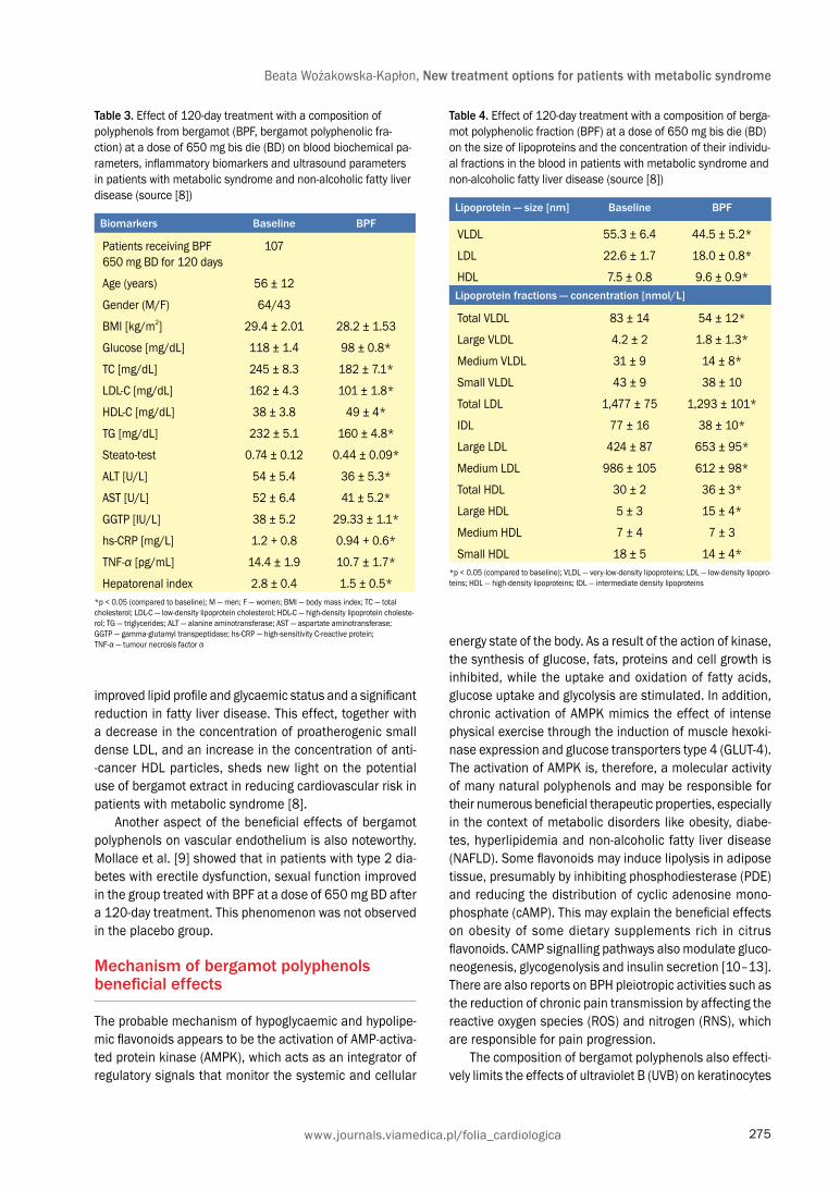
275www.journals.viamedica.pl/folia_cardiologica
Beata Wożakowska-Kapłon, New treatment options for patients with metabolic syndrome
improved lipid profile and glycaemic status and a significant reduction in fatty liver disease. This effect, together with a decrease in the concentration of proatherogenic small dense LDL, and an increase in the concentration of anti--cancer HDL particles, sheds new light on the potential use of bergamot extract in reducing cardiovascular risk in patients with metabolic syndrome [8].
Another aspect of the beneficial effects of bergamot polyphenols on vascular endothelium is also noteworthy. Mollace et al. [9] showed that in patients with type 2 dia-betes with erectile dysfunction, sexual function improved in the group treated with BPF at a dose of 650 mg BD after a 120-day treatment. This phenomenon was not observed in the placebo group.
Mechanism of bergamot polyphenols beneficial effects
The probable mechanism of hypoglycaemic and hypolipe-mic flavonoids appears to be the activation of AMP-activa-ted protein kinase (AMPK), which acts as an integrator of regulatory signals that monitor the systemic and cellular
Table 3. Effect of 120-day treatment with a composition of polyphenols from bergamot (BPF, bergamot polyphenolic fra-ction) at a dose of 650 mg bis die (BD) on blood biochemical pa-rameters, inflammatory biomarkers and ultrasound parameters in patients with metabolic syndrome and non-alcoholic fatty liver disease (source [8])
Biomarkers Baseline BPF
Patients receiving BPF 650 mg BD for 120 days
107
Age (years) 56 ± 12
Gender (M/F) 64/43
BMI [kg/m2] 29.4 ± 2.01 28.2 ± 1.53
Glucose [mg/dL] 118 ± 1.4 98 ± 0.8*
TC [mg/dL] 245 ± 8.3 182 ± 7.1*
LDL-C [mg/dL] 162 ± 4.3 101 ± 1.8*
HDL-C [mg/dL] 38 ± 3.8 49 ± 4*
TG [mg/dL] 232 ± 5.1 160 ± 4.8*
Steato-test 0.74 ± 0.12 0.44 ± 0.09*
ALT [U/L] 54 ± 5.4 36 ± 5.3*
AST [U/L] 52 ± 6.4 41 ± 5.2*
GGTP [IU/L] 38 ± 5.2 29.33 ± 1.1*
hs-CRP [mg/L] 1.2 + 0.8 0.94 + 0.6*
TNF-α [pg/mL] 14.4 ± 1.9 10.7 ± 1.7*
Hepatorenal index 2.8 ± 0.4 1.5 ± 0.5**p < 0.05 (compared to baseline); M — men; F — women; BMI — body mass index; TC — total cholesterol; LDL-C — low-density lipoprotein cholesterol; HDL-C — high-density lipoprotein choleste-rol; TG — triglycerides; ALT — alanine aminotransferase; AST — aspartate aminotransferase; GGTP — gamma-glutamyl transpeptidase; hs-CRP — high-sensitivity C-reactive protein; TNF-α — tumour necrosis factor α
Table 4. Effect of 120-day treatment with a composition of berga-mot polyphenolic fraction (BPF) at a dose of 650 mg bis die (BD) on the size of lipoproteins and the concentration of their individu-al fractions in the blood in patients with metabolic syndrome and non-alcoholic fatty liver disease (source [8])
Lipoprotein — size [nm] Baseline BPF
VLDL 55.3 ± 6.4 44.5 ± 5.2*
LDL 22.6 ± 1.7 18.0 ± 0.8*
HDL 7.5 ± 0.8 9.6 ± 0.9*Lipoprotein fractions — concentration [nmol/L]
Total VLDL 83 ± 14 54 ± 12*
Large VLDL 4.2 ± 2 1.8 ± 1.3*
Medium VLDL 31 ± 9 14 ± 8*
Small VLDL 43 ± 9 38 ± 10
Total LDL 1,477 ± 75 1,293 ± 101*
IDL 77 ± 16 38 ± 10*
Large LDL 424 ± 87 653 ± 95*
Medium LDL 986 ± 105 612 ± 98*
Total HDL 30 ± 2 36 ± 3*
Large HDL 5 ± 3 15 ± 4*
Medium HDL 7 ± 4 7 ± 3
Small HDL 18 ± 5 14 ± 4**p < 0.05 (compared to baseline); VLDL — very-low-density lipoproteins; LDL — low-density lipopro-teins; HDL — high-density lipoproteins; IDL — intermediate density lipoproteins
energy state of the body. As a result of the action of kinase, the synthesis of glucose, fats, proteins and cell growth is inhibited, while the uptake and oxidation of fatty acids, glucose uptake and glycolysis are stimulated. In addition, chronic activation of AMPK mimics the effect of intense physical exercise through the induction of muscle hexoki-nase expression and glucose transporters type 4 (GLUT-4). The activation of AMPK is, therefore, a molecular activity of many natural polyphenols and may be responsible for their numerous beneficial therapeutic properties, especially in the context of metabolic disorders like obesity, diabe-tes, hyperlipidemia and non-alcoholic fatty liver disease (NAFLD). Some flavonoids may induce lipolysis in adipose tissue, presumably by inhibiting phosphodiesterase (PDE) and reducing the distribution of cyclic adenosine mono-phosphate (cAMP). This may explain the beneficial effects on obesity of some dietary supplements rich in citrus flavonoids. CAMP signalling pathways also modulate gluco-neogenesis, glycogenolysis and insulin secretion [10–13]. There are also reports on BPH pleiotropic activities such as the reduction of chronic pain transmission by affecting the reactive oxygen species (ROS) and nitrogen (RNS), which are responsible for pain progression.
The composition of bergamot polyphenols also effecti-vely limits the effects of ultraviolet B (UVB) on keratinocytes

276
Folia Cardiologica 2019, vol. 14, no. 3
www.journals.viamedica.pl/folia_cardiologica
Table 5. Intervention strategies as a function of total cardiovascular (CV) risk and low-density lipoprotein cholesterol (LDL-C) level (source [4])
Total CV risk (SCORE) [%]
LDL-C levels
< 70 mg/dL 70 to < 100 mg/dL 100 to < 155 mg/dL 155 to < 190 mg/dL ≥ 190 mg/dL
< 1.8 mmol/L 1.8 to < 2.5 mmol/L 2.5 to < 4.0 mmol/L 4.0 to < 4.9 mmol/L ≥ 4.9 mmol/L
< 1 No lipid interven-tion
No lipid intervention No lipid intervention No lipid intervention Lifestyle intervention, consider drug if un-
controlled
≥ 1 and < 5 No lipid interven-tion
No lipid intervention Lifestyle interven-tion, consider drug if
uncontrolled
Lifestyle interven-tion, consider drug if
uncontrolled
Lifestyle intervention, consider drug if un-
controlled
≥ 5 and < 10 No lipid interven-tion
Lifestyle interven-tion, consider drug if
uncontrolled
Lifestyle intervention and concomitant drug intervention
Lifestyle intervention and concomitant drug intervention
Lifestyle intervention and concomitant drug intervention
≥ 10 Lifestyle interven-tion, consider drug if uncon-
trolled
Lifestyle intervention and concomitant drug intervention
Lifestyle intervention and concomitant drug intervention
Lifestyle intervention and concomitant drug intervention
Lifestyle intervention and concomitant drug intervention
SCORE — Systematic COronary Risk Evaluation
Figure 1. Possibilities of using a composition of bergamot polyphenolic fraction (BPF) depending on the clinical situation based on the total risk of developing cardiovascular diseases (CVD) and low-density lipoprotein cholesterol level (LDL-C) (author’s modification based on [4])
Total CVD risk (SCORE) [%]
LDL-C levels
< 70 mg/dL < 1.8 mmol/L
70 to < 100 mg/dL 1.8 to < 2.5 mmol/L
100 to < 155 mg/dL 2.5 to < 4.0 mmol/L
155 to < 190 mg/dL 4.0 to < 4.9 mmol/L
≥ 190 mg/dL ≥ 4.9 mmol/L
< 1 LDL-C < 115 mg/dl
No intervention
No intervention
No intervention
No intervention
Lifestyle interven-tion, consider drug
if uncontrolled
Class/level I C I C I C I C IIa A
≥ 1 to < 5 Target: LDL-C < 115 mg/dl
No intervention
No intervention
Lifestyle interven-tion, consider drug
if uncontrolled
Lifestyle interven-tion, consider drug
if uncontrolled
Lifestyle interven-tion, consider drug
if uncontrolled
Class/level I C I C IIa A IIa A I A
≥ 5 to < 10 OR HIGH RISK Target: LDL-C < 100 mg/dl < 50–100 mg/dl
No intervention
Lifestyle interven-tion, consider drug
if uncontrolled
Lifestyle interven-tion and concomi-tant drug interven-
tion
Lifestyle interven-tion and concomi-tant drug interven-
tion
Lifestyle interven-tion and concomi-tant drug interven-
tion
Class/level IIa A IIa A IIa A I A I A
≥ 10 OR VERY HIGH RISK Target: LDL-C < 70 mg/dl < 35–67.5 mg/dl
Lifestyle in-tervention,
consider drug if uncontrolled
Lifestyle interven-tion and concomi-tant drug interven-
tion
Lifestyle interven-tion and concomi-tant drug interven-
tion
Lifestyle interven-tion and concomi-tant drug interven-
tion
Lifestyle interven-tion and concomi-tant drug interven-
tion
Class/level IIa A IIa A I A I A I ASCORE — Systematic COronary Risk Evaluation
diagnosis of metabolic syndrome BPF in people who do not require statin therapy association of BFP with a statin in people not tolerating a higher dose of statins or combination therapy with lipid-lowering drugs
MS
MS MS
MS MSMS MS
MS

277www.journals.viamedica.pl/folia_cardiologica
Beata Wożakowska-Kapłon, New treatment options for patients with metabolic syndrome
in vitro. It seems that BPF modulates the basic pathways of cellular signal transduction, which leads to anti-proli-ferative, anti-ageing and immunomodulatory responses. Thanks to their antioxidant and anti-inflammatory proper-ties, polyphenols may also have a protective effect on the myocardium during chemotherapy [10–13].
Potential applications of bergamot polyphenols in clinical practice
The possibilities of using BPF go well beyond the metabolic syndrome mentioned in the title of this article, including a large population of people with low [< 1% SCORE (Sy-stematic COronary Risk Evaluation)] or moderate (1–4% SCORE) cardiovascular risk, with a 20–40% elevation in LDL-C when there is no need for aggressive lipid-lowering therapy (Table 5). Polyphenols from bergamot may be an option in the case of pharmacological intolerance of hypolipemic drugs or in patients categorically refusing statin therapy, although in this situation, according to the clinical condition, every effort should be made to convince the patient of the benefits of statin therapy. An important feature of bergamot polyphenols, distinguishing them from monacolin, is the possibility of statin-associated administration in patients who cannot be intensified with statin therapy (additive BPF effect). Patients with metabolic syndrome and impaired carbohydrate metabolism in the form of impaired fasting glucose (IFG) or impaired glucose tolerance (IGT), and those with metabolic syndrome and NAFLD, may benefit significantly from BPF.
Although further research is needed, it is worth remem-bering that BPF may also be a beneficial complement to therapy in erectile dysfunction in patients with carbohydrate disorders, and may reduce the effects of UVB radiation, as well as being cardioprotective during doxorubicin che-motherapy.
Summary
The composition of bergamot polyphenols positively affects the lipid and carbohydrate metabolism, and their strong antioxidant properties additionally have a posi-tive effect on the vascular endothelium. The effects of nutraceutical BPF seem to be comparable to synthetic drugs at a low dose, with negligible side effects. BPF can be an important supplement, and even occasionally an alternative, to conventional drugs as an addition to pro-health lifestyle modification and conventional the-rapy of metabolic syndrome. The features of bergamot polyphenols, such as their multidirectional lipid-reduction, hypoglycaemic activity, and modification of endothelial function, as well as the positive results of clinical trials in patients with metabolic syndrome, justify their use in this group of patients.
In Figure 1 presents a proprietary application for BFP in different populations of people with lipid disorders.
Conflict(s) of interest
Lectures fee: USP Zdrowie.
StreszczenieZespół metaboliczny jest uznawany za czynnik ryzyka schorzeń układu sercowo-naczyniowego. Zarówno małe, gęste czą-steczki lipoprotein o małej gęstości (LDL), jak i dysfunkcja wątroby pod postacią niealkoholowego stłuszczenia wątroby (NAFLD) są markerami ryzyka metabolicznego u chorych z zespołem metabolicznym. U pacjentów z zespołem meta-bolicznym leczonych kompozycją polifenoli z bergamoty stwierdza się obniżenie stężeń glukozy na czczo, triglicerydow i cholesterolu frakcji LDL oraz zwiększenia stężenia cholesterolu frakcji lipoprotein o dużej gęstości we krwi. Towarzyszy im cofanie się cech NAFLD w badaniu obrazowym, ultrasonograficznym.
Słowa kluczowe: zespół metaboliczny, kompozycja polifenoli z bergamotyFolia Cardiologica 2019; 14, 3: 272–279

278
Folia Cardiologica 2019, vol. 14, no. 3
www.journals.viamedica.pl/folia_cardiologica
References1. Alexander CM, Landsman PB, Teutsch SM, et al. Third National
Health and Nutrition Examination Survey (NHANES III), National Cho-lesterol Education Program (NCEP). NCEP-defined metabolic syn-drome, diabetes, and prevalence of coronary heart disease among NHANES III participants age 50 years and older. Diabetes. 2003; 52(5): 1210–1214, doi: 10.2337/diabetes.52.5.1210, indexed in Pubmed: 12716754.
2. Mamcarz A, Podolec P, Kopeć G, et al. Wytyczne Polskiego Forum Profilaktyki dotyczące zespołu metabolicznego. In: Podolec P. ed. Pod-ręcznik Polskiego Forum Profilaktyki. Vol. 2. Medycyna Praktyczna, Kraków 2010: 557–558.
3. Barylski M, Filipiak K, Okopień B, et al. Stanowisko grupy ekspertów wsparte przez Sekcję Farmakoterapii Sercowo-Naczyniowej Polskiego Towarzystwa Kardiologicznego dotyczące miejsca standaryzowanej kompozycji polifenoli z bergamoty w terapii dyslipidemii oraz jej innego potencjalnego zastosowania. Folia Cardiol. 2018; 13(3): 222–235, doi: 10.5603/fc.2018.0039.
4. Catapano AL, Graham I, Backer GD, et al. Grupa Robocza Europejskie-go Towarzystwa Kardiologicznego (ESC) i Europejskiego Towarzystwa Miażdżycowego (EAS) do spraw leczenia zaburzeń lipidowych. Wytycz-ne ESC/EAS dotyczące leczenia zaburzeń lipidowych w 2016 roku. Kardiol Pol. 2016; 74(11): 1234–1318, doi: 10.5603/kp.2016.0157.
5. Paszkiewicz M, Budzyńska A, Różalska B, et al. Immunomodulacyjna rola polifenoli roślinnych. Post Hig Med Dosw . 2012; 66: 637–646, doi: 10.5604/17322693.1009908.
6. Quiñones M, Miguel M, Aleixandre A. Beneficial effects of polyphe-nols on cardiovascular disease. Pharmacol Res. 2013; 68(1):
125–131, doi: 10.1016/j.phrs.2012.10.018, indexed in Pubmed: 23174266.
7. Mollace V, Sacco I, Janda E, et al. Hypolipemic and hypoglycaemic acti-vity of bergamot polyphenols: from animal models to human studies. Fitoterapia. 2011; 82(3): 309–316, doi: 10.1016/j.fitote.2010.10.014, indexed in Pubmed: 21056640.
8. Gliozzi M, Carresi C, Musolino V, et al. The effect of bergamot-derived polyphenolic fraction on LDL small dense particles and non alcoholic fatty liver disease in patients with metabolic syndrome. Adv Biol Chem. 2014; 4(2): 129–137.
9. Mollace V, Malara N, Gratteri S, et al. Bergamot polyphenolic fraction counteracts erectile dysfunction occurring in patients suffering from type 2 diabetes. PharmaNutrition. 2016; 4: S41–S46, doi: 10.1016/ /j.phanu.2015.11.006.
10. Carresi C, Gliozzi M, Giancotta C, et al. Studies on the protective role of Bergamot polyphenols in doxorubicin-induced cardiotoxicity. Phar-maNutrition. 2016; 4: S19–S26, doi: 10.1016/j.phanu.2015.11.005.
11. Holmes BF, Kurth-Kraczek EJ, Winder WW. Chronic activation of 5’-AMP-activated protein kinase increases GLUT-4, hexokinase, and glycogen in muscle. J Appl Physiol (1985). 1999; 87(5): 1990–1995, doi: 10.1152/jappl.1999.87.5.1990, indexed in Pubmed: 10562646.
12. Lauro F, Ilari S, Giancotti L, et al. The protective role of bergamot polyphenolic fraction on several animal models of pain. PharmaNutri-tion. 2016; 4: S35–S40, doi: 10.1016/j.phanu.2016.04.001.
13. Nisticò SP, Bottoni U, Gliozzi M, et al. Bergamot polyphenolic fraction counteracts photoageing in human keratinocytes. PharmaNutrition. 2016; 4: S32–S34, doi: 10.1016/j.phanu.2015.11.004.
www.journals.viamedica.pl/folia_cardiologica
Folia Cardiologica 2019 tom 14, nr 3, strony 279–282
DOI: 10.5603/FC.a2019.0021 Copyright © 2019 Via Medica
ISSN 2353–7752
PRACA KAZUISTYCZNA
279
Address for corespondence: Santosh Kumar Sinha MD, FAESC, Asst. Professor, Department of Cardiology, LPS Institute of Cardiology, G.S.V.M. Medical College, G.T. Road, Kanpur, Uttar Pradesh 208002, India, fax +91 0512 255 61 99/255 65 21, e-mail: [email protected]
Importance of apico-mitral axis during percutaneous transmitral commissurotomy
Znaczenie osi koniuszkowo-mitralnej w trakcie przezskórnej komisurotomii mitralnej
Santosh Kumar Sinha●iD, Puneet Aggarwal●iD, Mahmadula Razi, Ramesh Thakur
Department of Cardiology, LPS Institute of Cardiology, G.S.V.M. Medical College, Kanpur, India
AbstractThe development of acute severe mitral regurgitation (MR) requiring surgery is one of the most dreaded complications of percutaneous transmitral commissurotomy. We present the case of a 29 year-old female with severe rheumatic mitral stenosis whose mitral valve dilatation was performed using an Accura balloon. Once the balloon catheter had crossed the mitral valve to reach the left ventricle, it was facing away from the apico-mitral axis. It was inflated in distal portion, and mitral valve dilatation was done in the usual fashion. She developed acute severe MR. As it was misaligned with the apico-mitral axis, it caused a chordae rupture of the postero-medial papillary muscle which was identified during surgery. We conclude that a misaligned Accura balloon catheter during balloon inflation can pluck the chordae tendinae and can cause acute severe mitral regurgitation, and therefore can unearth the underlying subvalvular pathology.
Key words: severe mitral regurgitation, Accura balloon, apico-mitral axis, chordal ruptureFolia Cardiologica 2019; 14, 3: 279–282
Introduction
Since it was first described in 1984 by Inoue, percutaneous transmitral commissurotomy (PTMC) has been the gold standard in patients with symptomatic severe mitral steno-sis (MS) and suitable valve anatomy [1]. It leads to signifi-cant changes in mitral valve morphology and improvements in leaflet mobility. PTMC has a high success rate and a low complication rate. Such complications include tamponade, perforation, complete heart block, thromboembolism, left--to-right shunt across the septal puncture, and acute mitral regurgitation (MR). This last-named complication is usually mild and can be well tolerated up to a moderate degree, with the main concern being the development of severe MR following PTMC. This requires valve replacement in some patients. The incidence rate of severe MR following PTMC is 1.4–7.5% [2].
Case report
A 29 year-old female presented with exertional dyspnoea — New York Heart Association (NYHA) class III of three years’ duration. Transthoracic echocardiogram revealed severe mitral stenosis with a mitral valve area of 0.9 cm2, and a Wilkin’s score of 8/16 (M2, C2, T2, S2). Her height was 151 cm. Transoesophageal echocardiography was done to look for a left atrial clot, the degree of mitral regurgitation (MR), and her suitability for PTMC. PTMC was planned after procedural consent. The femoral artery and vein were accessed with a 6 F and an 8 F sheath respectively. A Mullins sheath was parked over the 0.035” guidewire in the left brachiocephalic vein. Wire was removed and a Brockenbrough needle was inserted into the sheath. Interatrial septum was punctured with the Brockenbrough needle after making a gradual descent, and subsequently

280
Folia Cardiologica 2019, vol. 14, no. 3
www.journals.viamedica.pl/folia_cardiologica
indicating acute severe MR (Figure 1B). As the patient was haemodynamically unstable, she was transferred to cardiac surgery for immediate mitral valve replacement. During surgery, a chordae rupture of the postero-medial papillary muscle attaching to the posterior mitral leaflet was confirmed. Both commissures were fused. The chordae attached to the anterior papillary muscle were shortened, thickened, and calcified, while the ruptured one was also shortened. Both mitral leaflets were noted to be thickened and calcified, especially around commissures. It was noted that our balloon before dilatation was not aligned with the apico-mitral axis (Figure 4B) and was in fact facing away from it (Figure 4A) indicating that it was trapped in the chordo-papillary complex which led to the complication. This was realised when the balloon was pushed into the left ventricle after mitral valve dilatation and it was noted that it was aligning with the apico-mitral axis, but it had already caused complications in the form of acute MR.
Discussion
The development of acute severe MR requiring surgery is one of the most dreaded complications of PTMC. It can result in pulmonary oedema and even death [3]. The pathophysiological mechanisms are rupture of the papillary muscles, chordae tendinae, mild leaflet prolapse, tearing of the leaflet, over-splitting of the commissure, and rarely annular dehiscence [4, 5]. Herrmann et al. [4] found that rupture of the chordae tendinae was the most common cause because of the balloon catheter straying among the chordae tendinae while crossing the catheter in the left ventricle and subsequent inflation, and the stretching of severely diseased subvalvular structures during balloon dilatation, resulting in rupture.
In our case, the balloon was appropriately sized, which ruled out any over-splitting of the commissures. Subvalvular
Figure 1A. Left atrial pressure trace before procedure; B. Rise in left atrial mean pressure with cardiovascular wave indicating acute severe mitral regurgitation
A B
Figure 2. Accura balloon misaligned with apico-mitral axis (white line — apico-mitral axis; red arrow indicating position of mitral valve)
the septum was dilated. An Accura balloon (Vascular Con-cepts, UK) was sized to 25 mm based on Hung’s formula. Preprocedure left atrial (LA) pressure was 35/22 mm Hg (mean = 25 mm Hg) (Figure 1A). Initially, the Accura balloon was entering into the left lower pulmonary vein, but the mitral valve was later crossed smoothly. To recheck its po-sition, contrast was injected through the balloon (Figure 2). The balloon was inflated in its distal part and pulled back to anchor it at the mitral valve and inflated further to achieve its full dilatation (Figure 3A). The mitral valve was dilated. The balloon was deflated and pushed into the left ventricle (Figure 3B, 4A). Postprocedural LA pressure shot up to 74/11 mm Hg (mean = 29 mm Hg) showing a giant cardiovascular wave of 72 mm Hg in pressure tra-cing and a new loud apical systolic murmur was audible,

281www.journals.viamedica.pl/folia_cardiologica
Santosh Kumar Sinha et al., Apico-mitral axis during PTMC
structure involvement can be identified by an accordion manoeuvre, balloon impasse, and an unusual sequence of inflation. Echocardiography in our case ruled out any severe involvement, as the score was 8/16 (M2, C2, T2, S2). We observed that if the balloon was aligning with the apico--mitral axis, it was free from any subvalvular structure. Initially, the balloon was deviating from the proposed axis, and shifted laterally (Figure 3B). Therefore during the PTMC, every effort should be made to keep the balloon aligned to the apico-mitral axis to prevent MR. If it is found not to
be aligned, a routine 0.035” guidewire may be pushed through the balloon to free it from underlying chordae or it should be re-introduced. Also, once the mitral valve has been crossed, the free movement of the partially inflated distal balloon in the left ventricle should be ascertained to avoid disastrous consequences. Inflation should be avoided until the balloon is aligned with the apico-mitral axis i.e. orifice-apex axis. Sometimes, the balloon may point more vertically and may still deviate away from the apico-mitral axis. This is a harbinger of the fact that the balloon has
Figure 3A. Accura balloon catheter during full dilatation across the mitral valve; B. Balloon being pushed into left ventricle during deflation
A B
Figure 4. Position of Accura balloon in relation to apico-mitral axis (white line — apico-mitral axis; co-axial alignment — A); misalignment with the axis (B; red arrow indicating position of mitral valve)
A A

282
Folia Cardiologica 2019, vol. 14, no. 3
www.journals.viamedica.pl/folia_cardiologica
strayed among the chordae. In this circumstance, the distal balloon should be inflated larger to prevent it from getting retracted back to atrium and making sure that it stays in the ventricle. The balloon should be carefully pulled back to assume a more horizontal orientation. Once satisfactory alignment with the apico-mitral axis has been ensured, it should be pushed towards the apex and an accordion ma-noeuvre should be performed. Next, the usual sequence of inflation should be carried out to achieve proper dilatation of the mitral valve.
In this case, it appears that deviation of the balloon catheter away from the apico-mitral axis could be another sign of the presence of severe valvular and/or subvalvular lesions. The ejection force of the Accura balloon catheter during the further balloon inflation in lieu of severe valvular or subvalvular lesions may result in a detrimental plucking
of the mitral chordae tendinae. Since the chordal rupture usually occurs instantaneously, it is very difficult to docu-ment the process of how the balloon catheter severs the chordae tendinae.
In conclusion, a misaligned Accura balloon catheter along the axis between the mitral orifice and the left ventricular apex during balloon inflation may pluck the chordae tendinae and cause acute severe MR, especially in the presence of severe valvular and subvalvular disease. Awareness of this potential for string-plucking [6] should alert one to abort the balloon inflation process, and to reassess the dilatation strategy.
Conflict(s) of interest
The authors declare no conflict of interest.
References1. Fawzy ME. Percutaneous mitral balloon valvotomy. Catheter Cardio-
vasc Interv. 2007; 69(2): 313–321, doi: 10.1002/ccd.21008, indexed in Pubmed: 17253604.
2. Hernandez R, Macaya C, Bañuelos C, et al. Predictors, mechanisms and outcome of severe mitral regurgitation complicating percuta-neous mitral valvotomy with the Inoue balloon. Am J Cardiol. 1992; 70(13): 1169–1174, indexed in Pubmed: 1414941.
3. Dean LS, Mickel M, Bonan R, et al. Complications and mortali-ty of percutaneous balloon mitral commissurotomy. A report from the National Heart, Lung, and Blood Institute Balloon Valvuloplasty Registry. Circulation. 1992; 85(6): 2014–2024, indexed in Pubmed: 1591821.
4. Herrmann HC, Lima JA, Feldman T, et al. Mechanisms and outcome of severe mitral regurgitation after Inoue balloon valvuloplasty. North American Inoue Balloon Investigators. J Am Coll Cardiol. 1993; 22(3): 783–789, indexed in Pubmed: 8354813.
5. Lau KW, Hung JS. Balloon impasse: a marker for severe mitral subvalvu-lar disease and a predictor of mitral regurgitation in Inoue-balloon percu-taneous transvenous mitral commissurotomy. Cathet Cardiovasc Diagn. 1995; 35(4): 310–319; discussion 320, indexed in Pubmed: 7497503.
6. Chern MS, Chang HJ, Lin FC, et al. String-plucking as a mecha-nism of chordal rupture during balloon mitral valvuloplasty using inoue balloon catheter. Catheter Cardiovasc Interv. 1999; 47(2): 213–217, doi: 10.1002/(SICI)1522-726X(199906)47:2<213::AID--CCD20>3.0.CO;2-D, indexed in Pubmed: 10376509.
StreszczenieRozwój ostrej ciężkiej niedomykalności zastawki mitralnej (MR) wymagającej leczenia chirurgicznego jest jednym z naj-poważniejszych powikłań przezskórnej komisurotomii mitralnej. W niniejszej pracy przedstawiono przypadek 29-letniej kobiety z ciężką reumatyczną stenozą zastawki mitralnej, u której zwężoną zastawkę poszerzono za pomocą balonu Accura. Po przejściu przez zastawkę mitralną i wejściu do lewej komory cewnik balonowy odchylił się od osi koniuszkowo--mitralnej. Wypełniono dystalną cześć cewnika i poszerzono zastawkę mitralną stosowaną zwykle techniką. U chorej rozwinęła się ciężka MR. Odchylenie cewnika od osi koniuszkowo-mitralnej spowodowało zerwanie struny ścięgnistej tylno-przyśrodkowego mięśnia brodawkowatego, co stwierdzono w czasie zabiegu chirurgicznego. Autorzy konkludują, że nieprawidłowe ustawienie cewnika balonowego Accura w trakcie napełniania balonu może spowodować zerwanie struny ścięgnistej i doprowadzić do ostrej ciężkiej MR, a tym samym ujawnić wadę podzastawkową.
Słowa kluczowe: ciężka niedomykalność mitralna, balon Accura, oś koniuszkowo-mitralna, pęknięcie struny ścięgnistejFolia Cardiologica 2019; 14, 3: 279–282

www.journals.viamedica.pl/folia_cardiologica
Folia Cardiologica 2019 tom 14, nr 3, strony 283–288 DOI: 10.5603/FC.2019.0060Copyright © 2019 Via Medica
ISSN 2353–7752
PRACA KAZUISTYCZNA
283
Address for correspondence: Santosh Kumar Sinha MD, FAESC, Asst. Professor, Department of Cardiology, LPS Institute of Cardiology, G.S.V.M. Medical College, G.T. Road, Kanpur, Uttar Pradesh 208002, India, fax +91 0512 255 61 99/255 65 21, e-mail: [email protected]
Acute radial collapse of a well deployed 3rd generation stent during bifurcation percutaneous coronary intervention
Ostre zapadnięcie się całkowicie rozprężonego stentu III generacji w trakcie przezskórnej interwencji wieńcowej w obrębie rozwidlenia tętnic
Santosh Kumar Sinha●iD, Mahmodula Razi, Kumar Himanshu, Puneet Aggarwal●iDDepartment of Cardiology, LPS Institute of Cardiology, G.S.V.M. Medical College, Kanpur, India
AbstractAcute stent recoil or radial collapse of a stent is a rare phenomenon leading to subsequent acute stent thrombosis or in-stent restenosis as delayed sequelae.Reduced strut thickness, a larger stent/vessel ratio, and a larger balloon/stent ratio are factors leading to stent recoil. Here, we report a case of acute recoil or radial collapse of Promus Element Plus, a third-generation everolimus-eluting stent with thin stent struts in a 66 year-old male who underwent bifurcation percutaneous coronary intervention of left anterior descending artery (LAD) and diagonal branch (D1). Following kissing balloon inflation at 16 atm pressure after deployment of stents in LAD and D1, acute radial collapse of stent in proximal LAD was noted. It was successfully bailed out by further multiple, short sequential inflations using same size noncompliant balloon. Acute radial collapse probably occurred due to inflation at higher pressure with an oversized balloon, and relatively thin struts of the stent.
Key words: acute stent recoil, bifurcation percutaneous coronary intervention, kissing balloon inflation, radial collapse, stent struts
Folia Cardiologica 2019; 14, 3: 283–288
Introduction
Drug-eluting stents (DES) are preferable to bare metal stents because they provide vessel wall scaffolding, prevent acute recoil, and are associated with lower restenosis of target sites. DES has been the subject of several advan-ces in technology, but the underlying stent platform still holds the key for clinical outcomes. Thin stent struts are associated with better conformability and deliverability, improved procedural outcomes, and fewer complications such as subsequent restenosis and thrombosis, but also has the potential for elastic stent recoil because it com-promises radial strength [1–4]. Such recoil may occur following deflation of the stent delivery balloon following
incomplete delivery balloon expansion, or inflation during post dilatation. It varies by stent design [5].
Case report
The patient was a 66 year-old male who presented with chronic stable angina of Canadian Cardiovascular Society (CCS) class III of two years’ duration, with a recent crescen-do pattern. He was diabetic and hypertensive. Blood pres-sure was 136/78 mm Hg in right arm while pulse rate was 76/min. Electrocardiogram suggested left ventricular hy-pertrophy with strain pattern. Echocardiography suggested mild left ventricular hypertrophy, diastolic dysfunction, and normal ejection fraction (EF = 68%). Coronary angiography

284
Folia Cardiologica 2019, vol. 14, no. 3
www.journals.viamedica.pl/folia_cardiologica
revealed bifurcation critical lesion in proximal left anterior descending artery (LAD) and diagonal branch (D1) which was classified as Medina Class-1,1,1 while right coronary artery (RCA) was normal. LAD and D1 were both wired with a 0.014” runthrough wire (Terumo, Japan) (Figure 1A, B). The proximal lesion of LAD was primarily predilated with a 2 × 10 mm and a 2.5 × 10 mm Sprinter legend balloon (Medtronic, USA) and stented with a 3 × 28 mm platinum chromium everolimus-eluting stent (Promus Element, Bo-ston Scientific, USA) at 10 atm pressure (Figure 2A). Proxi-mal optimisation (POT) using a 3.5 × 10 mm Meverik ballo-on in proximal LAD after pulling the jailed wire and D1 was re-wired with a 0.014” runthrough wire through the strut of LAD stent and predilated with a 2 × 10 mm Sprinter legend balloon (Figure 2B). D1 was stented with a 2.5 × 23 mm Promus Element stent using the TAP (T-stenting and protrusion) technique at 10 atm pressure while keeping another 3 × 10 mm Meverik noncompliant balloon (Boston Scientific, USA) across the ostia of D1 in LAD (Figure 2C). Stent balloon of D1 was pulled after deploying the stent and kissing balloon inflation was performed at 10 atm pressure (Figure 2D). As ostia of D1 didn’t open up, final kissing bal-loon inflation was performed for 15 seconds using Meverik noncompliant balloons: 2.5 × 10 mm for D1 and 3 × 10 mm balloon for LAD at 20 atm pressure (Figure 3A, B). In the final angiogram, both LAD and diagonal branches were completely open (Figure 3B). Both wires were removed and once angiogram had been performed, acute radial collapse of the stent in proximal LAD was noted (Figure 4). LAD was rewired, but balloon was not easily negotiable as guiding
wire was backing out. Therefore, a buddy wire was used in left circumflex artery and the LAD stent in collapsed portion was further dilated with a 3 × 10 mm Pantera Leo non-compliant balloon (Biotronik, USA) at 10 atm pressure three times, on each occasion for 30 seconds (Figure 5). Final angiogram showed well expanded stents in both LAD and D1 with TIMI III flow (Figure 6). His hospital course was uneventful and he was discharged in stable condition. After discharge the patient was followed regularly and his tread mill test after eight weeks was negative.
Discussion
Today, the estimated incidence of stent recoil or radial collap-se is 1.2% among patients receiving first- or second-genera-tion DES [6]. Platinum chromium (Pt-Cr) is the newest alloy to have been used as a DES backbone. It was introduced to improve deliverability and conformability while providing greater radial and longitudinal strength at a similar stent strut thickness as cobalt chromium (Co-Cr) [3]. The struts of third-generation Promus Element DES are thinner (74 μm) than those of previous generation stents (> 80 μm) because thin struts can minimise endothelial injury and subsequent neo-intimal hyperplasia [7]. PtCr-EES, despite having the same strut thickness as CoCr-EES, has higher radial strength and lower acute stent recoil. Radial strength is a quantitative measure of scaffolding strength which is the ability of a stent to maintain the vessel lumen. Reduced strut thickness is associated with decreased radial strength leading to more acute stent recoil, as shown by Koo et al. [8]. However, the
Figure 1A, B. Critical lesions in proximal left anterior descending artery (LAD) involving large diagonal branch (D1) — Medina Class-1,1,1 (A — antero-posterior caudal view); LAD and D1 were both wired with 0.014” runthrough wire (B, red arrow)
A B

285www.journals.viamedica.pl/folia_cardiologica
Santosh Kumar Sinha et al., Acute radial collapse of a well deployed 3rd generation stent
Figure 2A. Left anterior anterior descending artery (LAD) was stented with 3 × 28 mm Promus Element stent; B. Diagonal branch was re-wired with runthrough wire through the strut of LAD stent following proximal optimisation; C. D1 was stented with 2.5 × 23 mm Promus Element stent using T-stenting and protrusion technique; D. Stent balloon of D1 was pulled after deploying the stent and kissing balloon inflation
A B
C D
Figure 3A. As ostia of D1 didn’t open up, final kissing balloon inflation was performed using both noncompliant balloons; B. Angiogram following post-dilatation showing completely open left anterior descending artery and D1
A B

286
Folia Cardiologica 2019, vol. 14, no. 3
www.journals.viamedica.pl/folia_cardiologica
radial strength of thin strut PtCr-EES remains similar to that of a stainless steel stent with a thicker strut [9, 10]. There-fore, acute stent recoil depends not only on strut thickness but also on stent design and material. Low recoil correlates with malposition, which may increase the risk of subsequent stent thrombosis [11]. It also depends on frequency and duration of balloon inflation as multiple balloon inflations are related to a reduced risk of acute stent recoil because it gives better conformability to the stent. Interestingly, the frequency of stent delivery balloon inflations is significantly associated with fewer acute stent recoils, as shown by Ota
et al. [12]. A multiple (i.e. three times) inflation strategy is associated with a lower acute stent recoil rate compared to a one-time-inflation strategy, although the exact mechanism for this is still unknown [12]. Stents are folded and attached to the stent delivery balloon before the balloon is inflated; therefore, additional force may be needed to expand stent strut cells and connection when the stent delivery balloon is first inflated. Prolonged inflation time (60 sec) is associated with more optimal stent expansion compared to a shorter inflation time (10 sec), as shown by Kawasaki et al. [13] and Asano et al. [14]. However, prolonged inflation can cause
Figure 4. Acute radial collapse (white arrow head) of stent in proximal left anterior descending artery (inset shows magnified view)
A B C
Figure 5. Left anterior anterior descending artery was rewired and further post-dilated with 3 ×10 mm Pantera Leo non-compliant balloon in collapsed segment (A, B). Another wire used as a buddy wire in left circumflex artery
A B

287www.journals.viamedica.pl/folia_cardiologica
Santosh Kumar Sinha et al., Acute radial collapse of a well deployed 3rd generation stent
ischaemia and electrocardiographic changes. Multiple infla-tions of a shorter duration may be useful for critical lesions, especially left main coronary artery, proximal left descending artery, and jeopardised collateral coronary artery. Aggressive post dilatation by the same size balloon at higher pressure is another factor behind acute collapse, as reported by Takagi et al. [15]. A stent/vessel ratio > 1, and a balloon/stent ratio > 1 are other predictors of acute stent recoil, as reported by Bommel et al. [16]. In our case, aggressive inflation was done at 20 atm pressure for 10 seconds only, and the total
effective diameter of balloon in proximal artery was 4 mm (2/3 × 3 + 2) as the diameters of the two balloons were 3 mm and 2.5 mm. Therefore, rather than attempt high pressure (> 20 atm) post dilatation, multiple inflations are preferable with the goal of achieving optimal diameter to prevent stent collapse.
Conflict(s) of interest
The authors declare no conflict of interest.
A A
Figure 6A, B. Final angiogram showed well expanded stents in both left anterior anterior descending artery and D1 with TIMI-III flow
StreszczenieOstre zmniejszenie średnicy stentu lub zapadnięcie się stentu to rzadkie zjawisko, którego następstwem jest ostra zakrzepica w stencie lub późniejsze powikłanie w postaci restenozy w obrębie stentu.Czynnikami przyczyniającymi się do zmniejszenia średnicy stentu są mniejsza grubość rozpórek, wyższy współczynnik średnica stentu/średnica referencyjna naczynia oraz wyższy współczynnik średnica balonu/średnica stentu. Autorzy przedstawili przypadek ostrego zmniejszenia średnicy stentu lub zapadnięcia się stentu Promus Element Plus — stentu III generacji uwalniającego ewerolimus, z cienkimi rozpórkami — u 66-letniego pacjenta, u którego wykonano przezskór-ną interwencję wieńcową w obrębie rozwidlenia gałęzi międzykomorowej przedniej (LAD) i gałęzi diagonalnej D1. Po napełnieniu balonów (jednocześnie użyto 2 balonów — technika kissing balloon) pod ciśnieniem 16 atm i rozprężeniu stentów umieszczonych w LAD i D1 zaobserwowano ostre zapadnięcie się stentu w proksymalnym odcinku LAD. Sytu-ację udało się uratować, kilkakrotnie napełniając balon w krótkich sekwencjach. Zastosowano niepodatny balon o tym samym rozmiarze. Przyczynami ostrego zapadnięcia się stentu były przypuszczalnie napełnienie balonu o zbyt dużym rozmiarze z zastosowaniem zbyt wysokiego ciśnienia oraz stosunkowo cienkie rozpórki stentu.
Słowa kluczowe: ostre zmniejszenie średnicy stentu, przezskórna interwencja wieńcowa w obrębie rozwidlenia, technika kissing balloon, zapadnięcie się stentu, rozpórki stentu
Folia Cardiologica 2019; 14, 3: 283–288

288
Folia Cardiologica 2019, vol. 14, no. 3
www.journals.viamedica.pl/folia_cardiologica
References1. Ako J, Bonneau HN, Honda Y, et al. Design criteria for the ideal drug-
-eluting stent. Am J Cardiol. 2007; 100(8B): 3M–9M, doi: 10.1016/j.amjcard.2007.08.016, indexed in Pubmed: 17950830.
2. Baber U, Mehran R, Sharma SK, et al. Impact of the everolimus- -eluting stent on stent thrombosis: a meta-analysis of 13 randomized trials. J Am Coll Cardiol. 2011; 58(15): 1569–1577, doi: 10.1016/j.jacc.2011.06.049, indexed in Pubmed: 21924575.
3. Menown IBA, Noad R, Garcia EJ, et al. The platinum chromium ele-ment stent platform: from alloy, to design, to clinical practice. Adv Ther. 2010; 27(3): 129–141, doi: 10.1007/s12325-010-0022-9, in-dexed in Pubmed: 20437213.
4. Yamamoto Y, Brown DL, Ischinger TA, et al. Effect of stent design on reduction of elastic recoil: a comparison via quantitative intra-vascular ultrasound. Catheter Cardiovasc Interv. 1999; 47(2): 251–257, doi: 10.1002/(SICI)1522-726X(199906)47:2<251::AID-CCD26>3.0.CO;2-M, indexed in Pubmed: 10376515.
5. Aziz S, Morris JL, Perry RA, et al. Stent expansion: a combination of delivery balloon underexpansion and acute stent recoil reduces predicted stent diameter irrespective of reference vessel size. Heart. 2007; 93(12): 1562–1566, doi: 10.1136/hrt.2006.107052, indexed in Pubmed: 17483130.
6. Ohya M, Kadota K, Kubo S, et al. Incidence, predictive factors, and clinical impact of stent recoil in stent fracture lesion after drug-eluting stent implantation. Int J Cardiol. 2016; 214: 123–129, doi: 10.1016/j.ijcard.2016.03.013, indexed in Pubmed: 27060271.
7. Kastrati A, Mehilli J, Dirschinger J, et al. Intracoronary stenting and angiographic results: strut thickness effect on restenosis outcome (ISAR-STEREO) trial. Circulation. 2001; 103(23): 2816–2821, indexed in Pubmed: 11401938.
8. Koo BK, Waseda K, Ako J, et al. Incidence of diffuse and focal chronic stent recoil after implantation of current generation bare- -metal and drug-eluting stents. Int J Cardiol. 2010; 144(1): 132–134, doi: 10.1016/j.ijcard.2008.12.117, indexed in Pubmed: 19171393.
9. Leibundgut G, Gick M, Toma A, et al. Longitudinal compression of the platinum-chromium everolimus-eluting stent during coronary implantation: predisposing mechanical properties, incidence, and predictors in a large patient cohort. Catheter Cardiovasc Interv. 2013; 81(5): E206–E214, doi: 10.1002/ccd.24472, indexed in Pubmed: 22581708.
10. De la Torre Hernandez JM, Garcia Camarero T, Lerena P, et al. A real all-comers randomized trial comparing Xience Prime and Promus Ele-ment stents. J Invasive Cardiol. 2013; 25: 182–185.
11. Cook S, Eshtehardi P, Kalesan B, et al. Incomplete stent apposition and very late stent thrombosis after drug-eluting stent implantation. Circulation. 2007; 115(18): 2426–2434, doi: 10.1161/CIRCULA-TIONAHA.106.658237, indexed in Pubmed: 17485593.
12. Ota T, Ishii H, Sumi T, et al. Impact of coronary stent designs on acute stent recoil. J Cardiol. 2014; 64(5): 347–352, doi: 10.1016/j.jjcc.2014.02.013, indexed in Pubmed: 24679906.
13. Kawasaki T, Koga H, Serikawa T, et al. Impact of a prolonged deliv-ery inflation time for optimal drug-eluting stent expansion. Catheter Cardiovasc Interv. 2009; 73(2): 205–211, doi: 10.1002/ccd.21813, indexed in Pubmed: 19085915.
14. Asano T, Kobayashi Y, Fukushima K, et al. Effect of balloon inflation time on expansion of sirolimus-eluting stent. Heart Vessels. 2009; 24(5): 335–339, doi: 10.1007/s00380-008-1130-1, indexed in Pubmed: 19784815.
15. Takagi K, Yakushiij T, Basavarajaiah S, et al. Acute stent recoil from aggressive post-dilation of a second-generation drug-eluting stent. JACC Cardiovasc Interv. 2013; 6(3): 311–313, doi: 10.1016/j.jcin.2012.08.027, indexed in Pubmed: 23517845.
16. Bommel Rv, Lemmert M, Mieghem Nv, et al. Occurrence and predic-tors of acute stent recoil-A comparison between the xience prime cobalt chromium stent and the promus premier platinum chro-mium stent. Catheter Cardiovasc Interv. 2017; 91(3): E21–E28, doi: 10.1002/ccd.27096.
www.journals.viamedica.pl/folia_cardiologica
Folia Cardiologica 2019 tom 14, nr 3, strony 289–293 DOI: 10.5603/FC.2019.0061 Copyright © 2019 Via Medica
ISSN 2353–7752
PRACA KAZUISTYCZNA
289
Address for corespondence: Santosh Kumar Sinha MD, FAESC, Asst. Professor, Department of Cardiology, LPS Institute of Cardiology, G.S.V.M. Medical College, G.T. Road, Kanpur, Uttar Pradesh 208002, India, fax +91 0512 255 61 99/255 65 21, e-mail: [email protected]
‘Pharmacological distal protection’ with intragraft administration of diltiazem as pre-treatment
during saphenous vein graft intervention
„Dystalna ochrona farmakologiczna” przez podanie diltiazemu do pomostu naczyniowego w ramach leczenia wstępnego
podczas interwencji w obrębie pomostu naczyniowego z żyły odpiszczelowej
Santosh Kumar Sinha●iD, Siddarth Samrat, Kumar Himanshu●iD, Mahmodulla Razi
Department of Cardiology, LPS Institute of Cardiology, G.S.V.M. Medical College, Kanpur, India
AbstractPercutaneous coronary intervention (PCI) of vein graft lesions is associated with a high risk of peri-procedural myocar-dial infarction and greater mortality than routine native coronary intervention. Embolic protection devices have been advocated to reduce the risk of distal embolisation during vein graft PCI.Here, we report the case of a 72 year-old diabetic male smoker who had coronary artery bypass surgery three years previously who presented with acute coronary syndrome. Repeat coronary angiography revealed patent grafts except for a discrete eccentric critical lesion in ostium of saphenous vein graft to obtuse marginal. The lesion was crossed using a 0.014” runthrough wire (Terumo, Japan). Intragraft diltiazem (5 mg) was administered through the guiding catheter each time before predilatation and stenting (total dose = 30 mg). It was finally stented by deploying a 3.5 × 23 mm Xience Prime everolimus-eluting stent (Abbott, USA) at 13 atm pressure achieving TIMI III flow. He was discharged the next day with acetylsalicylic acid — 75 mg/day, ticagrelor — 90 mg twice daily, atorvastatin — 40 mg/day, metoprolol — 100 mg/day, and ramipril — 5 mg/day. The patient has been doing extremely well since then, with regular follow-ups at our institute.
Key words: embolic protection devices, intragraft diltiazem, percutaneous coronary intervention, coronary artery bypass graft
Folia Cardiologica 2019; 14, 3: 289–293
Introduction
The saphenous vein is commonly used as a graft (SVGs) along with the left internal mammary artery during coro-nary artery bypass graft surgery (CABG). However, they have poor long-term patency compared to arterial grafts because they suffer both from degeneration and a failure rate of as high as 25% in the first 12 months [1]. Percuta-neous coronary intervention (PCI) of vein graft lesions is
associated with a high risk of peri-procedural myocardial infarction (MI) and greater mortality than routine native coronary PCI as a result of distal embolisation manifesting as the slow flow and no-reflow phenomenon (SFNR) which is encountered in 10–15% of cases [1]. Furthermore, de-teriorating SVG lesions also possess thinner, more friable fibrous caps compared to native lesions, which further ag-gravates the incidence of plaque embolisation and platelet aggregation [2]. Although what causes no-reflow remains

290
Folia Cardiologica 2019, vol. 14, no. 3
www.journals.viamedica.pl/folia_cardiologica
dominant circulation. Right coronary artery showed mid tubular 90% lesion.
CABG was done using a left internal mammary artery (LIMA) graft as conduit to LAD, and SVG was anastomosed to obtuse marginal (OM), and posterior descending arte-ry (PDA). Repeat coronary angiography revealed patent LIMA and SVG to PDA while there was a discrete eccentric critical lesion in ostium of SVG to OM with 99% stenosis (Figure 1). Intervention of critical SVG was planned after proper consent. SVG was cannulated using a 6 F Judkins right (JR) guiding catheter after failing to cannulate with a multipurpose catheter (Figure 2). The lesion was crossed and parked distally using a 0.014” runthrough wire (Terumo, Japan). Intragraft diltiazem (5 mg) was administered through the guiding catheter. The lesion was sequentially and gradually predilated using a 2 × × 10 mm and a 2.4 × 10 mm Traveller semicompliant balloon (Abbott, USA). Diltiazem (5 mg) was injected each time before upsizing the balloon. As the lesion was not properly opening up because of its fibrotic nature, it was further dilated using a 3 × 10 mm cutting balloon (Flextome, Boston Scientific, USA) at 16 atm pressure by further administering 5 mg diltiazem (Figure 3). It was finally stented by deploying a 3.5 × 23 mm Xience Prime everolimus-eluting stent (Abbott, USA) at 13 atm pressure achieving TIMI III flow (Figures 4, 5). He was discharged the next day with ASA — 75 mg/day, ticagrelor — 90 mg twice daily, atorvastatin — 40 mg/day, metoprolol — 100 mg/day, and ramipril — 5 mg/day. The patient has
unclear, endothelial swelling, neutrophil infiltration, and platelet aggregation inducing microvasculature spasm and obstruction are a few of the plausible mechanisms. Combined together, these are associated with a higher rate of in-stent restenosis and target vessel revascularisation (TVR). The severity of these potential consequences makes finding the optimal technique essential during invasive SVG revascularisation.
Case report
A 72 year-old diabetic male smoker presented with sudden onset chest pain radiating to the right arm of 10 minutes’ duration with diaphoresis. Past medical history included CABG three years earlier in lieu of triple vessel disease involving distal left main bifurcation. He was receiving acetylsalicylic acid (ASA) — 75 mg, metoprolol — 100 mg, atorvastatin — 20 mg, and ramipril — 5 mg, and gilbenclaimide daily. His vitals, physical examination, and biochemistry were all unremarkable. Electrocardio-gram revealed mild ST-T changes in precordial leads. Echocardiography revealed mild concentric hypertrophy of left ventricle, and grade-II diastolic dysfunction with normal ejection fraction (EF = 55%). His troponin T level was raised (0.4 ng/L) when measured six hours after presentation. His coronary angiography three years be-fore had revealed a left main bifurcation lesion (medina class: 1, 1, 1) involving the left anterior descending artery (LAD) and the left circumflex artery (LCx) with left
A B
Figure 1A, B. Aorto-ostial lesion of saphenous vein graft to obtuse marginal showing discrete eccentric critical lesion (A — antero-posterior view; B — straight lateral view)
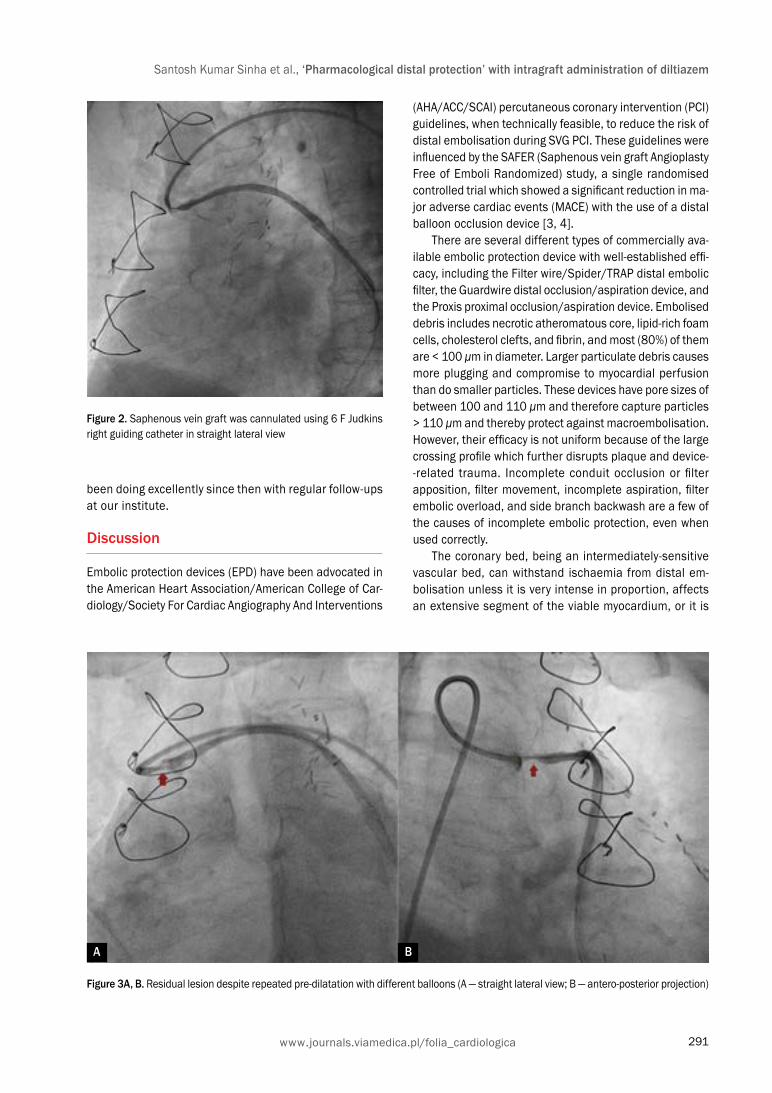
291www.journals.viamedica.pl/folia_cardiologica
Santosh Kumar Sinha et al., ‘Pharmacological distal protection’ with intragraft administration of diltiazem
been doing excellently since then with regular follow-ups at our institute.
Discussion
Embolic protection devices (EPD) have been advocated in the American Heart Association/American College of Car-diology/Society For Cardiac Angiography And Interventions
(AHA/ACC/SCAI) percutaneous coronary intervention (PCI) guidelines, when technically feasible, to reduce the risk of distal embolisation during SVG PCI. These guidelines were influenced by the SAFER (Saphenous vein graft Angioplasty Free of Emboli Randomized) study, a single randomised controlled trial which showed a significant reduction in ma-jor adverse cardiac events (MACE) with the use of a distal balloon occlusion device [3, 4].
There are several different types of commercially ava-ilable embolic protection device with well-established effi-cacy, including the Filter wire/Spider/TRAP distal embolic filter, the Guardwire distal occlusion/aspiration device, and the Proxis proximal occlusion/aspiration device. Embolised debris includes necrotic atheromatous core, lipid-rich foam cells, cholesterol clefts, and fibrin, and most (80%) of them are < 100 μm in diameter. Larger particulate debris causes more plugging and compromise to myocardial perfusion than do smaller particles. These devices have pore sizes of between 100 and 110 μm and therefore capture particles > 110 μm and thereby protect against macroembolisation. However, their efficacy is not uniform because of the large crossing profile which further disrupts plaque and device--related trauma. Incomplete conduit occlusion or filter apposition, filter movement, incomplete aspiration, filter embolic overload, and side branch backwash are a few of the causes of incomplete embolic protection, even when used correctly.
The coronary bed, being an intermediately-sensitive vascular bed, can withstand ischaemia from distal em-bolisation unless it is very intense in proportion, affects an extensive segment of the viable myocardium, or it is
Figure 2. Saphenous vein graft was cannulated using 6 F Judkins right guiding catheter in straight lateral view
Figure 3A, B. Residual lesion despite repeated pre-dilatation with different balloons (A — straight lateral view; B — antero-posterior projection)
A B

292
Folia Cardiologica 2019, vol. 14, no. 3
www.journals.viamedica.pl/folia_cardiologica
already a jeopardised myocardium with left ventricular dysfunction. Therefore, a normally functioning left ven-tricular myocardium can withstand small-to-moderate intensity transient ischaemic insult without any con-sequences, whereas even smaller territory transient ischaemia may be clinically significant in the setting of left ventricular dysfunction. Focal fibrotic lesions, aorto--ostial lesions, distal anastomotic lesions, and in-stent restenosis lesions have a low embolisation risk and can be treated without EPD.
A sole surviving graft, large area of viable myocardium supplied by the SVG, left ventricular dysfunction, or atrio-ventricular nodal artery supply through the SVG in the absence of an artificial pacemaker all increase the clinical risk after an episode of slow flow. They should therefore be treated with EPD if feasible. This includes direct stenting, stent undersizing, the use of covered stent grafts, aspira-tion thrombectomy, adjunctive administration of intragraft anti-thrombotic agents (abciximab and thrombolytics in setting of thrombus), and prophylactic use of vasodilators (verapamil, nicardipine, adenosine, and nitroprusside). These have yielded positive outcomes in anecdotal reports and small studies but their efficacy has not been confirmed in large randomised trials.
Figure 5. Saphenous vein graft to obtuse marginal showing TIMI III flow after successful stenting
Figure 4. Lesion was stented by deploying 3.5 × 23 mm Xience Prime everolimus-eluting stent at 13 atm pressure
Intragraft administration of vasodilators targets mic-rovasculature to combat slow and no-reflow phenomena. Pre-treatment with intracoronary adenosine, a potent dilator of arteries and arterioles, decreases peri-procedural myocardial infarction after elective PCI because it improves myocardial flow and lowers the incidence of no-reflow in the setting of acute MI [5–8]. High doses of intragraft adenosine (at least five boluses of 24 μg each) may help to reverse slow and no-reflow phenomena in patients undergoing SVG intervention [9, 10]. Similar results have been observed with prophylactic administration of intragraft verapamil [11–14], and nicardipine [15] in reducing no-reflow in SVG PCI.
Based on these findings, we decided to try intragraft diltiazem to reduce slow flow in SVG PCI. This is also another calcium channel blocker like verapamil and nicardipine. The reason for success in our case was that the lesion was discrete, aorto-ostial, and fibrotic, as a cutting balloon was used to open up the lesion.
Therefore, if the morpho-anatomy is carefully assessed before attempting an SVG, one can achieve success without using an EPD.
Conflict(s) of interest
The authors declare no conflict of interest.

293www.journals.viamedica.pl/folia_cardiologica
Santosh Kumar Sinha et al., ‘Pharmacological distal protection’ with intragraft administration of diltiazem
StreszczeniePrzezskórna interwencja wieńcowa (PCI) w obrębie wszczepionego pomostu żylnego wiąże się z wysokim ryzykiem oko-łozabiegowego zawału serca i większą śmiertelnością niż rutynowe zabiegi tego typu w naczyniach natywnych. Zaleca się stosowanie urządzeń zabezpieczających przed zatorami w celu obniżenia ryzyka odległych zatorów w trakcie PCI w pomoście żylnym.Autorzy przedstawili przypadek 72-letniego pacjenta, aktywnego palacza, z ostrym zespołem wieńcowym, u którego 3 lata wcześniej wykonano chirurgiczne pomostowanie aortalno-wieńcowe. Powtórna koronarografia wykazała drożność stentu poza nieciągłą ekscentryczną krytyczną zmianą w miejscu połączenia pomostu z żyły odpiszczelowej z gałęzią brzeżną. Przez zwężenie przeprowadzono prowadnik angioplastyczny 0,014” typu runthrough (Terumo, Japonia). Przed każdą predylacją balonową i implantacją stentu do pomostu podawano diltiazem (5 mg) przez cewnik prowadzący (łącz-na dawka = 30 mg). Ostatecznie w miejscu zwężenia umieszczono i rozprężono stent uwalniający ewerolimus Xience Prime 3,5 × 23 mm (Abbott, USA), stosując ciśnienie 13 atm. Uzyskano przepływ TIMI III. Pacjenta wypisano następnego dnia z zaleceniem przyjmowania następujących leków: kwasu acetylosalicylowego — 75 mg/dobę, tikagreloru — 90 mg 2 razy/dobę, atorwastatyny — 40 mg/dobę, metoprololu — 100 mg/dobę i ramiprilu — 5 mg/dobę. Od czasu zabiegu chory czuł się bardzo dobrze i regularnie zgłaszał się na wizyty kontrolne do placówki autorów.
Słowa kluczowe: urządzenia do ochrony przeciwzakrzepowej, podanie diltiazemu do pomostu naczyniowego, przezskórna interwencja wieńcowa, pomostowanie aortalno-wieńcowe
Folia Cardiologica 2019; 14, 3: 289–293
References1. Blachutzik F, Achenbach S, Troebs M, et al. Angiographic findings
and revascularization success in patients with acute myocardial in-farction and previous coronary bypass grafting. Am J Cardiol. 2016; 118(4): 473–476, doi: 10.1016/j.amjcard.2016.05.040, indexed in Pubmed: 27328951.
2. Leborgne L, Cheneau E, Pichard A, et al. Effect of direct stenting on clinical outcome in patients treated with percutaneous coronary inter-vention on saphenous vein graft. Am Heart J. 2003; 146(3): 501–506, doi: 10.1016/S0002-8703(03)00309-0, indexed in Pubmed: 12947370.
3. Levine G, Bates E, Blankenship J, et al. 2011 ACCF/AHA/SCAI Guide-line for percutaneous coronary intervention. Circulation. 2011; 124(23), doi: 10.1161/cir.0b013e31823ba622.
4. Baim DS, Wahr D, George B, et al. Saphenous vein graft Angioplasty Free of Emboli Randomized (SAFER) Trial Investigators. Randomized trial of a distal embolic protection device during percutaneous inter-vention of saphenous vein aorto-coronary bypass grafts. Circulation. 2002; 105(11): 1285–1290, indexed in Pubmed: 11901037.
5. Lee CH, Low A, Tai BC, et al. Pretreatment with intracoronary adenosine reduces the incidence of myonecrosis after non-urgent percutaneous coronary intervention: a prospective randomized study. Eur Heart J. 2007; 28(1): 19–25, doi: 10.1093/eurheartj/ehl411, indexed in Pubmed: 17132650.
6. Desmet WJR, Dens J, Coussement P, et al. Does adenosine prevent myocardial micronecrosis following percutaneous coronary interven-tion? The ADELINE pilot trial. ADEnosine Limit myocardial Necrosis. Heart. 2002; 88(3): 293–295, indexed in Pubmed: 12181228.
7. Marzilli M, Orsini E, Marraccini P, et al. Beneficial effects of intraco-ronary adenosine as an adjunct to primary angioplasty in acute myo-cardial infarction. Circulation. 2000; 101(18): 2154–2159, indexed in Pubmed: 10801755.
8. Stoel MG, Marques KMJ, de Cock CC, et al. High dose adenosine for suboptimal myocardial reperfusion after primary PCI: A randomi-
zed placebo-controlled pilot study. Catheter Cardiovasc Interv. 2008; 71(3): 283–289, doi: 10.1002/ccd.21334, indexed in Pubmed: 17985384.
9. Fischell TA, Carter AJ, Foster MT, et al. Reversal of “no reflow” during vein graft stenting using high velocity boluses of intracoronary ade-nosine. Cathet Cardiovasc Diagn. 1998; 45(4): 360–365, indexed in Pubmed: 9863736.
10. Sdringola S, Assali A, Ghani M, et al. Adenosine use during aortocoronary vein graft interventions reverses but does not prevent the slow-no reflow phenomenon. Catheterization and Cardiovascular Interventions. 2000; 51(4): 394–399, doi: 10.1002/1522-726x(200012)51:4<394::aid-ccd4>3.0.co;2-g.
11. Kaplan BM, Benzuly KH, Kinn JW, et al. Treatment of no-reflow in degene-rated saphenous vein graft interventions: comparison of intracoronary verapamil and nitroglycerin. Cathet Cardiovasc Diagn. 1996; 39(2): 113–118, doi: 10.1002/(SICI)1097-0304(199610)39:2<113::AID-CCD1>3.0.CO;2-I, indexed in Pubmed: 8922307.
12. Piana RN, Paik GY, Moscucci M, et al. Incidence and treatment of ‘no- -reflow’ after percutaneous coronary intervention. Circulation. 1994; 89(6): 2514–2518, indexed in Pubmed: 8205658.
13. Michaels AD, Appleby M, Otten MH, et al. Pretreatment with intragraft verapamil prior to percutaneous coronary intervention of saphenous vein graft lesions: results of the randomized, controlled vasodilator prevention on no-reflow (VAPOR) trial. J Invasive Cardiol. 2002; 14(6): 299–302, indexed in Pubmed: 12042618.
14. Resnic FS, Wainstein M, Lee MKY, et al. No-reflow is an independent predictor of death and myocardial infarction after percutaneous coro-nary intervention. Am Heart J. 2003; 145(1): 42–46, doi: 10.1067/ /mhj.2003.36, indexed in Pubmed: 12514653.
15. Abbo KM, Dooris M, Glazier S, et al. Features and outcome of no- -reflow after percutaneous coronary intervention. Am J Cardiol. 1995; 75(12): 778–782, indexed in Pubmed: 7717278.

www.journals.viamedica.pl/folia_cardiologica
Folia Cardiologica 2019 tom 14, nr 3, strony 294–297 DOI: 10.5603/FC.2019.0062 Copyright © 2019 Via Medica
ISSN 2353–7752
PRACA KAZUISTYCZNA
294
Adres do korespondencji: Piotr Koprowicz, Studenckie Koło Naukowe przy Klinice Kardiologii Katedry Kardiologii Uniwersytetu Medycznego w Łodzi, Wojewódzki Specjalistyczny Szpital im. dr. Wł. Biegańskiego, ul. Kniaziewicza 1/5, 91–347 Łódź, tel. +48 42 251 62 16, e-mail: [email protected]
Amiodaron — tarczyca — arytmia. Taniec na linie endokrynologa i kardiologa. Pacjent z poamiodaronowymi
zaburzeniami funkcji tarczycy
Amiodarone — thyroid — arrhythmia. Difficult cooperation between endocrinologist and cardiologist. A patient with amiodarone-induced thyroid disturbances
Piotr Koprowicz1, Jakub Włodarczyk1, Małgorzata Kurpesa2●iD, Jarosław D. Kasprzak2●iD, Tomasz Rechciński2●iD
1Studenckie Koło Naukowe przy Klinice Kardiologii Katedry Kardiologii Uniwersytetu Medycznego w Łodzi 2Klinika Kardiologii Katedry Kardiologii Uniwersytetu Medycznego w Łodzi
StreszczenieW pracy przedstawiono leczonego krioablacją pacjenta z trzepotaniem przedsionków, u którego po włączeniu amiodaro-nu wystąpiła niedoczynność tarczycy. Włączenie suplementacji L-tyroksyną wywołało nawrót arytmii. Przypadek ukazuje, jak trudne może być leczenie w takiej sytuacji oraz jak ważna jest ścisła współpraca kardiologa i endokrynologa podczas terapii poamiodaronowych zaburzeń czynności tarczycy.
Słowa kluczowe: amiodaron, trzepotanie przedsionków, niedoczynność tarczycyFolia Cardiologica 2019; 14, 3: 294–297
Wstęp
Amiodaron jest lekiem wykorzystywanym w leczeniu arytmii nadkomorowych, zaś w arytmiach komorowych stosuje się go jako uzupełnienie elektroterapii. Wśród działań niepo-żądanych leku obserwuje się zaburzenia funkcji tarczycy. U pacjenta, u którego wystąpią takie powikłania, leczenie staje się wyzwaniem wymagającym ścisłej współpracy kardiologa i endokrynologa, dla których cele terapii nie zawsze są zbieżne.
Opis przypadku
W pracy przedstawiono przypadek leczonego krioablacją pacjenta z trzepotaniem przedsionków, u którego po
włączeniu amiodaronu wystąpiły zaburzenia funkcji tar-czycy. Pacjent to 85-letni mężczyzna z chorobą wieńcową, kontrolowanym farmakologicznie nadciśnieniem tętniczym, pozawałową niewydolnością serca, cukrzycą typu 2 leczoną dietą oraz hipercholesterolemią. Chory nie palił papierosów, nie był rodzinnie obciążony miażdżycą.
Mężczyzna w wieku 77 lat przebył zawał serca z unie-sieniem odcinka ST (STEMI, ST-elevation myocardial in-farction); w przeprowadzonej w trybie pilnym koronarografii stwierdzono:
— w pniu lewej tętnicy wieńcowej zwężenie dystalne w 50–70%;
— w gałęzi przedniej zstępującej na całym przebiegu zmiany brzeżne zwężające światło do 70%;
— w gałęzi okalającej zwężenie w ujściu do 90%;

295www.journals.viamedica.pl/folia_cardiologica
Piotr Koprowicz i wsp., Pacjent z poamiodaronowymi zaburzeniami funkcji tarczycy
— prawą tętnicę wieńcową recesywną, amputowaną ostialnie — obwód wypełniał się z własnego krążenia mostkowego.Wykonano pierwotną angioplastykę balonową gałęzi
okalającej. Po 3 dniach w wyniku wczesnej restenozy wystą-pił wstrząs kardiogenny. Podczas ponownej angioplastyki założono stent. Frakcja wyrzutowa lewej komory (LVEF, left ventricular ejection fraction) oceniona przed wypisaniem wynosiła 38%, a w elektrokardiogramie (EKG), poza cecha-mi przebytego zawału, nie stwierdzono odchyleń.
Rok później wystąpił ponowny zawał serca, bez unie-sienia odcinka ST (NSTEMI, non-ST-elevation myocardial infarction). Zważywszy na nieoptymalny efekt leczenia przezskórnego sprzed roku, pacjenta zakwalifikowano do rewaskularyzacji chirurgicznej. Wszczepiono by-pass tętniczy do gałęzi przedniej zstępującej i żylny do gałęzi okalającej. Po operacji pacjent wymagał kontrapulsacji wewnątrzaortalnej. Przy wypisaniu LVEF wynosiła 40%. Choremu zalecono:
— bisoprolol w dawce 2,5 mg; — klopidogrel w dawce 75 mg; — atorwastatynę w dawce 20 mg; — pantoprazol w dawce 20 mg; — kwas acetylosalicylowy w dawce 150 mg; — eplerenon w dawce 25 mg.
U pacjenta w wieku 79 lat pierwszy raz pojawiły się napadowy częstoskurcz przedsionkowy, a następnie trze-potanie przedsionków. Z powodu krótkotrwałej skutecznoś-ci farmakoterapii pacjenta zakwalifikowano do leczenia interwencyjnego. W badaniu elektrofizjologicznym stwier-dzono trzepotanie przedsionków o częstotliwości 270/min. Przeprowadzono krioablację cieśni trójdzielno-żylnej. Po zabiegu uzyskano rytm zatokowy z blokiem prawej odnogi
pęczka Hisa oraz z blokiem przedniej wiązki lewej odnogi. Pierwszy raz oznaczono również profil hormonów tarczyco-wych — wszystkie wartości pozostawały w normie (stężenie hormonu stymulującego tarczycę [TSH, thyroid-stimulating hormone] 2,26 mlU/l, stężenie wolnej trójjodotyroniny [fT3, free triiodothyronine] 4,54 pmol/l, stężenie wolnej tyroksyny [fT4, free thyroxine] 21,14 pmol/l). Włączono acenokumarol.
U chorego w wieku 84 lat w monitorowaniu EKG meto-dą Holtera (wykonanym podczas programu rehabilitacji) zarejestrowano wydłużenie odcinka PQ, jeden epizod czę-stoskurczu komorowego (9 ewolucji o częstości 144/min); jeden epizod częstoskurczu nadkomorowego (3 ewolucje o częstości 120/min). W związku z bezobjawową arytmią nie wprowadzono żadnych zmian w leczeniu.
W 85. roku życia ponownie przeprowadzono monitoro-wanie holterowskie; zarejestrowano 2 epizody częstoskur-czu komorowego (27 i 6 ewolucji > 100/min) oraz 7 epi-zodów częstoskurczu nadkomorowego (od 3 do 6 ewolucji > 100/min). Ze względu na nasilenie arytmii zdecydowano o włączeniu na stałe 200 mg amiodaronu z jednoczesnym monitorowaniem funkcji tarczycy u endokrynologa.
Pół roku po włączeniu amiodaronu zarejestrowano wydłużenie odcinka PQ do 286 ms oraz odstępu QT do 526 ms (ryc. 1). W związku z tym zmniejszono dawki bisoprololu do 2,5 mg, zaś amiodaronu do 100 mg. W ba-daniu kontrolnym przeprowadzonym 2 miesiące później zmniejszenie dawek beta-adrenolityku i amiodaronu spo-wodowała skrócenie odcinka PQ do 228 ms, a odstępu QT do 464 ms (ryc. 2).
W kontrolnym EKG po pół roku zaobserwowano wydłu-żony do 304 ms odcinek PQ, a odstęp QT — do 514 ms. Z tego powodu dawki beta-adrenolityku oraz amiodaronu
Rycina 1. Zapis elektrokardiograficzny (EKG) pacjenta z kwietnia 2016 roku: odcinek PQ 286 ms, odstęp QT 526 ms; leki przyjmowane przez pacjenta: bisoprolol w dawce 5 mg, amiodaron w dawce 200 mg. Zapisy EKG i pomiary odstępów QTc były dokonywane za pomocą aparatu EKG Cardiovit MS-12 blue firmy Schiller (Szwajcaria)

296
Folia Cardiologica 2019, tom 14, nr 3
www.journals.viamedica.pl/folia_cardiologica
ponownie zmniejszono. Po 3 miesiącach terapii 1,25 mg bisoprololu i 50 mg amiodaronu na dobę w EKG odcinek PQ wynosił 274 ms, a odstęp QT — 488 ms.
W trakcie kolejnej wizyty kardiologicznej pacjent poin-formował, że przyjmuje nowy lek zalecony przez endokry-nologa oraz że często czuje szybkie bicie serca. W EKG stwierdzono częstoskurcz przedsionkowy z przewodzeniem 2:1. Okazało się, że ze względu na wysoką wartość TSH, tj. 28 mlU/l, włączono L-tyroksynę w dawce 25 μg/dobę. Ponieważ po pierwszym miesiącu leczenia TSH wyniosło 18 mlU/l, to dawkę L-tyroksyny zwiększono do 50 μg, co
wywołało ponowne epizody napadowego częstoskurczu przedsionkowego (ryc. 3).
W opisywanym przypadku rozważano następujące możliwości terapeutyczne:
— kolejną ablację, ale pacjent nie zgodził się na ponowne leczenie zabiegowe;
— odstawienie L-tyroksyny, co niosło za sobą zagrożenia wynikające z niedoczynności tarczycy, która może być jedną z przyczyn wydłużenia odstępu QT;
— odstawienie amiodaronu, co niosło za sobą ryzyko zaostrzenia arytmii u pacjenta.
Rycina 2. Zapis elektrokardiograficzny pacjenta z czerwca 2016 roku po zmniejszeniu dawek leków: odcinek PQ 228 ms, odstęp QT 464 ms; leki przyjmowane przez pacjenta: bisoprolol w dawce 2,5 mg, amiodaron w dawce 10 mg
Rycina 3. Zapis elektrokardiograficzny pacjenta po włączeniu 50 μg L-tyroksyny

297www.journals.viamedica.pl/folia_cardiologica
Piotr Koprowicz i wsp., Pacjent z poamiodaronowymi zaburzeniami funkcji tarczycy
w wyniku toksycznego działania leku na gruczoł tarczowy lub w związku z obecnością w nim dużej ilości jodu, który u części pacjentów prowadzi do wystąpienia niedoczynności (AIH, amiodarone-induced hypothyroidism). Nadczynność może wystąpić w wyniku indukowanej przez jod niekontro-lowanej produkcji hormonów — najczęściej u chorych z wo-lem guzkowym lub niejawną chorobą Gravesa-Basedowa (typ 1 tyreotoksykozy indukowanej amiodaronem — [AIT, amiodarone-induced thyrotoxicosis]). Typ 2 nadczynności jest skutkiem destruktywnego zapalenia tarczycy toczącego się we wcześniej zdrowym gruczole tarczowym [2]. Nieznane są czynniki prognostyczne wystąpienia poamiodaronowych zaburzeń czynności tarczycy. Wyniki badań retrospektyw-nych wskazują, że czynnikiem predykcyjnym może być surowiczy stosunek stężeń desetylamiodaronu (DEA) — me-tabolitu leku — do amiodaronu (AMD) [3]. W przypadkach AIT stwierdzono wyższy stosunek DEA/AMD niż u pacjentów z AIH. Amiodaron jest metabolizowany w wątrobie przez CYP 3A4 [4]. Dlatego polimorfizmy genu tego enzymu oraz jego induktory mogą wpływać na prawdopodobieństwo wystąpienia tarczycowych powikłań poamiodaronowych.
Przez kolejne miesiące pacjent nie pojawiał się u kar-diologa ze względu na trudną sytuację rodzinną. Okazało się, że po pół roku samodzielnie odstawił amiodaron, a L-tyroksynę przyjmuje w dawce 37,5 µg/dobę. Mimo odstawienia 6 miesięcy wcześniej amiodaronu w zapisie EKG odstęp QTc pozostaje wydłużony do 554 ms, a rytm zatokowy o częstości 76/min jest przerywany epizodami tachykardii 114/min. Pacjent nie odczuwa jednak żadnych dolegliwości, a zaproponowanej w celu optymalizacji leczenia hospitalizacji odmówił ze względu na sytuację osobistą.
Dyskusja
U pacjentów ze schorzeniami sercowo-naczyniowymi po-amiodaronowe zaburzenia tarczycy są szczególnie niebez-pieczne, ponieważ ciężka niedoczynność to jedna z przyczyn wydłużonego odstępu QT, co predysponuje do groźnych arytmii komorowych, zaś nadczynność może zaostrzać cho-robę serca i zwiększa umieralność [1]. Zaburzenia funkcji tarczycy podczas stosowania amiodaronu mogą wystąpić
AbstractWe present the case of a patient with atrial flutter (treated with cryoablation) and amiodarone-induced hypothyroidism (AIH). Administration of l-tyroxine supplementation triggered recurrence of arrhythmia. The report shows how difficult can be treating a patient with AIH and how important is cooperation between cardiologist and endocrinologist.
Key words: amiodarone, atrial flutter, hypothyroidism, AIHFolia Cardiologica 2019; 14, 3: 294–297
Piśmiennictwo1. O’Sullivan AJ, Lewis M, Diamond T. Amiodarone-induced thyrotoxico-
sis: left ventricular dysfunction is associated with increased mortality. Eur J Endocrinol. 2006; 154(4): 533–536, doi: 10.1530/eje.1.02122, indexed in Pubmed: 16556715.
2. Bogazzi F, Bartalena L, Martino E. Approach to the patient with amiodarone-induced thyrotoxicosis. J Clin Endocrinol Metab. 2010; 95(6): 2529–2535, doi: 10.1210/jc.2010-0180, indexed in Pubmed: 20525904.
3. Yamato M, Wada K, Hayashi T, et al. Association between Serum Amio-darone and N-Desethylamiodarone Concentrations and Development of Thyroid Dysfunction. Clin Drug Investig. 2018; 38(1): 39–48, doi: 10.1007/s40261-017-0582-4, indexed in Pubmed: 29080209.
4. Fabre G, Julian B, Saint-Aubert B, et al. Evidence for CYP3A-me-diated N-deethylation of amiodarone in human liver microsomal fractions. Drug Metab Dispos. 1993; 21(6): 978–985, indexed in Pubmed: 7905403.

www.journals.viamedica.pl/folia_cardiologica
Folia Cardiologica 2019 vol. 14, no. 3, pages 298–301 Copyright © 2019 Via Medica
ISSN 2353–7752
CASE REPORT
298
Address for correspondence: Piotr Koprowicz, Studenckie Koło Naukowe przy Klinice Kardiologii Katedry Kardiologii Uniwersytetu Medycznego w Łodzi, Wojewódzki Specjalistyczny Szpital im. dr. Wł. Biegańskiego, ul. Kniaziewicza 1/5, 91–347 Łódź, Poland, phone +48 42 251 62 16, e-mail: [email protected]
Amiodarone — thyroid — arrhythmia. Difficult cooperation between endocrinologist and cardiologist
A patient with amiodarone-induced thyroid disturbances
Amiodaron — tarczyca — arytmia. Taniec na linie endokrynologa i kardiologa. Pacjent z poamiodaronowymi zaburzeniami funkcji tarczycy
Piotr Koprowicz1, Jakub Włodarczyk1, Małgorzata Kurpesa2●iD, Jarosław D. Kasprzak2●iD, Tomasz Rechciński2●iD
1Student Society at the Chair and Department of Cardiology, Medical University of Lodz, Lodz, Poland 2 Chair and Department of Cardiology, Medical University of Lodz, Lodz, Poland
Artykuł jest tłumaczeniem pracy: Koprowicz P, Włodarczyk J, Kurpesa M et al. Amiodaron — tarczyca — arytmia. Taniec na linie endokrynologa i kardiologa. Pacjent z poamiodaronowymi zaburzeniami funkcji tarczycy. Folia Cardiol. 2019; 14 (3): 294–297. DOI: 10.5603/FC.2019.0062.
Należy cytować wersję pierwotną
AbstractWe present the case of a patient with atrial flutter (treated with cryoablation) and amiodarone-induced hypothyroidism (AIH). Administration of L-thyroxine supplementation triggered a recurrence of arrhythmia. This report shows how difficult it can be to treat a patient with AIH and how important it is to ensure cooperation between cardiologist and endocrinologist.
Key words: amiodarone, atrial flutter, hypothyroidism, AIHFolia Cardiologica 2019; 14, 3: 298–301
Introduction
Amiodarone is a drug used in the treatment of supraventri-cular arrhythmias, and in ventricular arrhythmias as a su-pplement to electrotherapy. Thyroid dysfunction is a known side effect. In a patient with such complications, treatment becomes a challenge requiring close cooperation between a cardiologist and an endocrinologist, for whom the goals of treatment are not always convergent.
Case study
We present the case of a patient with atrial flutter treated with cryoablation, with thyroid dysfunction occurring after the inception of amiodarone therapy. The patient was an 85 year--old man with coronary disease, pharmacologically controlled hypertension, post-infarction heart failure, diet-controlled type 2 diabetes, and hypercholesterolemia. The patient did not smoke and had a negative family history for atherosclerosis.

299www.journals.viamedica.pl/folia_cardiologica
Piotr Koprowicz et al., A patient with amiodarone-induced thyroid disturbances
At the age of 77, he suffered a ST-elevation myocardial infarction (STEMI): in urgent coronary angiography:
— left coronary artery distally narrowed to 50–70%; — left anterior descending artery with marginal changes
narrowing the light to 70% on the entire course; — ostium of the circumflex artery of the coronary artery
narrowed to 90%; — recessive right coronary artery, amputated ostia, pe-
riphery fills with its own collaterals.Primary balloon angioplasty of the circumflex branch
was performed. After three days, cardiogenic shock oc-curred as a result of early restenosis. A stent was placed during re-angioplasty. Left ventricular ejection fraction (LVEF) assessed before discharge was 38%, and in the electrocardiogram (ECG) there were no deviations, except for the features of previous infarction.
A year later, a second myocardial infarction, non-ST--elevation (NSTEMI) occurred. In view of the sub-optimal effect of percutaneous treatment from the previous year, the patient was qualified for surgical revascularisation. An arterial bypass was implanted into the anterior descending branch and venous bypass to the circumflex branch. After the operation, the patient required an intraaortic balloon contrapulsation. At discharge, LVEF was 40%. The patient was prescribed:
— bisoprolol 2.5 mg; — clopidogrel 75 mg; — atorvastatin 20 mg; — pantoprazole 20 mg; — acetylsalicylic acid 150 mg; — eplerenone 25 mg.
At the age of 79, paroxysmal atrial tachycardia followed by atrial flutter appeared for the first time. Due to the short- -term effectiveness of pharmacotherapy, the patient was
qualified for interventional treatment. The electrophysio-logical examination revealed an atrial flutter of 270/min. Cryoablation of the cavotricuspid isthmus was performed. After surgery, sinus rhythm was obtained with right branch bundle block and left anterior fascicular block. The thyroid hormone profile was assessed for the first time — all were normal [thyroid-stimulating hormone [TSH] 2.26 mlU/l, free triiodothyronine [fT3] 4.54 pmol/L, free thyroxine [fT4] 21.14 pmol/L). Acenocoumarol was started.
At the age of 84, Holter ECG monitoring performed during the rehabilitation programme recorded elongation of the PQ interval, one episode of ventricular tachycardia (nine evolutions at a frequency of 144/min); and one episode of supraventricular tachycardia (three evolutions at a frequency of 120/min). Asymptomatic arrhythmias did not cause any changes in pharmacological treatment.
At the age of 85, Holter monitoring was repeated, which revealed two episodes of ventricular tachycardia: 27 and six evolutions > 100/min, and seven episodes of supraventricular tachycardia (from three to six evolutions > 100/min). In view of the severity of the arrhythmia, it was decided to permanently use amiodarone 200 mg, while monitoring the thyroid function by an endocrinologist.
Six months after the amiodarone introduction, elonga-tion of the PQ interval to 286 ms and QT interval to 526 ms were recorded (Figure 1). Therefore, the dose of bisoprolol was reduced to 2.5 mg, and amiodarone to 100 mg. In a follow-up study performed two months later, the reduced beta-blocker and amiodarone doses had shortened the PQ interval to 228 ms and the QT interval to 464 ms (Figure 2).
In a control ECG after six months, an extended PQ interval (304 ms), and an extended QT interval (514 ms) were observed. For this reason, the beta-blocker and amio-darone doses were reduced again. After three months of
Figure 1. Electrocardiogram (ECG) of the patient in April 2016: PQ interval 286 ms, QT interval 526 ms; bisoprolol 5 mg, amiodarone 200 mg. ECG recordings and measurements of QTc intervals were made using the ECG Cardiovit MS-12 blue from Schiller (Switzerland)

300
Folia Cardiologica 2019, vol. 14, no. 3
www.journals.viamedica.pl/folia_cardiologica
treatment with 1.25 mg bisoprolol and 50 mg amiodarone per day, in ECG the PQ interval was 274 ms and the QT interval was 488 ms.
At the next cardiology follow-up visit, the patient infor-med a cardiologist that he was taking a new medicine from an endocrinologist and that he often felt fast palpitations. ECG revealed atrial tachycardia with 2:1 conduction. It turned out that L-thyroxine 25 μg/day was started due to the high TSH value of 28 mlU/l. Because TSH value was 18 mlU/l, after the first month of therapy, the dose of L-thy-roxine was escalated to 50 μg, which caused re-episodes of paroxysmal atrial tachycardia (Figure 3).
In the case described, the following therapeutic options were considered:
— another ablation — the patient did not agree to re--treatment;
— discontinuation of L-thyroxine — this carried the risks arising from hypothyroidism, which may be one of the causes of QT prolongation;
— discontinuation of amiodarone — this was associated with a risk of worsening the patient’s arrhythmia.Over the following months, the patient did not appear
at the Cardiology Clinic due to a difficult family situation. It turned out that after six months he had discontinued
Figure 2. Electrocardiogram of the patient in June 2016 after reduction of drug doses: PQ interval 228 ms, QT interval 464; bisoprolol 2.5 mg, amiodarone 100 mg
Figure 3. Electrocardiogram of the patient after the start of 50 μg of L-thyroxine

301www.journals.viamedica.pl/folia_cardiologica
Piotr Koprowicz et al., A patient with amiodarone-induced thyroid disturbances
toxic effects of the drug on the thyroid gland or due to the presence of a large amount of iodine, which in some patients leads to the occurrence of hypothyroidism (AIH). Hyperthyroidism may occur as a result of iodine-induced uncontrolled hormone production. This occurs most often in patients with nodular goitre or a silent Graves-Basedow disease [type 1 amiodarone-induced thyroid toxicity (AIT)]. Type 2 hyperthyroidism is the result of destructive thyroiditis in the previously healthy thyroid gland [2]. The prognostic factors for the occurrence of thyroid intestinal disorders are unknown. Retrospective studies indicate that the predictive factor may be the serum ratio of desethylenedioquinone (DEA) — a drug metabolite — to amiodarone (AMD) [3]. In AIT cases, a higher DEA/AMD ratio was found than in AIH patients. Amiodarone is metabolised in the liver via CYP 3A4 [4]. Therefore, the polymorphisms of this enzyme gene and its inducers may influence the probability of thyroid complications.
amiodarone on his own, and L-thyroxine was taken in a dose of 37.5 μg/day. Despite the discontinuation of amiodarone six months earlier, in the ECG the QTc interval was still extended to 554 ms, and sinus rhythm of 76/min was interrupted by episodes of tachycardia 114 /min. How-ever, the patient did not feel any discomfort and refused hospitalisation to optimise his treatment because of the personal situation.
Discussion
In patients with cardiovascular diseases, post-amiodarone thyroid disorders are particularly dangerous because se-vere hypothyroidism is one of the causes of prolonged QT interval which predisposes to severe ventricular arrhyth-mias, whereas hyperactivity can exacerbate heart disease and increase mortality [1]. Disorders of thyroid function during the use of amiodarone may occur as a result of
StreszczenieW pracy przedstawiono leczonego krioablacją pacjenta z trzepotaniem przedsionków, u którego po włączeniu amiodaro-nu wystąpiła niedoczynność tarczycy. Włączenie suplementacji L-tyroksyną wywołało nawrót arytmii. Przypadek ukazuje, jak trudne może być leczenie w takiej sytuacji oraz jak ważna jest ścisła współpraca kardiologa i endokrynologa podczas terapii poamiodaronowych zaburzeń czynności tarczycy.
Słowa kluczowe: amiodaron, trzepotanie przedsionków, niedoczynność tarczycyFolia Cardiologica 2019; 14, 3: 298–301
References1. O’Sullivan AJ, Lewis M, Diamond T. Amiodarone-induced thyrotoxico-
sis: left ventricular dysfunction is associated with increased mortality. Eur J Endocrinol. 2006; 154(4): 533–536, doi: 10.1530/eje.1.02122, indexed in Pubmed: 16556715.
2. Bogazzi F, Bartalena L, Martino E. Approach to the patient with amiodarone-induced thyrotoxicosis. J Clin Endocrinol Metab. 2010; 95(6): 2529–2535, doi: 10.1210/jc.2010-0180, indexed in Pubmed: 20525904.
3. Yamato M, Wada K, Hayashi T, et al. Association between serum amio-darone and N-desethylamiodarone concentrations and development of thyroid dysfunction. Clin Drug Investig. 2018; 38(1): 39–48, doi: 10.1007/s40261-017-0582-4, indexed in Pubmed: 29080209.
4. Fabre G, Julian B, Saint-Aubert B, et al. Evidence for CYP3A-mediated N-deethylation of amiodarone in human liver microsomal fractions. Drug Metab Dispos. 1993; 21(6): 978–985, indexed in Pubmed: 7905403.

www.journals.viamedica.pl/folia_cardiologica
Folia Cardiologica 2019 tom 14, nr 3, strony 302–304 DOI: 10.5603/FC.2019.0063 Copyright © 2019 Via Medica
ISSN 2353–7752
PRACA KAZUISTYCZNA
302
Address for correspondence: dr n. med. Roman Steckiewicz, I Katedra i Klinika Kardiologii, Warszawski Uniwersytet Medyczny, ul. Banacha 1a, 02–097 Warszawa, Poland, phone 22 599 19 58, fax 22 599 19 57, e-mail: [email protected]
Double left brachiocephalic vein: a rare systemic vein anomaly and a potential source of complication
during CIED procedures
Podwójna lewa żyła ramienno-głowowa — rzadka anomalia żył systemowych i potencjalne ryzyko powikłań procedur CIED
Przemysław Stolarz1●iD, Elżbieta Barbara Świętoń2, Roman Steckiewicz2
11st Department of Cardiology, Medical University of Warsaw, Poland 21st Department of Cardiology, Central University Hospital, Warsaw, Poland
AbstractAbnormal systemic vein embryogenesis results in the development of anomalies which, in the case of the left brachio-cephalic vein (LBCV), can manifest as a significant and morphometric deviation from the normal anatomy of the vessel.One rare example of such an anatomical variation is the presence of a double LBCV. Depending on its nature and extent, this anomalous LBCV variation, which may be incidentally detected during an invasive procedure, can not only pose difficulties in terms of the successful completion of the procedure, but can also result in injuries to the vessel itself as well as to adjacent structures. We here present a case featuring such a congenital LBCV anomaly, which was discovered during a cardiac pacemaker implantation procedure.
Key words: anomalous left brachiocephalic vein, venography, cardiac pacingFolia Cardiologica 2019; 14, 3: 302–304
Introduction
Abnormal systemic vein embryogenesis leads to the deve-lopment of vascular anomalies, which, in the case of the left brachiocephalic vein (LBCV), can manifest as signifi-cant topographic and morphometric deviations from the normal anatomy of the vessel. One rare example of such an anatomical variation is the presence of a double LBCV [1, 2]. Depending on its nature and extent, this anomalous LBCV variation, which can be incidentally detected during an invasive procedure, may not only pose difficulties in
terms of the successful completion of the procedure, but may also result in injuries to the vessel itself as well as to adjacent structures [3, 4].
Case report
An 82 year-old woman was qualified to receive permanent cardiac pacing therapy due to presyncopal episodes in-duced by Mobitz II second-degree atrioventricular block. Since the course of her left cephalic vein was not condu-cive to cardiac lead insertion, the operator decided on

303www.journals.viamedica.pl/folia_cardiologica
Przemysław Stolarz et al., Double LBCV and CIED implantation
the axillary vein puncture approach, with the use of an appropriate kit. Despite successful guidewire insertion into the LBCV, attempts to advance it into the superior vena cava were ineffective due to unexpected resistance. The operator was unable to advance the guidewire even after several attempts; at the same time, fluoroscopy showed various topographic configurations of the inserted guide-wire (Figures 1A and B). In order to find an explanation for this evident obstacle, an additional approach was attempted, which involved puncturing the venous angle downstream from a stenotic segment in the subclavian vein and selectively administering intravenous contrast. Fluoroscopy showed the presence of a rare double LBCV. Dynamic flow of contrast illustrated the morphometric parameters of both brachiocephalic veins: revealing a compressed LBCV proper and illustrating the topography of its doubled counterpart. In light of the mutual position of the two vessels and those of the adjacent anatomical structures, the operator decided to halt the procedure to prevent potential injury to the vessels.
A Medtronic Adapta ADD01 pacemaker was eventually implanted by inserting the CapSureFix Novus cardiac leads via the veins of the right clavipectoral triangle.
The prevalence of congenital LBCV anomalies depends on the assessed population and the employed imaging technique. Congenital LBCV anomalies constitute approxi-mately 1% of congenital heart defects (such as tetralogy of Fallot, atrial or ventricular septal defects). The prevalence of congenital LBCV anomalies in populations without heart defects is estimated to be less than 0.4%. LBCV anomalies can occur as an isolated phenomenon or co-occur with va-riations of persistent left superior vena cava (PLSVC). The prevalence of such systemic vein anomalies in the general population is 0.3–0.5%.
Conclusions
Abnormalities in the typical anatomical LBCV structure can complicate the intravenous advancement of guidewires, catheters or leads, especially if they are of a large diameter, have high bending stiffness, or are used to forcefully push through the encountered resistance. In the case presented here, the morphometric parameters of both LBCVs posed a high risk of injury during lead advancement, which ulti-mately led to the decision to perform the CIED implantation using a different point of venous access, on the right side.
Figure 1A. A guidewire inserted into the left brachiocephalic vein (LBCV) suggests patency of the vessel; guidewire position at the level of aortic arch artery origins (short arrow) indicates a typical LBCV topography; the tip of the guidewire situated at the level of the LBCV ostium into the superior vena cava; B. An alternative position of the guidewire (long arrow): clearly below the level shown in panel A; C. Venography illustrating the causes of blocked guidewire advance in both locations, namely compression of the LBCV proper by an enlarged aorta in the vicinity of aortic arch artery origins (short arrow), and the presence of a tortuous, double LBCV (long arrow)
304
Folia Cardiologica 2019, vol. 14, no. 3
www.journals.viamedica.pl/folia_cardiologica
StreszczenieZaburzenie embriogenezy żył systemowych skutkuje powstaniem wad, które w przypadkach dotyczących lewej żyły ramienno-głowowej (LBCV) cechuje znaczna odmienność topograficzna i morfometryczna w odniesieniu do budowy anatomicznej typowej dla tego naczynia. Przykładem jest sporadyczne występowanie obserwowanej podwójnej LBCV. Charakter i zakres tak zaistniałej rozwojowej odmienności LBCV, doraźnie wykryty podczas wykonywania inwazyjnej pro-cedury, niezależnie od trudności jej w realizacji, może sprzyjać traumatyzacji zarówno samego naczynia, jak i przyległych struktur. W opracowaniu zaprezentowano postać tej żylnej anomalii rozwojowej wykrytą podczas procedury implantacji stymulatora serca.
Słowa kluczowe: anomalia lewej żyły ramienno-głowowej, wenografia, stymulacja sercaFolia Cardiologica 2019; 14, 3: 302–304
3. Bachleda JP. Iatrogenic Injury to the Superior Vena Cava and Bra-chiocephalic Vein. Journal of Infectious Diseases and Therapy. 2014; 02(06), doi: 10.4172/2332-0877.1000169.
4. Ko SF, Ng SH, Fang FM, et al. Left brachiocephalic vein perfora-tion: computed tomographic features and treatment considera-tions. Am J Emerg Med. 2007; 25(9): 1051–1056, doi: 10.1016/ /j.ajem.2007.06.013, indexed in Pubmed: 18022501.
References1. Kahkouee S, Sadr M, Pedarzadeh E, et al. Anomalous left brachio-
cephalic vein: important vascular anomaly concomitant with conge-nital anomalies and heart diseases. Folia Morphol (Warsz). 2017; 76(1): 51–57, doi: 10.5603/FM.a2016.0031, indexed in Pubmed: 27830886.
2. Kondrachuk O, Yalynska T, Tammo R. Double left brachiocephalic vein. Pediatr Cardiol. 2013; 34(3): 767–768, doi: 10.1007/s00246-012-0542-y, indexed in Pubmed: 23052674.

305www.journals.viamedica.pl/folia_cardiologica
Folia Cardiologica 2019 tom 14, nr 3, strony 305–314 DOI: 10.5603/FC.2019.0064 Copyright © 2019 Via Medica
ISSN 2353–7752
NIEWYDOLNOŚĆ SERCA
Adres do korespondencji: prof. dr hab. n. med. Małgorzata Lelonek FESC, Zakład Kardiologii Nieinwazyjnej, Katedra Chorób Wewnętrznych i Kardiologii, Uniwersytet Medyczny w Łodzi, ul. Żeromskiego 113, 90–549 Łódź, e-mail: [email protected]
Kardiomiopatia połogowa — wciąż niepoznana Aktualny stan wiedzy
Peripartum cardiomyopathy — still unknown. Current state of knowledge
Kiet Anh Nguyen1, Kamila Gniady1, Małgorzata Lelonek2●iD1Studenckie Koło Naukowe przy Zakładzie Kardiologii Nieinwazyjnej Katedry Chorób Wewnętrznych i Kardiologii Uniwersytetu
Medycznego w Łodzi 2Zakład Kardiologii Nieinwazyjnej Uniwersytetu Medycznego w Łodzi
StreszczenieKardiomiopatię połogową (PPCM) definiuje się jako idiopatyczną kardiomiopatię, która występuje pod koniec ciąży lub w pierwszych kilku miesiącach po porodzie z objawami niewydolności serca (HF) wtórnie do zaburzeń czynności lewej komory i jednocześnie nie stwierdza się żadnej innej przyczyny tego stanu. Patomechanizm choroby nie został w pełni poznany, natomiast prawdopodobnie opiera się na działaniu złożonych czynników. Przebieg kliniczny PPCM jest zróż-nicowany: od zagrażającej życiu ostrej HF po łagodne objawy naśladujące dolegliwości charakterystyczne dla okresu okołoporodowego. W Europie PPCM jest dość rzadką chorobą, natomiast są rejony, gdzie występuje z częstością 1/300 przypadków (Haiti). Leczenie PPCM jest podobne jak dla HF z obniżoną frakcją wyrzutową, jednak ważne jest wyklu-czenie leków o działaniu teratogennym w trakcie trwania ciąży. Włączanie bromokryptyny do terapii PPCM wydaje się zasadne i w niektórych przypadkach poprawia rokowanie.
Słowa kluczowe: kardiomiopatia połogowa, PPCM, niewydolność serca, ciąża, bromokryptynaFolia Cardiologica 2019; 14, 3: 305–314
Wstęp
Pierwsze doniesienia na temat kardiomiopatii połogowej (PPCM, peripartum cardiomyopathy) miały miejsce jeszcze w połowie XIX wieku, kiedy to Virchow i wsp. [1] opisali sze-reg przypadków sekcji kobiet, które zmarły w czasie połogu. Wówczas badacze nie wiedzieli jeszcze z czym mają do czynienia, natomiast zauważyli, że u pacjentek występowało zwyrodnienie mięśnia sercowego. Minęło kolejnych kilka-dziesiąt lat zanim w 1937 roku Gouley i wsp. [1] powiązali okres okołoporodowy z kardiomiopatią. W swojej pracy opisali przypadki 7 ciężarnych pacjentek z ostrą i często śmiertelną niewydolnością serca (HF, heart failure), z któ-rych 4 zmarły. Charakterystyczna dla każdego przypadku była rozstrzeniowa kardiomiopatia w późnym etapie ciąży,
która utrzymywała się także po porodzie. Podczas sekcji zmarłych pacjentek wykazano przerost mięśnia sercowego z obszarami ciężkiej martwicy i zwłóknień. W 1971 roku Demakis i Rahimtoola wraz ze swoim zespołem po raz pierwszy zastosowali termin PPCM [1, 2]. W kolejnych latach dzięki udoskonaleniu technik badawczych, między innymi echokardiografii, biologii molekularnej oraz odkry-ciu nowych biomarkerów, opracowano nowe kryteria dla PPCM. Niniejsza praca przedstawia aktualny stan wiedzy na temat PPCM.
Definicja PPCM
Wcześniejsza definicja PPCM została zmieniona przez Heart Failure Association of the European Society of Cardiology

306
Folia Cardiologica 2019, tom 14, nr 3
www.journals.viamedica.pl/folia_cardiologica
jej krótsza forma o masie 16 kDa, która wykazuje silne działanie kardiotoksyczne. STAT-3 jest białkiem, które od-powiada za ochronę mięśnia sercowego poprzez indukcję enzymów antyoksydacyjnych. Zaburzenie funkcji STAT-3 prowadzi do nasilenia stresu oksydacyjnego i aktywacji wielu enzymów, a w tym katepsyny D, która odpowiada za powstanie krótszej formy prolaktyny (16 kDa). Wykazano, że u kobiet chorujących na PPCM poziom ekspresji białka STAT-3 jest zmniejszony, a produkcja 16 kDa prolaktyny zwiększona [9]. Na podstawie tej wiedzy oparta jest jedna z nowych strategii leczenia PPCM. Polega ona na dodaniu inhibitora prolaktyny — bromokryptyny — do standardowej terapii HF. Obecnie efekty terapii są zadowalające, a w wy-tycznych Europejskiego Towarzystwa Kardiologicznego (ESC, European Society of Cardiology) z 2018 roku leczeniu bromokryptyną przypisano klasę zaleceń IIa przy poziomie wiarygodności danych C [10].
sFLT1W początkowym okresie ciąży proces angiogenezy jest zwiększony, jednak tendencja ta zmienia się wraz z upły-wem czasu i w okresie okołoporodowym łożysko zaczyna wy-dzielać substancje antyangiogenne. Należy do nich między innymi rozpuszczalna forma kinazy tyrozynowej 1 podobna do fms (sFLT1, soluble fms-like tyrosine kinase-1) [11]. W prawidłowo przebiegającej ciąży są to procesy fizjologicz-ne i nie powodują żadnych powikłań, jednak w warunkach patologicznych, kiedy sFLT1 jest wydzielana w nadmiarze lub gdy upośledzone są mechanizmy chroniące serce przed czynnikami antyangiogennymi, może dojść do subklinicznej dysfunkcji kardiomiocytów. Wykazano, że pacjentki z PPCM mają istotnie podwyższone stężenie sFLT1, a ponadto wielokrotna ciąża oraz stan przedrzucawkowy powodują zwiększenie wydzielania tego antyangiogenu. To może wyjaśniać przyczynę, z powodu której wieloródki oraz
w 2010 roku i pozostaje wciąż aktualna. Jest to idiopatyczna kardiomiopatia, która objawia się HF występującą wtórnie do zaburzeń czynności lewej komory i jednocześnie nie stwierdza się żadnej innej przyczyny HF. Występuje pod koniec ciąży lub w pierwszych kilku miesiącach po porodzie. Dla rozpoznania PPCM nie ma konieczności obligatoryjnego udokumentowania powiększenia wymiaru lewej komory, natomiast jej frakcja wyrzutowa powinna wynosić 45% lub mniej.
W celu postawienia diagnozy stosuje się rozpoznanie na drodze wykluczenia. W odróżnieniu od definicji z 2000 roku nie uwzględnia ona ścisłego przedziału czasowego wy-stąpienia choroby (wcześniej był to ostatni miesiąc ciąży do 5–6 mies. po porodzie) ani też konkretnych kryteriów echokardiograficznych (wcześniej: obniżenie frakcji wy-rzutowej < 45% i frakcji skracania < 30% oraz powiększe-nie końcoworozkurczowego wymiaru jamy lewej komory > 2,7 cm/m2 powierzchni ciała). Obecna definicja jest mniej restrykcyjna, dzięki czemu zmniejsza się ryzyko przeoczenia pacjentki z tą jednostką chorobową [3, 4].
Patogeneza
Prawdopodobnie mechanizm wywołujący PPCM jest złożony i dlatego nie istnieje jedna teoria wyjaśniająca jej rozwój. W piśmiennictwie podaje się wiele czynników biorących udział w rozwoju choroby. Kilka ważniejszych z nich przed-stawiano w tabeli 1 [3, 5–9]. W niniejszej pracy opisano dwa patomechanizmy, co do których wydaje się, że mają największe znaczenie w PPCM [6].
ProlaktynaFizjologicznie prolaktyna o masie 23 kDa działa ochronnie na śródbłonek i sprzyja angiogenezie. W okresie stresu ok-sydacyjnego wywołanego na przykład ciążą może powstać
Tabela 1. Najważniejsze znane czynniki wywołujące kardiomiopatię połogową (PPCM, peripartum cardiomyopathy)
Patogeneza PPCM Krótka charakterystyka
Czynniki genetyczne/środowi-skowe
Dane epidemiologiczne na temat zwiększonej liczby przypadków PPCM na niektórych terenach i wśród wybranych grup etnicznych, zwłaszcza afrykańskich czy afroamerykańskich [3]. W niektó-rych pracach sugeruje się powiązanie z rodzinną kardiomiopatią rozstrzeniową [5, 6]
Czynniki infekcyjne/autoimmu-nologiczne
Udokumentowana obecność genomu wirusowego w kardiomiocytach u kobiet z PPCM (31%) [7]
Wzrost stężenia markerów zapalnych w PPCM: TNF-a, interferonu g, interleukiny 6, CRP [8]
Czynniki związane z angiogenezą Pod koniec ciąży łożysko zaczyna wydzielać substancje antyangiogenne. W przypadku upośledzo-nego działania mechanizmów ochronnych serca przed czynnikami antyangiogennymi lub nadmia-ru tych czynników dochodzi do subklinicznej dysfunkcji kardiomiocytów [8]
Czynniki związane z prolaktyną Fizjologicznie prolaktyna pełni funkcję ochronną śródbłonka i sprzyja angiogenezie, jednak w okresie zwiększonego stresu oksydacyjnego wywołanego ciążą może powstać jej krótsza forma działająca toksycznie na kardiomiocyty [9]
TNF-a (tumor necrosis factor a) — czynnik martwicy nowotworów a; CRP (C-reactive protein) — białko C-reaktywne

307www.journals.viamedica.pl/folia_cardiologica
Kiet Anh Nguyen i wsp., Kardiomiopatia połogowa — wciąż niepoznana. Aktualny stan wiedzy
kobiety ze stanem przedrzucawkowym częściej zapadają na PPCM [10]. Jedna z form terapii jaką zaproponowano w leczeniu PPCM opierała się na zwiększeniu procesu angiogenezy poprzez podawanie czynnika wzrostu śród-błonka naczyniowego (VEGF, vascular endothelial growth factor). Dotychczasowe próby były przeprowadzane tylko na myszach, a wynik stosowania samego VEGF nie dał ocze-kiwanych rezultatów. Dopiero leczenie VEGF w połączeniu z bromokryptyną okazało się zadowalające [12].
Epidemiologia
Częstotość występowania PPCM charakteryzuje znaczne zróżnicowanie geograficzne, a w wielu obszarach nie jest znana rzeczywista liczba przypadków i potrzebne są dalsze badania. Największe zagęszczenie rozpoznanych przypad-ków PPCM notuje się w Nigerii, 1:100 przypadków, oraz na Haiti — 1:300 przypadków. Dla porównania choroba dotyka 1:1000–1500 kobiet w Niemczech, 1:1000 w Republice Południowej Afryki (RPA) i 1:2500–4000 w Stanach Zjed-noczonych [3, 10]. Z danych, jakie w 2010 roku przytoczyli Sliwa i wsp. [3] jasno wynika, że pochodzenia afrykańskie oraz afrykańsko-amerykańskie predysponuje do częstszego występowania PPCM, natomiast średni wiek zapadalności na PPCM wyniósł 27–33 lata w zależności od regionu. Co więcej, średnia wieku ujawnienia się choroby okazywała się wyższa w badaniach przeprowadzonych w ciągu ostat-nich kilku lat w porównaniu ze starszymi publikacjami [3]. Ponadto wykazano także zwiększone występowanie PPCM na przestrzeni ostatnich lat — od 1/4350 kobiet w latach 1990–1993 do 1/1229 w latach 2000–2002. Zaobserwowana tendencja może mieć swoje uzasadnienie w postaci kilku czynników: późniejszy wiek zajścia w ciążę, wzrost liczby ciąż mnogich, rozwijający się dostęp do metod wspomaganego rozrodu czy zwiększenie rozpoznawalności PPCM [14].
Obraz kliniczny
Trudności związane z postawieniem właściwej diagnozy wiążą się z faktem, że obraz kliniczny PPCM może naślado-wać dolegliwości postrzegane jako typowe dla końcowego okresu ciąży lub wysiłku związanego z samym porodem. Nierzadko objawy nie mają dużego nasilenia, co dodatkowo utrudnia postawienie właściwego rozpoznania [3].
U zdecydowanej większości pacjentek objawy rozwijają się w pierwszych 4 miesiącach po porodzie (78%), a jedynie u 9% początek choroby następuje w ostatnim miesiącu ciąży. Pozostałe 13% przypadków dotyczy kobiet, u których choroba ujawniła się wcześniej niż na miesiąc przed poro-dem lub później niż 4 miesiące po porodzie [3].
Do typowych objawów należą: duszność, obrzęki kończyn dolnych, orthopnoë, zmęczenie, ból w klatce
piersiowej, zmniejszona tolerancja wysiłku i kołatania serca. W Patel i wsp. [14] w swojej pracy z 2015 roku wyodrębnili 19 szwedzkich pacjentek (wg kryteriów ESC z 2010 r.), by określić, jakie objawy występują najczęściej w PPCM. Udało się ustalić, że wszystkie badane zgłaszały więcej niż jeden objaw. Najczęstszymi dolegliwościami były: duszność (18/19), retencja płynów (15/19), nadmierna męczliwość (14/19), uporczywy kaszel (8/19). W badaniu zwrócono uwagę także na objawy psychiczne: spośród 19 kobiet jedenaście zgłosiło przynajmniej jeden z następujących symptomów: uczucie nieuniknionej śmierci (7), odczuwanie paniki (5), niepokoju (5) lub strachu (3) [14].
Diagnostyka PPCM
Rozpoznanie PPCM jest ustalane na podstawie wy-kluczenia innych przyczyn HF [15]. Chorobę należy podej-rzewać u wszystkich kobiet w okresie okołoporodowym z objawami HF lub u których następuje opóźnienie w po-wrocie do stanu zdrowia sprzed ciąży [16]. Dwa kluczowe badania, które muszą zostać wykonane przy podejrzeniu PPCM to echokardiografia oraz pomiar stężenia N-koń-cowego fragmentu propeptydu natriuretycznego typu B (NT-proBNP, N-terminal pro-B-type natriuretic peptide) lub peptydu natriuretycznego typu B (BNP, B-type natriuretic peptide). Jeśli frakcja wyrzutowa lewej komory (LVEF, left ventricular ejection fraction) wyniesie 45% lub mniej lub stężenie peptydów natriuretycznych będzie podwyższone, konieczne są dalsze badanie w kierunku PPCM [12]. Do badań przydatnych w postawieniu diagnozy należą: badanie radiologiczne klatki piersiowej (może pokazać powiększenie sylwetki serca, cechy zastoju w krążeniu małym i wysięku opłucnowego), elektrokardiografia (cechy przeciążenia lewej komory, niespecyficzne zmiany w odcinkach ST, wy-dłużony odstęp QT czy szerokie zespoły QRS). Dodatkowo można wykonać rezonans magnetyczny jako uzupełnienie echokardiografii [17] i biopsję mięśnia sercowego przy podejrzeniu HF o etiologii infekcyjnej [16].
Ponadto, obiecujące jest zastosowanie nowo odkrytych biomarkerów u kobiet chorujących na PPCM pomocne w diagnostyce różnicowej. Wybrane z nich przedstawiono w tabeli 2 [8, 18, 19].
Leczenie
Zgodnie z najnowszymi wytycznymi ESC z 2018 roku lecze-nie, jakie powinno być wdrożone w przypadku PPCM, nie odbiega od zaleceń stosowanych w ostrej HF, wstrząsie kar-diogennym czy przewlekłej HF o innej etiologii. Istotne jest, aby wykluczyć leki, które mogą mieć szkodliwy wpływ na rozwijający się płód lub mające przeciwwskazania w trakcie karmienia piersią (inhibitory konwertazy angiotensyny, sar-tany, antagoniści receptora dla angiotensyny II i inhibitora

308
Folia Cardiologica 2019, tom 14, nr 3
www.journals.viamedica.pl/folia_cardiologica
neprylizyny (ARNI, angiotensin receptor neprilysin inhibitor), leki oszczędzające potas, warfaryna, iwabradyna) [15]. Wybrane leki wraz z przeciwwskazaniami przedstawiono w tabeli 3 [15, 16]. Opieka nad ciężarną z PPCM powinna łączyć nadzór lekarza kardiologa oraz położnika, a możliwe powikłania wynikające dla płodu należy obserwować w ul-trasonografii (USG) [3].
Ciąża u pacjentki z chorobą sercowo-naczyniową jest zawsze obciążona wysokim ryzykiem powikłań. Dlatego konieczne jest poinformowanie chorej, że częstość wyko-nywania USG płodu będzie odbiegać od standardowego postępowania przy ciąży niepowikłanej. Do 28. tygodnia ciąży badanie powinno być wykonywane raz w miesiącu dla pacjentek w I lub II klasie według New York Heart Associa-tion (NYHA) oraz co 2 tygodnie lub częściej w III i IV klasie według NYHA. Po 28. tygodniu ciąży badanie powinno się odbywać co tydzień aż do porodu [20].
W przypadku ostrej HF o ciężkim przebiegu należy mieć na uwadze konieczność przedwczesnego rozwiązania ciąży z zabezpieczonym dostępem do systemu mechanicznego wspomagania krążenia (MCS, mechanical circulatory support) oraz wdrożeniem leczenia przyspieszającego dojrzewanie płuc u płodu powyżej 23. tygodnia ciąży + + 5 dni [15]. Ponadto obowiązuje włączenie intensywnego leczenia ostrej HF.
Zasady leczenia PPCM przedstawiono na rycinie 1 z uwzględnieniem okresu przed porodem i po porodzie.
Tabela 2. Wybrane potencjalne markery w kardiomiopatii połogo-wej (na podstawie [8, 18, 19])
Nazwa markera Stężenie w PPCM
mikro-RNA 146 Wzrost
Komórki śródbłonka oraz mikrocząsteczki monocy-tów
Zwiększona ilość w krwi krążącej
sFLT1 Wzrost w późnej ciąży, ale bar-dzo szybkie obniżenie po poro-dzie (trudność w wyznaczeniu wartości progowej)
Czynniki zapalne: CRP, TNF-a, interferon g, interleukina 6
Wzrost
Prolaktyna 16 kDa Wzrost
Katepsyna D Wzrost
Relaksyna 2 Obniżenie (trudność w wyzna-czeniu wartości progowych)
Oksydowane LDL Wzrost
TGF-β1 ObniżeniePPCM (peripartum cardiomyopathy) — kardiomiopatia połogowa; sFLT1 (soluble fms-like tyrosine kinase-1) — rozpuszczalna forma kinazy tyrozynowej 1 podobna do fms; CRP (C-reactive protein) — białko C-reaktywne; TNF-a (tumor necrosis factor a) — czynnik martwicy nowotworów a; LDL (low-density lipoprotein) — lipoproteina o niskiej gęstości; TGF-b1 (transforming growth factor b1) — transformujący czynnik wzrostu b1
Poza postępowaniem standardowym warto także zwrócić uwagę na możliwość włączenia bromokryptyny do leczenia PPCM. Zgodnie z najnowszymi wytycznymi ESC z 2018 roku dołączenie bromokryptyny do standardowej terapii HF może korzystnie wpływać na wydolność lewej komory oraz poprawić rokowanie u kobiet z ciężkim prze-biegiem PPCM [15]. W niepowikłanej PPCM zalecana jest dawka 2,5 mg dziennie przez co najmniej tydzień. U pa-cjentek z frakcją wyrzutową poniżej 25% i/lub wstrząsem kardiogennym można rozważać dawkę 2,5 mg 2 razy na dobę przez 2 tygodnie, a następnie 2,5 mg raz/dobę przez 6 tygodni.
Przy leczeniu bromokryptyną należy pamiętać o le-czeniu przeciwkrzepliwym (heparyna drobnocząstecz-kowa) przynajmniej w dawkach profilaktycznych [15]. W badaniach wykazano bardzo dobre efekty leczenia: w 6-miesięcznej obserwacji stwierdzono całkowity powrót do zdrowia w 60% przypadków oraz śmiertelność wyno-szącą 0%. Pacjentki nie wymagały także przeszczepienia serca ani zastosowania urządzeń wspomagających pracę serca [21].
Prewencja nawrotu oraz dalsze postępowanie
AntykoncepcjaPacjentki u których zdiagnozowano PPCM zwykle są w wieku, który umożliwia kolejne zajścia w ciążę. Związane z tym ryzyko nawrotu PPCM, zwłaszcza w przypadku braku normalizacji parametrów frakcji wyrzutowej, sprawia, że powinno się wdrożyć skuteczną i relatywnie bezpieczną metodę antykoncepcji [3].
Zgodnie z najnowszymi wytycznymi ESC z 2018 roku [15] środki zawierające etynyloestradiol są przeciwwska-zane u pacjentek z PPCM ze względu na wysokie ryzyko powikłań zakrzepowo-zatorowych. Sliwa i wsp. opisali możliwe konsekwencje [3]. Ich zdaniem doustne dwuskład-nikowe środki łączące estrogeny i pochodne progesteronu nie stanowią dobrej metody antykoncepcji ze względu na niekorzystny profil zakrzepowo-zatorowy. Alternatywą dla tych środków są gestageny. Uważa się, że mają one nie-wielki wpływ na ryzyko zakrzepowo-zatorowe w przypadku implantu podskórnego i iniekcji lub nawet jego brak dla preparatów doustnych i wkładki domacicznej. Szczególnie docenia się bezpieczny profil i wysoką skuteczność lewo-norgestrelu dostępnego w formie systemu domacicznego i implantu podskórnego [15].
Należy pamiętać, że właściwa forma antykoncepcji jest kluczowym sposobem prewencji nawrotu PPCM. Konieczne jest przedstawienie pacjentce możliwych sposobów zapobiegania kolejnej ciąży, a wybierając konkretną metodę, lekarz jest zobowiązany uwzględnić zarówno przesłanki medyczne, jak również komfort użyt-kowania dla kobiety.

309www.journals.viamedica.pl/folia_cardiologica
Kiet Anh Nguyen i wsp., Kardiomiopatia połogowa — wciąż niepoznana. Aktualny stan wiedzy
Tabela 3. Wybrane leki stosowane w kardiomiopatii połogowej (opracowano na podstawie [15, 16])
Grupa leków Nazwa substancji Komentarz
ACEI Kaptopril Przeciwwskazane w ciąży (ryzyko uszkodzenia nerek, wystąpie-nia malformacji i hipotensji u płodu)Enalapril
Ramipril Brak danych o ryzyku w okresie ciąży
Sartany Kandesartan Przeciwwskazane w okresie ciąży i w czasie laktacji
Walsartan
Leki oszczędzające potas Spironolakton Przeciwwskazane w okresie ciąży i w czasie laktacji (możliwy wpływ antyandrogenny na płód)
Eplerenon Negatywne działanie nie zostało w pełni poznane (kategorii B wg klasyfikacji FDA)
Beta-adrenolityki Metoprolol o przedłużonym uwalnianiu
Rzadkie ryzyko bradykardii i niewydolności oddechowej u nowo-rodka. Podczas ciąży preferowane preparaty kardioselektywne
Karwedilol
Atenolol Ryzyko wystąpienia małej masy urodzeniowej oraz bradykardii u płodu, gdy podawany w II lub III trymestrze
Leki rozkurczające naczynia Hydralazyna W połączeniu z nitratami jako bezpieczna alternatywa dla ACEI/sartanów w okresie ciąży
Nitrogliceryna Ryzyko hipotensji
Leki moczopędne Hydrochlorotiazyd Ryzyko zmniejszonego przepływu krwi przez łożysko. Stosować tylko w przypadku cech zastoju w krążeniu płucnym
Furosemid Stosować tylko w przypadku cech zastoju w krążeniu płucnym
Leki inotropowe Digoksyna Do rozważenia przy niskiej EF. Uwaga na zatrucia w przypadku stosowania w dużych dawkach
Dobutamina Brak wystarczających dowodów na bezpieczne stosowanie w okresie ciąży
Milrinon
Leki przeciwkrzepliwe Warfaryna Ryzyko nieprawidłowego rozwoju kości nosa, kończyn, chrzą-stek stawowych, a także ryzyko anomalii narządu słuchu, wzro-ku i ośrodkowego układu nerwowego
Heparyna drobnocząstecz-kowa
Stosować w przypadku leczenia bromokryptyną. Włączyć u ko-biet z EF < 35%
Inhibitor prolaktyny Bromokryptyna Zwiększa ryzyko zakrzepowo-zatorowe; klasa zaleceń IIa, po-ziom wiarygodności danych C
Schemat podawania: 2,5 mg 2 ×/d. przez 2 tyg., a następnie 2,5 mg raz/d. przez kolejnych 6 tyg.
Leki zmniejszające częstość akcji serca
Iwabradyna Stosować w przypadku wysokiego rytmu serca. Przeciwwskaza-ne w okresie ciąży i podczas karmienia piersią
ARNI Sakubitril/walsartan Przeciwwskazane w okresie ciąży i podczas karmienia piersią
Kolor czerwony — przeciwwskazane w okresie ciąży, kolor niebieski — brak badań na temat szkodliwości dla płodu; ACEI (angiotensin-converting enzyme inhibitors) — inhibitory konwertazy angiotensyny; ARNI (angiotensin receptor neprilysin inhibitors) — antagoniści receptora dla angiotensyny II i inhibitora neprylizyny; EF (ejection fraction) — frakcja wyrzutowa; FDA (Food and Drug Administration) — Agencja ds. Żywności i Leków
Karmienie piersiąW przypadku rozpoznanej PPCM powinno się rozważyć odstąpienie od karmienia piersią. Utrzymujące się wysokie stężenie prolaktyny w czasie laktacji może niekorzystnie
wpływać na przebieg choroby oraz rokowanie chorej. Dla-tego u pacjentek ze znacznym upośledzeniem czynności skurczowej lewej komory należy odejść od karmienia piersią i rozważyć włączenie do leczenia bromokryptyny. Wyjątkiem

310
Folia Cardiologica 2019, tom 14, nr 3
www.journals.viamedica.pl/folia_cardiologica
od powyższego zalecenia jest sytuacja, gdy niewielkie upośledzenie czynności skurczowej lewej komory u chorej współwystępuje z utrudnionym dostępem do mleka mody-fikowanego i niekorzystnymi warunkami środowiskowymi. Wówczas podtrzymanie możliwości karmienia piersią wy-daje się uzasadnione [22].
Wizyty kontrolneZaleca się wykonywanie badań echokardiograficznych co 6 miesięcy, aż do powrotu LVEF powyżej 50%. U kobiet
z przywróconą wydolnością lewej komory, które pozostają stabilne po zmniejszeniu dawek leku na HF, zalecana jest coroczna wizyta kontrolna przez kolejnych 10 lat [22].
Rokowanie
Mimo że przebieg kliniczny PPCM może się znacznie różnić, ogólne rokowanie pacjentek po prawidłowym leczeniu jest dobre. Około 50% kobiet w pełni zdrowieje (co zdefiniowano jako: LVEF > 55% i klasa I wg NYHA), podczas gdy 35–40%
Rycina 1. Algorytm leczenia kardiomiopatii połogowej (opracowano na podstawie [15]); SBP (systolic blood pressure) — skurczowe ciśnienie tętnicze; HR (heart rate) — częstość akcji serca; RR (respiratory rate) — częstość oddechów; SpO2 — saturacja krwi tętniczej tlenem; ScvO2 (central venous oxygen saturation) — saturacja centralnej krwi żylnej; MCS (mechanical circulatory support) — system mechanicznego wspomagania krążenia; EKG — elektrokardiografia; HF (heart failure) — niewydolność serca; ACEI (angiotensin-converting enzyme inhibitors) — inhibitory konwertazy angiotensyny; WCD (wearable cardioverter-defibrillator) — zewnętrzny kardiowerter-defibrylator; LVEF (left ventricular ejection fraction) — frakcja wyrzutowa lewej komory

311www.journals.viamedica.pl/folia_cardiologica
Kiet Anh Nguyen i wsp., Kardiomiopatia połogowa — wciąż niepoznana. Aktualny stan wiedzy
Tabe
la 4
. Pod
sum
owan
ie d
anyc
h z n
ajwa
żnie
jszyc
h pr
ac w
ykor
zyst
anyc
h w
arty
kule
Auto
rD
ata
i mie
jsce
ob
serw
acji
Licz
ba
obse
rwow
a-ny
ch p
acje
n-te
k z
PPCM
Śred
ni w
iek
post
awie
nia
diag
nozy
(la
ta)
War
tośc
i EF/
NYH
A (%
)W
yle-
czen
iaN
awro
tyZg
ony
(%
)In
ne
(%)
Pow
ikła
nia
(%
)
Sliw
a i w
sp.
[23]
Od 3
1.03
.201
6 43
kra
je41
130
,07
LVEF
: 32,
2NY
HA I–
II: 3
1,2
NYHA
III:
36,6
NYHA
IV: 3
2,2
n/a
n/a
2,4
Niew
ydol
ność
ser
ca: 6
0Ud
ar m
ózgu
: 10
Nagł
a śm
ierć
ser
cowa
: 30
PM: 0
,7IC
D: 1
,2CR
T: 0,
2VA
D: 2
,0
Niew
ydol
ność
ser
ca u
trzym
ując
a się
1 m
ies.
po
rozp
ozna
niu:
86,
9Ud
ar k
rwot
oczn
y móz
gu: 0
,7Ud
ar n
iedo
krwi
enny
móz
gu: 1
,0Za
krze
pica
żył g
łębo
kich
: 1,5
Zato
rowo
ść p
łucn
a: 2
,4Za
tor t
ętni
czy k
ończ
yny g
órne
j: 1,
0Za
tor t
ętni
czy k
ończ
yny d
olne
j: 0,
2Hi
lfiker
-Kle
i-ne
r i w
sp.
[24]
2005
–201
5 Ni
emcy
, Szk
ocja
, RP
A
3427
± 7
% d
la
LVEF
< 5
0%29
± 5
% d
la
LVEF
> 5
0%
31 ±
718
/34
16/3
42,
925
prz
y naw
rocie
w
kole
jnej
cią
ży
–Pr
zedw
cześ
nie
zako
ńczo
na c
iąża
: 8,8
Ersb
øll i
wsp.
[2
5]20
05–2
014
Dani
a61
31,7
± 6
,326
,7 ±
9,0
32/6
1n/
a3,
8–
Krwo
tok
popo
rodo
wy: 2
2,9
Prze
wlek
ła H
F z L
VEF
< 35
%: 1
,83
MCS
: 3,2
8M
CS i
prze
szcz
epie
nie
serc
a: 3
,28
MCS
i zg
on: 1
,64
Perv
een
i wsp
. [26
]20
12–2
013
Paki
stan
2227
,444
,7 ±
2,3
dl
a pa
cjent
ek
wyle
czon
ych
(LVEF
> 5
0%)
29,7
± 8
,0
dla
pacje
ntek
nie
wyle
czo-
nych
(LVE
F <
50%
)
14/2
2n/
a9,
1–
n/a
Wu
i wsp
. [2
7]19
97–2
011
Tajw
an92
530
,4 ±
5,7
n/a
n/a
n/a
Zgon
y z p
rzyc
zyn
serc
o-wy
ch: 3
,3Zg
ony o
gółe
m: 7
,8M
ACE:
7,0
Cewn
ikowa
nie
serc
a: 8
,1IA
BP: 0
,8EC
MO:
4,1
Prze
szcz
epie
nie
serc
a: 0
,5In
cyde
nt m
ózgo
wo-n
aczy
niow
y: 0,
4Za
wał s
erca
: 0,1
Pono
wna
hosp
italiz
acja
z po
wodu
HF:
3,6
Wzn
owie
nie
dial
iz: 0
,2M
cNam
ara
i wsp
. [28
]20
09–2
013
Stan
y Zje
dnoc
zone
100
30 ±
6LV
EF: 0
,35
± 0,
10NY
HA I:
12
NYHA
II: 4
6NY
HA II
I: 25
NYHA
IV: 1
7
72n/
a4,
4–
Spoś
ród
91 k
obie
t w p
ierw
szym
roku
:LV
AD: 4
oso
by, w
tym
2 zm
arły,
a 1
wym
aga-
ła p
rzes
zcze
pien
ia s
erca
Kolte
i ws
p.
[29]
2004
–201
1 St
any Z
jedn
oczo
ne34
219
30,3
± 7
,0n/
an/
an/
a1,
3 —
zgon
y wew
nątrz
-sz
pita
lne
–Za
trzym
anie
akc
ji ser
ca: 2
,1Pr
zesz
czep
ieni
e se
rca:
0,5
MCS
: 1,5
Ostry
obr
zęk
płuc
: 1,8
Chor
oba
zakr
zepo
wo-za
toro
wa: 6
,6W
strz
ąs k
ardi
ogen
ny: 2
,6
cd . Æ

312
Folia Cardiologica 2019, tom 14, nr 3
www.journals.viamedica.pl/folia_cardiologica
ma szansę częściowo wyzdrowieć (zdefiniowano jako: wzrost LVEF ≥ 10% i poprawa co najmniej o jedną klasę wg NYHA) [12]. Na podstawie przytoczonych publikacji śmiertelność w okresie 1–6 miesięcy po porodzie waha się między 2% w Niemczech do 12,6% w Republice Południowej Afryki (RPA). Śmiertelność między 6. a 12. miesiącem po porodzie wynosi 4–14%, a u kobiet pochodzenia afrykań-skiego 12–14%. Kolejno, w przedziale 1–5 lat śmiertelność w populacji 182 kobiet w Stanach Zjednoczonych w momen-cie badania kontrolnego w 19. miesiącu wynosiła 7%. Dla pacjentek czarnoskórych wskaźnik ten był wyższy i w 2. roku badania śmiertelność wyniosła 28% w RPA, 16% w Stanach Zjednoczonych oraz 15% na Haiti. Między 2. a 5. rokiem wartości te wahały się znacząco w zależności od regionu i wynosiły od 0–6% we Francji oraz Stanach Zjednoczonych do 15–30% w Chinach, Brazylii, Turcji, RPA i na Filipinach. Najmniej danych jest dostępnych dla rokowania powyżej 5 lat. W tym okresie śmiertelność wynosi 7–16% w Indiach oraz 8,3% w Malezji [8].
Ponadto, nadal brakuje publikacji na temat długoter-minowego rokowania pacjentek po przeszczepieniu serca oraz ze wszczepionym urządzeniem wspomagającym pracę lewej komory (LVAD, left ventricular assist device). W tabeli 4 zebrano najważniejsze informacje z prac do-tyczących PPCM.
Podsumowanie
W ostatnich latach obserwuje się wzrost świadomości na temat PPCM, a rosnące zainteresowanie tą jednostką chorobową sprzyja pojawianiu się kolejnych publikacji. Mimo że wciąż brakuje danych o liczbie chorujących kobiet w większości rejonów świata i wiele kwestii pozo-staje niewyjaśnionych, to w ostatnich latach dokonano szeregu odkryć, które przyczyniły się do poprawy diagno-styki i leczenia PPCM. Zaproponowano nowe biomarkery, poznano kolejne mechanizmy przyczyniające się do po-wstania choroby, wprowadzono leczenie bromokryptyną. Udokumentowano, że bromokryptyna w terapii PPCM jest korzystna, aczkolwiek potrzebne są dalsze badania nad stosowaniem tego leku.
Kam
iya
i wsp
. [30
]20
07–2
008
Japo
nia
102
32,7
31,6
± 1
2,0
63n/
a4
LVAS
: 2Ob
rzęk
płu
c: 0
,9Os
tra H
F: 0
,9Na
głe
zatrz
yman
ie k
rąże
nia:
0,9
Zaos
trzen
ie p
rzew
lekł
ej H
F: 0
,9Pi
llaris
etti
i wsp
. [31
]19
99–2
012
Stan
y Zje
dnoc
zone
100
30 ±
6,5
28 ±
9NY
HA I:
30
NYHA
II: 1
7NY
HA II
I: 45
NYHA
IV: 8
4235
zasz
ło
w ko
lejn
ą cią
żę,
a 11
%
z nich
zmar
-ło
11VA
D:1
ICD:
13
w ty
m
2 pr
zypa
dki
CRT-D
Poró
d pr
zedw
czes
ny: 4
0Pr
zesz
czep
ieni
e se
rca:
2
PPCM
(per
ipar
tum
car
diom
yopa
thy)
— k
ardi
omio
patia
poł
ogow
a; LV
EF (l
eft v
entri
cula
r eje
ctio
n fra
ctio
n) —
frak
cja w
yrzu
towa
lewe
j kom
ory;
EF (e
ject
ion
fract
ion)
— fr
akcja
wyr
zuto
wa; N
YHA
— Ne
w Yo
rk H
eart
Asso
ciatio
n; n
/a (n
ot a
vaila
ble)
— d
ane
nied
ostę
pne;
MAC
E (m
ajor
adv
erse
car
diac
eve
nts)
—
powa
żne
niep
ożąd
ane
incy
dent
y ser
cowe
; PM
(pac
emak
er) —
rozr
uszn
ik s
erca
; ICD
(im
plan
tabl
e ca
rdio
verte
r-defi
brilla
tor)
— im
plan
towa
lny k
ardi
ower
ter-d
efibr
ylato
r; CR
T (c
ardi
ac re
sync
hron
isatio
n th
erap
y) —
tera
pia
resy
nchr
onizu
jąca
; VAD
(ven
tricu
lar a
ssist
dev
ice) —
urz
ądze
nie
wspo
mag
ając
e pr
acę
kom
ór s
erca
; IAB
P (in
tra-a
ortic
bal
loon
pum
p) —
kon
trapu
lsacja
wew
nątrz
aorta
lna;
ECM
O (e
xtra
cor
pore
al m
embr
ane
oxyg
enat
ion)
— u
kład
poz
aust
rojo
wego
nat
leni
ania
krw
i; ES
C (E
urop
ean
Socie
ty o
f Car
diol
ogy)
— E
urop
ejsk
ie To
warz
ystw
o Ka
rdio
logi
czne
; HF
(hea
rt fa
ilure
) — n
iewy
doln
ość
serc
a; LV
AD (l
eft v
entri
cula
r ass
ist d
evice
) — u
rząd
zeni
e do
mec
hani
czne
go w
spom
agan
ia le
wej k
omor
y; CR
T-D (c
ardi
ac re
sync
hron
izatio
n th
erap
y with
a d
efibr
illato
r) —
tera
pia
resy
nchr
onizu
jąca
z de
fibry
lacją
; MCS
(mec
hani
cal c
ircul
ator
y sup
port)
— sy
stem
mec
hani
czne
go w
spom
agan
ia k
rąże
nia
Tabe
la 4
. cd.
Pod
sum
owan
ie d
anyc
h z n
ajwa
żnie
jszyc
h pr
ac w
ykor
zyst
anyc
h w
arty
kule
Auto
rD
ata
i mie
jsce
ob
serw
acji
Licz
ba
obse
rwow
a-ny
ch p
acje
n-te
k z
PPCM
Śred
ni w
iek
post
awie
nia
diag
nozy
(la
ta)
War
tośc
i EF/
NYH
A (%
)W
yle-
czen
ia
(%)
Naw
roty
Zgon
y
(%)
Inne
(%
)Po
wik
łani
a
(%)

313www.journals.viamedica.pl/folia_cardiologica
Kiet Anh Nguyen i wsp., Kardiomiopatia połogowa — wciąż niepoznana. Aktualny stan wiedzy
AbstractPeripartum cardiomyopathy (PPCM) is defined as idiopathic cardiomyopathy which occurs at the end of pregnancy or in the first few months after delivery, with symptoms of heart failure (HF) secondary to left ventricular dysfunction, and at the same time no other cause for this condition. The pathomechanism of the disease has not yet been fully understood, but it is probably based on the interaction of complex factors. The clinical course of PPCM varies from life-threatening acute heart failure to mild symptoms mimicking the symptoms typical of the perinatal period. In Europe, PPCM is a rare disease but there are areas of the world where it occurs in one in every 300 cases. The treatment of PPCM is similar to that of HF with reduced ejection fraction. However, it is important to exclude drugs with teratogenic effects during pregnancy. The inclusion of bromocriptine in PPCM therapy seems to be justified, and in some cases improves the prognosis.
Key words: peripartum cardiomyopathy, PPCM, heart failure, pregnancy, bromocriptineFolia Cardiologica 2019; 14, 3: 305–314
Piśmiennictwo1. Arany Z, Elkayam U. Peripartum cardiomyopathy. Circulation. 2016;
133(14): 1397–1409, doi: 10.1161/CIRCULATIONAHA.115.020491, indexed in Pubmed: 27045128.
2. Murali S, Baldisseri MR. Peripartum cardiomyopathy. Crit Care Med. 2005; 33(10 Suppl): S340–S346, indexed in Pubmed: 16215357.
3. Sliwa K, Hilfiker-Kleiner D, Petrie MC, et al. Heart Failure Association of the European Society of Cardiology Working Group on Peripartum Cardiomyopathy. Current state of knowledge on aetiology, diagnosis, management, and therapy of peripartum cardiomyopathy: a position statement from the Heart Failure Association of the European Society of Cardiology Working Group on peripartum cardiomyopathy. Eur J Heart Fail. 2010; 12(8): 767–778, doi: 10.1093/eurjhf/hfq120, indexed in Pubmed: 20675664.
4. Pearson GD, Veille JC, Rahimtoola S, et al. Peripartum cardiomy-opathy: National Heart, Lung, and Blood Institute and Office of Rare Diseases (National Institutes of Health) workshop recommendations and review. JAMA. 2000; 283(9): 1183–1188, indexed in Pubmed: 10703781.
5. Ware JS, Li J, Mazaika E, et al. IMAC-2 and IPAC Investigators. Shared genetic predisposition in peripartum and dilated cardiomyopathies. N Engl J Med. 2016; 374(3): 233–241, doi: 10.1056/NEJM-oa1505517, indexed in Pubmed: 26735901.
6. van Spaendonck-Zwarts KY, van Tintelen JP, van Veldhuisen DJ, et al. Peripartum cardiomyopathy as a part of familial dilated cardio-myopathy. Circulation. 2010; 121(20): 2169–2175, doi: 10.1161/ /CIRCULATIONAHA.109.929646, indexed in Pubmed: 20458010.
7. Bültmann BD, Klingel K, Näbauer M, et al. High prevalence of viral ge-nomes and inflammation in peripartum cardiomyopathy. Am J Obstet Gynecol. 2005; 193(2): 363–365, doi: 10.1016/j.ajog.2005.01.022, indexed in Pubmed: 16098856.
8. Azibani F, Sliwa K. Peripartum Cardiomyopathy: an Update. Curr Heart Fail Rep. 2018; 15(5): 297–306, doi: 10.1007/s11897-018-0404-x, indexed in Pubmed: 30051292.
9. Hilfiker-Kleiner D, Kaminski K, Podewski E, et al. A cathepsin D-clea-ved 16 kDa form of prolactin mediates postpartum cardiomyopathy. Cell. 2007; 128(3): 589–600, doi: 10.1016/j.cell.2006.12.036, inde-xed in Pubmed: 17289576.
10. Patten IS, Rana S, Shahul S, et al. Cardiac angiogenic imbalance leads to peripartum cardiomyopathy. Nature. 2012; 485(7398): 333–338, doi: 10.1038/nature11040, indexed in Pubmed: 22596155.
11. Bello NA, Arany Z. Molecular mechanisms of peripartum cardiomyo-pathy: A vascular/hormonal hypothesis. Trends Cardiovasc Med. 2015; 25(6): 499–504, doi: 10.1016/j.tcm.2015.01.004, indexed in Pubmed: 25697684.
12. Koenig T, Bauersachs J, Hilfiker-Kleiner D, et al. Peripartum cardio-myopathy. Herz. 2018; 43(5): 431–437, doi: 10.1007/s00059-018-4709-z, indexed in Pubmed: 29767811.
13. Hilfiker-Kleiner D, Sliwa K. Pathophysiology and epidemiology of pe-ripartum cardiomyopathy. Nat Rev Cardiol. 2014; 11(6): 364–370, doi: 10.1038/nrcardio.2014.37, indexed in Pubmed: 24686946.
14. Patel H, Berg M, Barasa A, et al. Symptoms in women with peripartum cardiomyopathy: a mixed method study. Midwifery. 2016; 32: 14–20, doi: 10.1016/j.midw.2015.10.001, indexed in Pubmed: 26515744.
15. Regitz-Zagrosek V, Roos-Hesselink JW, Bauersachs J, et al. ESC Scientific Document Group. 2018 ESC Guidelines for the management of cardio-vascular diseases during pregnancy. Eur Heart J. 2018; 39(34): 3165– –3241, doi: 10.1093/eurheartj/ehy340, indexed in Pubmed: 30165544.
16. Kim MJ, Shin MS. Practical management of peripartum cardiomy-opathy. Korean J Intern Med. 2017; 32(3): 393–403, doi: 10.3904/ /kjim.2016.360, indexed in Pubmed: 28407464.
17. Schelbert EB, Elkayam U, Cooper LT, et al. Investigations of Pregnancy Associated Cardiomyopathy (IPAC) Investigators. Myocardial damage detected by late gadolinium enhancement cardiac magnetic resonan-ce is uncommon in peripartum cardiomyopathy. J Am Heart Assoc. 2017; 6(4), doi: 10.1161/JAHA.117.005472, indexed in Pubmed: 28373243.
18. Halkein J, Tabruyn SP, Ricke-Hoch M, et al. MicroRNA-146a is a the-rapeutic target and biomarker for peripartum cardiomyopathy. J Clin Invest. 2013; 123(5): 2143–2154, doi: 10.1172/JCI64365, indexed in Pubmed: 23619365.
19. Walenta K, Schwarz V, Schirmer SH, et al. Circulating microparticles as indicators of peripartum cardiomyopathy. Eur Heart J. 2012; 33(12): 1469–1479, doi: 10.1093/eurheartj/ehr485, indexed in Pubmed: 22307461.

314
Folia Cardiologica 2019, tom 14, nr 3
www.journals.viamedica.pl/folia_cardiologica
20. Leśniak-Sobelga A, Gąsior Z, Kostkiewicz M, et al. Współczesna kar-diologiczna diagnostyka obrazowa w ciąży. Stanowisko grupy eksper-tów polskiego Klinicznego Forum Obrazowania Serca i Naczyń. Kardiol Pol. 2013; 71(11): 1194–1203, doi: 10.5603/kp.2013.0305.
21. Hilfiker-Kleiner D, Haghikia A, Berliner D, et al. Bromocriptine for the treatment of peripartum cardiomyopathy: a multicentre randomized study. Eur Heart J. 2017; 38(35): 2671–2679, doi: 10.1093/eur-heartj/ehx355, indexed in Pubmed: 28934837.
22. Sliwa K, Petrie MC, Hilfiker-Kleiner D, et al. Long-term prognosis, subsequent pregnancy, contraception and overall management of peripartum cardiomyopathy: practical guidance paper from the Heart Failure Association of the European Society of Cardiology Study Group on Peripartum Cardiomyopathy. Eur J Heart Fail. 2018; 20(6): 951–962, doi: 10.1002/ejhf.1178, indexed in Pubmed: 29578284.
23. Sliwa K, Mebazaa A, Hilfiker-Kleiner D, et al. Clinical characteristics of patients from the worldwide registry on peripartum cardiomyopathy (PPCM): EURObservational Research Programme in conjunction with the Heart Failure Association of the European Society of Cardiology Study Group on PPCM. Eur J Heart Fail. 2017; 19(9): 1131–1141, doi: 10.1002/ejhf.780, indexed in Pubmed: 28271625.
24. Hilfiker-Kleiner D, Haghikia A, Masuko D, et al. Outcome of subsequ-ent pregnancies in patients with a history of peripartum cardiomy-opathy. Eur J Heart Fail. 2017; 19(12): 1723–1728, doi: 10.1002/ /ejhf.808.
25. Ersbøll AS, Johansen M, Damm P, et al. Peripartum cardiomyopathy in Denmark: a retrospective, population-based study of incidence, ma-
nagement and outcome. Eur J Heart Fail. 2017; 19(12): 1712–1720, doi: 10.1002/ejhf.882, indexed in Pubmed: 28597481.
26. Perveen S, Ainuddin J, Jabbar S, et al. Peripartum cardiomyopathy: fre-quency and predictors and indicators of clinical outcome. J Pak Med Assoc. 2016; 66(12): 1517–1521, indexed in Pubmed: 27924958.
27. Wu VCC, Chen TH, Yeh JK, et al. Clinical outcomes of peripartum cardiomyopathy: a 15-year nationwide population-based study in Asia. Medicine (Baltimore). 2017; 96(43): e8374, doi: 10.1097/ /MD.0000000000008374, indexed in Pubmed: 29069030.
28. McNamara DM, Elkayam U, Alharethi R, et al. IPAC Investigators. Clinical outcomes for peripartum cardiomyopathy in North America: results of the IPAC Study (Investigations of Pregnancy-Associated Car-diomyopathy). J Am Coll Cardiol. 2015; 66(8): 905–914, doi: 10.1016/ /j.jacc.2015.06.1309, indexed in Pubmed: 26293760.
29. Kolte D, Khera S, Aronow WS, et al. Temporal trends in incidence and outcomes of peripartum cardiomyopathy in the United States: a nation-wide population-based study. J Am Heart Assoc. 2014; 3(3): e001056, doi: 10.1161/JAHA.114.001056, indexed in Pubmed: 24901108.
30. Kamiya CA, Kitakaze M, Ishibashi-Ueda H, et al. Different characte-ristics of peripartum cardiomyopathy between patients complicated with and without hypertensive disorders. Results from the Japanese Nationwide survey of peripartum cardiomyopathy. Circ J. 2011; 75(8): 1975–1981, indexed in Pubmed: 21617320.
31. Pillarisetti J, Kondur A, Alani A, et al. Peripartum cardiomyopathy: predictors of recovery and current state of implantable cardioverter--defibrillator use. J Am Coll Cardiol. 2014; 63(25 Pt A): 2831–2839, doi: 10.1016/j.jacc.2014.04.014, indexed in Pubmed: 24814494.

315www.journals.viamedica.pl/folia_cardiologica
Folia Cardiologica 2019 vol. 14, no. 3, pages 315–323 Copyright © 2019 Via Medica
ISSN 2353–7752
HEART FAILURE
Address for correspondence: Małgorzata Lelonek Professor MD, PhD, FESC, Zakład Kardiologii Nieinwazyjnej, Katedra Chorób Wewnętrznych i Kardiologii, Uniwersytet Medyczny w Łodzi, ul. Żeromskiego 113, 90–549 Łódź, Poland, e-mail: [email protected]
Peripartum cardiomyopathy: still unknown The current state of knowledge
Kardiomiopatia połogowa — wciąż niepoznana. Aktualny stan wiedzy
Kiet Anh Nguyen2, Kamila Gniady2, Małgorzata Lelonek2●iD1Department of Noninvasive Cardiology, Medical University of Lodz, Lodz, Poland
2Students’ Scientific Association, Department of Noninvasive Cardiology, Medical University of Lodz, Lodz, Poland
Artykuł jest tłumaczeniem pracy: Nguyen KA, Gniady K, Lelonek M. Kardiomiopatia połogowa — wciąż niepoznana. Aktualny stan wiedzy. Folia Cardiol. 2019; 14 (3): 305–314. DOI: 10.5603/FC.2019.0064. Należy cytować wersję pierwotną pierwotną
AbstractPeripartum cardiomyopathy (PPCM) is defined as idiopathic cardiomyopathy which occurs at the end of pregnancy or in the first few months after delivery, with symptoms of heart failure (HF) secondary to left ventricular dysfunction, and at the same time there is no other cause for this condition. The pathomechanism of the disease has not yet been fully understood, but it is probably based on the interaction of complex factors. The clinical course of PPCM varies from life--threatening acute heart failure to mild symptoms resembling the symptoms typical of the perinatal period. In Europe, PPCM is a rare disease but there are areas of the world where it occurs in one in every 300 cases. The treatment of PPCM is similar to that of HF with reduced ejection fraction. However, it is important to exclude drugs with teratogenic effects during pregnancy. The inclusion of bromocriptine in PPCM therapy seems to be justified, and in some cases improves the prognosis.
Key words: peripartum cardiomyopathy, PPCM, heart failure, pregnancy, bromocriptineFolia Cardiologica 2019; 14, 3: 315–323
Introduction
The first reports of peripartum cardiomyopathy (PPCM) were presented in the mid-19th century when Virchow et al. [1] described a number of post-mortem examinations of women who had died during the perinatal period. At that time, the researchers did not know the cause of death, but they noticed that the patients had myocardial degeneration. It took several decades before Gouley et al. [1] associated the perinatal period with cardiomyopathy in 1937. In their study, seven cases of pregnant patients with acute heart failure (HF) were described, of whom four died. Dilated cardiomyopathy at a late stage of pregnancy was charac-teristic for each case, which persisted also after delivery. Post-mortem examinations of deceased patients showed
myocardial hypertrophy with areas of severe necrosis and fibrosis. In 1971, PPCM was named for the first time by Demakis and Rahimtooli [1, 2]. New criteria for PPCM have been developed in subsequent years thanks to the development of diagnostic techniques that include echo-cardiography and molecular biology, and the discovery of new biomarkers. This article presents the current state of knowledge regarding PPCM.
Definition of PPCM
The current definition of PPCM was created by the Heart Failure Association of the European Society of Cardiology in 2010. It is idiopathic cardiomyopathy, which manifests as HF secondary to left ventricular dysfunction without other

316
Folia Cardiologica 2019, vol. 14, no. 3
www.journals.viamedica.pl/folia_cardiologica
including cathepsin D which is responsible for the formation of a prolactin shorter form (16 kDa). It has been shown that in women with PPCM, the level of STAT-3 protein expression is reduced, and the production of 16 kDa prolactin is increa-sed [9]. One of the new strategies for treating PPCM is based on this knowledge. It involves the addition to standard HF therapy of bromocriptine, a prolactin-inhibitor. Currently, the effects of therapy are satisfactory, and the 2018 guidelines of the European Society of Cardiology (ESC) assigned the class of recommendation IIa for bromocriptine treatment, with the reliability of data at level C [10].
sFLT1At the beginning of pregnancy, the process of angiogenesis is increased. However, this tendency changes with the pas-sage of time and especially in the perinatal period when the placenta begins to secrete anti-angiogenic substances. These include a soluble fms-like tyrosine kinase-1 (sFLT1) [11]. In a normal pregnancy, these are physiological pro-cesses and do not cause any complications. However, there may be subclinical dysfunction of cardiomyocytes in abnormal conditions of sFLT1 excess or when the mecha-nisms protecting the heart against antiangiogenic factors are compromised. PPCM patients have been shown to have a significantly elevated level of sFLT1. Moreover, multiple pregnancy and pre-eclampsia both increase the secretion of this antiangiogenic. This may explain why multiparous women with pre-eclampsia are more often diagnosed with PPCM [10]. An increase of the angiogenesis process by the administration of vascular endothelial growth factor (VEGF) has been proposed for PPCM treatment. Previous attempts were made only on mice, and the use of VEGF alone did not give the expected results. Only treatment with a combination of VEGF and bromocriptine turned out to be satisfactory [12].
causes. It occurs at the end of pregnancy or in the first few months after delivery. For a diagnosis of PPCM, the ejection fraction should be ≤ 45%, although there is no obligation to document the enlargement of the left ventricle dimension.
PPCM is a diagnosis of exclusion. In contrast to the previous definition from 2000, it does not take into acco-unt the exact time interval of the disease (previously it needed to occur between the final month of gestation and 5–6 months after delivery), or specific echocardiographic criteria (previously: lower ejection fraction < 45%, and shortening fraction < 30% and enlargement end-diastolic dimension of the left ventricle cavity > 2.7 cm/m2 of the body surface). The current definition is less restrictive, thus reducing the chances of missing a patient with this disease [3, 4].
Pathogenesis
Probably there is a complex mechanism that causes PPCM, and therefore there is no other theory that can explain its development. The literature describes many factors involved in the development of the disease. Some of the more important ones are presented in Table 1 [3, 5–9]. This paper describes two pathomechanisms that seem to be the most important in PPCM [6].
ProlactinPhysiologically, prolactin with a mass of 23 kDa protects the endothelium and promotes angiogenesis. In the period of oxidative stress caused by pregnancy for example, a shor-ter form of 16 kDa with a strong cardiotoxic effect may be formed. STAT-3 is a protein responsible for the protection of the myocardium by the induction of antioxidant enzymes. The disruption of STAT-3 function leads to an intensifica-tion of oxidative stress and activation of several enzymes,
Table 1. Major factors involved in the development of peripartum cardiomyopathy (PPCM)
Pathogenesis of PPCM Brief description
Genetic/environmental factors Epidemiological data on the increased incidence of PPCM in some areas and among selected ethnic groups, especially Africans or African-Americans [3]. Some studies suggest association with familial dilated cardiomyopathy [5, 6]
Infectious/autoimmune factors Presence of the viral genome has been evidenced in cardiomyocytes of women with PPCM (31%) [7]
Increase in the concentration of inflammatory markers is observed in PPCM: TNF-a, interferon g, interleukin 6, CRP [8]
Factors related to angiogenesis At the end of pregnancy, the placenta begins to secrete anti-angiogenic substances. In the case of impaired cardiac protective mechanisms against anti-angiogenic factors or an excess of these factors, subclinical dysfunction of cardiomyocytes may occur [8]
Factors related to prolactin Physiologically, prolactin protects the endothelium and promotes angiogenesis; however, under increased oxidative stress caused by pregnancy, short form of prolactin may be generated that is toxic to cardiomyocytes [9]
TNF-a — tumor necrosis factor a; CRP — C-reactive protein

317www.journals.viamedica.pl/folia_cardiologica
Kiet Anh Nguyen et al., Peripartum cardiomyopathy. The current state of knowledge
Epidemiology
The prevalence of PPCM is characterised by significant geographical variations. In many areas, the actual number of cases is unknown and further research is needed. The highest density of recognised PPCMs has been recorded in Nigeria (1:100 cases) and Haiti (1:300 cases). For comparison, the disease affects 1:1,000–1,500 women in Germany, 1:1,000 in South Africa and 1:2,500–4,000 in the USA [3, 10]. From the cited data described in 2010 by Sliwa et al. [3], it is clear that African origins predispose to a more frequent occurrence of peripartum cardiomyopathy. The average age of PPCM incidence rate is 27–33 years, depending on the region. Indeed, compared to older pub-lications, this average age has increased in recent publi-cations [3]. In addition, there has also been an increased incidence of PPCM over recent years — from 1 in 4,350 women in 1990–1993 to 1 in 1,229 in 2000–2002. This increase may be attributable to several factors: the later age at which women are becoming pregnant, an increase in the number of multiple pregnancies, developing access to methods of assisted reproduction, and increased recog-nition of PPCM [13].
Clinical presentation
Difficulties associated with making the right diagnosis are related to the fact that the clinical picture of PPCM resem-bles the perceived symptoms typical of the final period of pregnancy or the effort associated with the birth itself. Often the symptoms do not have a high severity, which makes it difficult to make the right diagnosis [3].
The vast majority of patients develop symptoms in the first four months after delivery (78%), and only 9% show disease onset in the last month of pregnancy. The rema-ining 13% of cases refer to women diagnosed with PPCM earlier than one month before delivery or later than four months after delivery [3].
Typical symptoms include shortness of breath, swel-ling of the lower limbs, orthopnoea, fatigue, chest pain, reduced exercise tolerance, and heart palpitations. In the study of Patel et al. [14] from 2015, 19 Swedish patients were chosen (according to the ESC criteria from 2010) to determine the most common symptoms of PPCM. All subjects reported more than one symptom. The most common complaints were: dyspnoea (18 out of 19), fluid retention (15 out of 19), excessive fatigue (14 out of 19), and a persistent cough (eight out of 19). The study also looked at mental symptoms: 11 out of 19 women reported at least one of the following symptoms: feelings of una-voidable death (seven), panic attacks (five), anxiety (five) or fear (fear) [14].
Diagnosis of PPCM
A diagnosis of PPCM is based on the exclusion of other HF causes [15]. The disease should be suspected in all peripartum women with symptoms of HF or those who have a delay in returning to their pre-pregnancy baseline [16]. Two key studies that must be performed when PPCM is suspected are echocardiography and N-terminal pro-B--type natriuretic peptide (NT-proBNP) or B-type natriuretic peptide (BNP). Further testing for PPCM is necessary if the left ventricular ejection fraction (LVEF) is ≤ 45% or the level of natriuretic peptides is increased [12]. The useful tests include chest radiograph (this can show enlargement of the heart, low blood circulation and pleural effusion), and ECG (left ventricular overload, non-specific changes in ST segments, prolonged QT interval or large QRS complexes). MRI may be performed as an addition to echocardiography [17] and a myocardial biopsy in suspected HF with infec-tious aetiology [16].
In addition, newly discovered biomarkers appear to be promising in women suffering from PPCM and could be helpful in the differential diagnosis. Selected ones are presented in Table 2 [8, 18, 19].
Treatment
According to the latest guidelines of the European Cardiac Society (ESC) from 2018, the treatment of PPCM does not
Table 2. Selected biomarkers for peripartum cardiomyopathy (ad-apted from [8, 18, 19])
Biomarker’s name Level in PPCM
micro-RNA 146 Increased
Endothelial cells and monocyte microparticles
Increased circulating levels in the blood
sFLT1 Increased in late pregnancy and rapid decrease after delivery (the threshold values are difficult to define)
Inflammatory proteins: CRP, TNF-a, interferon g, interleukin 6
Increased
Prolactin 16 kDa Increased
Catepsin D Increased
Relaxin 2 Decreased (the threshold values are difficult to define)
Oxidized LDL Increased
TGF-b1 DecreasedPPCM — peripartum cardiomyopathy; sFLT1 — soluble fms-like tyrosine kinase-1; CRP — C-reactive protein; TNF-a — tumor necrosis factor a; LDL — low-density lipoprotein; TGF-β1 — transforming growth factor β1

318
Folia Cardiologica 2019, vol. 14, no. 3
www.journals.viamedica.pl/folia_cardiologica
Table 3. Selected drugs used in peripartum cardiomyopathy (adapted from [15, 16])
Class of drugs Non-proprietary name Comment
ACEI Captopril Contraindicated during pregnancy (risk of kidney damage, mal-formations and hypotension in the fetus)Enalapril
Ramipril There is no data on the risk during pregnancy
Sartans Candesartan Contraindicated during pregnancy and breast-feeding
Valsartan
Potassium-sparing diuretics Spironolactone Contraindicated during pregnancy and breast-feeding (possible antiandrogenic effects in the fetus)
Eplerenon Negative effects have not been fully understood (category B according to the FDA classification)
Beta-blockers Extended-release metoprolol Low risk of bradycardia and respiratory failure in the newborn Cardioselective drugs preferred during pregnancyCarvedilol
Atenolol Risk of low birth weight and fetal bradycardia if administered in II or III trimester
Vasodilators Hydralazine In combination with nitrates as a safe alternative to ACEIs/ /sartans during pregnancy
Nitroglycerin Risk of hypotension
Diuretics Hydrochlorothiazide Risk of reduced placental blood flow. Use only in patients with signs of congestion in the pulmonary circulation
Furosemide Use only in patients with signs of congestion in the pulmonary circulation
Inotropes Digoxin Consider in patients with low EF. Keep in mind the risk of toxici-ty when used in high doses
Dobutamine Insufficient evidence for safe use during pregnancy
Milrinone
Anticoagulants Warfarin Risk of abnormal development of the bones of the nose, limbs, joint cartilage, as well as the risk of ear, eye and central nervous system anomalies
Low-molecular-weight heparin Use in patients treated with bromocriptine. Implement treat-ment if EF < 35%
Prolactin inhibitors Bromocriptine Increased thromboembolic risk; recommendation class IIa, level of evidence C
Dosing schedule: 2.5 mg twice daily for 2 weeks, and then 2.5 mg once daily for 6 weeks
Heart rate lowering agents Ivabradine Use in patients with high heart rate
Contraindicated during pregnancy and breast-feeding
ARNI Sacubitril/valsartan Contraindicated during pregnancy and breast-feeding
Red — drugs contraindicated during pregnancy, blue — there are no data indicating fetal toxicity; ACEI — angiotensin-converting enzyme inhibitors; ARNI — angiotensin receptor neprilysin inhibitors; EF — ejec-tion fraction; FDA — Food and Drug Administration
deviate from the recommendations used in acute HF, car-diogenic shock, or chronic HF of a different aetiology. It is important to exclude drugs that may have a harmful effect on the developing foetus or those that are contraindicated during breastfeeding [angiotensin-converting enzyme inhibitors (ACEI), sartans, angiotensin receptor neprilysin inhibitor (ARNI), potassium-sparing diuretics, warfarin, and ivabradine] [15]. Selected contraindicated medications are
presented in Table 3 [15, 16]. A pregnant woman with PPCM should be under the combined care of a cardiologist and an obstetrician, and possible complications for the foetus should be observed in ultrasound [3].
Pregnancy in a patient with cardiovascular disease is al-ways at high risk of complications. Therefore, it is necessary to inform the patient that the frequency of ultrasonography of the foetus will be different from the standard procedure

319www.journals.viamedica.pl/folia_cardiologica
Kiet Anh Nguyen et al., Peripartum cardiomyopathy. The current state of knowledge
for an uncomplicated pregnancy. Until the 28th week of pregnancy, the test should be performed once a month for patients in New York Heart Association (NYHA) class I and II and every two weeks or more often for those in NYHA class III and IV. After the 28th week of pregnancy, the examination should take place every week until delivery [20].
In case of acute severe HF, one should bear in mind the need for premature termination of pregnancy with access to a mechanical circulatory support system (MCS) and the implementation of lung maturation accelerating treatment in the foetus beyond the 23rd week of pregnancy + 5 days [15]. Furthermore, the inclusion of intensive acute HF treatment is obligatory.
The principles of PPCM treatment are presented in Figure 1 [15], including the period before and after delivery.
In addition to the standard procedure, it is also worth paying attention to the possibility of bromocriptine inclu-sion for the treatment of PPCM. According to the 2018 ESC guidelines, the addition of bromocriptine to standard HF therapy favourably affects left ventricular function and improves prognosis in women with a severe PPCM course [15]. For an uncomplicated PPCM, a dose of 2.5 mg per day is recommended for at least a week. Patients with ejection fraction < 25% and/or cardiogenic shock may be considered for a therapy of 2.5 mg twice daily for two weeks, followed by 2.5 mg once daily for six weeks. When treating with bromocriptine, it is important to remember about anticoagulation (prophylactic doses of low molecu-lar weight heparin) [15]. Studies have shown very good treatment effects: in a six-month follow-up, total recovery
Figure 1. Algorithm for the management of peripartum cardiomyopathy (PPCM) (adapted from [15]); SBP — systolic blood pressure; HR — heart rate; RR — respiratory rate; SpO2 — arterial blood oxygen saturation; ScvO2 — central venous oxygen saturation; MCS — mechanical circulatory support; ECG — electrocardiography; HF — heart failure; ACEI — angiotensin-converting enzyme inhibitors; WCD — wearable cardioverter-defibrillator; LVEF — left ventricular ejection fraction
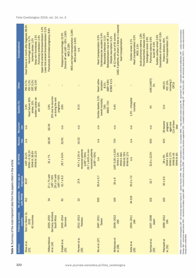
320
Folia Cardiologica 2019, vol. 14, no. 3
www.journals.viamedica.pl/folia_cardiologica
Tabl
e 4.
Sum
mar
y of t
he m
ost i
mpo
rtant
dat
a fro
m th
e pa
pers
citi
ed in
this
artic
le
Auth
ors
Follo
w-u
p pe
riod,
co
untr
yN
o. o
f pat
ient
s w
ith P
PCM
Mea
n ag
e at
di
agno
sis
EF/N
YHA
Rec
ove-
ries
Rec
urre
n-ce
sD
eath
s (%
)O
ther
Com
plic
atio
ns
Sliw
a et
al.
[23]
Star
t on
Mar
ch 3
1,
2016
43
cou
ntrie
s
411
30.0
7LV
EF: 3
2.2%
NY
HA I–
II: 3
1.2%
NY
HA II
I: 36
.6%
NY
HA IV
: 32.
2%
n/a
n/a
2.4%
He
art f
ailu
re: 6
0%
stro
ke: 1
0%
sudd
en c
ardi
ac d
e-at
h: 3
0%
PM: 0
.7%
; IC
D: 1
.2%
; CR
T: 0.
2%;
VAD:
2.0
%
Hear
t fai
lure
at 1
mon
th a
fter d
iagn
osis:
86.
9%
Hem
orrh
agic
stro
ke: 0
.7%
Is
chem
ic st
roke
: 1.0
%
Deep
-vein
thro
mbo
sis: 1
.5%
Pu
lmon
ary e
mbo
lism
: 2.4
%
Uppe
r lim
b ar
teria
l thr
ombo
embo
lism
: 1.0
%
Lowe
r lim
b ar
teria
l thr
ombo
embo
lism
: 0.2
%Hi
lfiker
-Kle
iner
et
al.
[24]
2005
–201
5 Ge
rman
y, Sc
otla
nd,
Sout
h Af
rica
3427
± 7
% w
ith
LVEF
< 5
0%
29 ±
5%
with
LV
EF >
50%
31 ±
7%
18/3
416
/34
2.9%
25
% d
ue to
recu
rren-
ce d
urin
g th
e ne
xt
preg
nanc
y
–Pr
emat
urel
y ter
min
ated
pre
gnan
cy: 8
.8%
Ersb
øll e
t al.
[25]
2005
–201
4 De
nmar
k61
31.7
± 6
.326
.7 ±
9.0
%32
/61
n/a
3.8%
–Po
stpa
rtum
hem
orrh
age:
22.
9%
Chro
nic
HF w
ith LV
EF <
35%
: 1.8
3%
MCS
: 3.2
8%
MCS
and
hea
rt tra
nspl
anta
tion:
3.2
8%
MCS
and
dea
th: 1
.64%
Perv
een
et a
l. [2
6]20
12–2
013
Paki
stan
2227
.444
.7 ±
2.3
% in
re
cove
red
patie
nts
(LVE
F >
50%
) 29
.7 ±
8.0
% in
non
--re
cove
red
patie
nts
(LVE
F <
50%
)
14/2
2n/
a9.
1%–
n/a
Wu
et a
l. [2
7]19
97–2
011
Taiw
an92
530
.4 ±
5.7
n/a
n/a
n/a
Card
iac
deat
hs: 3
.3%
ov
eral
l mor
talit
y: 7.
8%
MAC
E: 7
.0%
Hear
t cat
he-
teriz
atio
n:
8.1%
; IA
BP: 0
.8%
; EC
MO:
4.1
%
Hear
t tra
nspl
anta
tion:
0.5
%
Cere
brov
ascu
lar a
ccid
ent:
0.4%
M
yoca
rdia
l infa
rctio
n: 0
.1%
Re
hosp
italiz
atio
n du
e to
HF:
3.6
%
Dial
ysis
resu
mpt
ion:
0.2
%M
cNam
ara
et
al. [
28]
2009
–201
3 US
A10
030
± 6
LVEF
: 0,3
5 ±
0,10
%
NYHA
I: 1
2%
NYHA
II: 4
6%
NYHA
III:
25%
NY
HA IV
: 17%
72%
n/a
4.4%
–At
12
mon
ths,
out
of 9
1 wo
men
: LV
AD: 4
pat
ient
s, o
f who
m 2
die
d an
1 re
quire
d he
art t
rans
plan
tatio
n
Kolte
et a
l. [2
9]20
04–2
011
USA
34 2
1930
.3 ±
7.0
n/a
n/a
n/a
1.3%
– in
hosp
ital
mor
talit
y–
Card
iac
arre
st: 2
.1%
He
art t
rans
plan
tatio
n: 0
.5%
M
CS: 1
.5%
Ac
ute
pulm
onar
y ede
ma:
1,8
%
Thro
mbo
embo
lic d
iseas
e: 6
.6%
Ca
rdio
geni
c sh
ock:
2.6
%Ka
miya
et a
l. [3
0]20
07–2
008
Japa
n10
232
.731
,6 ±
12,
0%63
%n/
a4%
LVAS
[LVA
D?]:
2%Pu
lmon
ary e
dem
a: 0
.9%
Ac
ute
HF: 0
.9%
Su
dden
car
diac
arre
st: 0
.9%
Ex
acer
batio
n of
chr
onic
HF: 0
.9%
Pilla
riset
ti et
al
. [31
]19
99–2
012
USA
100
30 ±
6.5
28 ±
9%
NY
HA I:
30%
NY
HA II
: 17%
NY
HA II
I: 45
NY
HA IV
: 8%
42%
35 b
ecam
e pr
egna
nt
agai
n an
d 11
% of
them
di
ed
11%
VAD:
1%
ICD:
13%
, in
cludi
ng 2
CR
T-D
Pret
erm
del
ivery
: 40%
He
art t
rans
plan
tatio
n: 2
%
PPCM
— p
erip
artu
m c
ardi
omyo
path
y; LV
EF —
left
vent
ricul
ar e
ject
ion
fract
ion;
EF
— ej
ectio
n fra
ctio
n; N
YHA
— Ne
w Yo
rk H
eart
Asso
ciatio
n; n
/a —
not
ava
ilabl
e; M
ACE
— m
ajor
adv
erse
car
diac
eve
nts;
PM
– p
acem
aker
?; IC
D —
impl
anta
ble
card
iove
rter-d
efibr
illato
r; CR
T —
card
iac
resy
nchr
onisa
tion
ther
apy;
VAD
— ve
ntric
ular
ass
ist d
evice
; MCS
— m
echa
nica
l circ
ulat
ory s
uppo
rt; IA
BP —
intra
-aor
tic b
allo
on p
ump;
ECM
O —
extra
cor
pore
al m
embr
ane
oxyg
enat
ion;
ESC
— E
urop
ean
Socie
ty o
f Car
diol
ogy;
HF —
hea
rt fa
ilure
; LVA
D —
left
vent
ricul
ar a
ssist
dev
ice; C
RT-D
— c
ardi
ac re
sync
hron
izatio
n th
erap
y with
a d
efibr
illato
r

321www.journals.viamedica.pl/folia_cardiologica
Kiet Anh Nguyen et al., Peripartum cardiomyopathy. The current state of knowledge
with restored left ventricular function, which remain stable after HF medication reduction, an annual follow-up visit is recommended for the next 10 years [22].
Prognosis
Although the clinical course of PPCM may vary considerably, the general prognosis of patients after normal treatment is good. About 50% of women have a full recovery (defined as LVEF > 55% and NYHA class I), while 35–40% improve (defined as an increase of LVEF by more than 10% and improvement of at least one NYHA class) [12]. Based on the literature, mortality varies between 2% in Germany and 12.6% in South Africa 1–6 months after delivery. The mortality rate between six and 12 months after childbirth ranges from 4% to 14%; for women of African descent it is 12–14%. Subsequently, in the range of 1–5 years, mor-tality in a population of 182 women in the United States was 7% at the time of the follow-up examination in the 19th month. For African-American patients, this rate was higher and in the second year of the study, the mortality rate was 28% in South Africa, 16% in the USA and 15% in Haiti. Between the second and the fifth year, these values varied significantly depending on the region, and ranged from 0–6%, in France and the United States, to 15–30% in China, Brazil, Turkey, South Africa and the Philippines. There is little data available for a prognosis of more than five years. In this period, mortality is 7–16% in India and 8.3% in Malaysia [8].
In addition, there have as yet been no publications on the long-term prognosis for patients after heart transplan-tation and with an implanted left ventricle assist device (LVAD). Table 4 summarises the most important information from work on PPCM [23–31].
Conclusions
In recent years, the awareness about PPCM has risen, and a growing interest was sparked by recent publications. Although data on the extent of disease prevalence in most parts of the world is lacking, and many issues remains unexplained, in recent years a number of findings have been made that have contributed to improved PPCM diag-nosis and treatment. New biomarkers have been proposed, new underlying mechanisms contributing to its onset have been discovered, and bromocriptine treatment has been introduced. It has been documented that bromocriptine in PPCM therapy is beneficial, although more research is needed on the use of this medicine.
was found in 60% of cases, with 0% mortality. Patients did not require a transplant or the use of cardiac devices [21].
Prophylaxis and further management
Contraception Patients who have been diagnosed with PPCM are of childbearing age. The associated risk of PPCM recurrence, especially in the absence of ejection fraction parameters normalisation, makes it necessary to implement an effec-tive and relatively safe method of contraception [3].
According to the 2018 European Society of Cardiology (ESC) guidelines [15], ethinyl oestradiol drugs are con-traindicated in patients with PPCM due to the high risk of thromboembolic complications. Sliwa et al. described the possible consequences [3]. In their opinion, oral two--component agents combining oestrogen and progesterone derivatives are not a good method due to the adverse thromboembolic profile. Gestagens are an alternative to these measures. It is believed that they have little effect on the risk of thromboembolism in the case of subcutaneous implants and injections, and pose no thromboembolic risk in oral form and in intrauterine devices. The safety profile and high efficacy of levonorgestrel available in the form of an intrauterine system and a subcutaneous implant are particularly appreciated [15].
It should be remembered that the right form of contra-ception is the key to prevent the recurrence of PPCM. It is necessary to present a patient with possible ways of preven-ting another pregnancy. During the choice of optimal contra-ceptive therapy, we are obliged to take into account both the medical indications and the comfort in use for the woman.
BreastfeedingIn a case of diagnosed PPCM, breastfeeding cessation sho-uld be considered. Persistent high levels of prolactin during lactation may adversely affect the course of the disease and prognosis of the patient. Therefore, patients with significant left ventricular systolic dysfunction should not breastfeed and should be considered for bromocriptine. An exception to the above recommendation is a situation where a small impairment of left ventricular systolic function coexists with poor access to modified milk and unfavourable envi-ronmental conditions. In these circumstances, sustaining breastfeeding seems to be justified [22].
Follow-upIt is recommended that echocardiography should be perfor-med every six months until LVEF returns > 50%. In women

322
Folia Cardiologica 2019, vol. 14, no. 3
www.journals.viamedica.pl/folia_cardiologica
StreszczenieKardiomiopatię połogową (PPCM) definiuje się jako idiopatyczną kardiomiopatię, która występuje pod koniec ciąży lub w pierwszych kilku miesiącach po porodzie z objawami niewydolności serca (HF) wtórnie do zaburzeń czynności lewej komory i jednocześnie nie stwierdza się żadnej innej przyczyny tego stanu. Patomechanizm choroby nie został w pełni poznany, natomiast prawdopodobnie opiera się na działaniu złożonych czynników. Przebieg kliniczny PPCM jest zróż-nicowany: od zagrażającej życiu ostrej HF po łagodne objawy naśladujące dolegliwości charakterystyczne dla okresu okołoporodowego. W Europie PPCM jest dość rzadką chorobą, natomiast są rejony, gdzie występuje z częstością 1/300 przypadków (Haiti). Leczenie PPCM jest podobne jak dla HF z obniżoną frakcją wyrzutową, jednak ważne jest wyklu-czenie leków o działaniu teratogennym w trakcie trwania ciąży. Włączanie bromokryptyny do terapii PPCM wydaje się zasadne i w niektórych przypadkach poprawia rokowanie.
Słowa kluczowe: kardiomiopatia połogowa, PPCM, niewydolność serca, ciąża, bromokryptynaFolia Cardiologica 2019; 14, 3: 315–323
References1. Arany Z, Elkayam U. Peripartum cardiomyopathy. Circulation. 2016;
133(14): 1397–1409, doi: 10.1161/CIRCULATIONAHA.115.020491, indexed in Pubmed: 27045128.
2. Murali S, Baldisseri MR. Peripartum cardiomyopathy. Crit Care Med. 2005; 33(10 Suppl): S340–S346, indexed in Pubmed: 16215357.
3. Sliwa K, Hilfiker-Kleiner D, Petrie MC, et al. Heart Failure Association of the European Society of Cardiology Working Group on Peripartum Cardiomyopathy. Current state of knowledge on aetiology, diagnosis, management, and therapy of peripartum cardiomyopathy: a position statement from the Heart Failure Association of the European Society of Cardiology Working Group on peripartum cardiomyopathy. Eur J He-art Fail. 2010; 12(8): 767–778, doi: 10.1093/eurjhf/hfq120, indexed in Pubmed: 20675664.
4. Pearson GD, Veille JC, Rahimtoola S, et al. Peripartum cardiomy-opathy: National Heart, Lung, and Blood Institute and Office of Rare Diseases (National Institutes of Health) workshop recommen-dations and review. JAMA. 2000; 283(9): 1183–1188, indexed in Pubmed: 10703781.
5. Ware JS, Li J, Mazaika E, et al. IMAC-2 and IPAC Investigators. Shared genetic predisposition in peripartum and dilated cardiomyopathies. N Engl J Med. 2016; 374(3): 233–241, doi: 10.1056/NEJMoa1505517, indexed in Pubmed: 26735901.
6. van Spaendonck-Zwarts KY, van Tintelen JP, van Veldhuisen DJ, et al. Peripartum cardiomyopathy as a part of familial dilated cardio-myopathy. Circulation. 2010; 121(20): 2169–2175, doi: 10.1161/CIRCULATIONAHA.109.929646, indexed in Pubmed: 20458010.
7. Bültmann BD, Klingel K, Näbauer M, et al. High prevalence of viral ge-nomes and inflammation in peripartum cardiomyopathy. Am J Obstet Gynecol. 2005; 193(2): 363–365, doi: 10.1016/j.ajog.2005.01.022, indexed in Pubmed: 16098856.
8. Azibani F, Sliwa K. Peripartum Cardiomyopathy: an Update. Curr Heart Fail Rep. 2018; 15(5): 297–306, doi: 10.1007/s11897-018-0404-x, indexed in Pubmed: 30051292.
9. Hilfiker-Kleiner D, Kaminski K, Podewski E, et al. A cathepsin D-clea-ved 16 kDa form of prolactin mediates postpartum cardiomyopathy. Cell. 2007; 128(3): 589–600, doi: 10.1016/j.cell.2006.12.036, inde-xed in Pubmed: 17289576.
10. Patten IS, Rana S, Shahul S, et al. Cardiac angiogenic imbalance leads to peripartum cardiomyopathy. Nature. 2012; 485(7398): 333–338, doi: 10.1038/nature11040, indexed in Pubmed: 22596155.
11. Bello NA, Arany Z. Molecular mechanisms of peripartum cardiomyo-pathy: A vascular/hormonal hypothesis. Trends Cardiovasc Med. 2015; 25(6): 499–504, doi: 10.1016/j.tcm.2015.01.004, indexed in Pubmed: 25697684.
12. Koenig T, Bauersachs J, Hilfiker-Kleiner D, et al. Peripartum cardio-myopathy. Herz. 2018; 43(5): 431–437, doi: 10.1007/s00059-018-4709-z, indexed in Pubmed: 29767811.
13. Hilfiker-Kleiner D, Sliwa K. Pathophysiology and epidemiology of pe-ripartum cardiomyopathy. Nat Rev Cardiol. 2014; 11(6): 364–370, doi: 10.1038/nrcardio.2014.37, indexed in Pubmed: 24686946.
14. Patel H, Berg M, Barasa A, et al. Symptoms in women with Peripartum cardiomyopathy: a mixed method study. Midwifery. 2016; 32: 14–20, doi: 10.1016/j.midw.2015.10.001, indexed in Pubmed: 26515744.
15. Regitz-Zagrosek V, Roos-Hesselink JW, Bauersachs J, et al. ESC Scientific Document Group. 2018 ESC Guidelines for the management of cardio-vascular diseases during pregnancy. Eur Heart J. 2018; 39(34): 3165– –3241, doi: 10.1093/eurheartj/ehy340, indexed in Pubmed: 30165544.
16. Kim MJ, Shin MS. Practical management of peripartum cardiomy-opathy. Korean J Intern Med. 2017; 32(3): 393–403, doi: 10.3904/ /kjim.2016.360, indexed in Pubmed: 28407464.
17. Schelbert EB, Elkayam U, Cooper LT, et al. Investigations of Pregnancy Associated Cardiomyopathy (IPAC) Investigators. Myocardial damage detected by late gadolinium enhancement cardiac magnetic resonan-ce is uncommon in peripartum cardiomyopathy. J Am Heart Assoc. 2017; 6(4), doi: 10.1161/JAHA.117.005472, indexed in Pubmed: 28373243.
18. Halkein J, Tabruyn SP, Ricke-Hoch M, et al. MicroRNA-146a is a thera-peutic target and biomarker for peripartum cardiomyopathy. J Clin Invest. 2013; 123(5): 2143–2154, doi: 10.1172/JCI64365, indexed in Pubmed: 23619365.
19. Walenta K, Schwarz V, Schirmer SH, et al. Circulating microparti-cles as indicators of peripartum cardiomyopathy. Eur Heart J. 2012; 33(12): 1469–1479, doi: 10.1093/eurheartj/ehr485, indexed in Pubmed: 22307461.

323www.journals.viamedica.pl/folia_cardiologica
Kiet Anh Nguyen et al., Peripartum cardiomyopathy. The current state of knowledge
20. Leśniak-Sobelga A, Gąsior Z, Kostkiewicz M, et al. Współczesna kar-diologiczna diagnostyka obrazowa w ciąży. Stanowisko grupy eksper-tów polskiego Klinicznego Forum Obrazowania Serca i Naczyń. Kardiol Pol. 2013; 71(11): 1194–1203, doi: 10.5603/kp.2013.0305.
21. Hilfiker-Kleiner D, Haghikia A, Berliner D, et al. Bromocriptine for the treatment of peripartum cardiomyopathy: a multicentre randomized study. Eur Heart J. 2017; 38(35): 2671–2679, doi: 10.1093/eur-heartj/ehx355, indexed in Pubmed: 28934837.
22. Sliwa K, Petrie MC, Hilfiker-Kleiner D, et al. Long-term prognosis, subsequent pregnancy, contraception and overall management of peripartum cardiomyopathy: practical guidance paper from the Heart Failure Association of the European Society of Cardiology Study Group on Peripartum Cardiomyopathy. Eur J Heart Fail. 2018; 20(6): 951–962, doi: 10.1002/ejhf.1178, indexed in Pubmed: 29578284.
23. Sliwa K, Mebazaa A, Hilfiker-Kleiner D, et al. Clinical characteristics of patients from the worldwide registry on peripartum cardiomyopathy (PPCM): EURObservational Research Programme in conjunction with the Heart Failure Association of the European Society of Cardiology Study Group on PPCM. Eur J Heart Fail. 2017; 19(9): 1131–1141, doi: 10.1002/ejhf.780, indexed in Pubmed: 28271625.
24. Hilfiker-Kleiner D, Haghikia A, Masuko D, et al. Outcome of subse-quent pregnancies in patients with a history of peripartum cardiomyo-pathy. Eur J Heart Fail. 2017; 19(12): 1723–1728, doi: 10.1002/ /ejhf.808.
25. Ersbøll AS, Johansen M, Damm P, et al. Peripartum cardiomyopathy in Denmark: a retrospective, population-based study of incidence, man-
agement and outcome. Eur J Heart Fail. 2017; 19(12): 1712–1720, doi: 10.1002/ejhf.882, indexed in Pubmed: 28597481.
26. Perveen S, Ainuddin J, Jabbar S, et al. Peripartum cardiomyopathy: fre-quency and predictors and indicators of clinical outcome. J Pak Med Assoc. 2016; 66(12): 1517–1521, indexed in Pubmed: 27924958.
27. Wu VCC, Chen TH, Yeh JK, et al. Clinical outcomes of peripartum cardiomyopathy: a 15-year nationwide population-based study in Asia. Medicine (Baltimore). 2017; 96(43): e8374, doi: 10.1097/ /MD.0000000000008374, indexed in Pubmed: 29069030.
28. McNamara DM, Elkayam U, Alharethi R, et al. IPAC Investigators. Clini-cal outcomes for peripartum cardiomyopathy in North America: results of the IPAC Study (Investigations of Pregnancy-Associated Cardiomyo-pathy). J Am Coll Cardiol. 2015; 66(8): 905–914, doi: 10.1016/j.jacc.2015.06.1309, indexed in Pubmed: 26293760.
29. Kolte D, Khera S, Aronow WS, et al. Temporal trends in incidence and outcomes of peripartum cardiomyopathy in the United States: a nation-wide population-based study. J Am Heart Assoc. 2014; 3(3): e001056, doi: 10.1161/JAHA.114.001056, indexed in Pubmed: 24901108.
30. Kamiya CA, Kitakaze M, Ishibashi-Ueda H, et al. Different characte-ristics of peripartum cardiomyopathy between patients complicated with and without hypertensive disorders. Results from the Japanese Nationwide survey of peripartum cardiomyopathy. Circ J. 2011; 75(8): 1975–1981, indexed in Pubmed: 21617320.
31. Pillarisetti J, Kondur A, Alani A, et al. Peripartum cardiomyopathy: predictors of recovery and current state of implantable cardioverter- -defibrillator use. J Am Coll Cardiol. 2014; 63(25 Pt A): 2831–2839, doi: 10.1016/j.jacc.2014.04.014, indexed in Pubmed: 24814494.

324 www.journals.viamedica.pl/folia_cardiologica
Folia Cardiologica 2019 tom 14, nr 3, strony 324–327 DOI: 10.5603/FC.2019.0065 Copyright © 2019 Via Medica
ISSN 2353–7752
KARDIOCHIRURGIA
Address for correspondence: lek. Filip Pawliczak, Klinika Kardiologii i Wad Wrodzonych Dorosłych, Instytut Centrum Zdrowia Matki Polki w Łodzi, ul. Rzgowska 281/289, 93–338 Łódź, Poland, e-mail: [email protected]
A previously asymptomatic two month-old infant with unique presentation of cor triatriatum sinister, right partial anomalous pulmonary venous return, and critical supravalvular stenosis of mitral valve
Dotychczas bezobjawowe dwumiesięczne niemowlę z unikatową prezentacją trójprzedsionkowego lewego serca, prawego częściowego nieprawidłowego
spływu żył płucnych oraz krytycznej nadzastawkowej stenozy mitralnej
Filip Pawliczak1●iD, Katarzyna Ostrowska2●iD, Jacek Moll2●iD, Jadwiga A. Moll2●iD1Klinika Kardiologii i Wad Wrodzonych Dorosłych, Polish Mother’s Memorial Hospital Research Institute in Lodz, Lodz, Poland
2Department of Cardiology, Polish Mother’s Memorial Hospital Research Institute in Lodz, Lodz, Poland
AbstractMajor developments in diagnostic techniques in pre- and neonatal care have significantly reduced the rate of undetec-ted congenital heart defects (CHD). Despite such advances, several patients with critical congenital heart defects are discharged annually from neonatal units with no proper diagnosis or treatment.We present the case of a two month-old originally asymptomatic girl who represents the perfect example of such a situation. The infant was diagnosed just after pulmonary and cardiovascular decompensation with critical, complex CHD. The absence of disease symptoms of occurred due to a rare and specific morphology of pulmonary vessels and intracardiac membranes.
Key words: congenital heart defect, cor triatriatum sinister, anomalous pulmonary venous return, mitral stenosis, supravalvular, echocardiography, diagnosis, intervention, surgery, screening
Folia Cardiologica 2019; 14, 3: 324–327
Case report
The patient was born as a eutrophic neonate at the 38th week of gestation vaginally, reaching 10 in the Apgar score. The mother claimed it was her first pregnancy and she had received all recommended obstetrician ultrasonography (USG) examinations on time. The second trimester USG was repeated at the mother’s request. However, no defects were described in any of the prenatal examinations. The girl had no symptoms of cardiovascular disease. There was a negative result from the patients with critical congenital
heart defects (CCHD) protocol screening pulse oximetry test. The first signs of a cardiac problem appeared in the second month of life, a few days after a scheduled vaccina-tion. Slight fatigue and lack of appetite were noted, however no signs of infection or a gastrointestinal system problem were observed. The cautious mother decided to take the infant to see their general practitioner. Medical examination revealed no murmurs or objective signs of heart disease. Eventually, before leaving the doctor’s surgery, the child developed sudden peripheral cyanosis, consciousness dis-orders, and heavy dyspnoea. The patient was transported

325www.journals.viamedica.pl/folia_cardiologica
Filip Pawliczak et al., Unique presentation of cor triatriatum sinister, pulmonary partial anomalous pulmonary venous
Shortly after, the patient underwent full surgical cor-rection of pulmonary venous connections in conditions of extracorporeal circulation and deep hypothermia. The earlier diagnosis was finally confirmed intraoperatively as proper left pulmonary venous return was restricted by intraatrial structures blocking the mitral valve inflow. Additional membranes in the left atrium were resected with simultaneous restoration of the atrial septum with a pericardial patch. The overall condition of the patient soon after surgery was stable. Unfortunately, several post--operative complications occurred. The patient developed end-stage kidney failure, thus treatment with peritoneal dialysis was necessary as well as prolonged mechanical ventilation due to an inability to breathe unaided. Further-more, various bacterial species were found in the blood tests, resulting in administration of a wide spectrum of antibiotics. A few days later, fungi were discovered in the patient’s urine which necessitated antifungal therapy. Due to these multiple health problems, total hospitalisation time was significantly prolonged and lasted up to two months after surgery.
Nevertheless, the patient survived and conquered all her challenges. At discharge, the girl was in an excellent condition and had no signs of her previous illnesses. ECHO revealed normalisation of chamber sizes and an optimal haemodynamic result of the surgical intervention. The six-month follow-up proved cardiologic management of the defect to be working, as the patient was developing correctly without any disabilities (Figures 1–3).
to the Mother and Child Institute, Warsaw where severe cardiopulmonary failure was diagnosed. First transthoracic echocardiography (ECHO) suggested anomalous pulmonary venous return and additional structures in the left atrium (LA). It was decided to airlift her to the Paediatric Cardiology and Cardiosurgery Unit in the Polish Mothers’ Memorial Hospital Research Institute in Lodz. The girl was admitted in a critical condition. The next ECHO revealed an additio-nal vertical structure in the left atrium imitating intraatrial septum and an additional horizontal structure critically obstructing mitral inflow, and left pulmonary venous return located physiologically. Moreover, right pulmonary veins were returning directly to the right atrium.
The patient was primarily diagnosed with cor triatriatum sinister (CTS) and partial anomalous pulmonary venous re-turn (PAPVR). Due to the exacerbation of the patient’s con-dition, angiographic intervention was decided upon, which confirmed pathological right pulmonary venous return and displayed enlarged right chambers. Measurements of pulmonary circulation pressures allowed a diagnosis of massive pressure overload of the right heart. Further examination revealed the left atrium without any pulmonary inflow. This revelation suggested an explanation of the rapid progression of cardiopulmonary failure to cardiac shock due to a unique presentation of pathologies acting simi-larly to total anomalous pulmonary venous return (TAPVR). A life-saving Rashkind-like procedure was performed on the additional structures of the left atrium, resulting in stabilisation of the patient’s haemodynamic condition.
Figure 1. Preoperative state: A, B. Observable pressure and volume overload of right atrium and ventricle; C. Schematic presentation of concomitant defects
A
B C

326
Folia Cardiologica 2019, vol. 14, no. 3
www.journals.viamedica.pl/folia_cardiologica
Discussion
The presented case describes a very rare situation wherein a coexistence of several factors led to the misdiagnosis of a severely ill child. Early detection of CCHD is crucial as any delay in diagnosis significantly affects morbidity and mortality. Such early detection is possible in the antenatal period and is now common due to the implementation of the pulse oximetry screening test [1]. The examination re-sult is based on the observable decreased blood saturation level in neonates with CCHD. The test is highly sensitive and specific. However, several reports of undetected serious cardiac defects appear annually [2]. The main limitation of the method is the differing time of transformation of the foetal circulation into a post-natal one, especially when a unique heart morphology co-exists. If the pulse oximetry test is performed before shunt closure, it can give a false negative result. Such a situation probably occurred in the presented case. The patient screening test went negatively despite the life-threatening anomaly. The presentation of symptoms in such a situation is typically a matter of time. Prolonged shutting of physiological shunts and enlarging obstruction of inflow usually leads to haemodynamic de-compensation in infancy. The sudden aggravation of this patient’s condition appeared only after a rapid decrease in the amount of blood flowing into the left atrium, resulting in cardiac shock.
Nonetheless, standard post-natal pulse oximetry screening is not the only method to determine whether a child is in the high risk group for CHD existence. Obste-trician USG pre-natal screening is popular world-wide and provides moderately sensitive and specific data about the foetal heart and vessels [3]. The most common objection
Figure 2. Catheterisation: A. Left atrium with anomalous pulmonary venous return and obstructed outflow due to membrane over mitral valve; B. Rashkind procedure
A B
Figures 3A, B. Post-operative state: observable normalisation of chamber sizes
A
B
made against this method is that the variation in skill levels among USG operators determines the diagnostic outcome of the examination. If performed incautiously, or by a person not trained in foetal heart visualisation, CHD can easily be
327www.journals.viamedica.pl/folia_cardiologica
Filip Pawliczak et al., Unique presentation of cor triatriatum sinister, pulmonary partial anomalous pulmonary venous
missed [4]. Even so, rare or not prominent CHD may be described as a variant of a normal developing heart. This is probably true of the presented patient because double second trimester USGs (by two different operators) revealed no red flag symptoms requiring referral to the prenatal cardiology unit.
The difficulty level of diagnosing the presented patient was also increased due to a very uncommon and specific concomitance of several heart pathologies which may not give any symptoms in the first months of life. The prevalence of cor triatriatum sinister is estimated to be only 0.4% of all CHDs. It is best described as left atrium division into two compartments by an additional septum — the first with pulmonary venous return and the second with mitral inflow [5]. The presented patient was also complicated by horizontal configuration of abnormal membranes in the left atrium with trans-membranous obstructed flow resulting in severe supravalvular mitral stenosis (MS). Such a situation has only been described in a few cases [6]. Partial ano-malous pulmonary venous return is a condition defined as
abnormal connection of one-sided pulmonary veins to the incorrect atrial chamber or vessels. It is found in approxi-mately 0.4–0.7% of all CHDs [7]. The case described here is cardiac type right PAPVR. However, in the first two months of the girl’s life, the small connection between the atrial chambers and the foramen ovale was probably persistent. Unfortunately, the coexistence of all anomalies (CTS, MS and PAPVR), as well as heart growth with restricting flows, resulted in a very rare clinical presentation imitating total anomalous pulmonary venous return that affected the patient’s condition dramatically due to the critical limitation of inflow to the left ventricle and the massive overload of pulmonary circulation.
The essence of this case is a long chain of rare patho-physiologic events leading to a lack of diagnosis in the pre- and postnatal periods causing a life-threatening situ-ation. It also shows the limitations of screening techniques, and thus emphasises the role of proper training for USG operators and the significance of a cautious neonatal examination based on experience and additional tests.
StreszczenieOgromny rozwój technik diagnostycznych w opiece przed- i okołoporodowej znacząco zmniejszył odsetek pacjentów z przeoczoną wrodzoną wadą serca. Mimo tego postępu, corocznie kilku pacjentów z krytyczną anomalią układu ser-cowo-naczyniowego jest wypisywanych bez odpowiedniej diagnozy i leczenia. Przedstawiony przypadek 2-miesięcznej, pierwotnie niewykazującej objawów dziewczynki jest doskonałym przykładem takiej sytuacji. Niemowlę zdiagnozowano tuż po ostrej dekompensacji krążeniowo-oddechowej. Początkowy brak objawów wiązał się z rzadką i nietypową morfo-logią współwystępujących anomalii żył płucnych i dodatkowych błon wewnątrzsercowych.
Słowa kluczowe: wrodzona wada serca, lewe serce trójprzedsionkowe, nieprawidłowy spływ żył płucnych, stenoza mitralna, nadzastawkowa, echokardiografia, diagnoza, interwencja, operacja, badanie przesiewowe
Folia Cardiologica 2019; 14, 3: 324–327
References1. Rosati E, Chitano G, Dipaola L, et al. Indications and limitations
for a neonatal pulse oximetry screening of critical congenital he-art disease. J Perinat Med. 2005; 33(5): 455–457, doi: 10.1515/ /JPM.2005.080, indexed in Pubmed: 16238542.
2. Koppel RI, Druschel CM, Carter T, et al. Effectiveness of pulse oximetry screening for congenital heart disease in asymptomatic newborns. Pediatrics. 2003; 111(3): 451–455, doi: 10.1542/peds.111.3.451.
3. Zhang YF, Zeng XL, Zhao EF, et al. Diagnostic value of fetal echocar-diography for congenital heart disease: a systematic review and meta--analysis. Medicine (Baltimore). 2015; 94(42): e1759, doi: 10.1097/ /MD.0000000000001759, indexed in Pubmed: 26496297.
4. Ganesan S, Brook MM, Silverman NH, et al. Prenatal findings in total anomalous pulmonary venous return: a diagnostic road map starts with obstetric screening views. J Ultrasound Med. 2014; 33(7):
1193–1207, doi: 10.7863/ultra.33.7.1193, indexed in Pubmed: 24958406.
5. Geggel RL, Fulton DR, Chernoff HL, et al. Cor triatriatum associated with partial anomalous pulmonary venous connection to the coronary sinus: echocardiographic and angiocardiographic features. Pediatr Cardiol. 1987; 8(4): 279–283, doi: 10.1007/BF02427543, indexed in Pubmed: 3432119.
6. Slight RD, Nzewi OC, Buell R, et al. Cor-triatriatum sinister presenting in the adult as mitral stenosis: an analysis of factors which may be relevant in late presentation. Heart Lung Circ. 2005; 14(1): 8–12, doi: 10.1016/j.hlc.2004.10.003, indexed in Pubmed: 16352245.
7. Gustafson RA, Warden HE, Murray GF, et al. Partial anomalous pulmo-nary venous connection to the right side of the heart. J Thorac Cardio-vasc Surg. 1989; 98(5 Pt 2): 861–868, indexed in Pubmed: 2682021.

328
Folia Cardiologica 2019, tom 14, nr 3
www.journals.viamedica.pl/folia_cardiologica
KomentarzIreneusz Haponiuk1, 2, Katarzyna Gierat-Haponiuk2
1Oddział Kardiochirurgii Dziecięcej Szpitala św. Wojciecha w Gdańsku–Zaspie 2Katedra Fizjoterapii Wydziału Rehabilitacji i Kinezjologii Akademii Wychowania Fizycznego i Sportu im. Jędrzeja Śniadeckiego w GdańskuAutorzy przedstawili interesujący raport z leczenia pacjentki z rzadką wrodzoną wadą serca o przebiegu i obrazie klinicznym mogącym sugerować całkowity nieprawidłowy spływ żył płucnych [1]. Wady nie rozpoznano w badaniach prenatalnych ani we wczesnym okresie noworodkowym, a wykonywane sumiennie przesiewowe badania przedmiotowe oraz ocena czynnościowa noworodka wraz z testem pulsoksymetrycznym okazały się zawodne. Pełnoobjawowo wada ujawniła się u dziecka w okresie późniejszym, co osta-tecznie doprowadziło do rozpoznania częściowego nieprawidłowego spływu prawych żył płucnych oraz nietypowej morfologii lewego serca trójprzedsionkowego, dającego objawy nadzastawkowej stenozy mitralnej [1].Na uwagę zasługuje bardzo rzetelne przedstawienie problemów i pułapek diagnostycznych towarzyszących tej, mogącej powodować trudności, wrodzonej wady serca o asymptomatycz-nym wczesnym przebiegu u niemowlęcia. Nasilone objawy pod postacią napadowej duszno-ści z sinicą wystąpiły dopiero w 2. miesiącu życia. Na podstawie wstępnej oceny w przezklat-kowym badaniu echokardiograficznym (TTE, transthoracic echocardiography) rozpoznano nieprawidłowy spływ żył płucnych. Szczegółową ocenę dziecka z nasilającymi się objawami wstrząsu kardiogennego w badaniu angiograficznym uzupełniono o ratunkową procedurę balonowego poszerzenia dodatkowych struktur błoniastych w świetle lewego przedsionka (LA, left atrium), utrudniających napływ do zastawki mitralnej, co doraźnie poprawiło stan
pacjentki. Ostatecznie dziewczynkę operowano w trybie pilnym; w trakcie zabiegu uwolniono nadzastawkowe błoniaste zwężenia w świetle LA oraz wszczepiono kierunkową łatę osierdziową tunelizującą spływ prawych żył płucnych do LA.Podstawą diagnostyki większości wad wrodzonych, w tym także krytycznych wad serca i naczyń u noworodków i niemowląt, jest obecnie TTE [2]. W przypadku niejednoznacznych obrazów przezklatkowych kolejny krok to wykonanie przezprzełyko-wego badania echokardiograficznego (TEE, transesophageal echocardiography). Metodą niejednokrotnie rozstrzygającą o właściwym rozpoznaniu bywa diagnostyka inwazyjna — cewnikowanie serca z angiografią, a także tomografia kompute-rowa. Ostatecznej weryfikacji dokonuje kardiochirurg w śródoperacyjnej ocenie u pacjentów zakwalifikowanych do zabiegu.W dostępnym piśmiennictwie opisy wad o podobnej manifestacji z dynamicznie narastającą niewydolnością krążenio-wo-oddechową u niemowląt mają charakter kazuistyczny [2]. Diagnostyka i leczenie pacjenta z taką koegzystencją wad wymagają współpracy wielospecjalistycznej, z wykorzystaniem potencjału interwencji przezskórnych, oraz zaawanso-wanych metod intensywnej terapii, w tym wspomagania krążeniowo-oddechowego metodą ciągłego pozaustrojowego natleniania krwi (ECMO, extracorporeal membrane oxygenation). Szczególnie mylące w opinii wielu autorów bywają objawy przeciążenia i niewydolności prawej komory [3].Pragniemy pogratulować Autorom dobrej pracy i zachęcamy do jej lektury, zważywszy na wysoką wartość edukacyjną tego opracowania. Zdecydowanie, zgodnie z opinią Autorów, podzielamy wątpliwości dotyczące przesiewowego testu pulsoksymetrycznego, którego wynik negatywny nie wyklucza zasadności wykonywania przesiewowych badań echo-kardiograficznych w diagnostyce krytycznych wrodzonych wad serca i naczyń u noworodków [4].
Piśmiennictwo1. Pawliczak F, Ostrowska K, Moll J, et al. A previously asymptomatic
two month-old infant with unique presentation of cor triatriatum si-nister, right partial anomalous pulmonary venous return, and critical supravalvular stenosis of mitral valve. Folia Cardiol. 2019; 14(3): 324–327, doi: 10.5603/FC.2019.0065.
2. Burger C. A rare case of cor triatriatum leading to respiratory fai-lure and shock in an 8-month-old boy. Pediatr Emerg Care. 2019; 35(1): e6–e8, doi: 10.1097/PEC.0000000000001113, indexed in Pubmed: 28350721.
3. Binsalamah ZM, De León LE, Heinle JS. Cor triatriatum sinister with an intact interatrial septum and a decompressing vein in a toddler. Cardiol Young. 2017; 27(6): 1221–1224, doi: 10.1017/ /S1047951117000130, indexed in Pubmed: 28260553.
4. Robinson DL, Craig MS, Wells MS, et al. Newborn screening pulse oximetry to detect critical congenital heart disease: a follow-up of current practice at army, navy and air force hospitals. Mil Med 2019 May 15, doi: 10. 1093/milmed/usz119. (Epub ahead of print), doi: 10.1093/milmed/usz119.
ORGANIZATOR PATRONAT MEDIALNY PARTNER
IV Konferencja
CARDIOLIPIDpod patronatem
Sekcji Farmakoterapii Sercowo-Naczyniowej Polskiego Towarzystwa Kardiologicznego
2019 ROKU
prof. dr hab. n. med. Krzysztof J. Filipiakdr n. med. Marcin Barylski
www.cardiolipid.viamedica.pl
PATRONAT METYRORYCZNY
FIBRAT
STATYNY
EZETIMIBAFEREZA
INHIBITORPCSK9
MODYFIKACJA STYLU ŻYCIA

prof. dr hab. n. med. Beata Wożakowska-Kapłon, prof. dr hab. n. med. Krzysztof J. Filipiak, FESC
VII Forum Chorób Sercowo-Naczyniowych
-
WRZESIEŃ
Kraków 12.09.2019Łódź 13.09.2019Lublin 14.09.2019 Wrocław 20.09.2019
BEZPŁATNE UCZESTNICTWO
PAŹDZIERNIK
Katowice 11.10.2019