Large “Pillow” Fullerenes Hydrogenated at the Inter ... - CiteSeerX
Upload
independentCategory
view
0download
0
Potential Fossil Endoliths in Vesicular Pillow Basalt,Coral Patch Seamount, Eastern North Atlantic Ocean
Barbara Cavalazzi,1,2 Frances Westall,2 Sherry L. Cady,3 Roberto Barbieri,4 and Frederic Foucher2
Abstract
The chilled rinds of pillow basalt from the Ampere–Coral Patch Seamounts in the eastern North Atlantic werestudied as a potential habitat of microbial life. A variety of putative biogenic structures, which include fila-mentous and spherical microfossil-like structures, were detected in K-phillipsite–filled amygdules within thechilled rinds. The filamentous structures (*2.5 lm in diameter) occur as K-phillipsite tubules surrounded by anFe-oxyhydroxide (lepidocrocite) rich membranous structure, whereas the spherical structures (from 4 to 2 lm indiameter) are associated with Ti oxide (anatase) and carbonaceous matter.
Several lines of evidence indicate that the microfossil-like structures in the pillow basalt are the fossilizedremains of microorganisms. Possible biosignatures include the carbonaceous nature of the spherical structures,their size distributions and morphology, the presence and distribution of native fluorescence, mineralogicaland chemical composition, and environmental context. When taken together, the suite of possible biosignaturessupports the hypothesis that the fossil-like structures are of biological origin. The vesicular microhabitat ofthe rock matrix is likely to have hosted a cryptoendolithic microbial community. This study documents a varietyof evidence for past microbial life in a hitherto poorly investigated and underestimated microenvironment,as represented by the amygdules in the chilled pillow basalt rinds. This kind of endolithic volcanic habitatwould have been common on the early rocky planets in our Solar System, such as Earth and Mars. This studyprovides a framework for evaluating traces of past life in vesicular pillow basalts, regardless of whether theyoccur on early Earth or Mars. Key Words: Fossil microbes—Microhabitat—Vesicular pillow basalt. Astrobiology11, 619–632.
1. Introduction
The oceanic lithosphere contributes significantly tobiogeochemical cycling and the amount of biomass and
biodiversity on Earth. It is also an important habitat for mi-crobial life (Furnes and Staudigel, 1999; Furnes et al., 2007;Santelli et al., 2008; Staudigel et al., 2008; McLoughlin et al.,2009; Nielsen and Fisk, 2010). When the temperature of newlyformed oceanic rocks drops to values that become tolerablefor life, which is estimated to be less than 121–122�C (Takaiet al., 2001; Kashefi and Lovley, 2003), they can be colonizedby microbial communities that utilize the inorganic elementsand compounds of the rocky substratum as energy sources(Thorseth et al., 1995). A wide variety of evidence, which in-cludes the presence of fossilized microbes, spherical and tu-bular alteration cavities, and geochemical and molecularsignatures consistent with microbial remains and metabo-
lisms, is commonly associated with bioalteration of modernand ancient oceanic volcanic glass (for a comprehensive re-view see Staudigel et al., 2008). Chemolithoautotrophic mi-croorganisms have an affinity for many of the bioessentialelements (e.g., Fe2 + , Mn4 + , SO2�
4 , CO2) present in basalticsilicates, which include olivine, pyroxene, feldspar, andhornblende (Fisk et al., 2006, and references therein). Altera-tion of the silicate minerals as a result of weathering and mi-crobial extraction releases the metabolically significant cations(Kostka et al., 2002; Kim et al., 2004). Bioalteration of the glassybasalts, which develops progressively along fracture surfacesand in and around vesicles produced by degassing, is ac-companied by the formation of authigenic phases (e.g.,smectite and phillipsite) in the zone of alteration. Staudigeland coworkers (2008) presented a schematic model that il-lustrates the developmental stages and variety of mechanismsinvolved in microbial alteration of volcanic glasses.
1Department of Geology, University of Johannesburg, Johannesburg, South Africa.2Centre de Biophysique Moleculaire–CNRS, Orleans, France.3Department of Geology, Portland State University, Portland, Oregon, USA.4Dipartimento di Scienze della Terra e Geologico-Ambientali, Universita di Bologna, Bologna, Italy.
ASTROBIOLOGYVolume 11, Number 7, 2011ª Mary Ann Liebert, Inc.DOI: 10.1089/ast.2011.0657
619

The primary glassy rinds of basalts provide a microbialhabitat for endoliths, organisms that reside in the interiorof rocks, and euendoliths, microbes that actively bore intothe rock substratums and create microtubular cavities(McLoughlin et al., 2007). Chemical alteration of the glassyrinds of seafloor basalts and of extrusive volcanic rock in-creases the range of microenvironments that could supportsuch microbial communities. The diversity of glassy habitatssupports communities of epiliths (eukaryotic and prokary-otic microorganisms that live by attaching themselves to theexternal surfaces of rocks), chasmoendoliths (endoliths thatinhabit fissures and cracks in rocks), and cryptoendoliths(endoliths that are hypothesized to favor the colonization ofrocks that have preexisting interstices and porous structures,sensu Santelli et al., 2008).
Recent studies have shown that prokaryotes and eukary-otes also inhabit the primary and alteration rinds of crys-talline volcanic rocks. Schumann et al. (2004) describedputative fossilized fungal life in vesicles of deep oceanicbasalt crust collected from a site in the North Pacific (ODPSite 1224). Connell et al. (2009) described fungi they discov-ered on the surfaces of basalts that formed relatively recentlyat a still-active deep-sea volcano (Vailulu’u Seamount, Sa-moa). Ivarsson et al. (2008) reported the discovery of fossilchasmoendoliths in zeolite-filled veins associated with sub-seafloor basaltic rocks sampled at the Emperor Seamounts inthe Pacific Ocean. Peckmann et al. (2008) and Eickmann et al.(2009) found fossil evidence of marine cryptoendolithicprokaryotes within the carbonate amygdules (mineral-filledvesicles) of Devonian pillow basalts from RheinischesSchiefergebirge (Germany), Frankenwald, and ThuringerWald within the Saxothuringian zone in Germany. Theselatter workers, in particular, emphasized the need for moreresearch to reveal the ecological niche that cavities andamygdules in seafloor basalts could provide for life in vol-canic rocks.
Chasmo- and cryptoendolithic microorganisms that in-habit the margins, walls, and pore spaces of fluid-filledcavities, voids, veins, and fractures of basaltic rocks can be-come fossilized via the precipitation of authigenic minerals,
such as carbonates, clays, minerals, and zeolites (e.g., Ivars-son et al., 2008; Peckmann et al., 2008). In this study, wepresent the results of a detailed investigation of objects withmicroorganism-like morphologies that occur within phillip-site-filled amygdules located at and near the outer surfaces ofpillow basalts recovered from the Coral Patch Seamount inthe eastern North Atlantic Ocean (Fig. 1); the preliminaryfindings related to this research were included in Cavalazziet al. (2008). The combination of different methods of samplepreparation and analytical techniques, which include opticaland electron microscopy, confocal laser scanning micros-copy, and Raman microscopy, permitted the investigation oftwo types of putative microbial structures and their associ-ated chemical compositions. Our study provides additionalevidence that vesicular basalt can support microbial ecosys-tems and that such communities may leave traces in thegeological record. Of relevance to paleobiology and astrobi-ology search strategies is the likelihood that vesicular basaltswith similar types of microenvironments would have beencommon on any wet (or formally wet) rocky planet thatexperienced extrusive volcanism throughout its geologicalhistory.
2. Materials and Geological Setting
The sample, a fragment (*90 · 60 · 40 cm) of dark-reddishpillow basalt partially encrusted by fossiliferous pelagiclimestone, was collected (August/September) during thescientific cruise SWIM 2004 (principal investigator: N. Zi-tellini, ISMAR-CNR, Bologna) of the R/V URANIA in theGulf of Cadiz, eastern North Atlantic Ocean (Fig. 1). It wasdredged from station SWIM04-29 (34�58.310 N to 34�56.773,11�57.323 W to 11�57.140) on the flank of Coral Patch Sea-mount in the Ampere–Coral Patch Seamounts region frommoderate depths (1018 m) (star in Fig. 1 shows approximatelocality). Cores (2 cm in diameter) were drilled from the outerrind of the pillow basalt for this study.
The Ampere–Coral Patch Seamounts (31 Ma old) repre-sent the intermediate phases of a ‡ 72 Ma old hot spot(Geldmacher and Hoernle, 2000), which also formed the
FIG. 1. Location map of sample site (star).The sample studied was dredged from theflank of Coral Patch Seamount (34�58.310 Nto 34�56.773, 11�57.323 W to 11�57.140; wa-ter depth 1018 m) during the SWIM2004scientific cruise in Gulf of Cadiz, AtlanticOcean. The bathymetric map of seamountsand islands in the eastern North AtlanticOcean is modified from Geldmacher andHoernle (2000).
620 CAVALAZZI ET AL.

Ormonde, Unicorn, Seine, and Porto Santo Seamounts (Fig.1). Located 460 km SW of the Ampere–Coral Patch Sea-mounts region, the Madeira Archipelago is the present lo-cation of the Madeira hot spot track. The Serra de MonchiqueComplex (72–70 Ma old alkaline volcanic rocks) in southernPortugal represents the vestiges of the early activity of thehot spot (Fig. 1). Geldmacher and Hoernle (2000) interpretedthe alkaline volcanic rocks along the Madeira hot spot track,including the Ampere–Coral Patch Seamounts region, asresulting from the progressive melting and exhaustion ofrecycled (hydrothermally altered) basaltic oceanic crust in adiscrete pulse of plume material as it upwelled and spreadout at the base of the lithosphere. The composition of thebasaltic rocks along the hot spot track evolves from transi-tional tholeiites to basanites to hawaiites to trachytes. Thecrustal oceanic rocks at Ampere–Coral Patch Seamountsform part of an alkali (hawaiite type) basaltic suite (Geld-macher and Hoernle, 2000).
3. Methods
The pillow basalt was subsampled and characterized ini-tially by optical microscopy. Six subsamples drilled from the2 cm diameter cores were analyzed for this study. For eachsubsample, one or two uncovered polished petrographic thinsections (30 lm thick) were prepared. Thin sections wereinvestigated with the use of a transmitted optical light mi-croscope, an Olympus BX51 TH-200 equipped with anOlympus DP12 digital microscope camera, and an OlympusBX51 TRF equipped with an Olympus DP72 digital micro-scope camera. Subsequently, the thin sections were examinedwith a confocal laser scanning microscope (CLSM) and aconfocal Raman microscope.
Confocal laser scanning microscope images, obtained witha LSM 510/3 META microscope, provided a means to cor-relate features observed in the petrographic thin sectionswith native fluorescence found in the material. CLSM opticalmicrographs, acquired with the use of a Type FF fluores-cence-free microscopy immersion oil, were recorded withCarlZeiss LSM Image Browser software. The image size wasset at 512 · 512 pixels, and the area captured in each imagemeasured 0.28 · 0.28 lm2. The images were recorded withthe software operating in line mode, and four scanned im-ages were averaged to reduce background noise. The CLSMimages were acquired and stored as an electronic signal,which allowed us to apply several electronic image en-hancement methods (Amos and White, 2003). Native fluo-rescence (488 nm excitation filter, 505 nm long-pass emissionfilter) produced by the sample was captured digitally inimages acquired with the use of a plan-Apochromat 63 · oilobjective lens (numerical aperture = 1.4). Sequential imageswere recorded as 18 to 64 optical slices. Details about indi-vidual image acquisition parameters are provided (e.g., incaption for Fig. 5). In general, slices 0.30 lm thick were ac-quired, and the stack size was defined as a function of theresolution required for any one image and correspondingthickness of the scanned volume.
Raman analyses and compositional maps were acquiredwith an alpha500 WiTec AFM-confocal Raman microscope.Three objectives (Nikon 20 · , 50 · , and 100 · ) and a fre-quency doubled Nd:YAG (532 nm) Ar-ion 20 mW mono-chromatic laser source were used to collect the Raman
spectra. Beam centering and Raman spectra calibration wereperformed before spectra acquisition by using a Si standardwith a characteristic Si Raman peak at 520.4 cm - 1. Theoptimum power for in situ analyses of different minerals,between 1.67 and 1.70 nW at the sample surface, was deter-mined experimentally. The Raman spectra and maps werecollected a few micrometers below the surface of any onespecimen to eliminate possible contamination that mighthave resulted from the preparation and handling of the thinsections. Raman analyses and maps were recorded andtreated with WITecProject2.00 software. For final analysis,the thin sections that were uncontaminated with oil were cutinto subsections, etched in an aqueous solution of 1% HClfor 10 s (to remove any room contamination and hand oils),and then air dried. The thin section pieces were mountedon aluminum stubs covered with carbon-conductive adhe-sive tape.
Au-coated, C-coated, and non-coated thin sections wereobserved with an environmental scanning electron micro-scope (ESEM), FEI Quanta 200, equipped with an OxfordInstruments INCA 350 X-ray energy-dispersive spectrometer(EDS) system, and a Hitachi S4200 field emission gun scan-ning electron microscope (FEG-SEM), equipped with Si (Li)detectors from Oxford Instruments Link ISIS. A conductive(Ag) paint was spread sparingly around the perimeter of theuncoated sample prior to scanning electron microscope(SEM) analysis to reduce surface charging. Energy-dispersiveX-ray (EDX) analyses were qualitative and semi-quantitative.ESEM observations were conducted in high and low (1 and0.5 torr) vacuum. The operating conditions of the scanningelectron microscopes were 5–25 keV accelerating voltage forimaging and 5–15 keV for elemental analyses. All the in-struments used in this study are located at the Department ofGeology, University of Johannesburg (South Africa); at theCentre de Biophysique Moleculaire, CNRS-Orleans (France);at the Centre de Microscopie Electronique, Universite d’Or-leans (France); and at the Centro Interdipartimentale GrandiStrumenti, Universita di Modena (Italy).
4. Results
4.1. Rock textures
The Coral Patch Seamount pillow basalt is characterized bya vesicular (*15–20% vesicles) glass rind that contains nu-merous cooling fractures (i.e., quenched during degassing dueto rapid chilling) (Fig. 2). The glassy rind has experiencedextensive palagonitization, a process that involves low-temperature hydrolitic alteration of mafic glass by seawater(Stroncik and Schmincke, 2001). As a result of this process, theglass devitrifies to microcrystalline secondary minerals as thesample ages. In thin section, the palagonite mainly consists ofextremely fine-grained reddish-brown secondary minerals(Fig. 2). Rare fractured olivine (forsterite) relicts and pheno-crysts ( £ 500lm in diameter) of clinopyroxene (augite) andchromian spinel (magnesiochromite) occur throughout theglassy rind (Fig. 2; Table 1).
The vesicular texture occurs along the outer margin ofthe glassy rind of the pillow basalt. It consists of subsphericalto elongate vesicles characterized by a diameter from 200 to300 lm to less than 1.5 mm (Fig. 2). The vesicles are par-tially to totally filled by transparent to translucent zeolite(K-phillipsite) (Fig. 2; Table 2). Mineral-filled vesicles are
VESICULAR HABITAT 621

referred to as amygdules. Some amygdules are filledwith elongate fibrous K-phillipsite crystals that are radiallyarranged and protrude away from discrete nucleationcenters located along the vesicle walls. Other K-phillipsite–filled amygdules display a granular texture (Figs. 2 and3A).
The surface of the pillow basalt is characterized by eithera 100–150 lm thick reddish-black crust of Mn-Fe-Ti-Ni-enriched oxides or a crust of lithified Ca-carbonate ooze thatoriginally contained abundant microfossils such as calcare-ous nannoplankton and planktonic foraminiferal tests (Fig.2). Although difficult to distinguish in thin section, fossils ofcalcareous nannoplankton (rare specimens of Sphenolithusspp. and Cyclicargolithus spp.) were identified in the car-bonate. Rare fragments of radiolarians occur among theabundant remains of foraminifera. The planktonic forami-niferal assemblage is dominated by epi- and mesopelagicgenera, such as Globigerina, Globigerinoides, Globorotalia, Glo-boturborotalia, and Orbulina.
4.2. Microbe-like morphologies
Possible biogenic filamentous and spherical structuresand small grains of Ti-Fe oxides (anatase) were found in theK-phillipsite–filled amygdules that display a granular tex-ture and are located closest to the outer margin of the pillowbasalt (Fig. 3; Table 2). The orange-red microbe-like struc-tures appear diffuse and transparent when viewed withplane polarized light, as shown in the petrographic thinsection photomicrographs of Figs. 4–10.
4.2.1. Filamentous structures. The filamentous struc-tures appear as isolated objects that are characterized by aconstant diameter of 2.5–3.5 lm and an observed maximumlength of 30 lm (Fig. 4). Enclosed in a granular K-phillipsitematrix, the filamentous structures have a sinuous to straightmorphology with a smooth surface (Fig. 4A), although theouter surface of some rare specimens, such as the ones
FIG. 2. Mosaic of transmitted-light photomicrographs of a petrographic thin section of the vesicular basalt. The palago-nitized, reddish-brown glassy rind of the pillow basalt is characterized by amygdules, that is, partially or totally infilled byK-phillipsite, sparse grains of altered phenocrysts of olivine (ol) and clinopyroxene (cpx), and cooling fractures. Someamygdules (black arrows) are filled by granular K-phillipsite with disseminated Fe-Ti oxides and microbial-like structures. AFe-Mn-Ni-Ti-oxide crust separates the outer edge of the pillow basalt from the encrusting foraminifera-rich lithified pelagicooze and K-phillipsite.
Table 1. Raman Detection of Rare Relict Crystals
within the Glass Rind
Raman spectra
Raman spectrum 1: measured spectrum (532 nm) of olivine,forsterite (Mg2SiO4). Reference spectrum: forsterite(532 nm), RRUFF ID: R040018.Raman spectrum 2: measured spectrum (532 nm) ofclinopyroxene, augite ((Ca,Mg,Fe)2(Si,Al)2O6). Referencespectrum: augite (532 nm), RRUFF ID: R061086.Raman spectrum 3: measured spectrum (532 nm) ofchromium spinel, magnesiochromite (MgCr2O4).Reference spectrum: magnesiochromite (532 nm), RRUFFID: R060796.
All peaks in the region considered for each spectrum representtheir vibrational mode. Raman spectra were calibrated with refer-ence spectra after RRUFF DataBase (Laetsch and Downs, 2006).
622 CAVALAZZI ET AL.
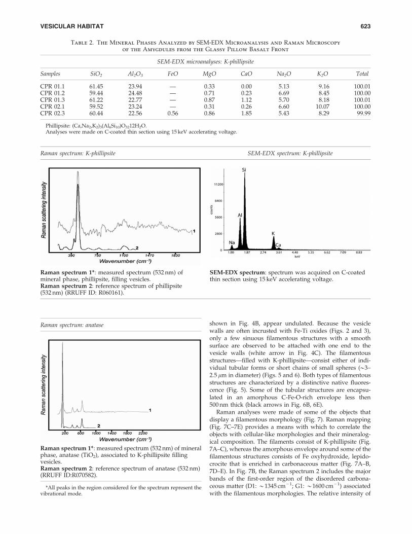
shown in Fig. 4B, appear undulated. Because the vesiclewalls are often incrusted with Fe-Ti oxides (Figs. 2 and 3),only a few sinuous filamentous structures with a smoothsurface are observed to be attached with one end to thevesicle walls (white arrow in Fig. 4C). The filamentousstructures—filled with K-phillipsite—consist either of indi-vidual tubular forms or short chains of small spheres (*3–2.5 lm in diameter) (Figs. 5 and 6). Both types of filamentousstructures are characterized by a distinctive native fluores-cence (Fig. 5). Some of the tubular structures are encapsu-lated in an amorphous C-Fe-O-rich envelope less then500 nm thick (black arrows in Fig. 6B, 6E).
Raman analyses were made of some of the objects thatdisplay a filamentous morphology (Fig. 7). Raman mapping(Fig. 7C–7E) provides a means with which to correlate theobjects with cellular-like morphologies and their mineralog-ical composition. The filaments consist of K-phillipsite (Fig.7A–C), whereas the amorphous envelope around some of thefilamentous structures consists of Fe oxyhydroxide, lepido-crocite that is enriched in carbonaceous matter (Fig. 7A–B,7D–E). In Fig. 7B, the Raman spectrum 2 includes the majorbands of the first-order region of the disordered carbona-ceous matter (D1: *1345 cm - 1; G1: *1600 cm - 1) associatedwith the filamentous morphologies. The relative intensity of
Table 2. The Mineral Phases Analyzed by SEM-EDX Microanalysis and Raman Microscopy
of the Amygdules from the Glassy Pillow Basalt Front
SEM-EDX microanalyses: K-phillipsite
Samples SiO2 Al2O3 FeO MgO CaO Na2O K2O Total
CPR 01.1 61.45 23.94 — 0.33 0.00 5.13 9.16 100.01CPR 01.2 59.44 24.48 — 0.71 0.23 6.69 8.45 100.00CPR 01.3 61.22 22.77 — 0.87 1.12 5.70 8.18 100.01CPR 02.1 59.52 23.24 — 0.31 0.26 6.60 10.07 100.00CPR 02.3 60.44 22.56 0.56 0.86 1.85 5.43 8.29 99.99
Phillipsite: (Ca,Na2,K2)3(Al6Si10)O3212H2O.Analyses were made on C-coated thin section using 15 keV accelerating voltage.
Raman spectrum: K-phillipsite SEM-EDX spectrum: K-phillipsite
Raman spectrum 1*: measured spectrum (532 nm) ofmineral phase, phillipsite, filling vesicles.
SEM-EDX spectrum: spectrum was acquired on C-coatedthin section using 15 keV accelerating voltage.
Raman spectrum 2: reference spectrum of phillipsite(532 nm) (RRUFF ID: R060161).
Raman spectrum: anatase
Raman spectrum 1*: measured spectrum (532 nm) of mineralphase, anatase (TiO2), associated to K-phillipsite fillingvesicles.Raman spectrum 2: reference spectrum of anatase (532 nm)(RRUFF ID:R070582).
*All peaks in the region considered for the spectrum represent thevibrational mode.
VESICULAR HABITAT 623

the D1 and G1 bands indicates a poorly crystalline, disor-dered carbonaceous matter.
4.2.2. Spherical structures. Two sizes of sphericalstructures were observed: macrospheres, with an averageouter diameter of 4 lm, and smaller microspheres, with an
outer diameter between 2 and 2.5 lm (Fig. 8). The outer wallsof the macrospheres display a reddish, transparent appear-ance in petrographic thin section. The macrospheres occurmainly as paired bodies or as aggregates (Fig. 8A–B). Themicrospheres also appear in petrographic thin section astransparent, smooth-surfaced individuals or paired bodies(Fig. 8C). SEM-EDX analysis revealed that the distinctive outerrims of the macrospheres and microspheres have a similarcomposition that shows an enrichment of carbon, Ti, and Fe
FIG. 3. Thin section transmitted-light photomicrograph ofan amygdule from the altered glass of pillow basalt rinds(A). (B) Illustrates detail of the boxed area in (A) and showsthe distribution of spherical and filamentous structures(black arrows). (C) Simplified sketch of an amygdule thatillustrates their dominant characteristics and distribution ofpurported microfossils. Details in (C) are not to scale. K-phi,K-phillipsite.
FIG. 4. Thin section transmitted-light photomicrographsthat reveal the presence of individual filamentous structures(diameter size: 2.5–3.5 lm) within K-phillipsite–filled vesi-cles. The filamentous structures show smooth sinuous [blackarrows in (A) and (C)] and corrugated [arrow in (B)]morphologies. In (C), (white arrows) a filament structure isattached to the vesicle wall and embedded in K-phillipsite.K-phi, K-phillipsite.
624 CAVALAZZI ET AL.

FIG. 5. Magnified view of optical transmitted-light (A–B) and CLSM images (C–F) of putative microfossils, which includefilamentous structures and short chains of small spheres embedded in the K-phillipsite–filled vesicles. The CLSM images providedetails of the structures observed in the boxed area shown in (B). The CLSM images reveal a native florescent signal associated withthe filamentous structures, highlighting the internal fine-scale morphological features, which include small spheres. CLSM ac-quisition parameters: (C) image from stack scan mode (stack size/lm: 35.7 · 35.7 · 5.1lm3; stack size/pixel: 512 · 512 · 18; pixeldepth: 8 bit; scaling: x = 0.070lm, y = 0.070lm, z = 0.30lm; wavelength: 488 nm 9.0%); (D) image from plane scan mode (stack size/pixel: 1 · 1 · 1; pixel depth: 8 bit) obtained from (C); (E) image from stack scan mode (stack size: 27.3 · 27.3 · 5.1lm3; stack size/pixel: 512 · 512 · 18; pixel depth: 8 bit; scaling: x = 0.053lm, y = 0.053lm, z = 0.30lm; wavelength: 488 nm 9.0%); (F) image fromplane scan mode (stack size/pixel: 1 · 1 · 1; pixel depth: 8 bit) obtained from (E). K-phi, K-phillipsite.
625
relative to the matrix (Fig. 9). Raman microscopy revealedthat the Ti-enriched phase is anatase (TiO2) (Table 2) and thatit is associated with carbonaceous matter (Fig. 10).
5. Discussion
We infer the biogenicity of the filamentous and sphericalstructures in the K-phillipsite amygdules from the CoralPatch Seamount pillow basalt based on the full suite ofmorphological, mineralogical, and chemical characteristicsthat were observed with different analytical instrumentsover a range of scales from the microscopic to submicro-scopic (cf. Buick, 1990; Knoll, 1999; Summons et al., 1999;Cady et al., 2003; Brasier et al., 2006; Schopf et al., 2007,
2010a). Although the morphology alone of bacteria-like ob-jects may not be a diagnostic biosignature, it serves an im-portant role in spatially locating within a potential ecologicalniche candidate biosignatures that should be characterizedchemically in more detail. The discovery of a suite of possiblebiosignatures associated with the morphological objectsstrengthens our interpretation of their biogenic origin.
In summary, the suite of possible biosignatures that areconsistent with a biogenic origin for the microbe-like remainsincludes the following:
(1) Morphology—distinctive micrometer-sized objectsthat display sizes, size distributions, and filamentousand spherical morphologies consistent for bacteria;
FIG. 6. ESEM images of filamentous morphotypes within K-phillipsite–filled vesicles. The filamentous structures consist ofK-phillipsite microtubes [X1: EDX spectrum in (D); white arrows in (A)] surrounded by an Fe-rich carbonaceous membrane-like envelope [X2: EDX spectrum in (E); black arrows in (B)]. Some filaments are formed by short chains of small spheres [X3:EDX spectrum in (D); white arrows in (C)]. The ESEM images were acquired on uncoated and lightly etched (HCl 1%, lessthan 4 s) thin sections in low vacuum (1 torr) with a 25 keV electron beam. The samples were subsequently Au-coated forESEM-EDX analyses. EDX data were acquired with a 5 keV accelerating voltage. K-phi, K-phillipsite.
626 CAVALAZZI ET AL.

(2) Chemical composition—carbonaceous composition ofmicrobial-like morphological features and a correlationof the carbonaceous remains and authigenic mineralprecipitates associated with the purported microfos-sils, which are consistent for bacteria;
(3) Ecological setting—habitat for microbial communitieswith enhanced preservation potential.
5.1. Morphology of putative microfossils consistentwith life
Though morphology alone cannot serve as a definitivebiosignature (e.g., Garcıa Ruiz et al., 2002), morphologicalevidence of microfossil-like objects helps to focus the search
for additional possible biosignatures from such structures(Cady et al., 2003). The purported microfossils in the basaltamygdules studied here display a number of key morpho-logical attributes that are consistent with a biological origin:(i) size distribution typical of filamentous and coccoidalprokaryotes; (ii) minimal size variation within any one par-ticular morphological group (filaments display diameters2.5–3 lm, Fig. 4; macrospheres*4 lm, and microspheres2–2.5 lm, Fig. 8); (iii) shape consistency (e.g., filamentous andspherical) with constant diameters (Figs. 4–10); and (iv) anarrangement and structural complexity of individual cells orcolonial aggregates (e.g., short chains of small spheres thatcomprise filaments, Figs. 5 and 6). The lack of variation in themorphological attributes is much more typical of microbial
FIG. 7. Optical photograph and Raman analy-ses of an area in a polished thin section thatcontains a filament. (A) Reflected light opticalphotomicrograph (focus plane: 7 lm below to thesurface). The number indicates the location fromwhich the representative Raman spectra of thehost matrix and filamentous structure (dashedlines) were collected, whereas the boxed areaindicates the location of the Raman maps. (B)The host matrix, spectrum 1, consists of phillip-site, and the filamentous structure as shown inthe multiphase spectra 2 is made of lepidocrocite(lep) associated with phillipsite (phi) and tracesof carbonaceous matter (com). Raman spectrawere calibrated after RRUFF DataBase (Laetschand Downs, 2006), Demoulin et al. (2010), andMarshall et al. (2010). Inset shows the referencespectrum of pure lepidocrocite (532 nm) afterDemoulin et al. (2010); see also reference spec-trum (780 nm) RRUFF ID R050454. (C–E) Ramanmaps that show the distribution of phillipsite(phi), lepidocrocite (lep), and carbonaceous matter (com) of the area that contains the filamentous structures (dashedlines). Data are presented as a black-white intensity ratio map where white regions indicate high concentrationsof the component labeled at the bottom of each panel. The Raman maps correspond to the same focal plane imagedin (A).
FIG. 8. Transmitted light photomicrographs of a petrographic thin section that illustrates two sizes of spherical structureswithin the K-phillipsite–filled vesicles. (A, B) The reddish-brown macrospheres (*4.5 lm outer diameter) appear as thick-walled semi-spheroids. (C) The transparent (*3 lm diameter) microspheres occur as isolated or paired bodies (black arrows).K-phi, K-phillipsite; vw, vesicle wall.
VESICULAR HABITAT 627

assemblages than it is of similar abiotic structures (Brasieret al., 2006).
Filamentous and spherical microorganisms are the mostcommon type of micrometer-scale morphology found inbasaltic subseafloor niches. Such microbial morphotypescolonize surfaces, fractures, and vesicles in volcanic rocks toobtain energy and nutrients from reduced chemical speciesreleased from basalt during dissolution/alteration, and theyestablish themselves in physically stable environmentswhere they can proliferate and, when necessary, escapepredation (e.g., Edwards et al., 2004, 2005; Cockell and Her-rera, 2008; Santelli et al., 2008; Chan et al., 2011). Older fila-mentous and spherical microfossils have been found inassociation with filled fractures and vesicles in basaltic vol-canic rocks (e.g., Ivarsson et al., 2008; Peckmann et al., 2008).
5.2. Chemical composition of putative microfossilsindicative of microbial life
The chemical characteristics of the purported microfossilsare consistent with a biogenic origin. Noteworthy is thecorrelative nature of the carbonaceous material and nativefluorescence produced by many of the purported microfos-sils (Figs. 5 and 6). Carbon enrichment and carbonaceousmatter were observed in both the filaments and coccoidalstructures relative to the mineral matrix in which they areembedded (Figs. 6, 7, 9, 10). The relatively low abundance of
carbon in the mineral matrix of igneous rocks has led to itsuse as a biosignature in this type of environmental settingwhen there is a relative enrichment in its concentration andan association with microbial-like structures (Furnes et al.,2002; Banerjee and Muehlenbachs, 2003; Ivarsson et al., 2008).
Laser Raman microscopy has been successfully used tocharacterize natural carbonaceous compounds (e.g., Jehlin�caand Beny, 1992; Marshall et al., 2010) and for chemical im-agery of microfossils (Schopf et al., 2005, 2006, 2010a, 2010b).Here, we were able to confirm the intimate association ofdisordered carbonaceous material with the candidate bio-signatures, using in situ Raman mapping. In this study, na-tive fluorescence of the microfossil-like objects in the basaltamygdules was observed with a CLSM (488 nm excitationand 505 nm emission wavelengths). Native fluorescence ofbacteria-like structures in the crevices of natural basaltsamples has been detected with deep-UV ( < 250 nm excita-tion wavelength) laser-induced fluorescence and used as anear real-time optical imaging method (Bhartia et al., 2010).The co-occurrence of biomolecules, native fluorescence, and(living and fossil) microbial morphotypes has been demon-strated with different (e.g., < 250 and 488 nm) CLSM excita-tion wavelengths in several natural environments (e.g., Amosand White, 2003; Schopf et al., 2006, 2010a; Bhartia et al., 2008,2010; Santelli et al., 2008). Of most interest here is a study inwhich in situ native fluorescence and resonance Raman
FIG. 9. ESEM image of spherical structures and their che-mical composition. They appear as circular structures withan outer rim of C-rich Ti-Fe oxides (EDX spectra). Some ofthese structures are filled by smaller spheres (arrow) that aresimilar in composition. The structures were imaged on un-coated samples, and the two spectra (x1: spherical structure;x2: host matrix) were acquired with 5 keV accelerating volt-age on a Au-coated sample. K-phi, K-phillipsite; ox, oxide.
FIG. 10. Optical photograph and Raman maps of the areacontaining spherical structures in a polished thin section. (A)Reflected light optical photograph (focus plane: 5 lm belowto the surface) of the spherical structures (*4 lm diameter).The boxed area indicates the location of the Raman maps.(B–D) Raman maps reveal the distribution of phillipsite(phi), anatase (anat), and carbonaceous matter (com), whichgenerate negative and positive ring shape structures, re-spectively. The data are presented as a black-white intensityratio map where white regions indicate the presence of thecomponent labeled at the bottom of each panel. The Ramanmaps correspond to the focal plane imaged in (A). Arrowslocate walled spheres in the different panels.
628 CAVALAZZI ET AL.

spectroscopy were coupled with other analytical techniquessuch as environmental scanning electron microscopy–energy-dispersive X-ray spectroscopy to detect microorgan-isms on the rim of small spherical cavities (vesicles) withinvolcanic glass (Fisk et al., 2003). For instance, the in situmultiple analytical techniques used in this study permit theidentification of similar clay-covered spherical forms. In bothcases, the detected morphological forms resemble knowndeep-sea microorganisms.
Native fluorescence of the lepidocrocite-rich areas thatsurround the filamentous structures (Fig. 6B) indicates thepresence of organic remains in the type of occurrence ex-pected when the extracellular polymeric substances ofsheathed microorganisms are mineralized (e.g., Bhartia et al.,2010). Sheathed Fe-oxidizing microorganisms (van Veenet al., 1978; Emerson and Moyer, 1997; Chan et al., 2011) arecharacterized by chains of cells that, when encased in theirtubular sheaths, appear as filaments (e.g., note the shortchains of sphere observed in Fig. 5C–F).Though it is possiblethat all the native fluorescence associated with the morpho-logical remains of the purported microfossils is produced bysmall autofluorescent crystals, the nonbiological explanation(the ‘‘null hypothesis’’ sensu Brasier et al., 2006) for the ob-served phenomenon is not favored because (i) the authigenicmineral precipitates (e.g., lepidocrocite, Fe hydroxide, andanatase Ti oxide; see below) are not known to be auto-fluorescent at the excitation wavelengths used in this studyand (ii) the native fluorescence is always correlated withmicrofossil-like morphologies.
Native fluorescence of minerals is related to the pres-ence of impurities, organic activators, and the excitationwavelengths used (Gorobets and Rogojine, 2002). Althoughfew data are available in the literature with regard to thepotential to induce native fluorescence in minerals for theexcitation wavelength used here (kexc = 488 nm), laser-induced fluorescence of the mineral phases associated withthe bacteria morphologies [the natural Fe(III)-hydroxide andTi-oxide mineral phases] are not known to fluoresce whenexcited with UV or visible radiation (Gaft et al., 2005). Ty-pically, lepidocrocite, c-Fe(III)O(OH), cannot be the source ofthe luminescence because of the nature of it; the type andstructure of this mineral render it impracticable for emissionof luminescence by the mineral itself even with impurities inits structure. Lepidocrocite can form, however, with externalmacroscopically mixed luminescent impurities.
5.3. Chemical composition of authigenic mineralsconsistent with microbial life
The mineralogy of the authigenic precipitates, Fe and Timetal oxides, associated with the purported microfossils isconsistent with the activities of a microbial population as-sociated with subseafloor environments. For example, theiron-oxide lepidocrocite precipitates when sheathed iron-oxidizing filamentous bacteria remove iron from solution(Emerson and Moyer, 2002). Though bacterial sheaths canprovide a physically and chemically protective environmentin which the cells can grow and divide, once mineralizedthey limit the diffusion of nutrients into cells. Microbialsurfaces and extracellular polymeric substances are knownto enhance the concentrations of certain elements, such as Feand Ti, relative to the concentrations of these elements in the
surrounding environment (Beveridge, 1989; Konhauser,1998). High concentrations of Fe in the sheaths and on mi-croorganisms in the seafloor environment are considered toresult from active microbial interactions with the volcanicmaterials. In such processes, lithotrophic microorganismsutilize the redox potential of the volcanic minerals as amechanism to obtain energy (Thorseth et al., 2001; Bach andEdwards, 2003; Edwards et al., 2005; Santelli et al., 2008).Although several microfossils’ structures are known to bepreserved or contain mineral mixtures dominated by metaloxides, and the role of metabolism and biomineralizationprocesses of Fe oxidizers are relatively well known (e.g.,Emerson and Moyer, 2002; Kappler and Straub, 2005), it isworth noting that, at present, the interactions between Ti andmicroorganisms and the role of Ti in microbial fossilizationare poorly understood (Shabtai and Fleminger, 1994; Gla-moclija et al., 2009). Though some prokaryotes are known toadsorb TiO2 during metabolic processes (Shabtai and Fle-minger, 1994; Bedard et al., 2006), the precipitation of tita-nium (and iron) oxide minerals is commonly thought torepresent abiogenic low-temperature alteration of basalticglass with seawater (e.g., Stroncik and Schmincke, 2001). Itwas impossible in this study to establish whether the selec-tive Ti mineralization of the spherical structures via anatasewas a passive or microbially controlled fossilization process.
5.4. Timing of fossilization
Vesicles are primary segregation structures that representthe fossilized vestiges of degassing bubbles trapped duringextrusion and decompression from a volatile-rich melt source(Peck, 1978). Although it is difficult to determine when theminerals infilled the vesicles, characteristics of the microhabi-tat and microfossil taphonomy provide some informationwith regard to the involvement of microorganisms in theprocess. Given their distribution within the K-phillipsite–filledamygdules, the microbial structures represent endolithicand, most likely, cryptoendolithic filamentous and sphericalmicroorganisms. Unfortunately, because our microfossils werenot associated with any multiple paragenetic sequences ofcements (e.g., Peckmann et al., 2008), we were unable to de-termine precisely when the vesicles hosted life. The presenceof the purported fossil microorganisms in amygdules in closeproximity to the outer margin of the pillow basalt, along withthe attachment of some of the microfossils to the walls of thevesicles (Fig. 4C) and the absence of granular or tubular al-teration textures in the walls of the vesicles and glass shards,suggests that early precipitation of the phillipsite in the vesi-cles and pore space reduced the fluid circulation and inhibitedthe growth of euendolithic microorganisms (see Fisk et al.,2003). The vesicular microhabitat would most likely have beena nutrient-rich micro-environment with the nutrients comingfrom alteration of the volcanic rocks. A circumneutral toalkaline (pH ‡ 7) fluid environment within the vesicles is in-dicated by the presence of abundant TiO2 in the K-phillipsite–filled amygdules. The Fe(III) (oxy)hydroxides observed mayhave been produced either chemically or biologically by mi-crobial and chemical iron(II) oxidation at a neutral pH(Emerson, 2000).
The fossilized microorganisms within vesicles of the CoralPatch pillow basalt are preserved as organic remains (car-bonaceous membrane) and mineral (K-phillipsite, Ti oxides,
VESICULAR HABITAT 629

and Fe hydroxides)–replaced structures. Because biogeniccarbonaceous matter degrades progressively and presum-ably continuously during mineralization and encrustation,the presence of native fluorescence from the microfossils andsurrounding area indicates that the organic remains retain asufficient number of functional groups to produce a detect-able signal. Their morphologies and distribution are consis-tent with a biotic origin and, though it is not possible todetermine exactly when they infiltrated the vesicles, theirmineral composition is consistent with early microbiallymediated authigenesis in a marine environment. Their micro-habitat suggests that they were chemolithotrophic and het-erotrophic cryptoendoliths that inhabited a low-temperaturecircumneutral to alkaline aqueous microhabitat.
5.5. Vesicular habitat: a suitable niche for lifewith high preservation potential
Life displays a wide range of survival strategies and canadapt to even the most inhospitable habitats. Endolithic en-vironments are ubiquitous terrestrial microbial habitats and,as such, represent an important area of study in the fieldsof geomicrobiology, early life research, and astrobiology.Endolithic microorganisms, including cryptoendoliths, cancolonize a wide variety of substratums, especially in extremeenvironments, and produce characteristic structures andbio-alteration textures with a high preservation potential(Friedmann and Koriem, 1989; Wynn-Williams and Ed-wards, 2000).
The study of terrestrial analogues of potential extrater-restrial environments is a prerequisite for astrobiology andplanetary exploration. Oceanic basalt rocks are rich in re-duced elements (e.g., iron) that play a crucial role in themicrobiology of basalt endolithic habitats. In fact, (chemo-lithoautotrophic) microbial alteration of oceanic basalts has asignificant influence on deep-sea carbon cycling and chemi-cal exchange between basalt and seawater (Santelli et al.,2008). Oceanic volcanic rocks were prevalent on early Earthand probably on early Noachian Mars (Westall, 2005). Thus,endolithic microorganisms would be strong candidates forthe colonization of early Earth and other volcanic planetarysurfaces (e.g., Friedmann and Koriem, 1989; Fisk et al., 2006).The microhabitats within vesicles would have provided aUV-protected environment for microorganisms on planets,such as early Earth, that had no ozone shield (Cockell andRaven, 2004). Vesicles also represent environments thatwould have been protected from the catastrophic effects ofvolcanism and meteoritic impact on early Earth and Mars(Cockell et al., 2002). However, the size, diversity, andfunctional activity of endolithic microbes on, and within, therock substrate at the seafloor are poorly known (Edwardset al., 2005). Biotic alteration morphologies, such as eu-endolithic microborings in Archean pillow lavas in SouthAfrica and Western Australia (Furnes et al., 2004; Banerjeeet al., 2006) have been described and advanced as a newastrobiological biosignature (McLoughlin et al., 2007).Though euendolithic bioalteration of volcanic rock/glasssurfaces has been well documented in the geological record(review in Staudigel et al., 2008), fossil cryptoendolithswithin vesicles of submarine basalts are little known (Schu-mann et al., 2004; Cavalazzi et al., 2008; Peckmann et al., 2008;Eickmann et al., 2009; this study). Cryptoendoliths have also
been found to colonize vesicles of basaltic rocks in terrestrialenvironments ( Jorge Villar et al., 2006). The discovery ofpopulated vesicles of volcanic rocks from the ocean basinsand in terrestrial environments has widened the prospect offinding new sites for these extreme environments on Earth.The astrobiological relevance of fossil cryptoendoliths withinvesicles in pillow is further emphasized by the finding ofextremely vesicular basalt within the Columbia Hills on theGusev Crater floor (Mars Exploration Rover Spirit mission)(Schmidt et al., 2008). From this perspective, the cryptichabitat of pillow basalts and their associated microorganismsrepresent an, as yet, underappreciated environment in whichto search for evidence of life.
6. Conclusions
Partly mineral-replaced (K-phillipsite, Fe hydroxides, andTi oxide) filaments and spherical structures occur withinK-phillipsite–filled amygdules in the chilled margin of pillowlavas from Coral Patch Seamount, eastern North AtlanticOcean.
Well-preserved and carbonaceous filamentous andspherical structures show low variability in size and shape.Their biogenicity was assessed on recognized biogenicitycriteria established for microfossils. In particular, the co-occurrence of their cellular morphologies (biomorphic fea-tures, arrangement and distribution), their mineralogical andchemical compositions, and associated autofluorescencesignal and environmental microhabitat strongly supporttheir biogenicity. The microbial population would have in-cluded lithoautotrophs and may have included mineralizedsheathed microorganisms, possibly Fe-oxidizing bacteria.The presence of phillipsite and anatase permineralized mi-crofossils, as well as anatase grains within the K-phillipsitematrix, indicates that the environment within the vesicleshad a circumneutral to acidic pH.
Since the vesicles are primary structures in the alteredglass of pillow basalt, the fossil microorganisms described inthis study must have been cryptoendoliths, and the vesicularhabitat suggests that they were chemotrophic/lithoauto-trophic organisms. This type of volcanic microenvironmentand the cryptoendolithic microorganisms that could inhabitit need further study. Such environments, which would havebeen common on early Earth and probably on early Mars,represent analogues of astrobiological interest.
Acknowledgments
We gratefully acknowledge N. Zitellini, A. Ceregato, andL. Capotondi, ISMAR-CNR Bologna for donating samples.We acknowledge funding for B.C. from CNR-CNRS Interna-tional Cooperation Program, for F.W. from GDR-Exobiologiegrant, and for S.L.C. from NASA under award No.NNG04GJ84G and NSF under award No. GEO0808211.Thanks are due to I. Raffi, Universita di Chieti-Pescara (Italy)for help in fossil nannoplankton identification. N. McLough-lin, G. Glamoclija, and anonymous reviewers provided usefulcomments.
Abbreviations
CLSM, confocal laser scanning microscope; EDX, energy-dispersive X-ray; ESEM, environmental scanning electron
630 CAVALAZZI ET AL.

microscope; FEG-SEM, field emission gun scanning electronmicroscope; SEM, scanning electron microscope.
References
Amos, W.B. and White, J.G. (2003) How the confocal laserscanning microscope entered biological research. Biology of theCell 95:335–342.
Bach, W. and Edwards, K.J. (2003) Iron and sulfide oxidationwithin the basaltic ocean crust: implications for chemo-lithoautotrophic microbial biomass production. Geochim Cos-mochim Acta 67:3871–3887.
Banerjee, N.R. and Muehlenbachs, K. (2003) Tuff life: bio-alteration in volcaniclastic rocks from the Ontong JavaPlateau. Geochemistry, Geophysics, Geosystems 4, doi:10.1029/2002GC000470.
Banerjee, N.R., Furnes, H., Muehlenbachs, K., Staudigel, H., andde Wit, M. (2006) Preservation of *3.4–3.5 Ga microbial bio-markers in pillow lavas and hyaloclastites from the BarbertonGreenstone Belt, South Africa. Earth Planet Sci Lett 241:707–722.
Bedard, D.L., Bailey, J.J., Reiss, B.L., and Van Slyke Jerzak, G.(2006) Development and characterization of stable sediment-free anaerobic bacterial enrichment cultures that dechlorinateAroclor 1260. Appl Environ Microbiol 72:2460–2470.
Beveridge, T.J. (1989) Role of cellular design in bacterial metalaccumulation and in mineralization. Annu Rev Microbiol43:147–171.
Bhartia, R., Hug, W.F., Salas, E.C., Reid, R.D., Sijapati, K.K.,Tsapin, A., Abbey, W., Nealson, K.H., Lane, A.L., and Conrad,P.G. (2008) Classification of organic and biological materialswith deep ultraviolet excitation. Appl Spectrosc 62:1070–1077.
Bhartia, R., Salas, E.C., Hug, W.F., Reid, R.D., Lane, A.L., Ed-wards, K.J., and Nealson, K.H. (2010) Label-free bacterialimaging with deep-UV-laser-induced native fluorescence.Appl Environ Microbiol 76:7231–7237.
Brasier, M., McLoughlin, N., Green, O., and Wacey, D. (2006) Afresh look at the fossil evidence for early Archaean cellular life.Philos Trans R Soc Lond B Biol Sci 361:887–902.
Buick, R. (1990) Microfossil recognition in Archean rocks: an ap-praisal of spheroids and filaments from a 3500 m.y. old chert-barite unit at North Pole, Western Australia. Palaios 5:441–459.
Cady, S.L., Farmer, J.D., Grotzinger, J.P., Schopf, J.W., andSteele, A. (2003) Morphological biosignatures and the searchfor life on Mars. Astrobiology 3:351–368.
Cavalazzi, B., Westall, F., and Barbieri, R. (2008) (Crypto-)En-doliths from vesicular pillow lavas, Coral Patch SeamountNorth Atlantic Ocean. Studi Trent Sci Nat Acta Geol 83:177–182.
Chan, C.S., Fakra, S.C., Emerson, D., Fleming, E.J., and Edwards,K.J. (2011) Lithotrophic iron-oxidizing bacteria produce or-ganic stalks to control mineral growth: implications for bio-signature formation. ISME J 5:717–727.
Cockell, C.S. and Herrera, A. (2008) Why are some microor-ganisms boring? Trends Microbiol 16:101–106.
Cockell, C.S. and Raven, J.A. (2004) Zones of photosyntheticpotential on Mars and early Earth. Icarus 169:300–310.
Cockell, C.S., Lee, P., Osinsky, G., Horneck, H., and Broady, P.(2002) Impact-induced microbial endolithic habitats. MeteoritPlanet Sci 37:1287–1298.
Connell, L., Barrett, A., Templeton, A., and Staudigel, H. (2009)Fungal diversity associated with an active deep sea volcano:Vailulu’u Seamount, Samoa. Geomicrobiol J 26:8597–8605.
Demoulin, A., Trigance, C., Neff, D., Foy, E., Dillmann, P., andL’Hostis, V. (2010) The evolution of the corrosion of iron in
hydraulic binders analysed from 46- and 260-year-old build-ings. Corros Sci 52:3168–3179.
Edwards, K.J., Bach, W., McCollom, T.M., and Rogers, D.R. (2004)Neutrophilic iron-oxidizing bacteria in the ocean: their habitats,diversity, and roles in mineral deposition, rock alteration, andbiomass production in the deep-sea. Geomicrobiol J 21:393–404.
Edwards, K.J., Bach, W., and McCollom, T. (2005) Geomicro-biology in oceanography: microbe mineral interactions at andbelow the seafloor. Trends Microbiol 13:449–456.
Eickmann, B., Bach, W., Kiel, S., Reitner, J., and Peckmann, J.(2009) Evidence for cryptoendolithic life in Devonian pillowbasalts of Variscan orogens, Germany. Palaeogeogr Palaeocli-matol Palaeoecol 283:120–125.
Emerson, D. (2000) Microbial oxidation of Fe(II) and Mn(II) atcircumneutral pH. In Environmental Microbe-Mineral Interactions,edited by D.R. Lovley, ASM Press, Washington DC, pp 109–144.
Emerson, D. and Moyer, C.L. (1997) Isolation and characteriza-tion of novel iron-oxidizing bacteria that grow at circumneu-tral pH. Appl Environ Microbiol 63:4784–4792.
Emerson, D. and Moyer, C.L. (2002) Neutrophilic Fe-oxidizingbacteria are abundant at the Loihi seamount hydrothermalvents and play a major role in Fe oxide deposition. Appl En-viron Microbiol 68:3085–3093.
Fisk, M.R., Storrie-Lombardi, M.C., Douglas, S., Popa, R.,McDonald, G., and Di Meo-Savoie, C. (2003) Evidence of bi-ological activity in Hawaiian subsurface basalts. Geochemistry,Geophysics, Geosystems 4, doi:10.1029/2002GC000387.
Fisk, M.R., Popa, R., Mason, O.U., Storrie-Lombardi, M.C., andVincenzi, E.P. (2006) Iron-magnesium silicate bioweatheringon Earth (and Mars?). Astrobiology 6:48–68.
Friedmann, E.I. and Koriem, A.M. (1989) Life on Mars: how itdisappeared (if it was ever there). Adv Space Res 9:167–172.
Furnes, H. and Staudigel, H. (1999) Biological mediation inocean crust alteration: how deep is the deep biosphere? EarthPlanet Sci Lett 166:97–103.
Furnes, H., Muehlenbachs, K., Torsvik, T., Tumyr, O., and Lang,S. (2002) Bio-signatures in metabasaltic glass of a Caledonianophiolite, West Norway. Geol Mag 139:601–608.
Furnes, H., Banerjee, N.R., Muehlenbachs, K., Staudigel, H., andde Wit, M. (2004) Early life recorded in Archean pillow lavas.Science 304:578–581.
Furnes, H., Banerjee, N.R., Staudigel, H., Muehlenbachs, K.,McLoughlin, N., de Wit, M., and Van Kranendonk, M. (2007)Comparing petrographic signatures of bioalteration in recentto Mesoarchean pillow lavas: tracing subsurface life in oceanicigneous rocks. Precambrian Res 158:156–176.
Gaft, M., Reisfeld, R., and Panczer, G. (2005) Modern Lumines-cence Spectroscopy of Minerals and Materials, Springer, Berlin.
Garcıa Ruiz, J.M., Carnerup, A.M., Christy, A.G., Welham, N.J.,and Hyde, S.T. (2002) Morphology: an ambiguous indicator ofbiogenicity. Astrobiology 2:353–369.
Geldmacher, J. and Hoernle, K. (2000) The 72 Ma geochemicalevolution of the Madeira hotspot (eastern North Atlantic):recycling of Paleozoic ( £ 500 Ma) oceanic lithosphere. EarthPlanet Sci Lett 183:73–92.
Glamoclija, M., Steele, A., Fries, M., Schieber, J., Voytek, M.A., andCockell, C.S. (2009) Association of anatase (TiO2) and microbes:unusual fossilization effect or a potential biosignature? Geolo-gical Society of America Special Papers 458:965–975.
Gorobets, B.S. and Rogojine, A.A., editors. (2002) LuminescentSpectra of Minerals Reference-Book, Coronet Books Inc., Phila-delphia, PA, USA.
Ivarsson, M., Lausmaa, J., Lindblom, S., Broman, C., and Holm,N.G. (2008) Fossilized microorganisms from the Emperor
VESICULAR HABITAT 631
Seamounts: implications for the search for a subsurface fossilrecord on Earth and Mars. Astrobiology 8:1139–1157.
Jehlin�ca, J. and Beny, C. (1992) Application of Raman micro-spectrometry in the study of structural changes in Precambriankerogens during regional metamorphism. Org Geochem 18:211–213.
Jorge Villar, S.E., Edwards, H.G.M., and Benning, L.G. (2006)Raman spectroscopic and scanning electron microscopicanalysis of a novel biological colonisation of volcanic rocks.Icarus 184:158–169.
Kappler, A. and Straub, K.L. (2005) Geomicrobiological cyclingof iron. Reviews in Mineralogy and Geochemistry 59:85–108.
Kashefi, K. and Lovley, D.R. (2003) Extending the upper tem-perature limit for life. Science 301:934.
Kim, J., Dong, H.L., Seabaugh, J., Newell, S.W., and Eberl, D.D.(2004) Role of microbes in the smectite-to-illite reaction. Sci-ence 303:830–832.
Knoll, A. (1999) Recognition of a biological signature in rocks.Discussion summary. In Size Limits of Very Small Microorgan-isms: Proceedings of a Workshop, Steering Group for the Work-shop on Size Limits of Very Small Microorganisms, NationalResearch Council, The National Academies Press, WashingtonDC, pp 85–87.
Konhauser, K.O. (1998) Diversity of bacterial iron mineraliza-tion. Earth-Science Reviews 43:91–121.
Kostka, J.E., Dalton, D.D., Skelton, H., Dollhopf, S., and Stucki,J.W. (2002) Growth of iron(III)-reducing bacteria on clayminerals as the sole electron acceptor and comparison ofgrowth yields on a variety of oxidized iron forms. Appl En-viron Microbiol 68:6256–6262.
Laetsch, T.A. and Downs, R.T. (2006) Software for identificationand refinement of cell parameters from powder diffractiondata of minerals using the RRUFF Project and AmericanMineralogist Crystal Structure Databases [abstract P08-25]. InProgram and Abstracts of the 19th General Meeting of the Inter-national Mineralogical Association, International MineralogicalAssociation.
Marshall, C.P., Edwards, H.G.M., and Jehlin�ca, J. (2010) Un-derstanding the application of Raman spectroscopy to thedetection of traces of life. Astrobiology 10:229–243.
McLoughlin, N., Brasier, M.D., Wacey, D., Green, O.R., andRandall, S.P. (2007) On biogenicity criteria for endolithic mi-croborings on early Earth and beyond. Astrobiology 7:10–26.
McLoughlin, N., Furnes, H., Banerjee, N.R., Muehlenbachs, K.,and Staudigel, H. (2009) Ichnotaxonomy of microbial tracefossils in volcanic glass. J Geol Soc London 166:159–169.
Nielsen, M.E. and Fisk, M.R. (2010) Surface area measurementsof marine basalts: implications for the subseafloor microbialbiomass. Geophys Res Lett 37, doi:10.1029/2010GL044074.
Peck, D.L. (1978) Cooling and vesiculation of Alae Lava Lake,Hawaii. U.S. Geological Survey Professional Paper 935-B, U.S.Geological Survey, Washington DC.
Peckmann, J., Bach, W., Behrens, K., and Reitner, J. (2008) Pu-tative cryptoendolithic life in Devonian pillow basalt, Rhein-isches Schiefergebirge, Germany. Geobiology 6:125–135.
Santelli, C.M., Orcutt, B.N., Banning, E., Bach, W., Moyer, C.L.,Sogin, M.L., Staudigel, H., and Edwards, K.J. (2008) Abun-dance and diversity of microbial life in ocean crust. Nature453:653–656.
Schmidt, M.E., Ruff, S.W., McCoy, T.J., Farrand, W.H., Johnson,J.R., Gellert, R., Ming, D.W., Morris, R.V., Cabrol, N., Lewis,K.W., and Schroeder, C. (2008) Hydrothermal origin of halo-gens at Home Plate, Gusev Crater. J Geophys Res 113,doi:10.1029/2007JE003027.
Schopf, J.W., Kudryavtsev, A.B., Agresti, D.G., Czaja, A.D., andWdowiak, T.J. (2005) Raman imagery: a new approach to as-sess the geochemical maturity and biogenicity of perminer-alized Precambrian fossils. Astrobiology 5:333–371.
Schopf, J.W., Tripathi, A.B., and Kudryavtsev, A.B. (2006) Three-dimensional confocal optical imagery of Precambrian micro-scopic organisms. Astrobiology 6:1–16.
Schopf, J.W., Kudryavtsev, A.B., Czaja, A.D., and Tripathi, A.B.(2007) Evidence of Archean life: stromatolites and microfos-sils. Precambrian Res 158:141–155.
Schopf, J.W., Kudryavtsev, A.B., and Sergeev, V.N. (2010a)Confocal laser scanning microscopy and Raman imagery ofthe late Neoproterozoic Chichkan microbiota of South Ka-zakhstan. J Paleontol 84:402–416.
Schopf, J.W., Kudryavtsev, A.B., Sugitani, K., and Walter, M.R.(2010b) Precambrian microbe-like pseudofossils: a promisingsolution to the problem. Precambrian Res 179:191–205.
Schumann, G., Manz, W., Reitner, J., and Lustrino, M. (2004)Ancient fungal life in North Pacific Eocene oceanic crust.Geomicrobiol J 21:241–246.
Shabtai, Y. and Fleminger, G. (1994) Adsorption of Rhodococcusstrain GIN-1 (NCIMB 40340) on titanium dioxide and coal flyash particles. Appl Environ Microbiol 60:3079–3088.
Staudigel, H., Furnes, H., McLoughlin, N., and Banerjee, N.R.(2008) 3.5 billion years of glass bioalteration: volcanic rocks asbasis for microbial life? Earth-Science Reviews 89:156–176.
Stroncik, N.A. and Schmincke, H.-U. (2001) Evolution ofpalagonite: crystallization, chemical changes, and elementalbudget. Geochemistry, Geophysics, Geosystems 2, doi:10.1029/2000GC000102.
Summons, R.E, Jahnke, L.L, Hope, M., and Logan, G.A. (1999) 2-Methylhopanoids as biomarkers for cyanobacterial oxygenicphotosynthesis. Nature 400:554–557.
Takai, K., Moser, D.P., DeFlaun, M.F., Onstott, T.C., andFredrickson, J.K. (2001) Archaeal diversity in waters fromdeep South African gold mines. Appl Environ Microbiol 67:5750–5760.
Thorseth, I.H., Torsvik, T., Furnes, H., and Muehlenbachs, K.(1995) Microbes play an important role in the alteration ofoceanic crust. Chem Geol 126:137–146.
Thorseth, I.H., Torsvik, T., Torsvik, V., Daae, F.L., and Pedersen,R.B. (2001) Keldysh-98 scientific party, diversity of life inocean floor basalt. Earth Planet Sci Lett 194:31–37.
van Veen, W.L., Mulder, E.G., and Deinema, M.H (1978) TheSphaerotilus-Leptothrix group of bacteria. Microbiol Rev 42:329–356.
Westall, F. (2005) Early life on Earth and analogies to Mars.In Water on Mars and Life, Advances in Astrobiology and Bio-geophysics 4, edited by T. Tokano, Springer, Berlin, pp 45–64.
Wynn-Williams, D.D. and Edwards, H.G.M. (2000) Antarcticecosystems as models for extraterrestrial surface habitats.Planet Space Sci 48:1065–1075.
Address correspondence to:Barbara Cavalazzi
Department of GeologyUniversity of Johannesburg
JohannesburgSouth Africa
E-mail: [email protected]
Submitted 26 March 2011Accepted 2 July 2011
632 CAVALAZZI ET AL.
Copyright © 2022 FDOKUMEN