Separation of discontinuous adventitious sounds from vesicular sounds using a wavelet-based filter

DOI: 10.1002/chem.201201299
Hydrogen-Bonding-Mediated Vesicular Assembly of FunctionalizedNaphthalene–Diimide-Based Bolaamphiphile and Guest-Induced
Gelation in Water
Mijanur Rahaman Molla and Suhrit Ghosh*[a]
Introduction
The self-assembly of amphiphilic molecules and macromole-cules has emerged as an exciting field of research in interdis-ciplinary fields that range from biology to materials science.Various classical small-molecule amphiphiles[1] (single-tailsurfactants, gemini surfactants, bolaamphiphiles, phospholi-pids, etc.) and amphiphilic polymers[2] have produced ele-gant nanoscale assemblies, such as spherical and rod-like mi-celles, vesicles, fractals, fibers, nanotubes, etc. Amazingly,most of these amphiphilic molecules, be they small mole-cules or macromolecules, are structurally identical ina broad sense, that is, they consist of hydrophobic and hy-drophilic segments, which differ in terms of their relativelength, volume, and how they are covalently linked to eachother. Over the last two decades, chemists have developedenormous knowledge to generate a diverse range of supra-molecular materials,[3] mostly in organic medium, by virtueof utilizing various directional non-covalent forces, such asp-stacking, hydrogen bonding, charge-transfer (CT) interac-
tions, and metal–ligand coordination. In an attempt to real-ize the implications of such directional interactions in aque-ous medium, amphiphiles that contain p-conjugated chro-mophores,[4] as well as those that can be generated by usingnon-covalent forces (commonly known as supramolecularamphiphiles[5]), have been investigated with great interest inthe recent past. An additional motivation to study this sub-class of amphiphiles stems from the possibility of relatingtheir rich photophysical properties[6] to biological applica-tions, such as biosensing, imaging, etc. Most of the reportedp-conjugated amphiphiles only differ from classical amphi-philes in terms of replacing the hydrophobic segment witha chromophore moiety. A natural next step towards enrich-ing the structural diversity of amphiphiles in general wouldbe to make multiple directional force work effectively intandem. In this context, the obvious choice would be hydro-gen-bonding interactions,[7a] considering the diverse range ofhydrogen-bonding synthons[7b] that are available today inthe chemist’s toolbox. This choice is also inspired by theamazing programmed assembled structures (such as foldedproteins and the DNA double-helix) that are fabricated innature. However, examples of the hydrogen-bonding-direct-ed self-assembly of abiotic building blocks in aqueousmedium are still rare,[8] presumably because of the inherentdifficulty that is associated with competitive hydrogen-bond-ing interactions with the solvent molecules. Thus, biomim-ACHTUNGTRENNUNGicking in the context of self-assembly (in terms of structureand not even function) is still mostly confined to the domain
[a] M. R. Molla, Dr. S. GhoshPolymer Science UnitIndian Association for the Cultivation of Science2A & 2B Raja S. C. Mullick RoadKolkata, INDIA-7000 32E-mail : [email protected]
Supporting information for this article is available on the WWWunder http://dx.doi.org/10.1002/chem.201201299.
Abstract: This paper describes thespontaneous vesicular assembly ofa naphthalene–diimide (NDI)-basednon-ionic bolaamphiphile in aqueousmedium by using the synergistic effectsof p-stacking and hydrogen bonding.Site isolation of the hydrogen-bondingfunctionality (hydrazide), a strategythat has been adopted so elegantly innature, has been executed in thissystem to protect these moieties fromthe bulk water so that the distinct roleof hydrogen bonding in the self-assem-bly of hydrazide-functionalized NDIbuilding blocks could be realized, evenin aqueous solution. Furthermore, the
electron-deficient NDI-based bolaam-phiphile could engage in donor–accept-or (D–A) charge-transfer (CT) interac-tions with a water-insoluble electron-rich pyrene donor by virtue of interca-lation of the latter chromophore in be-tween two NDI building blocks. Re-markably, even when pyrene was locat-ed between two NDI blocks, intermo-lecular hydrogen-bonding networks be-tween the NDI-linked hydrazidegroups could be retained. However,
time-dependent AFM studies revealedthat the radius of curvature of the al-ternately stacked D–A assembly in-creased significantly, thereby leading tointervesicular fusion, which eventuallyresulted in rupturing of the membraneto form 1D fibers. Such 2D-to-1D mor-phological transition produced CT-mediated hydrogels at relatively higherconcentrations. Instead of pyrene,when a water-soluble carboxylate-func-tionalized pyrene derivative was usedas the intercalator, non-covalent tun-ACHTUNGTRENNUNGable in-situ surface-functionalizationcould be achieved, as evidenced by thezeta-potential measurements.
Keywords: gels · hydrogen bonds ·surface chemistry · vesicles · water
� 2012 Wiley-VCH Verlag GmbH & Co. KGaA, Weinheim Chem. Eur. J. 2012, 18, 9860 – 98699860

of non-polar organic media.1,4,5,8-Naphthalene–tetACHTUNGTRENNUNGra ACHTUNGTRENNUNGcar-ACHTUNGTRENNUNGbox ACHTUNGTRENNUNGylic-acid–diimide (NDI)-de-rivatives[9] have been extensive-ly explored as a building blockfor the generation of varioussupramolecularly assembledsystems, both in organic- andaqueous media, owing to theirn-type semiconductivity,[10] pro-pensity for p-stacking, and elec-tron-accepting nature. Exam-ples of such systems includeorganogels,[11] catenanes,[12] ro-taxanes,[13] foldamers,[14] nano-ACHTUNGTRENNUNGtubes,[4b] hydroACHTUNGTRENNUNGgels,[15] nano ACHTUNGTRENNUNGpar-ACHTUNGTRENNUNGtiACHTUNGTRENNUNGcles,[4c] supramolecular photo-systems,[16] and synthetic ion-channels.[17]
In the recent past, we havealso used NDI-based systems todemonstrate self-sorting[18] and to understand structure-property relationships[19] in organogels. Herein, we reportthe spontaneous vesicular assembly of NDI-1 (Scheme 1) inaqueous medium and CT-interaction-mediated 2D-to-1Dvesicle-to-hydrogel[20–21] transformations in the presence ofan electron-rich pyrene intercalator. In NDI-1, the peripher-al hydrophilic wedge is attached to the NDI core by a hydra-zide group, which has been demonstrated[19a] to be a very ef-fective strategy for hydrogen-bonding-mediated self-assem-bly in non-polar organic media. We envisioned that such siteisolation of the hydrazide groups from bulk water is essen-tial for these groups to be involved in hydrogen-bonding in-teractions among themselves and, thus, by design, they arerigidly linked between two hydrophobic aromatic segments.
Results and Discussion
Self-assembly : In THF, which is a good solvent for NDI,sharp absorption bands in the range 300–400 nm were noted(Figure 1 a) with vibronic features, which indicated the exis-tence of the free monomer. Going from THF to water, a de-crease in the band intensity was observed with a concomitantred shift of about 5 nm, in addition to the intensity reversalof the first (375 nm) and second vibronic peaks[22] (356 nm),thus suggesting the presence of p-stacking. Concentration-dependent UV/Vis studies (Figure 1 b) revealed that, atlower concentrations, the spectroscopic features in water re-sembled those in THF, thus suggesting disassembly. The crit-ical aggregation concentration of NDI-1 was estimated to be0.35 mm from the concentration dependence of the extinc-tion coefficient (e) at 375 nm (see the Supporting Informa-tion, Figure S1).
To probe the role, if any, of hydrogen-bonding in the as-sembly process, the FTIR spectra of NDI-1 in THF andwater were compared. The peak that corresponded to the
Scheme 1. Top) Structure and vesicular assembly of NDI-1. Bottom) CT-interaction-mediated morphologicaltransition (2D-to-1D vesicle-to-fiber) and gelation by pyrene intercalation.
Figure 1. a) UV/Vis spectra of NDI-1 (1 mm) in THF (dotted line) andwater (solid line). b) Concentration-dependent UV/Vis spectra of NDI-1 in water; arrows indicated spectroscopic changes with dilution. c) FTIRspectra (selected region) of NDI-1 (2 mm) in THF (dotted line) andwater (solid line).
Chem. Eur. J. 2012, 18, 9860 – 9869 � 2012 Wiley-VCH Verlag GmbH & Co. KGaA, Weinheim www.chemeurj.org 9861
FULL PAPER
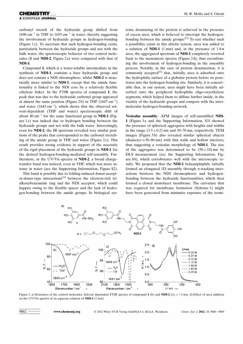
carbonyl stretch of the hydrazide group shifted from1696 cm�1 in THF to 1655 cm�1 in water, thereby suggestingthe involvement of hydrazide groups in hydrogen-bonding(Figure 1 c). To ascertain that such hydrogen-bonding exists,particularly between the hydrazide groups and not with thebulk water, the spectroscopic behavior of two control mole-cules (1 and NDI-2, Figure 2 a) were compared with that ofNDI-1.
Compound 1, which is a water-soluble intermediate in thesynthesis of NDI-1, contains a bare hydrazide group anddoes not contain a NDI chromophore, whilst NDI-2 is struc-turally more similar to NDI-1, except that the amide func-tionality is linked to the NDI core by a relatively flexibleethylene linker. In the FTIR spectra of compound 1, thepeak that was due to the hydrazide carbonyl group appearedat almost the same position (Figure 2 b) in THF (1647 cm�1)and water (1643 cm�1), which shows that the observed sol-vent-dependent (THF and water) spectroscopic shift ofabout 40 cm�1 for the same functional group in NDI-1 (Fig-ure 1 c) was indeed due to hydrogen bonding between thehydrazide groups and not with the bulk water. Interestingly,even for NDI-2, the IR spectrum revealed very similar posi-tions of the peaks that corresponded to the carbonyl stretch-ing of the amide group in THF and water (Figure 2 c). Thisresult provides strong evidence in support of the necessityof the rigid placement of the hydrazide groups in NDI-1 forthe desired hydrogen-bonding-mediated self-assembly. Fur-thermore, in the UV/Vis spectra of NDI-2, a broad charge-transfer band was noticed, even in THF, which was more in-tense in water (see the Supporting Information, Figure S2).
This band is possibly due to folding-induced donor-accept-or-donor-type interactions[14] between the electron-rich tri-ACHTUNGTRENNUNGalkoxybenzamide ring and the NDI acceptor, which couldhappen owing to the flexible spacer and the lack of hydro-gen-bonding between the amide groups. In biological sys-
tems, denaturing of the protein is achieved in the presenceof excess urea, which is believed to interrupt the hydrogen-bonding between the amide groups.[23] To test whether sucha possibility exists in this abiotic system, urea was added toa solution of NDI-1 (1 mm) and, in the presence of 1.8 m
urea, the aggregated spectrum of NDI-1 completely revertedback to the monomeric spectra (Figure 2 d), thus reconfirm-ing the involvement of hydrogen-bonding in the assemblyprocess. Notably, in the case of protein denaturation, it iscommonly accepted[23] that, initially, urea is adsorbed ontothe hydrophilic surface of a globular protein before its pene-trates into the hydrogen-bonding site. Similarly, it is conceiv-able that, in our system, urea might have been initially ad-sorbed onto the peripheral hydrophilic oligo-oxyethylenesegments, which helped them to diffuse further inside, in thevicinity of the hydrazide groups and compete with the inter-molecular hydrogen-bonding network.
Vesicular assembly : AFM images of self-assembled NDI-1 (Figure 3 a and the Supporting Information, S3) showedthe presence of spherical aggregates with heights and widthsin the range (3.5�0.2) nm and 50–70 nm, respectively. TEMimages (Figure 3 b) also revealed similar spherical objects(diameter�50–80 nm) with thin walls and hollow interiors,thus suggesting a vesicular morphology of NDI-1. The sizeof the aggregates was determined to be (50�10) nm byDLS measurement (see the Supporting Information, Fig-ure S4), which corroborates well with the microscopic re-sults. We proposed that the NDI-1 bolaamphiphile initiallyformed an elongated 1D assembly through p-stacking inter-actions between the NDI chromophores and hydrogen-bonding between the hydrazide functionalities, which thenformed a closed monolayer membrane. The curvature thatwas required for membrane formation (Scheme 1) mighthave been generated from minimize exposure of the termi-
Figure 2. a) Structures of the control molecules. Solvent dependent FTIR spectra of compound 1 (b) and NDI-2 (c); c =2 mm. d) Effect of urea additionon the UV/Vis spectra of an aqueous solution of NDI-1 (1 mm).
www.chemeurj.org � 2012 Wiley-VCH Verlag GmbH & Co. KGaA, Weinheim Chem. Eur. J. 2012, 18, 9860 – 98699862
M. R. Molla and S. Ghosh

nal hydrophobic NDI surface to water. The proposed forma-tion of the membrane monolayer could be substantiated bypowder XRD pattern of NDI-1, wherein sharp reflectionwas noted (Figure 3 c) in the low-angle region (d= 36.16 �),which closely matched with the theoretically calculated fullyextended length of one NDI-1molecule (34.90 �, Figure 3 d).Furthermore, a peak was also noticed at d=3.58 � (Fig-ure 3 e), thus reconfirming p-stacking between the NDIchromophores in the organized membrane.
To test its properties as a container, an aqueous solutionof NDI-1 was treated with a hydrophobic dye, Nile red,which has negligible solubility in water on its own. Theemission spectra of Nile red was monitored for a series ofsolutions at a fixed dye concentration with various amountof NDI-1. Beyond a certain concentration of NDI-1,a strong emission from the dye was noticed, thus suggesting
encapsulation (Figure 4 a). The emission intensity at 606 nmwas monitored as a function of the concentration of NDI-1 and, from the inflection point of the plot (Figure 4 b), theCAC was estimated to be 0.28 mm, which closely matchedwith the concentration-dependent UV/Vis studies (Fig-ure 1 b). Red-light-emitting spherical particles were ob-served when a Nile-red-encapsulated vesicular solution wasexamined under a florescence microscope (Figure 4 c), thusconfirming encapsulation.
Next, to examine the encapsulation of hydrophilic guests,a vesicular solution was mixed with the hydrophilic dye Cal-cein. Extensive dialysis was carried out to ensure the com-plete removal of any unencapsulated dye and then the emis-sion spectrum of the solution was checked, which showeda distinct emission band at 510 nm, owing to encapsulatedCalcein (Figure 5 a). When the emission intensity of the vesi-cle-encapsulated dye was compared with that of the absorp-tion-normalized aqueous solution of the free dye, self-quenching was observed in former case, which could be at-tributed to the confinement effect.[24] In this case, fluores-cence microscopy images revealed green-light-emittingspherical particles (Figure 5 b), as expected from the loca-tion of the emission band of Calcein. In the absence of anydye, the original vesicular aggregates appeared as weaklygreen-light-emitting particles (see the Supporting Informa-tion, Figure S5), which was in accordance with the emission
Figure 3. a) AFM height image (inset shows 3D view of one vesicle) andb) TEM image of NDI-1 vesicles; c,e) selected regions of the XRD pat-tern of NDI-1, and the energy-minimized structure (d).
Figure 4. a) Emission spectra (lex =530 nm) of Nile red that is encapsulated inside solutions of NDI-1 vesicles of various concentrations. b) Variation inthe emission intensity of Nile red at 606 nm as a function of the concentration of NDI-1. c) Fluorescence microscopy images of Nile-red-encapsulatedNDI-1 vesicles.
Figure 5. a) Absorbance-normalized emission spectra of Calcein (lex =
450 nm) that is encapsulated inside NDI-1 vesicles (solid line) and inbulk (dotted line); b) fluorescence microscopy images of Calcein-encap-sulated NDI-1 vesicles.
Chem. Eur. J. 2012, 18, 9860 – 9869 � 2012 Wiley-VCH Verlag GmbH & Co. KGaA, Weinheim www.chemeurj.org 9863
FULL PAPERHydrogen-Bonding-Mediated Vesicular Assembly of Bolaamphiphiles

spectra of NDI-1 chromophores (see the Supporting Infor-mation, Figure S5) in the aggregated state.
Because the denaturing agent, such as urea, was shown todestroy the hydrogen-bonding interactions and, consequent-ly, p-stacking, we examined the effect of this agent on therelease of encapsulated guest molecules from the vesicle.The gradual addition of urea to a Nile-red-encapsulated (hy-drophobic guest) vesicle solution resulted in the appearanceof a new emission peak at lmax =656 nm (Figure 6 a), whichwas where the emission band appeared for Nile red in bulkwater.[25] The emission intensity at 656 nm was plotted (Fig-ure 6 a, inset) as a function of urea, which revealed the com-plete release in the presence of 130 mg of urea. Further-more, we probed the urea-mediated release of encapsulatedCalcein, which is a water-soluble probe unlike Nile red. Inthis case, emission intensity at 515 nm was plotted versustime (Figure 6 b) and showed that the release of Calceincould be completed in about 3 h from the dialysis bag.
Pyrene intercalation and morphological transition (2D-to-1D): NDI is known to be a strong acceptor that forms stableCT complexes[26] with various donors, among which pyreneis the best candidate.[27] Thus, a curiosity-driven experimentwas performed to examine the nature of the co-assembly ina mixed NDI-1/pyrene system (1:1). Because pyrene alone isnot soluble in water, it was mixed with NDI-1 in THF (1 mm
each) when no visible color change was observed. However,upon evaporating the solvent, a deep-red, waxy materialwas obtained, thus indicating the formation of a CT com-plex. Dissolving this material in water (total chromophoreconcentration=2 mm) afforded an optically clear red solu-tion (Figure 7 a). Remarkably, in the presence of NDI-1, hy-drophobic pyrene molecules could be fully dissolved inwater,[28] which suggests complete intercalation to induce analternate donor–acceptor stacked assembly that is encasedby peripheral oligo-oxyethylene chains. As expected froma cursory observation, a broad and intense CT-absorptionband was noticed (lmax =532 nm, Figure 7 a). The associationconstant for the CT complex was estimated to be very high(9.69 �103
m�1) by monitoring the change in intensity of the
CT band with concentration (see the Supporting Informa-tion, Figure S6) and fitting the data to a standard equation[Eq. (1), see the ExperimentalSection].[29] The energy-mini-mized structure of the alternateD–A stack was calculated,which predicted that the hydra-zide functionalities couldremain hydrogen-bondedamong themselves, even afterpyrene insertion (see the Sup-porting Information, Figure S7).
To gather experimental evi-dence, the FTIR spectrum ofa solution of pyrene+NDI-1 (1:1) was recorded, in whichthe n ACHTUNGTRENNUNG(C=O) stretch of the hy-
drazide group appeared at 1645 cm�1 (see the Supporting In-formation, Figure S8), which closely matched that of NDI-1 alone (1655 cm�1). Furthermore, with the addition of urea,the CT-absorption band gradually decreased and completelydiminished (Figure 7 b) in the presence of 2.8 m denaturingagent, which strongly supported the involvement of hydro-gen-bonding, even in the alternate D–A stacked assembly.Interestingly, upon prolonged standing (2–3 days), the redsolution of pyrene+NDI-1 became significantly more vis-cous and, at relatively high concentration (5 mm of eachcomponent), produced a transparent red gel[20] (Figure 8,inset) after 3 days. The critical gelation concentration andthe gel-to-sol transition temperature were 0.3 wt. % and53 8C, respectively. The morphology of the gel was examined
Figure 6. a) Release of vesicle-encapsulated Nile red with the addition ofurea; inset: variation in emission intensity at 656 nm as a function of theconcentration of urea. b) Time-dependent variation in the emission inten-sity of released Calcein at 515 nm with the addition of excess urea; in-ACHTUNGTRENNUNGset: actual spectroscopic variation.
Figure 7. a) Selected region of the UV/Vis spectra of NDI-1 (gray line) and NDI-1+pyrene (1:1, black line)and photographs of the two solutions (c =5 mm in each case). b) Effect of the addition of urea (up to 70 mg/0.5 mL 5.0 mm NDI solution) on the CT-absorption band; inset: disappearance of the red color of the solutionin the presence of urea.
www.chemeurj.org � 2012 Wiley-VCH Verlag GmbH & Co. KGaA, Weinheim Chem. Eur. J. 2012, 18, 9860 – 98699864
M. R. Molla and S. Ghosh

by TEM (Figure 8), which revealed the presence of micro-meter-long 1D fibers, in sharp contrast to the spherical ve-sicular assemblies of NDI-1 (Figure 3 b). Notably, no suchincrease in viscosity or gelation was noticed for NDI-1 alone, even at a concentration of 20 mm, which clearly sug-gested that the morphology transition (2D vesicle to 1Dfiber) is directly related to pyrene intercalation. Further-more, variable-temperature UV/Vis studies showed that theCT band disappeared above 50 8C, which is close to the Tg,thus further supporting that the gelation is directly relatedto CT complexation. Moreover, at 50 8C, the baseline inten-sity in the absorption spectra increased, thereby indicatingthe macroscopic precipitation of the pyrene guests owing todisassembly of the CT complex.
To investigate the mechanism of this morphological tran-sition, AFM images of the mixed solution were recordedafter different time intervals (Scheme 2). A freshly prepared
sample showed the presence of individual spherical aggre-gates (Scheme 2, top-left image) with heights and diametersin the range (3.1�0.1) nm and 80–100 nm, respectively. Thediameter of the particles almost doubled after 24 h with theoccasional signature of an interparticle assembly (Scheme 2,top-middle). After 48 h, individual spherical particles coagu-lated together (Scheme 2, top-right image), which eventuallyresulted in the generation of 1D fibers after 72 h (Scheme 2,bottom-left image). Based on these experimental results, weproposed that, owing to pyrene intercalation, the radius ofcurvature of the membrane increases[8f] to accommodate theadditional strain that was induced by the intercalator. Thisinduced-swelling of the vesicular particles eventually leadsto intervesicle aggregation and, finally, to rupturing of themembrane (to release the strain) to produce 1D fibers. Inaddition to the AFM data, this model was also supported byDLS studies (see the Supporting Information, Figure S9),which revealed a gradual increase in the hydrodynamicradius of the particles from about 40 nm to about 100 nmover first 3 days, followed by an abrupt increase to the rangeof microns, thus revealing the generation of elongated 1Dfibers with a much-larger hydrodynamic radius.
Intercalation-mediated functionalization of vesicles : The in-tercalation of pyrene prompted us to explore the possible insitu non-covalent functionalization of the vesicular mem-brane by functionalized pyrene. Thus, an aqueous solutionof the sodium salt of pyrene butyric acid (which is soluble inwater, unlike pyrene) was mixed with NDI-1 (1:1) and spon-taneously produced an intense green-colored solution (Fig-ure 9 a, inset). Accordingly, a new absorption band appearedwith lmax = 575 nm, thus suggesting CT complexation.
Green coloration of the solution, in contrast to the com-monly seen red color for the pyrene/NDI CT complex, canbe attributed to the modified HOMO energy level of substi-tuted pyrene compared to the unsubstituted chromo-
Figure 8. TEM images of the NDI-1+pyrene (1:1) gel; inset: photographof the hydrogel (10 mm).
Scheme 2. Proposed model for the guest-induced stepwise morphological transition from 2D vesicles to 1D fibers for NDI-1, thereby leading to gelation(gel image is shown in the bottom-left corner).
Chem. Eur. J. 2012, 18, 9860 – 9869 � 2012 Wiley-VCH Verlag GmbH & Co. KGaA, Weinheim www.chemeurj.org 9865
FULL PAPERHydrogen-Bonding-Mediated Vesicular Assembly of Bolaamphiphiles

phore.[30] As above, in this case, the CT band also disap-peared in the presence of excess urea (Figure 9 a) and, con-sequently, the green solution became almost colorless (Fig-ure 9 a, inset), thus confirming the existence of hydrogen-bonding. In this case, the association constant of the CTcomplex was estimated to be 1.65 � 103
m�1 by using a similar
procedure (see the Supporting Information, Figure S10).AFM images (Figure 9 b) showed spherical particles with anaverage height and width of (10�1) and (200�20) nm, re-spectively, thereby confirming that the vesicular morphologyremained intact, even in the presence of the carboxylate-functionalized intercalator. However, as a result of intercala-tion, the size of the vesicles increased almost two fold, possi-bly owing to a release of the strain that was imposed by in-tercalation. This result was well-supported by the DLS data(see the Supporting Information, Figure S11), in which theaverage size of the functionalized vesicular particles was es-timated to be 100 nm, which is significantly larger than thatof NDI-1 alone. Surprisingly, unlike previous example of theintercalation of unsubstituted pyrene, in this case, no in-crease in viscosity was observed with time and, even afterseveral days, the solution did not transformed into a gel,even when the concentration was increased to 20 mm. Thisresult is attributed to electrostatic repulsion between thecarboxylate-functionalized vesicles, which prohibited inter-
particle aggregation, a key step for vesicle-to-fiber morpho-logical transition (Scheme 2).
In an attempt to tune the extent of surface functionaliza-tion, the amount of intercalator was varied, which producedsolutions with different CT-band intensities (Figure 10 a).The absorbance at 575 nm was plotted against the concen-
tration of the intercalator and showed an ideal linear behav-ior (Figure 10 , inset), thereby suggesting tunable functional-ACHTUNGTRENNUNGization. To get a more-direct confirmation of this surfacefunctionalization, zeta-potential measurements were carriedout, which revealed negative values (Figure 10 ) for all ofthe samples, thus suggesting the presence of negativelycharged particles, as expected for carboxylate-functionalizedvesicles. More importantly, the potential could be tuned bychanging the amount of intercalator, which suggested thatthis approach could produce the tunable in situ surface func-tionalization of a vesicular membrane. However, the varia-tion in zeta potential was not as regular as the intensity ofthe CT band. This result is because, with an increasingamount of intercalator, the size of the particles increasedfrom about 40 nm to 100 nm (see the Supporting Informa-tion, Figure S11), but not in a perfect linear fashion. Be-cause the zeta potential is a measure of the charge/size ofa given particle, the value does not solely depend on thecharge density.
Figure 9. a) Selected region of UV/Vis spectra of NDI-1+pyrene butyrate(1:1) before (black line) and after (red line) the addition of urea(120 mg); total concentration of the chromophore=5 mm, solutionvolume=0.2 mL, path length of the cuvette =1 mm. b) AFM heightimages of an aqueous solution of NDI-1+pyrene butyrate (1:1, c=1 mm).
Figure 10. a) UV/Vis spectra of NDI-1+pyrene butyrate (various ratios,total concentration =2 mm); inset: intensity of the CT band at 575 nm asa function of the concentration of pyrene butyrate. b) Variation of thezeta potential of solutions of NDI-1+pyrene butyrate (total concentra-tion=2 mm) with various concentrations of intercalator.
www.chemeurj.org � 2012 Wiley-VCH Verlag GmbH & Co. KGaA, Weinheim Chem. Eur. J. 2012, 18, 9860 – 98699866
M. R. Molla and S. Ghosh

Conclusion
In summary, we have reported the vesicular assembly ofa bis-hydrazide-functionalized NDI-based bolaamphiphile inaqueous medium by using the synergistic effect of hydro-gen-bonding and p-stacking interactions. Control experi-ments clearly showed the necessity of proper placement ofthe hydrazide functionality to ensure shielding from thebulk water so that they could remain engaged amongstthemselves in intermolecular hydrogen-bonding. Further-more, the electron-deficient NDI chromophores in the vesic-ular membrane quite-remarkably provided space for the in-tercalation of completely water-insoluble electron-richpyrene guest molecules in a stoichiometric ratio to gain ad-ditional stabilization through CT interactions without dis-rupting the hydrogen-bonding framework. Interestingly,such intercalation-induced 2D-to-1D vesicle-to-fiber mor-phological transitions through intervesicular fusion eventual-ly resulted in gelation at higher concentrations. This processcould be further extended to achieve a tunable, in situ, non-covalent functionalization of the membrane by usinga water-soluble-functionalized intercalator, such as pyrenebutyrate. These results could inspire the exploration of vari-ous other directional non-covalent interactions to enrich thefield of self-assembly of amphiphilic molecules and macro-molecules to generate complex self-assembled functionalmaterials. In particular, it will be interesting to examinesuch NDI-based vesicles as model systems to understand thefusion of cell membranes and also for selective ion-trans-port.[17] Intercalation-based non-covalent surface-functional-ACHTUNGTRENNUNGization approaches may also find great relevance in areassuch as targeted delivery and sensing; currently, such effortsare underway in our laboratory.
Experimental Section
UV/Vis spectroscopy: In a typical UV/Vis experiment, a stock solution ofNDI-1 (20 mm) was made in THF, from which an aliquot (0.1 mL) wastransferred into a glass vial and the solvent was evaporated to producea thin film. To this film was added a measured amount of THF/water toadjust the desired final concentration (1 mm) of the solution and the vialwas shaken vigorously to produce an optically clear solution. The solu-tion was allowed to equilibrate at RT for 2 h before spectroscopic meas-urements were recorded.
Atomic force microscopy (AFM): An aqueous solution of NDI-1 (20 mL,1 mm) was placed on a silicon wafer and allowed to dry overnight in airbefore the images were taken. In the case of the hydrogel, the gel(0.1 mL, 5 mm) was diluted with water (0.1 mL) and the samples wereprepared by following a similar procedure to that described above.
Transmission electron microscopy (TEM): A solution of the sample inwater (20 mL, 1 mm) was placed on a TEM grid (300 mesh carbon-coatedCu grid). The samples were allowed to dry under vacuum for a few hoursbefore the measurements were recorded.
FTIR spectroscopy : A solution of the sample in either THF or water(50 mL, 2 mm) was placed in the spectroscopic cell, which was sandwichedbetween two CaF2 windows, and the spectra of the solutions were record-ed.
X-ray diffraction : XRD data were recorded on a Seifert XRD3000P dif-fractometer with Cu Ka radiation (a =0.15406 nm) and a voltage and
a current of 40 kV and 30 mA, respectively. In a typical XRD experi-ment, a solution of NDI-1 in water (1 mm) was repeatedly drop-cast ontoa glass slide to make a thick film. The film was then dried under a highvacuum and the data were recorded from 1–308 with a sampling intervalof 0.028/step.
Fluorescence microscopy: A solution of NDI-1 (40 mL) and various dye-encapsulated solutions were placed between two clean glass slips andimages were taken on a fluorescence microscope (OLIMPUS BX-61) at� 40 magnification.
Determination of the critical aggregation concentration (CAC): TheCAC of NDI-1 was determined by using two independent methods: Inthe first method, a stock solution of NDI-1 in water (10 mm) was pre-pared and from this a series of solutions of various concentrations wereproduced (1–0.05 mm) that were equilibrated for 1 h at RT before UV/Vis spectroscopic analysis. Then, the absorbance of NDI-1 at 380 nm wasplotted against concentration and the CAC value was estimated from theinflection point. In the second method (fluorescence method by Nile-redencapsulation), a measured amount of a solution of Nile red in THF(100 mL, 0.1 mm) was placed in various screw-capped vials and the sol-vent was evaporated. Solutions of various concentrations of NDI-1 wereadded to vials that contained Nile red and the mixture was sonicated andallowed to stand for 2 h before fluorescence spectroscopic analysis (lex =
530 nm). The final concentration of Nile red was 10�5m. The emission in-
tensity of the encapsulated Nile red at 606 nm was plotted versus theconcentration of NDI-1 and the inflection point of such a plot was takenas the CAC of NDI-1.
Dynamic light scattering (DLS): DLS experiments were carried out ona Malvern instrument. A solution of (2 mL, 1 mm) the sample was pre-pared and equilibrated at RT for 2 h before the DLS analysis.
Calcein encapsulation : An aqueous solution of Calcein (0.2 mL, 10�3m)
was mixed with a solution of NDI-1 in water (1.8 mL, final concentrationof NDI-1 = 1 mm) and the mixture was sonicated for 15 min and thenstirred overnight at RT. The mixture was further dialyzed against waterby using 3000 Da MWCO membrane for 48 h (outside water was re-placed with fresh water every 2 h) to remove any unencapsulated dye.Then, the fluorescence spectra of the dialyzed solution was recorded andcompared with an absorption-normalized aqueous solution of the freedye.
Calcein-release experiment : A solution of NDI-1 vesicles (2 mL, 1 mm)was treated with Calcein and extensive dialysis was performed to removeany unencapsulated dye. When no more dye was coming out (confirmedby the lack of an emission band from the water outside the dialysis bag),excess urea (about 200 mg) was added to the solution inside the dialysisbag and the bag was dipped into a container of fresh water (20 mL),from which an aliquot (0.5 mL) was taken and emission spectra were re-corded at different time intervals.
Gelation test and the determination of Tgel : Solutions of NDI-1 andpyrene (0.2 mL, 10 mm each) in THF were mixed together and the sol-vent was evaporated to produce a deep-red-colored thin film; 0.4 mLwater was added and the mixture was sonicated for 3 min to produce anoptically clear red solution. The solution was allowed to stand for 3 daysat RT, after which time gelation was noted by the “stable to inversion ofa vial” method. The Tgel value was determined by using the “dropping-ball” method: In a typical experiment, a glass ball (weight: 85.0 mg) wasslowly placed on top of the gel (10 mm) in a screw-capped vial and thenthe vial was placed in a water bath. The temperature of the water bathwas gradually increased and the temperature at which the ball touchedthe bottom of the vial was taken as Tgel (the gel-to-sol transition tempera-ture).
Determination of the association constant (Ka) for the CT complex : Adeep-red-colored solution of NDI-1+pyrene (10 mm, 1:1) was graduallydiluted with water and spectra were recorded at each concentration. Theintensity of the CT-absorption band at 529 nm was monitored at eachconcentration and, from this data, the association constant (Ka) was de-termined according to Equation (1), in which c, A, e, and l are the con-centration, absorbance, extinction coefficient, and optical path length ofthe cuvette, respectively (l= 0.1 cm). To obtain the association constant,
Chem. Eur. J. 2012, 18, 9860 – 9869 � 2012 Wiley-VCH Verlag GmbH & Co. KGaA, Weinheim www.chemeurj.org 9867
FULL PAPERHydrogen-Bonding-Mediated Vesicular Assembly of Bolaamphiphiles

c/A was plotted against 1/A1/2 ; Ka was calculated from the slope of theplot.
cA¼ 1
ffiffiffiffiffiffiffiffiffiffi
Kaelp 1
ffiffiffiffi
Ap þ 1
elð1Þ
Zeta potential : Measurements were carried out on a Malvern instrument.Stock solutions of pyrene butyrate (10 mm) and NDI-1 (10 mm) weremixed in various ratios and diluted with water to make final total chro-mophoric concentration of 1 mm. All of the solutions were equilibratedat RT for 1 h before the experiments were performed.
Acknowledgements
S.G. thanks the Department of Science and Technology (DST), NewDelhi, India, (Project No. SR/FT/CS-039/2008) for financial support.M.R.M. thank the IACS for a research fellowship. We thank Ms. UjwalaDas for performing the DLS measurements and Mr. Gopal KrishnaManna for helping us to prepare Scheme 1 and Scheme 2.
[1] D. F. Evans, H, Wennerstrom, The Colloidal Domain 2nd ed.;Wiley-VCH: New York, 1999.
[2] a) R. K. O�Reilly, C. J. Hawker, K. L. Wooley, Chem. Soc. Rev. 2006,35, 1068; b) A. Blanazs, S. P. Armes, A. J. Ryan, Macromol. RapidCommun. 2009, 30, 267; c) T. S. Kale, A. Klaikherd, B. Popere, S.Thayumanavan, Langmuir 2009, 25, 9660.
[3] a) J-M. Lehn, Science 2002, 295, 2400; b) G. M. Whitesides, J. P. Ma-thias, C. T. Seto, Science 1991, 254, 1312; c) L. Brunsveld, B. J. B.Folmer, E. W. Meiher, R. P. Sijbesma, Chem. Rev. 2001, 101, 4071;d) Z. Chen, A. Lohr, C. R. Saha-Mçller, F. W�rthner, Chem. Soc.Rev. 2009, 38, 564.
[4] a) H. Shao, J. Seifert, N. C. Romano, M. Gao, J. J. Helmus, C. P. Jar-oniec, D. A. Modarelli, J. R. Parquette, Angew. Chem. 2010, 122,7762; Angew. Chem. Int. Ed. 2010, 49, 7598; b) M. Kumar, S. J.George, Nanoscale 2011, 3, 2130; c) G. Fern�ndez, F. Garc�a, L.S�nchez, Chem. Commun. 2008, 6567; d) X. Zhang, S. Rehm, M. M.Safont-Sempere, F. W�rthner, Nat. Chem. 2009, 1, 623; e) X. Zhang,Z. Chen, F. W�rthner, J. Am. Chem. Soc. 2007, 129, 4886; f) S. H.Seo, J. Y. Chang, G. N. Tew, Angew. Chem. 2006, 118, 7688; Angew.Chem. Int. Ed. 2006, 45, 7526; g) K. S. Moon, H.-J. Kim, E. Lee, M.Lee, Angew. Chem. 2007, 119, 6931; Angew. Chem. Int. Ed. 2007, 46,6807; h) B. Schade, K. Ludwig, C. Bçttcher, U. Hartnagel, A.Hirsch, Angew. Chem. 2007, 119, 4472; Angew. Chem. Int. Ed. 2007,46, 4393; i) D. A. Stone, L. Hsu, S. I. Stupp, Soft Matter 2009, 5,1990; j) J. F. Hulvat, M. Sofos, K. Tajima, S. I. Stupp, J. Am. Chem.Soc. 2005, 127, 366; k) A. F. M. Kilbinger, A. P. H. J. Schenning, F.Goldoni, W. J. Feast, E. W. Meijer, J. Am. Chem. Soc. 2000, 122,1820.
[5] a) K. Liu, C. Wang, Z. Li, X. Zhan, Angew. Chem. 2011, 123, 5054;Angew. Chem. Int. Ed. 2011, 50, 4952; b) X. Zhang, C. Wang, Chem.Soc. Rev. 2011, 40, 94.
[6] a) Y. Hong, J. W. Y. Lam, B. Z. Tang, Chem. Soc. Rev. 2011, 40,5361; b) F. J. M. Hoeben, P. Jonkheijm, E. W. Meijer, A. P. H. J.Schenning, Chem. Rev. 2005, 105, 1491; c) A. Ajayaghosh, V. K.Praveen, C. Vijayakumar, Chem. Soc. Rev. 2008, 37, 109.
[7] a) For a recent review on hydrogen-bonding-mediated supramolec-ular assembly, see: D. Gonz�lez-Rodr�guez, A. P. H. J. Schenning,Chem. Mater. 2011, 23, 310; b) G. R. Desiraju, Angew. Chem. 1995,107, 2541; Angew. Chem. Int. Ed. Engl. 1995, 34, 2311.
[8] a) N. Kimizuka, T. Kawasaki, T. Kunitake, J. Am. Chem. Soc. 1993,115, 4387; b) T. Kawasaki, M. Tokuhiro, N. Kimizuka, T. Kunitake,J. Am. Chem. Soc. 2001, 123, 6792; c) P. Besenius, G. Portale,P. H. H. Bomans, H. M. Janssen, A. R. A. Palmans, E. W. Meijer,Proc. Natl. Acad. Sci. USA 2010, 107, 17888; d) T. M. Hermans,M. A. C. Broeren, N. Gomopoulos, A. F. Smeijers, B. Mezari,
E. N. M. Van Leeuwen, M. R. J. Vos, P. C. M. M. Magusin, P. A.Hilbers, M. H. P. Van Genderen, N. A. J. M. Sommerdijk, G. Fytas,E. W. Meijer, J. Am. Chem. Soc. 2007, 129, 15631; e) A. Pal, P. Bese-nius, R. P. Sijbesma, J. Am. Chem. Soc. 2011, 133, 12987; f) Z. Yang,X. Wang, Y. Yang, Y. Liao, Y. Wei, X. Xie, Langmuir 2010, 26, 9386.
[9] a) S. V. Bhosale, C. H. Jani, S. Langford, Chem. Soc. Rev. 2008, 37,331; b) N. Sakai, J. Mareda, E. Vauthey, S. Matile, Chem. Commun.2010, 46, 4225.
[10] a) B. A. Jones, A. Facchetti, M. R. Wasielewski, T. J. Marks, J. Am.Chem. Soc. 2007, 129, 15259; b) B. J. H. Oh, S. L. Suraru, W. Y. Lee,M. Kçnemann, H. W. Hçffken, C. Rçger, R. Schmidt, Y. Chung,W. C. Chen, F. W�rthner, Z. Bao, Adv. Funct. Mater. 2010, 20, 2148.
[11] P. Mukhopadhyay, Y. Iwashita, M. Shirakawa, S.-i. Kawano, N.Fujita, S. Shinkai, Angew. Chem. 2006, 118, 1622; Angew. Chem. Int.Ed. 2006, 45, 1592.
[12] a) H. Y. Au-Yeung, G. D. Pantos, J. K. M. Sanders, Angew. Chem.2010, 122, 5459; Angew. Chem. Int. Ed. 2010, 49, 5331; b) H. Y. Au-Yeung, G. D. Pantos, J. K. M. Sanders, Proc. Natl. Acad. Sci. USA2009, 106, 10466; c) H. Y. Au-Yeung, G. D. Pantos, J. K. M. Sanders,J. Am. Chem. Soc. 2009, 131, 16030.
[13] a) S. A. Vignon, T. Jarrosson, T. Iijima, H.-R. Tseng, J. K. M. Sand-ers, J. F. Stoddart, J. Am. Chem. Soc. 2004, 126, 9884; b) T. Iijima,S. A. Vignon, H. R. Tseng, T. Jarrosson, J. K. M. Sanders, F. March-ioni, M. Venturi, E. Apostoli, E. Balzani, J. F. Stoddart, Chem. Eur.J. 2004, 10, 6375.
[14] a) R. S. Lokey, B. L. Iverson, Nature 1995, 375, 303; b) J. J. Reczek,K. R. Villazor, V. Lynch, T. M. Swager, B. L. Iverson, J. Am. Chem.Soc. 2006, 128, 7995; c) A. J. Zych, B. L. Iverson, J. Am. Chem. Soc.2000, 122, 8898; d) M. S. Cubberley, B. L. Iverson, J. Am. Chem.Soc. 2001, 123, 7560; e) G. J. Gabriel, B. L. Iverson, J. Am. Chem.Soc. 2002, 124, 15174; f) R. W. Sinkeldam, F. J. M. Hoeben, M. J.Pouderoijen, I. D. Cat, J. Z. S. Furukawa, S. D. Feyter, J. A. J. M. Ve-kemans, E. W. Meijer, J. Am. Chem. Soc. 2006, 128, 16113; g) S. De,S. Ramakrishnan, Chem. Asian J. 2011, 6, 149.
[15] a) H. Shao, J. R. Parquette, Chem. Commun. 2010, 46, 4285; b) H.Shao, T. Nguyen, N. C. Romano, D. A. Modarelli, J. R. Parquette, J.Am. Chem. Soc. 2009, 131, 16374.
[16] a) S. Bhosale, A. L. Sisson, P. Talukdar, A. F�rstenberg, N. Banerji,E. Vauthey, G. Bollot, J. Mareda, C. Rçger, F. W�rthner, N. Sakai,S. Matile, Science 2006, 313, 84; b) R. Bhosale, J. M�sek, N. Sakai, S.Matile, Chem. Soc. Rev. 2010, 39, 138; c) N. Sakai, A. L. Sisson, T.B�rgi, S. Matile, J. Am. Chem. Soc. 2007, 129, 15758.
[17] a) R. E. Dawson, A. Hennig, D. P. Weimann, D. Emery, V. Raviku-mar, J. Montenegro, T. Takeuchi, S. Gabutti, M. Mayor, J. Mareda,C. A. Schalley, S. Matile, Nat. Chem. 2010, 2, 533; b) P. Talukdar, G.Bollot, J. Mareda, N. Sakai, S. Matile, J. Am. Chem. Soc. 2005, 127,6528; c) J. Mareda, S. Matile, Chem. Eur. J. 2009, 15, 28; d) A. L.Sisson, M. R. Shah, S. Bhosale, S. Matile, Chem. Soc. Rev. 2006, 35,1269.
[18] a) M. R. Molla, A. Das, S. Ghosh, Chem. Eur. J. 2010, 16, 10084;b) A. Das, S. Ghosh, Chem. Commun. 2011, 47, 8922.
[19] a) M. R. Molla, S. Ghosh, Chem. Mater. 2011, 23, 95; b) A. Das,M. R. Molla, S. Ghosh, J. Chem. Sci. 2011, 123, 963; c) A. Das,M. R. Molla, B. Maity, D. Koley, S. Ghosh, Chem. Eur. J. , DOI:10.1002/chem.201201140.
[20] For CT-interaction-mediated gelation, see: a) K. V. Rao, K. Jayara-mulu, T. K. Maji, S. J. George, Angew. Chem. 2010, 122, 4314;Angew. Chem. Int. Ed. 2010, 49, 4218; b) R. K. Das, S. Banerjee, G.Raffy, A. D. Guerzo, J.-P. Desvergne, U. Maitra, J. Mater. Chem.2010, 20, 7227.
[21] For CT-interaction-mediated morphological transitions in supra-molecular amphiphilic systems, see: C. Wang, S. Yin, S. Chen, H.Xu, Z. Wang, X. Zhang, Angew. Chem. 2008, 120, 9189; Angew.Chem. Int. Ed. 2008, 47, 9049.
[22] W. Wang, L.-S. Li, G. Helms, H.-H. Zhou, A. D. Q. Li, J. Am. Chem.Soc. 2003, 125, 1120.
[23] a) D. R. Canchi, D. Paschek, A. E. Garc�a, J. Am. Chem. Soc. 2010,132, 2338; b) A. Das, C. Mukhopadhyay, J. Phys. Chem. B 2009, 113,12816; c) E. P. O�Brien, R. I. Dima, B. Brooks, D. Thirumalai, J.
www.chemeurj.org � 2012 Wiley-VCH Verlag GmbH & Co. KGaA, Weinheim Chem. Eur. J. 2012, 18, 9860 – 98699868
M. R. Molla and S. Ghosh

Am. Chem. Soc. 2007, 129, 7346; d) A. Wallqvist, D. G. Covell, J.Am. Chem. Soc. 1998, 120, 427.
[24] E. N. Savariar, S. V. Aathimanikandan, S. Thayumanavan, J. Am.Chem. Soc. 2006, 128, 16224.
[25] Unexpectedly, the emission intensity of hydrophobic Nile red inaqueous medium increased as a function of release. To explain thisapparent anomaly, we examined the emission spectra of Nile red(dispersed in water) and found that, with the addition of urea, theintensity increased (see the Supporting Information, Figure S12).Thus, in the presence of urea, whilst the observed red shift in thelem value is attributed to release, the unexpected increase in emis-sion intensity is attributed to non-specific interactions of the dyewith urea. See: P. Hazra, D. Chakrabarty, A. Chakraborty, N.Sarkar, Chem. Phys. Lett. 2004, 388, 150.
[26] R. Forster in Organic Charge-Transfer Complex, Academic Press,London, 1969.
[27] N. S. S. Kumar, M. D. Gujrati, J. N. Wilson, Chem. Commun. 2010,46, 5464.
[28] a) M. A. C. Broeren, J. G. Linhardt, H. Malda, B. F. M. de Waal,R. M. Versteegen, J. T. Meijer, D. W. P. M. Lçwik, J. C. M. v. Hest,M. H. P. v Xendern, E. W. Meijer, J. Polym. Sci. Part A 2005, 43,6431; b) D. Chun, F. Wudl, A. Nelson, Macromolecules 2007, 40,1782.
[29] a) M. B. Nielsen, J. O. Jeppesen, J. Lau, C. Lomholt, D. Damgaard,J. P. Jacobsen, J. Becher, J. F. Stoddart, J. Org. Chem. 2001, 66, 3559;b) S. De, D. Koley, S. Ramakrishnan, Macromolecules 2010, 43,3183.
[30] For a literature report that is related to the appearance of a CTband at relatively longer wavelength for a [substituted pyrene+NDI]complex, see: H. M. Colquhoun, Z. Zhu, D. J. Williams, Org. Lett.2003, 5, 4353.
Received: April 17, 2012Published online: July 5, 2012
Chem. Eur. J. 2012, 18, 9860 – 9869 � 2012 Wiley-VCH Verlag GmbH & Co. KGaA, Weinheim www.chemeurj.org 9869
FULL PAPERHydrogen-Bonding-Mediated Vesicular Assembly of Bolaamphiphiles
Copyright © 2022 FDOKUMEN