Mechanisms of polymyxin resistance: acquired and intrinsic resistance in bacteria
Upload
independentCategory
view
0download
0
Pp
CMa
b
c
d
e
a
ARRA
KPPTIP
I
hadaeTet(
CPa
3
0d
Immunobiology 217 (2012) 307– 315
Contents lists available at SciVerse ScienceDirect
Immunobiology
j ourna l homepage: www.elsev ier .de / imbio
olymyxin B increases the depletion of T regulatory cell induced byurinergic agonist
laudio Cappelli a,1, Ximena Lópeza,1, Yohana Labraa, Margarita Montoyaa, Ricardo Fernándezb,ónica Imaraia, Juan Luis Rojasc, Dante Mirandad, Alejandro Escobare, Claudio Acuna-Castilloa,c,∗
Centro de Biotecnología Acuícola and Departamento de Biología, Facultad de Química y Biología, ChileDepartamento de Ciencias Biologicas, Facultad de Ciencias Biologicas y Facultad de Medicina, Universidad Andres Bello, 837 0134 Santiago, ChileLaboratorio Emory Black, Facultad de Ciencias Médicas, Universidad de Santiago de Chile (USACH), Alameda 3363, Santiago, ChileDepartamento de Bioquímica y Biología Molecular, Facultad de Ciencias Químicas Universidad de Chile, Sergio Livingstone 1007, Independencia, Santiago, ChileDepartamento de Ciencias Básicas Facultad de Odontología, Universidad de Chile, Sergio Livingstone 943, Independencia, Santiago, Chile
r t i c l e i n f o
rticle history:eceived 13 April 2011eceived in revised form 11 October 2011ccepted 18 October 2011
eywords:2X7 receptorolymyxin B
a b s t r a c t
Regulatory T cells (Treg) are important in the development of immune tolerance under normal physiolog-ical conditions. However, in pathological situations such as cancer, Treg increases have been correlatedwith bad prognoses. Treg depletion can be achieved in vitro under several stimuli, including the activa-tion of the purinergic P2X7 receptor. Our aim was to determine whether polymyxin B (PMB), which isa positive modulator of this receptor, could affect mice Treg depletion by ATP and related compounds.For that purpose, we evaluated by flow cytometry changes in Treg populations under several treat-ments with PMB and/or purinergic agonists and antagonists. We found that both ATP and NAD induce
regulatory cellsmmune responseurinergic agonist
a dose-dependent decrease on the Treg CD4+ CD25+ population. PMB not only potentiated the effect ofexogenous ATP and NAD, but also decreased the CD4+ CD25+ population when it was applied alone. WhileATP mediated effects are related to the P2X7 receptor, PMB effects appear to be related to another mech-anism. We conclude that PMB positively modulates the depletion of the CD4+ CD25+ population of Treg.Therefore PMB could constitute a non-canonical drug with potential use on Treg depletion and cancertreatment.
C
ntroduction
Regulatory T CD4+ cells (Treg) are essential for immuneomeostasis acting as negative regulators which help preventutoimmune diseases. Several subtypes of Treg cells have beenescribed on the basis of their origin, generation, and mechanism ofction. Currently, there are two main origins of Treg cells identified,ither natural or thymically derived Foxp3+ Treg cells, or induciblereg cells. Phenotipically, CD4+ Treg cells are characterized by
xpression of the interleukin 2 (IL-2) receptor � chain (CD25),he transcription factor Foxp3, cytotoxic T-lymphocyte antigen-4CTLA-4), and glucocorticoid-induced tumor necrosis factor (TNF)Abbreviations: PMB, polymyxin B; Treg, regulatory T cells; IL-2, interleukin 2;D25, interleukin 2 receptor � chain; BBG, brilliant blue G; oATP, oxidized ATP;PADS, pyridoxal-phosphate-6-azophenyl-2′ ,4′-disulfonate; NAD, nicotinamidedenine dinucleotide; BzATP, benzoyl-ATP; ATP, adenosine-5′-triphosphate.∗ Corresponding author at: Universidad de Santiago de Chile, USACH, Alameda
363, Estación Central, Santiago, Chile. Tel.: +56 2 7181109.E-mail address: [email protected] (C. Acuna-Castillo).
1 Equally contributed.
171-2985/$ – see front matter. Crown Copyright © 2011 Published by Elsevier GmbH. Aoi:10.1016/j.imbio.2011.10.006
rown Copyright © 2011 Published by Elsevier GmbH. All rights reserved.
receptor (GITR). This population is referred as Treg CD4+ CD25+(reviewed in Gavin and Rudensky 2003; Gavin et al. 2007; Ohkuraand Sakaguchi 2010; Wing and Sakaguchi 2010).
In addition to the physiological functions, several current lines ofevidences support a role for Tregs in tolerance response that favorstumor development. Several solid tumors show an accumulationof Foxp3+ CD25+ CD4+ Treg cells in both humans and rodents. Thisinfiltration apparently hinders immune response to tumor cells,and moreover appears to impede the antitumor immune responsesin cancer patients (Needham et al. 2006; Nishikawa and Sakaguchi2010). The important role of Treg cells in suppressing anti-tumorimmune responses can be deduced by the fact that Treg cell levelsare significantly increased in tumor bearing individuals (Curiel et al.2004; Woo et al. 2002). Also, animal studies show that depletionof CD25+ T cells results in enhanced anti-tumor immunity (Joneset al. 2002; Sutmuller et al. 2001). Therefore, Treg cells create animmunosuppressive environment in tumor bearing hosts that may
further impede successful immunotherapies. In order to minimizethe effect on tumor establishment, several therapeutic approacheshave been developed to modify the tumor microenvironment andtarget Treg cells. In animal models, varying strategies have beenll rights reserved.

3 obiolo
dophN2
lp2bdiuwraomtsF(ar
cCiPucuPcu
M
A
rwotRswbrF5(cCtCswtfBpws
08 C. Cappelli et al. / Immun
eveloped including, for example, the application of low dosesf cyclophosphamide (Salem et al. 2008). These and other thera-ies, including administration of several drugs used in clinical trialsave been reviewed extensively elsewhere (Needham et al. 2006;ishikawa and Sakaguchi 2010; O’Day et al. 2007; Schabowsky et al.007).
Dennert’s group works determined that, in contrast to otherymphocytes, CD4+ CD25+ Treg cells highly express functionalurinergic P2X7 receptors (Aswad et al. 2005; Kawamura et al.005). The P2X7 receptor is a nonselective cationic channel mem-er of the ATP-gated P2X receptor family. This receptor is the mostivergent member of the P2X family; high ATP concentrations
nduce not only the opening of the cationic channel, but also theptake of molecules up to 900 Da due to a “macropore formation”,hich is associated with apoptotic cell death (North 2002). P2X7
eceptors are mainly expressed in the immune system, and theirctivation seems to be important for the release of mature IL-1� andther pro-inflammatory cytokines, such as IL-18 and TNF-� fromacrophages and dendritic cells (Di Virgilio 2007). P2X7 recep-
ors are sensitive to specific modulation by several compounds,uch as divalent cations (Acuna-Castillo et al. 2007). Interestingly,errari and coworkers reported that the antibiotic polymyxin BPMB) also modulates the responses elicited by P2X7 receptorctivation in cells expressing either the native or recombinanteceptor.
In the present work we tested whether PMB could sensitize Tregells to ATP-induced effects. We corroborated that ATP reducedD4+ CD25+ and CD4+ CD25+ Foxp3 cell populations cultured
n vitro, in a concentration-dependent manner, and showed thatMB by itself is able to reduce Treg cell populations, in a P2X7nrelated fashion. As expected, PMB augments ATP-induced Tregell depletion, while the conventional CD4+ cell population remainsnchanged after the treatments. Altogether, our results show thatMB used in therapeutic doses is able to reduce the number of Tregells by modulating the P2X7 receptor, with a potential therapeuticse in the antitumoral response.
aterials and methods
nimals, cells and chemicals
C57BL/6 mice were used in this study after institutionaleview and board approval. Animal care was in complianceith recommendations of the Guide for Care and Use of Lab-
ratory Animals, National Research Council. Male or female 6-o 8-week-old C57BL/6 mice were obtained from the USACHesearch Facility. Animals were euthanized by cervical dislocation,pleens were removed under sterile conditions, and splenocytesere obtained free of erythrocytes by treatment with ACK lysis
uffer. Splenocytes were cultured in RPMI 1640 medium (Invit-ogen Corporation, Carlsbad, CA, USA) supplemented with 10%BS (Biological Industries Ltd., Kibbutz Haemek 25115, Israel),0 U/mL penicillin–streptomycin, and 2.5 �g/mL amphotericin BSigma–Aldrich, St. Louis, Mo, USA). All salts used were of analyti-al degree. Antibodies to CD4 and CD25 were purchased from Santaruz Biotechnology (clones RM-4 and PC61, respectively). Antibodyo CD8 was obtained from BD Biosciences Pharmingen (San Diego,A). Regulatory T cell kit detection assay was obtained from eBio-cience (San Diego, CA). Annexin V-FITC Apoptosis Detection Kitas purchased from BD Biosciences Pharmingen. ATP determina-
ion kit was purchased from Promega. A740003 was purchasedrom Tocris (Tocris Bioscience, Ellis-ville, MO, USA), polymyxin
(PMB), brilliant blue G (BBG), oxidized ATP (oATP), pyridoxal-hosphate-6-azophenyl-2′,4′-disulfonate (PPADS), BzATP and ATPere purchased from Sigma Aldrich (St. Louis, MO, USA), and all
alt used were of analytical grade.
gy 217 (2012) 307– 315
Fluorescence-activated cell sorting (FACS) analysis and ATPanalysis
To monitor induction of cell death, splenocytes were incubatedwith various concentrations of P2X7 agonist ATP, BzATP and NAD(Sigma Aldrich, St. Louis, MO, USA). Sometimes cells were pre-viously preincubated for 30 min with different antagonists andchallenged with 60 or 100 �M ATP (Sigma–Aldrich) or, unless otherconditions are specified, cultured in RPMI 1640 supplemented with10% FBS, 50 U/mL penicillin–streptomycin, and 2.5 �g/mL ampho-tericin B. The effects of the treatment were evaluated after 24 h ofculture. Total CD4+ CD25+ and foxp3+ populations were evaluatedat 24 h after challenge as previously described by Leiva-Salcedoet al. (2011). In parallel, cells were recovered 3 h after-treatmentand stained to identify CD4+ and CD4+ plus CD25+ cells with theAnnexin V-FITC Apoptosis Detection Kit I (BD Biosciences). Stainedcells were detected in a FACS Canto II (Becton Dickinson, NJ, USA).
To determine whether treatment induced an increase on ATPextracellular levels, culture media from 24 h treated cells wererecovered, frozen and kept until use. Samples were unfrozen andATP concentration was determined in a Luminuskan Ascent Thermoreader (Thermo Fisher Scientific, USA) using the CellTiter-Glo Lumi-nescent Cell Viability Assay (Promega, Madison, WI, USA).
Data acquisition and analysis
All protocols were performed at least five times in eachanimal. ATP-induced activation was evaluated in parallel withother protocols. Point-to-point analysis was performed using theKruskal–Wallis test, with a Dunn post-test. All data were analyzedusing GraphPad software (GraphPad software Inc., La Jolla, CA, USA)and are shown as average ± standard error (SE). Statistical differ-ences were considered with p < 0.05.
Results
Purinergic agonist depletes Treg cell subpopulation in vitro
Previously, it was demonstrated that spleen CD4+ CD25+ Tregcells of mice are sensitive to depletion by NAD and ATP in a P2X7-dependent manner (Haag et al. 2007). In order to corroborate thoseresults, splenocytes from C57BL/6 mice were isolated and incu-bated for 24 h in the presence of various ATP concentrations, andthe effect on CD4+, CD8+, and CD4+ CD25+ T cells was quantified byflow cytometry; representative dot plots are shown in Fig. 1A. CD4+and CD8+ populations are expressed as percentage of total spleno-cytes, whereas CD4+ CD25+ cells (and also Foxp3+) are shown aspercentage of CD4+ total, unless otherwise specified. As shown inFig. 1B left panel the CD4+ CD25+ T cell population appears sensi-tive to ATP treatment in a dose-dependent manner. Quantificationof independent experiments showed a maximal Treg cell deple-tion of 75 ± 3% obtained at 600 �M ATP, with an apparent medianeffective concentration between 60 and 100 �M ATP. As shown inFig. 1B right panel, CD8+ T cells are insensitive to ATP treatment,whereas CD4+ (those negative for CD25) appear to be sensitive toATP concentrations higher than 100 �M. Next, the effects of NAD onCD4+ CD25+ T cell populations were evaluated (Fig. 1A, represen-tative dot plots). In agreement with previous reports, NAD inducedTreg depletion at lower concentrations than ATP with an apparentmedian effective concentration of 10–30 �M (Fig. 2A, left panel).NAD appears to be even less specific than ATP, because at con-
centrations above 30 �M it induced a significant depletion in bothCD4+ and CD8+ cell populations (Fig. 2B, right panel). To confirm theinvolvement of P2X7 receptors on purinergic induced Treg deple-tion, we compared the depletion using the preferred P2X7 agonist
C. Cappelli et al. / Immunobiology 217 (2012) 307– 315 309
Fig. 1. CD4+ CD25+ cells are sensitive to ATP and NAD induced cell death. B6 spleen cells were incubated with increasing concentrations of ATP for 24 h, then stained forCD25, CD4, CD8, and analyzed by FACS as shown in representative dot plots. Representative dot plots are shown for CD4+ CD25+ population treated with 0, 10, 60 and 100 �MATP are shown in A (from left to right). ATP (B, n = 12) induced effects were evaluated on independent experiments, and results were quantified and graphed for CD4+ CD25+cell (B left panel, p = 0.001 Kruskal–Wallis test) and for CD4+ and CD8+ (closed and open circles on B right panel, respectively. CD4 p = 0.001, CD8 p = 0.103 by Kruskal–Wallistest). Data are expressed as mean ± SE, * represent a statistically significant effect for Dunn post test analysis (p < 0.05).
Fig. 2. CD4+ CD25+ cells are sensitive to NAD induced cell death. B6 spleen cells were incubated with increasing concentrations of NAD for 24 h, then stained for CD25, CD4,CD8, and analyzed by FACS as shown in representative dot plots. Representatives dot plots are shown for CD4+ CD25+ population challenged with 0, 0.3, 3 and 10 �M NADare shown in A (from left to right). NAD (B, n = 6) induced effects were evaluated on independent experiments, and results were quantified and graphed for CD4+ CD25+ cell(left graph, p = 0.001 by Kruskal–Wallis test) and for CD4+ and CD8+ (closed and open circles on B right panel, respectively. CD4 = 0.001, CD8 p = 0.012 by Kruskal–Wallis test).Data are expressed as mean ± SE, * represent a statistically significant effect for Dunn post test analysis (p < 0.05).

310 C. Cappelli et al. / Immunobiology 217 (2012) 307– 315
Fig. 3. CD4+ CD25+ cells are more sensitive to BzATP than ATP. B6 spleen cells were incubated with increasing concentrations of BzATP or ATP for 24 h, then stained forCD25, CD4, CD8, and analyzed by FACS as shown in representative dot plots. BzATP (open circles) and ATP (closed circles) induced effects were evaluated on independente .0001
a 4 p > 0a .
BwtFv
P
td
FCebe
xperiments, and results were quantified and graphed for CD4+ CD25+ cell (A, p > 0nd CD8+ cell populations (closed and open circles on B right panel, respectively. CD
statistically significant effect for Mann–Whitney point-to-point analysis (p < 0.05)
zATP (Fig. 3A). BzATP induced a dose-dependent Treg depletionith a maximal effect at 100 �M, closed to 10-fold more effective
han those obtained with ATP (compare open and closed circles)urthermore this effect was Treg specific at and did not affect con-entional lymphocytes (Fig. 3B).
olymyxin B depletes Treg cell subpopulation in vitro
Next we evaluated the effect of PMB, a P2X7 positive modula-or (Ferrari et al. 2004). Strikingly, PMB alone induced Treg cellepletion in a dose-dependent fashion, becoming significant at
ig. 4. CD4+ CD25+ cells are sensitive to PMB-induced cell death. B6 spleen cells were incD8, and analyzed by FACS. (A) Dot plots showing representative effects for CD4+ CD25+xperiments are shown for CD4+ CD25+ population in the left panel on B (n = 4–8, p = 0.00y Kruskal–Wallis test) and CD8+ (closed circles, n = 8, p = 0.422 by Kruskal–Wallis test)
ffect for Dunn post test analysis (p < 0.05).
by Kruskal–Wallis test). Similarly the BzATP induced effect was evaluated on CD4+.1, CD8 p > 0.1 by Kruskal–Wallis test). Data are expressed as mean ± SE, * represent
1 �g/mL, reaching a plateau of 50 ± 8% of depletion at 6 �g/mL(Fig. 4A and B, left panel). In contrast to ATP, the PMB effect wasspecific for the CD4+ CD25+ population, since none of the con-centrations induced significant depletion of both CD4+ and CD8+populations (Fig. 4B, right panel). Then we evaluated the specificeffect of the treatments on CD4+ CD25+ Foxp3+ T cells. As shownin Fig. 5, CD4+ CD25+ Foxp3+ T cells were sensible to ATP, NAD and
PMB similar to CD4+ CD25+ T cells as representative dot plots inupper panel, middle panel and lower panel, respectively. A sum-mary of all experiments are showed in Fig 5B. In our experimentalmodel, Foxp3+ cell population levels corresponded to 85 ± 5% ofubated with increasing concentrations of PMB for 24 h, then stained for CD25, CD4, population treated with 1, 10, 30 mg/mL PMB. Quantification of 8–10 independent2 by Kruskal–Wallis test), and in the right panel CD4+ (open circles, n = 12, p = 0.215are shown. Data are expressed as mean ± SE, * represent a statistically significant

C. Cappelli et al. / Immunobiology 217 (2012) 307– 315 311
Fig. 5. Foxp3 CD4+ CD25+ cells are sensitive to ATP, NAD and PMB-induced cell death. B6 spleen cells were incubated with increasing concentrations of ATP, NAD and PMBfor 24 h, then stained for CD25, CD4, CD8, and analyzed by FACS. The effect of some concentration of drugs: 0, 10, 60 and 100 �M ATP (A), 0, 0.3, 3 and 10 �M NAD (C) and 1,10, 30 mg/mL PMB (D) on foxp3+ CD4+ CD25+ population are shown in dot plots (B). Quantifications of 8–10 independent experiments are shows. Representative dots plotsa SE, * r
tutaa
re showed for CD25+ Foxp3+, from CD4+ cell gated. Data are expressed as mean ±
he total CD4+ CD25+ population, similar to previous reported val-
es (Sakaguchi et al. 2008, 2009). Therefore, once we determinedhat in mice most CD4+ CD25+ cells are also Foxp3 positive, furthernalyses were done using only CD4+ CD25+ markers, unless othersre specifically indicated.epresent a statistically significant effect (p < 0.05).
In view that PMB itself is able to induce Treg depletion,
it is reasonable to think that the treatment could induce ATPdelivery resulting in a positive modulation of P2X7 by PMB. In orderto determine this, we measured ATP levels after PMB treatment,but we did not detect any increase on extracellular ATP levels in
312 C. Cappelli et al. / Immunobiology 217 (2012) 307– 315
Fig. 6. PMB induced a specific effect independent of P2X7 receptor activation. (A) The effect of P2X7 unspecific antagonist BBG (white bars) and the specific antagonistA740003 (black bars) were evaluated on the effects induced by 1 �g/mL PMB, 60 �M ATP and ATP plus 1 �g/mL PMB. (B). The PMB effect was compared to gramicidin,a aph) ar effec
rPeeaamidTsfgaoiTrda
Pi
bi
nother cyclic peptide with similar activity on CD4+ CD25+ population (B, left grespectively). Data are expressed as mean ± SE, * represent a statistically significant
esponse to treatment compared to control (p > 0.52). In view thatMB mediated effects appear to be independent of endogenousxtracellular ATP, our next approach was to pharmacologicallyvaluate the role of P2X7 receptor on this phenomenon. We evalu-ted the effect of the broad P2X antagonist, brilliant blue G (BBG),nd the P2X7 specific antagonist A740003 on the PMB and ATPediated depletions (Fig. 6A). While BBG and A740003, abolish-
ng the effect induced by ATP, did not affect the PMB induced Tregepletion (Fig. 6A). However BBG and PPADS abolished the smallreg depletion induced by lower PMB concentrations (data nothown). As we expected, neither BBG nor A740003 were harmlessor CD4+ conventional cells. PMB and other cyclic peptides such asramicidin were able to induce IL1-beta maturation and secretion,nd cell fusion. In order to determine the specificity of PMB activityn Treg depletion we evaluated the effect of gramicidin. As shownn the figure 6 gramicidin was unable to induce depletion in bothreg and conventional lymphocytes (Fig. 6B, left and right panel,espectively). Independent of those results, our next aim was toetermine whether the PMB effect could be directly related to P2X7ctivation.
MB effect is additive to ATP-induced effect on CD4+ CD25+ T cellsn vitro
In view that PMB and ATP mediated effects appear to be driveny different mechanisms, we evaluated whether is possible to
nduce an additive effect of PMB and ATP on Treg depletion.
nd on conventional CD4+ and CD8+ cells (B, right graph closed and open circles,t for Wilcoxon paired test compared to untreated conditions (p < 0.05).
Fig. 7A, left panel shows the effect of 2 PMB concentrations onATP treated population. PMB 1 �g/mL slightly modified the ATP-induced effect, and PMB at 10 �g/mL caused a significant downshifton Treg depletion. Similar additive effects were confirmed usingthe Foxp3+ population (Fig. 7B), while conventional CD4+ T cellswere not affected (data not shown). Furthermore, PMB causedsimilar changes on NAD-induced Treg depletion, downshiftingslightly and strongly the CD4+ CD25+ and foxp3+ NAD inducedTreg depletion curves, respectively (Fig. 7B and D, respectively).When the variables were changed using a fixed ATP concentrationand modifying PMB concentrations, we observed a dose depen-dent PMB-induced depletion with a maximal response close to40 ± 10%, lower than ATP alone as shown in Fig. 8A. No significanteffects were observed for CD4+ total population (Fig. 8B). Alto-gether these results suggest that the effect of ATP and PMB areadditive.
Discussion
In this work, we corroborated previous reports showing thatmouse Treg cells are killed by ATP and NAD in a mechanism involv-ing P2X7 receptor activation (Aswad et al. 2005; Kawamura et al.2005; Aswad and Dennert 2006; Seman et al. 2003). We determined
that PMB, which is a P2X7R positive modulator (Ferrari et al. 2004),also induces Treg cell death in vitro and facilitates P2X7R-mediatedcell death, with a mechanism that appears to be unrelated to theP2X7 receptor.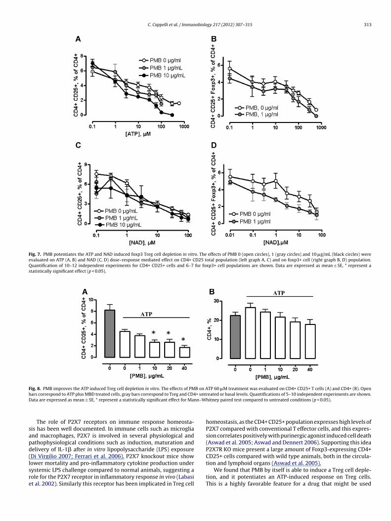
C. Cappelli et al. / Immunobiology 217 (2012) 307– 315 313
Fig. 7. PMB potentiates the ATP and NAD induced foxp3 Treg cell depletion in vitro. The effects of PMB 0 (open circles), 1 (gray circles) and 10 �g/mL (black circles) wereevaluated on ATP (A, B) and NAD (C, D) dose–response mediated effect on CD4+ CD25 total population (left graph A, C) and on foxp3+ cell (right graph B, D) population.Quantification of 10–12 independent experiments for CD4+ CD25+ cells and 6–7 for foxp3+ cell populations are shown. Data are expressed as mean ± SE, * represent astatistically significant effect (p < 0.05).
Fig. 8. PMB improves the ATP induced Treg cell depletion in vitro. The effects of PMB on ATP 60 �M treatment was evaluated on CD4+ CD25+ T cells (A) and CD4+ (B). Openb + untD n–W
sapd(lsre
ars correspond to ATP plus MBD treated cells, gray bars correspond to Treg and CD4ata are expressed as mean ± SE, * represent a statistically significant effect for Man
The role of P2X7 receptors on immune response homeosta-is has been well documented. In immune cells such as microgliand macrophages, P2X7 is involved in several physiological andathophysiological conditions such as induction, maturation andelivery of IL-1� after in vitro lipopolysaccharide (LPS) exposureDi Virgilio 2007; Ferrari et al. 2006). P2X7 knockout mice show
ower mortality and pro-inflammatory cytokine production underystemic LPS challenge compared to normal animals, suggesting aole for the P2X7 receptor in inflammatory response in vivo (Labasit al. 2002). Similarly this receptor has been implicated in Treg cellreated or basal levels. Quantifications of 5–10 independent experiments are shown.hitney paired test compared to untreated conditions (p < 0.05).
homeostasis, as the CD4+ CD25+ population expresses high levels ofP2X7 compared with conventional T effector cells, and this expres-sion correlates positively with purinergic agonist induced cell death(Aswad et al. 2005; Aswad and Dennert 2006). Supporting this ideaP2X7R KO mice present a large amount of Foxp3-expressing CD4+CD25+ cells compared with wild type animals, both in the circula-
tion and lymphoid organs (Aswad et al. 2005).We found that PMB by itself is able to induce a Treg cell deple-tion, and it potentiates an ATP-induced response on Treg cells.This is a highly favorable feature for a drug that might be used

3 obiolo
ttu(AetMaaepbdcbspaPpsosi1ihs
ebPpcv1fvwd(
ttdCatN2hetwaaa
A
fDw
14 C. Cappelli et al. / Immun
o induce an antitumoral response by means of Treg cell deple-ion and suggests that P2X7 receptors are endogenously activatednder our basal experimental conditions. Previously, Ferrari et al.2004) demonstrated that PMB potentiates responses elicited byTP-stimulated transfected cell lines, macrophages and B cellsxpressing P2X7 receptor. The mechanisms associated with induc-ion of activity appear to be common for several cyclic peptides.
oreover, IL1beta production in macrophages has been associ-ted with the presence and activity of other cationic peptides suchs the derivate peptide LL37 and SSA (Niemi et al. 2011; Elssnert al. 2004). Furthermore, Allam and co-workers determined thatolymyxin B and other cyclic antibiotics induce IL-1� secretion inone marrow dendritic cells and macrophages in a K+ efflux depen-ent fashion (Allam et al. 2011). This activity is also induced inells from P2X7 KO animals, suggesting that the mechanism coulde independent of P2X7 receptor. Similarly a pannexin 1 has beenuggested as responsible for several activities associated with theurinergic receptor such as caspase 1 activation, IL-1� maturationnd delivery, and ethidium bromide uptake (Kanneganti et al. 2007;elegrin and Surprenant 2006), but not for others such as macro-ore formation (Yan et al. 2008). Recently Lemaire and coworkershowed that P2X7 and pannexin are both crucial to the processf cell-to-cell fusion into multinucleated macrophages (MA). Theyhowed, in agreement with our results, that PMB itself is able tonduce cell fusion, and while P2X7 appears to play a role, pannexin
inhibition completely abrogates this effect. Altogether those stud-es suggest that PMB related effects could be mediated by pannexin,owever this specific observation will be addressed in a futuretudy.
PMB related effects have been described for APC cells, but theffects on T cell population and particularly on Treg cells have noteen previously tested. Interestingly, we also demonstrated thatMB at the concentrations used here had no effects on other T cellopulations. On the other hand, apoptosis was detected in Tregells after ATP but not after PMB treatment. PMB has been pre-iously associated with induction of cell death (Mihich group in993), i.e., this drug is cytotoxic to certain tumor cells, with dif-erential sensitivity. For instance, EL4 cells and EL4/ADM cells wereery sensitive (lysed by ≥10 �g/mL PMB); C1498 cells and REH cellsere moderately sensitive (lysed by ≥20 �g/mL PMB); and 6 othersifferent cell lines were resistant (lysed only by 100 �g/mL) to PMBVerstovsek et al. 1993).
Several efforts have been made in order to generate an adjuvantherapy against cancer by induction of Human Treg cell deple-ion. Until now, the most effective drugs in depleting Treg cells areriven specifically against Treg cell receptors, such as CTLA4 andD25 and cyclophosphamide. Unfortunately, both receptors arelso expressed in activated lymphocytes and conventional T cellshat could be depleted by action of this drugs (Needham et al. 2006;ishikawa and Sakaguchi 2010; O’Day et al. 2007; Schabowsky et al.007), moreover cyclophosphamide induces undesired effects atigher concentrations, inhibiting the immune response (Motoyoshit al. 2006). We described here that modulation of P2X7 appearso be a plausible alternative to deplete Treg cells in a more specificay. Our results suggest that the use of the antibiotic PMB could be
n important step towards developing an adjuvant immunother-py against cancer, however several experiments including in vivopproaches are still necessary.
cknowledgments
The authors thank Drs. Elias Leiva-Salcedo and Claudio Coddouor the critical review of this manuscript, and Carolina Tambley,arwin Sáez and Luis Solano for their technical assistance. Thisork was funded by FONDECYT 11070177 and 1110734 and DICYT
gy 217 (2012) 307– 315
USACH. This work is in memory of Ruth B. Quiroz Ibarra andSegundo Irribarren T. Special thanks go to Mr. Jacob M. Kames forproofreading the paper.
References
Acuna-Castillo, C., Coddou, C., Bull, P., Brito, J., Huidobro-Toro, J.P., 2007. Differ-ential role of extracellular histidines in copper, zinc, magnesium and protonmodulation of the P2X7 purinergic receptor. J. Neurochem. 101, 17.
Allam, R., Darisipudi, M.N., Rupanagudi, K.V., Lichtnekert, J., Tschopp, J., Anders,H.J., 2011. Cutting edge: cyclic polypeptide and aminoglycoside antibiotics trig-ger IL-1beta secretion by activating the NLRP3 inflammasome. J. Immunol.186, 2714.
Aswad, F., Dennert, G., 2006. P2X7 receptor expression levels determine lethal effectsof a purine based danger signal in T lymphocytes. Cell Immunol. 243, 58.
Aswad, F., Kawamura, H., Dennert, G., 2005. High sensitivity of CD4+ CD25+ regula-tory T cells to extracellular metabolites nicotinamide adenine dinucleotide andATP: a role for P2X7 receptors. J. Immunol. 175, 3075.
Curiel, T.J., Coukos, G., Zou, L., Alvarez, X., Cheng, P., Mottram, P., Evdemon-Hogan,M., Conejo-Garcia, J.R., Zhang, L., Burow, M., Zhu, Y., Wei, S., Kryczek, I., Daniel, B.,Gordon, A., Myers, L., Lackner, A., Disis, M.L., Knutson, K.L., Chen, L., Zou, W., 2004.Specific recruitment of regulatory T cells in ovarian carcinoma fosters immuneprivilege and predicts reduced survival. Nat. Med. 10, 942.
Di Virgilio, F., 2007. Liaisons dangereuses: P2X(7) and the inflammasome. TrendsPharmacol. Sci. 28, 465.
Elssner, A., Duncan, M., Gavrilin, M., Wewers, M.D., 2004. A novel P2X7 receptoractivator, the human cathelicidin-derived peptide LL37, induces IL-1 beta pro-cessing and release. J. Immunol. 172, 4987.
Ferrari, D., Pizzirani, C., Adinolfi, E., Forchap, S., Sitta, B., Turchet, L., Falzoni, S., Minelli,M., Baricordi, R., Di Virgilio, F., 2004. The antibiotic polymyxin B modulates P2X7receptor function. J. Immunol. 173, 4652.
Ferrari, D., Pizzirani, C., Adinolfi, E., Lemoli, R.M., Curti, A., Idzko, M., Panther, E., DiVirgilio, F., 2006. The P2X7 receptor: a key player in IL-1 processing and release.J. Immunol. 176, 3877.
Gavin, M., Rudensky, A., 2003. Control of immune homeostasis by naturally arisingregulatory CD4+ T cells. Curr. Opin. Immunol. 15, 690.
Gavin, M.A., Rasmussen, J.P., Fontenot, J.D., Vasta, V., Manganiello, V.C., Beavo, J.A.,Rudensky, A.Y., 2007. Foxp3-dependent programme of regulatory T-cell differ-entiation. Nature 445, 771.
Haag, F., Adriouch, S., Brass, A., Jung, C., Moller, S., Scheuplein, F., Bannas, P., Seman,M., Koch-Nolte, F., 2007. Extracellular NAD and ATP: partners in immune cellmodulation. Purinergic Signal. 3, 71.
Jones, E., Dahm-Vicker, M., Simon, A.K., Green, A., Powrie, F., Cerundolo, V., Gallimore,A., 2002. Depletion of CD25+ regulatory cells results in suppression of melanomagrowth and induction of autoreactivity in mice. Cancer Immun. 2, 1.
Kanneganti, T.D., Lamkanfi, M., Kim, Y.G., Chen, G., Park, J.H., Franchi, L., Van-denabeele, P., Nunez, G., 2007. Pannexin-1-mediated recognition of bacterialmolecules activates the cryopyrin inflammasome independent of Toll-likereceptor signaling. Immunity 26, 433.
Kawamura, H., Aswad, F., Minagawa, M., Malone, K., Kaslow, H., Koch-Nolte, F.,Schott, W.H., Leiter, E.H., Dennert, G., 2005. P2X7 receptor-dependent and -independent T cell death is induced by nicotinamide adenine dinucleotide. J.Immunol. 174, 1971.
Labasi, J.M., Petrushova, N., Donovan, C., McCurdy, S., Lira, P., Payette, M.M., Brissette,W., Wicks, J.R., Audoly, L., Gabel, C.A., 2002. Absence of the P2X7 receptor altersleukocyte function and attenuates an inflammatory response. J. Immunol. 168,6436.
Leiva-Salcedo, E., Coddou, C., Rodriguez, F.E., Penna, A., Lopez, X., Neira, T., Fernandez,R., Imarai, M., Rios, M., Escobar, J., Montoya, M., Huidobro-Toro, J.P., Escobar, A.,Acuna-Castillo, C., 2011. Lipopolysaccharide inhibits the channel activity of theP2X7 receptor. Mediat. Inflamm. 2011, 152625.
Motoyoshi, Y., Kaminoda, K., Saitoh, O., Hamasaki, K., Nakao, K., Ishii, N., Nagayama,Y., Eguchi, K., 2006. Different mechanisms for anti-tumor effects of low- andhigh-dose cyclophosphamide. Oncol. Rep. 16, 141.
Needham, D.J., Lee, J.X., Beilharz, M.W., 2006. Intra-tumoural regulatory T cells: apotential new target in cancer immunotherapy. Biochem. Biophys. Res. Com-mun. 343, 684.
Niemi, K., Teirila, L., Lappalainen, J., Rajamaki, K., Baumann, M.H., Oorni, K., Wolff, H.,Kovanen, P.T., Matikainen, S., Eklund, K.K., 2011. Serum amyloid A activates theNLRP3 inflammasome via P2X7 receptor and a cathepsin B-sensitive pathway.J. Immunol. 186, 6119.
Nishikawa, H., Sakaguchi, S., 2010. Regulatory T cells in tumor immunity. Int. J.Cancer 127, 759.
North, R.A., 2002. Molecular physiology of P2X receptors. Physiol. Rev. 82, 1013.O’Day, S.J., Hamid, O., Urba, W.J., 2007. Targeting cytotoxic T-lymphocyte antigen-4
(CTLA-4): a novel strategy for the treatment of melanoma and other malignan-cies. Cancer 110, 2614.
Ohkura, N., Sakaguchi, S., 2010. Regulatory T cells: roles of T cell receptor for their
development and function. Semin. Immunopathol. 32, 95.Pelegrin, P., Surprenant, A., 2006. Pannexin-1 mediates large pore formation andinterleukin-1beta release by the ATP-gated P2X7 receptor. EMBO J. 25, 5071.
Sakaguchi, S., Yamaguchi, T., Nomura, T., Ono, M., 2008. Regulatory T cells andimmune tolerance. Cell 133, 775.

obiolo
S
S
S
S
S
C. Cappelli et al. / Immun
akaguchi, S., Wing, K., Onishi, Y., Prieto-Martin, P., Yamaguchi, T., 2009. RegulatoryT cells: how do they suppress immune responses? Int. Immunol. 21, 1105.
alem, D.A., Gado, N.M., Abdelaziz, N.N., Essa, A.E., Abdelhafeez, Z.M., Kamel, T.H.,2008. Phase II trial of metronomic chemotherapy as salvage therapy for patientswith metastatic breast cancer. J. Egypt Natl. Cancer Inst. 20, 134.
chabowsky, R.H., Madireddi, S., Sharma, R., Yolcu, E.S., Shirwan, H., 2007. Tar-geting CD4+ CD25+ FoxP3+ regulatory T-cells for the augmentation of cancerimmunotherapy. Curr. Opin. Investig. Drugs 8, 1002.
eman, M., Adriouch, S., Scheuplein, F., Krebs, C., Freese, D., Glowacki, G., Deterre, P.,
Haag, F., Koch-Nolte, F., 2003. NAD-induced T cell death: ADP-ribosylation of cellsurface proteins by ART2 activates the cytolytic P2X7 purinoceptor. Immunity19, 571.utmuller, R.P., van Duivenvoorde, L.M., van Elsas, A., Schumacher, T.N., Wilden-berg, M.E., Allison, J.P., Toes, R.E., Offringa, R., Melief, C.J., 2001. Synergism of
gy 217 (2012) 307– 315 315
cytotoxic T lymphocyte-associated antigen 4 blockade and depletion of CD25(+)regulatory T cells in antitumor therapy reveals alternative pathways for sup-pression of autoreactive cytotoxic T lymphocyte responses. J. Exp. Med. 194,823.
Verstovsek, S., Maccubbin, D.L., Ehrke, M.J., Mihich, E., 1993. Polymyxin B-mediatedlysis of tumor cells. Int. Arch. Allergy Immunol. 100, 47.
Wing, K., Sakaguchi, S., 2010. Regulatory T cells exert checks and balances on selftolerance and autoimmunity. Nat. Immunol. 11, 7.
Woo, E.Y., Yeh, H., Chu, C.S., Schlienger, K., Carroll, R.G., Riley, J.L., Kaiser, L.R., June,
C.H., 2002. Cutting edge: regulatory T cells from lung cancer patients directlyinhibit autologous T cell proliferation. J. Immunol. 168, 4272.Yan, Z., Li, S., Liang, Z., Tomic, M., Stojilkovic, S.S., 2008. The P2X7 receptorchannel pore dilates under physiological ion conditions. J. Gen. Physiol. 132,563.
Copyright © 2022 FDOKUMEN