The RXR Agonist MSU42011 Is Effective for the Treatment of ... - MDPI
20
cancers Article The RXR Agonist MSU42011 Is Effective for the Treatment of Preclinical HER2+ Breast Cancer and Kras-Driven Lung Cancer Ana S. Leal 1 , Jessica A. Moerland 1 , Di Zhang 1 , Sarah Carapellucci 1 , Beth Lockwood 1 , Teresa Krieger-Burke 1,2 , Bilal Aleiwi 1,3 , Edmund Ellsworth 1,3 and Karen T. Liby 1, * Citation: Leal, A.S.; Moerland, J.A.; Zhang, D.; Carapellucci, S.; Lockwood, B.; Krieger-Burke, T.; Aleiwi, B.; Ellsworth, E.; Liby, K.T. The RXR Agonist MSU42011 Is Effective for the Treatment of Preclinical HER2+ Breast Cancer and Kras-Driven Lung Cancer. Cancers 2021, 13, 5004. https://doi.org/ 10.3390/cancers13195004 Academic Editor: Javier Cortes Received: 29 July 2021 Accepted: 2 October 2021 Published: 6 October 2021 Publisher’s Note: MDPI stays neutral with regard to jurisdictional claims in published maps and institutional affil- iations. Copyright: © 2021 by the authors. Licensee MDPI, Basel, Switzerland. This article is an open access article distributed under the terms and conditions of the Creative Commons Attribution (CC BY) license (https:// creativecommons.org/licenses/by/ 4.0/). 1 Department of Pharmacology and Toxicology, Michigan State University, East Lansing, MI 48824, USA; [email protected] (A.S.L.); [email protected] (J.A.M.); [email protected] (D.Z.); [email protected] (S.C.); [email protected] (B.L.); [email protected] (T.K.-B.); [email protected] (B.A.); [email protected] (E.E.) 2 In Vivo Facility, Michigan State University, East Lansing, MI 48824, USA 3 Medicinal Chemistry Facility, Michigan State University, East Lansing, MI 48824, USA * Correspondence: [email protected]; Tel.: +1-517-884-8955; Fax: +1-517-353-8915 Simple Summary: Breast and lung cancers are the most diagnosed cancers in the United States. Despite advances in treatment, over 176,000 deaths are expected in 2021, highlighting the need for new therapies. We evaluated MSU42011, a new RXR receptor activator, in murine models of breast and lung cancer, and this agonist reduced the tumor burden in both models. The tumor microenvironment is composed of numerous immune cells that can inhibit or promote tumor growth. MSU42011 modulated T cells within the tumor, reducing tumor-promoting immune cells and increasing tumor- killing cells. MSU42011 in combination with immunotherapy (anti-PDL1 and anti-PD1 antibodies) also decreased tumor burden when compared with individual treatments in the lung cancer model. Abstract: (1) Background: Notwithstanding numerous therapeutic advances, 176,000 deaths from breast and lung cancers will occur in the United States in 2021 alone. The tumor microenvironment and its modulation by drugs have gained increasing attention and relevance, especially with the introduction of immunotherapy as a standard of care in clinical practice. Retinoid X receptors (RXRs) are members of the nuclear receptor superfamily and upon ligand binding, function as transcription factors to modulate multiple cell functions. Bexarotene, the only FDA-approved RXR agonist, is still used to treat cutaneous T-cell lymphoma. (2) Methods: To test the immunomodulatory and anti-tumor effects of MSU42011, a new RXR agonist, we used two different immunocompetent murine models (MMTV-Neu mice, a HER2 positive model of breast cancer and the A/J mouse model, in which vinyl carbamate is used to initiate lung tumorigenesis) and an immunodeficient xenograft lung cancer model. (3) Results: Treatment of established tumors in immunocompetent models of HER2-positive breast cancer and Kras-driven lung cancer with MSU42011 significantly decreased the tumor burden and increased the ratio of CD8/CD4, CD25 T cells, which correlates with enhanced anti-tumor efficacy. Moreover, the combination of MSU42011 and immunotherapy (anti-PDL1 and anti-PD1 antibodies) significantly (p < 0.05) reduced tumor size vs. individual treatments. However, MSU42011 was ineffective in an athymic human A549 lung cancer xenograft model, supporting an immunomodulatory mechanism of action. (4) Conclusions: Collectively, these data suggest that the RXR agonist MSU42011 can be used to modulate the tumor microenvironment in breast and lung cancer. Keywords: RXR agonist; tumor microenvironment; lung cancer; breast cancer; immunotherapy 1. Introduction Breast cancer is the most common cancer diagnosis and is responsible for over 42,000 deaths in the United States per year [1]. Targeted therapies have reduced cancer deaths, but human epidermal growth factor receptor 2 (HER2) positive- and triple-negative Cancers 2021, 13, 5004. https://doi.org/10.3390/cancers13195004 https://www.mdpi.com/journal/cancers
-
Upload
khangminh22 -
Category
Documents
-
view
11 -
download
0
Transcript of The RXR Agonist MSU42011 Is Effective for the Treatment of ... - MDPI

cancers
Article
The RXR Agonist MSU42011 Is Effective for the Treatment ofPreclinical HER2+ Breast Cancer and Kras-Driven Lung Cancer
Ana S. Leal 1, Jessica A. Moerland 1, Di Zhang 1, Sarah Carapellucci 1, Beth Lockwood 1, Teresa Krieger-Burke 1,2,Bilal Aleiwi 1,3, Edmund Ellsworth 1,3 and Karen T. Liby 1,*
�����������������
Citation: Leal, A.S.; Moerland, J.A.;
Zhang, D.; Carapellucci, S.;
Lockwood, B.; Krieger-Burke, T.;
Aleiwi, B.; Ellsworth, E.; Liby, K.T.
The RXR Agonist MSU42011 Is
Effective for the Treatment of
Preclinical HER2+ Breast Cancer and
Kras-Driven Lung Cancer. Cancers
2021, 13, 5004. https://doi.org/
10.3390/cancers13195004
Academic Editor: Javier Cortes
Received: 29 July 2021
Accepted: 2 October 2021
Published: 6 October 2021
Publisher’s Note: MDPI stays neutral
with regard to jurisdictional claims in
published maps and institutional affil-
iations.
Copyright: © 2021 by the authors.
Licensee MDPI, Basel, Switzerland.
This article is an open access article
distributed under the terms and
conditions of the Creative Commons
Attribution (CC BY) license (https://
creativecommons.org/licenses/by/
4.0/).
1 Department of Pharmacology and Toxicology, Michigan State University, East Lansing, MI 48824, USA;[email protected] (A.S.L.); [email protected] (J.A.M.); [email protected] (D.Z.);[email protected] (S.C.); [email protected] (B.L.); [email protected] (T.K.-B.); [email protected] (B.A.);[email protected] (E.E.)
2 In Vivo Facility, Michigan State University, East Lansing, MI 48824, USA3 Medicinal Chemistry Facility, Michigan State University, East Lansing, MI 48824, USA* Correspondence: [email protected]; Tel.: +1-517-884-8955; Fax: +1-517-353-8915
Simple Summary: Breast and lung cancers are the most diagnosed cancers in the United States.Despite advances in treatment, over 176,000 deaths are expected in 2021, highlighting the need for newtherapies. We evaluated MSU42011, a new RXR receptor activator, in murine models of breast andlung cancer, and this agonist reduced the tumor burden in both models. The tumor microenvironmentis composed of numerous immune cells that can inhibit or promote tumor growth. MSU42011modulated T cells within the tumor, reducing tumor-promoting immune cells and increasing tumor-killing cells. MSU42011 in combination with immunotherapy (anti-PDL1 and anti-PD1 antibodies)also decreased tumor burden when compared with individual treatments in the lung cancer model.
Abstract: (1) Background: Notwithstanding numerous therapeutic advances, 176,000 deaths frombreast and lung cancers will occur in the United States in 2021 alone. The tumor microenvironmentand its modulation by drugs have gained increasing attention and relevance, especially with theintroduction of immunotherapy as a standard of care in clinical practice. Retinoid X receptors (RXRs)are members of the nuclear receptor superfamily and upon ligand binding, function as transcriptionfactors to modulate multiple cell functions. Bexarotene, the only FDA-approved RXR agonist, isstill used to treat cutaneous T-cell lymphoma. (2) Methods: To test the immunomodulatory andanti-tumor effects of MSU42011, a new RXR agonist, we used two different immunocompetentmurine models (MMTV-Neu mice, a HER2 positive model of breast cancer and the A/J mouse model,in which vinyl carbamate is used to initiate lung tumorigenesis) and an immunodeficient xenograftlung cancer model. (3) Results: Treatment of established tumors in immunocompetent models ofHER2-positive breast cancer and Kras-driven lung cancer with MSU42011 significantly decreased thetumor burden and increased the ratio of CD8/CD4, CD25 T cells, which correlates with enhancedanti-tumor efficacy. Moreover, the combination of MSU42011 and immunotherapy (anti-PDL1 andanti-PD1 antibodies) significantly (p < 0.05) reduced tumor size vs. individual treatments. However,MSU42011 was ineffective in an athymic human A549 lung cancer xenograft model, supporting animmunomodulatory mechanism of action. (4) Conclusions: Collectively, these data suggest thatthe RXR agonist MSU42011 can be used to modulate the tumor microenvironment in breast andlung cancer.
Keywords: RXR agonist; tumor microenvironment; lung cancer; breast cancer; immunotherapy
1. Introduction
Breast cancer is the most common cancer diagnosis and is responsible for over42,000 deaths in the United States per year [1]. Targeted therapies have reduced cancerdeaths, but human epidermal growth factor receptor 2 (HER2) positive- and triple-negative
Cancers 2021, 13, 5004. https://doi.org/10.3390/cancers13195004 https://www.mdpi.com/journal/cancers

Cancers 2021, 13, 5004 2 of 20
breast cancer (TNBC) are often deadly, mostly due to acquired resistance. Worldwide,lung cancer remains the foremost cause of cancer deaths. Overall survival in lung cancerpatients is less than 20%, despite the recent promise of targeted therapies and immunother-apy [1]. Over the last decade, immune checkpoint inhibitors, antibodies against PD1 andPDL1, have emerged as a standard of care to treat lung cancers without actionable targets.Nevertheless, patient responses to immunotherapy vary from 20% to 40%, highlighting theneed to develop effective new drugs to enhance efficacy and suppress resistance [2].
Immunotherapy, and more recently small molecules used in combination with im-munotherapy, were developed to target the immune compartment of the tumor microenvi-ronment [3–5]. Immune checkpoints are required to maintain homeostasis and to preventthe inappropriate activation of CD8 cytotoxic T cells. Cancers disrupt the regulation ofimmune checkpoints to elude eradication by the immune system [6]. Retinoid X receptor(RXR) agonists can modulate the tumor microenvironment in murine models, leading toincreased anti-tumor and decreased pro-tumor immune populations [7–10].
The RXR belongs to the nuclear receptor superfamily and acts as a transcriptionfactor following ligand binding [11,12]. RXR heterodimerizes with others in the nuclearreceptors superfamily, including the retinoic acid receptor (RAR), vitamin D receptor (VDR),peroxisome proliferator-activated receptor (PPAR), or liver X receptor (LXR). RXR homo- orheterodimers regulate the function of monocytes, linking cellular metabolism and immunefunction [8,13]. RXR agonists can stimulate phagocytosis, dampen responses to viruses,and increase cytokine secretion [8,14]. These immunomodulatory properties of RXR makeit a pharmacologically attractive addition to current cancer therapies [15].
In 1999, the first synthetic RXR agonist, bexarotene (Targretin), was granted FDAapproval to treat patients with cutaneous T-cell lymphoma [16]. Bexarotene has beentested in clinical trials for both lung and breast cancer but was not granted approval forthese cancers [17,18]. Bexarotene binds to RAR, leading to significant adverse effects [17].Other RXR agonists, including LG100268 and IRX194204 [19,20], have been synthesized toimprove pharmacokinetic properties and diminish side effects. LG100268 and IRX194204are active in murine models of lung [21] and breast cancer [7]. However, reported sideeffects from early clinical trials and FDA approval for bexarotene have hindered theirclinical development [15]. We recently reported structure–activity relationship studies ofnew RXR agonists [22]. These small molecules were screened for suppression of nitricoxide, which correlates with activity against preclinical lung cancer, and limited changes inthe expression of sterol regulatory element-binding protein (SREBP), which correlates withthe elevation of triglycerides, a reported side effect of RXR agonists. From these in vitrostudies, MSU42011 was chosen for additional efficacy studies in a murine lung cancermodel [22,23].
Infiltration of immune cells into tumors is a hallmark of cancer and resistance totreatment, both in lung and breast cancers [24–26]. Because of the immune modulatoryeffects of RXR in cancer and other diseases [27], we tested the effects of MSU42011 onmodulation of the tumor microenvironment in two different immunocompetent murinemodels of cancer. In HER2+ MMTV-Neu mice, the MMTV promoter directs the expressionof inactivated neu (ErbB2/HER-2) to the mammary gland [28]. In the A/J mouse modelof lung carcinogenesis, vinyl carbamate induces Kras mutations and the infiltration ofimmune cells observed in human lung cancer [21,29–32]. Our data show that MSU42011has anti-tumor properties in immunocompetent mice, but not in an immunodeficientxenograft lung cancer model. MSU42011 also increased cytotoxic CD8 T cell activity inMMTV-Neu and A/J mice. Moreover, the combination of MSU42011 and anti-PD1 andanti-PDL1 antibodies was more effective in the A/J lung cancer model.
2. Materials and Methods2.1. Drugs
MSU42011 was synthesized (>95% purity) as described [22]. Bexarotene (>99% purity)was acquired from LC Laboratories.

Cancers 2021, 13, 5004 3 of 20
2.2. Animal Experiments
All in vivo studies were compliant with animal protocols (20180050) approved bythe Institutional Animal Care and Use Committee at MSU in accordance with AAALAC-accredited Standards for the Management of Laboratory Animals.
MMTV-neu mice (Jackson Laboratory, Bar Harbor, ME, USA) were bred at MSU.Female MMTV-neu mice were fed standard rodent chow until palpable tumors weredetected. When tumors reached a size of 32–64 mm3, mice were randomized and fed eithercontrol 5002 diet or MSU42011 in 5002 diet (100 mg/kg diet) [33]. Tumor diameters weremeasured twice per week.
Female A/J mice (Jackson Laboratories, Bar Harbor, ME, USA) were injected i.p. with0.32 mg vinyl carbamate (Toronto Research Chemicals, Ontario, Canada) at both 7 and8 weeks of age. One week before initiation, mice were started on AIN-93G diet (BioServ,Flemington, NJ, USA), which continued throughout the study. Mice (n = 15/group)were randomized and treated for 12 weeks with 100 mg RXR agonist/kg AIN-93G diet(~25 mg/kg body weight). Anti-PDL1, anti-PD1, and isotype control antibodies (BiolegendGo Invivo, San Diego, CA, USA) were injected i.p. (50 µg per mouse, twice a week for12 weeks), beginning 10 weeks after initiation. Lungs were harvested and then inflatedwith PBS. The left lobe was fixed in neutral-buffered formalin (NBF), embedded in paraffin,step-sectioned (2 sections/lung, 600 microns apart), and stained with Hematoxylin andEosin. The slides were evaluated as described [21,22]. One-half of the right lung wasevaluated by flow cytometry and the other half was flash frozen for mRNA analysis.
Male athymic nude mice (Envigo, >5–6 weeks of age) were injected in the flank withhuman A549 lung cancer cells (5 × 10⁶ cells/mouse, cell authenticity confirmed by IDEXXBioAnalytics prior to injection). Calipers were used to measure tumor diameter and when4 mm in diameter, mice were randomized (n = 7/group) and injected i.p. M-F with vehiclecontrol (DMSO:Cremophor EL:saline 1:1:8) or RXR agonists (bexarotene or MSU4201125 mg/kg) and/or carboplatin (15 mg/kg in saline once per week). Tumor size wasmeasured with calipers twice per week and mice were weighed once per week. Tumorvolume = length × width × (length + width/2)/2. After 4 weeks, tumors and tissueswere harvested.
2.3. Flow Cytometry
In MMTV-Neu mice, a third of the tumor was minced and incubated with collagenase(300 U/mL, Sigma, St. Louis, MO, USA), dispase (1 U/mL, Worthington, Lakewood, NJ,USA), and DNAse (2 U/mL, Calbiochem, Burlington, MA, USA) for 30 min at 37 ◦C withstirring. A single-cell suspension was obtained by passing the cell solution through a 40 µmcell strainer (BD Falcon), and red blood cells were eliminated using a lysis buffer. Single cellswere resuspended in PBS/0.5% BSA/0.1% azide and stained at 4 ◦C with the followingantibodies for 30 min: CD45-VioGreen (30F11, Miltenyi, Bergisch Gladbach, Germany,10:100), Gr-1-PE (RB6-8C5, Miltenyi, Bergisch Gladbach, Germany, 10:100), CD11b-FITC(M1/70.15.11.5, Miltenyi, Bergisch Gladbach, Germany, 10:100), CD19-APC (1D3/CD19,Biolegend, San Diego, CA, USA, 1:100), B220-PerCP-Cy5.5 (RA3-6B2, Biolegend, San Diego,CA, USA, 1:100), CD3-PE (145-2C11, Biolegend, San Diego, CA, USA 1:100), CD4-FITC(Gk1.5, Miltenyi, Bergisch Gladbach, Germany, 10:100), CD8-APC (53–6.7, Biolegend, SanDiego, CA, USA, 1:100), CD25-PE.Cy7 (EBiosciences, San Diego, CA, USA, 1:100), and5 µg/mL anti-mouse CD16/CD32 antibody (Biolegend, San Diego, CA, USA,) to reduceantibody binding to Fc receptors. Propidium iodide staining was used to exclude deadcells. Cells were analyzed using a BD FACS ARIA (BD) with three laser sources (488 nm,633 nm, and 405 nm) and FlowJo x.10.0.7r2 software (Tree Star). Our gating strategy hasbeen published [7].
Two lobes of the right lung were harvested from each A/J mouse (n = 6/group)for flow cytometry and incubated in digestion media and processed as described for theMMTV-Neu mice but with the following antibodies: CD45-Brilliant violet 510 (30F11),CD24-Brilliant violet 604 (M1/69), CD64-Brilliant violet 711 (X54-5/7.1), AI/AE-Brilliant

Cancers 2021, 13, 5004 4 of 20
violet 650 (M5/114.15.2), CD8-Alexafluor 700 (53–6.7), CD4-FITC (GK1.5), LyC6/LyG6-APC (rb6-8C5), CD11b-PE-Cy7 (M1/70), CD11c-PE-Cy5 (N418), CD206-Brilliant violet421 (C068C2), CD69-Brilliant violet 785 (41.2F3), CD25-PE (3C7), and CD107a-Alexafluor647 (1D4B). Zombie NIR staining was used to exclude dead cells. Cells were analyzedusing a Cytek Aurora equipped with 4 lasers (405 nm, 488 nm, 561 nm, and 640 nm) andFlowJo x.10.0.7r2 software (Tree Star). Our gating strategy has been published [22].
2.4. Immunohistochemistry
A third of the tumor from MMTV-neu or the left lung from A/J mice were fixed in NBFfor 48 h, embedded in paraffin, and sectioned. Endogenous peroxidase was quenched byhydrogen peroxide. Sections were immunostained with the following antibodies: CD206(1:200, Abcam, Cambridge, United Kingdom), CD4 (1:40, GK1.5, Biolegend, San Diego,CA, USA), CD8 (1:40, 53–6.7, Biolegend, San Diego, CA, USA), FOXP3 (1:50, FJK-16s,EBiosciences, San Diego, CA, USA), PD-1 (1:100, EPR20665, Abcam, San Diego, CA, USA),p-ERK (1:100, 5A1E, Cell Signaling, Danvers, MA, USA), PCNA (1:200, sc-56, Santa CruzBiotechnologies, Dallas, TX, USA), and F4/80 (1:50, Abcam, Cambridge, United Kingdom)and visualized with biotinylated anti-rabbit or anti-rat secondary antibodies (Cell Signalingor Vector Labs). Signal was detected using a DAB substrate (Cell Signaling) followingthe manufacturer’s recommendations. Sections were counterstained with hematoxylin(Vector Labs).
2.5. Ultrasound
Fur was removed over the thoracic cavity using the Nair chemical depilatory. Micewere anesthetized with isofluorane at 0, 4, and 12 weeks of treatment. A Vevo 2100 (FujifilmVisualSonics, Toronto, ON, Canada) high-frequency ultrasound with a MS550D 40 MHztransducer was used to image the lung tumors [22].
2.6. Lipid Levels in Plasma
Plasma (n = 8 mice/group) harvested at necropsy was used to measure triglycerideand cholesterol level using commercial kits (Triglyceride Quantification Assay Kit fromAbcam) and a Cholesterol Quantification Kit from Sigma-Aldrich.
2.7. Patient Survival Analysis
RXRα was used to analyze relapse-free survival and compare tumor vs. adjacent nor-mal tissue. Aggregate data from KMPlot (http://www.kmplot.com, accessed on 6 January2021) was auto-selected for the best cutoff (25th and 75th percentiles). Ethical approval wasnot required due to the retrospective nature of this study.
2.8. Statistical Analysis
Results were expressed as the mean ± standard deviation or the mean ± standard erroras indicated in the respective figures/tables. All in vitro data were normally distributedand thus were analyzed using one-way ANOVA. The Tukey HSD multiple comparisonmethod was used to evaluate changes between groups (VassarStats.com, accessed on1 April 2021). Animal data were analyzed using SigmaStat 3.5 and one-way ANOVA. Ifdata were normally distributed, the Holm–Sidak test for multiple comparisons was used.If data were not normally distributed, a Kruskal–Wallis one-way ANOVA on ranks and aDunn test for multiple comparisons were used. Histological grades were analyzed using aMcNemar’s Z test. For all studies, p < 0.05 was required for significance.
3. Results3.1. RXR Expression Correlates with Increased Survival in Lung and Breast Cancers
The RXR nuclear receptor is expressed in all tissues in the body, as RXR regulatesmechanisms required for tissue maintenance such as glucose consumption, cell division,and immune regulation [12,27]. Because RXR is essential in normal physiology, we hypoth-

Cancers 2021, 13, 5004 5 of 20
esized that RXR expression will be downregulated in cancer. Using KMPLOT, a publiclyavailable database, the expression of RXR in paired samples of tumors vs. normal adjacenttissue was analyzed [34,35]. In lung adenocarcinomas, the level of RXRα mRNA wassignificantly (p = 0.0017) lower in the cancer tissue compared to adjacent normal tissue(Figure 1A) in the 57 paired samples available. In breast cancers, RXRα mRNA expressionwas significantly (p = 0.0000207) lower in the tumor samples than in adjacent normal tissuein 112 paired samples from advanced breast cancer patients (Figure 1B) [34]. This trendwas also observed in other cancer types and for the RXRβ isoform (Figure S1).
Figure 1. RXR expression correlates with overall survival in patients with lung adenocarcinomaand HER2-positive breast cancer. RXRα expression was evaluated by RNA sequencing in tumorsand adjacent normal tissue from patients with (A) lung adenocarcinoma (n = 57) or (B) invasivebreast cancer (n = 112). Prognostic value of high vs. low RXRα expression in patients with (C) lungadenocarcinoma (n = 504; 330 high and 174 low); (D) lung adenocarcinomas enriched for CD8 T cells(n = 273; 203 high and 72 low) and (E) HER2-positive breast cancer (n = 129; 80 high and 49 low).Data for all plots were accessed and analyzed using KMPLOT.
Because tumor samples had lower RXRα expression, we interrogated whether theexpression of RXR could predict survival in breast and lung cancer patients [35]. In lungadenocarcinomas, a high expression of RXRα significantly (p = 0.0031) correlated with

Cancers 2021, 13, 5004 6 of 20
the probability of increased survival. Overall survival was 58 months in patients withhigh RXRα expression compared to approximately 38 months in patients with low RXRαexpression (Figure 1C). When samples from lung adenocarcinoma patients were selectedfor enrichment in CD8 cytotoxic T cells, the overall survival was 60 months (five years) inpatients with the highest expression vs. 34 months in patients with the lowest expression(p = 0.0096; Figure 1D). In HER2-positive breast cancer, there was no significant differencein overall survival, although a trend toward extended long-term survival was found in thesubset with high RXRα expression (Figure 1E). These observations suggest that targetingand activating RXR could be beneficial to patients with lung and breast cancer.
3.2. MSU42011 Reduced Tumor Growth in the MMTV-Neu Model of Breast Cancer and the A/JMouse Model of Lung Cancer
We and others have shown that RXR agonists can prevent or treat HER2-positive andKras-driven murine models of breast and lung cancers [7,21–23,36–39]. To evaluate theanti-tumor activity of the novel RXR agonist, MSU42011, the same models were used.
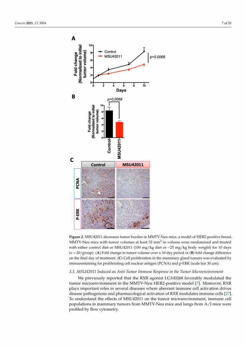
The MMTV-Neu model of HER2-positive breast cancer is driven by targeted expres-sion by the MMTV promoter of the Neu (also known as ErbB2) oncogene to the mammarygland [28]. HER2/neu is a tyrosine kinase receptor shown to be overexpressed in >20% ofbreast cancers. Patients with HER2-positive breast cancer are treated with trastuzumab(Herceptin), pertuzumab, or new antibody–drug conjugates [40–42]. Trastuzumab is effec-tive for treating tumors in MMTV-Neu mice, which validates the clinical relevance of themodel [28]. MMTV-Neu mice with tumor volumes of 32 mm3 were treated with MSU42011(100 mg/kg diet, or approximately 25 mg/kg of body weight) for 10 days [7]. MSU42011significantly (p = 0.0068) decreased both tumor growth (Figure 2A) and final tumor volumewhen compared to the initial volume (Figures 2B and S2A,B) vs. control mice. Fold changeswere utilized because of variability in tumor growth rates between mice (individual tumorgrowth shown in Figure S2C) as there was no difference in volume at the time treatmentwas initiated (Figure S2A). No changes in plasma triglycerides or cholesterol levels wereobserved in the mice (Figure S2D). Consistent with the decreased tumor growth, PCNA, abiomarker of proliferation, and p-ERK staining were also reduced in tumors in mice treatedwith MSU42011 (Figures 2C and S2E). HER2/neu triggers intracellular signaling, includingthe activation of the mitogen-activated protein kinases (MAPK) and consequently ERKphosphorylation, which is linked to proliferation and apoptosis [43,44].
To evaluate efficacy in a preclinical lung cancer model, A/J mice were challengedwith the carcinogen vinyl carbamate, which causes Kras mutations and subsequent lungcancer [23,45]. At eight weeks after initiation, mice were treated with a control diet orMSU42011 diet (100 mg/kg diet). After 12 weeks of treatment, the lungs were harvested andthe samples blinded and randomized prior to analysis. Mice in the MSU42011 treatmentgroup showed a significantly reduced tumor size (p = 0.0036) and tumor burden (total tumorvolume/number of lung sections; p = 0.0015) vs. the control group (Figure 3A, Table 1).The average tumor number and burden were 2.7± 0.4 tumors and 0.43± 0.06 mm3 in theMSU42011 group vs. 3.7 ± 0.4 tumors and 1.03± 0.25 mm3 in the control group, a 27–58%reduction. The percentage of tumors graded low or medium was significantly (p < 0.001)lower in the lungs of mice fed MSU42011 vs. control (48 vs. 34%, respectively). Bexaroteneis inactive in this model [22]. We observed a decrease of PCNA and p-ERK, a proteindownstream of Kras, in the lungs of mice treated with MSU42011 (Figures 3B and S3A),similar to the reduction documented in the MMTV-Neu mice (Figure 2B). Triglycerides andcholesterol were not elevated by MSU42011 at the time of necropsy, and no changes in thetotal body weight were observed between groups at the time of the necropsy (Figure S3B).
Cancers 2021, 13, 5004 7 of 20
Figure 2. MSU42011 decreases tumor burden in MMTV-Neu mice, a model of HER2-positive breast.MMTV-Neu mice with tumor volumes at least 32 mm3 in volume were randomized and treatedwith either control diet or MSU42011 (100 mg/kg diet or ~25 mg/kg body weight) for 10 days(n = 20/group). (A) Fold change in tumor volume over a 10-day period or (B) fold change differenceon the final day of treatment. (C) Cell proliferation in the mammary gland tumors was evaluated byimmunostaining for proliferating cell nuclear antigen (PCNA) and p-ERK (scale bar 30 µm).
3.3. MSU42011 Induced an Anti-Tumor Immune Response in the Tumor Microenvironment
We previously reported that the RXR agonist LG100268 favorably modulated thetumor microenvironment in the MMTV-Neu HER2-positive model [7]. Moreover, RXRplays important roles in several diseases where aberrant immune cell activation drivesdisease pathogenesis and pharmacological activation of RXR modulates immune cells [27].To understand the effects of MSU42011 on the tumor microenvironment, immune cellpopulations in mammary tumors from MMTV-Neu mice and lungs from A/J mice wereprofiled by flow cytometry.

Cancers 2021, 13, 5004 8 of 20
Figure 3. MSU42011 decreases tumor size and burden in AJ mice, a model of lung adenocarcinoma.Female A/J mice were injected with 2 doses of vinyl carbamate, 1 week apart, to initiate lungcarcinogenesis. After 8 weeks, mice were fed either control AIN-93G diet or MSU42011 in the samediet (100 mg/kg diet). (A) Mice were euthanized 12 weeks after starting diet, and tumor size andburden were evaluated on lung sections (n = 14–15/group). (B) Cell proliferation in the lung tumorswas evaluated by immunostaining for PCNA (proliferating cell nuclear antigen). (scale bar 30 µm).
Table 1. The RXR agonist MSU42011 decreases tumor size and burden in a carcinogen-induced A/Jmodel of lung cancer.
Groups Analyzed Control MSU42011100 mg/kg DIET
# SLIDES/GROUP 30 28# TUMORS/GROUP 110 75
TOTAL # TUMORS/SLIDE(% OF CONTROL)
3.67 ± 0.4(100%)
2.68 ± 0.36(73.1%)
TOTAL # L AND M GRADE(% TOTAL) 37 (34%) 36 (48%)
(p < 0.001)TOTAL # H GRADE (% TOTAL) 73 (66%) 39 (52%)TOTAL TUMOR VOLUME, mm3 30.90 11.96
AVE TUMOR SIZE(MM3)/TUMOR(% CONTROL)
0.28 ± 0.033(100%)
0.16 ± 0.02 (56.8%)(p = 0.0036)
AVE TUMOR BURDEN (MM3)(% CONTROL)
1.03 ± 0.15 (100%) 0.43 ± 0.062 (41.5%)(p = 0.0015)

Cancers 2021, 13, 5004 9 of 20
Analysis of mammary tumors exhibited no significant changes in total immune cells,macrophages, myeloid-derived suppressor cells (MDSCs), and total T cells (including CD4and CD8 T cells) present in the tumors (Figure 4A). However, activated CD4 T cells (thepercentage of CD4, CD25 T cells) were significantly (p = 0.0049) lower in tumors of micetreated with MSU42011 compared to control mice (20 vs. 30% of CD4 T cells, respectively).CD4, CD25 T cells are associated with a poor prognosis in breast cancer and correlatewith the expression of FOXP3, a tumor-promoting CD4 T cell, also termed regulatoryT cells [46,47]. Consistent with the decrease in the percentage of the CD4, CD25 populationin tumors from MMTV-Neu mice, we observed a trend toward decreased FOXP3 expression,shown by immunohistochemistry staining (Figure 4B). Because CD4, FOXP3+ T cells tendto aggregate in tumors, qPCR showed variability (Figure S4A) depending on the tumorsection used so we confirmed the decrease in FOXP3 mRNA expression in T cells sortedfrom tumors (Figure S4B, p = 0.0072). To investigate if the decrease in FOXP3 expressionwas a direct effect of MSU42011 on CD4 T cells, isolated CD4 T cells from murine spleenswere stimulated with TGFβ and IL-2 to induce FOXP3 and then treated with the drug forfour days. MSU42011 (100 nM) decreased the levels of FOXP3 mRNA expression in CD4T cells in vitro by 30% (Figure S4C).
Figure 4. MSU42011 increases the percentage of activated CD8 cytotoxic T cells in the MMTV-Neumodel of HER2-positive breast cancer. (A) Immune cell analysis by flow cytometry of whole tumorlysates (n = 8/group) or (B) immunohistochemistry for tumors from MMTV-Neu mice treated for10 days with MSU42011 or control diet. (scale bar 30 µm).

Cancers 2021, 13, 5004 10 of 20
The decrease in CD4, CD25 T cells complemented a higher ratio of the percentage ofCD8:CD4, CD25 T cells (p = 0.0074), which correlates with increased anti-tumor activityby CD8 T cells [48]. The increase in the cytotoxic capability of CD8 T cells resulted insignificantly (p = 0.0026) higher levels of interferon-gamma (IFNγ) mRNA in whole tumorlysates (Figure S4A). To determine if the increased IFNγ in whole tumors was derived fromT cells (CD3) or myeloid cells (CD11B), a single cell suspension from tumors was sorted bymagnetic separation beads and the resulting fraction was analyzed by qPCR. Both CD11Band CD3 cells isolated from tumors from mice treated with MSU42011 had increasedIFNγ expression. The increase in IFNγ mRNA in the CD11B population prompted us tointerrogate if the macrophages present had changed to a less tumor-promoting phenotypesince analysis by flow cytometry showed no change in the percentage of the CD11Bpopulation infiltrating into the tumors (Figure 4A). Immunohistochemistry of tumorsshowed a marked decrease in the expression of CD206 (Figure 4B), a marker of tumor-promoting macrophages [49].
To determine if similar effects were observed in the lung cancer model, immunopro-filing was conducted on the lungs of A/J mice on the control or treatment diet. Flowcytometry revealed a trend toward a decrease in alveolar macrophages (p = 0.0658), but weobserved no changes in other immune cell populations in the lung, including monocytes orT cells (Figure 5A). As previously reported [22] and as observed in the MMTV-Neu tumors(Figure 4B), MSU42011 treatment decreased CD206 macrophage expression (Figure 5B).Analysis of T cell activation revealed that the percentage of CD4, CD25 T cells was de-creased in the lungs of mice treated with MSU42011 (p = 0.0129 vs. control, 0.4% vs. 0.22%).This decrease was accompanied by a trend toward decreased FOXP3 staining within thetumors (Figure 5B), although no significant change (p = 0.3) in IFNγ mRNA levels inlung lysates was observed (Figure S5). The percentage of CD8 T cells infiltrating intothe lungs of A/J did not change when treated with MSU42011. However, because of thedecrease in activated CD4, CD25 T cells, the ratio of CD8: CD4, CD25 was increased inmice treated with MSU42011 (p = 0.016, 3 vs. 6). The increase in the ratio of CD8:CD4,CD25 is characteristic of increased anti-tumor CD8 T cells [50].
3.4. MSU42011 Was Ineffective for Suppressing Tumor Growth in a Human A549 Lung CancerXenograft Model
Xenograft models are widely used to test potential new cytotoxic therapeutics [51,52].However, RXR agonists lack apoptotic and/or anti-proliferative activity against cancercells in vitro [7,22]. To test the efficacy of MSU42011 and bexarotene against human lungcancer cells in vivo, A549 lung cancer cells were injected subcutaneously into the flankof nude mice. Once tumors grew to a size of 4 mm in diameter, mice were treated witheither bexarotene or MSU42011 (25 mg/kg) by i.p. injection five times per week for 20 days.Neither bexarotene nor MSU42011 slowed tumor growth (Figures 6A and S6A), and atthe time of necropsy, no differences in tumor weights were found between individualtreatment groups (Figure 6B).
Lung cancer patients with KRAS mutations are commonly treated with chemotherapy.To test if the A549 lung tumors would respond to chemotherapy and if the combination withbexarotene or MSU42011 would enhance efficacy, mice were treated orally five times a weekand carboplatin was added as an intraperitoneal injection once a week (15 mg/kg). Allgroups treated with carboplatin had a significant (p < 0.05) reduction in tumor growth overtime (Figures 6A and S6A) and reduced tumor weight at the time of necropsy (Figure 6B)when compared with the groups not receiving carboplatin. However, the combinationof carboplatin and MSU42011 or bexarotene was not more effective than the chemother-apy alone. We observed no changes in total body weight across groups at the time ofnecropsy (Figure S6B).

Cancers 2021, 13, 5004 11 of 20
Figure 5. MSU42011 increases the percentage of activated CD8 cytotoxic T cells and decreasesmacrophages in the AJ model of lung carcinogenesis. (A) Flow cytometry analysis of whole lunglysates from A/J mice injected with vinyl carbamate to induced lung cancer (n = 6/group). (B) Im-munohistochemistry of tumor sections to confirm the flow cytometry data. (scale bar 30 µm).
We and others have previously reported that RXR agonists can induce liver hypertro-phy [21,22], leading to increased liver weight and hyperlipidemia due to engagement ofthe RXR-LXR heterodimer. Our medicinal chemistry strategy minimized these effects withMSU42011 [22]. In this cohort, only mice treated with bexarotene plus carboplatin showeda significant (p = 0.0012) increase in liver weight, when compared with mice receivingcarboplatin alone or the vehicle control (Figure 6C). Moreover, bexarotene alone or in com-bination with carboplatin significantly (p < 0.001) increased the levels of triglycerides inplasma, compared with vehicle or carboplatin alone (Figure 6D). The same pattern was alsoobserved for cholesterol levels (Figure S6C). In contrast, MSU42011 did not increase eithertriglycerides or cholesterol in this model. The lack of effect on tumor growth in the A549xenograft model with both bexarotene and MSU42011 suggests that RXR agonists require afunctional immune system for anti-tumor activity, which is lacking in xenograft models.

Cancers 2021, 13, 5004 12 of 20
Figure 6. RXR agonists do not inhibit tumor growth in an A549 xenograft cancer model. Malenude mice were injected in the flank with human A549 lung cancer cells. When tumors reached4 mm in diameter, mice were treated for 4 weeks with either vehicle control or RXR agonists[25 mg/kg bexarotene (bex) or MSU42011] 5 days per week, alone or in combination with carboplatin(C, 15 mg/kg) once per week. Tumors were measured with calipers two times per week. n = 7 miceper group (A) Evolution of A459 xenograft tumor growth over time. (B) Tumor and (C) liver weights(n = 7/group) and (D) triglycerides levels in plasma (n = 5) at the time of necropsy.
3.5. MSU42011 in Combination with Anti-PD1 or Anti-PDL1 Antibodies Was Effective forTreating Established Lung Tumors in the A/J Mouse Model
Because we observed an increase in functional anti-tumor CD8 T cells (increased ratioof CD8: CD4, CD25) we interrogated if the combination of MSU42011 with anti-PD1 or anti-PDL1 antibodies would be beneficial in the A/J lung cancer model. Tumors were initiatedwith vinyl carbamate as described above and a diet containing MSU42011 (100 mg/kg)was started eight weeks post-carcinogen. Anti-PD1 and anti-PDL1 antibodies were giventwice a week by intraperitoneal injection at 50 µg/mouse, starting two weeks after themice were started on a treatment diet, for a total of 22 doses. Isotype control antibodiesplus a control diet or MSU42011 diet were used as control groups. This treatment regimenwas well-tolerated, with no signs of weight loss or increased triglycerides or cholesterol inplasma (Figure S7).
We previously reported that the evolution of lung tumors on the surface of the lungscan be visualized in living mice using a high-frequency ultrasound [22]. Eight weeksafter the injection of vinyl carbamate, A/J mice were imaged (baseline) prior to startingtreatment. Tumors 0.4 mm in diameter could be identified as they exhibited reducedechogenicity with hyperechogenic borders. Mice were imaged after 4 and 12 weeks oftreatment, which equates to 11 and 19 weeks after initiation (Figure 7A). While tumorsin the control group continued to increase in size over time, tumor size decreased in thelungs of mice treated with MSU42011. In mice treated with the combination of MSU42011

Cancers 2021, 13, 5004 13 of 20
and anti-PD1 antibody, tumors also decreased in size, with some tumors below the levelof detection following eight weeks of treatment. Additionally, the lung parenchymasurrounding tumors appeared less aerated in the control animals, with the disappearanceof horizontal A-lines and the appearance of B-lines, usually related to the presence ofinterstitial edema [53]. In contrast, the lung architecture in the treatment groups appearedmore normal, with A-lines remaining or reappearing upon treatment (Figure 7A).
Table 2. The combination of the RXR agonist MSU42011 and anti-PD1 or anti-PDL1 antibodies decreases tumor size andburden in carcinogen-induced model of lung cancer.
Groups Analyzed CONTROL+ ISO
MSU42011+ ISO
ANTIPD-1AB
ANTI-PDL1
ABMSU42011 +ANTI-PD1
MSU42011+ ANTI-PDL1
# OF SLIDES/GROUP 28 30 30 30 28 30# OF TUMORS/GROUP 127 98 110 90 91 81
TOTAL #TUMORS/SLIDE
(% OF CONTROL)4.54 ± 0.46
(100%)3.27 ± 0.4
(72%) a3.67 ± 0.33
(80.8%)3.00 ± 0.27(66.1%) b
3.25 ± 0.35(71.7%) c
2.70 ± 0.25(59.5%) d
TOTAL # L AND MGRADE (% TOTAL) 40 (32%) 35 (36%) 42 (38%) 35 (39%) 49 (54%) e, f 42 (52%) g
TOTAL # H GRADE(% TOTAL) 87 (68%) 63 (64%) 68 (62%) 55 (61%) 42 (46%) h 39 (48%) i
TOTAL TUMORVOLUME, mm3 28.54 23.536 32.304 22.48 12.78 13.01
AVE TUMOR SIZE(MM3)/TUMOR(% CONTROL)
0.22 ± 0.02(100%)
0.24 ± 0.023(106.9%)
0.29 ± 0.029(130.7%)
0.25 ± 0.039(111.1%)
0.14 ±0.014(62.5%) j, k, l
0.16 ± 0.016(71.5%) m, n
AVE TUMOR BURDEN(MM3)
1.02 ± 0.13(100)
0.78 ± 0.11(77%)
1.08 ± 0.17(105.6%)
0.75 ± 0.13(73.5%)
0.46 ± 0.073(44.8%) o, p
0.43 ± 0.061(42.5%) q
Female A/J mice were injected with vinyl carbamate and after 8 weeks, fed control diet or MSU-42011 in diet for 12 weeks +/− antibodies(Ab = antibody; iso = isotype control), as described in Figure 7. L = low, M = medium, H = high grade. a: p = 0.0487 ( vs. C+iso), b: p = 0.011(vs. C+iso), c: p = 0.0493 (vs. C+iso), d: p = 0.0017 (vs. C+iso), e: p = 0.046 (vs. C+anti-PD-1), f: p = 0.028 (vs. MSU42011+iso), g: p = 0.061 (vs.MSU42011+iso), h: p = 0.077 (vs. MSU42011+iso), i: p = 0.043 (vs. MSU42011+iso), j: p = 0.0298 (vs. C+iso), k: p = 0.0083 (vs. MSU-42011+iso),l: p < 0.0001 (vs. anti-PD1), m: p = 0.0455 (vs. MSU-42011+iso), n: p = 0.017 (vs. anti-PDL1), o: p = 0.0077 (vs. control + iso), p: p = 0.0028 (vs.anti-PD1), q: p = 0.0042 (vs. control + iso). Values represent mean ± SE. p values were calculated by ordinary one-way ANOVA and theposthoc Dunnetts multiple comparations test.
The combination of MSU42011 plus anti-PD1 or anti-PDL1 antibodies significantly(p = 0.0493 and p = 0.0017, respectively) reduced the number of tumors per slide at necropsywhen compared with the control plus isotype (71.7% and 59.5% of the control grouprespectively). The combination of MSU42011 plus anti-PD1 (130.7% vs. 62.5%, % of control,p < 0.0001) or anti-PDL1 (111.1% vs. 71.5%, % of control, p = 0.017) was more effectivefor reducing tumor size than antibodies alone (Table 2, Figure 7B) or the isotype controlgroup. Average tumor burden was also reduced by more than 50% with the combinationof MSU42011 plus anti-PD1 vs. control (0.46 ± 0.07 mm3 vs. 1.02 ± 0.13 mm3, p = 0.0077)or anti-PDL1 (0.43 ± 0.06 mm3, p = 0.0042) vs. control. Histologically, high grade lungtumors decreased from 68% in the control group to 46–48% in the combination groups(p = 0.077 and 0.043, respectively, Table 2). Similarly, low and medium grade tumors weresignificantly (p < 0.05) higher in the MSU42011 plus anti-PD1 or anti-PDL1 (52–54%) groupsvs. the control group (32%).
Flow cytometry analysis of selected groups showed expected changes in immunecell profiles (Figure S8). As observed in the control vs. MSU42011 groups (Figure 5A),a decrease in CD4, CD25 T cells was detected in mice receiving anti-PDL1 antibodiesand the combination of anti-PDL1 antibodies and MSU42011 (p = 0.0029 and p = 0.0403,versus control, respectively) (Figure S8). The reduction in CD4, CD25 T cells led to asignificant increase in the ratio of CD8:CD4, CD25 T cells in the MSU42011 group and thecombination group with anti-PDL1 when compared to the controls (3.1 vs. 5.6%, p = 0.016;3.1 vs. 4.6%, p = 0.038) (Figure S8). The increase in the ratio of CD8:CD4, CD25 T cellspredicts more active/anti-tumor cytotoxic CD8 T cells, and qPCR of whole lung lysatesshowed an increase in the expression of INFγ in all groups. However, only the MSU42011plus anti-PD1, anti-PDL1, and MSU42011 plus anti-PDL1 groups showed significantlyincreased values (p = 0.0054, 0.0113, 0.0029, respectively) (Figure 7C). To further interrogate

Cancers 2021, 13, 5004 14 of 20
the spatial location of the CD8 T cells in the tumors, immunohistochemistry was performed.All groups receiving anti-PD1 or anti-PDL1 antibodies had CD8 T cells within the tumor,but only the combination with MSU42011 showed a reduction in the FOXP3 populationof T cells within the tumors (Figure 7D). In summary, the combination of MSU42011 plusanti-PD1 or anti-PDL1 antibodies was efficacious in reducing tumor number, tumor size,and burden in the A/J mouse model of Kras-driven of lung cancer. Additionally, thesecombinations led to an increase in cytotoxic CD8 T cells with an increased capability toproduce IFNγ.
Figure 7. Combination of MSU42011 and anti-PD1 antibodies decreases tumor burden and increasesanti-tumor T cells in the AJ model of lung cancer. (A) Ultrasound imaging of lung tumor growth overtime in A/J mice challenged with vinyl carbamate to induce lung tumorigenesis in mice receiving onlycontrol diet, diet with MSU42011 (100 mg/kg) or MSU42011 and anti-PD1 antibody. (B) Graphicalrepresentation of Table 2 for tumor burden and size of A/J mice treated with MSU42011 (100 mg/kg-diet) plus anti-PD1 or anti-PDL1 antibodies. Mice were started on diet 8 weeks after vinyl carbamateinjection, and anti-PD1, anti-PDL1 or isotype control antibodies were started 2 weeks after diet wasstarted and given twice weekly (50 µg/mouse) for a total of 22 administrations. (C) RT-PCR analysisof whole lungs of A/J after treatment, for levels of IFNγ (n = 5). (D) Immunohistochemistry for CD8and FOXP3 of lung tumors in A/J mice treated with anti-PD1 or anti-PDL1 antibodies in combinationwith MSU42011 (11). (scale bar 30 µm).

Cancers 2021, 13, 5004 15 of 20
4. Discussion
The only FDA-approved RXR agonist, bexarotene, has been tested in clinical trialsfor breast and lung cancers, unfortunately without conclusive clinical benefits [17], butthe mutational burden of responders and the immunologic effects of this drug were neverexplored [39]. Our newest results support the hypothesis that the anti-tumor action ofMSU42011, an RXR agonist, is dependent on a fully functional immune system, presentin MMTV-Neu and A/J mice (Figures 2 and 3), but not in athymic nude mice (Figure 6).Moreover, the reduction of lung cancer burden and size reported here is reproducibleand similar in range to the reduction observed in an initial study with MSU42011 in theA/J mice [22]. RXR agonists were developed/repurposed with the expectation that theywould reduce cancer cell proliferation and induce apoptosis and/or differentiation [37,54].However, the immune regulatory effects of RXR agonists are important in diseases with astrong inflammatory component, such as viral infections, neurodegenerative diseases, andcancer [8,9,55,56].
Evidence of the relevance of the composition and activation status of the immunecells in the tumor microenvironment and their correlation to the likelihood of response toimmunotherapy is a growing field [57–59]. Anti-PD1 and anti-PDL1 are now approved totreat lung cancer when targeted therapy is not available, such as in patients with KRASmutations [60] that are not KRAS-G12C mutations, for which sotorasib is now approved.However, many patients do not qualify to receive immunotherapy because of the lackof expression of PD1 or PDL1, do not respond to therapy, or develop resistance [57,61].The use of small molecules to modulate the tumor microenvironment, increase traffickingof CD8 cytotoxic T cells into the tumor, or alter cytokine production is one of the manystrategies that has been developed to increase responses to immunotherapy [62]. MSU42011,in both the MMTV-Neu model of HER2-positive breast cancer and the A/J lung cancermodel, increased anti-tumor CD8 T cells (Figures 4 and 5), as seen by increased INFγlevels (MMTV-Neu, p = 0.0026), increased ratios of CD8:CD4, CD25 T cells, and decreasedimmunosuppressive FOXP3+ T cells (Figures 4 and 5). This increase in anti-tumor CD8 Tcell to CD4, CD25 T cell ratio led to a significant reduction in tumor size when MSU42011was combined with both anti-PD1 or anti-PDL1 antibodies in the A/J lung cancer modelwhen compared with MSU42011 or anti-PD(L)1 antibodies alone (Figure 7 and Table 2).The clinical use of anti-PD1 and anti-PDL1 antibodies is well known in lung cancer [61].However, in breast cancer, anti-PDL1 antibodies were only recently approved to treatpatients with triple-negative breast cancer [63]. To further evaluate the efficacy whencombining MSU42011 with anti-PDL1 antibodies, we are currently testing this protocol ina murine model of triple-negative breast cancer. Targeting RXR can potentially increasethe anti-tumor potential of anti-PD1 and/or anti-PDL1 antibodies without increasingtoxicities (Figure S7).
The changes in T cell infiltration in both models only reflect small changes in the expres-sion of FOXP3, both at the level of mRNA and protein expression (Figures 4B, 5B and S5).FOXP3 expression in cancer is usually associated with tumor-promoting CD4 T cells and thesuppression of anti-tumor immune cells, such as effector CD8 T cells [64]. The expression ofFOXP3 is a marker of terminal differentiation. However, experimental evidence has shownthat immune suppression of FOXP3 expressing Treg cells can be decreased without the lossof FOXP3 expression [65]. Additionally, in a murine model of pancreatic cancer, depletionof FOXP3 expressing cells led to rapid disease progression when compared to mice withFOXP3 expressing cells [66]. These data suggest that MSU42011 can regulate immunesuppressive features in FOXP3 T cells without significantly reducing FOXP3 expression.The full effects of RXR regulation in T cell physiology are likely context-dependent andvary in cancer versus autoimmune diseases [27,67]. Moreover, RXR function in T cells willvary in different cancer types, due to the cytokine and chemokine milieu.
The increased response to immunotherapy (anti-PD1 and anti-PDL1 antibodies) inthis study is associated with the increased functionality of anti-tumor CD8 cells and a trendtowards decreased tumor-suppressive FOXP3 T cells with the tumor microenvironment

Cancers 2021, 13, 5004 16 of 20
(Figure 7) [68,69]. CD3 and CD11B cells magnetically sorted from MMTV-Neu tumorsshowed increased expression of IFNγ (Figure S4B). Since CD11B can include macrophagesand dendritic cells and both can produce IFNγ, it is possible that a feedback mechanismbetween myeloid cells and T cells is present when tumors are treated with MSU42011,leading to increased production of IFNγ from more than one cell type. Additionally, RXRactivation, with both physiological or synthetic agonists, can increase the activation andrecruitment of dendritic cells [70–72]. Dendritic cells are a key antigen-presenting cell,whose activation has been correlated with the recruitment and activation of anti-tumorCD8 cells [73,74] and more recently implicated in responses to immunotherapy [75,76].Further studies are necessary to determine if RXR agonists increase the recruitment ofCD8 T cells through the activation of dendritic cells and how this influences responsesto immunotherapy.
KRAS mutations induce the formation of a highly immunosuppressive tumor microen-vironment [2,77,78]. The KRAS pathway is activated in both models, either by an activatingmutation in the A/J lung cancer model [29,30] or by activation of upstream signalingpathways in the MMTV-Neu model of HER2-positive breast cancer [28,43,44]. As a result,ERK phosphorylation is high in both models but MSU42011 reduced the expression ofp-ERK in both murine models (Figures 2 and 3). Targeting KRAS or downstream effectors,such as p-ERK, has been a goal for many years in the cancer research field with recentprogress made with MAPK inhibitors and more recently with KRAS G12C inhibitors [79,80].However, these inhibitors are toxic, ineffective against other Kras mutations (such as theQ61L mutation found in the A/J lung cancer model or the predominant G12D mutationfound in pancreatic cancer), or have not been explored in the context of the tumor microen-vironment [78]. The data reported here are encouraging since the RXR agonist MSU42011not only decreased the expression of p-ERK and tumor-promoting immune populations butalso simultaneously increased tumor-suppressive immune populations (Figures 4 and 5).
5. Conclusions
In summary, we described the efficacy of MSU42011 in preclinical models of HER2-positive breast cancer and A/J lung cancer with an activating Kras mutation, its ability toincrease anti-tumor immune cell populations in both animal models, and the ability of thisnovel RXR agonist to further decrease tumor burden when combined with anti-PD1 andanti-PDL1 antibodies. However, the complex interactions between cancer cells and immunecells within the tumor microenvironment and the modification of these interactions byRXR agonists require further inquiry. A greater understanding of the biology of RXR inthe tumor microenvironment can increase pharmacological applications for these smallmolecules as immunomodulators and provide opportunities to increase the efficacy ofimmunotherapy [27,81,82].
Supplementary Materials: The following are available online at https://www.mdpi.com/article/10.3390/cancers13195004/s1, Figure S1: Expression of RXRα (A) and RXRβ (B) in tumors vs. normaltissue, Figure S2: Tumor volume in MMTV-Neu mice treated with MSU42011, Figure S3: (A) Quan-tification of PCNA (n = 3) and p-ERK (n = 4) intensity staining by optical density (OD), Figure S4:Treatment with MSU42011 reduced FOXP3 and increased IFNγ, Figure S5: Evaluation of FOXP3and IFNγ mRNA by MSU42011 treatment, Figure S6: Nude mice were injected s.c. with humanA549 lung cancer cells, Figure S7: Final total body weight, levels of cholesterol and triglycerides inA/J mice treated with control, MSU42011, anti-PD1 or anti-PDL1 antibodies, or the combinations asdescribed in Figure 7, Figure S8: Flow cytometry analysis of selected groups of A/J mice treated asdescribed in Figure 7.
Author Contributions: Conceptualization: A.S.L., E.E. and K.T.L.; Methodology: A.S.L. and K.T.L.;Conducting experiments: A.S.L., J.A.M., S.C., B.L., D.Z., B.A. and T.K.-B.; Writing the originalmanuscript: A.S.L.; Review & editing the manuscript, A.S.L., E.E. and K.T.L.; Funding acquisition,K.T.L.; Supervision E.E. and K.T.L. All authors have read and agreed to the published version ofthe manuscript.

Cancers 2021, 13, 5004 17 of 20
Funding: This research was funded by MSU Molecular Discovery Group Pilot Grant, the MichiganEconomic Development Corporation, Mi-Kickstart and Mi-Trac Awards and Breast Cancer ResearchFoundation, grant number BCRF-20-096.
Institutional Review Board Statement: Animal studies were performed in accordance with the bestethical practices and AAALAC-accredited Standards for the Management of Laboratory Animals atMichigan State University (MSU). All animal protocols were approved by the Institutional AnimalCare and Use Committee at MSU, protocol 201800050.
Informed Consent Statement: Due to the retrospective nature of this study using only publiclyavailable data, ethics approval for the study was not required.
Data Availability Statement: For the meta-analysis cohort, we used aggregate data from KMPlot(http://www.kmplot.com, accessed on 6 January 2021) using auto-selection for best cutoff betweenthe 25th and 75th percentiles.
Acknowledgments: The flow cytometry analysis was done in the South Flow Cytometry Coreat Michigan State University, and we thank Matthew Bernard, the Director of this facility for hisassistance and advice with these protocols.
Conflicts of Interest: A.S.L., B.A., E.E., and K.T.L. are named inventors on a patent filed on novelrexinoids and owned by MSU. The other authors have no potential conflict of interest.
References1. Siegel, R.L.; Miller, K.D.; Fuchs, H.E.; Jemal, A. Cancer Statistics, 2021. CA Cancer J. Clin. 2021, 71. [CrossRef] [PubMed]2. Amanam, I.; Mambetsariev, I.; Gupta, R.; Achuthan, S.; Wang, Y.; Pharaon, R.; Massarelli, E.; Koczywas, M.; Reckamp, K.; Salgia,
R. Role of immunotherapy and co-mutations on KRAS-mutant nonsmall cell lung cancer survival. J. Thorac. Dis. 2020, 12.[CrossRef] [PubMed]
3. Fridman, W.H.; Pagès, F.; Sautès-Fridman, C.; Galon, J. The immune contexture in human tumours: Impact on clinical outcome.Nat. Rev. Cancer 2012, 12, 298–306. [CrossRef]
4. Albini, A.; Sporn, M.B. The tumor microenvironment as a target for chemoprevention. Nat. Rev. Cancer 2007, 7, 139–147.[CrossRef]
5. Klemm, F.; Joyce, J.A. Microenvironmental regulation of therapeutic response in cancer. Trends Cell Biol. 2015, 25, 198–213.[CrossRef] [PubMed]
6. Mittal, D.; Gubin, M.M.; Schreiber, R.D.; Smyth, M.J. New insights into cancer immunoediting and its three component phases-elimination, equilibrium and escape. Curr. Opin. Immunol. 2014, 27, 16–25. [CrossRef]
7. Leal, A.S.; Zydeck, K.; Carapellucci, S.; Reich, L.A.; Zhang, D.; Moerland, J.A.; Sporn, M.B.; Liby, K.T. Retinoid X receptor agonistLG100268 modulates the immune microenvironment in preclinical breast cancer models. NPJ Breast Cancer 2019, 5, 39. [CrossRef]
8. Roszer, T.; Menéndez-Gutiérrez, M.P.; Cedenilla, M.; Ricote, M. Retinoid X receptors in macrophage biology. Trends Endocrinol. Metab.2013, 24, 460–468. [CrossRef]
9. Kiss, M.; Czimmerer, Z.; Nagy, G.; Bieniasz-Krzywiec, P.; Ehling, M.; Pap, A.; Poliska, S.; Boto, P.; Tzerpos, P.; Horvath,A.; et al. Retinoid X receptor suppresses a metastasis-promoting transcriptional program in myeloid cells via a ligand-insensitivemechanism. Proc. Natl. Acad. Sci. USA 2017, 114, 10725–10730. [CrossRef]
10. Casanova-Acebes, M.; Menéndez-Gutiérrez, M.P.; Porcuna, J.; Álvarez-Errico, D.; Lavin, Y.; García, A.; Kobayashi, S.; Le Berichel,J.; Núñez, V.; Were, F.; et al. RXRs control serous macrophage neonatal expansion and identity and contribute to ovarian cancerprogression. Nat. Commun. 2020, 11, 1655. [CrossRef]
11. Mangelsdorft, D.J.; Ong, E.S.; Dyck, J.A.; Evans, R.M. Nuclear Receptor that Identifies a Novel Retinoic Acid Response Pathway.Nature 1990, 345, 224–229. [CrossRef] [PubMed]
12. Dawson, M.I.; Xia, Z. The retinoid X receptors and their ligands. BBA-Mol. Cell Biol. Lipids 2012, 1821, 21–56. [CrossRef] [PubMed]13. Szeles, L.; Poliska, S.; Nagy, G.; Szatmari, I.; Szanto, A.; Pap, A.; Lindstedt, M.; Sategoets, S.J.A.M.; Ruhl, R.; Dezso, B.; et al.
Research Resource: Transcriptome Profiling of Genes Regulated by RXR and Its Permissive and Nonpermissive Partners inDifferentiating. Mol. Endocrinol. 2010, 24, 2218–2231. [CrossRef] [PubMed]
14. Núñez, V.; Alameda, D.; Rico, D.; Mota, R.; Gonzalo, P.; Cedenilla, M.; Fischer, T. Retinoid X receptor α controls innate in flammatory responses through the up-regulation of chemokine expression. Proc. Natl. Acad. Sci. USA 2010, 107, 10626–10631.[CrossRef]
15. Liby, T.K.; Sporn, B.M. Rexinoids for Prevention and Treatment of Cancer: Opportunities and Challenges. Curr. Top. Med. Chem.2017, 17, 721–730. [CrossRef]
16. Qu, L.; Tang, Æ.X. Bexarotene: A promising anticancer agent. Cancer Chemother. Pharmacol. 2010, 65, 201–205. [CrossRef][PubMed]

Cancers 2021, 13, 5004 18 of 20
17. Esteva, F.J.; Glaspy, J.; Baidas, S.; Laufman, L.; Hutchins, L.; Dickler, M.; Tripathy, D.; Cohen, R.; DeMichele, A.; Yocum, R.C.; et al.Multicenter phase II study of oral bexarotene for patients with metastatic breast cancer. J. Clin. Oncol. 2003, 21, 999–1006.[CrossRef]
18. Tyagi, P.; Belani, C.P.; Jain, V.K. Bexarotene in combination with chemotherapy fails to prolong survival in patients with advancednon-small-cell lung cancer: Results from the SPIRIT I and II trials. Clin. Lung Cancer 2005, 7, 17–19. [CrossRef]
19. Pérez, E.; Bourguet, W.; Gronemeyer, H.; Lera, A.R. De Modulation of RXR function through ligand design. BBA-Mol. CellBiol. Lipids 2012, 1821, 57–69. [CrossRef]
20. Boehm, M.F.; Zhang, L.; Badea, B.A.; White, S.K.; Mais, D.E.; Berger, E.; Suto, C.M.; Goldman, M.E.; Heyman, R.A. Synthesis andStructure-Activity Relationships of Novel Retinoid X Receptor-Selective Retinoids. J. Med. Chem. 1994, 37, 2930–2941. [CrossRef]
21. Cao, M.; Royce, D.B.; Risingsong, R.; Williams, C.R.; Sporn, M.B.; Liby, K.T. The rexinoids LG100268 and LG101506 inhibitinflammation and suppress lung carcinogenesis in A/J mice. Cancer Prev. Res. 2016, 9, 105–114. [CrossRef] [PubMed]
22. Moerland, J.A.; Zhang, D.; Reich, L.A.; Carapellucci, S.; Lockwood, B.; Leal, A.S.; Krieger-Burke, T.; Aleiwi, B.; Ellsworth, E.; Liby,K.T. The novel rexinoid MSU-42011 is effective for the treatment of preclinical Kras-driven lung cancer. Sci. Rep. 2020, 10, 22244.[CrossRef] [PubMed]
23. Zhang, D.; Leal, A.S.; Carapellucci, S.; Shahani, P.H.; Bhogal, J.S.; Ibrahim, S.; Raban, S.; Jurutka, P.W.; Marshall, P.A.; Sporn,M.B.; et al. Testing novel pyrimidinyl rexinoids: A new paradigm for evaluating rexinoids for cancer prevention. Cancer Prev. Res.2019, 12, 211–224. [CrossRef]
24. DeNardo, D.G.; Brennan, D.J.; Rexhepaj, E.; Ruffell, B.; Shiao, S.L.; Madden, S.F.; Gallagher, W.M.; Wadhwani, N.; Keil, S.D.;Junaid, S.A.; et al. Leukocyte complexity predicts breast cancer survival and functionally regulates response to chemotherapy.Cancer Discov. 2011, 1, 54–67. [CrossRef] [PubMed]
25. Hanahan, D.; Weinberg, R. A Hallmarks of cancer: The next generation. Cell 2011, 144, 646–674. [CrossRef] [PubMed]26. Altorki, N.K.; Markowitz, G.J.; Gao, D.; Port, J.L.; Saxena, A.; Stiles, B.; McGraw, T.; Mittal, V. The lung microenvironment: An
important regulator of tumour growth and metastasis. Nat. Rev. Cancer 2019, 19, 9–31. [CrossRef]27. Leal, A.S.; Reich, L.A.; Moerland, J.A.; Zhang, D.; Liby, K.T. Potential Therapeutic Uses of Rexinoids, 1st ed.; Elsevier Inc.: Amsterdam,
The Netherlands, 2021.28. Fry, E.A.; Taneja, P.; Inoue, K. Clinical applications of mouse models for breast cancer engaging HER2/neu. Integr. Cancer Sci. Ther.
2016, 3, 593–603. [CrossRef] [PubMed]29. You, M.; Candrian, U.; Maronpot, R.R.; Stoner, G.D.; Anderson, M.W. Activation of the Ki-ras protooncogene in spontaneously
occurring and chemically induced lung tumors of the strain A mouse. Proc. Natl. Acad. Sci. USA 1989, 86, 3070–3074. [CrossRef]30. Massey, T.E.; Devereux, T.R.; Maronpot, R.R.; Foley, J.F.; Anderson, M.W. High frequency of K-ras mutations in spontaneous and
vinyl carbamate-induced lung tumors of relatively resistant B6CF1 (C57BL6J x BALCcJ) mice. Lung Cancer 1995, 15, 1065–1069.[CrossRef]
31. Zhang, D.; Rennhack, J.; Andrechek, E.R.; Rockwell, C.E.; Liby, K.T. Identification of an Unfavorable Immune Signature inAdvanced Lung Tumors from Nrf2-Deficient Mice. Antioxid. Redox Signal. 2018, 29, 1535–1552. [CrossRef]
32. Zhang, D.; Leal, A.S.; Carapellucci, S.; Zydeck, K.; Sporn, M.B.; Liby, K.T. Chemoprevention of Preclinical Breast and Lung Cancerwith the Bromodomain Inhibitor I-BET 762. Cancer Prev. Res. 2018, 11. [CrossRef] [PubMed]
33. Liby, K.; Risingsong, R.; Royce, D.B.; Williams, C.R.; Yore, M.M.; Honda, T.; Gribble, G.W.; Lamph, W.W.; Vannini, N.; Sogno,I.; et al. Prevention and treatment of experimental estrogen receptor- negative mammary carcinogenesis by the synthetictriterpenoid CDDO-methyl ester and the rexinoid LG100268. Clin. Cancer Res. 2008, 14, 4556–4563. [CrossRef] [PubMed]
34. Bartha, Á.; Gyorffy, B. Tnmplot.Com: A web tool for the comparison of gene expression in normal, tumor and metastatic tissues.Int. J. Mol. Sci. 2021, 22, 2622. [CrossRef] [PubMed]
35. Nagy, Á.; Munkácsy, G.; Gyorffy, B. Pancancer survival analysis of cancer hallmark genes. Sci. Rep. 2021, 11. [CrossRef]36. Wu, K.; Zhang, Y.; Xu, X.; Hill, J.; Celestino, J.; Kim, H.; Mohsin, S.K.; Hilsenbeck, S.G.; Lamph, W.W.; Bissonette, R.; et al. The
Retinoid X Receptor-Selective Retinoid, LGD1069, Prevents the Development of Estrogen Receptor-Negative Mammary Tumorsin Transgenic Mice Estrogen Receptor-Negative Mammary Tumors in Transgenic Mice 1. Cancer Res. 2002, 62, 6376–6380.
37. Li, Y.; Zhang, Y.; Hill, J.; Kim, H.T.; Shen, Q.; Bissonnette, R.P.; Lamph, W.W.; Brown, P.H. The rexinoid, bexarotene, prevents thedevelopment of premalignant lesions in MMTV-erbB2 mice. Br. J. Cancer 2008, 98, 1380–1388. [CrossRef]
38. Liby, K.; Rendi, M.; Suh, N.; Royce, D.B.; Risingsong, R.; Williams, C.R.; Lamph, W.; Labrie, F.; Krajewski, S.; Xu, X.; et al. Thecombination of the rexinoid, LG100268, and a selective estrogen receptor modulator, either arzoxifene or acolbifene, synergizes inthe prevention and treatment of mammary tumors in an estrogen receptor-negative model of breast cancer. Clin. Cancer Res. 2006,12, 5902–5909. [CrossRef] [PubMed]
39. Kim, H.-T.; Kong, G.; DeNardo, D.; Li, Y.; Uray, I.; Pal, S.; Mohsin, S.; Hilsenbeck, S.G.; Bissonnette, R.; Lamph, W.W.; et al.Identification of Biomarkers Modulated by the Rexinoid LGD1069 (Bexarotene) in Human Breast Cells Using OligonucleotideArrays. Cancer Res. 2006, 66, 12009–12018. [CrossRef] [PubMed]
40. Roskoski, R. Properties of FDA-approved small molecule protein kinase inhibitors: A 2021 update. Pharmacol. Res. 2021.[CrossRef]
41. Roskoski, R. Small molecule inhibitors targeting the EGFR/ErbB family of protein-tyrosine kinases in human cancers.Pharmacol. Res. 2019, 139. [CrossRef] [PubMed]
42. Waks, A.G.; Winer, E.P. Breast Cancer Treatment: A Review. JAMA - J. Am. Med. Assoc. 2019, 321, 288–300. [CrossRef]

Cancers 2021, 13, 5004 19 of 20
43. Harari, D.; Yarden, Y. Molecular mechanisms underlying ErbB2/HER2 action in breast cancer. Oncogene 2000, 19, 6102–6114.[CrossRef]
44. Kawasaki, Y.; Sakimura, A.; Park, C.M.; Tomaru, R.; Tanaka, T.; Ozawa, T.; Zhou, Y.; Narita, K.; Kishi, H.; Muraguchi, A.; et al.Feedback control of ErbB2 via ERK-mediated phosphorylation of a conserved threonine in the juxtamembrane domain. Sci. Rep.2016, 6, 31502. [CrossRef]
45. Hernandez, L.G.; Forkert, P.G. Inhibition of vinyl carbamate-induced lung tumors and Kras2 mutations by the garlic derivativediallyl sulfone. Mutat. Res.-Fundam. Mol. Mech. Mutagen. 2009, 662, 16–21. [CrossRef]
46. Tan, W.; Zhang, W.; Strasner, A.; Grivennikov, S.; Cheng, J.Q.; Hoffman, R.M.; Karin, M. Tumour-infiltrating regulatory T cellsstimulate mammary cancer metastasis through RANKL-RANK signalling. Nature 2011, 470, 548–553. [CrossRef]
47. Josefowicz, S.Z.; Lu, L.-F.F.; Rudensky, A.Y. Regulatory T Cells: Mechanisms of Differentiation and Function. Annu. Rev. Immunol.2012, 30, 531–564. [CrossRef] [PubMed]
48. Hiraoka, N.; Onozato, K.; Kosuge, T.; Hirohashi, S. Prevalence of FOXP3+ regulatory T cells increases during the progression ofpancreatic ductal adenocarcinoma and its premalignant lesions. Clin. Cancer Res. 2006, 12, 5423–5434. [CrossRef] [PubMed]
49. Linde, N.; Casanova-Acebes, M.; Sosa, M.S.; Mortha, A.; Rahman, A.; Farias, E.; Harper, K.; Tardio, E.; Reyes Torres, I.; Jones,J.; et al. Macrophages orchestrate breast cancer early dissemination and metastasis. Nat. Commun. 2018, 9, 1–14. [CrossRef]
50. Bremnes, R.M.; Busund, L.T.; Kilver, T.L.; Andersen, S.; Richardsen, E.; Paulsen, E.E.; Hald, S.; Khanehkenari, M.R.; Cooper, W.A.;Kao, S.C.; et al. The role of tumor-infiltrating lymphocytes in development, progression, and prognosis of non-small cell lungcancer. J. Thorac. Oncol. 2016, 11, 789–800. [CrossRef]
51. Ireson, C.R.; Alavijeh, M.S.; Palmer, A.M.; Fowler, E.R.; Jones, H.J. The role of mouse tumour models in the discovery anddevelopment of anticancer drugs. Br. J. Cancer 2019, 121. [CrossRef] [PubMed]
52. Jung, J. Human tumor xenograft models for preclinical assessment of anticancer drug development. Toxicol. Res. 2014, 30.[CrossRef] [PubMed]
53. Villalba-Orero, M.; López-Olañeta, M.M.; González-López, E.; Padrón-Barthe, L.; Gómez-Salinero, J.M.; García-Prieto, J.; Wai,T.; García-Pavía, P.; Ibáñez, B.; Jiménez-Borreguero, L.J.; et al. Lung ultrasound as a translational approach for non-invasiveassessment of heart failure with reduced or preserved ejection fraction in mice. Cardiovasc. Res. 2017, 113. [CrossRef] [PubMed]
54. Li, Y.; Shen, Q.; Kim, H.T.; Bissonnette, R.P.; Lamph, W.W.; Yan, B.; Brown, P.H. The rexinoid bexarotene represses cyclin D1transcription by inducing the DEC2 transcriptional repressor. Breast Cancer Res. Treat. 2011, 128, 667–677. [CrossRef]
55. Ma, F.; Liu, S.Y.; Razani, B.; Arora, N.; Li, B.; Kagechika, H.; Tontonoz, P.; Núñez, V.; Ricote, M.; Cheng, G. Retinoid X receptor αattenuates host antiviral response by suppressing type I interferon. Nat. Commun. 2014, 5, 5494. [CrossRef]
56. Cramer, P.E.; Cirrito, R.J.; Wesson, D.W.; Lee, C.Y.D.; Karlo, C.; Zinn, A.E.; Casali, B.T.; Restivo, J.L.; Goebel, W.D.; James, M.J.; et al.ApoE-Directed Therapeutics Rapidly Clear β-Amyloid and Reverse Deficits in AD Mouse Models. Science 2012, 3335, 1503–1506.[CrossRef]
57. Zappasodi, R.; Merghoub, T.; Wolchok, J.D. Emerging Concepts for Immune Checkpoint Blockade-Based Combination Therapies.Cancer Cell 2018, 33, 581–598. [CrossRef] [PubMed]
58. Shi, T.; Ma, Y.; Yu, L.; Jiang, J.; Shen, S.; Hou, Y.; Wang, T. Cancer immunotherapy: A focus on the regulation of immunecheckpoints. Int. J. Mol. Sci. 2018, 19, 1389. [CrossRef]
59. Beckers, R.K.; Selinger, C.I.; Vilain, R.; Madore, J.; Wilmott, J.S.; Harvey, K.; Holliday, A.; Cooper, C.L.; Robbins, E.; Gillett, D.; et al.Programmed death ligand 1 expression in triple-negative breast cancer is associated with tumour-infiltrating lymphocytes andimproved outcome. Histopathology 2016, 69, 25–34. [CrossRef]
60. Jones, G.S.; Baldwin, D.R. Recent advances in the management of lung cancer. Clin. Med. J. R. Coll. Physicians London 2018, 18.[CrossRef]
61. Horvath, L.; Thienpont, B.; Zhao, L.; Wolf, D.; Pircher, A. Overcoming immunotherapy resistance in non-small cell lung cancer(NSCLC) - Novel approaches and future outlook. Mol. Cancer 2020, 19, 141. [CrossRef]
62. Adams, J.L.; Smothers, J.; Srinivasan, R.; Hoos, A. Big opportunities for small molecules in immuno-oncology. Nat. Rev.Drug Discov. 2015, 14, 603–622. [CrossRef] [PubMed]
63. Schmid, P.; Adams, S.; Rugo, H.S.; Schneeweiss, C.H.; Barrios, H.; Iwata, H.; Diéras, R.; Hegg, R.; Im, S.-A.; Shaw Wright, G.; et al.Atezolizumab and Nab-Paclitaxel in Advanced Triple-Negative Breast Cancer. N. Engl. J. Med. 2018, 379, 2108–2121. [CrossRef]
64. Tanaka, A.; Sakaguchi, S. Regulatory T cells in cancer immunotherapy. Cell Res. 2017, 27, 109–118. [CrossRef] [PubMed]65. Miyao, T.; Floess, S.; Setoguchi, R.; Luche, H.; Fehling, H.J.; Waldmann, H.; Huehn, J.; Hori, S. Plasticity of Foxp3 + T Cells
Reflects Promiscuous Foxp3 Expression in Conventional T Cells but Not Reprogramming of Regulatory T Cells. Immunity 2012,36, 262–275. [CrossRef]
66. Zhang, Y.; Lazarus, J.; Steele, N.G.; Yan, W.; Lee, H.J.; Nwosu, Z.C.; Halbrook, C.J.; Menjivar, R.E.; Kemp, S.B.; Sirihorachai,V.R.; et al. Regulatory T-cell depletion alters the tumor microenvironment and accelerates pancreatic carcinogenesis. Cancer Discov.2020, 10, 422–439. [CrossRef]
67. Luo, Y.; Xu, C.; Wang, B.; Niu, Q.; Su, X.; Bai, Y.; Zhu, S.; Zhao, C.; Sun, Y.; Wang, J.; et al. Single-cell transcriptomic analysisreveals disparate effector differentiation pathways in human Treg compartment. Nat. Commun. 2021, 12. [CrossRef]
68. Farhood, B.; Najafi, M.; Mortezaee, K. CD8+ cytotoxic T lymphocytes in cancer immunotherapy: A review. J. Cell. Physiol. 2019,234. [CrossRef] [PubMed]

Cancers 2021, 13, 5004 20 of 20
69. González-Navajas, J.M.; Fan, D.D.; Yang, S.; Yang, F.M.; Lozano-Ruiz, B.; Shen, L.; Lee, J. The Impact of Tregs on the AnticancerImmunity and the Efficacy of Immune Checkpoint Inhibitor Therapies. Front. Immunol. 2021, 12.
70. Nagy, L.; Szanto, A.; Szatmari, I.; Széles, L. Nuclear hormone receptors enable macrophages and dendritic cells to sense theirlipid environment and shape their immune response. Physiol. Rev. 2012, 92. [CrossRef]
71. Geissmann, F.; Revy, P.; Brousse, N.; Lepelletier, Y.; Folli, C.; Durandy, A.; Chambon, P.; Dy, M. Retinoids regulate survival andantigen presentation by immature dendritic cells. J. Exp. Med. 2003, 198. [CrossRef]
72. Zapata-Gonzalez, F.; Rueda, F.; Petriz, J.; Domingo, P.; Villarroya, F.; de Madariaga, A.; Domingo, J.C. 9- cis -Retinoic Acid (9cRA),a Retinoid X Receptor (RXR) Ligand, Exerts Immunosuppressive Effects on Dendritic Cells by RXR-Dependent Activation:Inhibition of Peroxisome Proliferator-Activated Receptor γ Blocks Some of the 9cRA Activities, and Precludes Them to MaturePhenotype Development. J. Immunol. 2007, 178. [CrossRef]
73. Spranger, S.; Dai, D.; Horton, B.; Gajewski, T.F. Tumor-Residing Batf3 Dendritic Cells Are Required for Effector T Cell Traffickingand Adoptive T Cell Therapy. Cancer Cell 2017. [CrossRef]
74. Hegde, S.; Krisnawan, V.E.; Herzog, B.H.; Zuo, C.; Breden, M.A.; Knolhoff, B.L.; Hogg, G.D.; Tang, J.P.; Baer, J.M.; Mpoy, C.; et al.Dendritic Cell Paucity Leads to Dysfunctional Immune Surveillance in Pancreatic Cancer. Cancer Cell 2020, 37, 289–307.e9.[CrossRef]
75. Binnewies, M.; Mujal, A.M.; Pollack, J.L.; Combes, A.J.; Hardison, E.A.; Kevin, B.C.; Tsui, J.; Ruhland, M.K.; Kersten, K.;Abushawish, M.A.; et al. Unleashing Type-2 Dendritic Cells to Drive Protective Antitumor CD4 + T Cell Immunity. Cell 2019,177, 556–571. [CrossRef] [PubMed]
76. Mayoux, M.; Roller, A.; Pulko, V.; Sammicheli, S.; Chen, S.; Sum, E.; Jost, C.; Fransen, M.F.; Buser, R.B.; Kowanetz, M.; et al.Dendritic cells dictate responses to PD-L1 blockade cancer immunotherapy. Sci. Transl. Med. 2020, 20, 1–12. [CrossRef] [PubMed]
77. Hamarsheh, S.; Groß, O.; Brummer, T.; Zeiser, R. Immune modulatory effects of oncogenic KRAS in cancer. Nat. Commun. 2020,11. [CrossRef] [PubMed]
78. Kitajima, S.; Thummalapalli, R.; Barbie, D.A. Inflammation as a driver and vulnerability of KRAS mediated oncogenesis.Semin. Cell Dev. Biol. 2016, 58. [CrossRef]
79. Drosten, M.; Barbacid, M. Targeting the MAPK Pathway in KRAS-Driven Tumors. Cancer Cell 2020, 37. [CrossRef]80. Bar-Sagi, D.; Knelson, E.H.; Sequist, L.V. A bright future for KRAS inhibitors. Nat. Cancer 2020, 1. [CrossRef]81. Kerr, W.G.; Chisholm, J.D. The Next Generation of Immunotherapy for Cancer: Small Molecules Could Make Big Waves.
J. Immunol. 2018, 202, 11–19. [CrossRef]82. van der Zanden, S.Y.; Luimstra, J.J.; Neefjes, J.; Borst, J.; Ovaa, H. Opportunities for Small Molecules in Cancer Immunotherapy.
Trends Immunol. 2020, 41. [CrossRef] [PubMed]