Polychromy Analysis on Cypriot Archaic Statues by Non - CyI ...
19
POLYCHROMY ANALYSIS ON CYPRIOT ARCHAIC STATUES BY NON- AND MICRO-INVASIVE ANALYTICAL TECHNIQUES* S. GASANOVA, 1 † S. PAGÈS-CAMAGNA, 2 M. ANDRIOTI, 1 Q. LEMASSON, 2 L. BRUNEL, 2 C. DOUBLET 2 AND S. HERMON 1 1 The Cyprus Institute, Science and Technology for Archaeology Research Center, Nicosia, Cyprus 2 The Center for the Research and Restoration of the Museums of France, Paris, France The polychromy of limestone and terracotta statues from Cyprus attributed to the sixth to fifth centuries BC is analysed using non- and micro-invasive techniques in order to shed light on the use of pigments during the studied period. The strengths and limitations of these methods for the present application are discussed. The identified pigments are iron-containing compounds (red, yellow, green earths and iron–manganese black). No copper-containing compounds were detected on the studied objects, despite the wide availability of copper minerals in Cyprus. KEYWORDS: POLYCHROMY, NON-INVASIVE ANALYSIS, CELADONITE, CYPRUS, ARCHAIC SCULPTURE INTRODUCTION The importance of colour in ancient sculpture is being increasingly acknowledged through a growing number of relevant studies (Brecoulaki and Perdikatsis 2002; Bonn-Muller 2008; Panzanelli et al. 2008; Blume 2014). Apart from reconstructing the original appearance of sculp- tures in cases where colour has been lost, analysing the use of colour, and more specifically the pigments used to achieve those colours, leads to a better understanding of the methods employed by the artists. Establishing the pigment sources and the techniques of extraction and application provides insight into the technological achievements of the artists, their sources of influence and their inspiration, and ultimately contributes to a better understanding of the goals that they aimed at achieving through their works. Pigment identification is also of profound importance for purposes of conservation and restoration (Stuart 2007). Compared to other Mediterranean regions (Brecoulaki and Perdikatsis 2002; Shalev et al. 2006; Kakoulli 2009; Scott et al. 2009; Pagès- Camagna 2010; Mahmoud 2011; Beestone and Becker 2013), scientific examination of poly- chromy in Cypriot sculpture has received little attention so far. The sarcophagi from Amathus, Palaepafos and two from Larnaca have recently been studied by the teams of, respectively, the Metropolitan Museum of Art (Hendrix 2001), the Department of Antiquities of Cyprus (Flourentzos 2007) and the Centre for the Research and Restoration of the Museums of France (C2RMF) (Pagès-Camagna 2011). Considering the very prevalent position of sculpture in both limestone and terracotta within the arts of ancient Cyprus, and the overwhelming evidence for the role of colour on these works, it becomes evident that a systematic exploration of the *Received 8 September 2015; accepted 30 March 2016 †Corresponding author: email [email protected] Archaeometry 59, 3 (2017) 528–546 doi: 10.1111/arcm.12257 © 2016 University of Oxford
-
Upload
khangminh22 -
Category
Documents
-
view
0 -
download
0
Transcript of Polychromy Analysis on Cypriot Archaic Statues by Non - CyI ...
POLYCHROMY ANALYS IS ON CYPRIOT ARCHAIC STATUESBY NON- AND MICRO - INVAS IVE ANALYTICAL
TECHNIQUES*
S. GASANOVA,1† S. PAGÈS-CAMAGNA,2 M. ANDRIOTI,1 Q. LEMASSON,2
L. BRUNEL,2 C. DOUBLET2 AND S. HERMON1
1The Cyprus Institute, Science and Technology for Archaeology Research Center, Nicosia, Cyprus2The Center for the Research and Restoration of the Museums of France, Paris, France
The polychromy of limestone and terracotta statues from Cyprus attributed to the sixth tofifth centuries BC is analysed using non- and micro-invasive techniques in order to shed lighton the use of pigments during the studied period. The strengths and limitations ofthese methods for the present application are discussed. The identified pigments areiron-containing compounds (red, yellow, green earths and iron–manganese black). Nocopper-containing compounds were detected on the studied objects, despite the wideavailability of copper minerals in Cyprus.
KEYWORDS: POLYCHROMY, NON-INVASIVE ANALYSIS, CELADONITE, CYPRUS, ARCHAICSCULPTURE
INTRODUCTION
The importance of colour in ancient sculpture is being increasingly acknowledged through agrowing number of relevant studies (Brecoulaki and Perdikatsis 2002; Bonn-Muller 2008;Panzanelli et al. 2008; Blume 2014). Apart from reconstructing the original appearance of sculp-tures in cases where colour has been lost, analysing the use of colour, and more specifically thepigments used to achieve those colours, leads to a better understanding of the methods employedby the artists. Establishing the pigment sources and the techniques of extraction and applicationprovides insight into the technological achievements of the artists, their sources of influence andtheir inspiration, and ultimately contributes to a better understanding of the goals that they aimedat achieving through their works. Pigment identification is also of profound importance forpurposes of conservation and restoration (Stuart 2007). Compared to other Mediterranean regions(Brecoulaki and Perdikatsis 2002; Shalev et al. 2006; Kakoulli 2009; Scott et al. 2009; Pagès-Camagna 2010; Mahmoud 2011; Beestone and Becker 2013), scientific examination of poly-chromy in Cypriot sculpture has received little attention so far. The sarcophagi from Amathus,Palaepafos and two from Larnaca have recently been studied by the teams of, respectively, theMetropolitan Museum of Art (Hendrix 2001), the Department of Antiquities of Cyprus(Flourentzos 2007) and the Centre for the Research and Restoration of the Museums of France(C2RMF) (Pagès-Camagna 2011). Considering the very prevalent position of sculpture in bothlimestone and terracotta within the arts of ancient Cyprus, and the overwhelming evidence forthe role of colour on these works, it becomes evident that a systematic exploration of the
*Received 8 September 2015; accepted 30 March 2016†Corresponding author: email [email protected]
Archaeometry 59, 3 (2017) 528–546 doi: 10.1111/arcm.12257
© 2016 University of Oxford
bs_bs_banner

pigments employed is of fundamental importance. Thus, the present study aims at contributing tothe creation of a systematic framework for the chemical characterization of ancient Cypriotpolychromy.
Seven heads of male and female statues, made of limestone and terracotta, attributed to thesixth to fifth centuries BC (Karageorghis 2002), and currently displayed at the Bank ofCyprus Cultural Foundation Museum (BCCFM) in Nicosia, Cyprus, have been selected forthe analysis. These exhibit red, green, yellow and black colours, which are clearly observableon their surface. The Cypro-Archaic period, to which these statues are dated, saw a greatincrease in the use of sculptures as votive offerings and tomb markers, which suggests thatthese objects became central to the religious practice of the inhabitants of the island andan important means of personal expression. Cyprus is situated in the midst of the NearEastern, Egyptian and Greek spheres of activity, and the inhabitants of the island were them-selves actively engaged in the exchanges carried out during this period (Reyes 1994). Notsurprisingly, this had a considerable effect on the artistic production of the island, withdetails of dress, hairstyle or facial features adopted from neighbouring artistic traditionsand adapted to Cypriot needs (Counts 2008). Given this cosmopolitan character that Cypriotsculpture displays, it would be particularly useful to examine the aspect of colour decorationon the same sculptures. Identifying the type of pigments used, and especially their geograph-ical origin, would shed considerable light on the practices of the sixth to fifth century Cypriotsculptors.
An important aspect in the scientific examination of works of art is the proper selection ofthe analytical techniques employed. When working with historical materials, the integrity ofprecious objects under study is of primary concern. Various physico-chemical techniques havebeen adapted for non-invasive quantitative and qualitative analyses of materials withoutharming the studied artefacts (Cosentino 2014; Madariaga 2015). These include X-ray fluores-cence spectroscopy (XRF), Raman spectrometry, Fourier-transformed infra-red spectroscopy(FT–IR) and multispectral imaging. Their limitations, however, force us to complement thescientific analysis with destructive methods requiring (micro)sampling, but providing moredetailed information. The usual invasive techniques are X-ray diffraction (XRD), scanningelectron microscopy (SEM), mass spectrometry (MS) or chromatography (Kirby and White1996; Franquelo et al. 2012; Le Gac et al. 2013; Pauk et al. 2013; Duran et al. 2014). Whenchoosing an analytical method, one needs to compromise between the precision of characteri-zation and the integrity of the studied object. In the present study, the polychromy analysiswas first performed using non-invasive and then invasive analytical techniques. This approachallowed us to explore the strengths and weaknesses of non-invasive analytical techniques forthe study of artefacts.
The use of natural minerals as raw materials for pigment-making purposes has a long history(Eastaugh et al. 2008; Feller 2012). The use of ochres by the ancients in Cyprus has beenattested already as early as the Cypro-Pre-Pottery Neolithic period (Manning et al. 2010; Vigneet al. 2011). The island is famous for its mining industry, as it has large ore deposits, includingrich iron and copper sources (Muhly 1984; Iacovou 2012). The copper mines in the TroodosMountains have a long history of exploitation, going back to several millennia into the past.However, the hypothesis that earth minerals extracted from these mines were used as basesfor pigment preparation in the remote past has yet to be supported on scientific grounds. Byway of exploration of the strengths and limitations of non- and micro-invasive techniques,the present work attempts to examine the applicability of these techniques to study the prove-nance of the pigment.
Polychromy analysis on Cypriot Archaic statues 529
© 2016 University of Oxford, Archaeometry 59, 3 (2017) 528–546

THE ANALYSED MATERIALS
Geological samples
Geological samples representative of local natural pigments were compared with the polychromycomponents of the studied statues. Iron-containing red, yellow, brown and green minerals werecollected at (a) Skouriotissa mine, (b) Kokkinopezoulas mine and (c) the Geological Survey ofCyprus (GS). The samples from the GS were provided by specialists working in the Surveyand cannot be assigned to a specific geological origin within Cyprus. The geological samples(a few hundred grams each) were prepared for analysis by crushing them into small pieces andgrinding them to a powder in an agate mortar.
Polychromed statues
Heads of seven statues (two female and five male) were selected for polychromy analysis(Table 1). One large-scale terracotta head (G.C. 242), was found at the site of Idalion, whilethe others are from a private collection consisting of dozens of items collected more than acentury ago by Demetrios Pierides and donated to the BCCFM in 1999 (Karageorghis 2002).All items of this collection bearing traces of green colour were analysed. Two items were chosen
Table 1 Descriptions of the limestone and terracotta heads selected for the present study: L, limestone; T, terracotta
Inventory number Support/height (cm) Date Visual examination of colours
G.C.463 L/11.7 Mid-fifth century BC Red (lips, hair)Green (wreath)
NN L/9.8 Mid-fifth century BC Red (hair, eyes, lips)G.C.459 L/11.4 Mid-fifth century BC Red (lips, hair, garment near neck)
Yellow (flower centre)Green (two points on wreath leaves)
G.C.454 L/13.2 500–450 BC Red (eye pupils, lips, sakkos [hat], string aroundneck, arrow at the middle of the forehead, earrings)Yellow (earrings)Black (hair)Green (back of the hat)
G.C.349 L/12.1 Fifth century BC Red (lips, nostrils, hair, string of wreath)Yellow (eye pupils)
G.C.447 L/12.7 500–450 BC Red (lips, string of wreath, nostrils)Green (wreath)
G.C.242 T/21.5 Early sixth century BC Red (face, lips)Green (wreath)Black (eyes, eye contours, eyebrows, hair)White (preparation layer)
530 S. Gasanova et al.
© 2016 University of Oxford, Archaeometry 59, 3 (2017) 528–546

due to the large areas of preserved red and yellow colours, respectively (Table 1). Five out ofseven studied heads have wreathes and areas of potential application of green; one head is wear-ing the sakkos, characteristic of the sixth to fifth century BC Greek female headdress (Hermary1989); and one head is wreathless.
ANALYTICAL METHODS
The in-situ non-invasive analysis was performed in the BCCFM using the portable equipment ofThe Cyprus Institute (CyI). Microsample analysis was performed at the Centre for the Researchand Restoration of theMuseums of France (C2RMF). The geological samples were analysed usingthe same techniques as for the archaeological material, under the same experimental conditions.
Non-invasive analytical techniques
The objects were initially surveyed using ultraviolet (UV) light (a Blacklight set; 18W, 46 cm) inorder to isolate modern restoration areas or areas of possible application of organic dyes. Visible-induced luminescence (VIL) imaging served to detect possible traces of Egyptian blue. Theemployed light source was a set of four-channel DMX-512 LED stage lights, each with 86pcsultra-bright LEDs. The VIL images were captured using a Nikon D610 digital camera (24.3 millioneffective pixels; original filter exchanged for an RG830).
A KH-8700 Hirox Digital Microscope was used for close observation of the geologicalsamples and the painted areas of statues for the study of particle micromorphology, impurities,layer superposition and pigment mixtures. The studied materials were photographed at variousmagnifications (35× to 2500×) using the dual-illumination revolver zoom lenses.
Fibre-optics reflectance spectra (FORS) were recorded using a STIL Ruby spectrocolorimeter(spectral resolution 0.6 nm per pixel; range 400–800nm; collection area 1mm2). The anglebetween the visible light source (tungsten halogen lamp, 200W) and the measured area was10° in order to avoid specular reflection and the operating distance was 8 cm. At least three mea-surements of the same spot were taken for the purposes of reproducibility. Spectra were refer-enced against a white standard (WR), which provides 99% reflection in the range 250–2500nm.
Elemental analysis was performed using an ARTAX-200μ-XRF spectrometer (a molybdenumX-ray tube, an integrated CCD camera with sample illumination and laser spot, a SSD detectorwith a resolution of <150 eV and a 0.65mm2 collimator). The X-ray tube was operated at30 kV and 400μA, with an integration time of 180 s. All XRF measurements were performedwith a Mo filter and a He flush at a pressure of 3 bar in order to facilitate the measurement oflow-Z elements. The energy-to-channel calibration was performed with the aid of a bronze stan-dard using the Cu–Kα and Sn–Kα lines; and for the FWHF calibration, the Mn–Kα line of a man-ganese standard was used. A blank experiment was performed on a PTFE (Teflon™) standard.The treatment of the XRF and FORS spectra was performed using OriginPro 2015 software.
Micro-invasive analytical techniques
Six microsamples of green pigment were taken: two from G.C.463 (wreath), one from G.C. 447(wreath), two from G.C.242 (wreath) and one from G.C.454 (sakkos). One microsample of blackpigment was taken from G.C.242 (hair). The microsampling was performed with a size 11 surgi-cal blade and the materials collected in glass vials.
The crystalline phases of the studied materials were identified with an XRD device (an X-raytube with a Cu anode [λ=1.54186Å, Umax = 45 kV, Imax = 660μA], a Kirkpatrick–Baez optical
Polychromy analysis on Cypriot Archaic statues 531
© 2016 University of Oxford, Archaeometry 59, 3 (2017) 528–546

system and a Rigaku R-Axis IV 2D detector imaging plate). Data were collected during 6min for2θ values on the imaging plate, from which the diffractogram was extracted.
The crystal morphologywas studied using a JEOL JSM-7800F FEG–SEM (USA), equipped witha Bruker XFlash 6I30 detector (Germany). The FEG–SEM was coupled with an energy-dispersivespectrometry (EDS) device for elemental analysis. The system was operated at 2 kV and a workingdistance of 6mm for FEG–SEM images, and 15 kV and 9.5mm for EDS analysis, respectively.The images were taken at various magnifications covering a range of 10 000× to 50 000×. Toprevent the charging effect, the samples were coated with a 1 nm thick layer of platinum.
The quantitative analysis of the geological and microsamples was performed at the AGLAE(Accélérateur Grand Louvre pour l’Analyse Élémentaire) facility of C2RMF. AGLAE is a 2MV tandem Pelletron accelerator. The applied particle-induced X-ray emission (PIXE) techniquehas a very high sensitivity (at the ppm level), which delivers the bulk concentration of the major,minor and trace elements. Analyses were performed using a 3MeV proton beam of 2 nA. PIXEdata were acquired with four fast-counting SDD X-ray detectors (Pichon et al. 2014). One detec-tor flushed with helium gas and protected from backscattered protons by a magnetic deflector wasdedicated to measuring low-Z (major) elements. For the measurement of trace elements, the sys-tem had one detector screened with 50μm of Al absorber, to measure elements down to titaniumand up to copper, and two detectors screened with 13μm of Cr and 50μm of Al absorbers forhigher-Z traces. Quantitative concentrations were calculated using the GUPIX engine (Campbellet al. 2010) and the TRAUPIXE in-house software (Pichon et al. 2015).
RESULTS AND DISCUSSION
Analysis of geological samples
Iron red All red samples were identified by PIXE analysis (Table 2) to be iron red compounds.Two types of materials were distinguished by comparing the Fe, Si, Al, Ti, Ca and Mn concen-trations: (i) hematite containing various amounts of quartz; and (ii) hematite containing clay.These results are also supported by XRD analysis, which identified only hematite and quartzcrystal phases in the first set of samples, whereas the second set contained a mixture of hematite,quartz and an aluminosilicate material—kaolinite. The latter aspect allows us to identify the firstgroup as hematite and the second group as red earth (Table 3). It is also important to note thatsome studied samples of local hematite and red earth contain trace amounts of Pb. This couldserve as a signature of local iron-containing red materials for further comparative analysis witharchaeological samples; however, other possible sources in the Mediterranean region cannot beruled out. As reported by Katsaros and Bassiakos (2002) and Stratis et al. (2002), admixturesof Pb were also found in geological samples of iron red from Kea and Thasos islands, respec-tively. In terms of geographical location, Pb has not been found in any of the samples fromKokkinopezoulas mine; on the contrary, all samples supplied by the GS contain Pb. Interestingly,Pb was mostly found in the samples identified as hematite and only in one sample of red earthcollected from Skouriotissa mine (Table 3). The detection of Pb in the geological samples is inagreement with previous studies on the mineralogy of Cyprus, which also reported significantconcentrations of the element (Hudson-Edwards and Edwards 2005; Stylianou et al. 2014).The question of the state in which Pb is present in the samples of iron red minerals remains open.As a possible explanation, it may occur as an admixture of independent minerals such asplattnerite (black), cerussite (white), galena (grey) or minium (red) (Katsaros and Bassiakos2002) or it may be associated with iron-containing minerals (Hudson-Edwards et al. 1999).
532 S. Gasanova et al.
© 2016 University of Oxford, Archaeometry 59, 3 (2017) 528–546

Table2
ElementalP
IXEanalysisandcolour
hues
(spectrocolorimetry
results)of
geological
samples
from
Cyprus:GS,
GeologicalS
urveyof
Cyprus;S,
Skouriotissa
mine;
K,
Kokkinopezoulas
mine.Concentratio
nsof
oxides
areexpressedin
%,a
ndfortraceelem
entsin
ppm
asindicated;
blankcells
indicate
concentrations
belowthedetectionlim
its
Nam
eOrigin
Hue
Na 2O
MgO
Al 2O3
SiO2
P2O
5SO
3Cl
K2O
CaO
TiO2
V2O
3
Yello
wS-Y
-01
GS
Paleyello
w4.51
0.16
0.66
27.58
0.57
35.65
0.02
3.85
0.06
0.7
S-Y
-02
GS
Paleyello
w8.75
0.06
0.05
10.42
46.7
0.05
0.12
0.01
0.02
S-Y
-03
SPaleyello
w1.07
1.99
7.69
31.66
0.51
2.26
0.36
1.14
1.04
0.15
0.06
S-Y
-04
KPaleyello
w0.02
0.09
1.17
64.94
0.27
1.66
0.02
0.14
0.88
0.05
Brown
S-B-01
GS
Brown
0.67
2.59
6.91
25.71
1.18
1.21
0.03
11.76
0.22
0.12
S-B-02
SBrown
0.59
1.33
5.98
18.43
1.31
0.45
0.35
0.71
1.55
0.17
0.04
S-B-03
SBrown
0.38
1.07
4.61
13.72
1.01
0.25
0.25
0.62
1.47
0.15
0.05
Red
S-R-01
GS
Red
2.11
0.06
0.37
33.97
0.17
22.15
0.01
1.32
0.09
0.4
S-R-02
GS
Darkred
0.17
0.04
0.04
6.49
0.01
2.29
0.02
0.02
0.03
0.01
0.02
S-R-03
GS
Red–brown
2.6
0.08
0.16
5.52
0.13
19.26
0.05
1.21
0.28
0.02
S-R-04
KDarkpurple
0.76
0.66
10.88
65.82
0.15
2.46
0.03
0.18
0.74
1.32
0.02
S-R-05
KLight
purple
0.51
0.37
10.16
73.87
0.03
20.01
0.31
0.64
1.37
0.02
S-R-06
KRed
0.17
1.55
8.4
65.84
0.29
1.21
0.04
0.19
0.44
0.04
S-R-07
GS
Red–orange
3.79
0.1
0.22
17.61
0.6
37.3
0.13
3.74
0.03
0.28
0.01
S-R-08
GS
Red
0.65
0.07
0.29
25.89
0.69
4.33
0.08
0.26
0.04
0.53
0.02
S-R-09
SRed
10.69
2.77
12.28
2.69
3.29
0.53
0.05
0.24
0.02
0.24
S-R-10
KRed
3.27
0.21
23.26
43.21
0.32
19.54
0.01
0.62
0.61
0.97
0.04
S-R-11
KRed
0.67
0.06
2.05
55.47
0.21
3.58
0.02
0.05
0.09
0.93
0.04
Green
S-G
-01
GS
Green
0.14
6.4
1.69
64.37
0.03
0.29
0.01
9.1
0.22
0.01
0.01
S-G
-02
KGreen
0.23
4.87
9.46
64.46
0.29
0.24
0.01
7.46
1.24
0.06
0.01
S-G
-03
KGrey–
green
1.97
5.52
18.23
58.77
0.17
0.55
0.16
2.15
1.2
0.02
S-G
-04
GS
Green
0.12
6.5
4.03
61.67
0.02
0.03
0.01
8.59
1.38
0.14
0.02
L-G
-05
GS
Green
0.41
4.9
8.05
60.72
0.09
0.05
0.03
4.77
2.84
0.42
0.01
L-G
-06
GS
Green
0.24
1.99
1.51
15.65
0.62
0.01
1.51
73.06
0.08
L-G
-07
GS
Green
0.08
5.79
2.71
64.11
0.02
0.03
0.02
8.17
0.73
0.06
(Continues)
Polychromy analysis on Cypriot Archaic statues 533
© 2016 University of Oxford, Archaeometry 59, 3 (2017) 528–546

Table
2(Contin
ued)
Nam
eCr 2O3
ppm
MnO
Fe 2O3
CoO
(ppm
)NiO
(ppm
)CuO
(ppm
)Zn
O(ppm
)SrO
(ppm
)ZrO
(ppm
)SnO
(ppm
)Sb
2O5
(ppm
)PbO
(ppm
)
Yello
wS-Y
-01
26.02
547
8458
331
757
424
S-Y
-02
330.01
42.57
839
148
33299
328
546
249
S-Y
-03
860.46
51.3
1049
261
1032
279
358
53S-Y
-04
630.01
30.32
5246
1207
72147
144
2796
Brown
S-B-01
787.08
50.5
1143
403
1188
674
3816
1269
1430
S-B-02
6826.13
41.98
1137
949
4231
1709
1732
S-B-03
28.8
46.72
840
3858
1940
126
592045
Red
S-R-01
7639.11
232
60634
504
414
494
S-R-02
90.49
959
8546
167
2360
S-R-03
70.33
578
9353
2656
S-R-04
0.02
16.9
9950
617
S-R-05
135
0.04
10.59
136
261
152
161
S-R-06
880.02
21.75
291
4132
78251
S-R-07
133
0.01
35.72
385
100
841
278
2608
418
248
S-R-08
5766.86
787
152
56464
1288
S-R-09
103
0.23
74.85
1558
167
5424
2988
345
470
S-R-10
450.01
7.87
15457
74189
S-R-11
436.83
142
Green
S-G
-01
490.08
17.64
227
36S-G
-02
580.06
11.33
303
5495
788
1590
S-G
-03
460.25
10.77
316
58667
896
137
327
S-G
-04
290.07
17.17
225
1232
193
2050
L-G
-05
100.1
17.56
3090
315
292
L-G
-06
0.03
4.19
155
761
9437
799
L-G
-07
0.06
18.19
2160
367
105
534 S. Gasanova et al.
© 2016 University of Oxford, Archaeometry 59, 3 (2017) 528–546

The studied samples display differing colour appearances. Among the hematite and red earthsamples, one may distinguish red, dark red, red–brown, orange, dark purple and light purple hues(Table 2). The wide range of hues of iron oxide has been known from ancient times and used byartists to attain various tints of red without the necessity to mix pigments. The colour of natural
Table 3 Summary of results on the analysis of geological samples and components of polychromy
Polychromy analysis on Cypriot Archaic statues 535
© 2016 University of Oxford, Archaeometry 59, 3 (2017) 528–546

hematite and red earth is dependent not only on the iron oxide content and the presence of impu-rities, but also to a large extent on the particle size and aggregation state (Fuller 1973; Cornell andSchwertmann 2003). As the particle size of hematite increases, the colour changes from orange(0.1–0.5μm) through red (1–5μm) to purple (> 5μm). On the other hand, different chemicalstates of Fe atoms may result in various hues of iron oxide. Admixtures of hydrated iron oxide(goethite) or sulphate structures (jarosite type) cause yellowish hues of hematite. Among allthe analysed iron red samples, the orange one (S-R-07) has the highest S concentration(Table 2), suggesting admixtures of a sulphate compound such as jarosite.
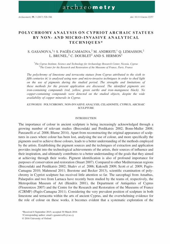
The applied non-invasive techniques are only able to identify the general type of iron red com-pounds. The FORS spectra of all red geological samples have a similar appearance, displayingtypical S-shape curves with a small slope absorption edge from 500 to 600 nm. The shift of theinflection point to shorter or longer wavelengths is related to different hues of the samples. In orderto more precisely determine the composition of the iron red pigment—that is, to differentiate be-tween pure hematite and clay containing red earth—the XRF analysis of secondary elements (Siand Al) might help. However, this approach has the disadvantage of the lower sensitivity ofXRF to light elements (Z< 18) than to the heavier ones. It results in inadequate measurementof light-element line intensities, even when XRF measurements are carried out in a He atmo-sphere. A comparison of the XRF spectra from hematite with a small amount of quartz (S-R-03) and red earth (S-R-05) is shown in Figure 1 (a). Despite the much higher concentration ofSi and Al in the red earth sample as compared to the hematite sample (see the PIXE analysis),the XRF spectrum of red earth shows barely distinguishable Si and Al lines. Thus, XRF analysisappears to be a powerful technique for the determination of iron-containing compounds; however,further differentiation between hematite and red earth by this technique is barely possible, espe-cially in those cases when other Al- and Si-containing compounds are present in support materials.
Iron yellow Two types of iron yellow, sulphate and iron oxide-hydroxide, were found in thegeological samples. PIXE analysis revealed high amounts of Na and S in S-Y-01 and S-Y-02,which suggests natrojarosite (NaFe3(OH)6(SO4)2). Much lower concentrations of Na and S inthe other two yellow samples, S-Y-03 and S-Y-04, suggest that the iron yellow is iron oxide-hydroxide (goethite) rather than sulphate (jarosite type). The geological samples of natrojarositeare characterized by a high level of purity in terms of clay admixtures; however, sample S-Y-01contains a considerable amount of Si. The samples S-Y-03 and S-Y-04, on the contrary, demon-strate high concentrations of clay elements – Al, Si and Ca – as well as Ti, Mn and Cu, whichallows us to identify them as yellow earth (Table 3). Traces of Pb were detected in both samplesof natrojarosite supplied by the GS and the yellow earth sample from Kokkinopezoulas mine. NoPb was found in the yellow earth sample, S-Y-03, collected from Skouriotissa mine. Similarly tohematite and red earth, the presence of Pb cannot be associated with only iron or clay materials,since traces of Pb were found in both pure natrojarosite and yellow earth.
Iron sulphate (jarosite type) and iron oxide-hydroxide (goethite type) are distinguishable by theimplemented non-invasive techniques. Despite reflectance behaviour in the visible range very sim-ilar to that of goethite, jarosite can be recognized by FORS analysis due to a characteristic spectralfeature near 430 nm (Montero 2005), which is absent from goethite or yellow earth spectra. XRFanalysis allows us to recognize jarosite by the presence of S lines. Additionally, indicative Si andAl lines can be used as earth indicators, thus allowing for differentiation between goethite andyellow earth by XRF analysis. Similarly to the case of iron red, the low sensitivity of XRF analysisto light elements is an obstacle in differentiation between goethite and yellow earth.
536 S. Gasanova et al.
© 2016 University of Oxford, Archaeometry 59, 3 (2017) 528–546
Figure 1 XRF spectra of iron red pigments: (a) local hematite (S-R-03) and red earth (S-R-05) from Cyprus; (b) redpigment and the limestone base of G.C.349; (c) red pigment and the limestone base of G.C.463.
Polychromy analysis on Cypriot Archaic statues 537
© 2016 University of Oxford, Archaeometry 59, 3 (2017) 528–546

Iron green Geological samples of iron green were found by XRD to contain celadonite, an iron-containing green mineral, at various degrees of purity and crystallization. Along with glauconite,celadonite is a constituent of the iron green pigment known as green earth, with the general for-mula K[(Al,Fe3+),(Fe2+,Mg)](AlSi3,Si4)O10(OH)2. Undistinguishable chemically, celadonite andglauconite have different formation origins: glauconite is a marine mineral and celadonite occursin volcanic rocks. This aspect is reflected in their structural properties, which allow the differen-tiation of the two minerals by XRD analysis.
Celadonite was found in S-G-01 and S-G-04, a calcite and celadonite mixture was detected inS-G-02, and a complex mixture of illite, quartz, albite, smectite, kaolinite and huntite was de-tected in S-G-03. No celadonite peaks were detected by XRD analysis in S-G-03, due to eithera ‘bad’ crystal orientation or a low degree of crystallization. The latter aspect, together withthe fact that S-G-03 represents a mixture of various materials, might explain its pale greyishcolour. The perfectly crystallized celadonite structures of S-G-01 and S-G-04 are shown in theFEG–SEM images (Fig. 2), where it appears in the form of laths. Other samples of iron greenappear under FEG–SEM as mixtures of various particles, where celadonite laths of differentshapes and sizes can be recognized.
Traces of Cu were found in S-G-03 from Kokkinopezoulas mine by PIXE analysis (Table 2),whereas no Cu was found in any other samples of iron green from other locations (the concen-tration of Cu in L-G-05 is at the limit of detection). Tracing the relation between the elementalcomposition (PIXE) and structural characteristics (FEG–SEM) of S-G-03, we propose that theCu traces are related to copper-containing mineral admixtures, rather than to the inclusion ofCu into celadonite structures.
Both celadonite and glauconite have very similar chemical structures, apart from a slightlyhigher K content in celadonite (9.5–10.0% in celadonite and 8.5–9.0% in glauconite: Odin1988). The XRF spectra of both minerals are dominated by Fe lines, whereas the light elementsSi, Al and Mg show low intensities, despite having concentrations comparable to that of Fe.These features allow us to recognize iron green material by XRF without distinguishing betweenthe celadonite or glauconite forms. Additionally, both minerals demonstrate similar reflectancebehaviour in the visible range (but see Hradil et al. 2004; Moretto et al. 2011). Thus, regardingthe identification of the iron green pigment, the capabilities of the applied non-invasive tech-niques are limited.
Iron brown PIXE analysis of iron brown samples revealed high concentrations of Si, Al and Mn.The presence of the first two elements indicates a clay material, while the presence of Mnexplains the brown colour. These considerations allow us to attribute the brown samples to atype of material commonly referred to as umber. Fe and Mn are likely to be present in differentphases, but not as mixed Fe–Mn oxides (Helwig 2007). Unlike other iron-containing geologicalsamples, iron brown materials typically have the highest level of contamination, as evidenced bysignificant concentrations of many elements (Table 2).
Identification of umbers by XRF analysis is based on the presence of intense Mn and Fe lines.However, this method of analysis faces the same problems in identification of Si and Al as forearths. Therefore, unambiguous XRF-based differentiation between umbers and black–brownmixtures of iron and manganese oxides is not possible. The capabilities of FORS in identificationof iron brown materials in terms of mineralogical composition are limited, as the shape of thespectra reflects the presence of iron compounds, but more detailed information on the colour-causing effects (differing chemical compositions or particle sizes) cannot be obtained.
538 S. Gasanova et al.
© 2016 University of Oxford, Archaeometry 59, 3 (2017) 528–546

Figure 2 FEG–SEM images of (a) local green earth samples from Cyprus and (b) archaeological green samples.
Polychromy analysis on Cypriot Archaic statues 539
© 2016 University of Oxford, Archaeometry 59, 3 (2017) 528–546

Analysis of polychromed statues
UV and VIL tests No characteristic bright-white luminescence in VIL images or pink–orangefluorescence in UV images were detected, thus excluding the use of Egyptian blue or madder dyefrom consideration on the studied objects (Verri 2009).
Red colour The FORS spectra of the red pigment on all statues show S-shaped curves similar tothose of iron red (example spectra from G.C.454 and G.C.463 are shown in Fig. 3 (a)). XRFanalysis confirmed the presence of an iron-containing compound for the red colours. The absenceof considerable Al lines and the presence of Si lines suggest either pure hematite, in the case of G.C.454, G.C.459 and N.N., or a hematite mixture with an Si compound (such as quartz) for G.C.447 and G.C. 463. Only the XRF spectra from the red pigment of G.C.349 have both Si andAl lines, suggesting red earth. A comparison of the XRF spectra for both types of iron red isshown in Figures 1 (b) and 1 (c). A characteristic feature, a Pb line of considerable intensity at
Figure 3 FORS spectra of iron-containing pigments: (a) iron red on G.C.463 (red lips) and G.C.454 (red flower onforehead); (b) iron yellow on G.C.454 (yellow ear cap) and G.C.349 (yellow eyes).
540 S. Gasanova et al.
© 2016 University of Oxford, Archaeometry 59, 3 (2017) 528–546

10.55 keV, can be seen in the spectra and was also observed for the red of N.N. and G.C. 447.Mixtures of iron red with lead white (2PbCO3·Pb(OH)2) or lead yellow (PbO) were excluded,since no white or yellow admixtures were observed in the red pigment using digital microscopy.Lead white was also excluded as a preparation layer, as no Pb lines were observed on XRFspectra of other coloured spots. Possible admixtures of red lead (Pb3O4) are also excluded, asno characteristic features were detected by FORS analysis (Fig. 3 (a)). The presence of a naturallyoccurring lead-containing compound in the iron red pigment is proposed, supported by theprevious findings made for local hematite and red earth (see above).
Analysis of red pigment on the terracotta head G.C.242 is complicated because of the type ofits support. Clay materials contain various chemical elements: Si and Al are supposed to be themajor elements because of the presence of aluminosilicates; Fe, Ni, Cu and Ti are the minorelements; and Pb, Y, Rb, Sr. and so on are trace elements, the analysis of which is often usedfor provenance studies. Although non-invasive XRF analysis identified iron red pigment onG.C.242, it is impossible to draw any conclusions from the traces of Pb, as the terracotta bodyitself contains significant amounts of this element. For the same reasons, analysis of Si and Allines is also inconclusive, restricting the differentiation between hematite and red earth on theterracotta head G.C.242 by the applied non-invasive methods (Table 3).
Yellow colour Microscopy observations detected the superposition of yellow and red pigmenton the centres of the earrings of G.C.454. This artistic decision was, perhaps, to attain a reddishhue to imitate jewellery. On the same head, traces of yellow were detected in close proximity tothe green area on the back of the sakkos. The yellow pigment of G.C.349 was detected in purecondition, without being mixed with other pigments. Results of FORS analysis suggest iron-containing yellow, as demonstrated in the example spectra in Figure 3 (b). As compared to ironred, the shape of the reflectance curves is similar, but the absorption edge is steeper and shifted tolonger wavelengths (500–550 nm). Iron yellow pigment was also confirmed by XRF analysis,thus excluding the arsenic-containing yellow pigment orpiment (As2S3), which was frequentlyused in Antiquity. Iron sulphate pigments, jarosite and natrojarosite, were also excluded due tothe low intensity of the S lines on the XRF spectra from both objects. At the same time, thehigher intensity of the Al and Si lines in the spectrum of iron yellow, compared to the limestonebase, indicates the presence of clay materials, suggesting yellow earth.
Green colour Translucent green particles of a fine size (< 1μm) were detected on all greenareas by digital microscopy. A single deep-blue crystal of angular shape and a size of ~10μmwas detected on a green spot G.C.454 (back of the sakkos). This might indicate that the greenpigment was mixed with a blue pigment, although Egyptian blue is excluded because of the lackof signal in VIL. Alternatively, its presence could be attributed to an accidental contamination ofthe green pigment. The FORS spectra of the green areas have an asymmetrical broad peak witha maximum at ~560 nm and a shoulder on the short-wavelength side at ~505 nm. The reflectanceis slightly higher in the long- than the short-wavelength region of the spectrum. These spectralfeatures in the visible range suggest iron green and are supported by XRF analysis, whichidentified Fe as the main element.
Although the most widely used green pigment in Antiquity was green earth (Mazzocchin et al.2003), it was not the only way to attain green hues. At least four other types of green were knownto the artists: iron green, copper green, yellow plus blue (e.g., yellow ochre and Egyptian blue)and yellow plus black (e.g., yellow ochre and carbon black) (Muhly 1984; Iacovou 2012;Brecoulaki 2014; van Brempt 2014). The detection of green earth in the studied objects is,
Polychromy analysis on Cypriot Archaic statues 541
© 2016 University of Oxford, Archaeometry 59, 3 (2017) 528–546

therefore, an important finding for the characterization of the ancient Cypriot palette. The factthat Cyprus has been known for centuries as one of the most famous sources of green earth (Bear1963) leads to a special interest in the discovery of green earth on the studied objects and, there-fore, the question of the local provenance of this pigment used to decorate locally producedstatues becomes relevant.
XRD analysis carried out on green microsamples revealed a celadonite form of green earth.The shape of the celadonite crystals studied on microsamples by FEG–SEM resembles that ofthe local celadonite (Fig. 2): laths up to 1μm long, with a length-to-width ratio of ~0.3. It is note-worthy that circular structures of fossilized micro-organisms of 5–10μm in diameter, identifiedas coccoliths, can be observed on FEG–SEM images of celadonite from G.C.454 and G.C.447.Fossilized micro-organisms of another type, with a long body twisted around crystals, were de-tected by FEG–SEM on the green of G.C.463. Owing to its geological history, the majority of thesedimentary rock formations in Cyprus are known to be of marine origin. Since these fossilizedmicro-organisms are abundant in marine sediments, their presence in the green archaeologicalsamples supports the assumption of the local provenance of the celadonite.
PIXE elemental analysis on the geological samples of green earth (Table 2) has not revealedany typical characteristics apart from trace amounts of Cu in one sample from Kokkinopezoulasmine (S-G-03). The low-intensity Cu line was observed on the XRF spectra of G.C.463, G.C.242and G.C.447, whereas no Cu was detected on the green of G.C.454. However, no structuralsimilarity between the local celadonite sample S-G-03 and the celadonite found on statues canbe observed, despite the fact that both contain traces of Cu. Moreover, the celadonite identifiedon statues appears differently under FEG–SEM than the celadonite of S-G-03. Thus, despitethe obvious local origin of the iron green pigment, there is still a lack of data on the preciselocalization of the raw material within Cyprus.
Black colour Black particles of a very fine size (< 1μm) and with an irregular shape weredetected on the limestone head G.C.454. Coarser black pigment particles (1–5μm) with a roundshape were detected on the terracotta head G.C.242. The XRF spectra of the limestone base andblack pigment of G.C.454 almost coincide, demonstrating only slightly higher intensity of the Feline. This suggests carbon black, such as charcoal or soot, which was probably mixed with theiron black pigment, such as magnetite Fe3O4. The absence of P lines (Kα at 2.01 keV) also allowsus to exclude ivory black; however, its presence might be hidden due to the low sensitivity ofXRF towards light elements.
The black pigment of terracotta head G.C.242 was found by XRF analysis to be a Mn,Fe-containing compound. Close microscopy observations of the painted areas of this object, as wellas the chemical nature of the preparation layer discussed below, suggest that the decoration onthis object was applied after firing, excluding the possible application of materials that turn intoblack under high temperature, such as ferruginous clays (Aloupi et al. 2000). An earth pigmentsuch as umber is proposed and also supported by FEG–SEM–EDS analysis, which revealed ahigh intensity of Si and Al lines. The black pigment was found by XRD to contain biotite, aniron-rich mineral commonly found in clays. No Mn-containing black compounds were detectedby XRD, probably due to their low degree of crystallization.
A remarkable feature, intense Pb lines, was detected by XRF on the black pigment of G.C.242.Their intensity in the spectrum of the black pigment greatly exceeded that on the spectrum of theterracotta base. In the case of other pigments found on this object (white, red and green), the Pblines are less intense than on the terracotta body. In other words, only the black pigment of G.C.242 features a high amount of Pb. The low intensity of the S lines allows us to exclude the
542 S. Gasanova et al.
© 2016 University of Oxford, Archaeometry 59, 3 (2017) 528–546

presence of galena (PbS), a naturally occurring black–brown mineral used as a black pigmentsince ancient times. Another black mineral that might naturally occur together with black earthminerals, plattnerite (PbO2), is proposed to explain the Pb lines in the spectrum of the blackpigment.
White colour White material was only detected on the terracotta head and was attributed to apreparation layer. XRF analysis suggests a calcium-containing compound, such as calciumcarbonate (CaCO3), but not gypsum (CaSO4·2H2O) or anhydrite of calcium sulphate (CaSO4),due to the absence of S lines. The presence of carbonate material on terracotta object suggestsunfired decoration. The thermal decomposition of CaCO3 starts at ~600 °C (Rodriguez-Navarro2009) and the temperatures used in the so-called ‘manganese black technique’ to produce variousMn–Fe spinels from Mn-rich ferruginous clays are above 1000 °C. Thus, thermal decompositionof CaCO3 would have taken place had the black decoration of this statue been fired.
CONCLUSIONS
The present work is one of the first systematic studies of the ancient pigment palette on Cypro-Archaic statues. The limited set of pigments identified on the six studied objects is comprisedmostly of iron-containing compounds (red, yellow, green earths and Fe–Mn black). Nocopper-containing compounds were detected on the studied objects, despite the wide availabilityof copper minerals in Cyprus. The study exemplifies some of the strengths and limitations ofnon-invasive analytical techniques, which were shown to be useful for preliminary analysis ofpolychromy and minimization of destructive procedures. Their capabilities are, however, limitedto the determination of the general type of chemical compounds, as demonstrated by the case ofiron red identified using non-invasive methods versus red earth and hematite, discriminationbetween which was possible only using micro-invasive methods. The study also gives indicativeexamples on when micro-destructive methods could be applied (e.g., for study of the provenanceof the pigment) and in which cases the chemical analysis should be limited only to non-invasiveprocedures (e.g., for the exclusion of orpiment and jarosite from yellow pigments). A possiblelocal provenance for the green earth was suggested based on the structural similarities observedbetween the celadonite from Cyprus and the celadonite identified on the statues examined,particularly the shape of the crystals and the presence of fossilized micro-organisms, coccoliths.The presence of traces of Pb in the geological samples of iron red and in the one identified on thestatues is a strong argument to support the assumption of the local provenance of this pigmentmaterial. More extensive study of the provenance of the pigment, as well as more precise attribu-tion of the studied pigments to a specific location within Cyprus, might be performed by studyingmore mineral samples in the future.
Thus, it has been argued that the paints on all of the tested sculptures are made of pigments thatmight have been sourced locally on the island. This allows us to reconstruct an important elementof the history of these sculptures. Interestingly, the presumably exclusive use of local materialsfollows the general practice on the island regarding sculpture during this period, since the vastmajority of the sculptures dating to this time are made of locally available limestone and clay.There are very few exceptions of sculptures made of imported stone such as marble, and thesewere imported as finished objects, not raw material to be worked on the island (e.g., the MarionKouros: Childs et al. 2012).
Polychromy analysis on Cypriot Archaic statues 543
© 2016 University of Oxford, Archaeometry 59, 3 (2017) 528–546

ACKNOWLEDGEMENTS
We acknowledge the financial support of the project ΝΕΑ ΥΠΟΔΟμΗ/ΣΤΡΑΤΗ/0308/30, co-financed by the European Development Fund and the Republic of Cyprus through the CyprusResearch Promotion Foundation. Several institutions contributed to the successful completionof the present study. We would like to thank Dr. Lefki Michaelidou from the Bank of CyprusCultural Foundation Museum for permission to carry out the present study; Dr. GeorgosConstantinou from the Geological Survey of Cyprus; Dr. Michel Menu and Ms. Anne Maigretfrom C2RMF, and Dr. Nikolas Bakirtzis and Dr. Iosif Hafez from CyI.
REFERENCES
Aloupi, E., Karydas, A., and Paradellis, T., 2000, Pigment analysis of wall paintings and ceramics from Greece andCyprus: the optimum use of X-ray spectrometry on specific archaeological issues, X-Ray Spectrometry, 29, 18–24.
Bear, L. M., 1963, The mineral sources and mining industry of Cyprus, Bulletin of the Geological Survey DepartmentMinistry of Commerce and Industry, Republic of Cyprus, 1, Geological Survey Department, Nicosia.
2013, Investigation of ancient Roman pigments by portable X-ray fluorescence spectroscopy and polarized light micros-copy, ch. 2, in Archaeological Chemistry VIII Beestone, R. F., and Becker, H. (eds.), Series, American ChemicalSociety, Washington, DC, ACS Symposium.
Blume, C., 2014, Bright pink, blue and other preferences: polychrome Hellenistic sculpture, in Transformations: classicalsculpture in colour, 166–89, Ny Carlsberg Glyptotek, Copenhagen.
Bonn-Muller, E., 2008, Carved in living color, Archaeology, 61(1), 14–5.Brecoulaki, H., 2014, Precious colours in Ancient Greek polychromy and painting: material aspects and symbolic values,
Revue archéologique, 1, 1–36.Brecoulaki, H., and Perdikatsis, V., 2002, Ancient painting on Macedonian funerary monuments, IV–III c. BC: a compar-
ative study on the use of color, in Color in ancient Greece (eds. M. A. Tiverios and D. S. Tsiafakis), 147–54, AristotleUniversity of Thessaloniki, Thessaloniki.
Campbell, J. L., Boyd, N. I., Grassi, N., Bonnick, P., and Maxwell, J. A., 2010, The Guelph PIXE software package IV,Nuclear Instruments and Methods in Physics Research B, 268, 3356–63.
Childs, W. A. P., Smith, J. S., and Padgett, J. M., 2012, City of gold: the archaeology of Polis Chrysochous, Cyprus,Princeton University Art Museum, Princeton, NJ/Yale University Press, New Haven. CT.
Cornell, R. M., and Schwertmann, U., 2003, The iron oxides: structure, properties, reactions and uses, 2nd edn, Wiley-VCH, New York.
Cosentino, A., 2014, Identification of pigments by multispectral imaging; a flowchart method, Heritage Science, 2(8), 1–12.Counts, D. B., 2008, Master of the lion: representation and hybridity in Cypriot sanctuaries, American Journal of Archae-
ology, 112, 3–27.Duran, A., López-Montes, A., Castaing, J., and Espejo, T., 2014, Analysis of a royal 15th century illuminated parchment
using a portable XRF–XRD system and micro-invasive techniques, Journal of Archaeological Science, 45, 52–8.Eastaugh, N., Walsh, V., Chaplin, T., and Siddall, R., 2008, Pigment compendium: a dictionary and optical microscopy of
historical pigments, Elsevier, Oxford.Feller, R. L. (ed.), 2012, Artist’s pigments: a handbook of their history and characteristics, Vol. 1, National Gallery of
Art, Washington, DC.Flourentzos, P., 2007, The sarcophagus of Palaepaphos, in Annual report of the Department of Antiquities for the year
2007, Republic of Cyprus, Ministry of Communication and Works, Lefkosia.Franquelo, M. L., Duran, A., Castaing, J., Arquillo, D., and Perez-Rodriguez, J. L., 2012, XRF, μ-XRD and μ-
spectroscopic techniques for revealing the composition and structure of paint layers on polychrome sculptures aftermultiple restorations, Talanta, 89, 462–9.
Fuller, C. W., 1973, Coloured iron oxide pigments, synthetic, in Pigment handbook, vol. 1: Properties and economics (ed.T. C. Patton), 333–8, Wiley, New York.
Helwig, K., 2007, Iron oxide pigments: natural and synthetic, in Artist’s pigments: a handbook of their history andcharacteristics (ed. B. H. Berrie), 39–110, Vol. 4, National Gallery of Art, Washington, DC.
Hendrix, E. A., 2001, Polychromy on the Amathus sarcophagus, a ‘rare gem of art’, Metropolitan Museum Journal, 36,43–59.
Hermary, A., 1989,Catalogue des antiquites de Chypre: sculptures, Musée du Louvre, Department des Antiquites orientales,Paris.
544 S. Gasanova et al.
© 2016 University of Oxford, Archaeometry 59, 3 (2017) 528–546

Hradil, D., Grygar, T., Hrusková, M., Bezdicka, P., Lang, K., Schneeweiss, O., and Chvátal, M., 2004, Green earthpigment from the Kadan region, Czech Republic: use of rare Fe-rich smectite, Clays and Clay Minerals, 52, 767–78.
Hudson-Edwards, K. A., and Edwards, S. J., 2005, Mineralogical controls on storage of As, Cu, Pb and Zn at theabandoned Mathiatis massive sulphide mine, Cyprus, Mineralogical Magazine, 69(5), 695–706.
Hudson-Edwards, K. A., Macklin, M. G., Finlayson, R., and Passmore, D. G., 1999, Mediaeval lead pollution in the RiverOuse at York, England, Journal of Archaeological Science, 26, 809–19.
Iacovou, M., 2012, From regional gateway to Cypriot kingdom: copper deposits and copper routes in the chora of Paphos,in Eastern Mediterranean metallurgy and metalwork in the second millennium BC (eds. V. Kassianidou and G.Papasavvas), 56–67, Oxbow Books, Oxford.
Kakoulli, I., 2009, Greek painting techniques and materials: from the fourth to the first century BC, Archetype, London.Karageorghis, V., 2002, Ancient art from Cyprus, Bank of Cyprus Cultural Foundation, Athens.Katsaros, T., and Bassiakos, Y., 2002, The colors of Theophrastus: sources, characterization and application, in Color in
ancient Greece: the role of color in ancient Greek art and architecture (700–31 B.C.), Proceedings of the conferenceheld in Thessaloniki, 12–16 April 2000 (eds. M. A. Tiverios and D. Tsiafakis), 201–9, Aristotle University ofThessaloniki, Thessaloniki.
Kirby, J., and White, R., 1996, The identification of red lake pigment dyestuffs and a discussion of their use, in NationalGallery Technical Bulletin Series (ed. A. Roy), 56–80, Vol. 17, National Gallery Publications, London.
Le Gac, A., Pessanha, S., Longelin, S., Guerra, M., Frade, J. C., Lourenço, F., Serrano, M. C., Manso, M., and Carvalho,M. L., 2013, New development on materials and techniques used in the heraldic designs of illuminated Manuelineforal charters by multi-analytical methods, Applied Radiation and Isotopes, 82, 242–57.
Madariaga, J. M., 2015, Analytical chemistry in the field of cultural heritage, Analytical Methods, 7, 4848–76.Mahmoud, H. H. M., 2011, Preliminary investigation of ancient pigments from the mortuary temple of Seti I, El-Qurna
(Luxor, Egypt), Mediterranean Archaeology and Archaeometry, 11(1), 99–106.Manning, S. W., McCartney, C., Kromer, B., and Stewart, S. T., 2010, The earlier Neolithic in Cyprus: recognition and
dating of a Pre-Pottery Neolithic A occupation, Antiquity, 84(325), 693–706.Mazzocchin, G. A., Agnoli, F., and Colpo, I., 2003, Analysis of pigments from Roman wall paintings found in Vicenza,
Talanta, 61, 565–72.Montero, I. C., Brimhalla, G. H., Alpers, C. N., and Swayze, G. A., 2005, Characterization of waste rock associated with
acid drainage at the Penn Mine, California, by ground-based visible to short-wave infrared reflectance spectroscopyassisted by digital mapping, Chemical Geology, 215, 453–472.
Moretto, L. M., Orsega, E. F., and Mazzocchin, G. A., 2011, Spectroscopic methods for the analysis of celadonite andglauconite in Roman wall paintings, Journal of Cultural Heritage, 12(4), 384–91.
Muhly, J. D., 1984, The role of the sea peoples in Cyprus during the LC III period, in Cyprus at the close of the LateBronze Age (eds. V. Karageorghis and J. D. Muhly), 39–56, A. G. Leventis Foundation, Nicosia.
Odin, G. S. (ed.), 1988, Green marine clays, Developments in Sedimentology 45, Elsevier, New York.Pagès-Camagna, S., 2011, Colours: pigments, tints and colorants, in Two exceptional sarcophagi from Larnaca (eds. K. O.
Lorentz, M. Menu, F. Niccolucci, and D. Pitzalis), Annual report of the Department of Antiquities of Cyprus, Nicosia.Panzanelli, R., with Schmidt, E. D., and Lapatin, K., 2008, The color of life: polychromy in sculpture from Antiquity to the
present, Getty Publications, Los Angeles, CA.Pauk, V., Havlíček, V., Papoušková, B., Sulovský, P., and Lemr, K., 2013, Simultaneous identification of historical
pigments Prussian blue and indigo in paintings by electrospray mass spectrometry, Journal of Mass Spectrometry,48(8), 927–30.
Pichon, L., Calligaro, T., Lemasson, Q., Moignard, B., and Pacheco, C., 2015, Programs for visualization, handling andquantification of PIXE maps at the AGLAE facility, Nuclear Instruments and Methods in Physics Research, SectionB, 363, 48–54.
Pichon, L., Moignard, B., Lemasson, Q., Pacheco, C., and Walter, P., 2014, Development of a multi-detector and asystematic imaging system on the AGLAE external beam, Nuclear Instruments and Methods in Physics Research,Section B, 318, 27–31.
Reyes, A. T., 1994, Archaic Cyprus: a study of the textual and archaeological evidence, Clarendon Press, Oxford.Rodriguez-Navarro, C., 2009, Thermal decomposition of calcite: mechanisms of formation and textural evolution of CaO
nanocrystals, American Mineralogist, 94, 578–93.Scott, D. A., Warmlander, S., and Quirke, S., 2009, Examination of some pigments, grounds and media from Egyptian
cartonnage fragments in the Petrie Museum, University College London, Journal of Archaeological Science, 36,923–32.
Shalev, S., Shilstein, S., and Yekutieli, Y., 2006, XRF study of archaeological and metallurgical material from an ancientcopper-smelting site near Ein-Yahav, Israel, Talanta, 70(5), 909–13.
Polychromy analysis on Cypriot Archaic statues 545
© 2016 University of Oxford, Archaeometry 59, 3 (2017) 528–546

Stratis, I., Varella, E. A., and Vavelidis, M., 2002, Pigments from the ochre mines on Thassos island, in Color in ancientGreece: the role of color in ancient Greek art and architecture (700–31 B.C.), Proceedings of the conference held inThessaloniki, 12–16 April 2000 (eds. M. A. Tiverios and D. Tsiafakis), 1–11, Aristotle University of Thessaloniki,Thessaloniki.
Stuart, B. H., 2007, Analytical techniques in materials conservation, Wiley, Chichester.Stylianou, M., Tsiftes, K., Gavriel, I., Kostarelos, K., Demetriou, C., and Papaioannou, A., 2014, Environmental impacts
of abandoned sulphide mines—the example of Mathiatis Mine in Cyprus, in Proceedings of the SYMBIOSIS Inter-national Conference, Athens, 19–21 June 2014.
Van Brempt, L., 2014, The production and trade of Cypriot copper in the Late Bronze Age, in The Narnia Project:integrating approaches to ancient material studies (eds. V. Kassianidou and M. Dikomitou-Eliadou), 110–21, TheNARNIA Project and the Archaeological Research Unit, University of Cyprus, Nicosia.
Verri, G., 2009, The spatially resolved characterisation of Egyptian blue, Han blue and Han purple by photo-inducedluminescence digital imaging, Analytical and Bioanalytical Chemistry, 394(4), 1011–21.
Vigne, J.-D., Briois, F., Zazzo, A., Carrère, I., Daujat, J., and Guilaine, J., 2011, A new Pre-Pottery Neolithic site inCyprus: Ayios Tychonas-Klimonas (ca. 8700 cal. BC), Neo-lithics, 1(11), 3–13.
546 S. Gasanova et al.
© 2016 University of Oxford, Archaeometry 59, 3 (2017) 528–546