Polyamine Oxidase, a Hydrogen Peroxide-Producing Enzyme, Is Up-Regulated by Light and Down-Regulated...
11
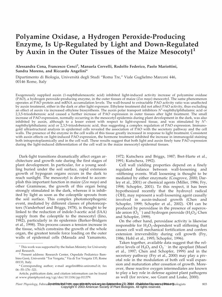
Polyamine Oxidase, a Hydrogen Peroxide-Producing Enzyme, Is Up-Regulated by Light and Down-Regulated by Auxin in the Outer Tissues of the Maize Mesocotyl 1 Alessandra Cona, Francesco Cenci 2 , Manuela Cervelli, Rodolfo Federico, Paolo Mariottini, Sandra Moreno, and Riccardo Angelini* Dipartimento di Biologia, Universita ` degli Studi “Roma Tre,” Viale Guglielmo Marconi 446, 00146 Rome, Italy Exogenously supplied auxin (1-naphthaleneacetic acid) inhibited light-induced activity increase of polyamine oxidase (PAO), a hydrogen peroxide-producing enzyme, in the outer tissues of maize (Zea mays) mesocotyl. The same phenomenon operates at PAO protein and mRNA accumulation levels. The wall-bound to extractable PAO activity ratio was unaffected by auxin treatment, either in the dark or after light exposure. Ethylene treatment did not affect PAO activity, thus excluding an effect of auxin via increased ethylene biosynthesis. The auxin polar transport inhibitors N 1 -naphthylphthalamic acid or 2,3,5-triiodobenzoic acid caused a further increase of PAO expression in outer tissues after light treatment. The small increase of PAO expression, normally occurring in the mesocotyl epidermis during plant development in the dark, was also inhibited by auxin, although to a lesser extent with respect to light-exposed tissue, and was stimulated by N 1 - naphthylphthalamic acid or 2,3,5-triiodobenzoic acid, thus suggesting a complex regulation of PAO expression. Immuno- gold ultrastructural analysis in epidermal cells revealed the association of PAO with the secretory pathway and the cell walls. The presence of the enzyme in the cell walls of this tissue greatly increased in response to light treatment. Consistent with auxin effects on light-induced PAO expression, the hormone treatment inhibited the increase in immunogold staining both intraprotoplasmically and in the cell wall. These results suggest that both light and auxin finely tune PAO expression during the light-induced differentiation of the cell wall in the maize mesocotyl epidermal tissues. Dark-light transitions dramatically affect organ ar- chitecture and growth rate during the first stages of plant development. In particular, for a young seed- ling buried under the soil surface, rapid extension growth of hypogean organs occurs in the dark to reach sunlight. The mesocotyl is devoted to accom- plish this important function in maize (Zea mays) and other Gramineae, the growth of this organ being strongly stimulated in the dark, whereas it is inhib- ited by light as soon as the coleoptile sprouts from the soil surface. This complex photomorphogenic event, mediated by different classes of photorecep- tors (Vanderhoef and Briggs, 1978), is thought to be linked to the reduction of indole-3-acetic acid (IAA) supply from the coleoptile to the mesocotyl (Iino, 1982), particularly in its epidermis (Barker-Bridgers et al., 1998). This process causes a tension increase in the tissue, which constrains the growth of the whole organ, the greatest tensile force loading on the outer walls of epidermal cells (Masuda and Yamamoto, 1972; Kutschera and Briggs, 1987; Bret-Harte et al., 1991; Kutschera, 1992). Cell wall yielding properties depend on a finely regulated balance between wall-loosening and -stiffening events. Wall loosening is thought to be mediated by either enzymatic (Cosgrove, 2000; Dar- ley et al., 2001) or chemical agents (Miller, 1986; Fry, 1998; Schopfer, 2001). To this respect, it has been hypothesized recently that the hydroxyl radical ( . OH), may represent a wall-loosening agent causally involved in auxin-induced growth (Chen and Schopfer, 1999; Schopfer et al., 2002). . OH can be produced by peroxidase in the presence of superox- ide anion (O 2 . ) and hydrogen peroxide (H 2 O 2 ; Chen and Schopfer, 1999). On the other hand, peroxidase activity is likewise responsible for H 2 O 2 -dependent wall stiffening that causes cell wall mechanical fortification and confers extension irreversibility during cell growth (Fry, 1986; Hohl et al., 1995; Schopfer, 1996). Taken together, available data suggest that the rel- ative levels of H 2 O 2 and O 2 . in the apoplast (Musel et al., 1997; Chen and Schopfer, 1999) and in the secretory pathway (Fry et al., 2000) may play a piv- otal role in the modulation of both cell wall expan- sion and maturation after cessation of growth. More- over, these reactive oxygen intermediates are known to play a key role in defense against plant pathogens as well (for review, see Grant and Loake, 2000). 1 This work was supported by the Italian Ministry for University and Research. 2 Present address: Research Center, Ospedale Pediatrico Bam- bino Gesu ` , Universita ` “Tor Vergata,” Via di Tor Vergata 135, Rome 00133, Italy. * Corresponding author; e-mail [email protected]; fax 06 –55–176 –321. Article, publication date, and citation information can be found at www.plantphysiol.org/cgi/doi/10.1104/pp.011379. Plant Physiology, February 2003, Vol. 131, pp. 803–813, www.plantphysiol.org © 2003 American Society of Plant Biologists 803 www.plant.org on September 23, 2014 - Published by www.plantphysiol.org Downloaded from Copyright © 2003 American Society of Plant Biologists. All rights reserved.
Transcript of Polyamine Oxidase, a Hydrogen Peroxide-Producing Enzyme, Is Up-Regulated by Light and Down-Regulated...
Polyamine Oxidase, a Hydrogen Peroxide-ProducingEnzyme, Is Up-Regulated by Light and Down-Regulatedby Auxin in the Outer Tissues of the Maize Mesocotyl1
Alessandra Cona, Francesco Cenci2, Manuela Cervelli, Rodolfo Federico, Paolo Mariottini,Sandra Moreno, and Riccardo Angelini*
Dipartimento di Biologia, Universita degli Studi “Roma Tre,” Viale Guglielmo Marconi 446,00146 Rome, Italy
Exogenously supplied auxin (1-naphthaleneacetic acid) inhibited light-induced activity increase of polyamine oxidase(PAO), a hydrogen peroxide-producing enzyme, in the outer tissues of maize (Zea mays) mesocotyl. The same phenomenonoperates at PAO protein and mRNA accumulation levels. The wall-bound to extractable PAO activity ratio was unaffectedby auxin treatment, either in the dark or after light exposure. Ethylene treatment did not affect PAO activity, thus excludingan effect of auxin via increased ethylene biosynthesis. The auxin polar transport inhibitors N1-naphthylphthalamic acid or2,3,5-triiodobenzoic acid caused a further increase of PAO expression in outer tissues after light treatment. The smallincrease of PAO expression, normally occurring in the mesocotyl epidermis during plant development in the dark, was alsoinhibited by auxin, although to a lesser extent with respect to light-exposed tissue, and was stimulated by N1-naphthylphthalamic acid or 2,3,5-triiodobenzoic acid, thus suggesting a complex regulation of PAO expression. Immuno-gold ultrastructural analysis in epidermal cells revealed the association of PAO with the secretory pathway and the cellwalls. The presence of the enzyme in the cell walls of this tissue greatly increased in response to light treatment. Consistentwith auxin effects on light-induced PAO expression, the hormone treatment inhibited the increase in immunogold stainingboth intraprotoplasmically and in the cell wall. These results suggest that both light and auxin finely tune PAO expressionduring the light-induced differentiation of the cell wall in the maize mesocotyl epidermal tissues.
Dark-light transitions dramatically affect organ ar-chitecture and growth rate during the first stages ofplant development. In particular, for a young seed-ling buried under the soil surface, rapid extensiongrowth of hypogean organs occurs in the dark toreach sunlight. The mesocotyl is devoted to accom-plish this important function in maize (Zea mays) andother Gramineae, the growth of this organ beingstrongly stimulated in the dark, whereas it is inhib-ited by light as soon as the coleoptile sprouts fromthe soil surface. This complex photomorphogenicevent, mediated by different classes of photorecep-tors (Vanderhoef and Briggs, 1978), is thought to belinked to the reduction of indole-3-acetic acid (IAA)supply from the coleoptile to the mesocotyl (Iino,1982), particularly in its epidermis (Barker-Bridgerset al., 1998). This process causes a tension increase inthe tissue, which constrains the growth of the wholeorgan, the greatest tensile force loading on the outerwalls of epidermal cells (Masuda and Yamamoto,
1972; Kutschera and Briggs, 1987; Bret-Harte et al.,1991; Kutschera, 1992).
Cell wall yielding properties depend on a finelyregulated balance between wall-loosening and-stiffening events. Wall loosening is thought to bemediated by either enzymatic (Cosgrove, 2000; Dar-ley et al., 2001) or chemical agents (Miller, 1986; Fry,1998; Schopfer, 2001). To this respect, it has beenhypothesized recently that the hydroxyl radical(.OH), may represent a wall-loosening agent causallyinvolved in auxin-induced growth (Chen andSchopfer, 1999; Schopfer et al., 2002). .OH can beproduced by peroxidase in the presence of superox-ide anion (O2
�.) and hydrogen peroxide (H2O2; Chenand Schopfer, 1999).
On the other hand, peroxidase activity is likewiseresponsible for H2O2-dependent wall stiffening thatcauses cell wall mechanical fortification and confersextension irreversibility during cell growth (Fry,1986; Hohl et al., 1995; Schopfer, 1996).
Taken together, available data suggest that the rel-ative levels of H2O2 and O2
�. in the apoplast (Muselet al., 1997; Chen and Schopfer, 1999) and in thesecretory pathway (Fry et al., 2000) may play a piv-otal role in the modulation of both cell wall expan-sion and maturation after cessation of growth. More-over, these reactive oxygen intermediates are knownto play a key role in defense against plant pathogensas well (for review, see Grant and Loake, 2000).
1 This work was supported by the Italian Ministry for Universityand Research.
2 Present address: Research Center, Ospedale Pediatrico Bam-bino Gesu, Universita “Tor Vergata,” Via di Tor Vergata 135, Rome00133, Italy.
* Corresponding author; e-mail [email protected]; fax06 –55–176 –321.
Article, publication date, and citation information can be foundat www.plantphysiol.org/cgi/doi/10.1104/pp.011379.
Plant Physiology, February 2003, Vol. 131, pp. 803–813, www.plantphysiol.org © 2003 American Society of Plant Biologists 803 www.plant.org on September 23, 2014 - Published by www.plantphysiol.orgDownloaded from Copyright © 2003 American Society of Plant Biologists. All rights reserved.

A main goal in the understanding of molecularevents underlying cell wall expansion and matura-tion is the analysis of the specific contribution of themolecular machineries synthesizing H2O2, O2
�., andtheir cognate reaction product .OH, in the apoplastand in the secretion pathway. O2
�. production in theapoplast has been thoroughly studied and it can beascribed to several enzyme activities, depending onthe particular physiological context or plant species(Bolwell, 1999). O2
�. synthesis in the apoplast seemsto be redundant because it can be accomplished byeither the NAD(P)H oxidizing activity of wall per-oxidases (Elstner and Heupel, 1976; Bolwell et al.,1998; Frahry and Schopfer, 2001) or by a plasmamembrane NAD(P)H oxidase similar to the inducibleNADPH oxidase complex of the mammalian phago-cytes (Wojtaszek, 1997, and refs. therein). H2O2 syn-thesis in the cell wall is likewise redundant because itcan originate either from O2
�. dismutation (eitherspontaneous or catalyzed by cell wall superoxidedismutases), or from the oxidative cycle of peroxi-dases in the presence of a reductant (Elstner andHeupel, 1976; Bolwell, 1999). Amine oxidases (Ange-lini and Federico, 1989; Allan and Fluhr, 1997; Møllerand McPherson, 1998; Laurenzi et al., 2001) or oxalateoxidases (Lane, 1994) can also be involved in H2O2production in the apoplast. Circumstantial evidencefosters the view that copper amine oxidases andflavin-containing polyamine oxidases (PAOs) play akey role in H2O2 production in the cell wall duringontogenesis as well as in response to wounding orpathogen invasion in different species (Angelini etal., 1990, 1993; Rea et al., 1998; Laurenzi et al., 1999;Wisniewski et al., 2000; Asthir et al., 2002; Rea et al.,2002; Cooley and Walters, 2002).
Plant PAOs are FAD-containing glycoproteins re-sponsible for the terminal catabolism of polyaminescontaining a secondary amino group (Sebela et al.,2001). These enzymes oxidize spermine and spermi-dine to the corresponding aminoaldehydes and 1,3-diaminopropane, releasing H2O2 upon reoxidation ofthe reduced enzyme (Federico et al., 1990; Tavla-doraki et al., 1998). PAO is especially abundant in theprimary and secondary cell walls of xylem, xylemparenchyma, endodermis, and epidermis of maizeseedlings where it has been localized by means ofbiochemical, histochemical, and immunocytochemi-cal methods (Kaur-Sawhney et al., 1981; Angelini andFederico, 1989; Slocum and Furey, 1991; Angelini etal., 1995; Laurenzi et al., 1999). Furthermore, in maizemesocotyl epidermis PAO expression has beenshown recently to be stimulated by light, this phe-nomenon being mediated by phytochrome (Laurenziet al., 1999). In particular, PAO mRNA and proteinlevels, as well as enzyme activity, increase in epider-mal tissues of maize mesocotyl in response to de-etiolation, both in the elongating and the maturezone. The time course of light-induced increase ofPAO activity in the outer tissues of apical growing
zone of the mesocotyl is tightly correlated to theinhibition of extension growth (Laurenzi et al., 1999).Moreover, H2O2 production in maize mesocotyl seg-ments is inhibited by guazatine, a powerful PAOinhibitor (Laurenzi et al., 1999). Characterization ofpromoter sequences in two genes encoding PAO(MPAO1 and MPAO2; Cervelli et al., 2000) revealedthe presence of putative cis-acting motifs responsiveto light or auxin, thus suggesting the possibility of atranscriptional control of PAO expression by the hor-mone and light.
The principal aim of the present study was to in-vestigate whether light-induced reduction of diffus-ible IAA in the maize mesocotyl could regulate PAOgene expression during cell differentiation after ces-sation of mesocotyl growth. For this purpose, wehave studied the effect of exogenous auxin supply onPAO expression levels in the outer tissues of non-growing zone of the maize mesocotyl either in thedark or after light pulses. A further scope of thisstudy was to understand how the variation of endog-enous auxin levels, obtained by using auxin polartransport inhibitors (ATIs), could affect light-inducedexpression of the PAO gene. Moreover, immunoelec-tron microscopic analysis was performed to investi-gate possible effects of auxin and light on PAO sub-cellular localization in the epidermal cells. Thephysiological significance of the modulation of PAOgene expression in the maize mesocotyl by light,auxin, or ATI and changes in subcellular distributionof the enzyme are discussed in relation to cell wallstiffening and differentiation.
RESULTS
Time Course of Light-Dependent Induction of PAOExpression Revealed an Early Enzyme Inactivation
A previous study showed the induction of PAOexpression in the outer tissues (i.e. cortical and epi-dermal tissues) of the nongrowing zone of maizemesocotyl in the days after the onset of white lightexposure (Laurenzi et al., 1999). The present study,besides confirming previous results on a long-termbasis, reports a time course analysis revealing anearly light-induced inhibition of PAO activity. In par-ticular, PAO activity levels expressed on a freshweight (Fig. 1A) or protein basis (data not shown)was 40% decreased in the mesocotyl outer tissues 3 hafter a 10-min white light exposure (18,000 lux), ascompared with dark controls, whereas an 8-fold in-crease of PAO activity levels was observed at 24 h.Western-immunoblotting analysis performed afterSDS-PAGE loaded on the basis of equal enzyme ac-tivity demonstrated a higher PAO protein level inde-etiolated tissues with respect to dark control at 3and 6 h (Fig. 1B). In contrast, when SDS-PAGE gelswere loaded on the basis of total protein level, nodifferences in PAO protein level were observed up to6 h, whereas a more intense band was evident at 24 h
Cona et al.
804 Plant Physiol. Vol. 131, 2003 www.plant.org on September 23, 2014 - Published by www.plantphysiol.orgDownloaded from Copyright © 2003 American Society of Plant Biologists. All rights reserved.

(Fig. 1C). This result suggests early enzyme inactiva-tion in light-exposed tissues, while confirming theincrease of PAO activity levels in the next hours. Itwas demonstrated previously that light had a stim-ulatory effect on the accumulation level of PAOmRNA a few hours after the onset of light treatment(Laurenzi et al., 1999). White light exposure up to 3 hof crude extracts from the outer tissues of etiolatedmesocotyl caused an enzyme activity decrease (thisstudy; 44% as compared dark control value) similarto that detected after irradiating the whole plant,whereas a lower inhibition (23%) was observed inhomogenates obtained from green tissues. Becausecatalytic activity of purified PAO was unaffected bylight exposure, a direct damaging effect of light onPAO protein or FAD cofactor could be excluded.Moreover, the lower light-induced inhibition of PAOactivity detected in crude homogenates from greentissues suggests the presence of some protecting sub-stances in tissues previously exposed to light. Duringlight exposure of dark-grown plants or crude homo-genates from etiolated tissues, the formation of un-known compounds affecting PAO protein occurred. Infact, after SDS-PAGE and western-immunoblotting
analyses of irradiated crude extracts obtained frometiolated shoots, additional bands probably related toPAO protein degradation were detectable (this study;data not shown). Enzyme inactivation was irrevers-ible because PAO immunoprecipitation and dialysisfailed to restore enzyme activity, suggesting that in-activation was not caused by a soluble inhibitor (thisstudy; data not shown). Based on these observations,we concluded that the early, light-induced inhibitionof PAO activity was probably caused by unknowncompounds produced during sudden exposure ofdark-grown plants or crude homogenates to high-intensity white light. The inhibitory effect was tran-sient (Fig. 1), and completely reversed thereafter. Asa consequence of this phenomenon, effect exerted byauxin on light induced-PAO expression was evalu-ated 24 h after plant irradiation.
Auxin Treatment Strongly Inhibited Light-InducedIncrease of PAO Expression in Outer Tissues of theMaize Mesocotyl
To ascertain the possible involvement of auxin inregulating PAO gene expression, we analyzed the
Figure 1. Effect of white light on PAO enzymeactivity and protein levels in the outer tissues ofthe maize mesocotyl. Etiolated plants (108 hafter soaking in the dark) were irradiated underwhite light (18,000 lux) for 10 min and extract-able PAO activity and protein levels determinedin the outer tissues of the mature zone of themaize mesocotyl at the times indicated. A, PAOactivity levels (mean values � SD; n � 3) ex-pressed on a fresh weight basis in dark-grownand light-irradiated plants (inset: PAO activitylevels plotted on a lower scale graph). P valueshave been calculated comparing PAO activitylevels in control and irradiated plants for eachtime. ns, Not significant; *, * *, and * * *, Pvalues � 0.05, 0.01, and 0.001, respectively(the nos. above the bars represent actual P val-ues). B and C, Western immunoblotting carriedout after SDS-PAGE loaded on the basis of theequal enzyme activity (B) or total protein con-tent (C).
Polyamine Oxidase Expression in Epidermal Tissues
Plant Physiol. Vol. 131, 2003 805 www.plant.org on September 23, 2014 - Published by www.plantphysiol.orgDownloaded from Copyright © 2003 American Society of Plant Biologists. All rights reserved.
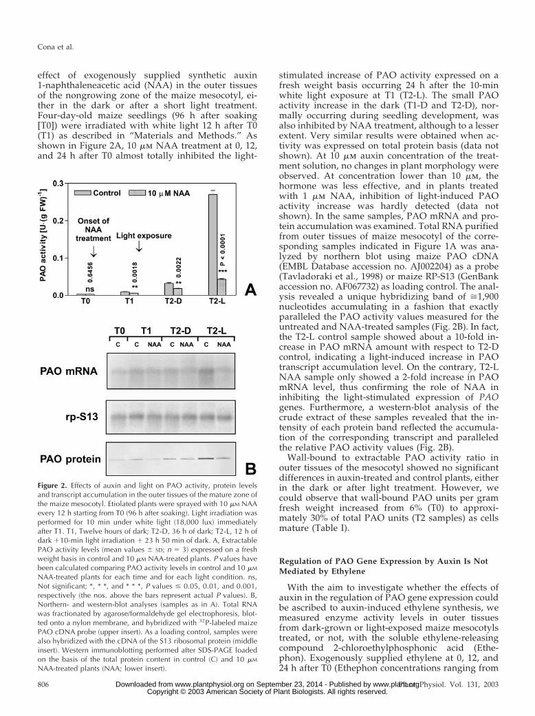
effect of exogenously supplied synthetic auxin1-naphthaleneacetic acid (NAA) in the outer tissuesof the nongrowing zone of the maize mesocotyl, ei-ther in the dark or after a short light treatment.Four-day-old maize seedlings (96 h after soaking[T0]) were irradiated with white light 12 h after T0(T1) as described in “Materials and Methods.” Asshown in Figure 2A, 10 �m NAA treatment at 0, 12,and 24 h after T0 almost totally inhibited the light-
stimulated increase of PAO activity expressed on afresh weight basis occurring 24 h after the 10-minwhite light exposure at T1 (T2-L). The small PAOactivity increase in the dark (T1-D and T2-D), nor-mally occurring during seedling development, wasalso inhibited by NAA treatment, although to a lesserextent. Very similar results were obtained when ac-tivity was expressed on total protein basis (data notshown). At 10 �m auxin concentration of the treat-ment solution, no changes in plant morphology wereobserved. At concentration lower than 10 �m, thehormone was less effective, and in plants treatedwith 1 �m NAA, inhibition of light-induced PAOactivity increase was hardly detected (data notshown). In the same samples, PAO mRNA and pro-tein accumulation was examined. Total RNA purifiedfrom outer tissues of maize mesocotyl of the corre-sponding samples indicated in Figure 1A was ana-lyzed by northern blot using maize PAO cDNA(EMBL Database accession no. AJ002204) as a probe(Tavladoraki et al., 1998) or maize RP-S13 (GenBankaccession no. AF067732) as loading control. The anal-ysis revealed a unique hybridizing band of �1,900nucleotides accumulating in a fashion that exactlyparalleled the PAO activity values measured for theuntreated and NAA-treated samples (Fig. 2B). In fact,the T2-L control sample showed about a 10-fold in-crease in PAO mRNA amount with respect to T2-Dcontrol, indicating a light-induced increase in PAOtranscript accumulation level. On the contrary, T2-LNAA sample only showed a 2-fold increase in PAOmRNA level, thus confirming the role of NAA ininhibiting the light-stimulated expression of PAOgenes. Furthermore, a western-blot analysis of thecrude extract of these samples revealed that the in-tensity of each protein band reflected the accumula-tion of the corresponding transcript and paralleledthe relative PAO activity values (Fig. 2B).
Wall-bound to extractable PAO activity ratio inouter tissues of the mesocotyl showed no significantdifferences in auxin-treated and control plants, eitherin the dark or after light treatment. However, wecould observe that wall-bound PAO units per gramfresh weight increased from 6% (T0) to approxi-mately 30% of total PAO units (T2 samples) as cellsmature (Table I).
Regulation of PAO Gene Expression by Auxin Is NotMediated by Ethylene
With the aim to investigate whether the effects ofauxin in the regulation of PAO gene expression couldbe ascribed to auxin-induced ethylene synthesis, wemeasured enzyme activity levels in outer tissuesfrom dark-grown or light-exposed maize mesocotylstreated, or not, with the soluble ethylene-releasingcompound 2-chloroethylphosphonic acid (Ethe-phon). Exogenously supplied ethylene at 0, 12, and24 h after T0 (Ethephon concentrations ranging from
Figure 2. Effects of auxin and light on PAO activity, protein levelsand transcript accumulation in the outer tissues of the mature zone ofthe maize mesocotyl. Etiolated plants were sprayed with 10 �M NAAevery 12 h starting from T0 (96 h after soaking). Light irradiation wasperformed for 10 min under white light (18,000 lux) immediatelyafter T1. T1, Twelve hours of dark; T2-D, 36 h of dark; T2-L, 12 h ofdark �10-min light irradiation � 23 h 50 min of dark. A, ExtractablePAO activity levels (mean values � SD; n � 3) expressed on a freshweight basis in control and 10 �M NAA-treated plants. P values havebeen calculated comparing PAO activity levels in control and 10 �M
NAA-treated plants for each time and for each light condition. ns,Not significant; *, * *, and * * *, P values � 0.05, 0.01, and 0.001,respectively (the nos. above the bars represent actual P values). B,Northern- and western-blot analyses (samples as in A). Total RNAwas fractionated by agarose/formaldehyde gel electrophoresis, blot-ted onto a nylon membrane, and hybridized with 32P-labeled maizePAO cDNA probe (upper insert). As a loading control, samples werealso hybridized with the cDNA of the S13 ribosomal protein (middleinsert). Western immunoblotting performed after SDS-PAGE loadedon the basis of the total protein content in control (C) and 10 �M
NAA-treated plants (NAA; lower insert).
Cona et al.
806 Plant Physiol. Vol. 131, 2003 www.plant.org on September 23, 2014 - Published by www.plantphysiol.orgDownloaded from Copyright © 2003 American Society of Plant Biologists. All rights reserved.

0.1 �m to 1 mm), did not affect PAO activity levelseither in the dark or after light exposure (data notshown).
Exogenous Supply of ATIs Amplified theLight-Induced Increase of PAO Expression
The role played by the light-induced reduction ofdiffusible auxin in the mesocotyl (Iino, 1982; Barker-Bridgers et al., 1998) in regulating PAO gene expres-sion was further analyzed utilizing two differentATIs. Because auxin synthesis mainly occurs in thecoleoptile (Iino and Carr, 1982), plants treated withATIs were expected to contain lower levels of thehormone in the mesocotyl. Based on the inhibitoryeffect exerted by auxin on light-induced PAO geneexpression, ATIs were supposed to amplify light-induced PAO expression. In line with this hypothe-sis, exogenous supply of the phytotropinic ATI N1-naphthylphthalamic acid (NPA) at a concentration of0.01 �m caused a further increase of PAO activitylevels after light exposure with respect to untreatedplants. In fact, the T2-L NPA sample showed anapproximately 30% increase in PAO activity withrespect to the T2-L control sample (Fig. 3A). On thecontrary, the increase of PAO activity in the dark wasstimulated by NPA to a lesser extent, as observed inT2-D NPA and T1-D NPA samples, when comparedwith T2-D control and T1-D control samples, respec-tively (Fig. 3A). Moreover, simultaneous supply ofNPA and NAA resulted in an inhibition of light-induced increase in PAO activity comparable withthat obtained with NAA alone (data not shown),consistent with the well-known evidence that auxindoes not compete with NPA for its binding site (Suss-man and Goldsmith, 1981). Also in this case, PAOmRNA and protein accumulation were examined inparallel to study PAO gene expression in response to
NPA treatment under light or dark conditions.Northern-blot analysis revealed that PAO mRNA ac-cumulates in a fashion that again parallels PAO ac-tivity levels measured in NPA-treated samples (Fig.3B). Western-blot analysis of the corresponding pro-tein extracts essentially reflects the accumulation ofthe corresponding transcript and enzyme activityvalues (Fig. 3B). NPA supply at concentrationshigher than 0.01 �m caused a lower effect in ampli-
Table I. Wall-bound PAO activity expressed as the percentage oftotal PAO activity (wall-bound plus extractable) in the outer tissuesof the mature zone of the maize mesocotyl
Etiolated plants were sprayed with 10 �M NAA every 12 h startingfrom T0 (96 h after soaking). Light irradiation was performed 12 hafter T0 (T1) for 10 min (18,000 lux) in a growth chamber. T1, Twelvehours after T0 in the dark; T2-D, 36 h after T0 in the dark; T2-L, 12 hafter T0 in the dark � 10-min light irradiation � 23 h 50 min in thedark. Mean values � SD.
Time/Light Treatment
Wall-Bound PAO Activity(% of Total PAO Activity Values)
ControlNAA-treated
plants
T0 (96 h after soaking) 6% (� 0.39) –T1 (12 h after T0 in the dark) 23% (�1.9) 18% (� 1.35)T2-D (36 h after T0 in the dark) 32% (�2.75) 31% (�2.13)T2-L (12 h after T0 in the dark
� 10-min light irradiation� 23 h 50 min in the dark)
30% (�2.42) 33% (�2.91)
Figure 3. Effects of NPA and light on PAO activity, protein levels,and transcript accumulation in the outer tissues of the mature zone ofthe maize mesocotyl. Etiolated plants were sprayed with 0.01 �M
NPA or 10 �M NAA every 12 h starting from T0 (96 h after soaking).Light irradiation was performed for 10 min under white light (18,000lux) immediately after T1. T1, Twelve hours of dark; T2-D, 36 h ofdark; T2-L, 12 h of dark �10-min light irradiation � 23 h 50 mindark. A, Extractable PAO activity levels (mean values � SD; n � 3)expressed on a fresh weight basis, in control, 0.01 �M NPA-, or 10 �M
NAA-treated plants. P values have been calculated comparing PAOactivity levels in NPA and NAA treated plants with respect to controlsfor each time and for each light condition. ns, Not significant; *, * *,and * * *, P values � 0.05, 0.01, and 0.001, respectively (the nos.above the bars represent actual P values). B, Northern- and western-blot analysis. Total RNA was fractionated by agarose/formaldehyde gelelectrophoresis, blotted onto a nylon membrane, and hybridized with32P-labeled maize PAO cDNA probe (upper insert). As a loadingcontrol, samples were also hybridized with the cDNA of the S13ribosomal protein (middle insert). Western immunoblotting performedafter SDS-PAGE loaded on the basis of the total protein content incontrol (C) and 0.01 �M NPA-treated plants (NPA; lower insert).
Polyamine Oxidase Expression in Epidermal Tissues
Plant Physiol. Vol. 131, 2003 807 www.plant.org on September 23, 2014 - Published by www.plantphysiol.orgDownloaded from Copyright © 2003 American Society of Plant Biologists. All rights reserved.

fying the PAO activity increase in T2-L samples, with10 �m NPA being totally ineffective (Fig. 4).
As shown in Figure 5A, exogenous supply of thenon-phytotropinic ATI 2,3,5-triiodobenzoic acid(TIBA) resulted in opposite effects depending on ATIconcentration. This result is consistent with what wasdescribed previously about the auxin activity of TIBA(Thomson et al., 1973). In particular, 0.01 �m TIBAslightly increased PAO activity level in light-exposedplants with respect to control plants (T2-L controland T2-L TIBA at 0.01 �m). On the contrary, 10 �mTIBA acted in an auxin-like fashion by inhibitinglight-induced increase of PAO activity levels to thesame extent of 10 �m NAA (Fig. 5, A and B).
PAO Ultrastructural Localization in the Epidermis ofLight-Exposed Maize Mesocotyl. Effect of Auxin
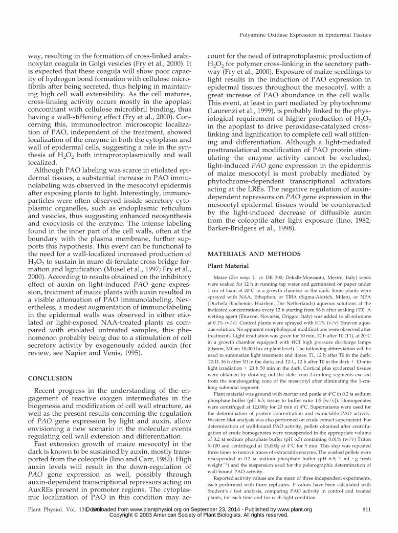
With the aim of studying whether light or auxintreatment influenced subcellular localization of PAOin epidermal cells, a transmission electron micro-scopic immunogold analysis was performed in theepidermal tissues of dark-grown or light-exposedmaize mesocotyls treated, or not, with 10 �m NAA. Arabbit polyclonal anti-PAO antiserum, fractionatedby affinity chromatography through a Sepharose 4Bcolumn coupled to bromelain to eliminate anti-glycan antibodies, was utilized as the primary probeas described in “Materials and Methods.” Ultrastruc-tural analysis revealed fair preservation of tissuemorphology, together with good retention of PAOantigenic properties (Fig. 6). Background labelingwas practically absent from the anti-PAO immuno-reacted sections and control grids (where the primaryantibody was substituted for pre-immune rabbit se-rum or anti-PAO antiserum pre-adsorbed on PAO-Sepharose, as described in “Materials and Meth-
ods”). Immunoelectron microscopy demonstrated aspecific PAO labeling in the tissue studied, althoughdifferent positivity degrees depending on the lightconditions were observed. In etiolated plants, faintPAO immunoreactivity in the cytoplasm and wall ofepidermal cells was detected (Fig. 6, A and B). Afterlight exposure, a remarkably increased PAO immu-nolabeling in the wall and, to a lesser extent, in thecytoplasm of epidermal cells was detected (Fig. 6, Cand D). Concerning the distribution of gold particlesin the cell wall, these showed preferential localiza-tion in the inner portion of this structure, and oftenwere observed at the interface with the plasma mem-brane (Fig. 6, C and D). In the cytoplasm, particlessometimes appeared associated to specific intracellu-lar compartments, such as endoplasmic reticulumcisternae and vesicles (Fig. 6C). The external cuticle,the mitochondria, and the large internal vacuolewere consistently unlabeled.
Figure 4. Effects of NPA and light on PAO activity levels in the outertissues of the mature zone of the maize mesocotyl. Extractable PAOactivity levels (mean values � SD; n � 3) expressed on a fresh weightbasis after treatments with NPA at different concentrations. Plantswere sampled at T2. P values have been calculated comparing PAOactivity levels in NPA- and NAA-treated plants with respect to con-trols. ns, Not significant; *, * *, and * * *, P values � 0.05, 0.01, and0.001, respectively (the nos. above the bars represent actual Pvalues).
Figure 5. Effects of TIBA and light on PAO activity levels in the outertissues of the mature zone of the maize mesocotyl. Etiolated plantswere sprayed with TIBA (ranging from 0.0l �M to 1 mM) every 12 hstarting from T0 (96 h after soaking). Light irradiation was performedfor 10 min under white light (18,000 lux) immediately after T1 time.P values have been calculated comparing PAO activity in control andTIBA-treated plants for each time and for each light condition. ns,Not significant; *, * *, and * * *, P values � 0.05, 0.01, and 0.001,respectively (the nos. above the bars represent actual P values). A,Extractable PAO activity levels (mean values � SD; n � 3) expressedon a fresh weight basis after treatments with TIBA at different con-centrations (plants were sampled at T2). B, PAO activity levels (meanvalues � SD; n � 3) expressed on a fresh weight basis in control, 10�M TIBA-, 0.01 �M TIBA-, and 10 �M NAA-treated plants. T1, Twelvehours of dark; T2-D, 36 h of dark; T2-L, 12 h of dark �10-min lightirradiation � 23 h 50 min of dark.
Cona et al.
808 Plant Physiol. Vol. 131, 2003 www.plant.org on September 23, 2014 - Published by www.plantphysiol.orgDownloaded from Copyright © 2003 American Society of Plant Biologists. All rights reserved.

The effect of the auxin analog NAA on PAO sub-cellular localization in epidermal cells was also stud-ied at the ultrastructural level by immunogold cyto-chemistry. In etiolated, NAA-treated plants, PAO
immunoreactivity was similar to that found in etio-lated untreated plants, except in the cell wall wherethe amount of gold particles was slightly higher (Fig.6, E and F). Light-exposed, NAA-treated plants
Figure 6. PAO immunoelectron microscopy ofepidermal cells from maize mesocotyls: effect oflight exposure and auxin treatment. A and B,Portions of epidermal cells from etiolated meso-cotyls showing scattered gold particles in thecytoplasm and the outer cell wall (arrows). Cand D, Portions of epidermal cells from light-exposed maize mesocotyls with numerous goldparticles in the cytoplasm and the outer cellwall. Note the preferential localization of label-ing in the inner half of the cell wall and thepresence of some grains in close proximity tothe plasma membrane (arrowheads). In the cy-toplasm, immunoparticles are sometimes foundinside endoplasmic reticulum cisternae and ves-icles (arrows). The cuticle, vacuole, and mito-chondria are negative. E and F, Portions of epi-dermal cells from etiolated, NAA-treated maizemesocotyls. Few gold particles are found in thecytoplasm and the outer cell wall (arrows). Thecuticle, vacuole, and mitochondria are unla-beled. G and H, Portions of epidermal cells fromlight-exposed, NAA-treated maize mesocotylshowing a moderate number of gold particles inthe cytoplasm and the outer cell wall (arrows).Magnification: A, �14,400, bar � 0.4 �m; B,�21,600, bar � 0.25 �m; C, �8,600, bar � 0.6�m; D, �21,600, bar � 0.25 �m; E, �36,000,bar � 0.15 �m; F, �36,000, bar � 0.15 �m; G,�21,600, bar � 0.25 �m; H, �21,600, bar �0.25 �m. c, Cuticle; v, vacuole; m, mitochon-drion. Micrographs shown are representativefields of sections obtained from three indepen-dent experiments.
Polyamine Oxidase Expression in Epidermal Tissues
Plant Physiol. Vol. 131, 2003 809 www.plant.org on September 23, 2014 - Published by www.plantphysiol.orgDownloaded from Copyright © 2003 American Society of Plant Biologists. All rights reserved.

showed a mild increase in PAO labeling in the cellwall of epidermal cells with respect to either un-treated or NAA-treated etiolated samples (Fig. 6, Gand H). In the cell wall, gold particles were often seenin the inner part of this structure (Fig. 6, G and H).
DISCUSSION
In this work, we report that light-mediated induc-tion of PAO expression in the outer tissues of maizemesocotyl is negatively regulated by auxin. Exog-enously supplied auxin totally reversed the increaseof PAO activity induced by light. In this organ, highauxin levels supplied from the coleoptile in the dark(Iino and Carr, 1982) are supposed to keep walls in aloosening state, possibly by triggering O2
�. produc-tion and, as a consequence, increasing .OH levels,thus promoting fast extension growth (Schopfer,2001). De-etiolation results in a strong reduction inthe level of diffusible auxin, especially in the epider-mis (Iino, 1982; Barker-Bridgers et al., 1998). It can beargued that the expression of H2O2-delivering sys-tems should be kept at low levels in the dark (theonly H2O2 source available being O2
�. dismutation),while being strongly induced after light exposure. Inthis scenario, it is conceivable that auxin down-regulates the expression or activity of these systems.The inhibition of light-induced increase in both PAOmRNA and protein levels by auxin treatment, to-gether with the occurrence of auxin-responsive cis-acting motifs in promoter regions of MPAO1 andMPAO2 (Cervelli et al., 2000), suggests that the reg-ulation of PAO expression is mainly accomplished atthe transcriptional level. The presence of several po-tential auxin and light-responsive elements (LREs) inthe 5�-flanking regions of MPAO1 and MPAO2 genesrevealed a rather complex multipartite promoter mo-tif arrangement. This promoter organization suggestsa common regulation operating on the cis-acting el-ements, an AuxRE box and a G box, located in theproximal region (Cervelli et al., 2000). It is notewor-thy that in the distal promoter regions of MPAO1 andMPAO2 genes, other putative LREs have been de-scribed (Cervelli et al., 2000). Although posttran-scriptional regulation of PAO expression cannot beruled out, the different promoter architecture shownby MPAO1 and MPAO2 genes, likewise the activity/presence of cognate trans-acting factors, could play arole in the modulation and/or fine control of PAOexpression in different organs and tissues of themaize seedling. PAO expression in mesocotyl stelartissues is independent of light conditions underwhich plants are grown (Laurenzi et al., 1999).
In this scenario, auxin should thus act as a negativeregulatory factor in the modulation of PAO geneexpression in the mesocotyl epidermis, the decreasein the hormone level occurring after plant exposureto light being a necessary condition for light-inducedstimulus on this event. On the other hand, dark-
grown plants treated with NPA or TIBA, in whichendogenous auxin levels have been decreased in themesocotyl by inhibiting auxin polar transport, failedto show a strong increase in PAO expression, thusexcluding that the latter be solely modulated by thevariation in diffusible auxin. As a consequence, it canbe argued that light-induced decrease in auxin levelsin the mesocotyl epidermis is not sufficient to stim-ulate an increase in PAO expression. It is likely thatlight-induced increase of PAO expression in themaize mesocotyl during de-etiolation is the result ofa complex regulation. Two or more different tran-scription factors, namely light-inducible activatorsand auxin-dependent repressors, could modulatePAO gene transcription. To this respect, light couldplay both a direct and an indirect role in the stimu-lation of PAO gene expression. Light-mediated re-duction of auxin supply from the coleoptile into themesocotyl could disable putative auxin-responsiverepressors. A helpful comparative model could beARF transcription factors first described in Arabi-dopsis (Ulmasov et al., 1997). These are either tran-scriptional activators or repressors, bound toTGTCTC (or the degenerate version TGTCCCAT)AuxRE composite elements that dimerize with AUX/IAA proteins at low IAA level, thus being inacti-vated. At high IAA levels, AUX/IAA proteins disso-ciate from ARF and are degraded by the proteasomecomplex, thus enabling ARF to exert their activity (inthis case it could be repression of PAO gene tran-scription; Tiwari et al., 2001). The functional signifi-cance of the TGTCTC AuxRE-like elements C(G/A)TCCCAT present in the proximal region ofMPAO1 and MPAO2 promoters (Cervelli et al., 2000)remains to be assessed. On the other hand, lightcould directly stimulate light-responsive trans-activators as a result of light signal transduction byphytochrome, in a way reminiscent of that describedin Arabidopsis for light-mediated regulation of geneexpression exerted by HY5 (Oyama et al., 1997) andassociated photomorphogenic repressor COP1 (Denget al., 1991). When exogenous auxin is supplied tolight-exposed plants, the auxin-responsive repressorwould be kept in an active form, thus repressing PAOgene transcription. However, it cannot be excludedthat light-responsive activators could be inactivated,degraded, or disabled to bind LREs on PAO pro-moter as a result of auxin treatment after lightexposure.
An additional factor possibly influencing the cellwall-loosening/-stiffening equilibrium is the forma-tion of intermolecular cross bridges between hemi-celluloses or structural proteins, which is compart-ment specific and cell age dependent. These eventshave a remarkable physiological significance duringelongation growth and cell wall maturation in latedevelopmental stages. In particular, the formation ofdiferuloyl bridges in arabinoxylans in young maizecells or tissues may occur early in the secretion path-
Cona et al.
810 Plant Physiol. Vol. 131, 2003 www.plant.org on September 23, 2014 - Published by www.plantphysiol.orgDownloaded from Copyright © 2003 American Society of Plant Biologists. All rights reserved.
way, resulting in the formation of cross-linked arabi-noxylan coagula in Golgi vesicles (Fry et al., 2000). Itis expected that these coagula will show poor capac-ity of hydrogen bond formation with cellulose micro-fibrils after being secreted, thus helping in maintain-ing high cell wall extensibility. As the cell matures,cross-linking activity occurs mostly in the apoplastconcomitant with cellulose microfibril binding, thushaving a wall-stiffening effect (Fry et al., 2000). Con-cerning this, immunoelectron microscopic localiza-tion of PAO, independent of the treatment, showedlocalization of the enzyme in both the cytoplasm andwall of epidermal cells, suggesting a role in the syn-thesis of H2O2 both intraprotoplasmically and walllocalized.
Although PAO labeling was scarce in etiolated epi-dermal tissues, a substantial increase in PAO immu-nolabeling was observed in the mesocotyl epidermisafter exposing plants to light. Interestingly, immuno-particles were often observed inside secretory cyto-plasmic organelles, such as endoplasmic reticulumand vesicles, thus suggesting enhanced neosynthesisand exocytosis of the enzyme. The intense labelingfound in the inner part of the cell walls, often at theboundary with the plasma membrane, further sup-ports this hypothesis. This event can be functional tothe need for a wall-localized increased production ofH2O2 to sustain in muro di-ferulate cross bridge for-mation and lignification (Musel et al., 1997; Fry et al.,2000). According to results obtained on the inhibitoryeffect of auxin on light-induced PAO gene expres-sion, treatment of maize plants with auxin resulted ina visible attenuation of PAO immunolabeling. Nev-ertheless, a modest augmentation of immunolabelingin the epidermal walls was observed in either etio-lated or light-exposed NAA-treated plants as com-pared with etiolated untreated samples, this phe-nomenon probably being due to a stimulation of cellsecretory activity by exogenously added auxin (forreview, see Napier and Venis, 1995).
CONCLUSION
Recent progress in the understanding of the en-gagement of reactive oxygen intermediates in thebiogenesis and modification of cell wall structure, aswell as the present results concerning the regulationof PAO gene expression by light and auxin, allowenvisioning a new scenario in the molecular eventsregulating cell wall extension and differentiation.
Fast extension growth of maize mesocotyl in thedark is known to be sustained by auxin, mostly trans-ported from the coleoptile (Iino and Carr, 1982). Highauxin levels will result in the down-regulation ofPAO gene expression as well, possibly throughauxin-dependent transcriptional repressors acting onAuxREs present in promoter regions. The cytoplas-mic localization of PAO in this condition may ac-
count for the need of intraprotoplasmic production ofH2O2 for polymer cross-linking in the secretory path-way (Fry et al., 2000). Exposure of maize seedlings tolight results in the induction of PAO expression inepidermal tissues throughout the mesocotyl, with agreat increase of PAO abundance in the cell walls.This event, at least in part mediated by phytochrome(Laurenzi et al., 1999), is probably linked to the phys-iological requirement of higher production of H2O2in the apoplast to drive peroxidase-catalyzed cross-linking and lignification to complete cell wall stiffen-ing and differentiation. Although a light-mediatedposttranslational modification of PAO protein stim-ulating the enzyme activity cannot be excluded,light-induced PAO gene expression in the epidermisof maize mesocotyl is most probably mediated byphytochrome-dependent transcriptional activatorsacting at the LREs. The negative regulation of auxin-dependent repressors on PAO gene expression in themesocotyl epidermal tissues would be counteractedby the light-induced decrease of diffusible auxinfrom the coleoptile after light exposure (Iino, 1982;Barker-Bridgers et al., 1998).
MATERIALS AND METHODS
Plant Material
Maize (Zea mays L. cv DK 300; Dekalb-Monsanto, Mestre, Italy) seedswere soaked for 12 h in running tap water and germinated on paper under1 cm of loam at 20°C in a growth chamber in the dark. Some plants weresprayed with NAA, Ethephon, or TIBA (Sigma-Aldrich, Milan), or NPA(Duchefa Biochemie, Haarlem, The Netherlands) aqueous solutions at theindicated concentrations every 12 h starting from 96 h after soaking (T0). Awetting agent (Etravon, Novartis, Origgio, Italy) was added to all solutionsat 0.1% (v/v). Control plants were sprayed with 0.1% (v/v) Etravon aque-ous solution. No apparent morphological modifications were observed aftertreatments. Light irradiation was given for 10 min, 12 h after T0 (T1), at 20°Cin a growth chamber equipped with HCI high pressure discharge lamps(Osram, Milan; 18,000 lux at plant level). The following abbreviation will beused to summarize light treatment and times: T1, 12 h after T0 in the dark;T2-D, 36 h after T0 in the dark; and T2-L, 12 h after T0 in the dark � 10-minlight irradiation � 23 h 50 min in the dark. Cortical plus epidermal tissueswere obtained by drawing out the stele from 2-cm-long segments excisedfrom the nonelongating zone of the mesocotyl after eliminating the 1-cm-long subnodal segment.
Plant material was ground with mortar and pestle at 4°C in 0.2 m sodiumphosphate buffer (pH 6.5; tissue to buffer ratio 1:5 [w/v]). Homogenateswere centrifuged at 12,000g for 20 min at 4°C. Supernatants were used forthe determination of protein concentration and extractable PAO activity.Western-blot analysis was also performed on crude extract supernatant. Fordetermination of wall-bound PAO activity, pellets obtained after centrifu-gation of crude homogenates were resuspended in the appropriate volumeof 0.2 m sodium phosphate buffer (pH 6.5) containing 0.01% (w/v) TritonX-100 and centrifuged at 15,000g at 4°C for 5 min. This step was repeatedthree times to remove traces of extractable enzyme. The washed pellets wereresuspended in 0.2 m sodium phosphate buffer (pH 6.5; 1 mL � g freshweight�1) and the suspension used for the polarographic determination ofwall-bound PAO activity.
Reported activity values are the mean of three independent experiments,each performed with three replicates. P values have been calculated withStudent’s t test analysis, comparing PAO activity in control and treatedplants, for each time and for each light condition.
Polyamine Oxidase Expression in Epidermal Tissues
Plant Physiol. Vol. 131, 2003 811 www.plant.org on September 23, 2014 - Published by www.plantphysiol.orgDownloaded from Copyright © 2003 American Society of Plant Biologists. All rights reserved.

Enzyme Assays, Protein Determination, andWestern-Blot Analysis
Extractable PAO activity was measured spectrophotometrically by fol-lowing the formation of a pink adduct (�515 � 2.6 � 104 m�1 cm�1) as aresult of the oxidation and following condensation of 4-aminoantipyrineand 3,5-dichloro-2-hydroxybenzene sulfonic acid (Sigma-Aldrich) catalyzedby peroxidase (Smith and Barker, 1988). The assays were performed in 0.2 msodium phosphate buffer (pH 6.5) containing 0.06 mg horseradish peroxi-dase (Sigma-Aldrich), with 2 mm spermidine as the substrate in 1 mL totalvolume. Wall-bound PAO activities were determined at 30°C measuringoxygen consumption in an oxygraph (Hansatech, Norfolk, UK) equippedwith a Clark electrode, as described by Augeri et al. (1990). Copper amineoxidase activity, determined by using the same extracts and assays withputrescine as the substrate, was undetectable in all samples. Thus, spermi-dine oxidation could be entirely ascribed to PAO activity.
Enzyme activities were expressed in International Units (1 unit is theamount of enzyme that catalyzes the oxidation of 1 �mol substrate per min).Protein content was estimated by the method of Bradford (1976) with bovineserum albumin as a standard. SDS-PAGE was performed according to themethod of Laemmli (1970). Western-blot analysis was performed after pro-tein deglycosylation (Woodward et al., 1985) according to Towbin et al.(1979). Analyses were performed on 20 �g of total soluble proteins of eachextract. After electroblotting, nitrocellulose membranes were tested forequal loading by staining with Ponceau S (Sigma-Aldrich; 0.1% [w/v]Ponceau S in 5% [v/v] acetic acid; data not shown). A 1,000-fold dilutedmaize-PAO rabbit polyclonal antibody and a 5,000-fold diluted peroxidase-conjugated goat anti-rabbit IgG (Sigma-Aldrich) were employed to detectPAO protein accumulation using 4-chloro-1-naphthol (Sigma-Aldrich) andH2O2 as substrates, according to the manufacturer’s instructions. Experi-ments were performed independently at least five times, yielding reproduc-ible results. Single representative experiments are shown in the figures.Immunoprecipitation was performed with rabbit polyclonal anti-maize-PAO antiserum using Protein A Sepharose CL-4B (Amersham Pharmacia,Uppsala) according to the manufacturer’s instructions.
RNA-Blot Analysis
Total RNA was isolated using the TRIzol reagent (Life Technologies/Gibco-BRL, Milan), following the manufacturer’s instructions. Twenty mi-crograms of total RNA was fractionated on a 1.2% (w/v) agarose/formal-dehyde gel and transferred to a Hybond N� nylon membrane (AmershamPharmacia). Hybridization was carried out according to Sambrook et al.(1989) with the QuickHyb (Stratagene, La Jolla, CA), following the manu-facturer’s instructions, and using P32-labeled maize PAO cDNA as probe. Asa control, samples were also hybridized with an internal portion of thecDNA specific for the ribosomal protein S13 (RP-S13, GenBank accession no.AF067732).
Electron Microscopic Immunolocalization
Subapical mesocotyl segments obtained either from etiolated or light-exposed maize seedlings, treated or not with auxin as described above(T2-D, T2-L, T2-D NAA, and T2-L NAA) were used for this study. One-millimeter-long fragments were fixed by immersion, under vacuum aspira-tion, in 4% (w/v) paraformaldehyde and 0.5% (w/v) glutaraldehyde in 50mm sodium cacodylate buffer (CB; pH 7.4) for 2 h at room temperature (RT),and for 2 h at 4°C. Specimens were treated with 0.25% (w/v) tannic acid inCB for 1 h at 4°C, with 50 mm ammonium chloride for 30 min at 4°C andwith 2% (w/v) uranyl acetate for 1 h. The above incubations were separatedby extensive washings in CB and the whole process was performed at 4°C.Samples were partially dehydrated in graded ethanol (to 70% [w/v]) at 4°C,immersed in 90% (w/v) ethanol for 45 min at �20°C, then graduallyinfiltrated with London Resin Gold (Agar Scientific Ltd., Stansted, UK) at�20°C. After immersion in pure resin overnight at �20°C, polymerizationwas carried out using fresh resin and 0.5% (w/v) benzoin methyl ether asUV catalyst at �20°C under UV light for 48 h. Ultrathin sections wereobtained by a Reichert Ultracut S ultramicrotome (Leica Microsystems,Milan) from randomly chosen embedding blocks from plants grown andtreated as above described. Sections were collected on collodium-coatednickel grids and then processed for immunogold labeling, using a rabbit
polyclonal anti-PAO antiserum, fractionated by affinity chromatographythrough a Sepharose 4B column (Amersham Pharmacia) coupled to brome-lain (Sigma-Aldrich), according to the manufacturer’s instructions, to elim-inate anti-glycan antibodies (Satoh, 1990). Grids were incubated in theprimary antibody diluted 1:50 (v/v) in phosphate-buffered saline contain-ing 1% (w/v) bovine serum albumin (medium A) for 1 h at RT. Afterwashings in medium A containing 0.01% (v/v) Tween 20 (Merck, Darm-stadt, Germany), sections were incubated in goat anti-rabbit IgG conjugatedto 15-nm colloidal gold particles (British BioCell Int., Cardiff, UK) diluted1:100 (w/v) in medium A containing 2% (w/v) fish gelatin for 1 h at RT.Anti-PAO antiserum pre-adsorbed onto a column of Sepharose 4B conju-gated to pure maize PAO was used as primary antibody control. Somesections were incubated with colloidal gold-conjugated goat anti-rabbit IgGas secondary antibody control. Grids were thoroughly rinsed in distilledwater and briefly contrasted with uranyl acetate and lead citrate. Immuno-reacted sections were observed in a 120 CM electron microscope (Philips,Monza, Italy) and images were electronically captured.
ACKNOWLEDGMENTS
We wish to thank Paraskevi Tavladoraki (Biology Department, Univer-sity “Roma Tre,” Italy) for critical reading of the manuscript and DanielaCesare (Biology Department, University “Roma Tre”) for her invaluablehelp and enthusiasm. We also gratefully acknowledge Annarosa Luzzatto(Biology Department, University “Roma Tre”) and Paola Bonfante (PlantBiology Department, Turin University, Italy) for many helpful suggestions.
Received July 23, 2002; returned for revision August 27, 2002; acceptedNovember 11, 2002.
LITERATURE CITED
Allan AC, Fluhr R (1997) Two distinct sources of elicited reactive oxygenspecies in tobacco epidermal cells. Plant Cell 9: 1559–1572
Angelini R, Bragaloni M, Federico R, Infantino A, Porta-Puglia A (1993)Involvement of polyamines, diamine oxidase and peroxidase in resis-tance of chickpea to Ascochyta rabiei. J Plant Physiol 142: 704–709
Angelini R, Federico R (1989) Histochemical evidence of polyamine oxida-tion and hydrogen peroxide generation in the cell wall. J Plant Physiol135: 212–217
Angelini R, Federico R, Bonfante P (1995) Maize polyamine oxidase: anti-body production and ultrastructural localization. J Plant Physiol 145:686–692
Angelini R, Manes F, Federico R (1990) Spatial and functional correlationbetween diamine oxidase and peroxidase activities and their dependenceupon de-etiolation and wounding in chickpea stem. Planta 182: 89–96
Asthir B, Duffus CM, Smith RC, Spoor W (2002) Diamine oxidase isinvolved in H2O2 production in the chalazal cells during barley grainfilling. J Exp Bot 53: 677–682
Augeri M, Angelini R, Federico R (1990) Sub-cellular localization andtissue distribution of polyamine oxidase in maize (Zea mays L.) seedlings.J Plant Physiol 136: 690–695
Barker-Bridgers M, Ribnicky DM, Cohen JD, Jones AM (1998) Red-light-regulated growth. Changes in the abundance of indole acetic acid in themaize (Zea mays L.) mesocotyl. Planta 204: 207–211
Bolwell GP (1999) Role of active oxygen species and NO in plant defenceresponses. Curr Opin Plant Biol 2: 287–294
Bolwell GP, Davies DR, Gerrish C, Auh CK, Murphy TM (1998) Compar-ative biochemistry of the oxidative burst produced by rose and Frenchbean cells reveals two distinct mechanism. Plant Physiol 116: 1379–1385
Bradford MM (1976) A rapid and sensitive method for the quantitation ofmicrogram quantities of protein utilising the principle of protein-dyebinding. Anal Biochem 72: 248–254
Bret-Harte M, Baskin TI, Green PB (1991) Auxin stimulates both depositionand breakdown of material in the pea outer epidermal cell wall, asmeasured interferometrically. Planta 185: 462–471
Cervelli M, Tavladoraki P, Di Agostino S, Angelini R, Federico R, Mari-ottini P (2000) Isolation and characterization of three polyamine oxidasegenes from Zea mays. Plant Physiol Biochem 38: 667–677
Chen S, Schopfer P (1999) Hydroxyl-radical production in physiologicalreactions, a novel function of peroxidase. Eur J Biochem 260: 726–735
Cona et al.
812 Plant Physiol. Vol. 131, 2003 www.plant.org on September 23, 2014 - Published by www.plantphysiol.orgDownloaded from Copyright © 2003 American Society of Plant Biologists. All rights reserved.

Cooley T, Walters DR (2002) Polyamine metabolism in barley reactinghypersensitively to the powdery mildew fungus Blumeria graminis f. sp.Hordei. Plant Cell Environ 25: 461–468
Cosgrove DJ (2000) Expansive growth of plant cell wall. Plant PhysiolBiochem 38: 109–124
Darley P, Forrester AM, McQueen-Mason SJ (2001) The molecular basis ofplant cell wall extension. Plant Mol Biol 47: 179–195
Deng XW, Caspar T, Quail PH (1991) cop1: A regulatory locus involved inlight-controlled development and gene expression in Arabidopsis. GenesDev 5: 1172–1182
Elstner EF, Heupel A (1976) Formation of hydrogen peroxide by isolatedcell walls from horseradish (Armoracia lapathifolia Gilib.) Planta 130:175–180
Federico R, Cona A, Angelini A, Schinina ME, Giartosio A (1990) Char-acterization of maize polyamine oxidase. Phytochemistry 29: 2411–2414
Frahry G, Schopfer P (2001) NADH-stimulated, cyanide-resistant superox-ide production in maize coleoptiles analyzed with a tetrazolium-basedassay. Planta 212: 175–183
Fry SC (1986) Cross-linking of matrix polymers in the growing cell walls ofangiosperms. Annu Rev Plant Physiol 37: 165–186
Fry SC (1998) Oxidative scission of plant cell wall polysaccharides byascorbate-induced hydroxyl radicals. Biochem J 332: 507–515
Fry SC, Willis SC, Paterson AEJ (2000) Intraprotoplasmic and wall-localised formation of arabinoxylan-bound diferulates and larger ferulatecoupling-products in maize cell-suspension cultures. Planta 211: 679–692
Grant JJ, Loake GJ (2000) Role of reactive oxygen intermediates and cognateredox signaling in disease resistance. Plant Physiol 124: 21–29
Hohl M, Greiner H, Schopfer P (1995) The cryptic growth response ofmaize coleoptiles and its relationship to H2O2-dependent cell-wall stiff-ening. Physiol Plant 94: 491–498
Iino M (1982) Inhibitory action of red light on the growth of the maizemesocotyl: evaluation of the auxin hypothesis. Planta 156: 388–395
Iino M, Carr DJ (1982) Sources of free IAA in the mesocotyl of etiolatedmaize seedlings. Plant Physiol 69: 1109–1112
Kaur-Sawhney R, Flores HE, Galston AW (1981) Polyamine oxidase in oatleaves, a cell-wall localized enzyme. Plant Physiol 68: 494–498
Kutschera U (1992) The role of the epidermis in the control of elongationgrowth in stems and coleoptiles. Bot Acta 105: 246–252
Kutschera U, Briggs WR (1987) Differential effect of auxin on in vivoextensibility of cortical cylinder and epidermis in pea internodes. PlantPhysiol 84: 1361–1366
Laemmli UK (1970) Cleavage of structural proteins during assembly of thehead of bacteriophage T4. Nature 277: 680–685
Lane BG (1994) Oxalate, germin, and the extracellular matrix of higherplants. FASEB J 8: 294–301
Laurenzi M, Rea G, Federico R, Tavladoraki P, Angelini R (1999) De-etiolation causes a phytochrome-mediated increase of polyamine oxidaseexpression in outer tissues of the maize mesocotyl: a role in the photo-modulation of growth and cell wall differentiation. Planta 208: 146–154
Laurenzi M, Tipping AJ, Marcus SE, Knox JP, Federico R, Angelini R,McPherson MJ (2001) Analysis of the distribution of copper amineoxidase in cell walls of legume seedlings. Planta 214: 37–45
Masuda Y, Yamamoto R (1972) Control of auxin-induced stem elongationby the epidermis (peas). Physiol Plant 27: 109–115
Miller AR (1986) Oxidation of cell wall polysaccharides by hydrogen per-oxide: a potential mechanism for cell wall breakdown in plants. BiochemBiophys Res Commun 141: 238–244
Møller SG, McPherson MJ (1998) Developmental expression and biochem-ical analysis of the Arabidopsis atao1 gene encoding an H2O2-generatingdiamine oxidase. Plant J 13: 781–791
Musel G, Schindler T, Bergfeld R, Ruel K, Jacquet G, Lapierre C, Speth V,Schopfer P (1997) Structure and distribution of lignin in primary andsecondary cell walls of maize coleoptiles analyzed by chemical andimmunological probes. Planta 201: 146–159
Napier RM, Venis MA (1995) Auxin action and auxin binding proteins.New Phytol 129: 167–201
Oyama T, Shimura Y, Okada K (1997) The Arabidopsis HY5 gene encodesa bZIP protein that regulates stimulus-induced development of root andhypocotyls. Genes Dev 11: 2983–2995
Rea G, Laurenzi M, Tranquilli E, D’Ovidio R, Federico R, Angelini R(1998) Developmentally and wound regulated expression of the geneencoding a cell wall copper amine oxidase in chickpea seedlings. FEBSLett 437: 177–182
Rea G, Metoui O, Infantino A, Federico R, Angelini R (2002) Copper amineoxidase expression in defense responses to wounding and Ascochyta rabieiinvasion. Plant Physiol 128: 865–875
Sambrook J, Fritsch EF, Maniatis T (1989) Molecular Cloning: A LaboratoryManual, Ed 2. Cold Spring Harbor Laboratory Press, Cold Spring Harbor,NY
Satoh S (1990) Improvement of the specificity of an antiserum raised againsta carrot glycoprotein by eliminating the anti-glycan antibody. Agric BiolChem 54: 3201–3204
Schopfer P (1996) Hydrogen peroxide-mediated cell-wall stiffening in vitroin maize coleoptiles. Planta 199: 43–49
Schopfer P (2001) Hydroxyl radical-induced cell-wall loosening in vitro andin vivo: implications for the control of elongation growth. Plant J 28:679–688
Schopfer P, Liszkay A, Bechtold M, Frahry G, Wagner A (2002) Evidencethat hydroxyl radicals mediate auxin-induced extension growth. Planta214: 821–828
Sebela M, Radova A, Angelini R, Tavladoraki P, Frebort I, Pec P (2001)Fad-containing polyamine oxidases: a timely challenge for researchers inbiochemistry and physiology of plants. Plant Sci 160: 197–207
Slocum RD, Furey MJ III (1991) Electron-microscopic cytochemical local-ization of diamine oxidase and polyamine oxidase in pea and maizetissue. Planta 183: 443–450
Smith TA, Barker JHA (1988) The di- and polyamine oxidase of plants. InV Zappia, AE Pegg, eds, Progress in Polyamine Research. Novel Bio-chemical, Pharmacological and Clinical Aspects. Plenum Press, NewYork, pp 573–587
Sussman MR, Goldsmith MHM (1981) The action of specific inhibitors ofauxin transport on uptake of auxin and binding of N-1-naphthylphthalamic acid to a membrane site in maize coleoptiles. Planta152: 13–18
Tavladoraki P, Schinina ME, Cecconi F, Di Agostino S, Manera F, Rea G,Federico R, Mariottini P, Angelini R (1998) Maize polyamine oxidase:primary structure from protein and cDNA sequencing. FEBS Lett 426:62–66
Thomson KS, Hertel R, Muller S, Tavares JE (1973) 1-N-Naphthylphthalamic acid and 2,3,5-triiodobenzoic acid: in vivo bindingto particulate fractions and action on auxin transport in corn coleoptiles.Planta 109: 337–352
Tiwari SB, Wang X, Hagen G, Guilfoyle TJ (2001) AUX/IAA proteins areactive repressors, and their stability and activity are modulated by auxin.Plant Cell 13: 2809–2822
Towbin H, Staehelin T, Gordon J (1979) Electrophoretic transfer of proteinsfrom polyacrylamide gels to nitrocellulose sheets: procedure and someapplication. Proc Natl Acad Sci USA 76: 4350–4354
Ulmasov T, Hagen G, Guilfoyle TJ (1997) ARF1, a transcription factor thatbinds auxin response elements. Science 276: 1865–1868
Vanderhoef LN, Briggs WR (1978) Red light-inhibited mesocotyl elongationin maize seedlings: I. The auxin hypothesis. Plant Physiol 61: 534–537
Wisniewski JP, Rathbun EA, Knox JP, Brewin NJ (2000) Involvement ofdiamine oxidase and peroxidase in insolubilization of the extracellularmatrix: implications for pea nodule initiation by Rhizobium leguminosa-rum. Mol Plant-Microbe Interact 13: 413–420
Wojtaszek P (1997) Oxidative burst: an early plant response to pathogeninfection. Biochem J 322: 681–692
Woodward MP, Young WW, Bloodgood RA (1985) Detection of monoclonalantibodies specific for carbohydrate epitopes using periodate oxidation.J Immunol Methods 78: 143–153
Polyamine Oxidase Expression in Epidermal Tissues
Plant Physiol. Vol. 131, 2003 813 www.plant.org on September 23, 2014 - Published by www.plantphysiol.orgDownloaded from Copyright © 2003 American Society of Plant Biologists. All rights reserved.