Differential effects of polyamine derivative compounds against promastigotes and axenic amastigotes
10
Differential effects of polyamine derivative compounds against Leishmania infantum promastigotes and axenic amastigotes J. Tavares a,b , A. Ouaissi c , P.K.T. Lin d , A. Toma ´s b,e , A. Cordeiro-da-Silva a,b, * a Laborato ´rio de Bioquı ´mica, Faculdade de Farma ´cia da Universidade do Porto, Rua Anibal Cunha, 164, 4050-047 Porto, Portugal b Instituto de Biologia Molecular e Celular, Universidade do Porto, Porto, Portugal c IRD UR008 ‘Pathoge ´nie des Trypanosomatide ´s’, Centre IRD de Montpellier, Montpellier, France d School of Life Sciences, The Robert Gordon University, Aberdeen, UK e ICBAS, Instituto de Cie ˆncias Biome ´dicas Abel Salazar da Universidade do Porto, Porto, Portugal Received 19 November 2004; received in revised form 21 January 2005; accepted 21 January 2005 Abstract The natural polyamines are ubiquitous polycationic compounds that play important biological functions in cell growth and differentiation. In the case of protozoan species that are causative agents of important human diseases such as Leishmaniasis, an exogenous supply of polyamines supports parasite proliferation. In the present study, we have investigated the effect of three polyamine derivatives, (namely bis- naphthalimidopropyl putrescine (BNIPPut), spermidine (BNIPSpd) and spermine (BNIPSpm)), on the proliferative stages of Leishmania infantum, the causative agent of visceral leishmaniasis in the Mediterranean basin. A significant reduction of promastigotes and axenic amastigotes growth was observed in the presence of increasing concentrations of the drugs, although the mechanisms leading to the parasite growth arrest seems to be different. Indeed, by using a number of biochemical approaches to analyse the alterations that occurred during early stages of parasite-drug interaction (i.e. membrane phosphatidylserine exposure measured by annexin V binding, DNA fragmentation, deoxynucleotidyltranferase-mediated dUTP end labelin (TUNEL), mitochondrial transmembrane potential loss), we showed that the drugs had the capacity to induce the death of promastigotes by a mechanism that shares many features with metazoan apoptosis. Surprisingly, the amastigotes did not behave in a similar way to promastigotes. The drug inhibitory effect on amastigotes growth and the absence of propidium iodide labelling may suggest that the compounds are acting as cytostatic substances. Although, the mechanisms of action of these compounds have yet to be elucidated, the above data show for the first time that polyamine derivatives may act differentially on the Leishmania parasite stages. Further chemical modifications are needed to make the polyamine derivatives as well as other analogues able to target the amastigote stage of the parasite. q 2005 Australian Society for Parasitology Inc. Published by Elsevier Ltd. All rights reserved. Keywords: Polyamines derivative compounds; Apoptosis-like death; Leishmania 1. Introduction In humans, parasites from the Leishmania donovani complex cause visceral leishmaniasis or Kala-azar (Zucke- man and Lainson, 1977). In Africa, L. donovani also infects adults but frequently presents a high resistance to antimonial treatment. Mediterranean Kala-azar, caused by Leishmania donovani infantum, especially affects children and South American visceral leishmaniasis caused by Leishmania donovani chagasi is a disease of both adults and children. Geographical and clinical data have been used in the past to classify the organisms causing this visceral disease. Chemotherapy is limited to the use of pentavalent antimonials such as sodium stibogluconate (Pentostan), N-methylglucamine (Glucantime), amphotericin B or penta- midine (Murry, 2001). However, due to the prolonged duration of therapy, causing adverse reactions and resistance to these compounds, the discovery of new antileishmanial compounds is required. The concept of drug resistance in International Journal for Parasitology 35 (2005) 637–646 www.parasitology-online.com 0020-7519/$30.00 q 2005 Australian Society for Parasitology Inc. Published by Elsevier Ltd. All rights reserved. doi:10.1016/j.ijpara.2005.01.008 * Corresponding author. Address: Laborato ´ rio de Bioquı ´mica, Faculdade de Farma ´cia da Universidade do Porto, Rua Anibal Cunha, 164, 4050-047 Porto, Portugal. Tel.: C351 22 2078906/07; fax: C351 22 2003977. E-mail address: [email protected] (A. Cordeiro-da-Silva).
-
Upload
independent -
Category
Documents
-
view
0 -
download
0
Transcript of Differential effects of polyamine derivative compounds against promastigotes and axenic amastigotes
Differential effects of polyamine derivative compounds against
Leishmania infantum promastigotes and axenic amastigotes
J. Tavaresa,b, A. Ouaissic, P.K.T. Lind, A. Tomasb,e, A. Cordeiro-da-Silvaa,b,*
aLaboratorio de Bioquımica, Faculdade de Farmacia da Universidade do Porto, Rua Anibal Cunha, 164, 4050-047 Porto, PortugalbInstituto de Biologia Molecular e Celular, Universidade do Porto, Porto, Portugal
cIRD UR008 ‘Pathogenie des Trypanosomatides’, Centre IRD de Montpellier, Montpellier, FrancedSchool of Life Sciences, The Robert Gordon University, Aberdeen, UK
eICBAS, Instituto de Ciencias Biomedicas Abel Salazar da Universidade do Porto, Porto, Portugal
Received 19 November 2004; received in revised form 21 January 2005; accepted 21 January 2005
Abstract
The natural polyamines are ubiquitous polycationic compounds that play important biological functions in cell growth and differentiation.
In the case of protozoan species that are causative agents of important human diseases such as Leishmaniasis, an exogenous supply of
polyamines supports parasite proliferation. In the present study, we have investigated the effect of three polyamine derivatives, (namely bis-
naphthalimidopropyl putrescine (BNIPPut), spermidine (BNIPSpd) and spermine (BNIPSpm)), on the proliferative stages of Leishmania
infantum, the causative agent of visceral leishmaniasis in the Mediterranean basin. A significant reduction of promastigotes and axenic
amastigotes growth was observed in the presence of increasing concentrations of the drugs, although the mechanisms leading to the parasite
growth arrest seems to be different. Indeed, by using a number of biochemical approaches to analyse the alterations that occurred during early
stages of parasite-drug interaction (i.e. membrane phosphatidylserine exposure measured by annexin V binding, DNA fragmentation,
deoxynucleotidyltranferase-mediated dUTP end labelin (TUNEL), mitochondrial transmembrane potential loss), we showed that the drugs
had the capacity to induce the death of promastigotes by a mechanism that shares many features with metazoan apoptosis. Surprisingly, the
amastigotes did not behave in a similar way to promastigotes. The drug inhibitory effect on amastigotes growth and the absence of propidium
iodide labelling may suggest that the compounds are acting as cytostatic substances. Although, the mechanisms of action of these compounds
have yet to be elucidated, the above data show for the first time that polyamine derivatives may act differentially on the Leishmania parasite
stages. Further chemical modifications are needed to make the polyamine derivatives as well as other analogues able to target the amastigote
stage of the parasite.
q 2005 Australian Society for Parasitology Inc. Published by Elsevier Ltd. All rights reserved.
Keywords: Polyamines derivative compounds; Apoptosis-like death; Leishmania
1. Introduction
In humans, parasites from the Leishmania donovani
complex cause visceral leishmaniasis or Kala-azar (Zucke-
man and Lainson, 1977). In Africa, L. donovani also infects
adults but frequently presents a high resistance to antimonial
treatment. Mediterranean Kala-azar, caused by Leishmania
0020-7519/$30.00 q 2005 Australian Society for Parasitology Inc. Published by
doi:10.1016/j.ijpara.2005.01.008
* Corresponding author. Address: Laboratorio de Bioquımica, Faculdade
de Farmacia da Universidade do Porto, Rua Anibal Cunha, 164, 4050-047
Porto, Portugal. Tel.: C351 22 2078906/07; fax: C351 22 2003977.
E-mail address: [email protected] (A. Cordeiro-da-Silva).
donovani infantum, especially affects children and South
American visceral leishmaniasis caused by Leishmania
donovani chagasi is a disease of both adults and children.
Geographical and clinical data have been used in the past to
classify the organisms causing this visceral disease.
Chemotherapy is limited to the use of pentavalent
antimonials such as sodium stibogluconate (Pentostan),
N-methylglucamine (Glucantime), amphotericin B or penta-
midine (Murry, 2001). However, due to the prolonged
duration of therapy, causing adverse reactions and resistance
to these compounds, the discovery of new antileishmanial
compounds is required. The concept of drug resistance in
International Journal for Parasitology 35 (2005) 637–646
www.parasitology-online.com
Elsevier Ltd. All rights reserved.
J. Tavares et al. / International Journal for Parasitology 35 (2005) 637–646638
Leishmaniasis is not straightforward; sensitivity to drugs has
to be evaluated carefully and considered in relation to the
differences in intrinsic drug sensitivity between species and
zoonotic area. Treatment efficacy is also compromised in case
of immunosuppression, in particular due to HIV co-infection.
This can correspond to the exacerbation of the disease or
emergence from latent infection due to the depletion of
immune capacity and consequently unsuccessful standard
chemotherapy (Berhe et al., 1999; Croft and Coombs, 2003;
Watkins, 2003). Therefore, a search for new chemotherapeutic
agents acting on parasite development is still warranted.
The natural polyamines, spermidine and spermine, and
their precursor diamine putrescine, are present in most
eukaryotic cells and have an important role in cell
proliferation and differentiation (Muller et al., 2001).
In trypanosomatid protozoa, polyamines have an additional
role participating in the endogenous redox equilibrium
through the compound (N1, N8-bis (glutathionyl) spermi-
dine), named trypanothione T(S)2, which is maintained in
its reduced dithiol form, T(SH)2, by trypanothione
reductase (TR) (Fairlamb and Cerami, 1992). T(S)2 is the
major redox reactive metabolite in trypanosomatids, this
molecule and the enzymes involved in its metabolism are
good drug targets (Fairlamb and Cerami, 1992; Barrett et al.,
1999). Spermidine could be an essential polyamine needs
for the maintenance of normal proliferation of trypanoso-
matid protozoa. This role is supported by the sensitivity of
L. donovani cell lines deficient in the spermidine synthase
gene (Roberts et al., 2001). Inhibition of proliferation occurs
when the endogenous spermidine level was reduced to one
third or less of its normal value, independently of the
intracellular concentrations of putrescine (Gonzalez et al.,
2001).
The intracellular concentrations of polyamines in
mammalian cells are regulated by feedback mechanisms
and involve multiple routes of synthesis and interconversion
(Muller et al., 2001). Furthermore, targeting parasitic
protozoan polyamines and their associated enzymes, like
TR, ornithine decarboxylase, have become attractive targets
for antiparasite therapy (Bonnet, 1997; Tovar et al., 1998;
Muller et al., 2001). Therefore, it is reasonable to assume
that selective interference with the parasitic polyamine
metabolism will lead to an alteration of natural defence
mechanisms of the parasite.
In fact, interference with the usual functions of
polyamine has been one strategy in the search for effective
antitumour drugs. The use of terminally alkylated poly-
amine analogues, able to mimic the natural polyamines in
their self-regulatory role, but unable to act as substitutes for
polyamines in their cell growth regulatory functions, has
been shown to induce the growth inhibitory effects on
several cancer cell lines (Ha et al., 1997; Dai et al., 1999;
Davidson et al., 2000; Pavlov et al., 2002). In some cases,
evidence reported showed that the polyamine analogues
induced cell death via apoptosis seen by the fact that the
dying cells displayed cell shrinkage, nuclear condensation
and fragmentation, though failing to obtain clear evidence
of the DNA ladder, typical of apoptosis (McCloskey et al.,
1995; Pavlov et al., 2002).
In this paper, we present the results of a comparative
study of the effects of polyamine derivative compounds,
assigned as BNIPPut, BNIPSpd and BNIPSpm, on the
proliferative stages of Leishmania infantum (promastigotes
and amastigotes). We show that the three compounds are
potent antiproliferative agents against both forms of the
parasite. However, analyses of the drug-induced alterations
leading to parasite growth arrest revealed a killing process
sharing many features with metazoan apoptosis only in the
case of promastigotes whereas the activity against the
amastigotes is likely due to a cytostatic effect.
2. Materials and methods
2.1. Polyamine compounds
The polyamine derivative compounds assigned as bis-
naphthalimidopropyl putrescine (BNIPPut), spermidine
(BNIPSpd), and spermine (BNIPSpm) were synthesised as
described previously (Lin and Pavlov, 2000). Stock
solutions of BNIPPut, BNIPSpd and BNIPSpm were
prepared in 20% dimethylsulfoxide (DMSO) and stored at
4 8C. Working solutions were freshly diluted with culture
medium until the desired final concentrations for the
different assays were reached. The final concentrations of
DMSO (0.02%) used did not interfere with any of the
biological activities tested in this work.
2.2. Parasites
Leishmania infantum (clone MHOM/MA671TMA-
P263) promastigotes were grown at 27 8C in RPMI medium
(Gibco) supplemented with 10% heat inactivated fetal
bovine serum (FBS-Gibco), 2 mM L-glutamine (Gibco),
20 mM Hepes (Gibco), 100 U/ml penicillin (Gibco) and
100 mg/ml streptomycin (Gibco) (Cordeiro-da-Silva et al.,
2003). The parasites (106/ml) in the logarithmic phase
(2 days of culture) were incubated with a serial range of
concentrations of each drug for 5 days at 28 8C. The growth
of parasites was determined by three methods: (i) the
counting in Newbauer chamber and by flow cytometry; (ii)
by optical density at 600 nm; (iii) by methylthiazoletetra-
zolium (MTT) assay. The percentage of growth inhibition
was calculated as (1-growth rate of the experimental
culture/growth rate of the control culture)!100. The IC50,
that is the concentration of the drug required to inhibit the
growth by 50%, was determined by linear regression
analysis.
Leishmania infantum axenic amastigote forms were
grown at 37 8C with 5% CO2 in a cell free medium called
MAA/20 (medium for axenically grown amastigotes)
(Sereno and Lemesre, 1997). MAA/20 consisted of
J. Tavares et al. / International Journal for Parasitology 35 (2005) 637–646 639
modified medium 199 (GIBCO) with Hank’s balanced salt
solution supplemented with 0.5% trypto-casein (Oxoid),
15 mM D8-glucose (SIGMA), 5 mM glutamine (GIBCO),
4 mM NaHCO3 (Sigma), 0.023 mM bovine hemin
(FLUKA), and 25 mM HEPES to a final pH of 6.5 and
supplemented with 20% heat inactivated FBS (Gibco). The
parasites (2!105/ml) were incubated with a serial range of
concentrations of each drug for 5 days at 37 8C with 5%
CO2. The growth of parasites was determined by the same
methods described above.
To monitor parasite death, short incubation periods were
used. Promastigotes (106/ml) or amastigotes (106/ml) were
treated with 10 mM of each drug for times ranging from 6 to
24 h. Different approaches were developed to explore
the mechanisms of drug induced cell growth inhibition
(see below).
2.3. Flow cytometric analysis of external
phosphatidylserine exposure
Promastigotes or axenic amastigotes (1!106/ml) were
incubated with 10 mM of polyamine derivative compounds,
for 22 h. Parasites were stained with FITC-conjugated
annexin V, for 15 min, at room temperature, in a Ca2C
enriched binding buffer (apoptosis detection kit, R&D
Systems). Then, propidium iodide (PI) was added to exclude
the necrotic cells with disrupted plasma membrane
permeability, and the parasites were analysed by flow
cytometry (FACSCalibur, BD Biosciences). Promastigotes
exposed to UV for 15 min were used as a positive control.
Axenic amastigotes were also treated with 4 mM of
staurosporine during 22 h and used as a positive control.
2.4. In situ TUNEL assay
DNA fragmentation was analysed in situ using a
fluorescent detection system (In Situ Cell Death Detection
Kit, Fluorescein-Roche). Slides containing promastigotes
treated for 6, 12, and 24 h with polyamine derivative
compounds and untreated promastigotes or amastigotes
were fixed for 1 h with 4% paraformaldehyde (Merck) in
PBS, washed with PBS, and stored at K20 8C until used.
The TUNEL assay was conducted following the manufac-
turer’s instructions. The parasites were observed under a
fluorescent microscope (Axioskop-Carl Zeiss, Germany) at
400! magnification and images captured with a digital
camera (Spot 2-Diagnostic Instruments, USA) and the
software Spot 3.1 (Diagnostic Instruments, USA).
2.5. Flow cytometric analysis of DNA content
The DNA content of 1!106/ml promastigotes or
amastigotes treated and untreated with 10 mM of either of
the three drugs during 24 h, was determined using
propidium iodide after cell permeabilisation with 2% of
saponin (Sigma) in PBS. Cells were analysed using FLH-3
detector and the Cell Quest software from a FACSCalibur
(BD Biosciences).
2.6. Immunofluorescence assay
After 24 h treatment with 10 mM of polyamine derivative
compounds, L. infantum promastigotes were fixed with 4%
paraformaldehyde in PBS for 20 min at room temperature.
After several washes, the parasites were permeabilised with
0.1% (v/v) Triton X-100 in PBS. Parasites were then
incubated with a rabbit immune serum to a L. infantum
recombinant mitochondrial protein belonging to the perox-
iredoxin family (anti-LimTXNPx antibodies) (Castro et al.,
2002) diluted 1:1000 in PBS containing 1% bovine serum
albumin (PBS-BSA). The secondary antibody used was
Alexa Fluor 488 goat anti-rabbit IgG diluted 1:2000 in PBS-
BSA (Molecular Probes). Washed parasites were mounted
in Vectashield (Vector Laboratories) and analysed with a
fluorescent microscope (Axioskop-Carl Zeiss, Germany) at
1000! magnification and images captured with a digital
camera (Spot 2-Diagnostic Instruments, USA) and the
software Spot 3.1 (Diagnostic Instruments, USA).
A negative control with untreated parasites was used.
2.7. Measurement of mitochondrial membrane potential in
live Leishmania promastigotes or amastigotes
Tetramethylrhodamine ethylester perchlorate (TMRE,
Molecular Probes) is a cationic lipophilic dye that
accumulates in the negatively charged mithocondrial
matrix according to the Nernst equation potential
(Ehrenberg et al., 1988). A stock of TMRE was prepared
at 4 mg/ml in DMSO and stored at K20 8C. For the
determination of mitochondrial membrane potential,
L. infantum promastigotes or amastigotes were incubated
with 10 mM of BNIPPut, BNIPSpd or BNIPSpm for 6,
12 or 24 h, washed once in PBS and resuspended at
2!106 cells/ml in PBS containing 100 nM TMRE. Cells
were incubated for 15 and 30 min at room temperature
and analysed by flow cytometry on the FL2-H channel.
The PI at 4 mg/ml was used before the last acquisition to
exclude dead cells. Propidium iodide (PI) fluorescence
was recorded on FL3-H channel. As a positive control,
cells already labeled with TMRE for 30 min were treated
during 15 min with 200 mM final concentration of
carbonyl cyanide m-clorophenylhydrazone (CCCP,
Sigma) during 15 min which depolarises mitochondria
by abolishing the proton gradient across the inner
mitochondrial membrane (Scaduto and Grotyohann,
1999).
2.8. Statistical analysis
The data were analysed using the Student’s t-test.
Probability values of ! 0.01 or 0.05 were considered
significant with 99 or 95% of confidence, respectively.
J. Tavares et al. / International Journal for Parasitology 35 (2005) 637–646640
3. Results
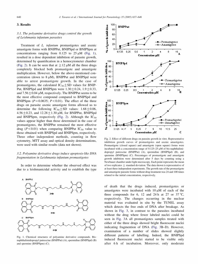
Fig. 2. Effect of different drugs on parasite growth in vitro. Representative
inhibition growth curves of promastigotes and axenic amastigotes.
Promastigote (closed square) and amastigote (open square) forms were
3.1. The polyamine derivative drugs control the growth
of Leishmania infantum parasites
Treatment of L. infantum promastigotes and axenic
amastigote forms with BNIPPut, BNIPSpd or BNIPSpm at
concentrations ranging from 0.125 to 25 mM (Fig. 1),
resulted in a dose dependent inhibition of parasite growth,
determined by quantification in a hemocytometer chamber
(Fig. 2). It can be seen that at R12 mM all the three drugs
completely blocked both promastigote and amastigote
multiplication. However, below the above-mentioned con-
centration (down to 8 mM), BNIPPut and BNIPSpd were
able to arrest promastigote growth. In the case of
promastigotes, the calculated IC50GSD values for BNIP-
Put, BNIPSpd and BNIPSpm were 1.30G0.24, 1.9G0.33,
and 7.56G0.04 mM, respectively. The BNIPPut seems to be
the most effective compound compared to BNIPSpd and
BNIPSpm (PZ0.0635; P!0.01). The effect of the three
drugs on parasite axenic amastigote forms allowed us to
determine the following IC50GSD values: 1.68G0.06,
4.56G0.23, and 12.28G1.30 mM, for BNIPPut, BNIPSpd
and BNIPSpm, respectively (Fig. 2). Although the IC50
values appear higher than those determined in the case of
promastigotes, the BNIPPut remained the most effective
drug (P!0.01) when comparing BNIPPut IC50 value to
those obtained with BNIPSpd and BNIPSpm, respectively.
Three other independent methods, counting in flow
cytometry, MTT assay and optical density determinations,
were used with similar results (data not shown).
incubated with a concentration range of 0.125–25 mM of bis-naphthalimi-dopropyl putrescine (BNIPPut) (A), spermidine (BNIPSpd) (B), and
spermine (BNIPSpm) (C). Percentages of promastigote and amastigote
growth inhibition were determined after 5 days by counting using a
Newbauer chamber under light microscopy. Each point represents the mean
of two replicates G standard deviation. The data shown is representative of
at least three independent experiments. The growth rate of the promastigote
3.2. Polyamine derivative drugs induce apoptosis-like DNA
fragmentation in Leishmania infantum promastigotes
In order to determine whether the observed effect was
due to a leishmanicidal activity and to establish the type
Fig. 1. Chemical structures of polyamine derivative compounds. Bis-
naphthalimidopropyl putrescine (BNIPPut) (A), spermidine (BNIPSpd) (B)
and spermine (BNIPSpm) (C).
and amastigote parasite forms without drug treatment was 24 and 100 times
related to the initial concentration, respectively.
of death that the drugs induced, promastigotes or
amastigotes were incubated with 10 mM of each of the
three compounds for 6, 12 and 24 h at 27 or 37 8C,
respectively. The changes occurring in the nuclear
material was evaluated in situ by the TUNEL assay
which detects the free ends of DNA after breakage. As
shown in Fig. 3, in contrast to the parasites incubated
without the drug where fewer labeled nuclei could be
seen in Fig. 3A all promastigotes samples treated with
either of the three drugs showed bright fluorescent nuclei
indicating fragmention of DNA (Fig. 3B–D). However,
examination of a number of slides showed slightly
different patterns of labelling. Indeed, the BNIPSpd
induced fluorescent nuclei started to be visible only
after 6 h of incubation. Moreover, only moderate
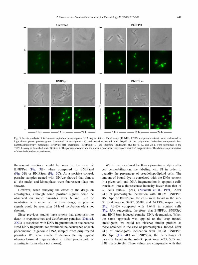
Fig. 3. In situ analysis of Leishmania infantum promastigotes DNA fragmentation. Tunel assay (TUNEL, FITC) and phase contrast, were performed on
logarithmic phase promastigotes. Untreated promastigotes (A) and parasites treated with 10 mM of the polyamine derivative compounds bis-
naphthalimidopropyl putrescine (BNIPPut) (B), spermidine (BNIPSpd) (C) and spermine (BNIPSpm) (D) for 6, 12, and 24 h, were submitted to the
TUNEL assay as described under Section 2. The parasites were examined under a fluorescent microscope at 400! magnification. The data are representative
of three independent experiments.
J. Tavares et al. / International Journal for Parasitology 35 (2005) 637–646 641
fluorescent reactions could be seen in the case of
BNIPPut (Fig. 3B) when compared to BNIPSpd
(Fig. 3B) or BNIPSpm (Fig. 3C). As a positive control,
parasite samples treated with DNAse showed that almost
all the nuclei and kinetoplasts were fluorescent (data not
shown).
However, when studying the effect of the drugs on
amastigotes, although some positive signals could be
observed on some parasites after 6 and 12 h of
incubation with either of the three drugs, no positive
signals could be seen after 24 h of incubation (data not
shown).
Since previous studies have shown that apoptosis-like
death in trypanosomes and Leishmania parasites (Ouaissi,
2003) is associated with DNA fragmentation in nucleosome
sized DNA fragments, we examined the occurrence of such
phenomenon in genomic DNA samples from drug-treated
parasites. We were unable to demonstrate any typical
oligonucleosomal fragmentation in either promatigote or
amastigote forms (data not shown).
We further examined by flow cytometry analysis after
cell permeabilisation, the labeling with PI in order to
quantify the percentage of pseudohypodiploid cells. The
amount of bound dye is correlated with the DNA content
in a given cell, and DNA fragmentation in apoptotic cells
translates into a fluorescence intensity lower than that of
G1 cells (sub-G1 peak) (Nicoletti et al., 1991). After
24 h of promastigote incubation with 10 mM BNIPPut,
BNIPSpd or BNIPSpm, the cells were found in the sub-
G1 peak region, 34.82, 36.88, and 34.13%, respectively
(Fig. 4B–D) compared with 7.64% in control cells
(Fig. 4A), suggesting, therefore, that BNIPPut, BNIPSpd
and BNIPSpm induced parasite DNA degradation. When
the same approach was applied to the drug treated
amastigotes, we could not observe similar profiles as
those obtained in the case of promastigotes. Indeed, after
24 h of amastigotes incubation with 10 mM BNIPPut,
BNIPSpd (Fig. 4F) or BNIPSpm, the percentages of
parasites found in the sub-G1 peak were 4.23, 5.55 and
3.81, respectively. These values are comparable with that
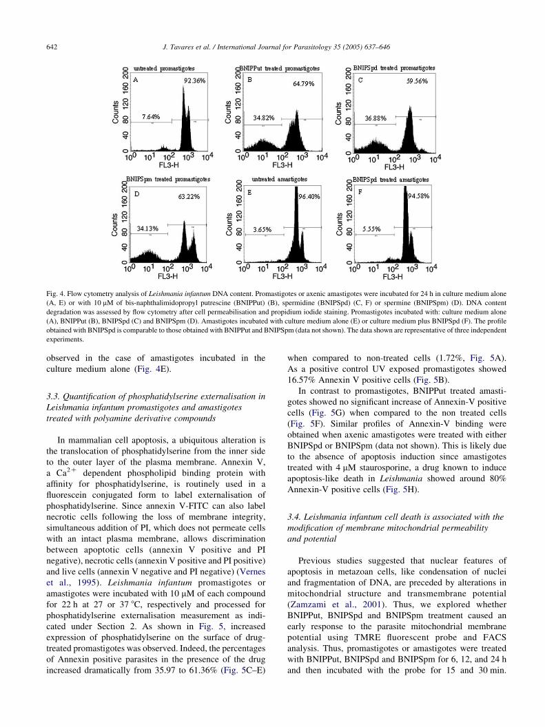
Fig. 4. Flow cytometry analysis of Leishmania infantum DNA content. Promastigotes or axenic amastigotes were incubated for 24 h in culture medium alone
(A, E) or with 10 mM of bis-naphthalimidopropyl putrescine (BNIPPut) (B), spermidine (BNIPSpd) (C, F) or spermine (BNIPSpm) (D). DNA content
degradation was assessed by flow cytometry after cell permeabilisation and propidium iodide staining. Promastigotes incubated with: culture medium alone
(A), BNIPPut (B), BNIPSpd (C) and BNIPSpm (D). Amastigotes incubated with culture medium alone (E) or culture medium plus BNIPSpd (F). The profile
obtained with BNIPSpd is comparable to those obtained with BNIPPut and BNIPSpm (data not shown). The data shown are representative of three independent
experiments.
J. Tavares et al. / International Journal for Parasitology 35 (2005) 637–646642
observed in the case of amastigotes incubated in the
culture medium alone (Fig. 4E).
3.3. Quantification of phosphatidylserine externalisation in
Leishmania infantum promastigotes and amastigotes
treated with polyamine derivative compounds
In mammalian cell apoptosis, a ubiquitous alteration is
the translocation of phosphatidylserine from the inner side
to the outer layer of the plasma membrane. Annexin V,
a Ca2C dependent phospholipid binding protein with
affinity for phosphatidylserine, is routinely used in a
fluorescein conjugated form to label externalisation of
phosphatidylserine. Since annexin V-FITC can also label
necrotic cells following the loss of membrane integrity,
simultaneous addition of PI, which does not permeate cells
with an intact plasma membrane, allows discrimination
between apoptotic cells (annexin V positive and PI
negative), necrotic cells (annexin V positive and PI positive)
and live cells (annexin V negative and PI negative) (Vernes
et al., 1995). Leishmania infantum promastigotes or
amastigotes were incubated with 10 mM of each compound
for 22 h at 27 or 37 8C, respectively and processed for
phosphatidylserine externalisation measurement as indi-
cated under Section 2. As shown in Fig. 5, increased
expression of phosphatidylserine on the surface of drug-
treated promastigotes was observed. Indeed, the percentages
of Annexin positive parasites in the presence of the drug
increased dramatically from 35.97 to 61.36% (Fig. 5C–E)
when compared to non-treated cells (1.72%, Fig. 5A).
As a positive control UV exposed promastigotes showed
16.57% Annexin V positive cells (Fig. 5B).
In contrast to promastigotes, BNIPPut treated amasti-
gotes showed no significant increase of Annexin-V positive
cells (Fig. 5G) when compared to the non treated cells
(Fig. 5F). Similar profiles of Annexin-V binding were
obtained when axenic amastigotes were treated with either
BNIPSpd or BNIPSpm (data not shown). This is likely due
to the absence of apoptosis induction since amastigotes
treated with 4 mM staurosporine, a drug known to induce
apoptosis-like death in Leishmania showed around 80%
Annexin-V positive cells (Fig. 5H).
3.4. Leishmania infantum cell death is associated with the
modification of membrane mitochondrial permeability
and potential
Previous studies suggested that nuclear features of
apoptosis in metazoan cells, like condensation of nuclei
and fragmentation of DNA, are preceded by alterations in
mitochondrial structure and transmembrane potential
(Zamzami et al., 2001). Thus, we explored whether
BNIPPut, BNIPSpd and BNIPSpm treatment caused an
early response to the parasite mitochondrial membrane
potential using TMRE fluorescent probe and FACS
analysis. Thus, promastigotes or amastigotes were treated
with BNIPPut, BNIPSpd and BNIPSpm for 6, 12, and 24 h
and then incubated with the probe for 15 and 30 min.
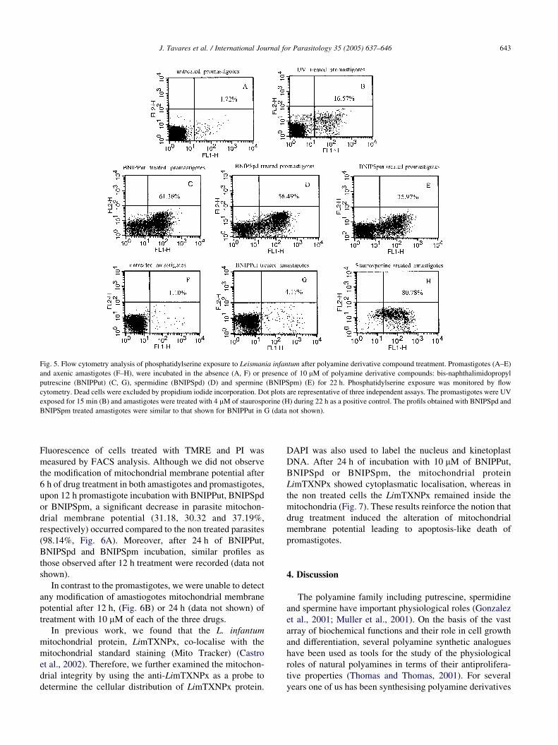
Fig. 5. Flow cytometry analysis of phosphatidylserine exposure to Leismania infantum after polyamine derivative compound treatment. Promastigotes (A–E)
and axenic amastigotes (F–H), were incubated in the absence (A, F) or presence of 10 mM of polyamine derivative compounds: bis-naphthalimidopropyl
putrescine (BNIPPut) (C, G), spermidine (BNIPSpd) (D) and spermine (BNIPSpm) (E) for 22 h. Phosphatidylserine exposure was monitored by flow
cytometry. Dead cells were excluded by propidium iodide incorporation. Dot plots are representative of three independent assays. The promastigotes were UV
exposed for 15 min (B) and amastigotes were treated with 4 mM of staurosporine (H) during 22 h as a positive control. The profils obtained with BNIPSpd and
BNIPSpm treated amastigotes were similar to that shown for BNIPPut in G (data not shown).
J. Tavares et al. / International Journal for Parasitology 35 (2005) 637–646 643
Fluorescence of cells treated with TMRE and PI was
measured by FACS analysis. Although we did not observe
the modification of mitochondrial membrane potential after
6 h of drug treatment in both amastigotes and promastigotes,
upon 12 h promastigote incubation with BNIPPut, BNIPSpd
or BNIPSpm, a significant decrease in parasite mitochon-
drial membrane potential (31.18, 30.32 and 37.19%,
respectively) occurred compared to the non treated parasites
(98.14%, Fig. 6A). Moreover, after 24 h of BNIPPut,
BNIPSpd and BNIPSpm incubation, similar profiles as
those observed after 12 h treatment were recorded (data not
shown).
In contrast to the promastigotes, we were unable to detect
any modification of amastiogotes mitochondrial membrane
potential after 12 h, (Fig. 6B) or 24 h (data not shown) of
treatment with 10 mM of each of the three drugs.
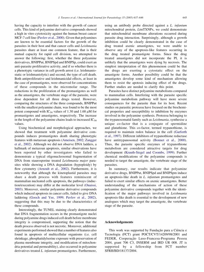
In previous work, we found that the L. infantum
mitochondrial protein, LimTXNPx, co-localise with the
mitochondrial standard staining (Mito Tracker) (Castro
et al., 2002). Therefore, we further examined the mitochon-
drial integrity by using the anti-LimTXNPx as a probe to
determine the cellular distribution of LimTXNPx protein.
DAPI was also used to label the nucleus and kinetoplast
DNA. After 24 h of incubation with 10 mM of BNIPPut,
BNIPSpd or BNIPSpm, the mitochondrial protein
LimTXNPx showed cytoplasmatic localisation, whereas in
the non treated cells the LimTXNPx remained inside the
mitochondria (Fig. 7). These results reinforce the notion that
drug treatment induced the alteration of mitochondrial
membrane potential leading to apoptosis-like death of
promastigotes.
4. Discussion
The polyamine family including putrescine, spermidine
and spermine have important physiological roles (Gonzalez
et al., 2001; Muller et al., 2001). On the basis of the vast
array of biochemical functions and their role in cell growth
and differentiation, several polyamine synthetic analogues
have been used as tools for the study of the physiological
roles of natural polyamines in terms of their antiprolifera-
tive properties (Thomas and Thomas, 2001). For several
years one of us has been synthesising polyamine derivatives
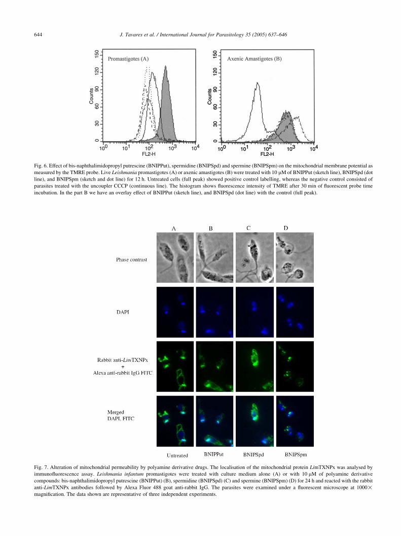
Fig. 6. Effect of bis-naphthalimidopropyl putrescine (BNIPPut), spermidine (BNIPSpd) and spermine (BNIPSpm) on the mitochondrial membrane potential as
measured by the TMRE probe. Live Leishmania promastigotes (A) or axenic amastigotes (B) were treated with 10 mM of BNIPPut (sketch line), BNIPSpd (dot
line), and BNIPSpm (sketch and dot line) for 12 h. Untreated cells (full peak) showed positive control labelling, whereas the negative control consisted of
parasites treated with the uncoupler CCCP (continuous line). The histogram shows fluorescence intensity of TMRE after 30 min of fluorescent probe time
incubation. In the part B we have an overlay effect of BNIPPut (sketch line), and BNIPSpd (dot line) with the control (full peak).
Fig. 7. Alteration of mitochondrial permeability by polyamine derivative drugs. The localisation of the mitochondrial protein LimTXNPx was analysed by
immunofluorescence assay. Leishmania infantum promastigotes were treated with culture medium alone (A) or with 10 mM of polyamine derivative
compounds: bis-naphthalimidopropyl putrescine (BNIPPut) (B), spermidine (BNIPSpd) (C) and spermine (BNIPSpm) (D) for 24 h and reacted with the rabbit
anti-LimTXNPx antibodies followed by Alexa Fluor 488 goat anti-rabbit IgG. The parasites were examined under a fluorescent microscope at 1000!magnification. The data shown are representative of three independent experiments.
J. Tavares et al. / International Journal for Parasitology 35 (2005) 637–646644
J. Tavares et al. / International Journal for Parasitology 35 (2005) 637–646 645
having the capacity to interfere with the growth of cancer
cells. This kind of polyamine derivative compounds showed
a high in vitro cytotoxicity against the human breast cancer
MCF-7 cell line (Pavlov et al., 2000). Given that polyamines
are known to be essential factors for the growth of the
parasites in their host and that cancer cells and Leishmania
parasites share at least one common feature, that is their
mutual capacity for rapid cell division, we attempted to
answer the following: first, whether the three polyamine
derivatives, BNIPPut, BNIPSpd and BNIPSp, could exert an
anti-parasite proliferative activity, and if so whether there is
a phenotypic variance in cell growth arrest (i.e. leishmanio-
static or leishmaniolytic) and second, the type of cell death.
Both antiproliferative and leishmanicidal effects, at least in
the case of promastigotes, were observed for concentrations
of these compounds in the micromolar range. The
reductions in the proliferation of the promastigotes as well
as the amastigotes, the vertebrate stage of the parasite, were
dose dependent for all three drugs tested. However,
comparing the structures of the three compounds, BNIPPut
with the smallest polyamine chain, was found to be the most
potent compound with IC50 values of 1.30 and 1.68 mM for
promastigotes and amastigotes, respectively. The increase
in the length of the polyamine chains leads to increased IC50
values.
Using biochemical and morphological approaches, we
showed that treatment with polyamine derivative com-
pounds induces promastigotes death sharing phenotypic
features with metazoan apoptosis (Ameisen, 2002; Zangger
et al., 2002). Although we did not observe DNA ladders, a
hallmark of metazoan apoptosis, similar observations have
been reported by other investigators who failed to
demonstrate a typical oligonucleosomal fragmentation of
DNA from staurosporine treated Leishmania major para-
sites while showing a DNA degradation (hypoploidy) by
flow cytometry (Arnoult et al., 2002). Furthermore, it is
noteworthy that although the kinetoplastid parasites may
share a death process with features reminiscent of
mammalian nucleated cells apoptosis, the pathways (induc-
tion/execution) may differ at the molecular level (Ouaissi,
2003). Moreover, similar polyamine derivative compounds
which induced apoptosis in cancer cells, failed to give DNA
laddering (Gooch and Yee, 1999; Pavlov et al., 2002),
suggesting that this may be due to the characteristics of
these compounds.
Interestingly, the TUNEL staining clearly demonstrated
that DNA fragmentation occurs in the promastigote nuclei
during polyamine drugs induced cell death before membrane
integrity is compromised, supporting the notion that the
death process observed is not necrotic. Moreover, additional
experiments performed showed that a number offeatures also
found in apoptosis of multicellular organisms (i.e. cell
shrinkage, phosphatidylserine exposure with preservation of
plasma membrane integrity, and modification of mitochon-
dria potential and permeability), also occurred in polyamine
derivatives treated L. infantum promastigotes. Furthermore,
using an antibody probe directed against a L. infantum
mitochondrial protein, LimTXNPx, we could demonstrate
that mitochondrial membrane alterations occurred during
parasite drug interaction. Surprisingly, although a growth
inhibition could be clearly demonstrated in the case of
drug treated axenic amastigotes, we were unable to
observe any of the apoptosis-like features occurring in
the drug treated promastigote forms. Since the drug
treated amastigotes did not incorporate the PI, it is
unlikely that the amastigotes were dying by necrosis. The
simplest interpretation of this phenomenon could be that
the drugs are exerting a cytostatic effect on the
amastigote forms. Another possibility could be that the
amastigotes develop some kind of mechanism allowing
them to resist the apotosis inducing effect of the drugs.
Further studies are needed to clarify this point.
Parasites have distinct polyamine metabolisms compared
to mammalian cells. Interfering with the parasite specific
polyamine metabolism pathway will have more severe
consequences for the parasite than for its host. Recent
studies on parasitic protozoa have focused on the biochemi-
cal properties and susceptibility to inhibition by enzymes
involved in the polyamine synthesis. Protozoa belonging to
the trypanosomatid family such as Leishmania, synthesise a
unique co-factor that is a conjugate of spermidine
and glutathione. This co-factor, termed trypanothione, is
required to maintain redox balance in the cell (Garforth
et al., 1997). Different inhibitors of trypanothione reductase
have antitrypanosomal activity (Bonnet et al., 2000).
Thus, the parasite specific enzymes of trypanothione
metabolism are considered attractive targets for drug
development (Krauth-Siegel and Coombs, 1999). Further
chemical modifications of the polyamine compounds is
needed to target the amastigote, the vertebrate stage of the
parasites.
In summary, our results indicate that polyamine
derivative drugs, BNIPPut, BNIPSpd and BNIPSpm induce
an apoptosis-like death in L. infantum promastigotes and
failed to exert similar effects on axenic amastigotes. Better
understanding of the mechanisms of action of these
polyamine derivative compounds together with the identi-
fication of the major pathways involved in Leishmania
apoptosis-like death is essential to the development of new
analogues which may target the amastigote, the vertebrate
stage of the parasite.
Acknowledgements
This work was supported by Fundacao para a Ciencia e
Tecnologia (FCT) grant POCT/CVT/14209/98/2001 and
FEDER, Cooperacao Luso-Francesa-Programa Pessoa
2004, grant 706 C3, INSERM and IRD UR 008. JT is
supported by a fellowship from FCT number
SFRH/BD/18137/2004.
J. Tavares et al. / International Journal for Parasitology 35 (2005) 637–646646
References
Ameisen, J.C., 2002. On the origin, evolution, and nature of programmed
cell death: a timeline of four billion years. Cell Death Differ. 9, 367–
393.
Arnoult, D., Akarid, K., Grodet, A., Petit, P.X., Estaquier, J., Ameisen, J.C.,
2002. On the evolution of programmed cell death: apoptosis of the
unicellular eukaryote Leishmania major involves cysteine proteinase
activation and mitochondrion permeabilization. Cell Death Differ. 9,
65–81.
Barrett, M.P., Mottram, J.C., Coombs, G.H., 1999. Recent advances in
identifying and validating drug targets in trypanosomes and leishma-
nias. Trends Microbiol. 7, 82–88.
Berhe, N., Wolday, D., Hailu, A., Abraham, Y., Ali, A., Gebre-Michael, T.,
Desjeux, P., Sonnerborg, A., Akuffo, H., Britton, S., 1999. HIV viral
load and response to antileishmanial chemotherapy in co-infected
patients. AIDS 13, 1921–1925.
Bonnet, B., Soullez, D., Davioud-Charvet, E., Landry, V., Horvath, D.,
Sergheraert, C., 1997. New spermine and spermidine derivatives as
potent inhibitors of Trypanosoma cruzi trypanothione reductase. Biorg.
Med. Chem. 5, 1249–1256.
Bonnet, B., Soullez, D., Girault, S., Maes, L., Landry, V., Davioud-
Charvet, E., Sergheraert, C., 2000. Trypanothione reductase inhibi-
tion/trypanocidal activity relationships in a 1,4-bis(3-aminopropyl)pi-
perazine series. Bioorg. Med. Chem. 8, 95–103.
Castro, H., Sousa, C., Santos, M., Cordeiro-da-Silva, A., Flohe, L.,
Tomas, A., 2002. Complementary antioxidant defense by cytoplasmic
and mitochondrial peroxiredoxins in Leishmania infantum. Free Radic.
Biol. Med. 33, 1552–1562.
Cordeiro-da-Silva, A., Cardoso, L., Araujo, N., Castro, H., Tomas, A.,
Rodrigues, M., Cabral, M., Vergnes, B., Sereno, D., Ouaissi, A., 2003.
Identification of antibodies to Leishmania silent information regulatory
2 (SIR2) protein homologue during canine natural infections:
pathological implications. Immunol. Lett. 86, 155–162.
Croft, S.L., Coombs, G.H., 2003. Leishmaniasis—current chemotherapy
and recent advances in the search for novel drugs. Trends Parasitol. 19,
502–508.
Dai, H., Kramer, D.L., Yang, C., Murti, K.G., Porter, C.W., Cleveland, J.L.,
1999. The polyamine oxidase inhibitor MDL-72,527 selectively
induces apoptosis of transformed hematopoietic cells through lysoso-
motropic effects. Cancer Res. 59, 4944–4954.
Davidson, N.E., Hahm, H.A., McCloskey, D.E., Woster, P.M.,
Casero, R.A., 2000. Clinical aspects of cell death in breast cancer:
the polyamine pathway as a new target for treatment. Endocrinol.
Related Cancer 6, 69–73.
Ehrenberg, B., Montana, V., Wei, M.D., Wuskell, J.P., Loew, L.M., 1988.
Membrane potential can be determined in individual cells from the
nernstian distribution of cationic dyes. Biophys. J. 53, 785–794.
Fairlamb, A.H., Cerami, A., 1992. Metabolism and functions of
trypanothione in the kinetoplastida. Annu. Rev. Microbiol. 46, 695–
729.
Garforth, J., Yin, H., McKie, J.H., Douglas, K.T., Fairlamb, A.H., 1997.
Rational design of selective ligands for trypanothione reductase from
Trypanosoma cruzi. Structural effects on the inhibition by dibenzaze-
pines based on imipramine. J. Enzyme Inhib. 12, 161–173.
Gonzalez, N.S., Huber, A., Algranati, I.D., 2001. Spermidine is essential for
normal proliferation of trypanosomatid protozoa. Fed. Eur. Biol. Soc.
Lett. 508, 323–326.
Gooch, J.L., Yee, D., 1999. Strain-specific differences in formation of
apoptotic DNA leaders in MCF-7 breast cancer cells. Cancer Lett. 144,
31–37.
Ha, C.H., Woster, P.M., Yager, J.D., Casero, R.A., 1997. The role of
polyamine catabolism in polyamine-induced programmed cell death.
Proc. Natl Acad. Sci. 94, 1557–1562.
Krauth-Siegel, R.L., Coombs, G.H., 1999. Enzymes of parasite thiol
metabolism as drug targets. Parasitol. Today 15, 404–409.
Lin, P.K.T., Pavlov, V.A., 2000. The synthesis and in vitro cytotoxic studies
of novel bis-naphthalimidopropyl polyamine derivatives. Bioorg. Med.
Chem. Lett. 10, 1609–1612.
McCloskey, D.E., Casero, R.A., Woster, P.M., Davidson, N.E., 1995.
Induction of programmed cell death in human breast cancer cells by an
unsymetrically alkylated polyamine analogue. Cancer Res. 53, 2071–
2075.
Muller, S., Coombs, G.H., Walter, R.D., 2001. Targeting polyamines of
parasitic protozoa in chemotherapy. Trends Parasitol. 17, 242–249.
Murry, H.W., 2001. Clinical and experimental advances in treatment of
visceral Leishmaniasis. Antimicrob. Agents Chemother. 45, 2185–
2197.
Nicoletti, I., Migliorati, G., Pagliacei, M.C., Grignani, F., Riccardi, C.,
1991. A rapid and simple method for measuring thymocyte apoptosis by
propidium iodide staining and flow cytometry. J. Immunol. Methods
139, 271–279.
Ouaissi, A., 2003. Apoptosis-like death in trypanosomatids: Search for
putative pathways and genes involved. Kinetoplastid Biol. Dis. 2,
1–5.
Pavlov, V., Rodilla, V., Lin, P.K.T., 2000. Cytotoxicity, DNA binding and
localisation of novel bis-naphthalimidopropyl polyamine derivatives.
Chem. Biol. Interact. 137, 15–24.
Pavlov, V., Rodilla, V., Lin, P.K.T., 2002. Growth, morphological and
biochemical changes in oxa-spermine derivative-treated MCF-7 human
breast cancer cells. Life Sci. 71, 1161–1173.
Roberts, S.C., Jiang, Y., Jardim, A., Carter, N.S., Heby, O., Ullman, B.,
2001. Genetic analysis of spermidine synthase from Leishmania
donovani. Mol. Biochem. Parasitol. 115, 217–226.
Scaduto, J.r.R.C., Grotyohann, L.W., 1999. Measurement of mitochondrial
membrane potential using fluorescent rhodamine derivatives. Biophys.
J. 76, 469–477.
Sereno, D., Lemesre, J.L., 1997. Axenically cultured amastigote forms as
an in vitro model for investigation of antileishmanial agents.
Antimicrob. Agents Chemother. 41, 972–976.
Thomas, T., Thomas, T.J., 2001. Polyamines in cell growth and cell death:
molecular mechanisms and therapeutic applications. Cell. Mol. Life
Sci. 58, 244–258.
Tovar, J., Wilkinson, S., Mottram, J.C., Fairlamb, A.H., 1998. Evidence
that trypanothione reductase is an essential enzyme in Leishmania by
targeted replacement of the tyrA gene locus. Mol. Microbiol. 29,
653–660.
Vernes, I., Haanen, H., Steffens-Nakken, H., Reutelingsperger, C., 1995. A
novel assay for apoptosis. Flow cytometry detection of phosphatidyl-
serine expression on early apoptotic cells using fluorescein labelled
annexin V. J. Immunol. Mehods 184, 39–51.
Watkins, B.M., 2003. Drugs for the control of parasitic diseases: current
status and development. Trends Parasitol. 19, 477–478.
Zamzami, N., Maisse, C., Metivier, D., Kroemer, G., 2001. Measurement of
membrane permeability and permeability transition of mitochondria.
Methods Cell Biol. 65, 147–158.
Zangger, H., Mottram, J.C., Fasel, N., 2002. Cell death in Leishmania
induced by stress and differentiation: programmed cell death or
necrosis?. Cell Death Differ. 9, 1126–1139.
Zuckeman, A., Lainson, R., 1977. Leishmania in Parasitic Protozoa, vol. 1.
Academic Press, New York, pp. 159–163.