Role of endocytosis in the internalization of spermidine-C2-BODIPY, a highly fluorescent probe of...
11
Biochem. J. (2002) 367, 347–357 (Printed in Great Britain) 347 Role of endocytosis in the internalization of spermidine-C 2 -BODIPY, a highly fluorescent probe of polyamine transport Denis SOULET*† 1 , Laurence COVASSIN‡ 1 , Mohammadi KAOUASS*† 2 , Rene ! CHAREST-GAUDREAULT§¶, Marie AUDETTE†s and Richard POULIN*† 3 *Department of Anatomy and Physiology, Faculty of Medicine, Laval University, Quebec, Canada G1K 7P4, †Molecular Endocrinology and Oncology Research Center, CHUL Medical Research Center (CHUQ), 2705 Laurier Blvd., Ste. Foy, Quebec, Canada G1V 4G2, ‡Faculty of Pharmacy, Laval University, Quebec, Canada G1K 7P4, §Department of Pharmacology, Laval University, Quebec, Canada G1K 7P4, sSt. Franc : ois d’Assise Hospital Research Center (CHUQ), 10 rue de l’Espinay, Quebec, Canada G1L 3L5, and ¶Department of Medical Biology, Faculty of Medicine, Laval University, Quebec, Canada G1K 7P4 The mechanism of transmembrane polyamine internalization in mammalian cells remains unknown. A novel fluorescent spermidine conjugate [Spd-C # -BODIPY ; N-(4,4-difluoro-5,7- dimethyl-4-bora-3a,4a-diaza-s-indacene-3-propionyl)-N« -²S- [spermidine-(N%-ethyl)]thioacetyl·ethylenediamine] was synthe- sized from N%-(mercaptoethyl)spermidine by a simple, one-step coupling procedure. In Chinese-hamster ovary (CHO) cells, Spd- C # -BODIPY accumulation was inhibited by exogenous putrescine, spermidine and spermine, was subject to feedback transport inhibition and was up-regulated by prior polyamine depletion achieved with a biosynthetic inhibitor. Probe internalization was decreased by about 85 % in a polyamine- transport-deficient CHO mutant cell line. Using confocal laser scanning fluorescence microscopy, internalized Spd-C # -BODIPY was concentrated in vesicle-like structures similar to the recycling endosomes observed with fluorescent transferrin, which partly co-localized with the polyamine probe. In yeast, Spd-C # - BODIPY uptake was stringently dependent on receptor-mediated endocytosis, as determined with a mutant defective in early- INTRODUCTION Polyamines are small ubiquitous molecules involved in various functions, including macromolecular synthesis, ion channel gating and the post-translational modification of eukaryotic initiation factor eIF-5A [1,2]. Intracellular polyamine pools are narrowly controlled by the enzymes involved in their biosynthesis and degradation [1,2], as well as by internalization via specific plasma-membrane carriers [3,4]. High-affinity polyamine trans- port can be demonstrated in a wide spectrum of tissues and cell lines, and is activated upon entry into the cell cycle, cellular transformation by oncogenes, and various hormonal signals [3,4]. Polyamine-transport activity is negatively regulated by intra- cellular polyamines, as shown by its up-regulation upon poly- amine depletion by agents such as α-difluoromethylornithine (DFMO) [3,5], a suicide substrate of ornithine decarboxylase [1]. Antizymes are major factors involved in the acute inhibition of polyamine transport by newly internalized polyamines [6,7], although the mechanism of their interaction with the polyamine Abbreviations used : CHO, Chinese hamster ovary ; MANT, N-methylanthranylic acid ; Spd-MANT, N-²spermidine-[N 4 -(3-aminopropyl)]· anthranylamide ; Spd-C 2 -BODIPY, N-(4,4-difluoro-5,7-dimethyl-4-bora-3a,4a-diaza-s-indacene-3-propionyl)-N«-²S-[spermidine-(N 4 -ethyl)]thioacetyl· ethylenediamine ; BODIPY2 FL iodoacetamide, N-(4,4-difluoro-5,7-dimethyl-4-bora-3a,4a-diaza-s-indacene-3-propionyl)-N«-iodoacetylethylene- diamine ; α-MEM, minimal essential medium with α modification ; DFMO, α-difluoromethylornithine ; MFI, mean fluorescence intensity. 1 These authors contributed equally to this work. 2 Current address : Laboratory of Molecular Biology, Clinical Research Institute of Montreal, 110 Pine Avenue West, Montreal, Quebec, Canada H2W 1R7. 3 To whom correspondence should be addressed, at the Molecular Endocrinology and Oncology Research Center (e-mail rpoulin!drs.crchul.ulaval.ca). endosome formation. On the other hand, Spd-C # -BODIPY did not mimic the substrate behaviour of natural polyamines in yeast, as shown by the lack of correlation of its uptake charac- teristics with the phenotypes of mutants defective in either polyamine transport or biosynthesis. These data suggest that endocytosis might be an integral part of the mechanism of polyamine transport in mammalian cells, and that the mam- malian and yeast transport systems use qualitatively different transport mechanisms. However, the current data do not rule out the possibility that sequestration of the probe into vesicular structures might be secondary to its prior uptake via a ‘ classical ’ plasma membrane carrier. Spd-C # -BODIPY, a highly sensitive probe of polyamine transport with biochemical parameters qualitatively similar to those of natural polyamines in mammalian cells, should be very useful for dissecting the pathway responsible for polyamine internalization. Key words : confocal microscopy, difluoromethylornithine, flow cytometry, membrane transport, yeast. carrier(s) is currently unknown. Polyamine uptake is membrane- potential-dependent, Na + -independent and requires bivalent cations such as Ca#+ or Mg#+ [8], but its actual contribution to polyamine homoeostasis under physiological conditions is still uncertain. Prokaryotic polyamine carriers have been extensively charac- terized, and vacuolar polyamine transporters have recently been identified in the yeast Saccharomyces cereisiae [9]. However, little is known about the molecular structure of mammalian polyamine carriers. Genetic evidence indicates that at least two loci control polyamine transport [10], and an as yet unidentified human gene for polyamine transport transfected into a Chinese- hamster ovary (CHO) mutant cell line could restore the polyamine-transport defect present in these cells [11]. Although mammalian polyamine carriers are highly specific for putrescine, spermidine and spermine, the polyamine-binding site of these putative proteins can accommodate substantial modifications of the basic polyamine structure, as shown by the utilization of paraquat (1,1«-dimethyl-4,4«-bipyridinium) and # 2002 Biochemical Society
-
Upload
independent -
Category
Documents
-
view
0 -
download
0
Transcript of Role of endocytosis in the internalization of spermidine-C2-BODIPY, a highly fluorescent probe of...

Biochem. J. (2002) 367, 347–357 (Printed in Great Britain) 347
Role of endocytosis in the internalization of spermidine-C2-BODIPY,a highly fluorescent probe of polyamine transportDenis SOULET*†1, Laurence COVASSIN‡1, Mohammadi KAOUASS*†2, Rene! CHAREST-GAUDREAULT§¶,Marie AUDETTE†s and Richard POULIN*†3
*Department of Anatomy and Physiology, Faculty of Medicine, Laval University, Quebec, Canada G1K 7P4, †Molecular Endocrinology and Oncology Research Center,CHUL Medical Research Center (CHUQ), 2705 Laurier Blvd., Ste. Foy, Quebec, Canada G1V 4G2, ‡Faculty of Pharmacy, Laval University, Quebec, Canada G1K 7P4,§Department of Pharmacology, Laval University, Quebec, Canada G1K 7P4, sSt. Franc: ois d’Assise Hospital Research Center (CHUQ), 10 rue de l’Espinay, Quebec,Canada G1L 3L5, and ¶Department of Medical Biology, Faculty of Medicine, Laval University, Quebec, Canada G1K 7P4
The mechanism of transmembrane polyamine internalization
in mammalian cells remains unknown. A novel fluorescent
spermidine conjugate [Spd-C#-BODIPY; N-(4,4-difluoro-5,7-
dimethyl-4-bora-3a,4a-diaza-s-indacene-3-propionyl)-N«-²S-
[spermidine-(N%-ethyl)]thioacetyl´ethylenediamine] was synthe-
sized from N%-(mercaptoethyl)spermidine by a simple, one-step
coupling procedure. In Chinese-hamster ovary (CHO) cells, Spd-
C#-BODIPY accumulation was inhibited by exogenous
putrescine, spermidine and spermine, was subject to feedback
transport inhibition and was up-regulated by prior polyamine
depletion achieved with a biosynthetic inhibitor. Probe
internalization was decreased by about 85% in a polyamine-
transport-deficient CHO mutant cell line. Using confocal laser
scanning fluorescence microscopy, internalized Spd-C#-BODIPY
was concentrated in vesicle-like structures similar to the recycling
endosomes observed with fluorescent transferrin, which partly
co-localized with the polyamine probe. In yeast, Spd-C#-
BODIPYuptake was stringently dependent on receptor-mediated
endocytosis, as determined with a mutant defective in early-
INTRODUCTION
Polyamines are small ubiquitous molecules involved in various
functions, including macromolecular synthesis, ion channel
gating and the post-translational modification of eukaryotic
initiation factor eIF-5A [1,2]. Intracellular polyamine pools are
narrowly controlled by the enzymes involved in their biosynthesis
and degradation [1,2], as well as by internalization via specific
plasma-membrane carriers [3,4]. High-affinity polyamine trans-
port can be demonstrated in a wide spectrum of tissues and
cell lines, and is activated upon entry into the cell cycle, cellular
transformation by oncogenes, and various hormonal signals [3,4].
Polyamine-transport activity is negatively regulated by intra-
cellular polyamines, as shown by its up-regulation upon poly-
amine depletion by agents such as α-difluoromethylornithine
(DFMO) [3,5], a suicide substrate of ornithine decarboxylase [1].
Antizymes are major factors involved in the acute inhibition of
polyamine transport by newly internalized polyamines [6,7],
although the mechanism of their interaction with the polyamine
Abbreviations used: CHO, Chinese hamster ovary ; MANT, N-methylanthranylic acid ; Spd-MANT, N-²spermidine-[N4-(3-aminopropyl)]´anthranylamide ; Spd-C2-BODIPY, N-(4,4-difluoro-5,7-dimethyl-4-bora-3a,4a-diaza-s-indacene-3-propionyl)-N«-²S-[spermidine-(N4-ethyl)]thioacetyl´ethylenediamine; BODIPY2 FL iodoacetamide, N-(4,4-difluoro-5,7-dimethyl-4-bora-3a,4a-diaza-s-indacene-3-propionyl)-N«-iodoacetylethylene-diamine; α-MEM, minimal essential medium with α modification; DFMO, α-difluoromethylornithine ; MFI, mean fluorescence intensity.
1 These authors contributed equally to this work.2 Current address : Laboratory of Molecular Biology, Clinical Research Institute of Montreal, 110 Pine Avenue West, Montreal, Quebec, Canada
H2W 1R7.3 To whom correspondence should be addressed, at the Molecular Endocrinology and Oncology Research Center (e-mail
rpoulin!drs.crchul.ulaval.ca).
endosome formation. On the other hand, Spd-C#-BODIPY did
not mimic the substrate behaviour of natural polyamines in
yeast, as shown by the lack of correlation of its uptake charac-
teristics with the phenotypes of mutants defective in either
polyamine transport or biosynthesis. These data suggest that
endocytosis might be an integral part of the mechanism of
polyamine transport in mammalian cells, and that the mam-
malian and yeast transport systems use qualitatively different
transport mechanisms. However, the current data do not rule out
the possibility that sequestration of the probe into vesicular
structures might be secondary to its prior uptake via a ‘classical ’
plasma membrane carrier. Spd-C#-BODIPY, a highly sensitive
probe of polyamine transport with biochemical parameters
qualitatively similar to those of natural polyamines inmammalian
cells, should be very useful for dissecting the pathway responsible
for polyamine internalization.
Key words: confocal microscopy, difluoromethylornithine, flow
cytometry, membrane transport, yeast.
carrier(s) is currently unknown. Polyamine uptake is membrane-
potential-dependent, Na+-independent and requires bivalent
cations such as Ca#+ or Mg#+ [8], but its actual contribution
to polyamine homoeostasis under physiological conditions is
still uncertain.
Prokaryotic polyamine carriers have been extensively charac-
terized, and vacuolar polyamine transporters have recently been
identified in the yeast Saccharomyces cere�isiae [9]. However,
little is known about the molecular structure of mammalian
polyamine carriers. Genetic evidence indicates that at least two
loci control polyamine transport [10], and an as yet unidentified
human gene for polyamine transport transfected into a Chinese-
hamster ovary (CHO) mutant cell line could restore the
polyamine-transport defect present in these cells [11].
Although mammalian polyamine carriers are highly specific
for putrescine, spermidine and spermine, the polyamine-binding
site of these putative proteins can accommodate substantial
modifications of the basic polyamine structure, as shown by the
utilization of paraquat (1,1«-dimethyl-4,4«-bipyridinium) and
# 2002 Biochemical Society

348 D. Soulet and others
methylglyoxal bis(guanylhydrazone) as substrates [3]. This prop-
erty has previously been exploited for the biochemical charac-
terization of the polyamine carrier. For instance, spermidine,
spermine and norspermine were derivatized with "#&I-labelled 4-
azidosalicylic acid to generate photoreactive probes for the
labelling of polyamine-binding proteins as potential candidates
for the mammalian polyamine carriers [12]. The polyamine-
transport system has also been used for the targeting of cytotoxic
polyamine analogues [1] or chemotherapeutic agents conjugated
to polyamines [13].
Conjugation of aromatic structures such as 4-azidosalicylic
acid [12] and chlorambucil [13] to the polyamine core structure
does not abrogate its strong binding to the polyamine carrier.
This interesting property has recently been exploited for the
design of fluorescent probes to study polyamine transport.
Monofluoresceinyl adducts of spermidine and spermine sub-
stituted on one of the primary amino groups have been described
by Aziz et al. [14], which displayed affinity comparable with the
parent polyamine and mostly accumulated in the cytoplasm of
mammalian cells. Likewise, amidation of N-methylanthranylic
acid (MANT) to an aminopropyl group of N%-(3-aminopropyl)-
spermidine led to an UV-excitable substrate for polyamine
transport in various mammalian cells [13]. Interestingly, N-
²spermidine-[N%-(3-aminopropyl)]´anthranylamide (Spd-MANT)
mainly accumulated into discrete subcellular structures reminis-
cent of endocytic vesicles [13].
Polyamines accumulated from exogenous sources exert regu-
latory effects on polyamine homoeostasis at levels representing
only a minor fraction of the total polyamine pool [15–17].
Likewise, indirect biochemical evidence suggests that the free (i.e.
unbound) pools of spermidine and spermine would be restricted
to less than 2 and 7% of total content respectively [18]. Thus a
better understanding of the mechanism of polyamine internal-
ization and compartmentalization is required to understand the
actual sites of action of these compounds under physiological
conditions. In this report we describe the synthesis and charac-
terization of a novel fluorescent probe of polyamine transport,
spermidine-C#-BODIPY [Spd-C
#-BODIPY; N-(4,4-difluoro-5,-
7-dimethyl-4-bora-3a,4a-diaza-s-indacene-3-propionyl)-N«-²S-
[spermidine-(N%-ethyl)]thioacetyl´ethylenediamine], obtained by
grafting a highly sensitive fluorophore to an N%-mercaptoethyl
side arm extending from the spermidine backbone. Like Spd-
MANT [13], Spd-C#-BODIPY mainly compartmentalizes into
vesicle-like intracellular structures and is excluded from the
nucleus. Moreover, the internalization of Spd-C#-BODIPY likely
proceeds via endocytosis of a complex formed between the probe
and the polyamine transporter(s), rather than carrier-mediated
influx.
EXPERIMENTAL
Reagents
Bovine calf serum and Eagle’s minimal essential medium (with α
modification; α-MEM) were purchased from Wisent (St-Bruno,
Quebec,Canada) andGibco-BRLLife Technologies (Burlington,
Ontario, Canada), respectively. ,-α-Difluoromethylornithine
hydrochloride was generously provided by ILEX Oncology (San
Antonio, TX, U.S.A.). o-Phthaldialdehyde was obtained from
MAT Laboratories (Quebec City, Quebec, Canada). BODIPY2FL iodoacetamide [N-(4,4-difluoro-5,7-dimethyl-4-bora-3a,4a-
diaza-s-indacene-3-propionyl)-N«-iodoacetylethylenediamine]
and Texas Red2- and BODIPY FL-labelled human serum
transferrin as well as FITC were purchased from Molecular
Probes (Eugene, OR, U.S.A.). [terminal-methylene-groups-$H(n)]
Spermidine trihydrochloride (3.1¬10& Ci}mol) was purchased
Figure 1 Structures of Spd-C2-BODIPY and Spd-MANT
from NEN Life Science Products (Lachine, Quebec, Canada).
Unless otherwise indicated, other biochemical and tissue culture
reagents were from Sigma–Aldrich.
Synthesis of Spd-C2-BODIPY
N%-(Mercaptoethyl)spermidine was synthesized according to the
method of Cohen et al. [19]. Spd-C#-BODIPY (Figure 1) was
prepared by alkylation of the thiol group of N%-
(mercaptoethyl)spermidine with BODIPY FL iodoacetamide.
Prior to conjugation of the fluorophore, N%-(mercaptoethyl)-
spermidine was dissolved in an aqueous solution containing a
5-fold molar excess of dithiothreitol to ensure complete reduction
of the thiol group. The mixture was left for 1 h at room tem-
perature, and N%-(mercaptoethyl)spermidine was re-purified by
ion-exchange chromatography as described above, followed
by lyophilization. All following operations were carried out
under subdued light. To 1 mol of a neutralized solution of
N%-(mercaptoethyl)spermidine in 100 µM Tris}HCl (pH 7.3)
was then added 4 mol of BODIPY FL iodoacetamide (extem-
poraneously prepared as a 20 mM solution in DMSO), and the
mixture was stirred overnight in the dark at room temperature.
Solvents were removed by lyophilization, the foam was dissolved
in deionized water, and the solution was passed through a PVDF
syringe membrane filter (Millex-HV4, 0.45 µm pore size ; Milli-
pore, Bedford, MA, U.S.A.). The aqueous phase was washed
with dichloromethane (HPLC grade) until the organic phase,
containing unchanged BODIPY FL iodoacetamide, became
colourless. Complete extraction of unchanged BODIPY FL
iodoacetamide and formation of a polar BODIPY–spermidine
conjugate was confirmed by silica-gel TLC using dichloro-
methane}methanol (9 :1, v}v) as an eluent. The aqueous phase
containing Spd-C#-BODIPY was reduced to a small volume
by lyophilization. The actual concentration of Spd-C#-BODIPY
was then determined on an aliquot of the latter solution by
derivatization with an o-phthaldialdehyde solution in water
[400 mM potassium borate, 0.5 g}l o-phthaldialdehyde, 1%
methanol (v}v), 0.5% (v}v) β-mercaptoethanol and 0.1% Brij
35 (v}v)] for 20 min in the dark at 37 °C [20], and determination
of fluorescence intensity using an SLM-AMINCO-Bowman AB2
spectrofluorimeter (excitation wavelength, 350 nm), using sper-
midine as standard. Standardization of the solution was con-
# 2002 Biochemical Society

349Fluorimetry of polyamine transport with BODIPY-labelled spermidine
firmed by measuring the fluorescence intensity of the BODIPY
adduct (excitation wavelength, 493 nm), and the final concen-
tration of Spd-C#-BODIPY was then adjusted to 1 mM.
Synthesis of Spd-MANT and of monofluoresceinylspermidineconjugates
Spd-MANT was synthesized by N,N«-dicyclohexylcarbodi-
imide-coupled amide formation between anthranilic acid and
N",N)-di(t-butoxycarbonyl)-N%-(3-aminopropyl)spermidine as
described in [19]. A mixture of N"- and N)-(monofluoresceinyl)-
spermidines was synthesized using FITC and purified by TLC on
silica-gel plates [14].
Cell lines and cell culture
The parental CHO cell line (CHO-TOR) and a polyamine-
transport-deficient subline (CHO-MG) isolated after chronic
selection for growth resistance to methylglyoxal
bis(guanylhydrazone) [10,21] were generously provided by Dr
Wayne Flintoff (Department of Microbiology and Immunology,
University of Western Ontario, London, Ontario, Canada). Both
lines were routinely grown in α-MEM supplemented with 10%
CosmicTM calf serum (Hyclone, Logan, UT, U.S.A.) and anti-
biotics in a water-saturated 5% CO#
atmosphere at 37 °C.
All yeast strains used in this study were haploid. The wild-type
yeast (S. cere�isiae) strain RH144-3D (MATa leu2 his4 ura3
bar1-1) and its RH266-1D (MATa leu2 his4 ura3 bar1-1 ts end3)
mutant derivative defective in an early internalization step of
endocytosis [22–24] were kindly provided by Dr H. Riezman
(Biozentrum, University of Basel, Basel, Switzerland). The
DBY747 wild-type strain (MATa leu2 his3 ura3 trp1) and its
mutant derivative DBY747spe2∆ (MATa leu2 his3 ura3 trp1
∆spe2-5::LEU2) carrying a chromosomal deletion of the SPE2
gene encoding S-adenosylmethionine decarboxylase have been
described previously [23]. The W303 ptk2 :∆ deletion mutant
(MATa ade2 ura3 trp1 his3 leu2 ptk2::TRP1) was generated
by the one-step disruption method [25]. Briefly, the full PTK2
sequence, including 683 and 262 bp adjacent to the 5« and 3«ends of the coding region, respectively, was isolated as a BamHI
fragment (3.4 kb) from a lambda PM-1436 S. cere�isiae
genomic clone from chromosome X (A.T.C.C. 70345) [26].
The BamHI fragment was subcloned into the same site of the
Bluescript II KS() plasmid. The resulting plasmid pKSPTK2
was digested with HpaI and NdeI restriction enzymes to
eliminate a 2631 bp fragment corresponding to the full sequence
of the PTK2 open reading frame. A ScaI–NdeI fragment
(3100 bp) bearing the TRP1 marker (from the pJG4-5 ex-
pression vector) was ligated into the blunt-end HpaI and NdeI
sites of pKSPTK2. The resulting 3900 bp BamHI fragment,
containing the TRP1 gene flanked with PTK2 sequences
(ptk2::TRP1), was isolated and used to transform the wild-
type haploid strain W303 (MATa ade2 ura3 trp1 his3 leu2)
with subsequent selection on tryptophan-free synthetic dextrose
(SD) medium [0.17% yeast nitrogen base without amino acids or
(NH%)#SO
%, with 0.5% (NH
%)#SO
%and 2% dextrose added]. The
yeast strains used in this study were routinely grown in YPD
medium (1% yeast extract, 2% peptone and 2% -glucose).
Epifluorescence microscopy
Both CHO cell lines were grown on sterile glass coverslips in six-
well plates for 24 h. Cells were then incubated in the presence of
Spd-C#-BODIPY (1 µM) for 2–4 h. Medium was removed, and
cell monolayers were washed three times with 1 ml of ice-cold
Ca#+}Mg#+-free PBS containing 1 mM spermidine and then
twice with 1 ml of PBS. Coverslips were then inverted on a
droplet of Hanks’ balanced salt solution. Intracellular fluor-
escence was observed using an Axioskop microscope equipped
with a Plan-Neofluar 403}0.75 objective (Zeiss) and filter set
487909 (bandpass, 450–490 nm; filter threshold, 510 nm; long
pass, 520 nm). Images were obtained with a CCD-300T-RC
camera (DAGE-MTI, Michigan City, IN, U.S.A.) coupled to the
MetaMorph2 imaging system (version 3.5; Universal Imaging
Corporation, West Chester, PA, U.S.A.).
FACS analysis of fluorescent probe uptake
Both CHO cell lines were seeded at 2.5¬10$ cells}well in 24-well
culture plates. Cells were incubated for the indicated time interval
with serum-free α-MEM containing 1 µM Spd-C#-BODIPY or
1 µM Spd-MANT. Medium was then removed and cell mono-
layers were washed three times with 1 ml of PBS containing
1 mM spermidine. Cell cultures were then rinsed twice with 1 ml
of PBS, and harvested after a 5-min incubation with
pancreatin}EDTA}Hepes solution (2.5 g}l}1 mM}7.25 mM) in
Hanks’ balanced salt solution. Cells were suspended in ice-cold
α-MEM and immediately processed for fluorescence analysis by
flow cytometry with an Epics Profile II cytofluorometer (Coulter
Corp., Miami, FL, U.S.A.). For Spd-C#-BODIPY, the argon
cytofluorimeter was tuned to 488 nm, using an FL1 photo-
multiplier (bandpass, 525³30 nm). For Spd-MANT, the He}Cd
lamp of the cytofluorimeter was tuned to 325 nm, using an FL2
photomultiplier. The mean value of fluorescence intensity (MFI)
distribution was recorded for the analysis of 1¬10% cells}sample.
MFI is the mean channel number recorded for the distribution of
fluorescence among the cells counted.
To determine the effect of polyamine depletion and protein
synthesis inhibition on Spd-C#-BODIPY uptake, cells were first
grown for 72 h in the presence of 5 mM DFMO or vehicle. Fresh
serum-free α-MEM containing 1 µM Spd-C#-BODIPY was then
added for a 4 h incubation, with or without 200 mM cyclo-
heximide and}or 5 mM DFMO.
Confocal laser scanning fluorescence microscopy
CHO-TOR cells grown on coverslips were first incubated for 4 h
with 1 µM Spd-C#-BODIPY and Texas Red–transferrin con-
jugate (3 µg}ml) in α-MEM at 37 °C. Culture medium was then
removed, and cells were washed three times with ice-cold PBS
containing 1 mM spermidine, and then twice with ice-cold
PBS. Specimens were visualized with a Bio-Rad MRC-1024 con-
focal imaging system equipped with a Kr}Ar laser and mounted
on a Diaphot-TMD inverted microscope (Nikon). A 60¬ oil-
immersion objective lens with a 1.4 numerical aperture was used
for imaging. The photomultiplier gain was set at maximum, and
the confocal aperture was adjusted for maximum resolution.
Analysis of fluorophore co-localization was performed using
the LaserSharp software (version 3.0; Bio-Rad) with background
subtraction.
Competition of spermidine uptake by fluorescent polyamines
The ability of Spd-C#-BODIPY, Spd-MANT and N%-(mercapto-
ethyl)spermidine to compete for [$H]spermidine uptake was
determined in CHO-TOR cells by a 20 min uptake assay in
the presence of increasing concentrations of competitor, using
5 µM [$H]spermidine as a substrate, as described in [27]. Data
were averaged from three separate experiments with triplicate
determinations of the Kivalue for each experiment. The K
mvalue
of spermidine transport was determined by Lineweaver–Burk
analysis as described in [27]. Kivalues for inhibition of spermidine
# 2002 Biochemical Society

350 D. Soulet and others
uptake were determined using the Cheng–Prusoff equation [28]
from the IC&!
value derived by iterative curve fitting of the
sigmoidal equation describing the velocity of spermidine uptake
in the presence of the respective competitor [5,27].
Uptake of Spd-C2-BODIPY in yeast
The wild-type DBY747 strain and its spe2∆ mutant derivative
were incubated at 30 °C for 96 h in H medium, an amine-free
minimal medium [29] supplemented with 5 mg}ml tryptophan
with or without 5 mg}ml leucine, respectively. Cell growth had
completely stopped in the spe2∆ mutant at the end of the
incubation period, indicating the complete depletion of
spermidine and spermine [23,29]. DBY747 cells and the spe2∆
mutants were then pre-incubated for 10 min at 30 °C in the
appropriately supplemented H medium at 3¬10( cells}ml. The
wild-type W303 strain and its ptk2∆ mutant derivative were
grown until mid-exponential stage in YPD at 30 °C, and 3¬10(
cells were then transferred into 1 ml of H medium at 30 °C. The
four strains were then incubated with 1 µM Spd-C#-BODIPY in
the dark in a shaking incubator at 250 cycles}min. At the
indicated times, cells were processed for FACS analysis as
described above.
Wild-type RH144-3D cells and the end3 mutant were grown to
mid-exponential stage in YPD at 25 °C, then rinsed and pre-
incubated for 10 min at 37 °C in H medium. Cells were
then incubated with either 1 µM Spd-C#-BODIPY for the indi-
cated time interval, or 10 µM [$H]spermidine (50 Ci}mol) for
20 min to determine the rate of high-affinity spermidine uptake
[23,26].
Statistical analysis
The statistical significance of differences between means was
assessed at the 5% level by unpaired Student’s t tests or by the
multiple-range Duncan–Kramer test [30]. Unless otherwise
indicated, results are expressed as the mean³S.D. from three
separate experiments with duplicate or triplicate determinations
for each experiment.
RESULTS
Design and synthesis of the BODIPY-spermidine conjugate
Spermidine analogues derivatized at the N% position are better
substrates for the mammalian polyamine-transport system than
those substituted on primary amino groups [31]. Moreover,
spermidine and norspermidine dimers cross-linked through al-
kylation of the secondary amino group with aliphatic or aromatic
linkers display low Kivalues for competition against polyamine
uptake [32]. In order to generate a sensitive probe of poly-
amine transport, we thus conjugated the iodoacetamide deriva-
tive of the highly fluorescent dye BODIPY FL [33] to the thiol
group of N%-(mercaptoethyl)spermidine [19]. The availability of
a thiol as a unique reactive group obviated the need for protecting
the amino groups of the spermidine derivative before conjugation
to the fluorophore. The resulting conjugate, Spd-C#-BODIPY
(Figure 1) could thus be prepared using a simple, one-step re-
action while avoiding the use of extreme pH conditions under
which the fluorophore is unstable [34].
The ability of Spd-C#-BODIPY to interact with the polyamine-
transport system was determined by measuring competition by
the probe against spermidine uptake in CHO-TOR cells. For
comparison, the Ki
value against spermidine uptake was also
determined for N%-(mercaptoethyl)spermidine and Spd-MANT
(Figure 1), a recently described fluorescent polyamine which
bears a smaller hydrophobic fluorophore than Spd-C#-BODIPY
Figure 2 Heterogeneous cytoplasmic labelling by Spd-C2-BODIPY in CHOcells
CHO-TOR cells grown on sterile glass coverslips were incubated for 4 h with 1 µM Spd-C2-
BODIPY. Note the uneven distribution of fluorescence in the cytoplasm in the form of granules
or vesicle-like structures.
[13]. The Kiof Spd-C
#-BODIPY (46³13 µM) was about 30-fold
higher than the Km
of spermidine (1.5³0.4 µM), consistent
with the weak inhibitory potency also measured for N%-
(mercaptoethyl)spermidine (Ki¯ 25³1 µM). Spd-MANT (K
i¯
0.4³0.07 µM) was a much stronger competitor of spermidine
uptake than Spd-C#-BODIPY, exhibiting an even stronger
affinity than spermidine for the polyamine transporter.
Labelling of intact cells with Spd-C2-BODIPY
As shown in Figure 2, the accumulation of Spd-C#-BODIPY
added in trace amounts (1 µM) to the growth medium was
readily detectable by epifluorescence microscopy in wild-type
CHO (CHO-TOR) cells. Intracellular fluorescence was clearly
distributed in a discrete fashion, leading to a granular pattern,
with a much less intense staining of the cytosol. To evaluate the
suitability of Spd-C#-BODIPY as a probe for polyamine trans-
port, its ability to differentially label CHO-TOR and polyamine-
transport-deficient mutant (CHO-MG) cells was next assessed. It
had been established previously that CHO-MG cells are com-
pletely deficient in diamine and polyamine transport [10,21]. Cells
were pre-treated for 72 h with DFMO and were co-incubated
with cycloheximide to maximize probe accumulation (see below).
Whereas CHO-TOR cells were intensely labelled with Spd-C#-
BODIPY, only a faint green intracellular fluorescence could be
detected in the CHO-MG cells (Figure 3). These data indicate
that conjugation of the hydrophobic BODIPY moiety to
spermidine via a long side chain preserves the specificity of
substrate binding to the polyamine carrier, and that Spd-C#-
BODIPY is a reliable probe of high-affinity polyamine uptake.
Polyamine uptake is an active-transport process and is thus
strongly dependent on temperature [3,5]. MFI was determined
by flow cytometry in CHO-TOR and CHO-MG cells incubated
for 1 h at either 4 or 37 °C with Spd-C#-BODIPY (1 µM) in
serum-free α-MEM, after pre-incubating cells at the respective
temperature for 60 min in serum-supplemented growth medium.
# 2002 Biochemical Society

351Fluorimetry of polyamine transport with BODIPY-labelled spermidine
Figure 3 Epifluorescence microscopy of Spd-C2-BODIPY accumulation in CHO-TOR and CHO-MG cells
CHO-TOR (A, B) or CHO-MG (C, D) cells grown on coverslips were incubated for 4 h with 1 µM Spd-C2-BODIPY in the presence of 5 mM DFMO and 200 µM cycloheximide. The same fields
were examined by epifluorescence microscopy (A, C) and phase-contrast microscopy (B, D).
The uptake of Spd-C#-BODIPY was strongly temperature-
dependent, being decreased from 2.8³0.2 and 0.6³0.01 channel
number units at 37 °C to 0.25³0.01 and 0.05³0.01
channel number units at 4 °C in CHO-TOR and CHO-MG cells,
respectively.
We next determined the ability of natural polyamines to
compete against intracellular labelling by the fluorescent probe.
At a 1000-fold molar excess, putrescine, spermidine and spermine
abolished 89³16%, 82³9% and 82³9% of the MFI in CHO-
TOR cells incubated for 4 h with 1 µM Spd-C#-BODIPY. On the
other hand, spermidine (1 mM) did not significantly decrease
intracellular fluorescence measured in the CHO-MG mutants
incubated under the same conditions (14³4 and 17³8 relative
fluorescence units for cells labelled in the absence or presence of
spermidine respectively ; means³S.D. from three independent
experiments). Thus cell labelling with Spd-C#-BODIPY largely
depends on the binding of the spermidine-like portion of the
conjugate to the polyamine carrier.
FACS analysis of intracellular labelling by Spd-C#-BODIPY
was performed over a 6 h period in wild-type and CHO-MG
cells. The rate of uptake of the probe was initially linear in CHO-
TOR cells for about 2 h and then steadily decreased (Figure 4).
In contrast, Spd-C#-BODIPY accumulated very slowly in CHO-
MG cells, and no net uptake could be detected 4 h after addition
of the probe. Spd-C#-BODIPY uptake in the transport mutant
represented about 15–20% of that detected in wild-type cells
throughout the incubation period, and could not be decreased by
co-incubation with a 1000-fold excess of spermidine (results not
shown).
The rapid decrease in the rate of Spd-C#-BODIPY accumu-
lation observed after a 2 h incubation was reminiscent of the
Figure 4 Time course of the uptake of Spd-C2-BODIPY in wild-type (CHO-TOR) and polyamine-transport-deficient mutant (CHO-MG) cells
Cells were incubated in medium containing 1 µM Spd-C2-BODIPY for the indicated interval prior
to FACS analysis. Data are means³S.D. of mean fluorescence emission values from three
separate experiments.
onset of feedback repression of polyamine transport induced by
polyamine internalization in mammalian cells [5]. This feedback
inhibition results from the rapid induction of antizyme synthesis
# 2002 Biochemical Society

352 D. Soulet and others
Figure 5 Confocal laser scanning microscopy of Spd-C2-BODIPY and Texas Red–transferrin accumulation in CHO-TOR cells
Cells were incubated for 4 h with either 1 µM Spd-C2-BODIPY or 3 µg/ml Texas Red–transferrin (Texas Red-Tf) and then examined by confocal laser microscopy as described in the Experimental
section. Three representative fields (A–C) are shown to illustrate the extent of partial co-localization (arrows) and the pattern of reticular perinuclear staining (pn). For each field shown, Spd-C2-
BODIPY fluorescence is shown in green, Texas Red–transferrin in red and the superimposition of the two images is shown on the right.
bypromotion of a1 frameshifting in the translation of antizyme
mRNA by polyamines [6]. We thus determined the effect of
protein synthesis inhibition on the accumulation of the polyamine
probe. Furthermore, we examined the effect of prior polyamine
depletion on Spd-C#-BODIPY uptake by pre-incubating cells
with DFMO, a treatment which up-regulates polyamine-trans-
port activity [3,5,11]. DFMO significantly (P! 0.05) increased
the apparent uptake of Spd-C#-BODIPY by about 2-fold (MFI,
108³27 and 45³6 in DFMO-treated and control cells, re-
spectively), as previously observed for the transport of natural
polyamines [11]. Moreover, cycloheximide significantly
(P! 0.05) increased MFI of wild-type CHO cells incubated with
Spd-C#-BODIPY to 149³6 and 196³39 in the absence and
presence of DFMO, respectively, consistent with the relief of
feedback inhibition of Spd-C#-BODIPY uptake by pre-empting
stimulation of de no�o synthesis of antizyme by the internalized
polyamine [5,35].
Spectrometric properties of Spd-C2-BODIPY and Spd-MANT
During the course of this work, the characteristics of two other
fluorescent polyamine probes, namely monofluoresceinyl-
spermidine [14] and Spd-MANT [13], have been reported. We
thus compared the properties of the latter probes with those of
Spd-C#-BODIPY. In our hands, monofluoresceinylspermidine
[14] (which is in fact a mixture of the N"- and N)-fluoresceinyl-
# 2002 Biochemical Society
353Fluorimetry of polyamine transport with BODIPY-labelled spermidine
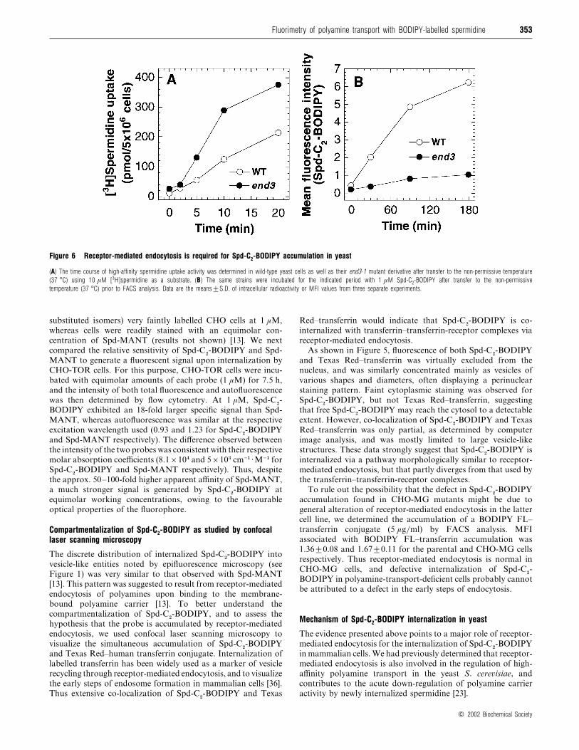
Figure 6 Receptor-mediated endocytosis is required for Spd-C2-BODIPY accumulation in yeast
(A) The time course of high-affinity spermidine uptake activity was determined in wild-type yeast cells as well as their end3-1 mutant derivative after transfer to the non-permissive temperature
(37 °C) using 10 µM [3H]spermidine as a substrate. (B) The same strains were incubated for the indicated period with 1 µM Spd-C2-BODIPY after transfer to the non-permissive
temperature (37 °C) prior to FACS analysis. Data are the means³S.D. of intracellular radioactivity or MFI values from three separate experiments.
substituted isomers) very faintly labelled CHO cells at 1 µM,
whereas cells were readily stained with an equimolar con-
centration of Spd-MANT (results not shown) [13]. We next
compared the relative sensitivity of Spd-C#-BODIPY and Spd-
MANT to generate a fluorescent signal upon internalization by
CHO-TOR cells. For this purpose, CHO-TOR cells were incu-
bated with equimolar amounts of each probe (1 µM) for 7.5 h,
and the intensity of both total fluorescence and autofluorescence
was then determined by flow cytometry. At 1 µM, Spd-C#-
BODIPY exhibited an 18-fold larger specific signal than Spd-
MANT, whereas autofluorescence was similar at the respective
excitation wavelength used (0.93 and 1.23 for Spd-C#-BODIPY
and Spd-MANT respectively). The difference observed between
the intensity of the two probes was consistent with their respective
molar absorption coefficients (8.1¬10% and 5¬10$ cm−"[M−" for
Spd-C#-BODIPY and Spd-MANT respectively). Thus, despite
the approx. 50–100-fold higher apparent affinity of Spd-MANT,
a much stronger signal is generated by Spd-C#-BODIPY at
equimolar working concentrations, owing to the favourable
optical properties of the fluorophore.
Compartmentalization of Spd-C2-BODIPY as studied by confocallaser scanning microscopy
The discrete distribution of internalized Spd-C#-BODIPY into
vesicle-like entities noted by epifluorescence microscopy (see
Figure 1) was very similar to that observed with Spd-MANT
[13]. This pattern was suggested to result from receptor-mediated
endocytosis of polyamines upon binding to the membrane-
bound polyamine carrier [13]. To better understand the
compartmentalization of Spd-C#-BODIPY, and to assess the
hypothesis that the probe is accumulated by receptor-mediated
endocytosis, we used confocal laser scanning microscopy to
visualize the simultaneous accumulation of Spd-C#-BODIPY
and Texas Red–human transferrin conjugate. Internalization of
labelled transferrin has been widely used as a marker of vesicle
recycling through receptor-mediated endocytosis, and to visualize
the early steps of endosome formation in mammalian cells [36].
Thus extensive co-localization of Spd-C#-BODIPY and Texas
Red–transferrin would indicate that Spd-C#-BODIPY is co-
internalized with transferrin–transferrin-receptor complexes via
receptor-mediated endocytosis.
As shown in Figure 5, fluorescence of both Spd-C#-BODIPY
and Texas Red–transferrin was virtually excluded from the
nucleus, and was similarly concentrated mainly as vesicles of
various shapes and diameters, often displaying a perinuclear
staining pattern. Faint cytoplasmic staining was observed for
Spd-C#-BODIPY, but not Texas Red–transferrin, suggesting
that free Spd-C#-BODIPY may reach the cytosol to a detectable
extent. However, co-localization of Spd-C#-BODIPY and Texas
Red–transferrin was only partial, as determined by computer
image analysis, and was mostly limited to large vesicle-like
structures. These data strongly suggest that Spd-C#-BODIPY is
internalized via a pathway morphologically similar to receptor-
mediated endocytosis, but that partly diverges from that used by
the transferrin–transferrin-receptor complexes.
To rule out the possibility that the defect in Spd-C#-BODIPY
accumulation found in CHO-MG mutants might be due to
general alteration of receptor-mediated endocytosis in the latter
cell line, we determined the accumulation of a BODIPY FL–
transferrin conjugate (5 µg}ml) by FACS analysis. MFI
associated with BODIPY FL–transferrin accumulation was
1.36³0.08 and 1.67³0.11 for the parental and CHO-MG cells
respectively. Thus receptor-mediated endocytosis is normal in
CHO-MG cells, and defective internalization of Spd-C#-
BODIPY in polyamine-transport-deficient cells probably cannot
be attributed to a defect in the early steps of endocytosis.
Mechanism of Spd-C2-BODIPY internalization in yeast
The evidence presented above points to a major role of receptor-
mediated endocytosis for the internalization of Spd-C#-BODIPY
in mammalian cells. We had previously determined that receptor-
mediated endocytosis is also involved in the regulation of high-
affinity polyamine transport in the yeast S. cere�isiae, and
contributes to the acute down-regulation of polyamine carrier
activity by newly internalized spermidine [23].
# 2002 Biochemical Society

354 D. Soulet and others
Figure 7 Spd-C2-BODIPY accumulation does not respond to known phenotypic alterations of polyamine transport in yeast
Yeast mutants deleted for the PTK2 (A) or SPE2 (B) genes, as well as their respective parental strains, were incubated for the indicated time intervals with 1 µM Spd-C2-BODIPY prior to FACS
analysis, as described in the Experimental section. Prior to the experiment shown in (B), cells were grown for 48 h in amine-free medium to achieve total polyamine depletion in the spe2∆ cells.
Data are the means³S.D. of MFI values from three separate experiments.
To assess the involvement of receptor-mediated endocytosis in
the uptake of Spd-C#-BODIPY in yeast, we determined the effect
of an end3 mutation on probe internalization. The product of
the END3 gene is involved in actin cytoskeleton organization
and at an early step of receptor-mediated endocytosis, and
the temperature-sensitive end3-1 mutant exhibits a defect in the
endocytic internalization of various plasma membrane receptors
and permeases [23,37]. As expected [23], high-affinity
[$H]spermidine uptake was increased markedly by the presence
of the end3 mutation at the non-permissive temperature (37 °C;
Figure 6A). This effect has been attributed to an increased
number of polyamine carriers due to their decreased recruitment
by recycling endosomes [23]. However, defective END3 function
strongly inhibited the intracellular accumulation of Spd-C#-
BODIPY (Figure 6B).
We next determined Spd-C#-BODIPY uptake in mutants
deleted for the PTK2 gene, encoding a serine}threonine protein
kinase that positively regulates polyamine transport [4,26]. As
shown in Figure 7(A), ptk2∆ mutants accumulated Spd-C#-
BODIPY at the same rate as wild-type cells, whereas
[$H]spermidine uptake was decreased by 70–80% in these
mutants (results not shown), as previously reported [26]. We also
assessed the effect of polyamine depletion on the uptake of the
fluorescent polyamine derivative in mutants (spe2∆) deleted for
the gene encoding S-adenosylmethionine decarboxylase. Upon
incubation in amine-free medium, spe2∆ cells exhibit a several-
fold up-regulation of polyamine transport due to the relief from
feedback inhibition of the high-affinity polyamine carrier by
endogenous polyamines [23]. Indeed, pre-incubation of spe2∆
cells under polyamine-free conditions led to a significant
(P! 0.01) increase in the apparent rate of Spd-C#-BODIPY
accumulation as compared with wild-type cells (Figure 7B).
However, maximal probe accumulation observed in spe2∆ cells
was only 40% greater than that found in wild-type cells after 3 h,
and differential accumulation per cell mass in the mutant strain
was actually less than the observed uptake, since a substantial
population (25–30%) of polyamine-depleted spe2∆ cells
exhibited a significant increase in cell volume (results not shown),
as previously reported [29]. Taken together, these data strongly
suggest that Spd-C#-BODIPY accumulation in yeast proceeds
via a pathway that is limited by endocytosis, unlike the natural
polyamines, leading to divergent dynamic behaviour as a sub-
strate of the high-affinity polyamine-transport system.
DISCUSSION
FACS analysis based on the differential uptake or display of
specific fluorescent probes has been successfully used for the
expression cloning of various membrane proteins [38,39]. More-
over, microscopic analysis of intracellular trafficking of transport
substrates such as organic cations [40] or folate conjugates [41]
has helped to elucidate their mode of delivery and}or compart-
mentalization. We have thus designed Spd-C#-BODIPY to obtain
insight into the fate of internalized polyamines, and as a tool
for monitoring the expression of functional polyamine trans-
porters in our current effort to identify DNA sequences encoding
these proteins. Spd-C#-BODIPY accumulation reliably detects
polyamine-transport activity in mammalian cells since (i) it
is suppressed by co-incubation with natural polyamines, (ii) it is
5–6-fold lower in polyamine-transport-deficient cells and (iii) it
is up-regulated upon prior polyamine depletion or concomitant
suppression of feedback transport inhibition by the internalized
polyamine.
In our fluorescence-microscopy assays with monofluores-
ceinated spermidines, the weak labelling intensity did not allow
clear visualization of the internalized polyamine, suggesting
poor uptake of the probe. The reason for the discrepancy with
results by Aziz et al. [14] is not clear. A potential disadvantage
of fluoresceinated spermidine derivatives is the fact that the
published synthetic procedure involves using a mixture of the N"-
and N)-(monofluoresceinyl)spermidine isoforms, as well as an
unknown proportion of the 5- and 6-FITC isomers [14]. Thus
lack of stereochemical definition of the mixture used might lead
to variable results with the use of fluoresceinated spermidine
conjugates. On the other hand, we confirmed the results obtained
by Cullis et al. [13] for Spd-MANT, and found that it could also
be used as a probe for polyamine transport. Thus, despite the
considerably longer and bulkier side arm of Spd-C#-BODIPY,
# 2002 Biochemical Society

355Fluorimetry of polyamine transport with BODIPY-labelled spermidine
Figure 8 Two models for the intravesicular accumulation of N 4-substituted polyamines
In model A (left), the polyamine (sphere) enters into the cytosol via a bona fide plasma-membrane transporter, and a fraction of it can then be sequestered into pre-formed intracellular vesicles
(a subpopulation of which mixed with the recycling endosomes) possibly via a putative H+/polyamine (PA) antiporter, perhaps similar to the vesicular monoamine transporters of synaptic
vesicles and chromaffin granules [45]. However, direct insertion into these vesicles by passive diffusion of the hydrophobic moiety of the probe (broken arrow) cannot be ruled out completely. In model
B (right), the polyamine first binds to a putative plasma-membrane receptor that undergoes endocytosis as a polyamine–receptor complex. Acidification of the endosome by insertion of vacuolar
(V-) ATPases (shown as truncated cones) might favour dissociation of the latter complex, and promote export of the polyamine towards the cytosol, perhaps via the action of a H+–polyamine
symporter similar to the NRAMP metal transporters [44].
both probes differentially labelled wild-type and polyamine-
transport-deficient CHO cells with the same relative fluorescence
intensity (see Figure 4) [13]. Unlike radiolabelled polyamines,
either probe generated a non-specific signal (i.e. not suppressed
by an excess of exogenous polyamines) in CHO-MG cells that
increased linearlywith time andwhich represented about 15–20%
of the fluorescence intensity measured in their wild-type counter-
parts. Such a fraction of non-specific labelling does not limit the
usefulness of Spd-C#-BODIPY, since parental COS-7 cells, which
exhibit a considerably lower velocity of polyamine uptake than
CHO cells, were labelled with the probe with the same relative
efficiency over their polyamine-transport-deficient mutants iso-
lated in our laboratory (D. Soulet, M. Kaouass, R. Charest-
Gaudreault, M. Audette and R. Poulin, unpublished work).
Spd-C#-BODIPY provided a much stronger fluorescence signal
than Spd-MANT at equimolar concentrations, using standard
argon laser equipment for flow cytometry. This property was
expected from the E 16-fold higher molar absorption coefficient
of Spd-C#-BODIPY, and from the relatively weak excitation
energy available in the multiline UV region of the argon-ion-laser
spectrum for optimal excitation of the Spd-MANT fluorophore
[33]. Thus the higher sensitivity of Spd-C#-BODIPY as a probe
allows its use in trace amounts for FACS analysis, whereas
concentrations of 50 µM were typically used for the cyto-
fluorimetric detection of Spd-MANT [13]. The lower affinity of
Spd-C#-BODIPY for the polyamine carrier was not solely due to
the larger size of the fluorophore, since the precursor used for its
synthesis, i.e. N%-(mercaptoethyl)spermidine, displayed a 60-fold
higher Ki
value than did Spd-MANT. On the other hand, a
derivative closely similar to Spd-MANT, but with a side arm
shorter by one ethylene group, N%-(azidosalicylamidoethyl)-
spermidine, exhibited a 50-fold lower affinity than spermidine for
the polyamine transporter in L1210 mouse leukaemia cells [12].
Therefore the thioethyl chain of Spd-C#-BODIPY might be less
optimal than the aminopropyl group of the polyamine linker
present in Spd-MANT, although the structural basis for these
differences is unclear.
Our data suggest that, in CHO cells, Spd-C#-BODIPY is
largely compartmentalized into vesicle-like structures with a
distribution morphologically similar to that observed for
# 2002 Biochemical Society

356 D. Soulet and others
fluorescently labelled transferrin, a marker of recycling endo-
somes [36]. However, the only partial co-localization of Spd-C#-
BODIPY and Texas Red–transferrin indicates that divergent
pathways of endocytic internalization might exist for the two
ligands. A first possibility is that Spd-C#-BODIPY bound to the
polyamine carrier is sorted from transferrin receptors at the level
of sorting endosomes [36], corresponding to the larger vesicles
where both probes were found to co-localize. A second hypothesis
is that two similar populations of recycling endosomes containing
polyamine carriers and transferrin receptors diverge due to the
existence of specialized lipid microdomains according to the
‘raft ’ hypothesis [42]. Alternatively, polyamine-transporter-
enriched vesicles might exist as specialized endosomes recycling
between the plasma membrane and an intracellular compartment,
similar to the latent pool of GLUT4 hexose transporters present
in insulin-responsive cells [43]. These hypotheses are currently
being assessed biochemically using specific protein markers of
the various endosomal compartments and detailed kinetics of
Spd-C#-BODIPY internalization.
Our data demonstrate that, in yeast, endocytosis is a rate-
limiting step for the internalization of Spd-C#-BODIPY, but not
for spermidine transport. Thus Spd-C#-BODIPY cannot be
considered as a mimic of the natural substrates of the yeast
polyamine permease, as further supported by the fact that its
accumulation was not affected by disruption of the PTK2 gene
that is required for polyamine transport [4,26], and was only
marginally responsive to polyamine depletion. These marked
qualitative differences between the yeast and mammalian poly-
amine plasma-membrane transporters remain to be elucidated,
since molecular information is lacking for either carrier(s).
The fact that Spd-C#-BODIPY is a good surrogate of the
natural polyamines for the biochemical parameters of polyamine
transport in mammalian cells, but not in yeast, might suggest
that receptor-mediated endocytosis is an integral part of the
mechanism of polyamine transport in higher eukaryotes. Only a
few substrates are known to rely mainly on an endocytic pathway
for intracellular delivery, e.g. the Fe(II)}transferrin complex [44]
and folates [41,42]. Such pathways involve binding of the
substrate to a membrane receptor, followed by endocytosis of
the substrate–receptor complex, and transfer of the sequestered
substrate from the acidic endosome to the cytosol. Spd-C#-
BODIPY indeed reaches the cytosolic compartment, as shown
by the induction of feedback repression of its own accumulation
and by the presence of diffuse intracellular staining by the probe.
Thus, if polyamine internalization initially proceeds via receptor-
mediated endocytosis, polyamine uptake would require two
steps, i.e. initial binding to a receptor-like membrane protein,
followed by endocytosis and release of the polyamine via a
putative endosomal polyamine exporter (see Figure 8, model B).
The efficiency of polyamine delivery through such a mechanism
would clearly be limited by the rate of recycling of the putative
polyamine ‘receptor ’ to the cell surface, since the substrate and
the polyamine transporter would most likely be internalized with
a 1:1 stoichiometry.
The present data do not rule out the possibility that the
accumulation of Spd-C#-BODIPY or Spd-MANT into vesicle-
like structures may not be representative of the fate of natural
polyamines. For instance, a substantial fraction of Spd-C#-
BODIPY might be endocytosed as a probe}transporter complex,
but remain sequestered into endosomes without an actual trans-
location of the probe into the cytosol because of steric constraints
due to the bulky fluorophore moiety. However, the fact that Spd-
MANT, which has a pattern of uptake similar to that of Spd-C#-
BODIPY, is intracellularly accumulated at a rate comparable
with that of spermidine import [13], supports the notion that
both probes are taken up by mechanisms basically similar to
those of natural polyamines. On the other hand, the current
results still allow for the possibility that Spd-C#-BODIPY or Spd-
MANT might be initially taken up by a ‘classical ’ polyamine
transporter, and then be sequestered into the observed vesicle-
like structures (Figure 8, model A). Such a secondary seques-
tration might occur via active carriers similar to H+-dependent
vesicular monoamine transporters [45]. Alternatively, the free
base form of theN%-substituted spermidine probes could passively
diffuse into small acidic compartments, as in the case of other
amphipathic amines such as chloroquine [46] or quinacrine [40],
although such a mechanism should be limited by the fact that the
probes are much stronger bases than the latter amines.
Notwithstanding the pathway of polyamine internalization, a
fluorescent probe with highly favourable spectral and biochemi-
cal properties such as Spd-C#-BODIPY should be very useful in
delineating the various steps in polyamine transport and to
identify proteins associated with the latter process(es).
This work was supported by a grant (MT-12551) from the Medical Research Councilof Canada. We are indebted to Mr Maurice Dufour and E; ric Pellerin for theirinvaluable expertise in FACS analysis and confocal laser scanning microscopy,respectively. We also thank Ms Genevie' ve Pare! for her contribution to the initialcharacterization of Spd-C2-BODIPY.
REFERENCES
1 Marton, L. J. and Pegg, A. E. (1995) Polyamines as targets for therapeutic
intervention. Annu. Rev. Pharmacol. Toxicol. 35, 55–91
2 Cohen, S. S. (1998) A Guide to the Polyamines, Oxford University Press, New York
3 Seiler, N., Delcros, J. G. and Moulinoux, J. P. (1996) Polyamine transport in
mammalian cells. An update. Int. J. Biochem. Cell. Biol. 28, 843–861
4 Igarashi, K. and Kashiwagi, K. (1999) Polyamine transport in bacteria and yeast.
Biochem. J. 344, 633–642
5 Lessard, M., Zhao, C., Singh, S. M. and Poulin, R. (1995) Hormonal and feedback
regulation of putrescine and spermidine transport in human breast cancer cells.
J. Biol. Chem. 270, 1685–1694
6 Hayashi, S., Murakami, Y. and Matsufuji, S. (1996) Ornithine decarboxylase
antizyme – a novel type of regulatory protein. Trends Biochem. Sci. 21, 27–30
7 Zhu, C., Lang, D. W. and Coffino, P. (1999) Antizyme 2 is a negative regulator of
ornithine decarboxylase and polyamine transport. J. Biol. Chem. 274, 26425–26430
8 Poulin, R., Lessard, M. and Zhao, C. (1995) Inorganic cation dependence of
putrescine and spermidine transport in human breast cancer cells. J. Biol. Chem.
270, 1695–1704
9 Tomitori, H., Kashiwagi, K., Asakawa, T., Kakinuma, Y., Michael, A. J. and Igarashi,
K. (2001) Multiple polyamine transport systems on the vacuolar membrane in yeast.
Biochem. J. 353, 681–688
10 Heaton, M. A. and Flintoff, W. F. (1988) Methylglyoxal-bis(guanylhydrazone)-resistant
Chinese hamster ovary cells : genetic evidence that more than a single locus controls
uptake. J. Cell. Physiol. 136, 133–139
11 Byers, T. L., Wechter, R., Nuttall, M. E. and Pegg, A. E. (1989) Expression of a
human gene for polyamine transport in Chinese-hamster ovary cells. Biochem. J.
263, 745–752
12 Felschow, D. M., MacDiarmid, J., Bardos, T., Wu, R., Woster, P. M. and Porter, C. W.
(1995) Photoaffinity labeling of a cell surface polyamine binding protein. J. Biol.
Chem. 270, 28705–28711
13 Cullis, P. M., Green, R. E., Merson-Davies, L. and Travis, N. (1999) Probing the
mechanism of transport and compartmentalisation of polyamines in mammalian cells.
Chem. Biol. 6, 717–729
14 Aziz, S. M., Yatin, M., Worthen, D. R., Lipke, D. W. and Crooks, P. A. (1998) A novel
technique for visualizing the intracellular localization and distribution of transported
polyamines in cultured pulmonary artery smooth muscle cells. J. Pharm. Biomed.
Anal. 17, 307–320
15 Pegg, A. E. (1988) Polyamine metabolism and its importance in neoplastic growth
and as a target for chemotherapy. Cancer Res. 48, 759–774
16 Davis, R. H., Krasner, G. N., DiGangi, J. J. and Ristow, J. L. (1985) Distinct roles of
putrescine and spermidine in the regulation of ornithine decarboxylase in Neurosporacrassa. Proc. Natl. Acad. Sci. U.S.A. 82, 4105–4109
17 Paulus, T. J., Cramer, C. L. and Davis, R. H. (1983) Compartmentation of spermidine
in Neurospora crassa. J. Biol. Chem. 258, 8608–8612
# 2002 Biochemical Society

357Fluorimetry of polyamine transport with BODIPY-labelled spermidine
18 Watanabe, S., Kusama-Eguchi, K., Kobayashi, H. and Igarashi, K. (1991) Estimation
of polyamine binding to macromolecules and ATP in bovine lymphocytes and rat
liver. J. Biol. Chem. 266, 20803–20809
19 Cohen, G. M., Cullis, P. M., Hartley, J. A., Mather, A., Symons, M. C. R. and
Wheelhouse, R. T. (1992) Targeting of cytotoxic agents by polyamines : synthesis of a
chlorambucil-spermidine conjugate. J. Chem. Soc. Chem. Commun. 298–300
20 Benson, J. R. and Hare, P. E. (1975) o-Phthalaldehyde : fluorogenic detection of
primary amines in the picomole range. Comparison with fluorescamine and ninhydrin.
Proc. Natl. Acad. Sci. U.S.A. 72, 619–622
21 Mandel, J. L. and Flintoff, W. F. (1978) Isolation of mutant mammalian cells altered
in polyamine transport. J. Cell. Physiol. 97, 335–343
22 Raths, S., Rohrer, J., Crausaz, F. and Riezman, H. (1993) end3 and end4 : two
mutants defective in receptor-mediated and fluid-phase endocytosis in Saccharomycescerevisiae. J. Cell Biol. 120, 55–65
23 Kaouass, M., Gamache, I., Ramotar, D., Audette, M. and Poulin, R. (1998) The
spermidine transport system is regulated by ligand inactivation, endocytosis, and by
the Npr1p Ser/Thr protein kinase in Saccharomyces cerevisiae. J. Biol. Chem. 273,2109–2117
24 Be! ne! detti, H., Raths, S., Crausaz, F. and Riezman, H. (1994) The END3 gene
encodes a protein that is required for the internalization step of endocytosis and for
actin cytoskeleton organization in yeast. Mol. Biol. Cell 5, 1023–1037
25 Rothstein, R. J. (1983) One-step gene disruption in yeast. Methods Enzymol. 101,202–211
26 Kaouass, M., Audette, M., Ramotar, D., Torossian, K., Gamache, I., DeMontigny, D.,
Verma, S. and Poulin, R. (1997) The STK2 gene, which encodes a putative
serine/threonine protein kinase, is required for high-affinity spermidine transport in
Saccharomyces cerevisiae. Mol. Cell. Biol. 17, 2994–3004
27 Torossian, K., Audette, M. and Poulin, R. (1996) Substrate protection against
inactivation of the mammalian polyamine transport system by 1-ethyl-3-(3-
dimethylaminopropyl)carbodi-imide. Biochem. J. 319, 21–26
28 Cheng, Y.-C. and Prusoff, W. H. (1973) Relationship between the inhibition constant
(Ki) and the concentration of inhibitor which causes 50% inhibition (IC50) of an
enzymatic reaction. Biochem. Pharmacol. 22, 3099–3108
29 Balasundaram, D., Tabor, C. W. and Tabor, H. (1991) Spermidine or spermine is
essential for the aerobic growth of Saccharomyces cerevisiae. Proc. Natl. Acad. Sci.
U.S.A. 88, 5872–5876
30 Kramer, C. Y. (1956) Extension of multiple-range tests to group means with unequal
numbers of replications. Biometrics 12, 307–310
31 Porter, C. W., Cavanaugh, Jr, P. F., Stolowich, N., Ganis, B., Kelly, E. and Bergeron,
R. J. (1985) Biological properties of N 4- and N1,N 8-spermidine derivatives in cultured
L1210 leukemia cells. Cancer Res. 45, 2050–2057
Received 13 May 2002/27 June 2002 ; accepted 3 July 2002
Published as BJ Immediate Publication 3 July 2002, DOI 10.1042/BJ20020764
32 Covassin, L., Desjardins, M., Charest-Gaudreault, R., Audette, M., Bonneau, M. J. and
Poulin, R. (1999) Synthesis of spermidine and norspermidine dimers as high affinity
polyamine transport inhibitors. Bioorg. Med. Chem. Lett. 9, 1709–1714
33 Slavik, J. (1994) Fluorescent Probes in Cellular and Molecular Biology, CRC Press,
Boca Raton, FL
34 Pagano, R. E. and Chen, C. S. (1998) Use of BODIPY-labeled sphingolipids to study
membrane traffic along the endocytic pathway. Ann. N.Y. Acad. Sci. 845, 152–160
35 Mitchell, J. L., Judd, G. G., Bareyal-Leyser, A. and Ling, S. Y. (1994) Feedback
repression of polyamine transport is mediated by antizyme in mammalian tissue-
culture cells. Biochem. J. 299, 19–22
36 Clague, M. J. (1998) Molecular aspects of the endocytic pathway. Biochem. J. 336,271–282
37 Wendland, B., Emr, S. D. and Riezman, H. (1998) Protein traffic in the yeast
endocytic and vacuolar protein sorting pathways. Curr. Opin. Cell Biol. 10, 513–522
38 Georgiou, G., Stathopoulos, C., Daugherty, P. S., Nayak, A. R., Iverson, B. L. and
Curtiss, 3rd, R. (1997) Display of heterologous proteins on the surface of
microorganisms : from the screening of combinatorial libraries to live recombinant
vaccines. Nat. Biotechnol. 15, 29–34
39 Schaffer, J. E. and Lodish, H. F. (1994) Expression cloning and characterization of a
novel adipocyte long chain fatty acid transport protein. Cell 79, 427–436
40 Miller, D. S., Villalobos, A. R. and Pritchard, J. B. (1999) Organic cation transport in
rat choroid plexus cells studied by fluorescence microscopy. Am. J. Physiol. 276,C955–C968
41 Turek, J. J., Leamon, C. P. and Low, P. S. (1993) Endocytosis of folate–protein
conjugates : ultrastructural localization in KB cells. J. Cell Sci. 106, 423–430
42 Kurzchalia, T. V. and Parton, R. G. (1999) Membrane microdomains and caveolae.
Curr. Opin. Cell Biol. 11, 424–431
43 Pessin, J. E., Thurmond, D. C., Elmendorf, J. S., Coker, K. J. and Okada, S. (1999)
Molecular basis of insulin-stimulated GLUT4 vesicle trafficking. Location ! Location !
Location ! J. Biol. Chem. 274, 2593–2596
44 Gruenheid, S., Canonne-Hergaux, F., Gauthier, S., Hackam, D. J., Grinstein, S. and
Gros, P. (1999) The iron transport protein NRAMP2 is an integral membrane
glycoprotein that colocalizes with transferrin in recycling endosomes. J. Exp. Med.
189, 831–841
45 Erickson, J. D. and Varoqui, H. (2000) Molecular analysis of vesicular amine
transporter function and targeting to secretory organelles. FASEB J. 14, 2450–2458
46 Hurwitz, S. J., Terashima, M., Mizunuma, N. and Slapak, C. A. (1997) Vesicular
anthracycline accumulation in doxorubicin-selected U-937 cells : participation of
lysosomes. Blood 89, 3745–3754
# 2002 Biochemical Society