Carbyne from First Principles: Chain of C Atoms, a Nanorod or a Nanorope
Upload
independentCategory
view
4download
0
Plasmon Coupling in Nanorod Assemblies: Optical Absorption, Discrete DipoleApproximation Simulation, and Exciton-Coupling Model
Prashant K. Jain, Susie Eustis, and Mostafa A. El-Sayed*Laser Dynamics Laboratory, School of Chemistry and Biochemistry, Georgia Institute of Technology,Atlanta, Georgia 30332-0400
ReceiVed: June 21, 2006; In Final Form: August 2, 2006
The shape anisotropy of nanorods gives rise to two distinct orientational modes by which nanorods can beassembled, i.e., end-to-end and side-by-side, analogous to the well-knownH andJ aggregation in organicchromophores. Optical absorption spectra of gold nanorods have earlier been observed to show a red-shift ofthe longitudinal plasmon band for the end-to-end linkage of nanorods, resulting from the plasmon couplingbetween neighboring nanoparticles, similar to the assembly of gold nanospheres. We observe, however, thatside-by-side linkage of nanorods in solution shows a blue-shift of the longitudinal plasmon band and a red-shift of the transverse plasmon band. Optical spectra calculated using the discrete dipole approximation methodwere used to simulate plasmon coupling in assembled nanorod dimers. The longitudinal plasmon band isfound to shift to lower energies for end-to-end assembly, but a shift to higher energies is found for the side-by-side orientation, in agreement with the optical absorption experiments. The strength of plasmon couplingwas seen to increase with decreasing internanorod distance and an increase in the number of interactingnanorods. For both side-by-side and end-to-end assemblies, the strength of the longitudinal plasmon couplingincreases with increasing nanorod aspect ratio as a result of the increasing dipole moment of the longitudinalplasmon. For both the side-by-side and end-to-end orientation, the simulation of a dimer of nanorods havingdissimilar aspect ratios showed a longitudinal plasmon resonance with both a blue-shifted and a red-shiftedcomponent, as a result of symmetry breaking. A similar result is observed for a pair of similar aspect rationanorods assembled in a nonparallel orientation. The internanorod plasmon coupling scheme concluded fromthe experimental results and simulations is found to be qualitatively consistent with the molecular excitoncoupling theory, which has been used to describe the optical spectra ofH andJ aggregates of organic molecules.The coupled nanorod plasmons are also suggested to be electromagnetic analogues of molecular orbitals.Investigation of the plasmon coupling in assembled nanorods is important for the characterization of opticalexcitations and plasmon propagation in these nanostructures. The surface plasmon resonance shift resultingfrom nanorod assembly also offers a promising alternative for analyte-sensing assays.
I. Introduction
Recently, increasing amount of attention has been directedtoward the assembly of metal nanoparticle building blocks intoordered structures1-11 for biodiagnostic1,2,8,11-16 and potentialoptoelectronic device applications17-23 like photonic wave-guiding24-30 and optical switching.31,32 The process of metalnanosphere assembly or aggregation is known to alter the opticalproperties of the nanostructure.12,33-38 For instance, the nano-sphere surface plasmon resonance shifts to lower energies inassembled structures, which is attributed to the electromagneticcoupling of plasmons of the individual nanospheres interactingin the assembly.12,38,39The Mirkin16 and Alivisatos11 groups havealready demonstrated biosensing schemes based on the pro-grammed assembly of gold and silver nanospheres using proteinsand nucleic acids, a more recent example being the “molecularplasmon ruler” described by Sonnichsen et al.40 There has beena strong interest in understanding plasmon coupling in assembledmetal nanostructures; detailed investigations have been carriedout by past experiments and computational electromagneticsimulations of metal nanosphere dimers,39,41-43 chains,29,36and
2D44 and 3D assemblies/aggregates.35,38 Besides applicationsin surface plasmon resonance-based biosensing, interparticleelectromagnetic coupling has been proposed to result in a stronglocalized enhancement of the electric field in the gap betweenthe particles,45,46 allowing enhanced Raman scattering,47,48
photoluminescence,49 multiphoton excitation,50 and other linearand nonlinear spectroscopies51,52 of adsorbed molecules. TheAtwater25-28 and Krenn groups24,29have also demonstrated theutility of the near-field plasmon coupling between assembledmetal nanospheres for photonic waveguiding below the diffrac-tion limit.
On account of their anisotropic structure, gold nanorodspossess much more interesting optical properties as comparedto nanospheres,3,53,54including stronger and tunable absorption(corresponding to the longitudinal plasmon oscillation),55-59
enhanced photoluminescence,60,61higher surface-enhanced Ra-man cross-sections,62,63 and the possibility of unidirectionalplasmon propagation,64 thus motivating interest in the controlledassembly of nanorods into functional architectures as well asfor analyte-sensing applications.3,65The Murphy group66,67andNikoobakht et al.68 have already demonstrated self-assemblyof gold nanorods. On account of their shape anisotropy,nanorods can be assembled via two orientational modes.
* To whom correspondence should be addressed. E-mail: [email protected]; Tel: 404-894-0292; Fax: 404-894-0294.
18243J. Phys. Chem. B2006,110,18243-18253
10.1021/jp063879z CCC: $33.50 © 2006 American Chemical SocietyPublished on Web 08/30/2006
Selective end-to-end functionalization and 1D assembly of goldnanorods has been achieved by the use of the streptavidin-biotin linkage,69,70 thiolated-DNA hybridization schemes71 andco-operative hydrogen-bonding schemes based on thiolatedmolecules.72 Recently, Kamat and co-workers72 as well asJoseph et al.73 used thioalkyl carboxylic acid-based bifunctionalmolecules for end-to-end alignment of nanorods in solution.They also demonstrated by optical spectroscopy that the end-to-end assembly process leads to a red-shift in the longitudinalplasmon resonance of the nanorod spectrum, similar to the caseof nanosphere assembly.
In the present work, we explore the orientational dependenceof plasmon coupling in assemblies of anisotropic plasmonicnanoparticles. We observed that the aggregation of goldnanorods in a side-by-side orientation in solution shows a blue-shift of the longitudinal nanorod plasmon band and a red-shiftof the transverse band. The plasmon coupling in side-by-sidevs end-to-end dimers of nanorods is systematically modeled bythe discrete dipole approximation method, showing qualitativeagreement with the experimental shifts in the optical resonancebands i.e., red-shift of the longitudinal plasmon band for end-to-end assembly in contrast to the blue-shift for the case of side-by-side assembly. For both end-to-end and side-by-side assem-bly, the strength of plasmon coupling is found to increase withdecreasing internanorod distance and an increase in the numberof rods in the assembly. Nanorods with increasing aspect ratioshow increasing strength of longitudinal plasmon coupling forboth assembly orientations. Dimers of nanorods of dissimilaraspect ratio, rod-sphere pairs, as well as nanorods assembledin a nonparallel orientation are also investigated. The generalscheme of plasmon coupling in assembled nanostructures asinferred from the experimental and simulation results is foundto be qualitatively consistent with the exciton coupling model.74,75
The situation of side-by-side vs end-to-end assembly of nanorodsis thus found analogous to theH andJ aggregation of organicmolecules.76
II. Experimental Section
A. Experimental Methods. Gold nanorods were preparedby the chemical seeding technique described previously.61
Briefly, a seed solution was generated by adding ice cold NaBH4
to a solution of HAuCl4 and cetyltrimethylammonium bromide(CTAB). The solution was kept at 25°C for a few minutesbefore use. A brownish-yellow color was observed in all seedsolutions used. The growth solution contained 5 mL of a solutionof 0.20 mM CTAB and 0.25 mM benzyldimethylammonioumchloride hydrate (BDAC), which was added to 0.20 mL of 4.0mM AgNO3. Then 5.0 mL of 0.90 mM HAuCl4 and 54µL of0.10 M ascorbic acid were added. Twelve milliliters of seedsolution was then added to the solution, which was leftundisturbed for 4 h for the nanorods to grow. Kinetic growthconditions determine the aspect ratio of the nanorods generated,with narrower widths in the higher aspect ratio samples. Opticalextinction spectra of the synthesized nanorods were recordedon a UV-Vis-NIR scanning spectrometer. The nanorodsamples were imaged on a JEOL100 transmission electronmicroscope (TEM). The gold nanorods have a positively chargedsurface due to the presence of adsorbed CTA+ ions and theaggregation of nanorods in solution was induced by the additionof 0.07 M sodium citrate solution to 1 mL of the nanorodsolution and monitored by UV-Vis optical extinction. Resultingnanorod aggregates were imaged by TEM by drop casting theaggregated nanorod solution onto a copper grid placed on a filterpaper and allowed to dry.
B. Calculation Methods. To model the plasmon couplingin the assembled nanorod structures, optical spectra of isolatednanorods, nanorod dimers, and nanorod trimers were calculatedby the discrete dipole approximation (DDA) methodsa dis-cretization procedure for solving the Maxwell’s equations wherethe target particle is geometrically reproduced by a cubic arrayof virtual point dipoles. DDA has been demonstrated to be oneof the most powerful and flexible electrodynamic methods forcomputing the optical spectra of particles with an arbitrarygeometry. Details of the DDA method have been describedelsewhere.77 The Schatz group78,79 has already carried outextensive studies showing that DDA is suited for opticalcalculations of the extinction spectrum and the local electricfield distribution in metal particles with different geometriesand environments. Recently, Pileni and co-workers have estab-lished the applicability of using DDA simulations to modelexperimental optical spectra of gold nanorods.80 Using the DDAmethod, Lee and El-Sayed have investigated the systematicdependence of nanorod absorption and scattering on their aspectratio, size, and medium refractive index.81
The spectra of the extinction efficiency of nanorods andassembled nanorod structures were calculated by employing theDDSCAT code developed by Draine and Flatau.77 The nanorodshape was approximated by a cubic array (40× 10 × 10) ofpoint dipoles representing a prolate spheroid. In DDSCAT, thesize of the nonspherical target of volumeV is expressed in termsof an effective radius,reff ) (3V/4π)1/3, which is the radius ofa sphere having a volume equal to that of the particle. Thenanorod target used for most of the calculations hadreff ) 16nm and aspect ratioAR (i.e., the ratio of the long axisa to theshort axisb of the particle) of 4.0, which is also equivalent toa ≈ 80 nm andb ≈ 20 nm. Calculations were carried alsocarried out for two other values of the nanorod aspect ratio,AR) 2.8 and 3.5 while the effective radius was kept constant.Dipole representations of assembled nanorod dimers (corre-spondingreff ) 20 nm) and trimers (correspondingreff ) 23nm) in different orientations i.e., assembled side-by-side, end-to-end, or with their axes mutually perpendicular were generated.The center-to-center distanceRbetween the interacting nanorodswas also varied. Although a quasi-static treatment of theinteraction between cylindrical particles (each represented by asingle point dipole) has been undertaken earlier,82 the DDAmethod does not place any dipolar limit on the center-to-centerdistance between the particles because each particle is modeledby a few thousand point-dipoles. The chief requirement for theapplicability of DDA is that the interdipole separationd be smallcompared to any structural dimensions of the target and thewavelength of lightλ, or, in other words, that a sufficient numberof dipoles be employed to represent the target. This criterion isquantitatively expressed as|n|kd < 1, wheren is the complexrefractive index of the target material. Validity of this criterionwas verified by the DDSCAT program. Note that the accuracyof the DDSCAT calculations can be increased by increasingthe number of dipoles used to simulate the target, however witha significant increase in the computational cost especially forassembled nanostructures. The target particle in the surroundingdielectric medium is considered by using a dielectric functionof the targetε relative to that of the mediumεm, which isreflected in the DDA calculation in the form of a dipolepolarizability. The dielectric function of gold was generated fromthe bulk experimental data of Johnson and Christy83 and themedium was assumed to have a refractive indexnm of 1.33corresponding to that of liquid water. Dipole polarizabilitieswere assigned by the lattice dispersion relation (LDR) developed
18244 J. Phys. Chem. B, Vol. 110, No. 37, 2006 Jain et al.
by Draine and Goodman,84 which includes a radiative reactioncorrection term and is suited for finite dipole arrays. The lightpolarization directionE with respect to the nanorod assemblywas selected so as to model the longitudinal plasmon excitationmode and the transverse mode separately.
III. Results and Discussion
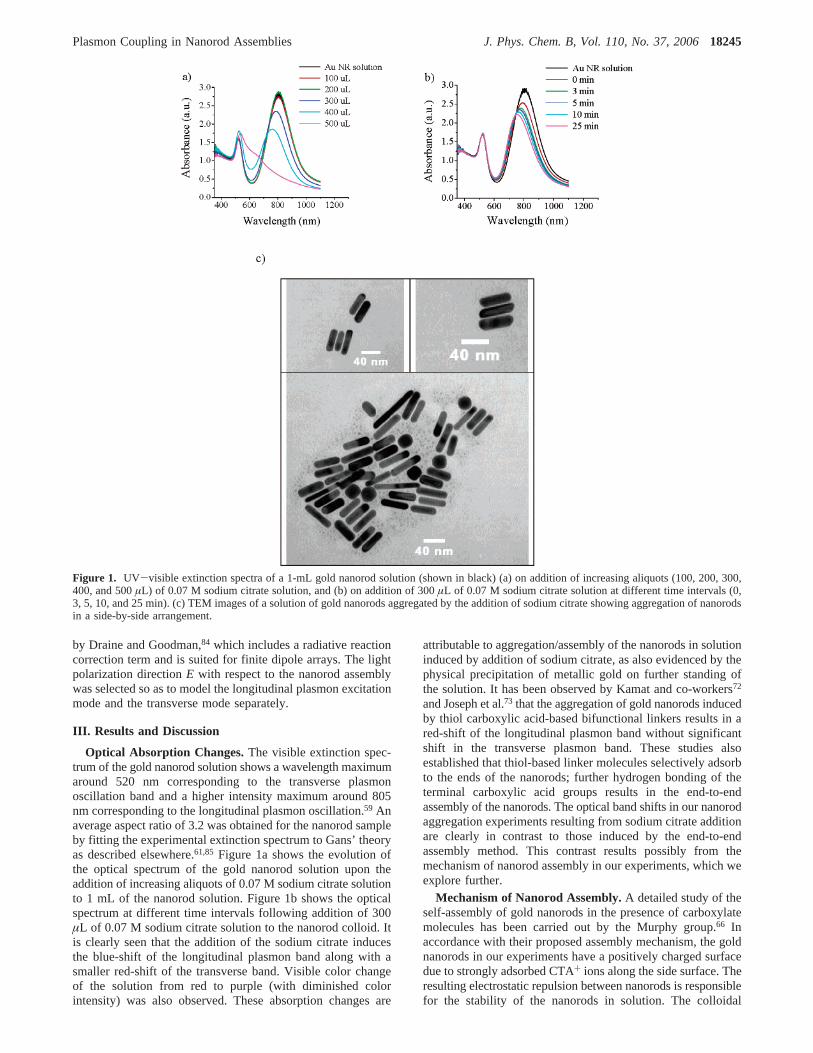
Optical Absorption Changes.The visible extinction spec-trum of the gold nanorod solution shows a wavelength maximumaround 520 nm corresponding to the transverse plasmonoscillation band and a higher intensity maximum around 805nm corresponding to the longitudinal plasmon oscillation.59 Anaverage aspect ratio of 3.2 was obtained for the nanorod sampleby fitting the experimental extinction spectrum to Gans’ theoryas described elsewhere.61,85 Figure 1a shows the evolution ofthe optical spectrum of the gold nanorod solution upon theaddition of increasing aliquots of 0.07 M sodium citrate solutionto 1 mL of the nanorod solution. Figure 1b shows the opticalspectrum at different time intervals following addition of 300µL of 0.07 M sodium citrate solution to the nanorod colloid. Itis clearly seen that the addition of the sodium citrate inducesthe blue-shift of the longitudinal plasmon band along with asmaller red-shift of the transverse band. Visible color changeof the solution from red to purple (with diminished colorintensity) was also observed. These absorption changes are
attributable to aggregation/assembly of the nanorods in solutioninduced by addition of sodium citrate, as also evidenced by thephysical precipitation of metallic gold on further standing ofthe solution. It has been observed by Kamat and co-workers72
and Joseph et al.73 that the aggregation of gold nanorods inducedby thiol carboxylic acid-based bifunctional linkers results in ared-shift of the longitudinal plasmon band without significantshift in the transverse plasmon band. These studies alsoestablished that thiol-based linker molecules selectively adsorbto the ends of the nanorods; further hydrogen bonding of theterminal carboxylic acid groups results in the end-to-endassembly of the nanorods. The optical band shifts in our nanorodaggregation experiments resulting from sodium citrate additionare clearly in contrast to those induced by the end-to-endassembly method. This contrast results possibly from themechanism of nanorod assembly in our experiments, which weexplore further.
Mechanism of Nanorod Assembly.A detailed study of theself-assembly of gold nanorods in the presence of carboxylatemolecules has been carried out by the Murphy group.66 Inaccordance with their proposed assembly mechanism, the goldnanorods in our experiments have a positively charged surfacedue to strongly adsorbed CTA+ ions along the side surface. Theresulting electrostatic repulsion between nanorods is responsiblefor the stability of the nanorods in solution. The colloidal
Figure 1. UV-visible extinction spectra of a 1-mL gold nanorod solution (shown in black) (a) on addition of increasing aliquots (100, 200, 300,400, and 500µL) of 0.07 M sodium citrate solution, and (b) on addition of 300µL of 0.07 M sodium citrate solution at different time intervals (0,3, 5, 10, and 25 min). (c) TEM images of a solution of gold nanorods aggregated by the addition of sodium citrate showing aggregation of nanorodsin a side-by-side arrangement.
Plasmon Coupling in Nanorod Assemblies J. Phys. Chem. B, Vol. 110, No. 37, 200618245
assembly of nanorods most likely results from the electrostaticattraction between the positively charged nanorod surface andthe negatively charged carboxylate ends of the citrate ions. Theadsorbed citrate ions may either bridge between nanorods, orlead to the formation of local negatively charged regions onthe nanorod that are electrostatically attracted to cationic regionson adjacent nanorods, or simply neutralize the electrostaticrepulsion between the nanorods. Such a mechanism is expectedto result predominantly in the side-by-side assembly of the goldnanorods.
Figure 1c shows examples of nanorods assembled side-by-side imaged by TEM. However, the TEM images are not entirelyrepresentative of the solution phase, due to the transfer of thesample to a planar solid support followed by evaporation ofthe solvent. The process of drying of a colloidal nanorod solutionon the TEM grid itself has been studied by Nikoobakht et al.68
and was found to result in the formation of side-by-side nanorodassemblies by the action of capillary forces. Such drying-mediated assemblies formed on the grids86,87 cannot be distin-guished from the assemblies formed in solution by the additionof the sodium citrate. Thus TEM, although a vital nanoparticlecharacterization tool, may not provide an accurate picture ofsolution phase behavior. Optical absorption measurements ofthe solution phase provide more definite characterization of thecolloidal assembly process, especially when the interpretationof the experimental absorption results is aided by electrodynamicsimulations of the optical spectra of model nanoparticle systems.We thus present electrodynamic simulations of the plasmon shiftresulting from the assembly of nanorods for both side-by-sideas well as end-to-end type assembly.
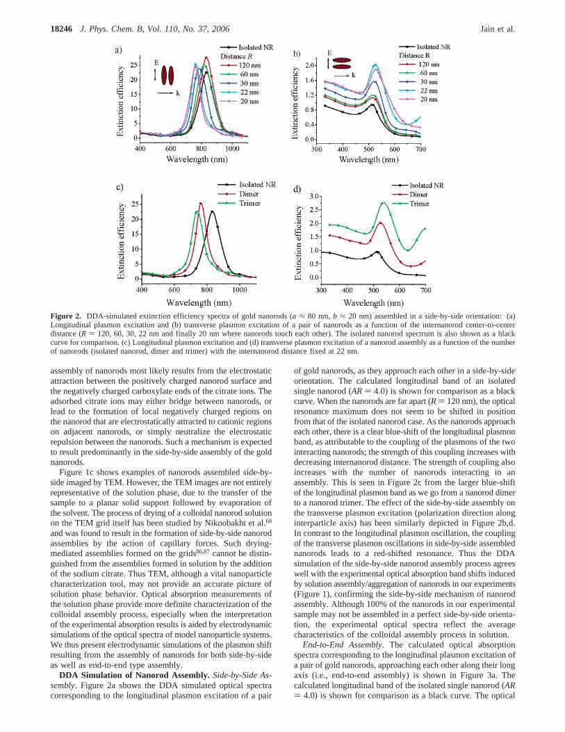
DDA Simulation of Nanorod Assembly.Side-by-Side As-sembly. Figure 2a shows the DDA simulated optical spectracorresponding to the longitudinal plasmon excitation of a pair
of gold nanorods, as they approach each other in a side-by-sideorientation. The calculated longitudinal band of an isolatedsingle nanorod (AR) 4.0) is shown for comparison as a blackcurve. When the nanorods are far apart (R) 120 nm), the opticalresonance maximum does not seem to be shifted in positionfrom that of the isolated nanorod case. As the nanorods approacheach other, there is a clear blue-shift of the longitudinal plasmonband, as attributable to the coupling of the plasmons of the twointeracting nanorods; the strength of this coupling increases withdecreasing internanorod distance. The strength of coupling alsoincreases with the number of nanorods interacting in anassembly. This is seen in Figure 2c from the larger blue-shiftof the longitudinal plasmon band as we go from a nanorod dimerto a nanorod trimer. The effect of the side-by-side assembly onthe transverse plasmon excitation (polarization direction alonginterparticle axis) has been similarly depicted in Figure 2b,d.In contrast to the longitudinal plasmon oscillation, the couplingof the transverse plasmon oscillations in side-by-side assemblednanorods leads to a red-shifted resonance. Thus the DDAsimulation of the side-by-side nanorod assembly process agreeswell with the experimental optical absorption band shifts inducedby solution assembly/aggregation of nanorods in our experiments(Figure 1), confirming the side-by-side mechanism of nanorodassembly. Although 100% of the nanorods in our experimentalsample may not be assembled in a perfect side-by-side orienta-tion, the experimental optical spectra reflect the averagecharacteristics of the colloidal assembly process in solution.
End-to-End Assembly. The calculated optical absorptionspectra corresponding to the longitudinal plasmon excitation ofa pair of gold nanorods, approaching each other along their longaxis (i.e., end-to-end assembly) is shown in Figure 3a. Thecalculated longitudinal band of the isolated single nanorod (AR) 4.0) is shown for comparison as a black curve. The optical
Figure 2. DDA-simulated extinction efficiency spectra of gold nanorods (a ≈ 80 nm,b ≈ 20 nm) assembled in a side-by-side orientation: (a)Longitudinal plasmon excitation and (b) transverse plasmon excitation of a pair of nanorods as a function of the internanorod center-to-centerdistance (R ) 120, 60, 30, 22 nm and finally 20 nm where nanorods touch each other). The isolated nanorod spectrum is also shown as a blackcurve for comparison. (c) Longitudinal plasmon excitation and (d) transverse plasmon excitation of a nanorod assembly as a function of the numberof nanorods (isolated nanorod, dimer and trimer) with the internanorod distance fixed at 22 nm.
18246 J. Phys. Chem. B, Vol. 110, No. 37, 2006 Jain et al.
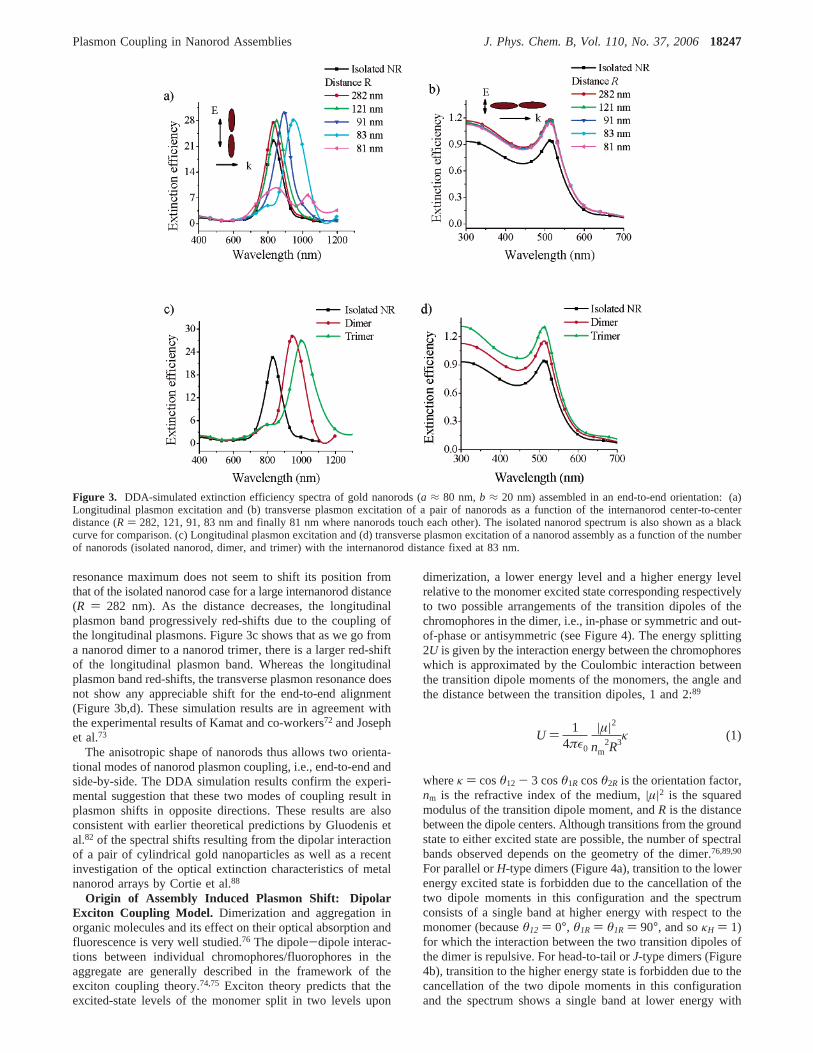
resonance maximum does not seem to shift its position fromthat of the isolated nanorod case for a large internanorod distance(R ) 282 nm). As the distance decreases, the longitudinalplasmon band progressively red-shifts due to the coupling ofthe longitudinal plasmons. Figure 3c shows that as we go froma nanorod dimer to a nanorod trimer, there is a larger red-shiftof the longitudinal plasmon band. Whereas the longitudinalplasmon band red-shifts, the transverse plasmon resonance doesnot show any appreciable shift for the end-to-end alignment(Figure 3b,d). These simulation results are in agreement withthe experimental results of Kamat and co-workers72 and Josephet al.73
The anisotropic shape of nanorods thus allows two orienta-tional modes of nanorod plasmon coupling, i.e., end-to-end andside-by-side. The DDA simulation results confirm the experi-mental suggestion that these two modes of coupling result inplasmon shifts in opposite directions. These results are alsoconsistent with earlier theoretical predictions by Gluodenis etal.82 of the spectral shifts resulting from the dipolar interactionof a pair of cylindrical gold nanoparticles as well as a recentinvestigation of the optical extinction characteristics of metalnanorod arrays by Cortie et al.88
Origin of Assembly Induced Plasmon Shift: DipolarExciton Coupling Model. Dimerization and aggregation inorganic molecules and its effect on their optical absorption andfluorescence is very well studied.76 The dipole-dipole interac-tions between individual chromophores/fluorophores in theaggregate are generally described in the framework of theexciton coupling theory.74,75 Exciton theory predicts that theexcited-state levels of the monomer split in two levels upon
dimerization, a lower energy level and a higher energy levelrelative to the monomer excited state corresponding respectivelyto two possible arrangements of the transition dipoles of thechromophores in the dimer, i.e., in-phase or symmetric and out-of-phase or antisymmetric (see Figure 4). The energy splitting2U is given by the interaction energy between the chromophoreswhich is approximated by the Coulombic interaction betweenthe transition dipole moments of the monomers, the angle andthe distance between the transition dipoles, 1 and 2:89
whereκ ) cosθ12 - 3 cosθ1R cosθ2R is the orientation factor,nm is the refractive index of the medium,|µ|2 is the squaredmodulus of the transition dipole moment, andR is the distancebetween the dipole centers. Although transitions from the groundstate to either excited state are possible, the number of spectralbands observed depends on the geometry of the dimer.76,89,90
For parallel orH-type dimers (Figure 4a), transition to the lowerenergy excited state is forbidden due to the cancellation of thetwo dipole moments in this configuration and the spectrumconsists of a single band at higher energy with respect to themonomer (becauseθ12 ) 0°, θ1R ) θ1R ) 90°, and soκH ) 1)for which the interaction between the two transition dipoles ofthe dimer is repulsive. For head-to-tail orJ-type dimers (Figure4b), transition to the higher energy state is forbidden due to thecancellation of the two dipole moments in this configurationand the spectrum shows a single band at lower energy with
Figure 3. DDA-simulated extinction efficiency spectra of gold nanorods (a ≈ 80 nm,b ≈ 20 nm) assembled in an end-to-end orientation: (a)Longitudinal plasmon excitation and (b) transverse plasmon excitation of a pair of nanorods as a function of the internanorod center-to-centerdistance (R ) 282, 121, 91, 83 nm and finally 81 nm where nanorods touch each other). The isolated nanorod spectrum is also shown as a blackcurve for comparison. (c) Longitudinal plasmon excitation and (d) transverse plasmon excitation of a nanorod assembly as a function of the numberof nanorods (isolated nanorod, dimer, and trimer) with the internanorod distance fixed at 83 nm.
U ) 14πε0
|µ|2nm
2R3κ (1)
Plasmon Coupling in Nanorod Assemblies J. Phys. Chem. B, Vol. 110, No. 37, 200618247
respect to the monomer (becauseθ12 ) θ1R ) θ1R ) 0° and soκJ ) -2) for which the interaction between dipoles is attractive.
By considering the nanorod plasmons to be effectivelyexcitons, the exciton coupling theory can used to explain theoptical spectra of the nanorod dimers. The side-by-side nanoroddimer, for the case of longitudinal polarization, represents anH aggregate and hence we observe a blue-shifted spectrum.Conversely, the end-to-end assembly is aJ aggregate, resultingin a red-shifted spectrum. For the transverse polarization, theside-by-side nanorod arrangement is aJ aggregate (providedthe polarization direction is along the interparticle axis) leadingto the red-shift and the end-to-end configuration is anHaggregate (however, the expected blue-shift of the transverseband may be too small to be observable because the transverseplasmon dipoles are far apart even when the rods touch eachother). Thus, the optical properties of the gold nanorodassemblies and their dependence on the assembly orientationseem to be qualitatively consistent with the exciton-couplingmodel.
Similar reasoning also applies to the polarization dependenceof the optical resonance shift in nanosphere dimers,41 where a
polarization along the interparticle axis (p-pol) results in a red-shift with respect to the single nanosphere resonance whereasa perpendicular polarization (s-pol) results in a blue-shift.(Supporting Information Figure S1) The near-infrared opticalresonances of silica-gold core-shell particles can also beexplained in terms of the exciton coupling between the plasmonsupported on the outer surface of a gold sphere and that on theinner surface of a cavity.91 In this case, the line along whichthe charges interact is parallel to the dipole moment vectors,corresponding to aJ-type symmetry resulting in a bonding orsymmetric resonance red-shifted with respect to the visibleresonance of the solid gold nanosphere (around 520 nm). Theantisymmetric coupling or antibonding resonance is also anallowed mode since the sphere and cavity plasmon dipolemoments do not cancel out but is expected to be on the UVend of the spectrum.
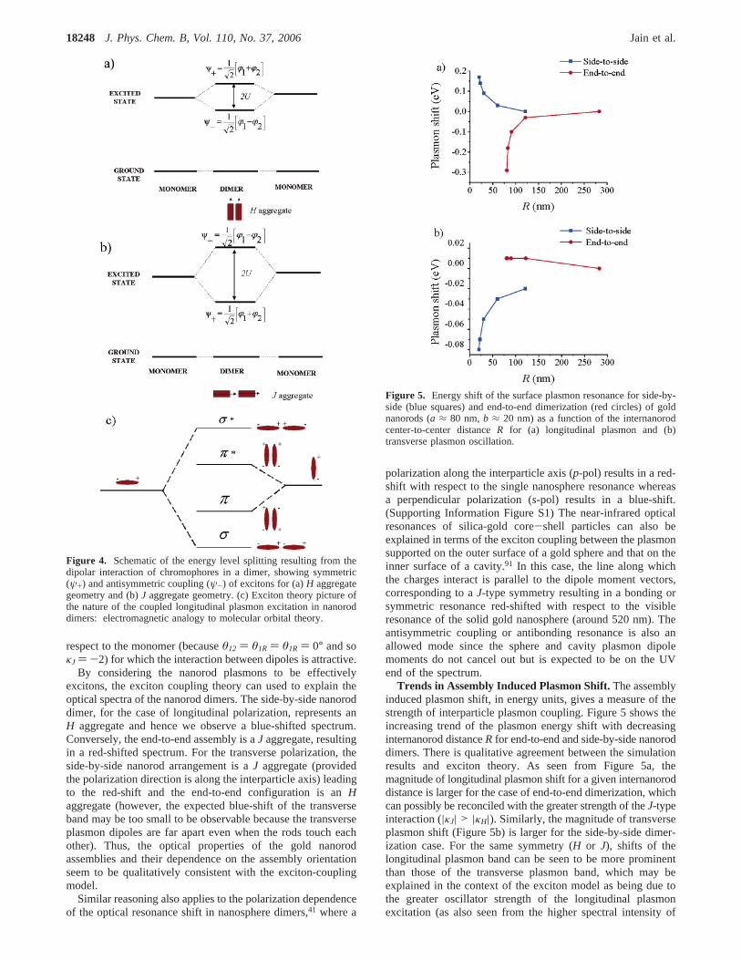
Trends in Assembly Induced Plasmon Shift.The assemblyinduced plasmon shift, in energy units, gives a measure of thestrength of interparticle plasmon coupling. Figure 5 shows theincreasing trend of the plasmon energy shift with decreasinginternanorod distanceR for end-to-end and side-by-side nanoroddimers. There is qualitative agreement between the simulationresults and exciton theory. As seen from Figure 5a, themagnitude of longitudinal plasmon shift for a given internanoroddistance is larger for the case of end-to-end dimerization, whichcan possibly be reconciled with the greater strength of theJ-typeinteraction (|κJ| > |κH|). Similarly, the magnitude of transverseplasmon shift (Figure 5b) is larger for the side-by-side dimer-ization case. For the same symmetry (H or J), shifts of thelongitudinal plasmon band can be seen to be more prominentthan those of the transverse plasmon band, which may beexplained in the context of the exciton model as being due tothe greater oscillator strength of the longitudinal plasmonexcitation (as also seen from the higher spectral intensity of
Figure 4. Schematic of the energy level splitting resulting from thedipolar interaction of chromophores in a dimer, showing symmetric(ψ+) and antisymmetric coupling (ψ-) of excitons for (a)H aggregategeometry and (b)J aggregate geometry. (c) Exciton theory picture ofthe nature of the coupled longitudinal plasmon excitation in nanoroddimers: electromagnetic analogy to molecular orbital theory.
Figure 5. Energy shift of the surface plasmon resonance for side-by-side (blue squares) and end-to-end dimerization (red circles) of goldnanorods (a ≈ 80 nm,b ≈ 20 nm) as a function of the internanorodcenter-to-center distanceR for (a) longitudinal plasmon and (b)transverse plasmon oscillation.
18248 J. Phys. Chem. B, Vol. 110, No. 37, 2006 Jain et al.
the longitudinal plasmon band as compared to transverseplasmon band). This makes nanorods much more attractive ascompared to nanospheres for analyte-sensing assays based onnanoparticle aggregation.15 Note that the dependence of theplasmon energy shift with internanorod distance does nothowever correlate with the 1/R3 dependence predicted by theexciton model (Equation 1). This may be because the exciton-coupling model only considers interactions between pointdipoles at relatively large inter-dipole distances; higher ordermultipole-multipole interactions are neglected in the weakinteraction limit. On the other hand, each nanorod target in ourelectrodynamic simulation is represented by a few thousanddipoles while accounting for the finite particle size effects (e.g.,electromagnetic retardation78,92) and the higher order interparticleinteractions43 (which become important in the strong couplinglimit and/or very small interparticle distances). Besides, due tothe finite size of the particles, the center-to-center distanceRdoes not represent the distance at which the charges inducedon neighboring particles interact.
Nature of the Coupled Plasmon Excitation: Electromag-netic Analogy to Molecular Orbitals. The effect of interparticlecoupling on the nature of plasmon excitation and electric-fieldenhancement is expected to have important consequences forthe use of nanorod assemblies in near-field-coupling basedapplications i.e., plasmon propagation24-29 and field-enhancedspectroscopy.92 The exciton model, in addition to the explanationof optical band shifts, also predicts the nature of the plasmonexcitation in the coupled system (see Figure 4c). As suggestedby the exciton model, the coupled longitudinal plasmon for theend-to-end dimer is bonding in nature analogous to the formationof a σ bond from twopz orbitals. The resulting electric fieldintensity is maximum in the junction between the interactingnanorods. On the other hand, the coupled longitudinal plasmonfor the side-by-side dimer has an antibonding nature analogousto the formation of aπ* bond frompx/y orbitals, with the electricfield concentrated on either side of the interparticle junction.
This contrast between the two modes of assembly is reinforcedby the following observation. In the case of the end-to-endassembly simulation, a new band emerges at higher energies(attributable to higher-order interactions or image interactions43)as the internanorod distance becomes very small93 (Figure 3a)or the number of nanorods interacting in an assembly increases(Figure 3c). The side-by-side assembly simulation does not showsuch an effect. The plasmons of closely spaced rods alignedend-to-end interact very strongly supporting an intense electricfield at the interparticle junction, consistent with an earliertheoretical study on electromagnetic coupling in large goldnanorods by Aizpurua et al.92
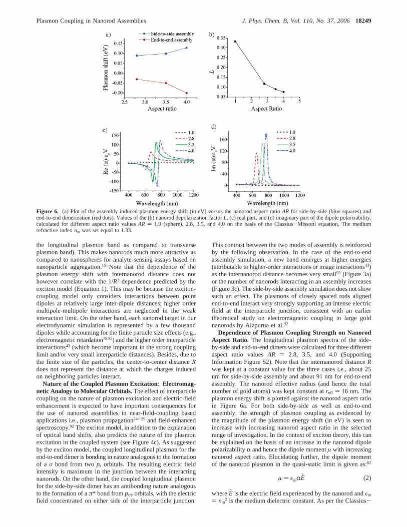
Dependence of Plasmon Coupling Strength on NanorodAspect Ratio. The longitudinal plasmon spectra of the side-by-side and end-to-end dimers were calculated for three differentaspect ratio valuesAR ) 2.8, 3.5, and 4.0 (SupportingInformation Figure S2). Note that the internanorod distanceRwas kept at a constant value for the three cases i.e., about 25nm for side-by-side assembly and about 91 nm for end-to-endassembly. The nanorod effective radius (and hence the totalnumber of gold atoms) was kept constant atreff ) 16 nm. Theplasmon energy shift is plotted against the nanorod aspect ratioin Figure 6a. For both side-by-side as well as end-to-endassembly, the strength of plasmon coupling as evidenced bythe magnitude of the plasmon energy shift (in eV) is seen toincrease with increasing nanorod aspect ratio in the selectedrange of investigation. In the context of exciton theory, this canbe explained on the basis of an increase in the nanorod dipolepolarizabilityR and hence the dipole momentµ with increasingnanorod aspect ratio. Elucidating further, the dipole momentof the nanorod plasmon in the quasi-static limit is given as:82
whereE is the electric field experienced by the nanorod andεm
) nm2 is the medium dielectric constant. As per the Classius-
Figure 6. (a) Plot of the assembly induced plasmon energy shift (in eV) versus the nanorod aspect ratioAR for side-by-side (blue squares) andend-to-end dimerization (red dots). Values of the (b) nanorod depolarization factorL, (c) real part, and (d) imaginary part of the dipole polarizability,calculated for different aspect ratio valuesAR ) 1.0 (sphere), 2.8, 3.5, and 4.0 on the basis of the Classius-Missotti equation. The mediumrefractive indexnm was set equal to 1.33.
µ ) εmRE (2)
Plasmon Coupling in Nanorod Assemblies J. Phys. Chem. B, Vol. 110, No. 37, 200618249
Mossoti relation54 for the polarizability corresponding to thelongitudinal excitation:
whereV is the nanorod volume,ε0 is the vacuum permittivityandε ) ε1 + iε2 is the complex dielectric function of gold.Lis the depolarization factor corresponding to the long-axis andcan be expressed as a function of the nanorod aspect ratioARas:56,82
The value ofL is 1/3 for a sphere and decreases with increasingnanorod aspect ratio (Figure 6b). The resonance condition fromeq 2 is given by:
This is providedε2 is small or weakly dependent on frequency,54
which is valid in the 600-850 nm region for gold.83 Equation6 explains the red-shift of the longitudinal plasmon resonancewith increasing aspect ratio since the negative real part of thedielectric functionε1 for gold increases with increasing wave-length.83 At resonance,R is given as:
The rapid decrease inL with increasing aspect ratio against aslight increase in the imaginary part of the dielectric constantwith wavelength (in the region of investigation) predicts a largerpolarizability for higher aspect ratios. This is depicted in thecalculated plots of the real and imaginary parts of the nanoroddipole polarizability for the different aspect ratio values (Figure6c,d). The explanation of increasing plasmon coupling strengthwith increasing nanorod aspect ratio on the basis of thequasistatic and dipole approximations serves only as a qualitativeone since finite particle size (as compared to the wavelength oflight) effects78,92and higher order interactions43 are not included.
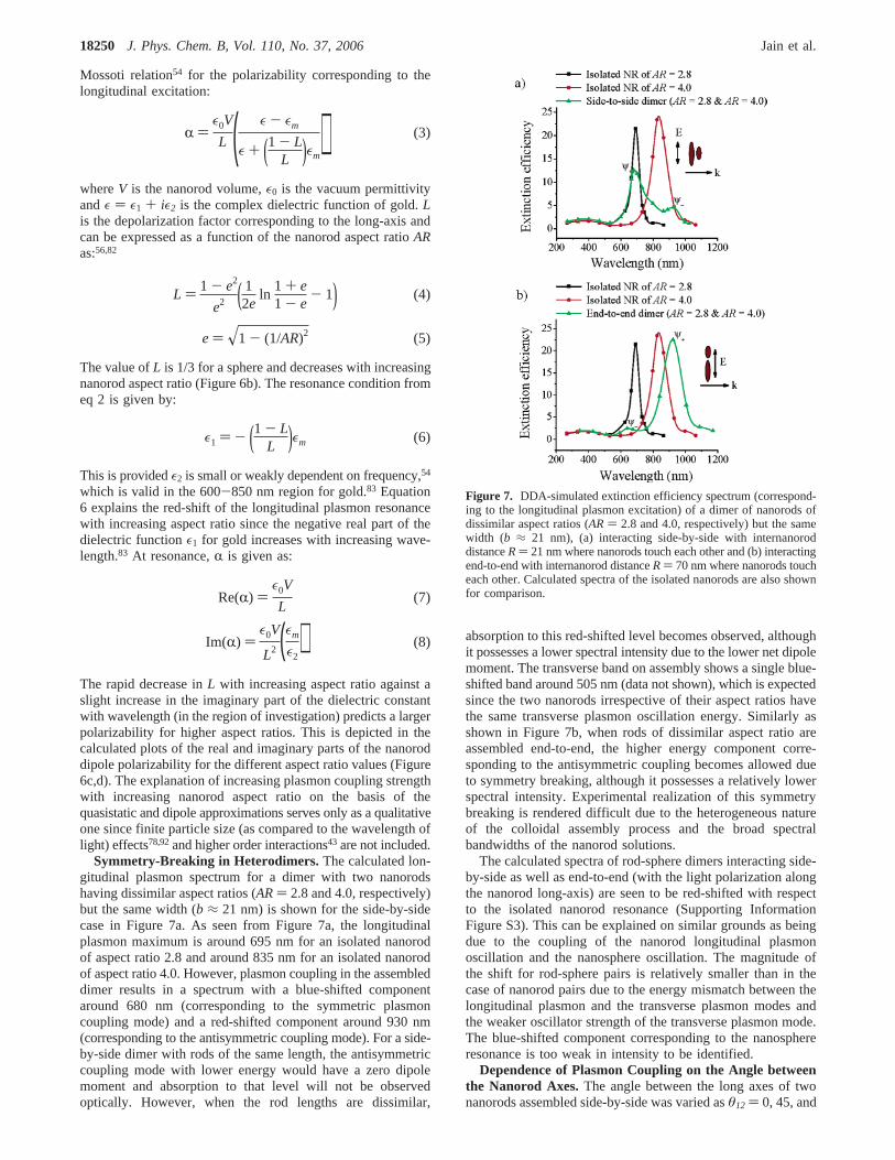
Symmetry-Breaking in Heterodimers.The calculated lon-gitudinal plasmon spectrum for a dimer with two nanorodshaving dissimilar aspect ratios (AR) 2.8 and 4.0, respectively)but the same width (b ≈ 21 nm) is shown for the side-by-sidecase in Figure 7a. As seen from Figure 7a, the longitudinalplasmon maximum is around 695 nm for an isolated nanorodof aspect ratio 2.8 and around 835 nm for an isolated nanorodof aspect ratio 4.0. However, plasmon coupling in the assembleddimer results in a spectrum with a blue-shifted componentaround 680 nm (corresponding to the symmetric plasmoncoupling mode) and a red-shifted component around 930 nm(corresponding to the antisymmetric coupling mode). For a side-by-side dimer with rods of the same length, the antisymmetriccoupling mode with lower energy would have a zero dipolemoment and absorption to that level will not be observedoptically. However, when the rod lengths are dissimilar,
absorption to this red-shifted level becomes observed, althoughit possesses a lower spectral intensity due to the lower net dipolemoment. The transverse band on assembly shows a single blue-shifted band around 505 nm (data not shown), which is expectedsince the two nanorods irrespective of their aspect ratios havethe same transverse plasmon oscillation energy. Similarly asshown in Figure 7b, when rods of dissimilar aspect ratio areassembled end-to-end, the higher energy component corre-sponding to the antisymmetric coupling becomes allowed dueto symmetry breaking, although it possesses a relatively lowerspectral intensity. Experimental realization of this symmetrybreaking is rendered difficult due to the heterogeneous natureof the colloidal assembly process and the broad spectralbandwidths of the nanorod solutions.
The calculated spectra of rod-sphere dimers interacting side-by-side as well as end-to-end (with the light polarization alongthe nanorod long-axis) are seen to be red-shifted with respectto the isolated nanorod resonance (Supporting InformationFigure S3). This can be explained on similar grounds as beingdue to the coupling of the nanorod longitudinal plasmonoscillation and the nanosphere oscillation. The magnitude ofthe shift for rod-sphere pairs is relatively smaller than in thecase of nanorod pairs due to the energy mismatch between thelongitudinal plasmon and the transverse plasmon modes andthe weaker oscillator strength of the transverse plasmon mode.The blue-shifted component corresponding to the nanosphereresonance is too weak in intensity to be identified.
Dependence of Plasmon Coupling on the Angle betweenthe Nanorod Axes.The angle between the long axes of twonanorods assembled side-by-side was varied asθ12 ) 0, 45, and
R )ε0V
L ( ε - εm
ε + (1 - LL )εm) (3)
L ) 1 - e2
e2 ( 12e
ln1 + e1 - e
- 1) (4)
e ) x1 - (1/AR)2 (5)
ε1 ) - (1 - LL )εm (6)
Re(R) )ε0V
L(7)
Im(R) )ε0V
L2 (εm
ε2) (8)
Figure 7. DDA-simulated extinction efficiency spectrum (correspond-ing to the longitudinal plasmon excitation) of a dimer of nanorods ofdissimilar aspect ratios (AR ) 2.8 and 4.0, respectively) but the samewidth (b ≈ 21 nm), (a) interacting side-by-side with internanoroddistanceR) 21 nm where nanorods touch each other and (b) interactingend-to-end with internanorod distanceR) 70 nm where nanorods toucheach other. Calculated spectra of the isolated nanorods are also shownfor comparison.
18250 J. Phys. Chem. B, Vol. 110, No. 37, 2006 Jain et al.
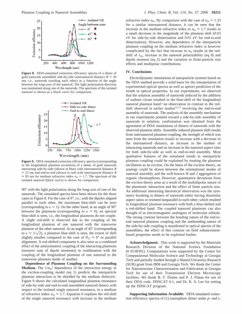
90° with the light polarization along the long-axis of one of thenanorods. The simulated spectra have been shown for the threecases in Figure 8. For the case of 0°, i.e., with the dipoles alignedparallel to each other, the maximum blue-shift can be seen(corresponding toκ ) 1). On the other hand, at an angle of 90°between the plasmons (corresponding toκ ) 0), no spectralblue-shift is seen, i.e., the longitudinal plasmons do not couple.A slight red-shift is observed due to the coupling of thelongitudinal plasmon of one nanorod with the transverseplasmon of the other nanorod. At an angle of 45° (correspondingto κ ) 1/x2), a plasmon blue-shift is seen, the extent of shiftslightly smaller compared to the case ofθ12 ) 0° or parallelalignment. A red-shifted component is also seen as a combinedeffect of the antisymmetric coupling of the interacting plasmons(nonzero sum of dipole moments) in combination with thecoupling of the longitudinal plasmon of one nanorod to thetransverse plasmon mode of another.
Dependence of Plasmon Coupling on the SurroundingMedium. The 1/nm
2 dependence of the interaction energy inthe exciton-coupling model (eq 1) predicts the interparticleplasmon interaction to be shielded by the medium dielectric.Figure 9 shows the calculated longitudinal plasmon resonanceof side-by-side and end-to-end assembled nanorod dimers withrespect to the isolated single nanorod resonance, in a mediumof refractive indexnm ) 1.7. Equation 6 explains the red-shiftof the single nanorod resonance with increase in the medium
refractive indexnm. By comparison with the case ofnm ) 1.33for a similar internanorod distance, it can be seen that theincrease in the medium refractive index tonm ) 1.7 results ina small decrease in the magnitude of the plasmon shift (0.03eV for side-by-side dimerization and 0.01 eV for end-to-enddimerization). However, any dependence of the interparticleplasmon coupling on the medium refractive index is howevercomplicated by the fact that increase innm results in the red-shift of λm, increase in the nanorod polarizability (eq 8) anddipole moment (eq 2) and the variation in finite-particle sizeeffects and multipolar contributions.
IV. Conclusions
Electrodynamic simulations of nanoparticle systems based onthe DDA method provide a solid basis for the interpretation ofexperimental optical spectra as well as apriori prediction of thetrends in optical properties. In our experiments, we observedthat the solution assembly of nanorods induced by the additionof sodium citrate resulted in the blue-shift of the longitudinalnanorod plasmon bandsan observation in contrast to the red-shift observed in earlier studies72,73 involving the end-to-endassembly of nanorods. The analysis of the assembly mechanismin our experiments pointed toward a side-by-side assembly ofnanorods in solution; confirmation was obtained from theagreement of DDA simulations of dimers of nanorods with theobserved plasmon shifts. Assembly induced plasmon shift resultsfrom internanorod plasmon coupling, the strength of which wasseen from the simulation results to increase with a decrease inthe internanorod distance, an increase in the number ofinteracting nanorods and an increase in the nanorod aspect ratiofor both side-by-side as well as end-to-end assembly. Mostqualitative features of the simulated trends in interparticleplasmon coupling could be explained by treating the plasmonoscillation as an exciton. On the basis of the exciton theory, ananalogy could be drawn between the orientational modes ofnanorod assembly and the well-knownH andJ aggregation oforganic chromophores. However, quantitative deviations fromthe exciton theory arise as a result of the multidipolar nature ofthe plasmonic interaction and the effect of finite particle size.An additional interesting theoretical observation was the sym-metry breaking in dimers of nanorods either having dissimilaraspect ratios or oriented nonparallel to each other, which resultedin longitudinal plasmon resonance with both a blue-shifted anda red-shifted band. The coupled nanorod plasmons can alsothought of as electromagnetic analogues of molecular orbitals.The strong contrast between the bonding nature of the end-to-end nanorod plasmon coupling and the antibonding nature ofthe side-by-side coupling is manifested in optical spectra of theassemblies; the effect of this contrast on field enhancement-based properties needs to be exploited further.
Acknowledgment. This work is supported by the MaterialsResearch Division of the National Science Foundation(# 0138391). Computations were supported by the Center forComputational Molecular Science and Technology at GeorgiaTech and partially funded through a Shared University Research(SUR) grant from IBM and Georgia Tech. We thank the Centerfor Nanostructure Characterization and Fabrication at GeorgiaTech for use of their Transmission Electron Microscopyfacilities. We thank B. T. Draine and P. J. Flatau for use oftheir DDA code, DDSCAT 6.1, and Dr. K. S. Lee for settingup the DDSCAT program.
Supporting Information Available: DDA-simulated extinc-tion efficiency spectra of (1) nanosphere dimer underp- ands-
Figure 8. DDA-simulated extinction efficiency spectra of a dimer ofgold nanorods assembled side-by-side (internanorod distanceR ) 20nm, i.e., nanorods touching each other) as a function of the anglebetween the long-axes of the nanorod. The light polarization directionwas maintained along one of the nanorods. The spectrum of the isolatednanorod is shown as a black curve for comparison.
Figure 9. DDA-simulated extinction efficiency spectra (correspondingto the longitudinal plasmon excitation) of a dimer of gold nanorodsinteracting side-by-side (shown in blue) with internanorod distanceR) 22 nm and end-to-end (shown in red) with internanorod distanceR) 83 nm for medium refractive indexnm ) 1.7. The spectrum of theisolated nanorod (black curve) is shown for comparison.
Plasmon Coupling in Nanorod Assemblies J. Phys. Chem. B, Vol. 110, No. 37, 200618251
polarizations, (2) nanorod dimer for different aspect ratios (AR) 2.8, 3.5 and 4.0), and (3) rod-sphere dimer for side-to-sideand end-to-end assembly. This material is available free ofcharge via the Internet at http://pubs.acs.org.
References and Notes
(1) Mann, S.; Shenton, W.; Li, M.; Connolly, S.; Fitzmaurice, D.AdV.Mater. (Weinheim, Ger.)2000, 12, 147.
(2) Mirkin, C. A. Inorg. Chem.2000, 39, 2258.(3) Murphy, C. J.; Sau, T. K.; Gole, A. M.; Orendorff, C. J.; Gao, J.;
Gou, L.; Hunyadi, S. E.; Li, T.J. Phys. Chem. B2005, 109, 13857.(4) Pileni, M. P.J. Phys. Chem. B2001, 105, 3358.(5) Pileni, M. P.Appl. Surface Sci.2001, 171, 1.(6) Pileni, M. P.J. Phys.: Condens. Matter2006, 18, S67.(7) Pileni, M. P.; Lalatonne, Y.; Ingert, D.; Lisiecki, I.; Courty, A.
Faraday Discuss.2003, 125, 251.(8) Storhoff, J. J.; Mirkin, C. A.Chem. ReV. (Washington, D. C.)1999,
99, 1849.(9) Teranishi, T.; Miyake, M.Encycl. Nanosci. Nanotechnol.2004, 5,
421.(10) Davis, S. A.; Breulmann, M.; Rhodes, K. H.; Zhang, B.; Mann, S.
Chem. Mater.2001, 13, 3218.(11) Alivisatos, A. P.; Johnsson, K. P.; Peng, X.; Wilson, T. E.; Loweth,
C. J.; Bruchez, M. P., Jr.; Schultz, P. G.Nature (London)1996, 382, 609.(12) Mirkin, C. A.; Letsinger, R. L.; Mucic, R. C.; Storhoff, J. J.Nature
(London)1996, 382, 607.(13) Alivisatos, P.Nat. Biotechnol.2004, 22, 47.(14) Shenton, W.; Davis, S. A.; Mann, S.AdV. Mater. (Weinheim, Ger.)
1999, 11, 449.(15) Sudeep, P. K.; Joseph, S. T. S.; Thomas, K. G.J. Am. Chem. Soc.
2005, 127, 6516.(16) Elghanian, R.; Storhoff, J. J.; Mucic, R. C.; Letsinger, R. L.; Mirkin,
C. A. Science (Washington, D. C.)1997, 277, 1078.(17) Ando, M.; Kawasaki, M.; Imazeki, S.; Sasaki, H.; Kamata, T.Appl.
Phys. Lett.2004, 85, 1849.(18) Feldheim, D. L.; Foss, C. A., Jr.Met. Nanopart.2002, 1.(19) Hu, M.-S.; Chen, H.-L.; Shen, C.-H.; Hong, L.-S.; Huang, B.-R.;
Chen, K.-H.; Chen, L.-C.Nat. Mater.2006, 5, 102.(20) Jin, Y.; Friedman, N.J. Am. Chem. Soc.2005, 127, 11902.(21) Maheshwari, V.; Saraf, R. F.Science (Washington, D. C.)2006,
312, 1501.(22) Tan, Y.; Li, Y.; Zhu, D.Encyclo. Nanosci. Nanotechnol.2004, 8,
9.(23) Pillai, S.; Catchpole, K. R.; Trupke, T.; Zhang, G.; Zhao, J.; Green,
M. A. Appl. Phys. Lett.2006, 88, 161102/1.(24) Krenn, J. R.; Salerno, M.; Felidj, N.; Lamprecht, B.; Schider, G.;
Leitner, A.; Aussenegg, F. R.; Weeber, J. C.; Dereux, A.; Goudonnet, J. P.J. Microsc. (Oxford)2001, 202, 122.
(25) Maier, S. A.; Atwater, H. A.J. Appl. Phys.2005, 98, 011101/1.(26) Maier, S. A.; Brongersma, M. L.; Kik, P. G.; Meltzer, S.; Requicha,
A. A. G.; Atwater, H. A. AdV. Mater. (Weinheim, Ger.)2001, 13, 1501.(27) Maier, S. A.; Friedman, M. D.; Barclay, P. E.; Painter, O.Appl.
Phys. Lett.2005, 86, 071103/1.(28) Maier, S. A.; Kik, P. G.; Atwater, H. A.; Meltzer, S.; Harel, E.;
Koel, B. E.; Requicha, A. A. G.Nat. Mater.2003, 2, 229.(29) Quinten, M.; Leitner, A.; Krenn, J. R.; Aussenegg, F. R.Opt. Lett.
1998, 23, 1331.(30) Barnes, W. L.; Dereux, A.; Ebbesen, T. W.Nature (London)2003,
424, 824.(31) Feldstein, M. J.; Keating, C. D.; Liau, Y.-H.; Natan, M. J.; Scherer,
N. F. J. Am. Chem. Soc.1997, 119, 6638.(32) Seker, F.; Malenfant, P. R. L.; Larsen, M.; Alizadeh, A.; Conway,
K.; Kulkarni, A. M.; Goddard, G.; Garaas, R.AdV. Mater. (Weinheim, Ger.)2005, 17, 1941.
(33) Liz-Marzan, L. M.Langmuir2006, 22, 32.(34) Grant, C. D.; Schwartzberg, A. M.; Norman, T. J., Jr.; Zhang, J.
Z. J. Am. Chem. Soc.2003, 125, 549.(35) Lazarides, A. A.; Schatz, G. C.J. Phys. Chem. B2000, 104, 460.(36) Lin, S.; Li, M.; Dujardin, E.; Girard, C.; Mann, S.AdV. Mater.
(Weinheim, Ger.)2005, 17, 2553.(37) Jain, P. K.; Qian, W.; El-Sayed, M. A.J. Phys. Chem. B2006,
110, 136.(38) Storhoff, J. J.; Lazarides, A. A.; Mucic, R. C.; Mirkin, C. A.;
Letsinger, R. L.; Schatz, G. C.J. Am. Chem. Soc.2000, 122, 4640.(39) Su, K. H.; Wei, Q. H.; Zhang, X.; Mock, J. J.; Smith, D. R.; Schultz,
S. Nano Lett.2003, 3, 1087.(40) Soennichsen, C.; Reinhard, B. M.; Liphardt, J.; Alivisatos, A. P.
Nat. Biotechnol.2005, 23, 741.
(41) Rechberger, W.; Hohenau, A.; Leitner, A.; Krenn, J. R.; Lamprecht,B.; Aussenegg, F. R.Opt. Commun.2003, 220, 137.
(42) Nordlander, P.; Oubre, C.; Prodan, E.; Li, K.; Stockman, M. I.NanoLett. 2004, 4, 899.
(43) Xiao, J. J.; Huang, J. P.; Yu, K. W.Phys. ReV. B: Condens. MatterMater. Phys.2005, 71, 045404/1.
(44) Haynes, C. L.; McFarland, A. D.; Zhao, L.; Van Duyne, R. P.;Schatz, G. C.; Gunnarsson, L.; Prikulis, J.; Kasemo, B.; Kaell, M.J. Phys.Chem. B2003, 107, 7337.
(45) Sweatlock, L. A.; Maier, S. A.; Atwater, H. A.; Penninkhof, J. J.;Polman, A.Phys. ReV. B: Condens. Matter Mater. Phys.2005, 71, 235408/1.
(46) Hao, E.; Schatz George, C.J. Chem. Phys.2004, 120, 357.(47) Jiang, J.; Bosnick, K.; Maillard, M.; Brus, L.J. Phys. Chem. B
2003, 107, 9964.(48) Michaels, A. M.; Jiang, J.; Brus, L.J. Phys. Chem. B2000, 104,
11965.(49) Ritchie, G.; Burstein, E.Phys. ReV. B: Condens. Matter Mater.
Phys.1981, 24, 4843.(50) Maillard, M.; Monchicourt, P.; Pileni, M. P.Chem. Phys. Lett.2003,
380, 704.(51) Shalaev, V. M.; Poliakov, E. Y.; Markel, V. A.Phys. ReV. B:
Condens. Matter Mater. Phys.1996, 53, 2437.(52) Markel, V. A.; Shalaev, V. M.; Zhang, P.; Huynh, W.; Tay, L.;
Haslett, T. L.; Moskovits, M.Phys. ReV. B: Condens. Matter Mater. Phys.1999, 59, 10903.
(53) Link, S.; El-Sayed, M. A.J. Phys. Chem. B1999, 103, 8410.(54) Kreibig, U.; Vollmer, M.Optical Properties of Metal Clusters,
Series in Materials Science 25; Springer: New York, 1995.(55) Gans, R.Ann. Phys. (Berlin)1915, 47, 270.(56) Link, S.; Mohamed, M. B.; El-Sayed, M. A.J. Phys. Chem. B1999,
103, 3073.(57) Link, S.; El-Sayed, M. A.; Mohamed, M. B.J. Phys. Chem. B2005,
109, 10531.(58) Jain, P. K.; Lee, K. S.; El-Sayed, I. H.; El-Sayed, M. A.J. Phys.
Chem. B2006, 110, 7238.(59) Gans, R. V.Ann. Phys. (Berlin)1912, 37, 881.(60) Mohamed, M. B.; Volkov, V.; Link, S.; El-Sayed, M. A.Chem.
Phys. Lett.2000, 317, 517.(61) Eustis, S.; El-Sayed, M.J. Phys. Chem. B2005, 109, 16350.(62) Nikoobakht, B.; El-Sayed, M. A.J. Phys. Chem. A2003, 107, 3372.(63) Nikoobakht, B.; Wang, J. P.; El-Sayed, M. A.Chem. Phys. Lett.
2002, 366, 17.(64) Dickson, R. M.; Lyon, L. A.J. Phys. Chem. B2000, 104, 6095.(65) Murphy, C. J.; Jana, N. R.; Gearheart, L. A.; Obare, S. O.; Caswell,
K. K.; Mann, S.; Johnson, C. J.; Davis, S. A.; Dujardin, E.; Edler, K. J.Chem. Nanomater.2004, 1, 285.
(66) Orendorff, C. J.; Hankins, P. L.; Murphy, C. J.Langmuir 2005,21, 2022.
(67) Jana, N. R.; Gearheart, L. A.; Obare, S. O.; Johnson, C. J.; Edler,K. J.; Mann, S.; Murphy, C. J.J. Mater. Chem.2002, 12, 2909.
(68) Nikoobakht, B.; Wang, Z. L.; El-Sayed, M. A.J. Phys. Chem. B2000, 104, 8635.
(69) Gole, A.; Murphy, C. J.Langmuir2005, 21, 10756.(70) Caswell, K. K.; Wilson, J. N.; Bunz, U. H. F.; Murphy, C. J.J.
Am. Chem. Soc.2003, 125, 13914.(71) Dujardin, E.; Mann, S.; Hsin, L.-B.; Wang, C. R. C.Chem.
Commun. (Cambridge)2001, 1264.(72) Thomas, K. G.; Barazzouk, S.; Ipe, B. I.; Joseph, S. T. S.; Kamat,
P. V. J. Phys. Chem. B2004, 108, 13066.(73) Joseph, S. T. S.; Ipe, B. I.; Pramod, P.; Thomas, K. G.J. Phys.
Chem. B2006, 110, 150.(74) Kasha, M.Radiat. Res.1963, 20, 55.(75) Kasha, M.; Rawls, H. R.; El-Bayoumi, M. A.Pure Appl. Chem.
1965, 11, 371.(76) Valdes-Aguilera, O.; Neckers, D. C.Acc. Chem. Res.1989, 22,
171.(77) Draine, B. T.; Flatau, P. J.J. Opt. Soc. Am. A1994, 11, 1491.(78) Kelly, K. L.; Coronado, E.; Zhao, L. L.; Schatz, G. C.J. Phys.
Chem. B2003, 107, 668.(79) Schatz, G. C.Theochem2001, 573, 73.(80) Brioude, A.; Jiang, X. C.; Pileni, M. P.J. Phys. Chem. B2005,
109, 13138.(81) Lee, K.-S.; El-Sayed, M. A.J. Phys. Chem. B2005, 109, 20331.(82) Gluodenis, M.; Foss, C. A., Jr.J. Phys. Chem. B2002, 106, 9484.(83) Johnson, P. B.; Christy, R. W.Phys. ReV. B: Condens. Matter
Mater. Phys.1972, 6, 4370.(84) Draine, B. T.; Goodman, J.Astrophys. J.1993, 405, 685.(85) Eustis, S.; El-Sayed, M. A.J. Appl. Phys.2006, in press.(86) Taleb, A.; Petit, C.; Pileni, M. P.Chem. Mater.1997, 9, 950.
18252 J. Phys. Chem. B, Vol. 110, No. 37, 2006 Jain et al.

(87) Motte, L.; Billoudet, F.; Lacaze, E.; Douin, J.; Pileni, M. P.J. Phys.Chem. B1997, 101, 138.
(88) Cortie, M. B.; Xu, X.; Ford, M. J.Phys. Chem. Chem. Phys.2006,8, 3520.
(89) Packard, B. Z.; Toptygin, D. D.; Komoriya, A.; Brand, L.J. Phys.Chem. B1998, 102, 752.
(90) Kasha, M.Phys. Process. Radiation Biol., Proc. Intern. Symp.,Mich. State UniV. 1964, 17.
(91) Prodan, E.; Radloff, C.; Halas, N. J.; Nordlander, P.Science(Washington, D. C.)2003, 302, 419.
(92) Aizpurua, J.; Bryant, G. W.; Richter, L. J.; Garcia de Abajo, F. J.;Kelley, B. K.; Mallouk, T. Phys. ReV. B: Condens. Matter Mater. Phys.2005, 71, 235420/1.
(93) The spectral band structure is significantly altered when the twonanorods are touching each other end-to-end (Figure 5a,R ) 81 nm).Interesting spectral characteristics of particles touching each other havealready been investigated by Atwater and co-workers (ref 45). At the sametime, exceptions to the applicability of electrodynamic simulations toparticles touching each other have also been cited (ref 46).
Plasmon Coupling in Nanorod Assemblies J. Phys. Chem. B, Vol. 110, No. 37, 200618253
Copyright © 2022 FDOKUMEN