PD2299 – Easier Plant Design Using Autodesk® Plant Design ...
Upload
independentCategory
view
2download
0
Pergamon 0031-9422 (95) 00243--X Phytochemistry, Vol. 40, No. 2. pp. 355 365. 1995 Elsevier Science Ltd
Printed in Great Britain 0031 9422/95 $9.50 + 0.00
REVIEW ARTICLE NUMBER 107
PLANT TRANSGLUTAMINASES
DONATELLA SERAFINI-FRACASSINI, STEFANO DEL DUCA and SIMONE BENINATI*
Department of Biology E. S., University of Bologna, via Irnerio 42, 40126 Bologna, Italy; * Department of Biology, lI University of Roma 'Tor Vergata', via della Ricerca Scientifica, 00173 Roma, Italy
(Received21 February 1995)
Key Word lndex--Polyamines; protein modification; transglutaminases; putrescine; spermidine; sper- mine.
Abstract--The identification procedures, the characteristics and the potential function of the recently detected plant transglutaminases, are discussed in the light of the knowledge of animal transglutaminases. The enzyme has been studied occasionally in lower organisms (bacteria, fungi and green algae) and more extensively in Angiosperms.
I N T R O D U C T I O N
Cross-linking of proteins, an event which occurs post- translationally, is one of the vital physiological processes involved in the stabilization of tissue and cellular ma- trices. Among the various cross-links identified, e-(?- glutamyl)lysine and N,N-bis(7-glutamyl)amine are the most abundant ones formed by enzymatic catalysis. The formation of these two types of cross-links is catalysed by Ca 2 +-dependent acyl transferases (glutaminyl-peptide ?- glutamyltransferase, EC 2.3.2.13), known as trans- glutaminases (TGases). In the reaction catalysed by TGases the ?-carboxamide groups of peptide-bound glutamine residues are the acyl donors while primary amino groups in a variety of compounds may function as acyl acceptors with the subsequent formation of a mono-substituted ?-amide of peptide-bound glutamic acid. At less than saturating levels of a suitable primary amine or in its absence of an amine, water can act as the acyl acceptor with the formation of peptide-bound glutamic acid (Fig. 1).
Based on their distinct catalytic characteristics and distribution, several forms of transglutaminase have been identified to date and they have been found to exhibit differences in specificity. These differences are expressed in terms of variations in susceptibility of glutamine resi- dues to catalytic modification and appear to be depen- dent, at least in part, upon amino acid residues surround- ing a given glutamine. In contrast to their limited glutamine substrate specificity, TGases possess an excep- tionally wide specificity for amine substrates. Among the latter, aliphatic polyamines (PA) are the more extensively studied. They are distributed in all living organisms, of which they regulate the growth [1, 2]. The most common ones are putrescine (1,4-diaminobutane) (PU), sper- midine (N-[3-aminopropyl]-l,4-butanediamine) (SD) and spermine (N,N'-bis[3-aminopropyl]-l ,4-butanedi-
amine) (SM) (Fig. 2). PU has an aliphatic tetramethylene backbone deriving directly from ornithine and indirectly from arginine depending on the organism. The biosynth- esis of higher polyamines occurs by the addition of one or two aminopropyl groups to PU and thus SD and SM, respectively, are formed; these higher PA also bear proto- nated internal iminic groups. They are organic polyca- tions at physiological pH, in fact the values Of their pK are: PU pK2 = 9.04, SD pK3 = 9.52 and SM pK4 = 8.9. [3]. PA are linear and flexible molecules of varying length and their backbone possesses low steric hindrance. Thus, they can interact precisely with several types of molecules and thereby affect their structure and function.
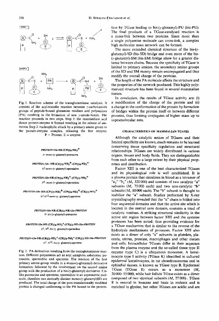
PA are covalently bound to proteins by TGase through a two-step reaction: attachment of the first pri- mary amino group of the PA to the acyl donor substrate to form mono-(y-glutamyl)-PA (mono-PA) and the bind- ing of the second amine group to another glutamine residue to form bis-(?-glutamyl)-PA (bis-PA) (Fig. 2) [4]. The first step is regulated by the PA concentration, since high levels of PA may saturate the acyl donor residues of the substrate proteins, thus preventing the second step.
TGases exhibit a different affinity for amine substrates: SD is preferred to SM and the latter is preferred to PU. This was observed in Helianthus tuberosus L. cv OB 1 etiolated sprout apices [5] and chloroplasts [6], in Physarum polycephalum M3cV spherules [7] and in rat sperm [8].
The various ?-glutamyl-PA derivatives differ signifi- cantly under several aspects. Mono-(y-glutamyl)-PU (mono-PU) confers to the modified protein an additional positive charge located at the terminal primary amino group not involved in the linkage. This further charge can cause a conformational re-arrangement of the pro- tein. A protein having this type of post-translational modification is highly susceptible to a further modifica-
355
356 D. SERAFINI-FRACASSIN1 et al.
,~0 SH
NH2 C ? + NH3
NH2
NH 2
Fig. 1. Reaction scheme of the transglutaminase catalysis. It consists of the acyl-transfer reaction between 7-carboxamide groups of peptide-bound glutamine residues and polyamines (PA), resulting in the formation of new 7-amide-bonds. The reaction proceeds in two steps. Step 1: the intermediate acyl donor protein-enzyme is formed resulting in the release of am- monia. Step 2: nucleophylic attack by a primary amine group to the protein-enzyme complex, releasing the free enzyme.
P = Protein, E = enzyme.
PROTEIN-Gln-NH-(CH2)4_NH3 +
N-mono (y-glutamyl)-putrescine
PROTEIN_GIn -NH-(CH2)3-NH2+-(CH2)4-NH3 +
Nl-mono (y-glutamyl)-spermidine
PROTEIN-GIn-NH-(CH2)4-NH2"I'-(CFI2)3-NH3 "t"
N 8- mono (y-glutamyl)-spermidine
PROTEIN-GIn -N H-(CH2)3-NH2+-(CH2)4-NH 2+-(CH 2)3-N H3 +
Nl /Nl 2-mono (,/-glutamyl)-spermine
PROTEIN-GIn-NH-(CH2)4-NH-Gln-PROTEIN N- bis (~/-glutamyl)-putrescine
PROTEIN-GIn-NH-(CH2)4-NH2+-(CH2)3-NH-GIn-PROTEIN
N 1. N 8- bis ('y-glutamyl)-spermidine
PROTEIN-GIn-NH-(CH2)3-NH2+-(CH2)4-NH2+-(CH2)3-NH-GIn-PROTEIN
N 1, N 12- bis (y-glutamyl)-spermine
Fig. 2. PA derivatives resulting from the transglutaminase reac- tion. Different polyamines act as acyl acceptors substrates: pu- trescine, spermidine and spermine. The reaction of the first primary amine-group results in a mono-(7-glutamyl) derivative formation, followed by the involvement on the second amine group with the production of a bis-(y-glutamyl) derivative. Un- like putrescine and spermine, spermidine is an asymmetric mol- ecule, therefore two sterically distinct mono-(7-glutamyl)SD are produced. The total charge of the post-translationally modified protein is changed conforming to the PA bound to the protein.
tion by TGase leading to bis-(~,-glutamyl)-PU (bis-PU). The final products of a TGase-catalysed reaction is a cross-link between two proteins. Since more than a single polyamine molecule can cross-link, a complex high molecular mass network can be formed.
The more extended chemical structure of the bis-(7- glutamyl)-SD (bis-SD) bridge and even more of the bis- (7-glutamyl)-SM (bis-SM) bridge allow for a greater dis- tance between chains. Because the specificity of TGase is limited to primary amines, the secondary amino groups of the SD and SM moiety remain unconjugated and thus modify the overall charge of the proteins.
The length of the PA molecule affects the structure and the properties of the network produced. This highly poly- merized structure has been found in several mammalian tissues.
In conclusion, the results of TGase activity are: (i) a modification of the charge of the protein and (ii) a change in the conformation of the protein by formation of bridges within the protein itself or between different proteins, thus forming conjugates of higher mass up to supramolecular nets.
CHARACTERISTICS OF MAMMALIAN TGASES
Although the catalytic action of TGases and their limited specificity are known, much remains to be learned concerning tissue specificity regulation and structural relationships. TGases are widely distributed in various organs, tissues and body fluids. They are distinguishable from each other to a large extent by their physical prop- erties and distribution.
Factor XIII is one of the best characterized TGases and its physiological role is well established. It is a plasma protein that circulates in blood as a tetramer of "a2" "b2" (M, 320 000) and consists of two catalytic "a" subunits (M, 75000 each) and two non-catalytic "b" subunits (M, 80 000 each). The "b" subunit is thought to stabilize the "a" subunit. Studies performed by X-ray crystallography revealed that the "a" chain is folded into four sequential domains and that the active site which is located in the central core domain, contains a triad of catalytic residues. A striking structural similarity in the active site region between factor XIII and the cysteine proteases has been noted, thus providing evidence for a TGase mechanism that is similar to the reverse of the hydrolysis mechanisms of proteases. Factor XIII also exists as a dimer of only "a" subunits in platelets, pla- centa, uterus, prostate, macrophages and other tissues and cells. Intracellular TGases differ in their sequence from the plasma enzyme and the so-called tissue type II enzyme (type C) is a ubiquitous monomer. A kerat- inocyte type I activity (TGase K) identified in cultured epidermal keratinocytes, in rat chondrosarcoma and in ephitelial tissues, is known as TGase type B. Epidermal TGase (TGase E) occurs as a monomer (M, 50000-55 000), while hair follicle TGase exists as a dimer composed of two identical subunits (M, 27 000). TGase E is neutral in humans and basic in rodents and is enriched in glycine, but other TGases are acidic and are

Plant transglutaminases 357
not glycine-rich. TGase K (M, 90000-92 000) is mem- brane-associated, while TGase C (M, 75 000-80 000) and TGase E are soluble. TGase E is a zymogen like the plasma enzyme factor XIIIa. The properties and com- plete amino acid sequence of TGase C from guinea-pig liver, human factor XIIIa and TGase K have been deter- mined. The amino acid sequences of the active sites of these three TGases are highly conserved (Fig. 3). Re- cently, it has been proposed that erythrocyte TGase is composed of two dense globular structures connected by a hinge region; Ca 2 ÷ binding affects the relative position of these globular structures favouring the exposition of the active site [9].
Three physiological roles have been unequivocally es- tablished for mammalian TGases: blood coagulation, construction of a cornified envelope in epidermal keratinocytes and apoptotic bodies and formation of a postejaculatory vaginal plug by prostate TGase in rodents. Some evidences has been found for additional functions that TGases may have. These include irrevers- ible membrane stiffening of erythrocytes, opacification of eye lens, receptor mediated endocytosis, masking of the immunogenic power of spermatozoa surface, thus fa- vouring fertilization, regulation of cell growth and differ- entiation, tumor metastasis and programmed cell death.
The TGase reaction catalysed by factor XIIIa leads to the cross-linking of a number of proteins in plasma. These include the dimerization of the y-chains of two different fibrin molecules followed by the polymerization of the or-chains of fibrin. These reactions are critical to the blood coagulation cascade and result in the formation of a tough insoluble fibrin clot. A second important reac- tion catalysed by factor XIIIa is the cross-linking of an g2-plasmin inhibitor to the fibrin chains. This reaction plays a significant role in the regulation of fibrinolysis. A third reaction catalysed by factor XIIIa is the cross- linking of fibronectin to the ~t-chain of fibrin and to collagen, a reaction closely associated with wound heal- ing. Accordingly, a deficiency of factor XIIIa can result in a lifelong bleeding tendency, defective wound healing and habitual abortion.
TGase is also present in nerve terminals and recog- nizes synapsin I, an abundant synaptic vesicle phosphop- rotein involved in neurotransmission, as an excellent substrate. Activation of TGase stabilizes the vesicle- synapsin I-actin complex by covalent linkage and should
Human Xllla Y G O C W V F A G V F N T F L R C L G I P A R I V T N Y F S
Guinea pig
TGa~ C Y G O C W V F A A V A C T V L R C L G I P T R V V T N F N S
Rabbit
TGa~ K Y G O C W V F A G V T T T V L R C L G L A T R T V T N F N S
Fig. 3. Active site region of transglutaminases. The data are from factor XIIIa, guinea-pig liver TGase C and TGase K from rabbit tracheobronchial epithelial cells (from Kim et al. 1-72])
The active site is underlined.
thereby block the phosphorylation-dependent release of synaptic vesicles from the cytoskeleton [10].
During terminal differentiation, mammalian epidermal cells acquire a deposit of protein on the intraceUular surface of the plasma membrane which is termed 'corni- fled envelope'. This cross-linked structure is the most insoluble component of the epidermis owing to disulfide as well as e-(v-glutamyl)lysine isodipeptide bonds. Several proteins including involucrin, keratolinin and loricrin, are thought to be components of the epidermal envelope, but so far only loricrin has been shown to be cross-linked to these structures by e-(7-glutamyl)lysine isodipeptide bonds. Envelopes are highly resistant to chemical treat- ment and contribute to the protective function of the integuments. It has been reported that epidermal cell envelopes contain also cross-links of Nl,NS-bis(v - glutamyl) SD. This additional PA linkage occurs at about one half the level of lysine cross-links in envelopes of normal individuals. In the skin of psoriatic patients, the levels of SD cross-links are much higher than those of normal individuals, resulting in imperfect cell envelopes, that do not achieve the biochemical prerequisite for the production of a normal stratum corneum. Protein cross- links by e-0,-glutamyl)lysine are irreversible and thus incompatible with most cell functions (proliferation, ad- hesiveness, movement, invasion etc.).
Cross-links have also been found in epidermal append- ages such as wool, guinea-pig hairs and human nails.
TRANSGLUTAMINASES FROM OTHER SOURCES
In addition to the TGases extensively studied in mam- mals, other TGases have been studied in other organisms and in plants. TGase has been studied in Streptoverticil- lium sp. [11]; the enzyme has a M, of 40000 with an isoelectric point (IP) of 8.9. This enzyme is peculiar not only for the molecular mass, but also for its Ca 2 +-inde- pendence, optimum pH range (6-7) and temperature (50°). In filarial nematode, Brugia malayi, TGase has a M, of 56000 and an IP of 7.2; an optimum ofpH at 8.5 and of temperature at 55 ° [12]. Annulin, a homolog of mammalian TGase, is a protein of M,, 97 000 which is expressed in grasshopper embryo and seems to be related to cell stabilization under mechanical stress or to mor- phogenetic activities [ 13]. In Limulus bemocytes a TGase of M,, 86 000 been purified, having mammalian type II- TGase-like enzymatic properties. Two major substrates have been identified: one a proline-rich protein of M,, 80000 and the other, a cysteine-rich 8600 protein able to form multimers. Limulus TGase shows significant sequence similarities with the mammalian TGase family; a phylogenetic tree representing an evolutionary rela- tionship among the family members was inferred [14]. It has also been proposed that this TGase and its protein substrates may play an important role in the defence of this animal against invading microorganisms.
Other tissue-type TGases were studied in fish and that of Pagrus major was found to cross-link myosin heavy chains, thus forming a gel plug [15].
358 D. SERAF1NI-FRACASSINI et al.
PLANT TRANSGLUTAMINASES
Some TGases have been recently found in lower and higher plants, where they are present in different organs. Their presence was suggested by the recovery of TCA- pelletable polyamines whose linkage to proteins was not broken by treatment with surfactants. These conjugates have been frequently separated on S D S - P A G E and the molecular mass of the modified protein determined. The proof that they are formed by a TGase activity is sup- ported by the recovery of labelled 7-glutamyl-PA after their proteolytic digestion. These conjugates have been obtained after an in vitro assay performed by incubating labelled PA with cell-free extracts of Beta vuloaris L. and H. tuberosus [16, 17]. The capacity of H. tuberosus TGase from sprout apices to recognize a synthetic dipeptide (Z-L-glutamynyl-L-leucine), a specific substrate for TGase of animal origin, confirms that this PA-conjugat-
ing enzyme is a TGase [18]. Moreover, some recent reports indicated that higher and lower plant TGases cross-react with antibodies raised against TGases of ani- mal origin [6, 19, 20].
Other characteristics, such as Ca 2+-dependence and dimethylated casein recognition as an exogenous sub- strate, are requirements not always or clearly met by plant TGases, as well as by some animal TGases.
Comparison o f the characteristics o f plant TGases
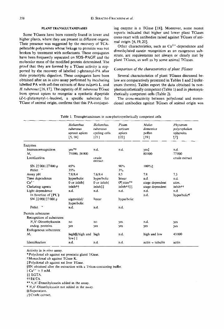
Several characteristics of plant TGases discussed be- low are comparatively presented in Tables 1 and 2 (refer- ences therein). Tables report the data obtained in non- photosynthetically competent (Table l) and in photosyn- thetically competent cells (Table 2).
The cross-reactivity between polyclonal and mono- clonal antibodies against TGases of animal origin was
Table 1. Transglutaminases in non-photosynthetically competent cells
H elianthus H eliant hus Pisum M alus Ph ysarum tuberosus tuberosus sativum domestica polycephalum sprout apices cycling cells apices pollen spherules [5, 18] [21] [22] [19] [7]
Enzymes Immunorecognition M, Localization
yes*t n,d. n.d. yes:~ n.d. 75000, 58000 80000 77000
crude crude extract extract
SN 22000/27000 g 65% 90% 100%§ Pellet " 35% 3%
Assay pH 7.8/8.4 7.6/8.4 8.5 7.8 7.5 Time dependence hyperbolic hyperbolic linear n.d. n.d. Ca 2+ 0 or inhibll 0 or inhibll 0~l/stim** stage dependent stim. Chelating agents inhibl"t inhibS:~ inhibtt$$ stage dependent inhibit Light dependence n.d. n.d. n.d. n.d. n.d.
In function of [PU]: n.d. hyperbolic¶ SN 22 000/27 000 9 sigmoidal/ linear hyperbolic
hyperbolic Pellet " n.d. n.d. n.d.
Protein substrates Recognition of substrates:
N,N'-Dimethylcasein endog, proteins
Endogenous substrates: M,
Identification
no no yes n.d. yes yes yes yes yes yes
high~/high and high n.d. high and low 43 000 lowll II n.d. n.d. n.d. actin + tubulin actin
Activity in in vitro assay. *Polyclonal ab against rat prostatic gland TGase. tMonoclonal ab against TGase K. SPolyclonal ab against rat liver TGase. §SN obtained after the extraction with a Triton-containing buffer. II Ca 2 + > 5 raM. :[::~ EGTA. t t EDTA. ** N,N'-Dimethylcasein added in the assay. ¶ N,N'-Dimethylcasein not added in the assay.
Supenatant. II II Crude extract.
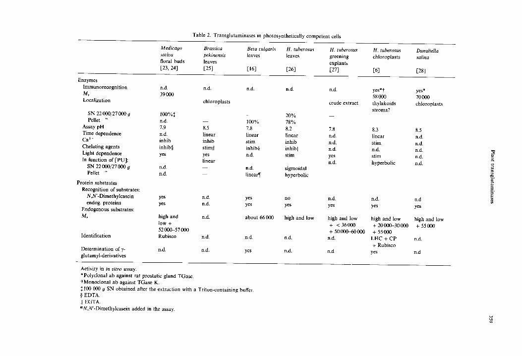
Table 2. Transglutaminases in photosynthetically competent cells
Medicago Brassica Beta vulyaris H. tuberosus H. tuberosus H. tuberosus Dunaliella sativa pekinensis leaves leaves greening chloroplasts salina floral buds leaves explants 1,23, 24] [25] 1,16] [26] [27] 1,6] [28]
Enzymes Immunorecognition n.d. M, 39 000 Localization
n.d. n.d. n.d.
chloroplasts
SN 22 000/27 000 g 100%:~ Pellet " n.d. - - 100%
Assay pH 7.9 8.5 7.8 Time dependence n.d. linear linear Ca 2 ÷ inhib inhib stim Chelating agents inhib§ stimrl inhib§ Light dependence yes yes n.d. In function of i-PU]: linear
SN 22 000/27 000 g n.d. n.d. Pellet n.d. - - linear¶
Protein substrates Recognition of substrates:
N,N'-Dimethylcasein yes n.d. yes endog, proteins yes n.d. yes
Endogenous substrates:
M, high and n.d. about 66 000 low + 52 000-57 000
Identification Rubisco n.d. n.d.
Determination of y- n.d. n.d. yes glutamyl-derivatives
2O% 78% 8.2 linear inhib inhibll stim
sigmoidal hyperbolic
no
yes
high and low
n.d.
n.d.
n.d,
crude extract
ycs*t 58OOO thylakoids stroma?
7.8 8.3 8.5 n.d. linear n.d. n.d. stim n.d. n.d. n.d. n.d. yes stim n.d. n.d. hyperbolic n.d.
n.d,
yes
high and low + <36000 + 50 00~60 000
n.d.
n.d
yes* 70000 chloroplasts
n.d. n.d yes yes
high and low high and low + 20000-30000 + 55000 + 55000
LHC + CP n.d. + Rubisco
yes n.d
.~-
Activity in in vitro assay. *Polyclonal ab against rat prostatic gland TGase. tMonoclonal ab against TGase K. :~ 100 000 g SN obtained after the extraction with a Triton-containing buffer. § EDTA. II EGTA. ¶N,N'-Dimethylcasein added in the assay.

360 D. SERAF1NI-FRACASS1NI et al.
assayed only in some plants and was positive with, as for example, the enzyme of leaf chloroplasts, of sprout apices of H. tuberosus and of the unicellular alga Dunaliella salina Teodor. In germinating and ungerminated pollen of Malus domestica Borckh. cv Red Chief, polyclonal antibody recognized a single protein.
The molecular mass of the enzymes of other plants was determined by the presence of activity in chromato- graphic fractions. In unopened flower buds of Medicago sativa L., the catalytically active subunit has a Mr of 39 000. In the slime mould P. polycephalum, the value of the dimeric native enzyme is 77000 and that of the monomer 39 600. In green leaves, either mature or devel- oping, of Brassica pekinensis Skeels, Beta vulgaris L. and H. tuberosus, in Dunaliella cells as well as in pollen of M. domestica, most of the activity seems to be localized in the membranes, from which it can be released by surfactants. In etiolated apices of H. tuberosus and of Pisum sativum L., on the contrary, most of the activity is found in the 22 000 or 27 000 9 supernatant. The differences observed in the subcellular distribution of this activity seems to be organ- and not species-specific.
The pH optima ranged between 7.6 and 8.5; these values are very similar to those of mammal TGases. Some features, such as the recognition of proteins of different Mr by antibodies, multiple pH optima and the different curves of activity as a function of PU concentra- tion, suggest that multiple forms of the enzyme may exist in sprout apices and leaves of H. tuberosus and that some of them may be organ-specific. Different forms of TGases located in the same organ have also been reported to occur in animals [29, 30].
Ca 2÷ is a relevant parameter and, especially in the past, it was considered an essential factor for TGase. Generally, it must be supplied in the enzymatic assay even though, in some cases [8], it was found bound to the enzyme. In plants, perhaps owing to the higher availabil- ity of this cation in cell-free extracts, Ca 2 ÷ addition seems to be unnecessary; at times it can favour the activity, except at relatively high concentrations when it becomes inhibitory. Chelating agents generally lower the activity. However, the organ-related occurrence in H. tuberosus cycling cells of an aspecific or non-enzymatic binding, which takes place also after boiling, makes it difficult to completely clarify some of the properties of this activity, such as its pH- or Ca 2 +-dependence.
Contrary to what happens in other plants, N,N'- dimethylcasein is not recognized as exogenous substrate by the TGase of H. tuberosus; this is also true for the enzyme of filarial parasites [31].
Light dependence has been studied only in green tis- sues and has been found to enhance the recovery of TCA-precipitable PA. The effect of light on thylakoid substrates is at present under investigation in our labor- atory.
The conjugates formed with endogenous proteins and separated on SDS-PAGE appear to be more than one. In several organs very high molecular mass, or water-insol- uble, conjugates are formed as products of TGase activ- ity; these are also found in animal systems and, owing to
their mass, are difficult to analyse [32]. In green tissues or in etiolated apices of H. tuberosus, where leaf primordia and immature leaves are also present, substrates having a M, lower than 30000, were found in addition to the high Mr ones.
The exclusive property of TGase to form mono- and bis-glutamyl PA has been only rarely checked. Mono- glutamyl-PU was found in Beta vulgaris leaves after incubation of N,N'-dimethylcasein in the assay mixture [16]. More recently, in addition to mono-glutamyl-PU, bis-glutamyl-PU and -SD were also detected in H. tuber- osus chloroplasts [17].
Polyamine conjugates formed in vivo
In addition to those reported in Tables 1 and 2 and obtained by cell free incubation, conjugates have been found also in vivo. When [14C]PU was added to the incubation medium, it was metabolized by H. tuberosus cycling cells not only to SD and SM but also to other unknown derivatives [33]. The SDS-PAGE of the labelled proteins revealed that during G 1 phase, radioac- tivity was present in two bands having an apparent M, of 17 500 and 19 000, subsequently (from S phase onwards) the label appeared in bands having a M, higher than 66 000. However, a large amount of radioactivity always remained in the wells, suggesting that mainly very high Mr or insoluble proteins had incorporated the label. Treatment of the TCA pellet with SDS ruled out the possibility that non-covalently linked PA were present therein [21, 33, 34].
Other authors also reported in vivo labelling of a M,, 18 000 protein in several plant systems by supplying SD in the growth medium [35, 36]. In the early phases of cycling mammalian cells, only a single protein of 18 000, identified as the initiation factor for protein synthesis eIF-5A [37,38], is modified by hypusine, an unusual amino acid derived from SD. The formation of this amino acid is not TGase-catalysed.
In protoplasts of Petunia hybrida Hort. ex Vilm. and Arena sativa L., after feeding [3HI SD, label was incorp- orated into proteins having a Mr exceeding 45 000 [39]. This could be the product of an in rico catalysis of TGase. Several plant proteins, as for example pea legumin are substrates of guinea-pig liver TGase [40].
Biological and functional aspects of plant transglutaminases
TGase in non-photosynthetically competent cells. Data from the literature about TGase activity in these plant cells is scanty [5,22,21,41-43]. Two different mecha- nisms which bind PA to proteins occur in H. tuberosus cycling cells: one that is destroyed by boiling (Ts TGase), the other that is temperature-insensitive (Ti TGase) [21]. The latter binding reaction might be of a nonenzymatic nature and the complexes formed show a very faint radioactivity on SDS-PAGE autoradiography, sugges- ting that these PA are not covalently bound. On the contrary, the Ts TGase, whose activity is low during

Plant transglutaminases 361
dormancy, possesses enzymatic characteristics similar to those observed with animal TGase. After mid-G1, the Ts TGase activity increases considerably due to the syn- thesis of both the enzyme and/or its substrates, as dem- onstrated by supplying cycloheximide for different time periods and by immunoblot analysis of the enzyme [20, 43]. In S phase and in division phase, TGase activity reached the highest levels 1-21,4l]. In C6 glioma cells a similar system was studied: the enzyme appears to be constitutive, but inactive, in non-stimulated cells and synthesized de novo during the progression of the cell cycle. It was suggested that the enzyme was previously bound to cell membranes and then released, after growth stimulation, into the cytoplasm where it was subjected to proteolytic digestion. Thereafter a de novo synthesis com- pensated for this deficiency and a second peak in activity resulted [44]. Of course, the metabolic state of dormant tuber cells (out of the cycle) and of non-stimulated glioma cells (arrested during the cycle) are different whereas, after induction of cycling, their metabolisms are probably more comparable. In both H. tuberosus and in C6 glioma cells, a new (different ?) enzyme appears to be synthesised and thus the synthesis of these enzymes may be related to particular functions of cycling cells.
The amine partner of the products of TGase activity in H. tuberosus cycling cells were, apart from PU itself supplied to the medium, also PU derivatives, especially unknown compounds having a migration close to zero on TLC plates. Labelled bound SD, SM or 7- aminobutyric acid albeit in very low amounts were detec- ted, indicating that PA metabolism occurred during the TGase assay 1,42].
At the end of the cell cycle of H. tuberosus, the elec- trophoretic pattern of proteins was quite different with respect to that observed in the GO phase; in particular the percentage of high Mr protein bands increased consider- ably, whereas other protein bands completely disap- peared. All these modifications were completely blocked by cycloheximide [43, 45]. The formation of these large complexes is accompanied by an heavier PU incorpora- tion therein both in vivo and in cell-free extracts, sugges- ting that TGase activity might play a role in the forma- tion of these large complexes. Some of them can be separated on 10% SDS-PAGE or by digesting them with cellulase and pectinase, in addition to protease [42]. In this phase it is known that fibrillar material from the cytoplasm migrates into the walls of H. tuberosus cells 1"46] and in Catharanthus roseus L. G. Don., wall protein synthesis has been reported 1,47]. Thus, it can be inferred that TGase is involved in cell wall organization.
Recently, TGases activity has also been studied in pollen of Malus domestica which represent a system of very rapid growth involving both cytoskeleton re- arrangement and building of a new cell wall [19]. Actin and tubulin are known to play an important role in the cytoskeletal organization being closely involved in the pollen tube elongation 1,48]. During germination TGase activity is high when actin elements are organized [49] and markedly decreases at the end of germination. Labelled PA are bound to high and low M, proteins but
especially to proteins of Mr 43 000 and 50 000-55 000. Incubation with purified actin and tubulin revealed that both are substrates for pollen TGase. In addition, in germinated pollen the supplied actin was modified giving rise to heavier labelled aggregates with respect to the unsupplemented samples. The conjugates formed in ger- minated and ungerminated pollen after actin supply have a different degree of polymerization.
Thus it can be inferred that in higher plants PA may be involved in the formation of cytoskeletal and of cell wall structures, whose formation is one of the main events of cytokinesis and cell enlargement.
Direct evidence that actin is a substrate for TGase also in fungi has been reported in P. polycephalum 1,7]. It was proposed that the role of TGase may be important for protein cross-linking during the conversion of microplas- modia to spherules, which can survive starvation and dessication. Actin modified in vitro by monodansyl- cadaverine via TGase of guinea-pig liver exhibits a lower critical concentration to polymerize and a faster rate of polymerization compared to those of the unmodified actin without affecting the binding to myosin heads [50]. In animals, a functional dependence on PA for the forma- tion of cytoskeletal structures, like actin filaments and microtubules, has been shown in PA-deficient CHO cells even though the M, was not clarified [51] ; in addition, PA depletion caused a high incidence of binucleate fibroblasts [52]. Tubulin was reported to be a substrate of TGase in neuronal cytoskeleton of mammals [53].
The confirmation of the identity of the plant conjugat- ing enzyme as being a TGase is provided by the following data [18]. The substrate for mammalian TGases, Z-L-gln- t-leu, is also a good substrate for TGase from etiolated apices of the sprouts growing from tubers of H. tuberosus. The activity is proportional to the concentration of PA supplied. The enzyme was partially purified by ion ex- change chromatography and two forms were present, eluted at 0.1 and 0.3 M NaC1. Both fractions recognized the dipeptide forming ),-glutamyl-PA. A main band of M,, 75000 in the 0.1 fraction and one of 58 000 in the 0.3 fraction cross-reacted both with a polyspecific antibody to prostate TGase and with a monospecific antibody to epidermal particulate TGase K.
These chromatographic characteristics are also shared by TGases from animal sources, as for example in rat chondrosarcoma 1,54]. The immunological cross-reactiv- ity showed that common sequences are present in animal and plants. The recovery of two enzyme forms in the apices could suggest that they exert different functions. An enzyme form of M, 58000 was found also in thylakoids of chloroplasts of green leaves. In etiolated sprout apices, leaflets are present and, potentially, they may have the same form of enzyme as active chloroplasts (see below).
TGase in photosynthetically competent cells. When non-photosynthetic parenchyma of H. tuberosus tubers is stimulated in vitro to differentiate chloroplasts, TGase can be induced to a greater extent than in a hormone- stimulated control which grows actively but does not turn green [27]. The high Mr substrates observed in

362 D. SERAF1NI-FRACASSINI et al.
non-photosynthetic cells are present in both explants, but in the green ones low Mr proteins were also modified by PA. The morphological characteristics of the two tissues are quite different: the non-green growing control con- sists of friable tissue whose cells have thin walls and empty intercellular spaces and resembles a tumour tissue (habituated callus); the green callus has well-developed chloroplasts, a compact consistency and dense material in the intercellular spaces. As a working hypothesis, it was inferred that a relationship exists between these morphological features and TGase activity. In animal systems (data in plants are too scanty), TGase activity is generally high in differentiated tissues, where it is in- volved in the organisation of protein structures respon- sible for the cohesion between cells and low in rapidly growing tumours in which the organisation of intra- and extra-cellular structural proteins is rather poor [29, 30, 55].
On the other hand, the presence of TGase in chloro- plasts has been shown. Cohen et al. [25] observed that, in isolated chloroplasts of B. pekinensis, SD was 'fixed' with a higher efficiency in the light than in the dark. However, they concluded that the stimulation by EDTA and the inhibition by Ca 2÷ did not support the hypothesis of a typical TGase-catalysed fixation. This stimulation by light was also reported by other authors (see Table 1). On the contrary, in another system, namely A. sativa proto- plasts, light markedly inhibited the uptake of label and consequently PA binding to TCA-precipitable material [39].
In isolated thylakoids and in entire choloroplasts from H. tuberosus leaves, label in TCA-precipitable material after the TGase assay was higher when they were incu- bated in the light rather than in the dark [6]. Autoradiography of a 2D gel of [t*C]SD- and [14C] PU-labelled chloroplasts and thylakoids which separ- ates, in the first dimension, chlorophyll-protein com- plexes and, in the second dimension, the relative apo- proteins (first dimension, Deriphat-PAGE; second di- mension, SDS-urea-PAGE) showed that a few and the same proteins (Mr 20 000-30 000) were labelled. The rec- ognition of these proteins as the chlorophyll-a/b antenna complexes LHCII, CP24, CP26 and CP29 was ascer- tained by cross-reactivity with antibodies.
The fact that the oligomeric form of the major light- harvesting complex (LHCII) is a better substrate since it is more heavily labelled by PA than the monomeric form, suggests a role for PA in stabilizing oligomers. A detect- able enhancement of the M, or these proteins was not observed after a 30 min assay; thus, either the aggrega- tion of these molecules does not take place or, possibly, polyamines are linked by only one terminal amino group or, finally, only intramolecular cross-bridges are formed. Concerning the role of PA in the post-translational modification of LHCII, it has been proposed that PA not only form cross-links but also add positive charges which may have the opposite effect to that of phosphorylation and thus could favour the transfer of excitation energy between the photosystems [56,57]. Recently it was shown that another phosphorylation mechanism is re-
sponsible for the protection of the PSII reaction centre from photoinhibition by inducing a conformational change in an inner antenna complex called CP29 [58]. On the other hand, it has been observed that D1, D2, cytochrome f and the large Rubisco subunit are all stabilized by PA, by a not yet clarified mechanism [59].
In entire chloroplasts of H. tuberosus, an additional faint radioactive spot having a Mr of 55 000 was recog- nised by antibodies as the large subunit of Rubisco. The large Rubisco subunit was indicated as a substrate also in floral buds [23].
It has been proposed that the polymerization of Rubisco to a dimer is the initial step in the assembly of the catalytically active L8S8 Rubisco structure [7, 60].
TGase activity was detected in the green alga D. salina both in salt-stressed cells and in the controls. At 24 h after inoculation TGase activity was four-fold higher in the former and most of the radioactivity was localized in a high M, region, in a protein of 55000 and in the chlorophyll migrating zone [28]. The activity, also detec- ted by immunocytochemical analysis, was mainly localiz- ed in the chloroplast, whereas the Mr of the enzyme evaluated by western blot analysis was ca 70000. Pro- teolytic digestion of the products of TGase obtained by incubating chloroplasts of H. tuberosus with both SD and PU, leads to the recovery in both cases of N-(~-glutamyl) PU, N1,N*-bis-(~,-glutamyl) PU, NI,NS-bis-(7-glutamyl) SD. The presence of bis-derivatives represent a direct indication of the formation of cross-links via TGase. The most abundant derivatives are mono-PU and bis-SD in a ratio generally around 4 : 1, suggesting that the two PA may play a different role in the post-translational modifi- cation of proteins [17].
In addition to glutamyl-PA, free PA and their deriva- tives, N l-(~-acetyl) SD and NS-(~-acetyl) SD, were detec- ted in chloroplasts, incubated either with PU or with SD [17]. Whereas the existence of a metabolic pathway that converts SD into PU and SM into SD via an acetylated intermediate was previously described for invertebrates and yeasts [61,62], the existence of this pathway in H. tuberosus indicates that a polyamine oxidase (PAO) similar to that of rat liver described by H611t/i [63] and by Bolkenius and Seiler [62] could also exist in plants. It has been reported that selective oxidation by FAD-dependent PAO may be one of the cellular mechanisms that regulates PA-protein cross-link levels [64]. This oxidative metabolism has as substrate NX-(7-glutamyl) SD and not NS-(7-glutamyl) SD, leading to the formation of N-(~-glutamyl)-3-amino- propionaldehyde and free PU (Fig. 4). The formation of bis-SD can thus be modulated. This oxidation, by releasing part of the PA molecule, determines the release of positive groups and thus a change in the total charge of the protein molecule. This is not a complete reversion of the protein to the initial state, which is achieved only by the activity of 7-glutamylamino cyclotransferase, which is able to detach the entire PA from the glutamyl residue. No information is available on the existence in plants of this enzyme, which is also little studied in mammals.

Plant transglutaminases 363
Prot-G In-CO-NH-(CH 2 ) 2-CHO
N -(~- glutamyl)-3 -aminopropionaldehyde
PAO
NH2 -( CH2)4- N H2
j putrescine
ProtoGIn-CO-NH-(CH2)3-NH-(CH2)4-NH-CO-GIn-Prot N 1, N 8_ bis(,t-glutamyl)spermidine
TGase
Pro.-Cln-CO-N,-IC,2)3-N,-I C,2),- N , y Prot-Cln-CON,2 N 1 -(~- glutamyl~spermidine (~-glutamyl) protein
TGase
Prot-Gln-CONH 2 ~ ~ N t t 2 - ( C H 2 ) 3 - N H - ( C H 2 ) 4 - N H 2 (y- glutamyl) protein ~ y spermidine
TGase
Prot-Gln-CO-NH-t CH2)4- N H-( CH2)3- N H 2 Prot-Gln-CONH 2 N 8-(v-glutamyl)spermidine " ~ S (~-glutamyl)protein
TGase \
Prot-GIn-CO-NH-(CH2)4- N H-I CH2)3-N H-CO-GIn- Prot N 1, N 8_ bis(r-glutamyl) spermidine
Fig. 4. Metabolic scheme of the potential modulation of transglutaminase action by FAD-dependent polyamine oxidase (PAO). The selective oxidation of Nl-(7-glutamyl) SD takes this substrate from the pool of mono deriva- tives. Therefore, NL(~,-glutamyl) SD can preferentially form the sterically defined bis-derivative N:,NS-(~-glu -
tamyl) SD.
CONCLUDING REMARKS
Information on TGase is relatively incomplete, but we can try to highlight its main aspects. The products of this enzymatic catalysis are different when PA or lysine resi- dues are the amine donors. Thus two possible properties of the final product of the reaction should be considered; the first one is directly a structural consequence of the length of the molecule acting as amine-donor. In the light of this quality, the network produced by the extensive protein cross-linking would acquire various features of reactivity and plasticity depending on the substrate in- corporated.
Highly polymerized structures are known to occur in collagen, elastin, fibrin and in the layers of epidermins. Thus the cellular or extracellular structural organization can be affected. The second property conferred by the modification by PA is the potential modulation of the cross-linked protein structure. In fact, the formation of mono-derivatives is regulated by the PA concentration since high levels of PA may saturate the acyl donor residues of substrate proteins, preventing the formation
of bis-derivatives and of glutamyl-lysine linkages. In this sense, the levels of PA have a critical role in the modula- tion of protein cross-links. The significance of this mech- anism may be explained in the light of the suitable phys- ical state requested for cell function. Moreover, the addi- tion of positive charges due to the linkage with these polycations, could further affect the conformation and function of protein substrates.
Nevertheless, TGases seem to be a widespread class of enzymes with many characteristics in common among plants, animals and also bacteria [65], supporting the hypothesis of its involvement in important biological functions. A role of TGase shared by animals and plants seems to be related to the organization of structural proteins: for example in the differentiation of P. poly- cephalum microspherules, in pollen tube elongation and perhaps during cell division of H. tuberosus as well as differentiation of keratinocytes, modulation of metastatic potential of cancer cells, cataract formation, formation of apoptotic body and extracellular matrix [66-70]. A sim- ilar behaviour to TGase activity has also been reported in rapidly growing cells of plants and animals, like hor-
pHYTO 40-2-8

364 D. SERAFINI-FRACASSINI et al.
mone-stimulated callus or tumours. Besides these roles, others seem to be related to more specific functions, like photosynthesis in plants and receptor-mediated en- docytosis in animals [71].
Acknowledgement--The authors thank RAISA (Sub-pro- ject N. 2, paper No. 2075) for financial support.
REFERENCES
1. Slocum, R. D. and Flores, H. E. (eds) (1991) Biochem- istry and Physiology of Polyamines in Plants, pp. 1-264. CRC Press Uniscience, Boca Raton.
2. Persson, L., Holm, I., Stjernborg, L. and Heby, O. (1988) Adv. Exp. Med. Biol. 250, 261.
3. Takeda, Y., Samejima, K., Nagano, K., Watanabe, M., Sugeta, H. and Kyogochu, Y. (1983) Eur. J. Biochem. 130, 383.
4. Folk, J. E., Park, M. H., Chung, S. I., Schrode, J., Lester, E. P. and Cooper, H. L. (1980) J. Biol. Chem. 255, 3695.
5. Serafini-Fracassini, D., Del Duca, S. and D'Orazi D. (1988) Plant. Physiol. 87, 757.
6. Del Duca, S., Tidu, V., Bassi, R., Esposito, C. and Serafini-Fracassini, D. (1994) Planta 193, 283.
7. Klein, J. D., Guzman, E. and Kuehn, G. D. (1992) J. Baeteriol. 174, 2599.
8. Paonessa, G., Metafora, S., Tajana, G., Abrescia, P., De Santis, A., Gentile, V. and Porta R. (1984) Science 226, 852.
9. Bergamini, C. (1994) Proceedings of the Fourth Inter- national Conference on Transglutaminase and Protein Crosslinking Reactions. Debrecen, Hungary. p. 73.
10. Facchiano, F., Valtorta, F., Benfenati, F. and Luini, A. (1993) TIBS 18, 327.
11. Gerber, U., Jucknischke, U., Putzien, S. and Fuchsbauer, H. L. (1994) Biochem. J. 299, 825.
12. Singh, R. N. and Metha, K. (1994) Proceedings of the Fourth International Conference on Transglutaminase and Protein Crosslinking Reactions. Debrecen, Hun- gary. p. 23.
13. Singer, M. A., Hortsch, M., Goodman, C. S. and Bentley, D. (1992) Dev. Biol. 154, 143.
14. Tokunaga, F., Muta, T., Iwanaga, S., Ichinose, A., Davie, E. W., Kuma, K. and Miyata, T. (1993) J. Biol. Chem. 268, 262.
15. Yasueda, H., Nakanishi, K., Kumazawa, Y., Matsui, H. and Motoki, M. (1994) Proceedings of the Fourth International Conference on Transglutaminase and Protein Crosslinking Reactions. Debrecen, Hungary. p. 72.
16. Signorini M., Beninati, S. and Bergamini, C. (1991) J. Plant. Physiol. 137, 547.
17. Del Duca, S., Beninati, S. and Serafini-Fracassini, D. (1995) Biochem. d. 305, 233.
18. Serafini-Fracassini, D., Del Duca, S., Bregoli, A. M., Beninati, S. and Bergamini, C. (1994) Proceedings of the Fourth International Conference on Trans- glutaminase and Protein Crosslinking Reactions. De- brecen, Hungary. p. 24.
19. Bregoli, A. M., Del Duca, S., Bergamini, C. and
Serafini-Fracassini, D. (1994) Proceedings of the Fourth International Conference on Transglutaminase and Protein Crosslinking Reactions. Debrecen, Hun-
gary. p. 68. 20. Del Duca, S. (1994) Ph.D. thesis. University of Bo-
logna. 21. Serafini-Fracassini, D., Del Duca, S. and Torrigiani,
P. (1989) Plant. Physiol. Biochem. 27, 659. 22. Icekson, I. and Apelbaum, A. (1987) Plant. Physiol.
84, 972. 23. Margosiak, S. A., Dharma, A., Bruce-Carver, M. R.,
Gonzales, A. P., Louie, D. and Kuehn, G. D. (1990) Plant. Physiol. 92, 88.
24. Kuehn, G. D., Sotelo, M., Morales, T., Bruce-Carver, M. R., Guzman, E. and Margosiak, S. A. (1991) FASEB J. 5, A1510.
25. Cohen, S. S., Marcu, D. E. and Balint, R. F. (1982) FEBS Letters 141, 93.
26. Falcone, P., Del Duca, S. and Serafini-Fracassini, D. (1993) J. Plant. Physiol. 142, 265.
27. Del Duca, S., Favali, M. A., Serafini-Fracassini, D. and Pedrazzini, R. (1993) Protoplasma 174, 1.
28. Dondini, L., Serafini-Fracassini, D., Del Duca, S., Bregoli, A. M. and Tsolova, M. (1994) in Polyamines: Biological and Clinical Aspects (Caldarera, C. M., C16, C. and Moruzzi, M. S., eds), pp. 183-194. CLUEB, Bologna.
29. Lorand, L. and Conrad, S. M. (1984) Molec. Cell. Biochem. 58, 9.
30. ,~schlimann, D. and Paulsson, M. (1994) Thromb. Haemost. 71, 402.
31. Metha, K. (1992) Proceedings of the Third Interna- tional Conference on Transglutaminase and Protein Crosslinking Reactions. Ardmore, USA. p. 17.
32. Schmidt R., Michel S., Schroot B. and Reichert U. (1988) FEBS Letters 229, 193.
33. Mossetti U., Serafini-Fracassini, D. and Del Duca, S. (1987) in Conjugated Plant Hormones. Structure, Metabolism and Function (Schreiber, K., Sch/itte, H. R. and Sembdner, G., eds), pp. 369-375. Deutscher Verlag der Wissenschaften, Berlin.
34. Serafini-Fracassini, D. and Mossetti, U. (1985) in Recent Progress in Polyamine Research (Selmeci, L., Brosnan, M. E. and Seiler, N., eds), pp. 551 560. Akademiai Kiad6, Budapest.
35. Metha, A. M., Saftner, A. R., Schaeffer, G. W. and Mattoo, A. K. (1990) Plant. Physiol. 95, 1294.
36. Apelbaum, A., Canellakis, Z. N., Applewhite, P. B., Kaur-Sawhney, R. and Galston, A. W. (1988) Plant. Physiol. 88, 996.
37. Cooper, H. L., Park, M. H., Folk, J. E., Safer, B. and Braverman, R. (1983) Proc. Natl Acad. Sci. USA 80, 1854.
38. Park, M. H., Wolff, E. C. and Folk, J. E. (1993) BioFactors 4, 95.
39. Mizrahi, Y., Applewhite, P. B. and Galston, A. W. (1989) Plant. Physiol. 91, 738.
40. Larr6, C., Kedzior, Z. M., Chenu, M. G., Viroben, G. and Gueguen, J. (1992) J. Agric. Food Chem. 40, 1121.
Plant transglutaminases 365
4l. Serafini-Fracassini, D., Del Duca, S., D'Orazi, D. and Mossetti, U. (1988) in Perspectives in Polyamine Re- search (Perin, A., Scalabrino, D,, Sessa, A. and Ferioli, L., eds), pp. 89-92. Wichtig Editore, Milano.
42. Dinnella, C., Serafini-Fracassini, D., Grandi, B. and Del Duca, S. (1992) Plant. Physiol. Biochem. 30, 531.
43. Grandi, B., Del Duca, S., Serafini-Fracassini, D. and Dinnella, C. (1992) Plant. Physiol. Biochem. 30, 415.
44. Korner, G. and Bachrach, U. (1987) J. Cell. Physiol. 130, 44.
45. Del Duca, S. and Serafini-Fracassini, D. (1993) in Molecular and Cell Biology of the Plant Cell Cycle (Ormrod, J. and Francis, D., eds), pp. 143-156. Kluwer, Dordrecht.
46. Favali, A., Serafini-Fracassini, D. and Sartorato, P. (1984) Protoplasma 123, 192.
47. Kodama, H., Ito, M. and Komamine, A. (1990) in Progress in Plant Cellular and Molecular Biology (Nijkamp, H. J. J., Van der Plas, L. H. W. and Van Aartrijk, J., eds), pp. 532-536. Kluwer, Dordrecht.
48. Pierson, E. S. and Cresti, M. (1992) in Sexual Repro- duction of Flowering Plants (Russell, S. D. and Dumas, C., eds), Vol. 140, pp. 73-125. Academic Press, San Diego.
49. Tiwari, S. C. and Polito, V. S. (1988) Protoplasma 147, 5.
50. Takashi, R. (1988) Biochemistry 27, 938. 51. Pohjanpelto, P., Virtanen, I. and H61tt/i, E. (1981)
Nature 293, 475. 52. Sunkara, P. S., Rao, P. N., Nishioka, K. and Brink-
ley, B. R. (1979) Exp. Cell Res. 119, 63. 53. Miller, C. C. J. and Anderton, B. H. (1986) J. Neur-
ochem. 46, 1912. 54. Chung, S. I., Chang, S. K., Cocuzzi E. T., Folk, J. E.,
Kim., H. C., Lee, S. Y., Martinet, N., Nigra, T. and Sun, H. S. (1988) Adv. Exp. Med. Biol. 231, 1.
55. Folk, J. E. (1980) Annu. Rev. Biochem. 49, 517. 56. Mullet, J. E. (1983) J. Biol. Chem. 258, 9941. 57. Allen, J. F., Bennett, J., Steinbeck, K. E. and Arntzen,
C. J. (1981) Nature 291, 25.
58. Dainese, P., Santini, C., Ghiretti-Magaldi, A., Mar- quardt, J., Tidu, V., Mauro, S., Bergantino, E. and Bassi, R. (1992) in Research in Photosynthesis (Murata N., ed.), Vol. 2, pp. 13-20. Kluwer, Dor- drecht.
59. Besford, R. T., Richardson, C. M., Campos, J. L. and Tiburcio, A. F. (1993) Planta 189, 201.
60. Roy, H., Cannon, S. and Gilson, M (1988) Biochim. Biophys. Acta 957, 323.
61. Gillyon, C., Haywood, G. W., Large, P. J., Nellen, B. and Robertson, A. (1987) J. Gen. Microbiol. 133, 2477.
62. Bolkenius, F. N. and Seiler, N. (1981) Int. J. Biochem. 13, 287.
63. H611t/i, E. (1977) Biochemistry 15, 91. 64. Beninati, S., Mantile, G., De Santis, A. and Abbruz-
zese, A. (1992) Life Chem. Rep. 10, 29. 65. Ando, H., Adachi, M., Umeda, K., Matsuura, A.,
Nonaka, M., Uchio, R., Tanaka, H. and Motoki, M. (1989) Agric. Biol. Chem. 53, 2613.
66. Beninati S., Abbruzzese, A. and Cardinali, M. (1993) Int. J. Cancer 53, 729.
67. Fesus, L., Thomazy, W., Autuori, F., Cerfi, M. P., Tarcsa, E. and Piacentini, M. (1989) FEBS Letters 245, 150.
68. Martinet, N., Beninati, S., Nigra, T. P. and Folk, J. E. (1990) Biochem. J. 271, 305.
69. Beninati, S., Senger, D. R., Cordella Miele, E., Muk- herjee, A. B., Singh, K. and Mukherjee, B. B. (1994) J. Biochem. 115, 675.
70. Lorand, L., Hsu, L. K. H., Siefring, G. E. Jr. and Rafferty, N. S. (1981) Proc. Natl Acad. Sci. USA 78, 1356.
71. Davies, J. A. P., Davies, D. R., Levitzki, A., Maxfield, F. R., Milhaud, P., Wiilingham, C. M. and Pastan, I. H. (1980) Nature 283, 162.
72. Kim, H. C., Idler, W. W., Kim, I. G., Han, J. H., Chung, S. I. and Steinert, P. M. (1991) J. Biol. Chem. 266, 536.
Copyright © 2022 FDOKUMEN