Placental glutathione S-transferase correlates with cellular proliferation during rat tongue...
8
Experimental and Toxicologic Pathology 59 (2007) 61–68 Placental glutathione S-transferase correlates with cellular proliferation during rat tongue carcinogenesis induced by 4-nitroquinoline 1-oxide Renata N. Silva a , Daniel A. Ribeiro b, , Daisy M.F. Salvadori a , Mariaˆngela E.A. Marques a a Department of Pathology, Botucatu Medical School, Sao Paulo State University, UNESP, Sa˜o Paulo, Brazil b Department of Health Sciences, Federal University of Sao Paulo, UNIFESP, Santos, SP, Brazil Received 16 November 2006; accepted 16 February 2007 Abstract Taking into consideration that glutatione S-transferase (GST) and cellular proliferation play a crucial role during carcinogenesis, the goal of this study was to investigate the expression of placental GST, called GST-P, and proliferating cellular nuclear antigen (PCNA) by means of immunohistochemistry during rat tongue carcinogenesis induced by 4-nitroquinoline 1-oxide (4NQO). This is a useful model for studying oral squamous cell carcinoma phase by phase. Male Wistar rats were distributed into three groups of 10 animals each and treated with 50 ppm 4NQO solution by drinking water for 4, 12 or 20 weeks. Ten animals were used as negative control. GST-P positive foci were detected in non-neoplastic oral cells at 4 weeks of 4NQO administration. In the same way, GST-P positive cells were detected in pre-neoplastic lesions and squamous cell carcinomas induced after 12 and 20 weeks-treatment, respectively. None of the control animals expressed GST-P positive cells. Regarding cellular proliferation, PCNA positive nuclei were higher at 12 and 20 weeks following 4NQO exposure (po0.05) when compared to negative control. These results suggest that the expression of GST-P is correlated with cellular proliferation, in which GST-P is associated with risk and progression of oral cancer, whereas PCNA is closely involved during neoplastic conversion. r 2007 Published by Elsevier GmbH. Keywords: Placental glutathione S-transferase; Rat tongue mucosa; Oral squamous cell carcinoma; 4-nitroquinoline 1-oxide Introduction Squamous cell carcinoma is the most common malig- nancy that affects the human oral cavity (Sakai and Tsuchida, 1992). Despite recent advances in therapeutic modalities, the prognosis of patients with oral squamous cell carcinoma has not been improved significantly in the last decades (Ribeiro et al., 2004). Herein, it is desirable to examine the precise pathobiological mechanisms involved in oral tumorigenesis in order to introduce reliable biomarkers to prevent oral squamous cell carcinomas. The most used animal models in oral cancer research are the hamster buccal pouch by fat-soluble 7,12 dimethylbenzanthracene (DMBA), and the rat tongue by water-soluble 4-nitroquino- line 1-oxide (4NQO). Considering that one of the most important routes of oral carcinogens is through liquid containing water-soluble carcinogens, 4NQO is well suited in examining the role of xenobiotics in experimental oral carcinogenesis ( Tanaka et al., 2002). Based on the multi- step process of carcinogenesis characterized by initiation, promotion and tumor progression, chronic administration ARTICLE IN PRESS www.elsevier.de/etp 0940-2993/$ - see front matter r 2007 Published by Elsevier GmbH. doi:10.1016/j.etp.2007.02.010 Corresponding author. Departamento de Cieˆ ncias da Sau´de, Universidade Federal de Sa˜o Paulo-UNIFESP, Av. Ana Costa 95, Zip code: 11060-001, Santos – SP, Brazil. Tel./fax: +55 13 32222048. E-mail address: [email protected] (D.A. Ribeiro).
-
Upload
independent -
Category
Documents
-
view
5 -
download
0
Transcript of Placental glutathione S-transferase correlates with cellular proliferation during rat tongue...

ARTICLE IN PRESS
0940-2993/$ - se
doi:10.1016/j.et
�CorrespondUniversidade F
Zip code: 11060
E-mail addr
Experimental and Toxicologic Pathology 59 (2007) 61–68
www.elsevier.de/etp
Placental glutathione S-transferase correlates with cellular proliferation
during rat tongue carcinogenesis induced by 4-nitroquinoline 1-oxide
Renata N. Silvaa, Daniel A. Ribeirob,�, Daisy M.F. Salvadoria,Mariangela E.A. Marquesa
aDepartment of Pathology, Botucatu Medical School, Sao Paulo State University, UNESP, Sao Paulo, BrazilbDepartment of Health Sciences, Federal University of Sao Paulo, UNIFESP, Santos, SP, Brazil
Received 16 November 2006; accepted 16 February 2007
Abstract
Taking into consideration that glutatione S-transferase (GST) and cellular proliferation play a crucial role duringcarcinogenesis, the goal of this study was to investigate the expression of placental GST, called GST-P, andproliferating cellular nuclear antigen (PCNA) by means of immunohistochemistry during rat tongue carcinogenesisinduced by 4-nitroquinoline 1-oxide (4NQO). This is a useful model for studying oral squamous cell carcinoma phaseby phase. Male Wistar rats were distributed into three groups of 10 animals each and treated with 50 ppm 4NQOsolution by drinking water for 4, 12 or 20 weeks. Ten animals were used as negative control. GST-P positive foci weredetected in non-neoplastic oral cells at 4 weeks of 4NQO administration. In the same way, GST-P positive cells weredetected in pre-neoplastic lesions and squamous cell carcinomas induced after 12 and 20 weeks-treatment, respectively.None of the control animals expressed GST-P positive cells. Regarding cellular proliferation, PCNA positive nucleiwere higher at 12 and 20 weeks following 4NQO exposure (po0.05) when compared to negative control. These resultssuggest that the expression of GST-P is correlated with cellular proliferation, in which GST-P is associated with riskand progression of oral cancer, whereas PCNA is closely involved during neoplastic conversion.r 2007 Published by Elsevier GmbH.
Keywords: Placental glutathione S-transferase; Rat tongue mucosa; Oral squamous cell carcinoma; 4-nitroquinoline 1-oxide
Introduction
Squamous cell carcinoma is the most common malig-nancy that affects the human oral cavity (Sakai andTsuchida, 1992). Despite recent advances in therapeuticmodalities, the prognosis of patients with oral squamouscell carcinoma has not been improved significantly in thelast decades (Ribeiro et al., 2004). Herein, it is desirable to
e front matter r 2007 Published by Elsevier GmbH.
p.2007.02.010
ing author. Departamento de Ciencias da Saude,
ederal de Sao Paulo-UNIFESP, Av. Ana Costa 95,
-001, Santos – SP, Brazil. Tel./fax: +5513 32222048.
ess: [email protected] (D.A. Ribeiro).
examine the precise pathobiological mechanisms involved inoral tumorigenesis in order to introduce reliable biomarkersto prevent oral squamous cell carcinomas. The most usedanimal models in oral cancer research are the hamsterbuccal pouch by fat-soluble 7,12 dimethylbenzanthracene(DMBA), and the rat tongue by water-soluble 4-nitroquino-line 1-oxide (4NQO). Considering that one of the mostimportant routes of oral carcinogens is through liquidcontaining water-soluble carcinogens, 4NQO is well suitedin examining the role of xenobiotics in experimental oralcarcinogenesis (Tanaka et al., 2002). Based on the multi-step process of carcinogenesis characterized by initiation,promotion and tumor progression, chronic administration
ARTICLE IN PRESSR.N. Silva et al. / Experimental and Toxicologic Pathology 59 (2007) 61–6862
of 4NQO in drinking water simulates rat tongue carcino-genesis like human counterpart (Ohne et al., 1985;Nishimura, 1999; Niwa et al., 2001; Okazaki et al., 2002;Vered et al., 2003; Ribeiro et al., 2005).
Glutatione S-transferases (GSTs) are a family ofenzymes involved in detoxification of xenobiotics. GSTsexist as homo- or heterodimers and have been groupedinto at least seven distinct classes (Hayes and Pulford,1995; Strange et al., 2000). The main function of GSTs isto catalyze the conjugation of glutathione to an electro-philic site of a broad range of potentially toxic andcarcinogenic compounds, thereby making such com-pounds less biologically active and enabling their excretion(Hayes and Pulford, 1995). Placental GST, called GST-P,is the main isoform in normal placental tissue andcomprises 67% of the total GST concentration in thisphase (Zusterzeel et al., 1999). During development, GST-P decreases in concentration and is absent in adult tissues(Fatemi et al., 2006). Interestingly, GST-P-positive focihave been detected in adult tissues during medium-termhepatocarcinogenesis assay being regarded a suitablebiomaker for early detection of liver neoplasms (Fonsecaet al., 2005; Kitano et al., 2006; Uda et al., 2006).However, the real significance of GST-P positive cellsduring rat tongue carcinogenesis induced by 4NQO isfairly limited in literature (Li, 1999).
It has been well established that tumor cell prolifera-tion activity plays an important role in tumorigenesis(Tumuluri et al., 2004). Among proliferating cellularmarkers, PCNA is a DNA polymerase delta auxiliaryprotein of 36 kDa, which is closely related to thereplication of DNA and is indispensable to cellproliferation. Since the PCNA level increases rapidlyin mid-G1, remains elevated throughout the S phase,and begins to decrease from G2/M to G1 (Linden et al.,1992), a decrease in the number of PCNA-positive cellsreflects a decrease in S phase cells and thus a reducedproliferative activity. To our knowledge, there are nostudies that addressed concomitant expression of GST-Pand PCNA during experimental oral carcinogenesis sofar. As a result and because of inapropriate evidence, thepresent study was undertaken to investigate the expres-sion of GST-P positive foci and PCNA-positive cells on4NQO-induced rat tongue carcinogenesis.
Material and methods
Animals and experimental design
All experimental protocols involving animals wereconformed to procedures described in the GuidingPrinciples for the Use of Laboratory Animals.
Forty male Wistar rats (8 weeks old) weightingapproximately 250 g, were obtained from Centro de
Bioterismo (CEMIB), Universidade Estadual de Cam-pinas, SP, Brazil. They were maintained under con-trolled conditions of temperature (2472 1C), light-darkperiods of 12 h, and with free access to water andcommercial diet (Nuvital PR, Brazil). The animals weredistributed into three groups of 10 and were treated with50 ppm 4NQO (Sigma Aldrich, St. Louis, USA) solutionby drinking water for 4, 12 or 20 weeks. Ten animalswere used as negative control. At the end of theexperimental period, the rats were sacrificed by 0.4%sodium pentobarbital (1mL/kg body wt, i.p.). Thetongues were longitudinally cut into halves for histo-pathological examinations. The tissues were fixed in10% buffered formalin (Merck, Darmstadt, Germany),embedded in paraffin blocks and stained with hematox-ylin and eosin (H.E., Merck).
Histopathological analysis
Histopathological evaluation was performed underlight microscope. Analyzes of the tongue sections weregraded as normal, hyperplasia, dysplasia and carcinomaper animal according to Kramer et al. (1978).
Immunohistochemistry
Serial longitudinal tongue sections of 4 mm weredeparaffinized in xylene and rehydrated in gradedethanol, then pretreated in a microwave (Eletrolux,SP, Brazil) with 10mM citric acid buffer (pH ¼ 6) forthree cycles of 5min each at 850W for antigen retrieval.They were pre-incubated with 0.3% hydrogen peroxidein PBS for 5min for inactivation of endogenousperoxidase, and then blocked with 5% normal goatserum in PBS for 10min. The specimens were thenincubated either with anti-GST-P polyclonal antibody(MBL, Nagoya, Japan) at a concentration of 1:1000 oranti-PCNA polycloncal antibody (PC 10, Dako Cor-poration, Denmark) at a concentration of 1:400.Incubation was carried out overnight at 4 1C withinthe refrigerator. This was followed by two washes inPBS for 10min. The sections were then incubated withbiotin-conjugated secondary antibody anti-mouse IgGfor PCNA (Vector Laboratories, Burlingame, CA,USA) at a concentration of 1:200 or anti-rabbit IgGfor GST-P (Vector Laboratories, Burlingame, CA,USA) at a concentration of 1:200 in PBS for 1 h. Afterthat, the sections were washed twice with PBS followedby the application of preformed avidin biotin complexconjugated to peroxidase (Vector Laboratories, Burlin-game, CA, USA) for 45min. The bound complexes werevisualized by the application of a 0.05% solution of3-30-diaminobenzidine solution, and counterstainedwith methyl-green. For control studies of the antibodies,the serial sections were treated with mouse IgG
ARTICLE IN PRESSR.N. Silva et al. / Experimental and Toxicologic Pathology 59 (2007) 61–68 63
(Vector Laboratories, Burlingame, CA, USA) at aconcentration of 1:200 or rabbit IgG (Vector Labora-tories, Burlingame, CA, USA) at a concentration of1:200 in place of the primary antibody. Additionally,internal positive controls were performed with eachstaining batch.
Quantification of immunohistochemistry
Tongue sections stained by immunohistochemistrywere analyzed for immunopositive cells under opticalmicroscope at � 400 magnification per animal. A totalof 1000 PCNA-positive cells were evaluated as estab-lished in a previous study conducted by our researchgroup (Matsumoto et al., 2006). These values wereexpressed in percentage (%). To quantify the expressionof GST-P positive cells, the number of GST-P positivefoci larger than 0.2mm in diameter were quantified asdescribed elsewhere (Suzuki et al., 2004).
Statistical methods
Statistical analysis was performed by Kruskal Wallisnon-parametric test followed by post-hoc analysis
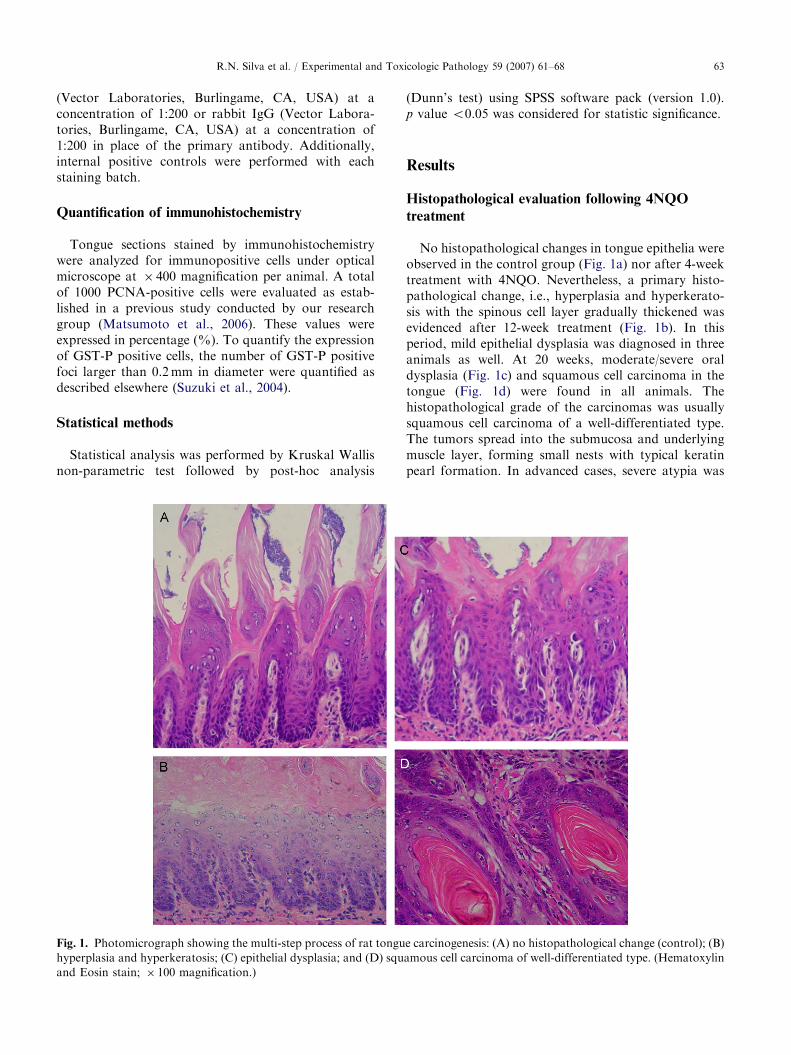
Fig. 1. Photomicrograph showing the multi-step process of rat tongu
hyperplasia and hyperkeratosis; (C) epithelial dysplasia; and (D) squ
and Eosin stain; � 100 magnification.)
(Dunn’s test) using SPSS software pack (version 1.0).p value o0.05 was considered for statistic significance.
Results
Histopathological evaluation following 4NQO
treatment
No histopathological changes in tongue epithelia wereobserved in the control group (Fig. 1a) nor after 4-weektreatment with 4NQO. Nevertheless, a primary histo-pathological change, i.e., hyperplasia and hyperkerato-sis with the spinous cell layer gradually thickened wasevidenced after 12-week treatment (Fig. 1b). In thisperiod, mild epithelial dysplasia was diagnosed in threeanimals as well. At 20 weeks, moderate/severe oraldysplasia (Fig. 1c) and squamous cell carcinoma in thetongue (Fig. 1d) were found in all animals. Thehistopathological grade of the carcinomas was usuallysquamous cell carcinoma of a well-differentiated type.The tumors spread into the submucosa and underlyingmuscle layer, forming small nests with typical keratinpearl formation. In advanced cases, severe atypia was
e carcinogenesis: (A) no histopathological change (control); (B)
amous cell carcinoma of well-differentiated type. (Hematoxylin
ARTICLE IN PRESSR.N. Silva et al. / Experimental and Toxicologic Pathology 59 (2007) 61–6864
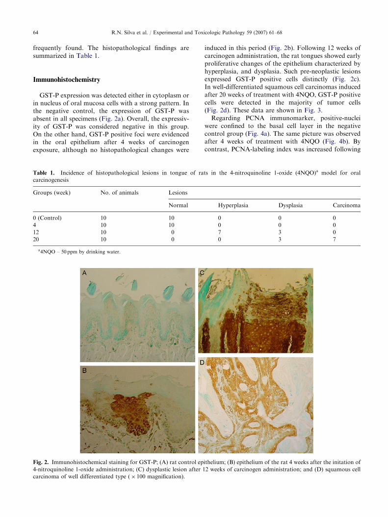
frequently found. The histopathological findings aresummarized in Table 1.
Immunohistochemistry
GST-P expression was detected either in cytoplasm orin nucleus of oral mucosa cells with a strong pattern. Inthe negative control, the expression of GST-P wasabsent in all specimens (Fig. 2a). Overall, the expressiv-ity of GST-P was considered negative in this group.On the other hand, GST-P positive foci were evidencedin the oral epithelium after 4 weeks of carcinogenexposure, although no histopathological changes were
Table 1. Incidence of histopathological lesions in tongue of ra
carcinogenesis
Groups (week) No. of animals Lesions
Normal
0 (Control) 10 10
4 10 10
12 10 0
20 10 0
a4NQO – 50 ppm by drinking water.
Fig. 2. Immunohistochemical staining for GST-P; (A) rat control ep
4-nitroquinoline 1-oxide administration; (C) dysplastic lesion after 1
carcinoma of well differentiated type (� 100 magnification).
induced in this period (Fig. 2b). Following 12 weeks ofcarcinogen administration, the rat tongues showed earlyproliferative changes of the epithelium characterized byhyperplasia, and dysplasia. Such pre-neoplastic lesionsexpressed GST-P positive cells distinctly (Fig. 2c).In well-differentiated squamous cell carcinomas inducedafter 20 weeks of treatment with 4NQO, GST-P positivecells were detected in the majority of tumor cells(Fig. 2d). These data are shown in Fig. 3.
Regarding PCNA immunomarker, positive-nucleiwere confined to the basal cell layer in the negativecontrol group (Fig. 4a). The same picture was observedafter 4 weeks of treatment with 4NQO (Fig. 4b). Bycontrast, PCNA-labeling index was increased following
ts in the 4-nitroquinoline 1-oxide (4NQO)a model for oral
Hyperplasia Dysplasia Carcinoma
0 0 0
0 0 0
7 3 0
0 3 7
ithelium; (B) epithelium of the rat 4 weeks after the initation of
2 weeks of carcinogen administration; and (D) squamous cell

ARTICLE IN PRESSR.N. Silva et al. / Experimental and Toxicologic Pathology 59 (2007) 61–68 65
12-weeks treatment (Fig. 4c).With respect to squamouscell carcinomas induced after 20 weeks of 4NQOexposure, PCNA-positive nuclei were evidenced in themajority of tumor cells (Fig. 4d). Data analysis revelatedthat both experimental periods (12 and 20 weeks) werestatistically significant (po0.05) when compared to thenegative control. These data are summarized in Fig. 5.
0
1
2
3
4
5
6
0 4 12 20
Weeks
Nu
mb
er
of
GS
T-P
po
sit
ive f
oc
* *
*
Fig. 3. Number of GST-P positive foci during rat tongue
carcinogenesis. Values were expressed as means7S.D.
*po0.05, when compared to negative control (zero).
Fig. 4. Immunohistochemical staining for PCNA; (A) rat control e
nitroquinoline 1-oxide administration; (C) dysplastic lesion after 12
carcinoma of well-differentiated type (� 100 magnification).
In the negative immunohistochemical controls, nostaining was seen for either antibody.
Discussion
Carcinogenesis is a multi-step process, which ischaracterized by genetic, epigenetic, and phenotypicchanges (Sugimura et al., 1992). Such changes involvegenetic damage, mutation in critical genes related to the
pithelium; (B) hyperplastic lesion after 12 weeks following 4-
weeks of carcinogen administration; and (D) squamous cell
Fig. 5. PCNA-labeling index in negative control and experi-
mental groups exposed to 4-nitroquinoline 1-oxide. Values are
expressed as means7S.D. *po0.05, when compared to
negative control (zero).
ARTICLE IN PRESSR.N. Silva et al. / Experimental and Toxicologic Pathology 59 (2007) 61–6866
control of cell division, cell death and metastaticpotential, and activation of signalizing or metabolicpathways that give the cells favorable growth andsurvival characteristics (Sarasin, 2003). In patients, themolecular analysis of these multiple steps is hampered,due to the unavailability of biopsies of all the stages ofcarcinogenesis. However, animal models of carcinogen-esis allow the isolation of all stages under controlledconditions, including normal tissues, which are thenamenable to pathological, genetic and biochemicalanalysis at lower costs (Herzig and Christofori, 2002).The results obtained from the present investigation,using 4NQO as carcinogen, demonstrated histopatho-logical changes in rat tongue mucosa along a time-course experiment from normal epithelium, hyperplasia,pre-malignant dysplasia, and carcinoma in situ toinvasive squamous cell carcinoma.
GSTs belong to a family of detoxification enzymesthat have an important role in protecting cells fromcytotoxic and carcinogenic agents (Jakoby, 1978). It hasbeen proposed that GST might be a useful marker forthe detection of pre-neoplastic and neoplastic cells, inwhich an increased GST expression has been noticed ina wide variety of human tumors (Rawal et al., 1999;Ribrag et al., 2003). Particularly, GST-P has beenrecognized to be one of the best tumor markers inhepatocarcinogenesis (Tatematsu et al., 1985; Zhanget al., 1992). Our own research group has used thisbiomarker with success in chemoprevention trials(Agner et al., 2004; Barbisan et al., 2003). In this study,we evaluated the expressivity of GST-P positive focifollowing 4NQO administration in order to determinethe role of this xenobiotic metabolizing enzyme follow-ing oral tumorigenesis phase by phase. In normal oralepithelia, represented by the control group, GST-Pprotein was absent in all specimens. Conversely, GST-Ppositive foci were evidenced in ‘normal’ oral mucosa 4weeks following the initiation of 4NQO administration.These findings are in line with another study which havealso demonstrated an up-regulation of GST-P in oralmucosa cells 4 weeks after 4NQO administration in rats(Li, 1999). It has been stated that overexpression ofGST-P is through gene amplification induced by carcino-gens (Shimizu et al., 1995). Furthermore, chromosomaltranslocation/gene rearrangement of GST-P gene mayalso result in overexpression (Shimizu et al., 1995).Changes to the level of CpG-site methylation alsoappear to play a role in gene activation/suppressionduring carcinogenesis (Esteves et al., 2005). In general,there is an inverse relationship between promotercythosine-guanine-site methylation and the potentialfor transcription. Recently, hypomethylation of theGST-P promoter region in GST-P positive liverneoplasms has been reported, indicating that methyla-tion of the promoter CpG site is at least one possiblemechanism that could potentially lead to transcriptional
activation of GST-P in hepatocarcinogenesis (Steinmetzet al., 1998). Independent of its primary mechanism ofaction, there seems to be evidence that histologicallynormal tissue harbors genetic altered cells able toexpress GST-P. This condition may trigger an increasedrisk of oral cancer. However, it is not known whether allor only a portion of the GST-P positive foci areprecursors of the progressive malignant lesions (Liet al., 1997). Further studies are necessary to elucidatethis issue.
In humans, only a small portion of initial lesionsdevelop to oral carcinoma. Therefore, the challenge is toidentify which lesions have a real malignant potential.Although it is postulated that oral epithelial dysplasia isa pre-neoplastic lesion that occasionally develops intosquamous cell carcinomas (Lee et al., 2006), under-standing the biologic basis of oral squamous cellcarcinoma development and progression is far fromsatisfactory. Our results demonstrated GST-P positivecells from pre-neoplastic lesions, diagnosed as hyper-plasia and dysplasia after 12 weeks of 4NQO exposure.With respect to oral squamous cell carcinomas 4NQO-induced herein, the expressivity of GST-P was seen inthe majority of tumor cells. Some studies have addressedan over-expression of GST-P in pre-neoplastic lesions ororal squamous cell carcinomas as well (Li et al., 1997;Chen et al., 2002).
Cell proliferation is regarded as one of the mostimportant biological mechanisms in oncogenesis (vanDiest et al., 1998). A number of studies has shown thatproliferative activity is of high prognostic significance inseveral types of cancer (Tubiana and Courdi, 1989). Inthe current study, exposure to 4NQO during 4 weeksdid not exert any activity on PCNA-labeling index whencompared to negative control group. Nevertheless,hyperplasia and dysplastic lesions pointed out highPCNA positivity following 12 weeks-treatment with4NQO. The same occurred for squamous cell carcino-mas induced after 20 weeks of 4NQO treatment. Similarfindings were reported by Niwa et al. (2001) and Kaplanet al. (2002). On the basis of the present data, weassumed that proliferation activity plays an importantrole from precancerous lesion to early invasive carcino-ma. In fact, some authors have proposed that theinduction of GST-P may facilitate cell proliferation,hence allowing the clonal expansion of a population ofaltered cells leading to carcinogenesis (Macaghan et al.,1994). Our results are consistent with these findings.
Concluding, our results suggest that expression ofGST-P is correlated with cellular proliferation, in whichGST-P is associated with the development of oralmalignant cells, being closely involved in the risk andprogression of cancer, emphasizing the relevance ofearly identification of GST-P positive foci. Cellularproliferation acitivity as depicted by PCNA positivenuclei is involved into malignant transformation.

ARTICLE IN PRESSR.N. Silva et al. / Experimental and Toxicologic Pathology 59 (2007) 61–68 67
This kind of approach should be considered to personswith high risk of oral cancer, such as in smokers oralcohol consumers as well as patients diagnosed withoral dysplasia or carcinoma.
Acknowledgments
The authors are grateful to Paulo Roberto Cardosoand Maria Luiza Falaguera Ardanaz for their technicalassistance. This work was supported by FAPESP(Fundacao de Amparo a Pesquisa do Estado de SaoPaulo (Grant numbers: 02/09454-0 and 03/10030-2).
References
Agner AR, Barbisan LF, Scolastici C, Salvadori DM. Absence
of carcinogenic and anticarcinogenic effects of annatto in
the rat liver medium-term assay. Food Chem Toxicol
2004;42:1687–93.
Barbisan LF, Spinardi-Barbisan AL, Moreira EL, Salvadori
DM, Ribeiro LR, da Eira AF, et al. Agaricus blazei
(Himematsutake) does not alter the development of rat
diethylnitrosamine-initiated hepatic preneoplastic foci.
Cancer Sci 2003;94:188–92.
Chen YK, Lin LM, Hsue SS, Lin DT. The mRNA expression
of placental glutathione S-transferase isoenzyme in hamster
buccal-pouch carcinomas using reverse transcription-poly-
merase chain reaction. Oral Oncol 2002;38:158–62.
Esteves LI, Javaroni AC, Nishimoto IN, Magrin J, Squire JA,
Kowalski LP, et al. DNA methylation in the CTCF-binding
site I and the expression pattern of the H19 gene: does
positive expression predict poor prognosis in early stage
head and neck carcinomas? Mol Carcinog 2005;44:102–10.
Fatemi F, Allameh A, Dadkhah A, Forouzandeh M,
Kazemnejad S, Sharifi R. Changes in hepatic cytosolic
glutathione S-transferase activity and expression of its
class-P during prenatal and postnatal period in rats treated
with aflatoxin B1. Arch Toxicol 2006; 24, in press.
Fonseca PM, Inoue RY, Kobarg CB, Crosara-Alberto DP,
Kobarg J, Franchini KG. Targeting to C-terminal myosin
heavy chain may explain mechanotransduction involving
focal adhesion kinase in cardiac myocytes. Circ Res
2005;96:73–81.
Hayes JD, Pulford DJ. The glutathione S-transferase super-
gene family: regulation of GST and the contribution of the
isoenzymes to cancer chemoprotection and drug resistance.
Crit Rev Biochem Mol Biol 1995;30:445–600.
Herzig M, Christofori G. Recent advances in cancer research:
mouse models of tumorigenesis. Biochim Biophys Acta
2002;1602:97–113.
Jakoby WB. The glutathione S-transferases: a group of
multifunctional detoxification proteins. Adv Enzymol Relat
Areas Mol Biol 1978;46:383–414.
Kaplan I, Hochstadt T, Dayan D. PCNA in palate and tongue
mucosal dysplastic lesions induced by topically applied
4NQO in desalivated rat. Med Oral 2002;7:336–43.
Kitano M, Wada J, Ariki Y, Kato M, Wanibuchi H,
Morimura K, et al. Possible tumor development from
double positive foci for TGF-alpha and GST-P observed in
early stages on rat hepatocarcinogenesis. Cancer Sci
2006;97:478–83.
Kramer IR, Lucas RB, Pindborg JJ, Sobin LH. Definition of
leukoplakia and related lesions: an aid to studies on oral
precancer. Oral Surg Oral Med Oral Pathol 1978;46:518–39.
Lee JJ, Hung HC, Cheng SJ, Chen YJ, Chiang CP, Liu BY,
et al. Carcinoma and dysplasia in oral leukoplakias in
Taiwan: prevalence and risk factors. Oral Surg Oral Med
Oral Pathol Oral Radiol Endod 2006;101:472–80.
Li TJ. Altered placental glutathione S-transferase foci as a
tumor marker during rat lingual carcinogenesis. Chin J
Dent Res 1999;2:49–53.
Li TJ, Hirayama Y, Kitano M. Glutathione S-transferase
p-class as a tumour marker in lingual preneoplastic and
neoplastic lesions of rats and humans. Virchows Arch
1997;431:37–43.
Linden MD, Torres FX, Kubus J, Zarbo RJ. Clinical
application of morphologic and imunocytochemical assess-
ments of cell proliferation [editorial]. Am J Clin Pathol
1992;97:S4–S13.
Macaghan FM, Brown A, Harrison DJ. The effect of
inhibition of glutathione S-transferase P on the growth of
the Jurkat human T cell line. J Pathol 1994;172:357–62.
Matsumoto MA, Nary Filho H, Jorge FM, Salvadori DM,
Marques ME, Ribeiro DA. Expression of cell cycle
regulatory proteins in epithelial components of dental
follicles. J Mol Histol 2006;37:127–31.
Nishimura A. Changes in Bcl-2 and Bax expression in rat
tongue during 4-nitroquinoline 1-oxide-induced carcino-
genesis. J Dent Res 1999;78:1264–9.
Niwa S, Ueno S, Shirasu R. Alteration of pRB expression in
the development of rat tongue carcinoma induced by
4-nitroquinoline 1-oxide. Oral Oncol 2001;37:579–85.
Ohne M, Satoh T, Yamada S, Takai H. Experimental tongue
carcinoma of rats induced by oral administration of
4-nitroquinoline 1-oxide (4NQO) in drinking water. Oral
Surg Oral Med Oral Pathol 1985;5:600–7.
Okazaki Y, Tanaka Y, Tonogi M, Yamane G. Investigation of
environmental factors for diagnosing malignant potential in
oral epithelial dysplasia. Oral Oncol 2002;38:562–73.
Rawal RM, Patel DD, Patel BP, Patel MM, Wadhwa MK,
Patel PS, et al. Assessment of glutathione-S-transferase and
glutathione reductase in patients with squamous-cell
carcinoma of buccal mucosa. Int J Cancer 1999;83:727–31.
Ribeiro DA, Favero Salvadori DM, da Silva RN, Ribeiro
Darros B, Alencar Marques ME. Genomic instability in
non-neoplastic oral mucosa cells can predict risk during
4-nitroquinoline 1-oxide-induced rat tongue carcinogenesis.
Oral Oncol 2004;40:910–5.
Ribeiro DA, Salvadori DM, Marques ME. Abnormal expres-
sion of bcl-2 and bax in rat tongue mucosa during the
development of squamous cell carcinoma induced by
4-nitroquinoline 1-oxide. Int J Exp Pathol 2005;86:375–81.
Ribrag V, Koscielny S, Carpiuc I, Cebotaru C, Vande Walle H,
Talbot M, et al. Prognostic value of GST-pi expression in
diffuse large B-cell lymphomas. Leukemia 2003;17:972–7.
Sakai E, Tsuchida N. Most human squamous cell carcinomas
in the oral cavity contain mutated p53 tumor-suppressor
genes. Oncogene 1992;7:927–33.
ARTICLE IN PRESSR.N. Silva et al. / Experimental and Toxicologic Pathology 59 (2007) 61–6868
Sarasin A. An overview of the mechanisms of mutagenesis and
carcinogenesis. Mutat Res 2003;544:99–106.
Shimizu K, Toriyama F, Yoshida H. The expression of
placental-type glutathione S-transferase (GST-pi) in human
cutaneous squamous cell carcinoma and normal human
skin. Virchows Arch 1995;425:589–92.
Steinmetz KL, Pogribny IP, James SJ, Pitot HC. Hypomethy-
lation of the rat glutathione S-transferase pi (GSTP)
promoter region isolated from methyl-deficient livers and
GSTP-positive liver neoplasms. Carcinogenesis 1998;19:
1487–94.
Strange RC, Jones PW, Fryer AA. Glutathione S-transferase:
genetics and role in toxicology. Toxicol Lett 2000;112–113:
357–63.
Sugimura T, Terada M, Yokota J, Hirohashi S, Wakabayashi
K. Multiple genetic alterations in human carcinogenesis.
Environ Health Perspect 1992;98:5–12.
Suzuki S, Asamoto M, Tsujimura K, Shirai T. Specific
differences in gene expression profile revealed by cDNA
microarray analysis of glutathione S-transferase placental
form (GST-P) immunohistochemically positive rat liver
foci and surrounding tissue. Carcinogenesis 2004;25:
439–43.
Tanaka T, Kohno H, Sakata K, Yamada Y, Hirose Y, Sugie S,
et al. Modifying effects of dietary capsaicin and rotenone
on 4-nitroquinoline 1-oxide-induced rat tongue carcinogen-
esis. Carcinogenesis 2002;23:1361–7.
Tatematsu M, Mera Y, Ito N, Satoh K, Sato K. Relative
merits of immunohistochemical demonstrations of placen-
tal, A, B and C forms of glutathione S-transferase and
histochemical demonstration of gamma-glutamyl transfer-
ase as markers of altered foci during liver carcinogenesis in
rats. Carcinogenesis 1985;6:1621–6.
Tubiana M, Courdi A. Cell proliferation kinetics in human
solid tumors: relation to probability of metastatic dissemi-
nation and long-term survival. Radiother Oncol 1989;
15:1–18.
Tumuluri V, Thomas GA, Fraser IS. The relationship of
proliferating cell density at the invasive tumour front with
prognostic and risk factors in human oral squamous cell
carcinoma. J Oral Pathol Med 2004;33:204–8.
Uda N, Kashimoto N, Sumioka I, Kyo E, Sumi S, Fukushima
S. Aged garlic extract inhibits development of putative
preneoplastic lesions in rat hepatocarcinogenesis. J Nutr
2006;136(3 Suppl):855S–60S.
van Diest PJ, Brugal G, Baak JP. Proliferation markers in
tumours: interpretation and clinical value. J Clin Pathol
1998;51:716–24.
Vered M, Daniel N, Hirshberg A, Dayan D. Histomorpholo-
gic and morphometric changes in minor salivary glands of
the rat tongue during 4-nitroquinoline 1-oxide-induced
carcinogenesis. Oral Oncol 2003;39:491–6.
Zhang L, Mock D, Cameron R. Development of glutathione
S-transferase placental form (GST-P) stained foci during
hamster buccal pouch mucosa carcinogenesis. Cancer Lett
1992;64:241–7.
Zusterzeel PL, Peters WH, De Bruyn MA, Knapen MF,
Merkus HM, Steegers EA. Glutathione S-transferase
isoenzymes in decidua and placenta of preeclamptic
pregnancies. Obstet Gynecol 1999;94:1033–8.