Sensitivity analysis of steel buildings subjected to column loss
Upload
independentCategory
view
0download
0
ORIGINAL PAPER
Physical Exercise Reverses Cognitive Impairment in RatsSubjected to Experimental Hyperprolinemia
Andrea G. K. Ferreira • Emilene B. Scherer • Maira J. da Cunha •
Fernanda R. Machado • Aline A. da Cunha • Jeferson S. Graeff •
Carlos A. Netto • Angela T. S. Wyse
Received: 18 May 2011 / Revised: 12 July 2011 / Accepted: 14 July 2011 / Published online: 27 July 2011
� Springer Science+Business Media, LLC 2011
Abstract This study investigated whether physical exer-
cise would reverse proline-induced performance deficits in
water maze tasks, as well as its effects on brain-derived
neurotrophic factor (BDNF) immunocontent and brain
acetylcholinesterase (AChE) activity in Wistar rats. Proline
administration followed partial time (6th–29th day of life)
or full time (6th–60th day of life) protocols. Treadmill
exercise was performed from 30th to 60th day of life, when
behavioral testing was started. After that, animals were
sacrificed for BDNF and AChE determination. Results
show that proline impairs cognitive performance, decreases
BDNF in cerebral cortex and hippocampus and increases
AChE activity in hippocampus. All reported effects were
prevented by exercise. These results suggest that cognitive,
spatial learning/memory, deficits caused by hyperproline-
mia may be associated, at least in part, to the decrease in
BDNF levels and to the increase in AChE activity, as well
as support the role of physical exercise as a potential
neuroprotective strategy.
Keywords Hyperprolinemia � Exercise � Spatial memory �BDNF � Acetylcholinesterase
Introduction
Hyperprolinemia is a phenotype common to two inherited
metabolic disorders caused by distinct genetic defects in
the L-proline catabolic pathway. Hyperprolinemia type I
(HPI) is caused by proline oxidase deficiency, the first step
in the conversion of proline to glutamate, whereas hyper-
prolinemia type II (HPII) is due to the absence of
D1-pyrroline-5-carboxylic acid dehydrogenase activity.
Although both enzymes deficiencies lead to tissue accu-
mulation of proline, HPII causes higher plasma levels and
shows a causal relationship with neurological manifesta-
tions such as seizures and mental retardation, while a
coincidental association between HPI and clinical features
has been discussed [1, 2].
Since the mechanisms underlying hyperprolinemia
symptoms are still obscure, our group has developed a
chemical model for hyperprolinemia in rats mimicking
tissue levels of proline found in human HPII [3, 4]. Con-
sidering that HP is an inherited disease, the animal model
of hyperprolinemia was designed to sustain high levels of
proline from the earliest days of life, comprising the period
of rapid brain development with intense synaptogenesis
and myelination in rats, which finalizes approximately at
21 days of life [5–7]. Rats chronically treated with proline
following such experimental model present acquisition and
memory deficits, as assessed in the Morris water maze,
when they reach adulthood (approximately 30 days after
the interruption of proline administration) [8, 9].
Dietary therapies, i.e., restriction of proline, are cur-
rently used in HP patients, but only modest control of
plasma proline is achieved with no impact in clinical
condition [2, 10]. Therefore, the search for new therapeutic
strategies it is necessary; one that can be considered is of
moderate physical activity, because of its positive effects
A. G. K. Ferreira � E. B. Scherer � M. J. da Cunha �F. R. Machado � A. A. Cunha � J. S. Graeff �C. A. Netto � A. T. S. Wyse (&)
Laboratorio de Neuroprotecao e Doenca Metabolica,
Departamento de Bioquımica, Instituto de Ciencias Basicas da
Saude, Universidade Federal do Rio Grande do Sul, Rua Ramiro
Barcelos, 2600-Anexo, Porto Alegre, RS, Brazil
e-mail: [email protected]
123
Neurochem Res (2011) 36:2306–2315
DOI 10.1007/s11064-011-0555-6

on cognitive function both in humans and rodents [11–16].
Clinical and preclinical studies show that aerobic activity
improves learning, increases trophic factors release, indu-
ces synaptic plasticity and promotes development of new
neuronal architecture [17]. One of the mechanisms pro-
posed for exercise effects on cognition is the exercise-
induced increase of brain derived neurotrophic factor
(BDNF) [12, 18], which has been described as a major
player in the mechanisms governing the multiple phases of
cognition, including acquisition, memory consolidation/
storage, and memory retrieval [11, 19]. BDNF seems to act
promoting synaptic facilitation [20] and neurotransmitter
release [21–24], and has been strongly implicated in spatial
memory modulation [25, 26].
Available evidence demonstrates that the cholinergic
system also plays an important role in cognition [27–29],
triggering a series of intracellular events related to neuronal
plasticity and memory encoding. Cholinergic activity is
mainly controlled by acetylcholinesterase (AChE), the
enzyme that hydrolyses acetylcholine (ACh), so finishing
cholinergic transmission [30]. Reduction of brain ACh
appears to be associated to cognitive deficits [28] and a
correlation between changes in AChE activity and cogni-
tive deficits in animal models of metabolic diseases has
been established [31, 32].
In order to investigate the influence of physical exercise
on the cognitive deficit in Morris water maze spatial tasks
produced by hyperprolinemia in rats, two different proto-
cols of proline administration were utilized, namely:
(a) partial time hyperprolinemia and (b) full time hyper-
prolinemia. The first protocol was designed to evaluate
whether the exercise is able to recover the cognitive deficit
already established by hyperprolinemia during the critical
period of brain development [8, 9], while the second pro-
tocol was performed to verify whether the putative bene-
ficial cognitive effects of exercise would remain if
hyperprolinemia is extended through adulthood. Further-
more, BDNF immunocontent and AChE activity were
determined in cerebral cortex and the hippocampus, two
cerebral structures known to participate in learning/mem-
ory modulation in experimental rats.
Experimental Procedure
Animals
Male Wistar rats were obtained from the Central Animal
House of the Department of Biochemistry of the Federal
University of Rio Grande do Sul, Porto Alegre, Brazil.
Animals were maintained on a 12 h light/12 h dark cycle at
a constant temperature (22 ± 1�C), with free access to
water and commercial protein chow. Animal care followed
the NIH ‘‘Guide for the Care and Use of Laboratory
Animals’’ (NIH publication no. 80-23, revised 1996) and
the experimental protocol was approved by the Ethics
Committee of the Federal University of Rio Grande do Sul.
Proline Administration
Chronic hyperprolinemia was chemically induced by daily
subcutaneous administration of proline, following two
different protocols regarding the duration of treatment. In
the protocol of partial time hyperprolinemia, rats received
proline administration from the 6th to the 29th day of life,
as described: (a) 12.8 lmol Pro/g body weight from 6th to
13th day of life; (b) 14.6 lmol Pro/g body weight from
14th to 17th day; (c) 16.4 lmol Pro/g body weight from
18th to 21th day and (d) 18.2 lmol Pro/g body weight
from 22th to 29th day of life. After treatment discontinu-
ation (30th day), animals started treadmill exercise until the
60th day of life. In the protocol of full time hyperproli-
nemia, proline was injected from the 6th to the 60th day of
life in the dosing regimen above described, except that the
higher dose, 18.2 lmol Pro/g body weight, was injected
from 22th to 60th day of life. In this protocol, exercise
training was performed from 30th to the 60th day of life,
concomitant with proline administration. Doses were cal-
culated from pharmacokinetic parameters of proline [4].
Proline solution (Sigma Chemical Co., USA) was prepared
in 0.9% NaCl and administered twice a day.
Rats subjected to this treatment achieved plasma proline
levels between 1.0 and 2.0 mM, which are similar to those
found in hyperprolinemic patients [2]. Control animals
received saline injections in the same volumes as those
applied to proline-treated rats.
Exercise Training
Twenty five-days-old animals were habituated with the
treadmill apparatus to minimize novelty stress and ran-
domly assigned to one of the four experimental groups: (1)
control (saline–sedentary); (2) proline (proline–sedentary);
(3) exercise (saline–exercise) and (4) proline plus exercise
(proline–exercise).
Treadmill exercise started on the 30th day of life and
finished on the 60th day. Training consisted of 20 min of
running sessions on an adapted motorized rodent treadmill
(INBRAMEDTK01, Porto Alegre, Brazil), three times per
week [33]. Animals from the control group (sedentary)
were transported to the experimental room, handled exactly
as the exercised ones and maintained in the turned off
treadmill for 20 min [34].
A moderate intensity exercise protocol was used
[33–35], i.e., exercise intensity was set at 60% of animal’s
maximal oxygen uptake [36]. The oxygen uptake (VO2)
Neurochem Res (2011) 36:2306–2315 2307
123
peak was estimated for all animals before training, con-
sidering the exhaustion, as follows: each rat ran on a
treadmill at a low initial speed followed by increases in
speed of 5 m/min every 3 min until the point of exhaustion
(i.e., failure of the rats to continue running); the time to
fatigue (in min) and workload (in m/min) were taken as
indexes of exercise capacity, that was assigned as VO2 max
[36, 37].
Training in the treadmill involved gradual increase of
running speed and time: in the 1st week, at 18 m/min for the
first 3 min, 24 m/min for the next 2 min, 36 or 48 m/min for
the following 10 min, 24 m/min for the following 2 min and
18 m/min for the last 3 min; in the 2nd week, at 18 m/min for
the first 3 min, 24 m/min for the next 1 min, 36 or 48 m/min
for the following 12 min, 24 m/min for the following 1 min
and 18 m/min for the last 3 min; in 3rd and 4th weeks, at
18 m/min for the first 3 min, 36 or 48 m/min for the next
14 min, and 18 m/min for the last 3 min. Animals that ini-
tially refused to run were encouraged by gently tapping their
backs [33, 35]. Behavioral testing was started 24 h after the
last exercise session, when the rats reached 61 days of life
(see Fig. 1).
Behavioral Testing
Behavioral studies were performed in the Morris water
maze, a task widely employed for the study of spatial
memory [38, 39]; experiments were conducted between 8
and 12 h a.m. The maze consisted of a black round tank,
200 cm in diameter and 100 cm high, filled to a depth of
50 cm with water maintained at constant temperature of
23�C; the tank was theoretically divided into four equal
quadrants for the purpose of analysis and several distal
visual cues were placed on the room walls. Trials were
recorded by a video camera mounted above the center of
the tank. Videotapes were analyzed using a dedicated
software (ANY-maze�).
Reference Memory Task
The task consisted of seven training and one test sessions.
In the acquisition phase, rats had daily sessions of four
trials per day for 7 days to find the platform, submerged
2 cm under the water surface, placed on the center of one
of the quadrants of the tank during all training days. For
each trial, the rat was placed in water facing tank wall, in
one of the four starting locations (N, S, W and E). The
order of starting position varied in every trial and any given
sequence was not repeated on acquisition phase days. Rats
were allowed to search for the platform during 60 s and, in
the case of failure, they were gently guided to it and
allowed to remain on the platform for 10 s. Latency to find
the platform was measured in each trial. The interval
between trials was of 15–20 min [38]. One day after the
last training trial, animals were subjected to a probe trial in
which the platform was removed; four parameters were
then recorded, namely latency to cross over the platform
location, the number of target crossings and the time spent
in target (the quadrant in which the platform was located in
training sessions) and opposite quadrants. Such parameters
were taken as a measure for spatial memory [38]. In order
to detect motor impairments that could affect performance
in experimental groups, the swimming speed was calcu-
lated by taking the distance traveled in the first 15 s of the
probe trial.
Working Memory Task
One week after the reference memory test, the working
memory version of Morris water maze was performed. The
task consisted of four consecutive trials per day, with inter-
trial interval of 1–2 min, when the animals were placed in
the tank facing the wall and allowed to search for the
submerged platform, positioned on the center of one of the
quadrants. Platform position changed every subsequent day
Fig. 1 Timeline of
experimental procedures
2308 Neurochem Res (2011) 36:2306–2315
123
during the four testing days. Latencies to find the platform
in every first, second, third and fourth trials were calculated
considering all testing days so to assess working memory
performance [38].
Biochemical Analyses
Animals were sacrificed, after behavioral testing, by
decapitation without anesthesia; the brain was removed and
the hippocampus and cerebral cortex were dissected out for
determination of BDNF immunocontent and AChE
activity.
Analysis of BDNF Immunocontent
Mature BDNF protein was assessed using the E-Max
ELISA kit (Promega) according to the manufacturer’s
recommendations and to Scherer and colleagues [40].
Briefly, cerebral cortex and hippocampus were individually
homogenized in lysis buffer containing: 137 mM NaCl,
20 mM Tris–HCl (pH 8.0), Igepal (1%), glycerol (10%),
1 mM phenylmethanesulfonyl fluoride (PMSF), 0.5 mM
sodium vanadate, 0.1 mM EDTA, and 0.1 mM EGTA, and
centrifuged for 3 min at 14,000 rpm at 4�C. Supernatant
was diluted (1:5 v/v) in sample buffer and incubated on
96-well flat-bottom plates previously coated with anti-
BDNF monoclonal antibody and blocked with Block and
Sample buffer. Plates were then incubated with polyclonal
anti-human antibody for 2 h and horseradish peroxidase for
1 h; color reaction with tetramethylbenzidine was quanti-
fied in a plate reader at 450 nm. The standard BDNF curve,
ranging from 0 to 500 pg/mL, was performed for each
plate.
Acetylcholinesterase Assay
For the AChE activity essay, cerebral cortex and hippo-
campus were homogenized in 10 volumes 0.1 mM
potassium phosphate buffer, pH 7.5, and centrifuged for
10 min at 1,0009g. The supernatants were used for AChE
activity analysis, according to Ellman and colleagues [41],
with some modifications. Hydrolysis rates were measured
at acetylthiocholine (AcSCh) concentration of 0.8 mM in
300 lL assay solution with 30 mM phosphate buffer, pH
7.5, and 1.0 mM DTNB at 25�C. 15 lL of cerebral cortex
and hippocampus supernatants were added to the reaction
mixture and preincubated for 3 min. The hydrolysis was
monitored by formation of the thiolate dianion of DTNB
at 412 nm for 2–3 min (intervals of 30 s) and specific
enzyme activity was determined as lmol AcSCh per hour
per milligram of protein. All samples were run in
triplicate.
Protein Determination
Protein was measured by the method of Lowry et al. [42],
using bovine serum albumin as standard.
Statistical Analysis
Statistics was performed using the Statistical Package for
the Social Science (SPSS) software in a PC-compatible
computer. Data from acquisition phase (training days) in
the Morris water maze and from working memory were
analyzed by factorial ANOVA for repeated measures
considering days and trials as the repeated measure,
respectively. Data from probe trial, BDNF immunocontent
and AChE activity were analyzed by two-way ANOVA.
AChE data were analyzed as original values (lmol AcSCh/
h/mg protein), but expressed as percentage of controls. For
all statistical tests, the factors were proline and exercise.
Data are expressed as mean ± SEM; P \ 0.05 is consid-
ered significant.
Results
Reference Memory Task
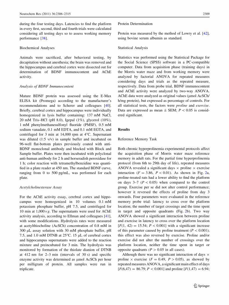
Both chronic hyperprolinemia experimental protocols affect
the acquisition phase of Morris water maze reference
memory in adult rats. For the partial time hyperprolinemia
protocol (from 6th to 29th day of life), repeated measures
ANOVA revealed a significant days 9 proline 9 exercise
interaction (F = 3.86, P \ 0.01). As shown in Fig. 2a,
proline-treated rats had a lower ability to find the platform
on days 3–7 (P \ 0.05) when compared to the control
group. Exercise per se did not alter control performance;
however it reversed the effects of proline from day 3
onwards. Four parameters were evaluated in the reference
memory probe trial: latency to cross over the platform
location; the number of target crossings and the time spent
in target and opposite quadrants (Fig. 2b). Two way
ANOVA showed a significant interaction between proline
and exercise in latency to cross over the platform location
[F(1, 42) = 15.54; P \ 0.001] with a significant increase
of this parameter caused by proline treatment (P \ 0.001);
this effect was also reversed by exercise. Proline and/or
exercise did not alter the number of crossings over the
platform location, neither the time spent in target or
opposite quadrants (P [ 0.05 in all cases).
Although there was no significant interaction of days 9
proline 9 exercise (F = 0.49, P [ 0.05), as showed by
repeated measures ANOVA, a significant main effect of days
[F(6,47) = 86.79; P \ 0.001] and proline [F(1,47) = 6.94;
Neurochem Res (2011) 36:2306–2315 2309
123
P \ 0.05], but not of exercise [F(1,47) = 2.26; P [ 0.05]
was revealed. As depicted in Fig. 3a, proline-treated rats had
a lower performance to find the platform from day 4 to 7
(P \ 0.05). Exercise per se did not affect control perfor-
mance, but reversed the proline cognitive effect. Two way
ANOVA of the probe trial (Fig. 3b) showed a significant
interaction of proline 9 exercise in the latency to cross over
the platform location [F = 12.80; P \ 0.01]. Proline treat-
ment significantly increased latency to cross over the plat-
form location when compared to control group (P \ 0.05)
and this effect was also reversed by exercise. However,
proline and/or exercise did not alter other probe trial vari-
ables (P [ 0.05 in all cases).
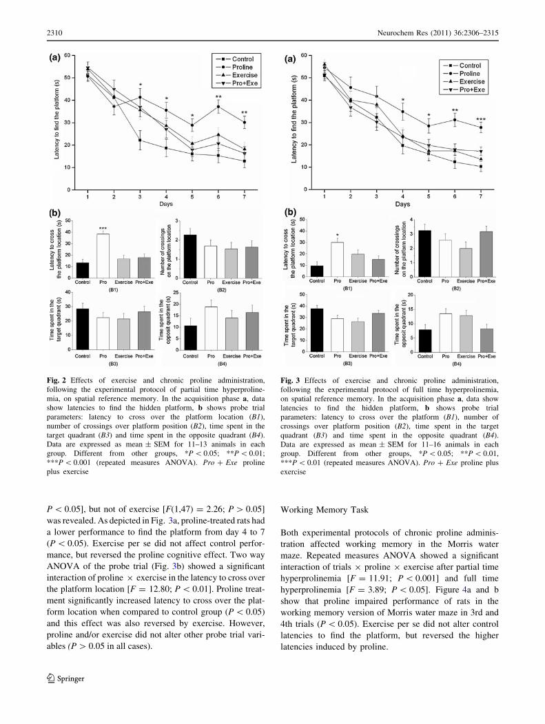
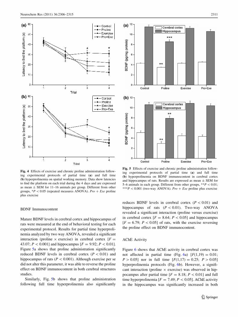
Working Memory Task
Both experimental protocols of chronic proline adminis-
tration affected working memory in the Morris water
maze. Repeated measures ANOVA showed a significant
interaction of trials 9 proline 9 exercise after partial time
hyperprolinemia [F = 11.91; P \ 0.001] and full time
hyperprolinemia [F = 3.89; P \ 0.05]. Figure 4a and b
show that proline impaired performance of rats in the
working memory version of Morris water maze in 3rd and
4th trials (P \ 0.05). Exercise per se did not alter control
latencies to find the platform, but reversed the higher
latencies induced by proline.
Fig. 2 Effects of exercise and chronic proline administration,
following the experimental protocol of partial time hyperproline-
mia, on spatial reference memory. In the acquisition phase a, data
show latencies to find the hidden platform, b shows probe trial
parameters: latency to cross over the platform location (B1),
number of crossings over platform position (B2), time spent in the
target quadrant (B3) and time spent in the opposite quadrant (B4).
Data are expressed as mean ± SEM for 11–13 animals in each
group. Different from other groups, *P \ 0.05; **P \ 0.01;
***P \ 0.001 (repeated measures ANOVA). Pro ? Exe proline
plus exercise
Fig. 3 Effects of exercise and chronic proline administration,
following the experimental protocol of full time hyperprolinemia,
on spatial reference memory. In the acquisition phase a, data show
latencies to find the hidden platform, b shows probe trial
parameters: latency to cross over the platform (B1), number of
crossings over platform position (B2), time spent in the target
quadrant (B3) and time spent in the opposite quadrant (B4).
Data are expressed as mean ± SEM for 11–16 animals in each
group. Different from other groups, *P \ 0.05; **P \ 0.01,
***P \ 0.01 (repeated measures ANOVA). Pro ? Exe proline plus
exercise
2310 Neurochem Res (2011) 36:2306–2315
123
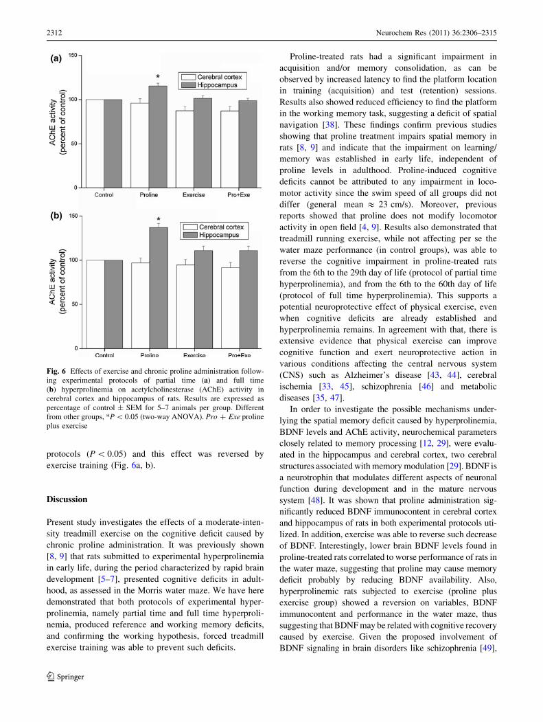
BDNF Immunocontent
Mature BDNF levels in cerebral cortex and hippocampus of
rats were measured at the end of behavioral testing for each
experimental protocol. Results for partial time hyperproli-
nemia analyzed by two way ANOVA, revealed a significant
interaction (proline 9 exercise) in cerebral cortex [F =
43.07; P \ 0.001] and hippocampus [F = 9.92; P \ 0.01].
Figure 5a shows that proline administration significantly
reduced BDNF levels in cerebral cortex (P \ 0.01) and
hippocampus of rats (P \ 0.001). Although exercise per se
did not alter this parameter, it was able to reverse the proline
effect on BDNF immunocontent in both cerebral structures
studies.
Similarly, Fig. 5b shows that proline administration
following full time hyperprolinemia also significantly
reduces BDNF levels in cerebral cortex (P \ 0.01) and
hippocampus of rats (P \ 0.01). Two-way ANOVA
revealed a significant interaction (proline versus exercise)
in cerebral cortex [F = 8.64; P \ 0.05] and hippocampus
[F = 6.79; P \ 0.05] of rats, with the exercise reversing
the proline effect on BDNF immunocontent.
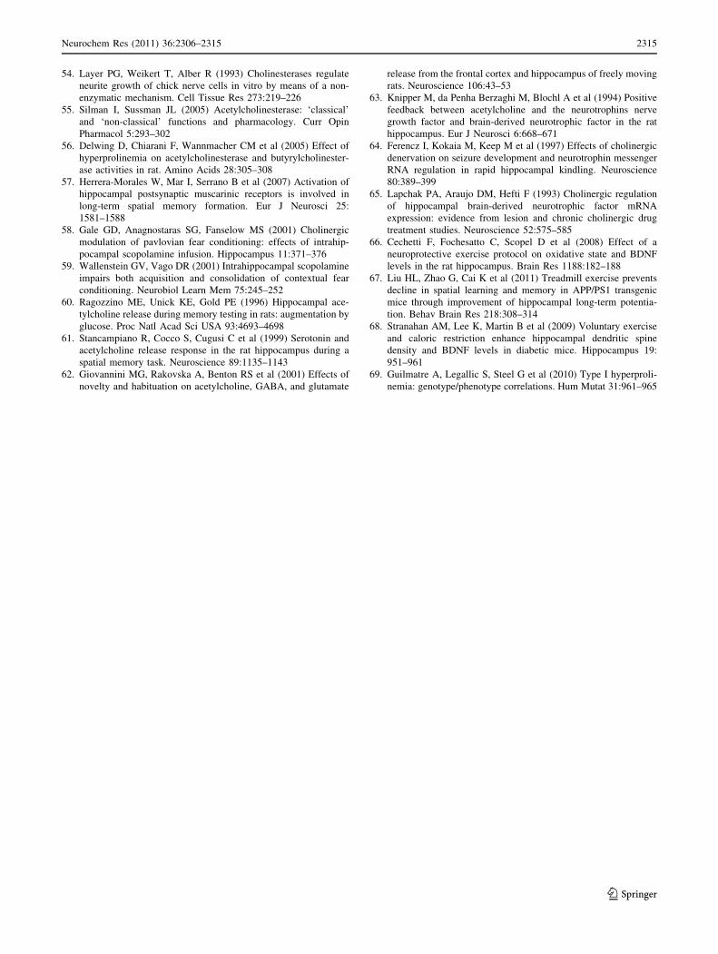
AChE Activity
Figure 6 shows that AChE activity in cerebral cortex was
not affected in partial time (Fig. 6a) [F(1,19) = 0.01;
P [ 0.05] nor in full time [F(1,17) = 0.25; P [ 0.05]
hyperprolinemia protocols (Fig. 6b). However, a signifi-
cant interaction (proline 9 exercise) was observed in hip-
pocampus after partial time [F = 8.18; P \ 0.01] and full
time hyperprolinemia [F = 7.49; P \ 0.05]. AChE activity
in the hippocampus was significantly increased in both
Fig. 4 Effects of exercise and chronic proline administration follow-
ing experimental protocols of partial time (a) and full time
(b) hyperprolinemia on spatial working memory. Data show latencies
to find the platform on each trial during the 4 days and are expressed
as mean ± SEM for 11–16 animals per group. Different from other
groups, *P \ 0.05 (repeated measures ANOVA). Pro ? Exe proline
plus exercise
Fig. 5 Effects of exercise and chronic proline administration follow-
ing experimental protocols of partial time (a) and full time
(b) hyperprolinemia on BDNF immunocontent in cerebral cortex
and hippocampus of rats. Results are expressed as mean ± SEM for
5–6 animals in each group. Different from other groups, **P \ 0.01;
***P \ 0.001 (two-way ANOVA). Pro ? Exe proline plus exercise
Neurochem Res (2011) 36:2306–2315 2311
123
protocols (P \ 0.05) and this effect was reversed by
exercise training (Fig. 6a, b).
Discussion
Present study investigates the effects of a moderate-inten-
sity treadmill exercise on the cognitive deficit caused by
chronic proline administration. It was previously shown
[8, 9] that rats submitted to experimental hyperprolinemia
in early life, during the period characterized by rapid brain
development [5–7], presented cognitive deficits in adult-
hood, as assessed in the Morris water maze. We have here
demonstrated that both protocols of experimental hyper-
prolinemia, namely partial time and full time hyperproli-
nemia, produced reference and working memory deficits,
and confirming the working hypothesis, forced treadmill
exercise training was able to prevent such deficits.
Proline-treated rats had a significant impairment in
acquisition and/or memory consolidation, as can be
observed by increased latency to find the platform location
in training (acquisition) and test (retention) sessions.
Results also showed reduced efficiency to find the platform
in the working memory task, suggesting a deficit of spatial
navigation [38]. These findings confirm previous studies
showing that proline treatment impairs spatial memory in
rats [8, 9] and indicate that the impairment on learning/
memory was established in early life, independent of
proline levels in adulthood. Proline-induced cognitive
deficits cannot be attributed to any impairment in loco-
motor activity since the swim speed of all groups did not
differ (general mean & 23 cm/s). Moreover, previous
reports showed that proline does not modify locomotor
activity in open field [4, 9]. Results also demonstrated that
treadmill running exercise, while not affecting per se the
water maze performance (in control groups), was able to
reverse the cognitive impairment in proline-treated rats
from the 6th to the 29th day of life (protocol of partial time
hyperprolinemia), and from the 6th to the 60th day of life
(protocol of full time hyperprolinemia). This supports a
potential neuroprotective effect of physical exercise, even
when cognitive deficits are already established and
hyperprolinemia remains. In agreement with that, there is
extensive evidence that physical exercise can improve
cognitive function and exert neuroprotective action in
various conditions affecting the central nervous system
(CNS) such as Alzheimer’s disease [43, 44], cerebral
ischemia [33, 45], schizophrenia [46] and metabolic
diseases [35, 47].
In order to investigate the possible mechanisms under-
lying the spatial memory deficit caused by hyperprolinemia,
BDNF levels and AChE activity, neurochemical parameters
closely related to memory processing [12, 29], were evalu-
ated in the hippocampus and cerebral cortex, two cerebral
structures associated with memory modulation [29]. BDNF is
a neurotrophin that modulates different aspects of neuronal
function during development and in the mature nervous
system [48]. It was shown that proline administration sig-
nificantly reduced BDNF immunocontent in cerebral cortex
and hippocampus of rats in both experimental protocols uti-
lized. In addition, exercise was able to reverse such decrease
of BDNF. Interestingly, lower brain BDNF levels found in
proline-treated rats correlated to worse performance of rats in
the water maze, suggesting that proline may cause memory
deficit probably by reducing BDNF availability. Also,
hyperprolinemic rats subjected to exercise (proline plus
exercise group) showed a reversion on variables, BDNF
immunocontent and performance in the water maze, thus
suggesting that BDNF may be related with cognitive recovery
caused by exercise. Given the proposed involvement of
BDNF signaling in brain disorders like schizophrenia [49],
Fig. 6 Effects of exercise and chronic proline administration follow-
ing experimental protocols of partial time (a) and full time
(b) hyperprolinemia on acetylcholinesterase (AChE) activity in
cerebral cortex and hippocampus of rats. Results are expressed as
percentage of control ± SEM for 5–7 animals per group. Different
from other groups, *P \ 0.05 (two-way ANOVA). Pro ? Exe proline
plus exercise
2312 Neurochem Res (2011) 36:2306–2315
123

traumatic injury [50], dementia [51] and Alzheimer’s disease
[52], the finding that a short and moderate exercise protocol is
sufficient to enhance learning and memory suggests that
exercise might be an accessible form of intervention that
could be used in conjunction with standard care measures for
hyperprolinemic patients [53].
AChE plays a key role in synaptic transmission by
hydrolyzing ACh. It has also been associated with brain
development, learning and memory [27, 28] and several
‘‘nonclassical’’ AChE activities have been described, as
neurite growth [54] and synaptic development and main-
tenance [55]. Presented results show that chronic proline
administration did not alter AChE in cerebral cortex of rats.
In this context, a previous study showed that cortical AChE
activity was decreased 1 h after the administration of
proline (acute treatment), but not 12 h after the last injec-
tion of proline in chronic treatment when the levels of this
amino acid returned to normal values [56], raising the
hypothesis that the presence of proline is necessary to
promote such effect in this cerebral structure. On the other
hand, our results also show that the activity of AChE was
significantly increased in the hippocampus of rats subjected
to both experimental models of chronic proline adminis-
tration (partial and full time hyperprolinemia) and sacri-
ficed after behavioral studies (adulthood rats). In this
scenario, it is conceivable that a decrease in ACh levels
will follow, reducing the cholinergic activity in this cere-
bral structure, what could be associated with the memory
impairment observed. Results also revealed that in exer-
cised hyperprolinemic rats (proline plus exercise group) the
activity of enzyme returned to normal (similar to control),
suggesting that exercise can influence brain cholinergic
mechanisms. These findings are supported by studies
showing the interaction of hippocampus with cholinergic
innervations and their relationship to memory processes. In
this context, it has been demonstrated that cholinergic
activation of the hippocampus is necessary for both spatial
and contextual memory consolidation [57–59] and that
extracellular ACh levels in the hippocampus increase
during exploration or learning [60–62]. Extensive literature
supports the idea that an ACh-mediated mechanism also
regulates BDNF gene expression in the hippocampus par-
ticularly [63–65], suggesting that ACh-mediated activation
of the hippocampus could underlie the regulation of BDNF
by exercise [12].
It is worth noting that the exercise protocol used in this
study did not influence per se the behavioral performance,
BNDF levels neither AChE in control rats. This observa-
tion is in agreement with other reports using a similar
exercise protocol [47, 66], although other studies have
shown that exercise increases BDNF levels [53, 67]. These
controversial results might be attributed to many factors,
such as distinct exercise duration and intensity, the
motivation for physical activity (either forced or voluntary)
as well as the age of experimental animals [11, 68].
Summarizing, present study demonstrates through two
experimental protocols that the impairment on cognition
caused by hyperprolinemia was established in early life and
appears not be related whether proline levels continues high
throughout life. Indeed, it has been described that any defect
causing even a modest increase in proline levels during
critical periods of development may have a dramatic effect
on CNS functioning [69]. Elucidation of the mechanisms
through which proline exerts its effects on spatial learning/
memory requires more studies; the reduction in neurotro-
phins levels, such as BDNF, and modulation of cholinergic
system appear to be involved. These data also reinforce the
potential neuroprotective role of physical exercise on
learning/memory in metabolic diseases such as hyperproli-
nemia, even when cognitive deficits are already established.
Acknowledgments This work was supported by grants from
Conselho Nacional de Desenvolvimento Cientıfico e Tecnologico
(CNPq—Brazil) and Fundacao de Amparo a Pesquisa do Estado do
Rio Grande do Sul (FAPERGS).
References
1. Hu CA, Bart Williams D, Zhaorigetu S et al (2008) Functional
genomics and SNP analysis of human genes encoding proline
metabolic enzymes. Amino Acids 35:655–664
2. Phang JM, Hu CA, Valle D (2001) Disorders of proline and
hydroxyproline metabolism. In: Scriver CR, Beaudet AL, Sly
WS, Valle D (eds) The metabolic and molecular bases of inher-
ited disease. McGraw-Hill, New York, pp 1821–1838
3. Pontes ZL, Oliveira LS, Franzon R et al (2001) Inhibition of Na?,
K?-ATPase activity from rat hippocampus by proline. Neuro-
chem Res 26:1321–1326
4. Moreira JC, Wannmacher CM, Costa SM et al (1989) Effect of
proline administration on rat behavior in aversive and nonaver-
sive tasks. Pharmacol Biochem Behav 32:885–890
5. Davis HP, Squire LR (1984) Protein synthesis and memory: a
review. Psychol Bull 96:518–559
6. Erecinska M, Cherian S, Silver IA (2004) Energy metabolism in
mammalian brain during development. Prog Neurobiol 73:
397–445
7. Aghajanian GK, Bloom FE (1967) The formation of synaptic
junctions in developing rat brain: a quantitative electron micro-
scopic study. Brain Res 6:716–727
8. Bavaresco CS, Streck EL, Netto CA et al (2005) Chronic
hyperprolinemia provokes a memory deficit in the Morris water
maze task. Metab Brain Dis 20:73–80
9. Delwing D, Bavaresco CS, Monteiro SC et al (2006) Alpha-
tocopherol and ascorbic acid prevent memory deficits provoked
by chronic hyperprolinemia in rats. Behav Brain Res 168:185–189
10. Mitsubuchi H, Nakamura K, Matsumoto S et al (2008) Inborn
errors of proline metabolism. J Nutr 138:2016–2020
11. Berchtold NC, Castello N, Cotman CW (2010) Exercise and
time-dependent benefits to learning and memory. Neuroscience
167:588–597
12. Cotman CW, Berchtold NC (2002) Exercise: a behavioral inter-
vention to enhance brain health and plasticity. Trends Neurosci
25:295–301
Neurochem Res (2011) 36:2306–2315 2313
123

13. Kramer AF (1999) Ageing, fitness and neurocognitive function.
Nature 402:750
14. Sutoo D, Akiyama K (2003) Regulation of brain function by
exercise. Neurobiol Dis 13:1–14
15. van Praag H, Shubert T, Zhao CM et al (2005) Exercise enhances
learning and hippocampal neurogenesis in aged mice. J Neurosci
25:8680–8685
16. Ang ET, Dawe GS, Wong PT et al (2006) Alterations in spatial
learning and memory after forced exercise. Brain Res 1113:
186–193
17. Wu CW, Chen YC, Yu L et al (2007) Treadmill exercise coun-
teracts the suppressive effects of peripheral lipopolysaccharide on
hippocampal neurogenesis and learning and memory. J Neuro-
chem 103:2471–2481
18. Griesbach GS, Hovda DA, Gomez-Pinilla F (2009) Exercise-
induced improvement in cognitive performance after traumatic
brain injury in rats is dependent on BDNF activation. Brain Res
1288:105–115
19. Bekinschtein P, Cammarota M, Izquierdo I et al (2008) BDNF
and memory formation and storage. Neuroscientist 14:147–156
20. Tyler WJ, Alonso M, Bramham CR et al (2002) From acquisition
to consolidation: on the role of brain-derived neurotrophic factor
signaling in hippocampal-dependent learning. Learn Mem 9:
224–237
21. Albensi BC (2001) Models of brain injury and alterations in
synaptic plasticity. J Neurosci Res 65:279–283
22. Levine ES, Crozier RA, Black IB et al (1998) Brain-derived
neurotrophic factor modulates hippocampal synaptic transmission
by increasing N-methyl-D-aspartic acid receptor activity. Proc
Natl Acad Sci USA 95:10235–10239
23. Levine ES, Dreyfus CF, Black IB et al (1995) Brain-derived
neurotrophic factor rapidly enhances synaptic transmission in
hippocampal neurons via postsynaptic tyrosine kinase receptors.
Proc Natl Acad Sci USA 92:8074–8077
24. Takei N, Sasaoka K, Inoue K et al (1997) Brain-derived neuro-
trophic factor increases the stimulation-evoked release of gluta-
mate and the levels of exocytosis-associated proteins in cultured
cortical neurons from embryonic rats. J Neurochem 68:370–375
25. Hall J, Thomas KL, Everitt BJ (2000) Rapid and selective
induction of BDNF expression in the hippocampus during con-
textual learning. Nat Neurosci 3:533–535
26. Yamada K, Mizuno M, Nabeshima T (2002) Role for brain-
derived neurotrophic factor in learning and memory. Life Sci
70:735–744
27. Zimmerman G, Soreq H (2006) Termination and beyond: ace-
tylcholinesterase as a modulator of synaptic transmission. Cell
Tissue Res 326:655–669
28. Ballard CG, Greig NH, Guillozet-Bongaarts AL et al (2005)
Cholinesterases: roles in the brain during health and disease. Curr
Alzheimer Res 2:307–318
29. Deiana S, Platt B, Riedel G (2011) The cholinergic system and
spatial learning. Behav Brain Res 221:389–411
30. Amenta F, Tayebati SK (2008) Pathways of acetylcholine syn-
thesis, transport and release as targets for treatment of adult-onset
cognitive dysfunction. Curr Med Chem 15:488–498
31. Zugno AI, Pereira LO, Mattos C et al (2008) Guanidinoacetate
administration increases acetylcholinesterase activity in striatum
of rats and impairs retention of an inhibitory avoidance task.
Metab Brain Dis 23:189–198
32. Stefanello FM, Monteiro SC, Matte C et al (2007) Hypermethi-
oninemia increases cerebral acetylcholinesterase activity and
impairs memory in rats. Neurochem Res 32:1868–1874
33. Cechetti F, Rhod A, Simao F et al (2007) Effect of treadmill
exercise on cell damage in rat hippocampal slices submitted to
oxygen and glucose deprivation. Brain Res 1157:121–125
34. Scopel D, Fochesatto C, Cimarosti H et al (2006) Exercise
intensity influences cell injury in rat hippocampal slices
exposed to oxygen and glucose deprivation. Brain Res Bull 71:
155–159
35. Ben J, Soares FM, Cechetti F et al (2009) Exercise effects on
activities of Na?, K?-ATPase, acetylcholinesterase and adenine
nucleotides hydrolysis in ovariectomized rats. Brain Res 1302:
248–255
36. Brooks GA, White TP (1978) Determination of metabolic and
heart rate responses of rats to treadmill exercise. J Appl Physiol
45:1009–1015
37. Arida RM, Scorza FA, dos Santos NF et al (1999) Effect of
physical exercise on seizure occurrence in a model of temporal
lobe epilepsy in rats. Epilepsy Res 37:45–52
38. Netto CA, Hodges H, Sinden JD et al (1993) Effects of fetal
hippocampal field grafts on ischaemic-induced deficits in spatial
navigation in the water maze. Neuroscience 54:69–92
39. D’Hooge R, De Deyn PP (2001) Applications of the Morris water
maze in the study of learning and memory. Brain Res Rev
36:60–90
40. Scherer EB, da Cunha MJ, Matte C et al (2010) Methylphenidate
affects memory, brain-derived neurotrophic factor immunocon-
tent and brain acetylcholinesterase activity in the rat. Neurobiol
Learn Mem 94:247–253
41. Ellman GL, Courtney KD, Andres V Jr et al (1961) A new and
rapid colorimetric determination of acetylcholinesterase activity.
Biochem Pharmacol 7:88–95
42. Lowry OH, Rosebrough NJ, Farr AL et al (1951) Protein mea-
surement with the Folin phenol reagent. J Biol Chem 193:
265–275
43. Adlard PA, Perreau VM, Pop V et al (2005) Voluntary exercise
decreases amyloid load in a transgenic model of Alzheimer’s
disease. J Neurosci 25:4217–4221
44. Um HS, Kang EB, Leem YH et al (2008) Exercise training acts as
a therapeutic strategy for reduction of the pathogenic phenotypes
for Alzheimer’s disease in an NSE/APPsw-transgenic model. Int
J Mol Med 22:529–539
45. Zhang F, Wu Y, Jia J (2011) Exercise preconditioning and brain
ischemic tolerance. Neuroscience 177:170–176
46. Pajonk FG, Wobrock T, Gruber O et al (2010) Hippocampal
plasticity in response to exercise in schizophrenia. Arch Gen
Psychiatry 67:133–143
47. Ben J, Soares FM, Scherer EB et al (2010) Running exercise
effects on spatial and avoidance tasks in ovariectomized rats.
Neurobiol Learn Mem 94:312–317
48. Gottmann K, Mittmann T, Lessmann V (2009) BDNF signaling
in the formation, maturation and plasticity of glutamatergic and
GABAergic synapses. Exp Brain Res 199:203–234
49. Egan MF, Weinberger DR, Lu B (2003) Schizophrenia, III: brain-
derived neurotropic factor and genetic risk. Am J Psychiatry
160:1242
50. Horsfield SA, Rosse RB, Tomasino V et al (2002) Fluoxetine’s
effects on cognitive performance in patients with traumatic brain
injury. Int J Psychiatry Med 32:337–344
51. Ando S, Kobayashi S, Waki H et al (2002) Animal model of
dementia induced by entorhinal synaptic damage and partial
restoration of cognitive deficits by BDNF and carnitine. J Neu-
rosci Res 70:519–527
52. Tsai SJ, Hong CJ, Liu HC et al (2004) Association analysis of
brain-derived neurotrophic factor Val66Met polymorphisms with
Alzheimer’s disease and age of onset. Neuropsychobiology
49:10–12
53. Vaynman S, Ying Z, Gomez-Pinilla F (2004) Hippocampal
BDNF mediates the efficacy of exercise on synaptic plasticity and
cognition. Eur J Neurosci 20:2580–2590
2314 Neurochem Res (2011) 36:2306–2315
123
54. Layer PG, Weikert T, Alber R (1993) Cholinesterases regulate
neurite growth of chick nerve cells in vitro by means of a non-
enzymatic mechanism. Cell Tissue Res 273:219–226
55. Silman I, Sussman JL (2005) Acetylcholinesterase: ‘classical’
and ‘non-classical’ functions and pharmacology. Curr Opin
Pharmacol 5:293–302
56. Delwing D, Chiarani F, Wannmacher CM et al (2005) Effect of
hyperprolinemia on acetylcholinesterase and butyrylcholinester-
ase activities in rat. Amino Acids 28:305–308
57. Herrera-Morales W, Mar I, Serrano B et al (2007) Activation of
hippocampal postsynaptic muscarinic receptors is involved in
long-term spatial memory formation. Eur J Neurosci 25:
1581–1588
58. Gale GD, Anagnostaras SG, Fanselow MS (2001) Cholinergic
modulation of pavlovian fear conditioning: effects of intrahip-
pocampal scopolamine infusion. Hippocampus 11:371–376
59. Wallenstein GV, Vago DR (2001) Intrahippocampal scopolamine
impairs both acquisition and consolidation of contextual fear
conditioning. Neurobiol Learn Mem 75:245–252
60. Ragozzino ME, Unick KE, Gold PE (1996) Hippocampal ace-
tylcholine release during memory testing in rats: augmentation by
glucose. Proc Natl Acad Sci USA 93:4693–4698
61. Stancampiano R, Cocco S, Cugusi C et al (1999) Serotonin and
acetylcholine release response in the rat hippocampus during a
spatial memory task. Neuroscience 89:1135–1143
62. Giovannini MG, Rakovska A, Benton RS et al (2001) Effects of
novelty and habituation on acetylcholine, GABA, and glutamate
release from the frontal cortex and hippocampus of freely moving
rats. Neuroscience 106:43–53
63. Knipper M, da Penha Berzaghi M, Blochl A et al (1994) Positive
feedback between acetylcholine and the neurotrophins nerve
growth factor and brain-derived neurotrophic factor in the rat
hippocampus. Eur J Neurosci 6:668–671
64. Ferencz I, Kokaia M, Keep M et al (1997) Effects of cholinergic
denervation on seizure development and neurotrophin messenger
RNA regulation in rapid hippocampal kindling. Neuroscience
80:389–399
65. Lapchak PA, Araujo DM, Hefti F (1993) Cholinergic regulation
of hippocampal brain-derived neurotrophic factor mRNA
expression: evidence from lesion and chronic cholinergic drug
treatment studies. Neuroscience 52:575–585
66. Cechetti F, Fochesatto C, Scopel D et al (2008) Effect of a
neuroprotective exercise protocol on oxidative state and BDNF
levels in the rat hippocampus. Brain Res 1188:182–188
67. Liu HL, Zhao G, Cai K et al (2011) Treadmill exercise prevents
decline in spatial learning and memory in APP/PS1 transgenic
mice through improvement of hippocampal long-term potentia-
tion. Behav Brain Res 218:308–314
68. Stranahan AM, Lee K, Martin B et al (2009) Voluntary exercise
and caloric restriction enhance hippocampal dendritic spine
density and BDNF levels in diabetic mice. Hippocampus 19:
951–961
69. Guilmatre A, Legallic S, Steel G et al (2010) Type I hyperproli-
nemia: genotype/phenotype correlations. Hum Mutat 31:961–965
Neurochem Res (2011) 36:2306–2315 2315
123
Copyright © 2022 FDOKUMEN