Function and immuno-localization of aquaporins in the Antarctic midge Belgica antarctica
Upload
independentCategory
view
1download
0
Biochimica et Biophysica Acta 1817 (2012) 1314–1321
Contents lists available at SciVerse ScienceDirect
Biochimica et Biophysica Acta
j ourna l homepage: www.e lsev ie r .com/ locate /bbabio
Photosynthetic electron flow to oxygen and diffusion of hydrogen peroxide throughthe chloroplast envelope via aquaporins☆
Maria M. (Mubarakshina) Borisova ⁎, Marina A. Kozuleva, Natalia N. Rudenko, Ilya A. Naydov,Irina B. Klenina, Boris N. IvanovInstitute of Basic Biological Problems RAS, 142290 Pushchino, Institutskaya st., 2, Moscow region, Russia
Abbreviations: Amplex Red, 10-acetyl-3,7-dihydroxyphCA, carbonic anhydrase; CHA, cyclic hydroxylamine; Chl, cGr D, gramicidin D; MV, methyl viologen; POBN, α-(4-pyriPQH2, plastohydroquinone, plastoquinol; PSI, photosystemtive oxygen species; SOD, superoxide dismutase; TMT-2,2,6,6-tetramethylpiperidinium☆ This article is part of a Special Issue entitled: Photosyntfrom Natural to Artificial.⁎ Corresponding author. Tel.: +7 4967732448; fax: +
E-mail address: [email protected] (M.M
0005-2728/$ – see front matter © 2012 Elsevier B.V. Aldoi:10.1016/j.bbabio.2012.02.036
a b s t r a c t
a r t i c l e i n f oArticle history:Received 19 October 2011Received in revised form 27 February 2012Accepted 28 February 2012Available online 6 March 2012
Keywords:ChloroplastThylakoidSuperoxide radicalHydrogen peroxideAquaporinCarbonic anhydrase
Light-induced generation of superoxide radicals and hydrogen peroxide in isolated thylakoids has been studiedwith a lipophilic spin probe, cyclic hydroxylamine 1-hydroxy-4-isobutyramido-2,2,6,6-tetramethylpiperidinium(TMT-H) to detect superoxide radicals, and the spin trap α-(4-pyridyl-1-oxide)-N-tert-butylnitron (4-POBN) todetect hydrogen peroxide-derived hydroxyl radicals. Accumulation of the radical products of the above reactionshas been followed using electron paramagnetic resonance. It is found that the increased production of superox-ide radicals and hydrogen peroxide in higher light is due to the enhanced production of these species within thethylakoid membrane, rather than outside the membrane. Fluorescent probe Amplex red, which forms fluores-cent product, resorufin, in the reactionwith hydrogen peroxide, has been used to detect hydrogen peroxide out-side isolated chloroplasts using confocal microscopy. Resorufin fluorescence outside the chloroplasts is found tobe suppressed by 60% in the presence of the inhibitor of aquaporins, acetazolamide (AZA), indicating that hydro-gen peroxide can diffuse through the chloroplast envelope aquaporins. It is demonstrated that AZA also inhibitscarbonic anhydrase activity of the isolated envelope. We put forward a hypothesis that carbonic anhydrase pre-sumably can be attached to the envelope aquaporins. This article is part of a Special Issue entitled: PhotosynthesisResearch for Sustainability: from Natural to Artificial.
© 2012 Elsevier B.V. All rights reserved.
1. Introduction
Chloroplasts use energy of sunlight to oxidize H2O and evolve O2.Electrons are transferred via the photosynthetic electron transportchain to NADP+, which in its reduced form (NADPH) is used for CO2
assimilation. An excess of light energy beyond the necessary one tofulfill CO2 assimilation can be achieved not only under strong lightbut also under moderate and even low light, as well as under otherenvironmental stress conditions. Additionally to thermal dissipationin antenna, the electron flow from the photosynthetic electron trans-port chain to molecular oxygen (the so-called Mehler reaction) caneliminate the excess of light energy, thus preventing the chain fromphotoinhibition (for review see Ref. [1]). Besides, the electron flowto O2 leads to formation of reactive oxygen species (ROS) such as su-peroxide anion radical, O2
•−, and hydrogen peroxide, H2O2. Under
enoxazine; AZA, acetazolamide;hlorophyll; cyt c, cytochrome c;dyl-1-oxide)-N-tert-butylnitron;I; PSII, photosystem II; ROS, reac-H, 1-hydroxy-4-isobutyramido-
hesis Research for Sustainability:
7 4967330532.. (Mubarakshina) Borisova).
l rights reserved.
normal functional conditions the reduction of molecular oxygenmay represent about 5–10% of the total photosynthetic electronflow in C3 plants, however it can increase to 30% and even higherunder stress conditions [2,3].
The production of O2•−, the primary product of the Mehler reac-
tion, was repeatedly shown in isolated thylakoids, using specificO2
•− detectors such as cytochrome c (cyt c), epinephrine [4], ascor-bate [5], Tiron [6,7], 5,5-dimethyl-pyrrolin n-oxide (DMPO) [7,8]and 2-ethoxycarbonyl-2-methyl-3,4-dihydro-2H-pyrrole-1-oxide(EMPO) [9]. These compounds are hydrophilic, meaning that theydetect O2
•− outside the thylakoid membrane. The possibility of O2•−
productionwithin the thylakoidmembranewas proposed by Takahashiand Asada [10], who provided some indirect indications of thisprocess. Recently, the first clear demonstration of O2
•− generationwithin the thylakoid membrane was presented using cyclic hydroxyl-amines to detect O2
•− [11]. The evidence supporting the formation ofH2O2, the secondary product of the Mehler reaction, outside andwithin the thylakoid membrane was also presented [12]. However,potential capacities of the different formation pathways of the twoROS under various light conditions still remain obscure.
ROS are known to be damaging molecules. However, in the earlystages of stress, the ROS that are produced also play a major role incellular signaling pathways and this is particularly the case withH2O2 (for review see Ref. [13]). Initiation of the signaling pathwaysis obligatory for acclimation of plants to environmental stress

1315M.M. (Mubarakshina) Borisova et al. / Biochimica et Biophysica Acta 1817 (2012) 1314–1321
conditions in order to maintain the main chloroplast function, i.e.,photosynthesis (for recent reviews see Refs. [14–17]). H2O2 leads tovast changes on the level of gene expression in plants [18–22]. For ex-ample, it was shown that H2O2 might be the redox signal that acti-vates the expression of cytosolic ascorbate peroxidase [23–25]. Theretrograde signaling with involvement of H2O2 is likely to be imple-mented by inducing protein phosphorylation of mitogen-activatedprotein kinases (MAPKs) [26], which are involved in signaling path-ways [27]. The reversible redoxmodulation of Cys residues of the pro-teins is most likely to be the mechanism of the H2O2-mediatedactivation of MAPK pathways. Previously we demonstrated that afraction of H2O2 produced in chloroplasts can diffuse from the chloro-plasts to cytoplasm [28]. This implies that H2O2 can initiate signalingpathways both inside and outside the organelle. The mechanism ofH2O2 diffusion through the chloroplast envelope however is stillunder debate.
Aquaporins, i.e., channel proteins, facilitate the transport not onlyof water but also of small neutral solutes (urea, boric acid, silicic acid)and gasses (NH3 and CO2) through the plasma membrane and intra-cellular membranes of plant cells (for review see Ref. [29]). It hasbeen recently found that the tobacco (Nicotiana tabacum) inner chlo-roplast envelope membrane contains the aquaporin Nt AQP1, whichenables CO2 transport through the membrane [30]. The expressionof human and plant aquaporins in yeast revealed that aquaporins fa-cilitated the diffusion of hydrogen peroxide across the membranes[31].
The aims of the present work were i) to study the capacities of thepathways of O2
•− and H2O2 generation by the photosynthetic electrontransport chain in different locations within the chloroplast, namelyin the chloroplast stroma (outside the thylakoid membrane) andwithin the thylakoid membrane, under different light conditions; ii)to study the role of the chloroplast envelope aquaporins in H2O2
diffusion. It is shown that the increase of O2•− and H2O2 production
by thylakoids with the increase in the light intensity results fromthe acceleration of these processes within the thylakoid membranerather than outside. Further, hydrogen peroxide is shown to diffuseout of isolated chloroplasts through the chloroplast envelope mostlyvia aquaporins. The evidence on the presence of carbonic anhydrasein the chloroplast envelope is presented, and it is suggested that car-bonic anhydrase can be attached to aquaporins.
2. Materials and methods
2.1. Plant material
Spinach plants (Spinacia oleracea L.) andpea plants (Pisum sativum L.)were grown in the greenhouse with 16 hour day-light period (lightintensity 150 μmol quanta m−2 s−1) at 22 °C and 8 hour night periodat approximately the same temperature. The experiments with the iso-lated thylakoids were conducted using 10–14 day old pea plants. Whenthe work was performed with intact chloroplasts, 4–5 week old spinachplants were used.
2.2. Thylakoid preparation
Thylakoidswere isolated frompea leaves as described byKhorobrykhand Ivanov [32], and resuspended in the medium containing 0.4 M su-crose, 20 mMNaCl, 5 mMMgCl2 and 25 mMHEPES (pH 7.6) and storedon ice. Ascorbate was not included in the isolation mediumwhen thyla-koids were used for detection of O2
•− by cyclic hydroxylamine.
2.3. Chloroplast preparation
Chloroplasts were isolated from spinach leaves according to Ref.[33] (centrifugation in a Percoll step gradient) with some modifica-tions. The medium for 40% Percoll contained 3.33 mM EDTA,
1.66 mM MgCl2, 83.3 mM HEPES (pH 7.6) and 0.55 M sorbitol. Themedium for 80% Percoll contained 10 mM EDTA, 5 mM MgCl2,250 mM HEPES (pH 7.6) and 1.65 M sorbitol. Two green bands wereseparated after Percoll gradient centrifugation. The lower band thatcorresponded to intact chloroplasts was used. The chloroplasts werewashed in resuspension buffer without Percoll. The degree of intact-ness of chloroplasts was higher than 95% as tested by ferricyanide(1 mM K3[Fe(CN)6]) photoreduction test [34] based on the ratio oflight-driven O2 evolution measured in intact and osmotically shockedchloroplasts.
2.4. Photosynthetic activity of chloroplasts and thylakoids
Photosynthetic activity of chloroplasts and thylakoids was mea-sured in a temperature-controlled (21 °C) vessel with a Clark-typeO2-electrode. The reaction medium for chloroplasts contained0.33 M sorbitol, 20 mM NaCl, 5 mM MgCl2, 25 mM HEPES (pH 7.6)at 50 μg Chl mL−1. Intact chloroplasts were shocked for 45 s in5 mMMgCl2 and 25 mMHEPES (pH 7.6). Photosynthetic oxygen evo-lution in the broken chloroplasts in the presence of K3[Fe(CN)6] was240±38 μmol O2 mg Chl−1 h−1. 1 μM Gramicidin D (GrD) wasadded in order to suppress proton gradient. The photosynthetic oxy-gen evolution rate by intact chloroplasts in the presence of 0.5 mMphosphoglycerate and 4 mM NaHCO3 was 58.7±5.9 μmol O2 mgChl−1 h−1.
The reaction medium for thylakoids contained 0.4 M sucrose,20 mM NaCl, 5 mM MgCl2, 25 mM HEPES (pH 7.6), 20 μg Chl mL−1
and 25 μM methyl viologen (MV).
2.5. Purification of the chloroplast envelope
Purification of the chloroplast envelope from spinach chloroplastswas done as described by Douce and Joyard [35] using sucrose gradi-ent. Sucrose solutions contained 20.5 and 31.8% sucrose (w/w) corre-sponding to 0.6 M and 0.93 M, respectively, and 10 mM tricine, 4 mMMgCl2, pH 7.8 (at 2 °C). Discontinuous sucrose gradient was preparedin the tubes by carefully layering in succession 12 mL each of 31.8%and 20.5% sucrose solutions. 15 mL of swelling chloroplasts werethen layered onto the gradient and the tubes were centrifuged at72,000×g for 1 h at 3 °C. The chloroplast components were clearlyseparated giving three distinguishable subfractions: a tightly-packed, dark green pellet at the bottom of the tubes (a thylakoid sub-fraction); a yellow band at the interface of the two sucrose layers (anenvelope membrane subfraction) and a brown supernatant (a solublesubfraction).
2.6. Treatment of chloroplast envelope with acetazolamide
The isolated envelope membranes were diluted and then concen-trated again by centrifugation at 12,000×g for 15 min using the cen-trifugal filter tube (0.5 mL) Microcon YM-10 (Millipore) in order tominimize the sucrose concentration. The obtained solution was divid-ed into two tubes, the content of one was incubated with 1 mM AZAduring 30 min. Then both tubes were centrifuged at 12,000×g for15 min using the same filter, and the supernatants were removed.
2.7. Electron paramagnetic resonance measurements
Electron paramagnetic resonance (EPR) measurements were per-formed with EMX-6 ESR spectrometer (Bruker, Germany). To detectO2•−, cyclic hydroxylamine (CHA) 1-hydroxy-4-isobutyramido-
2,2,6,6-tetramethylpiperidinium (TMT-H) was used. After reactionwith superoxide radical, TMT-H forms nitroxide radical (TMT•) thatcan be detected by EPR spectroscopy [36]. The advantage is that therate constant of TMT-H oxidation by O2
•− is 4.9×10³ M−1 s−1,which is two orders of magnitude higher than the constants of O2
•−

1316 M.M. (Mubarakshina) Borisova et al. / Biochimica et Biophysica Acta 1817 (2012) 1314–1321
trapping by such established spin traps as DMPO and EMPO [37].Stock solution of TMT-H (20 mM) in 0.9% NaCl, containing 50 μM des-feroxamine and purged with argon, was prepared daily and keptunder argon on ice. Desferoxamine was used to decrease autooxida-tion of CHA catalyzed by trace amounts of iron ions. The sampleswere placed in 1 mm i.d. glass capillaries and studied at room tem-perature in ER4102ST microwave cavity with 50% transparent irradi-ation grid. The TMT• spectra were recorded at room temperature,9.47 GHz microwave frequency, 100 kHz modulation frequency, 4 Gmodulation amplitude and 20 mW microwave power. The kineticsof TMT• accumulation were recorded by monitoring the amplitudeof the low field component of the TMT• spectrum at the same micro-wave power and modulation amplitude, the time constant was 0.3 s.To calibrate the spectrometer sensitivity, 10−5 M solution of Tempol,stable nitroxide radical, was measured under identical conditions.This calibration was used to calculate the nitroxide concentration.The reaction medium contained 0.4 M sucrose, 20 mM NaCl, 5 mMMgCl2, 25 mM HEPES (pH 7.6), 50 μM desferoxamine, thylakoids at100 μg Chl mL−1, 3 mM TMT-H and 1 μM GrD.
To detect H2O2-derived hydroxyl radicals, the spin trapα-(4-pyridyl-1-oxide)-N-tert-butylnitron (4-POBN) was used. The addition of 50 μMFeEDTA produced •OH from H2O2 via the so-called Fenton reaction. 4%ethanol was added to allow the reaction between the spin trap andH2O2-derived hydroxyl radical. The reaction medium contained 0.4 Msucrose, 20 mM NaCl, 5 mM MgCl2, 25 mM HEPES (pH 7.6), thylakoidsat 15 μg Chl mL−1 and 1 μM GrD. 50 mM POBN, ethanol and FeEDTAwere added immediately after illumination. The spectra were recordedafter 5 min of incubation in 1 mm i.d. glass capillaries at room tem-perature, 9.47 GHz microwave frequency, 100 kHz modulation fre-quency, 1 G modulation amplitude, 63 mW microwave power andwere the average of 2 scans.
Reaction mixtures with chloroplasts or thylakoids were illuminatedthrough the red cut-off filter (λ>600 nm).
2.8. Measurements of carbonic anhydrase activity
The carbonic anhydrase (CA) activity was assayed with a glasselectrode at 2 °C in 13.6 mM veronal buffer (pH 8.4). The reactionwas initiated by the addition of water saturated with CO2 at 0 °C.The time of pH change from 8.1 to 7.6 was measured. The CA activitywas calculated taking into account the time of spontaneous hydrationthat was measured under the same conditions by the addition of thecorresponding volume of the medium instead of the sample. The CAactivity was expressed in μmol H+min−1 mg Protein−1 after reas-sessment of the buffer capacities both of the medium and the samplesmeasured by titration with 0.1 N HCl.
2.9. Measurements of protein content
The protein content of the chloroplast envelope was determinedaccording to Lowry et al. [37].
2.10. Observation of H2O2 using confocal microscope
H2O2 was followed by the AmplexRed fluorescence assay as de-scribed in [28], using confocal microscope Leica TCS SPE. The AmplexRed (10-acetyl-3,7-dihydroxyphenoxazine) reacts with H2O2 in thepresence of horseradish peroxidase and forms resorufin, the fluores-cent product. Resorufin has a fluorescence emission maximum of587 nm. Where indicated, 1 mM AZA (in dimethyl sulfoxide) wasadded. The chloroplast suspension was scanned with 488 nm laserfor resorufin excitation and 635 nm laser for chlorophyll excitation.Emission was detected from 540 to 600 nm for resorufin fluorescenceand from 640 to 750 nm for chlorophyll fluorescence. Laser powers ofthe microscope were set to 5% and 10%, respectively, to avoid photo-bleaching and chlorophyll degradation. The lasers acted as the
sources of photosynthetically active light, no additional light sourcewas used.
3. Results
3.1. The effect of light intensity on O2•− and H2O2 production in a thylakoid
suspension
To investigate generation of O2•− and H2O2 by the photosynthetic
electron transport chain in different locations within the chloroplast,namely outside the membrane (hydrophilic area) and within the thy-lakoid membrane (lipophilic area), and the capacities of these path-ways under different light conditions, isolated thylakoids as a modelsystem were used.
Cyclic hydroxylamines (CHAs) are widely accepted for O2•− detec-
tion in animal cells and tissues (see Ref. [36] and references therein).In the present work, O2
•− in a thylakoid suspension was detectedusing lipophilic CHA, TMT-H. TMT-H was found to have no effect onthe photosynthetic electron transport chain of thylakoids and couldnot be oxidized by components of the chain, including P700+ [11].All measurements were conducted in the presence of desferoxamine,the chelator of iron ions, in order to exclude the possible reaction ofCHA with H2O2-derived hydroxyl radicals [36]. Owing to its ratherhigh lipophilicity (partition coefficient of 35 between octanol andphosphate buffer, pH 7.4), TMT-H was able to enter the thylakoidmembrane.
Light-induced accumulation of the nitroxide radical, TMT•, wasmeasured in the absence and in the presence of superoxide dismutase(SOD) under 250 and 500 μmol quanta m−2 s−1 of light (Fig. 1). Itwas previously shown that 3 mM was the saturating concentrationof TMT-H to detect all superoxide radicals produced in a thylakoidsuspension, competing efficiently with the reaction of spontaneousdismutation of superoxide radicals [11]. SOD, a water-soluble en-zyme, was applied to catalyze the dismutation of superoxide radicalsto prevent the reaction of TMT-H with O2
•− outside the thylakoidmembrane. Since SOD can adhere to the thylakoid membrane surface[38,39], it provides dismutation of superoxide radicals produced notonly in the bulk phase but also in the aqueous regions in the vicinityof the thylakoid membrane. Fig. 1 shows that the increase in the lightintensity led to an increase of the TMT• accumulation rate both in theabsence and in the presence of SOD; the latter indicated clearly an in-crease of O2
•− generation within the thylakoid membrane. Whiletotal TMT• accumulation in a thylakoid suspension increased appr.2.5-fold under change of the light intensity from 250 to 500 μmolquanta m−2 s−1, the TMT• accumulation in the presence of SOD in-creased appr. 4-fold (the average of the 5 experiments). It is difficultto estimate the exact capacities of O2
•− generation in lipophilic vs.hydro-philic areas, since TMT• formed within the membrane can be partlyreduced by components of the electron transport chain [11], andmore-over, the “intramembrane” O2
•− can react with PQH2 (for review seeRef. [40]). Both reactions could accelerate in higher light that might re-sult in underestimation of the actual rate of O2
•− generation within themembrane.
The estimation of H2O2 generation in lipophilic vs. hydrophilic areasdoes not have the above complications, the more so that the reaction ofO2•− with PQH2 produces H2O2. Generation of H2O2-derived hydroxyl
radicals was previously measured by EPR using the spin trap 4-POBN[41]. The addition of catalase suppressed the EPR signal in thylakoidscompletely, and no signal was obtained in the dark and in the absenceof FeEDTA as well [28]. The latter showed that •OH radicals were notgenerated directly in the light but originated from the H2O2 moleculesthat were accumulated during illumination. Thus, the used approachallows H2O2 production to be reliably measured in thylakoids.
Fig. 2 shows the EPR signal of the nitroxide radicals produced inthe reaction of 4-POBN with H2O2-derived hydroxyl radicals obtainedusing isolated thylakoids after 3 min of illumination in the absence
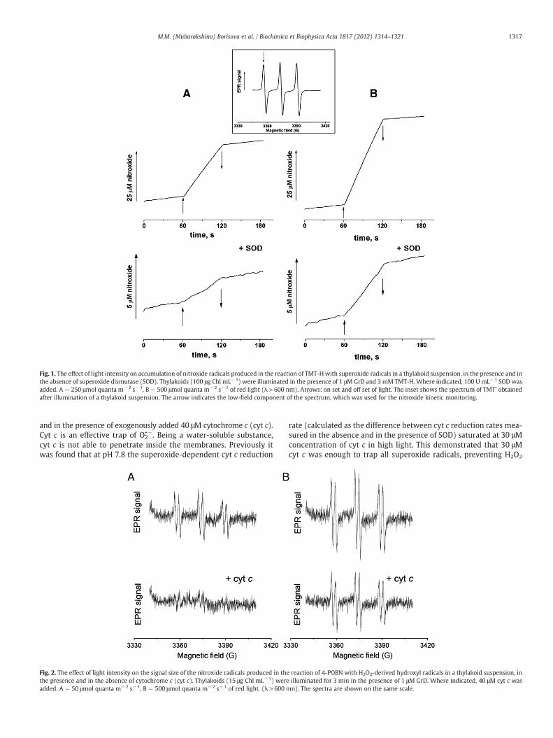
Fig. 1. The effect of light intensity on accumulation of nitroxide radicals produced in the reaction of TMT-Hwith superoxide radicals in a thylakoid suspension, in the presence and inthe absence of superoxide dismutase (SOD). Thylakoids (100 μg Chl mL−1) were illuminated in the presence of 1 μMGrD and 3 mM TMT-H. Where indicated, 100 U mL−1 SOD wasadded. A — 250 μmol quanta m−2 s−1, B — 500 μmol quanta m−2 s−1 of red light (λ>600 nm). Arrows: on set and off set of light. The inset shows the spectrum of TMT• obtainedafter illumination of a thylakoid suspension. The arrow indicates the low-field component of the spectrum, which was used for the nitroxide kinetic monitoring.
1317M.M. (Mubarakshina) Borisova et al. / Biochimica et Biophysica Acta 1817 (2012) 1314–1321
and in the presence of exogenously added 40 μM cytochrome c (cyt c).Cyt c is an effective trap of O2
•−. Being a water-soluble substance,cyt c is not able to penetrate inside the membranes. Previously itwas found that at pH 7.8 the superoxide-dependent cyt c reduction
Fig. 2. The effect of light intensity on the signal size of the nitroxide radicals produced in thethe presence and in the absence of cytochrome c (cyt c). Thylakoids (15 μg Chl mL−1) wereadded. A — 50 μmol quanta m−2 s−1, B — 500 μmol quanta m−2 s−1 of red light. (λ>600 n
rate (calculated as the difference between cyt c reduction rates mea-sured in the absence and in the presence of SOD) saturated at 30 μMconcentration of cyt c in high light. This demonstrated that 30 μMcyt c was enough to trap all superoxide radicals, preventing H2O2
reaction of 4-POBN with H2O2-derived hydroxyl radicals in a thylakoid suspension, inilluminated for 3 min in the presence of 1 μM GrD. Where indicated, 40 μM cyt c wasm). The spectra are shown on the same scale.

1318 M.M. (Mubarakshina) Borisova et al. / Biochimica et Biophysica Acta 1817 (2012) 1314–1321
production outside the membrane [12]. Added cyt c can be tightlyattached to the membrane [42,43], implying that it can react withO2
•− even in the aqueous regions in the vicinity of the thylakoidmembrane.
The EPR signal of the trapped H2O2-derived hydroxyl radicals inthe absence of cyt c (i.e., total H2O2 production) after illumination ofthylakoids with 500 μmol quanta m−2 s−1 of light was almost twiceas big as that obtained after illumination with 50 μmol quan-ta m−2 s−1 of light (Fig. 2). While a very small signal was detectedin the presence of cyt c after illumination with 50 μmol quan-ta m−2 s−1, a significant signal was obtained after illumination with500 μmol quanta m−2 s−1 that means that H2O2 production withinthe thylakoid membrane strongly increases with the increase in thelight intensity. It has been estimated that while almost no H2O2 wasproduced within the thylakoid membrane in low light (Fig. 2A),H2O2 production in high light within the membrane representedabout 50% of total H2O2 production by thylakoids (Fig. 2B). It is note-worthy that the signal presented in Fig. 2B in the absence of cyt c isalmost equal to the sum of the signals presented in Fig. 2A withoutcyt c and Fig. 2B in the presence of cyt c. Addition of KCN, the inhibitorof SOD, did not influence the EPR signal amplitude in the presence ofcyt c (data not shown). This indicated that the H2O2 production wasnot the result of possible dismutation of superoxide radicals, whichcould be catalyzed by chloroplastic SOD if this SOD attached to themembrane during isolation of thylakoids.
3.2. The role of aquaporins in H2O2 appearance outside isolated chloroplasts
Previously we have shown that a fraction of H2O2 produced insidechloroplasts can leave the chloroplasts, thus escaping the efficient an-tioxidant system located in the chloroplast stroma [28]. In the presentstudy the diffusion of H2O2 through the chloroplast envelope was in-vestigated using intact spinach chloroplasts in the presence and in theabsence of acetazolamide (AZA), which was established to be an effi-cient inhibitor of aquaporins [44–46]. AZA interacts with guanidylgroup of Arg, with the backbone carbonyl of Gly, with the carboxylof Asp, as well as with Ser, His, Ile and Asn of aquaporins. This leadsto blocking H2O diffusion via aquaporins [44,45]. AZA cannot easilypenetrate through the membranes and it has been found that 1 mMAZA did not penetrate through the membranes of isolated protoplastseven after 10 min of incubation [47]. For detection of H2O2, theAmplex Red reagent was used (see Materials and methods). It waschecked that without any chloroplasts AZA had no effect on the resor-ufin fluorescence, neither in the presence nor in the absence of addedH2O2 (not shown). Methyl viologen (MV) was added in all the casesas an efficient acceptor of electrons from Photosystem I (PSI). MVtransfers electrons to O2 leading to an increase of H2O2 generation(Table 1, see also Ref. [48]).
As shown in Fig. 3, no resorufin fluorescence was detected withoutillumination. Strong resorufin fluorescence was observed outside thechloroplasts after 3 min of illumination, showing that a significantamount of H2O2 diffused out of the chloroplasts. In the presence of1 mM AZA the level of resorufin fluorescence was much lower.Fig. 4 shows that the fluorescence of resorufin outside the chloro-plasts after 3.5 min of illumination was approximately 60% lower in
Table 1The influence of AZA on the rate of oxygen uptake in the light (500 μmol quanta m−2 s−1)in isolated pea thylakoids in the absence and in the presence of Gr D. MV (25 μM) wasused in all the cases. Where indicated, 1 μM Gr D and 1 mM AZA were added.
Additions O2 uptake, μmol O2 mg Chl−1 h−1
Thylakoids+MV 37.9±4.1Thylakoids+MV+Gr D 191.2±8.4Thylakoids+MV+AZA 35.4±2.7Thylakoids+MV+Gr D+AZA 131.4±2.2
the presence of AZA than in the absence of AZA. In the presence ofMV the Calvin cycle was not able to operate, thus the effect of AZAon the resorufin fluorescence was obviously not a result of the Calvincycle disruption via inhibition of carbonic anhydrase (CA) by AZA. Ad-dition of AZA had almost no effect on the photosynthetic electrontransport rate of isolated thylakoids in the presence of MV and,hence, H2O2 production; a small inhibitory effect was detected inthe presence of both MV and Gr D (Table 1). In the case of intact chlo-roplasts even such small inhibitory effect of AZA on H2O2 productionwould be minimal since AZA could not easily penetrate through themembranes and react with the components of the photosyntheticelectron transport chain [47]. Moreover, Gr D was excluded fromthe reaction medium when isolated chloroplasts were used. There-fore data demonstrating the inhibition of the resorufin fluorescenceby AZA outside isolated chloroplasts indicated that H2O2 diffusedout of the chloroplasts through the channels formed by aquaporinproteins in the chloroplast envelope.
3.3. Binding of acetazolamide to the chloroplast envelope
In order to study whether AZA can be attached to any of the chlo-roplast envelope components, the effect of AZA incubation with theenvelope isolated from spinach chloroplasts on the activity of the ex-ogenously added bovine CA was examined. AZA is a non-covalentlybinding inhibitor of CAs, and can easily leave its adhesion site after di-lution. Bovine CA is an enzyme, which belongs to the so-called alphafamily of CA and has high sensitivity to the inhibitor [49]. A very smalldecrease of the bovine CA activity was observed in the presence ofuntreated chloroplast envelope (Table 2), and this was probably theresult of recalculation of the CA activity per protein since the quantityof proteins increased in the presence of the envelope preparations.The activity of bovine CA was almost completely inhibited in the pres-ence of the chloroplast envelope treated with AZA (Table 2). This im-plied that after incubation of the envelope membrane with AZA, theinhibitor evidently combined with the envelope preparations.
We have found that the envelope membrane itself possesses CAactivity (Table 2). These data could evidence the presence of CA asso-ciated with the chloroplast envelope. Upon incubation with AZA, theCA activity of the envelope was inhibited by 57%. This pointed outthat AZA could be attached to the chloroplast envelope CA.
4. Discussion
In this study we have presented evidence that O2•− and H2O2 can
be generated by the electron carriers of the photosynthetic electrontransport chain in the light not only outside the thylakoid membrane(hydrophilic area, i.e., the stroma in vivo) but also within the thyla-koid membrane (lipophilic area) (Figs. 1 and 2). The data obtainedwith lipophilic O2
•− detector, TMT-H, (Fig. 1) indicated that in higherlight “intramembrane” O2
•− generation preferentially increased ascompared to the “extramembrane” one. Using the spectra presentedin Fig. 2 it is possible to estimate that “intramembrane” productionof H2O2 (measured in the presence of cyt c) increases approximately3 times greater than H2O2 production outside thylakoids (calculatedas the difference between the signals in the absence and in the pres-ence of cyt c) with the increase in the light intensity. Thus, an increaseof total H2O2 production by the photosynthetic electron transportchain in higher light resulted mostly from the increased productionof H2O2 within the thylakoid membrane. This conclusion is in agree-ment with the assumption made in Ref. [12], where the rates oftotal oxygen reduction in thylakoids and cyt c reduction were com-pared at different light intensities.
Taking into account the position of electron transport chain car-riers in the thylakoid membrane, their properties and the literaturedata, one may consider that FA and FB in PSI and plastosemiquinonemolecules in the PQ-pool reduce O2 to O2
•− close to the membrane

Fig. 3. The effect of acetazolamide (AZA) on the resorufin fluorescence produced in the reaction of Amplex Red with H2O2 in a chloroplast suspension. Chloroplasts were illuminatedduring 3 min in the presence of 25 μM methyl viologen (MV). Magenta color — chlorophyll fluorescence, green color — resorufin fluorescence. Where indicated, 1 mM AZA wasadded before illumination.
1319M.M. (Mubarakshina) Borisova et al. / Biochimica et Biophysica Acta 1817 (2012) 1314–1321
surface (for review see Ref. [40]); while A1 and FX in PSI generate O2•−
more deeply in the membrane [50], (for review see also Ref. [40]).In the former case the superoxide radicals leave the membranequite readily, dismutate and produce hydrogen peroxide outside thethylakoid membrane. The superoxide radicals generated deeply inthemembrane can, at least partly, move out of the thylakoidmembraneas well. Thus, the measured ratio of the “extramembrane” to the“intramembrane” O2
•− depends on the properties of the used traps,namely their lipophilicity, the rate constant of the reaction with O2
•−,the product stability and so on. In both cases superoxide radicalscan be reduced by plastoquinol (PQH2) situated predominantlynear the membrane boundary [51], producing H2O2 within thethylakoid membrane. Obviously, the superoxide radicals generateddeeply within the thylakoid membrane have more chances to be en-gaged in this reaction. Owing to the higher solubility in water, H2O2
molecules formed by this way leave the membrane and are detectedusing POBN outside the membrane together with those formed by dis-mutation of superoxides. In order to prevent the latter reaction, cyt cwas used in the experiments presented in Fig. 2.
It was shown that oxygen reduction in thylakoids was significanteven in the presence of both NADP+ and ferredoxin [52]. Ferredoxin-dependent oxygen reduction was saturated at lower light intensitiescompared to the rate of total oxygen reduction. This reveals that en-hanced oxygen reduction in high light is executed by the membrane-
0,0 0,5 1,0 1,5 2,0 2,5 3,0 3,515
20
25
30
35
40
45
Flu
ores
cenc
e (a
.u.)
Time of illumination (min)
Fig. 4. Light-induced changes of the resorufin fluorescence outside chloroplasts in the ab-sence (squares) and the presence (circles) of 1 mMAZA. The conditions are the same as inFig. 3. The levels of the resorufin fluorescence outside the chloroplasts were calculated asaverage fluorescence in the pixels, which did not display chlorophyll fluorescence.
bound carriers of the photosynthetic electron transport chain in thepresence of the efficient electron acceptor as well. As stated above, inhigh light both O2
•− and H2O2 are generated by these carriers mostlywithin the thylakoid membrane.
The change of the preferential places of these ROS generation canbe essential for plant acclimation to the fluctuations in light intensity,since the ways of ROS production are important for executing the sig-naling function by ROS [14]. Probably, ROS produced in different loca-tions within the chloroplast can initiate different signaling pathways.The signaling network in the chloroplast stroma is widely studied,and many of its characteristics become clear. H2O2 in the chloroplaststroma can interact with certain sensors that trigger the organelle-specific retrograde signaling in order to regulate the expression ofthe nuclear genes encoding the chloroplast components [14]. Someof signal sensors such as thylakoid protein kinases [53] are associatedwith the thylakoid membrane, and the signaling pathway initiated bysuch kinases may involve H2O2 produced within the membrane.
We have shown that H2O2 produced inside chloroplasts appearedoutside the chloroplasts (Fig. 3, see also Ref. [28]); in this case H2O2
can interact with the sensors involved in the integrated signaling net-work of the whole cell. The very efficient system of H2O2 scavengingin the chloroplast stroma gives a limited possibility for H2O2 toleave chloroplasts, and the fraction of H2O2, which diffuses out ofchloroplasts is not high [28]. However, the flow of H2O2 from thechloroplast stroma to cytoplasm should increase under stress condi-tions in the case of inefficient operation of the stromal antioxidantsystem (see Ref. [28] for H2O2 diffusion out of chloroplasts whenascorbate–ascorbate peroxidase system does not operate). The resultof such increased H2O2 outflow should differ from the one providedby another signaling pathway, which could possibly involve H2O2
produced within the membrane.The next question concerns the mechanism of H2O2 diffusion
through the chloroplast envelope. Mercury, silver and gold compoundsare well-known aquaporin inhibitors that work due to covalent modifi-cation of cysteine residues within the water pore. However, plant
Table 2The influence of the chloroplast envelope incubation with AZA on the bovine CA activityand the envelope CA activity (see Materials and methods). Where indicated, 0.0011 mgof the chloroplast envelope proteins and 0.004 mg of the bovine CA proteins were used.
Additions CA activity, μmol H+min−1 mg Protein−1
Bovine CA 7963±294Bovine CA+envelope 6127±0Bovine CA+envelope/AZA 248±5Envelope 1216±252Envelope/AZA 523±288
1320 M.M. (Mubarakshina) Borisova et al. / Biochimica et Biophysica Acta 1817 (2012) 1314–1321
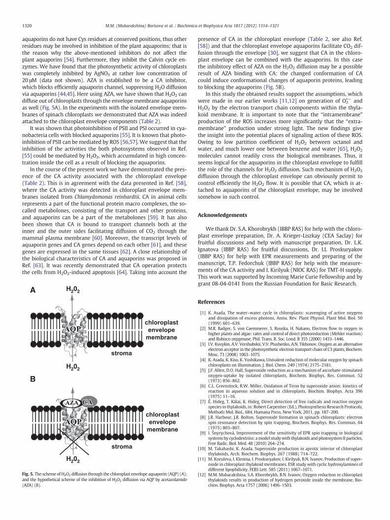
aquaporins do not have Cys residues at conserved positions, thus otherresidues may be involved in inhibition of the plant aquaporins; that isthe reason why the above-mentioned inhibitors do not affect theplant aquaporins [54]. Furthermore, they inhibit the Calvin cycle en-zymes. We have found that the photosynthetic activity of chloroplastswas completely inhibited by AgNO3 at rather low concentration of20 μM (data not shown). AZA is established to be a CA inhibitor,which blocks efficiently aquaporin channel, suppressing H2O diffusionvia aquaporins [44,45]. Here using AZA, we have shown that H2O2 candiffuse out of chloroplasts through the envelope membrane aquaporinsas well (Fig. 5A). In the experiments with the isolated envelope mem-branes of spinach chloroplasts we demonstrated that AZA was indeedattached to the chloroplast envelope components (Table 2).
It was shown that photoinhibition of PSII and PSI occurred in cya-nobacteria cells with blocked aquaporins [55]. It is known that photo-inhibition of PSII can be mediated by ROS [56,57]. We suggest that theinhibition of the activities the both photosystems observed in Ref.[55] could be mediated by H2O2, which accumulated in high concen-tration inside the cell as a result of blocking the aquaporins.
In the course of the present work we have demonstrated the pres-ence of the CA activity associated with the chloroplast envelope(Table 2). This is in agreement with the data presented in Ref. [58],where the CA activity was detected in chloroplast envelope mem-branes isolated from Chlamydomonas reinhardtii. CA in animal cellsrepresents a part of the functional protein macro complexes, the so-called metabolones, consisting of the transport and other proteins,and aquaporins can be a part of the metabolones [59]. It has alsobeen shown that CA is bound to transport channels both at theinner and the outer sides facilitating diffusion of CO2 through themammal plasma membrane [60]. Moreover, the transcript levels ofaquaporin genes and CA genes depend on each other [61], and thesegenes are expressed in the same tissues [62]. A close relationship ofthe biological characteristics of CA and aquaporins was proposed inRef. [63]. It was recently demonstrated that CA operation protectsthe cells from H2O2-induced apoptosis [64]. Taking into account the
H202
H202
H202A
B
Fig. 5. The scheme of H2O2 diffusion through the chloroplast envelope aquaporin (AQP) (A);and the hypothetical scheme of the inhibition of H2O2 diffusion via AQP by acetazolamide(AZA) (B).
presence of CA in the chloroplast envelope (Table 2, see also Ref.[58]) and that the chloroplast envelope aquaporins facilitate CO2 dif-fusion through the envelope [30], we suggest that CA in the chloro-plast envelope can be combined with the aquaporins. In this casethe inhibitory effect of AZA on the H2O2 diffusion may be a possibleresult of AZA binding with CA: the changed conformation of CAcould induce conformational changes of aquaporin proteins, leadingto blocking the aquaporins (Fig. 5B).
In this study the obtained results support the assumptions, whichwere made in our earlier works [11,12] on generation of O2
•− andH2O2 by the electron transport chain components within the thyla-koid membrane. It is important to note that the “intramembrane”production of the ROS increases more significantly that the “extra-membrane” production under strong light. The new findings givethe insight into the potential places of signaling action of these ROS.Owing to low partition coefficient of H2O2 between octanol andwater, and much lower one between benzene and water [65], H2O2
molecules cannot readily cross the biological membranes. Thus, itseems logical for the aquaporins in the chloroplast envelope to fulfillthe role of the channels for H2O2 diffusion. Such mechanism of H2O2
diffusion through the chloroplast envelope can obviously permit tocontrol efficiently the H2O2 flow. It is possible that CA, which is at-tached to aquaporins of the chloroplast envelope, may be involvedsomehow in such control.
Acknowledgements
We thank Dr. S.A. Khorobrykh (IBBP RAS) for help with the chloro-plast envelope preparation, Dr. A. Krieger-Liszkay (CEA Saclay) forfruitful discussions and help with manuscript preparation, Dr. L.K.Ignatova (IBBP RAS) for fruitful discussions, Dr. I.I. Proskuryakov(IBBP RAS) for help with EPR measurements and preparing of themanuscript, T.P. Fedorchuk (IBBP RAS) for help with the measure-ments of the CA activity and I. Kirilyuk (NIOC RAS) for TMT-H supply.This work was supported by Incoming Marie Curie Fellowship and bygrant 08-04-0141 from the Russian Foundation for Basic Research.
References
[1] K. Asada, The water–water cycle in chloroplasts: scavenging of active oxygensand dissipation of excess photons, Annu. Rev. Plant Physiol. Plant Mol. Biol. 50(1999) 601–639.
[2] M.R. Badger, S. von Caemmerer, S. Ruuska, H. Nakano, Electron flow to oxygen inhigher plants and algae: rates and control of direct photoreduction (Mehler reaction)and Rubisco oxygenase, Phil. Trans. R. Soc. Lond. B 355 (2000) 1433–1446.
[3] I.V. Kuvykin, A.V. Vershubskii, V.V. Ptushenko, A.N. Tikhonov, Oxygen as an alternativeelectron acceptor in the photosynthetic electron transport chain of C3 plants, Biochem.Mosc. 73 (2008) 1063–1075.
[4] K. Asada, K. Kiso, K. Yoshikawa, Univalent reduction ofmolecular oxygen by spinachchloroplasts on illumination, J. Biol. Chem. 249 (1974) 2175–2181.
[5] J.F. Allen, D.O. Hall, Superoxide reduction as a mechanism of ascorbate-stimulatedoxygen-uptake by isolated chloroplasts, Biochem. Biophys. Res. Commun. 52(1973) 856–862.
[6] C.L. Greenstock, R.W. Miller, Oxidation of Tiron by superoxide anion: kinetics ofreaction in aqueous solution and in chloroplasts, Biochim. Biophys. Acta 396(1975) 11–16.
[7] É. Hideg, T. Kálai, K. Hideg, Direct detection of free radicals and reactive oxygenspecies in thylakoids, in: Robert Carpentier (Ed.), Photosynthesis Research Protocols,Methods Mol. Biol., 684, Humana Press, New York, 2011, pp. 187–200.
[8] J.R. Harbour, J.R. Bolton, Superoxide formation in spinach chloroplasts: electronspin resonance detection by spin trapping, Biochem. Biophys. Res. Commun. 64(1975) 803–807.
[9] I. Šnyrychová, Improvement of the sensitivity of EPR spin trapping in biologicalsystems by cyclodextrins: amodel studywith thylakoids andphotosystem II particles,Free Radic. Biol. Med. 48 (2010) 264–274.
[10] M. Takahashi, K. Asada, Superoxide production in aprotic interior of chloroplastthylakoids, Arch. Biochem. Biophys. 267 (1988) 714–722.
[11] M. Kozuleva, I. Klenina, I. Proskuryakov, I. Kirilyuk, B.N. Ivanov, Production of super-oxide in chloroplast thylakoid membranes. ESR study with cyclic hydroxylamines ofdifferent lipophilicity, FEBS Lett. 585 (2011) 1067–1071.
[12] M.M. Mubarakshina, S.A. Khorobrykh, B.N. Ivanov, Oxygen reduction in chloroplastthylakoids results in production of hydrogen peroxide inside the membrane, Bio-chim. Biophys. Acta 1757 (2006) 1496–1503.

1321M.M. (Mubarakshina) Borisova et al. / Biochimica et Biophysica Acta 1817 (2012) 1314–1321
[13] I. Slesak, M. Libik, B. Karpinska, S. Karpinski, Z. Miszalski, The role of hydrogenperoxide in regulation of plant metabolism and cellular signalling in responseto environmental stresses, Acta Biochim. Pol. 54 (2007) 39–50.
[14] C.H. Foyer, G. Noctor, Redox regulation in photosynthetic organisms: signaling,acclimation, and practical implications, Antioxid. Redox Sign. 11 (2009) 861–905.
[15] Z. Li, S. Wakao, B.B. Fischer, K.K. Niyogi, Sensing and responding to excess light,Annu. Rev. Plant Biol. 60 (2009) 239–260.
[16] P.M. Mullineaux, ROS in retrograde signalling from the chloroplast to the nucleus,in: L.A. Rio, A. Puppo (Eds.), Reactive Oxygen Species in plant signaling. Signalingand communication in plants, Berlin Heidelberg, Germany, 2009, pp. 221–240.
[17] N. Suzuki, S. Koussevitzky, R.Mittler, G.Miller, ROS and redox signalling in the responseof plants to abiotic stress, Plant Cell Environ. (2011), doi:10.1111/j.1365-3040.2011.02336.x.
[18] I. Gadjev, S. Vanderauwera, T.S. Gechev, C. Laloi, I.N. Minkov, V. Shulaev, K. Apel, D.Inze, R. Mittler, F. Van Breusegem, Transcriptomic footprints disclose specificity ofreactive oxygen species signaling in Arabidopsis, Plant Physiol. 141 (2006) 436–445.
[19] M.Q. Ding, P.C. Hou, X. Shen, M.J. Wang, S.R. Deng, J. Sun, F. Xiao, R. Wang, X. Zhou,C. Lu, D. Zhang, X. Zheng, Z. Hu, S. Chen, Salt-induced expression of genes relatedto Na+/K+ and ROS homeostasis in leaves of salt-resistant and salt-sensitive poplarspecies, Plant Mol. Biol. 73 (2010) 251–269.
[20] U. Bechtold, O. Richard, A. Zamboni, C. Gapper, M. Geisler, B. Pogson, S. Karpinski,P. Mullineaux, Impact of chloroplastic- and extracellular-sourced ROS on highlight-responsive gene expression in Arabidopsis, J. Exp. Bot. 59 (2008) 121–133.
[21] J. Dat, S. Vandenabeele, E. Vranova, M. van Montagu, D. Inze, F. van Breusegem,Dual action of the active oxygen species during plant stress responses, Cell. Mol.Life Sci. 57 (2000) 779–795.
[22] H. Fahnenstich, T.E. Scarpeci, E.M. Valle, U.I. Fliigge, V.G. Maurino, Generation ofhydrogen peroxide in chloroplasts of Arabidopsis overexpressing glycolate oxidaseas an inducible system to study oxidative stress, Plant Physiol. 148 (2008) 719–729.
[23] S. Karpinski, H. Reynolds, B. Karpinska, G. Wingsle, G. Creissen, P. Mullineaux,Systemic signaling and acclimation in response to excess excitation energy inArabidopsis, Science 284 (1999) 654–657.
[24] M.J. Fryer, L. Ball, K. Oxborough, S. Karpinski, P.M. Mullineaux, N.R. Baker, Controlof ascorbate peroxidase 2 expression by hydrogen peroxide and leaf water statusduring excess light stress reveals a functional organization of Arabidopsis leaves,Plant J. 33 (2003) 691–705.
[25] Y. Yabuta, T. Maruta, K. Yoshimura, T. Ishikawa, S. Shigeoka, Two distinct redoxsignaling pathways for cytosolic APX induction under photooxidative stress,Plant Cell Physiol. 45 (2004) 1586–1594.
[26] R. Desikan, S. Mackerness, J.T. Hancock, S.J. Neill, Regulation of the Arabidopsistranscriptome by oxidative stress, Plant Physiol. 127 (2001) 159–172.
[27] J.J. Grant, B.W. Yun, G.J. Loake, Oxidative burst and cognate redox signallingreported by luciferase imaging: identification of a signal network that functionsindependently of ethylene, SA and Me-JA but is dependent on MAPKK activity,Plant J. 24 (2000) 569–582.
[28] M.M.Mubarakshina, B.N. Ivanov, I.A. Naydov,W.Hillier,M.R. Badger, A. Krieger-Liszkay,Production and diffusion of chloroplastic H2O2 and its implication to signaling, J. Exp.Bot. 61 (2010) 3577–3587.
[29] C. Maurel, L. Verdoucq, D.-T. Luu, V. Santoni, Plant aquaporins: membrane chan-nels with multiple integrated functions, Annu. Rev. Plant Biol. 59 (2008)595–624.
[30] N. Uehlein, B. Otto, D.T. Hanson, M. Fischer, N. McDowell, R. Kaldenhoff, Functionof Nicotiana tabacum aquaporins as chloroplast gas pores challenges the conceptof membrane CO2 permeability, Plant Cell 20 (2008) 648–665.
[31] G.P. Bienert, A.L.B. Møller, K.A. Kristiansen, A. Schulz, I.M. Møller, J.K. Schjoerring,T.P. Jahn, Specific aquaporins facilitate the diffusion of hydrogen peroxide acrossmembranes, J. Biol. Chem. 282 (2007) 1183–1192.
[32] S.A. Khorobrykh, B.N. Ivanov, Oxygen reduction in a plastoquinone pool of isolatedpea thylakoids, Photosynth. Res. 71 (2002) 209–219.
[33] J.E. Mullet, N.H. Chua, In vitro reconstitution of synthesis, uptake and assembly ofcytoplasmically synthesised chloroplast proteins, Methods Enzymol. 97 (1983)502–509.
[34] U. Heber, K.A. Santarius, Direct and indirect transfer of ATP and ADD across thechloroplast envelope, Z. Naturforsch. B 25 (1970) 718–728.
[35] R. Douce, J. Joyard, Purification of the chloroplast envelope, in: M. Edelman, R.B.Hallick, N.-H. Chua (Eds.), Methods in Chloroplast Molecular Biology, Elsivier Bio-medical Press, New York, 1982, pp. 239–256.
[36] S.I. Dikalov, I.A. Kirilyuk, M. Voinov, I.A. Grigor'ev, EPR detection of cellular andmitochondrial superoxide using cyclic hydroxylamines, Free. Radic. Res. 45(2011) 417–430.
[37] O.H. Lowry, N.J. Rosebrough, A.L. Fan, R.L. Randall, Protein measurement with Folinphenol reagent, J. Biol. Chem. 193 (1951) 256–266.
[38] K. Ogawa, S. Kanematsu, K. Takabe, K. Asada, Attachment of CuZn-superoxide dis-mutase to thylakoid membranes at the site of superoxide generation (PSI) inspinach chloroplasts: detection by immuno-gold labeling after rapid freezingand substitution method, Plant Cell Physiol. 36 (1995) 557–565.
[39] M. Pilon, K. Ravet,W. Tapken, The biogenesis and physiological function of chloroplastsuperoxide dismutases, Biochim. Biophys. Acta 1807 (2011) 989–998.
[40] M.M. Mubarakshina, B.N. Ivanov, The production and scavenging of reactive oxygenspecies in the plastoquinone pool of chloroplast thylakoid membranes, Physiol.Plant. 140 (2010) 103–110.
[41] E.G. Janzen, Y.Y.Wang, R.V. Shetty, Spin trappingwith.alpha.-pyridyl 1-oxide N-tert-butyl nitrones in aqueous solutions. A unique electron spin resonance spectrum forthe hydroxyl radical adduct, J. Am. Chem. Soc. 100 (1978) 2923–2925.
[42] G.W. Brudvig, S.T. Worland, K. Sauer, Procedure for rapid isolation of photosyn-thetic reaction centers using cytochrome c affinity chromatography, Proc. NatlAcad. Sci. USA 80 (1983) 683–686.
[43] J. Kruk, M. Jemioła-Rzemińska, K. Strzałka, Cytochrome c is reduced mainly byplastoquinol and not by superoxide in thylakoid membranes at low and mediumlight intensities: its specific interaction with thylakoidmembrane lipids, Biochem. J.375 (2003) 215–220.
[44] J. Gao, X.Wang, Y. Chang, J. Zhang, Q. Song,H. Yu, X. Li, Acetazolamide inhibits osmoticwater permeability by interaction with aquaporin-1, Anal. Biochem. 350 (2006)165–170.
[45] R. Haddoub,M. Rutzler, A. Robin, S.L. Flitsch, Design, synthesis and assaying of potentialaquaporin inhibitors, in: E. Beitz (Ed.), Handbook of Experimental Pharmacology:Aquaporins, Springer, Berlin, Germany, 2009, pp. 385–402.
[46] V.J. Huber, M. Tsujita, M. Yamazaki, K. Sakimura, T. Nakada, Identification of aryl-sulfonamides as Aquaporin 4 inhibitors, Bioorg. Med. Chem. Lett. 17 (2007)1270–1273.
[47] L.K. Ignatova, O.V. Moskvin, B.N. Ivanov, Effects of carbonic anhydrase inhibitorson proton exchange and photosynthesis in pea protoplasts, Russ. J. Plant Physiol.48 (2001) 545–550.
[48] B.N. Ivanov, The competition betweenmethyl viologen and monodehydroascorbateradical as electron acceptors in spinach thylakoids and intact chloroplasts, Free.Radic. Res. 33 (2000) 217–227.
[49] T. Mann, D. Keilin, Sulphanilamide as a specific inhibitor of carbonic anhydrase,Nature 146 (1940) 164–165.
[50] J. Kruk, M. Jemiola-Rzeminska, K. Burda, G. Schmid, K. Strzalka, Scavenging ofsuperoxide generated in photosystem I by plastoquinol and other prenyllipidsin thylakoid membranes, Biochemistry 42 (2003) 8501–8505.
[51] P.R. Rich, R. Harper, Partition coefficients of quinones and hydroquinones andtheir relation to biochemical reactivity, FEBS Lett. 269 (1990) 139–144.
[52] M.A. Kozuleva, B.N. Ivanov, Evaluation of the participation of ferredoxin in oxygenreduction in the photosynthetic electron transport chain of isolated pea thylakoids,Photosynth. Res. 105 (2010) 51–61.
[53] S. Lemeille, A. Willig, N. Depège-Fargeix, C. Delessert, R. Bassi, J.D. Rochaix, Analysisof the chloroplast protein kinase Stt7 during state transitions, PLoS Biology 3(2009) e45.
[54] M.J. Daniels, F. Chaumont, T.E. Mirkov, M.J. Chrispeels, Characterization of a newvacuolar membrane aquaporin sensitive to mercury at a unique site, Plant Cell8 (1996) 587–599.
[55] S.I. Allakhverdiev, A. Sakamoto, Y. Nishiyama, N.Murata, Inactivation of PhotosystemsI and II in response to osmotic stress in Synechococcus. Contribution ofwater channels,Plant Physiol. 122 (2000) 1201–1208.
[56] Y. Nishiyama, S.I. Allakhverdiev, N. Murata, Protein synthesis is the primary targetof reactive oxygen species in the photoinhibition of photosystem II, Physiol. Plant.142 (2011) 35–46.
[57] A. Krieger-Liszkay, P.B. Kós, E. Hideg, Superoxide anion radicals generated bymethylviologen in Photosystem I damage Photosystem II, Physiol. Plant. 142(2011) 17–25.
[58] A. Villarejo, N. Rolland, F. Martínez, D.F. Sültemeyer, A new chloroplast envelopecarbonic anhydrase activity is induced during acclimation to low inorganic carbonconcentrations in Chlamydomonas reinhardtii, Planta 213 (2001) 286–295.
[59] L.J. Bruce, R. Beckmann, M.L. Ribeiro, L.L. Peters, J.A. Chasis, J. Delaunay, N.Mohandas, D.J. Anstee, Mi.J.A. Tanner, A band 3-based macrocomplex of integraland peripheral proteins in the RBC membrane, Blood 101 (2003) 4180–4188.
[60] D. Sterling, B.V. Alvarez, J.R. Casey, The extracellular component of a transportmetabolon, J. Biol. Chem. 277 (2002) 25239–25246.
[61] J. Takenawa, Y. Kaneko, M. Kishishita, H. Higashitsuji, H. Nishiyama, T. Terachi, Y.Arai, O. Yoshida,M. Fukumoto, J. Fujita, Transcript levels of aquaporin 1 and carbonicanhydrase IV as predictive indicators for prognosis of renal cell carcinoma patientsafter nephrectomy, Int. J. Cancer 79 (1998) 1–7.
[62] W.S. Sly, P.Y. Hu, Human carbonic anhydrases and carbonic anhydrase deficiencies,Annu. Rev. Biochem. 64 (1995) 375–401.
[63] Y. Xiang, B. Ma, T. Li, J.-w. Gao, H.-m. Yu, X.-j. Li, Acetazolamide inhibits aquaporin-1protein expression and angiogenesis1, Acta Pharmacol. Sin. 25 (2004) 812–816.
[64] S.R. Raisanen, P. Lehenkari, M. Tasanen, P. Rahkila, P.L. Harkonen, H.K. Vaananen,Carbonic anhydrase III protects cells from hydrogen peroxide-induced apoptosis,FASEB J. 13 (1999) 513–522.
[65] A. Leo, C. Hansch, D. Elkins, Partition coefficients and their uses, Chem. Rev. 71(1971) 525–616.
Copyright © 2022 FDOKUMEN